EP1564554A1 - Method for the detection of early B cell populations in vaccine development - Google Patents
Method for the detection of early B cell populations in vaccine development Download PDFInfo
- Publication number
- EP1564554A1 EP1564554A1 EP04075439A EP04075439A EP1564554A1 EP 1564554 A1 EP1564554 A1 EP 1564554A1 EP 04075439 A EP04075439 A EP 04075439A EP 04075439 A EP04075439 A EP 04075439A EP 1564554 A1 EP1564554 A1 EP 1564554A1
- Authority
- EP
- European Patent Office
- Prior art keywords
- antigen
- binding
- label
- cells
- cell
- Prior art date
- Legal status (The legal status is an assumption and is not a legal conclusion. Google has not performed a legal analysis and makes no representation as to the accuracy of the status listed.)
- Withdrawn
Links
Images


Classifications
-
- G—PHYSICS
- G01—MEASURING; TESTING
- G01N—INVESTIGATING OR ANALYSING MATERIALS BY DETERMINING THEIR CHEMICAL OR PHYSICAL PROPERTIES
- G01N33/00—Investigating or analysing materials by specific methods not covered by groups G01N1/00 - G01N31/00
- G01N33/48—Biological material, e.g. blood, urine; Haemocytometers
- G01N33/50—Chemical analysis of biological material, e.g. blood, urine; Testing involving biospecific ligand binding methods; Immunological testing
- G01N33/53—Immunoassay; Biospecific binding assay; Materials therefor
- G01N33/569—Immunoassay; Biospecific binding assay; Materials therefor for microorganisms, e.g. protozoa, bacteria, viruses
- G01N33/56966—Animal cells
- G01N33/56972—White blood cells
-
- A—HUMAN NECESSITIES
- A61—MEDICAL OR VETERINARY SCIENCE; HYGIENE
- A61P—SPECIFIC THERAPEUTIC ACTIVITY OF CHEMICAL COMPOUNDS OR MEDICINAL PREPARATIONS
- A61P1/00—Drugs for disorders of the alimentary tract or the digestive system
- A61P1/04—Drugs for disorders of the alimentary tract or the digestive system for ulcers, gastritis or reflux esophagitis, e.g. antacids, inhibitors of acid secretion, mucosal protectants
-
- A—HUMAN NECESSITIES
- A61—MEDICAL OR VETERINARY SCIENCE; HYGIENE
- A61P—SPECIFIC THERAPEUTIC ACTIVITY OF CHEMICAL COMPOUNDS OR MEDICINAL PREPARATIONS
- A61P1/00—Drugs for disorders of the alimentary tract or the digestive system
- A61P1/16—Drugs for disorders of the alimentary tract or the digestive system for liver or gallbladder disorders, e.g. hepatoprotective agents, cholagogues, litholytics
-
- A—HUMAN NECESSITIES
- A61—MEDICAL OR VETERINARY SCIENCE; HYGIENE
- A61P—SPECIFIC THERAPEUTIC ACTIVITY OF CHEMICAL COMPOUNDS OR MEDICINAL PREPARATIONS
- A61P13/00—Drugs for disorders of the urinary system
- A61P13/08—Drugs for disorders of the urinary system of the prostate
-
- A—HUMAN NECESSITIES
- A61—MEDICAL OR VETERINARY SCIENCE; HYGIENE
- A61P—SPECIFIC THERAPEUTIC ACTIVITY OF CHEMICAL COMPOUNDS OR MEDICINAL PREPARATIONS
- A61P13/00—Drugs for disorders of the urinary system
- A61P13/10—Drugs for disorders of the urinary system of the bladder
-
- A—HUMAN NECESSITIES
- A61—MEDICAL OR VETERINARY SCIENCE; HYGIENE
- A61P—SPECIFIC THERAPEUTIC ACTIVITY OF CHEMICAL COMPOUNDS OR MEDICINAL PREPARATIONS
- A61P15/00—Drugs for genital or sexual disorders; Contraceptives
- A61P15/16—Masculine contraceptives
-
- A—HUMAN NECESSITIES
- A61—MEDICAL OR VETERINARY SCIENCE; HYGIENE
- A61P—SPECIFIC THERAPEUTIC ACTIVITY OF CHEMICAL COMPOUNDS OR MEDICINAL PREPARATIONS
- A61P15/00—Drugs for genital or sexual disorders; Contraceptives
- A61P15/18—Feminine contraceptives
-
- A—HUMAN NECESSITIES
- A61—MEDICAL OR VETERINARY SCIENCE; HYGIENE
- A61P—SPECIFIC THERAPEUTIC ACTIVITY OF CHEMICAL COMPOUNDS OR MEDICINAL PREPARATIONS
- A61P19/00—Drugs for skeletal disorders
-
- A—HUMAN NECESSITIES
- A61—MEDICAL OR VETERINARY SCIENCE; HYGIENE
- A61P—SPECIFIC THERAPEUTIC ACTIVITY OF CHEMICAL COMPOUNDS OR MEDICINAL PREPARATIONS
- A61P3/00—Drugs for disorders of the metabolism
- A61P3/12—Drugs for disorders of the metabolism for electrolyte homeostasis
- A61P3/14—Drugs for disorders of the metabolism for electrolyte homeostasis for calcium homeostasis
-
- A—HUMAN NECESSITIES
- A61—MEDICAL OR VETERINARY SCIENCE; HYGIENE
- A61P—SPECIFIC THERAPEUTIC ACTIVITY OF CHEMICAL COMPOUNDS OR MEDICINAL PREPARATIONS
- A61P35/00—Antineoplastic agents
-
- G—PHYSICS
- G01—MEASURING; TESTING
- G01N—INVESTIGATING OR ANALYSING MATERIALS BY DETERMINING THEIR CHEMICAL OR PHYSICAL PROPERTIES
- G01N33/00—Investigating or analysing materials by specific methods not covered by groups G01N1/00 - G01N31/00
- G01N33/48—Biological material, e.g. blood, urine; Haemocytometers
- G01N33/50—Chemical analysis of biological material, e.g. blood, urine; Testing involving biospecific ligand binding methods; Immunological testing
- G01N33/53—Immunoassay; Biospecific binding assay; Materials therefor
- G01N33/543—Immunoassay; Biospecific binding assay; Materials therefor with an insoluble carrier for immobilising immunochemicals
- G01N33/54306—Solid-phase reaction mechanisms
-
- G—PHYSICS
- G01—MEASURING; TESTING
- G01N—INVESTIGATING OR ANALYSING MATERIALS BY DETERMINING THEIR CHEMICAL OR PHYSICAL PROPERTIES
- G01N33/00—Investigating or analysing materials by specific methods not covered by groups G01N1/00 - G01N31/00
- G01N33/48—Biological material, e.g. blood, urine; Haemocytometers
- G01N33/50—Chemical analysis of biological material, e.g. blood, urine; Testing involving biospecific ligand binding methods; Immunological testing
- G01N33/53—Immunoassay; Biospecific binding assay; Materials therefor
- G01N33/543—Immunoassay; Biospecific binding assay; Materials therefor with an insoluble carrier for immobilising immunochemicals
- G01N33/54313—Immunoassay; Biospecific binding assay; Materials therefor with an insoluble carrier for immobilising immunochemicals the carrier being characterised by its particulate form
-
- G—PHYSICS
- G01—MEASURING; TESTING
- G01N—INVESTIGATING OR ANALYSING MATERIALS BY DETERMINING THEIR CHEMICAL OR PHYSICAL PROPERTIES
- G01N33/00—Investigating or analysing materials by specific methods not covered by groups G01N1/00 - G01N31/00
- G01N33/48—Biological material, e.g. blood, urine; Haemocytometers
- G01N33/50—Chemical analysis of biological material, e.g. blood, urine; Testing involving biospecific ligand binding methods; Immunological testing
- G01N33/53—Immunoassay; Biospecific binding assay; Materials therefor
- G01N33/543—Immunoassay; Biospecific binding assay; Materials therefor with an insoluble carrier for immobilising immunochemicals
- G01N33/54393—Improving reaction conditions or stability, e.g. by coating or irradiation of surface, by reduction of non-specific binding, by promotion of specific binding
Definitions
- the invention relates to the field of immunology and vaccine development.
- the invention further relates to the detection of early B cell responses after immunisation with specific antigens.
- Vaccines have proven to be extraordinary effective means to improve public health (Jackson et al ., 2002). However, conventional vaccine development can be hampered by technical problems in pathogen inactivation as well as high costs.
- peptide-based synthetic vaccines are valid alternatives (Jackson et al ., 2002). Peptide vaccination has been successfully used to prevent infectious diseases (Monzavi-Karbassi et al ,. 2002),and to treat tumours (Ribas et al ,. 2003; Noguchi et al ,. 2003), amyloidosis (Nicoll et al ,. 2003) and autoimmune disease (Liu et al ,. 2002).
- peptide vaccine candidates must elicit an immune response and cause clonal expansion of specific T cells and/or B cells.
- Said immune response is elicited by epitopes that are recognized by T cells and/or B cells and initiate a proliferative reaction of antigen specific T and B cells.
- the number of specific immune B cells increases by clonal expansion until high levels of effector cells are reached after three or four weeks after vaccination. The levels of these effector cells after clonal expansion are high enough for conventional methods to detect the effector cells.
- B cell response against an antigen is usually measured by measuring the level of antigen specific antibodies in circulating blood.
- B cells can be detected specifically by direct or indirect staining methods, usually achieved by coupling antigens or antibodies to a signalling molecule, like for example a fluorescent stain (fluorescein isothyocyanate, FITC, or Rhodamine), or a chemical, radioactive, or enzymatic signal or another marker substance that enables a skilled person to detect binding of the marker substance to a B cell.
- a signalling molecule like for example a fluorescent stain (fluorescein isothyocyanate, FITC, or Rhodamine), or a chemical, radioactive, or enzymatic signal or another marker substance that enables a skilled person to detect binding of the marker substance to a B cell.
- FITC fluorescein isothyocyanate
- Rhodamine a chemical, radioactive, or enzymatic signal or another marker substance that enables a skilled person to detect binding of the marker substance to a B cell.
- Many methods have been developed to increase the sensitivity of B cell detection
- Said clonal expansion process increases the numbers of B cells to a level where either B-cells or antibodies produced by said B cells, can be detected.
- flow cytometry was developed and further adapted to detect antigen specific B cells (McHeyzer-Williams et al. , 1993).
- investigators have employed and combined two different approaches to study antigen specific B cells.
- One approach is hybridoma technique to capture immune reactive B cells and the other approach is to study genetically manipulated animals with an enhanced number of antigen specific B cells. Both methods are too insensitive to detect the low levels of emerging antigen specific B cells after vaccination before clonal expansion has been completed.
- One method developed for the detection of B cells has been adapted from the staining of T cells.
- the sensitivity of detecting T cells was enhanced by incubating the cells with a tetrameric peptide-MHC molecule that binds to T cell receptors specific for said peptide-MHC complex (Altman et al. , 1996; Stetson et al. , 2002).
- the above-mentioned method has been adapted for the detection of antigen-specific B cells (Newman et al. , 2003)
- the sensitivity of the normal procedures was only increased tenfold. This is still far too insensitive for effective detection of emerging low- frequency antigen specific B cells, shortly after vaccination.
- This "single epitope multiple staining procedure" reduced the background staining, thereby enabling an increase in the sensitivity of flow cytometry by 1 or 2 orders of magnitude enabling the detection of high affinity antigen-specific B cells in vivo.
- This increase in sensitivity may enable the detection of transgenic B cells or B cells after clonal expansion is completed, but it is still too insensitive for the detection of low frequency early B cells within 7 days after immunization.
- the method is complicated and the authors warn the public that it is critical to note that several requirements must be met before this strategy will be effective in increasing the sensitivity of flow cytometry.
- the above-described method of Townsend et al. used as an antigen a foreign antigen for mice namely chicken egg white lysozyme.
- An affinity-binding assay is a test for detecting or measuring binding of two or more substances that have a certain affinity for each other. This can for example be the binding between an antibody and an antigen, or of an enzyme to its substrate, or of a hormone to its hormone receptor.
- affinity binding between the antigen specific B cell receptor to an antigen.
- said affinity binding may be detected and measured by various methods. As an example, we disclose the detection of affinity binding by an adapted flow cytometry method.
- the present application discloses a binding assay and a test method using this binding assay in which a particle or a cell comprising at least four target molecules, for example membrane bound antibodies or receptors or antigen-specific B cell receptors (BCRs), can be detected by contacting said particle or cell with at least two binding molecules that have a different label.
- Detecting of particles or cells by flow cytometry is based on staining said particle or cell with a fluorescent label and detecting labelled particles or cells by registering fluorescence signals using the fluorescence detectors.
- Each fluorescent label has its own characteristics for intensity and the level of background staining.
- This application discloses that staining with a molecule comprising a multiple form of an antigen, in which at least two, or three, preferably four, or more, binding epitopes of essentially the same antigen are present and exposed, increases the intensity of the staining.
- a further increase in sensitivity is achieved by combining the staining of above described multiple forms of an antigen with two different fluorescent labels.
- two quantities of essentially the same multiple antigen molecules are associated with two different labels.
- labels are selected that differ in their fluorescent signal, like for example R-phycoerythrin (PE)-labelled neutravidin and allophycocyanin (APC) coupled to streptavidin.
- PE R-phycoerythrin
- APC allophycocyanin
- the cells By contacting the cells with said both differently labelled antigenic molecules, the cells bind both molecules and combine the two different fluorescent labels on the surface.
- This contacting can for example be done in consecutive steps, for example by first incubating the cells with the first fluorescent-labelled antigen molecule in a sub-saturating amount, followed by incubation with the second fluorescent-labelled antigen in a more saturating or a saturated amount.
- Another example of a way of staining particles or cells with both fluorescent-labelled antigen molecules is by mixing the two differently labelled antigen molecules at an approximately equimolar amount and incubating the particles or cells with the mixture. Because the chance of aspecific binding of two separate molecules on the same particle is very small, the specificity of the method is increased.
- binding molecules The antigen molecules that bind to said target molecules are in this application named binding molecules.
- Said binding molecules may comprise one or more peptides or proteins or fragments thereof.
- said binding molecules may comprise a staining molecule.
- a particle in this application can be a virus or a microorganism, or a part of a cell or yeast.
- the size of said particle is at least about the size of a virus and at most about the size of a thousand cells, more preferably about the size of a hundred cells, even more preferably about the size of 10 cells, even more preferably about the size of one cell.
- label means in this context preferably a fluorescent staining label.
- the present invention discloses an affinity-binding assay comprising a particle having at least four copies of a target molecule and at least two binding molecules specific for said target molecule, wherein a first of said binding molecules is associated with a first label and a second of said binding molecules is associated with a second label, wherein a particle having said first label and a particle having said second label are distinguishable from a particle having both said first and said second label, wherein said first and said second binding molecule each comprise at least two binding regions specific for said target molecule.
- Said particle can also be a cell, therefore the invention also discloses the above-described assay, wherein said particle comprises a cell.
- Said cell may be a living cell or said cell may be treated with a fixative such as for example fixatives that are normally used in flow cytometry, like for example formalin, acetone, alcohol, or glutaraldehyde.
- the method of the invention is very sensitive, this is the first time that such a method is capable of detecting very low frequency antigen specific B cells, like for example very early immune B cells. These very early immune cells are only present at very low frequency because these cells have not yet accomplished the complete clonal expansion process. Therefore, the present invention discloses an affinity-binding assay as described above, wherein said cell comprises an activated immune cell in the clonal expansion phase of a primary immune response. In mice for example, this activation takes place within hours after first or second contact with an antigen and results in antibody levels in peripheral blood within three to four weeks.
- the method is especially suitable for the detection of very low frequency B cells and saves a lot of time because one does not have to wait any more for the antibody response to evolve. This shortens the animal phase for each peptide from at least 3 to 4 weeks to one week. Furthermore, the detection of very low frequency B cells is also very suitable for the detection of memory B cells. This may be important for assessing the immune status against certain diseases.
- a B cell in this application means a B cell in any state of activation or differentiation, including for example antigen specific precursor B cells, antibody secreting cells, plasma cells and memory B cells. Therefore, the present invention discloses in a preferred embodiment the assay as described above, wherein said cell comprises a B cell.
- B cells as disclosed in this application enable a person skilled in the art to measure the early immune response against immunization or infection, e.g., proliferation and differentiation of naive B cells into antibody secreting cells, memory B cells and plasma cells.
- the present invention discloses an affinity-binding assay as described above, wherein said particle having said first and said second label increases the sensitivity of the affinity-binding assay.
- the binding properties of said multiple antigen molecules are increased with the number of exposed antigenic sites on the molecule.
- tetramer molecules comprising four antigenic sites have been tested as an example.
- other numbers of antigens repeated in the same molecule may also have the desired effect. Therefore, the present invention discloses an affinity-binding assay as described above, wherein said first and said second binding molecule each comprise at least two binding regions specific for said target molecule. Preferably said number of binding regions is four.
- the binding may be increased even further if the antigenic sites or epitopes of said target molecule are essentially identical epitopes. Therefore, the present invention discloses an affinity-binding assay as described above, wherein said at least two binding regions are essentially identical.
- a big advantage of the assay is that the binding molecules of the detection assay may represent an epitope of a native protein, because in this way, immunization with altered antigens of said native protein can be monitored for the cross-specificity for said native protein.
- a native protein is in this application a protein as it is present in nature. Therefore, the present invention discloses an affinity-binding assay as described above, wherein said binding molecule represents an epitope of a native protein.
- the present invention is very suited for detecting low frequency B cells.
- B cells when activated by contact with an antigen, expose during their clonal expansion an antigen-specific B cell receptor or BCR on their cell surface.
- Suitable B cell receptors are for example the well-known cell bound antibody-like molecules that are inserted in the wall of the B cell with their Fc fragment and which are specifically recognising antigen with the variable region, said antigen being the antigen that was used to activate the B cells (Kouskoff et al ., 2000).
- said BCRs are the target molecules with which the binding molecules bind. Therefore, the present invention discloses an affinity-binding assay as described above, wherein said target molecule comprises a BCR.
- the present invention discloses an affinity-binding assay as described above, wherein said target molecule comprises a variable region of said BCR.
- the assay is performed with a set of binding molecules detecting the target molecule.
- Said binding molecule preferably comprising tetramer molecules. Therefore, the present invention in another embodiment discloses a composition comprising a first multiple binding molecule associated with a first label and a second multiple binding molecule associated with a second label, wherein the signal obtained from said first label and said second label is distinguishable from the combined signal of said first and second label, wherein each binding molecule comprises at least four binding regions specific for essentially the same target molecule, preferably for essentially the same epitope on said target molecule.
- a range of modified antigens can be produced, for example by the production of synthetic peptides. Changes in the peptides may increase the immunogenicity, and by testing said peptides, new and improved antigens may be detected and selected by the assay and the method of the invention. A range of changes made to a peptide in order to changes the antigenicity of said peptide results in various forms of said peptide, which is also called a collection of peptides.
- Antigen-specific B cell detection as disclosed in this application expedites the evaluation of vaccine immunogenicity, and enables analysis of the fine-specificity of the response.
- the present invention discloses a method for selecting a synthetic antigen from a collection of at least two antigens comprising using the composition as described above for detecting in an affinity-binding assay for immune cells specific for said synthetic antigen, clonal expansion of antigen specific immune cells in samples of cells obtained from a mammal immunized with an antigen of said collection of antigens, and comparing said clonal expansion with the clonal expansion of another mammal immunized with another antigen form said collection and selecting from said collection an antigen with which clonal expansion was more extensive than clonal expansion observed with at least one other antigen from said collection.
- a more extensive clonal expansion means in this application that the numbers of specific B cells increase faster and to higher levels compared to clonal expansions as reaction to other antigens.
- increased clonal expansion at 7 days is indicative for an increased clonal expansion at 14 days and usually for a high level of antibodies later in the immune response.
- this increase may show later or earlier in the immune response but be highly indicative of a more extensive clonal expansion, if compared to the clonal expansion in other rats or other animals immunized with another antigen of said collection of antigens.
- the above-described method may use any of the affinity-binding assays as are described above. Therefore, the present invention discloses a method for selecting a synthetic antigen as described above, wherein said affinity-binding assay comprises any assay as described herein.
- said synthetic peptides to be a good vaccine against a native protein
- cross reactivity of the antibodies directed against said synthetic peptide antigen with the native protein from which said peptides were derived is highly appreciated. Therefore, the method as described above is preferably used wherein said at least two binding molecules comprise as a binding part for said target molecule a synthetic antigen of said collection of antigens, or a native antigen or homologue of said antigen.
- a native antigen is in this application an antigen as it occurs in a protein in nature.
- a homologue of an antigen is a substance showing the same antigenic characteristics in kind, not necessarily in amount. Said homologue may comprise a natural peptide or protein or a synthetic peptide or a part thereof. Therefore, direct B cell staining procedures enable the assessment of specific B cell receptor cross-reactivity between peptide vaccines and the native antigens from which they are derived.
- the invention also discloses the method as described above, further comprising selecting an antigen for which clonal expansion of antigen-specific immune cells more extensive than the clonal expansion with at least two other antigens.
- B cells specific for a certain antigen can be selected and isolated. Said isolated B cells may also be further tested for the specificity and avidity of their binding to the native protein, also called the native antigen or homologue of said antigen. Therefore, the invention teaches a method as described above further comprising evaluating whether said antigen-specific immune cells are specific for a native antigen or homologue of said antigen.
- the invention discloses a method for an affinity-binding assay for selecting antigen specific immune cells, comprising: (a) contacting a cell having at least four copies of a target molecule with at least two binding molecule, preferably tetrameric binding molecules, said binding molecules specific for said target molecule, wherein a first of said binding molecules is associated with a first label and a second of said binding molecules is associated with a second label and; (b) detecting cells staining with each label and; (c) selecting cells binding both labels.
- the present invention teaches with above described methods a new way to test and select a synthetic antigen from a collection of antigens inducing a cross-reactive immune response with a native protein. This is an important improvement of the technical possibilities in this field and opens the possibility for new methods of selection and thus to other products. Therefore, the invention discloses a synthetic antigen, inducing a cross-reactive immune response with a native protein, selectable with a method as described above for use as a vaccine.
- said peptide motif can be made into dimer or trimeric or multimeric peptides wherein two or three or more peptides are linked directly to each other or through a spacer molecule. Combining two identical peptides into a dimer peptide further increases the immunogenic properties of said peptide. Therefore, the present invention discloses an immunogenic peptide, obtainable by above described method, wherein said peptide comprises a dimer peptide.
- the peptides can be linked by sulphur bridges or by a linkage molecule.
- Another method of increasing the immunogenicity of a peptide is by combining a peptide in a tandem peptide and/or a tandem dimer peptide. From WO 96/40755 it is known that the tandem-dimer principle applied to a variant of the GnRH-I molecule resulted in a vaccine that was highly effective in low doses and with a mild adjuvant. Therefore, the present application discloses a method for selecting an immunogenic peptide derived from an antigen comprising detecting clonal expansion of antigen specific immune cells as described above, wherein said peptide comprises a tandem dimer peptide.
- the antigenic properties of peptides provided herein are further optimised using a variety of techniques, such as replacement-net mapping, allowing detecting peptides with improved characteristics.
- one such peptide may be a peptide that binds to an antibody directed against an epitope, thereby mimicking the immunogenic properties of said epitope.
- Such a peptide is called a mimotope.
- This method has been disclosed in patent application WO 00/29851. Therefore, the present application also discloses an immunogenic peptide derived from a protein, comprising a mimotope of said protein.
- Amino acid substitution of at least one amino acid by a D-amino acid and/or a L-alanine as described in WO 02/22659 also increases the immunogenicity of a peptide. Therefore, the present invention discloses an immunogenic peptide derived from a protein, wherein at least one amino acid is substituted by a D-amino acid and/or a L-alanine.
- GnRH gonadotrophin releasing hormone
- Gonadotrophin releasing hormone is a decapeptide, produced by the hypothalamus. The GnRH travels through portal circulation to the pituitary to stimulate the release of gonadotrophins FSH and LH (Talwar, 1999).
- Anti-GnRH immunization blocks the fertility of both male and female animals (Meloen et al ., 2001), and GnRH vaccines have applications in prostate and in female cancer (Talwar, 1999; Fuerst et al. ,1997).
- mice were immunized with GnRH-like peptides conjugated to ovalbumin (OVA).
- OVA ovalbumin
- GnRH-monomer pEHWSYGLRPGC
- GnRH-tandem pEHWSYGLRPGQHWSYGLRPGC
- TDK GnRH tandem dimer
- the GnRH tandem and TDK peptide conjugated to a carrier protein have shown to be highly immunogenic, resulting in GnRH neutralizing antibodies and a full biological effect in all treated animals (Meloen et al., 1994; Oonk et a . 1998). Therefore, the present application provides an immunogenic peptide obtainable by the above-described method, wherein said peptide is derived from GnRH.
- Gastrin Another example of a peptide hormone for which peptides have been prepared according to the sequence of said protein and which are tested in abovementioned method is Gastrin. Therefore, the application discloses an immunogenic peptide derived from a peptide hormone and obtainable by above- mentioned methods, wherein said peptide is derived from Gastrin.
- Gastrin is a peptide hormone that is important in the regulation of acid secretion and the growth of both normal and malignant gastrointestinal epithelium (Jensen et al. , 2002).
- Tumor cells can respond both to circulating endocrine gastrin (Watson et al ., 1989) and locally produced gastrin, which acts in an autocrine or paracrine manner (Hoosein et al. , 1990). Effective inhibition of gastrin may be beneficial as a therapy for colorectal cancer, gastric cancer and pancreatic cancer, as gastrine acts as a growth factor for these tumours.
- Gastrine receptor antagonists are not fully effective because several receptor subtypes are involved in the action of gastrin and high amounts are required to displace gastrin. Neutralisation of gastrin may circumvent this problem.
- Peptides derived from gastrine can be used for vaccination against gastrin (amino acid sequence pEGPWLEEEEEAYGWMDF).
- a Cysteine is included at the N- or C-terminal end for conjugation purposes.
- the peptides can also be applied in tandem or tandem dimer formulation with a Cysteine added at the N- or C-terminus or a cysteine between the two gastrin sequences in the tandem, as in pEGPWLEEEECQGPWLEEEE.
- L-Lysines or D-Lysines are introduced in the tandem formulation to allow dimerisation of these peptides via the cysteine and conjugation via the L- or D-Lysine, like in in which 'k' represents a D-lysine residue.
- These peptides were tested as a vaccine in mice and at 7 and 14 days after immunization specific B cells were detected with the method of the invention using tetramer Gastrin peptide molecules.
- the following peptide was used to form a tetramer: pEGPWLEEEEEAYGWMDFK($)# , wherein # is amide; pE is pyroGlutamine; and K($) is biotinylated Lysin
- the present invention discloses an immunogenic peptide obtainable by any of the above-described methods wherein said peptide is derived from Gastrin.
- hCG- ⁇ Human Chorionic Gonadotropin-beta
- the protein can be detected in serum or urine.
- hCG beta is elevated during the first trimester of pregnancy and is often used as an indicator of pregnancy.
- hCG- ⁇ is also a tumor-associated antigen that is in various types of cancer. Most commonly, hCG beta is elevated, >10 mIU/ml (Gauchi et al ., 1981), in gynaecological cancers (Gauchi et al.
- Non-malignant elevations may be observed in pregnancy (Meyer et al ., 2001), ulcers (Manabe et al ., 1985), duke's disease (Carpelan-Holmstrom et al ., 1996), and cirrhosis (Hoermann et al. , 1992).
- Levels of hCG- ⁇ are useful in monitoring the effectiveness of treatment, such as chemotherapy, against tumor progression. Cancer patients with elevated levels of hCG- ⁇ have significantly shorter survival time than patients with levels below the median level.
- Effective inhibition of hCG- ⁇ is, therefore, beneficial as a therapy in gynaecological, colorectal, seminoma testicular, bladder, liver, stomach, pancreas, lung, brain, and kidney cancers, both in the prevention of the disease and in advanced disease.
- Effective inhibition of hCG- ⁇ is, therefore, also beneficial as a therapy in ulcers, duke's disease, and cirrhosis.
- the accession number of the hCG- ⁇ sequence is: P01233 .
- the Swiss-Protein entry name is: CGHB_HUMAN.
- the hormone comprises 165 amino acids, wherein amino acid 1-20 form a signal peptide
- Overlapping 20-mer peptides were used for immunization with a N-terminal Cystein for conjugation to a carrier protein.
- the following peptides were included: 21-40, 31-50, 41-60, 51-70, 61-80, 71-90, 81-100, 91-110, 101-120, 111-130,121-140, 129-165 and 141-165
- PTHrP Human Parathyroid hormone-related protein
- Hypercalcaemia is one of the most common metabolic complications in patients with cancer. Hypercalcaemia occurs in a majority of the epithelial cancers in an advanced stage and is caused by PTHrP secretion by the tumor. PTHrP levels are undetectable in healthy persons. PTHrP secreted by tumours causes high calcium levels because it acts on the common PTH receptor.
- PTHrP plays an important role in breast cancer. Approximately 50% of the primary tumours secrete PTHrP. This feature is associated with development of bone metastases. In these metastases PTHrP induces osteolytic bone lesions and is responsible for destruction of bone tissue. This osteolytic bone resorption results in release of TGF-B from the bone tissue, which in turn stimulates the production of more PTHrP
- PTHrP also plays a role in bone metastases. It can induce a local osteolysis near a bone metastasis, allowing the tumour metastasis to grow into the bone tissue. In breast cancer almost all metastases contain high levels of PTHrP whereas only 17% of the non-bone metastases expresses PTHrP. It causes fractures and bone-pain in these patients.
- the current treatment for hypercalcaemia is administration of bisphosphonates, which reduces calcium levels. But this treatment does not affect the PTHrP levels.
- the Swiss-Prot entry name is: PTHR_HUMAN.
- the accession number of the sequence is P12272.
- the length of human PTHrP is 177 amino acids 1-177, wherein amino acids 1 - 36 comprise a leader sequence.
- the peptides can be used as tandems and tandem dimers
- the present invention also discloses the use of an immunogenic protein and/or part thereof and/or an immunogenic peptide, selectable with a method as described above for the preparation of a medicament.
- specific emerging B cells can be sorted and isolated by for example cell sorting and/or magnetic bead separation.
- Said isolated B cells can be immortalized according to methods generally known in the field of hybridoma production and thus become immortalized B cells producing monoclonal antibodies. Therefore, the present invention also discloses a B-cell selected with the method as described above for the preparation of an antibody, and the use of such a B cell for the production of a hybridoma.
- Monoclonal antibodies produced by said B cell hybridoma are particularly useful for diagnostic purposes or for the preparation of a medicament, for example for the inactivation of GnRH in the circulation or for the specific destruction of a tumour cell, or for inhibiting the replication of a pathogen. Therefore, the present invention also discloses an isolated antibody produced by above-described B cell and the use of said antibody for the preparation of a medicament
- kits of parts comprising at least a first binding molecule associated with a first label and a second binding molecule associated with a second label, said binding molecules comprising at least two binding regions for the same target molecule.
- a kit as above described is very useful to select a peptide from a library of peptides. Therefore, the present invention discloses the use of said kit of for the selection of an antigen.
- mice 6-8 week old mice were used and were treated according to the ethical guidelines of our institutions. Mice were immunized intraperitoneally on day 0 with GnRH-TDK coupled with ovalbumin (100 ⁇ g) or with Gastrin-TDK1 coupled with diphtheria toxoid. (Pepscan Systems, Lelystad, NL), in Complete Freunds Adjuvant. Control mice were not immunized. On day 7, half of the mice of each group were boosted, using Incomplete Freunds Adjuvant, and half of the animals were sacrificed and spleens were harvested. On day 14, spleens were harvested from the remaining mice. All animal experiments were done with permission from the local ethical committee (DEC).
- DEC local ethical committee
- Tetrameric molecules were produced by mixing equal volumes of C-biotinylated, middle biotinylated or N-biotinylated GnRH at 200 ⁇ M (Pepscan Systems) with R-phycoerythrin (PE)-labelled neutravidin at 3.3 ⁇ M (PE-NA, Molecular Probes, Eugene, OR), or allophycocyanin (APC)-labelled streptavidin at 6.1 ⁇ M (APC-SA, Molecular Probes). Each mixture was incubated at 4°C for at least 12 hours. Then, tetramers were separated from non-complexed peptide by gel filtration using Bio-Gel P-30 spin columns (Bio-Rad, Hercules, CA).
- Cell suspensions of splenocytes were obtained by mechanical disruption of the spleens, and erythrocytes were lysed in 0.16 M chloride ammonium solution pH 7.4. The resulting single cell suspensions were stained with combinations of fluorochrome-labelled antibodies and GnRH tetramers at optimal dilution. Staining was conducted at 4°C in two steps. First, cells were stained with the PE-labelled tetramer for one hour, using the empirically determined optimal concentration. Cells were then washed three times with FACS buffer.
- ELIspot assay Nitrocellulose bottomed 96-well Multiscreen HA filtration plates (Millipore) were coated with 20 ⁇ g/ml of antigen or goat anti-mouse IgG antibody (SBA, Cat. No 1031-01) and incubated at 4°C overnight. Wells were washed 3 times with PBS and blocked for 30 minutes with PBS 5% FCS. Blocking medium was replaced with 100 ⁇ l of RMPI medium 10% FCS and the resuspended cells from the cell cultures. Plates were incubated for 24 h at 37°C in incubator 6% CO 2 . Wells were washed 3 times with PBS 0.05% Tween-20.
- Elisa test 96-well multi-well plates were coated with 20 ⁇ g/ml of antigen or goat anti-mouse IgG antibody (SBA, Cat. No 1031-01) and incubated at 4°C overnight. Wells were washed 3 times with PBS 0.05% Tween-20 and blocked for 30 minutes with PBS 5% BSA (Sigma). Wells were washed 3 times with PBS 0.05% Tween-20. In each well the samples were added and incubated for 1h.Wells were washed 3 times with PBS 0.05% Tween-20. 100 ⁇ l/well of PO-conjugated goat anti-mouse IgG was added and incubated for 1 h at room temperature. Wells were washed 3 times with PBA 0.05% Tween-20. 100 ⁇ l/well of ASBT substrate were added. After 3 minutes of incubation the plates were read with an Elisa reader (Bio Rad).
- Anti-peptide serum antibody level radio-autoimmune assay.
- Antibody titres against GnRH were determined with a radio-immune assay (RIA) as described by Meloen et al. (1994).
- An amount of 50 ⁇ l antiserum diluted 1:1000 in PBS with 0.4% bovine serum albumin (BSA) was allowed to bind with iodinated GnRH (Amersham Pharmacia Biotech, Buckinghamshire, England) in 50 ⁇ l PBS with 0.4% BSA. After incubation for two days at 4°C, the iodinated GnRH bound to the antibodies was separated from the unbound using dextran-coated charcoal. The supernatant was counted and the percentage of iodinated GnRH bound by the antibodies in the 1:2000 diluted serum was calculated.
- RIA radio-immune assay
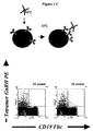
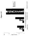
- GnRH-specific B cells To detect GnRH-specific B cells we used directly labelled native GnRH, or labelled tetramers with native GnRH, both in single and double staining protocols. GnRH tetramers were produced by incubating biotinylated GnRH with fluorescent-labelled streptavidin or neutravidin. Monoclonal antibodies against the B-cell marker CD19 and the T-cell marker CD3 were included in the staining procedure. The anti-CD3 staining was used to negatively select (irrelevant) T cells, as well as auto-fluorescent cells. As shown in Fig.1, our results demonstrated that only double tetramer staining produced discriminating results (Fig.
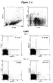
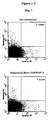
- Lymphocytes were stained with peptide-tetramers, and monoclonal antibody against CD19 and CD3. Since we expected only a very small population of specific B cells, debris were excluded with a live gate (forward and side scatter), and T cells were excluded with a negative gating on CD3e. Biotinylated GnRH tetrameric molecules or biotinylated gastrin tetrameric molecules were used in a double staining strategy to identify peptide-specific cells (Fig 2A). Using this approach, we were able to detect 20-30 specific peptide-tetramers double-positive cells in one million lymphocytes at day 7 after immunization, and 100 specific cells at day 14 after immunization (Fig. 2A).
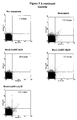
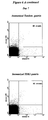
- tetramer binding was demonstrated by inhibition studies with unlabeled GnRH (see figure 3A) or Gastrin (data not shown). Lymphocytes were preincubated with unlabeled peptide followed by incubation with labelled tetramers, CD19 and CD3e antibodies. As shown in Fig 3A tetramer staining was inhibited by inclusion of unlabeled peptide in a dose-dependent manner.
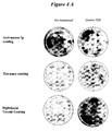
- splenocytes from gastrin-immunized mice were cultivated for 5 days under IL-2 and Pansorbin stimulation. At the end of this time the supernatants were used for performing an ELISA test, and the cells were used in an ELIspot assay. As shown in fig.4A, only the cells of the immunized mice were able to produce spots on the ELIspot well. These results were confirmed by ELISA test (Fig 4B) and they are consistent with the results of FACS analysis, proving that B cell detection is a reliable method for selecting peptide vaccines.
- mice 6-8 week old mice were used and treated for according to the ethical guidelines of University of Utrecht. Mice were immunized intraperitoneally on day 0 with , GnRH-like peptides conjugated to ovalbumin (OVA).
- OVA ovalbumin
- pEHWSYGLRPGC GnRH-monomer
- pEHWSYGLRPGQHWSYGLRPGC GnRH-tandem
- TDK GnRH tandem dimer
- TDK the dimerized form of pEHWSYkLRPGQHWSYkLRPGC in which 'k' represents a D-lysine residue
- mice were not immunized. On day 7 half of the mice of each group were boosted, using Incomplete Freunds Adjuvant, and half of the animals were sacrificed and spleens were harvested. On day 14, spleens were harvested from the remaining mice. All animal experiments were done with permission from the local ethical committee (DEC).
- DEC local ethical committee
- Cell cultivation Cell suspensions of splenocytes were cleared of erythrocytes and the cells were cultured in T75 flasks in the presence of Pansorbin (SAC, Calbiochem, Cat. No 507858) and recombinant human IL-2 (CETUS) for 5 days in incubator at 37 °C and 5% CO 2 . Cells were cultivated 5-10.10 7 spleen cells at 2.10 6 /ml in RMPI 10% FCS. After the incubation the cells were collected in tubes and centrifuged at 1200 rpm 10'. Supernatants were used for Elisa tests.
- Pansorbin SAC, Calbiochem, Cat. No 507858
- CETUS recombinant human IL-2
- Tetramer synthesis Tetrameric molecules were produced by mixing an equal amount of C-term biotinylated peptide at 200 ⁇ M (Pepscan B.V.) with R-phycoerythrin (PE)-labelled neutravidin at 3.3 ⁇ M (PE-NA, Molecular Probes, Eugene, OR), or allophycocyanin (APC)-labelled streptavidin at 6.1 ⁇ M (APC-SA, Molecular Probes). Each mixture was incubated at 4°C for a minimum of 12 hours. Than, tetramers were separated from non-complexed peptide by gel filtration using a Bio-Gel P-30 spin column (Bio-Rad, Hercules, CA). The amount of tetramers to use was determined by titration experiments. Peptides used were derived from GnRH (Pepscan B.V.).
- Flow cytometry Cell suspension of splenocytes was obtained by mechanical disruption of the spleens, and erythrocytes were lysed in 0.16 M chloride ammonium solution pH 7.4. The resultant cell suspensions were stained with combinations of fluorochrome-labelled antibodies and peptide's tetrameric molecules at optimal dilution. Staining was routinely conducted at 4°C in two steps: first with peptide's tetrameric molecule labelled with PE for one hour. Cells were then washed three times with FACS Buffer.
- FIG. 1 Visualization of antigen-specific B cells.
- flow cytometry strategies have been developed to detect specific B cells, and we have compared their effectiveness.
Abstract
The present invention discloses an affinity-binding assay comprising a particle
having at least four copies of a target molecule and at least two binding
molecules specific for said target molecule, wherein a first of said binding
molecules is associated with a first label and a second of said binding
molecules is associated with a second label, wherein a particle having said first
label and a particle having said second label are distinguishable from a
particle having both said first and said second label, wherein said first and
said second binding molecule each comprise at least two binding regions
specific for said target molecule.
The present invention also discloses a composition comprising a first binding
molecule associated with a first label and a second binding molecule associated
with a second label, wherein the signal obtained from said first label and said
second label is distinguishable from the combined signal of said first and
second label, characterised in that said first and said second binding molecule
each comprise at least two binding regions specific for essentially the same
target molecule, preferably for essentially the same epitope on said target
molecule and a method for selecting a synthetic antigen from a collection of at
least three antigens comprising using the disclosed composition and method.
The invention further discloses an antigen obtainable by above
described method and capable of inducing an early immune response, a kit of
parts to perform said method, the use of antigens selected by said methods for
use as a vaccine, and the use of antibodies and antigens as a medicament.
Description
The invention relates to the field of immunology and vaccine
development. The invention further relates to the detection of early B cell
responses after immunisation with specific antigens.
Vaccines have proven to be extraordinary effective means to improve
public health (Jackson et al., 2002). However, conventional vaccine
development can be hampered by technical problems in pathogen inactivation
as well as high costs.
Furthermore, many antigens are not readily recognised by the
immune system as foreign and consequently, they elicit no adequate immune
response. In these cases, peptide-based synthetic vaccines are valid
alternatives (Jackson et al., 2002). Peptide vaccination has been successfully
used to prevent infectious diseases (Monzavi-Karbassi et al,. 2002),and to treat
tumours (Ribas et al,. 2003; Noguchi et al,. 2003), amyloidosis (Nicoll et al,.
2003) and autoimmune disease (Liu et al,. 2002).
One difficulty in preparing vaccines against antigens that are not
recognised by the body as foreign is caused by the phenomenon of tolerance.
Normally, an immune response is elicited against antigens that are recognised
as foreign, but not against self-antigens. In order for a vaccine to be successful,
it must be sufficiently foreign. Only when a vaccine is foreign enough i.e.
sufficiently immunogenic will the immune system respond to the vaccine and
an immune response is induced. Conversely, however, the immune cells and/or
antibodies must still be capable of recognizing the self-substance or the
tolerated antigen, and thus the vaccine cannot be too "foreign". For this reason,
peptides of said antigens are made, and said peptides are modified to enhance
the immunogenicity of said antigens. Said peptides or modified peptides are
used as a peptide vaccine.
The development of a peptide vaccine is a labour-intensive and time-consuming
process. Peptide vaccine candidates must elicit an immune
response and cause clonal expansion of specific T cells and/or B cells. Said
immune response is elicited by epitopes that are recognized by T cells and/or B
cells and initiate a proliferative reaction of antigen specific T and B cells. Said
proliferative reaction syn.: "clonal expansion" starts within a few hours after
the initial contact of an antigen with the immune system, for example after
vaccination. The number of specific immune B cells increases by clonal
expansion until high levels of effector cells are reached after three or four
weeks after vaccination. The levels of these effector cells after clonal expansion
are high enough for conventional methods to detect the effector cells. To detect
whether peptides expose B or T cell epitopes, screening of a large set of
peptides in animal immunization procedures is necessary. Because the
immune repertoire of B cell and T cell specificities is very diverse (Davis and
Bjorkmann, 1988), and the frequency of antigen-specific B cells participating
in an antibody response is very low, detection of B cells presents a major
difficulty in its functional and molecular characterization (Newman et al.,
2003), and poses a major problem for investigators studying B-cell reactions.
After clonal expansion, activated B cells produce antigen specific antibodies
(Adams et al., 2003). Therefore, B cell response against an antigen is usually
measured by measuring the level of antigen specific antibodies in circulating
blood. As a result, a large number of experimental animals has to be kept for a
considerable amount of time, because after normal immunization procedures it
takes at least 4 to 5 weeks before antibodies, directed against an antigen have
reached levels at which they can be compared and be interpreted. In effect,
this provides a major obstacle for fast and systematic synthetic peptide vaccine
development.
B cells can be detected specifically by direct or indirect staining
methods, usually achieved by coupling antigens or antibodies to a signalling
molecule, like for example a fluorescent stain (fluorescein isothyocyanate,
FITC, or Rhodamine), or a chemical, radioactive, or enzymatic signal or
another marker substance that enables a skilled person to detect binding of
the marker substance to a B cell. Many methods have been developed to
increase the sensitivity of B cell detection to a level where small numbers of
antigen specific B cells can be detected in a large collection of cells, like for
example in spleen cells. Until now, the numbers of antigen specific B cells
before the process of clonal expansion were too low to be detected by exiting
detection methods. Said clonal expansion process increases the numbers of B
cells to a level where either B-cells or antibodies produced by said B cells, can
be detected. Based on fluorescent staining technology, flow cytometry was
developed and further adapted to detect antigen specific B cells (McHeyzer-Williams
et al., 1993). Historically, investigators have employed and combined
two different approaches to study antigen specific B cells. One approach is
hybridoma technique to capture immune reactive B cells and the other
approach is to study genetically manipulated animals with an enhanced
number of antigen specific B cells. Both methods are too insensitive to detect
the low levels of emerging antigen specific B cells after vaccination before
clonal expansion has been completed.
One method developed for the detection of B cells has been adapted
from the staining of T cells. The sensitivity of detecting T cells was enhanced
by incubating the cells with a tetrameric peptide-MHC molecule that binds to
T cell receptors specific for said peptide-MHC complex (Altman et al., 1996;
Stetson et al., 2002). Although the above-mentioned method has been adapted
for the detection of antigen-specific B cells (Newman et al., 2003), the
sensitivity of the normal procedures was only increased tenfold. This is still far
too insensitive for effective detection of emerging low- frequency antigen
specific B cells, shortly after vaccination.
Another method to enhance the detection level of low frequency
target cells has been described by Townsend et al., 2001, who succeeded in
lowering the detection threshold by staining B cells with two reagents specific
for the same B cell receptor, that had been conjugated to two different labels
(dubbed 'single epitope multiple staining'). The principle of this sequential
staining procedure (in which the first reagent is used at sub-saturating
concentrations) is that the probability of false positive cells binding both
reagents is much smaller than the probability of false positive cells binding
only one of the reagents. However, Townsend et al. only characterized
adoptively transferred BCR-transgenic cells, leaving the question unanswered
whether their methods could be extrapolated to the analysis of bona fide
immune responses, including those induced by vaccines.
This "single epitope multiple staining procedure" reduced the
background staining, thereby enabling an increase in the sensitivity of flow
cytometry by 1 or 2 orders of magnitude enabling the detection of high affinity
antigen-specific B cells in vivo. This increase in sensitivity may enable the
detection of transgenic B cells or B cells after clonal expansion is completed,
but it is still too insensitive for the detection of low frequency early B cells
within 7 days after immunization. Furthermore, the method is complicated
and the authors warn the public that it is critical to note that several
requirements must be met before this strategy will be effective in increasing
the sensitivity of flow cytometry. The above-described method of Townsend et
al. used as an antigen a foreign antigen for mice namely chicken egg white
lysozyme. When applied to the detection of specific B cells against a self-antigen
like GnRH, the method could not discriminate any specific B cells from
the background staining (see example 1 of the working examples in this
application). Therefore, the method of Townsend et al was not suited for the
detection of low-frequency B cells against self-antigens.
We have developed a highly sensitive and reproducible affinity-binding
assay to select peptide vaccine candidates based on specific staining of
emerging B cells during the clonal expansion at a very early stage of the
immune response. An affinity-binding assay is a test for detecting or
measuring binding of two or more substances that have a certain affinity for
each other. This can for example be the binding between an antibody and an
antigen, or of an enzyme to its substrate, or of a hormone to its hormone
receptor. In the present application we describe, as an example, the affinity
binding between the antigen specific B cell receptor to an antigen. Of course,
said affinity binding may be detected and measured by various methods. As an
example, we disclose the detection of affinity binding by an adapted flow
cytometry method.
The present application discloses a binding assay and a test method
using this binding assay in which a particle or a cell comprising at least four
target molecules, for example membrane bound antibodies or receptors or
antigen-specific B cell receptors (BCRs), can be detected by contacting said
particle or cell with at least two binding molecules that have a different label.
Detecting of particles or cells by flow cytometry is based on staining said
particle or cell with a fluorescent label and detecting labelled particles or cells
by registering fluorescence signals using the fluorescence detectors. Each
fluorescent label has its own characteristics for intensity and the level of
background staining. This application discloses that staining with a molecule
comprising a multiple form of an antigen, in which at least two, or three,
preferably four, or more, binding epitopes of essentially the same antigen are
present and exposed, increases the intensity of the staining. A further increase
in sensitivity is achieved by combining the staining of above described multiple
forms of an antigen with two different fluorescent labels. For this purpose, two
quantities of essentially the same multiple antigen molecules are associated
with two different labels. In this case, labels are selected that differ in their
fluorescent signal, like for example R-phycoerythrin (PE)-labelled neutravidin
and allophycocyanin (APC) coupled to streptavidin. By contacting the cells
with said both differently labelled antigenic molecules, the cells bind both
molecules and combine the two different fluorescent labels on the surface. This
contacting can for example be done in consecutive steps, for example by first
incubating the cells with the first fluorescent-labelled antigen molecule in a
sub-saturating amount, followed by incubation with the second fluorescent-labelled
antigen in a more saturating or a saturated amount. Another example
of a way of staining particles or cells with both fluorescent-labelled antigen
molecules is by mixing the two differently labelled antigen molecules at an
approximately equimolar amount and incubating the particles or cells with the
mixture. Because the chance of aspecific binding of two separate molecules on
the same particle is very small, the specificity of the method is increased. This
increased specificity, combined by the higher avidity of the binding with
multiple antigen molecules, causes a surprising increase in sensitivity and
allows for the specific detection of only 20 to 30 cells in one million cells. In this
application, the antibody molecules or the antigen-specific B cell receptors
present on the surface of the immune cells are called the target molecules. The
antigen molecules that bind to said target molecules are in this application
named binding molecules. Said binding molecules may comprise one or more
peptides or proteins or fragments thereof. In addition said binding molecules
may comprise a staining molecule. A particle in this application can be a virus
or a microorganism, or a part of a cell or yeast. Preferably, the size of said
particle is at least about the size of a virus and at most about the size of a
thousand cells, more preferably about the size of a hundred cells, even more
preferably about the size of 10 cells, even more preferably about the size of one
cell. In this patent application we use the phrase "distinguishable" for the fact
that by interpreting the results of preferably flow cytometry, double stained
particles or cells can be distinguished from particles or cells that have only one
label or no label at all. Preferably this distinction is made through distinction
of the signal from said first and second label. Label means in this context
preferably a fluorescent staining label.
Therefore, the present invention discloses an affinity-binding assay
comprising a particle having at least four copies of a target molecule and at
least two binding molecules specific for said target molecule, wherein a first of
said binding molecules is associated with a first label and a second of said
binding molecules is associated with a second label, wherein a particle having
said first label and a particle having said second label are distinguishable from
a particle having both said first and said second label, wherein said first and
said second binding molecule each comprise at least two binding regions
specific for said target molecule. Said particle can also be a cell, therefore the
invention also discloses the above-described assay, wherein said particle
comprises a cell. Said cell may be a living cell or said cell may be treated with
a fixative such as for example fixatives that are normally used in flow
cytometry, like for example formalin, acetone, alcohol, or glutaraldehyde.
Because the method of the invention is very sensitive, this is the
first time that such a method is capable of detecting very low frequency
antigen specific B cells, like for example very early immune B cells. These very
early immune cells are only present at very low frequency because these cells
have not yet accomplished the complete clonal expansion process. Therefore,
the present invention discloses an affinity-binding assay as described above,
wherein said cell comprises an activated immune cell in the clonal expansion
phase of a primary immune response. In mice for example, this activation
takes place within hours after first or second contact with an antigen and
results in antibody levels in peripheral blood within three to four weeks.
The method is especially suitable for the detection of very low
frequency B cells and saves a lot of time because one does not have to wait any
more for the antibody response to evolve. This shortens the animal phase for
each peptide from at least 3 to 4 weeks to one week. Furthermore, the
detection of very low frequency B cells is also very suitable for the detection of
memory B cells. This may be important for assessing the immune status
against certain diseases. A B cell in this application means a B cell in any
state of activation or differentiation, including for example antigen specific
precursor B cells, antibody secreting cells, plasma cells and memory B cells.
Therefore, the present invention discloses in a preferred embodiment the assay
as described above, wherein said cell comprises a B cell. The possibilities to
count and phenotypically characterize B cells as disclosed in this application
enable a person skilled in the art to measure the early immune response
against immunization or infection, e.g., proliferation and differentiation of
naive B cells into antibody secreting cells, memory B cells and plasma cells.
Because the combination of said multiple antigen molecules with the
multiple staining technique increases the sensitivity surprisingly much, the
present invention discloses an affinity-binding assay as described above,
wherein said particle having said first and said second label increases the
sensitivity of the affinity-binding assay.
The binding properties of said multiple antigen molecules are
increased with the number of exposed antigenic sites on the molecule. In this
application, tetramer molecules comprising four antigenic sites have been
tested as an example. Of course other numbers of antigens repeated in the
same molecule may also have the desired effect. Therefore, the present
invention discloses an affinity-binding assay as described above, wherein said
first and said second binding molecule each comprise at least two binding
regions specific for said target molecule. Preferably said number of binding
regions is four.
The binding may be increased even further if the antigenic sites or
epitopes of said target molecule are essentially identical epitopes. Therefore,
the present invention discloses an affinity-binding assay as described above,
wherein said at least two binding regions are essentially identical. A big
advantage of the assay is that the binding molecules of the detection assay
may represent an epitope of a native protein, because in this way,
immunization with altered antigens of said native protein can be monitored for
the cross-specificity for said native protein. A native protein is in this
application a protein as it is present in nature. Therefore, the present
invention discloses an affinity-binding assay as described above, wherein said
binding molecule represents an epitope of a native protein. The present
invention is very suited for detecting low frequency B cells. Said B cells, when
activated by contact with an antigen, expose during their clonal expansion an
antigen-specific B cell receptor or BCR on their cell surface. Suitable B cell
receptors are for example the well-known cell bound antibody-like molecules
that are inserted in the wall of the B cell with their Fc fragment and which are
specifically recognising antigen with the variable region, said antigen being
the antigen that was used to activate the B cells (Kouskoff et al., 2000). In the
described assay, said BCRs are the target molecules with which the binding
molecules bind. Therefore, the present invention discloses an affinity-binding
assay as described above, wherein said target molecule comprises a BCR.
Even more preferably the assay detects binding to the antigen
specific site of the BCR. Therefore, the present invention discloses an affinity-binding
assay as described above, wherein said target molecule comprises a
variable region of said BCR.
The assay is performed with a set of binding molecules detecting the
target molecule. Said binding molecule preferably comprising tetramer
molecules. Therefore, the present invention in another embodiment discloses a
composition comprising a first multiple binding molecule associated with a
first label and a second multiple binding molecule associated with a second
label, wherein the signal obtained from said first label and said second label is
distinguishable from the combined signal of said first and second label,
wherein each binding molecule comprises at least four binding regions specific
for essentially the same target molecule, preferably for essentially the same
epitope on said target molecule.
Now that there is an assay and a composition for detecting and
selecting B cells at a very early stage during clonal expansion, there is no need
any more to wait for at least 3 to 4 weeks for antibody levels before an antigen
can be evaluated for vaccine purposes. This enables a person skilled in the art
to select in a fast and reliable way an antigen from a large collection of
antigens. In another embodiment, relating to therapeutic vaccination
procedures involving, for instance, tumor antigens and hormones, individual
responses can be monitored and non-responsive individuals can be identified
more rapidly.
To overcome the tolerance of the immune system for certain
antigens, a range of modified antigens can be produced, for example by the
production of synthetic peptides. Changes in the peptides may increase the
immunogenicity, and by testing said peptides, new and improved antigens may
be detected and selected by the assay and the method of the invention. A range
of changes made to a peptide in order to changes the antigenicity of said
peptide results in various forms of said peptide, which is also called a
collection of peptides. Antigen-specific B cell detection as disclosed in this
application expedites the evaluation of vaccine immunogenicity, and enables
analysis of the fine-specificity of the response. For instance, the precise (sub)-serotype
specificities of the novel generation of multivalent conjugate vaccines
can now be analysed in unprecedented detail. Therefore, the present invention
discloses a method for selecting a synthetic antigen from a collection of at least
two antigens comprising using the composition as described above for
detecting in an affinity-binding assay for immune cells specific for said
synthetic antigen, clonal expansion of antigen specific immune cells in samples
of cells obtained from a mammal immunized with an antigen of said collection
of antigens, and comparing said clonal expansion with the clonal expansion of
another mammal immunized with another antigen form said collection and
selecting from said collection an antigen with which clonal expansion was more
extensive than clonal expansion observed with at least one other antigen from
said collection. A more extensive clonal expansion means in this application
that the numbers of specific B cells increase faster and to higher levels
compared to clonal expansions as reaction to other antigens. In mice for
example, increased clonal expansion at 7 days is indicative for an increased
clonal expansion at 14 days and usually for a high level of antibodies later in
the immune response. In rats or other animals, this increase may show later or
earlier in the immune response but be highly indicative of a more extensive
clonal expansion, if compared to the clonal expansion in other rats or other
animals immunized with another antigen of said collection of antigens.
Of course, the above-described method may use any of the affinity-binding
assays as are described above. Therefore, the present invention
discloses a method for selecting a synthetic antigen as described above,
wherein said affinity-binding assay comprises any assay as described herein.
Of course, for said synthetic peptides to be a good vaccine against a
native protein, cross reactivity of the antibodies directed against said synthetic
peptide antigen with the native protein from which said peptides were derived
is highly appreciated. Therefore, the method as described above is preferably
used wherein said at least two binding molecules comprise as a binding part
for said target molecule a synthetic antigen of said collection of antigens, or a
native antigen or homologue of said antigen. A native antigen is in this
application an antigen as it occurs in a protein in nature. A homologue of an
antigen is a substance showing the same antigenic characteristics in kind, not
necessarily in amount. Said homologue may comprise a natural peptide or
protein or a synthetic peptide or a part thereof. Therefore, direct B cell
staining procedures enable the assessment of specific B cell receptor cross-reactivity
between peptide vaccines and the native antigens from which they
are derived.
The invention also discloses the method as described above, further
comprising selecting an antigen for which clonal expansion of antigen-specific
immune cells more extensive than the clonal expansion with at least two other
antigens.
With the assay and the method as described above B cells specific for
a certain antigen can be selected and isolated. Said isolated B cells may also be
further tested for the specificity and avidity of their binding to the native
protein, also called the native antigen or homologue of said antigen. Therefore,
the invention teaches a method as described above further comprising
evaluating whether said antigen-specific immune cells are specific for a native
antigen or homologue of said antigen.
The above described methods and assays enable a person skilled in
the art to select and isolate antigen-specific cells. Therefore, the invention
discloses a method for an affinity-binding assay for selecting antigen specific
immune cells, comprising: (a) contacting a cell having at least four copies of a
target molecule with at least two binding molecule, preferably tetrameric
binding molecules, said binding molecules specific for said target molecule,
wherein a first of said binding molecules is associated with a first label and a
second of said binding molecules is associated with a second label and; (b)
detecting cells staining with each label and; (c) selecting cells binding both
labels.
The present invention teaches with above described methods a new
way to test and select a synthetic antigen from a collection of antigens
inducing a cross-reactive immune response with a native protein. This is an
important improvement of the technical possibilities in this field and opens the
possibility for new methods of selection and thus to other products. Therefore,
the invention discloses a synthetic antigen, inducing a cross-reactive immune
response with a native protein, selectable with a method as described above for
use as a vaccine.
To increase the antigenicity of a peptide, said peptide motif can be
made into dimer or trimeric or multimeric peptides wherein two or three or
more peptides are linked directly to each other or through a spacer molecule.
Combining two identical peptides into a dimer peptide further increases the
immunogenic properties of said peptide. Therefore, the present invention
discloses an immunogenic peptide, obtainable by above described method,
wherein said peptide comprises a dimer peptide.
In said dimer peptide, the peptides can be linked by sulphur bridges
or by a linkage molecule. Another method of increasing the immunogenicity of
a peptide is by combining a peptide in a tandem peptide and/or a tandem
dimer peptide. From WO 96/40755 it is known that the tandem-dimer principle
applied to a variant of the GnRH-I molecule resulted in a vaccine that was
highly effective in low doses and with a mild adjuvant. Therefore, the present
application discloses a method for selecting an immunogenic peptide derived
from an antigen comprising detecting clonal expansion of antigen specific
immune cells as described above, wherein said peptide comprises a tandem
dimer peptide.
The antigenic properties of peptides provided herein are further
optimised using a variety of techniques, such as replacement-net mapping,
allowing detecting peptides with improved characteristics. For example one
such peptide may be a peptide that binds to an antibody directed against an
epitope, thereby mimicking the immunogenic properties of said epitope. Such a
peptide is called a mimotope. This method has been disclosed in patent
application WO 00/29851. Therefore, the present application also discloses an
immunogenic peptide derived from a protein, comprising a mimotope of said
protein.
Amino acid substitution of at least one amino acid by a D-amino acid
and/or a L-alanine as described in WO 02/22659 also increases the
immunogenicity of a peptide. Therefore, the present invention discloses an
immunogenic peptide derived from a protein, wherein at least one amino acid
is substituted by a D-amino acid and/or a L-alanine.
One example of a peptide hormone for which peptides have been
prepared according to the sequence of said hormone and which are tested in
abovementioned method is gonadotrophin releasing hormone or GnRH.
Therefore, the application discloses abovementioned methods, wherein said
antigen comprises a GnRH-peptide hormone and/or part thereof or a GnRH-derived
peptide. Gonadotrophin releasing hormone (GnRH) is a decapeptide,
produced by the hypothalamus. The GnRH travels through portal circulation
to the pituitary to stimulate the release of gonadotrophins FSH and LH
(Talwar, 1999). Anti-GnRH immunization blocks the fertility of both male and
female animals (Meloen et al., 2001), and GnRH vaccines have applications in
prostate and in female cancer (Talwar, 1999; Fuerst et al.,1997). To compare
the usefulness of the different methods, mice were immunized with GnRH-like
peptides conjugated to ovalbumin (OVA). Three different peptides were
produced, i.e., GnRH-monomer (pEHWSYGLRPGC) GnRH-tandem
(pEHWSYGLRPGQHWSYGLRPGC) and GnRH tandem dimer (TDK, the
dimerized form of pEHWSYkLRPGQHWSYkLRPGC in which 'k' represents a
D-lysine residue). The GnRH tandem and TDK peptide conjugated to a carrier
protein have shown to be highly immunogenic, resulting in GnRH neutralizing
antibodies and a full biological effect in all treated animals (Meloen et al.,
1994; Oonk et a. 1998). Therefore, the present application provides an
immunogenic peptide obtainable by the above-described method, wherein said
peptide is derived from GnRH.
Another example of a peptide hormone for which peptides have been
prepared according to the sequence of said protein and which are tested in
abovementioned method is Gastrin. Therefore, the application discloses an
immunogenic peptide derived from a peptide hormone and obtainable by
above- mentioned methods, wherein said peptide is derived from Gastrin.
Gastrin is a peptide hormone that is important in the regulation of acid
secretion and the growth of both normal and malignant gastrointestinal
epithelium (Jensen et al., 2002). Tumor cells can respond both to circulating
endocrine gastrin (Watson et al., 1989) and locally produced gastrin, which
acts in an autocrine or paracrine manner (Hoosein et al., 1990). Effective
inhibition of gastrin may be beneficial as a therapy for colorectal cancer,
gastric cancer and pancreatic cancer, as gastrine acts as a growth factor for
these tumours. Gastrine receptor antagonists are not fully effective because
several receptor subtypes are involved in the action of gastrin and high
amounts are required to displace gastrin. Neutralisation of gastrin may
circumvent this problem.
Peptides derived from gastrine can be used for vaccination against
gastrin (amino acid sequence pEGPWLEEEEEAYGWMDF). Suitable peptides
for vaccination include amino acid 1-X, wherein X = 5 to 12. A Cysteine is
included at the N- or C-terminal end for conjugation purposes. The peptides
can also be applied in tandem or tandem dimer formulation with a Cysteine
added at the N- or C-terminus or a cysteine between the two gastrin sequences
in the tandem, as in pEGPWLEEEECQGPWLEEEE. L-Lysines or D-Lysines
are introduced in the tandem formulation to allow dimerisation of these
peptides via the cysteine and conjugation via the L- or D-Lysine, like in
 in which 'k' represents a D-lysine residue.
These peptides were tested as a vaccine in mice and at 7 and 14 days after immunization specific B cells were detected with the method of the invention using tetramer Gastrin peptide molecules. The following peptide was used to form a tetramer: pEGPWLEEEEEAYGWMDFK($)# , wherein # is amide; pE is pyroGlutamine; and K($) is biotinylated Lysin
These peptides were tested as a vaccine in mice and at 7 and 14 days after immunization specific B cells were detected with the method of the invention using tetramer Gastrin peptide molecules. The following peptide was used to form a tetramer: pEGPWLEEEEEAYGWMDFK($)# , wherein # is amide; pE is pyroGlutamine; and K($) is biotinylated Lysin
Therefore, the present invention discloses an immunogenic peptide
obtainable by any of the above-described methods wherein said peptide is
derived from Gastrin.
Yet another example of a hormone for which peptides have been
prepared is Human Chorionic Gonadotropin-beta (hCG-β), which is normally
produced by the placenta during pregnancy (Meyer et al., 2001). The protein
can be detected in serum or urine. hCG beta is elevated during the first
trimester of pregnancy and is often used as an indicator of pregnancy. hCG- β
is also a tumor-associated antigen that is in various types of cancer. Most
commonly, hCG beta is elevated, >10 mIU/ml (Gauchi et al., 1981), in
gynaecological cancers (Gauchi et al., 1981; Higashida et al., 2001; Kinugasa et
al., 1992), but also in colorectal (Lundin et al., 2000), seminoma testicular
(Hara et al., 2002), bladder (Hotakainen et al.,2002), liver (Szavay et al., 2000),
stomach (Bateman et al., 1995), pancreas (Syrigos et al., 1998), lung (Uckaya et
al., 1998), brain (Fujimaki et al., 2000), and kidney (Hotakainen et al., 2002)
cancers. Non-malignant elevations may be observed in pregnancy (Meyer et al.,
2001), ulcers (Manabe et al., 1985), duke's disease (Carpelan-Holmstrom et al.,
1996), and cirrhosis (Hoermann et al., 1992). Levels of hCG- β are useful in
monitoring the effectiveness of treatment, such as chemotherapy, against
tumor progression. Cancer patients with elevated levels of hCG- β have
significantly shorter survival time than patients with levels below the median
level. Effective inhibition of hCG- β is, therefore, beneficial as a therapy in
gynaecological, colorectal, seminoma testicular, bladder, liver, stomach,
pancreas, lung, brain, and kidney cancers, both in the prevention of the
disease and in advanced disease. Effective inhibition of hCG- β is, therefore,
also beneficial as a therapy in ulcers, duke's disease, and cirrhosis. The
accession number of the hCG- β sequence is: P01233 . The Swiss-Protein entry
name is: CGHB_HUMAN. The hormone comprises 165 amino acids, wherein
amino acid 1-20 form a signal peptide
Overlapping 20-mer peptides were used for immunization with a N-terminal
Cystein for conjugation to a carrier protein. The following peptides
were included: 21-40, 31-50, 41-60, 51-70, 61-80, 71-90, 81-100, 91-110, 101-120,
111-130,121-140, 129-165 and 141-165
Yet another hormone for which peptides have been prepared is
Human Parathyroid hormone-related protein (PTHrP).
Hypercalcaemia is one of the most common metabolic complications
in patients with cancer. Hypercalcaemia occurs in a majority of the epithelial
cancers in an advanced stage and is caused by PTHrP secretion by the tumor.
PTHrP levels are undetectable in healthy persons. PTHrP secreted by tumours
causes high calcium levels because it acts on the common PTH receptor.
PTHrP plays an important role in breast cancer. Approximately 50%
of the primary tumours secrete PTHrP. This feature is associated with
development of bone metastases. In these metastases PTHrP induces
osteolytic bone lesions and is responsible for destruction of bone tissue. This
osteolytic bone resorption results in release of TGF-B from the bone tissue,
which in turn stimulates the production of more PTHrP
There are 2 types of hypercalcaemia in cancer:
Humoral hypercalcaemia wherein primary tumours are producing
PTHrP, which acts as a hormone and after being secreted into the bloodstream
it acts on bone and kidney to increase calcium levels.
Local osteolytic hypercalcaemia: PTHrP also plays a role in bone
metastases. It can induce a local osteolysis near a bone metastasis, allowing
the tumour metastasis to grow into the bone tissue. In breast cancer almost
all metastases contain high levels of PTHrP whereas only 17% of the non-bone
metastases expresses PTHrP. It causes fractures and bone-pain in these
patients. The current treatment for hypercalcaemia is administration of
bisphosphonates, which reduces calcium levels. But this treatment does not
affect the PTHrP levels.
Neutralisation of PTHrP in cancers by vaccination against PTHrP
may contribute to an improved quality of life of patients with advanced cancer
regression. The Swiss-Prot entry name is: PTHR_HUMAN. The accession
number of the sequence is P12272. The length of human PTHrP is 177 amino
acids 1-177, wherein amino acids 1 - 36 comprise a leader sequence.
Effective peptides are N-terminal peptides of the PTHrP fragment
37-70, including 37-X, wherein X = 47 to 56. A C-terminal Cysteine is added
for conjugation. The peptides can be used as tandems and tandem dimers
The present invention also discloses the use of an immunogenic protein
and/or part thereof and/or an immunogenic peptide, selectable with a method
as described above for the preparation of a medicament.
In another embodiment of the invention, specific emerging B cells
can be sorted and isolated by for example cell sorting and/or magnetic bead
separation.
Said isolated B cells can be immortalized according to methods
generally known in the field of hybridoma production and thus become
immortalized B cells producing monoclonal antibodies. Therefore, the present
invention also discloses a B-cell selected with the method as described above
for the preparation of an antibody, and the use of such a B cell for the
production of a hybridoma.
Monoclonal antibodies produced by said B cell hybridoma are
particularly useful for diagnostic purposes or for the preparation of a
medicament, for example for the inactivation of GnRH in the circulation or for
the specific destruction of a tumour cell, or for inhibiting the replication of a
pathogen. Therefore, the present invention also discloses an isolated antibody
produced by above-described B cell and the use of said antibody for the
preparation of a medicament
The techniques described in this patent application may be used for
selecting suitable antigens for inducing an immune response. Once an antigen
is known, the present application shows how to increase the labelling
sensitivity by making binding molecules comprising multiple copies of said
antigen and how to label the binding molecules and how to use the labelled
binding molecules for the selection of an antigen. Therefore, this application
discloses a kit of parts comprising at least a first binding molecule associated
with a first label and a second binding molecule associated with a second label,
said binding molecules comprising at least two binding regions for the same
target molecule. A kit as above described is very useful to select a peptide from
a library of peptides. Therefore, the present invention discloses the use of said
kit of for the selection of an antigen.
The invention is further explained in the examples, without being
restricted to it.
6-8 week old mice were used and were treated according to the
ethical guidelines of our institutions. Mice were immunized intraperitoneally
on day 0 with GnRH-TDK coupled with ovalbumin (100 µg) or with Gastrin-TDK1
coupled with diphtheria toxoid. (Pepscan Systems, Lelystad, NL), in
Complete Freunds Adjuvant. Control mice were not immunized. On day 7, half
of the mice of each group were boosted, using Incomplete Freunds Adjuvant,
and half of the animals were sacrificed and spleens were harvested. On day 14,
spleens were harvested from the remaining mice. All animal experiments were
done with permission from the local ethical committee (DEC).
Tetrameric molecules were produced by mixing equal volumes of C-biotinylated,
middle biotinylated or N-biotinylated GnRH at 200 µM (Pepscan
Systems) with R-phycoerythrin (PE)-labelled neutravidin at 3.3 µM (PE-NA,
Molecular Probes, Eugene, OR), or allophycocyanin (APC)-labelled
streptavidin at 6.1 µM (APC-SA, Molecular Probes). Each mixture was
incubated at 4°C for at least 12 hours. Then, tetramers were separated from
non-complexed peptide by gel filtration using Bio-Gel P-30 spin columns (Bio-Rad,
Hercules, CA).
The following biotinylated gastrin peptide was used to form a
tetramer: pEGPWLEEEEEAYGWMDFK($)# , wherein # = amide; pE =
pyroGlutamine and K($) = biotinylated Lysine
Each tetramer was titrated in order to determine the optimal
concentration.
Cell suspensions of splenocytes were obtained by mechanical
disruption of the spleens, and erythrocytes were lysed in 0.16 M chloride
ammonium solution pH 7.4. The resulting single cell suspensions were stained
with combinations of fluorochrome-labelled antibodies and GnRH tetramers at
optimal dilution. Staining was conducted at 4°C in two steps. First, cells were
stained with the PE-labelled tetramer for one hour, using the empirically
determined optimal concentration. Cells were then washed three times with
FACS buffer. Next, cells were stained with the APC- or FITC-labelled tetramer
and combinations of PerCP-labelled anti-CD3 antibody (clone 145-2C11, BD
Biosciences), FITC-labelled anti-CD19 antibody (clone 1D3, BD Biosciences),
PE-labelled anti-CD27 antibody (clone LG.3A10, BD Biosciences) and PE-labelled
CD138 antibody (clone 281-2, BD Biosciences). For the staining
experiments involving directly labelled ligands, we used FITC- and biotin-labelled
GnRH (Pepscan Systems).
Data were acquired using a FACSCalibur flow cytometer and
CellQuest software (BD Immunocytometry Systems). 1.106 events were
acquired per sample. For FACS analysis lymphocytes were positively gated
based on their forward and side scatter profile (live lymphocyte gate), and
irrelevant T cells and auto-fluorescent cells were excluded with a negative
gating on CD3-PerCP.
ELIspot assay. Nitrocellulose bottomed 96-well Multiscreen HA
filtration plates (Millipore) were coated with 20 µg/ml of antigen or goat anti-mouse
IgG antibody (SBA, Cat. No 1031-01) and incubated at 4°C overnight.
Wells were washed 3 times with PBS and blocked for 30 minutes with PBS 5%
FCS. Blocking medium was replaced with 100 µl of RMPI medium 10% FCS
and the resuspended cells from the cell cultures. Plates were incubated for 24
h at 37°C in incubator 6% CO2. Wells were washed 3 times with PBS 0.05%
Tween-20. 100 µl/well of AP-conjugated goat anti-mouse IgG (SBA, Cat. No
1030-04) was added and incubates for 2-4 h at room temperature. Wells were
washed 3 times with PBA 0.05% Tween-20. 100 µl/well of BCIP/NBT substrate
were added. After spots development the plates were washed in tap water. The
number of the spot was evaluated using A.EL.VIS software.
Elisa test. 96-well multi-well plates were coated with 20 µg/ml of
antigen or goat anti-mouse IgG antibody (SBA, Cat. No 1031-01) and
incubated at 4°C overnight. Wells were washed 3 times with PBS 0.05%
Tween-20 and blocked for 30 minutes with PBS 5% BSA (Sigma). Wells were
washed 3 times with PBS 0.05% Tween-20. In each well the samples were
added and incubated for 1h.Wells were washed 3 times with PBS 0.05%
Tween-20. 100 µl/well of PO-conjugated goat anti-mouse IgG was added and
incubated for 1 h at room temperature. Wells were washed 3 times with PBA
0.05% Tween-20. 100 µl/well of ASBT substrate were added. After 3 minutes of
incubation the plates were read with an Elisa reader (Bio Rad).
Antibody titres against GnRH were determined with a radio-immune assay
(RIA) as described by Meloen et al. (1994). An amount of 50 µl antiserum
diluted 1:1000 in PBS with 0.4% bovine serum albumin (BSA) was allowed to
bind with iodinated GnRH (Amersham Pharmacia Biotech, Buckinghamshire,
England) in 50 µl PBS with 0.4% BSA. After incubation for two days at 4°C,
the iodinated GnRH bound to the antibodies was separated from the unbound
using dextran-coated charcoal. The supernatant was counted and the
percentage of iodinated GnRH bound by the antibodies in the 1:2000 diluted
serum was calculated.
We developed a novel B cell detection strategy able to lower the
discrimination's level to 20-30 specific cells in one million of lymphocytes using
tetrameric molecules. This method employed two different fluorescent-labelled
molecules for each peptide, allowing improved signal to noise ratios and
consequently, a very high sensitivity. To demonstrate the utility of our
approach to detect ultra low frequency B cells, we used as two model vaccine
formulations: GnRH-like peptide and Gastrin-like peptide. Several methods
have been described to detect antigen-specific B cells using flow cytometry
(McHeyzer-Williams et al., 1993 ; Townsend et al., 2001; Newman et al., 2003),
as shown schematically in Fig.1.
To detect GnRH-specific B cells we used directly labelled native
GnRH, or labelled tetramers with native GnRH, both in single and double
staining protocols. GnRH tetramers were produced by incubating biotinylated
GnRH with fluorescent-labelled streptavidin or neutravidin. Monoclonal
antibodies against the B-cell marker CD19 and the T-cell marker CD3 were
included in the staining procedure. The anti-CD3 staining was used to
negatively select (irrelevant) T cells, as well as auto-fluorescent cells. As
shown in Fig.1, our results demonstrated that only double tetramer staining
produced discriminating results (Fig. 1D), and none of the other staining
methods (Fig.1 A-C). Whereas direct staining with labelled GnRH (Fig. 1 A,B)
or with a single tetramer according to the method of Newman et al. (Fig.1 C)
produced only high background signals, and very little specific signal in the
case of double staining with labelled GnRH according to Townsend et al (Fig. 1
B). We found that the double tetramer staining as disclosed in this application
detects specific B cells at a frequency of 20-30 per 106 splenocytes at 7 days
after a single immunization (Fig.1 D).
Lymphocytes were stained with peptide-tetramers, and monoclonal
antibody against CD19 and CD3. Since we expected only a very small
population of specific B cells, debris were excluded with a live gate (forward
and side scatter), and T cells were excluded with a negative gating on CD3e.
Biotinylated GnRH tetrameric molecules or biotinylated gastrin tetrameric
molecules were used in a double staining strategy to identify peptide-specific
cells (Fig 2A). Using this approach, we were able to detect 20-30 specific
peptide-tetramers double-positive cells in one million lymphocytes at day 7
after immunization, and 100 specific cells at day 14 after immunization (Fig.
2A).
After the discrimination of the peptide-tetramer double-positive
cells, we studied the phenotypic characteristic of these cells. Although several
authors have used different markers for the characterization of mature B cells,
we decided to use CD19 as a pan B-cell marker (McHeyzer -Williams et al.,
1993). All the double-positive cells were CD19 positive classifying this cells as
B cells. Interestingly, the rare double-positive cells found in the non-immunized
mice are also CD19 positive (Fig. 2B) showing that these cells are
the B cell precursor population for the specific antigen that comprises
approximately 3-4 cells per million of lymphocytes in naive mice.
The specificity of tetramer binding was demonstrated by inhibition
studies with unlabeled GnRH (see figure 3A) or Gastrin (data not shown).
Lymphocytes were preincubated with unlabeled peptide followed by incubation
with labelled tetramers, CD19 and CD3e antibodies. As shown in Fig 3A
tetramer staining was inhibited by inclusion of unlabeled peptide in a dose-dependent
manner.
As a further specificity test, we produced tetramers using peptides
that were N-, middle, or C-terminally labelled. As shown in fig 3B, we found
that only the C-term biotinylated GnRH tetrameric were able to detect the
specific B cells.
After removing the erythrocytes from the spleen samples,
splenocytes from gastrin-immunized mice were cultivated for 5 days under IL-2
and Pansorbin stimulation. At the end of this time the supernatants were
used for performing an ELISA test, and the cells were used in an ELIspot
assay. As shown in fig.4A, only the cells of the immunized mice were able to
produce spots on the ELIspot well. These results were confirmed by ELISA
test (Fig 4B) and they are consistent with the results of FACS analysis,
proving that B cell detection is a reliable method for selecting peptide vaccines.
Therefore, in the present study, we have disclosed a novel method to
visualize antigen-specific B cells at very low frequencies. In our experiments,
the double tetramer staining procedure was more sensitive and more
reproducible then the previously described methods (e.g., labelled peptide
ligands, single tetramers). Our peptide tetramers have proven to be highly
specific B cell staining reagents.
6-8 week old mice were used and treated for according to the ethical
guidelines of University of Utrecht. Mice were immunized intraperitoneally on
day 0 with , GnRH-like peptides conjugated to ovalbumin (OVA). We prepared
three different peptides, i.e., GnRH-monomer (pEHWSYGLRPGC), GnRH-tandem
(pEHWSYGLRPGQHWSYGLRPGC) and GnRH tandem dimer (TDK,
the dimerized form of pEHWSYkLRPGQHWSYkLRPGC in which 'k'
represents a D-lysine residue) (Pepscan Systems, Lelystad, NL), in Complete
Freunds Adjuvant. Control mice were not immunized. On day 7 half of the
mice of each group were boosted, using Incomplete Freunds Adjuvant, and
half of the animals were sacrificed and spleens were harvested. On day 14,
spleens were harvested from the remaining mice. All animal experiments were
done with permission from the local ethical committee (DEC).
Cell cultivation. Cell suspensions of splenocytes were cleared of
erythrocytes and the cells were cultured in T75 flasks in the presence of
Pansorbin (SAC, Calbiochem, Cat. No 507858) and recombinant human IL-2
(CETUS) for 5 days in incubator at 37 °C and 5% CO2. Cells were cultivated
5-10.107 spleen cells at 2.106/ml in RMPI 10% FCS. After the incubation the
cells were collected in tubes and centrifuged at 1200 rpm 10'. Supernatants
were used for Elisa tests.
Tetramer synthesis. Tetrameric molecules were produced by
mixing an equal amount of C-term biotinylated peptide at 200 µM (Pepscan
B.V.) with R-phycoerythrin (PE)-labelled neutravidin at 3.3 µM (PE-NA,
Molecular Probes, Eugene, OR), or allophycocyanin (APC)-labelled
streptavidin at 6.1 µM (APC-SA, Molecular Probes). Each mixture was
incubated at 4°C for a minimum of 12 hours. Than, tetramers were separated
from non-complexed peptide by gel filtration using a Bio-Gel P-30 spin column
(Bio-Rad, Hercules, CA). The amount of tetramers to use was determined by
titration experiments. Peptides used were derived from GnRH (Pepscan B.V.).
Flow cytometry. Cell suspension of splenocytes was obtained by
mechanical disruption of the spleens, and erythrocytes were lysed in 0.16 M
chloride ammonium solution pH 7.4. The resultant cell suspensions were
stained with combinations of fluorochrome-labelled antibodies and peptide's
tetrameric molecules at optimal dilution. Staining was routinely conducted at
4°C in two steps: first with peptide's tetrameric molecule labelled with PE for
one hour. Cells were then washed three times with FACS Buffer. Next, cells
were stained with the APC- or FITC-labelled tetramer and combinations of
PerCP-labelled anti-CD3 antibody (clone 145-2C11, BD Biosciences), and
FITC-labelled anti-CD19 antibody (clone 1D3, BD Biosciences) were used for
the second step. Data were acquired using a FACSCalibur flow cytometer and
Cell Quest software (BD Immunocytometry Systems). 1.106 events were
acquired per sample. All data are representative plots derived from a
minimum of three independent experiments in which at least three
experimental and three control mice were analysed.
To detect GnRH-specific B cells we used labelled tetramers with
native GnRH, in double staining protocols. Monoclonal antibodies against the
B-cell marker CD19 and the T-cell marker CD3 were included in the staining
procedure. The anti-CD3 staining was used to negatively select (irrelevant) T
cells, as well as auto-fluorescent cells.
With the above-described method we compared the three different
peptide formulations of GnRH to vaccinate the mice. The results of the double
tetramer staining assay are given in Figure 5 A and 5 B. The results clearly
show that GnRH TDK-OVA caused the most extensive B cell expansion, which
was measured at both 7 and 14 days after vaccination.
With the above-described method we compared four different
peptide formulations of Gastrin to vaccinate the mice.

These peptides were tested as a vaccine in mice and at 7 and 14 days
after immunization specific B cells were detected with the method of the
invention using tetramer Gastrin peptide. The following biotinylated gastrin
peptide was used to form a tetramer: pEGPWLEEEEEAYGWMDFK($)# ,
wherein # = amide; pE = pyroGlutamine and K($) = biotinylated Lysine
The results of the double tetramer staining assay are presented in
Figure 6 A and 6 B. The results clearly show that Gastrin TDK2 -OVA caused
the most extensive B cell expansion, which was measured at 14 days after
vaccination.
Figure 1- Visualization of antigen-specific B cells. Several flow
cytometry strategies have been developed to detect specific B cells, and we
have compared their effectiveness.
Figure 2-
Identification of GnRH and of Gastrin specific B cells.
Figure 3-
Specificity of the B cell detection.
Figure 4- Detection of antigen specific B cells by ELIspot assay and
Elisa test.
Figure 5- Counting GnRH specific B cells after immunization with the
three GnRH vaccines.
Figure 6-
Counting Gastrin specific B cells after immunization with the four
Gastrin vaccines.
Adams, C.L., MacLeod, M.K.L., Milner-White, E.J., Aitken, R.,
Garside, P. and Stott, D.I. Complete analysis of the B cell response to a protein
antigen, from in vivo germinal centre formation to 3D modelling affinity
maturation. Immunology 108, 274-287 (2003).
Altman, J.D., Moss, P.A.H., Goulder, P.J.R., Barouch, D.H.,
McHeyzer-Williams, M., Bell, J., McMichael, A.J., Davis, M.M. Phenotypic
analysis of antigen-specific lymphocytes. Science 274, 94-96 (1996).
Bateman, H.E., B.S. Kasimis, C.R. Yook and V. Hiremath. 1995.
Case report: primary choriocarcinoma of the stomach. N J Med 92:459-462.
Carpelan-Holmstrom, M., C. Haglund, J. Lundin, H. Alfthan, U.H.
Stenman and P.J. Roberts. 1996. Independent prognostic value of preoperative
serum markers CA 242, specific tissue polypeptide antigen and human
chorionic gonadotrophin beta, but not of carcinoembryonic antigen or tissue
polypeptide antigen in colorectal cancer. Br J Cancer 74:925-929.
Cauchi, M.N., S.H. Koh, D. Lim and D.L. Hay. 1981. Oncofoetal
antigens in cancer of the cervix and ovary. Br J Cancer 44:403-409.
Davis, M.M., Bjorkman, P.J., T-cell antigen receptor genes and T-cell
recognition. Nature 334, 395 (1988).
Fuerst, J., Fiebiger, E., Jungwirth, A., Mack, D., Talwar, P.G., Frick,
J., Rovan, E. Effect of active immunization against LHRH on androgen
sensitive Dunning R3327-PAP and androgen independent Dunning R3327-AT2.1
prostate cancer cell lines. Prostate 32, 77-84 (1997).
Fujimaki, T., K. Mishima, A. Asai, K. Tabuchi, M. Kobayashi, I.
Suzuki and T. Kirino. 2000. Levels of beta-human chorionic gonadotropin in
cerebrospinal fluid of patients with malignant germ cell tumor can be used to
detect early recurrence and monitor the response to treatment. Jpn J Clin
Oncol 30:291-294.
Hara, I., Y. Yamada, H. Miyake, S. Hara, A. Gotoh, M. Fujisawa, H.
Okada, S. Arakawa and S. Kamidono. 2002. Detection of beta-human chorionic
gonadotropin expressing cells by nested reverse transcriptase-polymerase
chain reaction in the peripheral blood stem cells of patients with advanced
germ cell tumor. J Urol 167:1487-1491.
Higashida, T., T. Koizumi, S. Yamaguchi, T. Ichimura, K. Hasegawa
and R. Nishimura. 2001. Ovarian malignant mixed mesodermal tumor
producing the free form of the beta-subunit of human chorionic gonadotropin.
Int J Clin Oncol 6:97-100.
Hoermann, R., A.L. Gerbes, G. Spoettl, D. Jungst and K. Mann.
1992. Immunoreactive human chorionic gonadotropin and its free beta subunit
in serum and ascites of patients with malignant tumors. Cancer Res 52:1520-1524.
Hoosein, N.M., Kiener, P.A., Curry, R.C., Brattain, M.G. Evidence
for autocrine growth stimulation of cultured colon tumor cells by a Gastrin
cholecystokinin like peptide. Exp. Cel. Res. 186, 15-21 (1990).
Hotakainen, K., C. Haglund, A. Paju, S. Nordling, H. Alfthan, E.
Rintala and U.H. Stenman. 2002. Chorionic Gonadotropin beta-Subunit and
Core Fragment in Bladder Cancer: mRNA and Protein Expression in Urine,
Serum and Tissue. Eur Urol 41:677-685.
Hotakainen, K., B. Ljungberg, A. Paju, T. Rasmuson, H. Alfthan
and U.H. Stenman. 2002. The free beta-subunit of human chorionic
gonadotropin as a prognostic factor in renal cell carcinoma. Br J Cancer
86:185-189.
Jackson, D.C., Purcell, A.W., Fitzmaurice, C.J., Zeng, W., and Hart
D.N.J. The central role played by peptides in the immune response and the
design of peptide-based vaccines against infectious diseases and cancer.
Current Drug Targets 3, 175-196 (2002).
Jensen, R.T. Involvement of cholecystokinin Gastrin related
peptides and their receptors in clinical gastrointestinal disorders.
Pharmacology and Toxicology 91, 333-350 (2002).
Koh, T.J., Dockray, G.J., Varro, A., Cahill, R.J., Dangler, C.A., Fox,
J.G., Wang, T.C., Overexpression of glycine extended Gastrin in transgenic
mice results in increased colonic proliferation. J. Clin. Invest. 103, 1119-1126
(1999).
Kouskoff, V., Lacaud, G., Pape, K., Retter, M., Nemazee, D., B cell
receptor expression level determines the fate of developing B lymphocytes:
receptor editing versus selection. PNAS. 13, 743507439 (2000)
Liu, E., Abiru, N., Moriyama, H., Miao, D., Eisenbarth, G.S.
Induction of insulin auto-antibodies and protection from diabetes with
subcutaneous insulin B:9-23 peptide without adjuvant. Ann N Y Acad Sci. 958,
224-7 (2002).
Lundin, M., S. Nordling, M. Carpelan-Holmstrom, J. Louhimo, H.
Alfthan, U.H. Stenman and C. Haglund. 2000. A comparison of serum and
tissue hCG beta as prognostic markers in colorectal cancer. Anticancer Res
20:4949-4951.
Manabe, T., M. Adachi and K. Hirao. 1985. Human chorionic
gonadotropin in normal, inflammatory, and carcinomatous gastric tissue.
Gastroenterology 89:1319-1325.
McHeyzer-Williams, M.G., McLean, M.J., Lalor, P.A., Nossal, G.J.V.
Antigen-driven B cell differentiation in vivo. J Exp Med. 178(1), 295-307
(1993).
Meloen, R.H., Langeveld, J.P.M., Schaaper, W.M.M., Slootstra, J.W.
Synthetic peptide vaccines: unexpected fulfilment of discarded hope?
Biologicals 29, 233-236 (2001).
Monzavi-Karbassi, B., Cunto-Amesty, G., Luo, P., Kieber-Emmons,
T. Peptide mimotopes as surrogate antigens of carbohydrates in vaccine
discovery. Trends Biotechnol. 20(5), 207-14 (2002).
Meyer, T., L.A. Cole, P.I. Richman, H.D. Mitchell, J. Myers and G.J.
Rustin. 2001. High levels of hCG in choriocarcinoma can result in renal failure
and a false-negative pregnancy test in men. Clin Oncol (R Coll Radiol) 13:301-303.
Newman, J., Rice, J.S., Wang, C., Harris, S.L., Diamond, B.
Identification of an antigen specific B cell population. Journal of
Immunological Methods 272, 177-187 (2003).
Nicoll, J.A., Wilkinson, D., Holmes, C., Steart, P., Markham, H.,
Weller, R.O. Neuropathology of human Alzheimer disease after immunization
with amyloid-beta peptide: a case report. Nat. Med. 9(4), 448-52 (2003).
Noguchi, M., Kobayashi, K., Suetsugu, N., Tomiyasu, K., Suekane,
S., Yamada, A., Itoh, K., Noda, S. Induction of cellular and humoral immune
responses to tumor cells and peptides in HLA-A24 positive hormone-refractory
prostate cancer patients by peptide vaccination. Prostate 57(1), 80-92 (2003).
Oonk, H.B., Turkstra, J.A., Schaaper, W.M.M., Erkens, J.H.F.,
Schuitemaker de Weerd, M.H., Van Nes, A., Verheijden, J.H.M., and R.H.
Meloen. New GnRH like peptide construct to optimize efficient
immunocastration of male pigs by immunoneutralization of GnRH. Vaccine 16,
1074-1082 (1998).
Ribas, A., Timmerman, J.M., Butterfield, L.H., Economou, J.S.
Determinant spreading and tumor responses after peptide-based cancer
immunotherapy. Trends Immunol. 24(2), 58-61 (2003)
Smith, A.M., Justin, T., Michaeli, D., Watson, S. Phase I/II study of
G17-DT, an anti-gastrin immunogen, in advanced colorectal cancer. Clin.
Canc. Res. 6, 4719-4724 (2000)
Stetson, D.B., Mohrs, M., Mallet-Designe, V., Teyton, L., Locksley,
R.M. Rapid expansion and IL-4 expression by Leishmania specific naive helper
T cells in vivo. Immunity 17, 191-200 (2002).
Syrigos, K.N., I. Fyssas, M.M. Konstandoulakis, K.J. Harrington, S.
Papadopoulos, N. Milingos, P. Peveretos and B.C. Golematis. 1998. Beta
human chorionic gonadotropin concentrations in serum of patients with
pancreatic adenocarcinoma. Gut 42:88-91
Szavay, P.O., C. Wermes, J. Fuchs, M. Schrappe, P. Flemming and
D. von Schweinitz. 2000. Effective treatment of infantile choriocarcinoma in
the liver with chemotherapy and surgical resection: a case report. J Pediatr
Surg 35:1134-1135.
Talwar, G.P. Vaccines and passive immunological approaches for the
control of fertility and hormone-dependent cancers. Immunological review 171,
173-192 (1999).
Thorburn, C.M., Friedman, G.D., Dickinson, C.J., Vogelman, J.H.,
Orentreich, N., Parsonnet, J. Gastrin and colorectal cancer: a prospective
study. Gastroenterology 115, 275-280 (1998).
Townsend, S.E., Goodnow, C.C., Cornall, R.J. Single epitope
multiple staining to detect ultralow frequency B cells. Journal of
Immunological Methods 249, 137-146 (2001).
Uckaya, G., Uckaya, G., A. Ozet, A. Arpaci and S. Komurcu. 1998.
Human chorionic gonadotropin and CA 15-3 producing adenocarcinoma.
Nuklearmedizin 37:297-298.
Watson, S., Durrant, L. and Morris, D. Gastrin: growth enhancing
effects on human gastric and colonic tumor cells. Br. J. Cancer 59, 554-558
(1989).
Claims (45)
- An affinity-binding assay comprising a particle having at least four copies of a target molecule and at least two binding molecules specific for said target molecule, wherein a first of said binding molecules is associated with a first label and a second of said binding molecules is associated with a second label, wherein a particle having said first label and a particle having said second label are distinguishable from a particle having both said first and said second label, wherein said first and said second binding molecule each comprise at least two binding regions specific for said target molecule.
- An affinity-binding assay according to claim 1, wherein said particle comprises a cell.
- An affinity-binding assay according to claim 2, wherein said cell comprises an activated immune cell in the clonal expansion phase of a primary immune response.
- An affinity-binding assay according to claim 2, wherein said cell comprises a B cell.
- An affinity-binding assay according to any of claims 1-4, wherein said particle having said first and said second label increases the sensitivity of the affinity-binding assay.
- An affinity-binding assay according to any of claims 1-5, wherein said first and said second binding molecule each comprise at least four binding regions specific for said target molecule.
- An affinity-binding assay according to claim 6, wherein said at least four binding regions are essentially identical.
- An affinity-binding assay according to any of claims 1-7, wherein said binding molecule represents an epitope of a native protein.
- An affinity-binding assay according to any of claims 1-8, wherein said target molecule comprises a B cell receptor.
- An affinity-binding assay according to claims 9, wherein said target molecule comprises a variable region of said a B cell receptor.
- A composition comprising a first multiple binding molecule associated with a first label and a second multiple binding molecule associated with a second label, wherein the signal obtained from said first label and said second label is distinguishable from the combined signal of said first and second label, wherein each binding molecule comprises at least four binding regions specific for essentially the same target molecule, preferably for essentially the same epitope on said target molecule.
- A method for selecting a synthetic antigen from a collection of at least two antigens comprising using the composition of claim 11 for detecting in an affinity-binding assay for immune cells specific for said synthetic antigen, clonal expansion of antigen specific immune cells in samples of cells obtained from a mammal immunized with an antigen of said collection of antigens, and comparing said clonal expansion with the clonal expansion of another mammal immunized with another antigen of said collection, and selecting from said collection an antigen with which clonal expansion was more extensive than clonal expansion observed with at least one other antigen from said collection, wherein said collection of antigens comprises any antigen according to claim 18-22.
- A method according to claim 12, wherein said affinity-binding assay comprises an assay according to any one of claims 1-10 or a method according to any one of claims 42-45
- A method according to claim 13, wherein said at least two binding molecules comprise as a binding part for said target molecule, a synthetic antigen of said collection of antigens, or a native antigen or homologue of said antigen.
- A method according to claim 14, further comprising selecting antigen for which clonal expansion of antigen specific immune cells is more extensive than the clonal expansion with at least two other antigens
- A method according to claim 14 or 15, further comprising evaluating whether said antigen specific immune cells are specific for a native antigen or homologue of said antigen.
- A method for an affinity-binding assay for selecting antigen specific immune cells, comprising:a. contacting a cell having at least four copies of a target molecule with at least two binding molecules specific for said target molecule, wherein a first of said binding molecules is associated with a first label and a second of said binding molecules is associated with a second label and;b. detecting cells staining with each label and;c. selecting cells binding both labels.
- A synthetic antigen, inducing a cross-reactive immune response with a native protein, selectable with a method according to any of claims 12-16 for use as a vaccine.
- An antigen according to claim 18, wherein said antigen comprises a dimer peptide.
- An antigen according to claim 18 or 19, wherein said antigen comprises a tandem dimer peptide.
- An antigen according to any of claims 18 to 20, comprising a mimotope of said antigen.
- An antigen according to any of claims 18 to 21, wherein at least one amino acid is substituted by a D-amino acid and/or a L-alanine.
- An antigen according to any of claims 18 to 22, wherein said peptide is derived from GnRH.
- An antigen according to any of claims 18 to 22, wherein said peptide is derived from Gastrin.
- An antigen according to any of claims 18 to 22, wherein said peptide is derived from hCG .
- An antigen according to any of claims 18 to 22, wherein said peptide is derived from PTHrP.
- Use of an antigen according to any of claims 18 to 26 for the preparation of a medicament.
- Use of an antigen according to claim 23 for the preparation of a medicament for the treatment of prostate cancer and of female GnRH-related cancer.
- Use of an antigen according to claim 23 for the preparation of a medicament for blocking fertility.
- Use of an antigen according to claim 24 for the preparation of a medicament for the treatment of Gastrin-related colorectal cancer, and/ or gastric cancer.
- Use of an antigen according to claim 25 for the preparation of a medicament for the treatment of hCG-related gynaecological, colorectal, seminoma testicular, bladder, liver, stomach, pancreas, lung, brain, and kidney cancers.
- Use of an antigen according to claim 25 for the preparation of a medicament for the treatment of hCG-related ulcers, duke's disease, and cirrhosis.
- Use of an antigen according to claim 26 for the preparation of a medicament for the treatment of PTHrP-related cancer.
- Use of an antigen according to claim 25 for the preparation of a medicament for the treatment of PTHrP-related osteolytic bone disease.
- Use of an antigen according to claim 25 for the preparation of a medicament for the treatment of PTHrP-related hypercalcaemia.
- An isolated B-cell detected with the method according to claim 17, for the preparation of an antibody.
- A hybridoma based on the B cell of claim 36
- An isolated antibody produced by a B-cell according to claim 36 or a hybridoma according to claim 37.
- An isolated antibody according to claim 38 for the preparation of a medicament.
- A kit of parts comprising at least a first binding molecule associated with a first label and a second binding molecule associated with a second label, said binding molecules comprising at least two binding regions for the same target molecule.
- Use of a kit of parts according to claim 40 for the selection of an antigen.
- A method for detecting a particle having at least four copies of a target molecule comprising contacting said particle with a first and a second binding molecule having at least two binding sites for said target molecule, said first binding molecule coupled to a first label and said second binding molecule coupled to a second label, and detecting particles having both labels.
- A method for detecting a particle according to claim 42, wherein said detecting comprises detecting of a combined signal from said first and said second binding molecule.
- A method for detecting a particle according to claim 42, wherein said particle is derived from a mammal within 14 days after vaccination with a native antigen or homologue of said antigen.
- A method for detecting a particle according to claim 42, wherein said first binding molecule and said second binding molecule are present at approximately equimolar amounts.
Priority Applications (7)
| Application Number | Priority Date | Filing Date | Title |
|---|---|---|---|
| EP04075439A EP1564554A1 (en) | 2004-02-12 | 2004-02-12 | Method for the detection of early B cell populations in vaccine development |
| JP2006553073A JP2007527000A (en) | 2004-02-12 | 2005-02-11 | Early B cell detection for vaccine selection |
| PCT/NL2005/000101 WO2005078450A2 (en) | 2004-02-12 | 2005-02-11 | Method for the detection of early b cell populations in vaccine development |
| EP05710882A EP1714150A2 (en) | 2004-02-12 | 2005-02-11 | Method for the detection of early b cell populations in vaccine development |
| AU2005213571A AU2005213571A1 (en) | 2004-02-12 | 2005-02-11 | Method for the detection early B cell populations in vaccine development |
| CA002556149A CA2556149A1 (en) | 2004-02-12 | 2005-02-11 | Method for the detection of early b cell populations in vaccine development |
| US11/502,744 US20070065428A1 (en) | 2004-02-12 | 2006-08-11 | Early B-cell detection for selecting vaccines |
Applications Claiming Priority (1)
| Application Number | Priority Date | Filing Date | Title |
|---|---|---|---|
| EP04075439A EP1564554A1 (en) | 2004-02-12 | 2004-02-12 | Method for the detection of early B cell populations in vaccine development |
Publications (1)
| Publication Number | Publication Date |
|---|---|
| EP1564554A1 true EP1564554A1 (en) | 2005-08-17 |
Family
ID=34684718
Family Applications (2)
| Application Number | Title | Priority Date | Filing Date |
|---|---|---|---|
| EP04075439A Withdrawn EP1564554A1 (en) | 2004-02-12 | 2004-02-12 | Method for the detection of early B cell populations in vaccine development |
| EP05710882A Withdrawn EP1714150A2 (en) | 2004-02-12 | 2005-02-11 | Method for the detection of early b cell populations in vaccine development |
Family Applications After (1)
| Application Number | Title | Priority Date | Filing Date |
|---|---|---|---|
| EP05710882A Withdrawn EP1714150A2 (en) | 2004-02-12 | 2005-02-11 | Method for the detection of early b cell populations in vaccine development |
Country Status (6)
| Country | Link |
|---|---|
| US (1) | US20070065428A1 (en) |
| EP (2) | EP1564554A1 (en) |
| JP (1) | JP2007527000A (en) |
| AU (1) | AU2005213571A1 (en) |
| CA (1) | CA2556149A1 (en) |
| WO (1) | WO2005078450A2 (en) |
Cited By (2)
| Publication number | Priority date | Publication date | Assignee | Title |
|---|---|---|---|---|
| CN100428254C (en) * | 2006-07-20 | 2008-10-22 | 上海交通大学 | Cross reaction antigen computer-aided screening method |
| CN103049679A (en) * | 2012-12-28 | 2013-04-17 | 上海交通大学 | Method for predicting potential sensitization in protein |
Families Citing this family (11)
| Publication number | Priority date | Publication date | Assignee | Title |
|---|---|---|---|---|
| EP1910513B1 (en) | 2005-07-01 | 2016-08-10 | John Schrader | Methods of isolating antibody secreting cells from blood samples of immunized animals and generating monoclonal antibodies from said antibody secreting cells. |
| EP2575821B1 (en) | 2010-05-26 | 2015-08-12 | Satiogen Pharmaceuticals, Inc. | Bile acid recycling inhibitors and satiogens for treatment of diabetes, obesity, and inflammatory gastrointestinal conditions |
| EP2753924B1 (en) * | 2011-09-06 | 2022-10-26 | Becton Dickinson and Company | Methods and compositions for cytometric detection of circulating tumor cells in a sample |
| WO2013063512A1 (en) | 2011-10-28 | 2013-05-02 | Lumena Pharmaceuticals, Inc | Bile acid recycling inhibitors for treatment of pediatric cholestatic liver diseases |
| SG11201401816SA (en) | 2011-10-28 | 2014-05-29 | Lumena Pharmaceuticals Inc | Bile acid recycling inhibitors for treatment of hypercholemia and cholestatic liver disease |
| GB201121571D0 (en) | 2011-12-15 | 2012-01-25 | Astrimmune Ltd | Compounds and uses thereof |
| KR20160002773A (en) | 2013-03-15 | 2016-01-08 | 루메나 파마수티컬즈, 인코포레이티드 | Bile acid recycling inhibitors for treatment of primary sclerosing cholangitis and inflammatory bowel disease |
| US20140271734A1 (en) | 2013-03-15 | 2014-09-18 | Lumena Pharmaceuticals, Inc. | Bile acid recycling inhibitors for treatment of barrett's esophagus and gastroesophageal reflux disease |
| US20190350873A1 (en) * | 2016-12-30 | 2019-11-21 | Euromed, Inc. | Adhesive patch containing an improved release liner system |
| BR112021015799A2 (en) | 2019-02-12 | 2022-01-18 | Mirum Pharmaceuticals Inc | Methods to enhance growth in pediatric patients with cholestatic liver disease |
| CN110954706B (en) * | 2019-11-20 | 2020-10-02 | 宁波三生生物科技有限公司 | Method for measuring coupling rate of GnRH paralog dimer artificial antigen |
-
2004
- 2004-02-12 EP EP04075439A patent/EP1564554A1/en not_active Withdrawn
-
2005
- 2005-02-11 AU AU2005213571A patent/AU2005213571A1/en not_active Abandoned
- 2005-02-11 EP EP05710882A patent/EP1714150A2/en not_active Withdrawn
- 2005-02-11 JP JP2006553073A patent/JP2007527000A/en not_active Withdrawn
- 2005-02-11 CA CA002556149A patent/CA2556149A1/en not_active Abandoned
- 2005-02-11 WO PCT/NL2005/000101 patent/WO2005078450A2/en active Application Filing
-
2006
- 2006-08-11 US US11/502,744 patent/US20070065428A1/en not_active Abandoned
Non-Patent Citations (4)
| Title |
|---|
| ALTMAN J ET AL: "Phenotypic analysis of antigen-specific T lymphocytes", SCIENCE, AMERICAN ASSOCIATION FOR THE ADVANCEMENT OF SCIENCE, US, vol. 274, no. 5284, 4 October 1996 (1996-10-04), pages 94 - 96, XP002135711, ISSN: 0036-8075 * |
| NEWMAN J ET AL: "Identification of an antigen-specific B cell population", JOURNAL OF IMMUNOLOGICAL METHODS, ELSEVIER SCIENCE PUBLISHERS B.V.,AMSTERDAM, NL, vol. 272, no. 1-2, 15 January 2003 (2003-01-15), pages 177 - 187, XP004400050, ISSN: 0022-1759 * |
| STETSON DANIEL B ET AL: "Rapid expansion and IL-4 expression by Leishmania-specific naive helper T cells in vivo", IMMUNITY, vol. 17, no. 2, August 2002 (2002-08-01), pages 191 - 200, XP002283954, ISSN: 1074-7613 * |
| TOWNSEND S E ET AL: "Single epitope multiple staining to detect ultralow frequency B cells", JOURNAL OF IMMUNOLOGICAL METHODS, ELSEVIER SCIENCE PUBLISHERS B.V.,AMSTERDAM, NL, vol. 249, no. 1-2, 1 March 2001 (2001-03-01), pages 137 - 146, XP004317478, ISSN: 0022-1759 * |
Cited By (3)
| Publication number | Priority date | Publication date | Assignee | Title |
|---|---|---|---|---|
| CN100428254C (en) * | 2006-07-20 | 2008-10-22 | 上海交通大学 | Cross reaction antigen computer-aided screening method |
| CN103049679A (en) * | 2012-12-28 | 2013-04-17 | 上海交通大学 | Method for predicting potential sensitization in protein |
| CN103049679B (en) * | 2012-12-28 | 2017-07-11 | 上海交通大学 | The Forecasting Methodology of the potential sensitization of protein |
Also Published As
| Publication number | Publication date |
|---|---|
| JP2007527000A (en) | 2007-09-20 |
| AU2005213571A1 (en) | 2005-08-25 |
| US20070065428A1 (en) | 2007-03-22 |
| WO2005078450A2 (en) | 2005-08-25 |
| EP1714150A2 (en) | 2006-10-25 |
| WO2005078450A3 (en) | 2006-08-03 |
| CA2556149A1 (en) | 2005-08-25 |
Similar Documents
| Publication | Publication Date | Title |
|---|---|---|
| US20070065428A1 (en) | Early B-cell detection for selecting vaccines | |
| JP4607590B2 (en) | Methods and systems for detecting MHC class I and class II binding peptides | |
| CA2287872C (en) | Immunogenic compositions to the cck-b/gastrin receptor and methods for the treatment of tumors | |
| JP5576592B2 (en) | Monoclonal antibody against gastrin hormone | |
| CN102245767B (en) | Immunoassay method for human CXCL1 protein | |
| JP2008513536A (en) | Monoclonal antibody against progastrin | |
| AU2003226096A1 (en) | Method and device for trichomonas detection | |
| EP2156184A2 (en) | Methods for diagnosing and treating prostate and lung cancer | |
| WO1999040837A2 (en) | Compositions for detecting and surgically removing lymphoid tissue involved in tumor progression | |
| JP2007522447A (en) | Cytotoxicity assay | |
| JP5144742B2 (en) | Use of HE4 for breast cancer assessment | |
| KR101777259B1 (en) | Specific monoclonal antibody to EN2 protein or composition comprising the same for diagnosis of prostate cancer | |
| Harriman et al. | Antibody discovery via multiplexed single cell characterization | |
| JP5583648B2 (en) | Monoclonal antibody against gastrin hormone | |
| Samoylova et al. | ZP-binding peptides identified via phage display stimulate production of sperm antibodies in dogs | |
| Mecheri et al. | Immunogenic Ia-binding peptides immobilize the Ia molecule and facilitate its aggregation on the B cell membrane. Control by the M1s-1 gene. | |
| Zisman et al. | Ia-antigen-T-cell interactions for a thymus-independent antigen composed of D amino acids. | |
| US5605831A (en) | Human melanoma cell specific antigens and antibodies | |
| EP1051196A2 (en) | Compositions for detecting and surgically removing lymphoid tissue involved in tumor progression | |
| Scibelli et al. | Fast track selection of immunogens for novel vaccines through visualisation of the early onset of the B-cell response | |
| HUT77262A (en) | New immunocontraceptive peptides | |
| JP2021073888A (en) | Methods for producing tcr-like antibodies and uses thereof | |
| KR101158986B1 (en) | Monoclonal antibody specific to human DCC1, and uses thereof | |
| CN111499690A (en) | Novel antigenic peptide aiming at C L DN18-ARHGAP fusion mutation and application thereof | |
| WO2003082317A9 (en) | Peptide epitopes recognized by antigen specific cd8+ t lymphocytes |
Legal Events
| Date | Code | Title | Description |
|---|---|---|---|
| PUAI | Public reference made under article 153(3) epc to a published international application that has entered the european phase |
Free format text: ORIGINAL CODE: 0009012 |
|
| AK | Designated contracting states |
Kind code of ref document: A1 Designated state(s): AT BE BG CH CY CZ DE DK EE ES FI FR GB GR HU IE IT LI LU MC NL PT RO SE SI SK TR |
|
| AX | Request for extension of the european patent |
Extension state: AL LT LV MK |
|
| AKX | Designation fees paid | ||
| REG | Reference to a national code |
Ref country code: DE Ref legal event code: 8566 |
|
| STAA | Information on the status of an ep patent application or granted ep patent |
Free format text: STATUS: THE APPLICATION IS DEEMED TO BE WITHDRAWN |
|
| 18D | Application deemed to be withdrawn |
Effective date: 20060218 |