Technical Field
-
The present invention relates to an oocyte extract of
amphibian such as Xenopus laevis, in particular, an extract
of oocytes at the specific stage of maturation wherein the
protein synthetic activity of the oocyte is remarkably
enhanced, and to a method for producing a protein in cell-free
protein synthesis system using the extract.
Background Art
-
Xenopus oocytes and eggs are widely used to study DNA
replication, transcription, processing of transcripts,
translation, and post-translational protein modifications.
They are also used as an expression system for injected
exogenous genes. In 1977, Gurdon and Brown started injecting
exogenous genes into Xenopus oocytes and eggs to investigate
their functions, and reported the method for the first time.
This method is carried out by injecting mRNA into Xenopus
oocytes or eggs, or injecting cDNA into nuclei of Xenopus
oocytes. Previously, in the method of injecting genes such
as mRNA and cDNA into Xenopus oocytes or eggs, a manual or
automatic microinjection has been made with a pipette filled
with these samples and a manipulator under microscopic
observation (see, for example, Patent reference 1).
-
On the other hand, cell-free protein synthesis systems
comprised of a variety of cell extracts containing required
enzymes and the like, and energy sources have been developed.
These systems provide important experimental methods for
analyzing protein structure and function. The cell extracts
from, such as rabbit reticulocyte, E. coli, wheat germ, HeLa
cell, thermophilic bacteria, mouse L-cell, Ehrlich's ascitic
cancer cell, CHO cell, and budding yeast and the like, are used,
and several kits for cell-free protein synthesis system
derived from rabbit reticulocyte, E. coli, and wheat germ are
commercially available. Comparing the above two methods
briefly, it is generally believed that the protein synthesis
in cell-free system can be carried out more rapidly, and is
controllable more easily, than the synthesis in Xenopus
oocytes.
-
Xenopus oocytes are generally arrested in the cell cycle
at the first meiotic prophase. Only a few number of the cells
mature periodically under the influence of hormones, and
complete their first meiotic division to be secondary oocytes,
which are arrested at the metaphase II. The process of
maturation is triggered by progesterone. This hormone is
secreted by follicle cells surrounding the oocytes in the ovary,
which in turn are stimulated by gonadotropins released from
the pituitary. Maturation can also be induced in vitro by
treating isolated oocytes with progesterone. During the
maturation of Xenopus oocytes, the rate of endogenous protein
synthesis is reported to show a twofold increase (see, for
example, non-patent reference 1). The extract of Xenopus eggs
that matured to metaphase II to be capable of being fertilized,
can perform the post-translational modification of protein,
i.e. addition of carbohydrate chain, phosphorylation,
cleavage of signal peptide, localization, and formation of
higher structure (see, for example, non-patent reference 2).
-
Higher eukaryotic mRNA can be translated in the extract
of the above Xenopus eggs, but the translational efficiency
of prokaryotic mRNA is lower than that of eukaryote. Several
µg/ml levels of proteins are synthesized in a
ribonuclease-treated egg extract from added synthetic mRNAs
(see non-patent reference 2), however, in the oocyte extract,
efficient expression of exogenous (globin) mRNA required the
presence of reticulocyte postribosomal supernatant S-100,
suggesting that one or more factors required for mRNA
recruitment is limiting in these extracts (see non-patent
reference 1).
[Patent reference 1] Patent publication of
JP-A-2002-65240
-
[Non-patent reference 1] Tina D. Patrick et al.,
Preparation and characterization of cell-free protein
synthesis system from oocytes and eggs of Xenopus laevis,
Development, vol. 106, pp.1-9, (1989)
-
[Non-patent reference 2] Glenn Mathhews and Alan Colman,
A highly efficient, cell-free translation/translocation
system prepared from Xenopus eggs, Nucleic Acids Research, vol.
19, No.23, pp.6405-6412, (1991)
Disclosure of the Invention
-
Although the expression of exogenous genes
microinjected into Xenopus oocytes, eggs, and early embryos
has been investigated, the comparative analysis of expression
of exogenous genes in extracts obtained from the cells on
various stages of cell cycle has not been performed in detail.
Therefore, it is an object of the present invention to provide
a novel cell extract that can be used for cell-free protein
synthesis system by optimizing the system using Xenopus oocyte
extracts. It is also an object of the present invention to
provide a cell-free protein synthesis system using a Xenopus
oocyte extract, which method is superior to that of
microinjection to oocytes or that of using a conventional egg
extract in respect of protein synthetic efficiency.
-
The present invention has been completed to solve the
above problems, and is based on the findings that the cell
extract prepared at the specific stage of oocyte maturation
shows the remarkably enhanced activity of protein synthesis
and synthesizes a large amount of protein compared to the
conventional method with a matured egg extract or
microinjection to oocytes, as a result of investigating the
relationship between the respective stages of Xenopus oocyte,
egg (matured egg), and embryo in the cell cycle, and the
efficiencies of protein expression of introduced exogenous
genes in detail.
-
Accordingly, in a first aspect of the present invention,
there is provided an extract of Xenopus oocytes, which extract
is prepared from the oocytes at the early stage of maturation
entered from the state arrested at the first meiotic prophase
in the cell cycle.
-
In another aspect of the present invention, there is
provided a kit for use in a cell-free protein synthesis
comprising an extract prepared from Xenopus oocytes at the
early stage of maturation entered from the state arrested at
the first meiotic prophase in the cell cycle, an energy
regenerating system, and at least one amino acid. In a
preferred embodiment, the kit of the present invention further
comprises a plasmid DNA for the preparation of exogenous
polynucleotide.
-
In a still another aspect of the present invention, there
is provided a method of producing a protein in a cell-free
protein synthesis system comprising the steps of preparing a
polynucleotide coding for a protein, and expressing said
polynucleotide in an extract prepared from Xenopus oocytes at
the early stage of maturation entered from the state arrested
at the first meiotic prophase in the cell cycle.
-
In a preferred embodiment, the oocytes are at entry to
an early stage of maturation by treating oocytes arrested at
the first meiotic prophase with an egg maturation inducer. The
egg maturation inducer is at least one selected from the group
consisting of glucocorticoid, mineralcorticoid, progesterone,
testosterone, and insulin. The polynucleotide is an exogenous
mRNA that is synthesized and modified in vitro.
Brief Description of the Drawings
-
- Fig.1 is a schematic diagram that represents stages in
the cell cycle of Xenopus oocyte, egg, and early embryo.
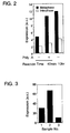
- Fig.2 shows expression levels of Xyk in metaphase and
interphase of Xenopus egg measured by immunoblotting.
- Fig.3 shows expression levels of Xyk in Xenopus oocytes,
an oocyte extract, and an egg extract measured by
immunoblotting.
- Fig.4 shows effect of progesterone on Xyk expression in
microinjected oocytes analyzed by immunoblotting using
immunoprecipitated samples with mAb327.
- Fig.5 shows effect of progesterone on Xyk expression in
microinjected oocytes measured by protein kinase activity with
poly(Gly,Tyr) as a substrate.
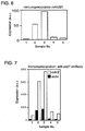
- Fig.6 shows effect of progesterone on Xyk expression in
Xenopus oocyte extract analyzed by immunoblotting using
immunoprecipitated samples with mAb327.
- Fig.7 shows effect of progesterone on Xyk expression in
Xenopus oocyte extract measured by protein kinase activity
with cdc2 and Xyk (auto: expression product itself) as
substrates and by the immunoprecipitation with anti-pepY
antibody.
- Fig.8 is a result of synthetic rate of Xyk in
microinjected Xenopus oocytes.
- Fig.9 is a result of synthetic rate of Xyk in Xenopus
egg extracts.
-
[Explanation of marks]
-
a.u.: arbitrary unit
Best Mode for Carrying Out the Invention
-
Preferred embodiments of the present invention are
explained as follows by reference to the figures.
(Preparation method of oocyte extracts)
-
Xenopus oocytes, eggs and embryos pass through the
different stages of cell cycle that are characterized by the
distinct levels of metabolic and protein synthetic activities
as shown in Fig.1, and result in maturation. Fully grown
Xenopus oocytes are naturally arrested in the first meiotic
prophase at the G2/M boundary. The oocytes have the intact
nuclear envelope, partially decondensed chromatin, high
activity of transcription, and low activity of the mitotic
regulators (factors present in cytoplasm, which regulate the
cell cycle) such as maturation promoting factor (MPF) and
cytostatic factor (CSF). Immature oocytes are not competent
to fertilization and can be arrested in this state for a long
time until the steroid hormone progesterone, secreted from
surrounding follicle cells, induces their transition from
prophase I to metaphase II arrested in the process of meiotic
maturation. The metaphase II arrested secondary oocytes are
then released from the ovary and complete the meiotic division
by stimulation of fertilization. Fig.1 shows the stages from
the oocyte arrested at the first meiotic prophase (prophase
I) via matured egg to fertilization and cleavages. In Fig.1,
curved lines indicating MAPK and Cdc2 kinase indicate the
activities of mitogen activated protein kinase and Cdc2kinase
in the cell cycles of Xenopus laevis. The term "oocytes" in
the present invention refers to oocytes at the stage from first
meiotic prophase to second meiotic metaphase in the cell cycle.
The term "eggs" refers to matured oocytes arrested at metaphase
II of the meiosis. The previous study of CAT gene expression
demonstrated that the gene is more actively expressed when
microinjected into the oocytes and dividing embryos, but not
into eggs of Xenopus laevis (Shiokawa et al., 1994).
Nevertheless, the described cell-free protein synthesis
system concerning Xenopus oocytes reported so far, mainly
utilizes the extract obtained from the eggs that have a
relatively low capacity for protein synthesis.
-
In one embodiment of the present invention,
translational efficiencies of various cell-free extracts
using mRNA encoding Src family kinase (Xyk) of Xenopus laevis,
as an exogenous mRNA, are disclosed. As a result of
investigations with extracts of Xenopus oocytes and eggs at
various stages in the cell cycle, it has been found that the
oocyte extract, in particular, the extract of oocytes just
after starting maturation by stimulation, has a remarkably
elevated protein synthetic activity, as shown in the following
examples.
-
Thus, in one embodiment of the present invention, there
is provided an extract of Xenopus oocytes, which extract is
prepared from the oocytes at the early stage of maturation
entered from the state arrested at the first meiotic prophase
in the cell cycle. In this embodiment, maturation of the
oocytes may be occurred spontaneously or enforced by
artificially administering hormones and the like. The phrase
"early stage of maturation" in this embodiment refers to the
stage until the completion of first meiotic division after
initiation of maturation from the arrested state at first
meiotic prophase in the cell cycle, preferably at the
initiation stage of the first meiotic division (just before
or after the start of first meiotic division) . Whether oocytes
are at the initiation stage of the first meiotic division or
not, can be decided by the indication of activity of MAPK or
Cdc2 kinase in cytoplasm as shown in Fig.1
-
A cytoplasmic activity from mature eggs that cause
complete maturation upon injection into immature oocytes was
originally defined by Masui et al. as a maturation promoting
factor (MPF) (Masui, Y., Differentiation 2001, 69 (1):1-17).
It consists of cyclin B and cyclin-dependent protein kinase
Cdc2. In the first meiotic prophase-arrested immature oocytes,
some part of Cdc2 is stored as an inactive complex, called
preMPF, where the catalytic activity of Cdc2 is inhibited by
phosphorylations on Thrl4 and Tyr15. Besides the low
activities of MPF and CSF are important for maintaining oocytes
at the first meiotic prophase. The Xenopus MAPK cascade,
including Mos, MAPKK, and MAPK is a major component of CSF that
causes the metaphase arrest of oocyte in the cell cycle.
Microinjections of Mos, or MAPKK mRNA into oocytes, or MAPK
protein itself can induce complete MPF activation and meiotic
progression in immature oocytes, whereas preventing MAPK
activation by MPK1 phosphatase or by MAPKK inhibitor
suppresses MPF activation and progesterone-induced oocyte
maturation.
-
Therefore, by any method of regulating the cell cycle
described above, for example, treatment of the oocytes
arrested at the first meiotic prophase with an egg maturation
inducer, or activation of any protein in the MAPK cascade, the
oocytes can be entered into the early stage of maturation. It
was reported that a morphological marker of meiosis I, germinal
vesicle break down (GVBD) can be equally triggered by
progesterone, testosterone, a kind of glucocorticoids, and a
kind of mineralocorticoids (Morrill, G.A., Bloch, E., J.
Steroid Biochem, 1977, 8(2):133-9). Thus, an egg maturation
inducer is preferably one or a combination of more than two
selected from the group consisting of glucocorticoid,
mineralocorticoid, progesterone, testosterone and insulin,
more preferably progesterone. The concentration and time of
treatment of the egg maturation inducer can be controlled so
as to enter the oocytes arrested at the first meiotic prophase
into the early stage of maturation, for example, in case of
progesterone, the treatment is in a concentration of 5 to 50µM
for 30 minutes to 4 hours, preferably about 10µM for 1 to 3
hours.
-
Preparation of oocyte extract can be performed according
to the known method (see Murray A. W., 1991, Cell cycle extracts,
Methods Cell Biol 36:581-605). For example, immature stage
VI oocytes are amassed by the injection of a hormone into
Xenopus laevis. Ovaries are isolated from the frog and the
stage VI oocytes are obtained by manual and enzymatic treatment
such as collagenase. Subsequently, the oocytes are crushed
to recover oocyte cytoplasm. To increase the stability of
obtained extract and efficiency of protein expression, the
oocyte extract can be prepared by supplying with additives such
as a cytostatic factor, protease inhibitor and energy
substance. After the process to obtain the stage VI oocyte,
or simultaneously therewith, the obtained oocytes are
preferably treated with an egg maturation inducer such as
progesterone. The conditions of progesterone-treatment can
be controlled so as to enter the oocytes arrested at the first
meiotic prophase into the early stage of maturation, for
example, in a concentration of 5 to 50µM for 30 minutes to 4
hours, preferably about 10µM for 1 to 3 hours. The crush of
the oocytes may be performed with a usual method such as
homogenization and sonication, however, the cytoplasmic
fraction can be easily obtained when the cell membrane is
disrupted by centrifugation. The obtained cytoplasm fraction
is further filtered through the porous membrane to remove
cellular debris. The extract can be prepared from any numbers
of oocytes, however, a certain number or more of oocytes are
preferably treated for easy treatment of crushing or easy
addition of stabilizers. In one embodiment of the present
invention, the oocyte extract is prepared from at least 100
oocytes.
-
Thus prepared oocyte extract are to be fresh, for example,
within about 8 hours, preferably about 3 hours, for obtaining
higher protein synthetic activity, it can be also stored by
freezing at -80 to -196°C. To prevent the reduction of protein
synthetic activity by freezing, various stabilizers are
preferably added to the extract. Alternatively, it is also
possible to lyophilize the extract by known methods, for
example, after rapid freezing of the oocyte extract by liquid
nitrogen, it is dried with a typical lyophilizer.
(Cell-free protein synthesis kit)
-
The oocyte extract of the present invention constitutes
a cell-free protein synthesis kit with an energy regeneration
system and at least one amino acid. The term "energy
regeneration system" means a component associating the
regeneration of energy source such as ATP and GTP necessary
for protein synthesis, and, for example, enzymes relating to
ATP regeneration (creatine kinase, pyruvate kinase and the
like) and/or substrates thereof (creatine phosphate,
phosphoenol pyruvate and the like). Amino acids include at
least one amino acid, preferably naturally occurring 20 kinds
of amino acids, but it may include other unnatural amino acids.
The kit further includes other components such as buffers (for
example, HEPES-potassium, and Tris-acetate etc.), various
salts, detergents, RNA polymerase (T7, T3 and SP6 RNA
polymerases etc.), chaperone proteins (DnaJ, DnaK, GroE, GroEL,
GroES, and HSP70 etc.), RNAs (mRNA, tRNA etc.), protease
inhibitors, or (ribo)nuclease inhibitors. These components
can be simultaneously included two or more. The cell-free
protein synthesis kit of the present invention may be a mixture
comprising at least the above extract, energy regeneration
system, and amino acids upon use, and these components to
constitute a kit may be stored separately before use.
-
In a preferable embodiment of the present invention, the
cell-free protein synthesis kit can further include a plasmid
DNA for the preparation of exogenous polynucleotide. The term
"exogenous polynucleotide" refers to a natural or artificial
nucleic acid molecule or nucleic acid construct derived from
heterogenous organismus or heterogenous cells. The nucleic
acid molecule may be DNA, RNA or analogues thereof, and it may
be natural or artificially synthesized. These nucleic acids
can be covalently or non-covalently linked to other molecules
such as proteins, lipids, chemical compounds, chemical
moieties, carriers or magnetic beads.
-
The exogenous polynucleotide of the present invention
is capable of expressing a protein in Xenopus oocytes extracts,
and can be prepared using expression vectors comprising DNA
that encode desirable proteins. When the extract contains a
transcriptional activity, for example, endogenous or
exogenous RNA polymerase, the expression vectors can be
directly added. The expression vector comprises a promoter
sequence upstream the protein coding sequence, which starts
the transcription. The promoter sequence is not particularly
limited to but includes various RNA polymerase promoters to
synthesize a single stranded mRNA, such as preferably Xenopus
RNA polymerase promoter, T7 RNA polymerase promoter, T3 RNA
polymerase promoter, and SP6 RNA polymerase promoter. Thus,
the phrase "plasmid DNA for the preparation of exogenous
polynucleotide" refers to a plasmid DNA comprising an RNA
polymerase promoter and polyadenylation signal, wherein a
desirable DNA fragment can be cloned.
-
In a further preferable embodiment of the present
invention, mRNA synthesized and modified in vitro is used as
an exogenous polynucleotide. In vitro transcription system
is, for example, a transcription reaction system derived from
T7 phage and that from E. coli. Synthetic reaction of mRNA
with this system can be carried out using a commercially
available kit such as MEGAscript™ (Ambion) , RiboMAX™ (Promega)
and the like. When a eukaryotic mRNA is expressed, it is
further preferable to add the cap structure at its 5'-end and
poly-A at its 3'-end as a post-transcriptional modification.
Most eukaryotic mRNAs have m7G(5')ppp(5')G cap structure at
its 5'-end, and this cap structure is important for binding
with translation initiation factors. It is also known that
this structure improves the translational efficiency by
stabilizing mRNA. For example, a capped transcript can be
easily synthesized by adding a cap analog (7-methyl-guanosine)
into in vitro transcription system. On the other hand, poly
A addition at the 3'-end is catalyzed by, for example, poly
A polymerase. Poly A addition prevents the cleavage from the
3' end of mRNA by exonucleases and can stabilize mRNA.
(Method of producing proteins by cell-free protein synthesis
system)
-
The protein synthesis in cell-free protein synthesis
system using a Xenopus oocyte extract is essentially carried
out by adding the mRNA to the cell-free extract having the
aforementioned protein synthetic activity. For example, mRNA
encoding a target protein, and its constituents such as amino
acids and energy source (ATP, GTP and the like) are added to
the reaction mixture containing the oocyte extract, and the
reaction mixture is incubated usually at 20 to 40°C, preferably
at 23 to 30°C, to synthesize the target protein. The cell-free
protein synthesis system of the present invention can be
employed a batch method, flow method, and any other techniques
previously known, for example, ultrafiltration method,
dialysis method, column chromatography method using a resin
on which templates for translation are immobilized (refer to
Spirin, AS. et al., Meth. In Enzymol. Volume 217, pp. 123-142,
1993), and the like. As for the above dialysis method, there
is disclosed a method described in Kigawa, T. et al. FEBS
Letters 1999, Vol. 442, pp. 15-19, and Japanese Patent Kokai
publication, JP-A-2000-175695, which method is carried out by
introducing the reaction mixture inside a dialysis membrane
having a fractional molecular weight of 10,000 or more,
preferably 50,000 or more, and dialyzed against the external
dialysate (comprising amino acids, energy sources and the
like) of 5 to 10-fold volume compared to the reaction mixture.
The dialysis is performed usually at 20 to 40°C, preferably
at 23 to 30°C, with agitation, and when the reaction rate
decreases, the external dialysate is exchanged with fresh one.
-
Further, the above cell-free protein synthesis system
has a translational activity to synthesize protein, and a
post-translational modification activity such as
glycosylation and phosphorylation. Therefore, by adding the
mRNA to the cell-free extract, the protein is synthesized from
the mRNA and then further modified to synthesize a glycoprotein
or a phosphorylated protein.
-
When the glycoprotein or phosphorylated protein is
synthesized as described above, the cell extract can be
prepared by adding a cytostatic factor, protease inhibitor,
ribonuclease inhibitor, and energy substance to the cell
extract. As an example, the oocyte extract can be prepared
in a final concentration of 10 µg/ml each cytochalasin B,
leupeptin, pepstatin, and chymostatin, 1 mM spermidine, 7.5
mM creatine phosphate, 1 mM ATP, and 1 mM MgCl2, to provide
for translation reaction. In addition, amino acid mixture is
preferably added to the cell extract. The added amino acids
are, for example, about 25 µM respectively in a final
concentration, which may be any of natural or unnatural.
-
The amount of mRNA added to the cell-free protein
synthesis system of the present invention is, for example, 10
to 100 µg/ml in a final concentration of the oocyte extract.
Alternatively, when the transcription/translation coupled
reaction is carried out in the cell-free protein synthesis
system, DNA constructed as the expression vector is directly
added. In case of the transcription activity in the oocyte
extract is low, T7, T3, or SP6 RNA polymerase is added with
ribonucleotide triphosphates (ATP, GTP, CTP, UTP) and the
like.
-
The protein synthesized by the production method of the
present invention, may be any protein derived from eukaryotic
or prolaryotic, and glycosilated or phosphorylated as a
post-translational modification. The protein forms a
characteristic three-dimensional or four-dimensional
structure to exert the function of the protein. Thus, the
synthesized protein can be used for its structural and
functional research and also for drug screening using an
interaction with other protein or low molecular weight
compound.
[Examples]
-
The present invention is explained in more detail by the
following examples, which describe the detailed analysis of
the expression of mRNA encoding Xenopus Src family kinase (Xyk)
in extracts of Xenopus oocytes and eggs, or in oocytes
microinjected with the mRNA. These examples are intended to
describe the invention more clearly and not to limit the
invention in any respect. In the examples, preparation of
Xenopus oocytes and eggs, preparation of egg and oocyte extract,
mRNA preparation, microinjection, translation reaction in
cell-free protein synthesis system, immunoprecipitation,
immunoblotting, and protein kinase assays are carried out
according to the following procedures.
[Preparation of Xenopus oocytes and eggs]
-
Wild type African clawed frogs (Xenopus Laevis) were
purchased from Hamamatsu Seibutsu Kyozai (Hamamatsu, Japan).
Frogs were maintained in polypropylene tanks filled with
deionized water (2L per animal) at temperature of 21-23°C with
a twice-a-week feeding. Frogs were primed with pregnant mare
serum gonadotropin (50 U/animal, Biogenesis) 5-7 days before
the experiments.
-
To obtain oocytes, frogs were anesthetized on ice, then
ovaries were surgically removed and placed into OR-2 solution
containing 82.5 mM NaCl, 2.5 mM KCl, 1 mM CaCl2, 1 mM MgCl2,
1 mM Na2HPO4, 5 mM HEPES, pH 7.8. Ovaries were manually
dissected into clumps of 50-100 oocytes and extensively washed
with OR-2 solution. The clumps of oocytes were treated with
0.5 mg/ml collagenase (Wako, 280 U/mg) in OR-2 at 23°C for 3
hours by shaking at 60 revs/min. Liberated oocytes were washed
with OR-2 solution and left for stabilization over 4 hours.
Undamaged stage VI oocytes were manually selected. Maturation
was induced by the addition of progesterone (Sigma) to a final
concentration of 10µM. Meiotic transition was monitored by
occurrence of germinal vesicle breakdown (GVBD) judged by
appearance of a white spot on the animal hemisphere of oocytes.
-
Eggs were obtained after inducing ovulation by the
injection of human chorionic gonadotropin (500 U/animal,
Teikokuzoki, Japan). In 8-12 hours after injection, eggs were
squeezed from the frogs into a plastic dish filled with MMR
solution (100 mM NaCl, 2 mM KCl, 1 mM MgCl2, 2 mM CaCl2, 0.1
mM EDTA, 5 mM HEPES), dejellied by treatment with 2% cysteine
(Sigma) in MMR, and washed extensively with the extract buffer,
containing 100 mM KCl, 0.1 mM CaCl2, 1 mM MgCl2, 5 mM EGTA, 50
mM sucrose, and 10 mM potassium HEPES, pH 7.7.
[Egg and oocyte extract preparation]
-
Cytostatic factor-arrested extracts of Xenopus eggs
were prepared from the unfertilized eggs essentially according
to the method of Murray et al.,(supra). The eggs were
transferred to the centrifuge tubes containing extract buffer
plus 100 µg/ml cytochalasin B (Sigma) and 10 µg/ml each
leupeptin, pepstatin, and chymostatin. Tubes were
centrifuged for 30 sec at 1,000 rpm, then for 30 sec at 1,500
rpm at 4°C. All buffer was removed from the top of the tubes
and eggs were crushed by a centrifugation for 15 minutes at
12,000 rpm. The cytoplasmic layer between lipid cap and
sedimented yolk was collected and subjected to the second
clarifying centrifugation under the same conditions.
Cytochalasin B, protease inhibitors, and energy mix (1/20
volume of 150 mM creatine phosphate, 20 mM ATP, 20 mM MgCl2)
were added to the extracts that were kept on ice until use.
All experiments with the extracts were performed at 23°C within
3 hours after preparation. Interphase released egg extracts
were obtained by the addition of calcium (final concentration
0.5 mM) to the cytostatic factor-arrested metaphase extracts.
Metaphase/interphase transition in the extracts usually took
about 30 minutes and was confirmed by the change in the nuclear
morphology of demembraned sperm nuclei added to extracts.
Nuclear morphology was observed and scored by fluorescent
microscopy after removing 1µl of extract and adding 4 µl of
stain solution (1 µg/ml Hoechst 33342 (sigma) in MMR buffer
containing 10% formaldehyde and 50% glycerol). Oocyte
extracts were prepared from the selected stage VI oocytes
washed in the extract buffer by the same procedure described
above for egg extracts.
[mRNA preparation]
-
The full length Xenopus Src kinase (Xyk) DNA (refer to
GenBank Accession No. M23422) containing two amino acid
substitutions at positions (Y527F and R121A) was subcloned
into pBluescript II vector (Toyobo) with the upstream T7
promoter. In vitro RNA transcription coupled with capping
reaction by 7-methyl-guanosine at the 5' end was performed from
SpeI-linearized plasmid using mMESSAGEmMASHINE T7 high yield
capped RNA transcription KIT (Ambion), according to the
manufacturer's manual. Purification of the synthesized mRNA
was carried out with RNeasy purification KIT (Qiagen).
Post-transcriptional polyadenylation of synthetic mRNA at the
3' end with E. coli poly(A) polymerase was done according to
the method of Wormington et al. (see Wormington, M., 1991,
Methods Cell Biol 36:167-183). Polyadenylation reaction was
performed at 37°C for 5 minutes in the reaction mixture
containing 50 mM Tris-HCl, pH 8.0, 250 mM NaCl, 10 mM MgCl2,
1 mM MnCl2, 2 mM DTT, 50 units of RNAse inhibitor, 50 µg/ml
bovine serum albumin, 50 µM ATP, 5 µg synthetic RNA in a final
volume of 50 µl. After purification with RNeasy purification
KIT, RNA was dissolved in RNase free water at 1 mg/ml which
solution was used for microinjections into eggs and oocytes
or direct additions into the extracts. RNA quality was
controlled by denaturing agarose/formaldehyde
electrophoresis, performed essentially as described Sambrook
et al. (Sambrook, J. et al., Molecular Cloning).
[Microinjection]
-
Quantitative injection of mRNA into stage VI Xenopus
oocytes was made under microscopic observation with a
pulse-directed injector system (Drummond, Nanoject). RNA
solution was loaded into the oil-filled glass microcapillary
needles with a tip diameter of 10-30 µm. Fifty ng of RNA
dissolved in RNase-free water was injected into an oocyte in
a total volume of 50 nl.
[Translation reaction in cell-free protein synthesis system]
-
The freshly prepared egg or oocyte extracts supplemented
with 10 µg/ml of cytochalasin B, leupeptin, pepstatin, and
chymostatin, as well as the energy mix (1/20 volume of 150 mM
creatine phosphate, 20 mM ATP, 20mM MgCl2) were used. The
extracts were incubated with in vitro synthesized Xenopus Src
RNA or Luciferase Control RNA (Promega) as a contrast at final
concentrations of 50 ng/µl or 20 ng/µl, correspondingly, in
the presence of 1 mM spermidine and 1 U/µl ribonuclease
inhibitor RNasin (Promega). Incubation time was 90-120
minutes at 21-23°C. The final volume of a reaction was 20 µl
with the extract dilution less than 20%. The reactions were
stopped by freezing in liquid nitrogen.
[Analysis by Immunoprecipitation]
-
Frozen eggs, or oocytes, or aliquots of the extracts were
ten-fold diluted with a homogenization buffer (20 mM Tris-HCl,
pH 7.5, 1% Triton X-100, 1 mM EDTA, 1 mM EGTA, 10 mM
β-mercaptoethanol, 1 mM sodium vanadate, 10 µg/ml leupeptin,
20 µM APMSF), then sonicated for 2 minutes on ice with a TOMY
UD-201 ultrasonic disrupter (Tomy Seiko, Tokyo). Samples were
centrifuged for 10 minutes at 15,000 rpm, then 20-µl aliquots
of supernatants were incubated with 5 µl of anti-Src family
kinase mAb327 antibody (Ab-1, Oncogene) for 2 hours at 4°C.
To collect immune complexes, protein A-Sepharose was added to
the samples at a final concentration of 10% and the mixture
was left for 1 hour. Nonspecifically bound proteins were
washed with the buffer containing 50 mM Tris-HCl, pH 7.5, 150
mM NaCl, 1% Triton-X-100, 1% sodium deoxycholate, 0.1% SDS.
For the analysis of expression level, samples were treated with
SDS-PAGE sample buffer and subjected to immunoblotting
analysis as described below. To confirm the expression of
functionally active Xenopus Src, protein kinase assay of
immunoprecipitated samples was performed.
[Analysis by Immunoblotting]
-
Immunoprecipitated samples or 20-µl aliquots of
ten-fold diluted extracts were mixed with a concentrated
SDS-PAGE sample buffer. Proteins were separated by the
electrophoresis on 10% polyacrylamide gels and transferred to
PVDF membranes using a semidry blotting device (Bio-Rad).
Membranes were blocked with T-TBS buffer (20 mM Tris-HCl, pH
7.5, 150 mM NaCl, 0.05% Tween 20) containing 3 mg/ml bovine
serum albumin and incubated for 2 hours with 100-fold diluted
anti-pep Y serum (Fukami et al., J. Biol. Chem. 1993,
268:1132-1140). After washing, the membranes were treated
with anti-mouse IgG rabbit polyclonal antibody (Cappel) at a
500-fold dilution, then with a 1000-fold diluted alkaline
phosphatase-conjugated goat polyclonal antibody against
rabbit IgG (Santa Cruz). Membranes were thoroughly washed
with T-TBS buffer and incubated in the developing buffer (100
mM Tris-HCl, pH 9.5, 5 mM MgCl2, 100 mM NaCl, 50 µg/ml
5-bromo-4-chloro-3-indolyl phosphate p-toluidine salt, and
150 µg/ml nitro blue tetrazolium) to visualize the immune
complexes.
[Protein kinase assays]
-
Total tyrosine kinase activity of the
immunoprecipitated sample was measured with poly (Gly, Try) 4:1
(Sigma), as a protein substrate, and specific activity of
immunoprecipitated Xenopus Src family kinase was estimated
with a synthetic cdc2 peptide, corresponding to residues 7-26
of the fission yeast cdc2 gene product (Fukami et al., supra).
The samples of extracts were five-fold diluted with a kinase
dilution buffer (80 mM β-glycerophosphate, pH 7.5, 20 mM EGTA,
15 mM MgCl2, 1 mM DTT, 0.1 mM NaF, 1mM Na3VO4, 0.2 mM APMSF,
10 µg/ml leupeptin, 10 µg/ml aprotinin). Prior to the kinase
assay, immunoprecipitated samples were washed with the kinase
dilution buffer. The reaction mixture of protein kinase assay
(20 µl) contained 50 mM Tris-HCl, pH 7.5, 5 mM MgCl2, 1 mM
dithiothreitol, 0.5 mg/ml poly(Gly, Tyr) or 1 mg/ml cdc2
peptide, 2 µM [γ-32P]ATP(1 µCi), and 5 µl of diluted or
immunoprecipitatetd extracts. Samples were incubated for 10
minutes at 30°C, then the reaction was terminated by the
addition of concentrated SDS-PAGE sample buffer. After
electrophoresis, the radioactive bands of phosphorylated
proteins were visualized and quantified by BAS2000 image
analyzer (FUJI Film). Incidentally, the expression level
measured by the immunoblotting method, or the kinase activity
measured by the protein kinase assay was quantified by using
a software (ImageGauge V.3.45) that calibrates the target band
intensity as an absorbance, and shown as arbitrary units
(a.u.).
[Comparative Example 1] Comparison of expression levels of
Xyk in Xenopus egg extracts arrested at the Metaphase and
Interphase.
-
Until now, the reports concerning the expression of
Xenopus Src kinase in a cell-free extract have not been
submitted. Therefore, we compared the expression using mRNA
prepared by the above method in the metaphase arrested and
interphase extracts of eggs. The result was shown in Fig.2.
Fig.2 shows expression levels of Xyk using the mRNA with (+)
or without (-) poly A in the extracts prepared from metaphase
or interphase of Xenopus egg measured by immunoblotting of
immunoprecipitated samples with Xyk specific antibody. As
shown in Fig.2, it was found that the expression level of Xyk
in the extracts released to interphase by the addition of
calcium was 2 to 3 fold lower than that in the metaphase extracts
in both reaction times 40 minutes and 10 hours irrespective
of poly A addition or not.
[Example 1] Comparison of expression levels of Xyk in
microinjected oocytes, and extracts of oocytes or eggs.
-
Next, the expression levels in microinjected Xenopus
oocytes, an oocyte extract, and an egg extract using mRNA
prepared by the above method were shown in Fig. 3. Sample No.1
in Fig.3 shows the expression level in the oocytes
microinjected with the mRNA followed by 24 hour incubation,
which oocytes are arrested at the first meiotic prophase.
Sample No.2 shows the expression level in the oocyte extract
with the mRNA followed by 90 minute incubation, which oocyte
is also arrested at the first meiotic prophase. Sample No.3
shows the expression level in the metaphase egg extract with
mRNA followed by 90 minute incubation. As shown in Fig.3, not
only the expression level of Xyk in the extract of oocytes
arrested at the first meiotic prophase (sample No.2) was much
higher than that in the metaphase egg extracts (sample No.3),
but also it significantly exceeded the level of expression in
the mRNA microinjected oocytes (sample No.1).
[Example 2] Analysis of correlation between the oocyte
maturation by progesterone-treatment and expression levels of
Xyk.
-
Next, we investigated the effect of progesterone
treatment on the expression of Xyk in the oocytes or in the
oocyte extracts. After the oocytes microinjected with Xyk
mRNA were treated with progesterone in various conditions, the
expression levels of Xyk were measured by immunoblotting
(Fig.4) and protein kinase assay (Fig.5). In each figure,
sample No.1 shows the expression level of Xyk after 4 hour
incubation at 23°C without mRNA microinjection. The minor
band derived from endogenous Srk kinase was detected (data not
shown). Sample No.2 shows the expression level in the oocyte
microinjected with Xyk mRNA after 4 hour incubation at 23°C
in the absence of progesterone. Sample No.3 shows the
expression level in the oocyte microinjected with Xyk mRNA
after 4 hour incubation at 23°C in the presence of 10 µM
progesterone. Sample No.4 shows the expression level in the
microinjected oocyte with Xyk mRNA, which oocytes are
pre-treated with 10 µM progesterone for 4 hours, and then
further incubated at 23°C for 4 hours in the absence of
progesterone. These results indicated that the progesterone
treatment after mRNA microinjection increased the Xyk
expression about two times, but the pre-treatment of the
oocytes with progesterone reversely decreased the expression
level. The reason why the expression level was reduced is
speculated to be due to the cell cycle progression of the
oocytes to the state of eggs (see Fig.1), which is consistent
to the results of Example 1 (Fig.3).
-
The expression levels of Xyk in the oocyte extract added
with the mRNA, which oocytes were treated with progesterone
in various conditions, were measured by immunoblotting (Fig.6)
and protein kinase assay (Fig.7). In each figure, sample No.1
shows the expression level of Xyk without addition of the mRNA.
Sample No.2 shows the expression level in the extract of oocytes
without progesterone treatment. Sample Nos.3 to 5 show the
expression levels in the extracts of oocytes treated with 10µM
progesterone for 2, 4, and 8 hours, respectively. These
results indicated that the expression of Xyk was increased only
in the extract prepared from oocytes treated with progesterone
for 2 hours, but was decreased in the extracts prepared from
oocytes treated with progesterone for 4 and 8 hours
respectively. The reason why the expression level was reduced
is speculated to be due to the cell cycle progression of the
oocytes to the state of eggs. In addition, the increase of
the expression level was about 1.5 times, which was less than
the result in Fig. 4 (about three time increase compared to the
expression level in the oocytes without progesterone
treatment). This suggests that the progesterone treatment for
less than 2 hours makes it possible to enhance the expression
level. From the above results, both in the oocytes and
extracts, short time (2 hour) progesterone treatment led to
the stimulation of Xyk production, whereas longer treatment
(4-8 hours) or pretreatment suppressed the synthesis much
below the level of the untreated control. Also, the results
presented in Figs. 4 to 7 demonstrate that the synthesized Xyk
is functionally active (has a tyrosine kinase activity).
[Example 3] Determination of expression level in oocyte
extract
-
To estimate the Xyk expression level in Xenopus oocyte
extracts, the reaction mixture for 90 minutes after addition
of the mRNA was electrophoresed, and calculated the protein
amount by CBB staining of the band. The estimation gave the
value of 7.8 µg/ml, in comparison with bovine serum albumin
(BSA), as a standard.
[Reference Example 1] Kinetics of Xyk synthetic rate in the
extracts
-
To further optimize the conditions of protein production
in the extracts, the kinetics of Xyk synthesis in the
microinjected oocytes and egg extracts were determined and
shown in Figs. 8 and 9, respectively. In accordance with the
previous reports, translation in the cell-free extracts
(saturated at 90 minute incubation, see Fig. 9) was much faster
than that of microinjected oocytes (increasing until 24 hours,
see Fig.8).
Industrial Applicability
-
The Xenopus oocyte extract of the present invention,
shows a significant higher protein synthetic activity than
that in the egg extract and microinjected oocytes. Thus, the
extract can be used for various uses of translation of desired
protein, especially an eukaryotic protein, and
post-translational modification such as glycosylation,
phospholylation , cleavage of signal sequence, localization,
and formation of protein higher structure. The present
invention provides a novel cell-free protein synthesis system
that can be used to alternatively express the proteins, in
particular, an eukaryotic protein, which can not be
successively expressed in the known cell-free system.