EP1554016B1 - In-vitro-synchronisation der kern- und zytoplasma-reifung von oozyten mit addition und entfernung von phosphodiesterase-hemmer typ 3 - Google Patents
In-vitro-synchronisation der kern- und zytoplasma-reifung von oozyten mit addition und entfernung von phosphodiesterase-hemmer typ 3 Download PDFInfo
- Publication number
- EP1554016B1 EP1554016B1 EP03757724A EP03757724A EP1554016B1 EP 1554016 B1 EP1554016 B1 EP 1554016B1 EP 03757724 A EP03757724 A EP 03757724A EP 03757724 A EP03757724 A EP 03757724A EP 1554016 B1 EP1554016 B1 EP 1554016B1
- Authority
- EP
- European Patent Office
- Prior art keywords
- oocytes
- hours
- oocyte
- medium
- hcg
- Prior art date
- Legal status (The legal status is an assumption and is not a legal conclusion. Google has not performed a legal analysis and makes no representation as to the accuracy of the status listed.)
- Expired - Lifetime
Links
Images






Classifications
-
- C—CHEMISTRY; METALLURGY
- C12—BIOCHEMISTRY; BEER; SPIRITS; WINE; VINEGAR; MICROBIOLOGY; ENZYMOLOGY; MUTATION OR GENETIC ENGINEERING
- C12N—MICROORGANISMS OR ENZYMES; COMPOSITIONS THEREOF; PROPAGATING, PRESERVING, OR MAINTAINING MICROORGANISMS; MUTATION OR GENETIC ENGINEERING; CULTURE MEDIA
- C12N5/00—Undifferentiated human, animal or plant cells, e.g. cell lines; Tissues; Cultivation or maintenance thereof; Culture media therefor
- C12N5/06—Animal cells or tissues; Human cells or tissues
- C12N5/0602—Vertebrate cells
- C12N5/0608—Germ cells
- C12N5/0609—Oocytes, oogonia
-
- A—HUMAN NECESSITIES
- A61—MEDICAL OR VETERINARY SCIENCE; HYGIENE
- A61P—SPECIFIC THERAPEUTIC ACTIVITY OF CHEMICAL COMPOUNDS OR MEDICINAL PREPARATIONS
- A61P15/00—Drugs for genital or sexual disorders; Contraceptives
- A61P15/08—Drugs for genital or sexual disorders; Contraceptives for gonadal disorders or for enhancing fertility, e.g. inducers of ovulation or of spermatogenesis
-
- C—CHEMISTRY; METALLURGY
- C12—BIOCHEMISTRY; BEER; SPIRITS; WINE; VINEGAR; MICROBIOLOGY; ENZYMOLOGY; MUTATION OR GENETIC ENGINEERING
- C12N—MICROORGANISMS OR ENZYMES; COMPOSITIONS THEREOF; PROPAGATING, PRESERVING, OR MAINTAINING MICROORGANISMS; MUTATION OR GENETIC ENGINEERING; CULTURE MEDIA
- C12N2500/00—Specific components of cell culture medium
- C12N2500/05—Inorganic components
- C12N2500/10—Metals; Metal chelators
- C12N2500/20—Transition metals
- C12N2500/24—Iron; Fe chelators; Transferrin
- C12N2500/25—Insulin-transferrin; Insulin-transferrin-selenium
-
- C—CHEMISTRY; METALLURGY
- C12—BIOCHEMISTRY; BEER; SPIRITS; WINE; VINEGAR; MICROBIOLOGY; ENZYMOLOGY; MUTATION OR GENETIC ENGINEERING
- C12N—MICROORGANISMS OR ENZYMES; COMPOSITIONS THEREOF; PROPAGATING, PRESERVING, OR MAINTAINING MICROORGANISMS; MUTATION OR GENETIC ENGINEERING; CULTURE MEDIA
- C12N2501/00—Active agents used in cell culture processes, e.g. differentation
- C12N2501/01—Modulators of cAMP or cGMP, e.g. non-hydrolysable analogs, phosphodiesterase inhibitors, cholera toxin
-
- C—CHEMISTRY; METALLURGY
- C12—BIOCHEMISTRY; BEER; SPIRITS; WINE; VINEGAR; MICROBIOLOGY; ENZYMOLOGY; MUTATION OR GENETIC ENGINEERING
- C12N—MICROORGANISMS OR ENZYMES; COMPOSITIONS THEREOF; PROPAGATING, PRESERVING, OR MAINTAINING MICROORGANISMS; MUTATION OR GENETIC ENGINEERING; CULTURE MEDIA
- C12N2501/00—Active agents used in cell culture processes, e.g. differentation
- C12N2501/10—Growth factors
- C12N2501/105—Insulin-like growth factors [IGF]
-
- C—CHEMISTRY; METALLURGY
- C12—BIOCHEMISTRY; BEER; SPIRITS; WINE; VINEGAR; MICROBIOLOGY; ENZYMOLOGY; MUTATION OR GENETIC ENGINEERING
- C12N—MICROORGANISMS OR ENZYMES; COMPOSITIONS THEREOF; PROPAGATING, PRESERVING, OR MAINTAINING MICROORGANISMS; MUTATION OR GENETIC ENGINEERING; CULTURE MEDIA
- C12N2501/00—Active agents used in cell culture processes, e.g. differentation
- C12N2501/30—Hormones
- C12N2501/31—Pituitary sex hormones, e.g. follicle-stimulating hormone [FSH], luteinising hormone [LH]; Chorionic gonadotropins
-
- C—CHEMISTRY; METALLURGY
- C12—BIOCHEMISTRY; BEER; SPIRITS; WINE; VINEGAR; MICROBIOLOGY; ENZYMOLOGY; MUTATION OR GENETIC ENGINEERING
- C12N—MICROORGANISMS OR ENZYMES; COMPOSITIONS THEREOF; PROPAGATING, PRESERVING, OR MAINTAINING MICROORGANISMS; MUTATION OR GENETIC ENGINEERING; CULTURE MEDIA
- C12N2501/00—Active agents used in cell culture processes, e.g. differentation
- C12N2501/30—Hormones
- C12N2501/38—Hormones with nuclear receptors
- C12N2501/39—Steroid hormones
- C12N2501/392—Sexual steroids
-
- C—CHEMISTRY; METALLURGY
- C12—BIOCHEMISTRY; BEER; SPIRITS; WINE; VINEGAR; MICROBIOLOGY; ENZYMOLOGY; MUTATION OR GENETIC ENGINEERING
- C12N—MICROORGANISMS OR ENZYMES; COMPOSITIONS THEREOF; PROPAGATING, PRESERVING, OR MAINTAINING MICROORGANISMS; MUTATION OR GENETIC ENGINEERING; CULTURE MEDIA
- C12N2501/00—Active agents used in cell culture processes, e.g. differentation
- C12N2501/70—Enzymes
-
- C—CHEMISTRY; METALLURGY
- C12—BIOCHEMISTRY; BEER; SPIRITS; WINE; VINEGAR; MICROBIOLOGY; ENZYMOLOGY; MUTATION OR GENETIC ENGINEERING
- C12N—MICROORGANISMS OR ENZYMES; COMPOSITIONS THEREOF; PROPAGATING, PRESERVING, OR MAINTAINING MICROORGANISMS; MUTATION OR GENETIC ENGINEERING; CULTURE MEDIA
- C12N2517/00—Cells related to new breeds of animals
- C12N2517/10—Conditioning of cells for in vitro fecondation or nuclear transfer
Definitions
- the present invention relates to a method for in vitro synchronisation of nuclear and cytoplasmatic maturation of germinal vesicle (hereinafter designated GV) oocytes from domestic animals or from primates. More particularly, the present invention relates to the aspects defined in the claims below.
- GV germinal vesicle
- Meiosis is the unique and ultimate event of germ cells on which sexual reproduction is based. Meiosis comprises two meiotic divisions. During the first division, exchange between maternal and paternal genes take place before the pairs of chromosomes are separated into the two daughter cells. These contain only half the number (1 n) of chromosomes and 2c DNA. The second meiotic division proceeds without a DNA synthesis. This division therefore results in the formation of the haploid germ cells with only 1c DNA.
- the meiotic events are similar in the male and female germ cells, but the time schedule and the differentiation processes which lead to ova and to spermatozoa differ profoundly.
- All female germ cells enter the prophase of the first meiotic division early in life, often before birth, but all are arrested as oocytes later in the prophase (dictyate state) until ovulation after puberty.
- the female has a stock of oocytes which is drawn upon until the stock is exhausted.
- Meiosis in females is not completed until after fertilization, and results in only one ovum and two abortive polar bodies per germ cell.
- only some of the male germ cells enter meiosis from puberty and leave a stem population of germ cells throughout life. Once initiated, meiosis in the male cell proceeds without significant delay and produces 4 spermatozoa.
- Oocyte development during the follicular growth phase in vivo is characterized by a prolonged arrest at prophase I until the pre-ovulatory surge of luteinizing hormone (hereinafter designated LH).
- LH luteinizing hormone
- Prophase I arrest in mammalian oocytes is sustained by different mechanisms at distinct times of development.
- Growing oocytes, enclosed in primordial up to the early antral follicles, are arrested at prophase I and are germinal vesicle breakdown (hereinafter designated GVBD) incompetent because of a functional insufficiency.
- GVBD germinal vesicle breakdown
- oocytes have not synthetised the cell cycle regulatory molecules essential for meiosis progression in sufficient quantities and/or these molecules are as yet not positioned correctly within the oocyte.
- prophase I arrest in GVBD competent oocytes is sustained by interaction with the somatic cells, which provide appropriate levels of cAMP to the oocyte via gap junctions.
- the somatic compartment transfers inhibitory factors to the oocyte.
- gonadotropins promote oocyte maturation indirectly via effects on granulosa cells, a process mediated predominantly via the cAMP system.
- a rise in follicular cAMP mediates LH action to induce oocyte maturation, and intraoocyte cAMP inhibits the process.
- Supplementing culture medium with compounds that maintain elevated cAMP can prevent spontaneous maturation. This can be achieved by several compounds: cAMP analogues such as dibutyryl cAMP (hereinafter designated dbcAMP), pharmacological agents that stimulate cAMP production via adenylate cyclase (forskolin) and inhibitors of the cAMP degrading isoenzymes phosphodiesterase (hereinafter designated PDE), such as 3-isobutyl-1-methylxanthine (IBMX).
- cAMP analogues such as dibutyryl cAMP (hereinafter designated dbcAMP)
- pharmacological agents that stimulate cAMP production via adenylate cyclase forskolin
- inhibitors of the cAMP degrading isoenzymes phosphodiesterase hereinafter designated PDE
- IBMX 3-isobutyl-1-methylxanthine
- PDEs Certain phosphodiesterases
- PDE are responsible for the breakdown of cAMP, leading to a decrease of its intracellular levels.
- PDE consist of a large group of proteins in which at least eleven different families have been characterized. Examples of these families are type 3 PDEs and type 4 PDEs (hereinafter designated PDE3 and PDE4, respectively).
- Non-selective PDE inhibitors have been used to understand the role of cAMP in the resumption of meiosis. Suppression of cAMP catabolism by using different specific PDE inhibitors demonstrated the meiosis arresting action of these chemical compounds.
- PDE4 is mainly involved in the metabolisation of cAMP in granulosa cells.
- PDE 3 has been demonstrated to act directly in the oocyte without interfering with somatic cell functions.
- expression of PDE3 was specific to the oocyte.
- the following statement can be found in the abstract in Developmental Biology 178 (1996), 393 et seq .: "In isolated oocytes, spontaneous GVBD was blocked by two inhibitors of type 3 PDE". As appears from the publication, this statement applies to rats. It is stated in the abstract in J. Clin. Invest. 102 (1998), 532 et seq ., that "inhibitors of phosphodiesterase 3 were used to block meiosis in ovulating oocytes in rodents. By this strategy, we [i.e., the authors of this paper] demonstrated that fertilization and pregnancy could be prevented”.
- cAMP signalling is a key factor during mammalian and amphibian oocyte maturation.
- germinal-vesicle stage hereinafter designated GV
- oocytes removed from immature antral follicles spontaneously resume meiosis in vitro but this process can be blocked in vivo and in vitro by certain compounds that maintain intraoocyte cAMP levels increased.
- One object of the present invention is to treat human infertility.
- Another object of the present invention is to improve the maturation of human oocytes.
- Another object of the present invention is to improve the synchrony of nuclear, cytoplasmic and/or membranous oocyte maturation.
- Another object of the present invention is to improve the fertility of oocytes.
- Another object of the present invention is to improve the rate of implantation of oocytes by human in vitro maturation and fertilisation.
- Another object of the present invention is to diminish the incidence of human preembryos with chromosome abnormalities (aneuploidy).
- Another object of the present invention is to improve the cleavage rate of human preembryos.
- Another object of the present invention is to improve the quality of human preembryos.
- Another object of this invention is to provide a method to better synchronise cytoplasmic and nuclear maturation for IVM.
- Another object of this invention is to furnish a method permitting the IVM laboratory to operate within the normal workings hours of the day.
- a further object of this invention is to find a method to increases commodity of IVM practice by making the IVM independent from the time of oocyte cumulus complexes (hereinafter designated OCC) collection. (i.e., the collection time can be uncoupled from meiosis progression and subsequent fertilisation steps).
- OCC oocyte cumulus complexes
- MI metaphase I.
- EGF epidermal growth factor.
- GH growth hormone.
- IVM in vitro maturation.
- IVF in vitro fertilisation.
- PDE3s are specific enzymes present in 2 isoforms: PDE3A in myocardial, smooth muscle and in the oocyte and PDE3B functions in cells' hormonal regulation of lipolysis and glyconeogenesis ( vide : J. Biol. Chem. 272 (1997), 6823-6826 ).
- PDE3A is the isotype of PDE3 which is present in the oocyte and regulates meiosis.
- the Type 3B i.e., PDE3B
- PDE3B is present in other cells (fat cells) and is regulating lypolysis and neoglucogenesis.
- R 1 represents one to four substituents, which may be the same or different and are selected from H, OH, halogen, NO 2 unsubstituted or C1-C4 alkyl substituted amino, C1-C4 alkyl, C1-C4 halogen substituted alkyl, O-ALK-NR 4 R 5 , C1-C4 alkoxy, whereby two substituents taken together may also represent a methylenedioxy group; R 2 and R 3 represent independently H or C1-C4 alkyl; R 4 and R 5 represent independently H or C1-C4 alkyl, or form together with the nitrogen a 5- or 6-membered ring; X represents S or O; the dotted line represents an optional bond; and their pharmaceutically acceptable salts could be reversed by washing out the compound of formula I and that a normal resumption of meiosis (normal time frame of me
- GVs germinal vesicles
- follicles having a diameter from about 8 to about 12 mm will undergo normal chromatin re-modeling (surrounded nucleolus or karyosphere) during culture.
- Extension of the prophase arrest in vitro may result in cytoplasmic changes, but not in apparent nuclear changes.
- PKA type I isozyme protein kinase A
- the present invention relates to a method for in vitro synchronisation of nuclear and cytoplasmatic maturation of GV oocytes as defined in the claims below.
- the method is a three step process.
- the oocytes are cultured in a first culture medium containing, inter alia , a compound of formula I and cytoplasmic maturation promoting substances or a co-culture system.
- the second step designated step (b) a substantial part or all of the compound of formula I is removed from the oocytes.
- the oocytes are cultured in a second culture medium or a co-culture so that they become completed up to metaphase II to a desired frequency.
- the oocytes used in step (a) are collected from follicles not being too small.
- small oocytes from small follicles are to be cultured longer (in step (a)) than oocytes from larger follicles.
- the oocytes used have a size which is at least about 70 %, preferably at least 80 %, more preferred at least about 90%, of the fully developed size of the oocytes of the species.
- COCs can be retrieved from immature antral follicles in the follicular phase of the cycle before and after exposure in vivo to LH activity.
- the oocytes used in step (a) are collected from follicles which were aspirated from the animal/human (cycling animal/human or not) before any hormone treatment that could effect either folliculogenesis and/or oogenesis).
- the hormone treatment of the woman from which the oocytes originates were performed with a steroid, a gonadotrophin, a GnRHanalogue, clomiphene citrate, tamoxiphen, an insulin sensitiser (e g. metformin), an aromatase inhibitor or any of the preceding drugs alone or combined.
- the oocytes are collected from follicles which were aspirated from the animal after any hormone or drug treatment that effects upon folliculogenesis and/or oogenesis.
- the hormone or drug treatment is performed with a gonadotropin, a GnRHanalogue, clompihene citrate, tamoxiphen, LH, HCG, a steroid or a steroid-like substance, an LHRH analogue, an aromatase inhibitor, an insulin sensitizing agent, and any analogue or combination thereof.
- the hormone used is hCG, LH or any compound which might induce similar effects.
- the medium used for culturing the oocytes is any medium containing the necessary nutrition components and which does not contain a substantial amount of compounds having an adverse influence of the cultivation of said oocytes.
- Such media are known to the skilled art worker.
- the skilled art worker is familiar with convenient culturing conditions such as temperature, osmolarity, humidity, time and the like.
- the first medium contains as basis a bicarbonate buffer system (allowing a pH value of about 7.4 by incubation in a CO 2 incubator) and which has an osmolarity of about 285 mOsm per kilogram water. Culture is done at the adequate temperature for the species (about 37°C for the human) and at a humidity of about 10%. Use can be made of several commercially available defined culture media or tissue culture media where a human serum albumin (which optionally is prepared by genetic engineering) is added as protein source.
- a bicarbonate buffer system allowing a pH value of about 7.4 by incubation in a CO 2 incubator
- osmolarity about 285 mOsm per kilogram water. Culture is done at the adequate temperature for the species (about 37°C for the human) and at a humidity of about 10%.
- Use can be made of several commercially available defined culture media or tissue culture media where a human serum albumin (which optionally is prepared by genetic engineering) is added as protein source.
- DMEM or TC-199 supplemented inter alia by insulin (e.g., 1 ng/ml), transferrin (e.g., 5 microgr/ml), selenium (e.g., 5 ng/ml), 0.1% HSA, pyruvate, e.g., 23 microMolar, glutamine, e.g., 2mM, and/or 17 ⁇ -estradiol (hereinafter designated beta-E2).
- insulin e.g., 1 ng/ml
- transferrin e.g., 5 microgr/ml
- selenium e.g., 5 ng/ml
- 0.1% HSA pyruvate
- glutamine e.g., 2mM
- beta-E2 17 ⁇ -estradiol
- the content of the compound of formula I in the first medium is in the range from about 0.1 to about 100 micromolar which, of course, can vary depending upon the potency of the specific compound of formula I used.
- cytoplasmatic promoting factors are added to the first medium.
- cytoplasmatic promoting factors are EGF ( vide Fertil. Steril55 (1991), 1000-1004 ; and Zygote 5 (1995), 345 ), GH ( vide Mol. Reprod. Dev. 45 (1996), 372-377 ), gonadotropin combinations ( vide Fertil. Steril. 52 (1989), 319-324 ; and Mol. Reprod. Dev. 45 (1996), 218-224 ), promoters of glutathione synthesis ( vide Theriogenology 53 (2000), 761-771 ), meiosis activating sterols ( vide Biol. Reprod.
- step (a) the oocytes are, conveniently, incubated in a culture vessel which, conveniently, is covered by a minimal volume of oil compared to the volume of medium.
- step (a) the oocytes are to be culturing for a time period sufficient for cytoplasmatic maturation to progress or to allow for normal working hours during the IVF procedure.
- the outcome of the subsequent oocyte fertilization step can be judged by any skilled art worker, e.g., number of 2 cells, the synchrony of nuclear, cytoplasmic and/or membranous oocyte maturation, the fertility of oocytes, the rate of implantation of oocytes by human in vitro maturation and fertilisation, the incidence of human preembryos with chromosome abnormalities (aneuploidy), the cleavage rate of human preembryos, and the quality of human preembryos.
- Outcome of fertilisation is usually checked by percent of 2 cells/inseminated oocyte.
- Outcome of embryo culture is judged by morphology of blastomeres (regularity or fragmentation), cleavage time intervals, blastocyst morphology and capacity to hatch.
- Another way of judging whether a time period is sufficient for the cytoplasmatic maturation to progress is to use a validated molecular biology method (custom microarray technique) to access the absence or presence of selected markers expressed by the cumulus cell.
- An example of a specific compound of formula I is 4,5-dihydro-6-(5,6-dimethoxybenzo[b]thien-2-yl)-5-methyl-3(2H)-pyridazinone or 4,5-dihydro-6-(5,6-dimethoxybenzo[b]-thiophene-2-yl)-5-methyl-3(2H)-pyridazinone (hereinafter designated ORG 9935)) and having the formula:
- the concentration of the DDMP in the first medium is conveniently in the range from about 1 ⁇ M to about 100 ⁇ M.
- domestic animals are dogs, cats, cows, pigs, horses, and sheep.
- primates are monkeys and humans, preferably humans.
- the oocytes used for culturing in the first medium are oocytes which have been stored in an oocyte collection medium being a CO 2 -independent medium containing compound of formula I.
- the oocyte has been placed in said oocyte collection medium not later than 4 hours, preferably not later than 2 hours, even more preferred not later than 1 hour, and even more preferred not later than 1 ⁇ 2 an hour, after said oocyte has been removed from the woman.
- the culturing according to step (a) takes place for a time period sufficient for cytoplasmatic maturation to be improved. In another preferred embodiment of this invention, the culturing according to step (a) takes place for a time period sufficient for cytoplasmatic maturation to be completed.
- the oocytes cultured in step (a) are GV oocytes.
- the first culture medium contains EGF, GH, gonadotropin combinations, promoters of glutathione synthesis, meiosis activating compounds, combinations of activin and inhibin, the analogues of the foregoing or combinations of the aforementioned compounds.
- the compound of formula I is DDMP.
- the culturing time in step (a) is at least about 4 hours, preferably at least about 8 hours, more preferred at least about 12 hours, even more preferred at least about 24 hours, more preferred at least about 48 hours and even more preferred at least about 72 hours.
- the first culture medium is minimally covered by a thin layer of an oil so that the ratio between the oily phase and the aqueous phase is less that about 2:10 (vol/vol), preferably less that about 1:10 (vol/vol).
- the oocyte collection medium is covered by oil.
- the second medium contains the same basis components as the first medium (see above) to which is added HCG, LH or MAS, analogues of these compounds alone or in any combination.
- the oocytes of step (a) are washed in order to remove the compound of formula I or decrease the amount thereof, so that any compound of formula I present has no substantial effect on the arresting of the oocytes.
- the washing of the oocytes from step (a) is performed so that the concentration of the compound of formula I after the washing out of the compound of formula I in the second medium is less than about 10%, preferably less than about 5 %, more preferred less than about 1% of the concentration of the compound of formula I in the first medium.
- the oocytes cultured in the second medium can be covered by an amount of oil.
- step (c) there are no or substantially no cumulus cells around the oocyte in step (c).
- the nuclear maturation is stimulated by adding HCG and/or EGF and/or a MAS compound to the second culture medium in step (c).
- HCG and/or EGF and/or a MAS compound can in such cases.
- the desired frequency in step (c) for nuclear maturation to be completed up to metaphase II is at least about 30 %, more preferred at least about 50 %, even more preferred at least about 70 %, and preferably at least about 90 %.
- the oocytes are fertilized with sperm, preferably using ICSI.
- ICSI sperm
- the nuclear maturation has been completed when there is a first polar body extrusion. This can, for example, be determined microscopically.
- Metaphase II is the stage where the first polar body is present in perivitteline space.
- the oocytes used in step (a) have been stored in an oocyte collection medium which, conveniently, is a CO 2 -independent medium containing a compound of formula I.
- an oocyte collection medium which, conveniently, is a CO 2 -independent medium containing a compound of formula I.
- it could be a HEPES buffered medium or a PhoSphate medium (for example Leibovits medium).
- the oocytes are stored in this oocyte collection medium as soon as they come out of the follicle. The reason for this is that, otherwise, the oocytes might be collected in distant places (satellite collection centres and field conditions and are then transported to the IVF/ICSI laboratories). In the latter case, if time goes over, there might not be a sufficient inhibition of oocyte maturation present.
- the use of such an oocyte collection medium is to avoid that oocytes start to resume meiosis, especially if they are of the more denuded-type from cum
- COCs cumulus complexes
- COCs a first group that had never been exposed to LH activity (pre-hCG) and a second group of follicles 36 h after an exposure to hCG (injected) or endogenous LH (spontaneous LH peak).
- pre-hCG pre-hCG
- follicle diameters were recorded by vaginal ultrasound measurement.
- COCs used in this study were retrieved from follicles with diameters between 6 and 14 mm.
- Human oocytes aspiration was performed by ultrasound-guided transvaginal follicle aspiration using a single lumen needle (Cook, K-OPS-1230-VUB, Switzerland AG) after paracervical infiltration with a local anesthetic.
- the aspiration pressure used was reduced from the usual 100 mmHg for recovery of mature oocytes to 60 mmHg.
- Follicles were flushed using HEPES-buffered Earle's medium at least three times.
- COCs Cumulus oocyte complexes
- the COCs were evaluated immediately after aspiration and after 24 hours and 48 hours culture and classified as follows: type (I), compact mass of 3-5 layers of granulosa cells; type (II), expanded distal layers of granulosa cells (cumulus), but a compact proximal granulosa cell layer; type (III), expanded distal (cumulus) and proximal (corona cells) layers of granulosa cells; and type (IV) partially denuded oocytes due to expanded granulosa cells.
- oocyte stages were recorded and oocytes showing a clear polar body extrusion were removed from culture. After 48 hours, all oocytes were assessed and GV, GVBD and polar body extruded ones were recorded.
- the basal medium used for oocyte culture was D-MEM (D-5280, Life Technologies, Merelbeke, Belgium) with 2 mM Glutamax-I, NaHCO 3 (2g/L) supplemented with 10 mIU/ml FSH (Gonal-F®, kindly donated by Ares Serono, Geneva, Switzerland), 100 ng/ml IGF-I (R&D, UK), 5 ng/ml insulin, 5 ng/ml selenium, 5 ⁇ g/ml transferrin (Life technologies, Merelbeke, Belgium) and 0.1% human serum albumin (CAF-DCF, Brussels, Belgium).
- 17 ⁇ -E 2 (Sigma, Bomem, Belgium) was diluted in ethanol and added to oil to reach a final concentration of 1 ⁇ g/ml in the 10 ⁇ l medium droplets underneath (this concentration was measured using specific radioimmunoassay). Microdroplets of 10 ⁇ l medium, were covered with 2ml of oil. A refreshment of 5 ⁇ l conditioned medium was performed each 24 hours. All cultures were carried out at 37°C in humidified atmosphere in an incubator gassed with 5% CO 2 in air.
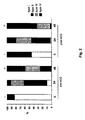
- FIG. 1 Schematic diagram of the study is shown in Fig. 1 .
- DDMP was added at a 10 ⁇ M final concentration (0.1 % DMSO). Directly after aspiration, single COCs were washed in flushing medium (D-MEM with HEPES) and placed into 10 ⁇ l droplets under oil. COCs were randomised over two culture conditions: with and without DDMP. Oocytes cultured with DDMP for 24 hours, 48 hours and 72 hours were studied for reversal of meiosis arrest by washing out the inhibitor. Reversal of PDE 3 inhibition was performed by mechanical denudation of the remaining somatic cells and placing the oocytes in the conditioned medium for an additional 24-30 h.
- part of the oocytes with and without DDMP were fixed for EM analysis.
- a total of 48 oocytes (4 at time of retrieval, 21 after 24 hours, 20 after 48 hours and 3 after 72 hours culture) were fixed in glutaraldehyde 2.5% in cacodylate buffer at pH 7.3 for at least 2 hours at 4°C at time 0 h, 24 hours and 48 hours of maturation. All oocytes were embedded individually. Afterwards, they were postfixed in OsO 4 1% in H 2 0 for 30 min, stained with uranyl acetate 2% in veronal buffer for 60 min, dehydrated through graded alcohols, embedded in SPURR (Taab; Bodson, Liège, Belgium) and left to polymerize overnight at 70°C.
- SPURR Taab; Bodson, Liège, Belgium
- Semithin sections of 0.5 ⁇ m and ultrathin sections of 75 nm were made at approximately 30 levels (range 15-35). The semithin sections were mounted on glass slides and stained with toluidine blue for light microscopical guidance. The ultrathin sections were transferred on wide single slot copper grids (Agar Scientific LTD, Stansted, UK), coated with a polyvinyl formal film (Formvar, Laborimpex, Brussels, Belgium), stained with lead citrate and examined using a Zeiss transmission electron microscope type EM 9S2.
- oocytes For localization of microfilaments, 39 polar body extruded oocytes (25 oocytes 48 hours after maturation in control medium and 14 oocytes after inhibitor removal) were fixed with 2.5% glutaraldehyde in cacodylate buffer for at least 2 hours, washed with PBS/1% BSA, placed into 0.1% Triton X-100 for 5 min and rinsed again with PBS. Afterwards, oocytes were placed into Texas red-labeled phalloidin staining f-actin diluted 1:40 PBS for f-actin and Pico green 1:2000 PBS for DNA staining.
- a total of 139 follicles were aspirated from 30 patients before any hCG administration.
- the follicles between 6 and 12 mm (45.3%) yielded an oocyte recovery rate of 80.9 % (mean ⁇ SD of oocytes per patient: 3.2 ⁇ 2.2; range 1-9).
- the aspirated follicles larger than 12 mm (54.7%) yielded an oocyte recovery rate of 35.5 % (mean ⁇ SD of oocytes per patient: 1.7 ⁇ 0.9; range 1-4).
- a total of 128 follicles between 6 and 12 mm were aspirated from 27 patients at 36 hours after hCG administration. These yielded an oocyte recovery of 50.0% (mean ⁇ SD of oocyte per patient: 2.4 ⁇ 1.5; range 1-6).
- a total of 54 follicles between 13 and 15 mm were aspirated from 9 patients with an endogenous LH peak a day before follicle aspiration. These yielded an oocyte recovery of 37.0% (mean ⁇ SD of oocyte per patient: 2.2 ⁇ 0.8; range 2-4).
- This group comprises the patients in whom hCG was administered for 36 hours (normal IVF/ICSI procedure) and those patients who had an endogenous LH surge.
- the COCs retrieved from the post-hCG patients presented a significantly lower proportion of type I COCs compared to pre-hCG patients (45.0%) (p ⁇ 0.01).
- a significantly higher proportion of oocytes had lost their connections with the cells compared to the cultured ones from the pre-hCG group (66.7%) (p ⁇ 0.05).
- the proportion of type I COCs retrieved from the post-hCG patients was significantly lower (48.8%) compared to those retrieved from pre-hCG patients (p ⁇ 0.05). Twenty-four hours later, only 4.7% from post-hCG cumuluses remained type I. This constitutes a significantly lower proportion than the pre-hCG cumulus (p ⁇ 0.05). There were 57.1 % of type II and 30.9% type III cumuluses (not significantly different from pre-hCG). After 24 hours, a similar proportion of oocytes as in the pre-hCG group had lost connection with cumulus cells (7.1 %). Finally at 48 hours culture, 62.1% of oocytes had lost connections with the somatic cells, this is higher than COCs retrieved from the pre-hCG group (p ⁇ 0.05).
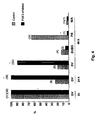
- COCs in which the nuclear stage was not clearly visualized were mechanically denuded. Those that were fixed for EM analysis had their maturational stage determined afterwards (on the semithin sections). Oocytes cultured in presence of the DDMP, remained for 88.9% at the GV-stage in contrast to only 19.3% in controls (p ⁇ 0.001). In the presence of DDMP, only 5.5% of oocytes were GVBD, 2.8% were PB and in 2.8% the stage could not be determined. In controls, 12.9% were GVBD, significantly more oocytes had a PB extruded (64.5%) (p ⁇ 0.001) and 3.2% had a non determinable stage.
- COCs cultured in presence of DDMP showed a significant greater proportion of oocytes at the GV-stage (83.3%) compared to controls (6.0%) (p ⁇ 0.001).
- DDMP 8.3% of oocytes were GVBD, only 5.6% were PB and 2.8% had a not determined stage.
- 12.1% were GVBD and a significant greater proportion of oocytes had a PB extruded (81.8%) (p ⁇ 0.001).
- the relation between polar body extruded oocytes and follicle diameter is shown in Tale 2.
- GV-stage oocytes were mechanically denuded from cumulus cells. Inhibitor was washed out (11 from pre-hCG and 11 from post-hCG group) and oocytes were further cultured for 24-30 h in control medium. From the pre-hCG patients group, 4 progressed to GVBD but 1 arrested at this stage and 3 of them extruded the polar body at 24-30h. From the post-hCG patients group, 11 oocytes progressed to GVBD but 5 arrested at this stage and 6 extruded the polar body (Table 3). Table 3.
- oocytes at different stages of meiotic progression (GV, GVBD, PB) as observed at 24-30 hours after removal of DDMP from the maturation medium.
- Data grouped by duration of culture days in DDMP supplemented medium and by type of patient treatment (hCG or not) Day of Inhibitor removal Patient treatment Number of oocytes Oocyte stage 24-30 hours after DDMP removal GV GVBD PB (%) 2 pre-hCG 11 7 1 3 (27) a post-hCG 11 - 5 6 (54) c 3 pre-hCG 13 6 1 6 (46) a post-hCG 10 - - 10 (100) bd a and b denotes a significant difference by Fisher 's exact test (p ⁇ 0.01). c and d denotes a significant difference by ⁇ 2 test (p ⁇ 0.05).
- Oocytes analyzed are grouped by type of patient treatment and culture conditions Number of oocytes analyzed at different times of culture in the different stages of nuclear maturity Patient treatment Number of oocytes at time of retrieval Culture conditions After 24 hours culture After 48 hours culture After 72 hours culture GV GVBD PB GV GVBD PB GV Pre-hCG 2 Control 2 4 9 Inhibitor 3 4 1 3 Post-hCG 2 Control 2 1 4 1 2 Inhibitor 3 1 1 3 Total 4 8 4 9 7 1 12 3
- the nucleus consisted of an envelope formed by two layers of membrane, containing one dense compact nucleolus associated with inter-chromatin granular complexes and extranucleolar chromatoid bodies on the periphery of nucleoli (Sathananthan, 1985).
- the nucleoli are regularly shaped, round or oval, homogeneous and formed by fibrillar granules.
- nucleolus-associated chromatin was observed as a continuous mass (karyosphere), which normally exists in oocytes obtained from antral follicles of 8 mm and larger (Tesarik et al, 1983).
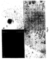
- the karyosphere also called as surrounded nucleolus (SN), occupied an excentric position in the nucleus and consists of loosely packed fibrills in contact to the nucleolus ( Fig. 6b ).
- the presence of karyosphere was independent of the position of the nucleus in the oocyte (only in one oocyte the nucleus was at the periphery).
- a characteristic of all the oocytes analyzed at retrieval was the presence of areas devoid of organelles at the cortex and intermediate cytoplasm. Clumps of mitochondriae were found distributed throughout the cytoplasm ( Fig. 6a,c ). At the surrounding of the GV aggregates of mitochondria were intermingled with vesicles of different diameter. Cortical granules were found dispersed throughout the cytoplasm and few were observed under the oolemma of the oocyte forming a single layer.
- Microvilli were noticed extending from the plasma membrane through the inner cortex of the zona pellucida (ZP). Numerous cell projections penetrated the zona pellucida to the oolemma. The granulosa cells (GC) appeared as oval or round cells apposed to the ZP with numerous cellular projections crossing the ZP reaching the oolemma. Cells displayed a compact arrangement and tightly adhered to each other by closely apposed plasma membranes resembling gap junctions ( Fig. 6d ). The nuclei of the cells are excentrically located. Granulosa cells of the COC did not show signs of pyknosis.
- GC granulosa cells
- Electron microscopy analyses of oocytes cultured in control medium demonstrated that in vitro maturation was associated with reorganization of mitochondria and smooth endoplasmic reticulum (SER) which appear as round vesicles into cytoplasm. This reorganization was not different for oocytes retrieved pre or post-hCG stimulation. Mitochondriae are spherical or oval and are dispersed throughout the cytoplasm or around aggregates of SER. These aggregates form characteristic complexes with peripheral mitochondria in the maturing human oocytes ( vide Ann. NY Acad Sci. 442 (1986), 251 et seq .. and Prog. Clin. Biol. Res. 296 (1989), 273 et seq .) and were predominantly present in the PB-extruded ones.
- SER smooth endoplasmic reticulum
- Cortical granules showed a discontinuous distribution on the surface of the GVBD oocytes and appeared in one to two layers at the cortex of PB-extruded oocytes.
- the oocyte that remained at the GV-stage after 24 hours in control medium had the same type of nucleus (with presence of karysphere) and organelle distribution as for the GVs fixed and analyzed at time of retrieval ( Fig. 7b ).
- the GV of the oocytes were centrally located with presence of a karyosphere around the nucleoli of all oocytes.
- the nuclear membranes of all oocytes had remained intact ( Fig. 8b ).
- oocytes showed signs of immature cytoplasm similar to the ones fixed at time of retrieval. Usually, the mitochondriae were found distributed in clumps throughout the cytoplasm ( Fig. 8c ). In the surroundings of the GV, aggregates of mitochondria were intermingled with vesicles of different diameter. Few cortical granules were found dispersed throughout the cytoplasm and beneath the oolemma of the oocyte, forming a single layer. A single GV-stage oocyte (retrieved from a 9mm post-hCG follicle aspiration) had no presence of karyosphere in the nucleus and a more homogeneous distribution of organelles in the cytoplasm.
- the cytoplasm of the GVBD oocyte analysed showed an inhibition of the distribution of organelles, which were aggregated as in the cytoplasm of a GV-stage oocyte.
- the oocyte that underwent PB extrusion had the same cytoplasmic morphology as the control ones.
- the cumulus cells started to expand and loose contact with the oocyte. Cumulus cells started their elongation, although some cells were still round or oval. The nuclei of these cells did not yet migrate to the periphery ( Fig. 8a ).
- the organelles, mitochondriae and SER were spread throughout the ooplasm as vesicles or as aggregates of tubular elements.
- the CG formed one to three layers under the oolema, and in one matured oocyte (pre-hCG), numerous CG were conglomerated beneath the oolema at the place of the meiotic spindle.
- the GV-stage oocytes fixed and analyzed after 48 hours and 72 hours culture in the presence of the inhibitor showed a centrally located nucleus with presence of karyosphere around the nucleoli in almost all oocytes ( Fig. 10a ).
- One GV-stage oocyte at 48 hours (retrieved from one 8 mm cross-hCG follicle aspiration) had no presence of karyosphere in the nucleus and a more homogeneous distribution of organelles in the cytoplasm.
- the organelles were still aggregated, although in some oocytes the mitochondria started to dissociate from their aggregates.
- the oocytes showed signs of vacuolization in the ooplasm, resulting from swelling of SER ( Fig. 11 a, b ). Microvilli were less evident and zona was more homogeneous with no transzonal processes from cumulus cells, indicating a complete loss of connections with the oocyte ( Fig. 11 b) .
- MII metaphase II
- Four oocytes had nuclei forming a clump of chromosomes at the place of metaphase plate, probably as a sign of ageing.
- Microfilaments were observed in the scanned levels of oocytes with a more prominent staining at the optical sections close to the site of first polar body extrusion described as generalized pattern (Terada et al, 1995). No superposed microfilaments of actin were observed at the site of polar body extrusion, showing a homogeneous staining at this level.
- oocytes presented the M II plate 24-30 hours after inhibitor removal (3 out of 3 pre-hCG, and 9 out of 11 post-hCG) in which 83.3% had were well-aligned chromosomes and positioned perpendicular to the oolema ( Fig. 13 ).
- Two oocytes had nuclei forming a clump of chromosomes at place of metaphase plate.
- the generalized aspect of microfilaments was also observed in those oocytes with homogeneous prominent staining at the level of the polar body extrusion site ( Fig. 13 ).
- DDMP DDMP-induced oocyte maturation.
- Prolonged nuclear arrest by DDMP in our in vitro maturation medium seemed to affect the migration of organelles in the GV-stage oocytes.
- the organelles start to dissociate from the aggregates after 48 hours in DDMP culture, even in GV-intact oocytes.
- cortical granules had formed a characteristic single layer, while in contrast controls had their cortical granules still conglomerated.
- MAS compound designates compounds which mediate the meiosis of oocytes. More specifically, a MAS compound is a compound which in the test described below in this example has a percentage germinal vesicle breakdown (hereinafter designated GVB) which is significantly higher than the control. Preferred MAS compounds are such having a percentage GVB of at least 50%, preferably at least 80%.
- MAS compounds are mentioned in WO 96/00235 , 96/27658 , 97/00884 , 98/28323 , 98/54965 and 98/55498 , more specifically in Claim 1 thereof.
- Oocytes were obtained from immature female mice (C57BL/6J x DBA/2J F1, Bomholtgaard, Denmark) weighing 13-16 grams, that were kept under controlled temperature (20-22 °C), light (lights on 06.00-18.00) and relative humidity (50-70%).
- the mice received an intraperitoneal injection of 0.2 ml gonadotropins (Gonal-F, Serono) containing 20 IU FSH and 48 hours later the animals were killed by cervical dislocation.
- the ovaries were dissected out and the oocytes were isolated in Hx-medium (see below) under a stereomicroscope by manual rupture of the follicles using a pair of 27 gauge needles.
- Spherical oocytes displaying an intact germinal vesicle (hereinafter designated GV) were divided in cumulus enclosed oocytes (hereinafter designated CEO) and naked oocytes (hereinafter designated NO) and placed in ⁇ -minimum essential medium ( ⁇ -MEM without ribonucleosides, Gibco BRL, Cat. No. 22561) supplemented with 3 mg/ml bovine serum albumin (BSA, Sigma Cat. No. A-7030), 5 mg/ml human serum albumin (HSA, Statens Seruminstitut, Denmark), 0.23mM pyruvate (Sigma, Cat. No S-8636), 2 mM glutamine (Flow Cat. No.
- BSA bovine serum albumin
- HSA human serum albumin
- HSA human serum albumin
- S-8636 2 mM glutamine
- Hx-medium 100 IU/ml penicillin and 100 ⁇ g/ml streptomycin (Flow, Cat No. 16-700). This medium was supplemented with 3 mM hypoxanthine (Sigma Cat. No. H-9377) and designated Hx-medium.
- the oocytes were rinsed three times in Hx-medium and oocytes of uniform size were divided into groups of CEO and NO. CEO and NO were cultured in 4-well multidishes (Nunclon, Denmark) in which each well contained 0.4 ml of Hx-medium and the compound to be tested in a concentration of 10 ⁇ M.
- One control well i.e., 35-45 oocytes cultured in identical medium with no addition of test compound
- 3 test wells 35-45 oocytes per well supplemented with test compound
- the oocytes were cultured in a humidified atmosphere of 5% CO 2 in air for 24 hours at 37°C.
- PB polar bodies
Landscapes
- Health & Medical Sciences (AREA)
- Engineering & Computer Science (AREA)
- Life Sciences & Earth Sciences (AREA)
- Biomedical Technology (AREA)
- Bioinformatics & Cheminformatics (AREA)
- Organic Chemistry (AREA)
- Chemical & Material Sciences (AREA)
- Genetics & Genomics (AREA)
- Wood Science & Technology (AREA)
- Zoology (AREA)
- Biotechnology (AREA)
- General Health & Medical Sciences (AREA)
- Reproductive Health (AREA)
- Microbiology (AREA)
- Biochemistry (AREA)
- General Engineering & Computer Science (AREA)
- Developmental Biology & Embryology (AREA)
- Cell Biology (AREA)
- Endocrinology (AREA)
- Pharmacology & Pharmacy (AREA)
- Gynecology & Obstetrics (AREA)
- Chemical Kinetics & Catalysis (AREA)
- General Chemical & Material Sciences (AREA)
- Medicinal Chemistry (AREA)
- Nuclear Medicine, Radiotherapy & Molecular Imaging (AREA)
- Pregnancy & Childbirth (AREA)
- Animal Behavior & Ethology (AREA)
- Public Health (AREA)
- Veterinary Medicine (AREA)
- Micro-Organisms Or Cultivation Processes Thereof (AREA)
- Enzymes And Modification Thereof (AREA)
- Preparation Of Compounds By Using Micro-Organisms (AREA)
- Measuring Or Testing Involving Enzymes Or Micro-Organisms (AREA)
Claims (29)
- Verfahren zur in-vitro-Synchronisation der Kern- und Zytoplasmareifung von GV-Oozyten von Haustieren oder Primaten, umfassend die Schritte:a. Kultivieren von einem oder mehreren GV-Oozyten oder MI-Oozyten von Haustieren oder Menschen in einem ersten Kulturmedium, wobei das Kulturmedium ein Pyridazinonderivat der allgemeinen Formel 1
 b. Waschen der Oozyten von Schritt (a) zum Entfernen der Verbindung der allgemeinen Formel I oder Vermindern der Menge davon;c. Kultivieren der gewaschenen Oozyten von Schritt (b) in einem zweiten Kulturmedium für eine Zeitdauer, die für die Vollständigkeit der Kernreifung bis zur Metaphase II mit einer gewünschten Häufigkeit ausreichend ist.
 - Verfahren nach Anspruch 1, wobei menschliche Oozyten verwendet werden.
- Verfahren nach einem der vorangehenden Ansprüche, wobei es sich bei den zum Kultivieren im ersten Medium verwendeten Oozyten um Oozyten handelt, die in einem Oozytensammelmedium, bei welchem es sich um ein CO2-unabhängiges Medium, enthaltend eine Verbindung der allgemeinen Formel I, handelt, aufbewahrt worden sind.
- Verfahren nach dem vorangehenden Anspruch, wobei der Oozyt nicht später als 4 Stunden, vorzugsweise nicht später als 2 Stunden, noch stärker bevorzugt nicht später als 1 Stunde und noch stärker bevorzugt nicht später als ½ Stunde nach dem Entfernen des Oozyten aus der Frau in das Oozytensammelmedium gegeben worden ist.
- Verfahren nach einem der vorangehenden Ansprüche, wobei das Kultivieren gemäß Schritt (a) für eine Zeitdauer stattfindet, die für eine Verbesserung der Zytoplasmareifung ausreichend ist.
- Verfahren nach dem vorangehenden Anspruch, wobei das Kultivieren gemäß Schritt (a) für eine Zeitdauer stattfindet, die zur Vollständigkeit der Zytoplasmareifung ausreichend ist.
- Verfahren nach einem der vorangehenden Ansprüche, wobei es sich bei den in Schritt (a) kultivierten Oozyten um GV-Oozyten handelt.
- Verfahren nach einem der vorangehenden Ansprüche, wobei das erste Kulturmedium EGF, GH, Gonadotropinkombinationen, Verstärker für die Glutathionsynthese, meioseaktivierende Verbindungen, Kombinationen von Activin und Inhibin, die Analoga von Vorstehendem oder Kombinationen der vorstehend erwähnten Verbindungen handelt.
- Verfahren nach einem der vorangehenden Ansprüche, wobei es sich bei der Verbindung der allgemeinen Formel I um DDMP handelt.
- Verfahren nach einem der vorangehenden Ansprüche, wobei die Kultivierungszeit in Schritt (a) mindestens etwa 4 Stunden beträgt.
- Verfahren nach einem der vorangehenden Ansprüche, wobei die Kultivierungszeit mindestens etwa 8 Stunden beträgt.
- Verfahren nach einem der vorangehenden Ansprüche, wobei die Kultivierungszeit mindestens etwa 12 Stunden beträgt.
- Verfahren nach einem der vorangehenden Ansprüche, wobei die Kultivierungszeit mindestens etwa 24 Stunden beträgt.
- Verfahren nach einem der vorangehenden Ansprüche, wobei die Kultivierungszeit mindestens etwa 48 Stunden beträgt.
- Verfahren nach einem der vorangehenden Ansprüche, wobei die Kultivierungszeit mindestens etwa 72 Stunden beträgt.
- Verfahren nach einem der vorangehenden Ansprüche, wobei die in Schritt (a) verwendeten Oozyten von Follikel stammen, die von dem Tier/Menschen (periodisierenden Tier/periodisierenden Menschen oder nicht) vor jeglicher Hormonbehandlung, die entweder auf die Follikulogenese und/oder Oogenese einwirkt, abgesaugt wurden.
- Verfahren nach dem vorangehenden Anspruch, wobei die Hormonbehandlung der Frau, von welcher die Oozyten stammen, mit einem Steroid, einem Gonadotropin, einem GnRH-Analogon, Clomiphencitrat, Tamoxiphen, einem Insulinsensibilisator (z.B. Metformin), einem Aromatasehemmer oder beliebigen der vorangehenden Arzneistoffe allein oder kombiniert durchgeführt worden ist.
- Verfahren nach einem der vorangehenden Ansprüche, wobei die Oozyten von Follikel stammen, die vom Tier nach jeglicher Hormon- oder Arzneistoffbehandlung, die auf die Follikulogenese und/oder Oogenese einwirkt, abgesaugt wurden.
- Verfahren nach dem vorangehenden Anspruch, wobei die Hormon- oder Arzneistoffbehandlung mit einem Gonadotropin, einem GnRH-Analogon, Clomiphencitrat, Tamoxipen, LH, HCG, einem Steroid oder einer steroidartigen Substanz, einem LHRH-Analogon, einem Aromatasehemmer, einem Insulinsensibilisator und einem beliebigen Analogon oder einer beliebigen Kombination davon durchgeführt worden ist.
- Verfahren nach einem der beiden vorangehenden Ansprüche, wobei das verwendete Hormon hCG, LH oder eine beliebige Verbindung, die ähnliche Wirkungen herbeiführen könnte, ist.
- Verfahren nach einem der vorangehenden Ansprüche, wobei das Waschen der Oozyten von Schritt (a) derart durchgeführt wird, dass die Konzentration der Verbindung der allgemeinen Formel I nach dem Herauswaschen der Verbindung im zweiten Medium weniger als etwa 10%, vorzugsweise weniger als etwa 5%, stärker bevorzugt weniger als etwa 1 % der Konzentration der Verbindung im ersten Medium beträgt.
- Verfahren nach einem der vorangehenden Ansprüche, wobei das erste Kulturmedium derart minimal durch eine dünne Schicht aus Öl bedeckt ist, dass das Verhältnis zwischen der öligen Phase und der wässrigen Phase weniger als etwa 2:10 Vol.-%, vorzugsweise weniger als etwa 1:10 Vol.-% beträgt.
- Verfahren nach einem der vorangehenden Ansprüche, wobei das Oozytensammelmedium mit Öl bedeckt ist.
- Verfahren nach einem der vorangehenden Ansprüche, wobei in Schritt (a) Kumuluszellen um den Oozyten vorliegen.
- Verfahren nach einem der vorangehenden Ansprüche, wobei in Schritt (b) das Waschen der Oozyten von Schritt (a) zum Entfernen der Verbindung der allgemeinen Formel I oder Vermindern der Menge davon derart durchgeführt wird, dass die Restmenge der Verbindung, falls überhaupt, im Wesentlichen keine Wirkung auf die Hemmung der Oozyten aufweist.
- Verfahren nach einem der vorangehenden Ansprüche, wobei in Schritt (c) keine oder im Wesentlichen keine Kumuluszellen um den Oozyten vorliegen.
- Verfahren nach einem der vorangehenden Ansprüche, wobei in Schritt (c) die Kernreifung durch Zugabe von HCG und/oder EGF und/oder einer MAS-Verbindung zu dem zweiten Kulturmedium stimuliert wird.
- Verfahren nach einem der vorangehenden Ansprüche, wobei in Schritt (c) die gewünschte Häufigkeit mindestens etwa 30%, stärker bevorzugt mindestens etwa 50%, noch stärker bevorzugt mindestens etwa 70% und vorzugsweise mindestens etwa 90% beträgt.
- Verfahren nach einem der vorangehenden Ansprüche, wobei nach Schritt (c) die Oozyten mit Sperma vorzugsweise unter Verwendung von ICSI befruchtet werden.
Applications Claiming Priority (3)
| Application Number | Priority Date | Filing Date | Title |
|---|---|---|---|
| DK200201581 | 2002-10-15 | ||
| DKPA200201581 | 2002-10-15 | ||
| PCT/DK2003/000703 WO2004035766A2 (en) | 2002-10-15 | 2003-10-15 | In vitro synchronisation of nuclear and cytoplasmatic maturation of oocytes involving addition and removal of phosphodiesterase type 3 inhibitor |
Publications (2)
| Publication Number | Publication Date |
|---|---|
| EP1554016A2 EP1554016A2 (de) | 2005-07-20 |
| EP1554016B1 true EP1554016B1 (de) | 2008-05-14 |
Family
ID=32103846
Family Applications (1)
| Application Number | Title | Priority Date | Filing Date |
|---|---|---|---|
| EP03757724A Expired - Lifetime EP1554016B1 (de) | 2002-10-15 | 2003-10-15 | In-vitro-synchronisation der kern- und zytoplasma-reifung von oozyten mit addition und entfernung von phosphodiesterase-hemmer typ 3 |
Country Status (7)
| Country | Link |
|---|---|
| US (2) | US20060051863A1 (de) |
| EP (1) | EP1554016B1 (de) |
| JP (1) | JP2006502717A (de) |
| AT (1) | ATE395102T1 (de) |
| AU (1) | AU2003273767A1 (de) |
| DE (1) | DE60321026D1 (de) |
| WO (1) | WO2004035766A2 (de) |
Families Citing this family (5)
| Publication number | Priority date | Publication date | Assignee | Title |
|---|---|---|---|---|
| DK2094086T3 (da) | 2006-11-08 | 2013-11-25 | Veritas Bio LLC | Indgivelse in vivo af dobbeltstrenget rna til en målcelle |
| CZ302232B6 (cs) * | 2008-07-15 | 2011-01-05 | Ceská zemedelská univerzita v Praze | Zpusob zvýšení vývojové schopnosti rostoucích oocytu inhibicí kalcineurinu |
| EP2430154A4 (de) * | 2009-05-14 | 2014-01-08 | Adelaide Res & Innovation Pty | Verfahren zur entnahme und reifung von oozyten |
| WO2016094970A1 (en) * | 2014-12-19 | 2016-06-23 | Vrije Universiteit Brussel | In vitro maturation of a mammalian cumulus oocyte complex |
| WO2024024742A1 (ja) * | 2022-07-25 | 2024-02-01 | 国立大学法人京都大学 | 霊長類の胎児卵巣細胞から卵胞を誘導する体外培養法 |
Family Cites Families (4)
| Publication number | Priority date | Publication date | Assignee | Title |
|---|---|---|---|---|
| FR2678506B1 (fr) * | 1991-07-01 | 2000-03-10 | Claude Ranoux | Procede de fecondation en cycle spontane. |
| US6110471A (en) * | 1996-09-13 | 2000-08-29 | The Board Of Trustees Of The Leland Stanford Junior University | Non-hormonal method of contraception |
| AU1692901A (en) * | 1999-11-25 | 2001-06-04 | Novo Nordisk A/S | Treatment of human infertility |
| JP2003529378A (ja) * | 2000-04-06 | 2003-10-07 | ノボ ノルディスク アクティーゼルスカブ | 卵母細胞の細胞質および核成熟の体外成熟方法 |
-
2003
- 2003-10-15 AT AT03757724T patent/ATE395102T1/de not_active IP Right Cessation
- 2003-10-15 DE DE60321026T patent/DE60321026D1/de not_active Expired - Fee Related
- 2003-10-15 AU AU2003273767A patent/AU2003273767A1/en not_active Abandoned
- 2003-10-15 EP EP03757724A patent/EP1554016B1/de not_active Expired - Lifetime
- 2003-10-15 WO PCT/DK2003/000703 patent/WO2004035766A2/en not_active Ceased
- 2003-10-15 JP JP2004543981A patent/JP2006502717A/ja active Pending
-
2005
- 2005-04-15 US US11/107,642 patent/US20060051863A1/en not_active Abandoned
-
2008
- 2008-08-12 US US12/228,349 patent/US20080311656A1/en not_active Abandoned
Also Published As
| Publication number | Publication date |
|---|---|
| US20060051863A1 (en) | 2006-03-09 |
| AU2003273767A1 (en) | 2004-05-04 |
| JP2006502717A (ja) | 2006-01-26 |
| DE60321026D1 (de) | 2008-06-26 |
| ATE395102T1 (de) | 2008-05-15 |
| WO2004035766A2 (en) | 2004-04-29 |
| WO2004035766A3 (en) | 2004-06-03 |
| US20080311656A1 (en) | 2008-12-18 |
| EP1554016A2 (de) | 2005-07-20 |
Similar Documents
| Publication | Publication Date | Title |
|---|---|---|
| EP1156821B1 (de) | Behandlung von unfruchtbarkeit | |
| Jones et al. | Evolution of a culture protocol for successful blastocyst development and pregnancy. | |
| Shi et al. | Aberrant methylation patterns at the two‐cell stage as an indicator of early developmental failure | |
| US6110741A (en) | Gonadotrophin releasor hormone-containing composition for embryo culture and method for in vitro fertilization | |
| EP1156822B1 (de) | In vitro methode zur synchronisation von oozytenreifung | |
| KR20120023097A (ko) | 난모세포의 수집 및 성숙 방법 | |
| US20080311656A1 (en) | Meiosis arrest in oocytes in vitro | |
| KR20010043828A (ko) | cAMP-증강 화합물 단독 또는 감수분열-자극 화합물하나 이상과의 조합에 의한 불임 치료 방법 | |
| EP1235899B1 (de) | Behandlung von menschlicher unfruchtbarkeit | |
| US6544166B1 (en) | Treatment of human infertility | |
| US7192768B2 (en) | Synchronization of the cytoplasmatic and the nuclear maturation of oocytes in vitro | |
| US20040224878A1 (en) | Fertilization of aged oocytes | |
| Cavilla | The effects of factors influencing human oocyte maturation upon fertilization and preimplantation embryo development | |
| Kunathikom et al. | Comparison of pronuclear stage embryo transfer and in vitro fertilization-embryo transfer following intracytoplasmic sperm injection | |
| Ayoub | Studies on in vitro maturation and fertilization of bovine follicular oocytes | |
| MXPA01008452A (en) | Treatment of infertility | |
| ZA200104803B (en) | Treatment of infertility. | |
| ZA200104653B (en) | Treatment of infertility. | |
| CA2362940A1 (en) | Treatment of infertility | |
| HK1197557B (en) | Compositions and methods for enhancing bioenergetic status in female germ cells |
Legal Events
| Date | Code | Title | Description |
|---|---|---|---|
| PUAI | Public reference made under article 153(3) epc to a published international application that has entered the european phase |
Free format text: ORIGINAL CODE: 0009012 |
|
| 17P | Request for examination filed |
Effective date: 20050517 |
|
| AK | Designated contracting states |
Kind code of ref document: A2 Designated state(s): AT BE BG CH CY CZ DE DK EE ES FI FR GB GR HU IE IT LI LU MC NL PT RO SE SI SK TR |
|
| AX | Request for extension of the european patent |
Extension state: AL LT LV MK |
|
| DAX | Request for extension of the european patent (deleted) | ||
| RAP1 | Party data changed (applicant data changed or rights of an application transferred) |
Owner name: NOVO NORDISK A/S Owner name: BAYER SCHERING PHARMA AG |
|
| RAP1 | Party data changed (applicant data changed or rights of an application transferred) |
Owner name: BAYER SCHERING PHARMA AKTIENGESELLSCHAFT Owner name: NOVO NORDISK A/S |
|
| GRAP | Despatch of communication of intention to grant a patent |
Free format text: ORIGINAL CODE: EPIDOSNIGR1 |
|
| GRAS | Grant fee paid |
Free format text: ORIGINAL CODE: EPIDOSNIGR3 |
|
| GRAA | (expected) grant |
Free format text: ORIGINAL CODE: 0009210 |
|
| AK | Designated contracting states |
Kind code of ref document: B1 Designated state(s): AT BE BG CH CY CZ DE DK EE ES FI FR GB GR HU IE IT LI LU MC NL PT RO SE SI SK TR |
|
| REG | Reference to a national code |
Ref country code: GB Ref legal event code: FG4D |
|
| REG | Reference to a national code |
Ref country code: CH Ref legal event code: EP |
|
| REG | Reference to a national code |
Ref country code: IE Ref legal event code: FG4D Free format text: LANGUAGE OF EP DOCUMENT: FRENCH |
|
| REF | Corresponds to: |
Ref document number: 60321026 Country of ref document: DE Date of ref document: 20080626 Kind code of ref document: P |
|
| PG25 | Lapsed in a contracting state [announced via postgrant information from national office to epo] |
Ref country code: SI Free format text: LAPSE BECAUSE OF FAILURE TO SUBMIT A TRANSLATION OF THE DESCRIPTION OR TO PAY THE FEE WITHIN THE PRESCRIBED TIME-LIMIT Effective date: 20080514 |
|
| PG25 | Lapsed in a contracting state [announced via postgrant information from national office to epo] |
Ref country code: FI Free format text: LAPSE BECAUSE OF FAILURE TO SUBMIT A TRANSLATION OF THE DESCRIPTION OR TO PAY THE FEE WITHIN THE PRESCRIBED TIME-LIMIT Effective date: 20080514 Ref country code: ES Free format text: LAPSE BECAUSE OF FAILURE TO SUBMIT A TRANSLATION OF THE DESCRIPTION OR TO PAY THE FEE WITHIN THE PRESCRIBED TIME-LIMIT Effective date: 20080825 |
|
| NLV1 | Nl: lapsed or annulled due to failure to fulfill the requirements of art. 29p and 29m of the patents act | ||
| PG25 | Lapsed in a contracting state [announced via postgrant information from national office to epo] |
Ref country code: AT Free format text: LAPSE BECAUSE OF FAILURE TO SUBMIT A TRANSLATION OF THE DESCRIPTION OR TO PAY THE FEE WITHIN THE PRESCRIBED TIME-LIMIT Effective date: 20080514 Ref country code: NL Free format text: LAPSE BECAUSE OF FAILURE TO SUBMIT A TRANSLATION OF THE DESCRIPTION OR TO PAY THE FEE WITHIN THE PRESCRIBED TIME-LIMIT Effective date: 20080514 |
|
| PG25 | Lapsed in a contracting state [announced via postgrant information from national office to epo] |
Ref country code: SE Free format text: LAPSE BECAUSE OF FAILURE TO SUBMIT A TRANSLATION OF THE DESCRIPTION OR TO PAY THE FEE WITHIN THE PRESCRIBED TIME-LIMIT Effective date: 20080814 Ref country code: PT Free format text: LAPSE BECAUSE OF FAILURE TO SUBMIT A TRANSLATION OF THE DESCRIPTION OR TO PAY THE FEE WITHIN THE PRESCRIBED TIME-LIMIT Effective date: 20081014 Ref country code: CZ Free format text: LAPSE BECAUSE OF FAILURE TO SUBMIT A TRANSLATION OF THE DESCRIPTION OR TO PAY THE FEE WITHIN THE PRESCRIBED TIME-LIMIT Effective date: 20080514 Ref country code: DK Free format text: LAPSE BECAUSE OF FAILURE TO SUBMIT A TRANSLATION OF THE DESCRIPTION OR TO PAY THE FEE WITHIN THE PRESCRIBED TIME-LIMIT Effective date: 20080514 |
|
| PGFP | Annual fee paid to national office [announced via postgrant information from national office to epo] |
Ref country code: DE Payment date: 20081027 Year of fee payment: 6 |
|
| PG25 | Lapsed in a contracting state [announced via postgrant information from national office to epo] |
Ref country code: BE Free format text: LAPSE BECAUSE OF FAILURE TO SUBMIT A TRANSLATION OF THE DESCRIPTION OR TO PAY THE FEE WITHIN THE PRESCRIBED TIME-LIMIT Effective date: 20080514 Ref country code: SK Free format text: LAPSE BECAUSE OF FAILURE TO SUBMIT A TRANSLATION OF THE DESCRIPTION OR TO PAY THE FEE WITHIN THE PRESCRIBED TIME-LIMIT Effective date: 20080514 Ref country code: RO Free format text: LAPSE BECAUSE OF FAILURE TO SUBMIT A TRANSLATION OF THE DESCRIPTION OR TO PAY THE FEE WITHIN THE PRESCRIBED TIME-LIMIT Effective date: 20080514 |
|
| PLBE | No opposition filed within time limit |
Free format text: ORIGINAL CODE: 0009261 |
|
| STAA | Information on the status of an ep patent application or granted ep patent |
Free format text: STATUS: NO OPPOSITION FILED WITHIN TIME LIMIT |
|
| 26N | No opposition filed |
Effective date: 20090217 |
|
| PG25 | Lapsed in a contracting state [announced via postgrant information from national office to epo] |
Ref country code: EE Free format text: LAPSE BECAUSE OF FAILURE TO SUBMIT A TRANSLATION OF THE DESCRIPTION OR TO PAY THE FEE WITHIN THE PRESCRIBED TIME-LIMIT Effective date: 20080514 Ref country code: BG Free format text: LAPSE BECAUSE OF FAILURE TO SUBMIT A TRANSLATION OF THE DESCRIPTION OR TO PAY THE FEE WITHIN THE PRESCRIBED TIME-LIMIT Effective date: 20080814 |
|
| PG25 | Lapsed in a contracting state [announced via postgrant information from national office to epo] |
Ref country code: MC Free format text: LAPSE BECAUSE OF NON-PAYMENT OF DUE FEES Effective date: 20081031 |
|
| REG | Reference to a national code |
Ref country code: CH Ref legal event code: PL |
|
| GBPC | Gb: european patent ceased through non-payment of renewal fee |
Effective date: 20081015 |
|
| REG | Reference to a national code |
Ref country code: IE Ref legal event code: MM4A |
|
| REG | Reference to a national code |
Ref country code: FR Ref legal event code: ST Effective date: 20090630 |
|
| PG25 | Lapsed in a contracting state [announced via postgrant information from national office to epo] |
Ref country code: IT Free format text: LAPSE BECAUSE OF FAILURE TO SUBMIT A TRANSLATION OF THE DESCRIPTION OR TO PAY THE FEE WITHIN THE PRESCRIBED TIME-LIMIT Effective date: 20080514 |
|
| PG25 | Lapsed in a contracting state [announced via postgrant information from national office to epo] |
Ref country code: LI Free format text: LAPSE BECAUSE OF NON-PAYMENT OF DUE FEES Effective date: 20081031 Ref country code: IE Free format text: LAPSE BECAUSE OF NON-PAYMENT OF DUE FEES Effective date: 20081015 Ref country code: FR Free format text: LAPSE BECAUSE OF NON-PAYMENT OF DUE FEES Effective date: 20081031 Ref country code: CH Free format text: LAPSE BECAUSE OF NON-PAYMENT OF DUE FEES Effective date: 20081031 |
|
| PG25 | Lapsed in a contracting state [announced via postgrant information from national office to epo] |
Ref country code: GB Free format text: LAPSE BECAUSE OF NON-PAYMENT OF DUE FEES Effective date: 20081015 |
|
| PG25 | Lapsed in a contracting state [announced via postgrant information from national office to epo] |
Ref country code: LU Free format text: LAPSE BECAUSE OF NON-PAYMENT OF DUE FEES Effective date: 20081015 Ref country code: HU Free format text: LAPSE BECAUSE OF FAILURE TO SUBMIT A TRANSLATION OF THE DESCRIPTION OR TO PAY THE FEE WITHIN THE PRESCRIBED TIME-LIMIT Effective date: 20081115 Ref country code: DE Free format text: LAPSE BECAUSE OF NON-PAYMENT OF DUE FEES Effective date: 20100501 Ref country code: CY Free format text: LAPSE BECAUSE OF FAILURE TO SUBMIT A TRANSLATION OF THE DESCRIPTION OR TO PAY THE FEE WITHIN THE PRESCRIBED TIME-LIMIT Effective date: 20080514 |
|
| PG25 | Lapsed in a contracting state [announced via postgrant information from national office to epo] |
Ref country code: TR Free format text: LAPSE BECAUSE OF FAILURE TO SUBMIT A TRANSLATION OF THE DESCRIPTION OR TO PAY THE FEE WITHIN THE PRESCRIBED TIME-LIMIT Effective date: 20080514 |
|
| PG25 | Lapsed in a contracting state [announced via postgrant information from national office to epo] |
Ref country code: GR Free format text: LAPSE BECAUSE OF FAILURE TO SUBMIT A TRANSLATION OF THE DESCRIPTION OR TO PAY THE FEE WITHIN THE PRESCRIBED TIME-LIMIT Effective date: 20080815 |