Technical Field
The invention of this application relates to a novel method of
screening a drug for obesity and/or diabetes by using the expression and
the activity of a ligand factor ERRL1 for a nuclear receptor ERR (estrogen
receptor- related receptor) as an index.
Background Art
The most typical molecular mechanism that underlies gene
expression regulated by nuclear receptors starts from the binding of their
ligands (e.g., small lipophilic molecules such as steroids, retinoic acid,
thyroid hormone and vitamin D3) (Mangelsdorf, D. J. et al., Cell 83:
835-839, 1995). Endogenous levels of such ligands are strictly
regulated by means of multiple enzymatic reaction steps working toward
their production and/or degradation (Honkakoski, P. & Negishi, M.
Biochem. J. 347: 321-337, 2000). In addition, these ligands are
collectively called lipophilic ligands or lipophilic hormones of the
endocrine system. By changing ligand levels, the endocrine system
contributes to the adaptation to changes in the in vitro or in vivo
environment, namely to homeostasis (Mangelsdorf, D. J. et al., Cell 83:
835-839, 1995; Giguere, V. Endocr. Rev. 20: 689-725, 1999). This
system appears advantageous for slow and long-term adaptations but
disadvantageous for quick responses because of the complicated
regulation of ligand production. On the other hand, genome analysis
has predicted the existence of numerous nuclear receptor-like molecules.
However, the lipophilic ligands specific to the molecules are currently
hardly known, and these molecules are collectively called orphan
receptors (Giguere, V. Endocr. Rev. 20: 689-725, 1999). The activation
mechanisms of orphan receptors remain totally unknown. Estrogen
receptor-related proteins 1 and 2 (ERR1 and -2) were the first identified
orphan receptors (Giguere, V. et al., Nature 331: 91-94, 1988; Shigeta, H.
et al., J. Mol. Endocrinol. 19: 299-309, 1997), and its third member
(ERR3) has recently been isolated (Eudy, J. D. et al., Genomics 50:
382-384, 1998; Hong, H. et al., J. Biol. Chem. 274: 22618-22626, 1999).
ERRs and estrogen receptors share structural similarity, however, ERRs
do not respond to estrogen (Giguere, V. Endocr. Rev. 20: 689-725, 1999).
On the other hand, ERR1 has been proposed to act as a key
transcriptional regulator of the gene encoding medium-chain acyl CoA
dehydrogenase (MCAD), namely a pivotal enzyme in mitochondrial fatty
acid β-oxidation (Sladek, R. et al., Mol. Cell. Biol. 17: 5400-5409, 1997;
Vega, R. B. & Kelly, D. P., J. Biol. Chem. 272: 31693-31699, 1997).
These observations lead to the idea that ERR-mediated gene regulation
may play important roles in the control of energy balance in the body by
regulating fatty acid β-oxidation, which is generally induced by the
physical exercises (Horowitz, J. F. & Klein, S., Am. J. Clin. Nutri. 72:
558S-563S, 2000). Therefore, daily performance of appropriate physical
exercises is considered to be the simplest and most effective way to cope
with obesity and diabetes (Baldwin, K. M., J. Appl. Physiol. 88: 332-336,
2000).
As described above, a nuclear orphan receptor, ERR serves as a
regulator for MCAD gene expression, and it is considered that the
resistance to obesity or diabetes can be maintained by controlling energy
balance in the body by regulating fatty acid β-oxidation by this MCAD
gene expression. However, the ligand molecule for the receptor ERR
which is the origin of a series of such mechanisms has not been
identified yet.
On the other hand, by researchers in the nuclear receptor field, it
has been revealed that several classes of transcriptional cofactor proteins,
such as SRC1/p160 family (Onate, S. A. et al., Science 270: 1354-1357,
1995), P/CAF (Blanco, J. C. G. et al., Genes Dev. 12: 1638-1651, 1998),
and CBP/p300 (Chakravarti, D. et al., Nature, 383: 99-103, 1996; Kamei,
Y. et al., Cell 85: 403-414, 1996) play key roles in ligand-dependent
transcriptional activation of the nuclear receptors. These are called
coactivators and are ubiquitously expressed. Further, their expression
levels appear not to change during differentiation of cells or in response
to changes in external and internal environments. More recently, a
unique coactivator, termed PPARγ-coactivator-1 (PGC-1) was identified
(Puigserver, P. et al., Cell, 92: 829-839, 1998). This coactivator
distinguishes itself from other coactivators by means of its tissue-specific
and regulated expression. In other words, PGC-1 is expressed in
different levels in brown adipose tissue (BAT), skeletal muscle, heart,
kidney, and brain and the expression is markedly up-regulated in BAT
after acute exposure to cold stress (Puigserver, P. et al., Cell, 92:
829-839, 1998). PGC-1 expression is also up-regulated in the liver (Yoon,
J. C. et al., Nature, 413: 131-138, 2001) and heart (Lehman, J. J. et al.,
J. Clin. Invest. 106: 847-856, 2000) under fasting conditions.
PPARγ is known to be the key regulator of adipogenesis (Tontonoz,
P. et al., Cell 79, 1147-1156, 1994), and its expression is augmented
during the differentiation of adipocytes. In addition, it has been
revealed that thiazolidine (TZD), which has been developed as an
improving agent for insulin resistance in diabetes, serves as the ligand
for PPARγ (Lehmann, et al., J. Biol. Chem. 270: 12953-12956, 1995), and
screening of novel drugs for diabetes using the transcriptional activation
of PPARγ as an index has been widely carried out.
However, the level of PGC-1 mRNA, which is a coactivator of
PPARγ, is at a very low level during adipocyte differentiation of 3T3-L1
cells. Therefore, it was speculated that a similar molecule to PGC-1,
which may function during adipocyte differentiation might exist. The
inventors of this application identified a novel binding factor, PGC2, to
PPARγ, and has filed a patent application (JP-A-2002-058489) for the
method of screening a ligand which regulates the transcriptional activity
of PGC2-PPARγ complex (in particular, a ligand for decreasing activity of
PGC2-PPARγ complex) as a means for developing a drug for insulin
resistance diabetes.
Disclosure of the invention
The inventors of this application searched for a ligand factor for a
nuclear receptor ERR, a regulator for the expression of MCAD which is a
resistance gene to obesity or diabetes. As a result, they found out that
this factor is PGC2 which had been identified before as a ligand factor for
PPARγ, and named this protein factor ERRL1 (abbreviation of ERR ligand
1).
The invention of this application is based on such a novel finding
by the inventors, and makes it an object to provide a method of screening
a drug targeting an endogenous pathway of anti-obesity and
anti-diabetes related to the nuclear receptor ERR and its target gene
MCAD, which is totally different from the action mechanism of PPARγ.
In order to solve the foregoing object, this invention provides a
method of screening a substance which serves as the active ingredient in
a drug for obesity and/or diabetes, which comprises treating cells or an
animal with a candidate substance and specifying a candidate substance
fulfilling one or more of the following requirements as the target active
ingredient substance:
In addition, this invention provides a drug for obesity and/or
diabetes comprising as the active ingredient one or more substances
specified by the foregoing screening method.
Further, this invention provides a transgenic non-human animal
having a purified polynucleotide encoding a ligand factor ERRL1 for a
nuclear receptor ERR in its genomic DNA and overexpressing the ligand
factor ERRL1.
In this invention, 'polynucleotide' means a molecule in which
phosphate esters of nucleosides in which a purine or a pyrimidine has
been bound to a sugar via a β-N-glycosidic bond (ATP, GTP, CTP, UTP; or
dATP, dGTP, dCTP or dTTP) have been bound to one another. Specific
examples include a genomic DNA encoding a protein, an mRNA
transcribed from the genomic DNA and a cDNA synthesized from the
mRNA. It may be either a double strand or a single strand. Further, a
sense strand and an antisense strand of these genomic DNA, mRNA and
cDNA are included. In addition, 'protein' and 'peptide' mean a molecule
composed of a plural number of amino acid residues bound one another
via an amide bond (peptide bond).
The terms and concepts in this invention will be defined in detail
in the description of the embodiments or Examples of the invention. In
addition, various techniques used for implementing this invention can be
easily and surely carried out by those skilled in the art based on known
literatures or the like except for the techniques whose sources are
particularly specified. For example, preparation of a drug is described
in Remington's Pharmaceutical Sciences, 18th Edition, ed. A. Gennaro,
Mack Publishing Co., Easton, PA, 1990, and techniques of genetic
engineering and molecular biology are described in Sambrook, and
Maniatis, in Molecular Cloning: A Laboratory Manual, Cold Spring
Harbor Laboratory Press, New York, 1989; Ausubel, F. M. et al., Current
Protocols in Molecular Biology, John Wiley & Sons, New York, N.Y, 1995
and the like.
Brief Description of Drawings
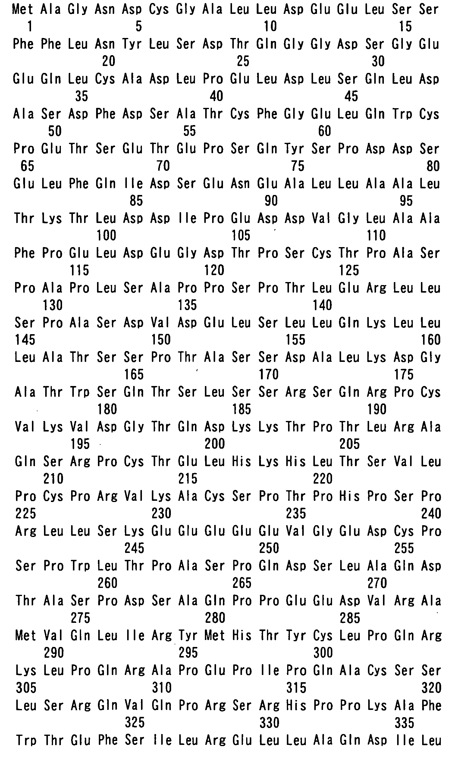
Fig. 1 shows the comparison of the primary amino acid sequences
between ERRL1 and PGC-1. Asterisks indicate identical amino acids.
The numbers represent the amino acid positions with the first
methionine counted as one. LXXLL motifs are shown in bold types.
Glutamic acid (E) repeats and serine-arginine (SR)-rich regions are
underlined. A putative RNA-binding motif is boxed. Tentative domain
borders are shown by vertical lines. A splicing variant of ERRL1, which
was newly identified at this time, lacks 39 amino acids (from the 156th
leucine to the 194th lysine).
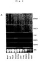
Fig. 2 shows the expression profiles of ERRL1 mRNA. Fig. 2A
shows the results of analyzing 20 µg of total RNA from various adult
mouse tissues using ERRL1, PGC-1, PRC, ERR1 and ERR3 cDNAs as
probes. Ethidium bromide staining of 28S RNAs is also shown. Fig. 2B
shows the results obtained by inducing 3T3-L1 cells to differentiate into
adipocytes by the treatment with dexamethasone,
1-methyl-3-isobutylxanthin and insulin (added day 0), isolating RNAs
and performing the Northern blot analysis. The blot was sequentially
hybridized with the respective probes. Fig. 2C shows the results of
observing the expression of ERRL1, ERR1 and MCAD mRNA in 3T3-L1
and 10T1/2 cells (-: preadipocytes, +: mature adipocytes).
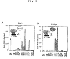
Fig. 3 shows the profiles of PGC-1 and ERRL1 as 'protein ligands'
for various nuclear receptors. Transcriptional activation ability of
PGC-1 (A) and ERRL1 (B) for various nuclear receptors was examined in
a transfection assay. Mean values of triplicate experiments are shown
as fold induction, where the Luc activity of each GAL4-fused nuclear
receptor in the absence of PGC-1 or ERRL1 serves as the reference
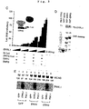
values. Error bars show standard deviation. Fig. 3C shows the
dose-dependent activity of ERRL1 on the full-length ERR-mediated
transcription via ERRE. pCMX-ERRL1 in different amounts (0 to 200
ng) was co-transfected with 30 ng of pCMX-ERR1 or pCMX-ERR3 in the
presence of 250 ng of TK-Luc or ERRE-Luc in CV-1 cells. Mean values
from triplicate experiments are shown as fold induction, where the Luc
activity of TK-Luc in the absence of ERRL1 serves as the reference value.
Error bars show standard deviation. Fig. 3D shows the results of
confirming that 35S-labeled ERRL1 shows strong interaction with
GST-ERR1 and GST-ERR3 in vitro. Autoradiographs show the pulled
down proteins (GST-ERR1 and GST-ERR3) and 10% of the total amount
of 35S-labeled ERRL1 used for the GST pull-down assay (Input). CBB
staining of the SDS-PAGE gel containing GST-fused proteins used in this
assay is also presented. Fig. 3E shows the results showing
augmentation of MCAD mRNA expression by ectopic expression of ERRL1
or PGC-1 in combination with ERR1 or ERR3 in 10T1/2 cells. One day
after confluence of the cells, total RNA was isolated, and analyzed by
Northern blot (20 µg per lane) using MCAD and ERRL1 cDNAs as probes.
The relative densitometric values of MCAD mRNAs are also included
beneath each lane.
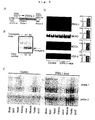
Fig. 4 relates to the creation of ERRL1 transgenic mouse. Fig.
4A shows the schematic drawing of the ERRL1 transgene and the
positions of probes used for Southern (probe 1) and Northern blots
(probe 1 and probe 2). Fig. 4B shows the results of showing that in the
two transgenic lines (A1 and A2), 10 and 12 copies of each of the
transgenes were contained, respectively. Fig. 4C shows the results of
Northern blot analysis of ERRL1 mRNA expression in the respective
tissues from ERRL1 mice (line A1) and littermate control mice. Each
lane contained 20 µg of total RNA. Fig. 4D shows the results of
measuring the expression of ERRL1, MCAD, ACC2 and UCP-3 in the
skeletal muscle of ERRL1 transgenic mice and wild-type control mice.
Three mice in each group were examined. Quantitative values of
Northern signals (control as 100%) are shown on the right. Data are
mean ± s.e.m. (Asterisk shows p < 0.05).
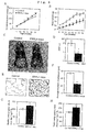
Fig. 5 relates to the phenotypes of ERRL1 transgenic mice. Fig.
5A shows the cumulative food intake per mouse during the indicated
period by measuring food intake every week. Data are means ± s.e.m. (n
= 6 per animal group, error bars are smaller than the symbols, asterisk
shows P < 0.05, double asterisk shows P < 0.01). Fig. 5B shows the
results of showing the changes in the body weight of ERRL1 transgenic
mice (closed circle) and wild-type controls (open circle). Values
represent mean body weight ± s.e.m. (n = 6 per animal group, asterisk
shows P < 0.05, double asterisk shows P < 0.01). For some data points,
the error bars are smaller than the symbols. Fig. 5C shows the results
of reduction of abdominal fat in ERRL1 transgenic mice. Fig. 5D shows
the comparison of epididymal WAT weight between wild-type control mice
and ERRL1 transgenic mice. The columns represent mean values of
WAT weight ± s.e.m. (n = 6 per animal group, asterisk shows P < 0.05).
Fig. 5E shows the comparison of the morphology of white adipose tissue
between ERRL1 transgenic mice and littermate wild-type control mice.
The scale bar indicates 50 µm. Fig. 5F shows the average diameter of
adipose cells. The diameters of the cells were measured from the
sections shown in Fig. 5E (n = 20, double asterisk shows P < 0.01). Figs
5G and 5H show the results of showing energy expenditures at resting
(G) and total (H) in 12-week-old control mice and ERRL1 transgenic mice.
The columns represent mean values of energy expenditure ± s.e.m. (n = 6
per animal group, asterisk shows P < 0.05, double asterisk shows P <
0.01).
Fig. 6A shows the results of measuring changes in body weight of
KKAy(+)ERRL1(-) (open square, n = 8), KKAy(-) ERRL1(-) (open circle, n =
5), KKAy(+)ERRL1(+) (closed square, n = 5), KKAy(-)ERRL1(+) (closed
circle, n = 5) male mice. The body weight curve of each group was
compared by repeated-measure analysis (Super ANOVA). Asterisk shows
P < 0.05, double asterisk shows P < 0.01. Fig. 6B shows representative
photographs of 12-week-old male KKAy(+)ERRL1(-) (left),
KKAy(+)ERRL1(+) (center) and KKAy(-)ERRLl(-) (right) mice.
Best Mode for Carrying Out the Invention
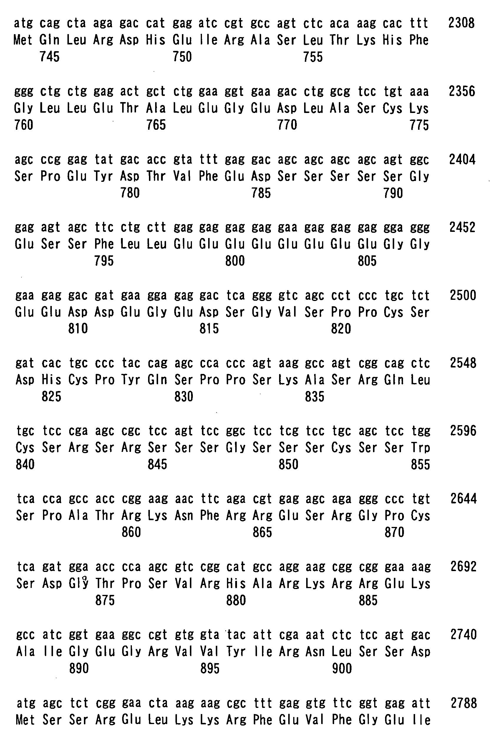
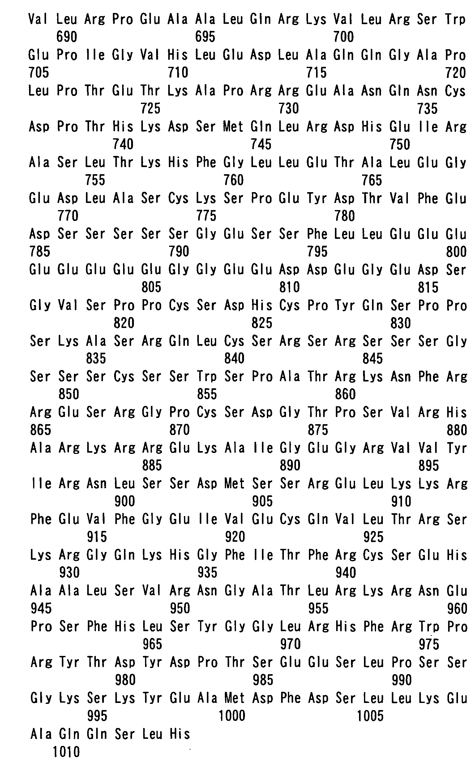
The ligand factor ERRL1 in the current invention is, for example,
mouse ERRL1 (SEQ ID NO: 2) which has been disclosed as 'mouse PGC2'
in SEQ ID NO: 1 in JP-A-2002-058489. Further, as for this ligand
factor ERRL1, its splicing variant (the one lacking 39 amino acids from
the 156th leucine to the 194th lysine of SEQ ID NO: 2) can be used as a
target. In addition, known human PERC (PPARγ coactivator β1: Entrez
protein database No. NP_573570), which is a human homologue to
mouse PGC2, can be also used as a target of this invention for a human
ERRL1. As the polynucleotide encoding mouse ERRL1, mouse PGC2
cDNA (ERRL1 cDNA: SEQ ID NO: 1) whose nucleotide sequence has been
disclosed in SEQ ID NO: 2 in JP-A-2002-058489 can be used. Further,
as the polynucleotide encoding human ERRL1, the foregoing human
PERC cDNA (GenBank No. NM_133263) can be used. Still further, for
example, human PRRC gene exists in chromosome 5q33.1 region, and its
genomic sequence is publicly known (GenBank No. NT_029289).
Therefore, this invention can be carried out by utilizing a promoter
region existing in this genomic gene sequence.
With regard to the nuclear receptor ERR of this invention, any
one or more selected from ERR1, ERR2 and ERR3 (Giguere, V. et al.,
Nature 331: 91-94, 1988; Shigeta, H. et al., J. Mol. Endocrinol. 19:
299-309, 1997; Eudy, J. D. et al., Genomics 50: 382-384, 1998; Hong, H.
et al., J. Biol. Chem. 274: 22618-22626, 1999) can be used as a target.
In addition, cDNAs encoding them, respectively, are also publicly known.
For example, ERR1 cDNA is known on GenBank No. NM_004451, and the
genomic gene (chromosome 11q13 region) sequence is known on
GenBank No. NT_033903. Therefore, this invention can be carried out
by using these polynucleotides (cDNA, genomic DNA, a DNA fragment of
the gene promoter region and the like). Further, as for MCAD gene, its
cDNA is also publicly known (GenBank No. AF251043).
The foregoing polynucleotides can be isolated by screening a
genomic DNA library or a cDNA library using, as a probe, a DNA
fragment consisting of a known nucleotide sequence or its partial
sequence, respectively. The obtained polynucleotide can be amplified by
a gene amplification method, which is usually performed, such as the
PCR (polymerase chain reaction) method, NASBN (nucleic acid sequence
based amplification) method, TMA (transcription- mediated amplification)
method or SDA (strand displacement amplification) method. In addition,
in the case of cDNA, by using a primer synthesized based on a known
sequence, a target cDNA can be obtained by also the RT-PCR method in
which mRNA isolated from the cell is used as a template.
The method of screening a substance which serves as the active
ingredient in a drug for obesity and/or diabetes of this invention
comprises treating cells or an animal with a candidate substance and
specifying a candidate substance fulfilling one or more of the following
requirements as the target substance by using the foregoing protein
factors or polynucleotides encoding them:
In other word, as explained above, a nuclear receptor ERR serves
as a regulator for MCAD gene expression, and energy balance in the body
is controlled by regulating fatty acid β-oxidation by this MCAD gene
expression, whereby the resistance to obesity or diabetes can be
maintained. As shown in the Examples described later, ERRL1 is a
ligand molecule for the receptor ERR which is the origin of a series of
such mechanisms. Indeed, it was confirmed that augmentation of
ERRL1 expression shows a significant effect on acquisition of the
resistance to obesity or diabetes in mouse. In other words, a substance
fulfilling the foregoing requirements (a) to (d) is a substance which acts
on the process of anti-obesity and anti-diabetic mechanism.
In the methods of this invention, a 'candidate substance' includes
an organic or inorganic substance, a protein, a peptide, a polynucleotide,
an oligonucleotide and the like, which are either unknown or known.
'Cells' are ones isolated from an established human cell line or
non-human animal cell line or from a tissue of a human or a non-human
animal body and appropriately maintained in a culture condition.
Examples of the established cell line include an adipocyte such as
3T3-L1 cell line. As described in Examples below, because the
expression of ERRL1 protein shows a sensitive response during the
differentiation of this adipocyte, those are preferred cells for the search
for a target substance. Alternatively, the cells may be an established
cell line or bacteria (E. coli or the like) transfected with ERRL1 cDNA
and/or ERR cDNA. In addition, as the cells isolated from an animal
body, those of isolated from BAT, heart, skeletal muscle, kidney or the
like, in which ERRL1 protein has been highly expressed, can be
exemplified. On the other hand, in the case of using a non-human
animal (such as mouse) as a target, a candidate substance is
administrated to this animal body (systemically or locally) or is taken as
food or drink, then the foregoing requirements (a) to (d) are measured in
a tissue or cells of the animal. At this time, a transgenic non-human
animal provided by this invention may be used as a positive control. In
other words, because this transgenic non-human animal overexpresses
ERRL1 protein and, as a result, highly expresses receptor ERR and
MCAD genes, therefore, by measuring whether or not a wild-type mouse
administered with a candidate substance fulfills the foregoing
requirements as much as this transgenic animal does, an effect of the
candidate substance can be judged more precisely. In addition, in the
case of using a transgenic non-human animal or a wild-type animal as a
target, a treatment of feeding with a high-fat diet, loading with physical
exercises or the like, or amount of food intake, body weight, energy
expenditure or the like can be included in the judging requirements.
The judgment of the foregoing requirements (a) to (d) can be
carried out by measuring the expression of the respective genes by
measuring the level of their transcriptional products (mRNA or protein)
using a known method. For example, the judgment of the foregoing
requirements (a) to (d) can be carried out by a known method such as in
situ hybridization, Northern blotting, dot blotting, RNase protection
assay, RT-PCR, Real-time PCR (Journal of Molecular Endocrinology, 25,
169-193 (2000) and the literatures cited therein), and DNA array
analysis method (edited by Mark Shena, "Microarray Biochip Technology",
Eaton Publishing (March 2000)). A screening of an active ingredient
substance in a drug for obesity and/or diabetes using such a technique
and a reagent, method, process, analysis program and the like to be used
therein are all included in this invention. Specific examples of the
judgment include, for example, the following methods.
(A) Screening of substance that increases expression level of ligand
factor ERRL1
A reporter plasmid is constructed in which the promoter region of
ERRL1 gene has been fused with cDNA of luciferase or β-galactosidase.
After this reporter plasmid is transfected into cells, a candidate
substance to be investigated is added to the cells and the activity of
luciferase or β-galactosidase in the extracted solution of the cultured
cells is measured after several days. If an increase of luciferase or
β-galactosidase is observed by the addition of the candidate substance,
the possibility is strongly suggested that this substance has an ability to
activate the promoter of ERRL1 gene. Then, this candidate substance is
added to cultured cells and an increase of mRNA expression level of
ERRL1 gene is evaluated by the Northern blot method, quantitative
RT-PCR method or the like. Similarly, using cultured cells, an increase
of expression level of ERRL1 protein can be also evaluated by the
Western blot method using an anti-ERRL1 antibody. Alternatively, a
candidate substance is fed to a non-human animal and an increase of
ERRL1 mRNA level or ERRL1 protein level in the cells isolated from the
animal (e.g., BAT, heart, skeletal muscle, kidney or the like) may be
evaluated in the same manner.
(B) Screening of substance that increases the transcriptional activity of
nuclear receptor ERR
After the reporter plasimd of luciferase or β-galactosidase having
an ERR responsive sequence or gal4 responsive sequence in the promoter
region is cotransfected into cultured cells with an expression vector of
the full-length ERR protein or a fused protein of the ligand binding
region of ERR with the DNA binding region of gal4, respectively, a
candidate substance to be investigated is added to the cells and the
activity of luciferase or β-galactosidase in the extracted solution of the
cultured cells is measured after several days. If an increase of
luciferase or β-galactosidase is observed depending on the existence of
the responsive sequence by the addition of the candidate substance, the
possibility is strongly suggested that this substance has an ability to
activate the transcription of ERR gene. Then, this candidate substance
is added to cultured cells and an increase of mRNA expression level of a
target gene of ERR, for example MCAD gene, is evaluated by the Northern
blot method, quantitative RT-PCR method or the like. Similarly, using
cultured cells, an increase of expression level of the product of a target
gene of ERR, for example MCAD protein, can be evaluated by the Western
blot method using an anti-MCAD antibody. Alternatively, a candidate
substance is fed to a non-human animal and an increase of MCAD mRNA
level or MCAD protein level in the cells isolated from the animal (e.g.,
BAT, heart, skeletal muscle, kidney or the like) may be evaluated in the
same manner.
(C) Screening of substance that promotes binding of ERRL1 to ERR
In yeast or cultured animal cells, using the two hybrid method,
the binding of ERRL1 to ERR is quantified as the activity of luciferase or
β-galactosidase. When a candidate substance to be investigated is
added to the system, if luciferase or β-galactosidase is increased
compared to the case where the substance has not been added, the
possibility is strongly suggested that this substance has an ability to
activate the binding of ERRL1 to ERR. Then, when this drug substance
is added in an immunoprecipitation method or a GST-pull down assay,
the level of coprecipitated ERRL1 and ERR proteins is evaluated by the
Western blot method. On the other hand, in a gel retardation assay
using ERR protein and an oligo DNA containing the ERR responsive
sequence, according to the index that is based on whether or not the
level of signal that super shifts in the presence of ERRL1 is increased by
the addition of this candidate substance, it can be confirmed whether or
not this substance has an effect on promoting the binding of ERRL1 to
ERR. Further, by adding this candidate substance to a detection assay
system to detect interaction using an apparatus such as Biacore, it can
be confirmed whether or not this substance has an effect on promoting
the binding of ERRL1 to ERR.
(D) Screening of substance that increases expression level of MCAD gene
product
A reporter plasmid is constructed in which the promoter region of
MCAD gene has been fused with cDNA of luciferase or β-galactosidase.
After this reporter plasmid is transfected into cells, a candidate
substance to be investigated is added to the cells and the activity of
luciferase or β-galactosidase in the extracted solution of the cultured
cells is measured after several days. If an increase of luciferase or
β-galactosidase is observed by the addition of the candidate substance,
the possibility is strongly suggested that this substance has an ability to
activate the promoter of MCAD gene. Then, this candidate substance is
added to cultured cells and an increase of mRNA expression level of
MCAD gene is evaluated by the Northern blot method, quantitative
RT-PCR method or the like. Similarly, using cultured cells, an increase
of expression level of MCAD protein can be evaluated by the Western blot
method using an anti-MCAD antibody. Alternatively, by the same
method as in the foregoing (B), an increase of MCAD mRNA level or
MCAD protein level may be evaluated in a non-human animal.
The genetic engineering techniques (recombinant DNA
techniques) used in each of the foregoing methods can be carried out in
accordance with the methods described in J. Sambrook, E. F. Fritsch & T.
Maniatis, "Molecular Cloning: A Laboratory Manual (2nd edition)", Cold
Spring Harbor Laboratory Press, Cold Spring Harbor, New York (1989); D.
M. Glover et al. ed., "DNA Cloning", 2nd ed., Vol. 1 to 4, (The Practical
Approach Series), IRL Press, Oxford University Press (1995); Japanese
Biochemical Society ed., "Zoku Seikagaku Jikken Kouza 1, Idenshi
Kenkyuho II" Tokyo Kagaku Dozin (1986); Japanese Biochemical Society
ed., "Shin Seikagaku Jikken Kouza 2, Kakusan III (Kumikae DNA
Gijutsu)" Tokyo Kagaku Dozin (1992); R. Wu ed., "Methods in
Enzymology", Vol. 68 (Recombinant DNA), Academic Press, New York
(1980); R. Wu et al. ed., "Methods in Enzymology", Vol. 100
(Recombinant DNA, Part B) & 101 (Recombinant DNA, Part C), Academic
Press, New York (1983); R. Wu et al. ed., "Methods in Enzymology", Vol.
153 (Recombinant DNA, Part D), 154 (Recombinant DNA, Part E) & 155
(Recombinant DNA, Part F), Academic Press, New York (1987), J. H.
Miller ed., "Methods in Enzymology", Vol. 204, Academic Press, New York
(1991); R. Wu et al., ed., "Methods in Enzymology", Vol. 218, Academic
Press, New York (1993); S. Weissman ed., "Methods in Enzymology", Vol.
303, Academic Press, New York (1999); J. C. Glorioso et al., ed.,
"Methods in Enzymology", Vol. 306, Academic Press, New York (1999),
etc. or the methods described in the references cited therein or
substantially the same methods or the modifications thereof.
The anti-ERRL1 antibody to be used in the foregoing method (A)
and the anti-MCAD antibody to be used in the foregoing methods (B) and
(D) are a polyclonal antibody or a monoclonal antibody that recognizes
either ERRL1 protein or MCAD protein, and include a whole molecule
that can bind to the epitope of the respective proteins, and all the
fragments of Fab, F(ab')2, Fv and the like. Such an antibody can be
obtained from serum after immunizing an animal by using a purified
ERRL1 protein or MCAD protein, or a partial peptide thereof as an
antigen. Alternatively, it can be prepared by introducing an expression
vector for a eukaryotic cell into the muscle or the skin of an animal with
an injector or a gene gun, and collecting the serum. As the animal,
mouse, rat, rabbit, goat, chicken or the like is used. If hybridoma is
produced by fusing B cells collected from the spleen of an immunized
animal with myeloma, a monoclonal antibody can be produced.
Preparation of such an antibody can be carried out in accordance with a
known method described in the literatures (Shin Seikagaku Jikken
Kouza 12, Molecular Immunology III, Antigens, Antibodies, and
Complements, Nancodo, 1992; "Monoclonal Antibody", co-authored by
Komei Nagamune and Hiroshi Terada, Hirokawa Shoten, 1990;
"Monoclonal Antibody", James W. Goding, third edition, Academic Press,
1996 and the like). Further, the measurement methods using such an
antibody can be carried out by referring to the literatures, for example,
Hiroshi Irie ed., "Radioimmunoassay" (published by Kodansha, 1974);
Hiroshi Irie ed., "Radioimmunoassay; Second Series" (published by
Kodansha, 1979); Eiji Ishikawa, et al. ed., "Enzyme Immunoassay"
(published by Igaku Shoin, 1978); Eiji Ishikawa, et al. ed., "Enzyme
Immunoassay" (Second Edition) (published by Igaku Shoin, 1982); Eiji
Ishikawa, et al. ed., "Enzyme Immunoassay" (Third Edition) (published
by Igaku Shoin, 1987); H. V. Vunakis et al. ed., "Methods in Enzymology",
Vol. 70 (Immunochemical Techniques, Part A), Academic Press, New York
(1980); J. J. Langone et al. ed., "Methods in Enzymology", Vol. 73
(Immunochemical Techniques, Part B), Academic Press, New York (1981);
J. J. Langone et al. ed., "Methods in Enzymology", Vol. 74
(Immunochemical Techniques, Part C), Academic Press, New York (1981);
J. J. Langone et al. ed., "Methods in Enzymology", Vol. 84
(Immunochemical Techniques, Part D: Selected Immunoassays),
Academic Press, New York (1982); J. J. Langone et al. ed., "Methods in
Enzymology", Vol. 92 (Immunochemical Techniques, Part E: Monoclonal
Antibodies and General Immunoassay Methods), Academic Press, New
York (1983); J. J. Langone et al. ed., "Methods in Enzymology", Vol. 121
(Immunochemical Techniques, Part I: Hybridoma Technology and
Monoclonal Antibodies), Academic Press, New York (1986); J. J. Langone
et al. ed., "Methods in Enzymology", Vol. 178 (Antibodies, Antigens, and
Molecular Mimicry), Academic Press, New York (1989); M. Wilchek et al.
ed., "Methods in Enzymology", Vol. 184 (Avidin-Biotin Technology),
Academic Press, New York (1990); J. J. Langone et al. ed., "Methods in
Enzymology", Vol. 203 (Molecular Design and Modeling: Concepts and
Applications, Part B: Anibodies and Antigens, Nucleic Acids,
Polysaccharides, and Drugs), Academic Press, New York (1991) and the
like, or the references cited therein.
In the case where screening is carried out by using a non-human
animal as a target in the foregoing methods (A), (B) and (D), a transgenic
non-human animal provided by this invention can be used as a positive
control. In other words, this transgenic non-human animal of this
invention overexpresses ERRL1 protein and, as a result, it highly
expresses receptor ERR and MCAD genes, thereby showing an
anti-obesity and anti-diabetic property. Accordingly, the action of the
candidate substance in the individual can be determined by using the
gene expression in the cells of this transgenic animal as a control.
The transgenic non-human animal in this invention can be
produced in accordance with a known transgenic animal production
method (e.g., Proc. Natl. Acad. Sci. USA 77; 7380-7384, 1980). In other
words, the foregoing mouse ERRL1 polynucleotide or human ERRL1
(human PERC) polynucleotide (hereinafter, referred to as 'introduced
gene' in some cases) is introduced into a totipotent cell of the
non-human animal and this cell is developed until it becomes an
individual. Then, by selecting an individual in which the introduced
gene has been integrated into the genome of the somatic cell, the target
transgenic non-human animal can be produced. As the non-human
animal, animals used as a laboratory animal such as mouse, rat, rabbit,
dog, cat and the like can be used as a target, however use of mouse is
preferred because the inbred line has been established. In addition, a
promoter sequence or an enhancer sequence is connected to the
introduced gene to control its expression. Depending on the selection of
this promoter/enhancer sequence, ERRL1 protein can be expressed
systemically or it can be selectively expressed in a specific tissue. Such
an introduced gene can be constructed by introducing and ligating the
foregoing polynucleotide or promoter/ enhancer sequence to a circular
DNA vector so as to be arranged in an effective position in the expression
regulation of the introduced gene. Then, after this vector DNA is
digested with a restriction enzyme, the one, from which the vector region
has been removed, is introduced into a totipotent cell. As the totipotent
cell to which a gene is introduced, a fertilized egg, an early embryo or an
embryonic stem cell (ES cell) can be used. In addition, as the method of
introducing a gene into a totipotent cell, when a ratio of generating
transgenic animal individuals or a ratio of inheriting an introduced gene
to the next generations are taken into account, a physical injection
(microinjection) method of DNA is the most preferred. A fertilized egg, to
which a gene is injected, is then transferred to the oviduct of a surrogate
mother and after it is generated to an individual and born, the animal is
grown with a foster parent and DNA is extracted from part of the body
(e.g., tail tip), and the existence of the introduced gene is confirmed by
the Southern analysis or the PCR method. Assuming that the individual
in which the existence of the introduced gene has been confirmed is the
first generation (founder), the introduced gene is inherited to 50% of its
offspring (F1). Further, by crossing this F1 individual with a wild-type
animal or other F1 animal, an individual (F2) having the introduced gene
in one (heterozygote) or both (homozygote) of diploid chromosomes can be
produced. The transgenic animal produced as above overexpresses
ERRL1 protein in all somatic cells or a specific tissue and, as described
in the after-mentioned Examples, it has a unique characteristic of being
lean although it is hyperphagic, and showing significantly high energy
expenditure. Such a transgenic animal is useful not only as a control in
the foregoing screening method but also as a model animal in the
elucidation of an anti-obesity or anti-diabetic mechanism at the whole
body level.
The invention of this application further provides a novel drug for
obesity and/or diabetes comprising as the active ingredient at least one
substance specified by the foregoing screening method. In other words,
this drug has at least one of the actions selected from, firstly, the action
of increasing the expression level of ERRL1, secondary, the action of
increasing the transcriptional activity of ERR, thirdly, the action of
increasing the interaction between ERRL1 and ERR, and fourthly, the
action of increasing the expression level of an MCAD gene product.
The drug of this invention can be formulated by homogenously
mixing the foregoing active ingredient and a pharmaceutically acceptable
carrier. The carrier can be appropriately selected from a wide range
according to the administration form of the drug. It is desired that the
drug of this invention be in a unit dosage form that can be administered
orally or by injection. The oral liquid preparation such as a suspension
or syrup can be produced by using water, a sugar such as sucrose,
sorbitol or fructose, a glycol such as polyethylene glycol, an oil such as
sesame oil or soybean oil, an antiseptic such as alkyl parahydroxy
benzoate, a flavor such as strawberry flavor or peppermint, or the like.
A powder, pill, capsule and tablet can be formulated by using a diluent
such as lactose, glucose, sucrose or mannitol, a disintegrating agent
such as starch or sodium alginate, a lubricant such as magnesium
stearate or talc, a binder such as polyvinyl alcohol, hydroxypropyl
cellulose and gelatin, a surface active agent such as a fatty acid ester, an
elasticizer such as glycerin or the like. The tablet and capsule are a
preferred unit dosage form in the formulation of this invention in that it
is easy to administer. When the tablet or capsule is produced, a solid
pharmaceutical carrier is used. In addition, the solution for injection
can be formulated by using a carrier composed of a salt solution, glucose
solution, or a mixture of a salt solution and glucose solution, a variety of
buffer solutions or the like. Further, an injectable solution may be also
prepared by formulating it in a powder state and then mixing it with the
foregoing liquid carrier prior to its use.
Hereunder, the invention of this application will be explained in
more detail and specifically by showing Examples, however, the invention
of this application is not intended to be limited to the following examples.
Examples
1. Methods
1.1. Database search
An EST homology search was performed using the BLAST
program (Altshul, S. F. et al., Nucleic Acids Res. 25: 3389-3402, 1997).
1.2. RNA analysis
Northern blot analyses were performed in accordance with the
description in the literature (Sambrook and Maniatis, in Molecular
Cloning-A Laboratory Manual, 7.2-7.87, Cold Spring Harbor Laboratory
Press, New York, 1989). The cDNA probes for MCAD (Genbank
accession No. U07159), PRC (BC013720), ERR1 (U85259), ERR2
(S82458), ERR3 (AF117254), ACC2 (AF290178), and UCP-3 (AF032902)
were obtained by RT-PCR.
1.3. Assays for transcriptional activity
Transfections and reporter assays were performed in accordance
with the description in the literature (Takada, I. et al., Mol. Endocrinol,
14: 733-740, 2000). More specifically, a reporter gene containing four
copies of a GAL4 binding sequence ((UAS)4-Luc) was transfected into
CV-1 cells in the presence or absence of a chimeric receptor expression
vector in which the DNA binding region of GAL4 had been fused with the
ligand binding region for the nuclear receptor (pCMX-GAL4-nuclear
receptor), and the transcriptional activity of GAL4-nuclear receptor-fused
protein was measured (Takada, I. et al., Mol. Endocrinol, 14: 733-740,
2000). All luciferase activities were normalized by the co-transfected
β-galactosidase activities. Amino acids 1-147 of GAL4 fused to the
ligand binding domain for the following nuclear receptors were used:
mouse AR (amino acids, 607-899, Genbank accession number, X59592),
human ERα (a.a. 251-595, X03635), human GR (a.a. 489-777, M10901),
rat FXR (a.a. 190-469, U18374), human RARα (a.a. 126-432, X06538),
human RXRα (a.a. 222-462, X52773), mouse PPARα (a.a. 156-468,
X57638), human PPARγ1 (a.a. 176-478, L40904), human PXR (a.a.
110-434, AF084645), human ERR1 (a.a. 147-422, L38487), human ERR2
(a.a. 171-433, X51417), human ERR3 (a.a. 173-436, AF058291), human
HNF4α (a.a. 125-465, X76930), human NOR (a.a. 361-626, D78579),
human NURR1 (a.a. 264-535, S77154), human RORα1 (a.a. 140-523,
U04897), human SF1 (a.a. 64-461, U76388), human COUP (a.a. 156-423,
X12795), human TR2-11 (a.a. 149-603, M29960), human RevErbA (a.a.
199-614, M24898). The abbreviations of nuclear receptors are described
in the references of respective Genbank files (Giguere, V., Endocr. Rev.
20: 689-725, 1999). GAL4-nuclear receptor expression plasmids were
provided from Dr. K. Umesono. Mouse PGC-1 cDNA was obtained by
screening a mouse embryo cDNA library. The full length cDNA of
human ERR1, 2 and 3 were obtained by RT/PCR. The amplified
products were subcloned into pCAGGS and pCMX expression vectors,
and confirmed by sequencing.
1.4. Protein interaction assays
In accordance with the description in the literature (Kamei, Y. et
al., Cell 85: 403-414, 1996), the constructs (expression vectors) of GST
fused with ERR1 and ERR3 were incubated with 35S-ERRL1 (TNT,
Promega) and washed. Then, the bound 35S-ERRL1 was separated by
SDS-PAGE, and quantified by autoradiography.
1.5. Stable cell lines
Phoenix 293 cells (gifted from Dr. G. P. Nolan, University of
Stanford) were used for the retrovirus packaging (Grignani, F. et al.,
Cancer Res. 58: 14-19, 1998). pLNCX-derived expression plasmid
(Clontech) containing cDNA of ERR1, ERR3 or GFP (control) and
pMX-derived expression plasmid (Misawa, K. et al., Proc. Natl. Acad. Sci.
USA 97: 3062-3066, 2000) containing cDNA of ERRL1, PGC-1 or GFP
were used in accordance with the manufacturer's instruction.
1.6. Generation of transgenic mice
ERRL1 cDNA was cloned into pCAGGS (Niwa, H., Gene 108:
193-200, 1991), the transgene (Fig. 4A) was excised and purified (2 ng
µl-1). Fertilized eggs were recovered from BDF1 females
(C57BL/6xDBA/2) crossed with BDF1 males and microinjected with the
transgene by the standard method (Gordon, J., in Guide to Techniques in
Mouse Development, (eds Wassarman, P. M. & DePamplilis, M. L.,
747-771, Academic press, San Diego, 1993). Care of the mice was
performed in accordance with the guideline of the institution to which
the inventors belong.
1.7. High-fat diet
Mice were fed with a regular chow diet (Oriental Yeast Inc., Tokyo,
Japan) or a high-fat diet (Aoki, N. et al., Obesity Res. 1: 126-131, 1993)
which contained casein (20% wt/wt), α-cornstarch (30.2%), sucrose
(10%), lard (25%), corn oil (5%), minerals (3.5%), vitamins (1%), cellulose
powder (5%), and D,L-methionine (0.3%).
1.8. Determination of energy expenditure
Oxygen consumption and carbon dioxide production were
determined by using an indirect calorimeter system composed of a mass
analyzer and a computer (Komenani, N. et al., J. Nutr. Sci. Vitaminol.
(Tokyo) 41: 395-407, 1995). Mice were individually placed in an
open-circuit plastic respiration chamber (24 x 46 x 18 cm) connected to a
gas mass spectrometer (WSMR-1400, Westron, Chiba, Japan). The
airflow was controlled at 2 1/min. Gas analysis was carried out from
10:00 to 9:00 on the following day. Samples were monitored
continuously in 2 minute blocks in room air as a reference. Locomotor
activity was recorded automatically every 10 minutes by an Animex-III
(Shimadzu, Kyoto, Japan) laid under each respiration chamber. Energy
expenditure was calculated in accordance with the description in the
literature (Komenani, N. et al., J. Nutr. Sci. Vitaminol. (Tokyo) 41:
395-407, 1995).
1.9. KKAy mice
KKAy mice were purchased from Clea Japan Inc. (Tokyo).
1.10. Statistical analyses
Statistical comparisons of data from two experimental groups
were made by using Student's t-test. Comparison of data from multiple
groups was made by one-way analysis of variance (ANOVA), and each
group was compared with the other groups by the Fisher's protected
least significant difference (PLSD) test (Statview 4.0, Abacus Concepts).
Statistical significance was defined as P < 0.05 or P < 0.01.
2. Results
2.1. Cloning of ERRL1 cDNA and its features
As a result of searching EST (expression sequence tags) for a
PGC-1-related molecule, EST having an extremely high homology to
PGC-1 was found, and the full length cDNA containing this EST was
isolated. This cDNA consists of about 3.4 kb (SEQ ID NO: 1), and
encodes a protein consisting of 1,014 amino acid sequences (sequence
number 2). This protein was named ERRL1 (abbreviation of ERR ligand
1) based on its properties as a 'protein ligand' for ERRs (as shown later
in detail). Incidentally, Lin et al. reported the cloning of a PGC-1
homologue named PGC-1β (Lin, J. et al., J. Biol. Chem. 277: 1645-1648,
2002), however, PGC-1β only has one amino acid difference from ERRL1
(The 260th leucine in the sequence number 2 is a proline in the PGC-1β).
ERRL1 and PGC-1 show a high degree of amino acid identity except for
the central unique region in ERRL1. The homologous region can be
divided into five domains based on sequence identity and predicted
functional properties (Fig. 1). The N-terminal region of ERRL1 (amino
acids 1-282) contains two LXXLL motifs, which are a proposed binding
motif to nuclear receptors (Torchia, J. et al., Nature, 387: 677-684,
1997; Heery, D. M., et al., Nature, 387: 733-736, 1997), and has 41%
identity with PGC-1. The second region (amino acids 283-579), which is
unique to ERRL1, contains E (glutamic acid) repeats and one LXXLL
motif. The third region (amino acids 580-656) is highly conserved (47%
amino acid identity). The fourth region (amino acids 657-882) is less
conserved (22% amino acid identity) and contains a very short
serine/arginine (SR)-rich domain (Tacke, R. & Manley, J. L. Curr. Opin.
Cell Biol. 11: 358-362, 1999) in ERRL1 compared with the long SR
domain in PGC-1. The C-terminal domain (amino acids 883-1014) has a
putative RNA-binding domain (Krecic, A. M. & Swanson, M. S., Curr.
Opin. Cell Biol. 11: 363-371, 1999) and is highly conserved (52% amino
acid identity).
2.2. Similar expression patterns between ERRL1 and ERR
The expression patterns of ERRL1 in different tissues from adult
mice were examined. Two ERRL1 mRNAs of about 10 kb and 4 kb in
length were observed. ERRL1 mRNAs were abundant in brain, BAT,
heart and skeletal muscle, and were also detected in kidney, stomach
and white adipose tissue (WAT). Consistent with a previous report
(Puigserver, P. et al., Cell 92: 829-839, 1998), PGC-1 expression was
increased in mouse BAT after exposure to cold stress, however, the
expression of ERRL1 mRNAs was only marginally up-regulated by this
stress.
Further, the expression pattern of ERRL1 closely resembles that
of ERR1 (Sladek, R. et al., Mol. Cell. Biol. 17: 5400-5409, 1997 and Fig.
2), with both mRNAs being highly expressed in tissues that can use
lipids as a source of cellular energy, e.g., BAT, heart, skeletal muscle,
and kidney (Fig. 2A). ERR2 mRNA expression was not be detected,
which is consistent with a previous study report (Giguere, V. et al.,
Nature 331: 91-94, 1988).
Then, it was examined whether ERRL1 expression is augmented
during adipocyte differentiation of 3T3-L1 cells. It is certain that ERRL1
mRNAs were present at very low levels in 3T3-L1 preadipocytes and were
markedly induced during adipocyte differentiation. In contrast, mRNAs
of PGC-1 and PRC, another PGC-1-related molecule (Andersson, U. &
Scarpulla, R. C. Mol. Cell. Biol. 21: 3738-3749, 2001) remained at low
levels during this adipocyte differentiation (Fig. 2B). Similar
augmentation of ERRL1 mRNAs was observed in mature adipocytes
(Smas, C. M. & Sul, H. S. Biochem. J. 309: 697-710, 1995) differentiated
from another preadipocytes, 10T1/2 cells (Fig. 2C). Further, expression
profiles of ERR1 and its target MCAD were quite similar to that of ERRL1
(Figs. 2B and 2C), however, ERR2 mRNA and ERR3 mRNA expressions
were not detected in these cells. These results suggest that ERRL1 is
involved in the gene regulations in mature adipocytes-related functions
such as the lipid metabolism.
2.3. PGC-1 as a general nuclear receptor agonist.
It was examined that whether ERRL1 can function as a
coactivator of PPARγ, using PGC-1 as a control coactivator. Contrary to
our expectations, any evidence indicating that ERRL1 could activate
PPARγ-mediated transcription could not be found out. In the control
experiments, it was confirmed that the expression of PGC-1 on its own
was able to drastically activate PPARγ-mediated transcription without the
addition of any exogenous lipophilic ligands. PGC-1 is known to
physically interact not only with PPARγ but also with several other
nuclear receptors (Puigserver, P. et al., Cell 92: 829-839, 1998; Yoon, J.
C. et al., Nature 413: 131-138, 2001; Tcherepanova, I. et al., J. Biol.
Chem. 275: 16302-16308, 2000; Vega, R. B. et al., Mol. Cell. Biol. 20:
1868-1876, 2000; Delerive, P. et al., J. Biol. Chem. 277: 3913-3917,
2002). Furthermore, PGC-1 has been shown to be induced by certain
environmental changes such as cold exposure (Puigserver, P. et al., Cell
92: 829-839, 1998) and fasting (Yoon, J. C. et al., Nature 413: 131-138,
2001; Lehman, J. J. et al., J. Clin. Invest. 106: 847-856, 2000). These
observations raise the possibility that the expression of PGC-1 alone can
activate multiple nuclear receptor pathways. This putative character is
analogous to classical lipophilic ligands for the nuclear receptors. To
address this possibility, nuclear receptors whose DNA-binding domains
were replaced by the GAL4 DNA-binding domain were used. By this
replacement, the comparison of transcriptional activity profiles of
multiple nuclear receptors with the same reporter (gene) such as
(UAS)4-Luc can be easily performed. Indeed, PGC-1 was able to activate
transcription by means of multiple nuclear receptors including orphan
HNF4α, SF1, and ERRs. Among these, PGC-1 most strongly activated
HNF4α, which is consistent with a recent report (Yoon, J. C. et al.,
Nature 413: 131-138, 2001). Subsequently, PGC-1 activated ERα, SF1,
ERRs, PPARs, PXR, RARα, and RXRα (Fig. 3A). From these observations,
it was confirmed that PGC-1 can function as a general and broad nuclear
receptor agonist.
2.4. ERRL1 as an ERR protein ligand
Using the same GAL4-fused nuclear receptor set as above, a
potential partner of ERRL1 was searched, and it was found out that
ERRL1 specifically activated ERR-mediated transcription. ERR3 was
most strongly activated by ERRL1, followed by ERR1 and ERR2 (Fig. 3B).
Then, the transcriptional activation properties of ERRL1 on full-length
ERRs were tested. Consistent with the experiments above, ERRL1
dose-dependently activated the full-length ERR3- and the full-length
ERR1-mediated transcription by means of the ERR responsive sequence
(ERRE) of the MCAD gene promoter (Sladeck, R. et al., Mol. Cell. Biol. 17:
5400-5409, 1997; Vega, R. B. & Kelly, D. P. J. Biol. Chem. 272:
31693-31699, 1997) (Fig. 3C). The former was activated much more
strongly than the latter.
Next, it was tested whether ERRL1 physically interacts with ERRs
by in vitro binding analysis with glutathione S-transferase-fused ERR
(GST-ERR) produced by using bacteria and ERRL1 translated in vitro.
Matrix-bound GST-ERR1 and GST-ERR3, but not GST alone, retained
radiolabeled ERRL1 efficiently (Fig. 3D). As reported previously (Sladek,
R. et al., Mol. Cell. Biol. 17: 5400-5409, 1997; Vega, R. B. & Kelly, D. P.
J. Biol. Chem. 272: 31693-31699, 1997), it was confirmed that ERRs had
been bound to radiolabeled MCAD ERRE oligonucleotides in a gel
mobility shift assay, and that the ERR-DNA complexes were supershifted
by the addition of the ERRL1 protein. Given that lipophilic hormones
were not added either in the synthesis of the proteins or in the binding
reactions, these results indicate that ERRL1 and ERRs can directly
interact with the ERR target promoter DNAs.
Next, ERRL1 and ERR were expressed in 10T1/2 cell line, and it
was examined whether ERRL1 can activate MCAD gene expression via
the ERR. 10T1/2 cells were infected with several combinations of
recombinant retroviruses, each containing a DNA encoding GFP (control),
ERR1, ERR3, ERRL1 or PGC-1. Northern blot analysis was performed to
confirm the expressions of the transduced genes in each cell line (Fig.
3E). The sole expression of either ERR1 or ERR3 led to a modest
increase of MCAD mRNA level compared with that in cells infected with
GFP alone. Coexpression of both ERR1 and ERRL1 or ERR3 and ERRL1
led to a further significant increase of the MCAD mRNA level (Fig. 3E).
These results collectively demonstrate that ERRL1 can function as a
'protein ligand' for ERRs and activate ERR-mediated transcription at
least in cultured cells.
2.5. Creation of ERRL1 mice
In order to examine the effects of increased ERRL1 expression in
vivo, ERRL1 transgenic mice were created. It was expected that these
mice would exhibit phenotypes that mimic those induced by activated
ERR-mediated transcription. The chicken β-actin promoter with the
cytomegalovirus immediate early enhancer (CAG promoter) was used to
promote the expression of the mouse ERRL1 transgene in mice (Fig. 4A).
Southern blot analysis of tail DNAs of the mice was performed to
determine the transgene copy numbers (Fig. 4B).
Expression of the ERRL1 transgene was evaluated by Northern
blot analysis on RNAs isolated from tissues of ERRL1 mice and control
littermate mouse at 8 weeks of age (Fig. 4C). When a transgene-specific
probe (Probe 2, Fig. 4A) was used, only a single 4-kb band was detected.
The CAG promoter has been reported to show a strong activity in any
tissues (Niwa, H. et al., Gene 108: 193-200, 1991). Unexpectedly,
however, the use of this promoter resulted in high expression levels of
the ERRL1 transgene in several restricted tissues such as brain, BAT,
heart, skeletal muscle, and testis, but a very low-level of expression in
liver (Fig. 4C). These expression profiles corresponded relatively well to
the endogenous ERRL1 expression patterns (Fig. 4C). These
observations suggest that tissue-specific regulatory cis-sequences crucial
for ERRL1 expression may reside in the ERRL1 cDNA used in this
construct.
As predicted from the in vitro data, the increased expression of
ERRL1 in the skeletal muscle of ERRL1 mice (Fig. 4D) enhanced MCAD
mRNA expression in vivo. In contrast, the increased expression of
ERRL1 did not affect the gene expression of acetyl-CoA carboxylase 2
(ACC2) (Abu-Elheiga, L. et al., Science 291: 2613-2616, 2001) or
uncoupling protein-3 (UCP3) (Clapham, J. C. et al., Nature 406: 415-418,
2000).
2.6. ERRL1 mice are hyperphagic but lean
The apparent phenotype observed in ERRL1 mice was lean, which
agreed well with those predicted from increased β-oxidation of fatty acids.
This phenotype was more prominent when mice were fed with a high-fat
diet (Aoki, N. et al., Obesity Res. 1: 126-131, 1993). Groups of six male
wild-type control mice and six male ERRL1 mice, at 9 weeks of age, were
freely fed with a high-fat diet, and the food consumption and body weight
of each mouse were measured every week. ERRL1 mice consumed
significantly more food than the control mice (Fig. 5A). However, the
transgenic mice weighed 15 to 25% less than the control mice before and
throughout the feeding periods with a high-fat diet (Fig. 5B), and they
accumulated significantly less fat in their adipose tissues (Fig. 5C). The
epididymal WAT in the ERRL1 mice only weighed 0.92 ± 0.28 g,
compared with 2.0 ± 0.30 g in the control mice (Fig.). In contrast, liver
weights were not significantly different (ERRL1 mice: 1.23 ± 0.06 g;
control mice: 1.4 ± 0.08 g). The adipocytes of the transgenic mice were
smaller than those of the control mice (average diameter of adipocytes in
ERRL1 mice: 25.1 ± 1.1 µm; control mice: 54.5 ± 2.2 µm; Figs. 5E and
5F).
The blood was collected from these mice, and serum components
were biochemically analyzed. The decrease in adipose mass resulted in
a decrease in the amount of leptin in the serum (ERRL1 mice: 5.9 ± 3.5
ng ml-1, control mice: 25.0 ± 6.6 ng ml-1). As leptin is an anti-appetite
hormone secreted from WAT (Friedman, J. M. Nature 404: 632-634,
2000), the decreased level of leptin observed in ERRL1 mice is consistent
with the observation that they are hyperphagic. Furthermore, decreased
level of insulin were observed in the transgenic mice compared with the
control mice fed with a high-fat diet. Mice, when fed with a high-fat diet,
generally develop resistance to glucose uptake, even with a high plasma
insulin concentration (Li, B. et al., Nat. Med. 6: 1115-1120, 2000). A
similar state, which is called insulin resistance, is well observed in
humans with obesity or who are suffering from type 2 diabetes (Lovejoy,
J. C. Curr. Atheroscler. Rep. 1: 215-220, 1999). The decreased body
weight and decreased insulin level observed in ERRL1 mice indicate that
the increased expression of ERRL1 can antagonize obesity and contribute
to an improvement of diabetic state, even taking a high-calorie diet
through overcoming insulin resistance.
2.7. Increased energy expenditure of ERRL1 mice
The energy expenditure of ERRL1 mice was examined. Gas
analyses were carried out by using test chambers for respiration analysis,
each containing a single mouse, and energy expenditure was calculated.
As a result, the energy expenditures of ERRL1 mice were significantly
higher than those of control mice at 12 weeks of age (resting energy
expenditure: ERRL1 mice, 126.3 ± 3.8 kcal day-1 kg-0.75; control mice,
101.5 ± 6.7 kcal day-1 kg-0.75; P < 0.05, n = 6, per group; and total energy
expenditure: ERRL1 mice, 211.2 ± 10.7 kcal day-1 kg-0.75; control mice,
155.9 ± 7.8 kcal day-1 kg-0.75; P < 0.01, n = 6, per group) (Figs. 5G and
5H). However, locomotor activity was not significantly different (ERRL1
mice, 7,338 ± 700 counts; control mice, 5,338 ± 390 counts, the data
represent the sum of all transits over a 24 h period). These results
indicate that the differences in body weights between the control mice
and the ERRL1 mice are most likely to be due to a difference in their
energy expenditure.
2.8. ERRL1 expression can antagonize a genetically programmed obesity
ERRL1 mice and KKAy mice were bred, and it was examined
whether the obese phenotype of KKAy mice was counteracted by the
increased expression of ERRL1. KKAy mice have a mutation of Ay,
which leads to ectopic expression of the agouti coat-color protein,
causing a dominantly inherited syndrome of obesity and yellow fur
(Siracusa, L. D., Trends Genet. 10: 423-428, 1994). The agouti protein
acts as an antagonist for the melanocortine receptors, which are
considered to act as a repressor receptor of appetite in the central
nervous system (Friedman, J. M. Nature 404: 632-634, 2000; Siracusa, L.
D., Trends Genet. 10: 423-428, 1994). KKAy males were bred with
ERRL1 females, and their offsprings were selected by hair color (yellow,
KKAy(+); black, KKAy(-)) and ERRL1 transgene expression. Comparison
of the body weights of each group revealed that the ERRL1 transgene
significantly suppressed the body weight increases of KKAy(+) mice (Figs.
6A and 6B), demonstrating that increased ERRL1 expression can
antagonize obesity caused by a genetic abnormality.
3. Discussion
From the above results, it was confirmed that ERRL1 functions as
a 'protein ligand' for ERRs and controls energy expenditure in vivo. In
other words, this indicates that by increasing the expression level of
ERRL1 in, for example skeletal muscle, this functions as a ERR protein
ligand this time, and activates MCAD gene expression to lead to
activation of fatty acid β-oxidation. When fatty acid β-oxidation is
increased, as a result, accumulated fat will be decreased. It is predicted
that the same effects can be achieved by activating the transcriptional
ability of ERR and by increasing MCAD gene expression. These effects
are very similar to a physiological state which is achieved by physical
exercises.
In addition, the above results clearly indicate that the body
weight can be decreased by pharmacological activation of ERRL1 or ERR
while maintaining the usual calorie intake or more.
Industrial Applicability
As described in detail above, according to the invention of this
application, there are provided a method of screening an ingredient in a
remedy for obesity or diabetes by using the interaction between a nuclear
orphan receptor ERR and its ligand molecule ERRL1 as an index and a
novel drug containing a substance specified by this method as the active
ingredient.