EP1544291A1 - Novel process for producing antibody enzyme, novel antibody enzyme and utilization thereof - Google Patents
Novel process for producing antibody enzyme, novel antibody enzyme and utilization thereof Download PDFInfo
- Publication number
- EP1544291A1 EP1544291A1 EP03765329A EP03765329A EP1544291A1 EP 1544291 A1 EP1544291 A1 EP 1544291A1 EP 03765329 A EP03765329 A EP 03765329A EP 03765329 A EP03765329 A EP 03765329A EP 1544291 A1 EP1544291 A1 EP 1544291A1
- Authority
- EP
- European Patent Office
- Prior art keywords
- antibody
- amino acid
- acid sequence
- sequence
- residue
- Prior art date
- Legal status (The legal status is an assumption and is not a legal conclusion. Google has not performed a legal analysis and makes no representation as to the accuracy of the status listed.)
- Withdrawn
Links
Images




















Classifications
-
- C—CHEMISTRY; METALLURGY
- C07—ORGANIC CHEMISTRY
- C07K—PEPTIDES
- C07K16/00—Immunoglobulins [IG], e.g. monoclonal or polyclonal antibodies
- C07K16/18—Immunoglobulins [IG], e.g. monoclonal or polyclonal antibodies against material from animals or humans
- C07K16/28—Immunoglobulins [IG], e.g. monoclonal or polyclonal antibodies against material from animals or humans against receptors, cell surface antigens or cell surface determinants
- C07K16/2866—Immunoglobulins [IG], e.g. monoclonal or polyclonal antibodies against material from animals or humans against receptors, cell surface antigens or cell surface determinants against receptors for cytokines, lymphokines, interferons
-
- C—CHEMISTRY; METALLURGY
- C07—ORGANIC CHEMISTRY
- C07K—PEPTIDES
- C07K16/00—Immunoglobulins [IG], e.g. monoclonal or polyclonal antibodies
- C07K16/40—Immunoglobulins [IG], e.g. monoclonal or polyclonal antibodies against enzymes
-
- C—CHEMISTRY; METALLURGY
- C12—BIOCHEMISTRY; BEER; SPIRITS; WINE; VINEGAR; MICROBIOLOGY; ENZYMOLOGY; MUTATION OR GENETIC ENGINEERING
- C12N—MICROORGANISMS OR ENZYMES; COMPOSITIONS THEREOF; PROPAGATING, PRESERVING, OR MAINTAINING MICROORGANISMS; MUTATION OR GENETIC ENGINEERING; CULTURE MEDIA
- C12N9/00—Enzymes; Proenzymes; Compositions thereof; Processes for preparing, activating, inhibiting, separating or purifying enzymes
- C12N9/0002—Antibodies with enzymatic activity, e.g. abzymes
-
- C—CHEMISTRY; METALLURGY
- C07—ORGANIC CHEMISTRY
- C07K—PEPTIDES
- C07K2317/00—Immunoglobulins specific features
- C07K2317/50—Immunoglobulins specific features characterized by immunoglobulin fragments
- C07K2317/56—Immunoglobulins specific features characterized by immunoglobulin fragments variable (Fv) region, i.e. VH and/or VL
Definitions
- the present invention relates to a novel process for efficiently producing an antibody enzyme that has high molecule recognition capability of an antibody and the enzymatic activity, an antibody enzyme produced according to the producing process, and an antibody enzyme having a stereostructurally specific structure that can be applied in the producing process.
- the present invention relates to, among the antibody enzymes in particular, an antibody against Helicobacter pylori urease and an antibody enzyme against chemokine receptor CCR-5 that is a coreceptor of AIDS-causing virus as well as an application method of the antibody enzymes.
- the present inventors obtained a monoclonal antibody of which antigen is a stable region of an envelope protein gp41 of AIDS virus (HIV) that causes AIDS (acquired immune deficiency syndrome) and analyzed a function of the monoclonal antibody in detail.
- HIV AIDS virus
- the inventors report that a light chain region of the monoclonal antibody has a very high activity in specifically decomposing the envelope protein gp41 of the HIV (Reference literature 3 : Super Catalytic Antibody [ I ] : Decomposition of targeted protein by its antibody light chain. Hifumi, E. , Okamoto, Y., Uda, T., J. Biosci. Bioeng., 88(3), 323-327 (1999)).
- the antibody enzyme having high molecule recognizing capability of antibody and the enzymatic activity, is expected to apply in many fields such as the healthcare, chemical industry, and food industry.
- the present invention was carried out in view of the abovementioned situations and intends to provide a novel process for efficiently producing antibody enzymes that have high molecule recognition capability of an antibody and the enzymatic activity and antibody enzymes produced according to the producing process, as well as antibody enzymes having a stereostructurally specific structure that can be applied to the producing process.
- the Helicobacter Pylori is a slender S-shaped gram-negative bacterium and can be detected with high frequency from biological specimens of stomachs of gastric ulcer suffering patients and peptic ulcer suffering patients. From this, the Helicobacter Pylori is considered to cause gastric ulcer/gastric catarrh and suggested to be finally relevant to occurrence of gastric cancer. This is reported also from WHO (World Health Organization).
- HP Helicobacter Pylori
- the present invention pays attention to the point and provides an antibody enzyme that, by exhibiting a decomposition action against, in particular among the above antibody enzymes, the Helicobacter Pylori, is effective in eliminating the Helicobacter Pylori and can avoid the appearance of drug-resistant bacteria. Furthermore, the invention provides genes that code the antibody enzymes, transformants in which the genes are introduced, and a remedy/infection inhibitor that makes use of these and is used for patients infected by the Helicobacter Pylori.
- the AIDS is a disease state where owing to the infection of human immunodeficiency virus I type (HIV-1), immune function is deteriorated, resultantly AIDS-associated opportunistic infection, malignant tumor, neurosis, dementia and so on are caused together.
- HIV-1 human immunodeficiency virus I type
- the HIV-1 After an acute period, a symptomless period, and AIDS-associated syndrome, the AIDS is developed. Owing to AIDS-related researches and advances of remedy, the AIDS is becoming from fatal disease to a remediable disease; however, at present, there is no sure anti-HIV drug and a treatment method due to drugs is not yet established.
- CD4 In researches of an infection mechanism of the HIV, in 1984, as a receptor of the HIV-1, CD4 was identified. It is known that the CD4 is present on a surface of a human cell and, when it combines with gp120 that is an envelope glycoprotein of the HIV-1 to infect the cell.
- the present invention further provides, among the abovementioned antibody enzymes, in particular, an antibody enzyme that can decompose the chemokine receptor CCR-5 to delete its function and can be utilized in preventing the AIDS viruses from infecting and remedying the AIDS. Furthermore, the invention provides a gene that codes the antibody enzyme, a transformant in which the gene is introduced, and an anti-HIV drug that makes use of the above.
- the present inventors in view of the abovementioned situations, studied hard and, when a structure and a function were analyzed in detail of an antibody having the enzymatic activity that cleaves and/or decomposes a polypeptide and an antigen protein, found independently that in stereostructures of all antibody enzymes, a catalytic triad residue exists and the catalytic triad residue is relevant with the polypeptide decomposing activity, and came to completion of the invention.
- the inventors in order to overcome the above problems, studied hard to obtain, among antibody enzymes that have, while being an antibody, an enzyme action and can completely decompose a target protein, in particular, an antibody enzyme against the HP urease. As a result, it is found that among antibodies against the HP urease, there are ones that exhibit a function as an antibody enzyme, and thereby the invention came to completion.
- the inventors in order to overcome the above problems, studied hard to obtain, among antibody enzymes, in particular, an antibody enzyme against chemokine receptor CCR-5. As a result, it is found that among antibodies against the chemokine receptor CCR-5, there are ones that exhibit a function as an antibody enzyme, and thereby the invention came to completion.
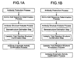
- a production process of an antibody enzyme involving the invention includes an antibody structure analysis process that performs a stereostructure estimation step where a stereostructure of an antibody is estimated from amino-acid sequences; and a catalytic triad residue structure confirmation step where, in an estimated stereostructure of the antibody, whether a catalytic triad residue structure where a serine residue, an aspartate residue and a histidine residue or a glutamate residue are present stereostructurally in proximity to each other is present or not is confirmed.
- any one of indices below can be preferably used.
- Index of type 1 ⁇ or 1 ⁇ * It has either a structure same as a catalytic triad residue structure common in known antibody enzymes (type 1 ⁇ ) or a structure where when among the known catalytic triad residue structures one amino-acid residue that is different in position is used, a catalytic triad residue structure can be assumed to constitute (type 1 ⁇ *).
- Index of type 2 ⁇ A serine residue, an aspartate residue and a histidine residue or a glutamate residue are present in the range of 3 to 20 angstrom in the stereostructure.
- Index of type 3 ⁇ When an antibody has a germ-cell gene type derived from a known antibody enzyme, it is used as an index.
- Index of type 4 ⁇ A complimentarity determining region 1 (CDR1) being constituted of 16 amino-acid residues and a histidine residue being located at the 93 rd according to Kabat numbering scheme are used as an index.
- Index of type 4 ⁇ * A complimentarity determining region 1 (CDR1) being constituted of 11 amino-acid residues and a histidine residue being located at the 91 st or 55 th according to Kabat numbering scheme are used as an index.
- the antibody enzyme production process preferably includes an antibody production process in which an antibody is produced by use of a hybridoma obtained by fusing a mouse spleen lymph corpuscle immunized with antigen and a mouse myeloma cell.
- a hybridoma obtained by fusing a mouse spleen lymph corpuscle immunized with antigen and a mouse myeloma cell.
- the antigen a hapten-conjugated protein obtained by coupling a substance that is made an antigenic determinant to a carrier protein can be used; however, it is not particularly restricted. It goes without saying that a protein itself as well can be used as an immunogen.
- the antibody enzyme production process may further include a catalytic triad residue structure introduction process where by use of the stereostructure information obtained after the antibody structure analysis process, a catalytic triad residue structure is introduced into an antibody according to a genetic engineering procedure.
- the catalytic triad residue structure is one that the present inventors found newly this time as a feature of an antibody enzyme having an activity of cleaving and/or decomposing a polypeptide or antigen protein. Accordingly, since according to the process, antibodies are screened by use of the catalytic triad residue structure unique to the antibody enzyme, the antibody enzymes can be produced more efficiently than ever.
- An antibody enzyme involving the invention has in a stereostructure a catalytic triad residue structure where a serine residue, an aspartate residue, and a histidine residue or glutamate residue are present stereostructurally in proximity.
- a catalytic triad residue structure where a serine residue, an aspartate residue, and a histidine residue or glutamate residue are present stereostructurally in proximity.
- heavy and light chains of i4SL1-2 and i41-7 antibodies can be cited.
- the heavy and light chains of the i4SL1-2 and i41-7 antibodies are experimentally confirmed that, while maintaining a nature, as an antibody, of being high in the affinity with a substrate, these exhibit the enzymatic activity.
- the "catalytic triad residue structure” means a structure in which it is inferred that three amino-acid residues containing at least serine are contained in an active site and form an active center.
- a protease having the catalytic triad residue structure contains serine in the active site; accordingly, it is called a serine protease. Accordingly, the antibody enzyme can be said one kind of the serine protease.
- An antibody enzyme involving the invention may be an antibody enzyme of which complimentarity determining region 1 (CDR1) is constituted of 16 amino-acid residues and that has a histidine residue at the 93 rd according to Kabat numbering scheme, or an antibody enzyme of which complimentarity determining region 1 (CDR1) is formed of 11 amino-acid residues and that has a histidine residue at the 91 st or 55 th according to Kabat numbering scheme, or furthermore one of which mouse germ cell gene type is produced from a gene selected from bb1, bl1, bd2, cr1, cs1, bj2, hf24, 12-41, 19-14, 19-17, 19-23, 19-25 and 21-12 according to the classification due to Thieve et al.
- CDR1 complimentarity determining region 1
- antibody enzymes that are produced from a mouse germ-cell gene selected from the bb1, bl1, bd2, cr1, cs1, bj2, hf24, 12-41, 19-14, 19-17, 19-23, 19-25, and 21-12 are fundamentally antibody enzymes that have the catalytic triad residue structure in a stereostructure and the activity of cleaving and/or decomposing a polypeptide and antigen protein.
- An antibody enzyme involving the invention may be a heavy chain or a light chain of an antibody. Furthermore, in the invention, a gene that codes the antibody enzyme is contained as well.
- an antibody enzyme involving the invention is, among the abovementioned antibody enzymes, in particular, an antibody of which antigen is Helicobacter Pylori urease or a fragment of the antibody, and that is characterized in that it works as a degrading enzyme of the urease.
- the "fragment of an antibody” means the respective peptide chains that constitute an antibody of which antigen is HP urease, and furthermore peptide fragments of a portion of region in the respective peptide chains.
- An antibody enzyme while being an antibody, has the enzymatic activity.
- the antibody enzymes described above in particular, one that exhibits high degradation activity against the antigen protein as a target is called a "super-antibody enzyme".
- the "super-antibody enzyme” can completely decompose a target protein and furthermore has the activity near to that of natural enzyme (reference literature 3).
- the abovementioned antibody enzyme is an antibody of which antigen is HP urease and completely decomposes the HP urease. Accordingly, it is contained in the "super-antibody enzyme”.
- the antibody enzyme with nature as an antibody of the HP urease strongly remained, has the nature of decomposing the HP urease that is an antigen thereof. Accordingly, the antibody enzyme is high in the specificity and can aim and decompose only the urease that is indispensable for the existence of the Helicobacter Pylori.
- the bacteria or the viruses mutate their proteins to acquire the tolerance against the antibiotic drug, and thereby drug resistant bacterium (cell) appears.
- portions indispensable for the existence of the bacterium or viruses themselves cannot be mutated.
- the antibody enzyme can aim and attack the indispensable portions and can completely delete the bioactivity thereof; accordingly, reoccurrence of the symptom as well as the appearance of the drug resistant bacterium (cell) can be avoided.
- the antibody enzyme preferably contains a variable region of an antibody of the HP urease.
- the "variable region” means, among H chains and L chains that constitute an antibody, a portion made of substantially 110 amino-acid residues from an N terminal.
- the “variable region” exhibits diversity in a primary structure depending on the kind of the antibody, and when the antibody has the enzymatic activity, an active center thereof is highly probably contained therein. Accordingly, when the antibody enzyme contains the variable region of the antibody, it can have high activity as enzyme.
- an antibody enzyme involving the invention specifically, ones that are formed by containing an amino acid sequence shown in sequence No. 14 or an amino acid sequence in which in an amino acid sequence shown in the sequence No.14 at least one amino acid is replaced, deleted, inserted and/or added can be cited.
- the amino acid sequence shown in sequence No.14 is a variable region of an L chain of a monoclonal antibody HpU-18 of the HP urease.
- the variable region of the L chain of the HpU-18 as shown also in an example described later, was recognized to be high in the activity as the degrading enzyme of the HP urease.
- the "at least one amino acid being replaced, deleted, inserted and/or added” may be replacement, deletion, insertion and/or addition of amino acids of the number to an extent that, according to a known variant protein production process such as a site-directed mutagenesis by use of, for instance, a genetic engineering procedure, can replace, delete, insert and/or add.
- a genetic engineering procedure is applied, in the amino acid sequence shown in the sequence No.14, an antibody enzyme that is made of an amino acid sequence in which at least one amino acid is replaced, deleted, inserted and/or added, in other words, is a variant of the antibody enzyme made of the amino acid sequence shown in the sequence No. 14.
- an antibody enzyme involving the invention ones that are formed by containing an amino acid sequence shown in any one of sequence Nos.15, 16, 17, 19 and 21, and furthermore ones that are formed by containing an amino acid sequence in which in an amino acid sequence shown in any one of sequence Nos.15, 16, 17, 19 and 21, at least one amino acid is replaced, deleted, inserted and/or added can be cited.
- An amino acid sequence shown in sequence No.15 is a variable region of an L chain of a monoclonal antibody HpU-9 of the HP urease and an amino acid sequence shown in sequence No.16 is a variable region of an H chain of a monoclonal antibody HpU-2 of the HP urease.
- an amino acid sequence shown in sequence No.17 is a variable region of an L chain of a monoclonal antibody HpU-20 of the HP urease and an amino acid sequence shown in sequence No.19 is a variable region of an H chain of a monoclonal antibody HpU-20 of the HP urease.
- amino acid sequence shown in sequence No.21 is a variable region of an L chain of a monoclonal antibody UA-15 of the HP urease.
- antibody enzymes having these amino acid sequences as well as shown in an example described later, as a degrading enzyme of the HP urease, high activity could be recognized.
- the abovementioned antibody enzymes have an action of decomposing the HP urease that is indispensable for the existence of the HP urease; accordingly, these can be used for elimination of HP bacteria. Accordingly, a curative drug involving the invention for a patient infected with the HP bacteria is prepared by containing the antibody enzyme. When the curative drug is orally taken in, it can directly attack the HP bacteria existing in a stomach; accordingly, side effects are less and it can work immediately.
- the antibody enzyme can be not only used as the curative drug but also taken in orally; accordingly, it can be contained in food to take in on a daily basis, and thereby the infection with the HP bacteria can be prevented. Accordingly, in the invention, an infection preventive agent of the HP bacteria formed by containing the antibody enzyme is also contained.
- the invention includes a gene that codes the antibody enzyme and when the gene is introduced in a proper host (such as bacterium and yeast), an antibody enzyme according to the invention can be expressed in the host.
- a proper host such as bacterium and yeast
- a base sequence shown in sequence No.18 is one of base sequences of a gene that codes a variable region of a light chain (L chain) of monoclonal antibody HpU-20 of the HP urease.
- a base sequence shown in sequence No.20 is a base sequence of a gene that codes a variable region of a heavy chain (H chain) of monoclonal antibody HpU-20 of the HP urease.
- a base sequence shown in sequence No.22 is a base sequence of a gene that codes a variable region of a light chain (L chain) of monoclonal antibody UA-15 of the HP urease.
- a base sequence shown in sequence No.47 is one of base sequences of a gene that codes a variable region of a light chain (L chain) of monoclonal antibody HpU-18 of the HP urease.
- a base sequence shown in sequence No.48 is one of base sequences of a gene that codes a variable region of a light chain (L chain) of monoclonal antibody HpU-9 of the HP urease.
- a base sequence shown in sequence No.49 is one of base sequences of a gene that codes a variable region of a heavy chain (H chain) of monoclonal antibody HpU-2 of the HP urease.
- the abovementioned “gene” contains not only a double-stranded DNA but also respective single-stranded DNAs such as a sense strand and an antisense strand that constitute the double-stranded DNA and RNA. Furthermore, the “gene” may be one that contains, other than sequences that code peptides according to the invention, sequences such as a sequence of an untranslated region (UTR) or a sequence of a vector sequence (including an expression vector sequence).
- UTR untranslated region
- a vector sequence including an expression vector sequence
- the invention contains a transformant in which the gene is introduced.
- the transformant can express the antibody enzyme in its body. Accordingly, when a plant is used as a host of the transformant and the gene is introduced in the plant and expressed, a transformant that contains the antibody enzyme can be obtained.
- the transformant contains the antibody enzyme provided with a function of preventing the HP infection; accordingly, when it is taken in on a daily basis, the HP can be suppressed from infecting; that is, it can be used as functional food.
- the antibody enzyme is destroyed in its structure when heated; accordingly, as plants in which the gene can be introduced, vegetables suitable for raw diet are preferable. As such vegetables, specific example includes tomato, cucumber and carrot.
- the antibody enzyme involving the invention is characterized in that it is an antibody of human-derived chemokine receptor CCR-5 or an antibody fragment thereof and can decompose the chemokine receptor CCR-5.
- the "antibody fragment” means the respective peptide chains that constitute an antibody of which antigen is the chemokine receptor CCR-5 and further peptide fragments of a portion of a region in the respective peptide chains.
- the antibody enzyme is an antibody of which antigen is the chemokine receptor CCR-5 and completely decompose the CCR-5; accordingly, it is included in the "super antibody enzyme”.
- the antibody enzyme with the nature as an antibody of the chemokine receptor CCR-5 strongly remained, has the nature of decomposing the chemokine receptor CCR-5 that is an antigen thereof. Accordingly, the antibody enzyme is high in the specificity and can aim and decompose only the chemokine receptor CCR-5.
- the chemokine receptor CCR-5 is a coreceptor that plays an important role in the infection of the HIV; accordingly, the antibody enzyme that can delete the function thereof can be effectively utilized as an anti-HIV drug that is used in the prevention of the HIV infection or remedy of AIDS.
- the antibody enzyme is preferably formed with a variable region of the chemokine receptor CCR-5 antibody contained therein.
- the "variable region” means, among H and L chains that constitute an antibody, a portion made of substantially 110 amino-acid residues from an N terminal.
- the variable region depending on the kind of the antibody, shows the diversity in its primary structure, and, when the antibody has the activity as the enzyme, the active center thereof is probably contained therein. Accordingly, when the antibody enzyme contains the variable region, it can have high activity as an enzyme.
- an antibody enzyme involving the invention specifically, one that is formed by containing an amino acid sequence shown in sequence No.26 or one that is formed by containing an amino acid sequence in which in an amino acid sequence shown in sequence No.26 at least one amino acid is replaced, deleted, inserted and/or added can be cited.
- the amino acid sequence shown in the sequence No.26 is a variable region of a light chain (L chain) of monoclonal antibody ECL2B-2 of the chemokine receptor CCR-5.
- the variable region of the L chain of the ECL2B-2 as shown in an example described later, was recognized to be high in the activity as a CCR-5 degrading enzyme.
- an antibody enzyme involving the invention one that is formed by containing an amino acid sequence shown in the sequence No.30, or an amino acid sequence in which in an amino acid sequence shown in the sequence No.30, at least one amino acid is replaced, deleted, inserted and/or added can be cited.
- the amino acid sequence shown in sequence No.30 is a variable region of a light chain (L chain) of monoclonal antibody ECL2B-3 of the chemokine receptor CCR-5.
- the activity as a CCR-5 degrading enzyme is confirmed to be high.
- an antibody enzyme involving the invention one that is formed by containing an amino acid sequence shown in the sequence No.34, or an amino acid sequence in which in an amino acid sequence shown in the sequence No.34, at least one amino acid is replaced, deleted, inserted and/or added can be cited.
- the amino acid sequence shown in sequence No.34 is a variable region of a light chain (L chain) of monoclonal antibody ECL2B-4 of the chemokine receptor CCR-5.
- the activity as a CCR-5 degrading enzyme is confirmed to be high.
- an antibody enzyme involving the invention one that is formed by containing an amino acid sequence shown in the sequence No.36, or an amino acid sequence in which in an amino acid sequence shown in the sequence No. 36, at least one amino acid is replaced, deleted, inserted and/or added can be cited.
- An amino acid sequence shown in sequence No.36 is a variable region of a heavy chain (H chain) of monoclonal antibody ECL2B-4 of the chemokine receptor CCR-5.
- H chain heavy chain
- CCR-5 chemokine receptor
- the "at least one amino acid being replaced, deleted, inserted and/or added” may be replacement, deletion, insertion and/or addition of amino acids of the number to an extent that, according to a known variant protein production process such as a site-directed mutagenesis by use of, for instance, a genetic engineering procedure, can be replaced, deleted, inserted and/or added.
- an antibody enzyme that is made of an amino acid sequence in which, in the amino acid sequence shown in any one of the sequence Nos.26, 30, 34 and 36, at least one amino acid is replaced, deleted, inserted and/or added is, in other words, a variant of the antibody enzyme made of the amino acid sequence shown in any one of the sequence Nos.26, 30, 34 and 36.
- the antibody enzyme according to the invention decomposes the chemokine receptor CCR-5 that plays an important role in the HIV infection to delete the function thereof; accordingly, it can be used also as an anti-HIV drug that disturbs the HIV infection. That is, the anti-HIV drug according to the invention is characterized by containing the antibody enzyme and by disturbing the infection of the AIDS viruses.
- the antibody enzyme according to the invention can delay the development of the AIDS to an HIV infected patient or inhibit disease from progressing. That is, the anti-HIV drug according to the invention, containing the antibody enzyme, may be one that works as a curative drug against the AIDS virus-infected patient.
- the anti-HIV drug according to the invention containing the antibody enzyme alone, can be used by directly administering by means of such as intravenous injection.
- a carrier that can be physiologically tolerated may be further contained. Production of such an anti-HIV drug can be carried out according to a so far known production process.
- the anti-HIV drug uniquely aims and decomposes a CCR-5 molecule alone; accordingly, it can be expected that not only the effect is remarkable but also the side effects are less.
- the invention includes also a gene that codes the antibody enzyme.
- the gene is expressibly introduced in an appropriate host (such as bacterium and yeast), the antibody enzyme according to the invention can be expressed in the host.
- the abovementioned “gene” contains not only a double-stranded DNA but also respective single-stranded DNAs such as a sense strand and an antisense strand that constitute the double-stranded DNA and RNA. Furthermore, the “gene” contains, other than sequences that code the antibody enzymes according to the invention, sequences such as a sequence of an untranslated region (UTR) or a sequence of a vector sequence (including an expression vector sequence).
- UTR untranslated region
- a vector sequence including an expression vector sequence
- the base sequence shown in sequence No.27 is a base sequence of a gene that codes a variable region of a light chain (L chain) of monoclonal antibody ECL2B-2 of the chemokine receptor CCR-5.
- the base sequence shown in the sequence No.31 is a base sequence of a gene that codes a variable region of a light chain (L chain) of monoclonal antibody ECL2B-3 of the chemokine receptor CCR-5.
- the base sequence shown in the sequence No.35 is a base sequence of a gene that codes a variable region of a light chain (L chain) of monoclonal antibody ECL2B-4 of the chemokine receptor CCR-5.
- the base sequence shown in the sequence No.37 is a base sequence of a gene that codes a variable region of a heavy chain (H chain) of monoclonal antibody ECL2B-4 of the chemokine receptor CCR-5.
- a transformant in which the gene is introduced is also included.
- the transformant is one in which the gene is expressibly introduced in an appropriate host (such as bacteria and yeast) and can express the antibody enzyme according to the invention in its body. Accordingly, the transformant as well can be utilized as an anti-HIV drug that prevents the infection of the AIDS viruses and an anti-HIV drug for treating an AIDS-infected patient.
- the invention contains a computer program that instructs a computer to carry out an antibody structure analysis process in the antibody enzyme production process; and a machine-readable recording medium in which a computer program that instructs a computer to carry out an antibody structure analysis process is recorded.
- a production process according to the present invention of antibody enzymes and an antibody enzyme produced according to the production process will be described as embodiment 1.
- the present invention is not restricted thereto.
- the invention proposes an antibody enzyme production method that not only enables to efficiently obtain natural antibody enzymes but also can produce antibody enzymes by use of a genetic engineering process, and further provides an example of novel and useful antibody enzymes obtained according to the process.
- an antibody enzyme production process involving the invention will be described, followed by describing an example of obtained antibody enzymes and gene thereof, functions of the antibody enzyme, and applications thereof.
- the inventors analyzed in detail features of nature and structure of several kinds of antibody enzymes having the activity of cleaving and/or decomposing polypeptides and antigen proteins. As a result, the inventors for the first time revealed that the antibody enzymes having the activity of cleaving and/or decomposing polypeptides and antigen proteins all have a catalytic triad residue structure in a stereostructure thereof.
- a site where the catalytic triad residue structure is present is not particularly restricted.
- An antibody is constituted of a heavy chain (H chain) and a light chain (L chain).
- the light chain is constituted of a variable region (VR) and a constant region (CR), and the variable region includes a complimentarity determining region (CDR).
- CDR complimentarity determining region
- An antibody enzyme production process involving the invention may include at least an antibody structure analysis process that carries out a stereostructure estimation step where from amino acid sequences a stereostructure of an antibody is estimated; and a catalytic triad residue structure confirmation step where whether a catalytic triad residue structure is present in the estimated stereostructure of an antibody or not is judged.
- an antibody structure analysis process that carries out a stereostructure estimation step where from amino acid sequences a stereostructure of an antibody is estimated
- a catalytic triad residue structure confirmation step where whether a catalytic triad residue structure is present in the estimated stereostructure of an antibody or not is judged.
- the production process may include an antibody production step, an amino acid sequence determination step, an antibody structure analysis step and an antibody enzymatic activity confirmation step.
- the antibody production step may produce monoclonal antibodies from hybridomas obtained by fusing mouse spleen lymph corpuscles immunized with antigens and mouse myeloma cells, alternatively, antibodies obtained from a library by use of a phage display method may be used.
- an antibody when, as an immunogen, a desired antigen, or a fragment containing an antigenic determinant (epitope) thereof, or a derivative thereof, or an analogue thereof, or a cell that can express these is used, an antibody can be produced.
- a hybridoma method reference literature 5: Kohler, G. and Milstein, C., Nature 256, 495-497 (1975)
- a trioma method a human B cell hybridoma method
- reference literature 6 Kozbor, Immunology Today 4, 72, 1983
- an EBV-hybridoma method reference literature 7: Monoclonal Antibodies and Cancer Therapy, Alan R. Liss, Inc., pp. 77-96, 1985).
- the antigen is a polypeptide or a protein, or a polysaccharide, there is no particular restriction.
- the antigen when the antigen is a hapten, it does not have the capability of producing antibodies; accordingly, it cannot produce the antibody.
- an antigen when an antigen is covalently bonded with a carrier made of a biopolymer such as a protein derived from a different kind to obtain an antigen protein and this is used to immunize, the antibody production can be induced.
- a carrier made of a biopolymer such as a protein derived from a different kind to obtain an antigen protein and this is used to immunize, the antibody production can be induced.
- the carrier without restricting to particular one, various kinds of proteins so far known in the field such as ovalbumin, gamma globulin, and hemocyanine can be preferably used.
- polypeptide here includes short peptides in which several amino acids are bonded and proteins. Furthermore, the polypeptide includes naturally existing ones and chemically, biologically synthesized ones. Still furthermore, the "polysaccharide” here means ones in which several of so-called monosaccharides such as glucose, maltose, acetylglucosamine, and glucuronic acid are bonded. Kinds of monosaccharide that constitutes the “polysaccharide” are not particularly restricted as far as these are so far known ones.
- the amino acid sequence determination step is not particularly restricted.
- the amino acid sequence may be determined by directly reading an amino acid sequence from the obtained antibody, or an amino acid sequence may be estimated from a base sequence of a gene of the antibody.
- the amino acid sequence may be determined by use of an Edman method.
- the base sequence may be determined by use of a so far known sequencer, and the amino acid sequence may be estimated by use of a so far known soft wares and so on. These are not particularly restricted.
- the antibody structure analysis process as far as it includes a stereostructure estimation step and a catalytic triad residue structure confirmation step, is not particularly restricted. Thereby, whether a desired antibody includes a catalytic triad residue structure or not can be efficiently confirmed.
- the stereostructure estimation step as far as it can estimate a stereostructure from data of the amino acid sequences obtained in the amino acid sequence determination step, is not particularly restricted. Specifically, all that is necessary is to be able to estimate a secondary structure and a tertiary structure in a protein. That is, in the invention, all that is necessary is to clarify up to a tertiary structure of an IgG class antibody.
- a quaternary structure in an antibody like for instance IgA and IgM class, a structure in which a plurality of subunits (IgG class antibody) having a tertiary structure meets to form an oligomer can be cited.
- IgG class antibody subunits having a tertiary structure meets to form an oligomer
- a structure is not particularly necessary in a later catalytic triad residue structure confirmation step; accordingly, it is not necessary to estimate in the invention.
- a quaternary structure of an antibody may be estimated in the stereostructure estimation step.
- a so far known higher-order structure analysis soft ware may be used, and a specific procedure thereof is not particularly restricted.
- the catalytic triad residue structure confirmation step as far as whether the catalytic triad residue structure described in (1) is present in a stereostructure of an antibody or not can be judged, there is no particular restriction. Accordingly, when the catalytic triad residue structure is judged, presence of the abovementioned structure may be judged directly or indirectly.
- the respective indices below can be cited.
- a more simple method where whether a histidine residue is present in an amino acid sequence of an antibody or not is investigated may be used to judge whether the catalytic triad residue structure is present or not. This is because in many antibodies, usually, in comparison with the serine residue, the histidine residue is less contained.
- a first index whether the catalytic triad residue structure has a structure same as a catalytic triad residue structure common in known antibody enzymes or not, alternatively, when, among the known catalytic triad residue structures, only one residue thereof uses an amino-acid residue in a different position, whether it is estimated that a catalytic triad residue structure can be constituted or not is made an index, the former case is called a type 1 ⁇ , and the latter one is called a type 1 ⁇ *.
- a case where in a single antibody heavy chain (H chain) or a single antibody light chain (L chain), in a stereostructure, in the range of 3 to 20 ⁇ , the respective functional groups of a serine residue, an aspartate residue, and a histidine residue or glutamate residue are present is made an index, and this is called a type 2 ⁇ index.
- an index that makes use of an analysis result of germline genes can be preferably used.
- the antibody light chain can be classified into two kinds of classes, ⁇ and ⁇ . In the case of a mouse, it is said that substantially 95% is the ⁇ chain. Furthermore, since analysis of germline genes of mouse ⁇ chains is being advanced, information thereof can be advantageously obtained. As will be described in an example described later, by analyzing the germlines, a very important knowledge that suggests that in particular germlines an antibody L chain having the enzymatic activity is already prepared is found (example 1-3 and Table 3). In this connection, when germlines from which known antibody enzymes are derived are investigated in advance and antibodies having the germlines are selected, of the light chains, antibody enzymes can be efficiently obtained at a high probability. This is made a type 3 ⁇ index.
- germlines that can be used for the light chains specifically, according to a classification due to Thiebe et al., bb1, cr1, bl1, cs1, bd2, bj2 or 19-25 can be cited.
- the germlines will be classified according to Thiebe et al.
- H chain when a method of thinking of the light chains is applied, when antibodies having Families of VH1, VH5 and VH10 are selected, antibody enzymes can be efficiently obtained at a very high probability. In the aging process of antibodies, mutation is caused, and some may lose the catalytic triad residue structure; however, when, after the removal of these, the germline or the Family is identified, substantially completely antibodies having the enzymatic activity can be obtained.
- a structurally characteristic constitution obtained from known antibody enzymes as well can be used as an index.
- a feature that a complimentarity determining region 1 (CDR1) is constituted of 16 amino-acid residues and a histidine residue is present at the 93 rd according to the Kabat numbering scheme is remarkable for the antibody enzyme. Accordingly, this can be used as an index of type 4 ⁇ .
- a feature that a complimentarity determining region 1 (CDR1) is constituted of 11 amino-acid residues and a histidine residue is present at the 91 st or 55 th according to the Kabat numbering scheme is also remarkable for the antibody enzyme; accordingly, this can be used as an index of type 4 ⁇ *.
- an amino acid sequence of an antibody is expressed according to the Kabat numbering scheme.
- indexes are the types 1 ⁇ ⁇ 1 ⁇ * or 2 ⁇ ; accordingly, in order to heighten the assuredness at the selection, these indices are preferably used, and type 3 ⁇ and types 4 ⁇ and 4 ⁇ * may be used complementarily. However, without using the indices of the types 1 ⁇ , 1 ⁇ * and 2 ⁇ , the indices of type 3 ⁇ and types 4 ⁇ and 4 ⁇ * alone may be used singularly or in a combination.
- an antibody structure analysis system described later in (3) can be cited.
- the invention is not restricted thereto.
- the amino acid sequence determination step may be contained as one step of the antibody structure analysis process. That is, as described in the amino acid sequence determination step, an amino acid sequence determination step where an amino acid sequence is estimated from base sequences of a gene of an antibody may be carried out as one step of the antibody structure analysis process.
- the antibody enzymatic activity confirmation step as far as it can confirm, after the analysis in the antibody structure analysis step, whether an antibody that has a catalytic triad residue structure and is very high in the likelihood of being an antibody enzyme actually has the activity of cleaving and/or decomposing a polypeptide and an antigen protein or not, is not particularly restricted.
- a polypeptide or an antigen protein that includes a structure that becomes an antigen or an antigen determinant may be reacted with an antibody to check the activity.
- reaction conditions such as a concentration of the antigen, a concentration of the antibody, a reaction temperature and a reaction time, and a concentration of a base (kind of buffer used) are not particularly restricted.
- a catalytic triad residue structure introduction step may be included. That is, even antibodies that inherently do not have a catalytic triad residue structure, when a catalytic triad residue structure is introduced therein according to a genetic engineering process, can produce antibody enzymes with high efficiency. Accordingly, after the antibody structure analysis step, a catalytic triad residue structure introduction step may be carried out.
- catalytic triad residue structure introduction step all that is necessary is to introduce a catalytic triad residue structure, by use of stereostructure information obtained in the antibody structure analysis process, in an antibody according to a genetic engineering process, and a specific procedure is not particularly restricted. Specifically, so far known methods such as a method of preparing a deletion mutant by use of an exonuclease and site-directed mutagenesis can be preferably applied.
- the catalytic triad residue structure introduction step may be carried out after the antibody structure analysis process, and it is very preferable that after the catalytic triad residue structure introduction step, an antibody enzymatic activity confirmation step is carried out to judge whether, when the catalytic triad residue structure is introduced, the activity of cleaving and/or decomposing a polypeptide or an antigen protein can be imparted or not.
- Means of carrying out the antibody structure analysis process according to the invention may be an antibody structure analysis system that includes a computer.
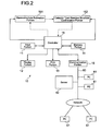
- an antibody structure analysis system 10 including an input portion 11, a display portion 12, a printing portion 13, a memory portion 14, a controller 15, a stereostructure estimation portion 151 and a catalytic triad residue structure confirmation portion 152 can be cited.
- the input portion 11 as far as it enables to input information involving an operation of the antibody structure analysis system 10, is not particularly restricted. So far known input means such as a keyboard, a tablet or a scanner can be preferably used.
- the display portion 12 displays various kinds of information such as information involving an operation of the antibody structure analysis system 10 and selection results including an estimated stereostructure of the antibody and a confirmation result of the catalytic triad residue structure.
- various kinds of display devices such as known CRT displays and liquid crystal display devices can be preferably used; however, there is no particular restriction.
- the printing portion 13 records (prints or picturizes) various kinds of information that can be displayed by the display portion 12 on recording materials such as PPC paper.
- recording materials such as PPC paper.
- known image formation devices such as an ink jet printer and a laser printer can be preferably used; however, there is no particular restriction.
- the display portion 12 and printing portion 13 in combination can be represented as output means. That is, the display portion 12 is means for outputting various kinds of information in a soft copy and the printing portion 13 is means for outputting various kinds of information as a hard copy. Accordingly, as the outputting means used in the invention, without restricting to the display portion 12 and the printing portion 13, other outputting means may be provided.
- the memory portion 14 memorizes various kinds of information (such as control information, selected results and other information) that is used in the antibody structure analysis system 10.
- various kinds of memory means such as semiconductor memories such as RAMs and ROMs, magnetic discs such as floppy discs and hard discs, disc types of optical discs such as CD-ROM/MO/MD/DVD and card types such as IC cards (including memory cards)/optical cards can be preferably used.
- the memory portion 14 may be integrated with the antibody structure analysis system 10 to form one device, or isolated as an external memory device, or constituted so as to have both the integrated memory portion 14 and the external memory device.
- the integrated memory portion 14 a built-in type hard-disc drive and a floppy disc-drive, a CD-ROM drive or a DVD-ROM drive incorporated in a device can be cited, and, as an external memory device, an external hard disc and external types of the abovementioned various kinds of disc-drives can be cited.
- the controller 15 controls an operation of the antibody structure analysis system 10. Specifically, as shown in Fig.2 with solid arrow marks, to the respective means of the input portion 11, the display portion 12, the printing portion 13, the memory portion 14, the stereostructure estimation portion 151 and the catalytic triad residue structure confirmation portion 152, the control information is outputted from the controller 15. On the basis of the control information, the respective means operate in combination to drive the antibody structure analysis system 10 as a whole. Furthermore, to the controller 15, instruction information to drive the antibody structure analysis system 10 can be inputted from the input portion 11. Accordingly, in Fig.2, the solid arrow marks that show an exchange of the control information are bi-directional.
- the stereostructure estimation portion 151 and the catalytic triad residue structure confirmation portion 152 work in combination as analysis means. Specifically, to the stereostructure estimation portion 151, the amino acid sequence data inputted from the input portion 11 are inputted through the controller 15 (dotted line in the drawing). Then, data of the stereostructure generated at the stereostructure estimation portion 151 are outputted to the catalytic triad residue structure confirmation portion 152 (dotted line in the drawing). In the catalytic triad residue structure confirmation portion 152, from the stereostructure data, whether the catalytic triad residue structure is present or not is confirmed, and based on the confirmation result, final analysis data are generated (dotted line in the drawing). The analysis data, as far as the data can be outputted by outputting means such as the display portion 12 and/or printing portion 13, are not particularly restricted.
- the data that are used to analyze a stereostructure in the stereostructure estimation portion 151 may be at least data of the amino acid sequences; however, it may be data of the base sequences. In this case, all that are necessary for the stereostructure estimation portion 151 are to estimate amino acid sequences from the data of base sequences to generate a stereostructure based on the data.
- controller 15, the stereostructure estimation portion 151 and the catalytic triad residue structure confirmation portion 152 are not particularly restricted; that is, so far known calculation means can be preferably used.
- the respective means may be independently constituted from each other; however, the respective means are preferably integrated into one calculation means to be an integrated control and analysis system. Specifically, it is very preferable that the respective means are integrated as a central processing unit (CPU) of a computer and an operation thereof is carried out according to a computer program.
- CPU central processing unit
- the stereostructure estimation portion 151 may carry out a calculation where a CPU estimates a stereostructure according to a so far known computer program that estimates a stereostructure of a protein; and the catalytic triad residue structure confirmation portion 152 may carry out a calculation where a CPU determines at least whether a catalytic triad residue structure is present or not according to a computer program.
- the antibody structure analysis system 10 may include other analysis means and, as analysis data, other data other than that of the catalytic triad residue structure and the stereostructure.
- the antibody structure analysis system 10 may be constituted so that, by providing a communication portion 16 as shown in Fig.2, various kinds of information may be inputted and outputted through a communication network including the Internet.
- the communication portion 16 is connected with a communication network and can send and receive various kinds of information.
- the antibody structure analysis system 10 a personal computer (PC) 61 and a server 62 in the same premise are connected to a communication line 60 to form a bus type LAN (local area network), and further the LAN is connected with a PC 61 in other area through the Internet.
- PC personal computer
- a specific configuration of the communication portion 16 is not particularly restricted.
- Known LAN cards, LAN boards, LAN adaptors and modems can be preferably used.
- the PC 61 As the PC 61, known personal computers provided with communication means such as a modem can be preferably used and the PC 61 is not restricted to desktop type and note-type ones.
- the PC 61 has a fundamental constitution that has a display portion such as a CRT display or a liquid crystal device and an input portion such as a keyboard or a mouse.
- a display portion and input portion that are provided to the PC 61 and not shown in the drawing are expressed as a PC display portion and a PC input portion.
- the PC 61 may be provided with a hardware (various kinds of input means such as a scanner and various kinds of output means such as a printer) that can be externally connected to a general personal computer.
- a hardware various kinds of input means such as a scanner and various kinds of output means such as a printer
- a specific configuration of the server 62 is neither particularly restricted and may be a computer that can offer a service to the PCs 61 that are clients constituting the LAN and the antibody structure analysis system 10. Furthermore, the server 62 may combine a data base server and a file service server.
- a specific configuration of the communication line 60 is neither particularly restricted; that is, so far known general communication lines can be used. Furthermore, a type and a bus type of the LAN that is constituted with the communication line 60 are neither particularly restricted; that is, so far known type such as a star type and a ring type may be used.
- the LAN may include shared printers and other terminals. Still furthermore, through not shown in the drawing, in the communication network including the LAN, various kinds of communicable and portable terminals may be included.
- the selected results can be not only outputted simply in the antibody structure analysis system 10 (that is, such as the display portion 12 and printing portion 13) but also transmitted through the LAN to the PCs 61.
- results obtained from the antibody structure analysis system 10 can be displayed on the PC display portion and printed out by use of a printer, and furthermore owing to an input from the PC input portion the selected results can be processed as well.
- the communication portion 16 can function not only as communication means but also as input means of the antibody structure analysis system 10. Furthermore, in particular, when, by use of the PC61, through the Internet, at an area remote from a location where the antibody structure analysis system 10 is located, information (such as amino acid sequences) involving implementation of the antibody structure analysis process is transmitted or selected results are received, to optional customers, a service that provides the antibody structure analysis process can be provided.
- the present invention can be more efficiently carried out.
- the server 62 combining a database server and a file server, selected results of the antibody structure analysis process that is performed through a communication network can be stored through the communication network in the server 62. As a result, the selected results can be more efficiently used.
- the antibody structure analysis process in the invention can be carried according to a program on a computer.
- a recording medium that records the program a medium that carries a program in a volatile manner such as downloading from a communication network is also included.
- the antibody structure analysis system 10 may appropriately download the program of the antibody structure analysis process from the server 62 to use.
- a program for downloading is stored in advance in a body of the antibody structure analysis system 10 or can be installed from a separate recording medium.
- the PC 61 when it is connected through a communication network to the server 62, when a program of the antibody structure analysis process is downloaded from the server 62, the PC 61 itself can be used as the antibody structure analysis system 10.
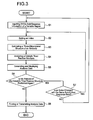
- step 1 (hereinafter, the step is abbreviated as S)
- S2 an index for confirming a catalytic triad residue structure in a catalytic triad residue structure confirmation portion 152 is set.
- the index types 1 ⁇ ⁇ 1 ⁇ *, 2 ⁇ , 3 ⁇ , 4 ⁇ , and 4 ⁇ * explained in the (2-3) can be cited.
- the indices may be used singularly or in a combination of a plurality thereof.
- the antibody is high in the likelihood of being an antibody enzyme.
- the antibody is low in the likelihood of being an antibody enzyme.
- the antibody structure analysis system 10 in the above-described embodiment may be realized on a computer with a program that functions the antibody structure analysis process including above explained steps from S1 to S8.
- the program may be stored in a recording medium that can be read with a computer.
- the memory portion 14 shown in Fig.3, specifically, for instance, a ROM itself may be a program medium.
- the memory portion 14 When the memory portion 14 is provided with a program reader, it may be a program medium that can be read when a recording medium is inserted therein.
- the program medium known configurations cited as specific examples of the memory portion 14 can be preferably used.
- the stored program may be carried out when the controller 15 accesses thereto, or the program is read, the read program is downloaded in a not shown program memory area and the program may be carried out.
- a downloading program is stored in the memory portion 14 beforehand.
- a content stored in the recording medium, without restricting to the program, may be other data for instance.
- a computer program that carries out the antibody structure analysis process on a computer and a machine-readable recording medium in which a computer program that makes a computer execute the program is recorded are included. Accordingly, in order to execute the antibody structure analysis process in the invention on a computer according to a program, the computer itself can be made the antibody structure analysis system 10. As a result, the versatility of the invention can be heightened and the invention can be readily used on a communication network.
- the antibody enzyme involving the invention includes an antibody enzyme having a stereostructurally unique structure that can be applied in the production process, and furthermore, includes also an antibody enzyme that can be produced according to the abovementioned production method.
- an antibody enzyme involving the invention will be detailed.
- An antibody enzyme involving the embodiment is (a) an antibody enzyme having a variable region made of an amino-acid sequence described in sequence No. 1, 3, 5 or 7 or (b) an antibody enzyme having a variable region made of an amino-acid sequence in which in an amino-acid sequence described in sequence No. 1, 3, 5 or 7, at least one amino acid is replaced, deleted, inserted and/or added and the activity of cleaving and/or decomposing a polypeptide or an antigen protein.
- the i41SL1-2 antibody is a monoclonal antibody against a synthetic peptide (shown in the sequence No. 9) of a complimentarity determining region 1 (CDR 1) of a light chain of an antibody 41S-2 that recognizes a constant region of envelope protein gp-41 of HIV virus.
- CDR 1 complimentarity determining region 1
- Amino acid sequences of variable regions of the heavy and light chains of the i41SL1-2 antibody and base sequences that code the amino acid sequences are shown in Figs. 4A and 4B.
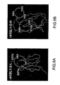
- a stereo structure of each of the light chain (i41SL1-2-L) and heavy chain (i41SL1-2-H) of the i41SL1-2 antibody is estimated and analyzed with a computer.
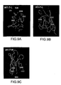
- Figs. 5A and 5B it was revealed that the i41SL1-2 antibody has a catalytic triad residue structure in both i41SL1-2-H and i41SL1-2-L. That is, the i41SL1-2-L, as shown in Figs.
- catalytic triad residue structure constituted of, according to Kabat numbering scheme, an aspartate residue at the 1 st (D1), a histidine residue at the 93 rd (H93), and a serine residue at the 27A (S27a) or a catalytic triad residue structure constituted of, according to Kabat numbering scheme, an aspartate residue at the 27D (D27d), a histidine residue at the 93 rd (H93), and a serine residue at the 27A (S27a).
- the i41SL1-2-H is estimated to have a catalytic triad residue structure constituted of, according to Kabat numbering scheme, an aspartate residue at the 55 th (D55), a histidine residue at the 52 nd (H52), and a serine residue at the 54 th (S54) or a catalytic triad residue structure constituted of, according to Kabat numbering scheme, an aspartate residue at the 100 th (D100), a histidine residue at the 35 th (H35), and a serine residue at the 98 th (S98).
- Locations of the catalytic triad residue structures are in the neighborhood of the complimentarity determining regions for both the light and heavy chains.
- the i41-7 antibody is a monoclonal antibody against a light chain of antibody 41S-2. Amino acid sequences of variable regions of the heavy and light chains of the i41-7 antibody and base sequences that code the amino acid sequences are shown in Figs. 8A and 8B.
- a stereo structure of each of the light chain (i41-7-L) and heavy chain (i41-7-H) of the i41-7 antibody is, similarly to the i41SL1-2 antibody, estimated and analyzed with a computer.
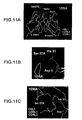
- the i41-7 antibody has a catalytic triad residue structure in each of the i41-7-L and i41-7-H. That is, the i41-7-L, as shown in Figs.
- catalytic triad residue structure constituted of, according to Kabat numbering scheme, an aspartate residue at the 1 st (D1), a histidine residue at the 91 st (H91), and a serine residue at the 26 th (S26) or a catalytic triad residue structure constituted of, according to Kabat numbering scheme, an aspartate residue at the 28 th (D28), a histidine residue at the 91 st (H91), and a serine residue at the 30 th (or 26 th ) (S30 or S26).
- Locations of the catalytic triad residue structures are in the neighborhood of a CDR1 and CDR3 that are the complimentarity determining region (CDR).
- the i41-7-L is estimated to have a catalytic triad residue structure constituted of, according to Kabat numbering scheme, an aspartate residue at the 60 th (D60), a histidine residue at the 55 th (H55), and a serine residue at the 52 nd (S52). This is located in the neighborhood of a CDR2.
- the i41-7-H is estimated to have a catalytic triad residue structure constituted of, according to Kabat numbering scheme, an aspartate residue at the 86 th (D86), a histidine residue at the 41 st (H41), and a serine residue at the 40 th (or 84 th or 87 th ) (S40 or S84, or S87).
- a gene involving the embodiment may be (a) a gene that codes an antibody enzyme having a variable region made of an amino-acid sequence shown in sequence No. 1, 3, 5 or 7, or (b) a gene that codes an antibody enzyme that has a variable region made of an amino-acid sequence that is obtained by replacing, deleting, inserting and/or adding at least one amino acid in an amino acid sequence shown in sequence No. 1, 3, 5 or 7 and has the activity of cleaving and/or decomposing a polypeptide and an antigen protein.
- 1 ⁇ a gene (i41SL1-2-H gene) having a variable region made of a base sequence (cDNA) shown in sequence No.2, 2 ⁇ a gene (i41SL1-2-L gene) having a variable region made of a base sequence (cDNA) shown in sequence No.4, 3 ⁇ a gene (i41-7-H gene) having a variable region made of a base sequence (cDNA) shown in sequence No.6, and 4 ⁇ a gene (i41-7-L gene) having a variable region made of a base sequence (cDNA) shown in sequence No.8 can be exemplified.
- the genes are derived from a mouse (Mus musculus).
- the "gene” according to the invention includes an RNA and a DNA.
- the RNA includes an mRNA
- the DNA includes a cDNA or a genome DNA that can be obtained by, for instance, cloning or chemical synthesis technology or a combination thereof.
- the DNA may be a double-stranded one or a single-stranded one.
- the single-stranded DNA may be a code DNA that becomes a sense strand or an antisense code that becomes an antisense strand (antisense strand can be used as a probe or an antisense drug).
- the "gene” according to the invention includes, other than sequences that code proteins of the (a) or (b), sequences such as a sequence of an untranslated region (UTR) or a sequence of a vector sequence (including an expression vector sequence).
- both the heavy and light chains of the i41-7 antibody have the peptidase activity, and, when a peptide is short, irrespective of the specificity, though different in the reaction rate, cleave and/or decompose the peptide. Still furthermore, shorter peptides were larger in the reaction rate than longer peptides. This is considered that, irrespective of the specificity, the shorter peptide is also small in the molecular weight and can readily access a bonding site of the i41-7 antibody.
- the i41-7-L decomposes 41S-2-L (25kDa) that is an antigen protein and a band of 22.5kDa appears. Furthermore, the i41-7-L did not decompose an irrelevant protein such as BSA; accordingly, it can be said it specifically decompose the antigen protein.
- the i41SL1-2-H, i41-7-H, i41SL1-2-L and i41-7-L when a substrate is a protein, specifically cleave and/or decompose; when the substrate is a polypeptide (polypeptides more than substantially 30 mer or more), to a certain extent specifically, cleave and/or decompose; and, when the substrate is a more shorter polypeptide (substantially 30 mer or less), rather non-specifically cleave and/or decompose.
- i41SL1-2-H, i41-7-H, i41SL1-2-L and i41-7-L are antibody enzymes that have excellent substrate recognition ability of antibody and substrate transforming capacity of enzyme in combination.
- MA-2 monoclonal antibody against methamphetamine
- Fig. 12 An antibody that does not have a catalytic triad residue structure in a stereostructure, MA-2 (monoclonal antibody against methamphetamine) does not at all cause the antigen decomposition reaction (Fig. 12).
- heavy chains of HpU-9 and HpU-18 antibodies do not have His; accordingly, these cannot have a catalytic triad residue structure. Both heavy chains did not at all exhibit the peptidase activity.
- an antibody enzyme that has a catalytic triad residue structure in a stereostructure has the activity of cleaving and/or decomposing a polypeptide and an antigen protein.
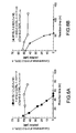
- the decomposition reactions of antigen peptides owing to the light and heavy chains of the i41SL1-2 antibody are kinetically analyzed (Figs. 7A and 7B and Table 5).
- the light chain exhibited such a high catalyst efficiency (kcat/Km) same as that of tripsin and the heavy chain exhibited a value substantially one tenth that of tripsin.
- both the light and heavy chains can be said have the high enzymatic activity comparable to that of a natural protease.
- the i41SL1-2-L and i41SL1-2-H with the nature as an antibody that has high affinity to a substrate remaining, exhibit the enzymatic activity of decomposing a polypeptide and an antigen protein.
- the antibody enzyme involving the invention was experimentally demonstrated that it has the activity identical as that of a natural protease and high affinity and specificity to a substrate unique to an antibody.
- the antibody enzymes involving the embodiment can be produced by use of the abovementioned antibody enzyme production process involving the invention.
- antibodies having the enzymatic activity are produced.
- HpU-2-H (J558.h) lacks a histidine residue, forms a catalytic triad residue structure from Asp86, Glu85 and Ser84 (orSer87) and exhibits the activity of decomposing polypeptides and antigen proteins. This is a very important finding that suggests that there are many antibodies having the enzymatic activity in nature and that the germline can be an index when this kind of antibody is searched. A detail thereof will be described in an embodiment described below.
- Antibody enzymes and genes involving the invention are novel antibody enzymes that can be produced based on a so far unknown knowledge that a catalytic triad residue structure is present in a stereostructure. Accordingly, these have the availability below.
- a stereostructure of the antibody is estimated by use of a computer, whether a structure that can be a catalytic triad residue structure is present in the stereostructure thereof or not is investigated to estimate an antibody enzyme that has the activity of cleaving and/or decomposing a polypeptide and an antigen protein, and thereby the antibody enzymes are enabled to efficiently obtain.
- a structure that can be a catalytic triad residue structure is introduced into a stereostructure, and also thereby an antibody enzyme having the activity of cleaving and/or decomposing a polypeptide and an antigen protein can be artificially prepared.
- the antibody enzyme obtained, prepared according to a process as mentioned above has highly specific substrate recognizing capability of the antibody and the enzymatic activity. Accordingly, it is considered that to infectious diseases caused, for instance, by intrusion of bacteria or viruses into a living body, when antibody enzymes specific to causative bacteria or viruses are obtained or prepared, the antibody enzymes can be used to diagnose or cure. Furthermore, when, for instance, to cancer as well, antibody enzymes specifically recognizing peptides that are specifically expressed in cancer cells are obtained or prepared, the cancer can be diagnosed and cured by use of the antibody enzymes.
- RIA Radioimmunoassay
- ELISA Enzyme-Linked Immunosorbent Assay
- the antibody enzyme that has, in combination, high molecule recognition capability of the antibody and the substrate variation capability that the enzyme has can be applied also to a novel biosensor as a new biomaterial and used in the diagnosis of disease and inspections such as environmental measurement.
- the antibody enzyme involving the invention having the activity substantially same as that of a natural enzyme, by making use of the catalytic activity (enzymatic activity) thereof, can be developed as well in a reaction promoter such as catalysts used in food industries and chemical industries.
- the invention includes a gene that codes the abovementioned antibody enzyme or a variant of the gene.
- the gene involving the invention or the variant of the gene can be introduced in various kinds of host cells to express an antibody enzyme involving the invention or a variant protein thereof.
- the gene that is introduced and involves the invention or the variant of the gene may be present as a vector in the host cell or contained in a genome DNA of the host cell as an "external" DNA or "additional” DNA.
- the "external” DNA here means a DNA that is not present naturally in a genome of the host cell but is inserted in the genome of the host cell according to an artificial operation.
- the "additional” DNA means a DNA that is present naturally in a genome of a particular host cell but is further additionally inserted in the genome of the host cell according to an artificial operation.
- Specific kind of the vector may be appropriately selected as one that can express in the host cell. That is, in accordance with the kind of the host cell, in order to assuredly express the gene, a promoter sequence is properly selected, this and the gene involving the invention are incorporated in various kinds of plasmids and so on, and resultant ones may be used as an expression vector. Furthermore, in the expression vector, not only a promoter sequence but also a terminator sequence may be included.
- markers may be used. For instance, by use of a gene that is deleted in the host cell as a marker, a plasmid or the like containing the marker is introduced in the host cell together with the expression vector that contains the gene involving the invention. Thereby, from the expression of the marker gene, the introduction of the gene involving the invention can be confirmed.
- the host cell is not restricted to particular one, and so far known various kinds of cells can be preferably used.
- bacteria such as Escherichia coli, yeasts (such as budding yeast, Saccharomyces cerevisiae or fission yeast, Schizosaccharomyces pombe), oocytes of Xenopus laevis, cultured cells of various kinds of mammals, or cultured cell of insects can be cited; however, there is no particular restriction.
- a method of introducing the expression vector in a host cell, that is, a transformation process is neither particularly restricted; that is, so far known methods such as the electroporation method, the calcium phosphate method, the liposome method, and the DEAE dextran method can be preferably used.
- the antibody enzymes involving the invention and the genes thereof can be applied to not only diagnosis and remedy of various kinds of infectious diseases and cancers but also various kinds of research reagents.
- the genes and antibody enzymes involving the invention are rendered medicines, so far known processes can be applied; that is, there is no particular restriction.
- the genes involving the invention may be rendered a transformation kit or the antibody enzymes involving the invention may be mass-produced and purified in a hetero-expression system.
- antibody fragments of monoclonal antibodies HpU-18, HpU-9 and HpU-2 of the HP urease will be exemplified and described.
- the antibody fragments of HpU-18, HpU-9 and HpU-2 are specifically a variable region of an L chain of HpU-18 antibody, a variable region of an L chain of HpU-9 antibody, and a variable region of an H chain of HpU-2 antibody, respectively.
- These three antibody enzymes are obtained according to the abovementioned antibody enzyme production process.
- amino acid sequences that can constitute a catalytic triad residue structure made of a serine residue, a histidine (or glutamate) residue and an aspartate residue.
- variable region of an L chain of the HpU-18 antibody (hereinafter, referred to as HpU-18-L), a variable region of an L chain of the HpU-9 antibody (hereinafter, referred to as HpU-9-L), and a variable region of an H chain of the HpU-2 antibody (hereinafter, referred to as HpU-2-H) work as the degrading enzyme of the HP urease.
- HpU-18-L variable region of an L chain of the HpU-9 antibody
- HpU-2-H a variable region of an H chain of the HpU-2 antibody
- the HP bacteria are rendered incapable of living in a strongly acidic stomach. That is, the antibody enzyme according to the invention that can decompose the peptides can efficiently eliminate the HP bacteria.
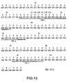
- the HpU-18-L is, as mentioned above, a variable region of an L chain of HpU-18 that is one of monoclonal antibodies of the HP urease and has an amino acid sequence shown in sequence No.14 as a primary structure.
- an amino acid sequence of the HpU-18-L and therebelow an example of a base sequence of a gene that codes the amino acid sequence are shown.
- the base sequence is a base sequence of a gene of the HpU-18 antibody L chain cloned in the embodiment.
- complimentarity determining sites that form a complimentary stereostructure with an antigen molecule (HP urease) and determine the complimentarity of the antibody are shown with double underlines as CDR-1, CDR-2 and CDR-3.
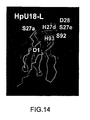
- Fig.14 after the molecular modeling is applied to amino acid sequence of the HpU-18-L, an estimated stereostructure is schematically shown.
- an aspartate residue at the 1 st in Fig. 14, shown as D1 by Kabat numbering scheme
- a histidine residue at the 98 th in Fig.14, shown as H93 by Kabat numbering scheme
- a serine residue at the 97 th in Fig.14, shown as S92 by Kabat numbering scheme
- a serine residue at the 28 th in Fig.14, shown as S27a by Kabat numbering scheme
- an aspartate residue at the 33 rd (in Fig.14, shown as D28 by Kabat numbering scheme), a histidine residue at the 31 st (in Fig.14, shown as H27d by Kabat numbering scheme), and a serine residue at the 32 nd (in Fig.14, shown as S27e by Kabat numbering scheme) are estimated to form a catalytic triad residue structure.
- the antibody enzyme according to the invention may be one that is made of an amino acid sequence that is obtained by replacing, deleting, inserting and/or adding at least one amino acid in an amino acid sequence shown in sequence No.14, that is, a variant of the HpU-18-L and works as a degrading enzyme of the HP urease. Still furthermore, the antibody enzyme may be one that is obtained by arbitrarily adding a remaining amino acid sequence of the HpU-18 antibody L chain to a C terminal side of the amino acid sequence shown in sequence No.14 or an entire length of the HpU-18 antibody L chain.
- the HpU-9-L is, as mentioned above, a variable region of an L chain of HpU-9 that is one of monoclonal antibodies of the HP urease and has an amino acid sequence shown in sequence No.15 as a primary structure.
- sequence No.15 an amino acid sequence of the HpU-9-L and therebelow an example of a base sequence of a gene that codes the amino acid sequence are shown.
- the base sequence is a base sequence of a gene of the HpU-9 antibody L chain cloned in the embodiment.
- complimentarity determining sites are shown with double underlines as CDR-1, CDR-2 and CDR-3.
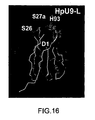
- Fig.16 after the molecular modeling is applied to an amino acid sequence of the HpU-9-L, an estimated stereostructure is schematically shown.
- an aspartate residue at the 1 st in Fig. 16, shown as D1 by Kabat numbering scheme
- a histidine residue at the 98 th in Fig.16, shown as H93 by Kabat numbering scheme
- a serine residue at the 26 th in Fig.16, shown as S26 by Kabat numbering scheme
- a serine residue at the 28 th in Fig.16, shown as S27a by Kabat numbering scheme
- the antibody enzyme according to the invention may be one that is made of an amino acid sequence that is obtained by replacing, deleting, inserting and/or adding at least one amino acid in an amino acid sequence shown in sequence No.15, that is, a variant of the HpU-9-L, and works as a degrading enzyme of the HP urease. Still furthermore, the antibody enzyme may be one that is obtained by arbitrarily adding a remaining amino acid sequence of the HpU-9 antibody L chain to a C terminal side of the amino acid sequence shown in sequence No.15 or an entire length of the HpU-9 antibody L chain.
- the HpU-2-H is, as mentioned above, a variable region of an H chain of HpU-2 that is one of monoclonal antibodies of the HP urease and has an amino acid sequence shown in sequence No.16 as a primary structure.
- Fig.17 an amino acid sequence of the HpU-2-H and therebelow an example of a base sequence of a gene that codes the amino acid sequence are shown.
- the base sequence is a base sequence of a gene of the HpU-2 antibody H chain cloned in the embodiment.
- complimentarity determining sites are shown with double underlines as CDR-1, CDR-2 and CDR-3.
- Fig.18 after the molecular modeling is applied to an amino acid sequence of the HpU-2-H, an estimated stereostructure is schematically shown.
- an aspartate residue at the 90 th in an amino acid sequence shown in sequence No.16, an aspartate residue at the 90 th (in Fig.18, shown as D86 by Kabat numbering scheme), a glutamate residue at the 89 th (in Fig.18, shown as E85 by Kabat numbering scheme), and a serine residue at the 88 th (in Fig.18, shown as S84 by Kabat numbering scheme) or a serine residue at the 91 st (in Fig.18, shown as S87 by Kabat numbering scheme) are estimated to form a catalytic triad residue structure.
- the antibody enzyme according to the invention may be one that is made of an amino acid sequence that is obtained by replacing, deleting, inserting and/or adding at least one amino acid in an amino acid sequence shown in sequence No.16, that is, a variant of the HpU-2-H, and works as a degrading enzyme of the HP urease. Still furthermore, the antibody enzyme may be one that is obtained by arbitrarily adding a remaining amino acid sequence of the HpU-2 antibody H chain to a C terminal side of the amino acid sequence shown in sequence No.16 or an entire length of the HpU-2 antibody L chain.
- genes involving the invention are genes that code the abovementioned antibody enzymes, and, for instance, genes made of base sequences shown in sequence Nos.47, 48 and 49 can be cited.
- genes involving the invention may be various genes that code antibody enzymes having amino acid sequence Nos.14, 15 and 16, and furthermore genes that code variants thereof.
- an antibody fragment of monoclonal antibody HpU-20 of the HP urease will be exemplified and described.
- the antibody fragment of HpU-20 is specifically a variable region of an L chain of HpU-20 antibody and a variable region of an H chain of HpU-20 antibody.
- variable region of an L chain of the HpU-20 antibody (hereinafter, referred to as HpU-20-L) and a variable region of an H chain of the HpU-20 (hereinafter, referred to as HpU-20-H) work as a degrading enzyme of the HP urease.
- HpU-20-L variable region of an L chain of the HpU-20 antibody
- HpU-20-H variable region of an H chain of the HpU-20
- the HpU-20-L is, as mentioned above, a variable region of an L chain of HpU-20 that is one of monoclonal antibodies of the HP urease and has an amino acid sequence shown in sequence No.17 as a primary structure.
- an amino acid sequence of the HpU-20-L and therebelow an example of a base sequence of a gene that codes the amino acid sequence are shown.
- the base sequence is a base sequence of a gene of the HpU-20 antibody L chain cloned in the embodiment.
- complimentarity determining sites that form a complimentary stereostructure with an antigen molecule (HP urease) and determine the complimentarity of the antibody are shown with double underlines as CDR-1, CDR-2 and CDR-3.
- Fig.21 a stereostructure estimated after the molecular modeling is applied to amino acid sequences of the HpU-20-L is schematically shown.
- Fig.21 in an amino acid sequence shown in sequence No.17, an aspartate residue at the 1 st (in Fig.21, shown as D1 by Kabat numbering scheme), a histidine residue at the 98 th (in Fig.21, shown as H93 by Kabat numbering scheme), and a serine residue at the 97 th (in Fig.21, shown as S92 by Kabat numbering scheme) or a serine residue at the 28 th (in Fig.21, shown as S27a by Kabat numbering scheme) are estimated to form a catalytic triad residue structure.
- the germline of the HpU-20-L is cr1.
- the antibody enzyme according to the invention may be one that is made of an amino acid sequence that is obtained by replacing, deleting, inserting and/or adding at least one amino acid in an amino acid sequence shown in sequence No.17, that is, a variant of the HpU-20-L, and works as a degrading enzyme of the HP urease. Still furthermore, the antibody enzyme may be one that is obtained by arbitrarily adding a remaining amino acid sequence of the HpU-20 antibody L chain to a C terminal side of the amino acid sequence shown in sequence No.17 or an entire length of the HpU-20 antibody L chain.
- the HpU-20-H is, as mentioned above, a variable region of an H chain of HpU-20 that is one of monoclonal antibodies of the HP urease and has an amino acid sequence shown in sequence No.19 as a primary structure.
- an amino acid sequence of the HpU-20-H and therebelow an example of a base sequence of a gene that codes the amino acid sequence are shown.
- the base sequence is a base sequence of a gene of the HpU-20 antibody H chain cloned in the embodiment.
- complimentarity determining sites are shown with double underlines as CDR-1, CDR-2 and CDR-3.
- a stereostructure estimated by applying the molecular modeling to an amino acid sequence of the HpU-20-H is schematically shown.
- an aspartate residue at the 90 th in Fig. 22, shown as D86 by Kabat numbering scheme
- a glutamate residue at the 89 th in Fig.22, shown as E85 by Kabat numbering scheme
- a serine residue at the 88 th in Fig.22, shown as S84 by Kabat numbering scheme
- the antibody enzyme according to the invention may be one that is made of an amino acid sequence that is obtained by replacing, deleting, inserting and/or adding at least one amino acid in an amino acid sequence shown in sequence No.19, that is, a variant of the HpU-20-H, and works as a degrading enzyme of the HP urease. Still furthermore, the antibody enzyme may be one that is obtained by arbitrarily adding a remaining amino acid sequence of the HpU-20 antibody H chain to a C terminal side of the amino acid sequence shown in sequence No.19 or an entire length of the HpU-20 antibody L chain.
- genes involving the invention are genes that code the antibody enzymes, and, for instance, genes made of base sequences shown in sequence Nos.18 and 20 can be cited.
- a gene made of a bas sequence shown in sequence No.18 is one of base sequences of a gene (cDNA) that codes a variable region of an L chain of the HpU-20
- a gene made of a base sequence shown in sequence No.20 is one of base sequences of a gene (cDNA) that codes a variable region of an H chain of the HpU-20.
- genes involving the invention may be various genes that code antibody enzymes having amino acid sequences shown in sequence Nos. 17 and 19, and furthermore may be genes that code variants thereof.
- an antibody fragment of monoclonal antibody UA-15 of the HP urease will be exemplified and described.
- the antibody fragment of UA-15 is specifically a variable region of an L chain of the UA-15 antibody.
- the antibody enzyme as well, similarly to the HpU-18-L and so on, is obtained from a plurality of monoclonal antibodies of the HP urease as an amino acid sequence (antibody fragment) that can constitute a catalytic triad residue structure made of a serine residue, a histidine residue and an aspartate residue when the molecular modeling is applied to amino acid sequences of variable regions of the monoclonal antibodies to estimate the stereostructure.
- variable region of an L chain of the UA-15 antibody (hereinafter, referred to as UA-15-L) works as a degrading enzyme of the HP urease. Then, by decomposing the HP urease to completely destroy a function of the HP urease, resultantly the HP bacteria are rendered incapable of living in a strongly acidic stomach. That is, the antibody enzyme according to the invention that can decompose the peptides can efficiently eliminate the HP bacteria.
- the UA-15-L is, as mentioned above, a variable region of an L chain of UA-15 that is one of monoclonal antibodies of the HP urease and has an amino acid sequence shown in sequence No.21 as a primary structure.
- a stereostructure estimated by applying the stereostructure modeling (molecular modeling) to a variable region of UA-15 that is made of light and heavy chains is schematically shown.
- a light chain is shown with L and a heavy chain is shown with H.
- an aspartate residue at the 1 st (in Fig.30, shown as Asp1 by Kabat numbering scheme), a serine residue at the 28 th (in Fig.30, shown as Ser27a by Kabat numbering scheme) and a histidine residue at the 94 th (in Fig.30, shown as His90 by Kabat numbering scheme) are estimated to form a catalytic triad residue structure.
- the antibody enzyme according to the invention may be one that is made of an amino acid sequence that is obtained by replacing, deleting, inserting and/or adding at least one amino acid in an amino acid sequence shown in sequence No.21, that is, a variant of the UA-15-L, and works as a degrading enzyme of the HP urease. Still furthermore, the antibody enzyme may be one that is obtained by arbitrarily adding a remaining amino acid sequence of the UA-15 antibody L chain to a C terminal side of the amino acid sequence shown in sequence No.21 or an entire length of the UA-15 antibody L chain.
- genes involving the invention are genes that code the abovementioned antibody enzymes, and, for instance, a gene made of a base sequence shown in sequence No.22 can be cited.
- the gene made of a base sequence shown in sequence No.22 is one of base sequences of a gene (cDNA) that codes a variable region of an L chain of the UA-15.
- genes involving the invention may be various genes that code antibody enzymes having an amino acid sequence shown in sequence No.22, and furthermore may be genes that code variants thereof.
- a variable region of a heavy chain of the UA-15 has an amino acid sequence shown in sequence No.23 as a primary structure; however, since, in the heavy chain of UA-15, a structure that is estimated to be a catalytic triad residue structure is not present, the UA-15 does not exhibit the activity as an antibody enzyme. Furthermore, a base sequence of a gene that codes the variable region of the heavy chain of the UA-15 is also shown as a sequence No. 24.
- an antibody enzyme involving the invention is an entire length of an H chain or an L chain of an antibody of the HP urease such as an HpU-18 L chain, an HpU-2 H chain, or L and H chains of HpU-20, or a UA-15 L chain
- an existing antibody obtaining process by making use of an existing antibody obtaining process, monoclonal antibodies of the corresponding HP urease are obtained followed by separating to H chains and L chains.
- the antibody enzyme involving the invention is an antibody fragment of the HP urease, firstly corresponding monoclonal antibody is obtained, followed by cleaving the monoclonal antibody with a proper protease so as to obtain a target antibody fragment.
- antibody enzymes of which amino acid sequences and gene sequences that code the amino acid sequences are known like the HpU-18-L, HpU-9-L, HpU-2-H, HpU-20-L, HpU-20-H and UA-15-L
- a technology of genetic manipulation can be applied to obtain.
- a process in which a gene that codes the antibody enzyme is incorporated in a vector or the like, followed by expressibly introducing in a host cell to purify translated peptides in the cell can be adopted.
- a gene that codes the antibody enzyme is incorporated together with a proper promoter that can express a lot, a target antibody enzyme may be efficiently obtained.
- an mRNA is obtained from a monoclonal antibody producing cell, a cDNA is synthesized from the mDNA, followed by reading a gene sequence thereof. Thereafter, an amino acid sequence is estimated from the gene sequence, a three-dimensional structure is estimated by the molecular modeling, and thereby whether a catalytic triad residue structure is contained or not is confirmed. Thereby, an antibody fragment in which the catalytic triad residue structure is contained can be obtained as an antibody enzyme.
- a primer on 3' side may be appropriately changed.
- a gene that codes an antibody enzyme involving the invention in the case a base sequence thereof being made clear, after obtaining a cDNA thereof (or genome DNA), with the cDNA as a template, PCR is carried out with appropriate primer to multiply a corresponding region, thereby the gene can be obtained. Still furthermore, when a gene in which mutation is introduced is obtained by making use of the site-directed mutagenesis, in transformants in which the gene is introduced, variants of antibody enzymes made of amino acid sequences shown in sequence Nos.14, 15, 16, 17, 19 and 21 can be obtained as translated products.
- the antibody enzyme can decompose the HP urease that is an antigen. Accordingly, it can be used as a disinfect agent of the HP bacteria that specifically works the HP urease. Furthermore, it is considered that since the antibody enzyme destroys the urease that is indispensable for the existence of the HP bacteria, different from the antibiotics that have been used as disinfect agents, the antibody enzyme does not impart the anti-drug resistance to the HP bacteria. Furthermore, the antibody enzyme not only can be developed as a drug, but also has likelihood, being able to be orally taken, by blending in healthy drinks or blending in milk products such as yogurt and butter, in the development of healthy foods for preventing HP bacteria infection.
- a curative drug involving the invention for HP bacteria-infected patients contains an antibody enzyme having the abovementioned nature; accordingly, it is superior to the antibiotics that have been used as curative drugs. That is, since the abovementioned curative drug is high in the specificity to the HP urease, side effects can be reduced. Still furthermore, an existing curative drug, after it is taken, is absorbed in a stomach, followed by being circulated to a whole body to intrude without prejudice into the respective cells; on the other hand, the curative drug according to the invention, when it is taken orally, is sent into a stomach and can directly attack HP bacteria living in the stomach.
- the side effects are slight, it is considered that an effect can be instantaneously exhibited. Furthermore, the antibody enzyme contained in the curative drug, after decomposing the HP urease, is anyway decomposed by other protease; accordingly, the side effects are expected to be slight.
- the antibody enzyme according to the invention can be used to prepare a transformant by introducing a gene that codes the antibody enzyme into a proper host. That is, a transformant involving the invention is a transformant into which a gene that codes the antibody enzyme is introduced.
- a transformant involving the invention is a transformant into which a gene that codes the antibody enzyme is introduced.
- gene is introduced means that a gene is expressibly introduced in a target cell (host cell) by use of a known genetic engineering procedure (genetic manipulation technology). The transformant can express the antibody enzyme in its body.
- the "transformant” includes transformed plants.
- a transformed plant When the gene is expressibly introduced in a plant, a transformed plant can be obtained.
- individual living matters various kinds of organs such as roots, stalks, leaves and sex organs (including flower organs and seeds); various tissues; cells; and so on are included, and furthermore protoplasts, induced calluses, regenerated individuals and offspring thereof are included as well.
- Such transformed plants can be used as a curative drug against HP bacteria-infected patients or an infection preventive agent against the HP bacteria.
- the transformed plants being produced, when for instance tomato, cucumber or carrot is used as a host plant, there is likelihood of obtaining a high-performance plant containing the antibody enzyme. When this is taken on a daily basis, the HP bacteria can be inhibited from infecting.
- the high-performance plant can be expected to commercialize as functional foods for preventing the infection of the HP bacteria.
- an antibody enzyme against chemokaine receptor CCR-5 will be explained.
- the invention is not restricted to the description.
- antibody fragments of monoclonal antibodies ECL2B-2 and ECL2B-3 of the chemokine receptor CCR-5 are exemplified and explained.
- the antibody fragments of monoclonal antibodies ECL2B-2 and ECL2B-3 are, more specifically, a variable region of a light chain of ECL2B-2 (that is, ECL2B-2-L) and a variable region of a light chain of ECL2B-3 (that is, ECL2B-3-L).
- the two antibody enzymes are obtained from monoclonal antibodies obtained by using peptides (RSSHFPYSQYQFWKNFQTLK (sequence No.38) or RSQKEGLHYTCS (sequence No.39)) that form an extracellular region of the chemokine receptor CCR-5 as an immunogen.
- peptides RSSHFPYSQYQFWKNFQTLK (sequence No.38) or RSQKEGLHYTCS (sequence No.39)
- the abovementioned antibody enzymes were found out as amino-acid sequences (antibody fragment) that can constitute a catalytic triad residue structure made of a serine residue, a histidine residue (or glutamate residue) and an aspartate residue.
- the antibody enzymes ECL2B-2-L and ECL2B-3-L according to the invention work as degrading enzyme of the chemokine receptor CCR-5. This, as will be shown also in an example described below, is obvious from a result that the ECL2B-2-L and ECL2B-3-L completely decompose an extracellular region peptide of the chemokine receptor CCR-5 (peptide made of an amino acid sequence shown in sequence No.38 or 39).
- CCR-5 chemokine receptor CCR-5
- the CCR-5 is a coreceptor of AIDS virus HIV-1, and considered to induce a conformational change in an HIV-1 envelope to cause membrane fusion between virus and host cell; accordingly, it is an important element when a human is infected with the HIV-1.
- a human having a defective mutant CCR-5 gene exhibits resistance to the infection and crisis of the HIV-1.
- the CCR-5 is one and only one that can be used as a coreceptor in the infection of a macrophage-directed clone of the HIV-1 that is responsible for the infection from a human to another human. From this, it is expected that when the function of the CCR-5 can be suppressed, the HIV can be prevented from infecting and disease can be inhibited from progressing.
- the abovementioned two antibody enzymes can completely decompose the CCR-5 and can delete the function thereof.
- a human who has a defective mutant CCR-5 gene has no obvious immunity deficiency and tissue lesion and has no problem in health; accordingly, the antibody enzymes that can delete the function of the CCR-5 are considered effectively utilized as an anti-HIV drug that prevents the HIV from infecting and AIDS symptom from progressing.
- the ECL2B-2-L is a variable region of a light chain of monoclonal antibody ECL2B-2 of which immunogen is an extracellular region peptide of the CCR-5 and has an amino acid sequence shown in sequence No.26 as a primary structure.
- Fig.35 a stereostructure estimated after the stereostructure modeling (molecular modeling) is applied to a variable region of the ECL2B-2 that is made of a light chain and a heavy chain is schematically shown.
- a light chain is expressed with L and a heavy chain is expressed with H.
- an aspartate residue at the 1 st in Fig.35, denoted as Asp1 by Kabat numbering scheme
- a serine residue at the 28 th in Fig.35, denoted as Ser27a by Kabat numbering scheme
- a histidine residue at the 98 th in an amino acid sequence shown in sequence No.26
- a histidine residue at the 31 st (in Fig. 35, denoted as His27d) are estimated to form a catalytic triad residue structure.
- the antibody enzyme according to the invention may be one that is made of an amino acid sequence that is obtained by replacing, deleting, inserting and/or adding at least one amino acid in an amino acid sequence shown in sequence No.26, that is, a variant of the HECL2B-2-L and works as a degrading enzyme of the CCR-5. Still furthermore, the antibody enzyme may be one that is obtained by arbitrarily adding a remaining amino acid sequence of the ECL2B-2 antibody L chain to a C terminal side of the amino acid sequence shown in sequence No.26.
- a variable region of a heavy chain of the ECL2B-2 has an amino acid sequence shown in sequence No.28 as a primary structure; however, in the heavy chain of the ECL2B-2, there is no structure that allows estimating a catalytic triad residue structure. Furthermore, a base sequence of a gene that codes the variable region of the heavy chain of the ECL2B-2 is shown together as a sequence No.29.
- the ECL2B-3-L is a variable region of a light chain of monoclonal antibody ECL2B-3 of which immunogen is an extracellular region peptide of the CCR-5 and has an amino acid sequence shown in sequence No.30 as a primary structure.
- Fig.36 a stereostructure estimated after the stereostructure modeling (molecular modeling) is applied to a variable region of the ECL2B-3 that is made of a light chain and a heavy chain is schematically shown.
- a light chain is expressed with L and a heavy chain is expressed with H.
- an aspartate residue at the 86 th (in Fig.36, denoted as Asp82 by Kabat numbering scheme), a serine residue at the 14 th (in Fig.36, denoted as Ser14 by Kabat numbering scheme), or in place of the serine residue at the 14 th , a serine residue at the 67 th (in Fig.36, denoted as Ser63 by Kabat numbering scheme), and a histidine residue at the 80 th (in Fig.36, denoted as His76 by Kabat numbering scheme) are estimated to form a catalytic triad residue structure.
- the antibody enzyme according to the invention may be one that is made of an amino acid sequence that is obtained by replacing, deleting, inserting and/or adding at least one amino acid in an amino acid sequence shown in sequence No. 30, that is, a variant of the ECL2B-3-L and works as a degrading enzyme of the CCR-5. Still furthermore, the antibody enzyme may be one that is obtained by arbitrarily adding a remaining amino acid sequence of the ECL2B-3 antibody L chain to a C terminal side of the amino acid sequence shown in sequence No.30.
- a variable region of a heavy chain of the ECL2B-3 has an amino acid sequence shown in sequence No.32 as a primary structure; however, in the heavy chain of the ECL2B-3, there is no structure that allows estimating a catalytic triad residue structure. Furthermore, a base sequence of a gene that codes the variable region of the heavy chain of the ECL2B-3 is shown together as a sequence No.33.
- the gene involving the invention is a gene that codes the antibody enzyme, more specifically, one made of a base sequence shown in sequence No.27 or one made of a base sequence shown in sequence No. 31 can be cited.
- a gene that is made of a base sequence shown in sequence No.27 is one of base sequences of a gene that codes the variable region of the L chain of the ECL2B-2 and a gene that is made of a base sequence shown in sequence No.31 is one of base sequences of a gene that code the variable region of the L chain of the ECL2B-3.
- antibody fragments of monoclonal antibody ECL2B-4 of the chemokines receptor CCR-5 are exemplified and explained.
- the antibody fragments of monoclonal antibody ECL2B-4 are, more specifically, a variable region of a light chain of ECL2B-4 (that is, ECL2B-4-L) and a variable region of a heavy chain of ECL2B-4 (that is, ECL2B-4-H).
- the two antibody enzymes as well are obtained from monoclonal antibodies obtained by using an extracellular region peptide of chemokine receptor CCR-5 as an immunogen.
- the antibody enzymes were found out as amino acid sequences (antibody fragments) that can constitute a catalytic triad residue structure made of a serine residue, a histidine residue (or glutamate residue) and an aspartate residue.
- the two antibody enzymes as well can completely decompose the CCR-5 and thereby can delete the function thereof. Accordingly, it is considered that the antibody enzymes can be effectively utilized as an anti-HIV drug that can prevent the HIV from infecting and suppress the AIDS from progressing.
- the ECL2B-4-L is a variable region of a light chain of monoclonal antibody ECL2B-2 of which immunogen is an extracellular region peptide of the CCR-5 and has an amino acid sequence shown in sequence No.34 as a primary structure.
- a stereostructure estimated after the stereostructure modeling (molecular modeling) is applied to a variable region of the ECL2B-4-L is schematically shown.
- an aspartate residue at the 1 st in Fig.39, denoted as D1 by Kabat numbering scheme
- a serine residue at the 28 th in Fig.39, denoted as S27a by Kabat numbering scheme
- a histidine residue at the 31 st in Fig.39, denoted as H27d by Kabat numbering scheme
- a histidine residue at the 98 th in Fig.39, denoted as H93 by Kabat numbering scheme
- any one of the 28 th in Fig.39, denoted as S27a), the 32 nd (in Fig. 39, denoted as S27e), and the 97 th (in Fig.39, denoted as S92) can be used.
- the antibody enzyme according to the invention may be one that is made of an amino acid sequence that is obtained by replacing, deleting, inserting and/or adding at least one amino acid in an amino acid sequence shown in sequence No.34, that is, a variant of the HECL2B-4-L and works as a degrading enzyme of the CCR-5. Still furthermore, the antibody enzyme may be one that is obtained by arbitrarily adding a remaining amino acid sequence of the ECL2B-4 antibody L chain to a C terminal side of the amino acid sequence shown in sequence No.34.
- the ECL2B-4-H is a variable region of a heavy chain of monoclonal antibody ECL2B-4 of which immunogen is an extracellular region peptide of the CCR-5 and has an amino acid sequence shown in sequence No.36 as a primary structure.
- Fig.40 a stereostructure estimated after the stereostructure modeling (molecular modeling) is applied to a variable region of the ECL2B-4-H is schematically shown.
- an aspartate residue at the 92 nd in Fig.40, denoted as D86 by Kabat numbering scheme
- a serine residue at the 65 th in Fig.40, denoted as S60 by Kabat numbering scheme
- a glutamate residue at the 48 th in Fig. 40, denoted as E46 by Kabat numbering scheme
- an aspartate residue at the 64 th (in Fig.40, denoted as D59 by Kabat numbering scheme) may be used.
- a serine residue at the 90 th (in Fig.40, denoted as S84 by Kabat numbering scheme) may be used.
- a glutamate residue at the 48 th a glutamate residue at the 91 st (in Fig.40, denoted as E85) may be used.
- the antibody enzyme according to the invention may be one that is made of an amino acid sequence that is obtained by replacing, deleting, inserting and/or adding at least one amino acid in an amino acid sequence shown in sequence No.36, that is, a variant of the ECL2B-4-H and works as a degrading enzyme of the CCR-5. Still furthermore, the antibody enzyme may be one that is obtained by arbitrarily adding a remaining amino acid sequence of the ECL2B-4 antibody H chain to a C terminal side of the amino acid sequence shown in sequence No.36.
- the gene involving the invention is a gene that codes the antibody enzyme, and, more specifically, one made of a base sequence shown in sequence No.35 or one made of a base sequence shown in sequence No.37 can be exemplified.
- a gene that is made of a base sequence shown in sequence No.35 is one of base sequences that code the variable region of the L chain of the ECL2B-4 and a gene that is made of a base sequence shown in sequence No.37 is one of base sequences of a gene that codes the variable region of the H chain of the ECL2B-4.
- an antibody enzyme involving the invention is an entire length of an L chain of an antibody of CCR-5 such as an entire length of an L chain of ECL2B-3, an entire length of an L chain of ECL2B-3, or an entire length of an L chain of ECL2B-4, an entire length of an H chain of ECL2B-4
- monoclonal antibodies of the corresponding CCR-5 are obtained followed by separating H chains and L chains.
- the antibody enzyme involving the invention is an antibody fragment of the CCR-5, firstly corresponding monoclonal antibodies are obtained, followed by cleaving the monoclonal antibodies with a proper protease so as to obtain target antibody fragments.
- antibody enzymes of which amino acid sequences and gene sequences that code the amino acid sequences are clarified like the ECL2B-2-L, ECL2B-3-L, ECL2B-4-L, and ECL2B-4-H
- a so far known technology of genetic manipulation can be applied to obtain.
- a process in which genes that code the antibody enzymes are incorporated in a vector or the like, followed by expressibly introducing in a host cell to purify translated peptides in the cell can be adopted.
- genes that code the antibody enzymes are incorporated together with a proper promoter that can express a lot, target antibody enzymes may be efficiently obtained.
- an amino acid sequence of the antibody enzyme is not clear, firstly, an mRNA is obtained from a monoclonal antibody producing cell or a hybridoma thereof, a cDNA is synthesized from the mDNA, followed by reading a gene sequence thereof. Thereafter, an amino acid sequence is estimated from the gene sequence, a three-dimensional structure is estimated by the molecular modeling, and thereby whether a catalytic triad residue structure is contained or not may be confirmed. Thereby, an antibody fragment in which the catalytic triad residue structure is contained can be obtained as an antibody enzyme.
- a gene that codes an antibody enzyme involving the invention in the case a base sequence thereof being made clear, after obtaining a cDNA thereof (or genome DNA), with the cDNA as a template, PCR is carried out with an appropriate primer to multiply a corresponding region, thereby the gene can be obtained. Still furthermore, when, by making use of the site-directed mutagenesis, a proper mutation is introduced into a gene that is made of a base sequence shown in sequence No.27, 31, 35 or 37, in a transformant in which the gene is introduced, a variant of an antibody enzyme made of an amino acid sequence shown in sequence No.26, 30, 34, or 36 can be obtained as a translated product.
- the antibody enzymes according to the invention based on an utterly different process from the abovementioned anti-HIV drugs, can attack the CCR-5 directly to delete the function. Accordingly, the antibody enzymes can be utilized as an anti-HIV drug.
- An anti-HIV drug containing the present antibody enzymes owing to a novel mode of action of enzymatically completely decomposing the CCR-5 to delete the function thereof, can inhibit the HIV from infecting and suppress the AIDS from developing.
- Such an anti-HIV drug specifically aims a CCR-5 molecule alone; accordingly, it is considered that a significant effect can be expected and at the same time the side effects are less.
- curative effects against the AIDS can be expected to improve.
- the antibody enzyme according to the invention when a gene that codes the antibody enzyme is introduced into an appropriate host, can be used to prepare a transformant. That is, a transformant involving the invention is a transformant into which a gene that codes the antibody enzyme is introduced.
- a gene is introduced means that a gene is expressively introduced into a target cell (host cell) by means of an existing genetic engineering procedure (genetic manipulation technology). The transformant can express the antibody enzyme in its body.
- transformant involving the invention more specifically, one in which a gene made of a base sequence shown in sequence No.27 or 31 (sequence No.35 or 37 may be used) is introduced in a host cell can be cited.
- a host cell usually used ones such as bacteria coli, yeast and baculovirus can be properly used.
- the transformant specifically recognizes the CCR-5 in its body and completely decompose to delete the function thereof. Accordingly, it can be used as an anti-HIV drug. Furthermore, when the transformant can express a lot of the antibody enzymes, it is considered that the HIV can be efficiently inhibited from infecting and remedy of the symptom of the AIDS can be efficiently performed; that is, there is likelihood of obtaining a more effective anti-HIV drug.
- amino acid numbers in the amino acid sequences of the respective antibody enzymes shown in Figs.4A, 4B, 8A, 8B, 13, 15, 17, 23, 24, 41 and 42 explained in the embodiment are due to Kabat numbering scheme; accordingly, these are different from numbers of amino acids shown in the corresponding respective sequence numbers.
- I41SL1-2 and i41-7 antibodies are prepared according to a method shown below.
- MBS m-maleimidobenzoyl-N-hydroxysuccinimide ester
- glutaraldehyde glutaraldehyde
- KLH Keyhole Limpet Hemocyanin
- a spleen was taken out of the immunized mouse to prepare spleen cells.
- the spleen cells and separately prepared myeloma cells derived from mouse bone marrow were fused by use of polyethylene glycol, followed by a HAT selection. Then, by screening by use of an ELISA method, antibody producing-positive cell groups were selected, and the cell groups were cloned to obtain antibody producing-hybridomas.
- the hybridomas were transplanted in a mouse abdominal cavity and abdominal dropsy was sampled as monoclonal antibodies. As the i41-7 antibodies, in a screening stage, ones well reacted with 41S-2 antibody light chains were selected. Prepared antibodies were purified by means of affinity chromatography.
- the purity of the purified antibodies was confirmed by use of SDS-PAGE.
- base sequences of various kinds of monoclonal antibodies were determined. Specifically, mRNAs were extracted from various kinds of antibody producing-cells, followed by synthesizing cDNAs by RT-PCR. Antibody genes were amplified by means of PCR with the cDNAs as a template, incorporated in pGEM-T vector (pGEM-T (registered trade mark) and p-GEM-T (registered trade mark) Easy Vector System, manufactured by Promega Corporation) and sub-cloned in E. coli JM109. After liquid culturing, plasmids in which a target gene was incorporated were purified and base sequences thereof were determined.
- pGEM-T vector registered trade mark
- p-GEM-T registered trade mark
- p-GEM-T registered trade mark
- Easy Vector System manufactured by Promega Corporation
- amino acid sequences of variable regions of monoclonal antibodies estimated from the base sequences (35 clones) and amino acid sequences of antibodies registered in Protein Data Bank (PDB) (Research Collaboratory for Structural Bioinformatics) (56 clones) were used.
- PDB Protein Data Bank
- the molecular modeling was applied to analyze a stereostructure of a variable region.
- the molecular modeling was carried out to analyze a stereostructure of a variable region. This is because the inventors considered that when all data are analyzed of the stereostructure with the same software, the data of all antibodies could be equally evaluated. That is, when in one case the evaluation was performed with data due to the X-ray crystal structure analysis and in another case, with data due to the molecular modeling, in evaluation, irregular data are considered used.
- mouse-derived ones When 56 clones randomly extracted from the PDB are broken down, 52 clones were mouse-derived ones and 4 clones were human-derived ones.
- the antibody L chains are classified into two kinds of classes, ⁇ and ⁇ , and it is said that, in the case of mouse, substantially 95% is ⁇ chain. Furthermore, since an analysis of germline genes of mouse ⁇ chains is advanced, information thereof can be advantageously obtained. In this connection, mouse ⁇ chains (47 clones) were analyzed in more detail.
- clones relevant to the catalytic triad residue structures were selected and classified as follows. Firstly, ones that constitute the catalytic triad residue structure from Asp1, Ser27A and His93 that are common with the abovementioned i41SL1-2-L were classified as type o ⁇ .
- N30 is mutated to D30, a triad from H27d or H93-S27e-D30 can be formed.
- 31 1IAl Anti-peritonitis virus antigen VGK1A 19-17 ⁇
- a germline-derived triad can be formed from S26-D28-H91.
- 32 1IBG Digoxin V0x-1 21-12 ⁇
- a germline-derived triad can be formed from D1-S26 or S27a-H90.
- a germline-derived triad can be formed from D1-S26-H90 or H91.
- a germline-derived triad can be formed from D1-S26-H90.
- the complimentarity determining region 1 (CDR1) was formed of 16 amino-acid residues.
- the CDR sequence is a place where a large mutation is caused so that an antibody may strongly recognize an antigen (this is a reason why it,is called a complimentarity determining region).
- Figs.11A, 11B and 11C results of the molecular modeling applied to 1DBA clone of which germline is bb1 by Thiebe and et al. convention are shown.
- An FR is shown with wire and a CDR is shown with ribbon.
- the Asp1, Ser27A and His93, respectively, are present in regions FR1, CDR1 and CDR3 different from each other in the primary structure.
- the three amino-acid residues are located in the neighborhood of a tip end where the His93 of the CDR3 loop is just present and at a height same as that of the His93, that is, on a substantially same plane when seen in a horizontal direction.
- Fig.11A when 16 amino acids constitute the CDR1, a loop of the CDR1 takes a structure that slowly undulates just in the neighborhood, and the tip end portion (8 amino-acid residues from 27B to 31) higher than the structure protrudes higher than other loops. Accordingly, the antibody enzyme has a catalytic triad residue structure in a position where an antigen peptide can be incorporated without the stereostructural drawbacks; accordingly it is considered that an antigen peptide can be smoothly decomposed.
- the stereostructural features were common in 10 antibody L chains shown in the Table 2 and the i41SL1-2-L.
- type o ⁇ represents ones that constitute a catalytic triad residue structure from Asp1, Ser27A and His93
- type ⁇ represents one that constitutes a catalytic triad residue structure from Asp1, Ser26 and His93
- type ⁇ represents ones that constitute a catalytic triad residue structure from combinations other than "Asp1, Ser27A or Ser26 and His93”
- type * represents one that cannot form a catalytic triad residue structure because of mutation from the germline
- type ⁇ represents ones that form a catalytic triad residue structure by including an amino acid mutated from the germlines.
- the i41-7 antibody belonged to a germline 19-25 of which CDR-1 has 11 amino-acid residues. Then, all germlines of which CDR-1 has 11 amino-acid residues were searched, and, other than 19-25, in 19-14, 19-17, 19-23 and 12-41 (these are shown in a box in Table 3), the catalytic triad residue structure was recognized. When these were investigated in more detail, there were found common points in that the CDR-1 is constituted of 11 amino-acid residues and a histidine residue is present at the 91 st (or the 55 th ) by Kabat numbering scheme (type 4 ⁇ *).
- Natural antibody enzymes are considered present, roughly divided, in two types.
- One is a type that has, already in a germline stage, sequences advantageous in constituting a catalytic triad residue structure, and, according to Thiebe et al. convention, at least 13 types of bb1, cr1, bl1, cs1, bd2, bj2, hf24, 19-25, 19-14, 19-17, 19-23, 12-41 and 21-12 conform thereto.
- hf24 there is likelihood also in that, other than His 91 present in the germline, His 53 appears owing to the mutation, and this constitutes a catalytic triad residue structure.
- 2 There are only 13 kinds (13.9% of 93 kinds in total) that have sequences advantageous for constituting a catalytic triad residue structure according to Thiebe et al. convention.
- the antibody enzyme can be efficiently obtained at rather high probability (conclusion 1 ⁇ ).
- heavy chains of the 41S-2, i41SL1-2 and i41-7 antibodies are recognized to constitute the catalytic triad residue structure and have the enzymatic activity.
- Families of heavy chains of the 41S-2, i41SL1-2 and i41-7 antibodies, respectively, are VH10, VH1 and VH1.
- heavy chains of 7C4 and HpU-2 antibodies there is found no catalytic triad residue structure due to His, Asp and Ser; however, Glu, Asp and Ser constitute a catalytic triad residue structure and the enzymatic activity was recognized.
- Families of heavy chains of 7C4 and HpU-2 antibodies, respectively, are VH5 and VH1.
- both (heavy chains of 7C4 and HpU-2 antibodies) constitute a catalytic triad residue structure from the utterly same combination (Ser84, Clu85 and Asp86). That is, as to the heavy chain, VH1, VH5 and VH10 can be said families that can efficiently exhibit the enzymatic activity as an antibody enzyme (conclusion 2 ⁇ ).
- a detection rate of the antibody light chains having the enzymatic activity is 45.2%. This is general probability capable of detecting antibody light chains.
- Ultrafiltration was applied repeatedly to purified antibodies to remove low molecular weight protease inhibitors. Subsequently, a reducing reaction (15 degrees centigrade and 3 hr) with 0.2 M ⁇ -mercaptoethanol and an alkylation reaction (15 degrees centigrade and 15 min) with 0.3 M iodoacetamide were applied to separate heavy chains and light chains. After these were subjected to ultrafiltration to concentrate, with a Protein-Pak 300 column (Manufactured by Millipore Corporation Waters Chromatography Division), HPLC purification was applied. In a moving phase, a 6 M guanidine solution (pH 6.5) was used, and a flow rate was set at 0.15 mL/min.
- Partitioned solutions of heavy and light chains were diluted with a 6 M guanidine solution to prepare, followed by dialyzing against a PBS solution (137 mM NaCl, 2.7 mM KCl, 1.5 mM KH 2 PO 4 , 8.0 mM Na 2 HPO 4 and pH 7.3) to refold, further followed by buffer changing according to experimental conditions.
- a PBS solution 137 mM NaCl, 2.7 mM KCl, 1.5 mM KH 2 PO 4 , 8.0 mM Na 2 HPO 4 and pH 7.3
- the reaction was carried out under the conditions; 5% DMSO, 9 mM HEPES, 7.5 mM phosphate buffer (pH 7.1) and 25 degrees centigrade. Experiments were carried out at concentrations of 0.4 ⁇ M, 0.2 ⁇ M and 50 ⁇ M, respectively, for i41SL1-2-L, i41SL1-2-H and an antigen peptide.
- Results are shown in Figs.6A and 6B.
- ( ⁇ ) shows results when i41SL1-2-L and the antigen peptide were reacted,
- ( ⁇ ) shows results when only antigen peptide was reacted, and
- ( ⁇ ) shows results when i41SL1-2-H and an antigen peptide were reacted.
- concentrations of the peptide go down, finally resulting in being incapable of detecting.
- the i41-7 antibody was obtained with a protein as an antigen and an epitope has not yet been determined. Under normal conditions, the enzymatic activity has to be investigated with a peptide corresponding to an epitope as a substrate.
- antibody enzymes that inventors have obtained so far exhibit high specificity to a protein but are low in the specificity to a short peptide.
- three kinds of synthesized peptides having molecular weight from several hundreds Da to 2.3 kDa were used as substrate.
- Results thereof are shown in Fig.10A.
- ( ⁇ ) shows results when peptide TP41-1 shown in sequence No.10 and i41SL1-7-L or i41-7-H were reacted,
- ( ⁇ ) shows results when only peptide TP41-1 shown in sequence No.10 was reacted,
- ( ⁇ ) shows results when peptide shown in sequence No.11 and i41-7-L or i41-7-H were reacted,
- ( ⁇ ) shows results when only peptide shown in sequence No.11 was reacted,
- ( ⁇ ) shows result when peptide shown in sequence No.12 and i41SL1-7-L or i41-7-H were reacted, and
- ( ⁇ ) shows results when only peptide shown in sequence No.12 was reacted.
- i41-7-L and i41-7-H decomposed all the peptides.
- a decomposition reaction of an antigen protein due to a light chain of i41-7 antibody was analyzed by means of SDS-PAGE.
- One that was used as a substrate was a light chain of an antibody 41S-2 (41S-2-L).
- the 41S-2-L that was used as a substrate was incomplete in the refolding and did not have the activity as an antibody enzyme.
- Decomposition reaction were carried out under the conditions same as that mentioned above, 41S-2-L was used at a concentration of 2 ⁇ M, and a light chain of the i41-7 antibody was used at a concentration of 0.8 ⁇ M. Results thereof are shown in Fig.10B.
- a lane M shows a result when a protein molecular weight marker is subjected to electrophoresis and lanes 1 through 5 show results when reaction liquids with i41-7-L and 41S-2-L at 0, 2, 4, 6 and 8 days after the start of the reaction were subjected to the electrophoresis.
- the i41-7 intact antibody is a complete antibody made of heavy and light chains.
- the MA-2 antibody (germline: aq4) is a monoclonal antibody against methamphetamine and an antibody that does not have a catalytic triad residue structure in the stereostructure.
- TP41-1 polypeptide TP41-1 (described as "TP" in Fig.12) shown in sequence No.10 was used at a concentration of 120 ⁇ M.
- Results are shown in Fig.12.
- ⁇ shows results when i41-7-L and TP41-1 were reacted
- ⁇ shows results when i41-7-H and TP41-1 were reacted
- ⁇ shows results when i41-7 intact antibody and TP41-1 were reacted
- ⁇ shows results when MA-2 antibody and TP41-1 were reacted
- ⁇ shows results when only TP41-1 as the control was reacted.
- both the heavy and light chains of i41-7 antibody have, for a while after the start of the reaction, an induction period during which the peptide is not decomposed, followed by an active period during which the peptide is decomposed. Furthermore, though results are not shown in the drawing, when, after the peptide was completely decomposed, peptide was added again, both the heavy and light chains of i41-7 antibody decomposed the peptide without the induction period. From the results, it is considered that both the heavy and light chains of i41-7 antibody, during transferring from the induction period to the active period, undergo a conformation change. Furthermore, it is considered that after the conformation change, the active type is maintained thereafter.
- the intact i41-7 antibody that has both the heavy and light chains did not decompose the polypeptide. This is considered that, in the i41-7 intact antibody, owing to an S-S bond or a non-covalent bond, the light chain and the heavy chain are strongly bonded; accordingly, it cannot undergo the conformation change into a mode that has the activity even when it is brought into contact with the polypeptide.
- an antibody enzyme that has a catalytic triad residue structure in a stereostructure has the activity of cleaving and/or degrading the polypeptide and the antigen protein.
- a catalyst efficiency (kcat/Km) of the light chain is identical as that of trypsin and that of the heavy chain is also such high as substantially one tenth that of trypsin; accordingly, it can be said that both the heavy and light chains of the antibody enzyme have high enzymatic activity comparable with that of natural enzymes.
- both the heavy and the light chains of the antibody enzyme are stronger than that of tripsin; on the other hand, in the number of rotation (kcat), both the heavy and light chains are substantially one two-hundredth that of trypsin, there is found a large difference between natures thereof.
- This result shows that i41AS1-2-L and i41SL1-2-H, with the nature as an antibody that it has high affinity with a substrate remaining, exhibit the enzymatic activity of completely decomposing the polypeptide.
- HP bacteria were cultured in a 7% FBS-containing brucella liquid medium under a nonaerobic atmosphere at 37 degrees centigrade.
- the cultured HP bacteria were centrifuged to recover, followed by washing with 0.15 M NaCl, further followed by suspending the bacteria in distilled water, still further followed by rigorously agitating, and thereby a bacteria component was extracted.
- a soluble component after centrifugation and dialysis was made a distilled water extract (DWExtract).
- anion exchange chromatography that uses a Q Sepharose Fast Flow, followed by gel filtration with Superose 6, and further followed by subjecting to anion exchange chromatography. This was further subjected to gel filtration and the HP urease was confirmed to be highly pure by use of the SDS-PAGE.
- Cells that produce three anti-Helicobacter Pylori monoclonal antibodies (HpU-2, HpU-9 and HpU-18) prepared in experiment 2-2 were recovered from frozen stocks.
- the respective antibody producing-cells were cultivated in an IMDM medium containing 20% cow fetus serum. Under the conditions of a temperature of 37 degrees centigrade and a concentration of carbon dioxide of 5.5%, passage culture was repeated until the number of cells became 5 ⁇ 10 7 . The cells were recovered by centrifuging.
- a primer that complimentarily couples with 5' and 3' sides of variable regions of antibody H and L chains contained in Mouse Ig-Prime Kit (NOVEGEN), dNTP Mix and AmpliTaq DNA polymerase (Roche) were blended and thoroughly agitated. After incubation at 95 degrees centigrade for 10 min, a reaction cycle of 94 degrees centigrade and 1 min, 50 degrees centigrade and 1 min and 72 degrees centigrade and 2 min was repeated 40 times. Amplified products were confirmed by use of agarose gel electrophoresis.
- a ligation reaction was carried out to the purified fragment and pGEM-T vector (Promega) with T4 DNA polymerase. It was incubated at room temperature for 1 hr, followed by blending with a competent cell JM109 and leaving on ice for 20 min. Thereafter, it was heated at 42 degrees centigrade for 45 sec, left on ice for 2 min, followed by adding a medium, further followed by cultivating at 37 degrees centigrade for 1 hr. On an agar plate coated with IPTG/X-gal, the cultivated bacteria solution was spread and incubated at 37 degrees centigrade overnight. Formed white colonies were optionally selected, followed by liquid culturing at 37 degrees centigrade overnight. According to the alkali-SDS method, plasmid was purified from a fungus body, followed by confirming whether a target fragment was inserted or not.
- variable regions of the HpU-18 (L chain), HpU-9 (L chain) and HpU-2 (H chain), respectively results obtained by reading base sequences and results obtained by transforming the base sequences into amino acid sequences are shown in Figs.13, 15 and 17.
- upper case characters and lower case characters, respectively show an amino acid sequence and a base sequence.
- the base sequences and amino acid sequences were determined.
- the amino acid sequences of the H and L chains of the HpU-2, HpU-9 and HpU-18 were inputted in an antibody variable region stereostructure estimation program abM (manufactured by Oxford Molecular Corp.) and stereostructures were constituted. Subsequently, an energy was minimized with Insight II (Discover) (manufactured by MSI Corp.), and thereby a stereostructure of each of the variable regions of the HpU-2, HpU-9 and HpU-18 was obtained.
- H chain of HpU-2 antibody, the H or L chain of HpU-9 and HpU-18 antibodies and HpU2EPI (amino acid sequence: 183 SVELDIGGNRRIFGNALVD 204 (sequence No.25)) that is a partial peptide of the HP urease were reacted in a 15 mM phosphate buffer (PB: pH 6.5).
- the HpU2EPI is a peptide corresponding to a region from the 183 rd to the 204 th in an ⁇ unit primary structure of a HP urease protein made of 238 amino acids. The operation was carried out in a clean bench.
- Purified peptide powder was dissolved with 15 mM PB (pH6.5), followed by filtering and sterilizing with Ultrafree MC (0.22 ⁇ m, manufactured by Millipore Corp.). Furthermore, each of L or H chain solutions of the HpU-2, HpU-9 and HpU-18 antibodies were blended so that a concentration in a reaction solution was 0.4 ⁇ M (20 ⁇ g/ml) for the H chain, 0.8 ⁇ M (20 ⁇ g/ml) for the L chain and 80 ⁇ M (184.4 ⁇ g/ml) for peptide. A reaction temperature was 25 degrees centigrade and for a reaction vessel a dry hot air sterilized small test tube was used. A reaction solution was analyzed with HPLC and peptide variation with time was traced.
- a horizontal axis expresses a reaction time (hr), and a vertical axis expresses a concentration ( ⁇ M) of a peptide (HPU2EPL) that is a substrate.
- 1 ⁇ shows a case where HpU-2 (H) and the peptide were blended
- 2 ⁇ shows a case where HpU-9 (H) and the peptide were blended
- 3 ⁇ shows a case where HpU-18 (H) and the peptide were blended
- 5 ⁇ shows a case where HpU-9 (L) and the peptide were blended
- 6 ⁇ shows a case where HpU-18 (L) and the peptide were blended
- 7 ⁇ shows a case where the peptide only was used.
- HpU-20 and UA-15 two kinds of anti-HP urease mouse monoclonal antibody producing-cells, HpU-20 and UA-15, were prepared. Then, according to a method similar to experiment 2-3, H and L chains of HpU-20 and UA-15 were prepared.
- a stereostructure estimated as a result of a variable region of HpU-20 (L chain) is schematically shown in Fig.21; a stereostructure of a variable region of HpU-20 (H chain), in Fig. 22; and stereostructures of variable regions of UA-15 (H and L chains), in Fig.30. From the stereostructure estimations, in the L and H chains of HpU-20 and the L chain of UA-15, the catalytic triad residue structure could be recognized. On the other hand, in the UA-15 (H chain), a catalytic triad residue structure could not be recognized.
- a horizontal axis denotes a reaction time (hr) and a vertical axis denotes a concentration ( ⁇ M) of the peptide that is a substrate.
- Figs. 27 through 29 are diagrams showing results of the SDS-PAGE of samples at 1 hr after the start of the reaction.
- M denotes a marker
- 1 ⁇ through 7 ⁇ denote the abovementioned respective reaction liquids.
- a horizontal axis denotes a reaction time (hr) and a vertical axis denotes a concentration ( ⁇ M) of the TP41 peptide that is a substrate.
- hr reaction time
- ⁇ M concentration of the TP41 peptide that is a substrate.
- Each of monoclonal antibodies ECL2B-2 and ECL2B-3 was prepared with a partial peptide (RSSHFPYSQYQFWKNFQTLK: sequence No.38) made of 20 amino acids that constitute an extracellular region of CCR-5 as an immunogen.
- RSSHFPYSQYQFWKNFQTLK sequence No.38
- glycine and cystine residues were introduced to conjugate with Keyhole Limpet Hemocyanin (KLH).
- KLH Keyhole Limpet Hemocyanin
- an N terminal of the partial peptide is a cystine residue; however, in a peptide that is used in the experiment, an arginine residue substituted for the cystine residue.
- the peptide and a conjugate with KLH of the peptide are ones that are manufactured by Takara Shuzo Co., Ltd.
- the peptide conjugated with KLH was immunized in a Balb/c mouse.
- First and second immunizations were carried out by use of a complete immunoactivator due to Freund at an interval of two weeks.
- a third immunization was carried out by use of an incomplete immunoactivator due to Freund four weeks after the second immunization.
- a final booster (additional immunization) was carried out six weeks after the third immunization.
- cell fusion of a spleen cell of a Balb/c mouse and a myeloma cell SP/NSI/1-Ag4-1 (NS-1)
- SP/NSI/1-Ag4-1 (NS-1) was conducted, followed by applying the HAT selection and screening.
- cell systems that excrete a monoclonal antibody ECL2B-2 and ECL2B-3; IgG 1 (k)
- ECL2B-2 and ECL2B-3 were purified based on a purification manual of Bio-Rad Protein A MAPS-II kit (manufactured by Nippon Bio-RAD Corp.).
- abdominal dropsy containing ECL2B-2 and ECL2B-3 was blended with a same volume of an ammonium sulfate saturated aqueous solution.
- a precipitate thereof was centrifuged, followed by recovering, further followed by adding 5 ml of PBS to the precipitate.
- the process was repeated twice, followed by applying dialysis to PBS twice.
- a definite amount of PBS solution containing ECL2B-2 or ECL2B-3 was blended with a same volume of a conjugate buffer MAPS-II, followed by placing a mixture on a base packed with Affi-Gel (protein A), further followed by eluting a conjugated antibody.
- the eluted antibody was subjected thrice to ultrafiltration by use of Centriprep 10 (manufactured by Amicon Corp.).
- Five milligrams of antibody was dissolved in 2.7 ml of a buffer solution (pH 8.0) made of 50 mM Tris and 0.15 M NaCl, followed by adding 0.3 ml of 2 M 2-mercaputoethanol, further followed by reducing at 15 degrees centigrade for 3 hr.
- ECL2B peptide Fifty micro-liters of a partial peptide (RSSHFPYSQYQFWKNFQTLKG (shown in sequence No.40): One that is synthesized with a cystine residue at a C terminal of the CCR-5 partial peptide used in the experiment 3-1 deleted.
- ECL2B peptide the peptide is referred to as ECL2B peptide
- PBS solution 5 ⁇ g/ml
- ECL2B-2 or ECL2B-3 was immune reacted, followed by conducting a reaction with anti-mouse Ig (G + A + M) conjugated with alkali phosphatase. After the substrate reaction, by use of an immunization plate reader (trade name NJ-2001, manufactured by InterMed Corp.), the absorbance at 405 nm was measured.
- an immunization plate reader trade name NJ-2001, manufactured by InterMed Corp.
- classes and subclasses of the obtained monoclonal antibodies ECL2B-2 and ECL2B-3 were IgG 1 ( ⁇ ).
- the monoclonal antibodies did not cross-react with other proteins such as KLH, HSA, OVA and human hemoglobin.
- the monoclonal antibodies did not react with peptides such as gp120 V3 cyclic peptide of HIV-1 (CTRPNNNTRKSIHIGPGRAFYTTGEIIGDIRQAHC: sequence No.41), peptide TP41-1 of a retention region of gp41 (HIV-1/TPRGPDRPEGIEEEGGERDRD: sequence No.42), RT-2 (KLLRGTKALTFVIPLTEEAE: sequence No.43), 41S-2mAb CDRL-1 (RSSKSLLYSNGNTYLY: sequence No.44), 1D3 mAb CDRH-2 (DKFNQNYSTAGNYPNIR: sequence No.45) and CA-2 (RSSKSLLYSNGNTYLYGPDKFNQNYSTAGNYPNIR: sequence No.46).
- peptides such as gp120 V3 cyclic peptide of HIV-1 (CTRPNNNTRKSIHIGPGRAFYTTGEIIGDIRQAHC: sequence No.41), peptide TP41-1
- the antibody peptides were identified by use of MALDI-TOF-MASS (trade mark: AUTOFLEX, manufactured by Brucker Ltd.). As a result, masses thereof were same as theoretical ion distributions. The same mass spectrometry conditions were applied to identify fragments found in catalytic reaction due to light chains of the ECL2B-2 and ECL2B-3.
- the mRNA of each of ECL2B-2 and ECL2B-3 was isolated from a hybridoma having ECL2B-2 or ECL2B-3. Thereafter, cDNAs of the light and heavy chains were synthesized by use of a single strand cDNA synthesis kit (manufactured by Life Science Inc.).
- the sequence determination was carried out by use of an Auto Read Sequencing Kit (manufactured by Amersham Pharmacia Biotec) and a DNA Auto Sequencer (trade name: ALF II, manufactured by Amersham Pharmacia Biotech).
- the computer analysis for constituting three-dimensional molecular structures was carried out by use of a workstation (manufactured by Silicon Graphics Inc.) and the AbM software (manufactured by Oxford Molecular Ltd.).
- amino acid sequences of variable regions of light and heavy chains of monoclonal antibodies ECL2B-2 and ECL2B-3 and sequences of cDNAs that code the amino acid sequences were determined.
- An amino acid sequence shown in sequence No.26 is an amino acid sequence of an ECL2B-2 light chain, and a base sequence shown in sequence No. 27 is a base sequence of cDNA that codes the amino acid sequence.
- An amino acid sequence shown in sequence No.28 is an amino acid sequence of an ECL2B-2 heavy chain, and a base sequence shown in sequence No.29 is a base sequence of cDNA that codes the amino acid sequence.
- An amino acid sequence shown in sequence No.30 is an amino acid sequence of an ECL2B-3 light chain, and a base sequence shown in sequence No.31 is a base sequence of cDNA that codes the amino acid sequence.
- An amino acid sequence shown in sequence No.32 is an amino acid sequence of an ECL2B-3 heavy chain, and a base sequence shown in sequence No.33 is a base sequence of cDNA that codes the amino acid sequence.
- ECL2B-2-L In a light chain of ECL2B-2 (ECL2B-2-L), three amino acid residues, Asp 1 (or Asp 27d ), Ser 27a and His 93 , were estimated to be able to form a catalytic triad residue structure in a molecular structure (Fig.35).
- VIPase-L A light chain (VIPase-L) of a natural antibody enzyme VIPase that can decompose antigenic vasoactive intestinal peptide (VIP) is reported to have three amino acid residues, Asp 1 (inside of FR-1), Ser 27a (inside of CDR1) and His 93 (inside of CDR3), in a structure thereof [reference literature 10: S. Paul et al., J. Biol. Chem. 269 (1994), page 32389].
- i41SL1-2-L that is a light chain of monoclonal antibody i41SL1-2 has a catalytic triad residue made of the same Asp 1 , Ser 27a and His 93 and exhibits the enzymatic activity of decomposing CDR-1 peptide that is an antigen thereof
- reference literature 11 Y. Zhou, E. Hifumi, H. Kondo and T. Uda, Biological Systems Engineering, ACS symposium Series 830, 200-208 page (2002)
- reference literature 12 T. Uda and E. Hifumi, Proceedings of International Symposium on Nano-Intelligent Materials/Systems, pp47 to 52, (2002 Tokyo)].
- Fig. 37 amino acid sequences of i41SL1-2-L (denoted as i41S2-L in Fig.37), VIPase-L (denoted as VIP in Fig.37) and ECL2B-2-L (denoted as ECL2B (L) in Fig.37) are compared.
- Fig.37 in the respective sequences, each of amino acids that constitute a catalytic triad residue structure is surrounded with a square frame.
- the ECL2B-2-L was confirmed to have three amino acid residues at positions same as that of VIPase-L and i41SL1-2-L. These belong the same germline bd2; accordingly, their sequence homology is very high.
- ECL2B-3-L a light chain of ECL2B-3 (ECL2B-3-L) was estimated to have a little bit different type of catalytic triad residue, that is, Ser 14 (or Ser 63 ), His 76 and Asp 82 (Fig.36).
- ECL2B-2-L (0.8 ⁇ M) or ECL2B-3-L (0.8 ⁇ M) -containing solution was blended at 25 degrees centigrade with a same volume of a solution containing 120 ⁇ M ECL2B peptide.
- a time course of the ECL2B peptide was monitored by use of the RT-HPLC.
- peptide B instead of ECL2B-peptide as a substrate, an experiment was carried out similarly.
- a horizontal axis shows a reaction time (hr) and a vertical axis shows a concentration ( ⁇ M) of the ECL2B peptide that is a substrate.
- ECL2B-2-L three residues that constitute a catalytic triad residue in the ECL2B-2-L are located at positions same as that of three catalytic triad residues of VIPase and i41SL1-2-L. This consistency suggests that the light chain of ECL2B-2 (ECL2B-2-L) has capability of catalytically decomposing the antigenic peptide (ECL2B peptide).
- ECL2B-2-L and ECL2B-3-L were antibody enzymes that can completely decompose the peptide that constitutes an extracellular region of the CCR-5.
- monoclonal antibody ECL2B-4 was prepared with a partial peptide (sequence No.38) made of 20 amino acids that constitute an extracellular region of the CCR-5 as an immunogen.
- the ECL2B-4 was purified.
- the antibody ECL2B-4 purified in the experiment was identified and quantitatively analyzed according to a method similar to the experiment 3-3.
- An amino acid sequence shown in sequence No.34 is an amino acid sequence of a light chain of the ECL2B-4 and a base sequence shown in sequence No.35 is a base sequence of a cDNA that codes the amino acid sequence.
- An amino acid sequence shown in sequence No.36 is an amino acid sequence of a heavy chain of the ECL2B-4 and a base sequence shown in sequence No.37 is a base sequence of a cDNA that codes the amino acid sequence.
- FIG.41 an amino acid sequence of a light chain of the ECL2B-4 and a base sequence of a cDNA that code the amino acid sequence are shown together and variable regions (CDR-1 through CDR-3) are shown.
- FIG.42 an amino acid sequence of a heavy chain of the ECL2B-4 and a base sequence of a cDNA that code the amino acid sequence are shown together and variable regions (CDR-1 through CDR-3) are shown.
- a three-dimensional structure of a variable region of the ECL2B-4-L estimated by means of the molecular modeling is shown.
- a three-dimensional structure of a variable region of the ECL2B-4-H estimated by means of the molecular modeling is shown.
- ECL2B-4-L In the light chain of the ECL2B-4 (ECL2B-4-L), three amino acid residues, S26 (or any one of S27a, S27e and S92), H27d (or H93) and D1, were estimated to be able to form a catalytic triad residue in a molecular structure (Fig.39).
- the decomposition reaction of ECL2B peptide was carried out in a 15 mM phosphate buffer (pH 6.5) at 25 degrees centigrade.
- 20 ⁇ l of a reaction liquid was injected in the RP-HPLC (manufactured by Jasco) under the conditions of a column temperature of 40 degrees centigrade and a constant composition.
- reaction liquid of the decomposition reaction 1 ⁇ through 5 ⁇ below were prepared.
- Figs. 43 through 45B Results thereof are shown in Figs. 43 through 45B.
- a horizontal axis expresses a reaction time (hr) and a vertical axis expresses a concentration ( ⁇ M) of ECL2B peptide that is a substrate.
- results obtained by monitoring with the HPLC are shown.
- peaks of the ECL2B peptide are shown with arrow marks.
- results of the kinetic analysis in the enzyme decomposition experiment are shown.
- Fig.45A is a graph showing relationship between a substrate (ECL2B peptide) concentration and a decomposition velocity
- Fig.45B is a graph showing a result of Hanes-Woolf plot.
- Vmax, Km, kcat and kcat/Km values (average plot) calculated by use of the Hanes-Woolf plot are shown in Table 8 below.
- ECL2B-4-L and ECL2B-4-H had the enzymatic activity in the decomposition reaction of the ECL2B peptide. Accordingly, it was confirmed that the ECL2B-4-L and ECL2B-4-H were antibody enzymes that can target peptide that constitutes an extracellular region of the CCR-5 and completely decompose it.
- reaction liquid of the decomposition reaction 1 ⁇ through 5 ⁇ below were prepared.
- Figs.46 through 48B Results thereof are shown in Figs.46 through 48B.
- a horizontal axis expresses a reaction time (hr) and a vertical axis expresses a concentration ( ⁇ M) of TP41-1 peptide that is a substrate.
- results obtained by monitoring with the HPLC are shown.
- results of kinetic analysis of the enzyme decomposition test are shown.
- peaks of the TP41-1 peptide are shown with arrow marks.
- Fig.48A is a graph showing relationship between a substrate (TP41-1 peptide) concentration and a decomposition velocity
- Fig. 48B is a graph showing a result of Hanes-Woolf plot.
- Vmax, Km, kcat and kcat/Km values (average plot) calculated by use of the Hanes-Woolf plot are shown in Table 9 below.
- Sequence Number kind of Sequence 1 An amino acid sequence of a variable region of I41SL1-2-H 2 A base sequence of cDNA that codes a variable region of I41SL1-2-H 3 An amino acid sequence of a variable region of I41SL-2-L 4 A base sequence of cDNA of a variable region of I41SL1-2-L 5 An amino acid sequence of a variable region of I41-7-H 6 A base sequence of cDNA that codes a variable region of I41-7-H 7 An amino acid sequence of a variable region of I41-7-L 8 Abase sequence of cDNA that codes a variable region of I41-7-L 14 An amino acid sequence of a variable region of HpU-18--L 47 A base sequence of cDNA that codes a variable region of HpU-18-L 15 An amino acid sequence of a variable region of Hp
- antibody enzymes and genes involving the present invention can be applied to treatment and diagnosis of diseases such as various infectious symptoms and cancers. Furthermore, when a target antibody enzyme can be obtained, it can lead to a development of a novel clinical diagnosis.
- the antibody enzymes and genes involving the invention can be applied to novel biosensors as new biomaterials, and can be applied also to diagnosis of diseases and inspections such as and environmental measurements.
- antibody enzymes having the enzymatic activity of cleaving or decomposing polypeptides and antigen proteins selectively among immunologically prepared antibodies can be efficiently obtained.
- antibody enzymes by use of an existing genetic engineering process, antibody enzymes having the enzymatic activity of readily cleaving or decomposing polypeptides and antigen proteins can be efficiently prepared.
- HP bacteria urease can be specifically decomposed to efficiently eliminate the HP bacteria.
- the antibody enzyme is considered that, different from existing bacteria eliminating agents, it does not impart the HP bacteria the drug resistance; accordingly, it can be effectively used as treatment drugs of HP bacteria infectious patients.
- the antibody enzymes can, by orally taking, exhibit the effect of eliminating the HP bacteria; accordingly, it can be used as drugs that prevent the HP bacteria from infecting.
- the antibody enzyme according to the invention specifically works against chemokine receptor CCR-5 that plays a very important role when the AIDS virus infects with a human and can decompose to delete its function; accordingly, the antibody enzyme is useful as an anti-HIV drug for preventing the HIV from infecting and for treating the AIDS.
- Antibody enzymes according to the invention decompose the CCR-5 according to a novel mechanism utterly different from existing anti-HIV drugs that suppress the CCR-5 from working; accordingly, it can be expected to effectively use in the treatment of the AIDS against which at present an effective treatment is not found.
Landscapes
- Health & Medical Sciences (AREA)
- Chemical & Material Sciences (AREA)
- Organic Chemistry (AREA)
- Life Sciences & Earth Sciences (AREA)
- Immunology (AREA)
- Genetics & Genomics (AREA)
- Biochemistry (AREA)
- General Health & Medical Sciences (AREA)
- Medicinal Chemistry (AREA)
- Molecular Biology (AREA)
- Proteomics, Peptides & Aminoacids (AREA)
- Biophysics (AREA)
- Engineering & Computer Science (AREA)
- Bioinformatics & Cheminformatics (AREA)
- Wood Science & Technology (AREA)
- Zoology (AREA)
- Biomedical Technology (AREA)
- Biotechnology (AREA)
- Microbiology (AREA)
- General Engineering & Computer Science (AREA)
- Peptides Or Proteins (AREA)
- Preparation Of Compounds By Using Micro-Organisms (AREA)
Abstract
A process for producing an antibody enzyme which
involves an antibody structure analysis step of confirming
the presence of a catalyst triplet residue structure wherein
a serine residue, an aspartate residue and a histidine
residue or a glutamate residue are located
stereostructurally close to each other in the
stereostructure of an antibldy anticipated based on its
amino acid sequence. Since the above-described catalyst
triplet residue structure is a structure specific to an
antibody enzyme, an antibody enzyme can be efficiently
screened by using the same. Examples of the antibody enzyme
as described above include an antibody enzyme against
Helicobacter pyloriurease and an antibody enzyme against
chemokine receptor CCR-5.
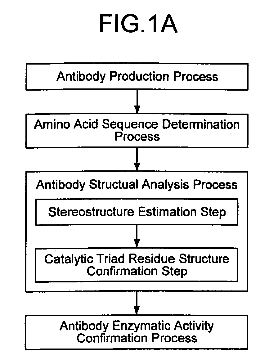
Description
The present invention relates to a novel process for
efficiently producing an antibody enzyme that has high
molecule recognition capability of an antibody and the
enzymatic activity, an antibody enzyme produced according
to the producing process, and an antibody enzyme having a
stereostructurally specific structure that can be applied
in the producing process.
Furthermore, the present invention relates to, among
the antibody enzymes in particular, an antibody against
Helicobacter pylori urease and an antibody enzyme against
chemokine receptor CCR-5 that is a coreceptor of
AIDS-causing virus as well as an application method of the
antibody enzymes.
Recently, there are various reports on an antibody
having enzyme-like activity, that is, an antibody enzyme.
For instance, S. Paul et al. report that an autoantibody
isolated from an autoimmune disease patient followed by
purifying has an activity of decomposing VIP (Vasoactive
Intestinal Peptide) (Reference literature 1: Paul, S., et
al., Science, 244: 1158, 1989). Furthermore, Gabibov et al.
report that an autoantibody isolated from a patient suffered
from autoimmune disease such as SLE (Systemic lupus
erythematosus) or immune cell proliferation has an antibody
that enzymatically decomposes a DNA (Reference literature
2: Shuster, A.M., et al., Science, 256: 665, 1992, and
Gabibov, A.G., et al., Appl. Biochem. Biotech., 47: 293,
1994).
Still furthermore, the present inventors obtained a
monoclonal antibody of which antigen is a stable region of
an envelope protein gp41 of AIDS virus (HIV) that causes
AIDS (acquired immune deficiency syndrome) and analyzed a
function of the monoclonal antibody in detail. As a result,
the inventors report that a light chain region of the
monoclonal antibody has a very high activity in specifically
decomposing the envelope protein gp41 of the HIV (Reference
literature 3 : Super Catalytic Antibody [ I ] : Decomposition
of targeted protein by its antibody light chain. Hifumi,
E. , Okamoto, Y., Uda, T., J. Biosci. Bioeng., 88(3), 323-327
(1999)).
Thus, the antibody enzyme, having high molecule
recognizing capability of antibody and the enzymatic
activity, is expected to apply in many fields such as the
healthcare, chemical industry, and food industry.
So far, as a process of preparing and obtaining such
antibody enzymes, only a method in which peptides or proteins
in a ground state are immunized in an animal and so on, and,
of all obtained antibodies, the enzymatic activity was
measured to select and obtain only antibodies having the
targeted enzymatic activity is known.
However, there are problems in the abovementioned
existing process for preparing and obtaining antibody
enzymes in that it necessitates much labor and long time,
and even when the enzymatic activity is measured of all
antibodies the probability of finding the antibody enzymes
is only less than 10%, that is, the efficiency is very low.
Thus, a process of efficiently preparing or obtaining
antibody enzymes is not yet found. Accordingly, it is
largely expected to develop an antibody enzyme having
excellent function and a process of efficiently obtaining
and preparing antibody enzymes and to further apply to
researches and industries.
The present invention was carried out in view of the
abovementioned situations and intends to provide a novel
process for efficiently producing antibody enzymes that
have high molecule recognition capability of an antibody
and the enzymatic activity and antibody enzymes produced
according to the producing process, as well as antibody
enzymes having a stereostructurally specific structure that
can be applied to the producing process.
The Helicobacter Pylori is a slender S-shaped
gram-negative bacterium and can be detected with high
frequency from biological specimens of stomachs of gastric
ulcer suffering patients and peptic ulcer suffering
patients. From this, the Helicobacter Pylori is considered
to cause gastric ulcer/gastric catarrh and suggested to be
finally relevant to occurrence of gastric cancer. This is
reported also from WHO (World Health Organization).
A rate of Japanese who are infected with the
Helicobacter Pylori (hereinafter, referred to as HP) is said
amounts to 80% in adults of 50 years old or more; accordingly,
researches and developments of bacteria elimination and
rapid diagnosis of the HP are socially in demand. In order
to eliminate the HP, several antibiotics have been developed,
and, in recent years, by administering three kinds of
antibiotics in combination, recognizable healing effect is
recognized.
As mentioned above, as antibacterial agents or
antiviral drugs for eliminating bacteria and viruses, the
antibiotic drugs have worked as a major player. However,
recently, the occurrence speed of drug resistant bacteria
against an antibiotic drug becomes very fast. In spite of
substantially 10 years being spent for developing an
antibiotic drug, according to recent reports, after the
administration of the antibiotic drug, in faster cases,
within only several months, drug resistant bacteria appear
and the antibiotic drug becomes ineffective. This is a very
problematic situation.
There are reports according to which similar problems
are occurring against the Helicobacter Pylori and in spite
of only a short time after the start of administration of
an antibiotic drug, drug resistant bacteria against the HP
have appeared. Furthermore, it is also found that, in some
cases, after the bacteria elimination by use of an antibiotic
drug, gastric ulcer reoccurs. From these backgrounds, a
development of novel drugs against the HP different from
existing ones is strongly demanded.
Since an interior of a human stomach is strongly acidic
owing to secretion of gastric-acid, normally externally
intruded bacteria cannot exist; however, the Helicobacter
Pylori, owing to an action of urease that is expressed in
its own cell envelope, can exist even in a strongly acidic
stomach. Accordingly, when the activity of the
Helicobacter Pylori urease can be suppressed, the bacteria
cannot exist in a stomach, resulting in inhibiting infection
from occurring.
The present invention pays attention to the point and
provides an antibody enzyme that, by exhibiting a
decomposition action against, in particular among the above
antibody enzymes, the Helicobacter Pylori, is effective in
eliminating the Helicobacter Pylori and can avoid the
appearance of drug-resistant bacteria. Furthermore, the
invention provides genes that code the antibody enzymes,
transformants in which the genes are introduced, and a
remedy/infection inhibitor that makes use of these and is
used for patients infected by the Helicobacter Pylori.
The AIDS (acquired immunodeficiency syndrome) is a
disease state where owing to the infection of human
immunodeficiency virus I type (HIV-1), immune function is
deteriorated, resultantly AIDS-associated opportunistic
infection, malignant tumor, neurosis, dementia and so on
are caused together. When infected with the HIV-1, after
an acute period, a symptomless period, and AIDS-associated
syndrome, the AIDS is developed. Owing to AIDS-related
researches and advances of remedy, the AIDS is becoming from
fatal disease to a remediable disease; however, at present,
there is no sure anti-HIV drug and a treatment method due
to drugs is not yet established.
In researches of an infection mechanism of the HIV,
in 1984, as a receptor of the HIV-1, CD4 was identified.
It is known that the CD4 is present on a surface of a human
cell and, when it combines with gp120 that is an envelope
glycoprotein of the HIV-1 to infect the cell.
However, since the CD4 alone does not cause fusion
between an envelope of virus and a cell membrane of a host
cell, infection of the HIV-1 is not caused; accordingly,
the presence of a coreceptor was suggested. When it has
passed more than 10 years after the discovery of the CD4,
a chemokine receptor that is a coreceptor of the HIV was
discovered. Furthermore, it was also reported that when the
chemokine receptor CCR-5 was not normal, resistance against
infection and crisis of the HIV-1 was exhibited.
Accordingly, at present, many researchers and enterprises
venture on development of drugs that can inhibit the HIV-1
from intruding with the coreceptor as a target.
Present situations of researches and developments of
anti-HIV drugs with such a HIV coreceptor as a target are
described in reference literature 4 (T. Murakami and N.
Yamamoto "Development of novel anti-HIV drug-drug affecting
on HIV coreceptor (second receptor)", Protein, Nucleic acid,
Enzyme, Vol.43, 677-685 p (1998)).
As shown in Table 1 in the reference literature 4, all
over the world pharmaceutical companies, in collaboration
with basic research laboratories such as universities,
venture on research and development of anti-HIV drugs with
the HIV coreceptor as a target. As a candidate of the
anti-HIV drug that can prevent the HIV-1 from infecting with
the chemokine receptor CCR-5 as a target, a compound that
works as an antagonist thereof is reported.
However, at present, an effective drug that directly
attacks the chemokine receptor CCR-5 and deletes its
function is not at all found.
In this connection, the present invention further
provides, among the abovementioned antibody enzymes, in
particular, an antibody enzyme that can decompose the
chemokine receptor CCR-5 to delete its function and can be
utilized in preventing the AIDS viruses from infecting and
remedying the AIDS. Furthermore, the invention provides a
gene that codes the antibody enzyme, a transformant in which
the gene is introduced, and an anti-HIV drug that makes use
of the above.
The present inventors, in view of the abovementioned
situations, studied hard and, when a structure and a function
were analyzed in detail of an antibody having the enzymatic
activity that cleaves and/or decomposes a polypeptide and
an antigen protein, found independently that in
stereostructures of all antibody enzymes, a catalytic triad
residue exists and the catalytic triad residue is relevant
with the polypeptide decomposing activity, and came to
completion of the invention.
Furthermore, the inventors, in order to overcome the
above problems, studied hard to obtain, among antibody
enzymes that have, while being an antibody, an enzyme action
and can completely decompose a target protein, in particular,
an antibody enzyme against the HP urease. As a result, it
is found that among antibodies against the HP urease, there
are ones that exhibit a function as an antibody enzyme, and
thereby the invention came to completion.
Still furthermore, the inventors, in order to overcome
the above problems, studied hard to obtain, among antibody
enzymes, in particular, an antibody enzyme against
chemokine receptor CCR-5. As a result, it is found that
among antibodies against the chemokine receptor CCR-5,
there are ones that exhibit a function as an antibody enzyme,
and thereby the invention came to completion.
A production process of an antibody enzyme involving
the invention, in order to overcome the abovementioned
problems, includes an antibody structure analysis process
that performs a stereostructure estimation step where a
stereostructure of an antibody is estimated from amino-acid
sequences; and a catalytic triad residue structure
confirmation step where, in an estimated stereostructure
of the antibody, whether a catalytic triad residue structure
where a serine residue, an aspartate residue and a histidine
residue or a glutamate residue are present
stereostructurally in proximity to each other is present
or not is confirmed.
In the catalytic triad structure confirmation step,
in order to confirm the catalytic triad structure, any one
of indices below can be preferably used.
Index of type 1 ○ or 1 ○*: It has either a structure same
as a catalytic triad residue structure common in known
antibody enzymes (type 1 ○) or a structure where when among
the known catalytic triad residue structures one amino-acid
residue that is different in position is used, a catalytic
triad residue structure can be assumed to constitute (type
1 ○*).
Index oftype 2 ○: A serine residue, an aspartate residue and
a histidine residue or a glutamate residue are present in
the range of 3 to 20 angstrom in the stereostructure.
Index oftype 3 ○: When an antibody has a germ-cell gene type
derived from a known antibody enzyme, it is used as an index.
Index oftype 4 ○: A complimentarity determining region 1
(CDR1) being constituted of 16 amino-acid residues and a
histidine residue being located at the 93rd according to
Kabat numbering scheme are used as an index.
Index oftype 4 ○*: A complimentarity determining region 1
(CDR1) being constituted of 11 amino-acid residues and a
histidine residue being located at the 91st or 55th according
to Kabat numbering scheme are used as an index.
Index of
Index of
Index of
Index of
Furthermore, the antibody enzyme production process
preferably includes an antibody production process in which
an antibody is produced by use of a hybridoma obtained by
fusing a mouse spleen lymph corpuscle immunized with antigen
and a mouse myeloma cell. As the antigen, a
hapten-conjugated protein obtained by coupling a substance
that is made an antigenic determinant to a carrier protein
can be used; however, it is not particularly restricted.
It goes without saying that a protein itself as well can
be used as an immunogen.
The antibody enzyme production process may further
include a catalytic triad residue structure introduction
process where by use of the stereostructure information
obtained after the antibody structure analysis process, a
catalytic triad residue structure is introduced into an
antibody according to a genetic engineering procedure.
The catalytic triad residue structure is one that the
present inventors found newly this time as a feature of an
antibody enzyme having an activity of cleaving and/or
decomposing a polypeptide or antigen protein. Accordingly,
since according to the process, antibodies are screened by
use of the catalytic triad residue structure unique to the
antibody enzyme, the antibody enzymes can be produced more
efficiently than ever.
An antibody enzyme involving the invention has in a
stereostructure a catalytic triad residue structure where
a serine residue, an aspartate residue, and a histidine
residue or glutamate residue are present stereostructurally
in proximity. As an example of the antibody enzyme, as shown
in examples described later, heavy and light chains of
i4SL1-2 and i41-7 antibodies can be cited. The heavy and
light chains of the i4SL1-2 and i41-7 antibodies are
experimentally confirmed that, while maintaining a nature,
as an antibody, of being high in the affinity with a substrate,
these exhibit the enzymatic activity.
The "catalytic triad residue structure" means a
structure in which it is inferred that three amino-acid
residues containing at least serine are contained in an
active site and form an active center. A protease having
the catalytic triad residue structure contains serine in
the active site; accordingly, it is called a serine protease.
Accordingly, the antibody enzyme can be said one kind of
the serine protease. When a structure that can be estimated
to be a catalytic triad residue is contained, high activity
can be expected as the protease.
An antibody enzyme involving the invention, further,
may be an antibody enzyme of which complimentarity
determining region 1 (CDR1) is constituted of 16 amino-acid
residues and that has a histidine residue at the 93rd
according to Kabat numbering scheme, or an antibody enzyme
of which complimentarity determining region 1 (CDR1) is
formed of 11 amino-acid residues and that has a histidine
residue at the 91st or 55th according to Kabat numbering scheme,
or furthermore one of which mouse germ cell gene type is
produced from a gene selected from bb1, bl1, bd2, cr1, cs1,
bj2, hf24, 12-41, 19-14, 19-17, 19-23, 19-25 and 21-12
according to the classification due to Thieve et al.
As shown in examples described below, as a result of
analysis of genes of antibody enzymes that are derived from
a mouse and have the activity of cleaving and/or decomposing
a polypeptide and antigen protein, it is experimentally
confirmed that many of the antibody enzymes are produced
from a mouse germ cell gene selected from, according to the
classification due to Thieve et al., bb1, bl1, bd2, cr1,
cs1, bj2, hf24, 12-41, 19-14, 19-17, 19-23, 19-25, and 21-12.
Accordingly, antibody enzymes that are produced from a mouse
germ-cell gene selected from the bb1, bl1, bd2, cr1, cs1,
bj2, hf24, 12-41, 19-14, 19-17, 19-23, 19-25, and 21-12 are
fundamentally antibody enzymes that have the catalytic
triad residue structure in a stereostructure and the
activity of cleaving and/or decomposing a polypeptide and
antigen protein.
An antibody enzyme involving the invention may be a
heavy chain or a light chain of an antibody. Furthermore,
in the invention, a gene that codes the antibody enzyme is
contained as well.
Furthermore, an antibody enzyme involving the
invention is, among the abovementioned antibody enzymes,
in particular, an antibody of which antigen is Helicobacter
Pylori urease or a fragment of the antibody, and that is
characterized in that it works as a degrading enzyme of the
urease. Here, the "fragment of an antibody " means the
respective peptide chains that constitute an antibody of
which antigen is HP urease, and furthermore peptide
fragments of a portion of region in the respective peptide
chains.
An antibody enzyme, while being an antibody, has the
enzymatic activity. Among the antibody enzymes described
above, in particular, one that exhibits high degradation
activity against the antigen protein as a target is called
a "super-antibody enzyme". The "super-antibody enzyme" can
completely decompose a target protein and furthermore has
the activity near to that of natural enzyme (reference
literature 3). The abovementioned antibody enzyme is an
antibody of which antigen is HP urease and completely
decomposes the HP urease. Accordingly, it is contained in
the "super-antibody enzyme".
The antibody enzyme, with nature as an antibody of the
HP urease strongly remained, has the nature of decomposing
the HP urease that is an antigen thereof. Accordingly, the
antibody enzyme is high in the specificity and can aim and
decompose only the urease that is indispensable for the
existence of the Helicobacter Pylori.
Furthermore, when an antibiotic drug is used as an
antibacterial agent or antiviral drug, the bacteria or the
viruses mutate their proteins to acquire the tolerance
against the antibiotic drug, and thereby drug resistant
bacterium (cell) appears. However, portions indispensable
for the existence of the bacterium or viruses themselves
cannot be mutated. The antibody enzyme can aim and attack
the indispensable portions and can completely delete the
bioactivity thereof; accordingly, reoccurrence of the
symptom as well as the appearance of the drug resistant
bacterium (cell) can be avoided.
The antibody enzyme preferably contains a variable
region of an antibody of the HP urease. The "variable
region" means, among H chains and L chains that constitute
an antibody, a portion made of substantially 110 amino-acid
residues from an N terminal. The "variable region" exhibits
diversity in a primary structure depending on the kind of
the antibody, and when the antibody has the enzymatic
activity, an active center thereof is highly probably
contained therein. Accordingly, when the antibody enzyme
contains the variable region of the antibody, it can have
high activity as enzyme.
As an antibody enzyme involving the invention,
specifically, ones that are formed by containing an amino
acid sequence shown in sequence No. 14 or an amino acid
sequence in which in an amino acid sequence shown in the
sequence No.14 at least one amino acid is replaced, deleted,
inserted and/or added can be cited.
The amino acid sequence shown in sequence No.14 is a
variable region of an L chain of a monoclonal antibody HpU-18
of the HP urease. The variable region of the L chain of the
HpU-18, as shown also in an example described later, was
recognized to be high in the activity as the degrading enzyme
of the HP urease.
Furthermore, the "at least one amino acid being
replaced, deleted, inserted and/or added" may be
replacement, deletion, insertion and/or addition of amino
acids of the number to an extent that, according to a known
variant protein production process such as a site-directed
mutagenesis by use of, for instance, a genetic engineering
procedure, can replace, delete, insert and/or add. Thus,
when the genetic engineering procedure is applied, in the
amino acid sequence shown in the sequence No.14, an antibody
enzyme that is made of an amino acid sequence in which at
least one amino acid is replaced, deleted, inserted and/or
added, in other words, is a variant of the antibody enzyme
made of the amino acid sequence shown in the sequence No. 14.
As another example of an antibody enzyme involving the
invention, ones that are formed by containing an amino acid
sequence shown in any one of sequence Nos.15, 16, 17, 19
and 21, and furthermore ones that are formed by containing
an amino acid sequence in which in an amino acid sequence
shown in any one of sequence Nos.15, 16, 17, 19 and 21, at
least one amino acid is replaced, deleted, inserted and/or
added can be cited.
An amino acid sequence shown in sequence No.15 is a
variable region of an L chain of a monoclonal antibody HpU-9
of the HP urease and an amino acid sequence shown in sequence
No.16 is a variable region of an H chain of a monoclonal
antibody HpU-2 of the HP urease. Furthermore, an amino acid
sequence shown in sequence No.17 is a variable region of
an L chain of a monoclonal antibody HpU-20 of the HP urease
and an amino acid sequence shown in sequence No.19 is a
variable region of an H chain of a monoclonal antibody HpU-20
of the HP urease. Still furthermore, an amino acid sequence
shown in sequence No.21 is a variable region of an L chain
of a monoclonal antibody UA-15 of the HP urease. Of the
antibody enzymes having these amino acid sequences as well,
as shown in an example described later, as a degrading enzyme
of the HP urease, high activity could be recognized.
The abovementioned antibody enzymes have an action of
decomposing the HP urease that is indispensable for the
existence of the HP urease; accordingly, these can be used
for elimination of HP bacteria. Accordingly, a curative
drug involving the invention for a patient infected with
the HP bacteria is prepared by containing the antibody enzyme.
When the curative drug is orally taken in, it can directly
attack the HP bacteria existing in a stomach; accordingly,
side effects are less and it can work immediately.
The antibody enzyme can be not only used as the
curative drug but also taken in orally; accordingly, it can
be contained in food to take in on a daily basis, and thereby
the infection with the HP bacteria can be prevented.
Accordingly, in the invention, an infection preventive
agent of the HP bacteria formed by containing the antibody
enzyme is also contained.
The invention includes a gene that codes the antibody
enzyme and when the gene is introduced in a proper host (such
as bacterium and yeast), an antibody enzyme according to
the invention can be expressed in the host.
As a gene involving the invention, specifically, ones
made of a base sequence shown in any one of sequence Nos.18,
20, 22, 47, 48 and 49 can be cited. A base sequence shown
in sequence No.18 is one of base sequences of a gene that
codes a variable region of a light chain (L chain) of
monoclonal antibody HpU-20 of the HP urease. A base
sequence shown in sequence No.20 is a base sequence of a
gene that codes a variable region of a heavy chain (H chain)
of monoclonal antibody HpU-20 of the HP urease. Furthermore,
a base sequence shown in sequence No.22 is a base sequence
of a gene that codes a variable region of a light chain (L
chain) of monoclonal antibody UA-15 of the HP urease. A base
sequence shown in sequence No.47 is one of base sequences
of a gene that codes a variable region of a light chain (L
chain) of monoclonal antibody HpU-18 of the HP urease. A
base sequence shown in sequence No.48 is one of base
sequences of a gene that codes a variable region of a light
chain (L chain) of monoclonal antibody HpU-9 of the HP urease.
A base sequence shown in sequence No.49 is one of base
sequences of a gene that codes a variable region of a heavy
chain (H chain) of monoclonal antibody HpU-2 of the HP
urease.
The abovementioned "gene" contains not only a
double-stranded DNA but also respective single-stranded
DNAs such as a sense strand and an antisense strand that
constitute the double-stranded DNA and RNA. Furthermore,
the "gene" may be one that contains, other than sequences
that code peptides according to the invention, sequences
such as a sequence of an untranslated region (UTR) or a
sequence of a vector sequence (including an expression
vector sequence).
Furthermore, the invention contains a transformant in
which the gene is introduced. The transformant can express
the antibody enzyme in its body. Accordingly, when a plant
is used as a host of the transformant and the gene is
introduced in the plant and expressed, a transformant that
contains the antibody enzyme can be obtained. The
transformant contains the antibody enzyme provided with a
function of preventing the HP infection; accordingly, when
it is taken in on a daily basis, the HP can be suppressed
from infecting; that is, it can be used as functional food.
The antibody enzyme is destroyed in its structure when
heated; accordingly, as plants in which the gene can be
introduced, vegetables suitable for raw diet are preferable.
As such vegetables, specific example includes tomato,
cucumber and carrot.
The antibody enzyme involving the invention is
characterized in that it is an antibody of human-derived
chemokine receptor CCR-5 or an antibody fragment thereof
and can decompose the chemokine receptor CCR-5. Here, the
"antibody fragment" means the respective peptide chains
that constitute an antibody of which antigen is the chemokine
receptor CCR-5 and further peptide fragments of a portion
of a region in the respective peptide chains.
The antibody enzyme is an antibody of which antigen
is the chemokine receptor CCR-5 and completely decompose
the CCR-5; accordingly, it is included in the "super antibody
enzyme".
The antibody enzyme, with the nature as an antibody
of the chemokine receptor CCR-5 strongly remained, has the
nature of decomposing the chemokine receptor CCR-5 that is
an antigen thereof. Accordingly, the antibody enzyme is
high in the specificity and can aim and decompose only the
chemokine receptor CCR-5. The chemokine receptor CCR-5 is
a coreceptor that plays an important role in the infection
of the HIV; accordingly, the antibody enzyme that can delete
the function thereof can be effectively utilized as an
anti-HIV drug that is used in the prevention of the HIV
infection or remedy of AIDS.
The antibody enzyme is preferably formed with a
variable region of the chemokine receptor CCR-5 antibody
contained therein. The "variable region" means, among H and
L chains that constitute an antibody, a portion made of
substantially 110 amino-acid residues from an N terminal.
The variable region, depending on the kind of the antibody,
shows the diversity in its primary structure, and, when the
antibody has the activity as the enzyme, the active center
thereof is probably contained therein. Accordingly, when
the antibody enzyme contains the variable region, it can
have high activity as an enzyme.
As an antibody enzyme involving the invention,
specifically, one that is formed by containing an amino acid
sequence shown in sequence No.26 or one that is formed by
containing an amino acid sequence in which in an amino acid
sequence shown in sequence No.26 at least one amino acid
is replaced, deleted, inserted and/or added can be cited.
The amino acid sequence shown in the sequence No.26
is a variable region of a light chain (L chain) of monoclonal
antibody ECL2B-2 of the chemokine receptor CCR-5. The
variable region of the L chain of the ECL2B-2, as shown in
an example described later, was recognized to be high in
the activity as a CCR-5 degrading enzyme.
As another example of an antibody enzyme involving the
invention, one that is formed by containing an amino acid
sequence shown in the sequence No.30, or an amino acid
sequence in which in an amino acid sequence shown in the
sequence No.30, at least one amino acid is replaced, deleted,
inserted and/or added can be cited.
The amino acid sequence shown in sequence No.30 is a
variable region of a light chain (L chain) of monoclonal
antibody ECL2B-3 of the chemokine receptor CCR-5. In the
variable region of the light chain of the ECL2B-3 as well,
as shown in an example described later, the activity as a
CCR-5 degrading enzyme is confirmed to be high.
As still another example of an antibody enzyme
involving the invention, one that is formed by containing
an amino acid sequence shown in the sequence No.34, or an
amino acid sequence in which in an amino acid sequence shown
in the sequence No.34, at least one amino acid is replaced,
deleted, inserted and/or added can be cited.
The amino acid sequence shown in sequence No.34 is a
variable region of a light chain (L chain) of monoclonal
antibody ECL2B-4 of the chemokine receptor CCR-5. In the
variable region of the light chain of the ECL2B-4 as well,
as shown in an example described later, the activity as a
CCR-5 degrading enzyme is confirmed to be high.
As further still another example of an antibody enzyme
involving the invention, one that is formed by containing
an amino acid sequence shown in the sequence No.36, or an
amino acid sequence in which in an amino acid sequence shown
in the sequence No. 36, at least one amino acid is replaced,
deleted, inserted and/or added can be cited.
An amino acid sequence shown in sequence No.36 is a
variable region of a heavy chain (H chain) of monoclonal
antibody ECL2B-4 of the chemokine receptor CCR-5. In the
variable region of the L chain of the ECL2B-4 as well, as
shown in an example described later, the activity as a CCR-5
degrading enzyme is confirmed to be high.
Furthermore, the "at least one amino acid being
replaced, deleted, inserted and/or added" may be
replacement, deletion, insertion and/or addition of amino
acids of the number to an extent that, according to a known
variant protein production process such as a site-directed
mutagenesis by use of, for instance, a genetic engineering
procedure, can be replaced, deleted, inserted and/or added.
Thus, when the genetic engineering procedure is applied,
an antibody enzyme that is made of an amino acid sequence
in which, in the amino acid sequence shown in any one of
the sequence Nos.26, 30, 34 and 36, at least one amino acid
is replaced, deleted, inserted and/or added is, in other
words, a variant of the antibody enzyme made of the amino
acid sequence shown in any one of the sequence Nos.26, 30,
34 and 36.
The antibody enzyme according to the invention
decomposes the chemokine receptor CCR-5 that plays an
important role in the HIV infection to delete the function
thereof; accordingly, it can be used also as an anti-HIV
drug that disturbs the HIV infection. That is, the anti-HIV
drug according to the invention is characterized by
containing the antibody enzyme and by disturbing the
infection of the AIDS viruses.
Furthermore, the antibody enzyme according to the
invention can delay the development of the AIDS to an HIV
infected patient or inhibit disease from progressing. That
is, the anti-HIV drug according to the invention, containing
the antibody enzyme, may be one that works as a curative
drug against the AIDS virus-infected patient.
The anti-HIV drug according to the invention,
containing the antibody enzyme alone, can be used by directly
administering by means of such as intravenous injection.
However, in addition to the antibody enzyme, a carrier that
can be physiologically tolerated may be further contained.
Production of such an anti-HIV drug can be carried out
according to a so far known production process. The
anti-HIV drug uniquely aims and decomposes a CCR-5 molecule
alone; accordingly, it can be expected that not only the
effect is remarkable but also the side effects are less.
The invention includes also a gene that codes the
antibody enzyme. When the gene is expressibly introduced
in an appropriate host (such as bacterium and yeast), the
antibody enzyme according to the invention can be expressed
in the host.
The abovementioned "gene" contains not only a
double-stranded DNA but also respective single-stranded
DNAs such as a sense strand and an antisense strand that
constitute the double-stranded DNA and RNA. Furthermore,
the "gene" contains, other than sequences that code the
antibody enzymes according to the invention, sequences such
as a sequence of an untranslated region (UTR) or a sequence
of a vector sequence (including an expression vector
sequence).
As the gene involving the invention, specifically, one
that is formed of a base sequence shown in sequence No.27
can be cited. The base sequence shown in sequence No.27 is
a base sequence of a gene that codes a variable region of
a light chain (L chain) of monoclonal antibody ECL2B-2 of
the chemokine receptor CCR-5.
As another example of the gene involving the invention,
one that is formed of a base sequence shown in the sequence
No. 31 can be cited. The base sequence shown in the sequence
No.31 is a base sequence of a gene that codes a variable
region of a light chain (L chain) of monoclonal antibody
ECL2B-3 of the chemokine receptor CCR-5.
As still another example of the gene involving the
invention, one that is formed of a base sequence shown in
the sequence No.35 can be cited. The base sequence shown
in the sequence No.35 is a base sequence of a gene that codes
a variable region of a light chain (L chain) of monoclonal
antibody ECL2B-4 of the chemokine receptor CCR-5.
As further still another example of the gene involving
the invention, one that is formed of a base sequence shown
in the sequence No.37 can be cited. The base sequence shown
in the sequence No.37 is a base sequence of a gene that codes
a variable region of a heavy chain (H chain) of monoclonal
antibody ECL2B-4 of the chemokine receptor CCR-5.
Furthermore, in the invention, a transformant in which
the gene is introduced is also included. The transformant
is one in which the gene is expressibly introduced in an
appropriate host (such as bacteria and yeast) and can express
the antibody enzyme according to the invention in its body.
Accordingly, the transformant as well can be utilized as
an anti-HIV drug that prevents the infection of the AIDS
viruses and an anti-HIV drug for treating an AIDS-infected
patient.
Still furthermore, the invention contains a computer
program that instructs a computer to carry out an antibody
structure analysis process in the antibody enzyme
production process; and a machine-readable recording medium
in which a computer program that instructs a computer to
carry out an antibody structure analysis process is
recorded.
A production process according to the present
invention of antibody enzymes and an antibody enzyme
produced according to the production process will be
described as embodiment 1. However, the present invention
is not restricted thereto.
The invention proposes an antibody enzyme production
method that not only enables to efficiently obtain natural
antibody enzymes but also can produce antibody enzymes by
use of a genetic engineering process, and further provides
an example of novel and useful antibody enzymes obtained
according to the process. In what follows, an antibody
enzyme production process involving the invention will be
described, followed by describing an example of obtained
antibody enzymes and gene thereof, functions of the antibody
enzyme, and applications thereof.
The inventors analyzed in detail features of nature
and structure of several kinds of antibody enzymes having
the activity of cleaving and/or decomposing polypeptides
and antigen proteins. As a result, the inventors for the
first time revealed that the antibody enzymes having the
activity of cleaving and/or decomposing polypeptides and
antigen proteins all have a catalytic triad residue
structure in a stereostructure thereof.
Specifically, that a catalytic triad residue
structure is present in a stereostructure thereof means that
in a stereostructure of an antibody or an antibody fragment
a serine residue, an aspartate residue and a histidine
residue or a glutamate residue are present
stereostructurally in proximity, and, more specifically,
it is better that distances between a serine residue, an
aspartate residue, and a histidine residue or a glutamate
residue, each other, are at least in the range of 3 to 20
Å, preferably in the range of 3 to 10 Å. This is because
when distances between functional groups that constitute
the catalytic triad residue structure are in the range of
3 to 20 Å, in particular, in the range of 3 to 10 Å, the
catalytic triad residue structure and a substrate (such as
polypeptide and antigen protein) are considered to be
reactable.
In an antibody enzyme involving the invention, a site
where the catalytic triad residue structure is present is
not particularly restricted. An antibody is constituted of
a heavy chain (H chain) and a light chain (L chain). The
light chain is constituted of a variable region (VR) and
a constant region (CR), and the variable region includes
a complimentarity determining region (CDR). As shown in an
example described later, it is revealed that the catalytic
triad residue structure is mainly present in light chains;
however it is present as well in heavy chains though not
so much as in the light chain. Furthermore, also
experimentally, it is revealed that an antibody that has
the catalytic triad residue structure in a stereostructure
has the enzymatic activity of cleaving and/or decomposing
a polypeptide or an antigen protein.
Accordingly, it is better that in the antibody enzyme
production process involving the invention, whether an
antibody has the catalytic triad residue structure therein
or not can be judged.
An antibody enzyme production process involving the
invention may include at least an antibody structure
analysis process that carries out a stereostructure
estimation step where from amino acid sequences a
stereostructure of an antibody is estimated; and a catalytic
triad residue structure confirmation step where whether a
catalytic triad residue structure is present in the
estimated stereostructure of an antibody or not is judged.
As is obvious from the knowledge in the (1), when the
catalytic triad residue structure is included in a
stereostructure of an antibody, the antibody is very
probable to be an antibody enzyme. Accordingly, thereby,
antibody enzymes can be efficiently screened.
When an example of the antibody enzyme production
process involving the invention is more specifically
described, as shown in Fig. 1A, the production process may
include an antibody production step, an amino acid sequence
determination step, an antibody structure analysis step and
an antibody enzymatic activity confirmation step.
The antibody production step, without restricting to
particular one, may produce monoclonal antibodies from
hybridomas obtained by fusing mouse spleen lymph corpuscles
immunized with antigens and mouse myeloma cells,
alternatively, antibodies obtained from a library by use
of a phage display method may be used.
Specifically, when, as an immunogen, a desired antigen,
or a fragment containing an antigenic determinant (epitope)
thereof, or a derivative thereof, or an analogue thereof,
or a cell that can express these is used, an antibody can
be produced. These are carried out according to an ordinary
immunization operation, or a hybridoma method (reference
literature 5: Kohler, G. and Milstein, C., Nature 256,
495-497 (1975)), a trioma method, a human B cell hybridoma
method (reference literature 6: Kozbor, Immunology Today
4, 72, 1983), and an EBV-hybridoma method (reference
literature 7: Monoclonal Antibodies and Cancer Therapy,
Alan R. Liss, Inc., pp. 77-96, 1985).
Here, as far as the antigen is a polypeptide or a
protein, or a polysaccharide, there is no particular
restriction.
Specifically, when the antigen is a hapten, it does
not have the capability of producing antibodies;
accordingly, it cannot produce the antibody. However, when
an antigen is covalently bonded with a carrier made of a
biopolymer such as a protein derived from a different kind
to obtain an antigen protein and this is used to immunize,
the antibody production can be induced. As the carrier,
without restricting to particular one, various kinds of
proteins so far known in the field such as ovalbumin, gamma
globulin, and hemocyanine can be preferably used.
The "polypeptide" here includes short peptides in
which several amino acids are bonded and proteins.
Furthermore, the polypeptide includes naturally existing
ones and chemically, biologically synthesized ones. Still
furthermore, the "polysaccharide" here means ones in which
several of so-called monosaccharides such as glucose,
maltose, acetylglucosamine, and glucuronic acid are bonded.
Kinds of monosaccharide that constitutes the
"polysaccharide" are not particularly restricted as far as
these are so far known ones.
The amino acid sequence determination step, as far as
an amino acid sequence can be determined from an antibody
obtained in the antibody production step, is not
particularly restricted. Specifically, the amino acid
sequence may be determined by directly reading an amino acid
sequence from the obtained antibody, or an amino acid
sequence may be estimated from a base sequence of a gene
of the antibody. The amino acid sequence may be determined
by use of an Edman method. Furthermore, the base sequence
may be determined by use of a so far known sequencer, and
the amino acid sequence may be estimated by use of a so far
known soft wares and so on. These are not particularly
restricted.
The antibody structure analysis process, as far as it
includes a stereostructure estimation step and a catalytic
triad residue structure confirmation step, is not
particularly restricted. Thereby, whether a desired
antibody includes a catalytic triad residue structure or
not can be efficiently confirmed.
The stereostructure estimation step, as far as it can
estimate a stereostructure from data of the amino acid
sequences obtained in the amino acid sequence determination
step, is not particularly restricted. Specifically, all
that is necessary is to be able to estimate a secondary
structure and a tertiary structure in a protein. That is,
in the invention, all that is necessary is to clarify up
to a tertiary structure of an IgG class antibody.
As a quaternary structure in an antibody, like for
instance IgA and IgM class, a structure in which a plurality
of subunits (IgG class antibody) having a tertiary structure
meets to form an oligomer can be cited. However, such a
structure is not particularly necessary in a later catalytic
triad residue structure confirmation step; accordingly, it
is not necessary to estimate in the invention. However,
when an analysis result including a result of the quaternary
structure can be effectively utilized in the production of
antibody enzymes, without being restricted to the above,
a quaternary structure of an antibody may be estimated in
the stereostructure estimation step.
When the stereostructure estimation step is carried
out, a so far known higher-order structure analysis soft
ware may be used, and a specific procedure thereof is not
particularly restricted.
In the catalytic triad residue structure confirmation
step, as far as whether the catalytic triad residue structure
described in (1) is present in a stereostructure of an
antibody or not can be judged, there is no particular
restriction. Accordingly, when the catalytic triad residue
structure is judged, presence of the abovementioned
structure may be judged directly or indirectly.
As a direct index that is used in the judgment of the
catalytic triad residue structure, the respective indices
below can be cited. As a step prior to judgment by use of
the respective indices below, a more simple method where
whether a histidine residue is present in an amino acid
sequence of an antibody or not is investigated may be used
to judge whether the catalytic triad residue structure is
present or not. This is because in many antibodies, usually,
in comparison with the serine residue, the histidine residue
is less contained.
In a first index, whether the catalytic triad residue
structure has a structure same as a catalytic triad residue
structure common in known antibody enzymes or not,
alternatively, when, among the known catalytic triad
residue structures, only one residue thereof uses an
amino-acid residue in a different position, whether it is
estimated that a catalytic triad residue structure can be
constituted or not is made an index, the former case is called
a type 1 ○, and the latter one is called a type 1 ○*.
In a second index, a case where in a single antibody
heavy chain (H chain) or a single antibody light chain (L
chain), in a stereostructure, in the range of 3 to 20 Å,
the respective functional groups of a serine residue, an
aspartate residue, and a histidine residue or glutamate
residue are present is made an index, and this is called
a type 2 ○ index.
In the next place, in the catalytic triad residue
structure confirmation step, as an indirect index that is
used to judge a catalytic triad residue structure, though
not particularly restricted, in the embodiment, an index
that makes use of an analysis result of germline genes can
be preferably used.
The antibody light chain (L chain) can be classified
into two kinds of classes, κ and λ. In the case of a mouse,
it is said that substantially 95% is the κ chain.
Furthermore, since analysis of germline genes of mouse κ
chains is being advanced, information thereof can be
advantageously obtained. As will be described in an example
described later, by analyzing the germlines, a very
important knowledge that suggests that in particular
germlines an antibody L chain having the enzymatic activity
is already prepared is found (example 1-3 and Table 3). In
this connection, when germlines from which known antibody
enzymes are derived are investigated in advance and
antibodies having the germlines are selected, of the light
chains, antibody enzymes can be efficiently obtained at a
high probability. This is made a type 3 ○ index.
As the germlines that can be used for the light chains,
specifically, according to a classification due to Thiebe
et al., bb1, cr1, bl1, cs1, bd2, bj2 or 19-25 can be cited.
In the present embodiment and example, in the case of light
chains, the germlines will be classified according to Thiebe
et al.
Furthermore, also of the heavy chains (H chain), when
a method of thinking of the light chains is applied, when
antibodies having Families of VH1, VH5 and VH10 are selected,
antibody enzymes can be efficiently obtained at a very high
probability. In the aging process of antibodies, mutation
is caused, and some may lose the catalytic triad residue
structure; however, when, after the removal of these, the
germline or the Family is identified, substantially
completely antibodies having the enzymatic activity can be
obtained.
In addition, a structurally characteristic
constitution obtained from known antibody enzymes as well
can be used as an index. Specifically, as will be described
in an example described below, a feature that a
complimentarity determining region 1 (CDR1) is constituted
of 16 amino-acid residues and a histidine residue is present
at the 93rd according to the Kabat numbering scheme is
remarkable for the antibody enzyme. Accordingly, this can
be used as an index of type 4 ○. Furthermore, a feature that
a complimentarity determining region 1 (CDR1) is
constituted of 11 amino-acid residues and a histidine
residue is present at the 91st or 55th according to the Kabat
numbering scheme is also remarkable for the antibody enzyme;
accordingly, this can be used as an index of type 4 ○*. In
the present embodiment and example, an amino acid sequence
of an antibody is expressed according to the Kabat numbering
scheme.
More sure indexes are the types 1 ○ · 1 ○* or 2 ○;
accordingly, in order to heighten the assuredness at the
selection, these indices are preferably used, and type 3 ○
and types 4 ○ and 4 ○* may be used complementarily. However,
without using the indices of the types 1 ○, 1 ○* and 2 ○, the
indices of type 3 ○ and types 4 ○ and 4 ○* alone may be used
singularly or in a combination.
As means for carrying out an antibody structure
analysis process including the catalytic triad residue
structure confirmation step, in the embodiment, an antibody
structure analysis system described later in (3) can be cited.
However, it goes without saying that the invention is not
restricted thereto.
Furthermore, in the case of a stereostructure of an
antibody being directly estimated from data of base
sequences of a gene by use of an antibody structure analysis
system, the amino acid sequence determination step may be
contained as one step of the antibody structure analysis
process. That is, as described in the amino acid sequence
determination step, an amino acid sequence determination
step where an amino acid sequence is estimated from base
sequences of a gene of an antibody may be carried out as
one step of the antibody structure analysis process.
The antibody enzymatic activity confirmation step, as
far as it can confirm, after the analysis in the antibody
structure analysis step, whether an antibody that has a
catalytic triad residue structure and is very high in the
likelihood of being an antibody enzyme actually has the
activity of cleaving and/or decomposing a polypeptide and
an antigen protein or not, is not particularly restricted.
Specifically, a polypeptide or an antigen protein that
includes a structure that becomes an antigen or an antigen
determinant may be reacted with an antibody to check the
activity. At this time, reaction conditions such as a
concentration of the antigen, a concentration of the
antibody, a reaction temperature and a reaction time, and
a concentration of a base (kind of buffer used) are not
particularly restricted.
As another example of the antibody enzyme production
method involving the invention, as shown in Fig.1B, a
catalytic triad residue structure introduction step may be
included. That is, even antibodies that inherently do not
have a catalytic triad residue structure, when a catalytic
triad residue structure is introduced therein according to
a genetic engineering process, can produce antibody enzymes
with high efficiency. Accordingly, after the antibody
structure analysis step, a catalytic triad residue
structure introduction step may be carried out.
In the catalytic triad residue structure introduction
step, all that is necessary is to introduce a catalytic triad
residue structure, by use of stereostructure information
obtained in the antibody structure analysis process, in an
antibody according to a genetic engineering process, and
a specific procedure is not particularly restricted.
Specifically, so far known methods such as a method of
preparing a deletion mutant by use of an exonuclease and
site-directed mutagenesis can be preferably applied.
The catalytic triad residue structure introduction
step may be carried out after the antibody structure analysis
process, and it is very preferable that after the catalytic
triad residue structure introduction step, an antibody
enzymatic activity confirmation step is carried out to judge
whether, when the catalytic triad residue structure is
introduced, the activity of cleaving and/or decomposing a
polypeptide or an antigen protein can be imparted or not.
Means of carrying out the antibody structure analysis
process according to the invention may be an antibody
structure analysis system that includes a computer. For
instance, as shown in Fig. 2, an antibody structure analysis
system 10 including an input portion 11, a display portion
12, a printing portion 13, a memory portion 14, a controller
15, a stereostructure estimation portion 151 and a catalytic
triad residue structure confirmation portion 152 can be
cited.
The input portion 11, as far as it enables to input
information involving an operation of the antibody
structure analysis system 10, is not particularly
restricted. So far known input means such as a keyboard,
a tablet or a scanner can be preferably used.
The display portion 12 displays various kinds of
information such as information involving an operation of
the antibody structure analysis system 10 and selection
results including an estimated stereostructure of the
antibody and a confirmation result of the catalytic triad
residue structure. Specifically, various kinds of display
devices such as known CRT displays and liquid crystal display
devices can be preferably used; however, there is no
particular restriction.
The printing portion 13 records (prints or picturizes)
various kinds of information that can be displayed by the
display portion 12 on recording materials such as PPC paper.
Specifically, known image formation devices such as an ink
jet printer and a laser printer can be preferably used;
however, there is no particular restriction.
The display portion 12 and printing portion 13 in
combination can be represented as output means. That is,
the display portion 12 is means for outputting various kinds
of information in a soft copy and the printing portion 13
is means for outputting various kinds of information as a
hard copy. Accordingly, as the outputting means used in the
invention, without restricting to the display portion 12
and the printing portion 13, other outputting means may be
provided.
The memory portion 14 memorizes various kinds of
information (such as control information, selected results
and other information) that is used in the antibody structure
analysis system 10. Specifically, so far known various
kinds of memory means such as semiconductor memories such
as RAMs and ROMs, magnetic discs such as floppy discs and
hard discs, disc types of optical discs such as
CD-ROM/MO/MD/DVD and card types such as IC cards (including
memory cards)/optical cards can be preferably used.
Furthermore, the memory portion 14 may be integrated
with the antibody structure analysis system 10 to form one
device, or isolated as an external memory device, or
constituted so as to have both the integrated memory portion
14 and the external memory device. For instance, as the
integrated memory portion 14, a built-in type hard-disc
drive and a floppy disc-drive, a CD-ROM drive or a DVD-ROM
drive incorporated in a device can be cited, and, as an
external memory device, an external hard disc and external
types of the abovementioned various kinds of disc-drives
can be cited.
The controller 15 controls an operation of the
antibody structure analysis system 10. Specifically, as
shown in Fig.2 with solid arrow marks, to the respective
means of the input portion 11, the display portion 12, the
printing portion 13, the memory portion 14, the
stereostructure estimation portion 151 and the catalytic
triad residue structure confirmation portion 152, the
control information is outputted from the controller 15.
On the basis of the control information, the respective means
operate in combination to drive the antibody structure
analysis system 10 as a whole. Furthermore, to the
controller 15, instruction information to drive the
antibody structure analysis system 10 can be inputted from
the input portion 11. Accordingly, in Fig.2, the solid
arrow marks that show an exchange of the control information
are bi-directional.
The stereostructure estimation portion 151 and the
catalytic triad residue structure confirmation portion 152
work in combination as analysis means. Specifically, to the
stereostructure estimation portion 151, the amino acid
sequence data inputted from the input portion 11 are inputted
through the controller 15 (dotted line in the drawing).
Then, data of the stereostructure generated at the
stereostructure estimation portion 151 are outputted to the
catalytic triad residue structure confirmation portion 152
(dotted line in the drawing). In the catalytic triad
residue structure confirmation portion 152, from the
stereostructure data, whether the catalytic triad residue
structure is present or not is confirmed, and based on the
confirmation result, final analysis data are generated
(dotted line in the drawing). The analysis data, as far as
the data can be outputted by outputting means such as the
display portion 12 and/or printing portion 13, are not
particularly restricted.
The data that are used to analyze a stereostructure
in the stereostructure estimation portion 151 may be at least
data of the amino acid sequences; however, it may be data
of the base sequences. In this case, all that are necessary
for the stereostructure estimation portion 151 are to
estimate amino acid sequences from the data of base sequences
to generate a stereostructure based on the data.
Specific constitutions of the controller 15, the
stereostructure estimation portion 151 and the catalytic
triad residue structure confirmation portion 152 are not
particularly restricted; that is, so far known calculation
means can be preferably used. The respective means may be
independently constituted from each other; however, the
respective means are preferably integrated into one
calculation means to be an integrated control and analysis
system. Specifically, it is very preferable that the
respective means are integrated as a central processing unit
(CPU) of a computer and an operation thereof is carried out
according to a computer program.
Accordingly, the stereostructure estimation portion
151 may carry out a calculation where a CPU estimates a
stereostructure according to a so far known computer program
that estimates a stereostructure of a protein; and the
catalytic triad residue structure confirmation portion 152
may carry out a calculation where a CPU determines at least
whether a catalytic triad residue structure is present or
not according to a computer program. In addition, though
not shown in the drawing, the antibody structure analysis
system 10 may include other analysis means and, as analysis
data, other data other than that of the catalytic triad
residue structure and the stereostructure.
Furthermore, the antibody structure analysis system
10 may be constituted so that, by providing a communication
portion 16 as shown in Fig.2, various kinds of information
may be inputted and outputted through a communication
network including the Internet. The communication portion
16 is connected with a communication network and can send
and receive various kinds of information. In Fig.2, the
antibody structure analysis system 10, a personal computer
(PC) 61 and a server 62 in the same premise are connected
to a communication line 60 to form a bus type LAN (local
area network), and further the LAN is connected with a PC
61 in other area through the Internet.
A specific configuration of the communication portion
16 is not particularly restricted. Known LAN cards, LAN
boards, LAN adaptors and modems can be preferably used.
As the PC 61, known personal computers provided with
communication means such as a modem can be preferably used
and the PC 61 is not restricted to desktop type and note-type
ones. The PC 61 has a fundamental constitution that has a
display portion such as a CRT display or a liquid crystal
device and an input portion such as a keyboard or a mouse.
For convenience of explanation, a display portion and input
portion that are provided to the PC 61 and not shown in the
drawing are expressed as a PC display portion and a PC input
portion.
The PC 61 may be provided with a hardware (various
kinds of input means such as a scanner and various kinds
of output means such as a printer) that can be externally
connected to a general personal computer.
A specific configuration of the server 62 is neither
particularly restricted and may be a computer that can offer
a service to the PCs 61 that are clients constituting the
LAN and the antibody structure analysis system 10.
Furthermore, the server 62 may combine a data base server
and a file service server.
A specific configuration of the communication line 60
is neither particularly restricted; that is, so far known
general communication lines can be used. Furthermore, a
type and a bus type of the LAN that is constituted with the
communication line 60 are neither particularly restricted;
that is, so far known type such as a star type and a ring
type may be used.
Furthermore, though not shown in the drawing, the LAN
may include shared printers and other terminals. Still
furthermore, through not shown in the drawing, in the
communication network including the LAN, various kinds of
communicable and portable terminals may be included.
In a network having the abovementioned configuration,
for instance, after the antibody structure analysis system
10 performed the antibody structure analysis process, the
selected results can be not only outputted simply in the
antibody structure analysis system 10 (that is, such as the
display portion 12 and printing portion 13) but also
transmitted through the LAN to the PCs 61. In the PC 61,
results obtained from the antibody structure analysis
system 10 can be displayed on the PC display portion and
printed out by use of a printer, and furthermore owing to
an input from the PC input portion the selected results can
be processed as well.
That is, the communication portion 16 can function not
only as communication means but also as input means of the
antibody structure analysis system 10. Furthermore, in
particular, when, by use of the PC61, through the Internet,
at an area remote from a location where the antibody
structure analysis system 10 is located, information (such
as amino acid sequences) involving implementation of the
antibody structure analysis process is transmitted or
selected results are received, to optional customers, a
service that provides the antibody structure analysis
process can be provided.
Furthermore, in the case of the PCs 61 being connected
through the LAN with the antibody structure analysis system
10, when one antibody structure analysis system 10 is in,
for instance, a research facility, other researchers can
share the antibody structure analysis system 10 through an
information terminal such as the PC 61. Accordingly, the
present invention can be more efficiently carried out.
Still furthermore, in the case of the server 62
combining a database server and a file server, selected
results of the antibody structure analysis process that is
performed through a communication network can be stored
through the communication network in the server 62. As a
result, the selected results can be more efficiently used.
In addition, in the invention, the antibody structure
analysis process in the invention can be carried according
to a program on a computer. However, in a recording medium
that records the program, a medium that carries a program
in a volatile manner such as downloading from a communication
network is also included. For instance, when a program of
the antibody structure analysis process is recorded in
recording means of the server 62, the antibody structure
analysis system 10 may appropriately download the program
of the antibody structure analysis process from the server
62 to use. However, when the antibody structure analysis
system 10 downloads the program from a communication network,
a program for downloading is stored in advance in a body
of the antibody structure analysis system 10 or can be
installed from a separate recording medium.
Furthermore, like the PC 61, when it is connected
through a communication network to the server 62, when a
program of the antibody structure analysis process is
downloaded from the server 62, the PC 61 itself can be used
as the antibody structure analysis system 10.
In the next place, a specific operation of the antibody
structure analysis system 10, that is, an example of an
antibody structure analysis process in the invention will
be described with reference to a flowchart shown in Fig.3.
Firstly, as a prior stage, base sequences of a
monoclonal antibody obtained in the antibody production
step are determined to finally obtain an amino acid sequence.
Subsequently, as a step 1 (hereinafter, the step is
abbreviated as S), of the amino acid sequence, an amino acid
sequence of a variable region is inputted from the input
portion 11. Then, as S2, an index for confirming a catalytic
triad residue structure in a catalytic triad residue
structure confirmation portion 152 is set. As the index,
types 1 ○ · 1 ○*, 2 ○, 3 ○, 4 ○, and 4 ○* explained in the (2-3) can
be cited. The indices may be used singularly or in a
combination of a plurality thereof.
In the next place, as S3, by use of inputted data of
the amino acid sequence of the variable region, a
stereostructure of an antibody is estimated by means of the
stereostructure estimation portion 151. Then, as S4, from
the data of the stereostructure obtained by the
stereostructure estimation portion 151, the catalytic triad
residue structure confirmation portion 152, with the data
of the amino acid sequence and the index set in the S2,
analyses a layout of amino-acid residues that are estimated
to constitute the catalytic triad residue structure and a
situation of variation thereof.
When a catalytic triad residue structure or a
structure that is estimated to be a variation structure
thereof is confirmed, the antibody is high in the likelihood
of being an antibody enzyme. When any of the above
structures is not found, the antibody is low in the
likelihood of being an antibody enzyme. Subsequently, in
S5, from the analysis data in the S4, final analysis data
are generated and displayed at the display portion 12.
Further thereafter, in S6, whether an analysis of the
catalytic triad residue structure is once more carried out
or not is judged. In the case of the analysis being performed
again (YES in the drawing), in S7, whether an analysis is
carried out with the data of the same amino acid sequence
and with a different index or not is judged. In S7, in the
case of data of the same amino acid sequence being used (YES
in the drawing), the step is returned to S2, and when data
of a new amino acid sequence are inputted (NO in the drawing),
the step is returned to S1.
On the other hand, in S6, in the case of the catalytic
triad residue structure analysis being not carried out again
(NO in the drawing), in S8, obtained analysis data are, when
there being a plurality thereof, printed together in the
printing portion 13, or sent through a communication
interface to other PC 61.
The antibody structure analysis system 10 in the
above-described embodiment may be realized on a computer
with a program that functions the antibody structure
analysis process including above explained steps from S1
to S8.
The program may be stored in a recording medium that
can be read with a computer. Specifically, the memory
portion 14 shown in Fig.3, specifically, for instance, a
ROM itself may be a program medium. When the memory portion
14 is provided with a program reader, it may be a program
medium that can be read when a recording medium is inserted
therein. As the program medium, known configurations cited
as specific examples of the memory portion 14 can be
preferably used.
In all cases, the stored program may be carried out
when the controller 15 accesses thereto, or the program is
read, the read program is downloaded in a not shown program
memory area and the program may be carried out. A
downloading program is stored in the memory portion 14
beforehand. Furthermore, a content stored in the recording
medium, without restricting to the program, may be other
data for instance.
Thus, in the invention, a computer program that
carries out the antibody structure analysis process on a
computer and a machine-readable recording medium in which
a computer program that makes a computer execute the program
is recorded are included. Accordingly, in order to execute
the antibody structure analysis process in the invention
on a computer according to a program, the computer itself
can be made the antibody structure analysis system 10. As
a result, the versatility of the invention can be heightened
and the invention can be readily used on a communication
network.
In the next place, the antibody enzyme involving the
invention includes an antibody enzyme having a
stereostructurally unique structure that can be applied in
the production process, and furthermore, includes also an
antibody enzyme that can be produced according to the
abovementioned production method. In the embodiment, in
what follows, an example of an antibody enzyme involving
the invention will be detailed.
An antibody enzyme involving the embodiment is (a) an
antibody enzyme having a variable region made of an
amino-acid sequence described in sequence No. 1, 3, 5 or
7 or (b) an antibody enzyme having a variable region made
of an amino-acid sequence in which in an amino-acid sequence
described in sequence No. 1, 3, 5 or 7, at least one amino
acid is replaced, deleted, inserted and/or added and the
activity of cleaving and/or decomposing a polypeptide or
an antigen protein. In the embodiment, 1 ○ a heavy chain of
a i41SL1-2 antibody (i41SL1-2-H) that has a variable region
having an amino acid sequence shown in the sequence No. 1
and made of 117 amino-acid residues, 2 ○ a light chain of a
i41SL1-2 antibody (i41SL1-2-L) that has a variable region
having an amino acid sequence shown in the sequence No. 3
and made of 114 amino-acid residues, 3 ○ a heavy chain of a
i41-7 antibody (i41-7-H) that has a variable region having
an amino-acid sequence shown in the sequence No. 5 and made
of 120 amino-acid residues, and 4 ○ a light chain of a i41-7
antibody (i41-7-L) that has a variable region having an amino
acid sequence shown in the sequence No. 7 and made of 109
amino acid residues can be exemplified.
The i41SL1-2 antibody is a monoclonal antibody against
a synthetic peptide (shown in the sequence No. 9) of a
complimentarity determining region 1 (CDR 1) of a light chain
of an antibody 41S-2 that recognizes a constant region of
envelope protein gp-41 of HIV virus. Amino acid sequences
of variable regions of the heavy and light chains of the
i41SL1-2 antibody and base sequences that code the amino
acid sequences are shown in Figs. 4A and 4B.
A stereo structure of each of the light chain
(i41SL1-2-L) and heavy chain (i41SL1-2-H) of the i41SL1-2
antibody is estimated and analyzed with a computer. As a
result, as shown in Figs. 5A and 5B, it was revealed that
the i41SL1-2 antibody has a catalytic triad residue
structure in both i41SL1-2-H and i41SL1-2-L. That is, the
i41SL1-2-L, as shown in Figs. 4B and 5A, is estimated to
have a catalytic triad residue structure constituted of,
according to Kabat numbering scheme, an aspartate residue
at the 1st (D1), a histidine residue at the 93rd (H93), and
a serine residue at the 27A (S27a) or a catalytic triad
residue structure constituted of, according to Kabat
numbering scheme, an aspartate residue at the 27D (D27d),
a histidine residue at the 93rd (H93), and a serine residue
at the 27A (S27a).
Furthermore, the i41SL1-2-H, as shown in Figs. 4A and
5B, is estimated to have a catalytic triad residue structure
constituted of, according to Kabat numbering scheme, an
aspartate residue at the 55th (D55), a histidine residue
at the 52nd (H52), and a serine residue at the 54th (S54)
or a catalytic triad residue structure constituted of,
according to Kabat numbering scheme, an aspartate residue
at the 100th (D100), a histidine residue at the 35th (H35),
and a serine residue at the 98th (S98). Locations of the
catalytic triad residue structures are in the neighborhood
of the complimentarity determining regions for both the
light and heavy chains.
The i41-7 antibody is a monoclonal antibody against
a light chain of antibody 41S-2. Amino acid sequences of
variable regions of the heavy and light chains of the i41-7
antibody and base sequences that code the amino acid
sequences are shown in Figs. 8A and 8B.
A stereo structure of each of the light chain (i41-7-L)
and heavy chain (i41-7-H) of the i41-7 antibody is, similarly
to the i41SL1-2 antibody, estimated and analyzed with a
computer. As a result, as shown in Figs. 9A, 9B and 9C, it
was revealed that the i41-7 antibody has a catalytic triad
residue structure in each of the i41-7-L and i41-7-H. That
is, the i41-7-L, as shown in Figs. 8B and 9A, is estimated
to have a catalytic triad residue structure constituted of,
according to Kabat numbering scheme, an aspartate residue
at the 1st (D1), a histidine residue at the 91st (H91), and
a serine residue at the 26th (S26) or a catalytic triad
residue structure constituted of, according to Kabat
numbering scheme, an aspartate residue at the 28th (D28),
a histidine residue at the 91st (H91), and a serine residue
at the 30th (or 26th) (S30 or S26). Locations of the catalytic
triad residue structures are in the neighborhood of a CDR1
and CDR3 that are the complimentarity determining region
(CDR).
Furthermore, the i41-7-L, as shown in Figs. 8B and 9B,
is estimated to have a catalytic triad residue structure
constituted of, according to Kabat numbering scheme, an
aspartate residue at the 60th (D60), a histidine residue at
the 55th (H55), and a serine residue at the 52nd (S52). This
is located in the neighborhood of a CDR2.
Still furthermore, the i41-7-H, as shown in Figs. 8A
and 9C, is estimated to have a catalytic triad residue
structure constituted of, according to Kabat numbering
scheme, an aspartate residue at the 86th (D86), a histidine
residue at the 41st (H41), and a serine residue at the 40th
(or 84th or 87th) (S40 or S84, or S87).
A gene involving the embodiment may be (a) a gene that
codes an antibody enzyme having a variable region made of
an amino-acid sequence shown in sequence No. 1, 3, 5 or 7,
or (b) a gene that codes an antibody enzyme that has a
variable region made of an amino-acid sequence that is
obtained by replacing, deleting, inserting and/or adding
at least one amino acid in an amino acid sequence shown in
sequence No. 1, 3, 5 or 7 and has the activity of cleaving
and/or decomposing a polypeptide and an antigen protein.
In the embodiment, 1 ○ a gene (i41SL1-2-H gene) having a
variable region made of a base sequence (cDNA) shown in
sequence No.2, 2 ○ a gene (i41SL1-2-L gene) having a variable
region made of a base sequence (cDNA) shown in sequence No.4,
3 ○ a gene (i41-7-H gene) having a variable region made of
a base sequence (cDNA) shown in sequence No.6, and 4 ○ a gene
(i41-7-L gene) having a variable region made of a base
sequence (cDNA) shown in sequence No.8 can be exemplified.
The genes are derived from a mouse (Mus musculus).
The "gene" according to the invention includes an RNA
and a DNA. The RNA includes an mRNA, and the DNA includes
a cDNA or a genome DNA that can be obtained by, for instance,
cloning or chemical synthesis technology or a combination
thereof. Furthermore, the DNA may be a double-stranded one
or a single-stranded one. The single-stranded DNA may be
a code DNA that becomes a sense strand or an antisense code
that becomes an antisense strand (antisense strand can be
used as a probe or an antisense drug). Furthermore, the
"gene" according to the invention includes, other than
sequences that code proteins of the (a) or (b), sequences
such as a sequence of an untranslated region (UTR) or a
sequence of a vector sequence (including an expression
vector sequence).
In the i41SL1-2 and i41-7 antibodies, as shown in an
example described later, it was experimentally confirmed
that both heavy chains (i41SL1-2-H and i41-7-H) and light
chains (i41SL1-2-L and i41-7-L) have the activity of
cleaving and/or decomposing a polypeptide or an antigen
protein (Figs. 6A and 6B and Figs. 10A and 10B). In addition,
though results are not shown, when as a control protein HAS
was reacted with the i41SL1-2-H, i41-7-H, i41SL1-2-L and
i41-7-L, the HAS was not decomposed. From this, it was shown
that, in the i41SL1-2 antibody and i41-7 antibody, both heavy
chains and light chains have high substrate specificity.
Furthermore, in particular, as shown in Figs. 10A and
10B, it was found that both the heavy and light chains of
the i41-7 antibody have the peptidase activity, and, when
a peptide is short, irrespective of the specificity, though
different in the reaction rate, cleave and/or decompose the
peptide. Still furthermore, shorter peptides were larger
in the reaction rate than longer peptides. This is
considered that, irrespective of the specificity, the
shorter peptide is also small in the molecular weight and
can readily access a bonding site of the i41-7 antibody.
In Fig. 10B, the i41-7-L decomposes 41S-2-L (25kDa)
that is an antigen protein and a band of 22.5kDa appears.
Furthermore, the i41-7-L did not decompose an irrelevant
protein such as BSA; accordingly, it can be said it
specifically decompose the antigen protein.
That is, it is considered that the i41SL1-2-H, i41-7-H,
i41SL1-2-L and i41-7-L, when a substrate is a protein,
specifically cleave and/or decompose; when the substrate
is a polypeptide (polypeptides more than substantially 30
mer or more), to a certain extent specifically, cleave and/or
decompose; and, when the substrate is a more shorter
polypeptide (substantially 30 mer or less), rather
non-specifically cleave and/or decompose.
Accordingly, it was made clear that the i41SL1-2-H,
i41-7-H, i41SL1-2-L and i41-7-L are antibody enzymes that
have excellent substrate recognition ability of antibody
and substrate transforming capacity of enzyme in
combination.
An antibody that does not have a catalytic triad
residue structure in a stereostructure, MA-2 (monoclonal
antibody against methamphetamine) does not at all cause the
antigen decomposition reaction (Fig. 12). Furthermore, as
described later, heavy chains of HpU-9 and HpU-18 antibodies
do not have His; accordingly, these cannot have a catalytic
triad residue structure. Both heavy chains did not at all
exhibit the peptidase activity.
Accordingly, it was revealed that an antibody enzyme
that has a catalytic triad residue structure in a
stereostructure has the activity of cleaving and/or
decomposing a polypeptide and an antigen protein.
Furthermore, as will be shown in an example described
later, the decomposition reactions of antigen peptides
owing to the light and heavy chains of the i41SL1-2 antibody
are kinetically analyzed (Figs. 7A and 7B and Table 5). As
a result, in the case of i41SL1-2 antibody, the light chain
exhibited such a high catalyst efficiency (kcat/Km) same
as that of tripsin and the heavy chain exhibited a value
substantially one tenth that of tripsin. Accordingly, in
the i41SL1-2, both the light and heavy chains can be said
have the high enzymatic activity comparable to that of a
natural protease. Furthermore, it was revealed that the
i41SL1-2-L and i41SL1-2-H, with the nature as an antibody
that has high affinity to a substrate remaining, exhibit
the enzymatic activity of decomposing a polypeptide and an
antigen protein.
Accordingly, the antibody enzyme involving the
invention was experimentally demonstrated that it has the
activity identical as that of a natural protease and high
affinity and specificity to a substrate unique to an
antibody.
The antibody enzymes involving the embodiment can be
produced by use of the abovementioned antibody enzyme
production process involving the invention. In particular,
in the embodiment, according to the stereostructure
analysis due to an analysis result of the germlines and the
molecular modeling, antibodies having the enzymatic
activity are produced.
Specifically, among mouse germlines, in light chains
derived from bd2, 19-25 and hf24 by Thiebe et al.
classification, the enzymatic activity was confirmed (In
particular, bd2 and 19-25 constitute a catalytic triad
residue structure from amino-acid residues derived from
germlines. In hf24, a histidine residue is generated by
variation). In addition, antibody light chains derived
from cr1 (HpU18) and cs1 (HpU9) (both are assumed to form
a catalytic triad residue structure from amino-acid
residues derived from germlines) are also recognized to have
the decomposing activity to peptides. HpU-2-H (J558.h)
lacks a histidine residue, forms a catalytic triad residue
structure from Asp86, Glu85 and Ser84 (orSer87) and exhibits
the activity of decomposing polypeptides and antigen
proteins. This is a very important finding that suggests
that there are many antibodies having the enzymatic activity
in nature and that the germline can be an index when this
kind of antibody is searched. A detail thereof will be
described in an embodiment described below.
Antibody enzymes and genes involving the invention are
novel antibody enzymes that can be produced based on a so
far unknown knowledge that a catalytic triad residue
structure is present in a stereostructure. Accordingly,
these have the availability below.
When the invention is applied, for instance, from an
amino acid sequence of an antibody prepared according to
a so far known immunological process (for instance, an
amino-acid sequence estimated from a base sequence that is
determined by use of a so far known genetic engineering
process), a stereostructure of the antibody is estimated
by use of a computer, whether a structure that can be a
catalytic triad residue structure is present in the
stereostructure thereof or not is investigated to estimate
an antibody enzyme that has the activity of cleaving and/or
decomposing a polypeptide and an antigen protein, and
thereby the antibody enzymes are enabled to efficiently
obtain.
Furthermore, when the invention is applied, for
instance, by use of a so far known genetic engineering
process, a structure that can be a catalytic triad residue
structure is introduced into a stereostructure, and also
thereby an antibody enzyme having the activity of cleaving
and/or decomposing a polypeptide and an antigen protein can
be artificially prepared.
The so far known genetic engineering procedure is
neither particularly restricted. For instance, by use of
a known variant protein preparation process such as the
site-directed mutagenesis (reference literature 8:
Hashimoto-Gotoh, Gene 152, 271-275 (1995)), a method in
which by use of a PCR method and so on a variation is
introduced in a base sequence, or a mutant preparation method
due to insertion of transposon, in a base sequence of a gene
obtained as mentioned above, a modification is applied so
as to replace, delete, insert and/or add at least one base,
and thereby a modified base sequence can be prepared. When
a variant protein is produced, a commercially available kit
can be used.
The antibody enzyme obtained, prepared according to
a process as mentioned above has highly specific substrate
recognizing capability of the antibody and the enzymatic
activity. Accordingly, it is considered that to infectious
diseases caused, for instance, by intrusion of bacteria or
viruses into a living body, when antibody enzymes specific
to causative bacteria or viruses are obtained or prepared,
the antibody enzymes can be used to diagnose or cure.
Furthermore, when, for instance, to cancer as well, antibody
enzymes specifically recognizing peptides that are
specifically expressed in cancer cells are obtained or
prepared, the cancer can be diagnosed and cured by use of
the antibody enzymes.
Furthermore, without restricting only to
applications to the abovementioned medical products, toward
so far unknown novel drugs, in future, there is likelihood
of leading to a development of epoch-making drugs that can
conquer incurable diseases and the drug resistance.
In immunological diagnosis, at present, RIA
(Radioimmunoassay) and ELISA (Enzyme-Linked Immunosorbent
Assay) are frequently used. However, by obtaining a target
antibody enzyme and by utilizing the antibody enzyme, a novel
clinical diagnosis may be developed.
Furthermore, the antibody enzyme that has, in
combination, high molecule recognition capability of the
antibody and the substrate variation capability that the
enzyme has can be applied also to a novel biosensor as a
new biomaterial and used in the diagnosis of disease and
inspections such as environmental measurement.
Still furthermore, it is considered that the antibody
enzyme involving the invention, having the activity
substantially same as that of a natural enzyme, by making
use of the catalytic activity (enzymatic activity) thereof,
can be developed as well in a reaction promoter such as
catalysts used in food industries and chemical industries.
The invention includes a gene that codes the
abovementioned antibody enzyme or a variant of the gene.
The gene involving the invention or the variant of the gene
can be introduced in various kinds of host cells to express
an antibody enzyme involving the invention or a variant
protein thereof. The gene that is introduced and involves
the invention or the variant of the gene may be present as
a vector in the host cell or contained in a genome DNA of
the host cell as an "external" DNA or "additional" DNA. The
"external" DNA here means a DNA that is not present naturally
in a genome of the host cell but is inserted in the genome
of the host cell according to an artificial operation. The
"additional" DNA means a DNA that is present naturally in
a genome of a particular host cell but is further
additionally inserted in the genome of the host cell
according to an artificial operation.
Specific kind of the vector, without being restricted
to particular one, may be appropriately selected as one that
can express in the host cell. That is, in accordance with
the kind of the host cell, in order to assuredly express
the gene, a promoter sequence is properly selected, this
and the gene involving the invention are incorporated in
various kinds of plasmids and so on, and resultant ones may
be used as an expression vector. Furthermore, in the
expression vector, not only a promoter sequence but also
a terminator sequence may be included.
Furthermore, in order to confirm whether the gene or
the variant of gene is introduced in the host cell or not,
and further whether the gene or the variant of gene is
assuredly expressed in the host cell or not, various kinds
of markers may be used. For instance, by use of a gene that
is deleted in the host cell as a marker, a plasmid or the
like containing the marker is introduced in the host cell
together with the expression vector that contains the gene
involving the invention. Thereby, from the expression of
the marker gene, the introduction of the gene involving the
invention can be confirmed.
The host cell is not restricted to particular one, and
so far known various kinds of cells can be preferably used.
Specifically, for instance, bacteria such as Escherichia
coli, yeasts (such as budding yeast, Saccharomyces
cerevisiae or fission yeast, Schizosaccharomyces pombe),
oocytes of Xenopus laevis, cultured cells of various kinds
of mammals, or cultured cell of insects can be cited; however,
there is no particular restriction.
A method of introducing the expression vector in a host
cell, that is, a transformation process is neither
particularly restricted; that is, so far known methods such
as the electroporation method, the calcium phosphate method,
the liposome method, and the DEAE dextran method can be
preferably used.
As mentioned above, the antibody enzymes involving the
invention and the genes thereof can be applied to not only
diagnosis and remedy of various kinds of infectious diseases
and cancers but also various kinds of research reagents.
When the genes and antibody enzymes involving the
invention are rendered medicines, so far known processes
can be applied; that is, there is no particular restriction.
For instance, in the case of research reagents, the genes
involving the invention may be rendered a transformation
kit or the antibody enzymes involving the invention may be
mass-produced and purified in a hetero-expression system.
In the embodiment 2, as a more specific example of an
antibody enzyme involving the invention, an antibody enzyme
against Helicobacter Pylori urease will be explained. The
present invention is not restricted to the descriptions
below.
About an antibody enzyme involving the invention,
antibody fragments of monoclonal antibodies HpU-18, HpU-9
and HpU-2 of the HP urease will be exemplified and described.
The antibody fragments of HpU-18, HpU-9 and HpU-2 are
specifically a variable region of an L chain of HpU-18
antibody, a variable region of an L chain of HpU-9 antibody,
and a variable region of an H chain of HpU-2 antibody,
respectively. These three antibody enzymes are obtained
according to the abovementioned antibody enzyme production
process. That is, from a plurality of monoclonal antibodies
of the HP urease, by applying the molecular modeling to amino
acid sequences of the variable regions thereof to estimate
stereostructures thereof, these are found out as amino acid
sequences (antibody fragment) that can constitute a
catalytic triad residue structure made of a serine residue,
a histidine (or glutamate) residue and an aspartate residue.
A variable region of an L chain of the HpU-18 antibody
(hereinafter, referred to as HpU-18-L), a variable region
of an L chain of the HpU-9 antibody (hereinafter, referred
to as HpU-9-L), and a variable region of an H chain of the
HpU-2 antibody (hereinafter, referred to as HpU-2-H) work
as the degrading enzyme of the HP urease. This is, as will
be shown in an example described later, obvious from a result
that these variable regions can completely decompose a
peptide that corresponds to a portion of region of the HP
urease. The peptide decomposed is one of important regions
when the HP urease exhibits the enzymatic activity.
Accordingly, when the region is decomposed and thereby a
function of the HP urease is completely destroyed, the HP
bacteria are rendered incapable of living in a strongly
acidic stomach. That is, the antibody enzyme according to
the invention that can decompose the peptides can
efficiently eliminate the HP bacteria.
Subsequently, a structure of the HpU-18-L will be
detailed below. The HpU-18-L is, as mentioned above, a
variable region of an L chain of HpU-18 that is one of
monoclonal antibodies of the HP urease and has an amino acid
sequence shown in sequence No.14 as a primary structure.
In Fig.13, an amino acid sequence of the HpU-18-L and
therebelow an example of a base sequence of a gene that codes
the amino acid sequence are shown. The base sequence is a
base sequence of a gene of the HpU-18 antibody L chain cloned
in the embodiment. In Fig.13, complimentarity determining
sites that form a complimentary stereostructure with an
antigen molecule (HP urease) and determine the
complimentarity of the antibody are shown with double
underlines as CDR-1, CDR-2 and CDR-3.
In Fig.14, after the molecular modeling is applied to
amino acid sequence of the HpU-18-L, an estimated
stereostructure is schematically shown. As shown in Fig.14,
in an amino acid sequence shown in sequence No.14, an
aspartate residue at the 1st (in Fig. 14, shown as D1 by Kabat
numbering scheme), a histidine residue at the 98th (in Fig.14,
shown as H93 by Kabat numbering scheme), and a serine residue
at the 97th (in Fig.14, shown as S92 by Kabat numbering
scheme) or a serine residue at the 28th (in Fig.14, shown
as S27a by Kabat numbering scheme) are estimated to form
a catalytic triad residue structure. Alternatively, in an
amino acid sequence shown in sequence No.14, an aspartate
residue at the 33rd (in Fig.14, shown as D28 by Kabat
numbering scheme), a histidine residue at the 31st (in Fig.14,
shown as H27d by Kabat numbering scheme), and a serine
residue at the 32nd (in Fig.14, shown as S27e by Kabat
numbering scheme) are estimated to form a catalytic triad
residue structure.
Furthermore, the antibody enzyme according to the
invention may be one that is made of an amino acid sequence
that is obtained by replacing, deleting, inserting and/or
adding at least one amino acid in an amino acid sequence
shown in sequence No.14, that is, a variant of the HpU-18-L
and works as a degrading enzyme of the HP urease. Still
furthermore, the antibody enzyme may be one that is obtained
by arbitrarily adding a remaining amino acid sequence of
the HpU-18 antibody L chain to a C terminal side of the amino
acid sequence shown in sequence No.14 or an entire length
of the HpU-18 antibody L chain.
In the next place, a structure of the HpU-9-L will be
detailed below. The HpU-9-L is, as mentioned above, a
variable region of an L chain of HpU-9 that is one of
monoclonal antibodies of the HP urease and has an amino acid
sequence shown in sequence No.15 as a primary structure.
In Fig.15, an amino acid sequence of the HpU-9-L and
therebelow an example of a base sequence of a gene that codes
the amino acid sequence are shown. The base sequence is a
base sequence of a gene of the HpU-9 antibody L chain cloned
in the embodiment. In Fig.15, complimentarity determining
sites are shown with double underlines as CDR-1, CDR-2 and
CDR-3.
In Fig.16, after the molecular modeling is applied to
an amino acid sequence of the HpU-9-L, an estimated
stereostructure is schematically shown. As shown in Fig.16,
in an amino acid sequence shown in sequence No.15, an
aspartate residue at the 1st (in Fig. 16, shown as D1 by Kabat
numbering scheme), a histidine residue at the 98th (in Fig.16,
shown as H93 by Kabat numbering scheme), and a serine residue
at the 26th (in Fig.16, shown as S26 by Kabat numbering
scheme) or a serine residue at the 28th (in Fig.16, shown
as S27a by Kabat numbering scheme) are estimated to form
a catalytic triad residue structure.
Furthermore, the antibody enzyme according to the
invention may be one that is made of an amino acid sequence
that is obtained by replacing, deleting, inserting and/or
adding at least one amino acid in an amino acid sequence
shown in sequence No.15, that is, a variant of the HpU-9-L,
and works as a degrading enzyme of the HP urease. Still
furthermore, the antibody enzyme may be one that is obtained
by arbitrarily adding a remaining amino acid sequence of
the HpU-9 antibody L chain to a C terminal side of the amino
acid sequence shown in sequence No.15 or an entire length
of the HpU-9 antibody L chain.
In the next place, a structure of the HpU-2-H will be
detailed below. The HpU-2-H is, as mentioned above, a
variable region of an H chain of HpU-2 that is one of
monoclonal antibodies of the HP urease and has an amino acid
sequence shown in sequence No.16 as a primary structure.
In Fig.17, an amino acid sequence of the HpU-2-H and
therebelow an example of a base sequence of a gene that codes
the amino acid sequence are shown. The base sequence is a
base sequence of a gene of the HpU-2 antibody H chain cloned
in the embodiment. In Fig.17, complimentarity determining
sites are shown with double underlines as CDR-1, CDR-2 and
CDR-3.
In Fig.18, after the molecular modeling is applied to
an amino acid sequence of the HpU-2-H, an estimated
stereostructure is schematically shown. As shown in Fig.18,
in an amino acid sequence shown in sequence No.16, an
aspartate residue at the 90th (in Fig.18, shown as D86 by
Kabat numbering scheme), a glutamate residue at the 89th (in
Fig.18, shown as E85 by Kabat numbering scheme), and a serine
residue at the 88th (in Fig.18, shown as S84 by Kabat
numbering scheme) or a serine residue at the 91st (in Fig.18,
shown as S87 by Kabat numbering scheme) are estimated to
form a catalytic triad residue structure.
Furthermore, the antibody enzyme according to the
invention may be one that is made of an amino acid sequence
that is obtained by replacing, deleting, inserting and/or
adding at least one amino acid in an amino acid sequence
shown in sequence No.16, that is, a variant of the HpU-2-H,
and works as a degrading enzyme of the HP urease. Still
furthermore, the antibody enzyme may be one that is obtained
by arbitrarily adding a remaining amino acid sequence of
the HpU-2 antibody H chain to a C terminal side of the amino
acid sequence shown in sequence No.16 or an entire length
of the HpU-2 antibody L chain.
The genes involving the invention are genes that code
the abovementioned antibody enzymes, and, for instance,
genes made of base sequences shown in sequence Nos.47, 48
and 49 can be cited. However, genes involving the invention,
without restricting thereto, may be various genes that code
antibody enzymes having amino acid sequence Nos.14, 15 and
16, and furthermore genes that code variants thereof.
Subsequently, as another example of the antibody
enzyme according to the invention, an antibody fragment of
monoclonal antibody HpU-20 of the HP urease will be
exemplified and described. The antibody fragment of HpU-20
is specifically a variable region of an L chain of HpU-20
antibody and a variable region of an H chain of HpU-20
antibody. These two antibody enzymes as well, similarly to
the HpU-18-L and so on, are obtained from a plurality of
monoclonal antibodies of the HP urease as amino acid
sequences (antibody fragments) that can constitute a
catalytic triad residue structure made of a serine residue,
a histidine residue and an aspartate residue when the
molecular modeling is applied to amino acid sequences of
variable regions of the monoclonal antibodies to estimate
stereostructures thereof.
A variable region of an L chain of the HpU-20 antibody
(hereinafter, referred to as HpU-20-L) and a variable region
of an H chain of the HpU-20 (hereinafter, referred to as
HpU-20-H) work as a degrading enzyme of the HP urease. This
is, as will be shown in an example described later, obvious
also from a result that these variable regions decompose
a β-subunit of the HP urease. The peptide decomposed is one
of important regions when the HP urease exhibits the
enzymatic activity. Accordingly, by decomposing the region
to completely destroy a function of the HP urease,
resultantly the HP bacteria are rendered incapable of living
in a strongly acidic stomach. That is, the antibody enzyme
according to the invention that can decompose the peptides
can efficiently eliminate the HP bacteria.
Subsequently, a structure of the HpU-20-L will be
detailed below. The HpU-20-L is, as mentioned above, a
variable region of an L chain of HpU-20 that is one of
monoclonal antibodies of the HP urease and has an amino acid
sequence shown in sequence No.17 as a primary structure.
In Fig.23, an amino acid sequence of the HpU-20-L and
therebelow an example of a base sequence of a gene that codes
the amino acid sequence are shown. The base sequence is a
base sequence of a gene of the HpU-20 antibody L chain cloned
in the embodiment. In Fig.23, complimentarity determining
sites that form a complimentary stereostructure with an
antigen molecule (HP urease) and determine the
complimentarity of the antibody are shown with double
underlines as CDR-1, CDR-2 and CDR-3.
In Fig.21, a stereostructure estimated after the
molecular modeling is applied to amino acid sequences of
the HpU-20-L is schematically shown. As shown in Fig.21,
in an amino acid sequence shown in sequence No.17, an
aspartate residue at the 1st (in Fig.21, shown as D1 by Kabat
numbering scheme), a histidine residue at the 98th (in Fig.21,
shown as H93 by Kabat numbering scheme), and a serine residue
at the 97th (in Fig.21, shown as S92 by Kabat numbering
scheme) or a serine residue at the 28th (in Fig.21, shown
as S27a by Kabat numbering scheme) are estimated to form
a catalytic triad residue structure. The germline of the
HpU-20-L is cr1.
Furthermore, the antibody enzyme according to the
invention may be one that is made of an amino acid sequence
that is obtained by replacing, deleting, inserting and/or
adding at least one amino acid in an amino acid sequence
shown in sequence No.17, that is, a variant of the HpU-20-L,
and works as a degrading enzyme of the HP urease. Still
furthermore, the antibody enzyme may be one that is obtained
by arbitrarily adding a remaining amino acid sequence of
the HpU-20 antibody L chain to a C terminal side of the amino
acid sequence shown in sequence No.17 or an entire length
of the HpU-20 antibody L chain.
In the next place, a structure of the HpU-20-H will
be detailed below. The HpU-20-H is, as mentioned above, a
variable region of an H chain of HpU-20 that is one of
monoclonal antibodies of the HP urease and has an amino acid
sequence shown in sequence No.19 as a primary structure.
In Fig.24, an amino acid sequence of the HpU-20-H and
therebelow an example of a base sequence of a gene that codes
the amino acid sequence are shown. The base sequence is a
base sequence of a gene of the HpU-20 antibody H chain cloned
in the embodiment. In Fig.24, complimentarity determining
sites are shown with double underlines as CDR-1, CDR-2 and
CDR-3.
In Fig. 22, a stereostructure estimated by applying the
molecular modeling to an amino acid sequence of the HpU-20-H
is schematically shown. As shown in Fig. 22, in an amino acid
sequence shown in sequence No.19, an aspartate residue at
the 90th (in Fig. 22, shown as D86 by Kabat numbering scheme),
a glutamate residue at the 89th (in Fig.22, shown as E85 by
Kabat numbering scheme), and a serine residue at the 88th
(in Fig.22, shown as S84 by Kabat numbering scheme) are
estimated to form a catalytic triad residue structure.
Furthermore, the antibody enzyme according to the
invention may be one that is made of an amino acid sequence
that is obtained by replacing, deleting, inserting and/or
adding at least one amino acid in an amino acid sequence
shown in sequence No.19, that is, a variant of the HpU-20-H,
and works as a degrading enzyme of the HP urease. Still
furthermore, the antibody enzyme may be one that is obtained
by arbitrarily adding a remaining amino acid sequence of
the HpU-20 antibody H chain to a C terminal side of the amino
acid sequence shown in sequence No.19 or an entire length
of the HpU-20 antibody L chain.
The genes involving the invention are genes that code
the antibody enzymes, and, for instance, genes made of base
sequences shown in sequence Nos.18 and 20 can be cited. A
gene made of a bas sequence shown in sequence No.18 is one
of base sequences of a gene (cDNA) that codes a variable
region of an L chain of the HpU-20, and a gene made of a
base sequence shown in sequence No.20 is one of base
sequences of a gene (cDNA) that codes a variable region of
an H chain of the HpU-20. However, genes involving the
invention, without restricting thereto, may be various
genes that code antibody enzymes having amino acid sequences
shown in sequence Nos. 17 and 19, and furthermore may be genes
that code variants thereof.
Subsequently, as still another example of an antibody
enzyme according to the invention, an antibody fragment of
monoclonal antibody UA-15 of the HP urease will be
exemplified and described. The antibody fragment of UA-15
is specifically a variable region of an L chain of the UA-15
antibody. The antibody enzyme as well, similarly to the
HpU-18-L and so on, is obtained from a plurality of
monoclonal antibodies of the HP urease as an amino acid
sequence (antibody fragment) that can constitute a
catalytic triad residue structure made of a serine residue,
a histidine residue and an aspartate residue when the
molecular modeling is applied to amino acid sequences of
variable regions of the monoclonal antibodies to estimate
the stereostructure.
The variable region of an L chain of the UA-15 antibody
(hereinafter, referred to as UA-15-L) works as a degrading
enzyme of the HP urease. Then, by decomposing the HP urease
to completely destroy a function of the HP urease,
resultantly the HP bacteria are rendered incapable of living
in a strongly acidic stomach. That is, the antibody enzyme
according to the invention that can decompose the peptides
can efficiently eliminate the HP bacteria.
Then, a structure of the UA-15-L will be detailed below.
The UA-15-L is, as mentioned above, a variable region of
an L chain of UA-15 that is one of monoclonal antibodies
of the HP urease and has an amino acid sequence shown in
sequence No.21 as a primary structure.
In Fig. 30, a stereostructure estimated by applying the
stereostructure modeling (molecular modeling) to a variable
region of UA-15 that is made of light and heavy chains is
schematically shown. In Fig.30, a light chain is shown with
L and a heavy chain is shown with H. As shown in Fig.30,
in an amino acid sequence shown in sequence No.21, an
aspartate residue at the 1st (in Fig.30, shown as Asp1 by
Kabat numbering scheme), a serine residue at the 28th (in
Fig.30, shown as Ser27a by Kabat numbering scheme) and a
histidine residue at the 94th (in Fig.30, shown as His90 by
Kabat numbering scheme) are estimated to form a catalytic
triad residue structure.
Furthermore, the antibody enzyme according to the
invention may be one that is made of an amino acid sequence
that is obtained by replacing, deleting, inserting and/or
adding at least one amino acid in an amino acid sequence
shown in sequence No.21, that is, a variant of the UA-15-L,
and works as a degrading enzyme of the HP urease. Still
furthermore, the antibody enzyme may be one that is obtained
by arbitrarily adding a remaining amino acid sequence of
the UA-15 antibody L chain to a C terminal side of the amino
acid sequence shown in sequence No.21 or an entire length
of the UA-15 antibody L chain.
The genes involving the invention are genes that code
the abovementioned antibody enzymes, and, for instance, a
gene made of a base sequence shown in sequence No.22 can
be cited. The gene made of a base sequence shown in sequence
No.22 is one of base sequences of a gene (cDNA) that codes
a variable region of an L chain of the UA-15. However, genes
involving the invention, without restricting thereto, may
be various genes that code antibody enzymes having an amino
acid sequence shown in sequence No.22, and furthermore may
be genes that code variants thereof.
A variable region of a heavy chain of the UA-15 has
an amino acid sequence shown in sequence No.23 as a primary
structure; however, since, in the heavy chain of UA-15, a
structure that is estimated to be a catalytic triad residue
structure is not present, the UA-15 does not exhibit the
activity as an antibody enzyme. Furthermore, a base
sequence of a gene that codes the variable region of the
heavy chain of the UA-15 is also shown as a sequence No. 24.
When an antibody enzyme involving the invention is an
entire length of an H chain or an L chain of an antibody
of the HP urease such as an HpU-18 L chain, an HpU-2 H chain,
or L and H chains of HpU-20, or a UA-15 L chain, by making
use of an existing antibody obtaining process, monoclonal
antibodies of the corresponding HP urease are obtained
followed by separating to H chains and L chains.
Furthermore, when the antibody enzyme involving the
invention is an antibody fragment of the HP urease, firstly
corresponding monoclonal antibody is obtained, followed by
cleaving the monoclonal antibody with a proper protease so
as to obtain a target antibody fragment.
Furthermore, as to antibody enzymes of which amino
acid sequences and gene sequences that code the amino acid
sequences are known like the HpU-18-L, HpU-9-L, HpU-2-H,
HpU-20-L, HpU-20-H and UA-15-L, a technology of genetic
manipulation can be applied to obtain. In this case, a
process in which a gene that codes the antibody enzyme is
incorporated in a vector or the like, followed by expressibly
introducing in a host cell to purify translated peptides
in the cell can be adopted. When a gene that codes the
antibody enzyme is incorporated together with a proper
promoter that can express a lot, a target antibody enzyme
may be efficiently obtained.
When an amino acid sequence of the antibody enzyme is
not made clear, as will be shown in an example described
later, firstly, an mRNA is obtained from a monoclonal
antibody producing cell, a cDNA is synthesized from the mDNA,
followed by reading a gene sequence thereof. Thereafter,
an amino acid sequence is estimated from the gene sequence,
a three-dimensional structure is estimated by the molecular
modeling, and thereby whether a catalytic triad residue
structure is contained or not is confirmed. Thereby, an
antibody fragment in which the catalytic triad residue
structure is contained can be obtained as an antibody enzyme.
When, of variants of HpU-18-L, HpU-9-L, HpU-2-H,
HpU-20-L, HpU-20-H and UA-15-L, an arbitrary number of amino
acid sequences remaining from an L chain or an H chain of
the respective antibodies is added to a C terminal of the
variants, for instance, in experiment 7 described later,
a primer on 3' side may be appropriately changed.
Furthermore, as to a gene that codes an antibody enzyme
involving the invention, in the case a base sequence thereof
being made clear, after obtaining a cDNA thereof (or genome
DNA), with the cDNA as a template, PCR is carried out with
appropriate primer to multiply a corresponding region,
thereby the gene can be obtained. Still furthermore, when
a gene in which mutation is introduced is obtained by making
use of the site-directed mutagenesis, in transformants in
which the gene is introduced, variants of antibody enzymes
made of amino acid sequences shown in sequence Nos.14, 15,
16, 17, 19 and 21 can be obtained as translated products.
Thus obtained antibody enzyme, with the nature as the
antibody remained, can decompose the HP urease that is an
antigen. Accordingly, it can be used as a disinfect agent
of the HP bacteria that specifically works the HP urease.
Furthermore, it is considered that since the antibody enzyme
destroys the urease that is indispensable for the existence
of the HP bacteria, different from the antibiotics that have
been used as disinfect agents, the antibody enzyme does not
impart the anti-drug resistance to the HP bacteria.
Furthermore, the antibody enzyme not only can be developed
as a drug, but also has likelihood, being able to be orally
taken, by blending in healthy drinks or blending in milk
products such as yogurt and butter, in the development of
healthy foods for preventing HP bacteria infection.
Furthermore, a curative drug involving the invention
for HP bacteria-infected patients contains an antibody
enzyme having the abovementioned nature; accordingly, it
is superior to the antibiotics that have been used as
curative drugs. That is, since the abovementioned curative
drug is high in the specificity to the HP urease, side effects
can be reduced. Still furthermore, an existing curative
drug, after it is taken, is absorbed in a stomach, followed
by being circulated to a whole body to intrude without
prejudice into the respective cells; on the other hand, the
curative drug according to the invention, when it is taken
orally, is sent into a stomach and can directly attack HP
bacteria living in the stomach. Accordingly, although it
goes without saying that the side effects are slight, it
is considered that an effect can be instantaneously
exhibited. Furthermore, the antibody enzyme contained in
the curative drug, after decomposing the HP urease, is anyway
decomposed by other protease; accordingly, the side effects
are expected to be slight.
Still furthermore, the antibody enzyme according to
the invention can be used to prepare a transformant by
introducing a gene that codes the antibody enzyme into a
proper host. That is, a transformant involving the
invention is a transformant into which a gene that codes
the antibody enzyme is introduced. Here, that "gene is
introduced" means that a gene is expressibly introduced in
a target cell (host cell) by use of a known genetic
engineering procedure (genetic manipulation technology).
The transformant can express the antibody enzyme in its body.
Furthermore, the "transformant" includes transformed
plants. When the gene is expressibly introduced in a plant,
a transformed plant can be obtained. In a category of the
transformant and the transformed plant, individual living
matters; various kinds of organs such as roots, stalks,
leaves and sex organs (including flower organs and seeds);
various tissues; cells; and so on are included, and
furthermore protoplasts, induced calluses, regenerated
individuals and offspring thereof are included as well.
Such transformed plants can be used as a curative drug
against HP bacteria-infected patients or an infection
preventive agent against the HP bacteria. In the case of
the transformed plants being produced, when for instance
tomato, cucumber or carrot is used as a host plant, there
is likelihood of obtaining a high-performance plant
containing the antibody enzyme. When this is taken on a
daily basis, the HP bacteria can be inhibited from infecting.
The high-performance plant can be expected to commercialize
as functional foods for preventing the infection of the HP
bacteria.
In the present embodiment 3, as a more specific example
of an antibody enzyme involving the invention, an antibody
enzyme against chemokaine receptor CCR-5 will be explained.
The invention is not restricted to the description.
Here, as an example of antibody enzymes according to
the invention, antibody fragments of monoclonal antibodies
ECL2B-2 and ECL2B-3 of the chemokine receptor CCR-5 are
exemplified and explained. The antibody fragments of
monoclonal antibodies ECL2B-2 and ECL2B-3 are, more
specifically, a variable region of a light chain of ECL2B-2
(that is, ECL2B-2-L) and a variable region of a light chain
of ECL2B-3 (that is, ECL2B-3-L).
The two antibody enzymes are obtained from monoclonal
antibodies obtained by using peptides
(RSSHFPYSQYQFWKNFQTLK (sequence No.38) or RSQKEGLHYTCS
(sequence No.39)) that form an extracellular region of the
chemokine receptor CCR-5 as an immunogen. When the
molecular modeling is applied to amino acid sequences in
variable regions of the monoclonal antibodies to estimate
a stereostructure thereof, the abovementioned antibody
enzymes were found out as amino-acid sequences (antibody
fragment) that can constitute a catalytic triad residue
structure made of a serine residue, a histidine residue (or
glutamate residue) and an aspartate residue.
The antibody enzymes ECL2B-2-L and ECL2B-3-L
according to the invention work as degrading enzyme of the
chemokine receptor CCR-5. This, as will be shown also in
an example described below, is obvious from a result that
the ECL2B-2-L and ECL2B-3-L completely decompose an
extracellular region peptide of the chemokine receptor
CCR-5 (peptide made of an amino acid sequence shown in
sequence No.38 or 39).
Now, the chemokine receptor CCR-5 (hereinafter,
referred as CCR-5) will be briefly explained.
The CCR-5 is a coreceptor of AIDS virus HIV-1, and
considered to induce a conformational change in an HIV-1
envelope to cause membrane fusion between virus and host
cell; accordingly, it is an important element when a human
is infected with the HIV-1. A human having a defective
mutant CCR-5 gene exhibits resistance to the infection and
crisis of the HIV-1.
Furthermore, the CCR-5 is one and only one that can
be used as a coreceptor in the infection of a
macrophage-directed clone of the HIV-1 that is responsible
for the infection from a human to another human. From this,
it is expected that when the function of the CCR-5 can be
suppressed, the HIV can be prevented from infecting and
disease can be inhibited from progressing.
The abovementioned two antibody enzymes can
completely decompose the CCR-5 and can delete the function
thereof. A human who has a defective mutant CCR-5 gene has
no obvious immunity deficiency and tissue lesion and has
no problem in health; accordingly, the antibody enzymes that
can delete the function of the CCR-5 are considered
effectively utilized as an anti-HIV drug that prevents the
HIV from infecting and AIDS symptom from progressing.
Subsequently, a structure of the ECL2B-2-L will be
detailed below. The ECL2B-2-L is a variable region of a
light chain of monoclonal antibody ECL2B-2 of which
immunogen is an extracellular region peptide of the CCR-5
and has an amino acid sequence shown in sequence No.26 as
a primary structure.
In Fig.35, a stereostructure estimated after the
stereostructure modeling (molecular modeling) is applied
to a variable region of the ECL2B-2 that is made of a light
chain and a heavy chain is schematically shown. In Fig.35,
a light chain is expressed with L and a heavy chain is
expressed with H. As shown in Fig.35, in an amino acid
sequence shown in sequence No.26, an aspartate residue at
the 1st (in Fig.35, denoted as Asp1 by Kabat numbering scheme),
a serine residue at the 28th (in Fig.35, denoted as Ser27a
by Kabat numbering scheme), and a histidine residue at the
98th (in Fig. 35, denoted as His93 by Kabat numbering scheme),
or, in place of the histidine at the 98th, a histidine residue
at the 31st (in Fig. 35, denoted as His27d) are estimated
to form a catalytic triad residue structure.
Furthermore, the antibody enzyme according to the
invention may be one that is made of an amino acid sequence
that is obtained by replacing, deleting, inserting and/or
adding at least one amino acid in an amino acid sequence
shown in sequence No.26, that is, a variant of the HECL2B-2-L
and works as a degrading enzyme of the CCR-5. Still
furthermore, the antibody enzyme may be one that is obtained
by arbitrarily adding a remaining amino acid sequence of
the ECL2B-2 antibody L chain to a C terminal side of the
amino acid sequence shown in sequence No.26.
A variable region of a heavy chain of the ECL2B-2 has
an amino acid sequence shown in sequence No.28 as a primary
structure; however, in the heavy chain of the ECL2B-2, there
is no structure that allows estimating a catalytic triad
residue structure. Furthermore, a base sequence of a gene
that codes the variable region of the heavy chain of the
ECL2B-2 is shown together as a sequence No.29.
Subsequently, a structure of the ECL2B-3-L will be
detailed below. The ECL2B-3-L is a variable region of a
light chain of monoclonal antibody ECL2B-3 of which
immunogen is an extracellular region peptide of the CCR-5
and has an amino acid sequence shown in sequence No.30 as
a primary structure.
In Fig.36, a stereostructure estimated after the
stereostructure modeling (molecular modeling) is applied
to a variable region of the ECL2B-3 that is made of a light
chain and a heavy chain is schematically shown. In Fig.36,
a light chain is expressed with L and a heavy chain is
expressed with H. As shown in Fig.36, in an amino acid
sequence shown in sequence No.30, an aspartate residue at
the 86th (in Fig.36, denoted as Asp82 by Kabat numbering
scheme), a serine residue at the 14th (in Fig.36, denoted
as Ser14 by Kabat numbering scheme), or in place of the serine
residue at the 14th, a serine residue at the 67th (in Fig.36,
denoted as Ser63 by Kabat numbering scheme), and a histidine
residue at the 80th (in Fig.36, denoted as His76 by Kabat
numbering scheme) are estimated to form a catalytic triad
residue structure.
Furthermore, the antibody enzyme according to the
invention may be one that is made of an amino acid sequence
that is obtained by replacing, deleting, inserting and/or
adding at least one amino acid in an amino acid sequence
shown in sequence No. 30, that is, a variant of the ECL2B-3-L
and works as a degrading enzyme of the CCR-5. Still
furthermore, the antibody enzyme may be one that is obtained
by arbitrarily adding a remaining amino acid sequence of
the ECL2B-3 antibody L chain to a C terminal side of the
amino acid sequence shown in sequence No.30.
A variable region of a heavy chain of the ECL2B-3 has
an amino acid sequence shown in sequence No.32 as a primary
structure; however, in the heavy chain of the ECL2B-3, there
is no structure that allows estimating a catalytic triad
residue structure. Furthermore, a base sequence of a gene
that codes the variable region of the heavy chain of the
ECL2B-3 is shown together as a sequence No.33.
The gene involving the invention is a gene that codes
the antibody enzyme, more specifically, one made of a base
sequence shown in sequence No.27 or one made of a base
sequence shown in sequence No. 31 can be cited. A gene that
is made of a base sequence shown in sequence No.27 is one
of base sequences of a gene that codes the variable region
of the L chain of the ECL2B-2 and a gene that is made of
a base sequence shown in sequence No.31 is one of base
sequences of a gene that code the variable region of the
L chain of the ECL2B-3.
Subsequently, as another example of antibody enzyme
according to the invention, antibody fragments of
monoclonal antibody ECL2B-4 of the chemokines receptor
CCR-5 are exemplified and explained. The antibody
fragments of monoclonal antibody ECL2B-4 are, more
specifically, a variable region of a light chain of ECL2B-4
(that is, ECL2B-4-L) and a variable region of a heavy chain
of ECL2B-4 (that is, ECL2B-4-H).
The two antibody enzymes as well, similarly to the
ECL2-3-L and the like, are obtained from monoclonal
antibodies obtained by using an extracellular region
peptide of chemokine receptor CCR-5 as an immunogen. When
the molecular modeling is applied to amino acid sequences
of a variable region of the monoclonal antibody to estimate
a stereostructure thereof, the antibody enzymes were found
out as amino acid sequences (antibody fragments) that can
constitute a catalytic triad residue structure made of a
serine residue, a histidine residue (or glutamate residue)
and an aspartate residue.
The antibody enzymes ECL2B-4-L and ECL2B-4-H as well,
as will be shown in an example described below, decompose
an extracellular region peptide (peptide made of an amino
acid sequence shown in sequence No.38) of the chemokine
receptor CCR-5; accordingly, these work as the degrading
enzyme of the chemokine receptor CCR-5.
That is, the two antibody enzymes as well can
completely decompose the CCR-5 and thereby can delete the
function thereof. Accordingly, it is considered that the
antibody enzymes can be effectively utilized as an anti-HIV
drug that can prevent the HIV from infecting and suppress
the AIDS from progressing.
In the next place, a structure of the ECL2B-4-L will
be detailed below. The ECL2B-4-L is a variable region of
a light chain of monoclonal antibody ECL2B-2 of which
immunogen is an extracellular region peptide of the CCR-5
and has an amino acid sequence shown in sequence No.34 as
a primary structure.
In Fig.39, a stereostructure estimated after the
stereostructure modeling (molecular modeling) is applied
to a variable region of the ECL2B-4-L is schematically shown.
As shown in Fig.39, in an amino acid sequence shown in
sequence No.34, an aspartate residue at the 1st (in Fig.39,
denoted as D1 by Kabat numbering scheme), a serine residue
at the 28th (in Fig.39, denoted as S27a by Kabat numbering
scheme), a histidine residue at the 31st (in Fig.39, denoted
as H27d by Kabat numbering scheme) are estimated to form
a catalytic triad residue structure. In addition, in place
of the histidine residue at the 31st, a histidine residue
at the 98th (in Fig.39, denoted as H93 by Kabat numbering
scheme) can be used. Furthermore, in place of the serine
residue at the 31st, any one of the 28th (in Fig.39, denoted
as S27a), the 32nd (in Fig. 39, denoted as S27e), and the 97th
(in Fig.39, denoted as S92) can be used.
Furthermore, the antibody enzyme according to the
invention may be one that is made of an amino acid sequence
that is obtained by replacing, deleting, inserting and/or
adding at least one amino acid in an amino acid sequence
shown in sequence No.34, that is, a variant of the HECL2B-4-L
and works as a degrading enzyme of the CCR-5. Still
furthermore, the antibody enzyme may be one that is obtained
by arbitrarily adding a remaining amino acid sequence of
the ECL2B-4 antibody L chain to a C terminal side of the
amino acid sequence shown in sequence No.34.
Subsequently, a structure of the ECL2B-4-H will be
detailed below. The ECL2B-4-H is a variable region of a
heavy chain of monoclonal antibody ECL2B-4 of which
immunogen is an extracellular region peptide of the CCR-5
and has an amino acid sequence shown in sequence No.36 as
a primary structure.
In Fig.40, a stereostructure estimated after the
stereostructure modeling (molecular modeling) is applied
to a variable region of the ECL2B-4-H is schematically shown.
As shown in Fig.40, in an amino acid sequence shown in
sequence No.36, an aspartate residue at the 92nd (in Fig.40,
denoted as D86 by Kabat numbering scheme), a serine residue
at the 65th (in Fig.40, denoted as S60 by Kabat numbering
scheme), and a glutamate residue at the 48th (in Fig. 40,
denoted as E46 by Kabat numbering scheme) are estimated to
form a catalytic triad residue structure. In place of
aspartate residue at the 92nd, an aspartate residue at the
64th (in Fig.40, denoted as D59 by Kabat numbering scheme)
may be used. Furthermore, in place of serine residue at the
65th, a serine residue at the 90th (in Fig.40, denoted as S84
by Kabat numbering scheme) may be used. Still furthermore,
in place of glutamate residue at the 48th, a glutamate residue
at the 91st (in Fig.40, denoted as E85) may be used.
Furthermore, the antibody enzyme according to the
invention may be one that is made of an amino acid sequence
that is obtained by replacing, deleting, inserting and/or
adding at least one amino acid in an amino acid sequence
shown in sequence No.36, that is, a variant of the ECL2B-4-H
and works as a degrading enzyme of the CCR-5. Still
furthermore, the antibody enzyme may be one that is obtained
by arbitrarily adding a remaining amino acid sequence of
the ECL2B-4 antibody H chain to a C terminal side of the
amino acid sequence shown in sequence No.36.
The gene involving the invention is a gene that codes
the antibody enzyme, and, more specifically, one made of
a base sequence shown in sequence No.35 or one made of a
base sequence shown in sequence No.37 can be exemplified.
A gene that is made of a base sequence shown in sequence
No.35 is one of base sequences that code the variable region
of the L chain of the ECL2B-4 and a gene that is made of
a base sequence shown in sequence No.37 is one of base
sequences of a gene that codes the variable region of the
H chain of the ECL2B-4.
When an antibody enzyme involving the invention is an
entire length of an L chain of an antibody of CCR-5 such
as an entire length of an L chain of ECL2B-3, an entire length
of an L chain of ECL2B-3, or an entire length of an L chain
of ECL2B-4, an entire length of an H chain of ECL2B-4, by
making use of a so far known antibody obtaining process,
monoclonal antibodies of the corresponding CCR-5 are
obtained followed by separating H chains and L chains.
Furthermore, when the antibody enzyme involving the
invention is an antibody fragment of the CCR-5, firstly
corresponding monoclonal antibodies are obtained, followed
by cleaving the monoclonal antibodies with a proper protease
so as to obtain target antibody fragments.
Furthermore, as to antibody enzymes of which amino
acid sequences and gene sequences that code the amino acid
sequences are clarified like the ECL2B-2-L, ECL2B-3-L,
ECL2B-4-L, and ECL2B-4-H, a so far known technology of
genetic manipulation can be applied to obtain. In this case,
a process in which genes that code the antibody enzymes are
incorporated in a vector or the like, followed by expressibly
introducing in a host cell to purify translated peptides
in the cell can be adopted. When genes that code the antibody
enzymes are incorporated together with a proper promoter
that can express a lot, target antibody enzymes may be
efficiently obtained.
When an amino acid sequence of the antibody enzyme is
not clear, firstly, an mRNA is obtained from a monoclonal
antibody producing cell or a hybridoma thereof, a cDNA is
synthesized from the mDNA, followed by reading a gene
sequence thereof. Thereafter, an amino acid sequence is
estimated from the gene sequence, a three-dimensional
structure is estimated by the molecular modeling, and
thereby whether a catalytic triad residue structure is
contained or not may be confirmed. Thereby, an antibody
fragment in which the catalytic triad residue structure is
contained can be obtained as an antibody enzyme.
Furthermore, as to a gene that codes an antibody enzyme
involving the invention, in the case a base sequence thereof
being made clear, after obtaining a cDNA thereof (or genome
DNA), with the cDNA as a template, PCR is carried out with
an appropriate primer to multiply a corresponding region,
thereby the gene can be obtained. Still furthermore, when,
by making use of the site-directed mutagenesis, a proper
mutation is introduced into a gene that is made of a base
sequence shown in sequence No.27, 31, 35 or 37, in a
transformant in which the gene is introduced, a variant of
an antibody enzyme made of an amino acid sequence shown in
sequence No.26, 30, 34, or 36 can be obtained as a translated
product.
So far, anti-HIV drugs that suppress the CCR-5 from
working have been developed; however, the antibody enzymes
according to the invention, based on an utterly different
process from the abovementioned anti-HIV drugs, can attack
the CCR-5 directly to delete the function. Accordingly, the
antibody enzymes can be utilized as an anti-HIV drug.
An anti-HIV drug containing the present antibody
enzymes, owing to a novel mode of action of enzymatically
completely decomposing the CCR-5 to delete the function
thereof, can inhibit the HIV from infecting and suppress
the AIDS from developing. Such an anti-HIV drug
specifically aims a CCR-5 molecule alone; accordingly, it
is considered that a significant effect can be expected and
at the same time the side effects are less. Furthermore,
considering from a reaction mechanism of the antibody enzyme,
when it is used in combination with other anti-HIV drugs
that are now in use, curative effects against the AIDS can
be expected to improve.
Furthermore, the antibody enzyme according to the
invention, when a gene that codes the antibody enzyme is
introduced into an appropriate host, can be used to prepare
a transformant. That is, a transformant involving the
invention is a transformant into which a gene that codes
the antibody enzyme is introduced. Here, that "a gene is
introduced" means that a gene is expressively introduced
into a target cell (host cell) by means of an existing genetic
engineering procedure (genetic manipulation technology).
The transformant can express the antibody enzyme in its body.
As the transformant involving the invention, more
specifically, one in which a gene made of a base sequence
shown in sequence No.27 or 31 (sequence No.35 or 37 may be
used) is introduced in a host cell can be cited. As the host
cell, usually used ones such as bacteria coli, yeast and
baculovirus can be properly used.
The transformant specifically recognizes the CCR-5 in
its body and completely decompose to delete the function
thereof. Accordingly, it can be used as an anti-HIV drug.
Furthermore, when the transformant can express a lot of the
antibody enzymes, it is considered that the HIV can be
efficiently inhibited from infecting and remedy of the
symptom of the AIDS can be efficiently performed; that is,
there is likelihood of obtaining a more effective anti-HIV
drug.
The amino acid numbers in the amino acid sequences of
the respective antibody enzymes shown in Figs.4A, 4B, 8A,
8B, 13, 15, 17, 23, 24, 41 and 42 explained in the embodiment
are due to Kabat numbering scheme; accordingly, these are
different from numbers of amino acids shown in the
corresponding respective sequence numbers.
An example relating to the embodiment 1 according to
the invention will be described below. In the present
example, results of the investigations of the physical
properties of antibody enzymes involving the invention:
i41SL1-2-H, i41-7-H, i41SL1-2-L and i41-7-L will be
explained in turn.
I41SL1-2 and i41-7 antibodies are prepared according
to a method shown below.
Specifically, firstly, a synthesized polypeptide
made of an amino acid sequence shown in sequence No.9 or
a light chain of antibody 41S-2 shown in sequence No.13,
respectively, is coupled with a carrier protein by use of
m-maleimidobenzoyl-N-hydroxysuccinimide ester (MBS) or
glutaraldehyde, and coupled one is used as an antigen protein
to immunize. As the carrier protein, KLH (Keyhole Limpet
Hemocyanin) was used. In the next place, by use of the
antigen protein, a Balb/c mouse was immunized.
Subsequently, a spleen was taken out of the immunized
mouse to prepare spleen cells. The spleen cells and
separately prepared myeloma cells derived from mouse bone
marrow were fused by use of polyethylene glycol, followed
by a HAT selection. Then, by screening by use of an ELISA
method, antibody producing-positive cell groups were
selected, and the cell groups were cloned to obtain antibody
producing-hybridomas. The hybridomas were transplanted in
a mouse abdominal cavity and abdominal dropsy was sampled
as monoclonal antibodies. As the i41-7 antibodies, in a
screening stage, ones well reacted with 41S-2 antibody light
chains were selected. Prepared antibodies were purified by
means of affinity chromatography.
The purity of the purified antibodies was confirmed
by use of SDS-PAGE.
Firstly, base sequences of various kinds of monoclonal
antibodies were determined. Specifically, mRNAs were
extracted from various kinds of antibody producing-cells,
followed by synthesizing cDNAs by RT-PCR. Antibody genes
were amplified by means of PCR with the cDNAs as a template,
incorporated in pGEM-T vector (pGEM-T (registered trade
mark) and p-GEM-T (registered trade mark) Easy Vector System,
manufactured by Promega Corporation) and sub-cloned in E.
coli JM109. After liquid culturing, plasmids in which a
target gene was incorporated were purified and base
sequences thereof were determined.
In the next place, as reference data, amino acid
sequences of variable regions of monoclonal antibodies
estimated from the base sequences (35 clones) and amino acid
sequences of antibodies registered in Protein Data Bank
(PDB) (Research Collaboratory for Structural
Bioinformatics) (56 clones) were used. Of these, firstly,
by use of software AbM (manufactured by Oxford molecular
Ltd.,), three-dimensional structures of the antibodies were
formed, followed by minimizing energy by use of Discover
(manufactured by Molecular Simulations Inc.,).
Among the antibodies thereto the three-dimensional
structural analysis was applied, of mouse-derived
antibodies in which a class of an L chain is κ type, by use
of Ig BLAST (manufactured by National Center for
Biotechnology Information), a Vκ germline was searched.
The germlines therefrom the respective antibodies were
derived were estimated and, of amino-acid residues
estimated to constitute a catalytic triad residue structure,
layouts of the amino-acid residues and situations of
variations from the germlines were analyzed in detail.
Specifically, the procedure was as follows.
To each of 56 clones of monoclonal antibodies
extracted at random from Protein Data Bank (PDB) and 34
clones of monoclonal antibodies extracted at random from
the inventor's laboratory, according to the method
described in the example 1-2, the molecular modeling was
applied to analyze a stereostructure of a variable region.
In the invention, with the sequence data of the PDB, the
molecular modeling was carried out to analyze a
stereostructure of a variable region. This is because the
inventors considered that when all data are analyzed of the
stereostructure with the same software, the data of all
antibodies could be equally evaluated. That is, when in one
case the evaluation was performed with data due to the X-ray
crystal structure analysis and in another case, with data
due to the molecular modeling, in evaluation, irregular data
are considered used.
When 56 clones randomly extracted from the PDB are
broken down, 52 clones were mouse-derived ones and 4 clones
were human-derived ones. The antibody L chains are
classified into two kinds of classes, κ and λ, and it is said
that, in the case of mouse, substantially 95% is κ chain.
Furthermore, since an analysis of germline genes of mouse
κ chains is advanced, information thereof can be
advantageously obtained. In this connection, mouse κ
chains (47 clones) were analyzed in more detail.
From these, clones relevant to the catalytic triad
residue structures were selected and classified as follows.
Firstly, ones that constitute the catalytic triad residue
structure from Asp1, Ser27A and His93 that are common with
the abovementioned i41SL1-2-L were classified as type o ○.
Then, ones that constitute the catalytic triad residue
structure from Asp1, Ser26 and His 93 were classified as
type ○, ones that constitute the catalytic triad residue
structure from combinations (for instance, His90 or His91
is used) other than combinations of [Asp1, Ser27A or Ser26,
and His93] were classified as type , ones that, owing to
the mutation from the germlines, can not constitute the
catalytic triad residue structure were classified as type
x, and ones that can constitute the catalytic triad residue
structure including amino acids mutated from the germlines
were classified as type *.
Firstly, results of the classification of the
germlines of all 47 clones are shown in Table 1-1 and Table
1-2. The Table 1-2 is continuation of Table 1-1.
| MOUSE-K | |||||
| Antigen | H chain | L chain | |||
| Vh germline | Vk germline | triad | |||
| 1 | 1A0Q | Norleucine phenyl phosphonate transition state analog | V23 | gj38c | |
| 2 | 1A3L | Diels-Alder protein | V6 | he24 | |
| 3 | 1A4K | Diels-Alder protein | VGK1A | bb1 | × |
| A triad cannot be formed because germline D1 mutated to E1. | |||||
| 4 | 1A5F | E-secretin | V130 | 8-19 | |
| 5 | 1AJ7 | Para-nitrophenyl phosphonate transition-state analog | V130 | cw9 | |
| 6 | 1AY1 | DNA polyI of thermophile | VH36-60 | at4 | |
| 7 | 1B4J (chimeric) | γ-interferon | V3 | 19-20 | |
| 8 | 1BAF | Dinitrophenol | VH36-60 | at4 | |
| 9 | 1BBD | Rhinovirus | V130 | 8-19 | |
| 10 | 1BFV | Steroid, glucuronide | VH283 | bb1 | o ○ |
| 11 | 1C1E | Diels-Alder protein | VGK1B | bb1 | × |
| A triad cannot be formed because germline D1 is mutated to E1. | |||||
| 12 | 1CLO | Cancer embryo antigen | V11 | am4 | |
| 13 | 1DBA | Steroid (androstanedione,progesterone) | VGK1A | bb1 | o ○ |
| 14 | 2DBL | Progesterone | VGK1A | bb1 | o ○ |
| 15 | 2DLF | Dansyl chloride | VH22.1 | bb1 | o ○ |
| 16 | 1DSF | Tumor (including LewisY) | VH37.1 | bb1 | ○ |
| A triad can be formed with S26 derived from a germline, though germline S27a is mutated to N27a. | |||||
| 17 | 2F58 | HIV-1 gp120, (HIGPGRAFGGG) | VH36-60 | 21-4 | * |
| A triad can be formed from D27c-S31-H92 because germline N92 is mutated to H92. | |||||
| 18 | 6FAB | P-azophenylarsonate | V23 | ce9 | |
| 19 | 1FA1 | Arsonic acid | V23 | ce9 | |
| 20 | 1FGN | Blood coagulation III factor | V130 | ba9 | |
| 21 | 1FOR | Rhinovirus | VMU-3.2 | ap4 | |
| 22 | 1FRG | Influenza virus | VH37.1 | 8-19 | |
| 23 | 2FVB | α (1-6), dextrin | V6 | kk4 |
| 24 | 1GAF | Para-nitrophenyl phosphonate transition-state analog | V130 | cw9 | |
| 25 | 1GGB | HIV-1, (CKRIHIGPGRAFYTTC) | CB17H-1 | 21-4 | |
| 26 | 1GPO | Cystatin, (cysteine inhibitor) | VH36-60 | 23-43 | |
| 27 | 2H1P | PA1 (cryptococcal capsular poly-saccharide) | VH7183.10 | bb1 | o ○ |
| 28 | 1HFM | Lysozym | VH36-60 | 23-43 | |
| 29 | 1HIN | Influenza virus, hemagglutinin peptide (YDVPDYAS) | VH37.1 VH69.1 | 8-19 | |
| 30 | 1HYY | Phosphonate transition state analog 3 | V-BK | cr1 | * |
| Since germline D1 is varied to E1, and S27a to T27a, a triad derived from germlines cannot be formed. However, since N30 is mutated to D30, a triad from H27d or H93-S27e-D30 can be formed. | |||||
| 31 | 1IAl | Anti-peritonitis virus antigen | VGK1A | 19-17 | |
| A germline-derived triad can be formed from S26-D28-H91. | |||||
| 32 | 1IBG | Digoxin | V0x-1 | 21-12 | |
| A germline-derived triad can be formed from D1-S26 or S27a-H90. | |||||
| 33 | 1IGC | Protein G | VHMOPC21 | 19-20 | |
| 34 | 1IGF | C helix of myohemerythrins | VH69.1 | cr1 | o ○ |
| 35 | 1IGI | Digoxin | 45.21.1 | bb1 | o ○ |
| 36 | 1JEL | Protein containing histidine (85 amino acid) | 45.21.1 | cr1 | o ○ |
| 37 | 1JHL | Hen egg lysozym | V3 | RF | |
| 38 | 1KB5 | Antigen receptor of T cell | 45.21.1 | 12-44 | |
| A germline-derived triad can be formed from D1-S26-H90 or H91. | |||||
| 39 | 1KEL | Periodate and nitroaryl sulfide | V11 | cr1 | o ○ |
| 40 | 1MF2 | HIV-1 protease | VHMOPC21 | 21-2 | |
| 41 | 1MIG | Para-nitrophenyl phosphonate transition-state analog | V0x-1 | 8-24 | |
| 42 | 1MIM | Interleukin 2 receptor (CD25) | V119.13 | kn4 | |
| 43 | 1QBL | Cytochrome C | V130 | 12-41 | |
| A germline-derived triad can be formed from D1-S26-H90. | |||||
| 44 | 1QNZ | HIV-1 gp120, (RKSIRIQRGPGRAFVTIG) | V102 | 21-4 | |
| 45 | 1SBS | Gonadotropin, (gonadtropic hormone) | VH22.1 | 8-30 | |
| 46 | 1UCB | Tumor, (including LewisY) | VH50.1 | cr1 | ○ |
| A triad can be formed with S26 derived from germline, though germline S27a is mutated to N27a. | |||||
| 47 | 1YEC | Phosphonate, transition state analog | V6 | bj2 | o ○ |
As shown in Tables 1 and 2, clones corresponding to
o ○, ○ and were found 16 (34.0% of total). In the
mouse-derived L chains of which class is κ chain, 12 clones
of 47 clones were fitted to the o ○ or ○.
From results of classification of the germlines of 47
clones shown in Tables 1-1 and 1-2, ones that can constitute
a catalytic triad residue structure are extracted and shown
in Table 2.
| No. (serial No through all) | PDB ID | antigen | germline | triad |
| 1 (3) | 1A4K | Diels-Alder protein | bb1 | × |
| 2 (10) | 1BFV | Steroid, glucuronide | bb1 | o ○ |
| 3 (11) | 1C1E | Diels-Alder protein | bb1 | × |
| 4 (13) | 1DBA | Steroid (androstanedione,progesterone) | bb1 | o ○ |
| 5 (14) | 2DBL | Progesterone | bb1 | o ○ |
| 6 (15) | 2DLF | Dansyl chloride | bb1 | o ○ |
| 7 (16) | 1DSF | Tumor, (including LewisY) | bb1 | ○ |
| 8 (27) | 2H1P | PA1 (cryptococcal capsular poly-saccharide) | bb1 | o ○ |
| 9 (30)* | 1HYY | Phosphonate | cr1 | * |
| 10 (31) | 11Al | Anti-peritonitis virus antigen | 19-17 | |
| 11 (32) | 11BG | Digoxin | 21-12 | |
| 12 (34) | 11GF | C helix of myohemerythrins | cr1 | o ○ |
| 13 (35) | 11Gl | Digoxin | bb1 | o ○ |
| 14 (36) | 2JEL | Protein including histidine (85 amino acid) | cr1 | o ○ |
| 15 (38) | 1KB5 | Antigen receptor of T cell | 12-44 | |
| 16 (39) | 1KEL | Periodate and nitroaryl sulfide | cr1 | o ○ |
| 17 (43) | 1QBL | Cytochrome C | 12-41 | |
| 18 (46) | 1UCB | Tumor, (including LewisY) | cr1 | ○ |
| 19 (47) | 1YEC | Phosphonate, transition state analog | bj2 | o ○ |
As shown in the Table 2, there is commonality between
antibodies that constitute a catalytic triad residue
structure of type 1 ○ (or 1 ○ *), that is, 9 clones of 19 clones
were bb1 and 3 clones were cr1. In addition, the germline
of the abovementioned i41SL1-2-L was bd2.
In the next place, characteristics common through the
germlines bb1, cr1 and db2 were investigated.


As shown in the above Table 3, in all of these germlines,
the complimentarity determining region 1 (CDR1) was formed
of 16 amino-acid residues. The CDR sequence is a place where
a large mutation is caused so that an antibody may strongly
recognize an antigen (this is a reason why it,is called a
complimentarity determining region).
Then, the relationship between Asp1, Ser27A and His93
sequences and the germlines was further analyzed.
In Figs.11A, 11B and 11C, results of the molecular
modeling applied to 1DBA clone of which germline is bb1 by
Thiebe and et al. convention are shown. An FR is shown with
wire and a CDR is shown with ribbon. The Asp1, Ser27A and
His93, respectively, are present in regions FR1, CDR1 and
CDR3 different from each other in the primary structure.
However, it is found that as shown in Figs.11B and 11C, the
three amino-acid residues are located in the neighborhood
of a tip end where the His93 of the CDR3 loop is just present
and at a height same as that of the His93, that is, on a
substantially same plane when seen in a horizontal
direction.
These correspond to a tip end portion of a space where
loops of the complimentarity determining region
concentrate; accordingly, it is considered that this is a
position where without stereostructural drawbacks an
antigen can be incorporated. Furthermore, as shown in
Fig.11A, when 16 amino acids constitute the CDR1, a loop
of the CDR1 takes a structure that slowly undulates just
in the neighborhood, and the tip end portion (8 amino-acid
residues from 27B to 31) higher than the structure protrudes
higher than other loops. Accordingly, the antibody enzyme
has a catalytic triad residue structure in a position where
an antigen peptide can be incorporated without the
stereostructural drawbacks; accordingly it is considered
that an antigen peptide can be smoothly decomposed. The
stereostructural features were common in 10 antibody L
chains shown in the Table 2 and the i41SL1-2-L.
In the next place, when occurrence frequencies of Asp1,
Ser27A and His93 in the germlines of the data due to Thiebe
et al. (reference literature 9: Thiebe R, Schanble KF et
al., Eur J Immnol, 29 (7), 2082-2081 (1999)) were
investigated, the occurrence frequency of one in which an
aspartate residue is used at the 1st was 58 kinds of 93 kinds
in total, that is, 62.3% of the whole (the aspartate residue
is an amino-acid residue that, without restricting to the
1st, distributes all over the variable region). The serine
(Ser) residue, 27A, is in the CDR1; however, a Ser in the
neighborhood of the 27A can form a catalytic triad residue
structure.
When such Sers are included, germlines containing
effective Sers amounted to 92 kinds. That is, there are many
germlines in which an Asp1 and a Ser in the CDR1 portion
are located in stereostructurally proximity to each other.
On the other hand, the occurrence rate of the His is low;
that is, germlines where the His is at the 93rd amounted only
to 7 kinds (bb1, cr1, b11, cs1, bd2, bj2 and 8-16).
Furthermore, 6 kinds thereof (ones other than 8-16) all can
form a catalytic triad residue structure and are the
germlines such as bb1 in which the CDR1 is constituted of
16 amino-acid residues. That is, 6 kinds of bb1, cr1, b11,
cs1, bd2 and bj2 shown in Table 3 can be said that these
are germlines in which amino-acid residues are located at
positions ideal for decomposing an antigen.
In the next place, whether antibodies produced from
the germlines have the antigen degrading activity or not
was experimentally investigated. The present inventors
have prepared one hundred and several tens kinds of
monoclonal antibodies and germlines (antibody light chain
(mouse type κ chain)) of 34 clones thereof were determined.
Among these, data of ones that were supplied for experiments
are summarized in Table 4.
| No | clone | germline | triad | No | clone | germline | triad | No | | germline | triad | |
| 1 | 41S-2 | hf24 | | 14 | HpU9 | cs1 | o ○ | 27 | ECL2B-2 | cr1 | o ○ | |
| 2 | i41SL1-2 | bd2 | o ○ | 15 | HpU10 | 19-25 | • | 28 | ECL2B-3 | 21-4 | ||
| 3 | | ba9 | 16 | HpU17 | cr1 | × | 29 | ECL2B-4 | cr1 | o ○ | ||
| 4 | MA2 | aa4 | 17 | HpU18 | cr1 | o ○ | 30 | ECL2B-5 | cr1 | o ○ | ||
| 5 | MA32) | cr1 | ○ | 18 | HpU19 | bb1 | × | 31 | ECL2B-6 | 21-4 | ||
| 6 | MA5 | cr1 | o ○ | 19 | HpU20 | cr1 | o ○ | 32 | UA1 | 21-12 | | |
| 7 | | ba9 | 20 | 7C4 | af4 | 33 | UA7 | 8-30 | ||||
| 8 | | ba9 | 21 | NU24 | hf24 | | 34 | UA10 | bb1 | o ○ | ||
| 9 | MA14 | cr1 | o ○ | 22 | 2C12 | 21-12 | | 35 | UA15 | 21-12 | | |
| 10 | | ba9 | 23 | 2H5 | 12-46 | 36 | ECL2A-1 | gj38c ? | ||||
| 11 | | aa4 | 24 | i41-3 | ce9 | 37 | ECL2A-19 | bd2 | o ○ | |||
| 12 | HpU1 | bd2 | 25 | i41-7 | 19-25 | | ||||||
| 13 | HpU2 | km4 | 26 | ECL2B-1 | cr1 | |||||||
| Germlines capable of forming a catalytic triad residue structure | ||||||||||||
| o ○: Triad due to D1-S27a-H93 | ||||||||||||
| ○: Triad due to D1-S26-H93 | ||||||||||||
| : Triad due to combinations other than "D1-S26 or S27a-H93". H90 or H91 is used. | ||||||||||||
| x: Triad is lost owing to mutation | ||||||||||||
| No.36 ECL2A-1 is low in the homology with germlines. | ||||||||||||
| Catalytic triad residue like structure derived from germlines | ||||||||||||
| Germlines supposed to be able to form with a layout same as 12-41: 12-44, 12-46 | ||||||||||||
| Germline supposed to be able to form with a layout same as 19-17: 19-25 | ||||||||||||
| Germlines supposed to be able to form with a layout same as 21-12: 21-5, 21-10 |
In Tables 1-1, 1-2, 2 and 4, type o ○ represents ones
that constitute a catalytic triad residue structure from
Asp1, Ser27A and His93, type ○ represents one that
constitutes a catalytic triad residue structure from Asp1,
Ser26 and His93, type represents ones that constitute a
catalytic triad residue structure from combinations other
than "Asp1, Ser27A or Ser26 and His93", type * represents
one that cannot form a catalytic triad residue structure
because of mutation from the germline, and type ×
represents ones that form a catalytic triad residue
structure by including an amino acid mutated from the
germlines.
As the clones derived from germlines capable of
forming a catalytic triad residue structure that has a His
at a tip end portion of a complimentarity determining region
as discussed above, 21 clones were found. Furthermore, in
two clones (Nos.16 and 18), a catalytic triad residue
structure having a His could not be formed.
That is, since in many of antibody subunits (light
chain, heavy chain) having the catalytic triad residue
structure, the peptitase activity and/or protease activity
was recognized, the germlines of the antibodies were
investigated. As a result, there were found common points
in that the CDR1 is constituted of 16 amino-acid residues
and a histidine residue is at the 93rd (or in the neighborhood
thereof) by Kabat numbering scheme (type 4 ○). This is an
important finding suggesting that in particular germlines
an antibody L chain having the enzymatic activity is already
prepared.
Furthermore, among antibodies having a catalytic
triad residue structure, the enzymatic activity was
experimentally investigated of clones 41S-2 (germline:
hf24/already disclosed) and i41SL1-2 (bd2). Clones other
than the above are now under experiment and a heavy chain
of HpU-2 antibody, a light chain of HpU-9 antibody, a light
chain of HpU-18 antibody and light and heavy chains of i41-7
were revealed to have the peptitase activity and protease
activity.
Still furthermore, the i41-7 antibody belonged to a
germline 19-25 of which CDR-1 has 11 amino-acid residues.
Then, all germlines of which CDR-1 has 11 amino-acid residues
were searched, and, other than 19-25, in 19-14, 19-17, 19-23
and 12-41 (these are shown in a box in Table 3), the catalytic
triad residue structure was recognized. When these were
investigated in more detail, there were found common points
in that the CDR-1 is constituted of 11 amino-acid residues
and a histidine residue is present at the 91st (or the 55th)
by Kabat numbering scheme (type 4 ○*).
When the above findings are summarized, what follows
can be said. 1 ○ Natural antibody enzymes are considered
present, roughly divided, in two types. One is a type that
has, already in a germline stage, sequences advantageous
in constituting a catalytic triad residue structure, and,
according to Thiebe et al. convention, at least 13 types
of bb1, cr1, bl1, cs1, bd2, bj2, hf24, 19-25, 19-14, 19-17,
19-23, 12-41 and 21-12 conform thereto. In the case of hf24,
there is likelihood also in that, other than His 91 present
in the germline, His 53 appears owing to the mutation, and
this constitutes a catalytic triad residue structure. 2 ○
There are only 13 kinds (13.9% of 93 kinds in total) that
have sequences advantageous for constituting a catalytic
triad residue structure according to Thiebe et al.
convention.
However, in substantially 20% in search due to the PDB
and in substantially 30% in monoclonal antibodies that the
inventors have, ones that can be a catalytic triad residue
structure are found. That is, at probability beyond
expectation, the germlines are recruited.
Furthermore, as conclusions, when a stereostructure
is estimated from base sequences of an antibody and an
antibody having a germline shown in a box in Table 3 is
selected, as to the light chains, the antibody enzyme can
be efficiently obtained at rather high probability
(conclusion 1 ○).
Still furthermore, when the consideration of the light
chain is applied to heavy chains, up to now, heavy chains
of the 41S-2, i41SL1-2 and i41-7 antibodies are recognized
to constitute the catalytic triad residue structure and have
the enzymatic activity. Families of heavy chains of the
41S-2, i41SL1-2 and i41-7 antibodies, respectively, are
VH10, VH1 and VH1. On the other hand, in heavy chains of
7C4 and HpU-2 antibodies, there is found no catalytic triad
residue structure due to His, Asp and Ser; however, Glu,
Asp and Ser constitute a catalytic triad residue structure
and the enzymatic activity was recognized. Families of
heavy chains of 7C4 and HpU-2 antibodies, respectively, are
VH5 and VH1. In particular, both (heavy chains of 7C4 and
HpU-2 antibodies) constitute a catalytic triad residue
structure from the utterly same combination (Ser84, Clu85
and Asp86). That is, as to the heavy chain, VH1, VH5 and
VH10 can be said families that can efficiently exhibit the
enzymatic activity as an antibody enzyme (conclusion 2 ○).
Still furthermore, it can be said from the present
invention that even in an antibody that inherently has not
a catalytic triad residue structure, when a catalytic triad
residue structure corresponding to a germline or a Family
is introduced according to a genetic engineering method,
antibody enzymes can be efficiently obtained (conclusion
3 ○).
Furthermore, as shown in Fig.49, among 47 kinds of
antibody light chains (κ type) of the PDB data, 18 kinds
had a catalytic triad residue structure having a His (38.3%) .
Still furthermore, as shown in Fig.50, among data of 37 kinds
of antibody light chains (κ type) that the inventors have,
20 kinds had a catalytic triad residue structure having a
His (50.4%). When these are summed up, among the antibody
light chains found from the stereostructures, a detection
rate of the antibody light chains having the enzymatic
activity is 45.2%. This is general probability capable of
detecting antibody light chains.
However, when the germlines are specified as mentioned
above, antibodies having a catalytic triad residue
structure having a His that is considered to have the
enzymatic activity are found at 88.1% (PBD date; 16/19 =
84.2% (Fig.49), data relating to clones that the inventors
have; 21/23 = 91.3% (Fig.50), and in total; (16 + 21)/(19
+ 23) = 37/42 = 88.1%) ; that is, the probability approaches
100%. A reason for 100% being not attained is that one that
inherently has a catalytic triad residue structure
undergoes the mutation in the course of ripening of the
antibody to lose a His to result in losing the catalytic
triad residue structure. One kind of the antibodies due to
the PDB data and two kinds of antibodies that the inventors
possess underwent the mutation. That is, when the germline
is specified, substantially completely antibodies that have
the enzymatic activity can be obtained (conclusion 4 ○).
Ultrafiltration was applied repeatedly to purified
antibodies to remove low molecular weight protease
inhibitors. Subsequently, a reducing reaction (15 degrees
centigrade and 3 hr) with 0.2 M β-mercaptoethanol and an
alkylation reaction (15 degrees centigrade and 15 min) with
0.3 M iodoacetamide were applied to separate heavy chains
and light chains. After these were subjected to
ultrafiltration to concentrate, with a Protein-Pak 300
column (Manufactured by Millipore Corporation Waters
Chromatography Division), HPLC purification was applied.
In a moving phase, a 6 M guanidine solution (pH 6.5) was
used, and a flow rate was set at 0.15 mL/min.
Partitioned solutions of heavy and light chains were
diluted with a 6 M guanidine solution to prepare, followed
by dialyzing against a PBS solution (137 mM NaCl, 2.7 mM
KCl, 1.5 mM KH2PO4, 8.0 mM Na2HPO4 and pH 7.3) to refold,
further followed by buffer changing according to
experimental conditions.
In order to clarify relationship between the catalytic
triad residue structure and the enzymatic activity, with
i41SL1-2 antibody light chains (i41SL1-2-L derived from
germline bd2) and heavy chains thereof (i41SL1-2-H), and
i41-7 antibody light chains (i41-7-L derived from germline
19-25) and heavy chains thereof (i41-7-H) that are all
estimated to have the catalytic triad residue structure,
a reaction of decomposing a peptide substrate was carried
out.
Specifically, to each of heavy and light chains of an
i41SL1-2 antibody, a CDR1 antigen peptide
(RSSKSLLYSNGNTYLY) shown in sequence No.9 was reacted.
The reaction was carried out under the conditions; 5%
DMSO, 9 mM HEPES, 7.5 mM phosphate buffer (pH 7.1) and 25
degrees centigrade. Experiments were carried out at
concentrations of 0.4 µM, 0.2 µM and 50 µM, respectively,
for i41SL1-2-L, i41SL1-2-H and an antigen peptide.
Results are shown in Figs.6A and 6B. () shows
results when i41SL1-2-L and the antigen peptide were reacted,
(○) shows results when only antigen peptide was reacted,
and (▴) shows results when i41SL1-2-H and an antigen peptide
were reacted. As shown in Figs.6A and 6B, for both the heavy
and light chains, with reaction time, concentrations of the
peptide go down, finally resulting in being incapable of
detecting. Then, when a peak portion newly detected on a
HPLC chromatogram with time was partitioned and subjected
to mass spectroscopy, it was found to be a fragment on a
C terminal side cleaved between an arginine residue at the
1st and a serine residue at the 2nd of the antigen peptide.
From this, it became obvious that the i41SL1-2-L and
i41S1-2-H decompose an antigen peptide.
Furthermore, to the light and heavy chains of the i41-7
antibody, three kinds of synthesized peptides shown in
sequence Nos.10, 11 and 12 were reacted. The i41-7 antibody
was obtained with a protein as an antigen and an epitope
has not yet been determined. Under normal conditions, the
enzymatic activity has to be investigated with a peptide
corresponding to an epitope as a substrate. However,
antibody enzymes that inventors have obtained so far exhibit
high specificity to a protein but are low in the specificity
to a short peptide. In this connection, in the present
experiment, three kinds of synthesized peptides having
molecular weight from several hundreds Da to 2.3 kDa were
used as substrate.
Reactions were carried out under conditions of 15 mM
phosphate buffer (pH 6.5) and 25 degrees centigrade.
Synthesized peptides shown in sequence Nos.10, 11 and 12
were used at a concentration of 120 µM, i41-7-L at 0.8 µM
and i41-7-H at 0.4 µM.
Results thereof are shown in Fig.10A. () shows
results when peptide TP41-1 shown in sequence No.10 and
i41SL1-7-L or i41-7-H were reacted, (○) shows results when
only peptide TP41-1 shown in sequence No.10 was reacted,
(▴) shows results when peptide shown in sequence No.11 and
i41-7-L or i41-7-H were reacted, (Δ) shows results when only
peptide shown in sequence No.11 was reacted, (▪) shows
result when peptide shown in sequence No.12 and i41SL1-7-L
or i41-7-H were reacted, and (□) shows results when only
peptide shown in sequence No.12 was reacted.
As shown in Fig.10A, i41-7-L and i41-7-H decomposed
all the peptides.
Furthermore, a decomposition reaction of an antigen
protein due to a light chain of i41-7 antibody (i41-7-L)
was analyzed by means of SDS-PAGE. One that was used as a
substrate was a light chain of an antibody 41S-2 (41S-2-L).
The 41S-2-L that was used as a substrate was incomplete in
the refolding and did not have the activity as an antibody
enzyme.
Decomposition reaction were carried out under the
conditions same as that mentioned above, 41S-2-L was used
at a concentration of 2 µM, and a light chain of the i41-7
antibody was used at a concentration of 0.8 µM. Results
thereof are shown in Fig.10B. A lane M shows a result when
a protein molecular weight marker is subjected to
electrophoresis and lanes 1 through 5 show results when
reaction liquids with i41-7-L and 41S-2-L at 0, 2, 4, 6 and
8 days after the start of the reaction were subjected to
the electrophoresis. Bands at a position of 25.0 kDa show
antigen protein 41S-2-L and bands at a position of 22.5 kDa
show a decomposition product of 41S-2-L. As controls, a
result when a light chain of i41-7 alone was reacted and
a result when 41S-2-L alone was reacted are shown. On the
basis of results due to the SDS-PAGE, the decomposition
reaction of the 41S-2-L due to the i41-7-L is graphed and
shown together.
From results of SDS-PAGE and a graph in the Fig.10B,
it is understood that a band at a position of 25.0 kDa is
decomposed to generate a band at a position of 22.5 kDa.
Furthermore, from the controls, in the reaction of the
41S-2-L that is a substrate alone, a band at a position of
25.0 kDa is not decomposed. From this, it is demonstrated
that the light chain of the i41-7 antibody decomposes the
antigen protein 41S-2-L.
In the next place, of an i41-7 intact antibody, a light
chain (i41-7-L), a heavy chain (i41-7-H) and a MA-2 antibody,
the polypeptide decomposition property was investigated.
The i41-7 intact antibody is a complete antibody made of
heavy and light chains. The MA-2 antibody (germline: aq4)
is a monoclonal antibody against methamphetamine and an
antibody that does not have a catalytic triad residue
structure in the stereostructure.
The reaction conditions were same as that in the above
experiment and, as a substrate, polypeptide TP41-1
(described as "TP" in Fig.12) shown in sequence No.10 was
used at a concentration of 120 µM. The i41-7 intact antibody,
i41-7-L, and MA-2 antibody were used at a concentration of
0.8 µM and the i41-7-H was used at a concentration of 0.4
µM.
Results are shown in Fig.12. () shows results when
i41-7-L and TP41-1 were reacted, (□) shows results when
i41-7-H and TP41-1 were reacted, (Δ) shows results when
i41-7 intact antibody and TP41-1 were reacted, (▪) shows
results when MA-2 antibody and TP41-1 were reacted, and (○)
shows results when only TP41-1 as the control was reacted.
As shown in Fig.12, it was found that both the heavy
and light chains of i41-7 antibody have, for a while after
the start of the reaction, an induction period during which
the peptide is not decomposed, followed by an active period
during which the peptide is decomposed. Furthermore,
though results are not shown in the drawing, when, after
the peptide was completely decomposed, peptide was added
again, both the heavy and light chains of i41-7 antibody
decomposed the peptide without the induction period. From
the results, it is considered that both the heavy and light
chains of i41-7 antibody, during transferring from the
induction period to the active period, undergo a
conformation change. Furthermore, it is considered that
after the conformation change, the active type is maintained
thereafter.
The intact i41-7 antibody that has both the heavy and
light chains did not decompose the polypeptide. This is
considered that, in the i41-7 intact antibody, owing to an
S-S bond or a non-covalent bond, the light chain and the
heavy chain are strongly bonded; accordingly, it cannot
undergo the conformation change into a mode that has the
activity even when it is brought into contact with the
polypeptide.
Furthermore, in the MA-2 antibody that does not have
the catalytic triad residue structure in its
stereostructure, both heavy and light chains thereof did
not decompose the polypeptide. Although results are not
shown in the drawing, none of a MA-15 antibody light chain
(germline: ba9), a 7C4 antibody light chain (germline: af4),
a HpU-9 antibody heavy chain and a HpU-18 antibody heavy
chain decomposed the polypeptide and the antigen protein.
From the results, it was demonstrated that an antibody
enzyme that has a catalytic triad residue structure in a
stereostructure has the activity of cleaving and/or
degrading the polypeptide and the antigen protein.
When profiles of decomposition reactions of
i41SL1-2-L and i41SL-2-H against a CDR1 antigen peptide were
traced in detail, in an initial period of reaction with the
antigen peptide, an "induction period" during which a
concentration of the peptide hardly changed (peptide is
hardly degraded) was recognized (length thereof varies
depending on the experimental conditions). However, when
the antigen peptide was once more added to an antibody
solution that is in an active state after the peptide was
once decomposed, this time, the induction period was not
recognized. In this connection, a decomposition initial
speed in an antigen peptide re-addition reaction was
measured and analyzed according to a Michaelis-Menten
equation.
As a result, as shown in Figs.7A and 7B, both the heavy
and light chains exhibited high correlation in the
Hanes-Woolf plots. Accordingly, these can be said an enzyme
reaction that follow the Michaelis-Menten equation.
Kinetic parameters obtained here are shown in Table 5
comparing with that of trypsin (values obtained with a
different synthesized peptide as a substrate).
| Km (M-1) | kcat (min-1) | kcat/Km (M-1min-1) | |
| i41SL1-2-L | 1.98 × 10⁻⁶ | 6.13 × 10⁻¹ | 3.10 × 10⁵ |
| i41SL1-2-H | 1.27 × 10⁻⁵ | 6.18 × 10⁻¹ | 4.87 × 10⁴ |
| trypsin | 3.49 × 10⁻⁴ | 1.08 × 10² | 3.09 × 10⁵ |
As shown in the Table 5, a catalyst efficiency
(kcat/Km) of the light chain is identical as that of trypsin
and that of the heavy chain is also such high as substantially
one tenth that of trypsin; accordingly, it can be said that
both the heavy and light chains of the antibody enzyme have
high enzymatic activity comparable with that of natural
enzymes. However, when the respective parameters are
compared, in the affinity (Km) with the substrate, both the
heavy and the light chains of the antibody enzyme are
stronger than that of tripsin; on the other hand, in the
number of rotation (kcat), both the heavy and light chains
are substantially one two-hundredth that of trypsin, there
is found a large difference between natures thereof. This
result shows that i41AS1-2-L and i41SL1-2-H, with the nature
as an antibody that it has high affinity with a substrate
remaining, exhibit the enzymatic activity of completely
decomposing the polypeptide.
An example relating to the embodiment 2 according to
the invention will be explained below.
HP bacteria were cultured in a 7% FBS-containing
brucella liquid medium under a nonaerobic atmosphere at 37
degrees centigrade. The cultured HP bacteria were
centrifuged to recover, followed by washing with 0.15 M NaCl,
further followed by suspending the bacteria in distilled
water, still further followed by rigorously agitating, and
thereby a bacteria component was extracted. Then, a soluble
component after centrifugation and dialysis was made a
distilled water extract (DWExtract). This was subjected to
anion exchange chromatography that uses a Q Sepharose Fast
Flow, followed by gel filtration with Superose 6, and further
followed by subjecting to anion exchange chromatography.
This was further subjected to gel filtration and the HP
urease was confirmed to be highly pure by use of the SDS-PAGE.
With high purity HP urease purified in experiment 2-1
as an antigen, a spleen cell was fused with NS-1 myeloma
and polyethylene glycol, after HAT selection, followed by
screening and cloning, and thereby three kinds of anti-HP
urease mouse monoclonal antibody producing-cells, that is,
HpU-2, HpU-9 and HpU-18 were prepared.
To each of the respective purified antibodies (HpU-2,
HpU-9 and HpU-19) obtained from the respective antibody
producing-cells, ultrafiltration was repeated to remove low
molecular weight protease inhibitors. Subsequently, a
reducing reaction (15 degrees centigrade and 3 hr) with 0.2
M β-mercaptoethanol and an alkylation reaction (15 degrees
centigrade and 15 min) with 0.3 M iodoacetamide were applied
to separate the H chain and the L chain. This was subjected
to ultrafiltration to concentrate, followed by HPLC
purification by use of a Protein-Pak 300 column
(manufactured by Millipore Corporation Waters
Chromatography Division). In a moving phase, a 6 M
guanidine solution (pH 6.5) was used and a flow rate was
set at 0.15 ml/min.
Fractionated solutions of the H and the L chains were
diluted with a 6 M guanidine solution, followed by dialyzing
against PBS to refold, further followed by buffer exchanging
in accordance with the experimental conditions.
Cells that produce three anti-Helicobacter Pylori
monoclonal antibodies (HpU-2, HpU-9 and HpU-18) prepared
in experiment 2-2 were recovered from frozen stocks. The
respective antibody producing-cells were cultivated in an
IMDM medium containing 20% cow fetus serum. Under the
conditions of a temperature of 37 degrees centigrade and
a concentration of carbon dioxide of 5.5%, passage culture
was repeated until the number of cells became 5 × 10⁷. The
cells were recovered by centrifuging.
According to a protocol of quickPrep™ mRNA
Purification Kit manufactured by Pharmacia Biotec, an
experiment was carried out. To the recovered respective
antibody producing-cells, Extraction Buffer was added to
homogenize, followed by centrifugation. A supernatant
liquid was added to Oligo (dT)-cellulose Spun Column,
followed by thoroughly agitating. Slight centrifugation
was applied to precipitate gel followed by removing a
solution. A washing operation was repeated three times,
followed by adding Elution Buffer at the end of the washing,
and thereby an eluted solution was recovered. The
absorbance at 260 nm was measured of the eluted solution
to quantitatively determine mRNA, followed by precipitating
with alcohol, further followed by storing at -80 degrees
centigrade.
According to a protocol of AMW Reverse Trascriptase
First-Strand cDNA Synthesis Kit (manufactured by LIFE
SCIENCE, INC.), an experiment was carried out. Each mRNA
extracted from the respective antibody producing-cells was
recovered by applying centrifugation and dissolved in
Rnase-free water. Thereto, pd(T)12-18Primer was added
followed by treating at 70 degrees centigrade for 10 min.
Thereafter, dithiothreitol and Rnase Inhibitor, AMW Reverse
Trascriptase were added and blended, followed by reacting
at 41 degrees centigrade for 40 min. Thereby, from the mRNAs
of the respective antibody producing-cells, the respective
cDNAs were synthesized.
With the synthesized cDNA as a template, a primer that
complimentarily couples with 5' and 3' sides of variable
regions of antibody H and L chains contained in Mouse
Ig-Prime Kit (NOVEGEN), dNTP Mix and AmpliTaq DNA polymerase
(Roche) were blended and thoroughly agitated. After
incubation at 95 degrees centigrade for 10 min, a reaction
cycle of 94 degrees centigrade and 1 min, 50 degrees
centigrade and 1 min and 72 degrees centigrade and 2 min
was repeated 40 times. Amplified products were confirmed
by use of agarose gel electrophoresis.
As to the HpU-2, as the results thereof are shown in
Fig.20, at estimated positions (at substantially 500 bp in
the HpU-2 H chain and at substantially 450 bp in the HpU-2
L chain), strong bands of DNA could be confirmed. In Fig.20,
in lane 1, lane 2 and lane 3, respectively, a marker, an
H chain and an L chain were electrophoresed. Furthermore,
though not shown in the drawing, in the HpU-9 H chain, the
HpU-9 L chain, the HpU-18 H chain and the HpU-18,
respectively, at substantially 500 bp, substantially 450
bp, substantially 500 bp, and substantially 450 bp, strong
bands of DNA were confirmed. The respective DNA fragments
were cleaved to purify.
A ligation reaction was carried out to the purified
fragment and pGEM-T vector (Promega) with T4 DNA polymerase.
It was incubated at room temperature for 1 hr, followed by
blending with a competent cell JM109 and leaving on ice for
20 min. Thereafter, it was heated at 42 degrees centigrade
for 45 sec, left on ice for 2 min, followed by adding a medium,
further followed by cultivating at 37 degrees centigrade
for 1 hr. On an agar plate coated with IPTG/X-gal, the
cultivated bacteria solution was spread and incubated at
37 degrees centigrade overnight. Formed white colonies were
optionally selected, followed by liquid culturing at 37
degrees centigrade overnight. According to the alkali-SDS
method, plasmid was purified from a fungus body, followed
by confirming whether a target fragment was inserted or not.
According to a protocol of Thermo Sequenase II dye
terminator cycle sequencing kit (manufactured by Amersham
Pharmacia), an experiment was carried out. To the purified
plasmid, a primer and dNTP, the respective ddNTP, and cycle
sequencing heat resistant DNA polymerase were blended,
followed by lightly agitating. After incubation at 94
degrees centigrade for 1 min, a reaction cycle of 94 degrees
centigrade for 1 min, 60 degrees centigrade for 1 min and
72 degrees centigrade for 2 min was repeated 40 times. After
the reaction terminated, ethanol precipitation was carried
out, and finally DNA was dissolved in a stop solution and
subjected to polyacrylamide gel electrophoresis to read a
base sequence. Of the variable regions of the HpU-18 (L
chain), HpU-9 (L chain) and HpU-2 (H chain), respectively,
results obtained by reading base sequences and results
obtained by transforming the base sequences into amino acid
sequences are shown in Figs.13, 15 and 17. In all sequences,
upper case characters and lower case characters,
respectively, show an amino acid sequence and a base sequence.
Though not shown in the drawing, in variable regions of the
HpU-18 (H chain), HpU-9 (H chain) and HpU-2 (L chain) as
well, the base sequences and amino acid sequences were
determined.
The amino acid sequences of the H and L chains of the
HpU-2, HpU-9 and HpU-18 were inputted in an antibody variable
region stereostructure estimation program abM
(manufactured by Oxford Molecular Corp.) and
stereostructures were constituted. Subsequently, an
energy was minimized with Insight II (Discover)
(manufactured by MSI Corp.), and thereby a stereostructure
of each of the variable regions of the HpU-2, HpU-9 and HpU-18
was obtained.
As a result, a stereo structure of the variable region
of the HpU-18 (L chain) estimated by the molecular modeling
of the three-dimensional structure, that of the variable
region of the HpU-9 (L chain) and that of the variable region
of the HpU-2 (H chain), respectively, are schematically
shown in Figs.14, 16 and 18. In the abovementioned three
antibody fragments each, a catalytic triad residue
structure was recognized. On the other hand, though not
shown in the drawing, in the HpU-18 (H chain), HpU-9 (H chain),
and HpU-2 (L chain), the catalytic triad residue structure
was not recognized.
The H chain of HpU-2 antibody, the H or L chain of HpU-9
and HpU-18 antibodies and HpU2EPI (amino acid sequence:
183SVELDIGGNRRIFGNALVD204 (sequence No.25)) that is a partial
peptide of the HP urease were reacted in a 15 mM phosphate
buffer (PB: pH 6.5). The HpU2EPI is a peptide corresponding
to a region from the 183rd to the 204th in an α unit primary
structure of a HP urease protein made of 238 amino acids.
The operation was carried out in a clean bench. Purified
peptide powder was dissolved with 15 mM PB (pH6.5), followed
by filtering and sterilizing with Ultrafree MC (0.22 µm,
manufactured by Millipore Corp.). Furthermore, each of L
or H chain solutions of the HpU-2, HpU-9 and HpU-18
antibodies were blended so that a concentration in a reaction
solution was 0.4 µM (20 µg/ml) for the H chain, 0.8 µM (20
µg/ml) for the L chain and 80 µM (184.4 µg/ml) for peptide.
A reaction temperature was 25 degrees centigrade and for
a reaction vessel a dry hot air sterilized small test tube
was used. A reaction solution was analyzed with HPLC and
peptide variation with time was traced. Analysis
conditions at this time were flow rate: 0.5 ml/min,
wavelength: 214 nm, moving phase: 0.1% TFA · 13%
acetonitrile · ultrapure water. Fractionation of the
reaction solution was carried out in a clean bench, a sample
was taken in ultrafree C3 (0.5 µm, manufactured by Millipore
Corp.), followed by centrifuging at 10,000 rpm for
substantially 1 min, further followed by filtering, still
further followed by injecting in the HPLC. Furthermore, as
a control, an H chain alone, an L chain alone and a peptide
alone were prepared at 15 mMPB (pH 6.5).
Results thereof are shown in Fig.19. In a graph of
Fig.19, a horizontal axis expresses a reaction time (hr),
and a vertical axis expresses a concentration (µM) of a
peptide (HPU2EPL) that is a substrate. Furthermore, 1 ○
shows a case where HpU-2 (H) and the peptide were blended,
2 ○ shows a case where HpU-9 (H) and the peptide were blended,
3 ○ shows a case where HpU-18 (H) and the peptide were blended,
5 ○ shows a case where HpU-9 (L) and the peptide were blended,
6 ○ shows a case where HpU-18 (L) and the peptide were blended,
and 7 ○ shows a case where the peptide only was used. As shown
in Fig.19, when the HpU-18 (L) was blended with the peptide,
at substantially 20 hr after the start of the reaction, a
concentration of the peptide became substantially zero,
that is, the peptide was almost completely decomposed. In
the case of the HpU-9 (L), the peptide was completely
decomposed after substantially 90 hr. In the case of the
HpU-2 (H), during an initial stage of the reaction, an
induction period during which the concentration of the
peptide hardly changes appeared, through an active period
thereafter, after substantially 160 hr, the peptide was
completely decomposed.
Ones that exhibited the enzymatic activity against the
peptide in the present experiment 2-11 (HpU-2 (H), HpU-9
(L) and HpU-18 (L)) were estimated to have the catalytic
triad residue structure in the three-dimensional structure
molecular modeling of experiment 2-10. On the other hand,
ones that did not exhibit the enzymatic activity in the
present experiment 2-11 (HpU-18 (H) and HpU-9 (L)) were
estimated in the experiment 2-10 that these could not
constitute the catalytic triad residue structure. From
this, it is suggested that in an antibody that has a catalytic
triad residue structure the enzymatic activity can be
recognized.
Similarly to the experiments 2-1 and 2-2, two kinds
of anti-HP urease mouse monoclonal antibody producing-cells,
HpU-20 and UA-15, were prepared. Then, according to a
method similar to experiment 2-3, H and L chains of HpU-20
and UA-15 were prepared.
According to methods similar to that of Experiments
2-4 through 2-8, cDNAs of L and H chains of HpU-20 and L
and H chains of UA-15 were cloned respectively. Then,
according to a method similar to Experiment 2-9, of cDNAs
of variable regions L and H chains of HpU-20 and L and H
chains of UA-15, base sequences were determined, and
therefrom amino acid sequences were determined as well.
Further thereafter, according to a method similar to
Experiment 2-10, of the L and H chains of HpU-20 and L and
H chains of UA-15, the molecular modeling of the
three-dimensional structure was applied to estimate the
stereostructures.
A stereostructure estimated as a result of a variable
region of HpU-20 (L chain) is schematically shown in Fig.21;
a stereostructure of a variable region of HpU-20 (H chain),
in Fig. 22; and stereostructures of variable regions of UA-15
(H and L chains), in Fig.30. From the stereostructure
estimations, in the L and H chains of HpU-20 and the L chain
of UA-15, the catalytic triad residue structure could be
recognized. On the other hand, in the UA-15 (H chain), a
catalytic triad residue structure could not be recognized.
The enzymatic activity tests of HpU-20-L and HpU-20-H
against TP41-1 peptide (sequence No.42) were carried out.
In the decomposition reaction, materials, conditions and
procedures below were adopted.
Experimental results thereof are shown in Fig.25 for
Lot.1 and in Fig.26 for Lot.2. In graphs of Figs.25 and 26,
a horizontal axis denotes a reaction time (hr) and a vertical
axis denotes a concentration (µM) of the peptide that is
a substrate.
As shown in graphs of Figs.25 and 26, in the prepared
Lot.1 and Lot.2, with only a slight difference in the
reactivity, both completely decomposed the TP41 peptide
with excellent reproducibility. From the results, both the
HpU-20-L and HpU-20-H were confirmed to have the enzymatic
activity in the decomposition reaction of the peptide.
Subsequently, with activated L and H chains of HpU-20,
a protein decomposition experiment was carried out. In the
decomposition experiment, materials, compositions and
kinds of reaction liquids, reaction conditions and
processes below were adopted.
Results of the present experiments are shown in
Figs. 27 through 29. Figs. 27 through 29 are diagrams showing
results of the SDS-PAGE of samples at 1 hr after the start
of the reaction. As to samples in the respective lanes, M
denotes a marker and 1 ○ through 7 ○ denote the abovementioned
respective reaction liquids.
From the results shown in Figs.27 through 29, it is
found that after substantially 1 hr of reaction the HpU-20-L
does not at all decompose the BSA. As to the HSA, although
a thin band is found at 28.5 kDa, the HSA as the control
hardly exhibits a change; that is, it is found that the
HpU-20-L causes only a slight decomposition of the HSA. On
the other hand, against the HP bacteria urease, after a
reaction of substantially 1 hr, a strong band at 50 kDa and
a weak band at 26 kDa were observed. Furthermore, a band
of a β-subunit was weakened by 26.7% compared with the
control. A α-subunit band hardly exhibited a change before
and after the reaction. From the results, it was revealed
that the HpU-20-L specifically decomposes the β-subunit of
the HP bacteria urease.
With UA-15-L and UA-15-H, an enzymatic activity test
against TP41-1 peptide (sequence No.42) was conducted. The
decomposition reactions were carried out with reaction
liquids below and under reaction conditions below.
Experimental results thereof are shown in Fig.31 for
Lot.1 and in Fig.32 for Lot. 2. In graphs of Figs.31 and 32 ,
a horizontal axis denotes a reaction time (hr) and a vertical
axis denotes a concentration (µM) of the TP41 peptide that
is a substrate. As obvious from graphs in Figs.31 and 32
as well, in the L chain of the UA-15, the enzymatic activity
of decomposing the TP41-1 peptide was confirmed; however,
in the H chain of the UA-15 the enzymatic activity was not
confirmed.
Subsequently, with reaction liquids obtained by
replenishing the peptide substrate (TP41-1) to a reaction
liquid of UA-15-L in Lot.2, the peptide decomposition
experiments were carried out at the respective temperatures
of 15, 25 and 37 degrees centigrade.
Results are shown in Fig.33. From the results, it was
found that in the case of the reaction temperature being
25 degrees centigrade, the enzymatic activity of the UA-15-L
was the highest.
Furthermore, under the above reaction conditions,
with Lot.1 [UA-15-L] in a reaction liquid, the TP41-1 peptide
was once completely decomposed. Thereafter, to the
solution, the TP41-1 peptide was added at the respective
concentrations of 5, 10, 15, 20, 30, 65 and 80 µM, followed
by applying kinetic analysis due to the UA-15-L. Results
thereof are shown in Fig.34 and Table 6.
| Hanes-Woolf plot | ||
| Km(M) | kcat (min-1) | kcat/Km (/M·min) |
| 11.6 × 10⁻⁶ | 4.3 × 10⁻² | 3.8 × 10³ |
An example relating to embodiment 3 of the invention
will be explained below.
Each of monoclonal antibodies ECL2B-2 and ECL2B-3 was
prepared with a partial peptide (RSSHFPYSQYQFWKNFQTLK:
sequence No.38) made of 20 amino acids that constitute an
extracellular region of CCR-5 as an immunogen.
Firstly, to a C terminal of the partial peptide glycine
and cystine residues were introduced to conjugate with
Keyhole Limpet Hemocyanin (KLH). In an actual CCR-5 peptide,
an N terminal of the partial peptide is a cystine residue;
however, in a peptide that is used in the experiment, an
arginine residue substituted for the cystine residue. The
peptide and a conjugate with KLH of the peptide are ones
that are manufactured by Takara Shuzo Co., Ltd.
The peptide conjugated with KLH was immunized in a
Balb/c mouse. First and second immunizations were carried
out by use of a complete immunoactivator due to Freund at
an interval of two weeks. A third immunization was carried
out by use of an incomplete immunoactivator due to Freund
four weeks after the second immunization. A final booster
(additional immunization) was carried out six weeks after
the third immunization. On third day after the final
booster, cell fusion of a spleen cell of a Balb/c mouse and
a myeloma cell (SP/NSI/1-Ag4-1 (NS-1)) was conducted,
followed by applying the HAT selection and screening. After
the cloning was carried out twice, cell systems that excrete
a monoclonal antibody (ECL2B-2 and ECL2B-3; IgG1(k)) were
established.
ECL2B-2 and ECL2B-3 were purified based on a
purification manual of Bio-Rad Protein A MAPS-II kit
(manufactured by Nippon Bio-RAD Corp.).
Firstly, abdominal dropsy containing ECL2B-2 and
ECL2B-3 was blended with a same volume of an ammonium sulfate
saturated aqueous solution. A precipitate thereof was
centrifuged, followed by recovering, further followed by
adding 5 ml of PBS to the precipitate. The process was
repeated twice, followed by applying dialysis to PBS twice.
A definite amount of PBS solution containing ECL2B-2 or
ECL2B-3 was blended with a same volume of a conjugate buffer
MAPS-II, followed by placing a mixture on a base packed with
Affi-Gel (protein A), further followed by eluting a
conjugated antibody. The eluted antibody was dialyzed
twice with a 50 mM Tris/0.15 M NaCl (pH = 8.0) buffer solution
(4 degrees centigrade). Thus obtained antibody was
subjected thrice to ultrafiltration by use of Centriprep
10 (manufactured by Amicon Corp.). Five milligrams of
antibody was dissolved in 2.7 ml of a buffer solution (pH
8.0) made of 50 mM Tris and 0.15 M NaCl, followed by adding
0.3 ml of 2 M 2-mercaputoethanol, further followed by
reducing at 15 degrees centigrade for 3 hr.
To the solution, 3 ml of 0.6 M iodoacetamide was added.
Thereafter, by use of 1 M Tris, the pH was controlled to
8. Then, the solution was incubated at 15 degrees
centigrade for 15 min. Thus obtained solution was subjected
to ultrafiltration to be 0.5 ml, thereafter one half thereof
was subjected to HPLC (column: Protein-Pak 300, 7.8 × 300
mm, manufactured by Nippon Waters Corp.) with 6 M guanidine
hydrochloride (pH = 6.5) as an elution solution at a flow
rate of 0.2 ml/min. Each of fractions of the light and heavy
chains was gathered, followed by diluting with 6 M guanidine
hydrochloride. These, while buffer exchanging 7 times
within 3 to 4 days, were dialyzed against PBS at 4 degrees
centigrade.
The ECL2B-2 and ECL2B-3 prepared in the abovementioned
experiment were identified and quantitatively determined
by experiments 3-3 and 3-4 below.
Fifty micro-liters of a partial peptide
(RSSHFPYSQYQFWKNFQTLKG (shown in sequence No.40): One that
is synthesized with a cystine residue at a C terminal of
the CCR-5 partial peptide used in the experiment 3-1 deleted.
Hereinafter, the peptide is referred to as ECL2B peptide)
of CCR-5 dissolved in a PBS solution (5 µg/ml) was
immobilized on an immunization plate (manufactured by Nunc
Corp.). The blocking was applied with 2% gelatin at room
temperature. After the plate was cleansed, ECL2B-2 or
ECL2B-3 was immune reacted, followed by conducting a
reaction with anti-mouse Ig (G + A + M) conjugated with alkali
phosphatase. After the substrate reaction, by use of an
immunization plate reader (trade name NJ-2001, manufactured
by InterMed Corp.), the absorbance at 405 nm was measured.
As a result of the immunoassay in the experiment,
classes and subclasses of the obtained monoclonal
antibodies ECL2B-2 and ECL2B-3 were IgG1 (κ). The
monoclonal antibodies did not cross-react with other
proteins such as KLH, HSA, OVA and human hemoglobin.
Furthermore, the monoclonal antibodies did not react with
peptides such as gp120 V3 cyclic peptide of HIV-1
(CTRPNNNTRKSIHIGPGRAFYTTGEIIGDIRQAHC: sequence No.41),
peptide TP41-1 of a retention region of gp41
(HIV-1/TPRGPDRPEGIEEEGGERDRD: sequence No.42), RT-2
(KLLRGTKALTFVIPLTEEAE: sequence No.43), 41S-2mAb CDRL-1
(RSSKSLLYSNGNTYLY: sequence No.44), 1D3 mAb CDRH-2
(DKFNQNYSTAGNYPNIR: sequence No.45) and CA-2
(RSSKSLLYSNGNTYLYGPDKFNQNYSTAGNYPNIR: sequence No.46).
In order to monitor a catalytic reaction, 20 µl of the
reaction liquid obtained in experiment 3-3, under the
conditions of a temperature of 40 degrees centigrade and
a constant composition of 13% acetonitrile/0.08% TFA, was
injected in a reverse phase (RP)-HLPC (column: Waters
Puresil C18 column (4.6 × 150 mm: manufactured by Waters NY,
HPLC: manufactured by Jasco Corp.)) at a flow rate of 0.5
ml/min. As a result, the purities of the antibody peptides
were 90% or more.
The antibody peptides were identified by use of
MALDI-TOF-MASS (trade mark: AUTOFLEX, manufactured by
Brucker Ltd.). As a result, masses thereof were same as
theoretical ion distributions. The same mass spectrometry
conditions were applied to identify fragments found in
catalytic reaction due to light chains of the ECL2B-2 and
ECL2B-3.
The mRNA of each of ECL2B-2 and ECL2B-3, by use of an
mRNA purification kit (manufactured by Amersham Pharmacia
Biotech Corp.), was isolated from a hybridoma having ECL2B-2
or ECL2B-3. Thereafter, cDNAs of the light and heavy chains
were synthesized by use of a single strand cDNA synthesis
kit (manufactured by Life Science Inc.).
The sequence determination was carried out by use of
an Auto Read Sequencing Kit (manufactured by Amersham
Pharmacia Biotec) and a DNA Auto Sequencer (trade name: ALF
II, manufactured by Amersham Pharmacia Biotech). The
computer analysis (molecular modeling) for constituting
three-dimensional molecular structures was carried out by
use of a workstation (manufactured by Silicon Graphics Inc.)
and the AbM software (manufactured by Oxford Molecular
Ltd.).
Thus obtained PDB data, by use of a software Discover
II (manufactured by Molecular Simulations Inc.), were
applied so that a total energy might be minimum. In order
to schematize structures, software, Protein Adviser Ver.
3.5 (manufactured by FSQ Ltd.), was used.
According to methods as mentioned above, amino acid
sequences of variable regions of light and heavy chains of
monoclonal antibodies ECL2B-2 and ECL2B-3 and sequences of
cDNAs that code the amino acid sequences were determined.
An amino acid sequence shown in sequence No.26 is an
amino acid sequence of an ECL2B-2 light chain, and a base
sequence shown in sequence No. 27 is a base sequence of cDNA
that codes the amino acid sequence. An amino acid sequence
shown in sequence No.28 is an amino acid sequence of an
ECL2B-2 heavy chain, and a base sequence shown in sequence
No.29 is a base sequence of cDNA that codes the amino acid
sequence. An amino acid sequence shown in sequence No.30
is an amino acid sequence of an ECL2B-3 light chain, and
a base sequence shown in sequence No.31 is a base sequence
of cDNA that codes the amino acid sequence. An amino acid
sequence shown in sequence No.32 is an amino acid sequence
of an ECL2B-3 heavy chain, and a base sequence shown in
sequence No.33 is a base sequence of cDNA that codes the
amino acid sequence.
In the next place, on the basis of the respective amino
acid sequences of which sequences were determined, the
molecular modeling was carried out. In Fig.35, a
three-dimensional structure of a variable region of ECL2B-2
estimated according to the molecular modeling was shown.
Furthermore, in Fig.36, a three-dimensional structure of
a variable region of ECL2B-3 estimated according to the
molecular modeling was shown.
In a light chain of ECL2B-2 (ECL2B-2-L), three amino
acid residues, Asp1 (or Asp27d), Ser27a and His93, were
estimated to be able to form a catalytic triad residue
structure in a molecular structure (Fig.35).
Many serine proteases such as trypsin, thrombin and
plasmin have a catalytic triad residue structure made of
Asp, Ser and His at an active site that cleaves a peptide
bond. A light chain (VIPase-L) of a natural antibody enzyme
VIPase that can decompose antigenic vasoactive intestinal
peptide (VIP) is reported to have three amino acid residues,
Asp1 (inside of FR-1), Ser27a (inside of CDR1) and His93
(inside of CDR3), in a structure thereof [reference
literature 10: S. Paul et al., J. Biol. Chem. 269 (1994),
page 32389]. Furthermore, the inventors of the present
invention have reported that i41SL1-2-L that is a light chain
of monoclonal antibody i41SL1-2 has a catalytic triad
residue made of the same Asp1, Ser27a and His93 and exhibits
the enzymatic activity of decomposing CDR-1 peptide that
is an antigen thereof [reference literature 11: Y. Zhou,
E. Hifumi, H. Kondo and T. Uda, Biological Systems
Engineering, ACS symposium Series 830, 200-208 page (2002),
and reference literature 12: T. Uda and E. Hifumi,
Proceedings of International Symposium on Nano-Intelligent
Materials/Systems, pp47 to 52, (2002 Tokyo)].
In Fig. 37, amino acid sequences of i41SL1-2-L (denoted
as i41S2-L in Fig.37), VIPase-L (denoted as VIP in Fig.37)
and ECL2B-2-L (denoted as ECL2B (L) in Fig.37) are compared.
In Fig.37, in the respective sequences, each of amino acids
that constitute a catalytic triad residue structure is
surrounded with a square frame. As shown in Fig.37, the
ECL2B-2-L was confirmed to have three amino acid residues
at positions same as that of VIPase-L and i41SL1-2-L. These
belong the same germline bd2; accordingly, their sequence
homology is very high.
It is reported that in the VIPase, owing to the
site-directed mutation, a catalytic triad residue made of
Asp1, Ser27a and His93 is an active site [reference literature
10]. From this as well, the ECL2B-2-L is estimated to have
the catalytic activity of decomposing the antigenic peptide
CCR-5.
On the other hand, a light chain of ECL2B-3 (ECL2B-3-L)
was estimated to have a little bit different type of
catalytic triad residue, that is, Ser14 (or Ser63), His76 and
Asp82 (Fig.36).
Subsequently, distances between residues that
constitute a catalytic triad residue were confirmed. The
molecular modeling, different from the X-ray structural
analysis, cannot accurately determine distances between
atoms; accordingly, distances between α-carbons in the
residues were estimated as approximate distances between
the residues that constitute the catalytic triad residue.
Thus obtained data and data of other natural antibody enzymes
are shown together in Table 7.

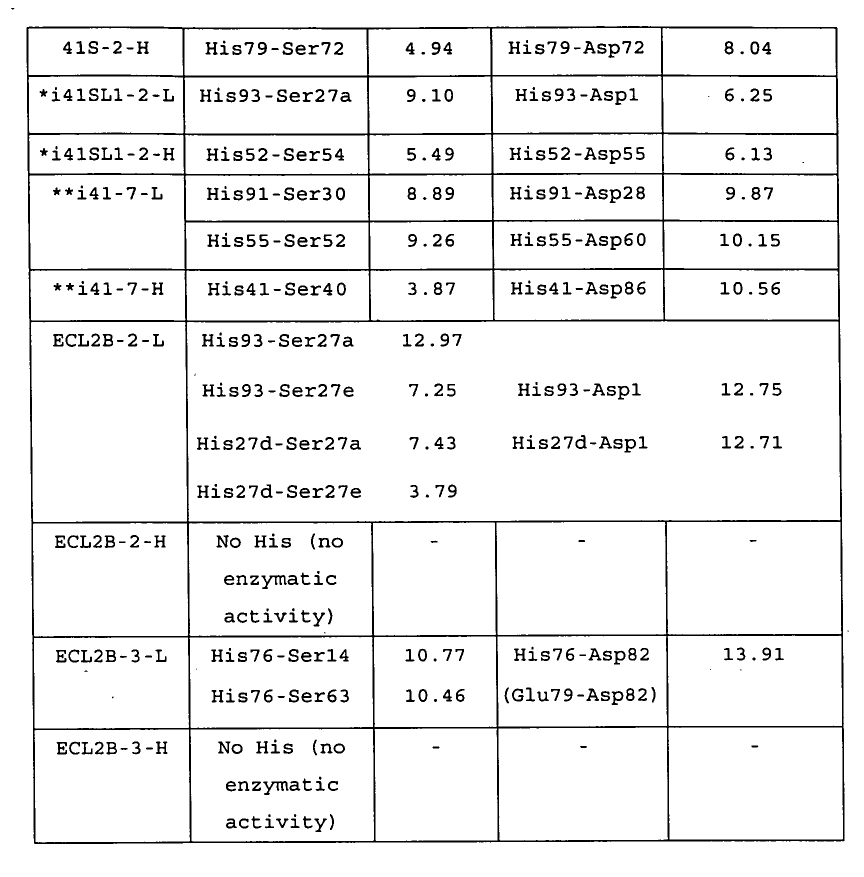
As shown in Table 7, distances between His(Cα) and
Ser(Cα) were comparable to that of enzymes (trypsin and
Thrombin) and natural antibody enzymes (41S-2-L, 41S-2-H,
i41SL1-2-L and i41-7-L). On the other hand, those of
41S-2-H, i41SL-2-H and i41-7-H were such small as
substantially 4 Å.
As to distances between α carbons of His and Asp, a
little different feature was exhibited. Trypsin, Thrombin,
i41SL1-2-L and i41SL1-2-H had substantially same distances,
and other than that had 2 to 5 Å longer distances.
Antibodysubunits such as 41S-2-L, 41S-2-H, i41SL1-2-H and
i41-7-H exhibited the enzymatic activity like the serine
protease. From this fact, ECL2B-2-L and ECL2B-3-L are
estimated to have the enzymatic activity like the serine
protease.
Subsequently, in order to confirm the enzymatic
activity of ECL2B-L and ECL2B-3, a peptide decomposition
experiment was carried out as follows.
Before decomposition experiments of an antigenic
peptide ECL2B due to monoclonal antibodies ECL2B-2, ECL2B-3,
almost all of glass and plastic instruments and buffering
solutions were sterilized by heating (at 180 degrees
centigrade and for 2 hr) or by treating with an autoclave
(121 degrees centigrade and for 20 min) or a 0.2 µm
sterilizing filter. Operations in the experiment were
carried out in a safety cabinet to inhibit foreign matters
in air from contaminating. The decomposition reactions
were conducted at a temperature of 25 degrees centigrade
and in a 7.5 mM phosphate buffer (pH 6.5). In order to
monitor the reaction, 20 µl of a reaction liquid was injected
in an RP-HPLC (manufactured by Jasco) under the conditions
of a temperature of 40 degrees centigrade and a constant
composition.
Firstly, with light chains of the monoclonal
antibodies ECL2B-2 and ECL2B-3 (ECL2B-2-L and ECL2B-3-L),
catalytic decomposition tests of an antigenic peptide
(ECL2B peptide: RSSHFPYSQYQFWKNFQTLKG) were carried out.
In a sterilized test tube, 500 µl of a purified
ECL2B-2-L (0.8 µM) or ECL2B-3-L (0.8 µM) -containing solution
was blended at 25 degrees centigrade with a same volume of
a solution containing 120 µM ECL2B peptide. A time course
of the ECL2B peptide was monitored by use of the RT-HPLC.
For the sake of reference, with peptide B instead of
ECL2B-peptide as a substrate, an experiment was carried out
similarly.
Results thereof are shown in Fig.38. In Fig.38, a
horizontal axis shows a reaction time (hr) and a vertical
axis shows a concentration (µM) of the ECL2B peptide that
is a substrate.
In the case of ECL2B-2-L being used (expressed with
in Fig.38), the decomposition of the ECL2B peptide began
with an induction period of substantially 80 hr after
ECL2B-2-L and ECL2B peptide were mixed and thereafter a
concentration of the ECL2B peptide rapidly decreased. This
kind of induction period is frequently found in many
reactions. The ECL2B peptide completely disappeared within
substantially 90 hr after the mixing.
In the case of ECL2B-3-L being used (expressed with
▴ in Fig.38), the decomposition of the ECL2B peptide began
with such a longer induction period as substantially 190
hr after ECL2B-3-L and ECL2B peptide were mixed. The ECL2B
peptide was rapidly degraded within substantially 250 hr
after the induction period. In the case of a similar
experiment being conducted with peptide B (expressed with
▪ in Fig.38), the peptide was not decomposed.
Subsequently, decomposition products obtained when
the ECL2B-2-L was used as an enzyme were analyzed with a
MALDI-TOF-MASS. As a result thereof, many kinds of peptide
fragments derived from the ECL2B peptide were confirmed.
At an initial stage of the decomposition, relatively long
peptides such as RSSHFPYSQYQFWKNFQ and SSHFPYSQYQFW were
observed. At the final stage of the decomposition (a stage
where the peptide shown in sequence No.40 disappears from
a reaction system), it was confirmed that many small peptides
such as RSSHFPYS, RSSHFPY, SSHFPYSQYQ, SSHFPYSQ,
HFPYSQYQFWKN, YSQYQFWKN and YSQYQ were generated. Since
the abovementioned many peptide fragments were generated,
it can be said that the ECL2B-2-L or ECL2B-3-L could
continuously decompose the antigenic peptide
RSSHFPYSQYQFWKNFQTLKG.
As already mentioned above, three residues that
constitute a catalytic triad residue in the ECL2B-2-L are
located at positions same as that of three catalytic triad
residues of VIPase and i41SL1-2-L. This consistency
suggests that the light chain of ECL2B-2 (ECL2B-2-L) has
capability of catalytically decomposing the antigenic
peptide (ECL2B peptide).
From results mentioned above, it was confirmed that
the ECL2B-2-L and ECL2B-3-L were antibody enzymes that can
completely decompose the peptide that constitutes an
extracellular region of the CCR-5.
Under the conditions same as that of the
abovementioned experiment, with heavy chains of monoclonal
antibodies ECL2B-2 and ECL2B-3 (ECL2B-2-H and ECL2B-3-H),
similar experiments were carried out. However, both these
heavy chains did not decompose the antigenic peptide ECL2B.
That is, in mass spectrometry, any peptide fragments could
not be detected.
Furthermore, a decomposition experiment of an
antigenic peptide with intact monoclonal antibody ECL2B-2
containing also a region other than a variable region was
carried out under the conditions same as that of the above
experiment. However, also in this case, the ECL2B peptide
that is a partial peptide of the CCR-5 was not at all
decomposed.
According to a method similar to experiment 3-1,
monoclonal antibody ECL2B-4 was prepared with a partial
peptide (sequence No.38) made of 20 amino acids that
constitute an extracellular region of the CCR-5 as an
immunogen.
Subsequently, according to a method similar to
experiment 3-2, the ECL2B-4 was purified. The antibody
ECL2B-4 purified in the experiment was identified and
quantitatively analyzed according to a method similar to
the experiment 3-3.
According to a method similar to that of the experiment
3-5, amino acid sequences of variable regions of light and
heavy chains of the ECL2B-4 and cDNA sequences that code
the amino acid sequences were determined. Thereafter, on
the basis of determined amino acid sequences, the molecular
modeling was carried out. Results thereof are shown below.
An amino acid sequence shown in sequence No.34 is an
amino acid sequence of a light chain of the ECL2B-4 and a
base sequence shown in sequence No.35 is a base sequence
of a cDNA that codes the amino acid sequence. An amino acid
sequence shown in sequence No.36 is an amino acid sequence
of a heavy chain of the ECL2B-4 and a base sequence shown
in sequence No.37 is a base sequence of a cDNA that codes
the amino acid sequence.
Furthermore, in Fig.41, an amino acid sequence of a
light chain of the ECL2B-4 and a base sequence of a cDNA
that code the amino acid sequence are shown together and
variable regions (CDR-1 through CDR-3) are shown. In Fig.42,
an amino acid sequence of a heavy chain of the ECL2B-4 and
a base sequence of a cDNA that code the amino acid sequence
are shown together and variable regions (CDR-1 through
CDR-3) are shown.
In the next place, based on the determined respective
amino acid sequences, the molecular modeling was carried
out. In Fig.39, a three-dimensional structure of a variable
region of the ECL2B-4-L estimated by means of the molecular
modeling is shown. Furthermore, in Fig.40, a
three-dimensional structure of a variable region of the
ECL2B-4-H estimated by means of the molecular modeling is
shown.
In the light chain of the ECL2B-4 (ECL2B-4-L), three
amino acid residues, S26 (or any one of S27a, S27e and S92),
H27d (or H93) and D1, were estimated to be able to form a
catalytic triad residue in a molecular structure (Fig.39).
Furthermore, in the heavy chain of the ECL2B-4
(ECL2B-4-H), three amino acid residues, S60 (or S84), E46
(or E85) and D59 (or D86), were estimated to be able to form
a catalytic triad residue in a molecular structure (Fig.40).
Subsequently, in order to confirm the enzymatic
activity of ECL2B-4-L and ECL2B-4-H, the peptide
decomposition experiments were carried out as follows.
According to a method similar to the experiment 3-6,
the enzymatic activity test was carried out of ECL2B-4-L
and ECL2B-4-H.
Firstly, the decomposition reaction of ECL2B peptide
(sequence No.38) was carried out in a 15 mM phosphate buffer
(pH 6.5) at 25 degrees centigrade. In order to monitor the
reaction, 20 µl of a reaction liquid was injected in the
RP-HPLC (manufactured by Jasco) under the conditions of a
column temperature of 40 degrees centigrade and a constant
composition.
As a reaction liquid of the decomposition reaction,
1 ○ through 5 ○ below were prepared.
Results thereof are shown in Figs. 43 through 45B. In
Fig.43, a horizontal axis expresses a reaction time (hr)
and a vertical axis expresses a concentration (µM) of ECL2B
peptide that is a substrate. Furthermore, in Figs.44A
through 44C, results obtained by monitoring with the HPLC
are shown. In Figs.44A through 44C, peaks of the ECL2B
peptide are shown with arrow marks. In addition, in
Figs.45A and 45B, results of the kinetic analysis in the
enzyme decomposition experiment are shown. Fig.45A is a
graph showing relationship between a substrate (ECL2B
peptide) concentration and a decomposition velocity, and
Fig.45B is a graph showing a result of Hanes-Woolf plot.
Then, Vmax, Km, kcat and kcat/Km values (average plot)
calculated by use of the Hanes-Woolf plot are shown in Table
8 below.
| Hanes-Woolf plot | |||
| Vmax (µM/min) | Km (M) | kcat (min-1) | kcat/Km (/M·min) |
| 0.31 | 3.5 × 10⁻⁵ | 0.72 | 2.2 × 10⁴ |
From the results, it was confirmed that the ECL2B-4-L
and ECL2B-4-H had the enzymatic activity in the
decomposition reaction of the ECL2B peptide. Accordingly,
it was confirmed that the ECL2B-4-L and ECL2B-4-H were
antibody enzymes that can target peptide that constitutes
an extracellular region of the CCR-5 and completely
decompose it.
Subsequently, the enzymatic activity test of the
ECL2B-4-L and ECL2B-4-H against TP41-1 peptide (sequence
No.42) was carried out. The decomposition reaction was
carried out in a 15 mM phosphate buffer (pH 6.5) at 25 degrees
centigrade. In order to monitor the reaction, 20 µl of a
reaction liquid was injected in the RP-HPLC (manufactured
by Jasco) under the conditions of a column temperature of
40 degrees centigrade and a constant composition.
As a reaction liquid of the decomposition reaction,
1 ○ through 5 ○ below were prepared.
Results thereof are shown in Figs.46 through 48B. In
Fig.46, a horizontal axis expresses a reaction time (hr)
and a vertical axis expresses a concentration (µM) of TP41-1
peptide that is a substrate. Furthermore, in Figs.47A
through 47C, results obtained by monitoring with the HPLC
are shown. Furthermore, in Figs.48A and 48B, results of
kinetic analysis of the enzyme decomposition test are shown.
In Figs.47A through 47C, peaks of the TP41-1 peptide are
shown with arrow marks. Fig.48A is a graph showing
relationship between a substrate (TP41-1 peptide)
concentration and a decomposition velocity, and Fig. 48B is
a graph showing a result of Hanes-Woolf plot.
Then, Vmax, Km, kcat and kcat/Km values (average plot)
calculated by use of the Hanes-Woolf plot are shown in Table
9 below.
| Hanes-Woolf plot | |||
| Vmax (µM/min) | Km (M) | kcat (min-1) | kcat/Km (/M·min) |
| 0.0624 | 1.25 × 10⁻⁵ | 0.16 | 1.25 × 10⁴ |
From the results, it was confirmed that the ECL2B-4-L
and ECL2B-4-H had low enzymatic activity also in the
decomposition reaction of the TP41-1 peptide. Furthermore,
as obvious from comparison of Tables 8 and 9, it was
experimentally shown that the ECL2B-4-L decomposed ECL2B
peptide that is an inherent antigen faster than the TP41-1
peptide, and it was verified that the ECL2B-4-L was assuredly
an antibody enzyme against the ECL2B.
Amino acid sequences of variable regions of antibodies
of which sequences were determined in the examples 1 through
3 and base sequences of cDNAs that code the amino acid
sequences are summarized and shown in table 10 below.
| Sequence Number | Kind of Sequence |
| 1 | An amino acid sequence of a variable region of I41SL1-2-H |
| 2 | A base sequence of cDNA that codes a variable region of I41SL1-2-H |
| 3 | An amino acid sequence of a variable region of I41SL-2-L |
| 4 | A base sequence of cDNA of a variable region of I41SL1-2-L |
| 5 | An amino acid sequence of a variable region of I41-7-H |
| 6 | A base sequence of cDNA that codes a variable region of I41-7-H |
| 7 | An amino acid sequence of a variable region of I41-7-L |
| 8 | Abase sequence of cDNA that codes a variable region of I41-7-L |
| 14 | An amino acid sequence of a variable region of HpU-18--L |
| 47 | A base sequence of cDNA that codes a variable region of HpU-18-L |
| 15 | An amino acid sequence of a variable region of HpU-9-L |
| 48 | A base sequence of cDNA that codes a variable region of HpU-9-L |
| 16 | An amino acid sequence of a variable region of HpU-2-H |
| 49 | Abase sequence of cDNA that codes a variable region of HpU-2-H |
| 17 | An amino acid sequence of a variable region of HpU-20-L |
| 18 | A base sequence of cDNA that codes a variable region of HpU-20-L |
| 19 | An amino acid sequence of a variable region of HpU-20-H |
| 20 | A base sequence of cDNA that codes a variable region of HpU-20-H |
| 21 | An amino acid sequence of a variable region of UA-15-L |
| 22 | A base sequence of cDNA that codes a variable region of UA-15-L |
| 23 | An amino acid sequence of a variable region of UA-15-H |
| 24 | A base sequence of cDNA that codes a variable region of UA-15-H |
| 26 | An amino acid sequence of a variable region of ECL2B-2-L |
| 27 | A base sequence of cDNA that codes a variable region of ECL2B-2-L |
| 28 | An amino acid sequence of a variable region of ECL2B-2-H |
| 29 | A base sequence of cDNA that codes a variable region of ECL2B-2-H |
| 30 | An amino acid sequence of a variable region of ECL2B-3-L |
| 31 | A base sequence of cDNA that codes a variable region of ECL2B-3-L |
| 32 | An amino acid sequence of a variable region of ECL2B-3-H |
| 33 | A base sequence of cDNA that codes a variable region of ECL2B-3-H |
| 34 | An amino acid sequence of a variable region of ECL2B-4-L |
| 35 | A base sequence of cDNA that codes a variable region of ECL2B-4-L |
| 36 | An amino acid sequence of a variable region of ECL2B-4-H |
| 37 | A base sequence of cDNA that codes a variable region of ECL2B-4-H |
Specific embodiments and examples exemplified in the
section of "Best Mode for Carrying Out the Invention" are
shown only for clarifying technical contents of the present
invention. The present invention should not be construed
by narrowly limiting to such specific examples and, within
a spirit of the invention and claims described below, can
be variously modified and carried out.
As mentioned above, antibody enzymes and genes
involving the present invention can be applied to treatment
and diagnosis of diseases such as various infectious
symptoms and cancers. Furthermore, when a target antibody
enzyme can be obtained, it can lead to a development of a
novel clinical diagnosis.
Furthermore, the antibody enzymes and genes involving
the invention can be applied to novel biosensors as new
biomaterials, and can be applied also to diagnosis of
diseases and inspections such as and environmental
measurements.
Still furthermore, when the antibody enzymes and genes
involving the present invention are used, it can be
considered to apply to develop effective synthesis means
and so on in the food industry and chemical industry.
Furthermore, according to a production method
according to the invention of antibody enzymes, antibody
enzymes having the enzymatic activity of cleaving or
decomposing polypeptides and antigen proteins selectively
among immunologically prepared antibodies can be
efficiently obtained.
Still furthermore, according to a production method
according to the invention of antibody enzymes, by use of
an existing genetic engineering process, antibody enzymes
having the enzymatic activity of readily cleaving or
decomposing polypeptides and antigen proteins can be
efficiently prepared.
Furthermore, according to antibody enzymes according
to the invention, HP bacteria urease can be specifically
decomposed to efficiently eliminate the HP bacteria. The
antibody enzyme is considered that, different from existing
bacteria eliminating agents, it does not impart the HP
bacteria the drug resistance; accordingly, it can be
effectively used as treatment drugs of HP bacteria
infectious patients. Still furthermore, the antibody
enzymes can, by orally taking, exhibit the effect of
eliminating the HP bacteria; accordingly, it can be used
as drugs that prevent the HP bacteria from infecting.
Furthermore, the antibody enzyme according to the
invention specifically works against chemokine receptor
CCR-5 that plays a very important role when the AIDS virus
infects with a human and can decompose to delete its
function; accordingly, the antibody enzyme is useful as an
anti-HIV drug for preventing the HIV from infecting and for
treating the AIDS. Antibody enzymes according to the
invention decompose the CCR-5 according to a novel mechanism
utterly different from existing anti-HIV drugs that
suppress the CCR-5 from working; accordingly, it can be
expected to effectively use in the treatment of the AIDS
against which at present an effective treatment is not found.


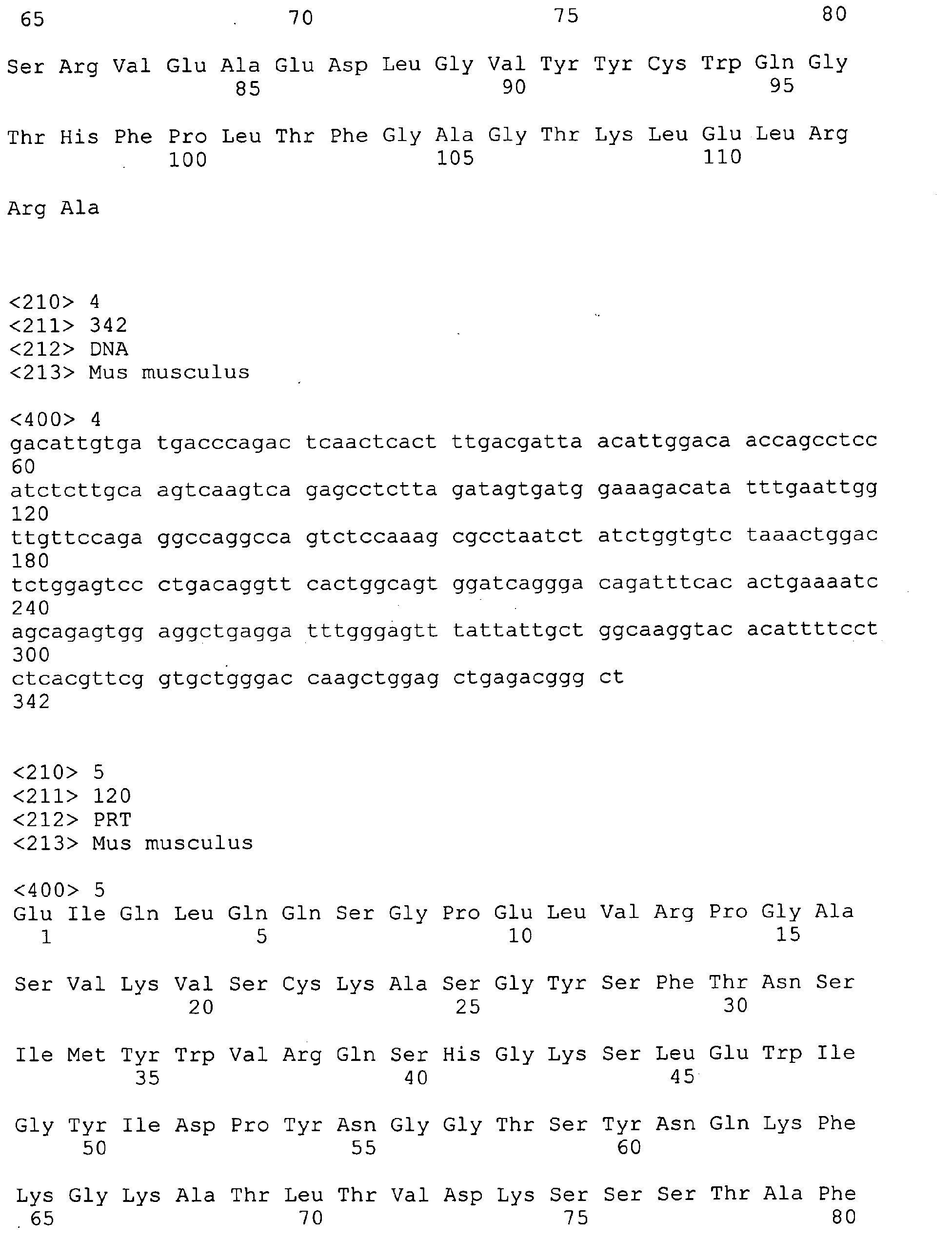
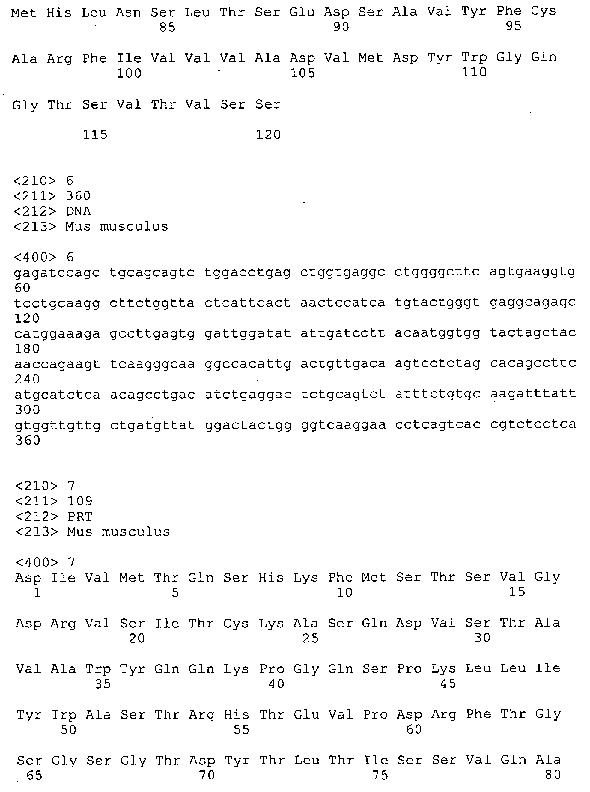


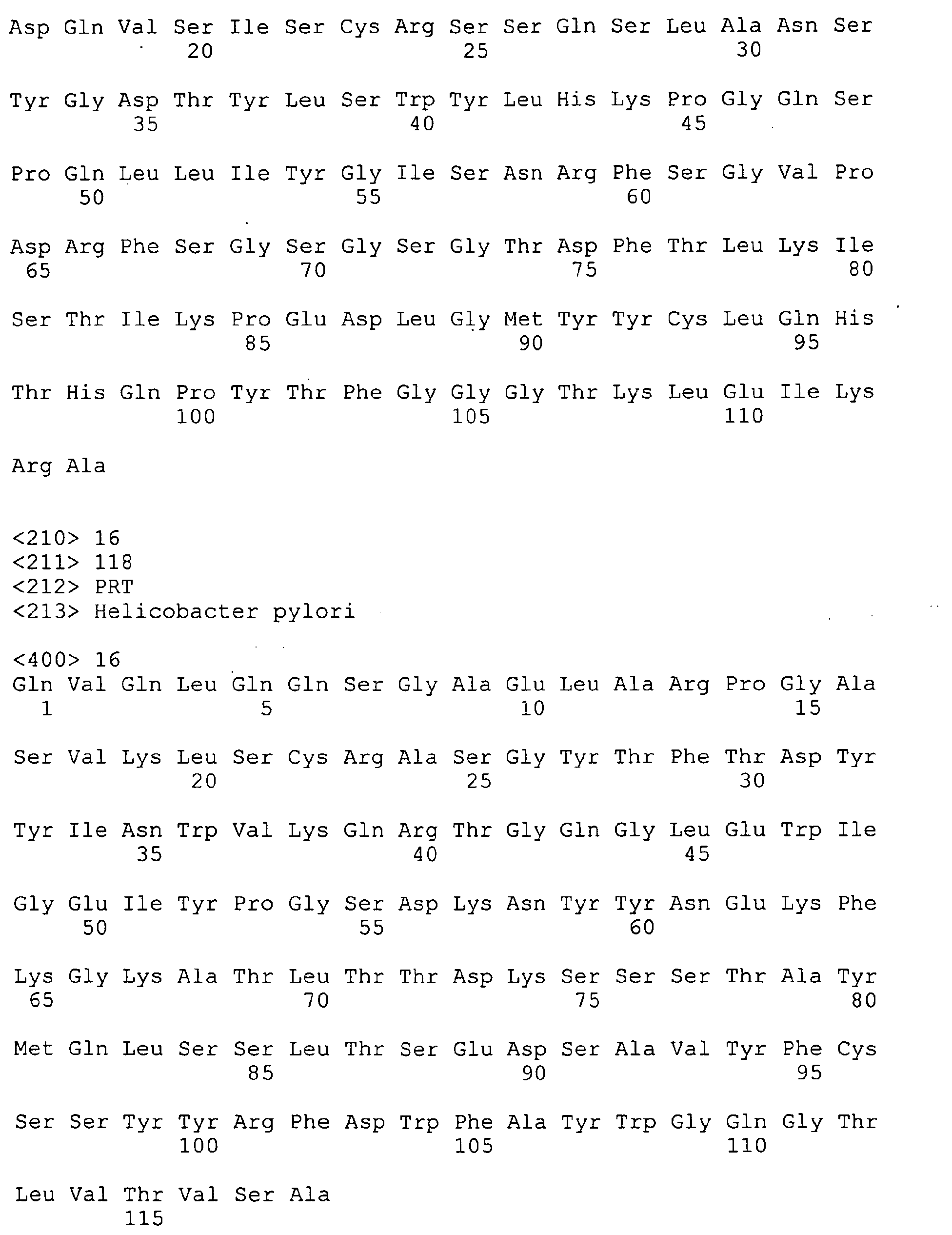
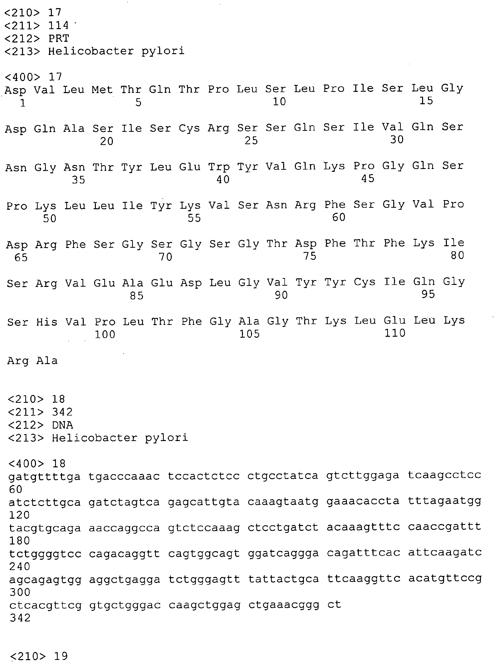

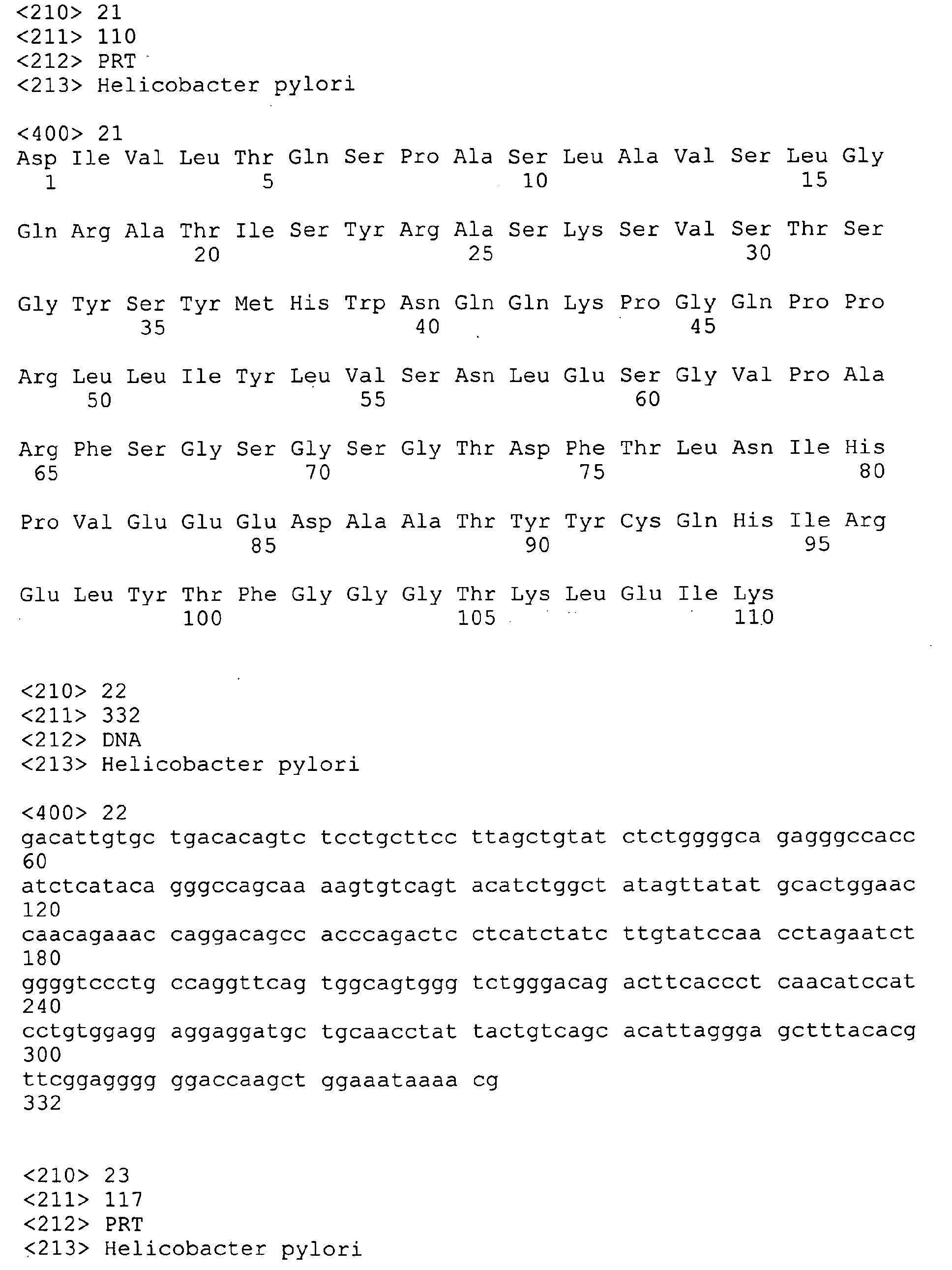

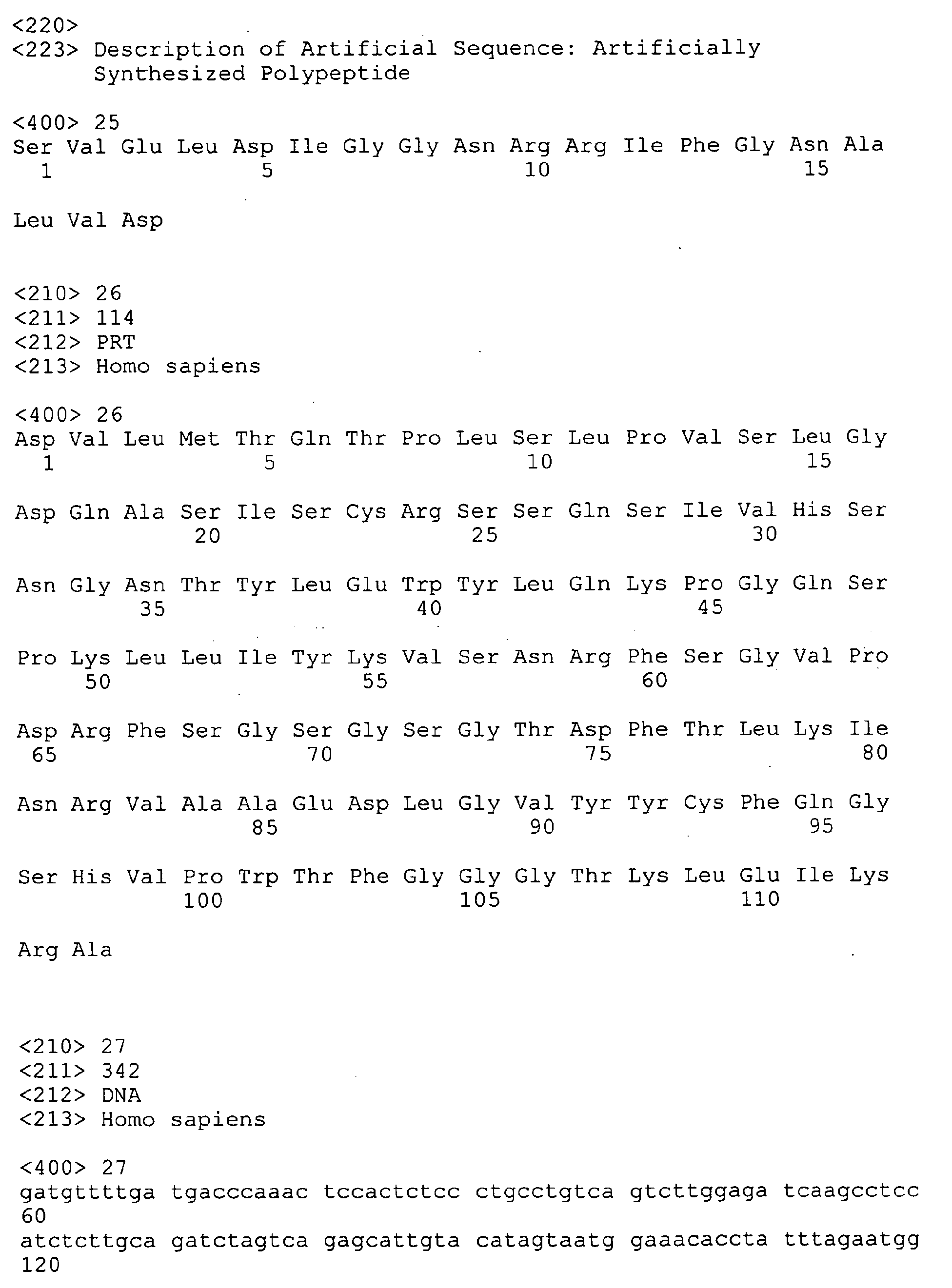
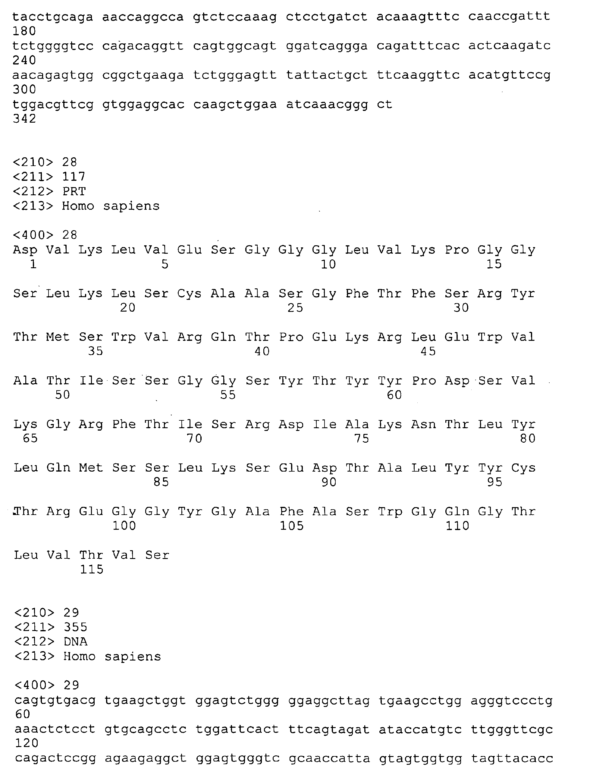

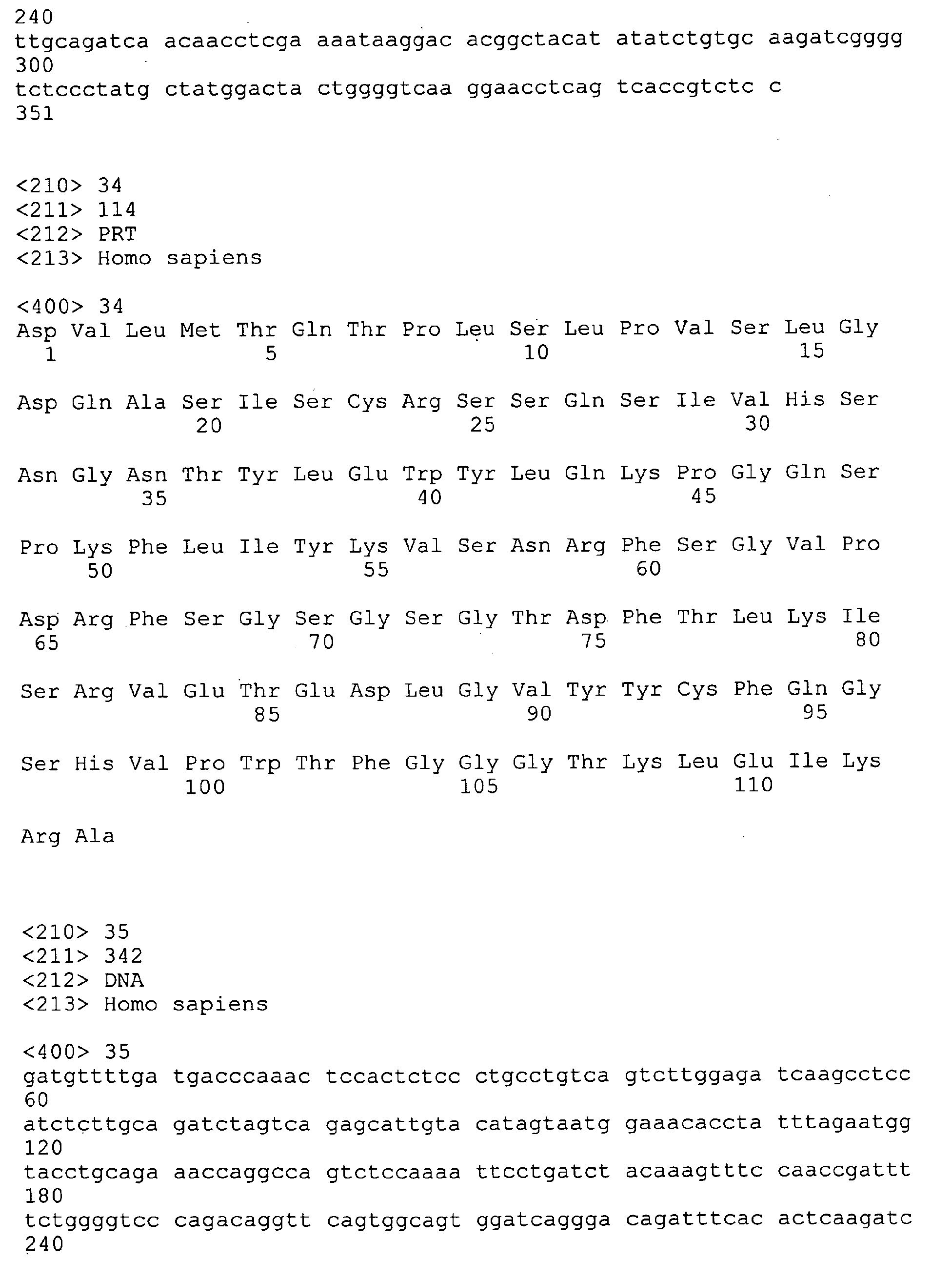
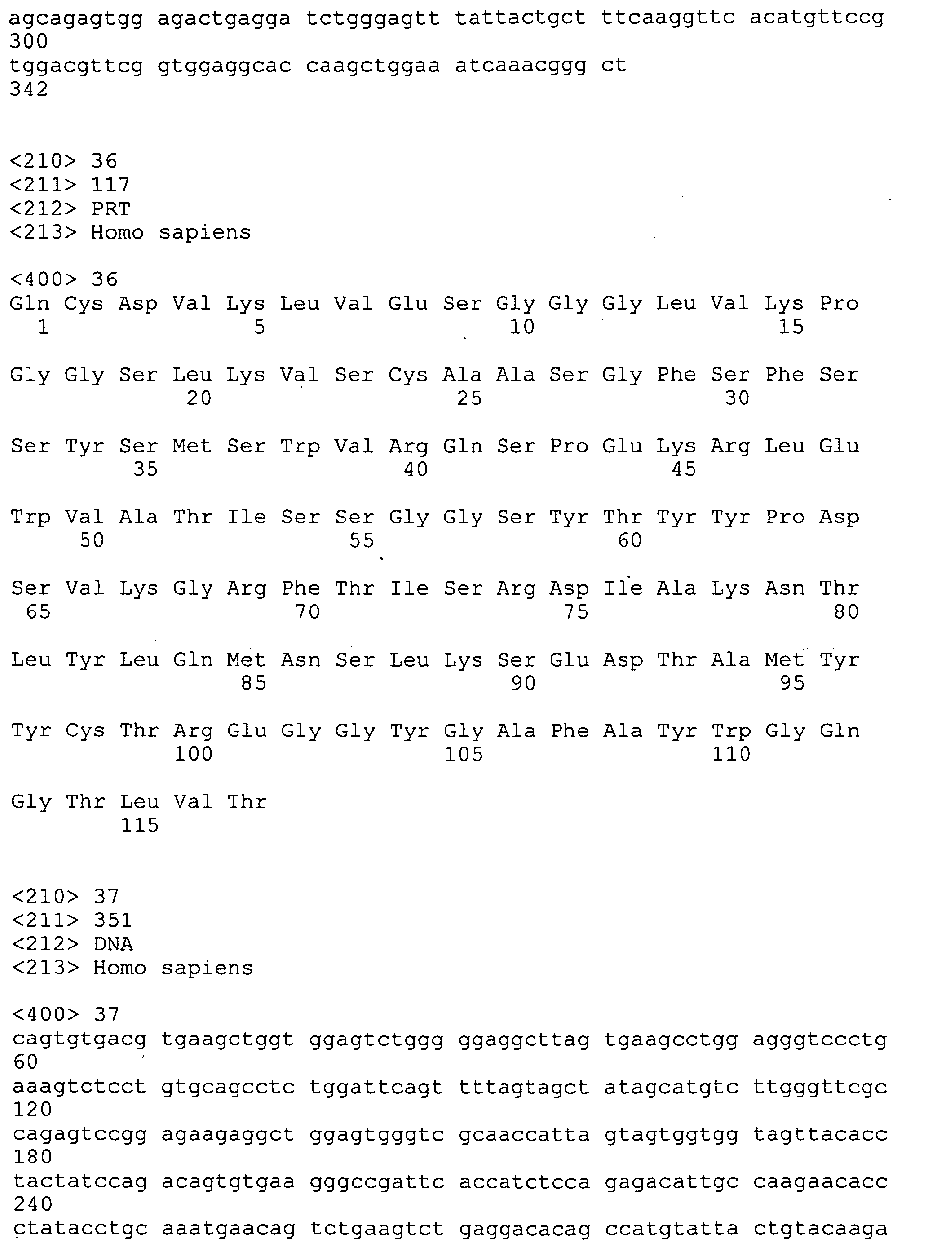


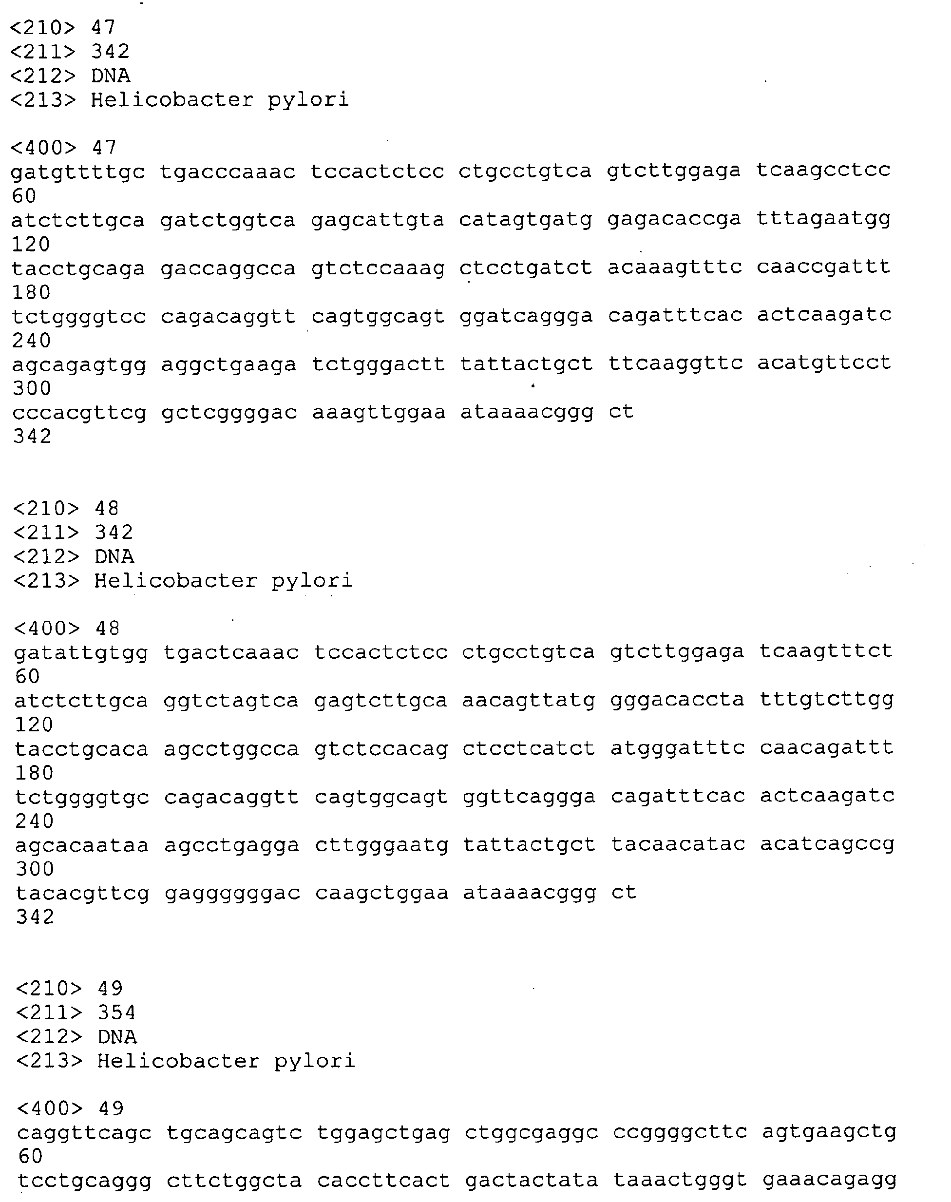
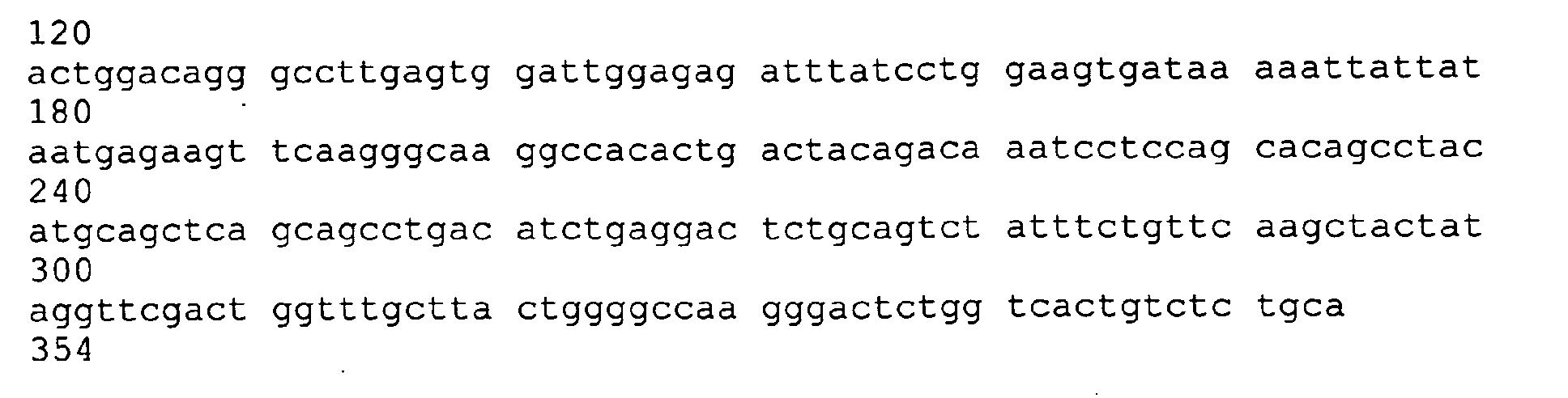
Claims (53)
- An antibody enzyme production method, comprising:an antibody structure analysis process, wherein the antibody structure analysis process includes performing a stereostructure estimation step of estimating a stereostructure of an antibody from an amino acid sequence and a catalytic triad residue structure confirmation step where whether a catalytic triad residue structure in which a serine residue, an aspartate residue, and a histidine residue or glutamate residue are present in stereostructurally close proximity is present or not in an estimated stereostructure of the antibody is confirmed.
- The antibody enzyme production method according to claim 1,
wherein in the catalytic triad residue structure confirmation step, in order to confirm a catalytic triad residue structure, that a serine residue, an aspartate residue and a histidine residue or glutamate residue are present within the range of 3 to 20 Å in a stereostructure is used as an index. - The antibody enzyme production method according to claim 1 or 2,
wherein in the catalytic triad residue structure confirmation step, in order to confirm a catalytic triad residue structure, either having a structure same as a catalytic triad residue structure common to existing antibody enzymes or having a structure that is estimated to form a catalytic triad residue structure by using by one amino acid residue in a different position from that of the existing catalytic triad residue structure is used as an index. - The antibody enzyme production method according to any one of claims 1, 2, and 3,
wherein in the catalytic triad residue structure confirmation step, in order to confirm a catalytic triad residue structure, that an antibody has a germ cell gene type from which a known antibody enzyme is derived is used as an index. - The antibody enzyme production method according to any one of claims 1, 2, 3, and 4,
wherein in the catalytic triad residue structure confirmation step, in order to confirm a catalytic triad residue structure, that a complimentarity determining region 1 (CDR1) is constituted of 16 amino acid residues and a histidine residue is at the 93rd according to Kabat numbering scheme is used as an index. - The antibody enzyme production method according to any one of claims 1, 2, 3 and 4,
wherein in the catalytic triad residue structure confirmation step, in order to confirm a catalytic triad residue structure, that a complimentarity determining region 1 (CDR1) is constituted of 11 amino acid residues and a histidine residue is at the 91st or 55th according to Kabat numbering scheme is used as an index. - The antibody enzyme production method according to any one of claims 1, 2, 3, 4, 5 and 6, further comprising:an antibody production process where an antibody is produced from a hybridoma obtained by fusing a mouse spleen lymph corpuscle immunized with an antigen and a mouse myeloma cell.
- The antibody enzyme production method according to claim 7,
wherein, as the antigen, an antigen protein obtained by coupling a substance that becomes an antigen determinant with a carrier protein is used. - The antibody enzyme production method according to any one of claims 1, 2, 3, 4, 5, 6, 7 and 8, further comprising:a catalytic triad residue structure introducing process where by use of stereostructure information obtained after the antibody structure analysis process, according to a genetic engineering process, a catalytic triad residue structure is introduced in an antibody.
- Antibody enzyme, comprising:in a stereostructure, a catalytic triad residue structure in which a serine residue, an aspartate residue and a histidine residue or glutamate residue are stereostructurally in proximity.
- An antibody enzyme,
wherein a complimentarity determining region 1 (CDR1) is constituted of 16 amino acid residues and a histidine residue is at the 93rd by Kabat numbering scheme. - An antibody enzyme,
wherein a complimentarity determining region 1 (CDR1) is constituted of 11 amino acid residues and a histidine residue is at the 91st or 55th by Kabat numbering scheme. - Antibody enzyme,
wherein the antibody enzyme is produced from a mouse germ cell gene selected from, by Thiebe et al. convention, bb1, bl1, bd2, cr1, cs1, bj2, hf24, 12-41, 19-14, 19-17, 19-23, 19-25, and 21-12. - The antibody enzyme according to any one of claims 10, 11, 12 and 13,
wherein the antibody enzyme is a heavy chain or a light chain of an antibody. - The antibody enzyme according to claim 14, comprising:an amino acid sequence shown in sequence No.1 or an amino acid sequence in which in the amino acid sequence shown in sequence No.1, at least one amino acid is replaced, deleted, inserted and/or added.
- The antibody enzyme according to claim 14, comprising:an amino acid sequence shown in sequence No.3 or an amino acid sequence in which in the amino acid sequence shown in sequence No.3, at least one amino acid is replaced, deleted, inserted and/or added.
- The antibody enzyme according to claim 14, comprising:an amino acid sequence shown in sequence No.5 or an amino acid sequence in which in the amino acid sequence shown in sequence No.5, at least one amino acid is replaced, deleted, inserted and/or added.
- The antibody enzyme according to claim 14, comprising:an amino acid sequence shown in sequence No.7 or an amino acid sequence in which in the amino acid sequence shown in sequence No.7, at least one amino acid is replaced, deleted, inserted and/or added.
- A gene that codes an antibody enzyme according to any one of claims 10, 11, 12, 13, 14, 15, 16, 17 and 18.
- The antibody enzyme according to claim 14 that is an antibody of Helicobacter Pylori bacterium urease or an antibody fragment thereof and works as a decomposing enzyme of the urease.
- The antibody enzyme according to claim 20,
wherein the antibody enzyme includes a variable region of an antibody of the urease. - The antibody enzyme according to claim 20 or 21, comprising an amino acid sequence shown in sequence No.14 or an amino acid sequence in which in the amino acid sequence shown in sequence No.14, at least one amino acid is replaced, deleted, inserted and/or added.
- The antibody enzyme according to claim 20 or 21, comprising an amino acid sequence shown in sequence No.15 or an amino acid sequence in which in the amino acid sequence shown in sequence No.15, at least one amino acid is replaced, deleted, inserted and/or added.
- The antibody enzyme according to claim 20 or 21, comprising an amino acid sequence shown in sequence No.16 or an amino acid sequence in which in the amino acid sequence shown in sequence No. 16, at least one amino acid is replaced, deleted, inserted and/or added.
- The antibody enzyme according to claim 20 or 21, comprising an amino acid sequence shown in sequence No.17 or an amino acid sequence in which in the amino acid sequence shown in sequence No.17, at least one amino acid is replaced, deleted, inserted and/or added.
- The antibody enzyme according to claim 20 or 21, comprising an amino acid sequence shown in sequence No.19 or an amino acid sequence in which in the amino acid sequence shown in sequence No.19, at least one amino acid is replaced, deleted, inserted and/or added.
- The antibody enzyme according to claim 20 or 21, comprising an amino acid sequence shown in sequence No.21 or an amino acid sequence in which in the amino acid sequence shown in sequence No.21, at least one amino acid is replaced, deleted, inserted and/or added.
- A curative drug to a Helicobacter Pylori bacteria infected patient, comprising the antibody enzyme according to any one of claims 20, 21, 22, 23, 24, 25, 26 and 27.
- An infection control agent of Helicobacter Pylori bacteria, comprising the antibody enzyme according to any one of claims 20, 21, 22, 23, 24, 25, 26 and 27.
- A gene that codes the antibody enzyme according to any one of claims 20, 21, 22, 23, 24, 25, 26 and 27.
- The gene according to claim 30, comprising a base sequence shown in sequence No.18.
- The gene according to claim 30, comprising a base sequence shown in sequence No.20.
- The gene according to claim 30, comprising a base sequence shown in sequence No.22.
- A transformant, wherein the gene according to any one of claims 30, 31, 32 and 33 is introduced.
- The transformant according to claim 34, wherein the gene is introduced in a plant to express.
- An infection control agent of Helicobacter Pylori bacteria, comprising the transformant according to claim 34 or 35.
- The antibody enzyme according to claim 14,
wherein the antibody enzyme is an antibody of human-derived chemokine receptor CCR-5 or an antibody fragment thereof and decomposes the chemokine receptor CCR-5. - The antibody enzyme according to claim 37,
wherein the antibody enzyme includes a variable region of the chemokine receptor CCR-5 antibody. - The antibody enzyme according to claim 37 or 38,
wherein the antibody enzyme includes an amino acid sequence shown in sequence No.26 or an amino acid sequence in which in the amino acid sequence shown in sequence No.26, at least one amino acid is replaced, deleted, inserted and/or added. - The antibody enzyme according to claim 37 or 38,
wherein the antibody enzyme includes an amino acid sequence shown in sequence No.30 or an amino acid sequence in which in the amino acid sequence shown in sequence No.30, at least one amino acid is replaced, deleted, inserted and/or added. - The antibody enzyme according to claim 37 or 38,
wherein the antibody enzyme includes an amino acid sequence shown in sequence No.34 or an amino acid sequence in which in the amino acid sequence shown in sequence No. 34, at least one amino acid is replaced, deleted, inserted and/or added. - The antibody enzyme according to claim 37 or 38,
wherein the antibody enzyme includes an amino acid sequence shown in sequence No.36 or an amino acid sequence in which in the amino acid sequence shown in sequence No. 36, at least one amino acid is replaced, deleted, inserted and/or added. - An anti-HIV drug that includes the antibody enzyme according to any one of claims 37, 38, 39, 40, 41 and 42 and inhibits AIDS virus from infecting.
- An anti-HIV drug to an AIDS virus-infected patient, comprising the antibody enzyme according to any one of claims 37, 38, 39, 40, 41 and 42.
- A gene that codes the antibody enzyme according to any one of claims 37, 38, 39, 40, 41 and 42.
- The gene according to claim 45, comprising a base sequence shown in sequence No.27.
- The gene according to claim 45, comprising a base sequence shown in sequence No.31.
- The gene according to claim 45, comprising a base sequence shown in sequence No.35.
- The gene according to claim 45, comprising a base sequence shown in sequence No.37.
- A transformant, wherein the gene according to any one of claims 45, 46, 47, 48 and 49 is introduced.
- An anti-HIV drug, comprising the transformant according to claim 50.
- A computer program that lets a computer carry out the antibody structural analysis process according to any one of claims 1, 2, 3, 4, 5 and 6.
- A machine-readable recording medium, wherein a computer program that lets a computer carry out a program that carries out the antibody structural analysis process according to claims 1, 2, 3, 4, 5 and 6 is recorded.
Applications Claiming Priority (13)
| Application Number | Priority Date | Filing Date | Title |
|---|---|---|---|
| JP2002211768 | 2002-07-19 | ||
| JP2002211768 | 2002-07-19 | ||
| JP2002211756 | 2002-07-19 | ||
| JP2002211756 | 2002-07-19 | ||
| JP2003051943 | 2003-02-27 | ||
| JP2003051943 | 2003-02-27 | ||
| JP2003198270 | 2003-07-17 | ||
| JP2003198292 | 2003-07-17 | ||
| JP2003198281 | 2003-07-17 | ||
| JP2003198281A JP4330947B2 (en) | 2002-07-19 | 2003-07-17 | Antibody enzyme against urease of Helicobacter pylori, gene encoding it, transformant introduced with the gene, and therapeutic and infection preventive agent for patients infected with Helicobacter pylori using the enzyme |
| JP2003198270A JP4334931B2 (en) | 2002-07-19 | 2003-07-17 | Novel antibody enzyme production method and novel antibody enzyme |
| JP2003198292A JP4330948B2 (en) | 2003-02-27 | 2003-07-17 | Antibody enzyme against chemokine receptor CCR-5, gene encoding the same, transformant introduced with the gene, and anti-HIV drug using the same |
| PCT/JP2003/009147 WO2004009805A1 (en) | 2002-07-19 | 2003-07-18 | Novel process for producing antibody enzyme, novel antibody enzyme and utilization thereof |
Publications (2)
| Publication Number | Publication Date |
|---|---|
| EP1544291A1 true EP1544291A1 (en) | 2005-06-22 |
| EP1544291A4 EP1544291A4 (en) | 2006-08-02 |
Family
ID=30773766
Family Applications (1)
| Application Number | Title | Priority Date | Filing Date |
|---|---|---|---|
| EP03765329A Withdrawn EP1544291A4 (en) | 2002-07-19 | 2003-07-18 | Novel process for producing antibody enzyme, novel antibody enzyme and utilization thereof |
Country Status (3)
| Country | Link |
|---|---|
| US (1) | US20060030015A1 (en) |
| EP (1) | EP1544291A4 (en) |
| WO (1) | WO2004009805A1 (en) |
Families Citing this family (13)
| Publication number | Priority date | Publication date | Assignee | Title |
|---|---|---|---|---|
| US9862770B2 (en) | 2005-10-19 | 2018-01-09 | Ibc Pharmaceuticals, Inc. | Multivalent antibody complexes targeting IGF-1R show potent toxicity against solid tumors |
| US8883162B2 (en) | 2005-10-19 | 2014-11-11 | Ibc Pharmaceuticals, Inc. | Multivalent antibody complexes targeting IGF-1R show potent toxicity against solid tumors |
| US20100226884A1 (en) | 2009-01-20 | 2010-09-09 | Immunomedics, Inc. | Novel Class of Monospecific and Bispecific Humanized Antibodies that Target the Insulin-like Growth Factor Type I Receptor (IGF-1R) |
| JP4861019B2 (en) * | 2006-01-31 | 2012-01-25 | 独立行政法人科学技術振興機構 | Antibody enzyme against human TNF-α and use thereof |
| WO2008035894A1 (en) | 2006-09-19 | 2008-03-27 | Sungkyunkwan University Foundation For Corporate Collaboration | Antiviral agent against animal viruses |
| US8168757B2 (en) * | 2008-03-12 | 2012-05-01 | Merck Sharp & Dohme Corp. | PD-1 binding proteins |
| WO2010002862A2 (en) * | 2008-07-01 | 2010-01-07 | Aveo Pharmaceuticals, Inc. | Fibroblast growth factor receptor 3 (fgfr3) binding proteins |
| IN2012DN05101A (en) * | 2010-01-19 | 2015-10-23 | Immunomedics Inc | |
| EP2894222B8 (en) | 2010-02-19 | 2019-04-24 | Japan Science and Technology Agency | Human abzyme |
| PL3489261T3 (en) | 2012-08-24 | 2021-08-16 | The Regents Of The University Of California | Antibodies and vaccines for use in the treatment of ROR1 expressing tumors and in the inhibition of metastasis |
| KR20230047507A (en) | 2016-06-27 | 2023-04-07 | 더 리젠츠 오브 더 유니버시티 오브 캘리포니아 | Cancer treatment combinations |
| EP3559045A4 (en) * | 2016-12-23 | 2020-08-19 | REMD Biotherapeutics, Inc. | Immunotherapy using antibodies that bind programmed death ligand-1 (pd-l1) |
| EP3807644A4 (en) * | 2018-06-12 | 2022-07-06 | Angiex, Inc. | ANTIBODY-OLIGONUCLEOTIDE CONJUGATES |
Family Cites Families (1)
| Publication number | Priority date | Publication date | Assignee | Title |
|---|---|---|---|---|
| US5919681A (en) * | 1995-06-07 | 1999-07-06 | Wohlstadter Jacob N | Method for promoting enzyme diversity |
-
2003
- 2003-07-18 EP EP03765329A patent/EP1544291A4/en not_active Withdrawn
- 2003-07-18 WO PCT/JP2003/009147 patent/WO2004009805A1/en not_active Ceased
-
2005
- 2005-01-19 US US11/037,199 patent/US20060030015A1/en not_active Abandoned
Also Published As
| Publication number | Publication date |
|---|---|
| WO2004009805A1 (en) | 2004-01-29 |
| US20060030015A1 (en) | 2006-02-09 |
| EP1544291A4 (en) | 2006-08-02 |
Similar Documents
| Publication | Publication Date | Title |
|---|---|---|
| AU776464B2 (en) | Monoclonal antibodies, antigens and diagnosis and therapy of malignant diseases | |
| EP2756077B1 (en) | Endoglycosidase from streptococcus pyogenes and methods using it | |
| US20060030015A1 (en) | Novel process for producing antibody enzyme, novel antibody enzyme and utilization thereof | |
| JP2008118996A (en) | Cloning and characterization of napsin, a type of aspartic protease. | |
| EP1891218A2 (en) | Novel nucleotide and amino acid sequences, and assays and methods of use thereof for diagnosis | |
| CA2782481A1 (en) | Co-crystal structure of factor d and anti-factor d antibody | |
| CN115215939B (en) | A fusion protein and its preparation method and application | |
| CN116589564B (en) | anti-AAV5 antibody and ELISA kit for rapid AAV5 titer determination | |
| Pérez de la Lastra et al. | Antibodies targeting enzyme inhibition as potential tools for research and drug development | |
| CN121522173B (en) | Detection method for detecting A-type botulinum toxin HA-70 and product | |
| JPH10501813A (en) | Compositions and methods for treating tumors | |
| CN113501874B (en) | Anti-human CD47 protein antibody, detection kit and application thereof | |
| CN119147767B (en) | Detection kit and detection method for botulinum neurotoxin type B | |
| CN109879963A (en) | A kind of monoclonal antibody and its preparation method and application for inhibiting histoorgan fibrosis and new vessels to be formed | |
| JP3307422B2 (en) | Immunological measurement method of human PIVKA-II | |
| EP0594879A1 (en) | Cathepsin L-specific monoclonal antibodies, hybridomas for producing the same and a method of using the same | |
| Hifumi et al. | Catalytic digestion of human tumor necrosis factor‐α by antibody heavy chain | |
| JP4334931B2 (en) | Novel antibody enzyme production method and novel antibody enzyme | |
| DK171836B1 (en) | Monoclonal antibody to protein C, hybridoma producing the antibody, method for measuring human protein C with a Gla region and method for recovering human protein C with a Gla region | |
| Murashige | Section 102/103 Issues in Biotechnology Patent Prosecution | |
| AU2004251161A1 (en) | Polypeptides that bind an anti-tissue factor antibody and uses thereof | |
| JPH10513484A (en) | Human cardiac CNBr troponin I isoforms and uses thereof | |
| WO2023284012A1 (en) | Monoclonal antibody for human activated protein c, and preparation method therefor and use thereof | |
| CN121319202B (en) | ATP7B antibody and its application | |
| JP2010011840A (en) | Method for producing antibody enzyme |
Legal Events
| Date | Code | Title | Description |
|---|---|---|---|
| PUAI | Public reference made under article 153(3) epc to a published international application that has entered the european phase |
Free format text: ORIGINAL CODE: 0009012 |
|
| 17P | Request for examination filed |
Effective date: 20050218 |
|
| AK | Designated contracting states |
Kind code of ref document: A1 Designated state(s): AT BE BG CH CY CZ DE DK EE ES FI FR GB GR HU IE IT LI LU MC NL PT RO SE SI SK TR |
|
| RAP1 | Party data changed (applicant data changed or rights of an application transferred) |
Owner name: TOWA KAGAKU CO., LTD. |
|
| A4 | Supplementary search report drawn up and despatched |
Effective date: 20060705 |
|
| 17Q | First examination report despatched |
Effective date: 20061122 |
|
| STAA | Information on the status of an ep patent application or granted ep patent |
Free format text: STATUS: THE APPLICATION IS DEEMED TO BE WITHDRAWN |
|
| 18D | Application deemed to be withdrawn |
Effective date: 20080327 |