EP1543474B1 - Segmentierung einer serie von 2d- und 3d-bildern - Google Patents
Segmentierung einer serie von 2d- und 3d-bildern Download PDFInfo
- Publication number
- EP1543474B1 EP1543474B1 EP03797432.6A EP03797432A EP1543474B1 EP 1543474 B1 EP1543474 B1 EP 1543474B1 EP 03797432 A EP03797432 A EP 03797432A EP 1543474 B1 EP1543474 B1 EP 1543474B1
- Authority
- EP
- European Patent Office
- Prior art keywords
- images
- series
- segmentation
- image
- organ
- Prior art date
- Legal status (The legal status is an assumption and is not a legal conclusion. Google has not performed a legal analysis and makes no representation as to the accuracy of the status listed.)
- Expired - Lifetime
Links
Images

Classifications
-
- G—PHYSICS
- G06—COMPUTING OR CALCULATING; COUNTING
- G06T—IMAGE DATA PROCESSING OR GENERATION, IN GENERAL
- G06T7/00—Image analysis
- G06T7/10—Segmentation; Edge detection
- G06T7/11—Region-based segmentation
-
- G—PHYSICS
- G06—COMPUTING OR CALCULATING; COUNTING
- G06T—IMAGE DATA PROCESSING OR GENERATION, IN GENERAL
- G06T2207/00—Indexing scheme for image analysis or image enhancement
- G06T2207/10—Image acquisition modality
- G06T2207/10072—Tomographic images
-
- G—PHYSICS
- G06—COMPUTING OR CALCULATING; COUNTING
- G06T—IMAGE DATA PROCESSING OR GENERATION, IN GENERAL
- G06T2207/00—Indexing scheme for image analysis or image enhancement
- G06T2207/30—Subject of image; Context of image processing
- G06T2207/30004—Biomedical image processing
- G06T2207/30061—Lung
Definitions
- the invention relates to a method and to an apparatus having means for segmenting a series of 2D or 3D images obtained of a patient's organ or other body part, as defined by the appended claims.
- Such a method and apparatus is known from US-A-5,903,664 , showing a cardiac segmentation system acquiring a series of images as slices through a volume, and as images at different time periods throughout a cardiac cycle.
- a first image of a series of images is segmented by making use of a threshold.
- the centroid of this image is used as a seed point in segmenting adjacent images. This is repeated for a number of images in order that all images are accordingly segmented.
- any type of segmentation can be applied initially as the first segmentation on the first image.
- This segmentation can be carried out either manually or (semi) automatically by means and methods that are known per se in the art.
- the result of this segmentation which can be moulded according to the needs of the user, is according to the invention simply and quickly propagated to the other images of the series of images.
- each transformation relates to adjacent or immediately successive images of the series of images.
- the differences between adjacent or immediately successive images of said series of images are rather limited allowing that the accuracy requirements of the transformation remain fairly limited.
- An important benefit of the invention lies in that it proves particularly useful when there are two or more series of images, whereby the segmentation applied to the first series of images can also be applied to the other series of images. This allows for ease of comparison among the said series.
- the method according to the invention applies to all types of organs and other patient's body parts.
- the apparatus, software and method according to the invention are preferably characterized in that prior to establishing the said series of transformations, the series of images are converted to a modified series of images showing the walls of the organ in a flat plane wherein the left and right part of said plane substantially correspond to the inside and outside of said organ, and that the said series of transformations are applied to the modified series of images.
- the invention applies to processing of both 2D and 3D images.
- medical diagnosis, therapy planning and monitoring of the effect of therapy it is often required to accurately segment various anatomical structures that are present in medical images of the patient. Medical image segmentation has therefore received considerable attention during the last few decades.
- the invention proposes an apparatus and method which make it easy to apply the selected segmentation to all images in a series of images or to several series of images, whereby it is only required to apply an initial segmentation to a selected first image of the series of images.
- An image 1 undergoes a segmentation in box 2 resulting in a desired segmented image 3.
- the said first image 1 and consecutively further images i, whereby i may range from 2 to n, are supplied to a box 4 in which a transformation T 1,i is calculated in order to arrive at a best fit of image 1 and image i.
- This transform T 1,i is supplied to a box 5, which also receives the initial segmented image 3 and which converts these both information flows into a segmented image 6 corresponding to the original image i.
- the apparatus of the invention repeats this process for every i in the range 2 to n, so that in relation to a region of interest of the concerning images, a series of transformations are established, wherein each separate transformation embodies a best fit between two images of said series of images, and wherein each image of the series of images is subject of such a transformation.
- Fig. 1 relates to each individual transformation among images such that always image 1 forms part thereof, it is also possible that each individual transformation relates to adjacent or - in other words - immediately successive images of the series of images.
- the invention is also applicable when there are two or more series of images whereby the segmentation of the first series of images (which is based on the segmentation of the first image from this first series of images) applies to all series of images.
- the respective series of images are collected with different means of monitoring the patient, which means are selected from the group MR, CT, NM and US.
- the respective series of images may also be collected at different times.
- the images may relate to a substantially sphere-like organ such as a heart.
- a conversion of the image of the heart shown at the left-hand part takes place to a modified image as shown in the right-hand part of Fig. 2 .
- This type of transformation is known as a resample operation and results in a showing of the walls of the concerning organ in a flat plane, wherein the left- and right-hand part of said plane substantially correspond to the inside and outside of the concerning organ.
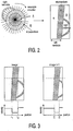
- the ease of working of the apparatus and method of the invention is highly supported by this prior operation allowing that the transformation can take place to the modified image which is simpler to operate. An example of this is shown with reference to Fig. 3 .
- Fig. 3 shows at the left-hand part an image i and on the right-hand part a subsequent image i+1.
- the region of interest of these images is the so-called myocardium of the left ventricle of the heart, the position of which is indicated in the left-hand Figure by r 1 -r 2 .
- this position has changed to r 1 - r 2 .
- the change in position from r 1 -r 2 to r 1 - r 2 represents the transformation that fits image i to image i+1 and which is used for applying the segmentation of image i to image i+1.
Landscapes
- Engineering & Computer Science (AREA)
- Computer Vision & Pattern Recognition (AREA)
- Physics & Mathematics (AREA)
- General Physics & Mathematics (AREA)
- Theoretical Computer Science (AREA)
- Apparatus For Radiation Diagnosis (AREA)
- Measuring And Recording Apparatus For Diagnosis (AREA)
- Magnetic Resonance Imaging Apparatus (AREA)
- Processing Or Creating Images (AREA)
- Image Processing (AREA)
Claims (13)
- Vorrichtung mit Mitteln zum Segmentieren einer Reihe von 2D- oder 3D-Bildern, die von einem Organ oder einem anderen Körperteil eines Patienten erhalten werden, wobei eine erste Segmentierung an einem ersten Bild der Reihe von Bildern durchgeführt wird und wobei die erste Segmentierung für die anschließende Segmentierung der restlichen Bilder der Reihe von Bildern verwendet wird, dadurch gekennzeichnet, dass die Mittel in Bezug auf die Bilder eine Berechnung einer Reihe von Umwandlungen durchführen, wobei jede separate Umwandlung, die berechnet wird, einen Anpassungsvorgang zwischen zwei Bildern der Reihe von Bildern darstellt, was zu einer optimalen Anpassung zwischen den beiden Bildern führt, und wobei im Wesentlichen alle Bilder der Reihe von Bildern einer solchen Umwandlungsberechnung unterzogen werden, und wobei die erste Segmentierung auf dem ersten Bild der Reihe von Bildern entsprechend der Umwandlung oder Sequenz von Umwandlungen modifiziert wird, die das erste Bild an das weitere Bild der Reihe von Bildern anpasst, und anschließend auf jedes weitere Bild der Reihe von Bildern angewendet wird.
- Vorrichtung nach Anspruch 1, dadurch gekennzeichnet, dass sich jede Umwandlung auf benachbarte oder unmittelbar aufeinanderfolgende Bilder der Reihe von Bildern bezieht.
- Vorrichtung nach Anspruch 1, dadurch gekennzeichnet, dass es zwei oder mehr Reihen von Bildern gibt und dass die Segmentierung einer ersten Reihe von Bildern auf alle Reihen von Bildern angewendet wird.
- Vorrichtung nach Anspruch 3, dadurch gekennzeichnet, dass die jeweilige Reihen von Bildern mit verschiedenen Überwachungsmitteln aufgenommen wird, die aus der Gruppe MR, CT, NM und US ausgewählt sind.
- Vorrichtung nach Anspruch 3 oder 4, dadurch gekennzeichnet, dass die jeweilige Reihen von Bildern zu unterschiedlichen Zeiten aufgenommen wird.
- Vorrichtung nach einem der Ansprüche 1-5, wobei sich die Bilder auf ein im Wesentlichen kugelförmiges Organ, wie beispielsweise ein Herz, beziehen, dadurch gekennzeichnet, dass vor der Festlegung der Reihe von Umwandlungen die Reihe von Bildern in eine modifizierte Reihe von Bildern umgewandelt wird, welche die Wände des Organs in einer flachen Ebene zeigen, wobei der linke und rechte Teil der Ebene im Wesentlichen der Innenseite und Außenseite des Organs entspricht, und dass die Reihe von Umwandlung auf die modifizierte Reihe von Bildern angewendet wird.
- Verfahren zum Segmentieren einer Reihe von 2D- oder 3D-Bildern, die von einem Organ oder einem anderen Körperteil eines Patienten erhalten werden, wobei eine erste Segmentierung an einem ersten Bild der Reihen von Bildern durchgeführt wird und wobei die erste Segmentierung für die anschließende Segmentierung der restlichen Bilder der Reihen von Bildern verwendet wird, dadurch gekennzeichnet, dass die Mittel in Bezug auf die Bilder eine Berechnung einer Reihe von Umwandlungen durchführen, wobei jede separate Umwandlung, die berechnet wird, einen Anpassungsvorgang zwischen zwei Bildern der Reihen von Bildern darstellt, was zu einer optimalen Anpassung zwischen den beiden Bildern führt, und wobei im Wesentlichen alle Bilder der Reihen von Bildern einer solchen Umwandlungsberechnung unterzogen werden, und wobei die erste Segmentierung auf dem ersten Bild der Reihen von Bildern entsprechend der Umwandlung oder Sequenz von Umwandlungen modifiziert wird, die das erste Bild an das weitere Bild der Reihen von Bildern anpasst, und anschließend auf jedes weitere Bild der Reihe von Bildern angewendet wird.
- Verfahren nach Anspruch 7, dadurch gekennzeichnet, dass sich jede Umwandlung auf benachbarte oder unmittelbar aufeinanderfolgende Bilder der Reihe von Bildern bezieht.
- Verfahren nach Anspruch 7, dadurch gekennzeichnet, dass es zwei oder mehr Reihen von Bildern gibt und dass die Segmentierung einer ersten Reihe von Bildern auf alle Reihen von Bildern angewendet wird.
- Verfahren nach Anspruch 9, dadurch gekennzeichnet, dass die jeweilige Reihe von Bildern mit verschiedenen Überwachungsmitteln aufgenommen wird, die aus der Gruppe MR, CT, NM und US ausgewählt sind.
- Verfahren nach Anspruch 9 oder 10, dadurch gekennzeichnet, dass die jeweilige Reihe von Bildern zu unterschiedlichen Zeiten aufgenommen wird.
- Verfahren nach einem der Ansprüche 7-11, wobei sich die Bilder auf ein im Wesentlichen kugelförmiges Organ, wie beispielsweise ein Herz, beziehen, dadurch gekennzeichnet, dass vor der Festlegung der Reihe von Umwandlungen die Reihe von Bildern in eine modifizierte Reihe von Bildern umgewandelt wird, welche die Wände des Organs in einer flachen Ebene zeigen, wobei der linke und rechte Teil der Ebene im Wesentlichen der Innenseite und Außenseite des Organs entspricht, und dass die Reihe von Umwandlung auf die modifizierte Reihe von Bildern angewendet wird.
- Software zum Segmentieren einer Reihe von 2D- oder 3D-Bildern, die von einem Organ oder einem anderen Körperteil eines Patienten erhalten wurden, wobei die Software Programmcodemittel umfasst, um einen Computer zu veranlassen, die Schritte des in einem der Ansprüche 7-12 definierten Verfahrens auszuführen, wenn die Software durch den Computer ausgeführt wird.
Priority Applications (1)
| Application Number | Priority Date | Filing Date | Title |
|---|---|---|---|
| EP03797432.6A EP1543474B1 (de) | 2002-09-19 | 2003-09-05 | Segmentierung einer serie von 2d- und 3d-bildern |
Applications Claiming Priority (4)
| Application Number | Priority Date | Filing Date | Title |
|---|---|---|---|
| EP02078922 | 2002-09-19 | ||
| EP02078922 | 2002-09-19 | ||
| EP03797432.6A EP1543474B1 (de) | 2002-09-19 | 2003-09-05 | Segmentierung einer serie von 2d- und 3d-bildern |
| PCT/IB2003/003898 WO2004027712A2 (en) | 2002-09-19 | 2003-09-05 | Segmenting a series of 2d or 3d images |
Publications (2)
| Publication Number | Publication Date |
|---|---|
| EP1543474A2 EP1543474A2 (de) | 2005-06-22 |
| EP1543474B1 true EP1543474B1 (de) | 2018-12-26 |
Family
ID=32011009
Family Applications (1)
| Application Number | Title | Priority Date | Filing Date |
|---|---|---|---|
| EP03797432.6A Expired - Lifetime EP1543474B1 (de) | 2002-09-19 | 2003-09-05 | Segmentierung einer serie von 2d- und 3d-bildern |
Country Status (5)
| Country | Link |
|---|---|
| US (1) | US7668370B2 (de) |
| EP (1) | EP1543474B1 (de) |
| JP (1) | JP2006500098A (de) |
| AU (1) | AU2003256022A1 (de) |
| WO (1) | WO2004027712A2 (de) |
Families Citing this family (12)
| Publication number | Priority date | Publication date | Assignee | Title |
|---|---|---|---|---|
| US9087380B2 (en) * | 2004-05-26 | 2015-07-21 | Timothy J. Lock | Method and system for creating event data and making same available to be served |
| CN101084526B (zh) * | 2004-12-21 | 2013-12-18 | 皇家飞利浦电子股份有限公司 | 数据集的处理 |
| US8111885B2 (en) * | 2005-02-10 | 2012-02-07 | Koninklijke Philips Electronics N.V. | Image processing device and method |
| DE102006026695A1 (de) * | 2006-06-08 | 2007-12-13 | Tomtec Imaging Systems Gmbh | Verfahren, Vorrichtung und Computerprogrammprodukt zum Auswerten von dynamischen Bildern einer Kavität |
| US7940977B2 (en) | 2006-10-25 | 2011-05-10 | Rcadia Medical Imaging Ltd. | Method and system for automatic analysis of blood vessel structures to identify calcium or soft plaque pathologies |
| US7860283B2 (en) | 2006-10-25 | 2010-12-28 | Rcadia Medical Imaging Ltd. | Method and system for the presentation of blood vessel structures and identified pathologies |
| US7940970B2 (en) | 2006-10-25 | 2011-05-10 | Rcadia Medical Imaging, Ltd | Method and system for automatic quality control used in computerized analysis of CT angiography |
| US7983459B2 (en) | 2006-10-25 | 2011-07-19 | Rcadia Medical Imaging Ltd. | Creating a blood vessel tree from imaging data |
| US7873194B2 (en) | 2006-10-25 | 2011-01-18 | Rcadia Medical Imaging Ltd. | Method and system for automatic analysis of blood vessel structures and pathologies in support of a triple rule-out procedure |
| WO2008067617A1 (en) * | 2006-12-08 | 2008-06-12 | Cuoretech Pty Ltd | Ultrasound catheter and method |
| US9436995B2 (en) | 2014-04-27 | 2016-09-06 | International Business Machines Corporation | Discriminating between normal and abnormal left ventricles in echocardiography |
| FR3068813B1 (fr) * | 2017-07-10 | 2021-07-02 | Univ Aix Marseille | Procede et dispositif de segmentation d'images par propagation automatique dans une (n+1)-ieme dimension d'une segmentation d'images initialisee en dimension n |
Family Cites Families (15)
| Publication number | Priority date | Publication date | Assignee | Title |
|---|---|---|---|---|
| US4953554A (en) * | 1988-03-04 | 1990-09-04 | Resonex, Inc. | Magnetic resonance imaging method |
| US5381791A (en) * | 1992-03-10 | 1995-01-17 | Siemens Medical Systems, Inc. | Automatic indentification of anatomical features of interest from data acquired in nuclear medicine studies and automatic positioning of scintillation cameras to carry out such studies at optimal positions |
| US5435310A (en) * | 1993-06-23 | 1995-07-25 | University Of Washington | Determining cardiac wall thickness and motion by imaging and three-dimensional modeling |
| US5680862A (en) * | 1995-02-01 | 1997-10-28 | The Board Of Trustees Of The Leland Stanford Junior University | Iterative method of determining trajectory of a moving region in a moving material using velocity measurements in a fixed frame of reference |
| JP3604467B2 (ja) * | 1995-09-27 | 2004-12-22 | 株式会社東芝 | 心筋のねじれ補正方法 |
| US5757953A (en) * | 1996-02-29 | 1998-05-26 | Eastman Kodak Company | Automated method and system for region decomposition in digital radiographic images |
| US5982909A (en) * | 1996-04-23 | 1999-11-09 | Eastman Kodak Company | Method for region tracking in an image sequence using a two-dimensional mesh |
| US6195445B1 (en) * | 1997-06-30 | 2001-02-27 | Siemens Corporate Research, Inc. | Motion compensation of an image sequence using optimal polyline tracking |
| US6396961B1 (en) * | 1997-11-12 | 2002-05-28 | Sarnoff Corporation | Method and apparatus for fixating a camera on a target point using image alignment |
| US6120453A (en) * | 1997-11-17 | 2000-09-19 | Sharp; William A. | Three-dimensional ultrasound system based on the coordination of multiple ultrasonic transducers |
| US6400831B2 (en) * | 1998-04-02 | 2002-06-04 | Microsoft Corporation | Semantic video object segmentation and tracking |
| US6346124B1 (en) * | 1998-08-25 | 2002-02-12 | University Of Florida | Autonomous boundary detection system for echocardiographic images |
| US6353679B1 (en) * | 1998-11-03 | 2002-03-05 | Compaq Computer Corporation | Sample refinement method of multiple mode probability density estimation |
| US6491636B2 (en) * | 2000-12-07 | 2002-12-10 | Koninklijke Philips Electronics N.V. | Automated border detection in ultrasonic diagnostic images |
| US6447454B1 (en) * | 2000-12-07 | 2002-09-10 | Koninklijke Philips Electronics N.V. | Acquisition, analysis and display of ultrasonic diagnostic cardiac images |
-
2003
- 2003-09-05 EP EP03797432.6A patent/EP1543474B1/de not_active Expired - Lifetime
- 2003-09-05 AU AU2003256022A patent/AU2003256022A1/en not_active Abandoned
- 2003-09-05 US US10/527,862 patent/US7668370B2/en not_active Expired - Lifetime
- 2003-09-05 WO PCT/IB2003/003898 patent/WO2004027712A2/en not_active Ceased
- 2003-09-05 JP JP2004537392A patent/JP2006500098A/ja active Pending
Non-Patent Citations (1)
| Title |
|---|
| None * |
Also Published As
| Publication number | Publication date |
|---|---|
| AU2003256022A8 (en) | 2004-04-08 |
| WO2004027712A3 (en) | 2004-08-26 |
| EP1543474A2 (de) | 2005-06-22 |
| AU2003256022A1 (en) | 2004-04-08 |
| US7668370B2 (en) | 2010-02-23 |
| JP2006500098A (ja) | 2006-01-05 |
| WO2004027712A2 (en) | 2004-04-01 |
| US20050271271A1 (en) | 2005-12-08 |
Similar Documents
| Publication | Publication Date | Title |
|---|---|---|
| Witschey et al. | Three-dimensional ultrasound-derived physical mitral valve modeling | |
| US9384546B2 (en) | Method and system for pericardium based model fusion of pre-operative and intra-operative image data for cardiac interventions | |
| EP3652747B1 (de) | Verfahren und systeme zur führung in der herzresynchronisationstherapie | |
| EP1543474B1 (de) | Segmentierung einer serie von 2d- und 3d-bildern | |
| EP3100236B1 (de) | Verfahren und system zur herstellung von personifizierten avataren unter verwendung eines parametrisierenden verformbaren netzes | |
| US8923590B2 (en) | Method and system for 3D cardiac motion estimation from single scan of C-arm angiography | |
| US5889524A (en) | Reconstruction of three-dimensional objects using labeled piecewise smooth subdivision surfaces | |
| JP6243535B2 (ja) | 解剖学的構造のモデルベースのセグメンテーション | |
| EP2646979B1 (de) | Bildregistrierungsvorrichtung | |
| US20130223711A1 (en) | Maching Learning Techniques for Pectoral Muscle Equalization and Segmentation in Digital Mammograms | |
| WO2008116565A2 (en) | Image deformation using multiple image regions | |
| US9462952B2 (en) | System and method for estimating artery compliance and resistance from 4D cardiac images and pressure measurements | |
| WO2014178705A1 (en) | A method for manufacturing a three-dimensional anatomical structure | |
| Mashari et al. | Making three-dimensional echocardiography more tangible: a workflow for three-dimensional printing with echocardiographic data | |
| JP4991697B2 (ja) | データセットにおいて構造体を分割する方法、システム及びコンピュータプログラム | |
| GB2364494A (en) | Predicting changes in characteristics of an object | |
| Dominguez et al. | Assessment of left ventricular contraction by parametric analysis of main motion (PAMM): theory and application for echocardiography | |
| WO2019097778A1 (ja) | 画像処理装置、画像処理方法、画像処理プログラム及び超音波撮像装置 | |
| Bhan et al. | Patient-specific cardiac computational modeling based on left ventricle segmentation from magnetic resonance images | |
| US12322045B2 (en) | Methods and systems for cardiac chamber imaging | |
| Wierzbicki et al. | Subject-specific models for image-guided cardiac surgery | |
| Duchateau et al. | Septal flash assessment on CRT candidates based on statistical atlases of motion | |
| EP4607530A1 (de) | Verfahren zur erzeugung eines personalisierten 4d-herzmodells | |
| LLmmmS | d) Patent Application Publication tio, Pub. No: US 2005/0271271 A1 | |
| Olafsdóttir et al. | Automatic assessment of cardiac perfusion MRI |
Legal Events
| Date | Code | Title | Description |
|---|---|---|---|
| PUAI | Public reference made under article 153(3) epc to a published international application that has entered the european phase |
Free format text: ORIGINAL CODE: 0009012 |
|
| 17P | Request for examination filed |
Effective date: 20050419 |
|
| AK | Designated contracting states |
Kind code of ref document: A2 Designated state(s): AT BE BG CH CY CZ DE DK EE ES FI FR GB GR HU IE IT LI LU MC NL PT RO SE SI SK TR |
|
| AX | Request for extension of the european patent |
Extension state: AL LT LV MK |
|
| DAX | Request for extension of the european patent (deleted) | ||
| RAP1 | Party data changed (applicant data changed or rights of an application transferred) |
Owner name: KONINKLIJKE PHILIPS N.V. |
|
| STAA | Information on the status of an ep patent application or granted ep patent |
Free format text: STATUS: EXAMINATION IS IN PROGRESS |
|
| GRAP | Despatch of communication of intention to grant a patent |
Free format text: ORIGINAL CODE: EPIDOSNIGR1 |
|
| STAA | Information on the status of an ep patent application or granted ep patent |
Free format text: STATUS: GRANT OF PATENT IS INTENDED |
|
| INTG | Intention to grant announced |
Effective date: 20180718 |
|
| RIN1 | Information on inventor provided before grant (corrected) |
Inventor name: NOBLE, NICHOLAS, M., I. Inventor name: SPREEUWERS, LIEUWE, J. Inventor name: BREEUWER, MARCEL |
|
| GRAS | Grant fee paid |
Free format text: ORIGINAL CODE: EPIDOSNIGR3 |
|
| GRAA | (expected) grant |
Free format text: ORIGINAL CODE: 0009210 |
|
| STAA | Information on the status of an ep patent application or granted ep patent |
Free format text: STATUS: THE PATENT HAS BEEN GRANTED |
|
| AK | Designated contracting states |
Kind code of ref document: B1 Designated state(s): AT BE BG CH CY CZ DE DK EE ES FI FR GB GR HU IE IT LI LU MC NL PT RO SE SI SK TR |
|
| REG | Reference to a national code |
Ref country code: GB Ref legal event code: FG4D |
|
| REG | Reference to a national code |
Ref country code: CH Ref legal event code: EP |
|
| REG | Reference to a national code |
Ref country code: AT Ref legal event code: REF Ref document number: 1082458 Country of ref document: AT Kind code of ref document: T Effective date: 20190115 |
|
| REG | Reference to a national code |
Ref country code: DE Ref legal event code: R096 Ref document number: 60351724 Country of ref document: DE |
|
| REG | Reference to a national code |
Ref country code: IE Ref legal event code: FG4D |
|
| REG | Reference to a national code |
Ref country code: DE Ref legal event code: R084 Ref document number: 60351724 Country of ref document: DE |
|
| PG25 | Lapsed in a contracting state [announced via postgrant information from national office to epo] |
Ref country code: BG Free format text: LAPSE BECAUSE OF FAILURE TO SUBMIT A TRANSLATION OF THE DESCRIPTION OR TO PAY THE FEE WITHIN THE PRESCRIBED TIME-LIMIT Effective date: 20190326 Ref country code: FI Free format text: LAPSE BECAUSE OF FAILURE TO SUBMIT A TRANSLATION OF THE DESCRIPTION OR TO PAY THE FEE WITHIN THE PRESCRIBED TIME-LIMIT Effective date: 20181226 |
|
| REG | Reference to a national code |
Ref country code: NL Ref legal event code: MP Effective date: 20181226 |
|
| PG25 | Lapsed in a contracting state [announced via postgrant information from national office to epo] |
Ref country code: SE Free format text: LAPSE BECAUSE OF FAILURE TO SUBMIT A TRANSLATION OF THE DESCRIPTION OR TO PAY THE FEE WITHIN THE PRESCRIBED TIME-LIMIT Effective date: 20181226 Ref country code: GR Free format text: LAPSE BECAUSE OF FAILURE TO SUBMIT A TRANSLATION OF THE DESCRIPTION OR TO PAY THE FEE WITHIN THE PRESCRIBED TIME-LIMIT Effective date: 20190327 |
|
| REG | Reference to a national code |
Ref country code: AT Ref legal event code: MK05 Ref document number: 1082458 Country of ref document: AT Kind code of ref document: T Effective date: 20181226 |
|
| PG25 | Lapsed in a contracting state [announced via postgrant information from national office to epo] |
Ref country code: NL Free format text: LAPSE BECAUSE OF FAILURE TO SUBMIT A TRANSLATION OF THE DESCRIPTION OR TO PAY THE FEE WITHIN THE PRESCRIBED TIME-LIMIT Effective date: 20181226 |
|
| PG25 | Lapsed in a contracting state [announced via postgrant information from national office to epo] |
Ref country code: CZ Free format text: LAPSE BECAUSE OF FAILURE TO SUBMIT A TRANSLATION OF THE DESCRIPTION OR TO PAY THE FEE WITHIN THE PRESCRIBED TIME-LIMIT Effective date: 20181226 Ref country code: PT Free format text: LAPSE BECAUSE OF FAILURE TO SUBMIT A TRANSLATION OF THE DESCRIPTION OR TO PAY THE FEE WITHIN THE PRESCRIBED TIME-LIMIT Effective date: 20190426 Ref country code: ES Free format text: LAPSE BECAUSE OF FAILURE TO SUBMIT A TRANSLATION OF THE DESCRIPTION OR TO PAY THE FEE WITHIN THE PRESCRIBED TIME-LIMIT Effective date: 20181226 Ref country code: IT Free format text: LAPSE BECAUSE OF FAILURE TO SUBMIT A TRANSLATION OF THE DESCRIPTION OR TO PAY THE FEE WITHIN THE PRESCRIBED TIME-LIMIT Effective date: 20181226 |
|
| PG25 | Lapsed in a contracting state [announced via postgrant information from national office to epo] |
Ref country code: RO Free format text: LAPSE BECAUSE OF FAILURE TO SUBMIT A TRANSLATION OF THE DESCRIPTION OR TO PAY THE FEE WITHIN THE PRESCRIBED TIME-LIMIT Effective date: 20181226 Ref country code: EE Free format text: LAPSE BECAUSE OF FAILURE TO SUBMIT A TRANSLATION OF THE DESCRIPTION OR TO PAY THE FEE WITHIN THE PRESCRIBED TIME-LIMIT Effective date: 20181226 Ref country code: SK Free format text: LAPSE BECAUSE OF FAILURE TO SUBMIT A TRANSLATION OF THE DESCRIPTION OR TO PAY THE FEE WITHIN THE PRESCRIBED TIME-LIMIT Effective date: 20181226 |
|
| REG | Reference to a national code |
Ref country code: DE Ref legal event code: R097 Ref document number: 60351724 Country of ref document: DE |
|
| PG25 | Lapsed in a contracting state [announced via postgrant information from national office to epo] |
Ref country code: AT Free format text: LAPSE BECAUSE OF FAILURE TO SUBMIT A TRANSLATION OF THE DESCRIPTION OR TO PAY THE FEE WITHIN THE PRESCRIBED TIME-LIMIT Effective date: 20181226 Ref country code: DK Free format text: LAPSE BECAUSE OF FAILURE TO SUBMIT A TRANSLATION OF THE DESCRIPTION OR TO PAY THE FEE WITHIN THE PRESCRIBED TIME-LIMIT Effective date: 20181226 |
|
| PLBE | No opposition filed within time limit |
Free format text: ORIGINAL CODE: 0009261 |
|
| STAA | Information on the status of an ep patent application or granted ep patent |
Free format text: STATUS: NO OPPOSITION FILED WITHIN TIME LIMIT |
|
| REG | Reference to a national code |
Ref country code: GB Ref legal event code: 746 Effective date: 20191104 |
|
| 26N | No opposition filed |
Effective date: 20190927 |
|
| PG25 | Lapsed in a contracting state [announced via postgrant information from national office to epo] |
Ref country code: SI Free format text: LAPSE BECAUSE OF FAILURE TO SUBMIT A TRANSLATION OF THE DESCRIPTION OR TO PAY THE FEE WITHIN THE PRESCRIBED TIME-LIMIT Effective date: 20181226 |
|
| PG25 | Lapsed in a contracting state [announced via postgrant information from national office to epo] |
Ref country code: TR Free format text: LAPSE BECAUSE OF FAILURE TO SUBMIT A TRANSLATION OF THE DESCRIPTION OR TO PAY THE FEE WITHIN THE PRESCRIBED TIME-LIMIT Effective date: 20181226 |
|
| PG25 | Lapsed in a contracting state [announced via postgrant information from national office to epo] |
Ref country code: MC Free format text: LAPSE BECAUSE OF FAILURE TO SUBMIT A TRANSLATION OF THE DESCRIPTION OR TO PAY THE FEE WITHIN THE PRESCRIBED TIME-LIMIT Effective date: 20181226 |
|
| REG | Reference to a national code |
Ref country code: CH Ref legal event code: PL |
|
| PG25 | Lapsed in a contracting state [announced via postgrant information from national office to epo] |
Ref country code: LU Free format text: LAPSE BECAUSE OF NON-PAYMENT OF DUE FEES Effective date: 20190905 Ref country code: IE Free format text: LAPSE BECAUSE OF NON-PAYMENT OF DUE FEES Effective date: 20190905 Ref country code: CH Free format text: LAPSE BECAUSE OF NON-PAYMENT OF DUE FEES Effective date: 20190930 Ref country code: LI Free format text: LAPSE BECAUSE OF NON-PAYMENT OF DUE FEES Effective date: 20190930 |
|
| REG | Reference to a national code |
Ref country code: BE Ref legal event code: MM Effective date: 20190930 |
|
| PG25 | Lapsed in a contracting state [announced via postgrant information from national office to epo] |
Ref country code: BE Free format text: LAPSE BECAUSE OF NON-PAYMENT OF DUE FEES Effective date: 20190930 |
|
| REG | Reference to a national code |
Ref country code: DE Ref legal event code: R081 Ref document number: 60351724 Country of ref document: DE Owner name: KONINKLIJKE PHILIPS N.V., NL Free format text: FORMER OWNER: KONINKLIJKE PHILIPS ELECTRONICS N.V., EINDHOVEN, NL |
|
| PG25 | Lapsed in a contracting state [announced via postgrant information from national office to epo] |
Ref country code: CY Free format text: LAPSE BECAUSE OF FAILURE TO SUBMIT A TRANSLATION OF THE DESCRIPTION OR TO PAY THE FEE WITHIN THE PRESCRIBED TIME-LIMIT Effective date: 20181226 |
|
| PG25 | Lapsed in a contracting state [announced via postgrant information from national office to epo] |
Ref country code: HU Free format text: LAPSE BECAUSE OF FAILURE TO SUBMIT A TRANSLATION OF THE DESCRIPTION OR TO PAY THE FEE WITHIN THE PRESCRIBED TIME-LIMIT; INVALID AB INITIO Effective date: 20030905 |
|
| PGFP | Annual fee paid to national office [announced via postgrant information from national office to epo] |
Ref country code: GB Payment date: 20220920 Year of fee payment: 20 Ref country code: DE Payment date: 20220628 Year of fee payment: 20 |
|
| PGFP | Annual fee paid to national office [announced via postgrant information from national office to epo] |
Ref country code: FR Payment date: 20220926 Year of fee payment: 20 |
|
| REG | Reference to a national code |
Ref country code: DE Ref legal event code: R071 Ref document number: 60351724 Country of ref document: DE |
|
| REG | Reference to a national code |
Ref country code: GB Ref legal event code: PE20 Expiry date: 20230904 |
|
| PG25 | Lapsed in a contracting state [announced via postgrant information from national office to epo] |
Ref country code: GB Free format text: LAPSE BECAUSE OF EXPIRATION OF PROTECTION Effective date: 20230904 |