EP1541679A1 - Method of analyzing organelle-localized protein and materials for analysis - Google Patents
Method of analyzing organelle-localized protein and materials for analysis Download PDFInfo
- Publication number
- EP1541679A1 EP1541679A1 EP03797539A EP03797539A EP1541679A1 EP 1541679 A1 EP1541679 A1 EP 1541679A1 EP 03797539 A EP03797539 A EP 03797539A EP 03797539 A EP03797539 A EP 03797539A EP 1541679 A1 EP1541679 A1 EP 1541679A1
- Authority
- EP
- European Patent Office
- Prior art keywords
- peptide
- protein
- fusion
- organelle
- cell
- Prior art date
- Legal status (The legal status is an assumption and is not a legal conclusion. Google has not performed a legal analysis and makes no representation as to the accuracy of the status listed.)
- Granted
Links
- UHOVQNZJYSORNB-UHFFFAOYSA-N c1ccccc1 Chemical compound c1ccccc1 UHOVQNZJYSORNB-UHFFFAOYSA-N 0.000 description 7
- 0 C=*1C=CC=C1 Chemical compound C=*1C=CC=C1 0.000 description 2
- AVURXISTQCFWKA-UEIZDISHSA-N C/C(/C1=C(CC(/C=C\C=C/C2)=CC3=C)C3=C1)=C/C=C\C=C/C2=C Chemical compound C/C(/C1=C(CC(/C=C\C=C/C2)=CC3=C)C3=C1)=C/C=C\C=C/C2=C AVURXISTQCFWKA-UEIZDISHSA-N 0.000 description 1
Images


Classifications
-
- G—PHYSICS
- G01—MEASURING; TESTING
- G01N—INVESTIGATING OR ANALYSING MATERIALS BY DETERMINING THEIR CHEMICAL OR PHYSICAL PROPERTIES
- G01N33/00—Investigating or analysing materials by specific methods not covered by groups G01N1/00 - G01N31/00
- G01N33/48—Biological material, e.g. blood, urine; Haemocytometers
- G01N33/50—Chemical analysis of biological material, e.g. blood, urine; Testing involving biospecific ligand binding methods; Immunological testing
- G01N33/68—Chemical analysis of biological material, e.g. blood, urine; Testing involving biospecific ligand binding methods; Immunological testing involving proteins, peptides or amino acids
- G01N33/6803—General methods of protein analysis not limited to specific proteins or families of proteins
- G01N33/6842—Proteomic analysis of subsets of protein mixtures with reduced complexity, e.g. membrane proteins, phosphoproteins, organelle proteins
-
- C—CHEMISTRY; METALLURGY
- C12—BIOCHEMISTRY; BEER; SPIRITS; WINE; VINEGAR; MICROBIOLOGY; ENZYMOLOGY; MUTATION OR GENETIC ENGINEERING
- C12N—MICROORGANISMS OR ENZYMES; COMPOSITIONS THEREOF; PROPAGATING, PRESERVING, OR MAINTAINING MICROORGANISMS; MUTATION OR GENETIC ENGINEERING; CULTURE MEDIA
- C12N15/00—Mutation or genetic engineering; DNA or RNA concerning genetic engineering, vectors, e.g. plasmids, or their isolation, preparation or purification; Use of hosts therefor
- C12N15/09—Recombinant DNA-technology
- C12N15/10—Processes for the isolation, preparation or purification of DNA or RNA
- C12N15/1034—Isolating an individual clone by screening libraries
- C12N15/1055—Protein x Protein interaction, e.g. two hybrid selection
-
- G—PHYSICS
- G01—MEASURING; TESTING
- G01N—INVESTIGATING OR ANALYSING MATERIALS BY DETERMINING THEIR CHEMICAL OR PHYSICAL PROPERTIES
- G01N33/00—Investigating or analysing materials by specific methods not covered by groups G01N1/00 - G01N31/00
- G01N33/48—Biological material, e.g. blood, urine; Haemocytometers
- G01N33/50—Chemical analysis of biological material, e.g. blood, urine; Testing involving biospecific ligand binding methods; Immunological testing
- G01N33/5005—Chemical analysis of biological material, e.g. blood, urine; Testing involving biospecific ligand binding methods; Immunological testing involving human or animal cells
- G01N33/5008—Chemical analysis of biological material, e.g. blood, urine; Testing involving biospecific ligand binding methods; Immunological testing involving human or animal cells for testing or evaluating the effect of chemical or biological compounds, e.g. drugs, cosmetics
- G01N33/5076—Chemical analysis of biological material, e.g. blood, urine; Testing involving biospecific ligand binding methods; Immunological testing involving human or animal cells for testing or evaluating the effect of chemical or biological compounds, e.g. drugs, cosmetics involving cell organelles, e.g. Golgi complex, endoplasmic reticulum
-
- G—PHYSICS
- G01—MEASURING; TESTING
- G01N—INVESTIGATING OR ANALYSING MATERIALS BY DETERMINING THEIR CHEMICAL OR PHYSICAL PROPERTIES
- G01N33/00—Investigating or analysing materials by specific methods not covered by groups G01N1/00 - G01N31/00
- G01N33/48—Biological material, e.g. blood, urine; Haemocytometers
- G01N33/50—Chemical analysis of biological material, e.g. blood, urine; Testing involving biospecific ligand binding methods; Immunological testing
- G01N33/68—Chemical analysis of biological material, e.g. blood, urine; Testing involving biospecific ligand binding methods; Immunological testing involving proteins, peptides or amino acids
Definitions
- the invention of the present application relates to a method for analyzing organelle-localized protein and a material for analysis. More particularly, the present invention relates to a simple and accurate method for analyzing a protein localized in various types of organelle of a eukaryotic cell and a material used in such method.
- each protein is localized in each organelle.
- Such protein localization is closely related to the function of a protein such that localization of a certain protein is often an essential indicator for determining its function. Therefore, by analyzing intracellular localization of a protein, its function may be identified, and furthermore, a new biological significance of such protein may be formulated.
- the invention of the present application has been accomplished in view of the above-mentioned circumstances, and aims at providing a novel method by which protein localization can be analyzed by simple and accurate means, which is applicable to all organelles and a material for analysis to be used in this method.
- the present application provides the following inventions (1) to (14).
- the analysis methods according to the above-mentioned inventions (1) to (4) are based on the reconstruction of a fluorescent protein by protein splicing of an intein (non-patent references 7 and 8), and can be implemented by using the various materials according to the above-mentioned inventions (5) to (14).
- protein and “peptide” are used to indicate those that are isolated and purified from a cell, those produced by genetic engineering, those synthesized, or their biologically active equivalent, namely amino acid polymers formed by a series of amide linkage known as peptide bond.
- test protein is a protein expressed in an organism cell (especially a eukaryotic cell) whose function is known or unknown and, especially, a protein whose organelle localization is unknown.
- a test protein whose amino acid sequence is known is preferable and a test protein whose base sequence encoding the amino acid sequence is known is more preferable.
- This test protein may be, for example, selected from a known protein library and used, or may be a protein produced by genetic engineering from each cDNA clone of a cDNA library (an existing library or a cDNA library prepared from a total RNA of an arbitrary cell) and used.
- a "eukaryotic cell” is a yeast cell, an insect cell, an animal cell or the like, and especially, a cell of a mammal including human.
- An "organelle” exists inside a eukaryotic cell membrane and is a structural unit which shares various functions of the cell. This includes, for example, cell nucleus, mitochondrion, endoplasmic reticulum, Golgi body, secretory granule, secretory vesicle, lysosome, phagosome, endosome, peroxisome and the like.
- organelle targeting signal peptide may be a full-length protein specifically localized in each organelle, or a transition signal (or localization signal) peptide that exists in such localized protein and functions for the localization of the protein; known proteins or peptides may be used.
- a nuclear targeting signal peptide an intranuclear protein (for example, histone, viral protein and the like) or its partial signal peptide may appropriately be used.
- an enzyme which is used as a marker enzyme for each organelle in methods such as cell fractionation (for example, cytochrome c oxidase for mitochondrion, glucose-6-phosphatase for endoplasmic reticulum, galactosyltransferase for Golgi body, catalase for peroxisome and the like) or a signal peptide thereof can be used.
- Amino acid sequence and base sequence of the polynucleotide encoding such amino acid for such an organelle targeting peptides may be obtained from known protein databases (for example, URL: HYPERLINK 'http://www.ncbi.nlm. nih.gov/Entrez' http://www.ncbi.nlm.nih.gov/Entrez).
- an “intein” is an internal protein segment which is excised by splicing from a protein after translation, and may be a wild-type intein derived from various types of organisms or the "functional domain" that is involved in protein splicing.
- Specific examples of an intein include, but are not limited to, VMA derived from Saccharomyces cerevisiae, Candida tropiallis, Thermoplasma asidophilum or the like, RecA or pps1 derived from Mycobacterium tuberculosis, DnaB or DnaE derived from Synechocystis, and the like.
- inteins The types of inteins that are applicable, as well as their amino acid sequences and base sequences may be found in InBase: the Intein Database (Nucleic Acids Res. 2002, 30(1), 383-384; URL: HYPERLINK 'http://www.neb.com/neb/inteins.html' http://www.neb.com/neb/ inteins.html).
- a “fluorescent protein” is a protein which emits fluorescence when it is irradiated with an excitation light, or its functional domain.
- the fluorescent protein include green fluorescent protein (GFP) derived from aequorea victoria, its mutants including EGFP, EYFP (yellow fluorescence), ECFP (cyan fluorescence), DsRed1 and DsRed2 (red fluorescence), green fluorescent protein hrGFP derived from Renilla and the like.
- Information such as the amino acid sequences of the fluorescent proteins and the base sequences encoding them may also be obtained from known protein databases (for example, URL: HYPERLINK 'http://www.ncbi.nlm. nih.gov/Entrez' http: //www.ncbi.nlm.nih.gov/Entrez).
- a ''half-peptide'' is a peptide having the C-terminal or the N-terminal amino acid sequence of each of the above-mentioned intein and fluorescence protein.
- a full-length protein or a functional domain of the full-length protein of the intein or the fluorescent protein is formed.
- one of the half-peptides is the C-terminal side
- the other half-peptide is the N-terminal side
- the other is the C-terminal side.
- "half" does not necessarily mean half in a strict sense but rather implies that the functional domain of a protein is separated into two parts by breaking a particular amide bond.
- a “fusion peptide” is a peptide in which each of the above-mentioned half-peptides or targeting signal peptide is tandemly fused and the C-terminus and the N-terminus of each peptide are connected by a peptide bond.
- each peptide may be connected via a "linker peptide".
- linker peptide For example, in the above-mentioned intein VDE, a mutant in which the endonuclease domain is replaced by a flexible dodecapeptide linker is know to show high splicing activity (Cooper, A. A., Chen, Y. J., Lindorfer, M.A., and Stevens, T. H., EMBO J., 12, 2575-2583, 1993; Chong, S. and Xu, M.-Q., J. Biol. Chem., 272, 15587-15590, 1997).
- Invention (1) is a method for analyzing whether or not a test protein is localized in an arbitrary organelle, comprising the following steps.
- the method of the present invention (1) can be implemented by using fusion peptide (a) (Invention (5)) and fusion peptide (b) (Invention (6)) provided by the present invention.
- Half-peptides of an intein and half-peptides of a fluorescent protein used for each of the fusion peptides are produced from the same intein and fluorescent protein, respectively. Further, the C-terminal half-peptides of each protein are bound together and the N-terminal half-peptides of each protein are bound together.
- the fusion peptide (a) is a combination of C-terminal half-peptides
- the fusion peptide (b) should be a combination of N-terminal half-peptides, or vice versa.
- the order of combination should be the N-terminal half-peptide of the fluorescent protein (FPn) and the N-terminal half peptide of the intein (INTn) (N-FPn/INTn-C), or the C-terminal half peptide of the intein (INTc) and the C-terminal half peptide of the fluorescent protein (FPc) (N-INTc/FPc-C).
- FPn fluorescent protein
- INTn/INTn-C the N-FPn/INTn-C
- fusion peptide (a) is N-INTc/FPc-C
- fusion peptide (b) is N-FPn/INTn-C.
- the organelle targeting signal peptide (OTS) in fusion peptide (a) may be bound to the C-terminal side or the N-terminal side of N-INTc/FPc-C (N-OTS/INTc/FPc-C or N-INTc/FPc/OTS-C).
- the test protein (testP) may be bound to either side of fusion peptide (b) (N-testP/FPn/INTn-C or N-FPn/INTn/testP-C).
- Fusion peptide (a) and fusion peptide (b)/testP can be produced by peptide-bonding of the peptide/protein through known methods. In addition, they can be produced by chemical synthesis through known solid phase synthesis methods or the like. Alternatively, they can also be produced by expressing a fusion polynucleotide prepared by connecting polynucleotides encoding each of the peptides in an in vitro transcription-translation system or an appropriate host-vector system.
- RNA polymerase promoter examples include T7, T3, SP6 and the like.
- vector containing such an RNA polymerase promoter examples include pKA1, pCDM8, pT3/T7 18, pT7/3 19, pBluescript II and the like.
- the expression vector for E. coli include the pUC system, the pBluescript II, the pET expression system, the pGEX expression system and the like.
- a recombinant vector is produced by inserting the above-mentioned fusion polynucleotide into an expression vector for eukaryotic cell having a promoter, a splicing site, a poly(A) addition site and the like and introduced into a eukaryotic cell.
- the fusion peptide can be expressed in a transformed eukaryotic cell.
- Examples of the expression vector include pKA1, pCDM8, pSVK3, pMSG, pSVL, pBK-CMV, pBK-RSV, EBV vector, pRS, pcDNA3, pMSG, pYES2 and the like.
- mammalian cultured cells such as monkey kidney cell COS7 or Chinese hamster ovary cell CHO, budding yeast, fission yeast, a silkworm cell, a Xenopus egg cell or the like is generally used; however, any eukaryotic cell may be used as long as it can express the desired fusion peptide.
- a known method such as the electroporation method, the calcium phosphate method, the liposome method, and the DEAE-dextran method can be used.
- the target peptide may be isolated from the culture and purified by combining known separation operations. For example, treatment with a denaturing agent such as urea or a surfactant, supersonic treatment, enzyme digestion, salt precipitation or solvent precipitation method, dialysis, centrifugation, ultrafiltration, gel filtration, SDS-PAGE, isoelectric focusing, ion-exchange chromatography, hydrophobic chromatography, affinity chromatography, reverse phase chromatography and the like may be applied.
- a denaturing agent such as urea or a surfactant, supersonic treatment, enzyme digestion, salt precipitation or solvent precipitation method, dialysis, centrifugation, ultrafiltration, gel filtration, SDS-PAGE, isoelectric focusing, ion-exchange chromatography, hydrophobic chromatography, affinity chromatography, reverse phase chromatography and the like may be applied.
- steps (a) and (b) to introduce the fusion peptide (a) and the fusion peptide (b)/testP into a cell, for example, an intracellular introduction method that uses lipid (BioPORTER (Gene Therapy Systems, Inc., USA), Chariot (Active Motif, Inc., USA) and the like) can be adopted.
- the fusion peptide can be introduced into a cell by ligating PTD (protein transduction domain) of HIV-1 TAT or PTD of Drosophila homeobox protein Antennapedia, which is a cell membrane permeable peptide, to the above-mentioned fusion peptide.
- the target fusion peptide can be introduced into a cell by the methods (Inventions (3) and (4)) using recombinant vector (A) (Invention (7)) and recombinant vector (B) (Invention (8)) of the present invention.
- the methods of inventions (7) and (8) are preferable in that the introduction of the fusion peptide into a cell can be achieved more simply and surely.
- the recombinant vectors (A) and (B) can be produced by using an expression vector for a eukaryotic cell and a fusion polynucleotide, for which the genetic engineering production of a fusion peptide was described above. If these recombinant vectors are introduced into a eukaryotic cell by the above-mentioned known methods, the fusion peptide encoded by the fusion polynucleotide can be expressed in the cell.
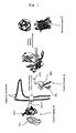
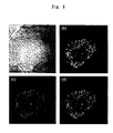
- step (a) fusion peptide (a) introduced into a cell according to the above-mentioned method is transferred into a designated organelle by its OTS (Fig. 1). Furthermore, regarding fusion peptide (b)/testP, which is introduced into the cell in step (b), if testP has the designated organelle localization, it is transferred into the organelle, and interacts with fusion peptide (a) that is localized therein; then, INTn and INTc assemble, is excised by protein splicing, and FPn and FPc are reconstructed, thus emitting fluorescence signal (Fig. 1).
- testP shows designated organelle localization
- the fluorescence signal may also be detected by observing the cell through fluorescent microscopy.
- cells that emit fluorescence signal may be sorted by a fluorescence-activated cell sorting (FACS) method.
- FACS fluorescence-activated cell sorting
- Invention (2) of the present application is an embodiment of the analysis method of the above-mentioned invention (1).
- step (a) respective fusion peptides (a) are introduced into two or more different organelles in a cell.
- Each fusion peptide (a) contain an organelle targeting signal peptide that targets different organelles, and the fluorescent proteins each show distinct characteristic (such as color).
- fusion peptides (a) with half-peptides of green fluorescent protein (EGFP), yellow fluorescent protein (EYFP) and cyan fluorescent protein (ECFP) are localized in mitochondria, endoplasmic reticulum and Golgi body, respectively.
- step (b) fusion peptide (b) having the other half-peptide of the above-mentioned respective fluorescent proteins, and the test protein bound thereto are introduced into the cell.
- the location at which the test protein localizes i.e. mitochondria, endoplasmic reticulum or Golgi body, can be determined.
- inventions (1) and (2) may be performed efficiently by using the probe sets provided by the present application (inventions (9) and (10)). Furthermore, by using the cells provided by the present application (inventions (11) and (13)), step (a) can be omitted.
- these cells may be made into cell kits (Inventions (12) and (14)) comprising two or more of these cell populations.
- the cell kit of invention (12) may consist of a plurality of cell populations wherein all of the cells contain fusion peptide (a) in the same organelle, or may consist of a plurality of cell populations wherein each cell contains fusion peptide (a) in varying organelles.
- the cell kit of invention (14) may consist of a plurality of cell populations wherein all cells contain fusion peptide (a) in the same two or more organelles, or may consist of a plurality of cell populations wherein each cell contains fusion peptide (a) in varying two or more organelles.
- each cell when the cell is a floating cell, each cell may be suspended in an appropriate liquid medium, and when a cell is an adhesive cell, the cell may be immobilized in the form of a "cell chip".
- cells that constitute such cell kits may be the same kind of cells, or may be different types of cells.
- a cell kit may be composed of a combination of normal cells and disease cells (for example, cancer cells or the like).
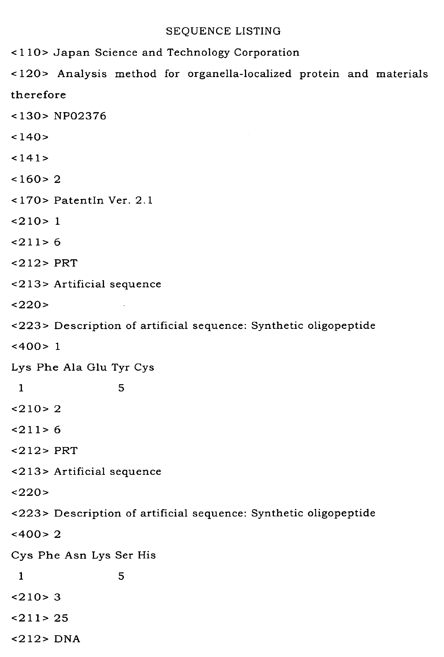
- the enhanced EGFP cDNA of its amino acid 1-157 was amplified by polymerase chain reaction (PCR) to introduce Lys-Phe-Ala-Glu-Tyr-Cys (SEQ ID NO: 1) to the C-terminus of spEGFP.
- This cDNA was fused to the cDNA of the N-terminal splicing domain of the DnaE intein and subcloned in the prokaryotic vector Bluescript.
- the PCR product was sequenced to confirm the base sequence and was subcloned into pMX vector at SalI restriction sites.
- fusion peptide (b) composed of the N-terminal half-peptide of EGFP (EGFPn) and the N-terminal half-peptide of DnaE (DnaEn) bound with a mitochondrial targeting signal peptide (MTS) or calmodulin
- EGFPn N-terminal half-peptide of EGFP
- DnaEn N-terminal half-peptide of DnaE bound with a mitochondrial targeting signal peptide (MTS) or calmodulin
- MTS mitochondrial targeting signal peptide
- calmodulin mitochondrial targeting signal peptide
- the cDNA of the C-terminal half-peptide of DnaE (DnaEc) bound with MTS was amplified by PCR.
- the cDNA of the carboxyl-terminal half of EGFP corresponding to 158-238 was amplified by PCR with extending the peptide of Cys-Phe-Asn-Lys-Ser-His (SEQ ID NO: 2) to the amino terminus.
- SEQ ID NO: 2 extending the peptide of Cys-Phe-Asn-Lys-Ser-His
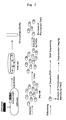
- the fusion gene was transfected into PlatE cells with Lipofectamine Plus (Invitrogen). After two days of culture, high-titer retroviruses were collected and transfected into BNL1ME cells. Stable expressing cells were obtained after approximately 10 days of selective culture in G418 (Invitrogen) containing the growth medium (see Fig. 3).
- Poly(A)+ RNA was purified from 1 x 10 8 BNL1ME cells using a FastTrack kit (Invitrogen).
- cDNA was synthesized from the Poly(A)+ RNA by random hexamers using a cDNA synthesis kit (Invitrogen).
- the resulting cDNAs were size-fractionated through column chromatography and agarose gel electrophoresis, and cDNA fragments of 600 kbp or longer were extracted from the agarose gel by using a Qiaex II kit (Qiagen).
- the cDNA fragments were inserted into BstXI sites of pMX-Mito/LiB by using BstXI adapters (Invitrogen).
- Plasmid DNA was purified by using Qiaex (Qiagen) after 200 mL of culture for 16 hours. The plasmids were transfected into packaging cell line PlatE with Lipofectamine Plus (Invitrogen). After two days of culture, high-titer retroviruses were collected (see Fig. 3).
- Subconfluent (70%) BNL1ME cell layers were infected with the constructed retrovirus library with an infection efficiency of 20% or less.
- the infection efficiency was estimated by a control experiment using pMX-EGFP.
- the cells were detached 48 hours after infection and spread into four 6-cm-diameter dishes. After a 72-hour incubation, the cells were stripped with tripsin-EDTA and dissolved in a PBS buffer (Gibco BRL). FACS analysis was performed on an ALTRA flow cytometer (Beckmann Coulter) for sorting GFP-positive single cells. These cells were incubated in a 96-well plate or spread into a 10-cm-diameter dish followed by subcloning using chips (see Fig. 3).
- Genomic cDNAs extracted from BNL1ME clones were amplified by the nested PCR method to recover the integrated cDNAs.
- As the primers a set of 5'-AGGACCTTACACAGTCCTGCTGACC-3' (SEQ ID NO: 3) and 5'-GCCCTCGCCGGACACGCTGAACTTG-3' (SEQ ID NO: 4), and a set of 5'-CCGCCCTCAAAGTAGACGGCATCGCAGC-3' (SEQ ID NO: 5) and 5'-CGCCGTCCAGCTCGACCAGGAT-3' (SEQ ID NO: 6) were used.
- the PCR was run for 30 cycles (30 sec. at 98°C for denaturation, 30 sec.
- Each cDNA sequence was compared with the cDNA sequences in databases including GenBank, PDB, SwissProt, PIR, PRF using BLASTn. Orientation of the cDNA strands was identified by the RIKEN clone sets, which were categorized in several stages, and their functions were analyzed. Homology analysis was performed using the Blast program.
- BNL1ME clones were spread on a glass-base dish and incubated for 24 hours in the presence of the growth medium.
- the medium was replaced by a PBS solution supplemented by 5% FCS and the live cells were directly imaged using a confocal laser-scanning microscope (Carl Zeiss). After imaging, mitochondria were stained with tetramethylrhodamine ethyl ester (TMRE; Molecular Probes). The final concentration of the TMRE in the PBS buffer was adjusted at 1 ⁇ M. Incubation was performed for 10 minutes.
- the cells were irradiated with a wavelength of 543 nm and the image was taken through a 560 nm LP filter.
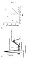
- the fluorescence intensity of EGFP reconstituted in mitochondria is highly sensitive and strong enough to be detected by FACS analysis.
- the cells that include a protein in the presence of MTS can be selectively separated and collected from those in the absence of MTS.
- proteins for which the intracellular localization are well characterized were tested in mouse liver cells (BNL1ME).
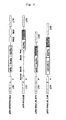
- the plasmid pMX-MTS/DEc(Neo) which encodes cDNAs corresponding to the C-terminal half-peptides of EGFP and DnaEc, and a mitochondrial targeting signal corresponding to the precursor of subunit VIII of cytochrome C oxidase, was constructed (Fig.
- cDNA sequences encoding additional 5 amino acids were inserted for efficient splicing to occur (Fig. 2) (Evans, J. et al., J. Biol. Chem. 2000, 275, 9091-9094).
- the plasmid was converted into retroviruses and they were infected into BNL1ME cells.
- a stable cell line expressing the corresponding test protein in mitochondria was developed (BNL1MEmito).
- As the test protein a known cytosolic protein, calmodulin, or a signal peptide, MTS, was used.
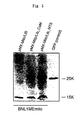
- BNL1MEmito cells were infected at various multiplicities of infection (MOI, which is defined as the number of cDNAs per cell) with retroviruses producing MTS-EGFPn-DnaEn.
- MOI multiplicities of infection
- Control of the MOI is particularly important because multiple integration of cDNAs in the BNL1MEmito cells may result in the isolation of false positive cDNAs after cell sorting. Therefore, it was needed to control the infection efficiency as a single-hit event. To assess this, 48 hours after the infection with various MOIs, the number of the cells including the reconstructed EGFP was evaluated by flow cytometry.
- cDNA-EGFPn-DnaEn The order of the tandem fusion fragments, cDNA-EGFPn-DnaEn, was crucial for analyzing its intracellular localization, because most MTSs are known to attach to the amino-terminal end of a mitochondrial protein (Roise, D. et al., EMBO J. 1988, 7, 649-653; Von Heijne, G. EMBO J.
- the cDNA library thus constructed contained 1.1 x 10 6 independent clones, with the size of cDNAs averaging 1.4 kbp.
- the library was converted to retroviruses by using a high-titer retrovirus packaging cell line, Plat-E cells (Morita, S. et al., Gene Therapy 2000, 7, 1063-1066).
- 1 x 10 7 cells were infected with 50 ⁇ L of the retroviral supernatant to achieve 20 % infection efficiency.
- the fluorescence intensity of the 1 x 10 5 cells was measured by FACS analysis 3 days after the infection.
- the population of the infected cells consisted of a mixture of cells in the presence and absence of the reconstructed EGFP (Fig. 8).
- a population of fluorescent cells in region L was collected by FACS analysis.
- Data rate defined as the number of cells analyzed per second, was controlled to be (1.0 ⁇ 0.1) x 10 3 .
- 10 7 cells could be examined within a few hours.
- a total of 1 x 10 3 cells were counted as a fluorescent cell in region L, but half of the cells were aborted.
- the fluorescent cells that were actually collected were therefore 500 to 1000 cells, indicating that this EGFP reconstruction technology in combination with FACS enables high-speed collection of MTS-tagged fusion peptides.
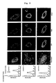
- the fluorescence intensity and intracellular localization of each isolated clone were analyzed. If the cDNA was integrated in the host genome, the corresponding protein should be constitutively expressed in the BNL1MEmito cell and therefore EGFP reconstruction should be kept in the mitochondria. In order to confirm this, fluorescence intensities of the collected 200 clones were analyzed by FACS, among which 169 clones showed fluorescence of various intensities (Fig. 9). The rest of the 31 clones of which cDNA could not be recovered by genomic PCR did not fluoresce, indicating that the cDNA was not integrated in the nuclear genome or that the cDNA, after being integrated, dropped out of the nuclear genome during cell division.
- the nuclear genome was extracted from each clone and the integrated cDNA was recovered by PCR amplification, which was subjected to sequence analysis.
- the expressed sequence tags obtained included 32 tags that occurred once and 28 tags that were identified multiple times.
- 56 clones were identified in GenBank.
- the other 4 genes were identified newly, and were found to include mitochondrial targeting signals. The localization of each novel gene product in the mitochondria was confirmed by confocal microscopy.
- cDNA derived from clone No. 10 was identified in public sequence database DDBJ (RIKEN full-length cDNA clones) (Hayashizaki, Y. et al., Nature 2001, 409, 685-690). Reading frames and expected start codons of cDNAs obtained from clone No. 10 completely matched those found in the database. Homology analysis using public databases showed that there was a 23% homology at the DNA level between the cloned cDNA fragment and putative cytochrome c oxidase assembly protein derived from Schizosaccharomyces pombe. Therefore mouse clone No.
- cDNA derived from clone No. 92 was found to be a 76% homologue of the cDNA of human mitochondrial 28S ribosomal protein (S18-1). This high homology and its mitochondrial localization obtained in this experiment confirmed that the cDNA for clone No. 92 is a mouse mitochondrial ribosomal protein.
- Another ribosomal protein S18 (clone No. 51) has already been identified as a mouse ribosomal protein, but its localization had not been discussed in detail.
- MTS is composed of some 20 to 60 amino acid residues that have the potential to form amphiphilic ⁇ -helices with one hydrophobic face and one positively charged face.
- the other newly annotated genes are summarized with their gene names in Table 1.
- cDNAs of 3 clones shown in Table 1 whose reading frame and start codon were found to be a complete match to the RIKEN full-length cDNA clones, did not show significant similarity to other eukaryotic cells. This indicates that these 3 clones are novel proteins localized in mitochondria.
- the analysis method of the present invention enables the provision of a rapid approach for identifying novel gene products that are localized in the mitochondria, and for annotating their functions.
- the high-throughput screening technology also allows easy identification of groups of proteins localized in organelles such as nucleus, endoplasmic reticulum, Golgi body or peroxisome, by using respective signals. Because of the simplicity of the present method, one skilled in the art capable of constructing a cDNA library and equipped with a FACS facility would be able to perform the technology without resorting to excessive tests.
- the combination of the present method with a cDNA subtraction method gives more flexibility in that, for example, comparison of expression genes under normal conditions and disease conditions or comparison of expression genes of different tissues, is made possible.
Landscapes
- Life Sciences & Earth Sciences (AREA)
- Health & Medical Sciences (AREA)
- Engineering & Computer Science (AREA)
- Biomedical Technology (AREA)
- Molecular Biology (AREA)
- Chemical & Material Sciences (AREA)
- Immunology (AREA)
- Physics & Mathematics (AREA)
- Hematology (AREA)
- Urology & Nephrology (AREA)
- Biotechnology (AREA)
- Bioinformatics & Cheminformatics (AREA)
- General Health & Medical Sciences (AREA)
- Biochemistry (AREA)
- Microbiology (AREA)
- Analytical Chemistry (AREA)
- Biophysics (AREA)
- Food Science & Technology (AREA)
- Cell Biology (AREA)
- Medicinal Chemistry (AREA)
- Genetics & Genomics (AREA)
- General Physics & Mathematics (AREA)
- Pathology (AREA)
- Bioinformatics & Computational Biology (AREA)
- Zoology (AREA)
- Wood Science & Technology (AREA)
- Organic Chemistry (AREA)
- General Engineering & Computer Science (AREA)
- Proteomics, Peptides & Aminoacids (AREA)
- Tropical Medicine & Parasitology (AREA)
- Plant Pathology (AREA)
- Toxicology (AREA)
- Crystallography & Structural Chemistry (AREA)
- Peptides Or Proteins (AREA)
- Measuring Or Testing Involving Enzymes Or Micro-Organisms (AREA)
- Micro-Organisms Or Cultivation Processes Thereof (AREA)
- Investigating Or Analysing Biological Materials (AREA)
- Investigating Or Analysing Materials By The Use Of Chemical Reactions (AREA)
Abstract
Description
| Category of sense cDNAs | Clone No. |
| Identical to Mouse Protein | |
| Malate dehydrogenase | 11, 52, 53, 54 |
| Cytochrome c oxidase, subunit Vb (Cox5b) | 20 |
| ATP synthetase alpha subunit | 23, 27, 84, 85, 95 |
| Uncoupling protein 2 (Ucp2) | 35 |
| Glutaryl-CoA dehydrogenase (Gcdh) | 40, 43, 49, 57, 93 |
| Acetyl-coenzyme A dehydrogenase (Acad1) | 58 |
| Cytochrome b | 1 |
| Aldehyde dehydrogenase 2 | 140 |
| ATP synthetase H+ transporting, mitochondrial F1 complex, | 143 |
| gamma polypeptide 1 | |
| Mitochondrial ribosomal protein S11 | 147 |
| Similar to mouse gene | |
| Phosphoenolpyruvate carboxykinase 2 | 71, 100 |
| 60S ribosomal protein L3 (L4) | 108 |
| NADH-ubiquinone oxidoreductase 13 kDa-A subunit | 144, 150 |
| Inorganic phosphatase | 148 |
| Homologue to mammal gene | |
| Putative cytochrome c oxidase assembly protein | 10, 94 |
| (Schizosaccharomyces pombe, 23%) | |
| Heat shock protein 75 (Homo sapiens, 89%) | 46, 70, 77 |
| Ribosomal protein S 18 (Rsp18) (Homo sapiens, 76%) | 51,63 |
| Membrane associated protein SLP-2 | 87 |
| (Homo sapiens, 93%) | |
| Mitochondrial 28S ribosomal protein S18-1 | 92 |
| (Homo sapiens, 77%) | |
| NADH-ubiquinone oxidoreductase 30 kDa subunit precursor | 99 |
| (Homo sapiens, 88%) | |
| Succinate dehydrogenase complex, subunit B, iron | 135 |
| sulfur (Homo sapiens, 91%) | |
| Biphenyl hydrolase-related protein | 145 |
| (Homo sapiens, 75%) | |
| Predicted protein | |
| GI: 12852607 | 16 |
| GI: 12840016 | 33,37 |
| GI: 12859851 | 59,72,82 |

Claims (14)
- A method for analyzing an organelle-localized protein, which enables one to determine whether or not a test protein localizes to an organelle, and comprises the following steps:(a) a step of introducing a fusion peptide (a), which comprises one half-peptide of an intein, one half-peptide of a fluorescent protein and an organelle-targeting signal peptide, into a eukaryotic cell;(b) a step of introducing a test protein bound to a fusion peptide (b), which comprises the other half-peptide of the fluorescent protein and the other half-peptide of the intein, into the eukaryotic cell; and(c) a step of detecting fluorescence signal emitted by the fluorescent protein.
- The method of Claim 1, wherein,in step (a), two or more types of fusion peptide (a), each comprising one half-peptide of different fluorescent proteins and different organelle-targeting signal peptides, are introduced into a eukaryotic cell;in step (b), two or more types of fusion peptides (b), each comprising the other half-peptide of the different fluorescent proteins, and each bound to a test protein, is introduced into the eukaryotic cell; andin step (c), the fluorescent signal is detected.
- The method of Claim 1 or 2, wherein, in step (a), the fusion peptide (a) is introduced into a eukaryotic cell by transfecting a recombinant vector (A), which expresses the fusion peptide (a), into the eukaryotic cell.
- The analysis method of Claim 1 or 2, wherein, in step (b), the test protein and the fusion peptide (b) are introduced into a eukaryotic cell by transfecting a recombinant vector (B), which expresses the fusion peptide (b) and the test protein as a unit, into the eukaryotic cell.
- A fusion peptide (a), which comprises a half-peptide of an intein, a half-peptide of a fluorescent protein and an organelle targeting signal peptide.
- A fusion peptide (b), which comprises a half-peptide of a fluorescent protein and a half-peptide of an intein.
- A recombinant vector (A), which expresses a fusion peptide (a) comprising a half-peptide of an intein, a half-peptide of a fluorescent protein and an organelle targeting signal peptide.
- A recombinant vector (B), which expresses a fusion peptide (b) comprising a half-peptide of a fluorescent protein and a half-peptide of an intein, and an arbitrary test protein bound thereto.
- A probe set for analyzing organelle-localized protein, comprising the fusion peptide (a) of Claim 5 or the recombinant vector (A) of Claim 7, and the fusion peptide (b) of Claim 6 or the recombinant vector (B) of Claim 8.
- The probe set according to Claim 9, wherein
the fusion peptide (a) or the fusion peptide (a) expressed by the recombinant vector (A) comprises two or more types of fusion peptides, each fusion peptide comprising one half-peptide of a fluorescent protein having different signal characteristics and a different organelle targeting signal peptide; and
the fusion peptide (b) comprises two or more types of fusion peptides, each fusion peptide comprising the other half of the fluorescent protein. - A eukaryotic cell, containing a fusion peptide (a), which comprises a half-peptide of an intein, a half-peptide of a fluorescent protein and an organelle targeting signal peptide.
- A cell kit, comprising two or more of the eukaryotic cells of Claim 11.
- A eukaryotic cell, comprising two or more types of fusion peptide (a), wherein each fusion peptide comprises one half-peptide of a fluorescent protein and an organelle targeting signal peptide, the fluorescent protein of each fusion peptide have different signal characteristics and the organelle targeting signal peptide of each fusion peptide target different organelle.
- A cell kit, comprising two or more of the eukaryotic cells of Claim 13.
Applications Claiming Priority (3)
| Application Number | Priority Date | Filing Date | Title |
|---|---|---|---|
| JP2002272043A JP4287633B2 (en) | 2002-09-18 | 2002-09-18 | Analyzing method and materials for organelle localization protein |
| JP2002272043 | 2002-09-18 | ||
| PCT/JP2003/011156 WO2004027057A1 (en) | 2002-09-18 | 2003-09-01 | Method of analyzing organelle-localized protein and materials for analysis |
Publications (3)
| Publication Number | Publication Date |
|---|---|
| EP1541679A1 true EP1541679A1 (en) | 2005-06-15 |
| EP1541679A4 EP1541679A4 (en) | 2006-06-07 |
| EP1541679B1 EP1541679B1 (en) | 2009-12-23 |
Family
ID=32024894
Family Applications (1)
| Application Number | Title | Priority Date | Filing Date |
|---|---|---|---|
| EP03797539A Expired - Lifetime EP1541679B1 (en) | 2002-09-18 | 2003-09-01 | Method of analyzing organelle-localized protein and materials for analysis |
Country Status (6)
| Country | Link |
|---|---|
| US (2) | US20050118649A1 (en) |
| EP (1) | EP1541679B1 (en) |
| JP (1) | JP4287633B2 (en) |
| CA (1) | CA2474680A1 (en) |
| DE (1) | DE60330684D1 (en) |
| WO (1) | WO2004027057A1 (en) |
Cited By (2)
| Publication number | Priority date | Publication date | Assignee | Title |
|---|---|---|---|---|
| US8658777B2 (en) | 2007-05-09 | 2014-02-25 | The University Of Tokyo | Activated protease indicator |
| CN108456694A (en) * | 2017-02-22 | 2018-08-28 | 浙江和也健康科技有限公司 | A kind of genetic manipulation method and the application of fusion positioning system, electromagnetic induction system and target protein |
Families Citing this family (6)
| Publication number | Priority date | Publication date | Assignee | Title |
|---|---|---|---|---|
| KR100863283B1 (en) | 2002-10-11 | 2008-10-15 | 아람 바이오시스템 주식회사 | Observation method of intracellular process and recombinant genes used therefor |
| US20070178464A1 (en) * | 2004-03-09 | 2007-08-02 | Japan Science And Technology Agency | Probes for detecting protein nuclear transport and method for detecting and quantifying protein nuclear transport using the same |
| WO2007037482A1 (en) * | 2005-09-30 | 2007-04-05 | The University Of Tokyo | Kit and method for use in the detection of protein-protein interaction in eukaryotic cell |
| JPWO2008015841A1 (en) * | 2006-08-02 | 2009-12-17 | 梅澤 喜夫 | Kinase inhibitory fusion proteins and pharmaceutical compositions |
| US8431149B2 (en) * | 2007-03-01 | 2013-04-30 | Boston Scientific Scimed, Inc. | Coated medical devices for abluminal drug delivery |
| JP6955243B2 (en) * | 2017-02-24 | 2021-10-27 | 学校法人北里研究所 | Necrosis detection kit and its use |
Family Cites Families (5)
| Publication number | Priority date | Publication date | Assignee | Title |
|---|---|---|---|---|
| US6413776B1 (en) * | 1998-06-12 | 2002-07-02 | Galapagos Geonomics N.V. | High throughput screening of gene function using adenoviral libraries for functional genomics applications |
| JP4544601B2 (en) * | 1998-12-18 | 2010-09-15 | ザ・ペン・ステート・リサーチ・ファンデーション | Cyclic peptide |
| CN1676606A (en) * | 1999-05-24 | 2005-10-05 | 新英格兰生物实验室公司 | Method for reconstructing target protein from protein segment in plant |
| WO2001087919A2 (en) * | 2000-05-12 | 2001-11-22 | Yale University | Methods of detecting interactions between proteins, peptides or libraries thereof using fusion proteins |
| JP3772193B2 (en) * | 2000-07-26 | 2006-05-10 | 独立行政法人科学技術振興機構 | Protein-protein interaction analysis probe and method for analyzing protein-protein interaction using the same |
-
2002
- 2002-09-18 JP JP2002272043A patent/JP4287633B2/en not_active Expired - Fee Related
-
2003
- 2003-09-01 EP EP03797539A patent/EP1541679B1/en not_active Expired - Lifetime
- 2003-09-01 US US10/501,947 patent/US20050118649A1/en not_active Abandoned
- 2003-09-01 WO PCT/JP2003/011156 patent/WO2004027057A1/en not_active Ceased
- 2003-09-01 DE DE60330684T patent/DE60330684D1/en not_active Expired - Lifetime
- 2003-09-01 CA CA002474680A patent/CA2474680A1/en not_active Abandoned
-
2010
- 2010-01-21 US US12/691,110 patent/US20100184619A1/en not_active Abandoned
Cited By (2)
| Publication number | Priority date | Publication date | Assignee | Title |
|---|---|---|---|---|
| US8658777B2 (en) | 2007-05-09 | 2014-02-25 | The University Of Tokyo | Activated protease indicator |
| CN108456694A (en) * | 2017-02-22 | 2018-08-28 | 浙江和也健康科技有限公司 | A kind of genetic manipulation method and the application of fusion positioning system, electromagnetic induction system and target protein |
Also Published As
| Publication number | Publication date |
|---|---|
| EP1541679B1 (en) | 2009-12-23 |
| US20050118649A1 (en) | 2005-06-02 |
| DE60330684D1 (en) | 2010-02-04 |
| WO2004027057A1 (en) | 2004-04-01 |
| EP1541679A4 (en) | 2006-06-07 |
| JP4287633B2 (en) | 2009-07-01 |
| CA2474680A1 (en) | 2004-04-01 |
| US20100184619A1 (en) | 2010-07-22 |
| JP2004108943A (en) | 2004-04-08 |
Similar Documents
| Publication | Publication Date | Title |
|---|---|---|
| US6242183B1 (en) | Interaction trap systems for detecting protein interactions | |
| US6596485B2 (en) | Green fluorescent protein fusions with random peptides | |
| US20100184619A1 (en) | Method for analyzing organelle-localized protein and material for analysis | |
| KR20020059370A (en) | Methods and compositions for the construction and use of fusion libraries | |
| WO2019143529A1 (en) | Engineered promiscuous biotin ligases for efficient proximity labeling | |
| CA2272823A1 (en) | Method for identifying genes encoding novel secreted or membrane-associated proteins | |
| US20050277116A1 (en) | Compositions and methods for the identification of protein interactions in vertebrate cells | |
| US6399296B1 (en) | Interaction trap systems for detecting protein interactions | |
| US7195869B2 (en) | Method of searching for gene encoding nuclear transport protein | |
| WO2006008539A1 (en) | Cell cycle markers | |
| EP1268842B1 (en) | Improved reverse n-hybrid screening method | |
| US20030211495A1 (en) | Reverse n-hybrid screening method | |
| US20120282643A1 (en) | Cyan and yellow fluorescent color variants of split gfp | |
| JP2002530074A (en) | Methods for validating polypeptide targets that correlate with cell phenotype | |
| AU2001245584B2 (en) | Improved reverse n-hybrid screening method | |
| US20050032197A1 (en) | Membrane protein interaction assays | |
| EP1278762A2 (en) | Nucleic acid construct system and method utilizing same useful for identifying protein-protein interactions | |
| HK1065560A (en) | Interaction trap systems for detecting protein interactions | |
| US20030100022A1 (en) | Nucleic acid construct system and method utilizing same useful for identifying protein-protein interactions | |
| JP2004024078A (en) | Random oligonuceleotide library and method for detecting protein interaction | |
| WO2004012574A2 (en) | Negative selections assays, and compositions thereof | |
| HK1012006B (en) | Interaction trap systems for detecting protein interactions | |
| Renna | The role of sec24 in protein export from the plant endoplasmic reticulum |
Legal Events
| Date | Code | Title | Description |
|---|---|---|---|
| PUAI | Public reference made under article 153(3) epc to a published international application that has entered the european phase |
Free format text: ORIGINAL CODE: 0009012 |
|
| 17P | Request for examination filed |
Effective date: 20040830 |
|
| AK | Designated contracting states |
Kind code of ref document: A1 Designated state(s): AT BE BG CH CY CZ DE DK EE ES FI FR GB GR HU IE IT LI LU MC NL PT RO SE SI SK TR |
|
| RBV | Designated contracting states (corrected) |
Designated state(s): CH DE FR GB LI NL SE |
|
| A4 | Supplementary search report drawn up and despatched |
Effective date: 20060508 |
|
| 17Q | First examination report despatched |
Effective date: 20071015 |
|
| GRAP | Despatch of communication of intention to grant a patent |
Free format text: ORIGINAL CODE: EPIDOSNIGR1 |
|
| GRAS | Grant fee paid |
Free format text: ORIGINAL CODE: EPIDOSNIGR3 |
|
| GRAA | (expected) grant |
Free format text: ORIGINAL CODE: 0009210 |
|
| AK | Designated contracting states |
Kind code of ref document: B1 Designated state(s): CH DE FR GB LI NL SE |
|
| REG | Reference to a national code |
Ref country code: GB Ref legal event code: FG4D |
|
| REG | Reference to a national code |
Ref country code: CH Ref legal event code: EP Ref country code: CH Ref legal event code: NV Representative=s name: BOHEST AG |
|
| REF | Corresponds to: |
Ref document number: 60330684 Country of ref document: DE Date of ref document: 20100204 Kind code of ref document: P |
|
| REG | Reference to a national code |
Ref country code: SE Ref legal event code: TRGR |
|
| PLBE | No opposition filed within time limit |
Free format text: ORIGINAL CODE: 0009261 |
|
| STAA | Information on the status of an ep patent application or granted ep patent |
Free format text: STATUS: NO OPPOSITION FILED WITHIN TIME LIMIT |
|
| 26N | No opposition filed |
Effective date: 20100924 |
|
| REG | Reference to a national code |
Ref country code: CH Ref legal event code: PCAR Free format text: NEW ADDRESS: HOLBEINSTRASSE 36-38, 4051 BASEL (CH) |
|
| REG | Reference to a national code |
Ref country code: FR Ref legal event code: PLFP Year of fee payment: 14 |
|
| PGFP | Annual fee paid to national office [announced via postgrant information from national office to epo] |
Ref country code: NL Payment date: 20160808 Year of fee payment: 14 |
|
| PGFP | Annual fee paid to national office [announced via postgrant information from national office to epo] |
Ref country code: CH Payment date: 20160808 Year of fee payment: 14 Ref country code: DE Payment date: 20160805 Year of fee payment: 14 Ref country code: GB Payment date: 20160808 Year of fee payment: 14 |
|
| PGFP | Annual fee paid to national office [announced via postgrant information from national office to epo] |
Ref country code: SE Payment date: 20160805 Year of fee payment: 14 Ref country code: FR Payment date: 20160809 Year of fee payment: 14 |
|
| REG | Reference to a national code |
Ref country code: DE Ref legal event code: R119 Ref document number: 60330684 Country of ref document: DE |
|
| REG | Reference to a national code |
Ref country code: CH Ref legal event code: PL |
|
| REG | Reference to a national code |
Ref country code: SE Ref legal event code: EUG |
|
| REG | Reference to a national code |
Ref country code: NL Ref legal event code: MM Effective date: 20171001 |
|
| GBPC | Gb: european patent ceased through non-payment of renewal fee |
Effective date: 20170901 |
|
| PG25 | Lapsed in a contracting state [announced via postgrant information from national office to epo] |
Ref country code: NL Free format text: LAPSE BECAUSE OF NON-PAYMENT OF DUE FEES Effective date: 20171001 |
|
| REG | Reference to a national code |
Ref country code: FR Ref legal event code: ST Effective date: 20180531 |
|
| PG25 | Lapsed in a contracting state [announced via postgrant information from national office to epo] |
Ref country code: DE Free format text: LAPSE BECAUSE OF NON-PAYMENT OF DUE FEES Effective date: 20180404 Ref country code: CH Free format text: LAPSE BECAUSE OF NON-PAYMENT OF DUE FEES Effective date: 20170930 Ref country code: GB Free format text: LAPSE BECAUSE OF NON-PAYMENT OF DUE FEES Effective date: 20170901 Ref country code: LI Free format text: LAPSE BECAUSE OF NON-PAYMENT OF DUE FEES Effective date: 20170930 |
|
| PG25 | Lapsed in a contracting state [announced via postgrant information from national office to epo] |
Ref country code: FR Free format text: LAPSE BECAUSE OF NON-PAYMENT OF DUE FEES Effective date: 20171002 |
|
| PG25 | Lapsed in a contracting state [announced via postgrant information from national office to epo] |
Ref country code: SE Free format text: LAPSE BECAUSE OF NON-PAYMENT OF DUE FEES Effective date: 20170902 |