EP1538207B1 - Human anti-human mcp-1 antibody and antibody fragment thereof - Google Patents
Human anti-human mcp-1 antibody and antibody fragment thereof Download PDFInfo
- Publication number
- EP1538207B1 EP1538207B1 EP03795363A EP03795363A EP1538207B1 EP 1538207 B1 EP1538207 B1 EP 1538207B1 EP 03795363 A EP03795363 A EP 03795363A EP 03795363 A EP03795363 A EP 03795363A EP 1538207 B1 EP1538207 B1 EP 1538207B1
- Authority
- EP
- European Patent Office
- Prior art keywords
- human
- mcp
- antibody
- scfv
- nucleotide sequence
- Prior art date
- Legal status (The legal status is an assumption and is not a legal conclusion. Google has not performed a legal analysis and makes no representation as to the accuracy of the status listed.)
- Expired - Lifetime
Links
- 241000282414 Homo sapiens Species 0.000 title claims abstract description 51
- 108010021625 Immunoglobulin Fragments Proteins 0.000 title claims abstract description 8
- 102000008394 Immunoglobulin Fragments Human genes 0.000 title claims abstract description 8
- 101100504320 Caenorhabditis elegans mcp-1 gene Proteins 0.000 title 1
- 101000897480 Homo sapiens C-C motif chemokine 2 Proteins 0.000 claims abstract description 51
- 102000046768 human CCL2 Human genes 0.000 claims abstract description 51
- 101710155857 C-C motif chemokine 2 Proteins 0.000 claims abstract description 49
- 102100021943 C-C motif chemokine 2 Human genes 0.000 claims abstract description 48
- 206010061218 Inflammation Diseases 0.000 claims abstract description 13
- 239000003814 drug Substances 0.000 claims abstract description 13
- 230000004054 inflammatory process Effects 0.000 claims abstract description 13
- 238000000034 method Methods 0.000 claims abstract description 8
- 108090000623 proteins and genes Proteins 0.000 claims description 26
- 239000012634 fragment Substances 0.000 claims description 16
- 239000002773 nucleotide Substances 0.000 claims description 16
- 125000003729 nucleotide group Chemical group 0.000 claims description 16
- 206010003210 Arteriosclerosis Diseases 0.000 claims description 12
- 208000011775 arteriosclerosis disease Diseases 0.000 claims description 12
- 230000000694 effects Effects 0.000 claims description 10
- 230000006698 induction Effects 0.000 claims description 9
- 208000037976 chronic inflammation Diseases 0.000 claims description 7
- 230000002401 inhibitory effect Effects 0.000 claims description 7
- 208000037893 chronic inflammatory disorder Diseases 0.000 claims description 6
- 208000010125 myocardial infarction Diseases 0.000 claims description 5
- 208000023275 Autoimmune disease Diseases 0.000 claims description 4
- 239000013604 expression vector Substances 0.000 claims description 4
- 239000003795 chemical substances by application Substances 0.000 claims description 3
- 102000035118 modified proteins Human genes 0.000 claims description 3
- 108091005573 modified proteins Proteins 0.000 claims description 3
- 239000002202 Polyethylene glycol Substances 0.000 claims description 2
- 229920001223 polyethylene glycol Polymers 0.000 claims description 2
- 230000006798 recombination Effects 0.000 claims description 2
- 239000004480 active ingredient Substances 0.000 claims 1
- 239000000126 substance Substances 0.000 abstract description 2
- 210000004027 cell Anatomy 0.000 description 35
- 108020004414 DNA Proteins 0.000 description 31
- 239000000243 solution Substances 0.000 description 16
- 230000027455 binding Effects 0.000 description 14
- 238000002965 ELISA Methods 0.000 description 12
- 108060003951 Immunoglobulin Proteins 0.000 description 12
- 102000018358 immunoglobulin Human genes 0.000 description 12
- 230000012292 cell migration Effects 0.000 description 10
- 238000005406 washing Methods 0.000 description 10
- 150000001413 amino acids Chemical group 0.000 description 9
- 230000014509 gene expression Effects 0.000 description 9
- 230000001404 mediated effect Effects 0.000 description 9
- 241000588724 Escherichia coli Species 0.000 description 8
- 238000006243 chemical reaction Methods 0.000 description 8
- 238000000746 purification Methods 0.000 description 8
- 238000003556 assay Methods 0.000 description 7
- 201000010099 disease Diseases 0.000 description 7
- 208000037265 diseases, disorders, signs and symptoms Diseases 0.000 description 7
- 210000002540 macrophage Anatomy 0.000 description 7
- 235000018102 proteins Nutrition 0.000 description 7
- 102000004169 proteins and genes Human genes 0.000 description 7
- 235000001014 amino acid Nutrition 0.000 description 6
- 230000035605 chemotaxis Effects 0.000 description 6
- 239000001963 growth medium Substances 0.000 description 6
- 230000009545 invasion Effects 0.000 description 6
- 239000013612 plasmid Substances 0.000 description 6
- 108010076504 Protein Sorting Signals Proteins 0.000 description 5
- 238000002835 absorbance Methods 0.000 description 5
- 229940079593 drug Drugs 0.000 description 5
- 239000013613 expression plasmid Substances 0.000 description 5
- 238000003752 polymerase chain reaction Methods 0.000 description 5
- 230000002265 prevention Effects 0.000 description 5
- 102000019034 Chemokines Human genes 0.000 description 4
- 108010012236 Chemokines Proteins 0.000 description 4
- 241000699670 Mus sp. Species 0.000 description 4
- 241000700159 Rattus Species 0.000 description 4
- FAPWRFPIFSIZLT-UHFFFAOYSA-M Sodium chloride Chemical compound [Na+].[Cl-] FAPWRFPIFSIZLT-UHFFFAOYSA-M 0.000 description 4
- 230000000692 anti-sense effect Effects 0.000 description 4
- 206010003246 arthritis Diseases 0.000 description 4
- 239000003593 chromogenic compound Substances 0.000 description 4
- 238000011835 investigation Methods 0.000 description 4
- 230000005012 migration Effects 0.000 description 4
- 238000013508 migration Methods 0.000 description 4
- 210000001616 monocyte Anatomy 0.000 description 4
- 210000005259 peripheral blood Anatomy 0.000 description 4
- 239000011886 peripheral blood Substances 0.000 description 4
- 108090000765 processed proteins & peptides Proteins 0.000 description 4
- 238000012216 screening Methods 0.000 description 4
- 239000007858 starting material Substances 0.000 description 4
- FWMNVWWHGCHHJJ-SKKKGAJSSA-N 4-amino-1-[(2r)-6-amino-2-[[(2r)-2-[[(2r)-2-[[(2r)-2-amino-3-phenylpropanoyl]amino]-3-phenylpropanoyl]amino]-4-methylpentanoyl]amino]hexanoyl]piperidine-4-carboxylic acid Chemical compound C([C@H](C(=O)N[C@H](CC(C)C)C(=O)N[C@H](CCCCN)C(=O)N1CCC(N)(CC1)C(O)=O)NC(=O)[C@H](N)CC=1C=CC=CC=1)C1=CC=CC=C1 FWMNVWWHGCHHJJ-SKKKGAJSSA-N 0.000 description 3
- 102100031151 C-C chemokine receptor type 2 Human genes 0.000 description 3
- 101710149815 C-C chemokine receptor type 2 Proteins 0.000 description 3
- WQZGKKKJIJFFOK-GASJEMHNSA-N Glucose Natural products OC[C@H]1OC(O)[C@H](O)[C@@H](O)[C@@H]1O WQZGKKKJIJFFOK-GASJEMHNSA-N 0.000 description 3
- 241001465754 Metazoa Species 0.000 description 3
- 230000004071 biological effect Effects 0.000 description 3
- 239000000872 buffer Substances 0.000 description 3
- 238000007796 conventional method Methods 0.000 description 3
- 239000012228 culture supernatant Substances 0.000 description 3
- 230000007812 deficiency Effects 0.000 description 3
- 230000001419 dependent effect Effects 0.000 description 3
- 238000011161 development Methods 0.000 description 3
- 230000018109 developmental process Effects 0.000 description 3
- UQLDLKMNUJERMK-UHFFFAOYSA-L di(octadecanoyloxy)lead Chemical compound [Pb+2].CCCCCCCCCCCCCCCCCC([O-])=O.CCCCCCCCCCCCCCCCCC([O-])=O UQLDLKMNUJERMK-UHFFFAOYSA-L 0.000 description 3
- 239000008103 glucose Substances 0.000 description 3
- 238000004128 high performance liquid chromatography Methods 0.000 description 3
- 208000027866 inflammatory disease Diseases 0.000 description 3
- 210000004698 lymphocyte Anatomy 0.000 description 3
- 238000004519 manufacturing process Methods 0.000 description 3
- 239000002609 medium Substances 0.000 description 3
- 239000000203 mixture Substances 0.000 description 3
- 238000004091 panning Methods 0.000 description 3
- 102000013415 peroxidase activity proteins Human genes 0.000 description 3
- 108040007629 peroxidase activity proteins Proteins 0.000 description 3
- 230000028327 secretion Effects 0.000 description 3
- 239000006228 supernatant Substances 0.000 description 3
- YBJHBAHKTGYVGT-ZKWXMUAHSA-N (+)-Biotin Chemical compound N1C(=O)N[C@@H]2[C@H](CCCCC(=O)O)SC[C@@H]21 YBJHBAHKTGYVGT-ZKWXMUAHSA-N 0.000 description 2
- 108091032973 (ribonucleotides)n+m Proteins 0.000 description 2
- QKNYBSVHEMOAJP-UHFFFAOYSA-N 2-amino-2-(hydroxymethyl)propane-1,3-diol;hydron;chloride Chemical compound Cl.OCC(N)(CO)CO QKNYBSVHEMOAJP-UHFFFAOYSA-N 0.000 description 2
- UAIUNKRWKOVEES-UHFFFAOYSA-N 3,3',5,5'-tetramethylbenzidine Chemical compound CC1=C(N)C(C)=CC(C=2C=C(C)C(N)=C(C)C=2)=C1 UAIUNKRWKOVEES-UHFFFAOYSA-N 0.000 description 2
- 102000002260 Alkaline Phosphatase Human genes 0.000 description 2
- 108020004774 Alkaline Phosphatase Proteins 0.000 description 2
- 101710155856 C-C motif chemokine 3 Proteins 0.000 description 2
- 102000001902 CC Chemokines Human genes 0.000 description 2
- 108010040471 CC Chemokines Proteins 0.000 description 2
- 102000000013 Chemokine CCL3 Human genes 0.000 description 2
- 108020004635 Complementary DNA Proteins 0.000 description 2
- 102000004190 Enzymes Human genes 0.000 description 2
- 108090000790 Enzymes Proteins 0.000 description 2
- 208000009386 Experimental Arthritis Diseases 0.000 description 2
- 108010010803 Gelatin Proteins 0.000 description 2
- DHMQDGOQFOQNFH-UHFFFAOYSA-N Glycine Chemical compound NCC(O)=O DHMQDGOQFOQNFH-UHFFFAOYSA-N 0.000 description 2
- NTYJJOPFIAHURM-UHFFFAOYSA-N Histamine Chemical compound NCCC1=CN=CN1 NTYJJOPFIAHURM-UHFFFAOYSA-N 0.000 description 2
- 102000008100 Human Serum Albumin Human genes 0.000 description 2
- 108091006905 Human Serum Albumin Proteins 0.000 description 2
- 108091028043 Nucleic acid sequence Proteins 0.000 description 2
- 239000012980 RPMI-1640 medium Substances 0.000 description 2
- 239000006146 Roswell Park Memorial Institute medium Substances 0.000 description 2
- 238000012300 Sequence Analysis Methods 0.000 description 2
- UIIMBOGNXHQVGW-UHFFFAOYSA-M Sodium bicarbonate Chemical compound [Na+].OC([O-])=O UIIMBOGNXHQVGW-UHFFFAOYSA-M 0.000 description 2
- 210000001744 T-lymphocyte Anatomy 0.000 description 2
- 101710120037 Toxin CcdB Proteins 0.000 description 2
- 238000001042 affinity chromatography Methods 0.000 description 2
- 239000011543 agarose gel Substances 0.000 description 2
- 125000000539 amino acid group Chemical group 0.000 description 2
- AVKUERGKIZMTKX-NJBDSQKTSA-N ampicillin Chemical compound C1([C@@H](N)C(=O)N[C@H]2[C@H]3SC([C@@H](N3C2=O)C(O)=O)(C)C)=CC=CC=C1 AVKUERGKIZMTKX-NJBDSQKTSA-N 0.000 description 2
- 229960000723 ampicillin Drugs 0.000 description 2
- 230000003321 amplification Effects 0.000 description 2
- 238000004458 analytical method Methods 0.000 description 2
- 210000004102 animal cell Anatomy 0.000 description 2
- 239000000427 antigen Substances 0.000 description 2
- 102000036639 antigens Human genes 0.000 description 2
- 108091007433 antigens Proteins 0.000 description 2
- 210000003719 b-lymphocyte Anatomy 0.000 description 2
- 210000003651 basophil Anatomy 0.000 description 2
- 230000000903 blocking effect Effects 0.000 description 2
- 238000005119 centrifugation Methods 0.000 description 2
- 238000010276 construction Methods 0.000 description 2
- 235000018417 cysteine Nutrition 0.000 description 2
- 150000001945 cysteines Chemical class 0.000 description 2
- 239000000706 filtrate Substances 0.000 description 2
- 239000000499 gel Substances 0.000 description 2
- 239000008273 gelatin Substances 0.000 description 2
- 229920000159 gelatin Polymers 0.000 description 2
- 235000019322 gelatine Nutrition 0.000 description 2
- 235000011852 gelatine desserts Nutrition 0.000 description 2
- 230000028993 immune response Effects 0.000 description 2
- 210000004969 inflammatory cell Anatomy 0.000 description 2
- 230000005764 inhibitory process Effects 0.000 description 2
- 230000000977 initiatory effect Effects 0.000 description 2
- 210000000265 leukocyte Anatomy 0.000 description 2
- 238000003199 nucleic acid amplification method Methods 0.000 description 2
- 210000003819 peripheral blood mononuclear cell Anatomy 0.000 description 2
- 229920000136 polysorbate Polymers 0.000 description 2
- 108091008146 restriction endonucleases Proteins 0.000 description 2
- 239000012488 sample solution Substances 0.000 description 2
- 239000011780 sodium chloride Substances 0.000 description 2
- 208000024891 symptom Diseases 0.000 description 2
- 230000010474 transient expression Effects 0.000 description 2
- 239000013598 vector Substances 0.000 description 2
- IVLXQGJVBGMLRR-UHFFFAOYSA-N 2-aminoacetic acid;hydron;chloride Chemical compound Cl.NCC(O)=O IVLXQGJVBGMLRR-UHFFFAOYSA-N 0.000 description 1
- XZKIHKMTEMTJQX-UHFFFAOYSA-N 4-Nitrophenyl Phosphate Chemical compound OP(O)(=O)OC1=CC=C([N+]([O-])=O)C=C1 XZKIHKMTEMTJQX-UHFFFAOYSA-N 0.000 description 1
- YYSWCHMLFJLLBJ-ZLUOBGJFSA-N Ala-Ala-Ser Chemical compound C[C@H](N)C(=O)N[C@@H](C)C(=O)N[C@@H](CO)C(O)=O YYSWCHMLFJLLBJ-ZLUOBGJFSA-N 0.000 description 1
- FRBAHXABMQXSJQ-FXQIFTODSA-N Arg-Ser-Ser Chemical compound [H]N[C@@H](CCCNC(N)=N)C(=O)N[C@@H](CO)C(=O)N[C@@H](CO)C(O)=O FRBAHXABMQXSJQ-FXQIFTODSA-N 0.000 description 1
- KZYSHAMXEBPJBD-JRQIVUDYSA-N Asn-Thr-Tyr Chemical compound [H]N[C@@H](CC(N)=O)C(=O)N[C@@H]([C@@H](C)O)C(=O)N[C@@H](CC1=CC=C(O)C=C1)C(O)=O KZYSHAMXEBPJBD-JRQIVUDYSA-N 0.000 description 1
- AYFVRYXNDHBECD-YUMQZZPRSA-N Asp-Leu-Gly Chemical compound [H]N[C@@H](CC(O)=O)C(=O)N[C@@H](CC(C)C)C(=O)NCC(O)=O AYFVRYXNDHBECD-YUMQZZPRSA-N 0.000 description 1
- 101100136076 Aspergillus oryzae (strain ATCC 42149 / RIB 40) pel1 gene Proteins 0.000 description 1
- 108050005711 C Chemokine Proteins 0.000 description 1
- 102000017483 C chemokine Human genes 0.000 description 1
- 101150083327 CCR2 gene Proteins 0.000 description 1
- 102000004325 CX3C Chemokines Human genes 0.000 description 1
- 108010081635 CX3C Chemokines Proteins 0.000 description 1
- 108050006947 CXC Chemokine Proteins 0.000 description 1
- 102000019388 CXC chemokine Human genes 0.000 description 1
- 102000000018 Chemokine CCL2 Human genes 0.000 description 1
- 208000017667 Chronic Disease Diseases 0.000 description 1
- 108010047041 Complementarity Determining Regions Proteins 0.000 description 1
- 102000004127 Cytokines Human genes 0.000 description 1
- 108090000695 Cytokines Proteins 0.000 description 1
- KCXVZYZYPLLWCC-UHFFFAOYSA-N EDTA Chemical compound OC(=O)CN(CC(O)=O)CCN(CC(O)=O)CC(O)=O KCXVZYZYPLLWCC-UHFFFAOYSA-N 0.000 description 1
- 241001131785 Escherichia coli HB101 Species 0.000 description 1
- 241001524679 Escherichia virus M13 Species 0.000 description 1
- 101150096839 Fcmr gene Proteins 0.000 description 1
- 238000002738 Giemsa staining Methods 0.000 description 1
- 208000032612 Glial tumor Diseases 0.000 description 1
- 206010018338 Glioma Diseases 0.000 description 1
- RBWKVOSARCFSQQ-FXQIFTODSA-N Gln-Gln-Ser Chemical compound NC(=O)CC[C@H](N)C(=O)N[C@@H](CCC(N)=O)C(=O)N[C@@H](CO)C(O)=O RBWKVOSARCFSQQ-FXQIFTODSA-N 0.000 description 1
- SXFPZRRVWSUYII-KBIXCLLPSA-N Gln-Ser-Ile Chemical compound CC[C@H](C)[C@@H](C(=O)O)NC(=O)[C@H](CO)NC(=O)[C@H](CCC(=O)N)N SXFPZRRVWSUYII-KBIXCLLPSA-N 0.000 description 1
- UFPXDFOYHVEIPI-BYPYZUCNSA-N Gly-Gly-Asp Chemical compound NCC(=O)NCC(=O)N[C@H](C(O)=O)CC(O)=O UFPXDFOYHVEIPI-BYPYZUCNSA-N 0.000 description 1
- OJNZVYSGVYLQIN-BQBZGAKWSA-N Gly-Met-Asp Chemical compound [H]NCC(=O)N[C@@H](CCSC)C(=O)N[C@@H](CC(O)=O)C(O)=O OJNZVYSGVYLQIN-BQBZGAKWSA-N 0.000 description 1
- 239000004471 Glycine Substances 0.000 description 1
- 101000777387 Homo sapiens C-C motif chemokine 3 Proteins 0.000 description 1
- 241000701044 Human gammaherpesvirus 4 Species 0.000 description 1
- 206010020880 Hypertrophy Diseases 0.000 description 1
- 108090001005 Interleukin-6 Proteins 0.000 description 1
- 206010024305 Leukaemia monocytic Diseases 0.000 description 1
- 108091036060 Linker DNA Proteins 0.000 description 1
- YFXXRYFWJFQAFW-JHYOHUSXSA-N Phe-Thr-Thr Chemical compound C[C@H]([C@@H](C(=O)N[C@@H]([C@@H](C)O)C(=O)O)NC(=O)[C@H](CC1=CC=CC=C1)N)O YFXXRYFWJFQAFW-JHYOHUSXSA-N 0.000 description 1
- 206010035226 Plasma cell myeloma Diseases 0.000 description 1
- 239000004793 Polystyrene Substances 0.000 description 1
- VTFXTWDFPTWNJY-RHYQMDGZSA-N Pro-Leu-Thr Chemical compound [H]N1CCC[C@H]1C(=O)N[C@@H](CC(C)C)C(=O)N[C@@H]([C@@H](C)O)C(O)=O VTFXTWDFPTWNJY-RHYQMDGZSA-N 0.000 description 1
- 206010039509 Scab Diseases 0.000 description 1
- PIQRHJQWEPWFJG-UWJYBYFXSA-N Ser-Tyr-Ala Chemical compound [H]N[C@@H](CO)C(=O)N[C@@H](CC1=CC=C(O)C=C1)C(=O)N[C@@H](C)C(O)=O PIQRHJQWEPWFJG-UWJYBYFXSA-N 0.000 description 1
- HOVLHEKTGVIKAP-WDCWCFNPSA-N Thr-Leu-Gln Chemical compound [H]N[C@@H]([C@@H](C)O)C(=O)N[C@@H](CC(C)C)C(=O)N[C@@H](CCC(N)=O)C(O)=O HOVLHEKTGVIKAP-WDCWCFNPSA-N 0.000 description 1
- RMRFSFXLFWWAJZ-HJOGWXRNSA-N Tyr-Tyr-Tyr Chemical compound C([C@H](N)C(=O)N[C@@H](CC=1C=CC(O)=CC=1)C(=O)N[C@@H](CC=1C=CC(O)=CC=1)C(O)=O)C1=CC=C(O)C=C1 RMRFSFXLFWWAJZ-HJOGWXRNSA-N 0.000 description 1
- 210000001015 abdomen Anatomy 0.000 description 1
- 239000002253 acid Substances 0.000 description 1
- 230000004913 activation Effects 0.000 description 1
- 239000000853 adhesive Substances 0.000 description 1
- 230000001070 adhesive effect Effects 0.000 description 1
- 230000003698 anagen phase Effects 0.000 description 1
- 239000005557 antagonist Substances 0.000 description 1
- 229940121363 anti-inflammatory agent Drugs 0.000 description 1
- 239000002260 anti-inflammatory agent Substances 0.000 description 1
- 238000013459 approach Methods 0.000 description 1
- 210000002565 arteriole Anatomy 0.000 description 1
- 210000001367 artery Anatomy 0.000 description 1
- 230000002917 arthritic effect Effects 0.000 description 1
- 108010068265 aspartyltyrosine Proteins 0.000 description 1
- 238000007845 assembly PCR Methods 0.000 description 1
- QVGXLLKOCUKJST-UHFFFAOYSA-N atomic oxygen Chemical compound [O] QVGXLLKOCUKJST-UHFFFAOYSA-N 0.000 description 1
- 230000001363 autoimmune Effects 0.000 description 1
- 230000015572 biosynthetic process Effects 0.000 description 1
- 229960002685 biotin Drugs 0.000 description 1
- 235000020958 biotin Nutrition 0.000 description 1
- 239000011616 biotin Substances 0.000 description 1
- 230000036765 blood level Effects 0.000 description 1
- 210000004899 c-terminal region Anatomy 0.000 description 1
- 238000010805 cDNA synthesis kit Methods 0.000 description 1
- 230000004709 cell invasion Effects 0.000 description 1
- 239000003153 chemical reaction reagent Substances 0.000 description 1
- 238000004587 chromatography analysis Methods 0.000 description 1
- 230000001684 chronic effect Effects 0.000 description 1
- 230000006020 chronic inflammation Effects 0.000 description 1
- 238000004440 column chromatography Methods 0.000 description 1
- 238000000502 dialysis Methods 0.000 description 1
- ZBCBWPMODOFKDW-UHFFFAOYSA-N diethanolamine Chemical compound OCCNCCO ZBCBWPMODOFKDW-UHFFFAOYSA-N 0.000 description 1
- 238000004520 electroporation Methods 0.000 description 1
- 238000010828 elution Methods 0.000 description 1
- 239000002158 endotoxin Substances 0.000 description 1
- 238000011841 epidemiological investigation Methods 0.000 description 1
- 210000002950 fibroblast Anatomy 0.000 description 1
- 238000001914 filtration Methods 0.000 description 1
- 238000002523 gelfiltration Methods 0.000 description 1
- 238000010353 genetic engineering Methods 0.000 description 1
- 229960001340 histamine Drugs 0.000 description 1
- 102000043726 human CCL3 Human genes 0.000 description 1
- 210000004408 hybridoma Anatomy 0.000 description 1
- 230000002209 hydrophobic effect Effects 0.000 description 1
- 238000000338 in vitro Methods 0.000 description 1
- 238000001727 in vivo Methods 0.000 description 1
- 238000003780 insertion Methods 0.000 description 1
- 230000037431 insertion Effects 0.000 description 1
- 238000005342 ion exchange Methods 0.000 description 1
- 238000002955 isolation Methods 0.000 description 1
- BPHPUYQFMNQIOC-NXRLNHOXSA-N isopropyl beta-D-thiogalactopyranoside Chemical compound CC(C)S[C@@H]1O[C@H](CO)[C@H](O)[C@H](O)[C@H]1O BPHPUYQFMNQIOC-NXRLNHOXSA-N 0.000 description 1
- 210000003734 kidney Anatomy 0.000 description 1
- 238000011813 knockout mouse model Methods 0.000 description 1
- 101150066555 lacZ gene Proteins 0.000 description 1
- 108010073093 leucyl-glycyl-glycyl-glycine Proteins 0.000 description 1
- 150000002617 leukotrienes Chemical class 0.000 description 1
- 210000004072 lung Anatomy 0.000 description 1
- 210000003712 lysosome Anatomy 0.000 description 1
- 230000001868 lysosomic effect Effects 0.000 description 1
- 238000005259 measurement Methods 0.000 description 1
- 239000012528 membrane Substances 0.000 description 1
- 108020004999 messenger RNA Proteins 0.000 description 1
- 238000012986 modification Methods 0.000 description 1
- 230000004048 modification Effects 0.000 description 1
- 201000006894 monocytic leukemia Diseases 0.000 description 1
- 238000010172 mouse model Methods 0.000 description 1
- 229940126619 mouse monoclonal antibody Drugs 0.000 description 1
- 201000000050 myeloid neoplasm Diseases 0.000 description 1
- 201000008383 nephritis Diseases 0.000 description 1
- 238000006386 neutralization reaction Methods 0.000 description 1
- 230000003472 neutralizing effect Effects 0.000 description 1
- 201000008482 osteoarthritis Diseases 0.000 description 1
- 229910052760 oxygen Inorganic materials 0.000 description 1
- 239000001301 oxygen Substances 0.000 description 1
- 230000007170 pathology Effects 0.000 description 1
- 101150040383 pel2 gene Proteins 0.000 description 1
- 101150050446 pelB gene Proteins 0.000 description 1
- 210000001322 periplasm Anatomy 0.000 description 1
- 238000002823 phage display Methods 0.000 description 1
- 239000004033 plastic Substances 0.000 description 1
- 229920003023 plastic Polymers 0.000 description 1
- 229920002223 polystyrene Polymers 0.000 description 1
- 239000011148 porous material Substances 0.000 description 1
- 230000003449 preventive effect Effects 0.000 description 1
- 230000002035 prolonged effect Effects 0.000 description 1
- 230000002685 pulmonary effect Effects 0.000 description 1
- 208000002815 pulmonary hypertension Diseases 0.000 description 1
- 239000012264 purified product Substances 0.000 description 1
- 108020003175 receptors Proteins 0.000 description 1
- 102000005962 receptors Human genes 0.000 description 1
- 230000009467 reduction Effects 0.000 description 1
- 230000003252 repetitive effect Effects 0.000 description 1
- 238000011160 research Methods 0.000 description 1
- 238000003757 reverse transcription PCR Methods 0.000 description 1
- 206010039073 rheumatoid arthritis Diseases 0.000 description 1
- 238000005185 salting out Methods 0.000 description 1
- 239000000523 sample Substances 0.000 description 1
- 230000002784 sclerotic effect Effects 0.000 description 1
- 238000012163 sequencing technique Methods 0.000 description 1
- 238000013207 serial dilution Methods 0.000 description 1
- 210000002966 serum Anatomy 0.000 description 1
- 235000020183 skimmed milk Nutrition 0.000 description 1
- 229910000030 sodium bicarbonate Inorganic materials 0.000 description 1
- 238000002415 sodium dodecyl sulfate polyacrylamide gel electrophoresis Methods 0.000 description 1
- 239000000725 suspension Substances 0.000 description 1
- 210000001179 synovial fluid Anatomy 0.000 description 1
- 238000000108 ultra-filtration Methods 0.000 description 1
- 210000003556 vascular endothelial cell Anatomy 0.000 description 1
- 230000002861 ventricular Effects 0.000 description 1
Images


Classifications
-
- C—CHEMISTRY; METALLURGY
- C12—BIOCHEMISTRY; BEER; SPIRITS; WINE; VINEGAR; MICROBIOLOGY; ENZYMOLOGY; MUTATION OR GENETIC ENGINEERING
- C12N—MICROORGANISMS OR ENZYMES; COMPOSITIONS THEREOF; PROPAGATING, PRESERVING, OR MAINTAINING MICROORGANISMS; MUTATION OR GENETIC ENGINEERING; CULTURE MEDIA
- C12N15/00—Mutation or genetic engineering; DNA or RNA concerning genetic engineering, vectors, e.g. plasmids, or their isolation, preparation or purification; Use of hosts therefor
- C12N15/09—Recombinant DNA-technology
- C12N15/11—DNA or RNA fragments; Modified forms thereof; Non-coding nucleic acids having a biological activity
-
- C—CHEMISTRY; METALLURGY
- C07—ORGANIC CHEMISTRY
- C07K—PEPTIDES
- C07K16/00—Immunoglobulins [IGs], e.g. monoclonal or polyclonal antibodies
- C07K16/18—Immunoglobulins [IGs], e.g. monoclonal or polyclonal antibodies against material from animals or humans
- C07K16/24—Immunoglobulins [IGs], e.g. monoclonal or polyclonal antibodies against material from animals or humans against cytokines, lymphokines or interferons
-
- A—HUMAN NECESSITIES
- A61—MEDICAL OR VETERINARY SCIENCE; HYGIENE
- A61K—PREPARATIONS FOR MEDICAL, DENTAL OR TOILETRY PURPOSES
- A61K39/00—Medicinal preparations containing antigens or antibodies
- A61K39/395—Antibodies; Immunoglobulins; Immune serum, e.g. antilymphocytic serum
-
- A—HUMAN NECESSITIES
- A61—MEDICAL OR VETERINARY SCIENCE; HYGIENE
- A61P—SPECIFIC THERAPEUTIC ACTIVITY OF CHEMICAL COMPOUNDS OR MEDICINAL PREPARATIONS
- A61P29/00—Non-central analgesic, antipyretic or antiinflammatory agents, e.g. antirheumatic agents; Non-steroidal antiinflammatory drugs [NSAID]
-
- A—HUMAN NECESSITIES
- A61—MEDICAL OR VETERINARY SCIENCE; HYGIENE
- A61P—SPECIFIC THERAPEUTIC ACTIVITY OF CHEMICAL COMPOUNDS OR MEDICINAL PREPARATIONS
- A61P37/00—Drugs for immunological or allergic disorders
- A61P37/02—Immunomodulators
-
- A—HUMAN NECESSITIES
- A61—MEDICAL OR VETERINARY SCIENCE; HYGIENE
- A61P—SPECIFIC THERAPEUTIC ACTIVITY OF CHEMICAL COMPOUNDS OR MEDICINAL PREPARATIONS
- A61P37/00—Drugs for immunological or allergic disorders
- A61P37/02—Immunomodulators
- A61P37/06—Immunosuppressants, e.g. drugs for graft rejection
-
- A—HUMAN NECESSITIES
- A61—MEDICAL OR VETERINARY SCIENCE; HYGIENE
- A61P—SPECIFIC THERAPEUTIC ACTIVITY OF CHEMICAL COMPOUNDS OR MEDICINAL PREPARATIONS
- A61P43/00—Drugs for specific purposes, not provided for in groups A61P1/00-A61P41/00
-
- A—HUMAN NECESSITIES
- A61—MEDICAL OR VETERINARY SCIENCE; HYGIENE
- A61P—SPECIFIC THERAPEUTIC ACTIVITY OF CHEMICAL COMPOUNDS OR MEDICINAL PREPARATIONS
- A61P9/00—Drugs for disorders of the cardiovascular system
- A61P9/10—Drugs for disorders of the cardiovascular system for treating ischaemic or atherosclerotic diseases, e.g. antianginal drugs, coronary vasodilators, drugs for myocardial infarction, retinopathy, cerebrovascula insufficiency, renal arteriosclerosis
-
- C—CHEMISTRY; METALLURGY
- C07—ORGANIC CHEMISTRY
- C07K—PEPTIDES
- C07K2317/00—Immunoglobulins specific features
- C07K2317/20—Immunoglobulins specific features characterized by taxonomic origin
- C07K2317/21—Immunoglobulins specific features characterized by taxonomic origin from primates, e.g. man
-
- C—CHEMISTRY; METALLURGY
- C07—ORGANIC CHEMISTRY
- C07K—PEPTIDES
- C07K2317/00—Immunoglobulins specific features
- C07K2317/60—Immunoglobulins specific features characterized by non-natural combinations of immunoglobulin fragments
- C07K2317/62—Immunoglobulins specific features characterized by non-natural combinations of immunoglobulin fragments comprising only variable region components
- C07K2317/622—Single chain antibody (scFv)
-
- C—CHEMISTRY; METALLURGY
- C07—ORGANIC CHEMISTRY
- C07K—PEPTIDES
- C07K2317/00—Immunoglobulins specific features
- C07K2317/60—Immunoglobulins specific features characterized by non-natural combinations of immunoglobulin fragments
- C07K2317/64—Immunoglobulins specific features characterized by non-natural combinations of immunoglobulin fragments comprising a combination of variable region and constant region components
-
- C—CHEMISTRY; METALLURGY
- C07—ORGANIC CHEMISTRY
- C07K—PEPTIDES
- C07K2317/00—Immunoglobulins specific features
- C07K2317/70—Immunoglobulins specific features characterized by effect upon binding to a cell or to an antigen
- C07K2317/76—Antagonist effect on antigen, e.g. neutralization or inhibition of binding
Definitions
- the present invention relates to a single chain Fv (scFv) fragment of a human anti-human Monocyte chemoattractant protein-1 (hereinafter referred to as "human MCP-1") antibody that binds to human MCP-1 to thereby block the biological activity thereof or a fragment of said antibody.
- human MCP-1 human anti-human Monocyte chemoattractant protein-1
- the antibody and a fragment of said antibody are expected to be useful as a medicament for treating inflammation and immunopathy caused by MCP-1.
- Chemokines are a peptide of 8 to 10 kDa that plays an important role in migration and activation of leukocytes. Chemokines are classified into four subgroups, i.e. "C chemokines", “CC chemokines”, “CXC chemokines” and “CX3C chemokines”, based on positions of the first two cysteines (C) among the four cysteines present at the N-terminus of chemokines.
- MCP-1 one of chemokines belonging to CC chemokines subfamily, is a monocyte chemotactic-activating factor with 76 amino acid residues that was cloned from human glioma cell line and monocytic leukemia cell line in 1989 (see e.g. Yoshimura, T. et al., "FEBS Letter", 1989, Vol. 244, p. 487-493 ).
- MCP-1 is a multifunctional molecule that is produced by monocytes, vascular endothelial cells, and fibroblasts and acts on monocytes, T cells and basophiles to enhance their migrating activity, production and release of active oxygen and lysosome enzyme, production and induction of cytokines, degranulation of basophiles, induction of adhesive molecules expression, production and release of histamine and leukotrienes, etc.
- MCP-1 has been indicated to be responsible for some inflammatory diseases (see e.g. Schrier, D.J. et al., "Journal of Leukocyte Biology", 1998, Vol. 63, p. 359-363 ). Besides, it has been reported that inhibition of MCP-1 activity in these disease model animals resulted in reduction of symptoms. For instance, it has been reported that administration of anti-MCP-1 antibody to collagen-induced arthritis (hereinafter also referred to as "CIA”) model or adjuvant-induced arthritis model of rats provides preventive and treating effects for arthritis to alleviate arthritic symptoms (see e.g. Youssef, S.
- CIA collagen-induced arthritis
- MCP-1 deficiency in some inflammatory diseases MCP-1/CCR2 is essential for macrophage invasion involved in onset of disease.
- MCP-1 deficiency in autoimmune mice inhibited migration of macrophages and T cells to protect the kidney, the lung and skin, resulting in prolonging of life, and that macrophage invasion to inflammation experimentally induced in the abdomen was inhibited in knockout mice with disrupted CCR2 gene (see e.g. Kurihara, T. et al., "Journal of Experimental Medicine", 1997, Vol. 186, p.1757-1762 ).
- MCP-1 synovial fluid in rheumatoid arthritis
- RA rheumatoid arthritis
- MCP-1 may be involved in onset of myocardial infarction and arteriosclerosis and an activity to inhibit the cell migration mediated by MCP-1 can be a risk factor of these diseases. It is thus expected that an anti-MCP-1 antibody may be used for inhibiting a cell migration mediated by MCP-1 to thereby prevent and treat myocardial infarction and arteriosclerosis.
- MCP-1 is involved in invasion of inflammatory cells and induction of inflammation in chronic inflammatory diseases and arteriosclerosis. It is thus expected that development of a specific monoclonal antibody that neutralizes the biological activity of MCP-1 would provide a clinical means for effectively treating diseases where macrophage invasion is a main factor.
- monoclonal antibodies binding to MCP-1 have already been obtained from mice and rats and were reported to inhibit macrophage invasion in rat Masugi type nephritis and to inhibit macrophage invasion, increase in right ventricular pressure and hypertrophy of the inner membrane of pulmonary arteriole in rat pulmonary hypertension model (see e.g. Wada, T. et al., "FASEB Journal", 1996, Vol. 10, p.1418-1425 ; and Kimura, H. et al., "Lab. Invest.”, 1998, Vol. 78, p.571-581 ).
- the anti-MCP-1 monoclonal antibodies as described above are derived from heterologous animals, they would be recognized and removed as a foreign substance when administered to human and hence would not be suited for use as a medicament. This is in particular the case in the treatment of chronic autoimmune diseases such as RA where continual administration of drugs is required for a long period of time and hence occurrence of antibodies to the administered antibody becomes a problem.
- a method for obtaining an anti-human MCP-1 monoclonal antibody derived from human is known (see e.g. Japanese patent publication No. 67399/1997 ).
- human lymphocytes producing an anti-human MCP-1 antibody were transformed with Epstein-Barr virus (hereinafter also referred to as "EBV") and the resulting transformant cells were cell-fused with myeloma cells to produce hybridomas from which a human anti-human MCP-1 monoclonal antibody has been obtained.
- EBV Epstein-Barr virus
- the antibody obtained in said publication is an IgM class antibody.
- EBV transformant cells could produce antibodies only at a low level.
- said IgM antibody against human MCP-1 is confirmed to have a binding activity to human MCP-1 but not a neutralizing activity.
- WO 02/02640 discloses a human anti-human MCP-1 antibody that inhibits MCP-1 activity in vitro and in vivo as well as genes coding for said antibody and the DNA sequences coding for the heavy and light chain variable domains.
- an antibody (inhibition antibody) inhibiting the activity of an anti-human MCP-1 antibody is produced in chronic disease patients who receive repetitive or prolonged administration of drugs.
- scFv single chain Fv
- the scFv against human MCP-1 derived from human according to the present invention is shown to specifically bind to human MCP-1 to thereby inhibit induction of inflammation in chronic inflammatory diseases and arteriosclerosis, which is expressed by the genetic recombination technique from an expression vector in which the nucleotide sequence coding for the VH domain of SEQ ID NO:2 and the nucleotide sequence coding for the VL domain of SEQ ID NO:7 are incorporated.
- the invention also provides a medicament comprising said scFv for preventing or treating inflammation, autoimmune diseases, myocardial infarction and arteriosclerosis. With these scFv, it is also possible to measure blood level of human MCP-1 to thereby monitor the progress of the diseased conditions.
- the human antibody and a fragment of said antibody of the present invention may be prepared e.g. by the procedures as described hereinbelow.
- mRNAs were extracted from peripheral blood B lymphocytes from healthy adults and immunoglobulin VH chain and VL chain genes were amplified by RT-PCR with primer pairs defining both ends of the VH chain and VL chain genes to provide each population of H chain and L chain V region genes with diverse sequences. Then, amplification was further performed with a DNA encoding a peptide linker and with primer pairs defining both ends of said DNA so that the ends of said DNA are linked to the H chain gene and L chain gene, respectively, to prepare a population of scFv DNAs with random combination of H chain and L chain V region genes.
- scFv DNAs were incorporated into phagemid vector pCANTAB5E to prepare an scFv display phage library.
- the library is then reacted with human MCP-1 immobilized on a plastic tube. After scFv phages not reacted were removed by washing, scFv phage clones bound to human MCP-1 were eluted with an acid.
- scFv DNAs are prepared from the isolated phage clones and incorporated into an expression vector and host cells transformed with said expression vector are cultured by the conventional manner to provide the desired scFv protein alone.
- scFv DNAs For expression of scFv DNAs, the expression may be performed in E. coli .
- a signal sequence for secretion of an antibody may functionally be linked to scFv to be expressed with such a useful promoter as routinely used in the art.
- a promoter includes, for instance, lacZ promoter, araB promoter, etc.
- pelB signal sequence For a signal sequence for secretion of scFv, pelB signal sequence may be used ( Lei, SP. et al., J. Bacteriol., 1987, 169 : 4379-4383 ) for expression in periplasm of E. coli .
- a signal sequence of g3 protein of M13 phage may also be used.
- the scFv thus expressed may be isolated from within and without the cells and purified to uniformity. Since the scFv expressed in accordance with the present invention has an E tag sequence at its C-terminal, it can easily be purified with affinity chromatography using an anti-E tag antibody in a short period of time. It can also be purified by a combination of the conventional isolation/purification processes used in the protein chemistry. For instance, the antibody may be isolated and purified by a combination of ultrafiltration, salting-out method, and column chromatography such as gel filtration, ion exchange, or hydrophobic chromatography.
- the scFv protein obtained in accordance with the present invention was found to have a binding activity to human MCP-1.
- ELISA antigen-binding activity of the anti-human MCP-1 antibody as used in the present invention
- a sample containing the desired anti-human MCP-1 antibody or a fragment of said antibody, such as culture supernatant of E. coli or a purified antibody may be added to a 96-well plate to which human MCP-1 is immobilized.
- a secondary antibody labeled with an enzyme such as peroxidase.
- the plate may be incubated, washed, and added with a chromogenic substrate TMBZ and absorbance is determined to thereby assess an antigen-binding activity.
- the scFv protein obtained in accordance with the present invention was found to inhibit the cell migration mediated by human MCP-1.
- Migration (Chemotaxis) of sensitive cells by human MCP-1 may be investigated with chemotaxis assay routinely used in the art, e.g. as described by Grob et al. ( Grob PM. et al., J. Biol. Chem., 1990, 265: 8311-8316 ).
- each of the anti-human MCP-1 antibody and human MCP-1 are diluted with a culture solution such as RPMI1640 and mixed together, and the mixture is incubated at room temperature for a fixed time and then added to the lower part of the chamber partitioned with a filter. Then, a suspension of human MCP-1 sensitive cells such as, for instance, monocytic cell line THP-1, or human peripheral blood mononuclear cells (hereinafter also referred to as "PBMC”) is added to the upper part of the chamber and left to stand at 37°C for a fixed time. Migrating cells will move towards the lower part of the chamber through the filter attached thereto.
- PBMC peripheral blood mononuclear cells
- cells adhered to the filter may be dyed with e.g. Giemsa staining for counting a cell number.
- a cell number may be counted for cells moved to the lower part of the chamber with e.g. a Coulter counter.
- commercially available disposable assay cells for chemotaxis assay may also be used.
- the chemotaxis assay system revealed that the scFv protein of the present invention inhibited the cell migration mediated by human MCP-1.
- the scFv protein obtained in accordance with the present invention may inhibit the cell migration mediated by human MCP-1 in a concentration dependent manner, it is expected to be efficacious for the prevention or treatment of diseases induced by said cell migration.
- amino acid sequences of VH and VL chains of the above scFv clone having the inhibitory activity as well as the nucleotide sequences coding therefor are indicated in SEQ ID NOs: 1 and 2 (VH chain) and in SEQ ID NOs: 6 and 7 (VL chain), respectively.
- CDR1 to CDR3 complementarity determining regions
- the present invention encompasses human anti-human MCP-1 antibody fragment such as Fab, Fab' or F(ab') 2 wherein the disclosed VH chain and/or VL chain are combined with a portion of a constant region of a human immunoglobulin, and other human anti-human MCP-1 antibody fragment such as a human anti-human MCP-1 single chain antibody (scAb) wherein scFv is combined with a constant region of a human immunoglobulin, as well as gene fragments encoding the antibody fragments.
- human anti-human MCP-1 antibody fragment such as Fab, Fab' or F(ab') 2 wherein the disclosed VH chain and/or VL chain are combined with a portion of a constant region of a human immunoglobulin
- other human anti-human MCP-1 antibody fragment such as a human anti-human MCP-1 single chain antibody (scAb) wherein scFv is combined with a constant region of a human immunoglobulin, as well as gene fragments encoding the
- the present invention further encompasses a modified protein molecule wherein a high molecular weight modifying agent such as polyethylene glycol is combined with these antibody and antibody fragment protein molecules.
- a peptide linker to be used may be any single chain peptide having e.g. 10-25 amino acid residues.
- the scFv fragment of the human anti-human MCP-1 antibody according to the present invention may potentially interact with human MCP-1 to thereby inhibit the binding between human MCP-1 and a human MCP-1 receptor.
- the scFv fragment of the human anti-human MCP-1 antibody according to the present invention may inhibit various immune responses induced by human MCP-1 and hence may be used as a medicament for the prevention and treatment of inflammation and immunopathy induced by said immune responses, e.g. as an anti-inflammatory agent or a medicament for the treatment and prevention of autoimmune diseases.
- the scFv fragment of the present invention is expected to contribute to the prevention and treatment of myocardial infarction and arteriosclerosis.
- Example 1 Construction of phage library from healthy donors
- Phage library was constructed as reported by J. D. Marks et al., J. Mol. Biol., 222: 581-597, 1991 with some modification, using lymphocytes from peripheral blood taken from 20 healthy donors as a starting material.
- lymphocytes were isolated from peripheral blood taken from 20 healthy donors by sedimentary centrifugation with Ficol, washed thoroughly with PBS and then treated with ISOGEN (NIPPON GENE CO., LTD) to prepare a total RNA.
- the obtained total RNA was divided into four samples and from each of the samples were prepared cDNAs with primers specific to constant regions of either human IgG, IgM, ⁇ chain or ⁇ chain using first strand cDNA synthesis kit (Pharmacia biotech).
- each of antibody V region genes were amplified by polymerase chain reaction (PCR) using primers specific to either of combinations of VH(y or ⁇ ) and JH, V ⁇ and J ⁇ , or V ⁇ and J ⁇ , as described by Marks et al.
- VH ( ⁇ or ⁇ ) and V ⁇ , and VH ( ⁇ or ⁇ ) and V ⁇ were linked together with a linker DNA by assembly PCR ( McCafferty, J. et al.: Antibody Engineering - A Practical Approach, IRL Press, Oxford, 1996 ) to prepare single chain scFv DNAs.
- the obtained scFv DNAs were added with NotI and SfiI restriction sites using PCR, electrophoresed on agarose gel and then purified.
- the purified scFv DNAs were digested with the restriction enzymes NotI (Takara) and SfiI (Takara) and then cloned into phagemid pCANTAB5E (Pharmacia).
- the obtained phagemids pCANTAB5E where scFv DNA was bound were introduced into E. coli TG1 cells by electroporation for each of VH( ⁇ )-V ⁇ , VH( ⁇ )-V ⁇ , VH( ⁇ )-V ⁇ , and VH( ⁇ )-V ⁇ . From the number of the transformed TG1 cells, it was assessed that VH( ⁇ )-V ⁇ , VH( ⁇ )-V ⁇ , VH( ⁇ )-V ⁇ and VH( ⁇ )-V ⁇ exhibited diversity of 1.1 ⁇ 10 8 , 2.1 ⁇ 10 8 , 8.4 ⁇ 10 7 and 5.3 ⁇ 10 7 clones, respectively. With M13KO7 helper phage, phage antibodies were expressed on the transformed TG1 cells to prepare scFv display phage library derived from healthy donors.
- Human MCP-1 was dissolved in 1mL 0.1M NaHCO 3 and the solution was incubated in 35mm dish (Iwaki) at 4°C overnight to immobilize IL-6. To the dish was added 0.5% gelatin/PBS for blocking at 20°C for 2 hours and then the dish was washed six times with 0.1% Tween20-PBS. To the dish was then added 0.9mL of the single chain antibody display phage solution (1 ⁇ 10 12 tu/mL of the antibody phage library derived from healthy donors) for reaction.
- 1.0mL glycine buffer pH 2.2 was added to elute single chain antibody display phages bound to human MCP-1. After adjusting pH by adding 1M Tris (hydroxymethyl)-aminomethane-HCl, pH9.1, the eluted phages were infected to E. coli TG1 cells at logarithmic growth phase. The infected TG1 cells were centrifuged at 3,000xg for 10 minutes.
- TG1 solution (50 ⁇ L) _ was inoculated on 30mL 2 ⁇ YT culture medium and rescued with a helper phage to prepare a phage library after screening.
- phage libraries VH( ⁇ )-V ⁇ , VH( ⁇ )-V ⁇ , VH( ⁇ )-V ⁇ and VH( ⁇ )-V ⁇ derived from healthy donors four pannings in total were performed with the human MCP-1 immobilized plate. After the fourth panning, any clone was extracted arbitrarily from the SOBAG plate. The scFv expression was confirmed, specificity was confirmed by human MCP-1 ELISA and a nucleotide sequence was analyzed.
- Example 3 Human MCP-1 ELISA for screening
- ELISA was performed as follows: Human MCP-1 and human MIP-1 ⁇ (macrophage inflammatory protein 1- ⁇ ) were immobilized on an ELISA plate for screening. Each 2 ⁇ g/mL of a human MCP-1 or human (MIP-1 ⁇ , or 2.5 ⁇ g/mL of a human serum albumin (HSA) were placed in an ELISA plate (Nunc) which was kept standing at 4°C for 16 hours for immobilization. To the immobilized plate was added 400 ⁇ L/well of a PBS solution containing 0.5% BSA, 0.5% gelatin and 5% skimmed milk and was kept standing at 4°C for 2 hours for blocking.
- HSA human serum albumin
- a DNA nucleotide sequence of the isolated clones was determined for scFv gene VH and VL using Dye terminator cycle sequencing FS Ready Reaction kit (Applied Biosystems) (SEQ ID NOs: 1 and 6). As a result of ELISA and sequence analysis, the isolated clones were classified into four classes.
- Example 5 Expression and purification of fully human anti-human MCP-1 scFv
- Plasmid DNAs were recovered from the four scFv clones MC8, MC15, MC32 and MC59 reactive with human MCP-1 isolated in Examples 2 and 3 as described above and E. coli HB1251 was transformed with said plasmid DNAs in accordance with the conventional technique.
- the E. coli cells were cultured overnight on 2xYT medium containing 2% glucose and then a portion of the cells were transferred to 2 ⁇ YT medium free from glucose and thereto was added IPTG at a final concentration of 1mM for overnight culture to induce expression of scFv. After completion of culture, the cells were recovered, suspended in PBS containing 1mM EDTA and placed on ice for 30 minutes. Then, centrifugation was performed at 8,900xg for 30 minutes. A supernatant was recovered and passed through 0.45 ⁇ m filter and the filtrate was used as a starting material for purifying scFv from a periplasmic fraction.
- the thus prepared starting material for purification was purified by affinity chromatography with an anti-E tag antibody in accordance with the conventional technique.
- endotoxins were removed with an endotoxin-removing column Detoxi-gel (PIERCE) in accordance with the protocol attached thereto.
- Binding of the purified scFv with human MCP-1 was then measured by ELISA.
- a 96-well plate NUNC. MAXISORP
- human MCP-1 prepared at 0.5 ⁇ g/mL with PBS
- 100 ⁇ L of the purified antibody for reaction at 37°C for 1 hour was added 100 ⁇ L of the purified antibody for reaction at 37°C for 1 hour.
- Tween-PBS hereinafter also referred to as "PBST”
- the plate was further reacted with an anti-E tag antibody labeled with peroxidase at 37°C for 1 hour.
- PBST Tween-PBS
- PBST 0.05% Tween-PBS
- the plate was further reacted with an anti-E tag antibody labeled with peroxidase at 37°C for 1 hour.
- PBST a chromogenic substrate solution for development and absorbance at 450nm was measured to assess the binding.
- Fig. 2 All the four antibodies were found to
- the inhibitory activity of the antibody of the present invention to the cell migration mediated by human MCP-1 to monocytes was investigated by chemotaxis assay.
- Transwells with a pore size of 8 ⁇ m (Costar) were set on each well of a 24-well plate.
- 1% FCS-RPMI RPMI 1640 medium containing 1% FCS
- CHEMICON 2 ⁇ 10 -8 M human MCP-1
- Example 8 Construction of plasmid expressing human anti-MCP-1 antibody in the immunoglobulin form
- the amplified DNAs of the VH chain and VL chain were each cloned into plasmid DNA pUC18, in which a leader sequence necessary for secretion in animal cells is incorporated, at the downstream of said leader sequence.
- the thus obtained plasmid DNAs were digested with HindIII (TAKARA BIO INC.)-BamHI (TAKARA BIO INC.) at 37°C for 2 hours and were electrophoresed on 2% agarose gel (TAKARA BIO INC.) to recover VH chain and VL chain DNA fragments containing the signal sequence.
- the expression plasmid pCAG-H in which the H chain constant region (hinge-CH1-CH2-CH3) gene of a human antibody IgG1 is incorporated, was digested with HindIII-BamHI at 37°C for 2 hours. To the prepared vector DNA fragment was inserted the HindIII-BamHI fragment of the VH chain previously prepared. E. coli HB101 cells were transformed with the resulting expression plasmid and the plasmid was prepared from drug (ampicillin) resistant colonies and treated with the restriction enzymes to confirm the insertion of the VH chain.
- VL chain DNA fragment was inserted into the expression plasmid pCAG-L in which the L chain ( ⁇ chain) constant region (C ⁇ ) gene of a human antibody is incorporated.
- Example 9 Transient expression of human anti-MCP-1 antibody MC32 in the immunoglobulin form in animal cells and purification thereof
- BMT-10 cells were used for transient expression.
- Each 5mL of BMT-10 cells maintained on D'MEM (Invitrogen) with 8% FCS (Invitrogen) were dispensed into sterilized small laboratory dishes (diameter 6cm; Corning) at a cell concentration of 1.5 ⁇ 10 5 cells/mL and incubated in CO 2 incubator at 37°C overnight.
- PBS PBS
- the culture medium was replaced with 5mL of OPTI-MEM (Invitrogen) with a lower serum level.
- Two disposable centrifuge tubes (FALCON) made of polystyrene were provided.
- Lipofectamine solution 10 ⁇ L of Lipofectamine reagent (Invitrogen) and 90 ⁇ L of OPTI-MEM culture medium were mixed together (hereinafter referred to as "Lipofectamine solution”).
- each 3 ⁇ g of the expression plasmid DNAs of the H chain and L chain as previously prepared were added and thereto 100 ⁇ L of OPTI-MEM was further added (hereinafter referred to as "DNA solution”).
- DNA solution was added drop by drop to the Lipofectamine solution and the mixture was stirred at room temperature for 30 minutes for reaction. After completion of the reaction, a total amount (200 ⁇ L) of the solution was added drop by drop to laboratory dish and incubated in CO 2 incubator at 37°C for 6 hours.
- the culture medium was removed by suction, D'MEM with 8% FCS was gently added and the dish was incubated at 37°C for 4 days. After four days, supernatant was recovered and passed through 0.22 ⁇ m filter and the filtrate was used as a starting material for purification.
- Purification was performed in accordance with the conventional technique using a purification system of Biologic Duo Flow (BIO RAD) and Protein G column (Pharmacia).
- the Protein G column was equilibrated with PBS and then 50mL of the above culture supernatant was applied to the column at a flow rate of 1 mL/min. After washing the column with PBS at a 50-folds larger volume than a gel bed, elution was carried out with 0.1M glycine-HCl, pH2.7. Each 1mL of the eluate was recovered to an ET free disposable tube (FALCON 2063 etc.) to which 50 ⁇ l of 1M Tris-HCl, pH9.0 was previously added for neutralization.
- ET free disposable tube FALCON 2063 etc.
- Example 10 Binding of purified MC32 antibody in the immunoglobulin form with MCP-1
- Binding of the purified MC32 antibody in the immunoglobulin form with MCP-1 was assessed by ELISA. After a 96-well plate (Maxisorp; Nunc) immobilized with human MCP-1 (Chemicon) prepared at 0.5 ⁇ g/mL with PBS was blocked with 1% BSA/PBS, the purified anti-MCP-1 antibody MC32 in the immunoglobulin form was used with two-fold serial dilution with 1% BSA-0.05% Tween/PBS starting from 5 ⁇ g/mL.
Abstract
Description
- The present invention relates to a single chain Fv (scFv) fragment of a human anti-human Monocyte chemoattractant protein-1 (hereinafter referred to as "human MCP-1") antibody that binds to human MCP-1 to thereby block the biological activity thereof or a fragment of said antibody. The antibody and a fragment of said antibody are expected to be useful as a medicament for treating inflammation and immunopathy caused by MCP-1.
- Chemokines are a peptide of 8 to 10 kDa that plays an important role in migration and activation of leukocytes. Chemokines are classified into four subgroups, i.e. "C chemokines", "CC chemokines", "CXC chemokines" and "CX3C chemokines", based on positions of the first two cysteines (C) among the four cysteines present at the N-terminus of chemokines. MCP-1, one of chemokines belonging to CC chemokines subfamily, is a monocyte chemotactic-activating factor with 76 amino acid residues that was cloned from human glioma cell line and monocytic leukemia cell line in 1989 (see e.g. Yoshimura, T. et al., "FEBS Letter", 1989, Vol. 244, p. 487-493). MCP-1 is a multifunctional molecule that is produced by monocytes, vascular endothelial cells, and fibroblasts and acts on monocytes, T cells and basophiles to enhance their migrating activity, production and release of active oxygen and lysosome enzyme, production and induction of cytokines, degranulation of basophiles, induction of adhesive molecules expression, production and release of histamine and leukotrienes, etc.
- With progress of analysis using disease model animals, especially for chronic inflammation, MCP-1 has been indicated to be responsible for some inflammatory diseases (see e.g. Schrier, D.J. et al., "Journal of Leukocyte Biology", 1998, Vol. 63, p. 359-363). Besides, it has been reported that inhibition of MCP-1 activity in these disease model animals resulted in reduction of symptoms. For instance, it has been reported that administration of anti-MCP-1 antibody to collagen-induced arthritis (hereinafter also referred to as "CIA") model or adjuvant-induced arthritis model of rats provides preventive and treating effects for arthritis to alleviate arthritic symptoms (see e.g. Youssef, S. et al., "Journal of Clinical Investigation", 2000, Vol. 106, p.361-371; and Ogata, H. et al., "Journal of Pathology", 1997, Vol. 182, p.106-114). It has also been reported that, in case of MRL-lpr mice where arthritis spontaneously occurs and lasts throughout the life, arthritis aggravates when MCP-1 is administered but is reduced when antagonist to MCP-1 is administered (see e.g. Gong, J.H. et al., "Journal of Experimental Medicine", 1997, Vol. 186, p. 131-137).
- Moreover, with progress of analysis using mice with deficiency in genes of MCP-1 and its receptor CCR2, it has been indicated that in some inflammatory diseases MCP-1/CCR2 is essential for macrophage invasion involved in onset of disease. For instance, it has been reported that MCP-1 deficiency in autoimmune mice inhibited migration of macrophages and T cells to protect the kidney, the lung and skin, resulting in prolonging of life, and that macrophage invasion to inflammation experimentally induced in the abdomen was inhibited in knockout mice with disrupted CCR2 gene (see e.g. Kurihara, T. et al., "Journal of Experimental Medicine", 1997, Vol. 186, p.1757-1762). There is also a report that deficiency in MCP-1 or CCR2 in arteriosclerosis model mice inhibited macrophage migration on the artery wall and formation of sclerotic focus (see e.g. Gosling, J. et al., "Journal of Clinical Investigation", 1999, Vol. 103, p.773-778; and Boring L. et al., "Nature", 1998, Vol. 394, p.894-897).
- In relation to human diseases, higher MCP-1 level in synovial fluid in rheumatoid arthritis (hereinafter also referred to as "RA") patients was found as compared to that of osteoarthritis patients, implying that MCP-1 may play a major role in induction/enhancement of inflammatory cell invasion and inflammation (see e.g. Akahoshi, T. et al. "Arthritis and Rheumatism", 1993, Vol. 36, p.762-771; and Koch, AE. et al., "Journal of Clinical Investigation", 1992, Vol. 90, p.772-779). Epidemiological investigation also revealed that MCP-1 may be involved in onset of myocardial infarction and arteriosclerosis and an activity to inhibit the cell migration mediated by MCP-1 can be a risk factor of these diseases. It is thus expected that an anti-MCP-1 antibody may be used for inhibiting a cell migration mediated by MCP-1 to thereby prevent and treat myocardial infarction and arteriosclerosis.
- As described above, it has been revealed that MCP-1 is involved in invasion of inflammatory cells and induction of inflammation in chronic inflammatory diseases and arteriosclerosis. It is thus expected that development of a specific monoclonal antibody that neutralizes the biological activity of MCP-1 would provide a clinical means for effectively treating diseases where macrophage invasion is a main factor. Several monoclonal antibodies binding to MCP-1 have already been obtained from mice and rats and were reported to inhibit macrophage invasion in rat Masugi type nephritis and to inhibit macrophage invasion, increase in right ventricular pressure and hypertrophy of the inner membrane of pulmonary arteriole in rat pulmonary hypertension model (see e.g. Wada, T. et al., "FASEB Journal", 1996, Vol. 10, p.1418-1425; and Kimura, H. et al., "Lab. Invest.", 1998, Vol. 78, p.571-581).
- However, since the anti-MCP-1 monoclonal antibodies as described above are derived from heterologous animals, they would be recognized and removed as a foreign substance when administered to human and hence would not be suited for use as a medicament. This is in particular the case in the treatment of chronic autoimmune diseases such as RA where continual administration of drugs is required for a long period of time and hence occurrence of antibodies to the administered antibody becomes a problem. As a means to obviate this problem, a method for obtaining an anti-human MCP-1 monoclonal antibody derived from human is known (see e.g. Japanese patent publication No.
67399/1997 67399/1997 - As an alternative to the methods described above, it might also be possible to humanize a mouse monoclonal antibody against human MCP-1 using the genetic engineering technique. In particular,
WO 02/02640 - Under the circumstances, the present inventors, as a result of diligent investigation, have obtained a single chain Fv (scFv) molecule of fully human anti-human MCP-1 antibody from a phage display library constructed from immunoglobulin VH chain and VL chain genes prepared from peripheral blood B lymphocytes from healthy adults, and elucidated VH and VL chains of said antibody. Said scFv fragment binds to human MCP-1 and inhibits the biological activity thereof and hence is provided for the prevention/treatment of inflammatory diseases.
- The scFv against human MCP-1 derived from human according to the present invention is shown to specifically bind to human MCP-1 to thereby inhibit induction of inflammation in chronic inflammatory diseases and arteriosclerosis, which is expressed by the genetic recombination technique from an expression vector in which the nucleotide sequence coding for the VH domain of SEQ ID NO:2 and the nucleotide sequence coding for the VL domain of SEQ ID NO:7 are incorporated. The invention also provides a medicament comprising said scFv for preventing or treating inflammation, autoimmune diseases, myocardial infarction and arteriosclerosis. With these scFv, it is also possible to measure blood level of human MCP-1 to thereby monitor the progress of the diseased conditions.
-
-
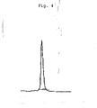
Fig. 1 is a graph showing the results of ELISA where specificity of scFv of isolated clones with human MCP-1 is assessed. -
Fig. 2 is a graph showing the results of ELISA where a binding of the purified scFv derive from human with human MCP-1 was measured. -
Fig. 3 is a graph showing that scFv inhibits cell migration of human monocytic cell line THP-1 mediated by human MCP-1. -
Fig. 4 shows HPLC pattern of the purified MC32 in the immunoglobulin form wherein flow rate: 0.5 mL/min.; initiation buffer: 100mM PB, pH 7.2 + 0.5M NaCl. -
Fig. 5 is a graph showing a binding of the purified MC32 in the immunoglobulin form with MCP-1. - The human antibody and a fragment of said antibody of the present invention may be prepared e.g. by the procedures as described hereinbelow.
- mRNAs were extracted from peripheral blood B lymphocytes from healthy adults and immunoglobulin VH chain and VL chain genes were amplified by RT-PCR with primer pairs defining both ends of the VH chain and VL chain genes to provide each population of H chain and L chain V region genes with diverse sequences. Then, amplification was further performed with a DNA encoding a peptide linker and with primer pairs defining both ends of said DNA so that the ends of said DNA are linked to the H chain gene and L chain gene, respectively, to prepare a population of scFv DNAs with random combination of H chain and L chain V region genes. The obtained scFv DNAs were incorporated into phagemid vector pCANTAB5E to prepare an scFv display phage library. The library is then reacted with human MCP-1 immobilized on a plastic tube. After scFv phages not reacted were removed by washing, scFv phage clones bound to human MCP-1 were eluted with an acid. scFv DNAs are prepared from the isolated phage clones and incorporated into an expression vector and host cells transformed with said expression vector are cultured by the conventional manner to provide the desired scFv protein alone.
- For expression of scFv DNAs, the expression may be performed in E. coli. For expression in E. coli, a signal sequence for secretion of an antibody may functionally be linked to scFv to be expressed with such a useful promoter as routinely used in the art. Such a promoter includes, for instance, lacZ promoter, araB promoter, etc. For a signal sequence for secretion of scFv, pelB signal sequence may be used (Lei, SP. et al., J. Bacteriol., 1987, 169 : 4379-4383) for expression in periplasm of E. coli. For expression in culture supernatant, a signal sequence of g3 protein of M13 phage may also be used.
- The scFv thus expressed may be isolated from within and without the cells and purified to uniformity. Since the scFv expressed in accordance with the present invention has an E tag sequence at its C-terminal, it can easily be purified with affinity chromatography using an anti-E tag antibody in a short period of time. It can also be purified by a combination of the conventional isolation/purification processes used in the protein chemistry. For instance, the antibody may be isolated and purified by a combination of ultrafiltration, salting-out method, and column chromatography such as gel filtration, ion exchange, or hydrophobic chromatography.
- The scFv protein obtained in accordance with the present invention was found to have a binding activity to human MCP-1. As a measurement of an antigen-binding activity of the anti-human MCP-1 antibody as used in the present invention, ELISA, BIAcore, etc. may be used. For instance, in case of ELISA, a sample containing the desired anti-human MCP-1 antibody or a fragment of said antibody, such as culture supernatant of E. coli or a purified antibody, may be added to a 96-well plate to which human MCP-1 is immobilized. To the plate may then be added a secondary antibody labeled with an enzyme such as peroxidase. The plate may be incubated, washed, and added with a chromogenic substrate TMBZ and absorbance is determined to thereby assess an antigen-binding activity.
- Moreover, the scFv protein obtained in accordance with the present invention was found to inhibit the cell migration mediated by human MCP-1. Migration (Chemotaxis) of sensitive cells by human MCP-1 may be investigated with chemotaxis assay routinely used in the art, e.g. as described by Grob et al. (Grob PM. et al., J. Biol. Chem., 1990, 265: 8311-8316). Specifically, using a commercially available chemotaxis chamber, each of the anti-human MCP-1 antibody and human MCP-1 are diluted with a culture solution such as RPMI1640 and mixed together, and the mixture is incubated at room temperature for a fixed time and then added to the lower part of the chamber partitioned with a filter. Then, a suspension of human MCP-1 sensitive cells such as, for instance, monocytic cell line THP-1, or human peripheral blood mononuclear cells (hereinafter also referred to as "PBMC") is added to the upper part of the chamber and left to stand at 37°C for a fixed time. Migrating cells will move towards the lower part of the chamber through the filter attached thereto. Thus, cells adhered to the filter may be dyed with e.g. Giemsa staining for counting a cell number. Alternatively, a cell number may be counted for cells moved to the lower part of the chamber with e.g. a Coulter counter. In place of the chamber described above, commercially available disposable assay cells for chemotaxis assay may also be used. The chemotaxis assay system revealed that the scFv protein of the present invention inhibited the cell migration mediated by human MCP-1.
- As described above, since the scFv protein obtained in accordance with the present invention may inhibit the cell migration mediated by human MCP-1 in a concentration dependent manner, it is expected to be efficacious for the prevention or treatment of diseases induced by said cell migration.
- The amino acid sequences of VH and VL chains of the above scFv clone having the inhibitory activity as well as the nucleotide sequences coding therefor are indicated in SEQ ID NOs: 1 and 2 (VH chain) and in SEQ ID NOs: 6 and 7 (VL chain), respectively.
- In addition, the amino acid sequences of complementarity determining regions (CDR1 to CDR3), which are included in the above amino acid sequences, of VH and VL chains are shown below.
- [VH chain]
CDR1: Ser Tyr Ala Ile Ser (SEQ ID NO: 3)
CDR2:CDR3: Asp Leu Gly Gly Gly Asp Tyr Tyr Tyr Gly Met Asp Val (SEQ ID NO: 5)
- [VL chain]
CDR1: Arg Ser Ser Gln Ser Ile Asn Thr Tyr Leu His (SEQ ID NO: 8)
CDR2: Ala Ala Ser Thr Leu Gln Ser (SEQ ID NO: 9)
CDR3: Gln Gln Ser Phe Thr Thr Pro Leu Thr (SEQ ID NO: 10) - Although the VH chain and/or the VL chain of the human anti-human MCP-1 antibody as disclosed herein were obtained in the form of scFv by using the phage antibody technique, the present invention encompasses human anti-human MCP-1 antibody fragment such as Fab, Fab' or F(ab')2 wherein the disclosed VH chain and/or VL chain are combined with a portion of a constant region of a human immunoglobulin, and other human anti-human MCP-1 antibody fragment such as a human anti-human MCP-1 single chain antibody (scAb) wherein scFv is combined with a constant region of a human immunoglobulin, as well as gene fragments encoding the antibody fragments. The present invention further encompasses a modified protein molecule wherein a high molecular weight modifying agent such as polyethylene glycol is combined with these antibody and antibody fragment protein molecules. For preparing scFv in which each Fv of the H chain and the L chain are linked together with a suitable linker, a peptide linker to be used may be any single chain peptide having e.g. 10-25 amino acid residues.
- As described above, the scFv fragment of the human anti-human MCP-1 antibody according to the present invention, containing a specific variable region of a human anti-human MCP-1 antibody, may potentially interact with human MCP-1 to thereby inhibit the binding between human MCP-1 and a human MCP-1 receptor. In addition, the scFv fragment of the human anti-human MCP-1 antibody according to the present invention may inhibit various immune responses induced by human MCP-1 and hence may be used as a medicament for the prevention and treatment of inflammation and immunopathy induced by said immune responses, e.g. as an anti-inflammatory agent or a medicament for the treatment and prevention of autoimmune diseases. Besides, the scFv fragment of the present invention is expected to contribute to the prevention and treatment of myocardial infarction and arteriosclerosis.
- The present invention is explained in more detail by means of the following Examples.
- Phage library was constructed as reported by J. D. Marks et al., J. Mol. Biol., 222: 581-597, 1991 with some modification, using lymphocytes from peripheral blood taken from 20 healthy donors as a starting material.
- Namely, lymphocytes were isolated from peripheral blood taken from 20 healthy donors by sedimentary centrifugation with Ficol, washed thoroughly with PBS and then treated with ISOGEN (NIPPON GENE CO., LTD) to prepare a total RNA. The obtained total RNA was divided into four samples and from each of the samples were prepared cDNAs with primers specific to constant regions of either human IgG, IgM, κ chain or λ chain using first strand cDNA synthesis kit (Pharmacia biotech). Using each of the obtained cDNAs as a template, each of antibody V region genes were amplified by polymerase chain reaction (PCR) using primers specific to either of combinations of VH(y or µ) and JH, Vκ and Jκ, or Vλ and Jλ, as described by Marks et al.
- Then, VH (γ or µ) and Vκ, and VH (γ or µ) and Vλ, were linked together with a linker DNA by assembly PCR (McCafferty, J. et al.: Antibody Engineering - A Practical Approach, IRL Press, Oxford, 1996) to prepare single chain scFv DNAs. The obtained scFv DNAs were added with NotI and SfiI restriction sites using PCR, electrophoresed on agarose gel and then purified. The purified scFv DNAs were digested with the restriction enzymes NotI (Takara) and SfiI (Takara) and then cloned into phagemid pCANTAB5E (Pharmacia). The obtained phagemids pCANTAB5E where scFv DNA was bound were introduced into E. coli TG1 cells by electroporation for each of VH(γ)-Vκ, VH(γ)-Vλ, VH(µ)-Vκ, and VH(µ)-Vλ. From the number of the transformed TG1 cells, it was assessed that VH(γ)-Vκ, VH(γ)-Vλ, VH(µ)-Vκ and VH(µ)-Vλ exhibited diversity of 1.1×10⁸, 2.1×10⁸, 8.4×10⁷ and 5.3×10⁷ clones, respectively. With M13KO7 helper phage, phage antibodies were expressed on the transformed TG1 cells to prepare scFv display phage library derived from healthy donors.
- Human MCP-1 was dissolved in 1mL 0.1M NaHCO3 and the solution was incubated in 35mm dish (Iwaki) at 4°C overnight to immobilize IL-6. To the dish was added 0.5% gelatin/PBS for blocking at 20°C for 2 hours and then the dish was washed six times with 0.1% Tween20-PBS. To the dish was then added 0.9mL of the single chain antibody display phage solution (1×10¹² tu/mL of the antibody phage library derived from healthy donors) for reaction.
- After washing the dish ten times with 0.1% Tween20-PBS, 1.0mL glycine buffer (pH 2.2) was added to elute single chain antibody display phages bound to human MCP-1. After adjusting pH by adding 1M Tris (hydroxymethyl)-aminomethane-HCl, pH9.1, the eluted phages were infected to E. coli TG1 cells at logarithmic growth phase. The infected TG1 cells were centrifuged at 3,000xg for 10 minutes. Supernatant was removed, suspended in 200µL 2xYT culture medium, plated on SOBAG plate (SOB plate containing 2% glucose, 100 µg/ml ampicillin) and then incubated overnight in an incubator at 30°C. The resulting colonies were suspended and recovered in a suitable amount of 2×YT culture medium with a scraper (Coastor).
- The obtained TG1 solution (50µL) _was inoculated on
30mL 2×YT culture medium and rescued with a helper phage to prepare a phage library after screening. For each of the phage libraries VH(γ)-Vκ, VH(γ)-Vλ, VH(µ)-Vκ and VH(µ)-Vλ derived from healthy donors, four pannings in total were performed with the human MCP-1 immobilized plate. After the fourth panning, any clone was extracted arbitrarily from the SOBAG plate. The scFv expression was confirmed, specificity was confirmed by human MCP-1 ELISA and a nucleotide sequence was analyzed. - For screening the isolated clones, ELISA was performed as follows: Human MCP-1 and human MIP-1α (macrophage inflammatory protein 1-α) were immobilized on an ELISA plate for screening. Each 2 µg/mL of a human MCP-1 or human (MIP-1α, or 2.5 µg/mL of a human serum albumin (HSA) were placed in an ELISA plate (Nunc) which was kept standing at 4°C for 16 hours for immobilization. To the immobilized plate was added 400 µL/well of a PBS solution containing 0.5% BSA, 0.5% gelatin and 5% skimmed milk and was kept standing at 4°C for 2 hours for blocking.
- To the plate was added 40 µL/well of sample solutions containing scFv display phage for reaction. The sample solutions were discarded and the plate was washed with a washing solution five times. The plate was reacted with a biotin-labeled anti-M13 monoclonal antibody (Pharmacia biotech) and then with an anti-mouse IgG antibody labeled with alkaline phosphatase (AP). After washing with a washing solution five times, the plate was added with 50 µL/well of a chromogenic substrate solution, i.e. a PBS solution containing 1 g/mL p-nitrophenyl phosphate (Wako) and 10% diethanolamine (Wako), light-shielded, and developed at room temperature to 37°C for 5 to 10 minutes. Absorbance at 405nm was measured using Multiplate Autoreader NJ-2001 (Inter Med). As a result, all the clones assessed were confirmed to be specific to human MCP-1 (
Fig. 1 ). - A DNA nucleotide sequence of the isolated clones was determined for scFv gene VH and VL using Dye terminator cycle sequencing FS Ready Reaction kit (Applied Biosystems) (SEQ ID NOs: 1 and 6). As a result of ELISA and sequence analysis, the isolated clones were classified into four classes.
- Plasmid DNAs were recovered from the four scFv clones MC8, MC15, MC32 and MC59 reactive with human MCP-1 isolated in Examples 2 and 3 as described above and E. coli HB1251 was transformed with said plasmid DNAs in accordance with the conventional technique. The E. coli cells were cultured overnight on 2xYT medium containing 2% glucose and then a portion of the cells were transferred to 2×YT medium free from glucose and thereto was added IPTG at a final concentration of 1mM for overnight culture to induce expression of scFv. After completion of culture, the cells were recovered, suspended in PBS containing 1mM EDTA and placed on ice for 30 minutes. Then, centrifugation was performed at 8,900xg for 30 minutes. A supernatant was recovered and passed through 0.45µm filter and the filtrate was used as a starting material for purifying scFv from a periplasmic fraction.
- The thus prepared starting material for purification was purified by affinity chromatography with an anti-E tag antibody in accordance with the conventional technique. After dialysis with PBS, endotoxins were removed with an endotoxin-removing column Detoxi-gel (PIERCE) in accordance with the protocol attached thereto. After concentration with Centricon (Amicon) with a molecular weight cut-off of 10,000, filtration through 0.45µm filter provided a purified product.
- Binding of the purified scFv with human MCP-1 was then measured by ELISA. To a 96-well plate (NUNC. MAXISORP) immobilized with human MCP-1 prepared at 0.5 µg/mL with PBS was added 100µL of the purified antibody for reaction at 37°C for 1 hour. After washing five times with 0.05% Tween-PBS (hereinafter also referred to as "PBST"), the plate was further reacted with an anti-E tag antibody labeled with peroxidase at 37°C for 1 hour. After washing five times with PBST, to the plate was added a chromogenic substrate solution for development and absorbance at 450nm was measured to assess the binding. The results are shown in
Fig. 2 . All the four antibodies were found to bind to human MCP-1 in a concentration dependent manner. - The inhibitory activity of the antibody of the present invention to the cell migration mediated by human MCP-1 to monocytes was investigated by chemotaxis assay. Transwells with a pore size of 8µm (Costar) were set on each well of a 24-well plate. To this 24-well plate was added 540µL of RPMI 1640 medium containing 1% FCS (hereinafter also referred to as "1% FCS-RPMI"). Then, each equivalent amount of scFv of an adjusted concentration and 2×10⁻⁸ M human MCP-1 (CHEMICON) were mixed and incubated at room temperature for 30 minutes. 60µL of the reaction solution was added to a 24-well plate containing 540µL of the medium. To Transwells were added 100µL of 1% FCS-RPMI and 1×10⁶ cells/mL of human monocytic cell line THP-1 (200µL) and the plate was kept to stand at 37°C for 4 hours. Thus, the cells reside in the upper Transwell portion that is partitioned with the 8µm filter from the lower 24-well plate where the mixture of the antibody is placed. The cells that migrate through the filter towards the 24-well plate were counted with a Coulter counter (Coulter). The results of this assay are shown in
Fig. 3 . Among the four antibodies, MC15 and MC32 were found to have an inhibitory activity to the cell migration mediated by human MCP-1. - From the expression plasmid in which scFv DNA of scFv clone MC32 isolated in Example 3 was incorporated, each DNA encoding VH chain and VL chain regions were amplified by PCR. Each PCR primers used for the amplification are indicated below.
- [VH sense chain]

- [VH antisense chain]
5'-TGA GGA TAC GGT GAC CGT GG-3' (SEQ ID NO: 12) - [VL sense chain]

- [VL antisense chain]
5'-ACG TTT GAT CTC CAC CTT GG-3' (SEQ ID NO: 14) - The amplified DNAs of the VH chain and VL chain were each cloned into plasmid DNA pUC18, in which a leader sequence necessary for secretion in animal cells is incorporated, at the downstream of said leader sequence.
- The thus obtained plasmid DNAs were digested with HindIII (TAKARA BIO INC.)-BamHI (TAKARA BIO INC.) at 37°C for 2 hours and were electrophoresed on 2% agarose gel (TAKARA BIO INC.) to recover VH chain and VL chain DNA fragments containing the signal sequence.
- The expression plasmid pCAG-H, in which the H chain constant region (hinge-CH1-CH2-CH3) gene of a human antibody IgG1 is incorporated, was digested with HindIII-BamHI at 37°C for 2 hours. To the prepared vector DNA fragment was inserted the HindIII-BamHI fragment of the VH chain previously prepared. E. coli HB101 cells were transformed with the resulting expression plasmid and the plasmid was prepared from drug (ampicillin) resistant colonies and treated with the restriction enzymes to confirm the insertion of the VH chain.
- Likewise, the VL chain DNA fragment was inserted into the expression plasmid pCAG-L in which the L chain (κ chain) constant region (Cκ) gene of a human antibody is incorporated.
- BMT-10 cells were used for transient expression. Each 5mL of BMT-10 cells maintained on D'MEM (Invitrogen) with 8% FCS (Invitrogen) were dispensed into sterilized small laboratory dishes (diameter 6cm; Corning) at a cell concentration of 1.5×10⁵ cells/mL and incubated in CO2 incubator at 37°C overnight. After washing the cells twice with PBS (SIGMA), the culture medium was replaced with 5mL of OPTI-MEM (Invitrogen) with a lower serum level. Two disposable centrifuge tubes (FALCON) made of polystyrene were provided. In one tube, 10µL of Lipofectamine reagent (Invitrogen) and 90µL of OPTI-MEM culture medium were mixed together (hereinafter referred to as "Lipofectamine solution"). In the other tube, each 3µg of the expression plasmid DNAs of the H chain and L chain as previously prepared were added and thereto 100µL of OPTI-MEM was further added (hereinafter referred to as "DNA solution"). The DNA solution was added drop by drop to the Lipofectamine solution and the mixture was stirred at room temperature for 30 minutes for reaction. After completion of the reaction, a total amount (200µL) of the solution was added drop by drop to laboratory dish and incubated in CO2 incubator at 37°C for 6 hours. After six hours, the culture medium was removed by suction, D'MEM with 8% FCS was gently added and the dish was incubated at 37°C for 4 days. After four days, supernatant was recovered and passed through 0.22µm filter and the filtrate was used as a starting material for purification.
- Purification was performed in accordance with the conventional technique using a purification system of Biologic Duo Flow (BIO RAD) and Protein G column (Pharmacia).
- Specifically, the Protein G column was equilibrated with PBS and then 50mL of the above culture supernatant was applied to the column at a flow rate of 1 mL/min. After washing the column with PBS at a 50-folds larger volume than a gel bed, elution was carried out with 0.1M glycine-HCl, pH2.7. Each 1mL of the eluate was recovered to an ET free disposable tube (FALCON 2063 etc.) to which 50µl of 1M Tris-HCl, pH9.0 was previously added for neutralization. Absorbance at 280nm was immediately measured for each fraction with a spectrophotometer and major fractions were pooled (normally 2mL) and were dialyzed against PBS at 4°C overnight. Purification assay of the purified antibody was performed by HPLC with G3000SW column (Toso) and by SDS-PAGE. One of the results of HPLC is shown in
Fig. 4 where flow rate: 0.5 mL/min.; initiation buffer: 100mM PB, pH7.2 + 0.5M NaCl. - Binding of the purified MC32 antibody in the immunoglobulin form with MCP-1 was assessed by ELISA. After a 96-well plate (Maxisorp; Nunc) immobilized with human MCP-1 (Chemicon) prepared at 0.5 µg/mL with PBS was blocked with 1% BSA/PBS, the purified anti-MCP-1 antibody MC32 in the immunoglobulin form was used with two-fold serial dilution with 1% BSA-0.05% Tween/PBS starting from 5 µg/mL. After reaction at 37°C for 1 hour, the plate was washed five times with 0.05% Tween/PBS and further reacted with an anti-human IgG antibody labeled with peroxidase at 37°C for 1 hour. After washing five times with PBST, a chromogenic substrate TMBZ was added to the plate for development and absorbance at 450nm was measured to assess the binding. The results are shown in
Fig. 5 . The purified MC32 antibody in the immunoglobulin form bound to MCP-1 in a concentration dependent manner as in the case of scFv. -
- <110> The Chemo-Sero-Therapeutic Research Institute
- <120> Human anti-human MCP-1 antibody and fragment thereof
- <130> 663985
- <150>
JP 2002-267184 - <151> 2002-09-12
- <160> 14
- <210> 1
- <211> 366
- <212> DNA
- <213> Homo sapiens
- <400> 1


- <210> 2
- <211> 122
- <212> PRT
- <213> Homo sapiens
- <400> 2


- <210> 3
- <211> 5
- <212> PRT
- <213> Homo sapiens
- <220>
- <223> CDR1 corresponding to amino acids No. 31 to No. 35 in SEQ ID NO: 2
- <400> 3

- <210> 4
- <211> 17
- <212> PRT
- <213> Homo sapiens
- <220>
- <223> CDR2 corresponding to amino acids No. 50 to No. 66 in SEQ ID NO: 2
- <400> 4

- <210> 5
- <211> 13
- <212> PRT
- <213> Homo sapiens
- <220>
- <223> CDR3 corresponding to amino acids No. 99 to No. 111 in SEQ ID NO: 2
- <400> 5

- <210> 6
- <211> 324
- <212> DNA
- <213> Homo sapiens
- <400> 6


- <210> 7
- <211> 108
- <212> PRT
- <213> Homo sapiens
- <400> 7

- <210> 8
- <211> 11
- <212> PRT
- <213> Homo sapiens
- <220>
- <223> CDR1 corresponding to amino acids No. 24 to No. 34 in SEQ ID NO: 7
- <400> 8

- <210> 9
- <211> 7
- <212> PRT
- <213> Homo sapiens
- <220>
- <223> CDR2 corresponding to amino acids No. 50 to No. 56 in SEQ ID NO: 7
- <400> 9

- <210> 10
- <211> 9
- <212> PRT
- <213> Homo sapiens
- <220>
- <223> CDR3 corresponding to amino acids No. 89 to No. 97 in SEQ ID NO: 7
- <400> 10

- <210> 11
- <211> 42
- <212> DNA
- <213> Artificial
- <220>
- <223> VH chain sense primer
- <400> 11
cgtggctcct gggcccacag ccaggtacag ctgcagcagt ca 42 - <210> 12
- <211> 20
- <212> DNA
- <213> Artificial
- <220>
- <223> VH chain antisense primer
- <400> 12
tgaggatacg gtgaccgtgg 20 - <210> 13
- <211> 42
- <212> DNA
- <213> Artificial
- <220>
- <223> VL chain sense primer
- <400> 13
cgtggctcct gggcccacag cgacatccag ttgacccagt ct 42 - <210> 14
- <211> 20
- <212> DNA
- <213> Artificial
- <220>
- <223> VL chain antisense primer
- <400> 14
acgtttgatc tccaccttgg 20
Claims (7)
- A nucleotide sequence coding for a single chain Fv (scFv) that binds to human MCP-1 and inhibits induction of inflammation in chronic inflammatory diseases and arteriosclerosis, said nucleotide sequence consisting of the nucleotide sequence coding for the VH domain of SEQ ID NO:2 combined with the nucleotide sequence coding for the VL domain of SEQ ID NO:7.
- A nucleotide sequence coding for an scFv that binds to human MCP-1 and inhibits induction of inflammation in chronic inflammatory diseases and arteriosclerosis, said nucleotide sequence consisting of the nucleotide sequence coding for the VH domain of SEQ ID NO:2 combined with a portion of a human antibody heavy chain constant region gene and the nucleotide sequence coding for the VL domain of SEQ ID NO:7 combined with a portion of a human antibody light chain constant region gene.
- A nucleotide sequence coding for a human anti-human MCP-1 antibody fragment that binds to human MCP-1 and inhibits induction of inflammation in chronic inflammatory diseases and arteriosclerosis, said nucleotide sequence consisting of the scFv gene fragment of claim 1 combined with either a portion of a human antibody heavy chain constant region gene or with a portion of a human antibody light chain constant region gene.
- A human anti-human MCP-1 scFv antibody that binds to human MCP-1 and inhibits induction of inflammation in chronic inflammatory diseases and arteriosclerosis, which is expressed by the genetic recombination technique from an expression vector in which the nucleotide sequence coding for the VH domain of SEQ ID NO:2 and the nucleotide sequence coding for the VL domain of SEQ ID NO:7 are incorporated.
- A modified protein molecule consisting of the scFv as defined in claim 4 to which a polyethylene glycol is bound.
- An agent for inhibiting the activity of human MCP-1 comprising as an active ingredient the scFv of claim 4 or the modified protein molecule of claim 5.
- A medicament for preventing or treating inflammation, autoimmune diseases, myocardial infarction and arteriosclerosis, said medicament comprising the agent for inhibiting the activity of human MCP-1 of claim 6.
Applications Claiming Priority (3)
| Application Number | Priority Date | Filing Date | Title |
|---|---|---|---|
| JP2002267184 | 2002-09-12 | ||
| JP2002267184 | 2002-09-12 | ||
| PCT/JP2003/011560 WO2004024921A1 (en) | 2002-09-12 | 2003-09-10 | Human antihuman mcp-1 antibody and antibody fragment thereof |
Publications (3)
| Publication Number | Publication Date |
|---|---|
| EP1538207A1 EP1538207A1 (en) | 2005-06-08 |
| EP1538207A4 EP1538207A4 (en) | 2006-05-24 |
| EP1538207B1 true EP1538207B1 (en) | 2010-06-02 |
Family
ID=31986692
Family Applications (1)
| Application Number | Title | Priority Date | Filing Date |
|---|---|---|---|
| EP03795363A Expired - Lifetime EP1538207B1 (en) | 2002-09-12 | 2003-09-10 | Human anti-human mcp-1 antibody and antibody fragment thereof |
Country Status (11)
| Country | Link |
|---|---|
| US (1) | US7342106B2 (en) |
| EP (1) | EP1538207B1 (en) |
| JP (1) | JP4426449B2 (en) |
| KR (1) | KR20050042801A (en) |
| CN (1) | CN1681926A (en) |
| AT (1) | ATE469969T1 (en) |
| AU (1) | AU2003262051B2 (en) |
| CA (1) | CA2497804A1 (en) |
| DE (1) | DE60332847D1 (en) |
| DK (1) | DK1538207T3 (en) |
| WO (1) | WO2004024921A1 (en) |
Families Citing this family (25)
| Publication number | Priority date | Publication date | Assignee | Title |
|---|---|---|---|---|
| US7732162B2 (en) | 2003-05-05 | 2010-06-08 | Probiodrug Ag | Inhibitors of glutaminyl cyclase for treating neurodegenerative diseases |
| CA2540308C (en) | 2003-09-26 | 2013-08-06 | Alza Corporation | Drug coating providing high drug loading and methods for providing the same |
| US8114964B2 (en) * | 2005-05-19 | 2012-02-14 | Centocor, Inc. | Anti-MCP-1 antibodies, compositions, methods and uses |
| PE20061444A1 (en) * | 2005-05-19 | 2007-01-15 | Centocor Inc | ANTI-MCP-1 ANTIBODY, COMPOSITIONS, METHODS AND USES |
| US8278345B2 (en) | 2006-11-09 | 2012-10-02 | Probiodrug Ag | Inhibitors of glutaminyl cyclase |
| JP5523107B2 (en) | 2006-11-30 | 2014-06-18 | プロビオドルグ エージー | Novel inhibitors of glutaminyl cyclase |
| EP2097451A2 (en) * | 2006-12-22 | 2009-09-09 | Ablynx N.V. | Anti-chemokine (ccl2, ccl3, ccl5, cxcl11, cxcl12) single-domain antibodies |
| NZ579310A (en) | 2007-03-01 | 2012-03-30 | Probiodrug Ag | Use of glutaminyl cyclase inhibitors for the treatment of mild cognitive impairment and diagnostic purposes thereof |
| EP2142514B1 (en) | 2007-04-18 | 2014-12-24 | Probiodrug AG | Thiourea derivatives as glutaminyl cyclase inhibitors |
| CA2695237A1 (en) * | 2007-08-02 | 2009-04-30 | Novimmune S.A. | Anti-rantes antibodies and methods of use thereof |
| US8710191B2 (en) | 2008-08-18 | 2014-04-29 | Pfizer Inc. | Antibodies to CCR2 |
| NZ591187A (en) | 2008-08-20 | 2012-07-27 | Probiodrug Ag | Antibodies directed against pyroglutamate monocyte chemoattractant protein-1 (mcp-1 n1pe) |
| CA2772488C (en) | 2009-09-11 | 2018-04-17 | Probiodrug Ag | Heterocyclic derivatives as inhibitors of glutaminyl cyclase |
| US9181233B2 (en) | 2010-03-03 | 2015-11-10 | Probiodrug Ag | Inhibitors of glutaminyl cyclase |
| US8269019B2 (en) | 2010-03-10 | 2012-09-18 | Probiodrug Ag | Inhibitors |
| EP2560953B1 (en) | 2010-04-21 | 2016-01-06 | Probiodrug AG | Inhibitors of glutaminyl cyclase |
| JP6050264B2 (en) | 2011-03-16 | 2016-12-21 | プロビオドルグ エージー | Benzimidazole derivatives as inhibitors of glutaminyl cyclase |
| AU2015301530A1 (en) * | 2014-08-15 | 2017-03-02 | PixarBio Corporation | Compositions for inhibiting inflammation in a subject with a spinal cord injury and methods of using the same |
| EP3551047A1 (en) | 2016-12-07 | 2019-10-16 | Progenity, Inc. | Gastrointestinal tract detection methods, devices and systems |
| EP3554541B1 (en) | 2016-12-14 | 2023-06-07 | Biora Therapeutics, Inc. | Treatment of a disease of the gastrointestinal tract with a chemokine/chemokine receptor inhibitor |
| PL3461819T3 (en) | 2017-09-29 | 2020-11-30 | Probiodrug Ag | Inhibitors of glutaminyl cyclase |
| CN116726362A (en) | 2018-11-19 | 2023-09-12 | 比奥拉治疗股份有限公司 | Methods and devices for treating diseases with biologic therapeutic agents |
| CN115666704A (en) | 2019-12-13 | 2023-01-31 | 比奥拉治疗股份有限公司 | Ingestible device for delivery of therapeutic agents to the gastrointestinal tract |
| CN112358547A (en) * | 2020-09-30 | 2021-02-12 | 浙江大学 | Anti-human CXCL-2 monoclonal antibody 3-D3 and coding gene and application thereof |
| CN112778414B (en) * | 2020-12-31 | 2022-04-15 | 中南大学湘雅二医院 | cBIN1 antibody and application thereof |
Family Cites Families (4)
| Publication number | Priority date | Publication date | Assignee | Title |
|---|---|---|---|---|
| JPH0967399A (en) * | 1995-08-30 | 1997-03-11 | Mitsui Toatsu Chem Inc | Anti-mcp-1 human monoclonal antibody |
| JP4298051B2 (en) * | 1999-04-13 | 2009-07-15 | 和久 杉村 | Anti-MCP-1 antibody-recognizing peptide mimix, production method thereof and use thereof |
| WO2001089582A1 (en) * | 2000-05-26 | 2001-11-29 | Takeda Chemical Industries, Ltd. | Preventives and remedies for pulmonary hypertension |
| GB0016138D0 (en) | 2000-06-30 | 2000-08-23 | Novartis Ag | Organic compounds |
-
2003
- 2003-09-10 DE DE60332847T patent/DE60332847D1/en not_active Expired - Lifetime
- 2003-09-10 KR KR1020057004050A patent/KR20050042801A/en not_active Application Discontinuation
- 2003-09-10 EP EP03795363A patent/EP1538207B1/en not_active Expired - Lifetime
- 2003-09-10 US US10/527,823 patent/US7342106B2/en not_active Expired - Fee Related
- 2003-09-10 CN CNA038215527A patent/CN1681926A/en active Pending
- 2003-09-10 WO PCT/JP2003/011560 patent/WO2004024921A1/en active Application Filing
- 2003-09-10 CA CA002497804A patent/CA2497804A1/en not_active Abandoned
- 2003-09-10 DK DK03795363.5T patent/DK1538207T3/en active
- 2003-09-10 AU AU2003262051A patent/AU2003262051B2/en not_active Ceased
- 2003-09-10 JP JP2004535928A patent/JP4426449B2/en not_active Expired - Fee Related
- 2003-09-10 AT AT03795363T patent/ATE469969T1/en active
Non-Patent Citations (1)
| Title |
|---|
| LIU E ET AL: "Conversion of an antibody into an enzyme which cleaves the protein HPr.", MOLECULAR IMMUNOLOGY NOV 1998, vol. 35, no. 16, November 1998 (1998-11-01), pages 1069 - 1077, ISSN: 0161-5890 * |
Also Published As
| Publication number | Publication date |
|---|---|
| JP4426449B2 (en) | 2010-03-03 |
| KR20050042801A (en) | 2005-05-10 |
| DK1538207T3 (en) | 2010-07-05 |
| AU2003262051B2 (en) | 2008-09-25 |
| ATE469969T1 (en) | 2010-06-15 |
| DE60332847D1 (en) | 2010-07-15 |
| EP1538207A1 (en) | 2005-06-08 |
| JPWO2004024921A1 (en) | 2006-01-12 |
| AU2003262051A1 (en) | 2004-04-30 |
| EP1538207A4 (en) | 2006-05-24 |
| US20060246069A1 (en) | 2006-11-02 |
| WO2004024921A1 (en) | 2004-03-25 |
| CA2497804A1 (en) | 2004-03-25 |
| US7342106B2 (en) | 2008-03-11 |
| CN1681926A (en) | 2005-10-12 |
Similar Documents
| Publication | Publication Date | Title |
|---|---|---|
| EP1538207B1 (en) | Human anti-human mcp-1 antibody and antibody fragment thereof | |
| US7482436B2 (en) | Human antihuman interleukin-6 antibody and fragment of antibody | |
| EP1529063B1 (en) | Antibodies anti-c5 component of the complement system and their use | |
| Steidl et al. | In vitro affinity maturation of human GM-CSF antibodies by targeted CDR-diversification | |
| CN108251431B (en) | Double-carrier system and application thereof | |
| CA2643226C (en) | Antibodies against human il-22 and uses therefor | |
| CA2642570C (en) | Methods of using antibodies against human il-22 | |
| CA2564432A1 (en) | Human anti-amyloid .beta. peptide antibody and fragment of said antibody | |
| KR101501736B1 (en) | Recombinant monoclonal antibody specifically binding to l1cam expressed in humans and mice and the use thereof | |
| KR20230153495A (en) | Antibodies to ticagrelor and methods of use | |
| Yoshinaga et al. | Ig L-chain shuffling for affinity maturation of phage library-derived human anti-human MCP-1 antibody blocking its chemotactic activity | |
| US7384633B2 (en) | Human type antihuman IgE receptor antibody and fragment | |
| KR101525919B1 (en) | Bispecific antibody based on TNFR2 for preventing and traeating autoimmune disease | |
| CA2411515A1 (en) | Generation of libraries of antibodies in yeast and uses thereof | |
| KR20200135331A (en) | Antibodies to Feline McDonough's Sarcoma (FMS)-Like Tyrosine Kinase 3 Receptor Ligand (FLT3L) and Its Use to Treat Autoimmune and Inflammatory Diseases | |
| RU2788128C2 (en) | Antibodies to ligand of receptor tyrosine kinase 3 similar to product of protooncogene of feline sarcoma virus (fms) of mcdonough (flt3l) strain and options of their use for treatment of autoimmune and inflammatory diseases |
Legal Events
| Date | Code | Title | Description |
|---|---|---|---|
| PUAI | Public reference made under article 153(3) epc to a published international application that has entered the european phase |
Free format text: ORIGINAL CODE: 0009012 |
|
| 17P | Request for examination filed |
Effective date: 20050304 |
|
| AK | Designated contracting states |
Kind code of ref document: A1 Designated state(s): AT BE BG CH CY CZ DE DK EE ES FI FR GB GR HU IE IT LI LU MC NL PT RO SE SI SK TR |
|
| A4 | Supplementary search report drawn up and despatched |
Effective date: 20060406 |
|
| 17Q | First examination report despatched |
Effective date: 20060831 |
|
| 17Q | First examination report despatched |
Effective date: 20060831 |
|
| GRAP | Despatch of communication of intention to grant a patent |
Free format text: ORIGINAL CODE: EPIDOSNIGR1 |
|
| RTI1 | Title (correction) |
Free format text: HUMAN ANTI-HUMAN MCP-1 ANTIBODY AND ANTIBODY FRAGMENT THEREOF |
|
| GRAS | Grant fee paid |
Free format text: ORIGINAL CODE: EPIDOSNIGR3 |
|
| GRAA | (expected) grant |
Free format text: ORIGINAL CODE: 0009210 |
|
| AK | Designated contracting states |
Kind code of ref document: B1 Designated state(s): AT BE BG CH CY CZ DE DK EE ES FI FR GB GR HU IE IT LI LU MC NL PT RO SE SI SK TR |
|
| REG | Reference to a national code |
Ref country code: GB Ref legal event code: FG4D |
|
| REG | Reference to a national code |
Ref country code: CH Ref legal event code: EP Ref country code: CH Ref legal event code: NV Representative=s name: BOHEST AG |
|
| REG | Reference to a national code |
Ref country code: DK Ref legal event code: T3 |
|
| REG | Reference to a national code |
Ref country code: IE Ref legal event code: FG4D |
|
| REF | Corresponds to: |
Ref document number: 60332847 Country of ref document: DE Date of ref document: 20100715 Kind code of ref document: P |
|
| REG | Reference to a national code |
Ref country code: NL Ref legal event code: VDEP Effective date: 20100602 |
|
| PG25 | Lapsed in a contracting state [announced via postgrant information from national office to epo] |
Ref country code: SE Free format text: LAPSE BECAUSE OF FAILURE TO SUBMIT A TRANSLATION OF THE DESCRIPTION OR TO PAY THE FEE WITHIN THE PRESCRIBED TIME-LIMIT Effective date: 20100602 |
|
| PGFP | Annual fee paid to national office [announced via postgrant information from national office to epo] |
Ref country code: CH Payment date: 20100927 Year of fee payment: 8 |
|
| PG25 | Lapsed in a contracting state [announced via postgrant information from national office to epo] |
Ref country code: SI Free format text: LAPSE BECAUSE OF FAILURE TO SUBMIT A TRANSLATION OF THE DESCRIPTION OR TO PAY THE FEE WITHIN THE PRESCRIBED TIME-LIMIT Effective date: 20100602 Ref country code: FI Free format text: LAPSE BECAUSE OF FAILURE TO SUBMIT A TRANSLATION OF THE DESCRIPTION OR TO PAY THE FEE WITHIN THE PRESCRIBED TIME-LIMIT Effective date: 20100602 |
|
| PGFP | Annual fee paid to national office [announced via postgrant information from national office to epo] |
Ref country code: AT Payment date: 20100927 Year of fee payment: 8 Ref country code: FR Payment date: 20100818 Year of fee payment: 8 Ref country code: IT Payment date: 20100918 Year of fee payment: 8 |
|
| PG25 | Lapsed in a contracting state [announced via postgrant information from national office to epo] |
Ref country code: CY Free format text: LAPSE BECAUSE OF FAILURE TO SUBMIT A TRANSLATION OF THE DESCRIPTION OR TO PAY THE FEE WITHIN THE PRESCRIBED TIME-LIMIT Effective date: 20100602 Ref country code: GR Free format text: LAPSE BECAUSE OF FAILURE TO SUBMIT A TRANSLATION OF THE DESCRIPTION OR TO PAY THE FEE WITHIN THE PRESCRIBED TIME-LIMIT Effective date: 20100903 |
|
| PGFP | Annual fee paid to national office [announced via postgrant information from national office to epo] |
Ref country code: GB Payment date: 20100920 Year of fee payment: 8 |
|
| PG25 | Lapsed in a contracting state [announced via postgrant information from national office to epo] |
Ref country code: NL Free format text: LAPSE BECAUSE OF FAILURE TO SUBMIT A TRANSLATION OF THE DESCRIPTION OR TO PAY THE FEE WITHIN THE PRESCRIBED TIME-LIMIT Effective date: 20100602 Ref country code: EE Free format text: LAPSE BECAUSE OF FAILURE TO SUBMIT A TRANSLATION OF THE DESCRIPTION OR TO PAY THE FEE WITHIN THE PRESCRIBED TIME-LIMIT Effective date: 20100602 |
|
| PGFP | Annual fee paid to national office [announced via postgrant information from national office to epo] |
Ref country code: DK Payment date: 20100913 Year of fee payment: 8 |
|
| PG25 | Lapsed in a contracting state [announced via postgrant information from national office to epo] |
Ref country code: BE Free format text: LAPSE BECAUSE OF FAILURE TO SUBMIT A TRANSLATION OF THE DESCRIPTION OR TO PAY THE FEE WITHIN THE PRESCRIBED TIME-LIMIT Effective date: 20100602 Ref country code: CZ Free format text: LAPSE BECAUSE OF FAILURE TO SUBMIT A TRANSLATION OF THE DESCRIPTION OR TO PAY THE FEE WITHIN THE PRESCRIBED TIME-LIMIT Effective date: 20100602 Ref country code: PT Free format text: LAPSE BECAUSE OF FAILURE TO SUBMIT A TRANSLATION OF THE DESCRIPTION OR TO PAY THE FEE WITHIN THE PRESCRIBED TIME-LIMIT Effective date: 20101004 Ref country code: RO Free format text: LAPSE BECAUSE OF FAILURE TO SUBMIT A TRANSLATION OF THE DESCRIPTION OR TO PAY THE FEE WITHIN THE PRESCRIBED TIME-LIMIT Effective date: 20100602 Ref country code: SK Free format text: LAPSE BECAUSE OF FAILURE TO SUBMIT A TRANSLATION OF THE DESCRIPTION OR TO PAY THE FEE WITHIN THE PRESCRIBED TIME-LIMIT Effective date: 20100602 |
|
| PGFP | Annual fee paid to national office [announced via postgrant information from national office to epo] |
Ref country code: DE Payment date: 20101125 Year of fee payment: 8 |
|
| PLBE | No opposition filed within time limit |
Free format text: ORIGINAL CODE: 0009261 |
|
| STAA | Information on the status of an ep patent application or granted ep patent |
Free format text: STATUS: NO OPPOSITION FILED WITHIN TIME LIMIT |
|
| PG25 | Lapsed in a contracting state [announced via postgrant information from national office to epo] |
Ref country code: MC Free format text: LAPSE BECAUSE OF NON-PAYMENT OF DUE FEES Effective date: 20100930 |
|
| 26N | No opposition filed |
Effective date: 20110303 |
|
| REG | Reference to a national code |
Ref country code: DE Ref legal event code: R097 Ref document number: 60332847 Country of ref document: DE Effective date: 20110302 |
|
| PG25 | Lapsed in a contracting state [announced via postgrant information from national office to epo] |
Ref country code: IE Free format text: LAPSE BECAUSE OF NON-PAYMENT OF DUE FEES Effective date: 20100910 |
|
| REG | Reference to a national code |
Ref country code: CH Ref legal event code: PL Ref country code: DK Ref legal event code: EBP |
|
| GBPC | Gb: european patent ceased through non-payment of renewal fee |
Effective date: 20110910 |
|
| PG25 | Lapsed in a contracting state [announced via postgrant information from national office to epo] |
Ref country code: IT Free format text: LAPSE BECAUSE OF NON-PAYMENT OF DUE FEES Effective date: 20110910 |
|
| REG | Reference to a national code |
Ref country code: FR Ref legal event code: ST Effective date: 20120531 |
|
| REG | Reference to a national code |
Ref country code: DE Ref legal event code: R119 Ref document number: 60332847 Country of ref document: DE Effective date: 20120403 |
|
| PG25 | Lapsed in a contracting state [announced via postgrant information from national office to epo] |
Ref country code: CH Free format text: LAPSE BECAUSE OF NON-PAYMENT OF DUE FEES Effective date: 20110930 Ref country code: LI Free format text: LAPSE BECAUSE OF NON-PAYMENT OF DUE FEES Effective date: 20110930 Ref country code: DE Free format text: LAPSE BECAUSE OF NON-PAYMENT OF DUE FEES Effective date: 20120403 |
|
| PG25 | Lapsed in a contracting state [announced via postgrant information from national office to epo] |
Ref country code: GB Free format text: LAPSE BECAUSE OF NON-PAYMENT OF DUE FEES Effective date: 20110910 Ref country code: FR Free format text: LAPSE BECAUSE OF NON-PAYMENT OF DUE FEES Effective date: 20110930 |
|
| PG25 | Lapsed in a contracting state [announced via postgrant information from national office to epo] |
Ref country code: HU Free format text: LAPSE BECAUSE OF FAILURE TO SUBMIT A TRANSLATION OF THE DESCRIPTION OR TO PAY THE FEE WITHIN THE PRESCRIBED TIME-LIMIT Effective date: 20101203 Ref country code: BG Free format text: LAPSE BECAUSE OF FAILURE TO SUBMIT A TRANSLATION OF THE DESCRIPTION OR TO PAY THE FEE WITHIN THE PRESCRIBED TIME-LIMIT Effective date: 20100602 Ref country code: LU Free format text: LAPSE BECAUSE OF NON-PAYMENT OF DUE FEES Effective date: 20100910 |
|
| PG25 | Lapsed in a contracting state [announced via postgrant information from national office to epo] |
Ref country code: TR Free format text: LAPSE BECAUSE OF FAILURE TO SUBMIT A TRANSLATION OF THE DESCRIPTION OR TO PAY THE FEE WITHIN THE PRESCRIBED TIME-LIMIT Effective date: 20100602 Ref country code: DK Free format text: LAPSE BECAUSE OF NON-PAYMENT OF DUE FEES Effective date: 20110930 |
|
| REG | Reference to a national code |
Ref country code: AT Ref legal event code: MM01 Ref document number: 469969 Country of ref document: AT Kind code of ref document: T Effective date: 20110910 |
|
| PG25 | Lapsed in a contracting state [announced via postgrant information from national office to epo] |
Ref country code: AT Free format text: LAPSE BECAUSE OF NON-PAYMENT OF DUE FEES Effective date: 20110910 |
|
| PG25 | Lapsed in a contracting state [announced via postgrant information from national office to epo] |
Ref country code: BG Free format text: LAPSE BECAUSE OF FAILURE TO SUBMIT A TRANSLATION OF THE DESCRIPTION OR TO PAY THE FEE WITHIN THE PRESCRIBED TIME-LIMIT Effective date: 20100902 |
|
| PG25 | Lapsed in a contracting state [announced via postgrant information from national office to epo] |
Ref country code: ES Free format text: LAPSE BECAUSE OF FAILURE TO SUBMIT A TRANSLATION OF THE DESCRIPTION OR TO PAY THE FEE WITHIN THE PRESCRIBED TIME-LIMIT Effective date: 20100913 |