EP1534830B1 - In vitro peptide expression libraray - Google Patents
In vitro peptide expression libraray Download PDFInfo
- Publication number
- EP1534830B1 EP1534830B1 EP03793896A EP03793896A EP1534830B1 EP 1534830 B1 EP1534830 B1 EP 1534830B1 EP 03793896 A EP03793896 A EP 03793896A EP 03793896 A EP03793896 A EP 03793896A EP 1534830 B1 EP1534830 B1 EP 1534830B1
- Authority
- EP
- European Patent Office
- Prior art keywords
- dna
- peptide
- binding
- protein
- library
- Prior art date
- Legal status (The legal status is an assumption and is not a legal conclusion. Google has not performed a legal analysis and makes no representation as to the accuracy of the status listed.)
- Expired - Lifetime
Links
- 108090000765 processed proteins & peptides Proteins 0.000 title claims abstract description 263
- 230000014509 gene expression Effects 0.000 title claims abstract description 58
- 238000000338 in vitro Methods 0.000 title claims abstract description 49
- 108020004414 DNA Proteins 0.000 claims abstract description 480
- 238000000034 method Methods 0.000 claims abstract description 114
- 108090000623 proteins and genes Proteins 0.000 claims abstract description 86
- 102000004169 proteins and genes Human genes 0.000 claims abstract description 78
- 102000004196 processed proteins & peptides Human genes 0.000 claims abstract description 71
- 230000004568 DNA-binding Effects 0.000 claims abstract description 22
- 230000027455 binding Effects 0.000 claims description 73
- 101150044854 repA gene Proteins 0.000 claims description 61
- 101100301559 Bacillus anthracis repS gene Proteins 0.000 claims description 60
- 101100247969 Clostridium saccharobutylicum regA gene Proteins 0.000 claims description 60
- 101100412434 Escherichia coli (strain K12) repB gene Proteins 0.000 claims description 60
- 101100114425 Streptococcus agalactiae copG gene Proteins 0.000 claims description 60
- 239000013612 plasmid Substances 0.000 claims description 47
- 239000012634 fragment Substances 0.000 claims description 44
- 230000000694 effects Effects 0.000 claims description 42
- 239000003795 chemical substances by application Substances 0.000 claims description 34
- 238000012216 screening Methods 0.000 claims description 28
- 238000013519 translation Methods 0.000 claims description 26
- 150000001413 amino acids Chemical class 0.000 claims description 23
- 102000052510 DNA-Binding Proteins Human genes 0.000 claims description 21
- 238000013518 transcription Methods 0.000 claims description 21
- 230000035897 transcription Effects 0.000 claims description 21
- 210000004027 cell Anatomy 0.000 claims description 19
- 239000003446 ligand Substances 0.000 claims description 18
- 101710096438 DNA-binding protein Proteins 0.000 claims description 14
- 230000001580 bacterial effect Effects 0.000 claims description 13
- 239000013604 expression vector Substances 0.000 claims description 9
- 102000005962 receptors Human genes 0.000 claims description 9
- YBJHBAHKTGYVGT-ZKWXMUAHSA-N (+)-Biotin Chemical compound N1C(=O)N[C@@H]2[C@H](CCCCC(=O)O)SC[C@@H]21 YBJHBAHKTGYVGT-ZKWXMUAHSA-N 0.000 claims description 8
- 210000004899 c-terminal region Anatomy 0.000 claims description 8
- 238000004519 manufacturing process Methods 0.000 claims description 8
- 238000010367 cloning Methods 0.000 claims description 7
- 150000001875 compounds Chemical class 0.000 claims description 7
- HTTJABKRGRZYRN-UHFFFAOYSA-N Heparin Chemical compound OC1C(NC(=O)C)C(O)OC(COS(O)(=O)=O)C1OC1C(OS(O)(=O)=O)C(O)C(OC2C(C(OS(O)(=O)=O)C(OC3C(C(O)C(O)C(O3)C(O)=O)OS(O)(=O)=O)C(CO)O2)NS(O)(=O)=O)C(C(O)=O)O1 HTTJABKRGRZYRN-UHFFFAOYSA-N 0.000 claims description 6
- 101710185494 Zinc finger protein Proteins 0.000 claims description 6
- 102100023597 Zinc finger protein 816 Human genes 0.000 claims description 6
- 239000000262 estrogen Substances 0.000 claims description 6
- 229960002897 heparin Drugs 0.000 claims description 6
- 229920000669 heparin Polymers 0.000 claims description 6
- 239000000284 extract Substances 0.000 claims description 5
- 229920000642 polymer Polymers 0.000 claims description 5
- 102000001706 Immunoglobulin Fab Fragments Human genes 0.000 claims description 4
- 108010054477 Immunoglobulin Fab Fragments Proteins 0.000 claims description 4
- 101710163270 Nuclease Proteins 0.000 claims description 4
- 229960002685 biotin Drugs 0.000 claims description 4
- 235000020958 biotin Nutrition 0.000 claims description 4
- 239000011616 biotin Substances 0.000 claims description 4
- 230000004850 protein–protein interaction Effects 0.000 claims description 4
- 239000007787 solid Substances 0.000 claims description 4
- 230000001747 exhibiting effect Effects 0.000 claims description 3
- GNBHRKFJIUUOQI-UHFFFAOYSA-N fluorescein Chemical compound O1C(=O)C2=CC=CC=C2C21C1=CC=C(O)C=C1OC1=CC(O)=CC=C21 GNBHRKFJIUUOQI-UHFFFAOYSA-N 0.000 claims description 3
- 239000002202 Polyethylene glycol Substances 0.000 claims description 2
- 238000001976 enzyme digestion Methods 0.000 claims description 2
- 229920001223 polyethylene glycol Polymers 0.000 claims description 2
- 108091008146 restriction endonucleases Proteins 0.000 claims description 2
- 108091028043 Nucleic acid sequence Proteins 0.000 abstract description 19
- 238000002955 isolation Methods 0.000 abstract description 6
- 238000006243 chemical reaction Methods 0.000 description 80
- 239000007795 chemical reaction product Substances 0.000 description 58
- 239000002953 phosphate buffered saline Substances 0.000 description 58
- XLYOFNOQVPJJNP-UHFFFAOYSA-N water Chemical compound O XLYOFNOQVPJJNP-UHFFFAOYSA-N 0.000 description 43
- 101710090029 Replication-associated protein A Proteins 0.000 description 34
- LFQSCWFLJHTTHZ-UHFFFAOYSA-N Ethanol Chemical compound CCO LFQSCWFLJHTTHZ-UHFFFAOYSA-N 0.000 description 32
- 239000000499 gel Substances 0.000 description 30
- 239000000872 buffer Substances 0.000 description 29
- 239000000203 mixture Substances 0.000 description 29
- HEDRZPFGACZZDS-UHFFFAOYSA-N Chloroform Chemical compound ClC(Cl)Cl HEDRZPFGACZZDS-UHFFFAOYSA-N 0.000 description 28
- ISWSIDIOOBJBQZ-UHFFFAOYSA-N Phenol Chemical compound OC1=CC=CC=C1 ISWSIDIOOBJBQZ-UHFFFAOYSA-N 0.000 description 28
- 108091003079 Bovine Serum Albumin Proteins 0.000 description 26
- 229940098773 bovine serum albumin Drugs 0.000 description 26
- 239000000047 product Substances 0.000 description 26
- 229920001213 Polysorbate 20 Polymers 0.000 description 22
- 235000010486 polyoxyethylene sorbitan monolaurate Nutrition 0.000 description 22
- 239000000256 polyoxyethylene sorbitan monolaurate Substances 0.000 description 22
- 238000002965 ELISA Methods 0.000 description 20
- 230000003321 amplification Effects 0.000 description 20
- 238000003199 nucleic acid amplification method Methods 0.000 description 20
- 239000013598 vector Substances 0.000 description 19
- NKDFYOWSKOHCCO-YPVLXUMRSA-N 20-hydroxyecdysone Chemical compound C1[C@@H](O)[C@@H](O)C[C@]2(C)[C@@H](CC[C@@]3([C@@H]([C@@](C)(O)[C@H](O)CCC(C)(O)C)CC[C@]33O)C)C3=CC(=O)[C@@H]21 NKDFYOWSKOHCCO-YPVLXUMRSA-N 0.000 description 17
- 108010014303 DNA-directed DNA polymerase Proteins 0.000 description 17
- 102000016928 DNA-directed DNA polymerase Human genes 0.000 description 17
- 239000011543 agarose gel Substances 0.000 description 17
- 230000001588 bifunctional effect Effects 0.000 description 17
- 102000037865 fusion proteins Human genes 0.000 description 17
- 108020001507 fusion proteins Proteins 0.000 description 17
- 239000008223 sterile water Substances 0.000 description 17
- 239000011535 reaction buffer Substances 0.000 description 16
- ZMANZCXQSJIPKH-UHFFFAOYSA-N Triethylamine Chemical compound CCN(CC)CC ZMANZCXQSJIPKH-UHFFFAOYSA-N 0.000 description 15
- 229960000723 ampicillin Drugs 0.000 description 15
- AVKUERGKIZMTKX-NJBDSQKTSA-N ampicillin Chemical compound C1([C@@H](N)C(=O)N[C@H]2[C@H]3SC([C@@H](N3C2=O)C(O)=O)(C)C)=CC=CC=C1 AVKUERGKIZMTKX-NJBDSQKTSA-N 0.000 description 15
- 101000655188 Homo sapiens Tachykinin-3 Proteins 0.000 description 12
- 102100033009 Tachykinin-3 Human genes 0.000 description 12
- 239000002773 nucleotide Substances 0.000 description 11
- 125000003729 nucleotide group Chemical group 0.000 description 11
- 229920000936 Agarose Polymers 0.000 description 10
- WQZGKKKJIJFFOK-GASJEMHNSA-N Glucose Natural products OC[C@H]1OC(O)[C@H](O)[C@@H](O)[C@@H]1O WQZGKKKJIJFFOK-GASJEMHNSA-N 0.000 description 10
- DHMQDGOQFOQNFH-UHFFFAOYSA-N Glycine Chemical compound NCC(O)=O DHMQDGOQFOQNFH-UHFFFAOYSA-N 0.000 description 10
- 108010067902 Peptide Library Proteins 0.000 description 10
- 239000008103 glucose Substances 0.000 description 10
- 230000003993 interaction Effects 0.000 description 10
- 239000008188 pellet Substances 0.000 description 10
- 238000011084 recovery Methods 0.000 description 10
- 108010006785 Taq Polymerase Proteins 0.000 description 9
- 230000000903 blocking effect Effects 0.000 description 9
- 229920001184 polypeptide Polymers 0.000 description 9
- 108010043461 Deep Vent DNA polymerase Proteins 0.000 description 8
- CSNNHWWHGAXBCP-UHFFFAOYSA-L Magnesium sulfate Chemical compound [Mg+2].[O-][S+2]([O-])([O-])[O-] CSNNHWWHGAXBCP-UHFFFAOYSA-L 0.000 description 8
- 238000007845 assembly PCR Methods 0.000 description 8
- 239000006228 supernatant Substances 0.000 description 8
- QKNYBSVHEMOAJP-UHFFFAOYSA-N 2-amino-2-(hydroxymethyl)propane-1,3-diol;hydron;chloride Chemical compound Cl.OCC(N)(CO)CO QKNYBSVHEMOAJP-UHFFFAOYSA-N 0.000 description 7
- 108091026890 Coding region Proteins 0.000 description 7
- 108700020911 DNA-Binding Proteins Proteins 0.000 description 7
- 108090000626 DNA-directed RNA polymerases Proteins 0.000 description 7
- 102000004163 DNA-directed RNA polymerases Human genes 0.000 description 7
- 241000588724 Escherichia coli Species 0.000 description 7
- 108010058846 Ovalbumin Proteins 0.000 description 7
- FAPWRFPIFSIZLT-UHFFFAOYSA-M Sodium chloride Chemical compound [Na+].[Cl-] FAPWRFPIFSIZLT-UHFFFAOYSA-M 0.000 description 7
- 239000007983 Tris buffer Substances 0.000 description 7
- 125000003275 alpha amino acid group Chemical group 0.000 description 7
- 238000010276 construction Methods 0.000 description 7
- 229940092253 ovalbumin Drugs 0.000 description 7
- LENZDBCJOHFCAS-UHFFFAOYSA-N tris Chemical compound OCC(N)(CO)CO LENZDBCJOHFCAS-UHFFFAOYSA-N 0.000 description 7
- 241000894006 Bacteria Species 0.000 description 6
- KCXVZYZYPLLWCC-UHFFFAOYSA-N EDTA Chemical compound OC(=O)CN(CC(O)=O)CCN(CC(O)=O)CC(O)=O KCXVZYZYPLLWCC-UHFFFAOYSA-N 0.000 description 6
- 230000009471 action Effects 0.000 description 6
- BFNBIHQBYMNNAN-UHFFFAOYSA-N ammonium sulfate Chemical compound N.N.OS(O)(=O)=O BFNBIHQBYMNNAN-UHFFFAOYSA-N 0.000 description 6
- 229910052921 ammonium sulfate Inorganic materials 0.000 description 6
- 108010038795 estrogen receptors Proteins 0.000 description 6
- 238000011534 incubation Methods 0.000 description 6
- 229910052943 magnesium sulfate Inorganic materials 0.000 description 6
- 102000040430 polynucleotide Human genes 0.000 description 6
- 108091033319 polynucleotide Proteins 0.000 description 6
- 239000002157 polynucleotide Substances 0.000 description 6
- 238000000746 purification Methods 0.000 description 6
- UAIUNKRWKOVEES-UHFFFAOYSA-N 3,3',5,5'-tetramethylbenzidine Chemical compound CC1=C(N)C(C)=CC(C=2C=C(C)C(N)=C(C)C=2)=C1 UAIUNKRWKOVEES-UHFFFAOYSA-N 0.000 description 5
- 102000014914 Carrier Proteins Human genes 0.000 description 5
- 239000004471 Glycine Substances 0.000 description 5
- 229920002527 Glycogen Polymers 0.000 description 5
- 108010001336 Horseradish Peroxidase Proteins 0.000 description 5
- QAOWNCQODCNURD-UHFFFAOYSA-N Sulfuric acid Chemical compound OS(O)(=O)=O QAOWNCQODCNURD-UHFFFAOYSA-N 0.000 description 5
- 241000700605 Viruses Species 0.000 description 5
- 108091008324 binding proteins Proteins 0.000 description 5
- 239000012228 culture supernatant Substances 0.000 description 5
- 102000015694 estrogen receptors Human genes 0.000 description 5
- 229960003180 glutathione Drugs 0.000 description 5
- 229940096919 glycogen Drugs 0.000 description 5
- 239000013642 negative control Substances 0.000 description 5
- 108020003175 receptors Proteins 0.000 description 5
- 230000003612 virological effect Effects 0.000 description 5
- 108091032973 (ribonucleotides)n+m Proteins 0.000 description 4
- 241000972773 Aulopiformes Species 0.000 description 4
- 244000063299 Bacillus subtilis Species 0.000 description 4
- 102000053602 DNA Human genes 0.000 description 4
- 108010010803 Gelatin Proteins 0.000 description 4
- 102100038895 Myc proto-oncogene protein Human genes 0.000 description 4
- 238000012408 PCR amplification Methods 0.000 description 4
- 238000007792 addition Methods 0.000 description 4
- 238000005119 centrifugation Methods 0.000 description 4
- 239000002299 complementary DNA Substances 0.000 description 4
- 230000001419 dependent effect Effects 0.000 description 4
- 238000005516 engineering process Methods 0.000 description 4
- 238000012869 ethanol precipitation Methods 0.000 description 4
- 230000007717 exclusion Effects 0.000 description 4
- 239000008273 gelatin Substances 0.000 description 4
- 229920000159 gelatin Polymers 0.000 description 4
- 235000019322 gelatine Nutrition 0.000 description 4
- 235000011852 gelatine desserts Nutrition 0.000 description 4
- 229940094991 herring sperm dna Drugs 0.000 description 4
- BPHPUYQFMNQIOC-NXRLNHOXSA-N isopropyl beta-D-thiogalactopyranoside Chemical compound CC(C)S[C@@H]1O[C@H](CO)[C@H](O)[C@H](O)[C@H]1O BPHPUYQFMNQIOC-NXRLNHOXSA-N 0.000 description 4
- 235000019341 magnesium sulphate Nutrition 0.000 description 4
- 238000002823 phage display Methods 0.000 description 4
- 238000003752 polymerase chain reaction Methods 0.000 description 4
- 230000008569 process Effects 0.000 description 4
- 235000019515 salmon Nutrition 0.000 description 4
- 238000012163 sequencing technique Methods 0.000 description 4
- 239000011780 sodium chloride Substances 0.000 description 4
- 230000009870 specific binding Effects 0.000 description 4
- 238000006467 substitution reaction Methods 0.000 description 4
- 239000000758 substrate Substances 0.000 description 4
- 241000701161 unidentified adenovirus Species 0.000 description 4
- 238000011144 upstream manufacturing Methods 0.000 description 4
- USFZMSVCRYTOJT-UHFFFAOYSA-N Ammonium acetate Chemical compound N.CC(O)=O USFZMSVCRYTOJT-UHFFFAOYSA-N 0.000 description 3
- 239000005695 Ammonium acetate Substances 0.000 description 3
- 108010067770 Endopeptidase K Proteins 0.000 description 3
- 101710193132 Pre-hexon-linking protein VIII Proteins 0.000 description 3
- VMHLLURERBWHNL-UHFFFAOYSA-M Sodium acetate Chemical compound [Na+].CC([O-])=O VMHLLURERBWHNL-UHFFFAOYSA-M 0.000 description 3
- 108010090804 Streptavidin Proteins 0.000 description 3
- 229940043376 ammonium acetate Drugs 0.000 description 3
- 235000019257 ammonium acetate Nutrition 0.000 description 3
- 102000036639 antigens Human genes 0.000 description 3
- 108091007433 antigens Proteins 0.000 description 3
- 238000011161 development Methods 0.000 description 3
- 230000029087 digestion Effects 0.000 description 3
- 210000003527 eukaryotic cell Anatomy 0.000 description 3
- 230000004927 fusion Effects 0.000 description 3
- 229930027917 kanamycin Natural products 0.000 description 3
- 229960000318 kanamycin Drugs 0.000 description 3
- SBUJHOSQTJFQJX-NOAMYHISSA-N kanamycin Chemical compound O[C@@H]1[C@@H](O)[C@H](O)[C@@H](CN)O[C@@H]1O[C@H]1[C@H](O)[C@@H](O[C@@H]2[C@@H]([C@@H](N)[C@H](O)[C@@H](CO)O2)O)[C@H](N)C[C@@H]1N SBUJHOSQTJFQJX-NOAMYHISSA-N 0.000 description 3
- 229930182823 kanamycin A Natural products 0.000 description 3
- 238000012986 modification Methods 0.000 description 3
- 230000004048 modification Effects 0.000 description 3
- 239000002245 particle Substances 0.000 description 3
- 239000000843 powder Substances 0.000 description 3
- 238000002360 preparation method Methods 0.000 description 3
- 230000001105 regulatory effect Effects 0.000 description 3
- 230000010076 replication Effects 0.000 description 3
- 150000003839 salts Chemical class 0.000 description 3
- 239000001632 sodium acetate Substances 0.000 description 3
- 235000017281 sodium acetate Nutrition 0.000 description 3
- 239000000243 solution Substances 0.000 description 3
- 239000013603 viral vector Substances 0.000 description 3
- 235000014469 Bacillus subtilis Nutrition 0.000 description 2
- 125000001433 C-terminal amino-acid group Chemical group 0.000 description 2
- 102000012410 DNA Ligases Human genes 0.000 description 2
- 108010061982 DNA Ligases Proteins 0.000 description 2
- 102000017727 Immunoglobulin Variable Region Human genes 0.000 description 2
- 108010067060 Immunoglobulin Variable Region Proteins 0.000 description 2
- FFEARJCKVFRZRR-BYPYZUCNSA-N L-methionine Chemical compound CSCC[C@H](N)C(O)=O FFEARJCKVFRZRR-BYPYZUCNSA-N 0.000 description 2
- 108091005804 Peptidases Proteins 0.000 description 2
- 239000004365 Protease Substances 0.000 description 2
- 108020004511 Recombinant DNA Proteins 0.000 description 2
- 102100037486 Reverse transcriptase/ribonuclease H Human genes 0.000 description 2
- 241000714474 Rous sarcoma virus Species 0.000 description 2
- 240000004808 Saccharomyces cerevisiae Species 0.000 description 2
- 239000000427 antigen Substances 0.000 description 2
- 238000013459 approach Methods 0.000 description 2
- 239000011230 binding agent Substances 0.000 description 2
- 210000004369 blood Anatomy 0.000 description 2
- 239000008280 blood Substances 0.000 description 2
- 239000000969 carrier Substances 0.000 description 2
- 230000015556 catabolic process Effects 0.000 description 2
- 230000008859 change Effects 0.000 description 2
- 239000003997 corticotropin derivative Substances 0.000 description 2
- 238000004132 cross linking Methods 0.000 description 2
- 238000006731 degradation reaction Methods 0.000 description 2
- 238000012217 deletion Methods 0.000 description 2
- 230000037430 deletion Effects 0.000 description 2
- 238000001514 detection method Methods 0.000 description 2
- 239000003599 detergent Substances 0.000 description 2
- 230000002255 enzymatic effect Effects 0.000 description 2
- 238000000605 extraction Methods 0.000 description 2
- 238000001727 in vivo Methods 0.000 description 2
- 101150109249 lacI gene Proteins 0.000 description 2
- 210000004962 mammalian cell Anatomy 0.000 description 2
- 239000003550 marker Substances 0.000 description 2
- 229930182817 methionine Natural products 0.000 description 2
- 238000002156 mixing Methods 0.000 description 2
- 238000012544 monitoring process Methods 0.000 description 2
- 102000039446 nucleic acids Human genes 0.000 description 2
- 108020004707 nucleic acids Proteins 0.000 description 2
- 150000007523 nucleic acids Chemical class 0.000 description 2
- 210000000287 oocyte Anatomy 0.000 description 2
- -1 polyethylene Polymers 0.000 description 2
- 235000019419 proteases Nutrition 0.000 description 2
- 235000020183 skimmed milk Nutrition 0.000 description 2
- 239000002904 solvent Substances 0.000 description 2
- 230000001052 transient effect Effects 0.000 description 2
- 241001430294 unidentified retrovirus Species 0.000 description 2
- DIGQNXIGRZPYDK-WKSCXVIASA-N (2R)-6-amino-2-[[2-[[(2S)-2-[[2-[[(2R)-2-[[(2S)-2-[[(2R,3S)-2-[[2-[[(2S)-2-[[2-[[(2S)-2-[[(2S)-2-[[(2R)-2-[[(2S,3S)-2-[[(2R)-2-[[(2S)-2-[[(2S)-2-[[(2S)-2-[[2-[[(2S)-2-[[(2R)-2-[[2-[[2-[[2-[(2-amino-1-hydroxyethylidene)amino]-3-carboxy-1-hydroxypropylidene]amino]-1-hydroxy-3-sulfanylpropylidene]amino]-1-hydroxyethylidene]amino]-1-hydroxy-3-sulfanylpropylidene]amino]-1,3-dihydroxypropylidene]amino]-1-hydroxyethylidene]amino]-1-hydroxypropylidene]amino]-1,3-dihydroxypropylidene]amino]-1,3-dihydroxypropylidene]amino]-1-hydroxy-3-sulfanylpropylidene]amino]-1,3-dihydroxybutylidene]amino]-1-hydroxy-3-sulfanylpropylidene]amino]-1-hydroxypropylidene]amino]-1,3-dihydroxypropylidene]amino]-1-hydroxyethylidene]amino]-1,5-dihydroxy-5-iminopentylidene]amino]-1-hydroxy-3-sulfanylpropylidene]amino]-1,3-dihydroxybutylidene]amino]-1-hydroxy-3-sulfanylpropylidene]amino]-1,3-dihydroxypropylidene]amino]-1-hydroxyethylidene]amino]-1-hydroxy-3-sulfanylpropylidene]amino]-1-hydroxyethylidene]amino]hexanoic acid Chemical compound C[C@@H]([C@@H](C(=N[C@@H](CS)C(=N[C@@H](C)C(=N[C@@H](CO)C(=NCC(=N[C@@H](CCC(=N)O)C(=NC(CS)C(=N[C@H]([C@H](C)O)C(=N[C@H](CS)C(=N[C@H](CO)C(=NCC(=N[C@H](CS)C(=NCC(=N[C@H](CCCCN)C(=O)O)O)O)O)O)O)O)O)O)O)O)O)O)O)N=C([C@H](CS)N=C([C@H](CO)N=C([C@H](CO)N=C([C@H](C)N=C(CN=C([C@H](CO)N=C([C@H](CS)N=C(CN=C(C(CS)N=C(C(CC(=O)O)N=C(CN)O)O)O)O)O)O)O)O)O)O)O)O DIGQNXIGRZPYDK-WKSCXVIASA-N 0.000 description 1
- 102000007469 Actins Human genes 0.000 description 1
- 108010085238 Actins Proteins 0.000 description 1
- 102000008203 CTLA-4 Antigen Human genes 0.000 description 1
- 108010021064 CTLA-4 Antigen Proteins 0.000 description 1
- 229940045513 CTLA4 antagonist Drugs 0.000 description 1
- 101100421200 Caenorhabditis elegans sep-1 gene Proteins 0.000 description 1
- 101710132601 Capsid protein Proteins 0.000 description 1
- 101710094648 Coat protein Proteins 0.000 description 1
- 108020004705 Codon Proteins 0.000 description 1
- 241000701022 Cytomegalovirus Species 0.000 description 1
- 241000702421 Dependoparvovirus Species 0.000 description 1
- 238000012286 ELISA Assay Methods 0.000 description 1
- 101100240657 Emericella nidulans (strain FGSC A4 / ATCC 38163 / CBS 112.46 / NRRL 194 / M139) swoF gene Proteins 0.000 description 1
- 102000004190 Enzymes Human genes 0.000 description 1
- 108090000790 Enzymes Proteins 0.000 description 1
- 101100126085 Escherichia coli incB gene Proteins 0.000 description 1
- 241000724791 Filamentous phage Species 0.000 description 1
- 102100039556 Galectin-4 Human genes 0.000 description 1
- 102100021181 Golgi phosphoprotein 3 Human genes 0.000 description 1
- 208000009889 Herpes Simplex Diseases 0.000 description 1
- 241000238631 Hexapoda Species 0.000 description 1
- 102000006947 Histones Human genes 0.000 description 1
- 108010033040 Histones Proteins 0.000 description 1
- 101000608765 Homo sapiens Galectin-4 Proteins 0.000 description 1
- 241000701024 Human betaherpesvirus 5 Species 0.000 description 1
- 102000008394 Immunoglobulin Fragments Human genes 0.000 description 1
- 108010021625 Immunoglobulin Fragments Proteins 0.000 description 1
- 102100024319 Intestinal-type alkaline phosphatase Human genes 0.000 description 1
- 101710184243 Intestinal-type alkaline phosphatase Proteins 0.000 description 1
- 108010054278 Lac Repressors Proteins 0.000 description 1
- 101710128836 Large T antigen Proteins 0.000 description 1
- 101100536883 Legionella pneumophila subsp. pneumophila (strain Philadelphia 1 / ATCC 33152 / DSM 7513) thi5 gene Proteins 0.000 description 1
- 241000713666 Lentivirus Species 0.000 description 1
- 101710125418 Major capsid protein Proteins 0.000 description 1
- 102000003792 Metallothionein Human genes 0.000 description 1
- 108090000157 Metallothionein Proteins 0.000 description 1
- 241000713869 Moloney murine leukemia virus Species 0.000 description 1
- 241001529936 Murinae Species 0.000 description 1
- 101710135898 Myc proto-oncogene protein Proteins 0.000 description 1
- 101100240662 Neurospora crassa (strain ATCC 24698 / 74-OR23-1A / CBS 708.71 / DSM 1257 / FGSC 987) gtt-1 gene Proteins 0.000 description 1
- 101150043338 Nmt1 gene Proteins 0.000 description 1
- 101710141454 Nucleoprotein Proteins 0.000 description 1
- 108091034117 Oligonucleotide Proteins 0.000 description 1
- 239000004698 Polyethylene Substances 0.000 description 1
- 101710083689 Probable capsid protein Proteins 0.000 description 1
- 101710188003 Replication and maintenance protein Proteins 0.000 description 1
- 101710087869 Replication protein RepA Proteins 0.000 description 1
- 108010034634 Repressor Proteins Proteins 0.000 description 1
- 102000009661 Repressor Proteins Human genes 0.000 description 1
- 229930006000 Sucrose Natural products 0.000 description 1
- CZMRCDWAGMRECN-UGDNZRGBSA-N Sucrose Chemical compound O[C@H]1[C@H](O)[C@@H](CO)O[C@@]1(CO)O[C@@H]1[C@H](O)[C@@H](O)[C@H](O)[C@@H](CO)O1 CZMRCDWAGMRECN-UGDNZRGBSA-N 0.000 description 1
- NINIDFKCEFEMDL-UHFFFAOYSA-N Sulfur Chemical compound [S] NINIDFKCEFEMDL-UHFFFAOYSA-N 0.000 description 1
- 239000005864 Sulphur Substances 0.000 description 1
- 101710150448 Transcriptional regulator Myc Proteins 0.000 description 1
- 102000008579 Transposases Human genes 0.000 description 1
- 108010020764 Transposases Proteins 0.000 description 1
- 239000007984 Tris EDTA buffer Substances 0.000 description 1
- 241000269368 Xenopus laevis Species 0.000 description 1
- HCHKCACWOHOZIP-UHFFFAOYSA-N Zinc Chemical compound [Zn] HCHKCACWOHOZIP-UHFFFAOYSA-N 0.000 description 1
- JLCPHMBAVCMARE-UHFFFAOYSA-N [3-[[3-[[3-[[3-[[3-[[3-[[3-[[3-[[3-[[3-[[3-[[5-(2-amino-6-oxo-1H-purin-9-yl)-3-[[3-[[3-[[3-[[3-[[3-[[5-(2-amino-6-oxo-1H-purin-9-yl)-3-[[5-(2-amino-6-oxo-1H-purin-9-yl)-3-hydroxyoxolan-2-yl]methoxy-hydroxyphosphoryl]oxyoxolan-2-yl]methoxy-hydroxyphosphoryl]oxy-5-(5-methyl-2,4-dioxopyrimidin-1-yl)oxolan-2-yl]methoxy-hydroxyphosphoryl]oxy-5-(6-aminopurin-9-yl)oxolan-2-yl]methoxy-hydroxyphosphoryl]oxy-5-(6-aminopurin-9-yl)oxolan-2-yl]methoxy-hydroxyphosphoryl]oxy-5-(6-aminopurin-9-yl)oxolan-2-yl]methoxy-hydroxyphosphoryl]oxy-5-(6-aminopurin-9-yl)oxolan-2-yl]methoxy-hydroxyphosphoryl]oxyoxolan-2-yl]methoxy-hydroxyphosphoryl]oxy-5-(5-methyl-2,4-dioxopyrimidin-1-yl)oxolan-2-yl]methoxy-hydroxyphosphoryl]oxy-5-(4-amino-2-oxopyrimidin-1-yl)oxolan-2-yl]methoxy-hydroxyphosphoryl]oxy-5-(5-methyl-2,4-dioxopyrimidin-1-yl)oxolan-2-yl]methoxy-hydroxyphosphoryl]oxy-5-(5-methyl-2,4-dioxopyrimidin-1-yl)oxolan-2-yl]methoxy-hydroxyphosphoryl]oxy-5-(6-aminopurin-9-yl)oxolan-2-yl]methoxy-hydroxyphosphoryl]oxy-5-(6-aminopurin-9-yl)oxolan-2-yl]methoxy-hydroxyphosphoryl]oxy-5-(4-amino-2-oxopyrimidin-1-yl)oxolan-2-yl]methoxy-hydroxyphosphoryl]oxy-5-(4-amino-2-oxopyrimidin-1-yl)oxolan-2-yl]methoxy-hydroxyphosphoryl]oxy-5-(4-amino-2-oxopyrimidin-1-yl)oxolan-2-yl]methoxy-hydroxyphosphoryl]oxy-5-(6-aminopurin-9-yl)oxolan-2-yl]methoxy-hydroxyphosphoryl]oxy-5-(4-amino-2-oxopyrimidin-1-yl)oxolan-2-yl]methyl [5-(6-aminopurin-9-yl)-2-(hydroxymethyl)oxolan-3-yl] hydrogen phosphate Polymers Cc1cn(C2CC(OP(O)(=O)OCC3OC(CC3OP(O)(=O)OCC3OC(CC3O)n3cnc4c3nc(N)[nH]c4=O)n3cnc4c3nc(N)[nH]c4=O)C(COP(O)(=O)OC3CC(OC3COP(O)(=O)OC3CC(OC3COP(O)(=O)OC3CC(OC3COP(O)(=O)OC3CC(OC3COP(O)(=O)OC3CC(OC3COP(O)(=O)OC3CC(OC3COP(O)(=O)OC3CC(OC3COP(O)(=O)OC3CC(OC3COP(O)(=O)OC3CC(OC3COP(O)(=O)OC3CC(OC3COP(O)(=O)OC3CC(OC3COP(O)(=O)OC3CC(OC3COP(O)(=O)OC3CC(OC3COP(O)(=O)OC3CC(OC3COP(O)(=O)OC3CC(OC3COP(O)(=O)OC3CC(OC3COP(O)(=O)OC3CC(OC3CO)n3cnc4c(N)ncnc34)n3ccc(N)nc3=O)n3cnc4c(N)ncnc34)n3ccc(N)nc3=O)n3ccc(N)nc3=O)n3ccc(N)nc3=O)n3cnc4c(N)ncnc34)n3cnc4c(N)ncnc34)n3cc(C)c(=O)[nH]c3=O)n3cc(C)c(=O)[nH]c3=O)n3ccc(N)nc3=O)n3cc(C)c(=O)[nH]c3=O)n3cnc4c3nc(N)[nH]c4=O)n3cnc4c(N)ncnc34)n3cnc4c(N)ncnc34)n3cnc4c(N)ncnc34)n3cnc4c(N)ncnc34)O2)c(=O)[nH]c1=O JLCPHMBAVCMARE-UHFFFAOYSA-N 0.000 description 1
- 238000002835 absorbance Methods 0.000 description 1
- 238000001261 affinity purification Methods 0.000 description 1
- 238000004458 analytical method Methods 0.000 description 1
- GIXWDMTZECRIJT-UHFFFAOYSA-N aurintricarboxylic acid Chemical compound C1=CC(=O)C(C(=O)O)=CC1=C(C=1C=C(C(O)=CC=1)C(O)=O)C1=CC=C(O)C(C(O)=O)=C1 GIXWDMTZECRIJT-UHFFFAOYSA-N 0.000 description 1
- 239000011324 bead Substances 0.000 description 1
- 238000005452 bending Methods 0.000 description 1
- 230000033228 biological regulation Effects 0.000 description 1
- 239000002981 blocking agent Substances 0.000 description 1
- 229910052793 cadmium Inorganic materials 0.000 description 1
- BDOSMKKIYDKNTQ-UHFFFAOYSA-N cadmium atom Chemical compound [Cd] BDOSMKKIYDKNTQ-UHFFFAOYSA-N 0.000 description 1
- 244000309466 calf Species 0.000 description 1
- 230000006037 cell lysis Effects 0.000 description 1
- 230000007541 cellular toxicity Effects 0.000 description 1
- 239000003153 chemical reaction reagent Substances 0.000 description 1
- 239000011248 coating agent Substances 0.000 description 1
- 238000000576 coating method Methods 0.000 description 1
- 238000012790 confirmation Methods 0.000 description 1
- LOKCTEFSRHRXRJ-UHFFFAOYSA-I dipotassium trisodium dihydrogen phosphate hydrogen phosphate dichloride Chemical compound P(=O)(O)(O)[O-].[K+].P(=O)(O)([O-])[O-].[Na+].[Na+].[Cl-].[K+].[Cl-].[Na+] LOKCTEFSRHRXRJ-UHFFFAOYSA-I 0.000 description 1
- 238000004520 electroporation Methods 0.000 description 1
- 239000003623 enhancer Substances 0.000 description 1
- 238000007824 enzymatic assay Methods 0.000 description 1
- 238000002474 experimental method Methods 0.000 description 1
- 238000002825 functional assay Methods 0.000 description 1
- 230000002538 fungal effect Effects 0.000 description 1
- 238000001502 gel electrophoresis Methods 0.000 description 1
- 238000001415 gene therapy Methods 0.000 description 1
- 230000013595 glycosylation Effects 0.000 description 1
- 238000006206 glycosylation reaction Methods 0.000 description 1
- 238000010438 heat treatment Methods 0.000 description 1
- 229910001385 heavy metal Inorganic materials 0.000 description 1
- 230000006801 homologous recombination Effects 0.000 description 1
- 238000002744 homologous recombination Methods 0.000 description 1
- 238000009396 hybridization Methods 0.000 description 1
- 230000001900 immune effect Effects 0.000 description 1
- 229940124541 immunological agent Drugs 0.000 description 1
- 230000006698 induction Effects 0.000 description 1
- 239000003999 initiator Substances 0.000 description 1
- 238000003780 insertion Methods 0.000 description 1
- 230000037431 insertion Effects 0.000 description 1
- 208000032839 leukemia Diseases 0.000 description 1
- 239000000463 material Substances 0.000 description 1
- 210000003574 melanophore Anatomy 0.000 description 1
- 108020004999 messenger RNA Proteins 0.000 description 1
- 235000013336 milk Nutrition 0.000 description 1
- 239000008267 milk Substances 0.000 description 1
- 210000004080 milk Anatomy 0.000 description 1
- 108091005601 modified peptides Proteins 0.000 description 1
- 238000002205 phenol-chloroform extraction Methods 0.000 description 1
- 239000013600 plasmid vector Substances 0.000 description 1
- 239000004033 plastic Substances 0.000 description 1
- 229920003023 plastic Polymers 0.000 description 1
- 230000008488 polyadenylation Effects 0.000 description 1
- 229920000573 polyethylene Polymers 0.000 description 1
- 229920000136 polysorbate Polymers 0.000 description 1
- 230000004481 post-translational protein modification Effects 0.000 description 1
- 210000001236 prokaryotic cell Anatomy 0.000 description 1
- 238000000159 protein binding assay Methods 0.000 description 1
- 230000006916 protein interaction Effects 0.000 description 1
- 238000011160 research Methods 0.000 description 1
- 230000004044 response Effects 0.000 description 1
- 238000002702 ribosome display Methods 0.000 description 1
- 239000000523 sample Substances 0.000 description 1
- 238000010187 selection method Methods 0.000 description 1
- 238000000926 separation method Methods 0.000 description 1
- 102000023888 sequence-specific DNA binding proteins Human genes 0.000 description 1
- 108091008420 sequence-specific DNA binding proteins Proteins 0.000 description 1
- 210000002966 serum Anatomy 0.000 description 1
- 238000004904 shortening Methods 0.000 description 1
- 239000007790 solid phase Substances 0.000 description 1
- 241000894007 species Species 0.000 description 1
- 238000012421 spiking Methods 0.000 description 1
- 239000011550 stock solution Substances 0.000 description 1
- 239000005720 sucrose Substances 0.000 description 1
- 239000001117 sulphuric acid Substances 0.000 description 1
- 235000011149 sulphuric acid Nutrition 0.000 description 1
- 239000000725 suspension Substances 0.000 description 1
- 238000012360 testing method Methods 0.000 description 1
- 238000012546 transfer Methods 0.000 description 1
- 230000009261 transgenic effect Effects 0.000 description 1
- 230000010474 transient expression Effects 0.000 description 1
- 241000701447 unidentified baculovirus Species 0.000 description 1
- 238000003260 vortexing Methods 0.000 description 1
- 238000005406 washing Methods 0.000 description 1
- 210000005253 yeast cell Anatomy 0.000 description 1
- 229910052725 zinc Inorganic materials 0.000 description 1
- 239000011701 zinc Substances 0.000 description 1
Images


Classifications
-
- C—CHEMISTRY; METALLURGY
- C12—BIOCHEMISTRY; BEER; SPIRITS; WINE; VINEGAR; MICROBIOLOGY; ENZYMOLOGY; MUTATION OR GENETIC ENGINEERING
- C12N—MICROORGANISMS OR ENZYMES; COMPOSITIONS THEREOF; PROPAGATING, PRESERVING, OR MAINTAINING MICROORGANISMS; MUTATION OR GENETIC ENGINEERING; CULTURE MEDIA
- C12N15/00—Mutation or genetic engineering; DNA or RNA concerning genetic engineering, vectors, e.g. plasmids, or their isolation, preparation or purification; Use of hosts therefor
- C12N15/09—Recombinant DNA-technology
- C12N15/10—Processes for the isolation, preparation or purification of DNA or RNA
- C12N15/1034—Isolating an individual clone by screening libraries
- C12N15/1075—Isolating an individual clone by screening libraries by coupling phenotype to genotype, not provided for in other groups of this subclass
-
- C—CHEMISTRY; METALLURGY
- C12—BIOCHEMISTRY; BEER; SPIRITS; WINE; VINEGAR; MICROBIOLOGY; ENZYMOLOGY; MUTATION OR GENETIC ENGINEERING
- C12N—MICROORGANISMS OR ENZYMES; COMPOSITIONS THEREOF; PROPAGATING, PRESERVING, OR MAINTAINING MICROORGANISMS; MUTATION OR GENETIC ENGINEERING; CULTURE MEDIA
- C12N15/00—Mutation or genetic engineering; DNA or RNA concerning genetic engineering, vectors, e.g. plasmids, or their isolation, preparation or purification; Use of hosts therefor
- C12N15/09—Recombinant DNA-technology
- C12N15/10—Processes for the isolation, preparation or purification of DNA or RNA
-
- C—CHEMISTRY; METALLURGY
- C07—ORGANIC CHEMISTRY
- C07K—PEPTIDES
- C07K2319/00—Fusion polypeptide
- C07K2319/80—Fusion polypeptide containing a DNA binding domain, e.g. Lacl or Tet-repressor
Definitions
- the present invention relates generally to recombinant DNA technology and, more particularly, to in vitro methods for constructing and screening DNA libraries for DNA sequences that encode biologically active molecules.
- Isolating an unknown gene which encodes a desired peptide from a recombinant DNA library can be a difficult task.
- the use of hybridisation probes may facilitate the process, but their use is generally dependent on knowing at least a portion of the sequence of the gene which encodes the protein.
- DNA libraries can be expressed in an expression vector, and antibodies have been used to screen plaques or colonies for the desired protein antigen. This procedure has been useful in screening small libraries, but rarely occurring sequences which are represented in less than about 1 in 10 5 clones, as is the case with rarely occurring cDNA molecules or synthetic peptides, can be easily missed, making screening libraries larger than 10 6 clones at best laborious and difficult.
- Screening larger libraries has required the development of methods designed to address the isolation of rarely occurring sequences, which are based on the co-selection of molecules, along with the DNAs that encode them.
- In vivo methods have been developed to screen large libraries, such as phage display and "peptides on plasmids" using lacI fusions of peptides.
- Phage display is based on DNA libraries fused to the N-terminal end of filamentous bacteriophage coat proteins and their expression in a bacterial host resulting in the display of foreign peptides on the surface of the phage particle with the DNA encoding the fusion protein packaged in the phage particle ( Smith G. P., 1985, Science 228: 1315-1317 ). Libraries of fusion proteins incorporated into phage, can then be selected for binding members against targets of interest (ligands). Bound phage can then be allowed to reinfect Escherichia coli ( E. coli ) bacteria and then amplified and the selection repeated, resulting in the enrichment of binding members ( Parmley, S. F., & Smith, G. P.
- LacI fusion plasmid display is based on the DNA binding ability of the lac repressor. Libraries of random peptides are fused to the C-terminal end of the lacI repressor protein. Linkage of the LacI-peptide fusion to its encoding DNA occurs via the lacO sequences on the plasmid, forming a stable peptide-LacI-peptide complex. These complexes are released from their host bacteria by cell lysis, and peptides of interest isolated by affinity purification on an immobilised receptor target. The plasmids thus isolated can then be reintroduced into E. coli by electroporation to amplify the selected population for additional rounds of screening ( Cull, M. G. et al. 1992. Proc. Natl. Acad. Sci. U.S.A. 89:1865-1869 ).
- Ribosome display has also been used for the selection of single-chain Fv antibody fragments (scFv) ( Matheakis, L. C. et al., 1994 Proc. Natl. Acad. Sci. USA, 91: 9022-9026 ; Hanes, J. & Pluckthun, A. 1997 Proc. Natl. Acad. Sci. USA, 94: 4937-4942 ).
- This method suffers from the lower stability of the RNA genetic material and the increased degradation likely under certain selection conditions where RNAse is likely to be present.
- CDT covalent display technology
- This method relies on covalent linkage of protein to DNA to retain the linkage of genotype to phenotype, through the cis action of the crosslinking protein.
- This method teaches that two requirements are needed for successful use of this technique. Firstly, proteins are required which interact in vitro with the DNA sequence which encodes them (cis action), and secondly, said proteins must establish a covalent linkage to their own DNA template. This method suffers from the fact that the DNA is chemically modified which can prevent the recovery and identification of the binding peptide of interest.
- the present invention therefore provides a method for producing an in vitro peptide expression library comprising a plurality of peptides, wherein each peptide is linked to a DNA construct encoding the peptide, comprising the steps of:
- the present invention extends to the peptide libraries produced by such methods and to the DNA constructs used in such methods.
- the present invention also provides methods of screening in vitro peptide expression libraries of the invention.
- a method of identifying a specific ligand binding peptide comprising at least the steps of (a) screening an in vitro peptide expression library produced according to the method of the invention with ligand molecules which are optionally bound to a solid support; (b) selecting and isolating a library member binding to said target molecule; and (c) isolating the peptide which binds specifically to said target molecule.
- a method of identifying and/or purifying a peptide having the ability to bind a specific DNA target sequence comprising at least the steps of (a) providing an in vitro expression library according to the invention wherein said peptide or protein of (iii) is a library member peptide having DNA binding activity and wherein said DNA target sequence of (i) is the target sequence of interest; (b) selecting and isolating a library member in which the encoded protein binds to said target sequence; (c) isolating the peptide which binds to said target sequence.
- the screening methods of the invention may be used to isolate and/or identify the DNA encoding specific peptides from the library.
- SEQ ID Nos 1 to 11, 19 to 23, 26 to 35 and 45 to 47 show the primers used in the Examples.
- SEQ ID NO: 12 shows the sequence of the TAC-MYC-CK-REPA-CIS-ORI construct
- SEQ ID NO: 13 shows the sequence of the TAC-MYC-V5-REPA-CIS-ORI construct
- SEQ ID NO: 24 shows the sequence of the TAC-V5-REPA-CIS-ORI-408 construct
- SEQ ID NO: 25 shows the sequence of the TAC-NNB-REPA-CIS-ORI-408 construct.
- SEQ ID NO: 14 shows the estrogen receptor target recognition sequence.
- SEQ ID Nos 15 and 16 show the DNA and amino acid sequences of the repA gene from the R1 plasmid of the incFII incompatibility group.
- SEQ ID Nos 17 and 18 show the sequences of the CIS DNA element and ori sequence form the same system.
- SEQ ID Nos 36 to 39 show the sequences of peptides isolated after selection in Example 5.
- SEQ ID Nos 39 to 43 show the sequences of clones isolated in Example 6.
- the present invention relates to the construction and screening of a library for a nucleotide sequence which encodes a peptide of interest in vitro.
- the constructs encoding the peptide of interest are designed such that the expressed peptide shows cis activity for the construct.
- Cis activity is defined as the ability of the peptide to bind to the DNA from which the peptide was produced, i.e. from which it was transcribed and translated.
- In vitro expression of the construct results in binding of the peptide to the DNA encoding that same peptide molecule by non-covalent interaction. This differs from the teaching of WO 98/37186 , which does not allow for the possibility of in vitro non-covalent interaction between protein and the DNA it encodes, and indeed specifically excludes such interactions from having any practical use for library screening.
- Non-covalent binding refers to an association that may be disrupted by methods well known to those skilled in the art, such as the addition of an appropriate solvent, or a change in ionic conditions, for example, the addition of low pH glycine or high pH triethylamine.
- a typical example of non-covalent binding would be the non-covalent interaction between a DNA binding protein and a DNA molecule.
- the displayed peptide or protein will not be released from the DNA by ionic conditions and solvents that would disrupt non-covalent DNA binding protein:DNA interactions.
- the bacterial replication protein repA binds non-covalently to its target DNA sequence oriR and can be released from this target DNA sequence at salt concentrations greater than 0.2M KCl ( Giraldo R. & Diaz R. 1992 J. Mol. Biol. 228: 787-802 ).
- This salt concentration would not affect a covalent linkage, which would require much harsher conditions to release the covalently bound protein, with the increased risk of damage to the recovered DNA.
- the current invention describes cis activity and non-covalent binding which allow the encoded peptide or protein to remain associated with the DNA construct with a half life sufficient to allow individual peptides and the associated DNA encoding that peptide with an activity of interest to be separated from the resulting mixture of protein DNA complexes.
- the association between the encoded protein and its DNA may have a half life of up to 30 minutes, up to 45 minutes, up to one hour, up to 2 hours, up to 6 hours or up to 12 hours.
- the screening methods of the invention may therefore be carried out immediately after construction of the library, or later, for example up to one, up to two, up to six, up to twelve hours or up to twenty four hours or more than twenty four hours later.
- the invention described herein demonstrates that such encoded peptides or proteins can be expressed in vitro and bound to the DNA encoding that peptide in the presence of other DNA sequences.
- the invention also demonstrates that covalent linkage between protein and DNA is not required to maintain such cis activity, and that a non-covalent interaction between DNA and binding protein is sufficient to allow selection of peptides in an in vitro expression and selection system.
- individual DNA library members each of which encodes a peptide to be expressed in the peptide expression library (library member peptide) are placed in a suitable DNA construct.
- the DNA construct into which the DNA library member is placed includes all the sequences necessary to allow expression of the library member peptide from the construct and to allow the peptide encoded by the construct to bind to the DNA construct which encoded it.
- Each peptide in the library will typically comprise a fusion protein comprising the library member peptide fused to a peptide involved in binding of the fusion protein to the relevant DNA construct.
- Such fusion proteins may comprise further sequences and said library peptide may be joined to said binding peptide via a linker sequence.
- a plurality of such constructs, encoding a plurality of different library member peptides form a DNA library of the invention.
- Expressing such a library of DNA molecules results in the non-covalent binding of individual encoded proteins to the DNA which encoded them and from which they have been transcribed and translated, in the presence of many other DNA molecules that encode other members of the library.
- the sequence encoding the peptide library member present in a particular encoded protein will therefore be present in the DNA which is bound to that protein.
- This process therefore links the library member peptide, in a biologically active form (usually having a binding activity) to the specific library member DNA sequence encoding that peptide, allowing selection of peptides of interest, for example due to a particular binding activity, and subsequent isolation and identification of the DNA encoding that library member peptide.
- a DNA library is therefore a population of DNA constructs.
- Each construct comprises a DNA sequence encoding a peptide to be expressed as a library member peptide and each contains appropriate promoter, translation start and stop signals.
- a DNA library of the invention will contain a plurality of such DNA molecules.
- a plurality of DNA constructs are provided each encoding a library member peptide to provide a plurality of different library members.
- a DNA library will contain at least 10 4 discrete DNA molecules.
- a DNA library may contain more than 10 6 , more than 10 8 , more than 10 10, more than 10 12 or more than 10 14 discrete DNA molecules.
- a peptide expression library is defined as a population of peptide sequences expressed from a library of DNA molecules.
- a peptide expression library of the present invention therefore encompasses a library of peptides which are non-covalently bound to the DNA which encoded them.
- a peptide expression library of the present invention may be a library of at least 10 4 discrete proteins each comprising a library peptide sequence, expressed from a library of DNA molecules.
- a peptide expression library of the invention may be any library formed by the expression of a DNA library of the present invention.
- a peptide library member can be defined as an amino acid chain of variable composition of at least two amino acids in length, or part or all of a naturally occurring protein such as an enzyme, a binding molecule such as a receptor or an antibody or a fragment thereof.
- a suitable peptide library member may be a peptide having random amino acid composition.
- the peptide of variable or random composition may be flanked by known amino acid sequences a the N- and/or C-terminus to constrain the structure. These known sequences may vary in length.
- the peptide of variable or random composition may be inserted at various positions in a known protein scaffold, such as a receptor or antibody or other protein or fragment thereof. The peptide may be inserted into the same protein scaffold once or more than once, for example two or more times.
- a DNA construct according to the present invention may comprise DNA encoding a library member peptide and means for the encoded peptide to bind to the encoding DNA construct.
- a suitable DNA construct of the invention comprises at least a DNA target sequence and DNA encoding a peptide capable of binding directly or indirectly to the DNA target sequence.
- the DNA construct and the encoded protein are chosen to have cis-activity. That is, the encoded protein has the ability to bind specifically to the DNA molecule which encoded it.
- cis-activity may function to allow the encoded DNA binding peptide to bind specifically (directly or indirectly) to the target sequence of the DNA construct which encoded it rather than to the target sequence of another DNA construct.
- cis activity may be provided due to the presence of a DNA element that directs cis-activity, i.e. that allows or forces the protein encoded by the DNA construct to have cis-activity, and therefore to bind to its encoding sequence.
- a separate DNA element per se may not be required where the nature of the encoding DNA inherently confers cis activity on the encoded peptide.
- a DNA element that directs cis-activity may be provided in the DNA construct together with the DNA encoding a peptide that interacts with that cis element.
- DNA encoding a fragment of the repA sequence comprising at least 20 amino acids from the C terminal of repA may be provided along with the cis DNA element. It may be possible to confer cis activity upon a DNA binding peptide that is not normally cis-acting by including in the DNA construct such a DNA element and any further sequences necessary for its action.
- DNA encoding a peptide that interacts with the cis element used may be included in the DNA construct.
- a peptide that interacts with the cis element may be part of the DNA binding peptide.
- the DNA binding peptide may be repA which comprises the sequence that interacts with the repA cis element.
- the DNA binding peptide may bind to its encoding DNA in cis without the need for a separate cis element.
- a suitable DNA element may be any element which allows or directs cis-activity. Such a DNA element may act, for example, by interacting with the machinery involved in translation and transcription of the DNA construct to delay the production and release of the encoded peptide.
- Any DNA element which allows the encoded peptide to bind specifically to the DNA molecule which encoded it may be used as a DNA element according to the present invention.
- a suitable DNA element is that of the repA-cis system described in more detail below. In that system, RNA polymerase is paused by loops in the 5' cis sequence prior to the rho dependent termination of transcription. The action of the DNA element therefore allows the encoded binding peptide to bind to the DNA target sequence in the construct from which it was produced.
- the cis DNA element will be located 3' in the DNA construct to the library member peptide and to the peptide or protein capable of binding to the DNA target sequence. This means that these sequences may be transcribed and translated before the RNA polymerase reaches the cis acting sequence.
- the binding peptide may be linked to the DNA construct directly or indirectly.
- the binding peptide binds directly and non-covalently to the DNA target sequence.
- the link between the binding peptide and DNA construct is provided by a further molecule.
- a further molecule for example a bifunctional agent as described below, will associate with both the peptide and the DNA target sequence.
- a suitable DNA construct may comprise further sequences, for example suitable promoter sequences to allow expression of the encoded peptide.
- repA a cis acting incompatibility group plasmid replication protein
- Numerous plasmids include sequences encoding repA and cis DNA elements.
- the repA sequence and cis DNA element present in a DNA construct of the invention may be derived from the same plasmid strain or may be derived from different plasmid strains.
- RNA polymerase is paused by loops in the 5'-CIS sequence prior to rho dependent termination of transcription. This allows transient C-terminal repA peptide interaction with CIS, and possibly DNA bending. RepA peptide then binds to ori, which is a defined distance away from the terminal amino acid of the repA coding sequence ( Prazkier et al. 2000 J. Bacteriology 182: 3972-3980 ; Praszkier and Pittard 1999 J. Bacteriol. 181: 2765-2772 ; Masai and Arai. 1958 Nucleic Acids Res. 16: 6493-6514 ).
- the compatibility of a RepA sequence from a plasmid with a cis sequence from another plasmid can be readily determined by monitoring for the interaction of RepA with the selected cis sequence.
- Suitable repA proteins and sequences and cis DNA elements include those of the IncI complex plasmids or the IncF, IncB, IncK, IncZ and IricL/M plasmids, which are distantly related at the DNA level, but which control plasmid replication through the action of the cis acting repA protein ( Nikoletti et al. 1986 J. Bacteriol. 170:1311-1318 ; Prazkier J. et al. 1991 J. Bacteriol. 173: 2393-2397 ).
- Specific plasmids which may be used to provide these sequences include the R1 plasmid of the IncII incompatibility group and the incB plasmid pMU720 (described by Praskier J.
- the DNA and amino acid sequences of repA derived from the R1 plasmid of IncII are given in SEQ ID Nos: 15 and 16.
- the DNA sequence of the cis DNA element from the R1 plasmid of IncII is given in SEQ ID NO: 17.
- the cis element is 150 to 200 nucleotides in length. Shorter or larger sequences may be used, so long as the sequence maintains the ability to interact with RepA and display cis activity. Minor variations, such as substitutions or deletions within the cis sequence are also contemplated such as modifications at 1, 5, 10 up to 20 nucleotides within the wildtype cis sequence.
- the cis element is required for cis activity of the repA protein ( Praszkier and Pittard 1999 J. Bacteriol. 181: 2765-2772 ).
- the cis DNA element should therefore also be located 3' in the DNA construct to the DNA encoding the repA sequence. On reaching the cis sequence, the RNA polymerase will be paused, allowing the encoded protein to bind the DNA target sequence.
- the DNA binding protein itself comprises RepA or a fragment or variant thereof capable of DNA binding, including at least the 20 C-terminal amino acids of RepA capable of binding to the cis DNA element.
- the DNA target sequence comprises an ori sequence, for example the oriR sequence.
- the DNA binding protein is provided by an alternative protein with the relevant DNA target sequences recognised by such binding protein incorporated into the sequence.
- DNA-protein binding is direct in that the peptide encoded by the DNA construct will bind directly to the encoding DNA construct.
- the DNA-protein binding may be indirect through the use of a peptide tag-DNA tag, bifunctional agent and/or suitable linkers.
- the same sequence may therefore provide both the peptide capable of binding the DNA target sequence and the C terminal amino acids of repA.
- a sequence may be or may comprise a complete repA sequence, or a fragment or variant thereof of a repA sequence which retains the ability to bind to the DNA target sequence used.
- the repA acts as a DNA binding protein
- both cis and ori sequences Praszkier and Pittard 1999 J. Bacteriol. 181: 2765-2772
- the DNA target sequence is an ori sequence and the peptide or protein capable of binding said target is a repA protein.
- the position of ori in the DNA constructs of the invention may be varied.
- suitable repA, cis and ori sequences may be provided by one or more plasmids.
- suitable sequences may be provided from the IncI complex plasmids or the IncF, IncB, IncK, IncZ and IncL/M plasmids.
- the DNA sequence of the ori from the R1 plasmid of IncII is given in SEQ ID NO: 18. This sequence, or a fragment thereof may be included in a DNA construct of the invention.
- a DNA construct of the invention may include a complete ori sequence or may include a fragment thereof which is capable of being bound by the repA protein being used.
- the RepA protein used in accordance with the present invention may also comprise a fragment or variant of RepA, so long as such variant or fragment of RepA maintains the ability to bind to the selected ori sequence.
- Such variant or fragment of RepA may include substitutions, for example, at 1, 2, 3 up to 20 amino acids within the RepA sequence so long as such variants maintain the ability to bind to the ori sequence.
- a suitable fragment of RepA is an ori binding sequence of RepA.
- Ori sequences include those which are present in wild type plasmids as described above. Typically, such an ori sequence is 170 to 220 nucleotides in length. Fragments and variants of wild type ori sequences may also be used, so long as such ori sequences maintain the ability to be recognised by RepA.
- RepA family of proteins is found on plasmids with a broad host range i.e. one RepA plasmid may be found in different bacterial species. Isolation of a repA family plasmid from (for example) a thermophilic, sulfophilic, halophilic or acidophilic bacterium, would provide repA-cis-ori DNA that could be used under the current invention at elevated temperatures or extremes of salt, pH or sulphur concentrations. Such members of the RepA family would be advantageous in isolating library members that can bind to target molecules under such extreme conditions. Suitable ori sequences for use in combination with selected RepA proteins can readily be determined by monitoring for the interaction of RepA with such an ori sequence.
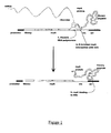
- FIG. 1 shows an example of a DNA construct of the invention.
- This construct comprises, from 5' to 3', a promoter sequence, a sequence encoding a library member peptide, a sequence encoding a repA protein, a cis DNA element and an ori sequence.
- the DNA sequence is transcribed from the promoter by RNA polymerase to RNA.
- the rho dependent termination function present in the cis DNA element causes the RNA polymerase to pause at this part of the sequence.
- the repA protein is then able to bind to the ori sequence, linking the encoded protein to the encoding DNA construct.
- library member DNA sequence(s) are fused to the repA, cis and ori DNA of the IncFII plasmid R1 ( Masai H et al. 1983 Proc Natl Acad Sci USA 80: 6814-6818 ).
- the library member DNA sequence(s) of interest may be joined by a region of DNA encoding a flexible amino acid linker, to the 5'-end of the repA DNA, under the control of an appropriate promoter and translation sequences for in vitro transcription/translation.
- promoters are known to those skilled in the art, such as the araB, tac promoter or the T7, T3 or SP6 promoters, amongst others. The promoter should be upstream of the polypeptide sequence to be expressed.
- repA family of proteins is used herein by way of example, not limitation. Other unrelated non-covalently binding cis acting DNA binding proteins could be used in this invention.
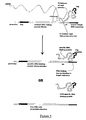
- non-cis acting DNA binding proteins may be converted to having cis-activity (see Figure 2 ). This may be achieved by using such proteins, capable of binding the DNA target sequence, either directly or indirectly, in combination with sequences which can confer cis-activity upon them.
- Cis activity may be conferred on a binding protein that does not normally act in cis by including in the DNA construct a DNA element that directs cis-activity such as the cis element of the repA system. Such an element may be included to ensure that the DNA binding by the DNA binding protein is cis, that is, an encoded DNA binding protein will bind to the DNA construct from which it has been transcribed and translated.
- a suitable DNA construct may therefore comprise the DNA element that directs cis-activity (the cis DNA element) from the repA system.
- Such an element may further comprise DNA encoding a portion of the C-terminal end of RepA, preferably at least 20 amino acids, more preferably 30 amino acids, up to 40, 50, 60 or 70 amino acids from the C-terminal portion of repA, wherein said fragment of repA is capable of interacting with the DNA element within the construct.
- proteins such as the cis acting transposases, Tn5 and IS903, amongst others, could be used under the current invention ( McFall E. J Bacteriol. 1986 Aug 167:429-432 ; Derbyshire KM & Grindley ND.
- DNA encoding sequences of the present invention may comprise wild type sequences encoding the desired fragment of RepA, degenerate sequences encoding fragments of wild type RepA or sequences encoding variants of such fragments of RepA which maintain the ability to interact with the cis element incorporated into the DNA construct.
- Such variants may include substitution of 1, 2, 3 or 4 amino acids within the 20 amino acid C-terminal of RepA.
- repA family of proteins is used herein by way of example, not limitation. Any DNA element capable of conferring cis-activity on a non-cis acting protein could be used .
- any non-cis acting protein may be converted in this way.
- the estrogen receptor DNA binding domain DBD
- DBD can be converted into a cis acting DNA binding protein.
- the oestrogen receptor DNA binding domain fragment (amino acids 176-282) has been expressed in E. coli and shown to bind to the specific double stranded DNA oestrogen receptor target HRE nucleotide sequence, with a similar affinity (0.5nM) to the parent molecule ( Murdoch et al. 1990, Biochemistry 29: 8377-8385 ; Mader et al., 1993, DNAs Research 21: 1125-1132 ).
- the DNA encoding this sequence is fused, preferably at the 3'-end, to the DNA encoding at least the last 20 amino acids of repA, the cis DNA element, and the DNA up to the ori sequence followed by the estrogen receptor target recognition sequence (5'-TCAGG TCAGA GTGAC CTGAG CTAAA ATAAC ACATT CAG-3', SEQ ID NO: 14) which replaces the repA ori recognition sequence.
- the DNA sequence(s) of interest may then be joined by a region of DNA encoding a flexible amino acid linker, to the 5'-end of to the estrogen receptor DNA fragment, under the control of an appropriate promoter and translation sequences for in vitro transcription/translation.
- Protein-DNA complexes can then be isolated by capture on a target protein. Unbound protein-DNA complexes can be washed away, allowing enrichment for DNA encoding peptides or proteins of interest, which can then be recovered by PCR, and enriched further by performing several further cycles of in vitro expression and protein-DNA complex capture using methods described previously.
- libraries of randomized DNA binding proteins such as zinc finger proteins, helix-loop-helix proteins or helix-turn-helix proteins by way of example, may be screened for specific binding to a target sequence of interest (see Figure 3 ).
- the ori recognition sequence of repA may be replaced by a target sequence of interest, and the majority of the repA coding sequence by a library of randomised zinc finger proteins.
- the DNA binding proteins therefore act as both the library member peptides and the proteins capable of binding the DNA target sequence in this aspect.
- each zinc finger protein may additionally be joined, at the 5'-end, to a peptide tag sequence which can be recognized by an another capture protein such as an antibody, and at the 3'-end, to the DNA encoding at least the last 20 amino acids of repA, the cis DNA element, and the DNA up to the ori sequence followed by the target sequence of interest.
- Expression of this polypeptide directs the zinc finger protein to the target sequence of interest, present in place of the normal ori sequence, on the DNA encoding that polypeptide. Binding to the target sequence will only occur if the randomised zinc finger domain is capable of binding to the sequence of interest.
- Protein-DNA complexes can then be isolated by capture with a binding protein which recognizes the peptide tag at the N-terminus of the fusion protein polypeptide. Unbound DNA can be washed away, allowing enrichment for DNA encoding zinc finger proteins capable of binding the target sequence, which can then be recovered by PCR, and enriched further by performing several further cycles of in vitro expression and protein-DNA complex capture.
- the binding peptide may bind directly to the DNA target sequence, for example in the case of a DNA binding protein-target sequence pair, or it may bind indirectly to the DNA target sequence, for example via a bifunctional agent and optionally a DNA tag (see Figure 4 ):
- DNA encoding a peptide tag which is not able to bind directly to the DNA target sequence is joined to the 5'-end of library member DNA sequence(s) of interest, optionally by a region of DNA encoding a flexible amino acid linker, under the control of an appropriate promoter and translation sequences for in vitro transcription/translation.
- the DNA target sequence may be or may comprise a DNA tag.
- Such a DNA tag may be a single modified base.
- the DNA may be tagged at the 3'-end with, by way of example not limitation, molecules such as fluorescein or biotin.
- the library DNA fragments may be mixed with a bifunctional agent, one function of which is to recognize and bind to the target sequence which may be at the 5' end of the DNA, in a ratio of one DNA fragment: one bifunctional molecule.
- the other functional element of this bifunctional agent is a binding agent that can recognize and bind to the peptide tag which may be encoded at the 5'-end of the DNA fragment.
- the bifunctional agent can be composed of an Fab fragment recognizing the fluorescein or biotin tag on the DNA, and another Fab fragment recognizing the peptide tag encoded in the DNA.
- this bifunctional agent can be made by many different methods such as chemically cross-linking the two elements, or by expressing the two elements as a fusion protein, or as a bi-specific antibody. Said methods of creating a bifunctional agent are given by way of example not exclusion.
- the bifunctional agent may be bound to the DNA construct prior to expression of the encoded peptide or may be provided during expression.
- the fusion protein is then transcribed and translated from the DNA construct while bound to the bifunctional agent.
- the peptide tag is translated first, and can be bound by the second element of the bifunctional agent, prior to release of messenger RNA or RNA polymerase from the DNA.
- the peptide tag molecule is therefore linked indirectly, but specifically, to the DNA target (tag).
- Protein-DNA complexes can then be isolated by capture of a target protein. Unbound protein-DNA complexes can be washed away, allowing enrichment for DNA encoding peptides or proteins of interest, which can then be recovered by PCR, and enriched further by performing several further cycles of in vitro expression and protein-DNA complex capture using methods described previously.
- the DNA can be bound directly, for example by covalent binding, to a bifunctional agent such as a polymer.
- a bifunctional agent such as a polymer.
- a polymer can contain more than one binding element that could recognise the peptide tag, allowing multivalent display of a peptide expression library molecule in a unit containing the DNA encoding the displayed peptide.
- said polymers can be composed of polyethylene as well as other polymeric compounds, capable of being fused to DNA.
- the DNA construct of the invention may therefore be provided bound to such a bifunctional agent, or bound to a DNA tag as decsribed above which is capable of being bound by such a bifunctional agent.
- the DNA constructs include appropriate promoter and translation sequences for in vitro transcription/translation.
- Any suitable promoter can be used, such as the ara B, tac promoter, T7, T3 or SP6 promoters amongst others.
- the promoter is placed so that it is operably linked to the DNA sequences of the invention such that such sequences are expressed.
- the DNA encoding the library member peptides may be produced by any sourcible means.
- such DNA may comprise DNA isolated from cDNA, obtained by DNA shuffling, and synthetic DNA.
- the DNA construct may also encode amino acid linkers within the expressed fusion protein.
- a flexible amino acid linker may be included to join the DNA binding peptide/RepA to the library member peptide.
- peptide or protein expression libraries linked to the DNA encoding them, can be generated and peptides with the desired activity selected by the following steps:
- a DNA library of peptides or proteins may be fused to DNA encoding a peptide capable of binding to the DNA target sequence, such as a cis acting DNA binding protein DNA, by a region of DNA encoding a flexible amino acid linker, under the control of an appropriate promoter and with a translation, or ribosome binding site, start and stop codons, in a manner suitable for in vitro expression of the peptide library members and binding proteins.
- the DNA (such as DNA) library members are fused to the repA DNA binding protein DNA, or a fragment thereof.
- the cis and ori sequences may be included in the construct downstream of the other elements. In the case of a DNA library, said DNA constructs are designed to be suitable for in vitro transcription and translation.
- a coupled bacterial transcription/translation environment such as the S30 extract system (Zubay, G. 1973. Ann. Rev. Genet. 7: 267) may be used.
- Expression of the peptide, such as the DNA library member peptide-repA fusion protein, in this environment, will result in binding of the fusion protein to the DNA encoding that fusion protein, provided that both cis and ori sequences are present.
- BSA bovine serum albumin
- An in vitro peptide expression library produced by a method of the present invention may be used to screen for particular members of the library.
- the library may be screened for peptides with a particular activity or a particular binding affinity.
- Protein-DNA complexes of interest may be selected from a library by, for example, affinity or activity enrichment techniques. This can be accomplished by means of a ligand specific for the protein of interest, such as an antigen if the protein of interest is an antibody.
- the ligand may be presented on a solid surface such as the surface of an ELISA plate well, or in solution, for example, with biotinylated ligand followed by capture onto a streptavidin coated surface or magnetic beads, after a library of protein-DNA complexes had been incubated with the ligand to allow ligand-ligand interaction. Following either solid phase or in solution incubation, unbound complexes are removed by washing, and bound complexes isolated by disrupting ligand-ligand interactions by altering pH in the well, or by other methods known to those skilled in the art such as protease digestion, or by releasing the DNA directly from the complexes by heating or phenolchloroform extraction to denature the repA-ori DNA binding.
- DNA can also be released by one of the methods above, directly into PCR buffer, and amplified. Alternatively, DNA may be PCR amplified directly without release from the complexes.
- DNA not bound by the binding for example repA protein can be protected from degradation by non-specific DNA binding proteins such as histones, by way of example. It will be clear to one skilled in the art that many other non-specific DNA binding proteins could be used for this purpose.
- suitable compounds include detergents, blocking proteins such as found in milk powder or bovine serum albumin (BSA), heparin or aurintricarboxylic acid.
- Recovering bound complexes, reamplifying the bound DNA, and repeating the selection procedure provides an enrichment of clones encoding the desired sequences, which may then be isolated for sequencing, further cloning and/or expression.
- the DNA encoding the peptide of interest may be isolated and amplified by, for example PCR.
- repeated rounds of selection and DNA recovery may be facilitated by the use of sequential nesting of PCR primers.
- DNA ends are generally damaged after multiple PCR steps. To recover DNA from such damaged molecules required the primers to be annealed away from the ends of the DNA construct, thereby sequentially shortening the construct with every round of selection.
- the DNA construct and/or the encoded protein may be configured to include a tag.
- a tag such as described above, may be used in the separation and isolation of a library member of interest.
- Such a tag may also be used to hold the library members, for example on a solid support for use in the screening methods described herein.
- the screening methods of the present invention may include the further step of selecting and isolating the relevant library member peptide, allowing the peptide exhibiting the desired properties, and also the DNA encoding that peptide, to be identified and purified.
- the invention therefore encompasses peptides and DNAs that have been identified by a method of the invention. These peptides and DNAs may be isolated and/or purified.
- the peptides or DNAs isolated by a method of the invention may be modified, for example by deletion, addition or substitution of amino acids or nucleotides. Suitable modified peptides or DNAs may show at least 50%, at least 75%, at least 90%, at least 95% or more amino acid or nucleotide sequence identity to the peptide or DNA isolated by the method of the invention.
- Peptides identified by a method of the invention may be modified for delivery and/or stability purposes.
- such peptides may be pegylated (attached to polyethylene glycol) to prolong serum half life or to prevent protease attack.
- Peptides identified by a method of the invention may be modified in other display systems such as phage display or by synthesising and screening peptide variants. A collection of such modified sequences may form a new library which may be incorporated into constructs of the invention and further screened to find, for example, a variant sequence showing improved binding to a particular ligand.
- a library of peptides for use in the methods of the invention may be a library of structurally related peptides.
- a library of essentially random peptide sequences may be used.
- Numerous types of libraries of peptides fused to the cis acting DNA-binding protein can be screened under this embodiment including:
- the invention concerns methods for screening a DNA library whose members require more than one chain for activity, as required by, for example, antibody Fab fragments for ligand binding.
- heavy or light chain antibody DNA is joined to a nucleotide sequence encoding a DNA binding domain of, for example, repA.
- the unknown antibody DNA library sequences for either the heavy (VH and CH1) or light chain (VL and CL) genes are inserted in the 5' region of the repA DNA, behind an appropriate promoter and translation sequences.
- repA fused to a DNA library member-encoded protein is produced bound to the DNA encoding that protein.
- the second known chain, encoding either light or heavy chain protein is expressed separately either:
- the known chain associates with the library of unknown fusion proteins that are fused to the repA protein and thereby bound to the DNA for the unknown chain.
- the functional Fab library can then be selected by means of a ligand specific for the antibody.
- the DNA identified by a screening method of the invention may be cloned into a vector.
- the DNA identified by a method of the invention is operably linked to a control sequence which is capable of providing for the expression of the coding sequence by the host cell, i.e. the vector is an expression vector.
- operably linked refers to a juxtaposition wherein the components described are in a relationship permitting them to function in their intended manner.
- a regulatory sequence, such as a promoter, "operably linked" to a coding sequence is positioned in such a way that expression of the coding sequence is achieved under conditions compatible with the regulatory sequence.
- Such expression vectors are routinely constructed in the art of molecular biology and may for example involve the use of plasmid DNA and appropriate initiators, promoters, enhancers and other elements, such as for example polyadenylation signals which may be necessary, and which are positioned in the correct orientation, in order to allow for protein expression.
- Other suitable vectors would be apparent to persons skilled in the art.
- the vectors may be for example, plasmid, virus or phage vectors provided with a origin of replication, optionally a promoter for the expression of the said DNA and optionally a regulator of the promoter.
- the vectors may contain one or more selectable marker genes, for example an ampicillin resistence gene in the case of a bacterial plasmid or a resistance gene for a fungal vector.
- Vectors may be used in vitro, for example for the production of DNA or RNA or used to transfect or transform a host cell, for example, a mammalian host cell.
- the vectors may also be adapted to be used in vivo, for example in a method of gene therapy.
- Promoters and other expression regulation signals may be selected to be compatible with the host cell for which expression is designed.
- yeast promoters include S. cerevisiae GAL4 and ADH promoters, S. pombe nmt 1 and adh promoter.
- Mammalian promoters include the metallothionein promoter which can be induced in response to heavy metals such as cadmium.
- Viral promoters such as the SV40 large T antigen promoter or adenovirus promoters may also be used. All these promoters are readily available in the art.
- Mammalian promoters such as ⁇ -actin promoters, may be used. Tissue-specific promoters are especially preferred. Viral promoters may also be used, for example the Moloney murine leukaemia virus long terminal repeat (MMLV LTR), the rous sarcoma virus (RSV) LTR promoter, the SV40 promoter, the human cytomegalovirus (CMV) IE promoter, adenovirus, HSV promoters (such as the HSV IE promoters), or HPV promoters, particularly the HPV upstream regulatory region (URR). Viral promoters are readily available in the art.
- MMLV LTR Moloney murine leukaemia virus long terminal repeat
- RSV rous sarcoma virus
- CMV human cytomegalovirus
- HSV promoters such as the HSV IE promoters
- HPV promoters particularly the HPV upstream regulatory region (URR).
- Viral promoters are readily available in the art.
- the vector may further include sequences flanking the polynucleotide of interest giving rise to polynucleotides which comprise sequences homologous to eukaryotic genomic sequences, preferably mammalian genomic sequences, or viral genomic sequences.
- sequences flanking the polynucleotides of interest giving rise to polynucleotides which comprise sequences homologous to eukaryotic genomic sequences, preferably mammalian genomic sequences, or viral genomic sequences.
- viral vectors include herpes simplex viral vectors and retroviruses, including lentiviruses, adenoviruses, adeno-associated viruses and HPV viruses. Gene transfer techniques using these viruses are known to those skilled in the art. Retrovirus vectors for example may be used to stably integrate the polynucleotide giving rise to the polynucleotide into the host genome. Replication-defective adenovirus vectors by contrast remain episomal and therefore allow transient expression.
- Such expression vectors may be used to identify ligands of interest, i.e. molecules that bind to the peptide library member by standard binding assays such as ELISA, or enzymatic assays where appropriate substrates give, for example a colour change, light emission or fluorescence. Other functional assays could be used, where available.
- a DNA idientified ny a method of the invention may be cloned into a non-expression vector.
- a vector may be used to further characterise the DNA, for example by sequencing.
- ligands of interest may be identified without cloning.
- suitable methods include the in vitro expression of individual DNA sequences recovered from a screening method of the invention, and sequencing of individual DNAs recovered from such a screening method. Such individual DNA sequences may optionally be amplified.
- the invention also includes cells that have been modified to express a peptide identified by a method of the invention, for example by introducing an expression vector as described above into the cell.
- Such cells include transient, or preferably stable higher eukaryotic cell lines, such as mammalian cells or insect cells, using for example a baculovirus expression system, lower eukaryotic cells, such as yeast or prokaryotic cells such as bacterial cells.
- Particular examples of cells which may be modified by insertion of vectors encoding for a peptide identified by a method of the invention include mammalian HEK293T, CHO, HeLa and COS cells.
- the cell line selected will be one which is not only stable, but also allows for mature glycosylation and cell surface expression of the peptide. Expression may be achieved in transformed oocytes.
- a peptide identified by a method of the invention may be expressed in cells of a transgenic non-human animal, preferably a mouse.
- a peptide identified by a method of the invention may also be expressed in Xenopus laevis oocytes or melanophores.
- GeneClean DNA gel purification kits were obtained from BIO101 (La Jolla, California, U.S.A.), anti-human Ig ⁇ antibodies from Immunologicals Direct Ltd (Oxfordshire, U.K.), anti-c-myc polyclonal from Vector Labs Inc (Cambridgeshire U.K.), and anti-V5 antibody from Abcam Ltd (Cambridgeshire U.K.).
- Superblock blocking agent was obtained from Perbio Science (Cheshire, U.K.).
- the in vitro expression constructs were prepared by sequentially adding the TAC promoter, the c-myc epitope, either the human kappa constant region or the V5 epitope to the RepA-CIS-ORI region, by PCR amplification.
- Such constructs can be prepared by many methods known to one skilled in the art, for example, by amplifiying different fragments of DNA followed by assembly PCR.
- the initial amplification template was the R1 plasmid which contains the RepA-CIS-ORI region ( Masai, H. and Arai, K.(1988). DNAs Res. 16, 6493-6514 ).
- NUNC star immunotubes were coated with 10 ⁇ g/ml of either anti-c-myc antibody, anti-V5 antibody, or anti-human kappa chain antibody, in 500 ⁇ l PBS per tube overnight at 4°C. An additional tube was left blank as a negative control. Tubes were washed 2x PBS and blocked for 1 hour at room temperature with Superblock/PBS/0.1mg/ml herring sperm DNA/ 0.1% Tween 20 and then washed 2x PBS. 500 ⁇ l of diluted transcription/translation reaction was added to each tube and incubated at room temperature for 1 hour.
- Tubes were washed 5x PBS/0.1% Tween 20, then 1x 30 minutes with 2ml Superblock/PBS/0.1mg/ml herring sperm DNA/ 0.1% Tween 20, then 5x PBS.
- DNA was recovered with 300 ⁇ l T.E. buffer plus 300 ⁇ l phenol/chloroform for 5 minutes with shaking. This was centrifuged at 13,200g for 5 minutes and DNA precipitated with 0.5 volume of 7.5M ammonium acetate, 20 ⁇ g glycogen and three volumes of absolute ethanol. Following centrifugation, pellets were washed with 70% ethanol, vacuum dried and resuspended in 20 ⁇ l water.
- Glycine tube was incubated with 500 ⁇ l of 200mM Glycine, 150mM NaCl (pH2.0) for 10 minutes. The glycine eluate was then transferred to a fresh eppendorf tube and 50 ⁇ l of 2M Tris (pH 8.5) added.
- Triethylamine the tube was incubated 500 ⁇ l of 0.1M Triethylamine for 10 minutes and the triethylamine eluate was then transferred to a fresh eppendorf tube and 250 ⁇ l of 1M Tris (pH 7.4) added.
- Proteinase K the tube was incubated with 500 ⁇ l of 100mM Tris (pH 8.0), 10 mM EDTA (pH 8.0), 0.5% SDS for 30 minutes at 37°C. The Proteinase K eluate was then transferred to a fresh eppendorf tube.
- EDTA the tube was incubated with 250 ⁇ l of 10mM Tris (pH 8.0), 1 mM EDTA 500mM NaCl and 250 ⁇ l of Phenol/Chloroform for 5 minutes. The EDTA eluate was then transferred to a fresh eppendorf tube.
- Example 3 Detection of specific anti-V5 binders in a V5-spiking expeiment using CIS display technology.
- the in vitro expression constructs were prepared by adding the TAC promoter and either the V5 epitope or a 12-mer NNB library to the RepA-CIS-ORI region, by PCR amplification. Such constructs can be prepared by many methods known to one skilled in the art, for example, by amplifiying different fragments of DNA followed by assembly PCR.
- the initial amplification template was the R1 plasmid which contains the RepA-CIS-ORI region ( Masai, H. and Arai, K.(1988). Nucleic Acids Res. 16, 6493-6514 ).
- NUNC star immunotubes were coated with 10 ⁇ g/ml of anti-V5 antibody in 500 ⁇ l PBS overnight at 4°C. An additional tube was left blank as a negative control. Tubes were washed 2x PBS and blocked for 1 hour at room temperatue with blocking buffer (2% Marvel, 0.1% Tween 20, 0.1mg/ml herring sperm DNA) and then washed 2x PBS. 1 ml of diluted transcription/translaiton reaction was added to each tube and incubated at room temperature for 1 hour. Tubes were washed 5x PBS/0.1% Tween 20 and then 5x PBS. DNA was recovered with 500 ⁇ l TE buffer plus 500 ⁇ l phenol/chloroform.
- blocking buffer 2% Marvel, 0.1% Tween 20, 0.1mg/ml herring sperm DNA
- the cut plasmid was gelpurified using a QIAquick Gelextracation kit according to the manufacturers instructions (QIAGEN LtdWest Wales, U.K.), then treated with 0.01 units of Calf Intestinal Alkaline Phosphatase (Promega, Southampton, U.K.) followed by phenol/chloroform extraction and ethanol precipitation as described above. Precipitated DNA was dissolved in 20 ⁇ l of water. The cut PCR product was transferred to Streptavidin coated strips (Roche Diagnostics Ltd, East Wales, U.K.) in 1x TBS, 0.3 mg/ml BSA, 0.1% Tween 20 and incubated for 30 minutes at room temperature, shaking.
- Precipitated DNA was dissolved in 10 ⁇ l of water and electroporated into electrocompetent TG1 cells according to the manufacturers instructions (Stratagene, U.S.A.) and selected on plates with 2xTY, 100 ⁇ g/ml ampicillin, and 2% glucose.
- Anti-V5 antibody ELISA screening of selected clones 88 colonies were picked into 400 ⁇ l of 2x TY, 2% glucose, and 100 ⁇ g/ml ampicillin and grown overnight at 37°C, shaking 300 rpm. 50 ⁇ l of the overnight cultures were transferred into 1ml of 2x TY, 2% glucose, and 100 ⁇ g/ml ampicillin and grown at 37°C, shaking 300 rpm until OD 0.5. Then the cells were centrifuged at 1000x g for 10 minutes. The supernatants were discarded and pellets were resuspended in 600 ⁇ l of 2x TY, 0.4M sucrose, 100 ⁇ g/ml ampicillin, and 1mM IPTG and grown for 4 hours at 37°C, 300 rpm.
- NUNC Maxisorp plates were coated with 100 ⁇ l of 1 ⁇ g/ml in 1x PBS of either anti-human kappa region antibody or anti-V5 antibody or 50 ⁇ g/ml of BSA for 7 hours at room temperature. An additional plate was left bank, only coated with PBS. Wells were rinsed 2x PBS followed by blocking for 1 hour at room temperature with 300 ⁇ l of 4% Marvel, 0.1% Tween in 1x PBS.
- HRP horse radish peroxidase
- the HRP signal was detected by adding 200 ⁇ l of TMB substrate. Reaction was stopped with 100 ⁇ l of 0.5M sulphuric acid. Absorbance was read at 450nm. 35 out of 88 clones expressed well judged by HRP signal from clones screened against anti-human kappa region antibody. 7 out of these 35 clones showed specific binding to anti-V5 antibody, thereby enriching V5-peptides from 1 in 5000 to 1 in 5, i.e. an enrichment factor of 1000 ( Figure 6 ).
- Example 4 CIS display Library construction, selection & screening against Bacillus globigii
- a promoter library DNA fragment and the RepA-CIS-ori fragment must be generated, then linked together by digestion-ligation.
- the tac promoter from a P2A-HA vector was used in this example, but many available promoters could be used, and are well known to those skilled in the art.
- the initial PCR of Rep-CIS-ori and TAC fragments appends Bsp1201 site and the random library/NotI site respectively. Two master mixes were prepared:
- Reaction was curried out at 37°C for two hours. 20 ⁇ l was assessed by gel electrophoresis, 30 ⁇ l was PCR amplified directly in 10x 50 ⁇ l reactions, and the remainder was gel purified and the library band excised, Qiagen column purified and PCR amplified in 20x 50 ⁇ l reactions with primers TACFAR4 (SEQ ID 30) and ORIREV (SEQ ID 02) for 30 cycles of 94°C, 40 seconds; 60°C, 40 seconds; 72°C, 90 seconds; followed by a 5 minutes extension at 72°C. DNA was gel purified in 4 Qiagen columns and the 200 ⁇ l eluate pooled for ITT reactions/selection.
- blocking buffer is 4% Marvel, 100 ⁇ g/ml sheared salmon sperm DNA, 0.1% Tween 20, 2.5mg/ml heparin, in TBS), spun at 10,000g for 2 minutes, transferred to a fresh tube, then placed on ice.
- the mix was then centrifuged at 16,100g for 1 minute and the spore pellet was washed six times with 1 ml of TBS/0.1% Tween 20 by mixing with a pipette and vortexing prior to centrifugation. The pellet was finally washed in 1ml TBS and the supernatant was discarded.
- DNA was eluted by incubation of the spores in 120 ⁇ l 0.5M sodium acetate pH5.5 for 10 minutes on a mixer. The spores were centrifuged at 16,100g for 1 minute and the supernatant was neutralised by the addition of 120 ⁇ l Tris pH8.0 and then phenol/CHCl3 extracted for 5minutes at 16,100g. DNA was precipitated with 20 ⁇ g carrier glycogen and two and a half volumes of ethanol. DNA was pelleted at 16,100g for 20 minutes and the pellet washed three times with 0.75ml 70% ethanol, centrifuging for 3 minutes at 16,100g in between each wash, then air dried and re-suspended in 20 ⁇ l water.
- 10 ⁇ l recovered DNA was PCR amplified in 10 ⁇ 50 ⁇ l reaction with primers CISREV (SEQ ID 19) and TACFAR5 (SEQ ID 31) and 2 units of 20:1 Taq DNA polymerase: Deep Vent DNA polymerase mixture (NEB) for 30 cycles of 94°C, 40 seconds; 60°C, 40 seconds; 72°C, 90 seconds; followed by a 5 minutes extension at 72°C.
- the DNA was purified, ethanol precipitated and re-suspended in 10 ⁇ l water. 5 ⁇ l were further amplified by PCR using the conditions above but using the primers NOTRECREV2 (SEQ ID 32) and TACFAR5 (SEQ ID 31) for 10 cycles.
- the product was purified using a Qiagen PCR purification kit and eluted into 50 ⁇ l 5mM Tris pH 8.0.
- Second round selection was carried out as for round 1, with the following changes: Approximately 3 ⁇ g of input DNA were used. Block buffer was 2% bovine serum albumin, 1% gelatin, 100 ⁇ g/ml sheared salmon sperm DNA, 2.5mg/ml heparin, in TBS. 10 ⁇ l washed spores used in each selection. Recovery PCRs used TACFAR5.2 (SEQ ID 34) and NOTRECREV2 (SEQ ID 32) primers. Finally, pull through PCR used TACFAR5.2 (SEQ ID 34) and ORIREV (SEQ ID 02) primers for 10 cycles.
- Round 4 was carried out as for round 3, except that approximately 2 ⁇ g of input DNA was used for the selection.
- the stored the recovered DNA from round 5 was PCR amplified with biotinylated TAC6 (SEQ ID 26) primer and NOTRECREV2 (SEQ ID 32). Digestion with NotI was followed by purification using Qiagen PCR purification kit, digestion with NcoI followed by incubation in a plate coated with streptavidin. Following phenol/CHCl3 purification and ethanol precipitation, the digested DNA was then ligated into a similarly digested pVIII phagemid vector and transformed into ER2738 E.coli , then plated on 2% glucose, 2xTY, 100 ⁇ g/ml ampicillin plates and incubated o/n at 37°C.
- the wells were washed 5 ⁇ with 200 ⁇ l TBS/0.1% Tween20 by centrifugation at 3000g for 5 minutes in between each wash before incubation with anti-M13 horseradish peroxidase conjugated antibody 0.2 ⁇ g/ml in 4% bovine serum albumin, 1% gelatin in TBS.
- the spores were incubated at room temperature for 1 hour whilst shaking.
- the wells were then washed 5 ⁇ with TBS/0.1% Tween20 and the spores were transferred into a fresh plate.
- the spores were then washed once with TBS as described above before development with TMB substrate.
- the development was stopped with 0.5M H 2 SO 4 and the solution was transferred to a fresh flat-bottomed plate for reading at 450nm. Binding data for selected peptides is shown in Figure 7 .
- ITT in vitro transcription/translation reaction
- a 70x11 mm NUNC Maxisorp Immunotube (Life Technologies, Paisley, Scotland U.K.) was coated with 1ml of 10 ⁇ g/ml of polyclonal anti-V5 peptide antibody (Harlan-Seralab) in PBS for 1 hour at 37°C.
- the tube was rinsed three times with PBS (fill & empty) and blocked with 3ml block buffer for 1 hour at 37°C and washed as before.
- Library protein-DNA complexes in block buffer were added, and incubated for 1 hour standing at room temperature. The tube was washed five times with PBS/0.1% Tween 20, then a further five times with PBS only.
- DNA was eluted into 500 ⁇ l 1M Sodium acetate pH 5.2 for 10 minutes on the blood mixer, neutralized with 100 ⁇ l 1M Tris-HCl pH 8.0, then phenol/CHCl3 extracted for 5minutes at 16,100g. DNA was precipitated with 20 ⁇ g carrier glycogen, 1 ⁇ 2 volume 7.5M ammonium acetate, and three volumes of ethanol. DNA was pelleted at 16,100g for 20 minutes and the pellet washed with 0.5ml 70% ethanol for 5 minutes at 16,100g then vacuum dried, and re-suspended in 20 ⁇ l water.
- 10 ⁇ l recovered DNA was PCR amplified in 1x 50 ⁇ l reaction with primers NOT1RECREV2 (SEQ ID 32) and TACFAR4 (SEQ ID 30) and 2 units of 20:1 Taq DNA polymerase: Deep Vent DNA polymerase mixture (NEB) for 30 cycles of 94°C, 40 seconds; 60°C, 40 seconds; 72°C, 90 seconds; followed by a 5 minutes extension at 72°C. 50 ⁇ l of reaction product were electrophoresed on a 1% agarose/TAE gel and photographed, then GeneClean purified into 10 ⁇ l water. DNA was reattached to RepA DNA and reamplified for round two as described in example 4 using TACFAR5 (SEQ ID 31) and ORIREV (SEQ ID 02) primers.
- NEB Deep Vent DNA polymerase mixture
- Second round selection was carried out as for round 1, using the same primer pairs as described in example 4, with the following changes: Anti-V5 antibody coating concentration was reduced to 5 ⁇ g/ml. Input DNA was approximately 4 ⁇ g.
- Third round selection was carried out as for round 2, with the following changes: Approximately 4 ⁇ g of input DNA was used. Recovery PCRs used TACFAR5.1 (SEQ ID 33) and NOTRECREV2 (SEQ ID 32) primers. Finally, pull through PCR used TACFAR6 (SEQ ID 35) and ORIREV (SEQ ID 02) primers for 10 cycles. Round 4 was carried out as for round 3.
- the stored the recovered DNA from round 4 was PCR amplified with biotinylated (SEQ ID 26) TAC6 and NOTIREPRECREV2 (SEQ ID 32) primers and cloned into pVIII phagemid vector and electroporated into electrocompetent TG-1 E.coli , as described in example 4.
- a NUNC Maxisorp ELISA plate was coated with 100ng/well anti-V5 peptide antibody in 100 ⁇ l /well PBS for one hour at 37°C.
- the plate was washed 2x200 ⁇ l/well PBS and blocked for 1 hour at 37°C with 200 ⁇ l/well 2% BSA/PBS and then washed 2x200 ⁇ l/well PBS.
- 50 ⁇ l phage culture supernatant was added to each well containing 50 ⁇ l/well 4% BSA/PBS, and allowed to bind for 1 hour at room temperature.
- the plate was washed two times with 200 ⁇ l/well PBS/0.1% Tween 20, then two times with 200 ⁇ l/well PBS.
- Bound phage were detected with 100 ⁇ l/well, 1:5000 diluted anti-M13-HRP conjugate (Amersham-Pharmacia) in 2% BSA/PBS for 1 hour at room temperature and the plate washed four times as above. The plate was developed for 5 minutes at room temperature with 100 ⁇ l/well TMB (3,3',5,5'-Tetramethylbenzidine) substrate buffer. The reaction was stopped with 100 ⁇ l/well 0.5N H 2 SO 4 and read at 450nm. Phagemid DNA of ELISA positive clones were then sequenced with standard pUC forward and reverse sequencing primers. The amino acid sequence of these clones isolated is shown below.
- P1C12 (SEQ ID 36) C G C P T M A A R V R P V L N S K H P2H1 (SEQ ID 37) M T T V P V L M I S V P1B5 (SEQ ID 38) T L S T R H H N V I D R F N L R N F P2B8 (SEQ ID 39) S I R T L T G S T P A Q F D A T A D
- selected peptides are selected and synthesized to allow confirmation of target binding. Random 12mer peptide library construction was carried out as described in Example 3. Four rounds of selection were carried out as described in example 4 with 100 ⁇ g/ml ovalbumin (SIGMA, Dorset, UK) coated onto immunotubes.
- the recovered DNA from round 4 was PCR amplified with biotinylated TAC6 (SEQ ID 26) and NOTIREPRECREV2 (SEQ ID 32) primers and cloned into a pVIII phagemid vector and electroporated into electrocompetent TG-1 E.coli, as described in example 4.
- a NUNC Maxisorp ELISA plate was coated with 100 ⁇ g/well ovalbumin in 100 ⁇ l /well PBS overnight at 4°C. The plate was washed 2x200 ⁇ l/well PBS and blocked for 1 hour at 37°C with 200 ⁇ l/well 2% BSA/PBS and then washed 3x200 ⁇ l/well PBS. 50 ⁇ l phage culture supernatant was added to each well containing 50 ⁇ l/well 4% BSA/PBS, and allowed to bind for 1 hour at room temperature. The plate was washed two times with 200 ⁇ l/well PBS/0.1% Tween 20, then two times with 200 ⁇ l/well PBS.
- Bound phage were detected with 100 ⁇ l/well, 1:5000 diluted anti-M13-HRP conjugate (Amersham-Pharmacia) in 2% BSA/PBS for 1 hour at room temperature and the plate washed four times as above. The plate was developed for 5 minutes at room temperature with 100 ⁇ l/well TMB (3,3',5,5'-Tetramethylbenzidine) substrate buffer. The reaction was stopped with 100 ⁇ l/well 0.5N H 2 SO 4 and read at 450nm. Phagemid DNA of ELISA positive clones were then sequenced with M13REV primer. The amino acid sequence of these clones isolated is shown below.
- Peptide sequences from four representative ELISA positive clones were synthesized (SIGMA-Genosys Ltd) with biotin added to the C-terminus to aid detection in ELISA. These peptides were tested in ELISA against ovalbumin, along with a control peptide previously isolated by phage display selection against B.globigii spores.
- a NUNC Maxisorp ELISA plate was coated with 100 ⁇ g/well ovalbumin in 100 ⁇ l /well PBS, or, 200ng/well ant-V5 polyclonal antibody in PBS, overnight at 4°C.
- the plate was washed 2x200 ⁇ l/well PBS and blocked for 1 hour at 37°C with 200 ⁇ l/well 2% skimmed milk powder/PBS and then washed 2x200 ⁇ l/well PBS. 1 ⁇ g of diluted peptides were added to each well in 100 ⁇ l/well 2% BSA/PBS, and allowed to bind for 1 hour at room temperature. The plate was washed two times with 200 ⁇ l/well PBS/0.1% Tween 20, then two times with 200 ⁇ l/well PBS. Bound peptides were detected with 100 ⁇ l/well, 1:2000 diluted streptavidin-HRP conjugate (Pierce) in 2% BSA/PBS for 1 hour at room temperature and the plate washed four times as above.
- the plate was developed for 5 minutes at room temperature with 100 ⁇ l/well TMB (3,3',5,5'-Tetramethylbenzidine) substrate buffer. The reaction was stopped with 100 ⁇ l/well 0.5N H 2 SO 4 and read at 450nm ( Figure 9 ).
- Example 7 Display of single-chain Fv antibody (scFv) fragments in a CIS display system
- a tac-scFv-RepA-CIS-ori construct was constructed by PCR overlap extension essentially as described previously in example 1.
- Anti-mecoprop scFv DNA (Haptogen Ltd, Aberdeen, UK) was amplified in a 50 ⁇ l reaction volume containing 200 ⁇ M dNTPs, 1xNEB polymerase amplification buffer (10mM KCl, 10mM (NH 4 ) 2 SO 4 , 20mM Tris-HCl pH 8.8, 2mM MgS04, 0.1% TritonX-100) with 10pmol of each of the primers TACMECOFOR (SEQ ID 45) and REPAMECOBAK (SEQ ID 46) and 2 units of 20:1 Taq DNA polymerase: Deep Vent DNA polymerase mixture (NEB) for 30 cycles of 94°C, 40 seconds; 60°C, 40 seconds; 72°C, 80 seconds; followed by a 5 minutes extension at 72°C.
- NEB Deep Vent DNA polymerase mixture
- DNA was reamplified in 10x 50 ⁇ l reactions containing 200 ⁇ M dNTPs, 1xNEB polymerase amplification buffer (10mM KCl, 10mM (NH 4 ) 2 SO 4 , 20mM Tris-HCl pH 8.8, 2mM MgSO4, 0.1% TritonX-100) with 10pmol of each of the primers TAC3 (SEQ ID 09) and ORIREV (SEQ ID 02) and 2 units of 20:1 Taq DNA polymerase: Deep Vent DNA polymerase mixture (NEB) for 30 cycles of 94°C, 40 seconds; 60°C, 40 seconds; 72°C, 80 seconds; followed by a 5 minutes extension at 72°C. Products were electrophoresed on a 1% agarose/TAE gel and purified with a Geneclean II kit into 100 ⁇ l water.
- 1xNEB polymerase amplification buffer 10mM KCl, 10mM (NH 4 ) 2 SO 4 , 20mM Tris-HCl pH 8.8, 2
- Reactions were incubated at 30°C for 30 minutes then 1 ⁇ l 0.25M ox-glutathione added to reaction 2 and incubation at 30°C continued for a further 30 minutes. 1ml of blocking buffer was added to each reaction (Block buffer is 1% gelatin, 100 ⁇ g/ml sheared salmon sperm DNA, 2.5mg/ml heparin, in TBS), spun at 10,000g for 2 minutes, then placed on ice.
- Block buffer is 1% gelatin, 100 ⁇ g/ml sheared salmon sperm DNA, 2.5mg/ml heparin, in TBS
- NUNC star immunotubes were coated with 0.5ml 10 ⁇ g/ml BSA-mecoprop conjugate, or 10 ⁇ g/ml BSA in PBS for 1 hour at 37°C. Tubes were washed 2x PBS, then blocked for 1 hour at room temperature with 3ml blocking buffer on a blood mixer, then tubes were washed 2x PBS.
- Bound DNA was eluted for 10 minutes at room temperature with 0.5ml of 0.5M NaCl/10mM Tris pH 8, 1mM EDTA, then extracted with 0.5mL phenol/chloroform and precipitated with 20 ⁇ g carrier glycogen, 1 ⁇ 2 volume 7.5M ammonium acetate, and three volumes of ethanol. DNA was pelleted at 14,000g for 20 minutes and the pellet washed with 0.5ml 70% ethanol for 5 minutes at 14,000g then vacuum dried, and re-suspended in 20 ⁇ l water.
- 10 ⁇ l of recovered DNA was PCR amplified in a 50 ⁇ l reaction volume containing 200 ⁇ M dNTPs, 1xNEB polymerase amplification buffer (10mM KCl, 10mM (NH 4 ) 2 SO 4 , 20mM Tris-HCl pH 8.8, 2mM MgSO4, 0.1% TritonX- 100) with 10pmol of each of the primers TACMECOFOR (SEQ ID 45) and REPAMECOBAK (SEQ ID 46) and 2 units of 20:1 Taq DNA polymerase: Deep Vent DNA polymerase mixture (NEB) for 30 cycles of 94°C, 40 seconds; 60°C, 40 seconds; 72°C, 80 seconds; followed by a 5 minutes extension at 72°C.
- 1xNEB polymerase amplification buffer 10mM KCl, 10mM (NH 4 ) 2 SO 4 , 20mM Tris-HCl pH 8.8, 2mM MgSO4, 0.1% TritonX- 100
- TACMECOFOR
Landscapes
- Health & Medical Sciences (AREA)
- Life Sciences & Earth Sciences (AREA)
- Genetics & Genomics (AREA)
- Chemical & Material Sciences (AREA)
- Engineering & Computer Science (AREA)
- Organic Chemistry (AREA)
- Zoology (AREA)
- Biomedical Technology (AREA)
- Wood Science & Technology (AREA)
- Biotechnology (AREA)
- General Engineering & Computer Science (AREA)
- Bioinformatics & Cheminformatics (AREA)
- Plant Pathology (AREA)
- Molecular Biology (AREA)
- Microbiology (AREA)
- Biophysics (AREA)
- Physics & Mathematics (AREA)
- Biochemistry (AREA)
- General Health & Medical Sciences (AREA)
- Crystallography & Structural Chemistry (AREA)
- Bioinformatics & Computational Biology (AREA)
- Peptides Or Proteins (AREA)
- Preparation Of Compounds By Using Micro-Organisms (AREA)
- Measuring Or Testing Involving Enzymes Or Micro-Organisms (AREA)
Abstract
Description
- The present invention relates generally to recombinant DNA technology and, more particularly, to in vitro methods for constructing and screening DNA libraries for DNA sequences that encode biologically active molecules.
- Isolating an unknown gene which encodes a desired peptide from a recombinant DNA library can be a difficult task. The use of hybridisation probes may facilitate the process, but their use is generally dependent on knowing at least a portion of the sequence of the gene which encodes the protein. When the sequence is not known, DNA libraries can be expressed in an expression vector, and antibodies have been used to screen plaques or colonies for the desired protein antigen. This procedure has been useful in screening small libraries, but rarely occurring sequences which are represented in less than about 1 in 105 clones, as is the case with rarely occurring cDNA molecules or synthetic peptides, can be easily missed, making screening libraries larger than 106 clones at best laborious and difficult. Screening larger libraries has required the development of methods designed to address the isolation of rarely occurring sequences, which are based on the co-selection of molecules, along with the DNAs that encode them. In vivo methods have been developed to screen large libraries, such as phage display and "peptides on plasmids" using lacI fusions of peptides.
- Phage display is based on DNA libraries fused to the N-terminal end of filamentous bacteriophage coat proteins and their expression in a bacterial host resulting in the display of foreign peptides on the surface of the phage particle with the DNA encoding the fusion protein packaged in the phage particle (Smith G. P., 1985, Science 228: 1315-1317). Libraries of fusion proteins incorporated into phage, can then be selected for binding members against targets of interest (ligands). Bound phage can then be allowed to reinfect Escherichia coli (E. coli) bacteria and then amplified and the selection repeated, resulting in the enrichment of binding members (Parmley, S. F., & Smith, G. P. 1985., Gene 73: 305-318; Barrett R. W. et al., 1992, Analytical Biochemistry 204: 357-364 Williamson et al., Proc. Natl. Acad. Sci. USA, 90: 4141-4145; Marks et al., 1991, J. Mol. Biol. 222: 581-597).
- LacI fusion plasmid display is based on the DNA binding ability of the lac repressor. Libraries of random peptides are fused to the C-terminal end of the lacI repressor protein. Linkage of the LacI-peptide fusion to its encoding DNA occurs via the lacO sequences on the plasmid, forming a stable peptide-LacI-peptide complex. These complexes are released from their host bacteria by cell lysis, and peptides of interest isolated by affinity purification on an immobilised receptor target. The plasmids thus isolated can then be reintroduced into E. coli by electroporation to amplify the selected population for additional rounds of screening (Cull, M. G. et al. 1992. Proc. Natl. Acad. Sci. U.S.A. 89:1865-1869).
- These bacterial methods are limited by the size of the library that can be created by current methods of introducing DNA into host bacteria, the potential cellular toxicity of the expressed peptides introduced, and by the inability to introduce post-translational modifications, or to incorporate novel amino acids into the expressed peptide.
- An entirely in vitro ribosome system has been described based on the linkage of peptides to the RNA encoding them through the ribosome (
WO91/05058 - The in vitro method described by Griffiths and Tawfik (
WO 99/02671 WO 00/40712 - Another prior art method, covalent display technology, or CDT, is described in
WO9837186 - There remains a need for more versatile in vitro methods of constructing peptide libraries in addition to the methods described above, which can allow direct selection of binding activity, as well as for enzymatic activity, and that allow efficient production of complex peptide structures, while still allowing recovery of intact genetic material encoding the peptide of interest.
- The present invention therefore provides a method for producing an in vitro peptide expression library comprising a plurality of peptides, wherein each peptide is linked to a DNA construct encoding the peptide, comprising the steps of:
- (a) providing a DNA construct comprising:
- (i) a DNA target sequence;
- (ii) DNA encoding a library member peptide; and
- (iii) DNA encoding a peptide capable of non-covalently binding directly or indirectly to said DNA target sequence of (ii);
- (b) expressing a plurality of DNA constructs according to (a), wherein said DNA constructs encode a plurality of library member peptides such that each expressed peptide is non-covalently linked to the DNA from which it was produced.
- Also provided is a method for producing an in vitro peptide expression library comprising a plurality of peptides, wherein each peptide is linked to the DNA construct encoding the peptide, comprising the steps of:
- (a) providing a DNA construct comprising:
- (i) DNA encoding a library member peptide; and
- (ii) DNA encoding a peptide capable of non-covalently binding to an agent;
wherein said DNA construct and encoded protein are selected to have cis-activity;
- (b) binding said agent or a DNA tag capable of binding said agent to said DNA construct of (a), wherein said agent is capable of binding to the peptide encoded by said DNA of (ii); and
- (c) expressing a plurality of DNA constrcuts according to (b), wherein said DNA constructs encode a plurality of library member peptides such that each expressed peptide is linked via said agent to the DNA from which it was produced.
- The present invention extends to the peptide libraries produced by such methods and to the DNA constructs used in such methods.
- The present invention also provides methods of screening in vitro peptide expression libraries of the invention. In one aspect there is provided a method of identifying and/or purifying a peptide exhibiting desired properties from an in vitro peptide expression library produced according to the method of any one of the preceding claims, comprising at least the steps of (a) screening said library and (b) selecting and isolating the relevant library member. In a second aspect, there is provided a method of identifying a specific ligand binding peptide, said method comprising at least the steps of (a) screening an in vitro peptide expression library produced according to the method of the invention with ligand molecules which are optionally bound to a solid support; (b) selecting and isolating a library member binding to said target molecule; and (c) isolating the peptide which binds specifically to said target molecule. In a third aspect there is provided a method of identifying and/or purifying a peptide having the ability to bind a specific DNA target sequence comprising at least the steps of (a) providing an in vitro expression library according to the invention wherein said peptide or protein of (iii) is a library member peptide having DNA binding activity and wherein said DNA target sequence of (i) is the target sequence of interest; (b) selecting and isolating a library member in which the encoded protein binds to said target sequence; (c) isolating the peptide which binds to said target sequence.
- In addition to isolating and/or identifying specific peptides from the libraries of the invention, the screening methods of the invention may be used to isolate and/or identify the DNA encoding specific peptides from the library.
-
-
Figure 1 gives a schematic representation of a method by which a DNA construct of the invention may be linked to the peptide that it encodes. -
Figure 2 give a schematic representation of a method of the invention by which a DNA binding protein may be converted to a cis-acting DNA binding protein. -
Figure 3 gives a schematic representation of how a target sequence specific DNA binding protein may be isolated from a library of the invention. -
Figure 4 gives a schematic representation of how a library protein may be linked to its coding DNA through cis action and the use of a bi-specific binding molecule. -
Figure 5 demonstrates cis activity: 1:1 mixture of two different sized input DNAs (CK-RepA or V5-RepA) selected against either antibody. 1-Marker DNA; 2-PCR amplification after selection on anti-human CK antibody; 3-PCR amplification after selection on anti-V5 peptide antibody. -
Figure 6 shows the specificty of anti-V5 antibody binding clones. ELISA screening, read at 450nM, of the seven clones (1-7) that show specific binding to anti-V5 antibody. The bars in group of four represent the ELISA signal of the clones screened against from left to right; anti-human kappa region antibody, anti-V5 antibody, BSA, and blank. A negative control that neither express CK nor V5 is also presented (8). -
Figure 7 shows culture supernatant ELISA OD 450nm signals for peptides recovered after 5 rounds of selection against B.globigii spores in Example 4. A. = clone1e; B. = c1one1f; C. = clone1g; D. = clone8a; E. = clone10c; F. = clone10e; G. = negative control. -
Figure 8 shows OD 450nm signals for peptides isolated after 4 rounds of selection against anti-V5 antibody in Example 5. A. = P1C12; B. = P2H1; C. = P1B5; D. = P2B8. Peptide-phage were tested against anti-V5 and anti-ACTH peptide antibodies. -
Figure 9 shows OD 450nm signals for synthetic peptides isolated after 4 rounds of selection against ovalbumin. A. = C1; B. = C4; C. = C6; D. = C8; E. = negative control. Peptides were tested against ovalbumin, anti-V5 antibody and blocked plate (plastic). -
Figure 10 . shows PCR recoveries of scFv DNA after selection on BSA or BSA-mecoprop. A. Anti-mecoprop scFv selected on BSA, 2.5mM ox-glutathione. B. Anti-mecoprop scFv selected on mecoprop-BSA, 2.5mM ox-glutathione. C. Anti-mecoprop scFv selected on BSA, no ox-glutathione. D. Anti-mecoprop scFv selected on mecoprop-BSA, no ox-glutathione. -
SEQ ID Nos 1 to 11, 19 to 23, 26 to 35 and 45 to 47 show the primers used in the Examples. - SEQ ID NO: 12 shows the sequence of the TAC-MYC-CK-REPA-CIS-ORI construct, SEQ ID NO: 13 shows the sequence of the TAC-MYC-V5-REPA-CIS-ORI construct, SEQ ID NO: 24 shows the sequence of the TAC-V5-REPA-CIS-ORI-408 construct and SEQ ID NO: 25 shows the sequence of the TAC-NNB-REPA-CIS-ORI-408 construct.
- SEQ ID NO: 14 shows the estrogen receptor target recognition sequence.
- SEQ ID Nos 15 and 16 show the DNA and amino acid sequences of the repA gene from the R1 plasmid of the incFII incompatibility group. SEQ ID Nos 17 and 18 show the sequences of the CIS DNA element and ori sequence form the same system.
- SEQ ID Nos 36 to 39 show the sequences of peptides isolated after selection in Example 5. SEQ ID Nos 39 to 43 show the sequences of clones isolated in Example 6.
- The present invention relates to the construction and screening of a library for a nucleotide sequence which encodes a peptide of interest in vitro. The constructs encoding the peptide of interest are designed such that the expressed peptide shows cis activity for the construct. Cis activity is defined as the ability of the peptide to bind to the DNA from which the peptide was produced, i.e. from which it was transcribed and translated. In vitro expression of the construct results in binding of the peptide to the DNA encoding that same peptide molecule by non-covalent interaction. This differs from the teaching of
WO 98/37186 - Non-covalent binding refers to an association that may be disrupted by methods well known to those skilled in the art, such as the addition of an appropriate solvent, or a change in ionic conditions, for example, the addition of low pH glycine or high pH triethylamine. In the present case, a typical example of non-covalent binding would be the non-covalent interaction between a DNA binding protein and a DNA molecule. Conversely, when a covalent linkage is formed between the DNA and the encoded polypeptide, the displayed peptide or protein will not be released from the DNA by ionic conditions and solvents that would disrupt non-covalent DNA binding protein:DNA interactions. For example, the bacterial replication protein repA binds non-covalently to its target DNA sequence oriR and can be released from this target DNA sequence at salt concentrations greater than 0.2M KCl (Giraldo R. & Diaz R. 1992 J. Mol. Biol. 228: 787-802). This salt concentration would not affect a covalent linkage, which would require much harsher conditions to release the covalently bound protein, with the increased risk of damage to the recovered DNA.
- The current invention describes cis activity and non-covalent binding which allow the encoded peptide or protein to remain associated with the DNA construct with a half life sufficient to allow individual peptides and the associated DNA encoding that peptide with an activity of interest to be separated from the resulting mixture of protein DNA complexes. For example, the association between the encoded protein and its DNA may have a half life of up to 30 minutes, up to 45 minutes, up to one hour, up to 2 hours, up to 6 hours or up to 12 hours. The screening methods of the invention may therefore be carried out immediately after construction of the library, or later, for example up to one, up to two, up to six, up to twelve hours or up to twenty four hours or more than twenty four hours later.
- Surprisingly, therefore, the invention described herein demonstrates that such encoded peptides or proteins can be expressed in vitro and bound to the DNA encoding that peptide in the presence of other DNA sequences. The invention also demonstrates that covalent linkage between protein and DNA is not required to maintain such cis activity, and that a non-covalent interaction between DNA and binding protein is sufficient to allow selection of peptides in an in vitro expression and selection system.
- According to the present invention, individual DNA library members, each of which encodes a peptide to be expressed in the peptide expression library (library member peptide), are placed in a suitable DNA construct. The DNA construct into which the DNA library member is placed includes all the sequences necessary to allow expression of the library member peptide from the construct and to allow the peptide encoded by the construct to bind to the DNA construct which encoded it. Each peptide in the library will typically comprise a fusion protein comprising the library member peptide fused to a peptide involved in binding of the fusion protein to the relevant DNA construct. Such fusion proteins may comprise further sequences and said library peptide may be joined to said binding peptide via a linker sequence.
- A plurality of such constructs, encoding a plurality of different library member peptides form a DNA library of the invention. Expressing such a library of DNA molecules results in the non-covalent binding of individual encoded proteins to the DNA which encoded them and from which they have been transcribed and translated, in the presence of many other DNA molecules that encode other members of the library. The sequence encoding the peptide library member present in a particular encoded protein will therefore be present in the DNA which is bound to that protein. This process therefore links the library member peptide, in a biologically active form (usually having a binding activity) to the specific library member DNA sequence encoding that peptide, allowing selection of peptides of interest, for example due to a particular binding activity, and subsequent isolation and identification of the DNA encoding that library member peptide.
- For the purposes of the invention a DNA library is therefore a population of DNA constructs. Each construct comprises a DNA sequence encoding a peptide to be expressed as a library member peptide and each contains appropriate promoter, translation start and stop signals. A DNA library of the invention will contain a plurality of such DNA molecules. A plurality of DNA constructs are provided each encoding a library member peptide to provide a plurality of different library members. Preferably a DNA library will contain at least 104 discrete DNA molecules. For example, a DNA library may contain more than 106, more than 108, more than 1010, more than 1012 or more than 1014 discrete DNA molecules.
- A peptide expression library is defined as a population of peptide sequences expressed from a library of DNA molecules. A peptide expression library of the present invention therefore encompasses a library of peptides which are non-covalently bound to the DNA which encoded them. For example, a peptide expression library of the present invention may be a library of at least 104 discrete proteins each comprising a library peptide sequence, expressed from a library of DNA molecules. A peptide expression library of the invention may be any library formed by the expression of a DNA library of the present invention.
- A peptide library member can be defined as an amino acid chain of variable composition of at least two amino acids in length, or part or all of a naturally occurring protein such as an enzyme, a binding molecule such as a receptor or an antibody or a fragment thereof. A suitable peptide library member may be a peptide having random amino acid composition. The peptide of variable or random composition may be flanked by known amino acid sequences a the N- and/or C-terminus to constrain the structure. These known sequences may vary in length. The peptide of variable or random composition may be inserted at various positions in a known protein scaffold, such as a receptor or antibody or other protein or fragment thereof. The peptide may be inserted into the same protein scaffold once or more than once, for example two or more times.
- A DNA construct according to the present invention may comprise DNA encoding a library member peptide and means for the encoded peptide to bind to the encoding DNA construct. In addition to DNA encoding a library member peptide, a suitable DNA construct of the invention comprises at least a DNA target sequence and DNA encoding a peptide capable of binding directly or indirectly to the DNA target sequence.
- According to the present invention, the DNA construct and the encoded protein are chosen to have cis-activity. That is, the encoded protein has the ability to bind specifically to the DNA molecule which encoded it. For example, cis-activity may function to allow the encoded DNA binding peptide to bind specifically (directly or indirectly) to the target sequence of the DNA construct which encoded it rather than to the target sequence of another DNA construct.
- In some cases, cis activity may be provided due to the presence of a DNA element that directs cis-activity, i.e. that allows or forces the protein encoded by the DNA construct to have cis-activity, and therefore to bind to its encoding sequence. In other cases, a separate DNA element per se may not be required where the nature of the encoding DNA inherently confers cis activity on the encoded peptide.
- A DNA element that directs cis-activity may be provided in the DNA construct together with the DNA encoding a peptide that interacts with that cis element. For example, in the case of the cis element from the repA system discussed below, DNA encoding a fragment of the repA sequence comprising at least 20 amino acids from the C terminal of repA may be provided along with the cis DNA element. It may be possible to confer cis activity upon a DNA binding peptide that is not normally cis-acting by including in the DNA construct such a DNA element and any further sequences necessary for its action. For example, DNA encoding a peptide that interacts with the cis element used may be included in the DNA construct.
- Alternatively, a peptide that interacts with the cis element may be part of the DNA binding peptide. For example, the DNA binding peptide may be repA which comprises the sequence that interacts with the repA cis element. Alternatively, the DNA binding peptide may bind to its encoding DNA in cis without the need for a separate cis element.
- A suitable DNA element may be any element which allows or directs cis-activity. Such a DNA element may act, for example, by interacting with the machinery involved in translation and transcription of the DNA construct to delay the production and release of the encoded peptide.
- Any DNA element which allows the encoded peptide to bind specifically to the DNA molecule which encoded it may be used as a DNA element according to the present invention. One example of a suitable DNA element is that of the repA-cis system described in more detail below. In that system, RNA polymerase is paused by loops in the 5' cis sequence prior to the rho dependent termination of transcription. The action of the DNA element therefore allows the encoded binding peptide to bind to the DNA target sequence in the construct from which it was produced.
- Preferably, the cis DNA element will be be located 3' in the DNA construct to the library member peptide and to the peptide or protein capable of binding to the DNA target sequence. This means that these sequences may be transcribed and translated before the RNA polymerase reaches the cis acting sequence.
- According to the present invention, the binding peptide may be linked to the DNA construct directly or indirectly. In the case of direct binding, the binding peptide binds directly and non-covalently to the DNA target sequence. In the case of indirect binding, the link between the binding peptide and DNA construct is provided by a further molecule. Such a molecule, for example a bifunctional agent as described below, will associate with both the peptide and the DNA target sequence.
- A suitable DNA construct may comprise further sequences, for example suitable promoter sequences to allow expression of the encoded peptide.
- One example of a system in which cis-activity exists is the a cis acting incompatibility group plasmid replication protein, termed repA, system. Aspects of this system may be utilised in the present invention as explained below.
- Numerous plasmids include sequences encoding repA and cis DNA elements. The repA sequence and cis DNA element present in a DNA construct of the invention may be derived from the same plasmid strain or may be derived from different plasmid strains.
- It is believed that the repA-cis system acts as shown in
Figure 1 . Briefly, RNA polymerase is paused by loops in the 5'-CIS sequence prior to rho dependent termination of transcription. This allows transient C-terminal repA peptide interaction with CIS, and possibly DNA bending. RepA peptide then binds to ori, which is a defined distance away from the terminal amino acid of the repA coding sequence (Prazkier et al. 2000 J. Bacteriology 182: 3972-3980; Praszkier and Pittard 1999 J. Bacteriol. 181: 2765-2772; Masai and Arai. 1958 Nucleic Acids Res. 16: 6493-6514). - The compatibility of a RepA sequence from a plasmid with a cis sequence from another plasmid can be readily determined by monitoring for the interaction of RepA with the selected cis sequence.
- Suitable repA proteins and sequences and cis DNA elements include those of the IncI complex plasmids or the IncF, IncB, IncK, IncZ and IricL/M plasmids, which are distantly related at the DNA level, but which control plasmid replication through the action of the cis acting repA protein (Nikoletti et al. 1986 J. Bacteriol. 170:1311-1318; Prazkier J. et al. 1991 J. Bacteriol. 173: 2393-2397). Specific plasmids which may be used to provide these sequences include the R1 plasmid of the IncII incompatibility group and the incB plasmid pMU720 (described by Praskier J. & Pittard J. 1999 Role of CIS in replication of an IncB plasmid. J. Bacteriol. 181: 2765-2772). The DNA and amino acid sequences of repA derived from the R1 plasmid of IncII are given in SEQ ID Nos: 15 and 16. The DNA sequence of the cis DNA element from the R1 plasmid of IncII is given in SEQ ID NO: 17. Typically, the cis element is 150 to 200 nucleotides in length. Shorter or larger sequences may be used, so long as the sequence maintains the ability to interact with RepA and display cis activity. Minor variations, such as substitutions or deletions within the cis sequence are also contemplated such as modifications at 1, 5, 10 up to 20 nucleotides within the wildtype cis sequence.
- The cis element is required for cis activity of the repA protein (Praszkier and Pittard 1999 J. Bacteriol. 181: 2765-2772). The cis DNA element should therefore also be located 3' in the DNA construct to the DNA encoding the repA sequence. On reaching the cis sequence, the RNA polymerase will be paused, allowing the encoded protein to bind the DNA target sequence.
- In one embodiment of the present invention, the DNA binding protein itself comprises RepA or a fragment or variant thereof capable of DNA binding, including at least the 20 C-terminal amino acids of RepA capable of binding to the cis DNA element. In this embodiment, the DNA target sequence comprises an ori sequence, for example the oriR sequence. In alternative aspects of the present invention, the DNA binding protein is provided by an alternative protein with the relevant DNA target sequences recognised by such binding protein incorporated into the sequence. In each of these embodiments, DNA-protein binding is direct in that the peptide encoded by the DNA construct will bind directly to the encoding DNA construct. In alternative aspects of the invention, as described in more detail below, the DNA-protein binding may be indirect through the use of a peptide tag-DNA tag, bifunctional agent and/or suitable linkers.
- In one aspect, the same sequence may therefore provide both the peptide capable of binding the DNA target sequence and the C terminal amino acids of repA. Such a sequence may be or may comprise a complete repA sequence, or a fragment or variant thereof of a repA sequence which retains the ability to bind to the DNA target sequence used. Where the repA acts as a DNA binding protein, both cis and ori sequences (Praszkier and Pittard 1999 J. Bacteriol. 181: 2765-2772) are required for cis activity (cis) and DNA binding (ori). In this aspect, therefore, the DNA target sequence is an ori sequence and the peptide or protein capable of binding said target is a repA protein. The position of ori in the DNA constructs of the invention may be varied. As described earlier, suitable repA, cis and ori sequences may be provided by one or more plasmids. For example, suitable sequences may be provided from the IncI complex plasmids or the IncF, IncB, IncK, IncZ and IncL/M plasmids. The DNA sequence of the ori from the R1 plasmid of IncII is given in SEQ ID NO: 18. This sequence, or a fragment thereof may be included in a DNA construct of the invention. A DNA construct of the invention may include a complete ori sequence or may include a fragment thereof which is capable of being bound by the repA protein being used.
- The RepA protein used in accordance with the present invention may also comprise a fragment or variant of RepA, so long as such variant or fragment of RepA maintains the ability to bind to the selected ori sequence. Such variant or fragment of RepA may include substitutions, for example, at 1, 2, 3 up to 20 amino acids within the RepA sequence so long as such variants maintain the ability to bind to the ori sequence. A suitable fragment of RepA is an ori binding sequence of RepA. Ori sequences include those which are present in wild type plasmids as described above. Typically, such an ori sequence is 170 to 220 nucleotides in length. Fragments and variants of wild type ori sequences may also be used, so long as such ori sequences maintain the ability to be recognised by RepA. Further cis acting members of the RepA protein family can be used. For example, the RepA family of proteins is found on plasmids with a broad host range i.e. one RepA plasmid may be found in different bacterial species. Isolation of a repA family plasmid from (for example) a thermophilic, sulfophilic, halophilic or acidophilic bacterium, would provide repA-cis-ori DNA that could be used under the current invention at elevated temperatures or extremes of salt, pH or sulphur concentrations. Such members of the RepA family would be advantageous in isolating library members that can bind to target molecules under such extreme conditions. Suitable ori sequences for use in combination with selected RepA proteins can readily be determined by monitoring for the interaction of RepA with such an ori sequence.
- The basic principle of the invention may therefore be described with reference to the repA/cis/ori system, as shown in
Figure 1 . This shows an example of a DNA construct of the invention. This construct comprises, from 5' to 3', a promoter sequence, a sequence encoding a library member peptide, a sequence encoding a repA protein, a cis DNA element and an ori sequence. Briefly, the DNA sequence is transcribed from the promoter by RNA polymerase to RNA. The rho dependent termination function present in the cis DNA element causes the RNA polymerase to pause at this part of the sequence. This allows the repA protein and the library peptide to be translated. The repA protein is then able to bind to the ori sequence, linking the encoded protein to the encoding DNA construct. - In one preferred embodiment, library member DNA sequence(s) are fused to the repA, cis and ori DNA of the IncFII plasmid R1 (Masai H et al. 1983 Proc Natl Acad Sci USA 80: 6814-6818). In this embodiment, the library member DNA sequence(s) of interest may be joined by a region of DNA encoding a flexible amino acid linker, to the 5'-end of the repA DNA, under the control of an appropriate promoter and translation sequences for in vitro transcription/translation. Many suitable promoters are known to those skilled in the art, such as the araB, tac promoter or the T7, T3 or SP6 promoters, amongst others. The promoter should be upstream of the polypeptide sequence to be expressed.
- The repA family of proteins is used herein by way of example, not limitation. Other unrelated non-covalently binding cis acting DNA binding proteins could be used in this invention.
- In a further embodiment, non-cis acting DNA binding proteins may be converted to having cis-activity (see
Figure 2 ). This may be achieved by using such proteins, capable of binding the DNA target sequence, either directly or indirectly, in combination with sequences which can confer cis-activity upon them. Cis activity may be conferred on a binding protein that does not normally act in cis by including in the DNA construct a DNA element that directs cis-activity such as the cis element of the repA system. Such an element may be included to ensure that the DNA binding by the DNA binding protein is cis, that is, an encoded DNA binding protein will bind to the DNA construct from which it has been transcribed and translated. - In one embodiment, a suitable DNA construct may therefore comprise the DNA element that directs cis-activity (the cis DNA element) from the repA system. Such an element may further comprise DNA encoding a portion of the C-terminal end of RepA, preferably at least 20 amino acids, more preferably 30 amino acids, up to 40, 50, 60 or 70 amino acids from the C-terminal portion of repA, wherein said fragment of repA is capable of interacting with the DNA element within the construct. In a further example, proteins such as the cis acting transposases, Tn5 and IS903, amongst others, could be used under the current invention (McFall E. J Bacteriol. 1986 Aug 167:429-432; Derbyshire KM & Grindley ND. Mol Microbiol. 1996 Sep 1:1261-72.). DNA encoding sequences of the present invention may comprise wild type sequences encoding the desired fragment of RepA, degenerate sequences encoding fragments of wild type RepA or sequences encoding variants of such fragments of RepA which maintain the ability to interact with the cis element incorporated into the DNA construct. Such variants may include substitution of 1, 2, 3 or 4 amino acids within the 20 amino acid C-terminal of RepA.
- The repA family of proteins is used herein by way of example, not limitation. Any DNA element capable of conferring cis-activity on a non-cis acting protein could be used .
- Any non-cis acting protein may be converted in this way. By way of example, not exclusion, the estrogen receptor DNA binding domain (DBD) can be converted into a cis acting DNA binding protein. The oestrogen receptor DNA binding domain fragment (amino acids 176-282) has been expressed in E. coli and shown to bind to the specific double stranded DNA oestrogen receptor target HRE nucleotide sequence, with a similar affinity (0.5nM) to the parent molecule (Murdoch et al. 1990, Biochemistry 29: 8377-8385; Mader et al., 1993, DNAs Research 21: 1125-1132). In one embodiment, the DNA encoding this sequence is fused, preferably at the 3'-end, to the DNA encoding at least the last 20 amino acids of repA, the cis DNA element, and the DNA up to the ori sequence followed by the estrogen receptor target recognition sequence (5'-TCAGG TCAGA GTGAC CTGAG CTAAA ATAAC ACATT CAG-3', SEQ ID NO: 14) which replaces the repA ori recognition sequence. The DNA sequence(s) of interest may then be joined by a region of DNA encoding a flexible amino acid linker, to the 5'-end of to the estrogen receptor DNA fragment, under the control of an appropriate promoter and translation sequences for in vitro transcription/translation. Expression of this polypeptide directs the estrogen receptor DBD to its target sequence, present in place of the normal ori sequence, on the DNA encoding that polypeptide. Protein-DNA complexes can then be isolated by capture on a target protein. Unbound protein-DNA complexes can be washed away, allowing enrichment for DNA encoding peptides or proteins of interest, which can then be recovered by PCR, and enriched further by performing several further cycles of in vitro expression and protein-DNA complex capture using methods described previously.
- It will be clear that this approach will apply to other DNA binding proteins simply by using the cis DNA element and a sequence encoding at least the C-terminal 20 amino acids of repA, or equivalent elements from a different cis-acting system in the DNA constructs.
- In another embodiment, libraries of randomized DNA binding proteins, such as zinc finger proteins, helix-loop-helix proteins or helix-turn-helix proteins by way of example, may be screened for specific binding to a target sequence of interest (see
Figure 3 ). In this embodiment, the ori recognition sequence of repA may be replaced by a target sequence of interest, and the majority of the repA coding sequence by a library of randomised zinc finger proteins. The DNA binding proteins therefore act as both the library member peptides and the proteins capable of binding the DNA target sequence in this aspect. The DNA encoding each zinc finger protein, may additionally be joined, at the 5'-end, to a peptide tag sequence which can be recognized by an another capture protein such as an antibody, and at the 3'-end, to the DNA encoding at least the last 20 amino acids of repA, the cis DNA element, and the DNA up to the ori sequence followed by the target sequence of interest. Expression of this polypeptide directs the zinc finger protein to the target sequence of interest, present in place of the normal ori sequence, on the DNA encoding that polypeptide. Binding to the target sequence will only occur if the randomised zinc finger domain is capable of binding to the sequence of interest. Protein-DNA complexes can then be isolated by capture with a binding protein which recognizes the peptide tag at the N-terminus of the fusion protein polypeptide. Unbound DNA can be washed away, allowing enrichment for DNA encoding zinc finger proteins capable of binding the target sequence, which can then be recovered by PCR, and enriched further by performing several further cycles of in vitro expression and protein-DNA complex capture. - As explained above, the binding peptide may bind directly to the DNA target sequence, for example in the case of a DNA binding protein-target sequence pair, or it may bind indirectly to the DNA target sequence, for example via a bifunctional agent and optionally a DNA tag (see
Figure 4 ): - In one embodiment, DNA encoding a peptide tag which is not able to bind directly to the DNA target sequence is joined to the 5'-end of library member DNA sequence(s) of interest, optionally by a region of DNA encoding a flexible amino acid linker, under the control of an appropriate promoter and translation sequences for in vitro transcription/translation. This forms the DNA encoding the binding peptide, as the encoded peptide is linked indirectly to the DNA target sequence. Optionally at the 3'-end of the library member DNA sequence is the DNA encoding at least the last 20 amino acids of repA and the cis DNA element, but not the ori target sequence of repA. The DNA target sequence may be or may comprise a DNA tag. Such a DNA tag may be a single modified base. For example, when preparing the library DNA construct containing the elements described, the DNA may be tagged at the 3'-end with, by way of example not limitation, molecules such as fluorescein or biotin.
- Prior to in vitro expression, the library DNA fragments may be mixed with a bifunctional agent, one function of which is to recognize and bind to the target sequence which may be at the 5' end of the DNA, in a ratio of one DNA fragment: one bifunctional molecule. The other functional element of this bifunctional agent is a binding agent that can recognize and bind to the peptide tag which may be encoded at the 5'-end of the DNA fragment. By way of example not exclusion, the bifunctional agent can be composed of an Fab fragment recognizing the fluorescein or biotin tag on the DNA, and another Fab fragment recognizing the peptide tag encoded in the DNA. It is clear to those skilled in the art that this bifunctional agent can be made by many different methods such as chemically cross-linking the two elements, or by expressing the two elements as a fusion protein, or as a bi-specific antibody. Said methods of creating a bifunctional agent are given by way of example not exclusion.
- The bifunctional agent may be bound to the DNA construct prior to expression of the encoded peptide or may be provided during expression.
- The fusion protein is then transcribed and translated from the DNA construct while bound to the bifunctional agent. The peptide tag is translated first, and can be bound by the second element of the bifunctional agent, prior to release of messenger RNA or RNA polymerase from the DNA. This creates a functional protein-DNA complex where both expressed polypeptide and DNA encoding that peptide are linked through the bifunctional agent. The peptide tag molecule is therefore linked indirectly, but specifically, to the DNA target (tag). By linking the protein to the DNA construct in this way, it is possible to screen for a protein having particular properties, as described below, and then to identify the encoding DNA which is linked to that protein. By using a bifunctional agent rather than covalent binding between the protein and DNA, the DNA construct may be more easily separated form the protein without the risk of damaging the DNA.
- Protein-DNA complexes can then be isolated by capture of a target protein. Unbound protein-DNA complexes can be washed away, allowing enrichment for DNA encoding peptides or proteins of interest, which can then be recovered by PCR, and enriched further by performing several further cycles of in vitro expression and protein-DNA complex capture using methods described previously.
- Additionally, under this embodiment, the DNA can be bound directly, for example by covalent binding, to a bifunctional agent such as a polymer. Such a polymer can contain more than one binding element that could recognise the peptide tag, allowing multivalent display of a peptide expression library molecule in a unit containing the DNA encoding the displayed peptide. By way of example, not limitation, said polymers can be composed of polyethylene as well as other polymeric compounds, capable of being fused to DNA. The DNA construct of the invention may therefore be provided bound to such a bifunctional agent, or bound to a DNA tag as decsribed above which is capable of being bound by such a bifunctional agent.
- In all embodiments of the invention, the DNA constructs include appropriate promoter and translation sequences for in vitro transcription/translation. Any suitable promoter can be used, such as the ara B, tac promoter, T7, T3 or SP6 promoters amongst others. The promoter is placed so that it is operably linked to the DNA sequences of the invention such that such sequences are expressed.
- The DNA encoding the library member peptides may be produced by any sourcible means. In particular, such DNA may comprise DNA isolated from cDNA, obtained by DNA shuffling, and synthetic DNA.
- The DNA construct may also encode amino acid linkers within the expressed fusion protein. In particular, a flexible amino acid linker may be included to join the DNA binding peptide/RepA to the library member peptide.
- According to the invention, with reference to this preferred embodiment, peptide or protein expression libraries, linked to the DNA encoding them, can be generated and peptides with the desired activity selected by the following steps:
- A DNA library of peptides or proteins may be fused to DNA encoding a peptide capable of binding to the DNA target sequence, such as a cis acting DNA binding protein DNA, by a region of DNA encoding a flexible amino acid linker, under the control of an appropriate promoter and with a translation, or ribosome binding site, start and stop codons, in a manner suitable for in vitro expression of the peptide library members and binding proteins. In the example of the repA protein, the DNA (such as DNA) library members are fused to the repA DNA binding protein DNA, or a fragment thereof. The cis and ori sequences may be included in the construct downstream of the other elements. In the case of a DNA library, said DNA constructs are designed to be suitable for in vitro transcription and translation.
- In order to allow cis activity, a coupled bacterial transcription/translation environment such as the S30 extract system (Zubay, G. 1973. Ann. Rev. Genet. 7: 267) may be used. Expression of the peptide, such as the DNA library member peptide-repA fusion protein, in this environment, will result in binding of the fusion protein to the DNA encoding that fusion protein, provided that both cis and ori sequences are present. When libraries of peptide-repA fusion proteins are expressed in this manner, this process results in the production of libraries of protein-DNA complexes where the protein attached to the DNA is encoded by that fragment of DNA from which it was expressed, thereby allowing subsequent selection of both peptides or protein of interest, and the DNA encoding said peptides. The complexity of these libraries is enhanced by the in vitro nature of the method, libraries of at least 1010-1014 DNA fragments, if not even larger libraries, can easily be generated.
- Compounds that prevent nuclease activity, or reduce non-specific DNA-protein or protein-protein interactions may be added during this transcription/translation reaction and cis-binding. Examples of suitable compounds include detergents and blocking proteins such as bovine serum albumin (BSA).
- An in vitro peptide expression library produced by a method of the present invention may be used to screen for particular members of the library. For example, the library may be screened for peptides with a particular activity or a particular binding affinity. Protein-DNA complexes of interest may be selected from a library by, for example, affinity or activity enrichment techniques. This can be accomplished by means of a ligand specific for the protein of interest, such as an antigen if the protein of interest is an antibody. The ligand may be presented on a solid surface such as the surface of an ELISA plate well, or in solution, for example, with biotinylated ligand followed by capture onto a streptavidin coated surface or magnetic beads, after a library of protein-DNA complexes had been incubated with the ligand to allow ligand-ligand interaction. Following either solid phase or in solution incubation, unbound complexes are removed by washing, and bound complexes isolated by disrupting ligand-ligand interactions by altering pH in the well, or by other methods known to those skilled in the art such as protease digestion, or by releasing the DNA directly from the complexes by heating or phenolchloroform extraction to denature the repA-ori DNA binding. DNA can also be released by one of the methods above, directly into PCR buffer, and amplified. Alternatively, DNA may be PCR amplified directly without release from the complexes. Optionally, DNA not bound by the binding for example repA protein, can be protected from degradation by non-specific DNA binding proteins such as histones, by way of example. It will be clear to one skilled in the art that many other non-specific DNA binding proteins could be used for this purpose. Further, compounds that prevent nuclease activity, or reduce non-specific DNA-protein or protein-protein interactions may be present during the selection process. Examples of suitable compounds include detergents, blocking proteins such as found in milk powder or bovine serum albumin (BSA), heparin or aurintricarboxylic acid.
- Recovering bound complexes, reamplifying the bound DNA, and repeating the selection procedure provides an enrichment of clones encoding the desired sequences, which may then be isolated for sequencing, further cloning and/or expression. For example, the DNA encoding the peptide of interest may be isolated and amplified by, for example PCR. In one embodiment, repeated rounds of selection and DNA recovery may be facilitated by the use of sequential nesting of PCR primers. DNA ends are generally damaged after multiple PCR steps. To recover DNA from such damaged molecules required the primers to be annealed away from the ends of the DNA construct, thereby sequentially shortening the construct with every round of selection.
- In one aspect, the DNA construct and/or the encoded protein may be configured to include a tag. Such a peptide or DNA tag, for example as described above, may be used in the separation and isolation of a library member of interest. Such a tag may also be used to hold the library members, for example on a solid support for use in the screening methods described herein.
- It can therefore be seen that the screening methods of the present invention may include the further step of selecting and isolating the relevant library member peptide, allowing the peptide exhibiting the desired properties, and also the DNA encoding that peptide, to be identified and purified.
- The invention therefore encompasses peptides and DNAs that have been identified by a method of the invention. These peptides and DNAs may be isolated and/or purified. The peptides or DNAs isolated by a method of the invention may be modified, for example by deletion, addition or substitution of amino acids or nucleotides. Suitable modified peptides or DNAs may show at least 50%, at least 75%, at least 90%, at least 95% or more amino acid or nucleotide sequence identity to the peptide or DNA isolated by the method of the invention. Peptides identified by a method of the invention may be modified for delivery and/or stability purposes. For example, such peptides may be pegylated (attached to polyethylene glycol) to prolong serum half life or to prevent protease attack. Peptides identified by a method of the invention may be modified in other display systems such as phage display or by synthesising and screening peptide variants. A collection of such modified sequences may form a new library which may be incorporated into constructs of the invention and further screened to find, for example, a variant sequence showing improved binding to a particular ligand. Thus in one embodiment, a library of peptides for use in the methods of the invention may be a library of structurally related peptides.
- Alternatively, a library of essentially random peptide sequences may be used. Numerous types of libraries of peptides fused to the cis acting DNA-binding protein can be screened under this embodiment including:
- (i) Random peptide sequences encoded by synthetic DNA of variable length.
- (ii) Antibodies or antibody fragments, for example single-chain Fv antibody fragments. These consist of the antibody heavy and light chain variable region domains joined by a flexible linker peptide to create a single-chain antigen binding molecule.
- (iii) Random cDNA fragments of naturally occurring proteins isolated from a cell population containing an activity of interest.
- (iv) Random peptide sequences inserted into, or replacing a region of a known protein, whereby the known protein sequence acts as a scaffold, which constrains the random peptide sequence. Many such scaffolds have been described, by way of example, not exclusion, CTLA-4 (
WO 00/60070 - In another embodiment the invention concerns methods for screening a DNA library whose members require more than one chain for activity, as required by, for example, antibody Fab fragments for ligand binding. In this embodiment heavy or light chain antibody DNA is joined to a nucleotide sequence encoding a DNA binding domain of, for example, repA. Typically the unknown antibody DNA library sequences for either the heavy (VH and CH1) or light chain (VL and CL) genes are inserted in the 5' region of the repA DNA, behind an appropriate promoter and translation sequences. Thus, repA fused to a DNA library member-encoded protein is produced bound to the DNA encoding that protein. The second known chain, encoding either light or heavy chain protein, is expressed separately either:
- (a) from the same DNA fragment containing the repA and the first polypeptide fusion protein library, or
- (b) from a separate fragment of DNA present in the in vitro transcription/translation reaction.
- The known chain associates with the library of unknown fusion proteins that are fused to the repA protein and thereby bound to the DNA for the unknown chain. The functional Fab library can then be selected by means of a ligand specific for the antibody.
- The DNA identified by a screening method of the invention, e.g. the DNA encoding the selected library member peptide, may be cloned into a vector. In one embodiment, the DNA identified by a method of the invention is operably linked to a control sequence which is capable of providing for the expression of the coding sequence by the host cell, i.e. the vector is an expression vector. The term "operably linked" refers to a juxtaposition wherein the components described are in a relationship permitting them to function in their intended manner. A regulatory sequence, such as a promoter, "operably linked" to a coding sequence is positioned in such a way that expression of the coding sequence is achieved under conditions compatible with the regulatory sequence.
- Such expression vectors are routinely constructed in the art of molecular biology and may for example involve the use of plasmid DNA and appropriate initiators, promoters, enhancers and other elements, such as for example polyadenylation signals which may be necessary, and which are positioned in the correct orientation, in order to allow for protein expression. Other suitable vectors would be apparent to persons skilled in the art. By way of further example in this regard we refer to Sambrook et al. 1989.
- The vectors may be for example, plasmid, virus or phage vectors provided with a origin of replication, optionally a promoter for the expression of the said DNA and optionally a regulator of the promoter. The vectors may contain one or more selectable marker genes, for example an ampicillin resistence gene in the case of a bacterial plasmid or a resistance gene for a fungal vector. Vectors may be used in vitro, for example for the production of DNA or RNA or used to transfect or transform a host cell, for example, a mammalian host cell. The vectors may also be adapted to be used in vivo, for example in a method of gene therapy.
- Promoters and other expression regulation signals may be selected to be compatible with the host cell for which expression is designed. For example, yeast promoters include S. cerevisiae GAL4 and ADH promoters, S. pombe nmt1 and adh promoter. Mammalian promoters include the metallothionein promoter which can be induced in response to heavy metals such as cadmium. Viral promoters such as the SV40 large T antigen promoter or adenovirus promoters may also be used. All these promoters are readily available in the art.
- Mammalian promoters, such as β-actin promoters, may be used. Tissue-specific promoters are especially preferred. Viral promoters may also be used, for example the Moloney murine leukaemia virus long terminal repeat (MMLV LTR), the rous sarcoma virus (RSV) LTR promoter, the SV40 promoter, the human cytomegalovirus (CMV) IE promoter, adenovirus, HSV promoters (such as the HSV IE promoters), or HPV promoters, particularly the HPV upstream regulatory region (URR). Viral promoters are readily available in the art.
- The vector may further include sequences flanking the polynucleotide of interest giving rise to polynucleotides which comprise sequences homologous to eukaryotic genomic sequences, preferably mammalian genomic sequences, or viral genomic sequences. This will allow the introduction of the polynucleotides of the invention into the genome of eukaryotic cells or viruses by homologous recombination. In particular, a plasmid vector comprising the expression cassette flanked by viral sequences can be used to prepare a viral vector suitable for delivering the polynucleotides of the invention to a mammalian cell. Other examples of suitable viral vectors include herpes simplex viral vectors and retroviruses, including lentiviruses, adenoviruses, adeno-associated viruses and HPV viruses. Gene transfer techniques using these viruses are known to those skilled in the art. Retrovirus vectors for example may be used to stably integrate the polynucleotide giving rise to the polynucleotide into the host genome. Replication-defective adenovirus vectors by contrast remain episomal and therefore allow transient expression.
- Such expression vectors may be used to identify ligands of interest, i.e. molecules that bind to the peptide library member by standard binding assays such as ELISA, or enzymatic assays where appropriate substrates give, for example a colour change, light emission or fluorescence. Other functional assays could be used, where available.
- In an alternative embodiment, a DNA idientified ny a method of the invention may be cloned into a non-expression vector. Such a vector may be used to further characterise the DNA, for example by sequencing.
- Alternatively, ligands of interest may be identified without cloning. Examples of suitable methods include the in vitro expression of individual DNA sequences recovered from a screening method of the invention, and sequencing of individual DNAs recovered from such a screening method. Such individual DNA sequences may optionally be amplified.
- The invention also includes cells that have been modified to express a peptide identified by a method of the invention, for example by introducing an expression vector as described above into the cell. Such cells include transient, or preferably stable higher eukaryotic cell lines, such as mammalian cells or insect cells, using for example a baculovirus expression system, lower eukaryotic cells, such as yeast or prokaryotic cells such as bacterial cells. Particular examples of cells which may be modified by insertion of vectors encoding for a peptide identified by a method of the invention include mammalian HEK293T, CHO, HeLa and COS cells. Preferably the cell line selected will be one which is not only stable, but also allows for mature glycosylation and cell surface expression of the peptide. Expression may be achieved in transformed oocytes. A peptide identified by a method of the invention may be expressed in cells of a transgenic non-human animal, preferably a mouse. A peptide identified by a method of the invention may also be expressed in Xenopus laevis oocytes or melanophores.
- In order that the invention is more fully understood, embodiments will now be described in more detail by way of example only and not by way of limitation with reference to the figures below.
- Examples of some of the embodiments of the invention are given below:
- The following procedures used by the present applicant are described in Sambrook, J., et al., 1989 supra.: analysis of restriction enzyme digestion products on agarose gels, DNA purification using phenol/chloroform stock solutions, preparation of phosphate buffered saline.
- General purpose reagents were purchased from SIGMA-Aldrich Ltd (Poole, Dorset, U.K.). Oligonucleotides were obtained from SIGMA-Genosys Ltd (Cambridgeshire, U.K.). Amino acids, and S30 extracts were obtained from Promega Ltd (Southampton, Hampshire, U.K.). Deep Vent and Taq DNA polymerases were obtained from New England Biolabs (Cambridgeshire, U.K.). Taqplus DNA polymerase was obtained from Stratagene Inc. (Amsterdam, Netherlands). GeneClean DNA gel purification kits were obtained from BIO101 (La Jolla, California, U.S.A.), anti-human Igκ antibodies from Immunologicals Direct Ltd (Oxfordshire, U.K.), anti-c-myc polyclonal from Vector Labs Inc (Cambridgeshire U.K.), and anti-V5 antibody from Abcam Ltd (Cambridgeshire U.K.). Superblock blocking agent was obtained from Perbio Science (Cheshire, U.K.).
- The in vitro expression constructs were prepared by sequentially adding the TAC promoter, the c-myc epitope, either the human kappa constant region or the V5 epitope to the RepA-CIS-ORI region, by PCR amplification. Such constructs can be prepared by many methods known to one skilled in the art, for example, by amplifiying different fragments of DNA followed by assembly PCR. In this example, the initial amplification template was the R1 plasmid which contains the RepA-CIS-ORI region (Masai, H. and Arai, K.(1988). DNAs Res. 16, 6493-6514).
- (a) Primary amplification. The RepA-CIS-ORI region was PCR amplified from a single colony of the strain ECO K12 harbouring plasmid R1 using 12.5pmol of each of the primers REPAFOR (SEQ ID 01) and ORIREV (SEQ ID 02) in a 50µl reaction containing 0.25mM dNTPs, 2.5 units Taqplus Precision DNA polymerase, 1x PCR reaction buffer (Stratagene Inc, Amsterdam, Netherlands). The REPAFOR primer anneals to the 5'-end of the RepA coding region. The ORIREV primer anneals to the 3'-end of the non-coding ORI region.
PCR reactions were carried out on a Eppendorf Master Cycler for 1 cycle of 4 minutes and 15 seconds at 94°C followed by 30 cycles of 94°C, 45 seconds; 60°C, 45 seconds; 72°C, 45 seconds, followed by a single cycle 10 minutes at 72°C. Reaction products were electrophoresed on an agarose gel, excised and products purified from the gel into 40µl sterile water using a Geneclean II kit according to the manufacturers instructions (Bio101, La Jolla, California, U.S.A.). - (b) Secondary amplification. One µl (500 pg) of 100 times diluted gel-purified primary reaction product was re-amplified using 12.5pmol of each of the primers CKREPFOR (SEQ ID 03) and ORIREV (SEQ ID 02) in a 50µl reaction containing 0.25mM dNTPs, 2.5 units Taqplus Precision DNA polymerase, and 1x PCR reaction buffer (Stratagene Inc, Amsterdam, Netherlands). The CKREPFOR primer anneals to the 5'-end of the primary reaction product and appends the 3' part of the kappa constant region DNA. The ORIREV primer anneals to the 3'-end of the primary reaction product.
PCR reactions were carried out on a Eppendorf Master Cycler for 1 cycle of 2 minutes and 15 seconds at 94°C followed by 30 cycles of 94°C, 45 seconds; 60°C, 45 seconds; 72°C, 2 minutes, followed by a single cycle 10 minutes at 72°C. Reaction products were electrophoresed on an agarose gel, excised and products purified from the gel into 40µl sterile water using a Geneclean II kit according to the manufacturers instructions (Bio101, La Jolla, California, U.S.A.). - (c) Third amplification. One µl (500 pg) of 100 times diluted gel-purified primary reaction product was re-amplified using 12.5pmol of each of the primers V5REPFOR (SEQ ID 04) and ORIREV (SEQ ID 02) in a 50µl reaction containing 0.25mM dNTPs, 2.5 units Taqplus Precision DNA polymerase, and 1x PCR reaction buffer (Stratagene Inc, Amsterdam, Netherlands). The V5REPFOR primer anneals to the 5'-end of the primary reaction product and appends the 3' part of the V5 epitope DNA. The ORIREV primer anneals to the 3'-end of the primary reaction product.
PCR reactions were carried out on a Eppendorf Master Cycler for 1 cycle of 2 minutes and 15 seconds at 94°C followed by 30 cycles of 94°C, 45 seconds; 60°C, 45 seconds; 72°C, 2 minutes, followed by a single cycle 10 minutes at 72°C. Reaction products were electrophoresed on an agarose gel, excised and products purified from the gel into 40µl sterile water using a Geneclean II kit according to the manufacturers instructions (Bio101, La Jolla, California, U.S.A.). - (d) Fourth amplification. One µl (500 pg) of 100 times diluted pCKV5 plasmid using 12.5pmol of each of the primers MYCCKFOR (SEQ ID 05) and CKREV (SEQ ID 06) in a 50µl reaction containing 0.25mM dNTPs, 2.5 units Taqplus Precision DNA polymerase, and 1x PCR reaction buffer (Stratagene Inc, Amsterdam, Netherlands). The pCKV5 plasmid contains the human kappa constant region cDNA (McGregor DP, Molloy PE, Cunningham C, & Harris WJ. 1994 Mol. Immunol. 31: 219-26) and the V5 epitope DNA (Southern JA, Young DF, Heaney F, Baumgartner WK, Randall RE. 1991 J. Gen. Virol. 72: 1551-7). The MYCCKFOR primer anneals to the 5'-end of the kappa constant region DNA and appends the 3' part of the MYC epitope DNA. The CKREV primer anneals to the 3'-end of the kappa constant region DNA.
PCR reactions were carried out on a Eppendorf Master Cycler for 1 cycle of 2 minutes and 15 seconds at 94°C followed by 30 cycles of 94°C, 45 seconds; 60°C, 45 seconds; 72°C, 2 minutes, followed by a single cycle 10 minutes at 72°C. Reaction products were electrophoresed on an agarose gel, excised and products purified from the gel into 40µl sterile water using a Geneclean II kit according to the manufacturers instructions (Bio101, La Jolla, California, U.S.A.). - (e) Fifth amplification. One µl (500 pg) of 100 times diluted pCKV5 plasmid using 12.5pmol of each of the primers MYCV5FOR (SEQ ID 07) and V5REV (SEQ ID 08) in a 50µl reaction containing 0.25mM dNTPs, 2.5 units Taqplus Precision DNA polymerase, and 1x PCR reaction buffer (Stratagene Inc, Amsterdam, Netherlands). The MYCV5FOR primer anneals to the 5'-end of the V5 epitope DNA and appends the 3' part of the MYC epitope DNA. The V5REV primer anneals to the 3'-end of the V5 epitope DNA.
PCR reactions were carried out on a Eppendorf Master Cycler for 1 cycle of 2 minutes and 15 seconds at 94°C followed by 30 cycles of 94°C, 45 seconds; 60°C, 45 seconds; 72°C, 30 seconds, followed by a single cycle 10 minutes at 72°C. Reaction products were electrophoresed on an agarose gel, excised and products purified from the gel into 40µl sterile water using a Geneclean II kit according to the manufacturers instructions (Bio101, La Jolla, California, U.S.A.). - (f) Sixth amplification. One µl (500 pg) of 100 times diluted pTACP2A plasmid (ref) using 12.5pmol of each of the primers TAC3 (SEQ ID 09) and MYCTACREV (SEQ ID 10) in a 50µl reaction containing 0.25mM dNTPs, 2.5 units Taqplus Precision DNA polymerase, and 1x PCR reaction buffer (Stratagene Inc, Amsterdam, Netherlands). The TAC3 primer anneals to the 5'-end of the TAC promoter DNA. The MYCTACREV primer anneals to the 3'-end of the TAC promoter DNA and appends the 5' part of the MYC epitope DNA.
PCR reactions were carried out on a Eppendorf Master Cycler for 1 cycle of 2 minutes and 15 seconds at 94°C followed by 30 cycles of 94°C, 45 seconds; 60°C, 45 seconds; 72°C, 30 seconds, followed by a single cycle 10 minutes at 72°C. Reaction products were electrophoresed on an agarose gel, excised and products purified from the gel into 40µl sterile water using a Geneclean II kit according to the manufacturers instructions (Bio101, La Jolla, California, U.S.A.). - (g) First assembly PCR. One µl (50 ng) of each of the reaction products in (f) and (d) using 50 pmol of each of the primers TAC5 (SEQ ID 11) and CKREV (SEQ ID 06) in a 50µl reaction containing 0.25mM dNTPs, 2.5 units TaqDeepVent DNA polymerase mixture (20:1), and 1x PCR reaction buffer (New England Biolabs, Beverly, MA, U.S.A.). The TAC5 primer anneals to the 5'-end of the reaction product (f) and adds 20 nucleotides. The CKREV primer anneals to the 3'-end of the reaction product (d).
PCR reactions were carried out on a Eppendorf Master Cycler for 1 cycle of 2 minutes and 15 seconds at 94°C followed by 30 cycles of 94°C, 45 seconds; 60°C, 45 seconds; 72°C, 45 seconds, followed by a single cycle 10 minutes at 72°C. Reaction products were electrophoresed on an agarose gel, excised and products purified from the gel into 40µl sterile water using a Geneclean II kit according to the manufacturers instructions (Bio101, La Jolla, California, U.S.A.). - (h) Second assembly PCR. One µl (50 ng) of each of the reaction products in (f) and (e) using 50 pmol of each of the primers TAC5 (SEQ ID 11) and V5REV (SEQ ID 08) in a 50µl reaction containing 0.25mM dNTPs, 2.5 units TaqDeepVent DNA polymerase mixture (20:1), and 1x PCR reaction buffer (New England Biolabs, Beverly, MA, U.S.A.). The TAC5 primer anneals to the 5'-end of the reaction product (f) and adds 20 nucleotides. The V5REV primer anneals to the 3'-end of the reaction product (e).
PCR reactions were carried out on a Eppendorf Master Cycler for 1 cycle of 2 minutes and 15 seconds at 94°C followed by 30 cycles of 94°C, 45 seconds; 60°C, 45 seconds; 72°C, 45 seconds, followed by a single cycle 10 minutes at 72°C. Reaction products were electrophoresed on an agarose gel, excised and products purified from the gel into 40µl sterile water using a Geneclean II kit according to the manufacturers instructions (Bio101, La Jolla, California, U.S.A.). - (i) Third assembly PCR. One µl (50 ng) of each of the reaction products in (b) and (g) or using 50 pmol of each of the primers TAC3 (SEQ ID 09) and ORIREV (SEQ ID 02) in a 50µl reaction containing 0.25mM dNTPs, 2.5 units TaqDeepVent DNA polymerase mixture (20:1), and 1x PCR reaction buffer (New England Biolabs, Beverly, MA, U.S.A.). The TAC3 primer anneals 20 nucleotides downstream to the 5'-end of the reaction product (g). The ORIREV primer anneals to the 3'-end of the reaction product (b). The reaction product in (i) is called TAC-MYC-CK-REPA-CIS-ORI (SEQ ID 12).
PCR reactions were carried out on a Eppendorf Master Cycler for 1 cycle of 2 minutes and 15 seconds at 94°C followed by 30 cycles of 94°C, 45 seconds; 60°C, 45 seconds; 72°C, 1 minute, followed by a single cycle 10 minutes at 72°C. Reaction products were electrophoresed on an agarose gel, excised and products purified from the gel into 40µl sterile water using a Geneclean II kit according to the manufacturers instructions (Bio101, La Jolla, California, U.S.A.). - (j) Fourth assembly PCR. One µl (50 ng) of each of the reaction products in (b) and (h) or using 50 pmol of each of the primers TAC3 (SEQ ID 09) and ORIREV (SEQ ID 02) in a 50µl reaction containing 0.25mM dNTPs, 2.5 units TaqDeepVent DNA polymerase mixture (20:1), and 1x PCR reaction buffer (New England Biolabs, Beverly, MA, U.S.A.). The TAC3 primer anneals 20 nucleotides downstream to the 5'-end of the reaction product (g). The ORIREV primer anneals to the 3'-end of the reaction product (b). The reaction product in (i) is called TAC-MYC-V5-REPA-CIS-ORI (SEQ ID 13).
PCR reactions were carried out on a Eppendorf Master Cycler for 1 cycle of 2 minutes and 15 seconds at 94°C followed by 30 cycles of 94°C, 45 seconds; 60°C, 45 seconds; 72°C, 1 minute, followed by a single cycle 10 minutes at 72°C. Reaction products were electrophoresed on an agarose gel, excised and products purified from the gel into 40µl sterile water using a Geneclean II kit according to the manufacturers instructions (Bio101, La Jolla, California, U.S.A.). - Preparation of in vitro transcription/translation reaction. The reaction was set up on ice, using a Promega bacterial linear template S30 coupled in vitro transcription/translation reaction kit as follows:
- 20µl TAC-MYC-CK-REPA-CIS-ORI template (0.5µg of final construct DNA SEQ ID 012 above); 20µl TAC-MYC-V5-REPA-CIS-ORI template (0.5µg of final construct DNA SEQ ID 013 above); 20µl complete amino acid mix (Promega); 80µl S30 Premix; 60µl S30 mix;
- and the reaction was allowed to proceed at 25°C for 30 minutes and placed on ice, then diluted 10 fold with blocking buffer (Superblock (Perbio Ltd), 0.1 % Tween 20, 200µg/ml herring sperm DNA).
- DNA-protein complex capture. NUNC star immunotubes were coated with 10µg/ml of either anti-c-myc antibody, anti-V5 antibody, or anti-human kappa chain antibody, in 500µl PBS per tube overnight at 4°C. An additional tube was left blank as a negative control. Tubes were washed 2x PBS and blocked for 1 hour at room temperature with Superblock/PBS/0.1mg/ml herring sperm DNA/ 0.1% Tween 20 and then washed 2x PBS. 500µl of diluted transcription/translation reaction was added to each tube and incubated at room temperature for 1 hour. Tubes were washed 5x PBS/0.1% Tween 20, then 1x 30 minutes with 2ml Superblock/PBS/0.1mg/ml herring sperm DNA/ 0.1% Tween 20, then 5x PBS. DNA was recovered with 300µl T.E. buffer plus 300µl phenol/chloroform for 5 minutes with shaking. This was centrifuged at 13,200g for 5 minutes and DNA precipitated with 0.5 volume of 7.5M ammonium acetate, 20µg glycogen and three volumes of absolute ethanol. Following centrifugation, pellets were washed with 70% ethanol, vacuum dried and resuspended in 20µl water. 10µl of recovered DNA was reamplified in 50µl reactions with TAC3 (SEQ ID 09) and ORIREV (SEQ ID 02) primers. Reaction products were electrophoresed on a 1% agarose/TAE gel (
Figure 5 ). - The two in vitro expression constructs (SEQID12 and SEQID13) already described in example 1 were used in a selection experiment against anti-human C-kappa antibody as described in Example 1, except that DNA was recovered and released from RepA by using either of following methods; Glycine, Triethylamine, Phenol/Chloroform, Proteinase K, and EDTA. These methods are described below.
- Glycine: tube was incubated with 500µl of 200mM Glycine, 150mM NaCl (pH2.0) for 10 minutes. The glycine eluate was then transferred to a fresh eppendorf tube and 50µl of 2M Tris (pH 8.5) added.
- Triethylamine: the tube was incubated 500µl of 0.1M Triethylamine for 10 minutes and the triethylamine eluate was then transferred to a fresh eppendorf tube and 250µl of 1M Tris (pH 7.4) added.
- Phenol/Chloroform: as example 1 above.
- Proteinase K: the tube was incubated with 500µl of 100mM Tris (pH 8.0), 10 mM EDTA (pH 8.0), 0.5% SDS for 30 minutes at 37°C. The Proteinase K eluate was then transferred to a fresh eppendorf tube.
- EDTA: the tube was incubated with 250µl of 10mM Tris (pH 8.0), 1 mM EDTA 500mM NaCl and 250µl of Phenol/Chloroform for 5 minutes. The EDTA eluate was then transferred to a fresh eppendorf tube.
- After recovery of DNA the DNA was Phenol/Chloroform extracted, where appropriate, followed by Ethanol precipitation as described in Example 1. 10ul of resuspended DNA was reamplified in 50ul reactions with TAC3 (SEQID09) and CISREV (SEQID019) primers. The CISREV primer anneals 196 bases upstream of the binding site of ORIREV (SEQID02). Reaction products were electrophoresed on a 1% agarose/TAE gel (data not shown). Only the CK-DNA containing construct (SEQID 12) was amplified, in approximately equivalent amounts.
- This not only tells us that any of the methods described above for recovering and releasing DNA from RepA can be used, but this result also suggests that RepA interacts in a non-covalent manner with its cognate DNA.
- The in vitro expression constructs were prepared by adding the TAC promoter and either the V5 epitope or a 12-mer NNB library to the RepA-CIS-ORI region, by PCR amplification. Such constructs can be prepared by many methods known to one skilled in the art, for example, by amplifiying different fragments of DNA followed by assembly PCR. In this example, the initial amplification template was the R1 plasmid which contains the RepA-CIS-ORI region (Masai, H. and Arai, K.(1988). Nucleic Acids Res. 16, 6493-6514).
- (a). Primary amplification. The RepA-CIS-ORI region was PCR amplified from a single colony of the strain ECO K12 harbouring plasmid R1 using 12.5pmol of each of the primers REPAFOR (SEQ ID 01) and ORIREV408 (SEQ ID 20) in a 50µl reaction containing 0.25mM dNTPs, 2.5 units TaqDeepVent DNA polymerase mixture (20:1), and 1x PCR reaction buffer (New England Biolabs, Beverly, MA, U.S.A.). The REPAFOR primer anneals to the 5'-end of the RepA coding region. The ORIREV408 primer anneals to the downstream of the 3'-end of the non-coding ORI region.
PCR reactions were carried out on a Eppendorf Master Cycler for 1 cycle of 4 minutes and 30 seconds of 94°C followed by 25 cycles of 94°C, 30 seconds; 60°C, 45 seconds; 72°C, 1 minute, followed by a single cycle 10 minutes at 72°C. Reaction products were electrophoresed on an agarose gel, excised and products purified from the gel into 40µl sterile water using a Geneclean II kit according to the manufacturers instructions (Bio101, La Jolla, California, U.S.A.). - (b). Secondary amplification. One µl (500 pg) of 100 times diluted gel-purified primary reaction product was re-amplified using 12.5pool of each of the primers V5(NNB)REPFOR (SEQ ID 21) and ORIREV408 (SEQ ID 20) in a 50µl reaction containing 0.25mM dNTPs, 2.5 units TaqDeepVent DNA polymerase mixture (20:1), and 1x PCR reaction buffer (New England Biolabs, Beverly, MA, U.S.A.). The V5(NNB)REPFOR primer anneals to the 5'-end of the primary reaction product and appends the V5 epitope DNA. The ORIREV408 primer anneals to the 3'-end of the primary reaction product.
PCR reactions were carried out on a Eppendorf Master Cycler for 1 cycle of 4 minutes and 30 seconds of 94°C followed by 25 cycles of 94°C, 30 seconds; 60°C, 45 seconds; 72°C, 1 minute, followed by a single cycle 10 minutes at 72°C. Reaction products were electrophoresed on an agarose gel, excised and products purified from the gel into 40µl sterile water using a Geneclean II kit according to the manufacturers instructions (Bio101, La Jolla, California, U.S.A.). - (c). Third amplification. One µl (500 pg) of 100 times diluted gel-purified primary reaction product was re-amplified using 12.5pmol of each of the primers NNBREPFOR (SEQ ID 22) and ORIREV408 (SEQ ID 20) in a 50µl reaction containing 0.25mM dNTPs, 2.5 units TaqDeepVent DNA polymerase mixture (20:1), and 1x PCR reaction buffer (New England Biolabs, Beverly, MA, U.S.A.). The NNBREPFOR primer anneals to the 5'-end of the primary reaction product and appends a random amino acid 12-mer NNB library DNA. The ORIREV408 primer anneals to the 3'-end of the primary reaction product.
PCR reactions were carried out on a Eppendorf Master Cycler for 1 cycle of 4 minutes and 30 seconds of 94°C followed by 25 cycles of 94°C, 30 seconds; 60°C, 45 seconds; 72°C, 1 minute, followed by a single cycle 10 minutes at 72°C. Reaction products were electrophoresed on an agarose gel, excised and products purified from the gel into 40µl sterile water using a Geneclean II kit according to the manufacturers instructions (Bio101, La Jolla, California, U.S.A.). - (d). Fourth amplification. One µl (500 pg) of 100 times diluted pTACP2A plasmid (ref) using 12.5pmol of each of the primers TACFARUP (SEQ ID 23) and TACREV (SEQ ID 27) in a 50µl reaction containing 0.25mM dNTPs, 2.5 units TaqDeepVent DNA polymerase mixture (20: 1), and 1x PCR reaction buffer (New England Biolabs, Beverly, MA, U.S.A.). The TACFARUP primer anneals to the 5'-end of the TAC promoter DNA. The TACREV primer anneals to the 3'-end of the TAC promoter DNA.
PCR reactions were carried out on a Eppendorf Master Cycler for 1 cycle of 1 minutes and 45 seconds of 94°C followed by 25 cycles of 94°C, 15 seconds; 60°C, 30 seconds; 72°C, 30 seconds, followed by a single cycle 10 minutes at 72°C. Reaction products were electrophoresed on an agarose gel, excised and products purified from the gel into 40µl sterile water using a Geneclean II kit according to the manufacturers instructions (Bio101, La Jolla, California, U.S.A.). - (e). First assembly PCR. One µl (50 ng) of each of the reaction products in (b) and (d) using 50 pmol of each of the primers TACFARUP (SEQ ID 23) and ORIREV408 (SEQ ID 20) in a 50µl reaction containing 0.25mM dNTPs, 2.5 units TaqDeepVent DNA polymerase mixture (20:1), and 1x PCR reaction buffer (New England Biolabs, Beverly, MA, U.S.A.). The TACFARUP primer anneals to the 5'-end of the reaction product (d). The ORIREV480 primer anneals to the 3'-end of the reaction product (b). The reaction product in (e) is called TAC-V5-REPA-CIS-ORI-408 (SEQ ID 24).
PCR reactions were carried out on a Eppendorf Master Cycler for 1 cycle of 1 minutes and 45 seconds of 94°C followed by 25 cycles of 94°C, 15 seconds; 60°C, 30 seconds; 72°C, 1 minute and 30 seconds, followed by a single cycle 10 minutes at 72°C. Reaction products were electrophoresed on an agarose gel, excised and products purified from the gel into 40µl sterile water using a Geneclean II kit according to the manufacturers instructions (Bio101, La Jolla, California, U.S.A.). - (f). Second assembly PCR. One µl (50 ng) of each of the reaction products in (c) and (d) using 50 pmol of each of the primers TACFARUP (SEQ ID 23) and ORIREV408 (SEQ ID 20) in a 50µl reaction containing 0.25mM dNTPs, 2.5 units TaqDeepVent DNA polymerase mixture (20:1), and 1x PCR reaction buffer (New England Biolabs, Beverly, MA, U.S.A.). The TACFARUP primer anneals to the 5'-end of the reaction product (d). The ORIREV480 primer anneals to the 3'-end of the reaction product (c).
PCR reactions were carried out on a Eppendorf Master Cycler for 1 cycle of 1 minutes and 45 seconds of 94°C followed by 25 cycles of 94°C, 15 seconds; 60°C, 30 seconds; 72°C, 1 minute and 30 seconds, followed by a single cycle 10 minutes at 72°C. Reaction products were electrophoresed on an agarose gel, excised and products purified from the gel into 40µl sterile water using a Geneclean II kit according to the manufacturers instructions (Bio101, La Jolla, California, U.S.A.). The reaction product in (f) is called TAC-NNB-REPA-CIS-ORI-408 (SEQ ID 25). - Preparation of in vitro transcription/translation reaction: The reaction set was set up on ice, using a Promega bacterial linear template S30 coupled in vitro transcription/translation reaction kit as follows:
- 20µl of 5000 times diluted TAC-V5-REPA-CIS-ORI-408 template (0.1ng of final construct DNASEQ ID 24 above)
- 20µl of 5 TAC-NNB-REPA-CIS-ORI-408 template (0.5µg of final construct DNASEQ ID 25 above)
- 20µl complete amino acid mix (Promega)
- 80µl S30 Premix
- 60µl S30 mix
- DNA-protein complex capture. NUNC star immunotubes were coated with 10µg/ml of anti-V5 antibody in 500µl PBS overnight at 4°C. An additional tube was left blank as a negative control. Tubes were washed 2x PBS and blocked for 1 hour at room temperatue with blocking buffer (2% Marvel, 0.1% Tween 20, 0.1mg/ml herring sperm DNA) and then washed 2x PBS. 1 ml of diluted transcription/translaiton reaction was added to each tube and incubated at room temperature for 1 hour. Tubes were washed 5x PBS/0.1% Tween 20 and then 5x PBS. DNA was recovered with 500µl TE buffer plus 500µl phenol/chloroform. This was centrifuged at 13,200g for 5 minutes and DNA precipitated with 1/10 volume of 3M sodium acetate, 50µg/ml glycogen and two voulmes of absolute ethanol. Following centrifugation, pellets were washed with 70% ethanol, vacuum dried and resuspended in 40µl water. 20 µl of recovered DNA was reamplified in 50 µl reactions with the biotinylated primers bTAC6 (SEQ ID 26) and bCISREV (SEQ ID 19). Reaction products were electrophoresed on a 1% agarose/TAE gel.
- Cloning of recovered DNA into the expression vector pDMG-K (SEQ ID 27). Reaction product were gelpurified and eluted with 50µl sterile water using a QIAquick Gelextracation kit according to the manufacturers instructions (QIAGEN LtdWest Sussex, U.K.). Both the purified reaction product and the plasmid pDMG-K were digested with 20 units of NcoI and NotI (New England Biolabs, Beverly, MA, U.S.A.). The cut plasmid was gelpurified using a QIAquick Gelextracation kit according to the manufacturers instructions (QIAGEN LtdWest Sussex, U.K.), then treated with 0.01 units of Calf Intestinal Alkaline Phosphatase (Promega, Southampton, U.K.) followed by phenol/chloroform extraction and ethanol precipitation as described above. Precipitated DNA was dissolved in 20µl of water. The cut PCR product was transferred to Streptavidin coated strips (Roche Diagnostics Ltd, East Sussex, U.K.) in 1x TBS, 0.3 mg/ml BSA, 0.1% Tween 20 and incubated for 30 minutes at room temperature, shaking. This approach removes the flanking biotinylated DNA upstream and downstream of the NcoI and NotI site of the PCR product and enables recovery of the small DNA fragment containing the selected peptide sequence. Supernatant was phenol/chloroform extracted and ethanol precipitated as described above. Precipitated DNA was dissolved in 10µl of water. Cut plasmid and the isloated small DNA fragment containing the selected peptide sequence, both having NcoI and NotI overhangs, were ligated using a Quick ligation kit according to the manufacturers instructions (New England Biolabs, Beverly, MA, U.S.A.) followed by phenol/chloroform extraction and ethanol precipitation as described above. Precipitated DNA was dissolved in 10µl of water and electroporated into electrocompetent TG1 cells according to the manufacturers instructions (Stratagene, U.S.A.) and selected on plates with 2xTY, 100µg/ml ampicillin, and 2% glucose.
- Anti-V5 antibody ELISA screening of selected clones. 88 colonies were picked into 400µl of 2x TY, 2% glucose, and 100µg/ml ampicillin and grown overnight at 37°C, shaking 300 rpm. 50µl of the overnight cultures were transferred into 1ml of 2x TY, 2% glucose, and 100µg/ml ampicillin and grown at 37°C, shaking 300 rpm until OD 0.5. Then the cells were centrifuged at 1000x g for 10 minutes. The supernatants were discarded and pellets were resuspended in 600µl of 2x TY, 0.4M sucrose, 100µg/ml ampicillin, and 1mM IPTG and grown for 4 hours at 37°C, 300 rpm. After induction the cells were centrifuged at 1000x g for 10 minutes. 15µl of the supernatants were used in the ELISA test. NUNC Maxisorp plates were coated with 100µl of 1 µg/ml in 1x PBS of either anti-human kappa region antibody or anti-V5 antibody or 50µg/ml of BSA for 7 hours at room temperature. An additional plate was left bank, only coated with PBS. Wells were rinsed 2x PBS followed by blocking for 1 hour at room temperature with 300µl of 4% Marvel, 0.1% Tween in 1x PBS. Wells were rinsed 2x PBS, then 150µl of supernatant and 150µl of 4% Marvel, 0.1% Tween 20 in 1x PBS were added to wells and incubated for 1 hour at room temperature. Wells were then washed 2xPBS, 0.1% Tween 20 and 2x PBS. Secondary antibody anti-human kappa region antibody conjugated to horse radish peroxidase (HRP)(final concentration 1.6µg/ml) was diluted 500 times in 4% Marvel, 0.1% Tween 20, 1x PBS and added to wells and incubated for 1 hour at room temperature. Wells were then washed 4x PBS, 0.1% Tween 20 and 2x PBS. The HRP signal was detected by adding 200µl of TMB substrate. Reaction was stopped with 100µl of 0.5M sulphuric acid. Absorbance was read at 450nm. 35 out of 88 clones expressed well judged by HRP signal from clones screened against anti-human kappa region antibody. 7 out of these 35 clones showed specific binding to anti-V5 antibody, thereby enriching V5-peptides from 1 in 5000 to 1 in 5, i.e. an enrichment factor of 1000 (
Figure 6 ). - To generate library DNA, a promoter library DNA fragment and the RepA-CIS-ori fragment must be generated, then linked together by digestion-ligation. The tac promoter from a P2A-HA vector was used in this example, but many available promoters could be used, and are well known to those skilled in the art. The initial PCR of Rep-CIS-ori and TAC fragments appends Bsp1201 site and the random library/NotI site respectively. Two master mixes were prepared:
- 10µl of 1:50 diluted P2A-HA plasmid DNA (25ng/reaction) was PCR amplified in 20x 50µl reaction volume containing 200µM dNTPs, 1xNEB polymerase amplification buffer (10mM KCl, 10mM (NH4)2SO4, 20mM Tris-HCl pH 8.8, 2mM MgS04, 0.1% TritonX-100) with 10pmol of each of the primers TACFARUP (SEQ ID 23) and NTERM18MER (SEQ ID 28) primers and 2 units of 20:1 Taq DNA polymerase: Deep Vent DNA polymerase mixture (NEB) for 25 cycles of 94°C, 40 seconds; 60°C, 40 seconds; 72°C, 60 seconds; followed by a 5 minutes extension at 72°C. 20µl of reaction product were electrophoresed on a 1% agarose/TAE gel and photographed, while the remainder was Qiagen column purified into 200µl water.
- 10µl of Bsp120I corrected Rep-CIS-ori DNA (50ng/reaction) was PCR amplified in 10x 50µl reaction volume containing 200µM dNTPs, 1xNEB polymerase amplification buffer (10mM KCl, 10mM (NH4)2SO4, 20mM Tris-HCl pH 8.8, 2mM MgSO4, 0.1% TritonX-100) with 10pmol of each of the primers BSPREPAFOR (SEQ ID 29) and ORIREV (SEQ ID 02) primers and 2 units of 20:1 Taq DNA polymerase: Deep Vent DNA polymerase mixture (NEB) for 30 cycles of 94°C, 40 seconds; 60°C, 40 seconds; 72°C, 90 seconds; followed by a 5 minutes extension at 72°C. 20µl of reaction product were electrophoresed on a 1% agarose/TAE gel and photographed, while the remainder was Qiagen column purified into 120µl water.
- Library-TAC product was then digested with 10µl NotI (NEB) (100u) for 1 hour at 37°C in a 300µl reaction volume, then Qiagen column purified into a 120µl volume of water. The two products were then joined by restriction-ligation as follows:
10xNEB buffer 417µl 100mM ATP (SIGMA) 15µl 10mg/ml acetylated BSA (NEB) 1µl RepA DNA 40µl TAC-library DNA 40µl Bsp120I (10u/µl Fermentas) 5µl NotI (10u/µl NEB) 5µl T4 DNA ligase (400u/µl NEB) 5µl Water 39µl - Reaction was curried out at 37°C for two hours. 20µl was assessed by gel electrophoresis, 30µl was PCR amplified directly in 10x 50µl reactions, and the remainder was gel purified and the library band excised, Qiagen column purified and PCR amplified in 20x 50µl reactions with primers TACFAR4 (SEQ ID 30) and ORIREV (SEQ ID 02) for 30 cycles of 94°C, 40 seconds; 60°C, 40 seconds; 72°C, 90 seconds; followed by a 5 minutes extension at 72°C. DNA was gel purified in 4 Qiagen columns and the 200µl eluate pooled for ITT reactions/selection.
- 2 x 200µl ITT reaction was set up and incubated at room temperature for 1 hour as follows:
REACTION 1 Library DNA 56µl (7µg) 2.5x buffer 80µl 100mM methionine 2µl S30 extract 60µl - 1ml of blocking buffer was added to each reaction (Block buffer is 4% Marvel, 100µg/ml sheared salmon sperm DNA, 0.1% Tween 20, 2.5mg/ml heparin, in TBS), spun at 10,000g for 2 minutes, transferred to a fresh tube, then placed on ice.
- 100 µl Bacillus globigii (Bg) spore suspension was washed twice with 1ml TBS/0.1% Tween 20 and was resuspended in 100µl of Block buffer. This was then added to the Block buffer and allowed to bind at room temperature for 1 hour whilst mixing.
- The mix was then centrifuged at 16,100g for 1 minute and the spore pellet was washed six times with 1 ml of TBS/0.1% Tween 20 by mixing with a pipette and vortexing prior to centrifugation. The pellet was finally washed in 1ml TBS and the supernatant was discarded.
- DNA was eluted by incubation of the spores in 120µl 0.5M sodium acetate pH5.5 for 10 minutes on a mixer. The spores were centrifuged at 16,100g for 1 minute and the supernatant was neutralised by the addition of 120µl Tris pH8.0 and then phenol/CHCl3 extracted for 5minutes at 16,100g. DNA was precipitated with 20µg carrier glycogen and two and a half volumes of ethanol. DNA was pelleted at 16,100g for 20 minutes and the pellet washed three times with 0.75ml 70% ethanol, centrifuging for 3 minutes at 16,100g in between each wash, then air dried and re-suspended in 20µl water.
- 10µl recovered DNA was PCR amplified in 10 × 50µl reaction with primers CISREV (SEQ ID 19) and TACFAR5 (SEQ ID 31) and 2 units of 20:1 Taq DNA polymerase: Deep Vent DNA polymerase mixture (NEB) for 30 cycles of 94°C, 40 seconds; 60°C, 40 seconds; 72°C, 90 seconds; followed by a 5 minutes extension at 72°C. The DNA was purified, ethanol precipitated and re-suspended in 10µl water. 5 µl were further amplified by PCR using the conditions above but using the primers NOTRECREV2 (SEQ ID 32) and TACFAR5 (SEQ ID 31) for 10 cycles. The product was purified using a Qiagen PCR purification kit and eluted into 50µl 5mM Tris pH 8.0.
- This was carried out in a 30µl reaction for 1 hour at 37°C to reattach RepA-CIS-ori DNA to recovered peptides for a further round of selection.
10xNEB buffer 43µl 100mM ATP (SIGMA) 1.5µl 10mg/ml acetylated BSA (NEB) 0.3µl RepA DNA 2µl TAC-library DNA 10µl Bsp120I (10u/µl Fermentas) 1.5µl NotI (10u/µl NEB) 1.5µl T4 DNA ligase (400u/µl NEB) 1.5µl Water 8.7µl - 20µl was PCR amplified directly in 10x 50µl reactions with primers TACFAR5.1 (SEQ ID 33) and ORIREV (SEQ ID 02) for 20 cycles of 94°C, 40 seconds; 60°C, 40 seconds; 72°C, 90 seconds; followed by a 5 minutes extension at 72°C. DNA was gel purified in 1 Qiagen column and the eluate used for
Round 2 of ITT reactions/selection (58µl used in R2). - Second round selection was carried out as for
round 1, with the following changes: Approximately 3µg of input DNA were used. Block buffer was 2% bovine serum albumin, 1% gelatin, 100µg/ml sheared salmon sperm DNA, 2.5mg/ml heparin, in TBS. 10µl washed spores used in each selection. Recovery PCRs used TACFAR5.2 (SEQ ID 34) and NOTRECREV2 (SEQ ID 32) primers. Finally, pull through PCR used TACFAR5.2 (SEQ ID 34) and ORIREV (SEQ ID 02) primers for 10 cycles. - Third round selection was carried out as for
round 2, with the following changes: Approximately 2.5µg of input DNA was used. Recovery PCRs used TACFAR6 (SEQ ID 35) and NOTRECREV2 (SEQ ID 32) primers. Finally, pull through PCR used TACFAR6 (SEQ ID 35) and ORIREV (SEQ ID 02) primers for 10 cycles. -
Round 4 was carried out as forround 3, except that approximately 2µg of input DNA was used for the selection. Recovery PCRs used TAC3 (SEQ ID 09) and NOTRECREV2 (SEQ ID 32) primers. Finally, pull through PCR used TAC3 (SEQ ID 09) and ORIREV (SEQ ID 02) primers for 10 cycles. -
Round 5 was carried out as forround 4. - For cloning out as NcoI-NotI fragments, the stored the recovered DNA from
round 5 was PCR amplified with biotinylated TAC6 (SEQ ID 26) primer and NOTRECREV2 (SEQ ID 32). Digestion with NotI was followed by purification using Qiagen PCR purification kit, digestion with NcoI followed by incubation in a plate coated with streptavidin. Following phenol/CHCl3 purification and ethanol precipitation, the digested DNA was then ligated into a similarly digested pVIII phagemid vector and transformed into ER2738 E.coli, then plated on 2% glucose, 2xTY, 100µg/ml ampicillin plates and incubated o/n at 37°C. - Individual colonies were picked into
200µl 2% glucose, 2xTY, 100µg/ml ampicillin medium in 96 well plates, and grown at 37°C/200rpm for 6 hours. 100µl was transferred to a deep-well plate containing 100µl 2% glucose, 2xTY, 100µg/ml ampicillin plus 10µl M13K07 helper phage/well and incubated for 1 hour without shaking at 37°C. 500µl per well of 2xTY, 100µg/ml ampicillin/ 25µg/ml kanamycin/20µM IPTG medium was added and incubation carried out o/n at 37°C/200rpm. - Round bottom 96 well plates were blocked with 4% Marvel in TBS/0.1%Tween 20 in PBS for 1 hour at room temperature. Picked phage cultures were centrifuged at 3000g for 5 minutes and the phage supernatant was assayed in an ELISA. In each well 50µl of phage supernatant were mixed with 5µl Bg spores in
50µl 4% bovine serum albumin, 1% gelatin in TBS and incubated whilst shaking at room temperature for 1 hour. The wells were washed 5 × with 200µl TBS/0.1% Tween20 by centrifugation at 3000g for 5 minutes in between each wash before incubation with anti-M13 horseradish peroxidase conjugated antibody 0.2µg/ml in 4% bovine serum albumin, 1% gelatin in TBS. The spores were incubated at room temperature for 1 hour whilst shaking. The wells were then washed 5 × with TBS/0.1% Tween20 and the spores were transferred into a fresh plate. The spores were then washed once with TBS as described above before development with TMB substrate. The development was stopped with 0.5M H2SO4 and the solution was transferred to a fresh flat-bottomed plate for reading at 450nm. Binding data for selected peptides is shown inFigure 7 . - Library Construction was carried out as described in Example 4.
- 1x 200µl in vitro transcription/translation reaction (ITT) reaction was set up and incubated at room temperature for 1 hour as described in Example 4. 1ml of blocking buffer was added to each reaction (Block buffer is 5% skimmed milk powder, 100µg/ml sheared salmon sperm DNA, 2.5mg/ml heparin, in TBS), then placed on ice..
- For the first round of library selection a 70x11 mm NUNC Maxisorp Immunotube (Life Technologies, Paisley, Scotland U.K.) was coated with 1ml of 10µg/ml of polyclonal anti-V5 peptide antibody (Harlan-Seralab) in PBS for 1 hour at 37°C. The tube was rinsed three times with PBS (fill & empty) and blocked with 3ml block buffer for 1 hour at 37°C and washed as before. Library protein-DNA complexes in block buffer were added, and incubated for 1 hour standing at room temperature. The tube was washed five times with PBS/0.1% Tween 20, then a further five times with PBS only.
- DNA was eluted into 500µl 1M Sodium acetate pH 5.2 for 10 minutes on the blood mixer, neutralized with 100µl 1M Tris-HCl pH 8.0, then phenol/CHCl3 extracted for 5minutes at 16,100g. DNA was precipitated with 20µg carrier glycogen, ½ volume 7.5M ammonium acetate, and three volumes of ethanol. DNA was pelleted at 16,100g for 20 minutes and the pellet washed with 0.5ml 70% ethanol for 5 minutes at 16,100g then vacuum dried, and re-suspended in 20µl water.
- 10µl recovered DNA was PCR amplified in 1x 50µl reaction with primers NOT1RECREV2 (SEQ ID 32) and TACFAR4 (SEQ ID 30) and 2 units of 20:1 Taq DNA polymerase: Deep Vent DNA polymerase mixture (NEB) for 30 cycles of 94°C, 40 seconds; 60°C, 40 seconds; 72°C, 90 seconds; followed by a 5 minutes extension at 72°C. 50µl of reaction product were electrophoresed on a 1% agarose/TAE gel and photographed, then GeneClean purified into 10µl water. DNA was reattached to RepA DNA and reamplified for round two as described in example 4 using TACFAR5 (SEQ ID 31) and ORIREV (SEQ ID 02) primers.
- Second round selection was carried out as for
round 1, using the same primer pairs as described in example 4, with the following changes: Anti-V5 antibody coating concentration was reduced to 5µg/ml. Input DNA was approximately 4µg. Third round selection was carried out as forround 2, with the following changes: Approximately 4µg of input DNA was used. Recovery PCRs used TACFAR5.1 (SEQ ID 33) and NOTRECREV2 (SEQ ID 32) primers. Finally, pull through PCR used TACFAR6 (SEQ ID 35) and ORIREV (SEQ ID 02) primers for 10 cycles.Round 4 was carried out as forround 3. - For cloning out as NcoI-NotI fragments, the stored the recovered DNA from
round 4 the recovered DNA fromround 4 was PCR amplified with biotinylated (SEQ ID 26) TAC6 and NOTIREPRECREV2 (SEQ ID 32) primers and cloned into pVIII phagemid vector and electroporated into electrocompetent TG-1 E.coli, as described in example 4. - Individual colonies were picked into
200µl 2% glucose, 2xTY, 100µg/ml ampicillin medium in 96 well plates, and grown at 37°C/200rpm for 6 hours. 100µl was transferred to a deep-well plate containing 100µl 2% glucose, 2xTY, 100µg/ml ampicillin plus 109 kru M13K07 helper phage/well and incubated for 1 hour without shaking at 37°C. 400µl per well of 2xTY, 100µg/ml ampicillin/ 25µg/ml kanamycin/20µM IPTG medium was added and phage amplification continued for 16 hours at 37°C while shaking at 200rpm. Bacterial cultures were spun in microtitre plate carriers at 2000g for 10 minutes at 4°C in a benchtop centrifuge to pellet bacteria and culture supernatant used for ELISA. - A NUNC Maxisorp ELISA plate was coated with 100ng/well anti-V5 peptide antibody in 100µl /well PBS for one hour at 37°C. The plate was washed 2x200µl/well PBS and blocked for 1 hour at 37°C with 200µl/well 2% BSA/PBS and then washed 2x200µl/well PBS. 50µl phage culture supernatant was added to each well containing 50µl/well 4% BSA/PBS, and allowed to bind for 1 hour at room temperature. The plate was washed two times with 200µl/well PBS/0.1% Tween 20, then two times with 200µl/well PBS. Bound phage were detected with 100µl/well, 1:5000 diluted anti-M13-HRP conjugate (Amersham-Pharmacia) in 2% BSA/PBS for 1 hour at room temperature and the plate washed four times as above. The plate was developed for 5 minutes at room temperature with 100µl/well TMB (3,3',5,5'-Tetramethylbenzidine) substrate buffer. The reaction was stopped with 100µl/well 0.5N H2SO4 and read at 450nm. Phagemid DNA of ELISA positive clones were then sequenced with standard pUC forward and reverse sequencing primers. The amino acid sequence of these clones isolated is shown below. Four ELISA positive clones were grown in 10ml culture volumes and phage particles precipitated with PEG-NaCl and re-suspended in 1ml PBS and 50µl retested in ELISA as described above. OD450nm signals against anti-V5 and control anti-ACTH peptide antibody are shown in
Figure 8 . - Peptide sequences isolated after selection:
P1C12 (SEQ ID 36) C G C P T M A A R V R P V L N S K H P2H1 (SEQ ID 37) M T T V P V L M I S V P1B5 (SEQ ID 38) T L S T R H H N V I D R F N L R N F P2B8 (SEQ ID 39) S I R T L T G S T P A Q F D A T A D - For any selection methodology it is important that the selected entities are capable of binding to the target selected against, independently of the carrier molecule associated with it during selection and screening. In this example, selected peptides are selected and synthesized to allow confirmation of target binding. Random 12mer peptide library construction was carried out as described in Example 3. Four rounds of selection were carried out as described in example 4 with 100µg/ml ovalbumin (SIGMA, Dorset, UK) coated onto immunotubes.
- For cloning out as NcoI-NotI fragments, the recovered DNA from
round 4 was PCR amplified with biotinylated TAC6 (SEQ ID 26) and NOTIREPRECREV2 (SEQ ID 32) primers and cloned into a pVIII phagemid vector and electroporated into electrocompetent TG-1 E.coli, as described in example 4. - Individual colonies were picked into
200µl 2% glucose, 2xTY, 100µg/ml ampicillin medium in 96 well plates, and grown at 37°C/200rpm for 6 hours. 100µl was transferred to a deep-well plate containing 100µl 2% glucose, 2xTY, 100µg/ml ampicillin plus 109 kru M13K07 helper phage/well and incubated for 1 hour without shaking at 37°C. 400µl per well of 2xTY, 100µg/ml ampicillin/ 25µg/ml kanamycin/20µM IPTG medium was added and phage amplification continued for 16 hours at 37°C while shaking at 200rpm. Bacterial cultures were spun in microtitre plate carriers at 2000g for 10 minutes at 4°C in a benchtop centrifuge to pellet bacteria and culture supernatant used for ELISA. - A NUNC Maxisorp ELISA plate was coated with 100µg/well ovalbumin in 100µl /well PBS overnight at 4°C. The plate was washed 2x200µl/well PBS and blocked for 1 hour at 37°C with 200µl/well 2% BSA/PBS and then washed 3x200µl/well PBS. 50µl phage culture supernatant was added to each well containing 50µl/well 4% BSA/PBS, and allowed to bind for 1 hour at room temperature. The plate was washed two times with 200µl/well PBS/0.1% Tween 20, then two times with 200µl/well PBS. Bound phage were detected with 100µl/well, 1:5000 diluted anti-M13-HRP conjugate (Amersham-Pharmacia) in 2% BSA/PBS for 1 hour at room temperature and the plate washed four times as above. The plate was developed for 5 minutes at room temperature with 100µl/well TMB (3,3',5,5'-Tetramethylbenzidine) substrate buffer. The reaction was stopped with 100µl/well 0.5N H2SO4 and read at 450nm. Phagemid DNA of ELISA positive clones were then sequenced with M13REV primer. The amino acid sequence of these clones isolated is shown below.
C1 (SEQ ID 40) A N L W R I V L H G W W C4 (SEQ ID 41) V S F M L L G P H R H R C6 (SEQ ID 42) L V L H W L S L G S R C8 (SEQ ID 43) S N Q V V L I L H L R P Control (SEQ ID 44) A E S W L H Q S W I H L - Peptide sequences from four representative ELISA positive clones were synthesized (SIGMA-Genosys Ltd) with biotin added to the C-terminus to aid detection in ELISA. These peptides were tested in ELISA against ovalbumin, along with a control peptide previously isolated by phage display selection against B.globigii spores. A NUNC Maxisorp ELISA plate was coated with 100µg/well ovalbumin in 100µl /well PBS, or, 200ng/well ant-V5 polyclonal antibody in PBS, overnight at 4°C. The plate was washed 2x200µl/well PBS and blocked for 1 hour at 37°C with 200µl/well 2% skimmed milk powder/PBS and then washed 2x200µl/well PBS. 1µg of diluted peptides were added to each well in 100µl/well 2% BSA/PBS, and allowed to bind for 1 hour at room temperature. The plate was washed two times with 200µl/well PBS/0.1% Tween 20, then two times with 200µl/well PBS. Bound peptides were detected with 100µl/well, 1:2000 diluted streptavidin-HRP conjugate (Pierce) in 2% BSA/PBS for 1 hour at room temperature and the plate washed four times as above. The plate was developed for 5 minutes at room temperature with 100µl/well TMB (3,3',5,5'-Tetramethylbenzidine) substrate buffer. The reaction was stopped with 100µl/well 0.5N H2SO4 and read at 450nm (
Figure 9 ). - A tac-scFv-RepA-CIS-ori construct was constructed by PCR overlap extension essentially as described previously in example 1. Anti-mecoprop scFv DNA (Haptogen Ltd, Aberdeen, UK) was amplified in a 50µl reaction volume containing 200µM dNTPs, 1xNEB polymerase amplification buffer (10mM KCl, 10mM (NH4)2SO4, 20mM Tris-HCl pH 8.8, 2mM MgS04, 0.1% TritonX-100) with 10pmol of each of the primers TACMECOFOR (SEQ ID 45) and REPAMECOBAK (SEQ ID 46) and 2 units of 20:1 Taq DNA polymerase: Deep Vent DNA polymerase mixture (NEB) for 30 cycles of 94°C, 40 seconds; 60°C, 40 seconds; 72°C, 80 seconds; followed by a 5 minutes extension at 72°C. Products were electrophoresed on a 1% agarose/TAE gel and purified with a Geneclean II kit into 20µl water. This was assembled with RepA-CIS-ori DNA generated with ORIREV408 (SEQ ID 20) and MECOREPAFOR (SEQ ID 47), and Tac promoter DNA generated with TACFARUP (SEQ ID 23) and MECOTACBAK (SEQ ID 48) in a 50µl reaction volume containing 200µM dNTPs, 1xNEB polymerase amplification buffer (10mM KCl, 10mM (NH4)2SO4, 20mM Tris-HCl pH 8.8, 2mM MgSO4, 0.1% TritonX-100) with 10pmol of each of the primers TAC3 (SEQ ID 09) and ORIREV (SEQ ID 02) and 2 units of 20:1 Taq DNA polymerase: Deep Vent DNA polymerase mixture (NEB) for 30 cycles of 94°C, 40 seconds; 60°C, 40 seconds; 72°C, 80 seconds; followed by a 5 minutes extension at 72°C. Products were electrophoresed on a 1% agarose/TAE gel and purified with a Geneclean II kit into 20µl water.
- DNA was reamplified in 10x 50µl reactions containing 200µM dNTPs, 1xNEB polymerase amplification buffer (10mM KCl, 10mM (NH4)2SO4, 20mM Tris-HCl pH 8.8, 2mM MgSO4, 0.1% TritonX-100) with 10pmol of each of the primers TAC3 (SEQ ID 09) and ORIREV (SEQ ID 02) and 2 units of 20:1 Taq DNA polymerase: Deep Vent DNA polymerase mixture (NEB) for 30 cycles of 94°C, 40 seconds; 60°C, 40 seconds; 72°C, 80 seconds; followed by a 5 minutes extension at 72°C. Products were electrophoresed on a 1% agarose/TAE gel and purified with a Geneclean II kit into 100µl water.
- ScFvDNA was then translated in the following two reaction conditions:
REACTION 1 2 Tac-ScFv-RepA DNA 28µl (1µg) 28µl (1µg) 2.5x buffer 40µl 40µl 10mM methionine 1µl 1µl H2O 1µl - S30 extract 30µl 30µl - Reactions were incubated at 30°C for 30 minutes then 1µl 0.25M ox-glutathione added to
reaction 2 and incubation at 30°C continued for a further 30 minutes. 1ml of blocking buffer was added to each reaction (Block buffer is 1% gelatin, 100µg/ml sheared salmon sperm DNA, 2.5mg/ml heparin, in TBS), spun at 10,000g for 2 minutes, then placed on ice. - NUNC star immunotubes were coated with 0.5ml 10µg/ml BSA-mecoprop conjugate, or 10µg/ml BSA in PBS for 1 hour at 37°C. Tubes were washed 2x PBS, then blocked for 1 hour at room temperature with 3ml blocking buffer on a blood mixer, then tubes were washed 2x PBS.
- 0.5ml of each diluted ITT was added to either a blocked BSA coated or BSA-mecoprop coated tube and incubated at room temperature for 1 hour. Tubes were washed 5x TBS/0.1% Tween 20, 5x TBS.
- Bound DNA was eluted for 10 minutes at room temperature with 0.5ml of 0.5M NaCl/
10mM Tris pH 8, 1mM EDTA, then extracted with 0.5mL phenol/chloroform and precipitated with 20µg carrier glycogen, ½ volume 7.5M ammonium acetate, and three volumes of ethanol. DNA was pelleted at 14,000g for 20 minutes and the pellet washed with 0.5ml 70% ethanol for 5 minutes at 14,000g then vacuum dried, and re-suspended in 20µl water. - 10µl of recovered DNA was PCR amplified in a 50µl reaction volume containing 200µM dNTPs, 1xNEB polymerase amplification buffer (10mM KCl, 10mM (NH4)2SO4, 20mM Tris-HCl pH 8.8, 2mM MgSO4, 0.1% TritonX- 100) with 10pmol of each of the primers TACMECOFOR (SEQ ID 45) and REPAMECOBAK (SEQ ID 46) and 2 units of 20:1 Taq DNA polymerase: Deep Vent DNA polymerase mixture (NEB) for 30 cycles of 94°C, 40 seconds; 60°C, 40 seconds; 72°C, 80 seconds; followed by a 5 minutes extension at 72°C. Products were electrophoresed on a 1% agarose/TAE gel and photographed. Greater amounts of DNA were observed from selections on antibody target than with recovered from BSA coated tubes, indicating that functional scFv-RepA-DNA complexes were being selected (
Figure 10 ). -
- <110> ISOGENICA LIMITED
- <120> PEPTIDE LIBRARY DISPLAY METHOD
- <130> N.86234C SER
- <150>
GB 0220759.5
<151> 2002-09-06 - <150>
GB 0304521.8
<151> 2003-02-27 - <150>
GB 0304657.0
<151> 2003-02-28 - <160> 48
- <170> PatentIn version 3.1
- <210> 1
<211> 23
<212> DNA
<213> Artificial sequence - <220>
<223> Primer - <400> 1
actgatcttc accaaacgta tta 23 - <210> 2
<211> 22
<212> DNA
<213> Artificial sequence - <220>
<223> Primer - <400> 2
tgcatatctg tctgtccaca gg 22 - <210> 3
<211> 48
<212> DNA
<213> Artificial sequence - <220>
<223> Primer - <400> 3
gagcttcaac aggggagggg gaggaggatc aactgatctt caccaaac 48 - <210> 4
<211> 50
<212> DNA
<213> Artificial sequence - <220>
<223> Primer - <400> 4
ctaggactgg attcaacggg gggaggagga tcaactgatc ttcaccaaac 50 - <210> 5
<211> 49
<212> DNA
<213> Artificial sequence - <220>
<223> Primer - <400> 5
cagaagagga tctgaatggg ggaggagggt ccactgtggc tgcaccatc 49 - <210> 6
<211> 22
<212> DNA
<213> Artificial sequence - <220>
<223> Primer - <400> 6
tcccctgttg aagctctttg tg 22 - <210> 7
<211> 40
<212> DNA
<213> Artificial sequence - <220>
<223> Primer - <400> 7
cagaagagga tctgaatggg ggaggagggt ccggaaaacc 40 - <210> 8
<211> 27
<212> DNA
<213> Artificial sequence - <220>
<223> Primer - <400> 8
gctacgttga atccagtcct aggagag 27 - <210> 9
<211> 22
<212> DNA
<213> Artificial sequence - <220>
<223> Primer - <400> 9
catattgtcg ttagaacgcg gc 22 - <210> 10
<211> 58
<212> DNA
<213> Artificial sequence - <220>
<223> Primer - <400> 10
attcagatcc tcttctgaga tgagtttttg ttcctcgagc atggtagatc ctgtttcc 58 - <210> 11
<211> 42
<212> DNA
<213> Artificial sequence - <220>
<223> Primer - <400> 11
cgatacctag cgttcggatc catattgtcg ttagaacgcg gc 42 - <210> 12
<211> 1788
<212> DNA
<213> Artificial sequence - <220>
<223> DNA construct - <400> 12

- <210> 13
<211> 1518
<212> DNA
<213> Artificial sequence - <220>
<223> DNA construct - <400> 13

- <210> 14
<211> 38
<212> DNA
<213> Artificial sequence - <220>
<223> Estrogen Receptor Target Recognition Sequence - <400> 14
tcaggtcaga gtgacctgag ctaaaataac acattcag 38 - <210> 15
<211> 828
<212> DNA
<213> Artificial sequence - <220>
<223> repA sequence - <220>
<221> CDS - <222> (1)..(828)
<223> - <400> 15


- <210> 16
<211> 275
<212> PRT
<213> Artificial sequence - <220>
<223> repA sequence - <400> 16


- <210> 17
<211> 172
<212> DNA
<213> Artificial sequence - <220>
<223> CIS DNA element - <400> 17


- <210> 18
<211> 195
<212> DNA
<213> Artificial sequence - <220>
<223> ori sequence - <400> 18

- <210> 19
<211> 20
<212> DNA
<213> Artificial sequence - <220>
<223> Primer - <400> 19
aattccccgt cgctgaggcg 20 - <210> 20
<211> 20
<212> DNA
<213> Artificial sequence - <220>
<223> Primer - <400> 20
cgtaagccgg tactgattga 20 - <210> 21
<211> 110
<212> DNA
<213> Artificial sequence - <220>
<223> Primer - <400> 21

- <210> 22
<211> 20
<212> DNA
<213> Artificial sequence - <220>
<223> Primer - <220>
<221> misc_feature
<222> (29)..(30)
<223> n = a, g, c or t - <220>
<221> misc_feature
<222> (32)..(33)
<223> n = a, g, c or t - <220>
<221> misc_feature
<222> (35)..(36)
<223> n = a, g, c or t - <220>
<221> misc_feature
<222> (38)..(39)
<223> n = a, g, c or t - <220>
<221> misc_feature
<222> (41)..(42)
<223> n = a, g, c or t - <220>
<221> misc_feature
<222> (44)..(45)
<223> n = a, g, c or t - <220>
<221> misc_feature
<222> (47)..(48)
<223> n = a, g, c or t - <220>
<221> misc_feature
<222> (50)..(51)
<223> n = a, g, c or t - <220>
<221> misc_feature
<222> (53)..(54)
<223> n = a, g, c or t - <220>
<221> misc_feature
<222> (56)..(57)
<223> n = a, g, c or t - <220>
<221> misc_feature
<222> (59)..(60)
<223> n = a, g, c or t - <220>
<221> misc_feature
<222> (62)..(63)
<223> n = a. g, c or t - <400> 22

- <210> 23
<211> 20
<212> DNA
<213> Artificial sequence - <220>
<223> Primer - <400> 23
cagttgatcg gcgcgagatt 20 - <210> 24
<211> 2390
<212> DNA
<213> Artificial sequence - <220>
<223> TAC-V5-REPA-CIS-ORI-408 construct - <400> 24


- <210> 25
<211> 2384
<212> DNA
<213> Artificial sequence - <220>
<223> TAC-NNB-REPA-CIS-ORI-408 construct - <220>
<221> misc_feature
<222> (695)..(696)
<223> n = a, g, c or t - <220>
<221> misc_feature
<222> (698)..(699)
<223> n = a, g, c or t - <220>
<221> misc_feature
<222> (701)..(702)
<223> n = a, g, c or t - <220>
<221> misc_feature
<222> (704)..(705)
<223> n = a, g, c or t - <220>
<221> misc_feature
<222> (707)..(708)
<223> n = a, g, c or t - <220>
<221> misc_feature
<222> (710)..(711)
<223> n = a, g, c or t - <220>
<221> misc_feature
<222> (713)..(714)
<223> n = a, g, c or t - <220>
<221> misc_feature
<222> (716)..(717)
<223> n = a, g, c or t - <220>
<221> misc_feature
<222> (719)..(720)
<223> n = a, g, c or t - <220>
<221> misc_feature
<222> (722)..(723)
<223> n = a, g, c or t - <220>
<221> misc_feature
<222> (725)..(726)
<223> n = a, g, c or t - <220>
<221> misc_feature
<222> (728)..(729)
<223> n = a, g, c or t - <400> 25

- <210> 26
<211> 26
<212> DNA
<213> Artificial sequence - <220>
<223> Primer - <400> 26
ccccatcccc ctgttgacaa ttaatc 26 - <210> 27
<211> 22
<212> DNA
<213> Artificial sequence - <220>
<223> Primer - <400> 27
ggtagatcct gtttcctgtg tg 22 - <210> 28
<211> 110
<212> DNA
<213> Artificial sequence - <220>
<223> Primer - <220>
<221> misc_feature
<222> (37)..(38)
<223> n = a, g, c or t - <220>
<221> misc_feature
<222> (40)..(41)
<223> n = a, g, c or t - <220>
<221> misc_feature
<222> (43)..(44)
<223> n = a, g, c or t - <220>
<221> misc_feature
<222> (46)..(47)
<223> n = a, g, c or t - <220>
<221> misc_feature
<222> (49)..(50)
<223> n = a, g, c or t - <220>
<221> misc_feature
<222> (52)..(53)
<223> n = a, g, c or t - <220>
<221> misc_feature
<222> (55)..(56)
<223> n = a, g, c or t - <220>
<221> misc_feature
<222> (58)..(59)
<223> n = a, g, c or t - <220>
<221> misc_feature
<222> (61)..(62)
<223> n = a, g, c or t - <220>
<221> misc_feature
<222> (64)..(65)
<223> n = a, g, c or t - <220>
<221> misc_feature
<222> (67)..(68)
<223> n = a, g, c or t - <220>
<221> misc_feature
<222> (70)..(71)
<223> n = a, g, c or t - <220>
<221> misc_feature
<222> (73)..(74)
<223> n = a, g, c or t - <220>
<221> misc_feature
<222> (76)..(77)
<223> n = a, g, c or t - <220>
<221> misc_feature
<222> (79)..(80)
<223> n = a, g, c or t - <220>
<221> misc_feature
<222> (82)..(83)
<223> n = a, g, c or t - <220>
<221> misc_feature
<222> (85)..(86)
<223> n = a, g, c or t - <220>
<221> misc_feature
<222> (88)..(89)
<223> n = a, g, c or t - <400> 28

- <210> 29
<211> 49
<212> DNA
<213> Artificial sequence - <220>
<223> Primer - <400> 29
ctggagatgg catcaagggc cccaactgat cttcaccaaa cgtattacc 49 - <210> 30
<211> 20
<212> DNA
<213> Artificial sequence - <220>
<223> Primer - <400> 30
ggcgctatca tgccataccg 20 - <210> 31
<211> 20
<212> DNA
<213> Artificial sequence - <220>
<223> Primer - <400> 31
accattcggc tagcgatgac 20 - <210> 32
<211> 33
<212> DNA
<213> Artificial sequence - <220>
<223> Primer - <400> 32
ggtgaagatc agttgcggcc gctgatcctc ctc 33 - <210> 33
<211> 22
<212> DNA
<213> Artificial sequence - <220>
<223> Primer - <400> 33
gattggttcg ctgaccattt cc 22 - <210> 34
<211> 21
<212> DNA
<213> Artificial sequence - <220>
<223> Primer - <400> 34
cggcgttacc agaaactcag a 21 - <210> 35
<211> 20
<212> DNA
<213> Artificial sequence - <220>
<223> Primer - <400> 35
aaccgactct gacggcagtt 20 - <210> 36
<211> 18
<212> PRT
<213> Artificial sequence - <220>
<223> Peptide - <400> 36

- <210> 37
<211> 11
<212> PRT
<213> Artificial sequence - <220>
<223> Peptide - <400> 37

- <210> 38
<211> 18
<212> PRT
<213> Artificial sequence - <220>
<223> Peptide - <400> 38

- <210> 39
<211> 18
<212> PRT
<213> Artificial sequence - <220>
<223> Peptide - <400> 39

- <210> 40
<211> 12
<212> PRT
<213> Artificial sequence - <220>
<223> Peptide - <400> 40

- <210> 41
<211> 12
<212> PRT
<213> Artificial sequence - <220>
<223> Peptide - <400> 41

- <210> 42
<211> 11
<212> PRT
<213> Artificial sequence - <220>
<223> Peptide - <400> 42

- <210> 43
<211> 12
<212> PRT
<213> Artificial sequence - <220>
<223> Peptide - <400> 43

- <210> 44
<211> 12
<212> PRT
<213> Artificial sequence - <220>
<223> Peptide - <400> 44

- <210> 45
<211> 42
<212> DNA
<213> Artificial sequence - <220>
<223> Primer - <400> 45
caggaaacag gatctaccat gctgcaggag tcaggacctg ag 42 - <210> 46
<211> 53
<212> DNA
<213> Artificial sequence - <220>
<223> Primer - <400> 46
gtttggtgaa gatcagttga tcctcctccc ccccgctcga ggaagatgga tac 53 - <210> 47
<211> 53
<212> DNA
<213> Artificial sequence - <220>
<223> Primer - <400> 47
gtatccactct tcctcgagcg ggggggagga ggatcaactg atcttcacca aac 53 - <210> 48
<211> 42
<212> DNA
<213> Artificial sequence - <220>
<223> Primer - <400> 48
ctaggtcct gactcctgca gcatggtaga tcctgtttcc tg 42
Claims (40)
- A method for producing an in vitro peptide expression library comprising a plurality of peptides, wherein each peptide is linked to the DNA construct encoding the peptide, comprising the steps of:(a) providing a DNA construct comprising:(i) a DNA target sequence;(ii) DNA encoding a library member peptide; and(iii) DNA encoding a peptide capable of non-covalently binding directly or indirectly to said DNA target sequence of (i);
wherein said DNA construct and encoded protein are selected to have cis-activity(b) expressing a plurality of DNA constructs according to (a) wherein said DNA constructs encode a plurality of library member peptides such that each expressed peptide is non-covalently linked to the DNA from which it was produced. - A method according to claim 1 wherein said DNA construct further comprises: (iv) a DNA element that directs cis-activity.
- A method according to claim 2 wherein said DNA construct of (a) further comprises(v) DNA encoding a fragment comprising at least the C-terminal 20 amino acids of a repA protein wherein said fragment is capable of interacting with said DNA element of (iv);
wherein said DNA element of (iv) is located 3' to said DNA of (ii), (iii) and (v). - A method according to any one of the preceding claims wherein the peptide encoded by said DNA of (iii) is capable of recognising and directly binding said DNA target sequence of (i).
- A method according to claim 4 wherein the peptide encoded by said DNA of (iii) is a repA protein and wherein said DNA target sequence of (i) is ori.
- A method according to claim 4 or 5 wherein said DNA of (ii) is linked to said DNA of (i) and (iii) by restriction enzyme digestion and ligation.
- A method according to any one of claims 3 to 6 wherein said repA is selected from repA of the IncI complex plasmids and repA of the IncF, IncB, IncK, IncZ and IncL/M plasmids.
- A method according to claim 5 wherein said DNA construct comprises the sequence encoding repA, the cis DNA element and the ori DNA of the IncFII plasmid R1.
- A method according to any one of the preceding claims wherein said repA protein has the sequence given in SEQ ID NO: 16 and wherein said cis DNA element has the sequence given in SEQ ID NO: 17.
- A method according to any one of the preceding claims wherein DNA not bound by the peptide encoded by said DNA of (iii) is bound by non-specific DNA binding protein.
- A method according to claim 4 wherein the peptide encoded by said DNA of (iii) is an oestrogen receptor DNA binding domain and wherein said DNA target sequence of (i) is an oestrogen receptor target sequence.
- A method according to claim 9 wherein said DNA binding domain comprises amino acids 176 to 282 of the oestrogen receptor DNA binding fragment and wherein said DNA target sequence comprises the oestrogen receptor target sequence given in SEQ ID NO: 14.
- A method according to claim 1, 2 or 3 wherein the peptide encoded by said DNA of (iii) indirectly binds said DNA target sequence of (i) via an agent wherein said agent binds said DNA target sequence of (i) and wherein said agent also binds the peptide encoded by said DNA of (iii).
- A method according to claim 13 wherein said DNA target sequence comprises a DNA tag capable of being bound by said agent, said tag being optionally selected from biotin and fluorescein.
- A method according to claim 13 or 14 wherein the binding activities of said agent are conferred by means of two antibodies or fragments thereof.
- A method according to claim 15 wherein one or both of said binding activities are conferred by means of an Fab fragment.
- A method according to any one of claims 13 to 16 wherein said agent is provided prior to step (b).
- A method according to claim 13 wherein said agent is bound to said DNA target sequence of (i) and is capable of binding to the peptide encoded by said DNA of (iii).
- A method according to claim 18 wherein said agent is a polymer.
- A method according to any one of the preceding claims wherein said DNA is under the control of suitable promoter and translation sequences to allow for in vitro transcription and translation.
- A method according to any one of the preceding claims wherein said library member peptide is an antibody or fragment thereof.
- A method according to any one of the preceding claims wherein said library comprises at least 104 molecules.
- A method according to any one of the preceding claims wherein said expression is carried out in the presence of a compound that prevents nuclease activity, or reduces non-specific DNA-protein or protein-protein interactions.
- A method according to any one of the preceding claims wherein said expression is carried out in a coupled bacterial transcription/translation environment.
- A method according to claim 24 wherein said coupled bacterial transcription/translation environment is the S30 extract system.
- A method for producing an in vitro peptide expression library comprising a plurality of peptides, wherein each peptide is linked to the DNA construct encoding the peptide, comprising the steps of:(a) providing a DNA construct comprising:(i) DNA encoding a library member peptide; and(ii) DNA encoding a peptide capable of binding to an agent; wherein said DNA construct and encoded protein are selected to have cis-activity;(b) binding said agent or a DNA tag capable of binding said agent to said DNA construct of (a), wherein said agent is capable of binding to the peptide encoded by said DNA of (ii); and(c) expressing a plurality of DNA constructs according to (b), wherein said DNA constructs encode a plurality of library member peptides such that each expressed peptide is linked via said agent to the DNA from which it was produced.
- A method of identifying and/or purifying a peptide exhibiting desired properties from an in vitro peptide expression library produced according to the method of any one of the preceding claims, comprising at least the steps of (a) screening said library and (b) selecting and isolating the relevant library member.
- A method of identifying a specific ligand binding peptide, said method comprising at least the steps of (a) screening an in vitro peptide expression library produced according to the method of any one of claims 1 to 26 with ligand molecules which are optionally bound to a solid support; (b) selecting and isolating a library member binding to said ligand molecule; and (c) isolating the peptide which binds specifically to said ligand molecule.
- A method according to claim 27 or 28 wherein said library member peptides are antibodies or fragments thereof.
- A method of identifying and/or purifying a peptide having the ability to bind a specific DNA target sequence comprising at least the steps of,(a) providing an in vitro expression library according to any one of claims 1 to 26 wherein the peptide encoded by the DNA of (iii) is a library member peptide having DNA binding activity and wherein said DNA target sequence of (i) is the target sequence of interest;(b) selecting and isolating a library member in which the encoded protein binds to said target sequence; and(c) isolating the peptide which binds to said target sequence.
- A method according to claim 30 wherein said library member peptides are zinc finger proteins, helix-loop-helix proteins or helix-turn-helix proteins.
- A method according to any one of claims 27 to 31 wherein said screening and/or selecting step is carried out in the presence of a compound that prevents nuclease activity or reduces non-specific DNA-protein or protein-protein interactions.
- A method according to claim 32 wherein said compound is heparin.
- A method according to any one of claims 27 to 33 wherein additionally the DNA expressing said isolated peptide is isolated.
- A method according to claim 34 further comprising cloning said DNA into an expression vector.
- A method according to claim 35 further comprising instroducing said expression vector into a cell in vitro.
- A method according to claim 35 or 36 further comprising expressing the peptide encoded by said DNA.
- An in vitro peptide expression library produced according to the method of any one of claims 1 to 26.
- A DNA construct as described in any one of claims 1 to 26.
- A method according to any one of claims 1 to 30 wherein said peptides or library member peptides are attached to polyethylene glycol.
Priority Applications (1)
| Application Number | Priority Date | Filing Date | Title |
|---|---|---|---|
| EP10010747A EP2385118A1 (en) | 2002-09-06 | 2003-09-05 | In vitro peptide expression library |
Applications Claiming Priority (7)
| Application Number | Priority Date | Filing Date | Title |
|---|---|---|---|
| GB0220759 | 2002-09-06 | ||
| GB0220759A GB0220759D0 (en) | 2002-09-06 | 2002-09-06 | Display library |
| GB0304521A GB0304521D0 (en) | 2003-02-27 | 2003-02-27 | Display library |
| GB0304521 | 2003-02-27 | ||
| GB0304657 | 2003-02-28 | ||
| GB0304657A GB0304657D0 (en) | 2003-02-28 | 2003-02-28 | Display library |
| PCT/GB2003/003860 WO2004022746A1 (en) | 2002-09-06 | 2003-09-05 | In vitro peptide expression libraray |
Related Child Applications (1)
| Application Number | Title | Priority Date | Filing Date |
|---|---|---|---|
| EP10010747.3 Division-Into | 2010-09-27 |
Publications (2)
| Publication Number | Publication Date |
|---|---|
| EP1534830A1 EP1534830A1 (en) | 2005-06-01 |
| EP1534830B1 true EP1534830B1 (en) | 2010-11-17 |
Family
ID=31982028
Family Applications (2)
| Application Number | Title | Priority Date | Filing Date |
|---|---|---|---|
| EP10010747A Withdrawn EP2385118A1 (en) | 2002-09-06 | 2003-09-05 | In vitro peptide expression library |
| EP03793896A Expired - Lifetime EP1534830B1 (en) | 2002-09-06 | 2003-09-05 | In vitro peptide expression libraray |
Family Applications Before (1)
| Application Number | Title | Priority Date | Filing Date |
|---|---|---|---|
| EP10010747A Withdrawn EP2385118A1 (en) | 2002-09-06 | 2003-09-05 | In vitro peptide expression library |
Country Status (14)
| Country | Link |
|---|---|
| US (2) | US7842476B2 (en) |
| EP (2) | EP2385118A1 (en) |
| JP (1) | JP4664677B2 (en) |
| KR (1) | KR101051245B1 (en) |
| CN (1) | CN100351375C (en) |
| AT (1) | ATE488586T1 (en) |
| AU (1) | AU2003260786B2 (en) |
| CA (1) | CA2497496C (en) |
| DE (1) | DE60335000D1 (en) |
| DK (1) | DK1534830T3 (en) |
| IL (1) | IL166903A (en) |
| NO (1) | NO332949B1 (en) |
| PT (1) | PT1534830E (en) |
| WO (1) | WO2004022746A1 (en) |
Families Citing this family (64)
| Publication number | Priority date | Publication date | Assignee | Title |
|---|---|---|---|---|
| GB0418651D0 (en) * | 2004-08-20 | 2004-09-22 | Medical Res Council | Method |
| GB0505420D0 (en) * | 2005-03-16 | 2005-04-20 | Isogenica Ltd | Stable ligand selection method |
| GB0515115D0 (en) | 2005-07-22 | 2005-08-31 | Isogenica Ltd | Peptide characterisation |
| GB0806562D0 (en) * | 2008-04-10 | 2008-05-14 | Fermentas Uab | Production of nucleic acid |
| EP3629022A1 (en) | 2008-07-25 | 2020-04-01 | Richard W. Wagner | Protein screening methods |
| BRPI0919881B1 (en) | 2008-10-31 | 2021-09-08 | Centocor Ortho Biotech Inc | FRAMEWORK PROTEIN AND ITS GENERATION METHOD, LIBRARY AND ITS CONSTRUCTION METHOD, NUCLEIC ACID MOLECULE, NUCLEIC ACID VECTOR, HOST CELL, COMPOSITION, MEDICAL DEVICE AND MANUFACTURING ARTICLE |
| WO2010099477A2 (en) | 2009-02-27 | 2010-09-02 | Atyr Pharma, Inc. | Polypeptide structural motifs associated with cell signaling activity |
| GB201000352D0 (en) | 2010-01-11 | 2010-02-24 | Isogenica Ltd | Antimicrobial peptides |
| CN103118692A (en) | 2010-04-26 | 2013-05-22 | Atyr医药公司 | Innovative discovery of therapeutic, diagnostic, and antibody compositions related to protein fragments of cysteinyl-tRNA synthetase |
| CA2797362C (en) | 2010-04-27 | 2020-12-08 | Atyr Pharma, Inc. | Innovative discovery of therapeutic, diagnostic, and antibody compositions related to protein fragments of isoleucyl trna synthetases |
| JP6240500B2 (en) | 2010-04-27 | 2017-11-29 | エータイアー ファーマ, インコーポレイテッド | Innovative discovery of therapeutic, diagnostic and antibody compositions related to protein fragments of threonyl-tRNA synthetase |
| CN103097524B (en) | 2010-04-28 | 2016-08-03 | Atyr医药公司 | The innovation for the treatment of, diagnosis and the antibody compositions relevant to the protein fragments of Alanyl-tRNA synthetase finds |
| US8986680B2 (en) | 2010-04-29 | 2015-03-24 | Atyr Pharma, Inc. | Innovative discovery of therapeutic, diagnostic, and antibody compositions related to protein fragments of Asparaginyl tRNA synthetases |
| AU2011248457B2 (en) | 2010-04-29 | 2017-02-16 | Pangu Biopharma Limited | Innovative discovery of therapeutic, diagnostic, and antibody compositions related to protein fragments of valyl tRNA synthetases |
| EP3103478B1 (en) | 2010-04-30 | 2019-04-17 | Janssen Biotech, Inc. | Stabilized fibronectin domain compositions, methods and uses |
| ES2623805T3 (en) | 2010-05-03 | 2017-07-12 | Atyr Pharma, Inc. | Innovative discovery of therapeutic, diagnostic and antibody compositions related to phenylalanyl-alpha-tRNA synthetase protein fragments |
| EP2566496B1 (en) | 2010-05-03 | 2018-02-28 | aTyr Pharma, Inc. | Innovative discovery of therapeutic, diagnostic, and antibody compositions related to protein fragments of methionyl-trna synthetases |
| WO2011139988A2 (en) | 2010-05-03 | 2011-11-10 | Atyr Pharma, Inc. | Innovative discovery of therapeutic, diagnostic, and antibody compositions related to protein fragments of seryl-trna synthetases |
| CN103096925A (en) | 2010-05-03 | 2013-05-08 | Atyr医药公司 | Innovative discovery of therapeutic, diagnostic, and antibody compositions related to protein fragments of arginyl-tRNA synthetases |
| CN102985103A (en) | 2010-05-04 | 2013-03-20 | Atyr医药公司 | Innovative discovery of therapeutic, diagnostic, and antibody compositions related to protein fragments of p38 multi-tRNA synthetase complex |
| EP2566492B1 (en) | 2010-05-04 | 2016-10-26 | aTyr Pharma, Inc. | Innovative discovery of therapeutic compositions related to protein fragments of glutamyl-prolyl-trna synthetases |
| CA2799197C (en) | 2010-05-14 | 2019-10-29 | Atyr Pharma, Inc. | Innovative discovery of therapeutic, diagnostic, and antibody compositions related to protein fragments of phenylalanyl-beta-trna synthetases |
| CA2799480C (en) | 2010-05-17 | 2020-12-15 | Atyr Pharma, Inc. | Innovative discovery of therapeutic, diagnostic, and antibody compositions related to protein fragments of leucyl-trna synthetases |
| CA2800375C (en) | 2010-05-27 | 2021-03-09 | Atyr Pharma, Inc. | Innovative discovery of therapeutic, diagnostic, and antibody compositions related to protein fragments of glutaminyl-trna synthetases |
| CN103118694B (en) | 2010-06-01 | 2016-08-03 | Atyr医药公司 | Discovery of therapeutic, diagnostic and antibody compositions related to protein fragments of lysyl-tRNA synthetase |
| AU2011289831C1 (en) | 2010-07-12 | 2017-06-15 | Pangu Biopharma Limited | Innovative discovery of therapeutic, diagnostic, and antibody compositions related to protein fragments of glycyl-tRNA synthetases |
| US8999321B2 (en) | 2010-07-12 | 2015-04-07 | Atyr Pharma, Inc. | Innovative discovery of therapeutic, diagnostic, and antibody compositions related to protein fragments of glycyl-tRNA synthetases |
| KR102099730B1 (en) | 2010-07-12 | 2020-04-16 | 에이티와이알 파마, 인코포레이티드 | Innovative discovery of therapeutic, diagnostic, and antibody compositions related to protein fragments of aspartyl-trna synthetases |
| WO2012021249A2 (en) | 2010-07-12 | 2012-02-16 | Atyr Pharma, Inc. | Innovative discovery of therapeutic, diagnostic, and antibody compositions related to protein fragments of histidyl-trna synthetases |
| GB201013284D0 (en) | 2010-08-06 | 2010-09-22 | Isogenica Ltd | Scaffold peptides |
| AU2011293294B2 (en) | 2010-08-25 | 2016-03-24 | Pangu Biopharma Limited | Innovative discovery of therapeutic, diagnostic, and antibody compositions related to protein fragments of Tyrosyl-tRNA synthetases |
| CA2812795C (en) | 2010-10-06 | 2021-08-31 | Atyr Pharma, Inc. | Innovative discovery of therapeutic, diagnostic, and antibody compositions related protein fragments of tryptophanyl trna synthetases |
| US9753040B2 (en) | 2010-12-01 | 2017-09-05 | Mitsubishi Tanabe Pharma Corporation | Polynucleotide construct capable of displaying fab in a cell-free translation system, and method for manufacturing and screening fab using same |
| US9714419B2 (en) | 2011-08-09 | 2017-07-25 | Atyr Pharma, Inc. | PEGylated tyrosyl-tRNA synthetase polypeptides |
| MX387605B (en) | 2011-09-27 | 2025-03-18 | Janssen Biotech Inc | Fibronectin type iii repeat based protein scaffolds with alternative binding surfaces |
| US20130090247A1 (en) | 2011-10-11 | 2013-04-11 | Biolauncher Ltd. | Methods and systems for identification of binding pharmacophores |
| US20130090266A1 (en) | 2011-10-11 | 2013-04-11 | Biolauncher Ltd. | Methods and systems for optimization of peptide screening |
| US20130090265A1 (en) | 2011-10-11 | 2013-04-11 | Biolauncher Ltd. | Systems and methods for generation of context-specific, molecular field-based amino acid substitution matrices |
| WO2013086228A1 (en) | 2011-12-06 | 2013-06-13 | Atyr Pharma, Inc. | Pegylated aspartyl-trna synthetase polypeptides |
| WO2013086216A1 (en) | 2011-12-06 | 2013-06-13 | Atyr Pharma, Inc. | Improved aspartyl-trna synthetases |
| EP2797959A4 (en) | 2011-12-29 | 2015-08-26 | Atyr Pharma Inc | Aspartyl-trna synthetase-fc conjugates |
| GB2500243A (en) * | 2012-03-15 | 2013-09-18 | Isogenica Ltd | Identifying members of immobilised peptide libraries comprising protein-DNA complexes |
| AU2013347962B2 (en) | 2012-11-21 | 2018-10-25 | Janssen Biotech, Inc. | Bispecific EGFR/c-Met antibodies |
| US9695228B2 (en) | 2012-11-21 | 2017-07-04 | Janssen Biotech, Inc. | EGFR and c-Met fibronectin type III domain binding molecules |
| ES2847383T3 (en) | 2013-03-15 | 2021-08-03 | Atyr Pharma Inc | Fc-histidyl-tRNA synthetase conjugates |
| CN105899226B (en) | 2013-10-14 | 2020-05-12 | 詹森生物科技公司 | Cysteine engineered fibronectin type III domain binding molecules |
| EP3262185B1 (en) | 2015-02-27 | 2020-08-26 | The University of Hong Kong | Dna display and methods thereof |
| PH12017501834B1 (en) | 2015-04-06 | 2024-01-17 | Arcellx Inc | De novo binding domain containing polypeptides and uses thereof |
| WO2016179534A2 (en) | 2015-05-06 | 2016-11-10 | Janssen Biotech, Inc. | Prostate specific membrane antigen binding fibronectin type iii domains |
| EP3463430B1 (en) * | 2016-06-03 | 2025-01-01 | Janssen Biotech, Inc. | Serum albumin-binding fibronectin type iii domains |
| JP2019527540A (en) | 2016-06-21 | 2019-10-03 | ヤンセン バイオテツク,インコーポレーテツド | Cysteine-engineered fibronectin type III domain binding molecule |
| CN109715199A (en) | 2016-09-14 | 2019-05-03 | 詹森生物科技公司 | Chimeric antigen receptor and application thereof comprising BCMA specificity type III fibronectin domain |
| EP3554561B1 (en) | 2016-12-14 | 2023-06-28 | Janssen Biotech, Inc. | Cd137 binding fibronectin type iii domains |
| AU2017378226A1 (en) | 2016-12-14 | 2019-06-20 | Janssen Biotech, Inc. | CD8A-binding fibronectin type III domains |
| EP3554535A4 (en) | 2016-12-14 | 2020-10-21 | Janssen Biotech, Inc. | Pd-l1 binding fibronectin type iii domains |
| CA3057619A1 (en) | 2017-03-22 | 2018-09-27 | Research Corporation Technologies, Inc. | Engineered stable ch2 polypeptides |
| ES2994451T3 (en) | 2017-04-20 | 2025-01-24 | Atyr Pharma Inc | Compositions for treating lung inflammation |
| EP3672611A4 (en) | 2017-08-25 | 2021-07-14 | Janssen Biotech, Inc. | FCYRII-BINDING FIBRONECTIN TYPE III DOMAINS, THEIR CONJUGATES AND THE MULTISPECIFIC MOLECULES CONTAINING THESE |
| EP4045061A4 (en) | 2019-10-14 | 2024-04-17 | ARO Biotherapeutics Company | FIBRONECTIN TYPE III DOMAINS BINDING TO CD137 |
| WO2021076574A2 (en) | 2019-10-14 | 2021-04-22 | Aro Biotherapeutics Company | Fn3 domain-sirna conjugates and uses thereof |
| US12491255B2 (en) | 2019-10-14 | 2025-12-09 | Aro Biotherapeutics Company | EPCAM binding fibronectin type III domains |
| EP4076505A4 (en) | 2019-12-18 | 2024-01-24 | ARO Biotherapeutics Company | Serum albumin-binding fibronectin type iii domains and uses thereof |
| CA3214552A1 (en) | 2021-04-14 | 2022-10-20 | Russell C. Addis | Cd71 binding fibronectin type iii domains |
| KR20240031947A (en) | 2021-04-14 | 2024-03-08 | 에이알오 바이오테라퓨틱스 컴패니 | FN3 domain-siRNA conjugate and uses thereof |
Family Cites Families (13)
| Publication number | Priority date | Publication date | Assignee | Title |
|---|---|---|---|---|
| DK0494955T3 (en) | 1989-10-05 | 1998-10-26 | Optein Inc | Cell-free synthesis and isolation of novel genes and polypeptides |
| ATE165397T1 (en) * | 1991-06-27 | 1998-05-15 | Genelabs Tech Inc | SCREENING TEST FOR THE DETECTION OF DNA-BINDING MOLECULES |
| US5733731A (en) | 1991-10-16 | 1998-03-31 | Affymax Technologies N.V. | Peptide library and screening method |
| GB9703369D0 (en) * | 1997-02-18 | 1997-04-09 | Lindqvist Bjorn H | Process |
| US6620587B1 (en) * | 1997-05-28 | 2003-09-16 | Discerna Limited | Ribosome complexes as selection particles for in vitro display and evolution of proteins |
| ES2283936T3 (en) | 1997-07-07 | 2007-11-01 | Medical Research Council | IN VITRO CLASSIFICATION PROCEDURE. |
| GB9718455D0 (en) | 1997-09-02 | 1997-11-05 | Mcgregor Duncan P | Chimeric binding peptide library screening method |
| GB9900298D0 (en) | 1999-01-07 | 1999-02-24 | Medical Res Council | Optical sorting method |
| WO2000060070A1 (en) | 1999-04-01 | 2000-10-12 | Innogenetics N.V. | A polypeptide structure for use as a scaffold |
| JP3750020B2 (en) | 2000-12-07 | 2006-03-01 | 学校法人慶應義塾 | C-terminal modified protein and production method thereof, modifying agent and translation template used for production of C-terminal modified protein, and protein interaction detection method using C-terminal modified protein |
| JP4963142B2 (en) | 2000-12-14 | 2012-06-27 | 学校法人慶應義塾 | Genotype-phenotype mapping molecule and its constituents, and method for producing and using mapping molecule |
| WO2003014734A1 (en) | 2001-08-07 | 2003-02-20 | Keio University | Method of detecting interaction between substance and protein, method of screening protein interacting with substance, and method of forming complex of substance and protein interacting with the substance |
| US20060156511A1 (en) * | 2005-01-19 | 2006-07-20 | Li Yuan J | Caster |
-
2003
- 2003-09-05 DK DK03793896.6T patent/DK1534830T3/en active
- 2003-09-05 AT AT03793896T patent/ATE488586T1/en active
- 2003-09-05 PT PT03793896T patent/PT1534830E/en unknown
- 2003-09-05 DE DE60335000T patent/DE60335000D1/en not_active Expired - Lifetime
- 2003-09-05 JP JP2004533653A patent/JP4664677B2/en not_active Expired - Fee Related
- 2003-09-05 AU AU2003260786A patent/AU2003260786B2/en not_active Ceased
- 2003-09-05 EP EP10010747A patent/EP2385118A1/en not_active Withdrawn
- 2003-09-05 US US10/526,479 patent/US7842476B2/en not_active Expired - Lifetime
- 2003-09-05 WO PCT/GB2003/003860 patent/WO2004022746A1/en not_active Ceased
- 2003-09-05 CA CA2497496A patent/CA2497496C/en not_active Expired - Fee Related
- 2003-09-05 EP EP03793896A patent/EP1534830B1/en not_active Expired - Lifetime
- 2003-09-05 KR KR1020057003832A patent/KR101051245B1/en not_active Expired - Lifetime
- 2003-09-05 CN CNB038211343A patent/CN100351375C/en not_active Expired - Lifetime
-
2005
- 2005-02-15 IL IL166903A patent/IL166903A/en active IP Right Grant
- 2005-04-05 NO NO20051685A patent/NO332949B1/en not_active IP Right Cessation
-
2010
- 2010-10-21 US US12/909,420 patent/US8679781B2/en active Active
Also Published As
| Publication number | Publication date |
|---|---|
| US8679781B2 (en) | 2014-03-25 |
| PT1534830E (en) | 2011-01-31 |
| US20120149591A1 (en) | 2012-06-14 |
| JP4664677B2 (en) | 2011-04-06 |
| EP1534830A1 (en) | 2005-06-01 |
| IL166903A (en) | 2012-06-28 |
| EP2385118A1 (en) | 2011-11-09 |
| NO20051685L (en) | 2005-06-06 |
| CN100351375C (en) | 2007-11-28 |
| US7842476B2 (en) | 2010-11-30 |
| WO2004022746A1 (en) | 2004-03-18 |
| JP2005537795A (en) | 2005-12-15 |
| DK1534830T3 (en) | 2011-02-14 |
| CA2497496A1 (en) | 2004-03-18 |
| DE60335000D1 (en) | 2010-12-30 |
| CA2497496C (en) | 2012-05-15 |
| CN1681924A (en) | 2005-10-12 |
| ATE488586T1 (en) | 2010-12-15 |
| KR101051245B1 (en) | 2011-07-21 |
| US20060035232A1 (en) | 2006-02-16 |
| AU2003260786A1 (en) | 2004-03-29 |
| HK1072957A1 (en) | 2005-09-16 |
| AU2003260786B2 (en) | 2008-03-13 |
| NO332949B1 (en) | 2013-02-04 |
| KR20050057216A (en) | 2005-06-16 |
Similar Documents
| Publication | Publication Date | Title |
|---|---|---|
| EP1534830B1 (en) | In vitro peptide expression libraray | |
| EP2193199B1 (en) | Pvii phage display | |
| US7416847B1 (en) | In vitro peptide or protein expression library | |
| EP3737754A1 (en) | Recombination systems for high-throughput chromosomal engineering of bacteria | |
| WO2006050346A2 (en) | Ultra high throughput capture lift screening methods | |
| US20200140573A1 (en) | Nucleic acid compositions, methods and kits for rapid pairing of affinity agents | |
| WO2004013276A9 (en) | Novel tricistronic vectors and uses therefor | |
| EP2401373B1 (en) | Signal sequence-independent pix phage display | |
| US10093920B2 (en) | Protein display | |
| HK1072957B (en) | In vitro peptide expression libraray | |
| ES2355973T3 (en) | IN VITRO PEPTID EXPRESSION BANK. | |
| EP1811031A1 (en) | Method for selecting a peptide or polypeptide which binds to a target molecule | |
| JP2011030428A (en) | Vector for screening antibody |
Legal Events
| Date | Code | Title | Description |
|---|---|---|---|
| PUAI | Public reference made under article 153(3) epc to a published international application that has entered the european phase |
Free format text: ORIGINAL CODE: 0009012 |
|
| 17P | Request for examination filed |
Effective date: 20050310 |
|
| AK | Designated contracting states |
Kind code of ref document: A1 Designated state(s): AT BE BG CH CY CZ DE DK EE ES FI FR GB GR HU IE IT LI LU MC NL PT RO SE SI SK TR |
|
| AX | Request for extension of the european patent |
Extension state: AL LT LV MK |
|
| REG | Reference to a national code |
Ref country code: HK Ref legal event code: DE Ref document number: 1072957 Country of ref document: HK |
|
| DAX | Request for extension of the european patent (deleted) | ||
| RIN1 | Information on inventor provided before grant (corrected) |
Inventor name: ODEGRIP, RICHARD Inventor name: HEDERER, ROSEMARIE Inventor name: COOMBER, DAVID Inventor name: ULLMAN, CHRIS Inventor name: KUHLMAN, PHILIP Inventor name: FITZGERALD, KEVIN Inventor name: ELDRIDGE, BILL Inventor name: MCGREGOR, DUNCAN |
|
| GRAP | Despatch of communication of intention to grant a patent |
Free format text: ORIGINAL CODE: EPIDOSNIGR1 |
|
| GRAS | Grant fee paid |
Free format text: ORIGINAL CODE: EPIDOSNIGR3 |
|
| GRAA | (expected) grant |
Free format text: ORIGINAL CODE: 0009210 |
|
| AK | Designated contracting states |
Kind code of ref document: B1 Designated state(s): AT BE BG CH CY CZ DE DK EE ES FI FR GB GR HU IE IT LI LU MC NL PT RO SE SI SK TR |
|
| REG | Reference to a national code |
Ref country code: GB Ref legal event code: FG4D |
|
| REG | Reference to a national code |
Ref country code: CH Ref legal event code: EP |
|
| REG | Reference to a national code |
Ref country code: IE Ref legal event code: FG4D |
|
| REF | Corresponds to: |
Ref document number: 60335000 Country of ref document: DE Date of ref document: 20101230 Kind code of ref document: P |
|
| REG | Reference to a national code |
Ref country code: SE Ref legal event code: TRGR |
|
| REG | Reference to a national code |
Ref country code: PT Ref legal event code: SC4A Free format text: AVAILABILITY OF NATIONAL TRANSLATION Effective date: 20110124 Ref country code: CH Ref legal event code: NV Representative=s name: R. A. EGLI & CO. PATENTANWAELTE |
|
| REG | Reference to a national code |
Ref country code: DK Ref legal event code: T3 |
|
| REG | Reference to a national code |
Ref country code: NL Ref legal event code: T3 |
|
| REG | Reference to a national code |
Ref country code: HK Ref legal event code: GR Ref document number: 1072957 Country of ref document: HK |
|
| REG | Reference to a national code |
Ref country code: ES Ref legal event code: FG2A Ref document number: 2355973 Country of ref document: ES Kind code of ref document: T3 Effective date: 20110401 |
|
| PG25 | Lapsed in a contracting state [announced via postgrant information from national office to epo] |
Ref country code: BG Free format text: LAPSE BECAUSE OF FAILURE TO SUBMIT A TRANSLATION OF THE DESCRIPTION OR TO PAY THE FEE WITHIN THE PRESCRIBED TIME-LIMIT Effective date: 20110217 Ref country code: SI Free format text: LAPSE BECAUSE OF FAILURE TO SUBMIT A TRANSLATION OF THE DESCRIPTION OR TO PAY THE FEE WITHIN THE PRESCRIBED TIME-LIMIT Effective date: 20101117 Ref country code: CY Free format text: LAPSE BECAUSE OF FAILURE TO SUBMIT A TRANSLATION OF THE DESCRIPTION OR TO PAY THE FEE WITHIN THE PRESCRIBED TIME-LIMIT Effective date: 20101117 |
|
| PG25 | Lapsed in a contracting state [announced via postgrant information from national office to epo] |
Ref country code: GR Free format text: LAPSE BECAUSE OF FAILURE TO SUBMIT A TRANSLATION OF THE DESCRIPTION OR TO PAY THE FEE WITHIN THE PRESCRIBED TIME-LIMIT Effective date: 20110218 |
|
| PG25 | Lapsed in a contracting state [announced via postgrant information from national office to epo] |
Ref country code: CZ Free format text: LAPSE BECAUSE OF FAILURE TO SUBMIT A TRANSLATION OF THE DESCRIPTION OR TO PAY THE FEE WITHIN THE PRESCRIBED TIME-LIMIT Effective date: 20101117 Ref country code: EE Free format text: LAPSE BECAUSE OF FAILURE TO SUBMIT A TRANSLATION OF THE DESCRIPTION OR TO PAY THE FEE WITHIN THE PRESCRIBED TIME-LIMIT Effective date: 20101117 |
|
| PG25 | Lapsed in a contracting state [announced via postgrant information from national office to epo] |
Ref country code: RO Free format text: LAPSE BECAUSE OF FAILURE TO SUBMIT A TRANSLATION OF THE DESCRIPTION OR TO PAY THE FEE WITHIN THE PRESCRIBED TIME-LIMIT Effective date: 20101117 Ref country code: SK Free format text: LAPSE BECAUSE OF FAILURE TO SUBMIT A TRANSLATION OF THE DESCRIPTION OR TO PAY THE FEE WITHIN THE PRESCRIBED TIME-LIMIT Effective date: 20101117 |
|
| PLBE | No opposition filed within time limit |
Free format text: ORIGINAL CODE: 0009261 |
|
| STAA | Information on the status of an ep patent application or granted ep patent |
Free format text: STATUS: NO OPPOSITION FILED WITHIN TIME LIMIT |
|
| 26N | No opposition filed |
Effective date: 20110818 |
|
| REG | Reference to a national code |
Ref country code: DE Ref legal event code: R097 Ref document number: 60335000 Country of ref document: DE Effective date: 20110818 |
|
| PG25 | Lapsed in a contracting state [announced via postgrant information from national office to epo] |
Ref country code: MC Free format text: LAPSE BECAUSE OF NON-PAYMENT OF DUE FEES Effective date: 20110930 |
|
| PG25 | Lapsed in a contracting state [announced via postgrant information from national office to epo] |
Ref country code: LU Free format text: LAPSE BECAUSE OF NON-PAYMENT OF DUE FEES Effective date: 20110905 |
|
| PG25 | Lapsed in a contracting state [announced via postgrant information from national office to epo] |
Ref country code: TR Free format text: LAPSE BECAUSE OF FAILURE TO SUBMIT A TRANSLATION OF THE DESCRIPTION OR TO PAY THE FEE WITHIN THE PRESCRIBED TIME-LIMIT Effective date: 20101117 |
|
| PG25 | Lapsed in a contracting state [announced via postgrant information from national office to epo] |
Ref country code: HU Free format text: LAPSE BECAUSE OF FAILURE TO SUBMIT A TRANSLATION OF THE DESCRIPTION OR TO PAY THE FEE WITHIN THE PRESCRIBED TIME-LIMIT Effective date: 20101117 |
|
| REG | Reference to a national code |
Ref country code: FR Ref legal event code: PLFP Year of fee payment: 14 |
|
| REG | Reference to a national code |
Ref country code: FR Ref legal event code: PLFP Year of fee payment: 15 |
|
| REG | Reference to a national code |
Ref country code: FR Ref legal event code: PLFP Year of fee payment: 16 |
|
| PGFP | Annual fee paid to national office [announced via postgrant information from national office to epo] |
Ref country code: FI Payment date: 20200909 Year of fee payment: 18 Ref country code: IE Payment date: 20200909 Year of fee payment: 18 Ref country code: FR Payment date: 20200812 Year of fee payment: 18 |
|
| PGFP | Annual fee paid to national office [announced via postgrant information from national office to epo] |
Ref country code: AT Payment date: 20200825 Year of fee payment: 18 Ref country code: IT Payment date: 20200812 Year of fee payment: 18 |
|
| PGFP | Annual fee paid to national office [announced via postgrant information from national office to epo] |
Ref country code: ES Payment date: 20201005 Year of fee payment: 18 |
|
| REG | Reference to a national code |
Ref country code: FI Ref legal event code: MAE |
|
| PG25 | Lapsed in a contracting state [announced via postgrant information from national office to epo] |
Ref country code: FI Free format text: LAPSE BECAUSE OF NON-PAYMENT OF DUE FEES Effective date: 20210905 |
|
| REG | Reference to a national code |
Ref country code: AT Ref legal event code: MM01 Ref document number: 488586 Country of ref document: AT Kind code of ref document: T Effective date: 20210905 |
|
| PG25 | Lapsed in a contracting state [announced via postgrant information from national office to epo] |
Ref country code: IE Free format text: LAPSE BECAUSE OF NON-PAYMENT OF DUE FEES Effective date: 20210905 Ref country code: FR Free format text: LAPSE BECAUSE OF NON-PAYMENT OF DUE FEES Effective date: 20210930 |
|
| PGFP | Annual fee paid to national office [announced via postgrant information from national office to epo] |
Ref country code: PT Payment date: 20220628 Year of fee payment: 20 |
|
| PG25 | Lapsed in a contracting state [announced via postgrant information from national office to epo] |
Ref country code: AT Free format text: LAPSE BECAUSE OF NON-PAYMENT OF DUE FEES Effective date: 20210905 |
|
| PGFP | Annual fee paid to national office [announced via postgrant information from national office to epo] |
Ref country code: NL Payment date: 20220715 Year of fee payment: 20 |
|
| REG | Reference to a national code |
Ref country code: ES Ref legal event code: FD2A Effective date: 20221026 |
|
| PG25 | Lapsed in a contracting state [announced via postgrant information from national office to epo] |
Ref country code: IT Free format text: LAPSE BECAUSE OF NON-PAYMENT OF DUE FEES Effective date: 20210905 |
|
| PGFP | Annual fee paid to national office [announced via postgrant information from national office to epo] |
Ref country code: SE Payment date: 20220714 Year of fee payment: 20 Ref country code: GB Payment date: 20220714 Year of fee payment: 20 Ref country code: DK Payment date: 20220909 Year of fee payment: 20 Ref country code: DE Payment date: 20220609 Year of fee payment: 20 |
|
| PGFP | Annual fee paid to national office [announced via postgrant information from national office to epo] |
Ref country code: BE Payment date: 20220819 Year of fee payment: 20 |
|
| PG25 | Lapsed in a contracting state [announced via postgrant information from national office to epo] |
Ref country code: ES Free format text: LAPSE BECAUSE OF NON-PAYMENT OF DUE FEES Effective date: 20210906 |
|
| PGFP | Annual fee paid to national office [announced via postgrant information from national office to epo] |
Ref country code: CH Payment date: 20221001 Year of fee payment: 20 |
|
| REG | Reference to a national code |
Ref country code: DE Ref legal event code: R071 Ref document number: 60335000 Country of ref document: DE |
|
| REG | Reference to a national code |
Ref country code: NL Ref legal event code: MK Effective date: 20230904 |
|
| REG | Reference to a national code |
Ref country code: DK Ref legal event code: EUP Expiry date: 20230905 |
|
| REG | Reference to a national code |
Ref country code: CH Ref legal event code: PL |
|
| REG | Reference to a national code |
Ref country code: GB Ref legal event code: PE20 Expiry date: 20230904 |
|
| REG | Reference to a national code |
Ref country code: BE Ref legal event code: MK Effective date: 20230905 |
|
| PG25 | Lapsed in a contracting state [announced via postgrant information from national office to epo] |
Ref country code: PT Free format text: LAPSE BECAUSE OF EXPIRATION OF PROTECTION Effective date: 20230914 Ref country code: GB Free format text: LAPSE BECAUSE OF EXPIRATION OF PROTECTION Effective date: 20230904 |
|
| REG | Reference to a national code |
Ref country code: SE Ref legal event code: EUG |