EP1534736B1 - Antagonisten des nogo-rezeptors - Google Patents
Antagonisten des nogo-rezeptors Download PDFInfo
- Publication number
- EP1534736B1 EP1534736B1 EP03785123A EP03785123A EP1534736B1 EP 1534736 B1 EP1534736 B1 EP 1534736B1 EP 03785123 A EP03785123 A EP 03785123A EP 03785123 A EP03785123 A EP 03785123A EP 1534736 B1 EP1534736 B1 EP 1534736B1
- Authority
- EP
- European Patent Office
- Prior art keywords
- nogo receptor
- antibody
- seq
- amino acid
- antigen
- Prior art date
- Legal status (The legal status is an assumption and is not a legal conclusion. Google has not performed a legal analysis and makes no representation as to the accuracy of the status listed.)
- Expired - Lifetime
Links
Images


Classifications
-
- C—CHEMISTRY; METALLURGY
- C07—ORGANIC CHEMISTRY
- C07K—PEPTIDES
- C07K14/00—Peptides having more than 20 amino acids; Gastrins; Somatostatins; Melanotropins; Derivatives thereof
- C07K14/435—Peptides having more than 20 amino acids; Gastrins; Somatostatins; Melanotropins; Derivatives thereof from animals; from humans
- C07K14/705—Receptors; Cell surface antigens; Cell surface determinants
-
- C—CHEMISTRY; METALLURGY
- C07—ORGANIC CHEMISTRY
- C07K—PEPTIDES
- C07K16/00—Immunoglobulins [IGs], e.g. monoclonal or polyclonal antibodies
- C07K16/18—Immunoglobulins [IGs], e.g. monoclonal or polyclonal antibodies against material from animals or humans
- C07K16/28—Immunoglobulins [IGs], e.g. monoclonal or polyclonal antibodies against material from animals or humans against receptors, cell surface antigens or cell surface determinants
- C07K16/2863—Immunoglobulins [IGs], e.g. monoclonal or polyclonal antibodies against material from animals or humans against receptors, cell surface antigens or cell surface determinants against receptors for growth factors, growth regulators
-
- A—HUMAN NECESSITIES
- A61—MEDICAL OR VETERINARY SCIENCE; HYGIENE
- A61K—PREPARATIONS FOR MEDICAL, DENTAL OR TOILETRY PURPOSES
- A61K39/00—Medicinal preparations containing antigens or antibodies
- A61K2039/505—Medicinal preparations containing antigens or antibodies comprising antibodies
-
- C—CHEMISTRY; METALLURGY
- C07—ORGANIC CHEMISTRY
- C07K—PEPTIDES
- C07K2317/00—Immunoglobulins specific features
- C07K2317/20—Immunoglobulins specific features characterized by taxonomic origin
- C07K2317/21—Immunoglobulins specific features characterized by taxonomic origin from primates, e.g. man
-
- C—CHEMISTRY; METALLURGY
- C07—ORGANIC CHEMISTRY
- C07K—PEPTIDES
- C07K2317/00—Immunoglobulins specific features
- C07K2317/50—Immunoglobulins specific features characterized by immunoglobulin fragments
- C07K2317/55—Fab or Fab'
-
- C—CHEMISTRY; METALLURGY
- C07—ORGANIC CHEMISTRY
- C07K—PEPTIDES
- C07K2317/00—Immunoglobulins specific features
- C07K2317/50—Immunoglobulins specific features characterized by immunoglobulin fragments
- C07K2317/56—Immunoglobulins specific features characterized by immunoglobulin fragments variable (Fv) region, i.e. VH and/or VL
- C07K2317/565—Complementarity determining region [CDR]
-
- C—CHEMISTRY; METALLURGY
- C07—ORGANIC CHEMISTRY
- C07K—PEPTIDES
- C07K2319/00—Fusion polypeptide
-
- C—CHEMISTRY; METALLURGY
- C07—ORGANIC CHEMISTRY
- C07K—PEPTIDES
- C07K2319/00—Fusion polypeptide
- C07K2319/30—Non-immunoglobulin-derived peptide or protein having an immunoglobulin constant or Fc region, or a fragment thereof, attached thereto
Definitions
- This invention relates to neurobiology and molecular biology. More particularly, this invention relates to immunogenic Nogo receptor-1 polypeptides, Nogo receptor-1 antibodies, antigen-binding fragments thereof, soluble Nogo receptors and fusion proteins thereof and nucleic acids encoding the same. This invention further relates to compositions comprising, and methods for making and using, such Nogo receptor antibodies, antigen-binding fragments thereof, immunogenic Nogo receptor-1 polypeptides, soluble Nogo receptors and fusion proteins thereof and nucleic acids encoding the same.
- Axons and dendrites of neurons are long cellular extensions from neurons.
- the distal tip of an extending axon or neurite comprises a specialized region, known as the growth cone.
- Growth cones sense the local environment and guide axonal growth toward the neuron's target cell. Growth cones respond to several environmental cues, for example, surface adhesiveness, growth factors, neurotransmitters and electric fields. The guidance of growth at the cone involves various classes of adhesion molecules, intercellular signals, as well as factors that stimulate and inhibit growth cones.
- the growth cone of a growing neurite advances at various rates, but typically at the speed of one to two millimeters per day.
- Growth cones are hand shaped, with broad flat expansion (microspikes or filopodia) that differentially adhere to surfaces in the embryo.
- the filopodia are continually active, some filopodia retract back into the growth cone, while others continue to elongate through the substratum.
- the elongations between different filopodia form lamellipodia.
- the growth cone explores the area that is ahead of it and on either side with its lamellipodia and filopodia. When an elongation contacts a surface that is unfavorable to growth, it withdraws. When an elongation contacts a favorable growth surface, it continues to extend and guides the growth cone in that direction. The growth cone can be guided by small variations in surface properties of the substrata. When the growth cone reaches an appropriate target cell a synaptic connection is created.
- Nerve cell function is greatly influenced by the contact between the neuron and other cells in its immediate environment ( U. Rutishauser, T. M. Jessell, Physiol. Rev. 1988, 68, p. 819 ). These cells include specialized glial cells, oligodendrocytes in the central nervous system (CNS), and Schwann cells in the peripheral nervous system (PNS), which ensheathe the neuronal axon with myelin (an insulating structure of multi-layered membranes) ( G. Lemke, in An Introduction to Molecular Neurobiology, Z. Hall, Ed. [Sinauer, Sunderland, Mass., 1992], p. 281 ).
- CNS neurons While CNS neurons have the capacity to regenerate after injury, they are inhibited from doing so because of the presence of inhibitory proteins present in myelin and possibly also by other types of molecules normally found in their local environment ( Brittis and Flanagan, Neuron 2001, 30, pp. 11-14 ; Jones et al., J. Neurosci. 2002, 22, pp. 2792-2803 ; Grimpe et al., J. Neurosci. 2002, 22, pp. 3144-3160 ).
- oligodendrocytes Several myelin inhibitory proteins that are found on oligodendrocytes have been characterized, e.g ., NogoA ( Chen et al., Nature, 2000, 403, 434-439 ; Grandpre et al., Nature 2000, 403, 439-444 ), myelin associated glycoprotein (MAG, McKerracher et al, Neuron 1994, 13, 805-811 ; Mukhopadhyay et al, Neuron 1994, 13, 757-767 ) and oligodendrocyte glycoprotein (OM-gp, Mikol and Stefansson, J. Cell. Biol. 1988, 106, 1273-1279 ).
- NogoA Chen et al., Nature, 2000, 403, 434-439 ; Grandpre et al., Nature 2000, 403, 439-444
- MAG McKerracher et al, Neuron 1994, 13, 805-811 ; Mukhopadhyay et al, Neuron
- Each of these proteins has been separately shown to be a ligand for the neuronal Nogo receptor-1 ( Wang et al., Nature 2002, 417, 941-944 ; Liu et al., Science, 2002, 297, 1190-93 ; Grandpre et al., Nature 2000, 403, 439-444 ; Chen et al., Nature, 2000, 403, 434-439 ; Domeniconi et al., Neuron, 2002, 35, 283-90 ).
- Nogo receptor-1 is a GPI-anchored membrane protein that contains 8 leucine rich repeats ( Fournier et al., Nature 2001, 409, 341-346 ; WO 01/51520 ).
- an inhibitory protein e.g., NogoA, MAG and OM-gp
- the Nogo receptor-1 complex transduces signals that lead to growth cone collapse and inhibition of neurite outgrowth.
- the present disclosure relates to soluble Nogo receptor-1 polypeptides and fusion proteins comprising them, and antibodies and antigenic fragments thereof directed against specific immunogenic regions of Nogo receptor-1.
- the disclosure also relates to immunogenic Nogo receptor-1 polypeptides that bind to the antibodies of the disclosure.
- the disclosure further relates to nucleic acids encoding the polypeptides of this disclosure, vectors and host cells comprising such nucleic acids and methods of making the peptides.
- the antibodies, soluble receptors and receptor fusion proteins of this disclosure antagonize or block Nogo receptor-1 and are useful for inhibiting binding of Nogo receptor-1 to its ligands, inhibiting growth cone collapse in a neuron and decreasing the inhibition of neurite outgrowth or sprouting in a neuron.
- the disclosure provides an immunogenic polypeptide selected from the group consisting of SEQ ID NO: 1, SEQ ID NO: 2, SEQ ID NO: 3, SEQ ID NO: 4 and SEQ ID NO: 5.
- the disclosure provides nucleic acids encoding said immunogenic polypeptides, vectors comprising said nucleic acids and host cells comprising said nucleic acids or vectors.
- the nucleic acid is operably linked to an expression control sequence.
- the disclosure provides a method of producing the immunogenic polypeptide comprising the steps of (a) culturing a host cell comprising the nucleic acid encoding the immunogenic peptide or the vector encoding the same; and (b) recovering the polypeptide from the host cell or culture medium.
- the disclosure provides a method of producing an antibody that specifically binds a Nogo receptor-1,comprising the steps of (a) immunizing a host with a polypeptide selected from the group consisting of SEQ ID NO: 1, SEQ ID NO: 2, SEQ ID NO: 3, SEQ ID NO: 4 and SEQ ID NO: 5 or a host cell expressing said polypeptide; and (b) recovering the antibody.
- the antibody or antigen-binding fragment thereof is produced by this method.
- the antibody or antigen-binding fragment thereof specifically binds to a polypeptide selected from the group consisting of SEQ ID NO: 1, SEQ ID NO: 2, SEQ ID NO: 3, SEQ ID NO: 4 and SEQ ID NO: 5.
- the antibody or antigen-binding fragment (a) inhibits growth cone collapse of a neuron; (b) decreases the inhibition of neurite outgrowth and sprouting in a neuron; and (c) inhibits Nogo receptor-1 binding to a ligand.
- the antibody or antigen-binding fragment promotes survival of a neuron at risk of dying.
- the neuron at risk of dying is in an animal, e.g., a mammal.
- the neurite outgrowth and sprouting is axonal growth.
- the neuron is a central nervous system (CNS) neuron.
- CNS central nervous system
- the antibody or antigen-binding fragment is a monoclonal antibody. In some instances, the antibody or antigen-binding fragment is a murine antibody. In some instances, the antibody is a humanized antibody, a chimeric antibody, or a single chain antibody.
- the present disclosure provides a hybridoma cell line selected from the group consisting of HB 7E11 (ATCC ® accession No. PTA-4587), HB 1H2 (ATCC ® accession No. PTA-4584), HB 3G5 (ATCC ® accession No. PTA-4586), HB 5B10 (ATCC ® accession No. PTA-4588) and HB 2F7 (ATCC ® accession No. PTA-4585).
- the present disclosure provides an antibody or antigen-binding fragment thereof that is produced by the hybridoma cell line.
- the antibody or antigen-binding fragment thereof comprises a light chain comprising an amino acid sequence selected from the group consisting of (a) the amino acid sequence of SEQ ID NO: 15; (b) the amino acid sequence of SEQ ID NO: 16; and (c) an amino acid sequence comprising the CDR1, CDR2, and CDR3 amino acid sequences of SEQ ID NOs: 22, 23, and 24.
- the antibody or antigen-binding fragment thereof comprises a heavy chain comprising an amino acid sequence selected from the group consisting of (a) the amino acid sequence of SEQ ID NO: 17; (b) the amino acid sequence of SEQ ID NO: 18; and (c) an amino acid sequence comprising the CDR1, CDR2, and CDR3 amino acid sequences of SEQ ID NOs: 19, 20, and 21.
- the present disclosure provides a nucleic acid encoding said antibody or antigen-binding fragment thereof.
- the nucleic acid is operably linked to an expression control sequence.
- the disclosure provides a vector comprising said nucleic acid.
- the disclosure provides a host cell comprising said nucleic acid or comprising a vector comprising the nucleic acid.
- the antibody or antigen-binding fragment thereof competitively inhibits the binding of an antibody produced by the hybridoma cell line of the present disclosure to a Nogo receptor-1 or to an immunogenic polypeptide selected from the group consisting of SEQ ID NO: 1, SEQ ID NO: 2, SEQ ID NO: 3, SEQ ID NO: 4 and SEQ ID NO: 5.
- the disclosure provides a method of inhibiting Nogo receptor-1 binding to a ligand, comprising the step of contacting Nogo receptor-1 with an antibody or antigen-binding fragment of this disclosure.
- the ligand is selected from the group consisting of NogoA, NogoB, NogoC, MAG and OM-gp.
- the disclosure provides a method for inhibiting growth cone collapse in a neuron, comprising the step of contacting the neuron with the antibody or antigen-binding fragment thereof of this disclosure. In some instances, the disclosure provides a method for decreasing the inhibition of neurite outgrowth or sprouting in a neuron, comprising the step of contacting the neuron with the antibody or antigen-binding fragment of this disclosure.
- the neuron is a CNS neuron. In some of these methods, the neurite outgrowth or sprouting is axonal growth.
- the disclosure provides a method of promoting survival of a neuron at risk of dying, comprising contacting the neuron with an effective amount of (a) an anti-Nogo receptor-1 antibody or antigen-binding fragment thereof; or (b) a soluble Nogo receptor-1 polypeptide.
- the soluble Nogo receptor-1 polypeptide is a fusion protein, e.g ., an Fc-fusion protein.
- the fusion protein is the sNogoR344-Fc protein.
- the neuron is in vitro .
- the neuron is in a mammal displaying signs or symptoms of, e.g ., multiple sclerosis, ALS, Huntington's disease, Alzheimer's disease, Parkinson's disease, diabetic neuropathy, stroke, traumatic brain injuries and spinal cord injury.
- signs or symptoms of e.g ., multiple sclerosis, ALS, Huntington's disease, Alzheimer's disease, Parkinson's disease, diabetic neuropathy, stroke, traumatic brain injuries and spinal cord injury.
- the disclosure provides a method of promoting survival of a neuron at risk of dying, the neuron being in a mammal, comprising (a) providing a cultured host cell expressing (i) an anti-Nogo receptor-1 antibody or antigen-binding fragment thereof; or (ii) a soluble Nogo receptor-1 polypeptide; and (b) introducing the host cell into the mammal at or near the site of the neuron.
- the disclosure provides a gene therapy method of promoting survival of a neuron in a mammal, which neuron is at risk of dying, comprising administering at or near the site of the neuron a viral vector comprising a nucleotide sequence that encodes (a) an anti-Nogo receptor-1 antibody or antigen-binding fragment; or (b) a soluble Nogo receptor-1 polypeptide, wherein the anti-Nogo receptor-1 antibody, antigen-binding fragment, or soluble Nogo receptor-1 polypeptide is expressed from the nucleotide sequence in the mammal in an amount sufficient to promote survival of the neuron.
- the disclosure provides a soluble Nogo receptor-1 polypeptide consisting essentially of a N-terminal domain (NT), 8 leucine rich repeat domains (LRR) and a LRR C-terminal domain (LRRCT) of Nogo receptor-1.
- said soluble Nogo receptor-1 polypeptide is joined to a signal sequence.
- the LRR comprises a heterologous LRR.
- the disclosure provides a soluble Nogo receptor-1 polypeptide selected from the group consisting of: amino acid residues 26-344 of SEQ ID NO: 6; amino acid residues 26-310 of SEQ ID NO: 7; amino acid residues 26-344 of SEQ ID NO: 8; amino acid residues 26-310 of SEQ ID NO: 9; amino acid residues 27-344 of SEQ ID NO: 8; and amino acid residues 27-310 of SEQ ID NO: 9.
- the disclosure provides a nucleic acid encoding said soluble Nogo receptor-1 polypeptide.
- the nucleic acid is operably linked to an expression control sequence.
- the disclosure provides a vector comprising said nucleic acid.
- the disclosure provides a host cell comprising said nucleic acid or a vector comprising the nucleic acid.
- the disclosure provides a method of producing a soluble Nogo receptor-1 polypeptide of the disclosure comprising the steps of (a) culturing a host cell comprising a nucleic acid encoding the soluble Nogo receptor-1 polypeptide or a vector comprising the nucleic acid; and (b) recovering the polypeptide from the host cell or culture medium.
- the disclosure provides a Nogo receptor-1 fusion protein comprising a soluble Nogo receptor-1 and a heterologous polypeptide.
- the soluble Nogo receptor-1 polypeptide consists essentially of a N-terminal domain (NT), 8 leucine rich repeat domains (LRR) and a LRR C-terminal domain (LRRCT) of Nogo receptor-1.
- the soluble Nogo receptor-1 polypeptide is joined to a signal sequence.
- the Nogo receptor-1 fusion protein comprises a heterologous LRR.
- the Nogo receptor-1 fusion protein comprises a polypeptide selected from the group consisting of: amino acid residues 26-344 of SEQ ID NO: 6; amino acid residues 26-310 of SEQ ID NO: 7; amino acid residues 26-344 of SEQ ID NO: 8; amino acid residues 26-310 of SEQ ID NO: 9; amino acid residues 27-344 of SEQ ID NO: 8; and amino acid residues 27-310 of SEQ ID NO: 9.
- the heterologous polypeptide comprises an immunoglobulin constant region.
- the immunoglobulin constant region is an immunoglobulin heavy chain constant region.
- the immunoglobulin heavy chain constant region is an IgG heavy chain constant region.
- the heterologous polypeptide is an Fc region.
- the Nogo receptor-1 fusion protein is a dimer.
- the disclosure provides a nucleic acid encoding the Nogo receptor-1 fusion protein.
- the nucleic acid encoding the Nogo receptor-1 fusion protein is operably linked to an expression control sequence.
- the disclosure provides a vector comprising the nucleic acid encoding the Nogo receptor-1 fusion protein.
- the disclosure provides a host cell comprising the nucleic acid encoding the Nogo receptor-1 fusion protein or the vector comprising the nucleic acid encoding the Nogo receptor-1 fusion protein.
- the disclosure provides a method of producing the Nogo receptor-1 fusion protein comprising the steps of (a) culturing a host cell comprising a nucleic acid encoding the Nogo receptor-1 fusion protein or a vector comprising the nucleic acid; and (b) recovering the Nogo receptor-1 fusion protein from the host cell or culture medium.
- the disclosure provides a method of inhibiting Nogo receptor-1 binding to a ligand, comprising the step of contacting the ligand with the soluble Nogo receptor-1 polypeptide or the Nogo receptor-1 fusion protein of this disclosure.
- the disclosure provides a method of modulating an activity of a Nogo receptor-1 ligand, comprising the step of contacting the Nogo receptor-1 ligand with a soluble Nogo receptor-1 polypeptide or a Nogo receptor-1 fusion protein of the disclosure.
- the disclosure provides a method for inhibiting growth cone collapse in a neuron, comprising the step of contacting a Nogo receptor-1 ligand with a soluble Nogo receptor-1 polypeptide or a Nogo receptor-1 fusion protein of this disclosure.
- the disclosure provides a method for decreasing the inhibition of neurite outgrowth or sprouting in a neuron, comprising the step of contacting a Nogo receptor-1 ligand with the soluble Nogo receptor-1 polypeptide or the Nogo receptor-1 fusion protein of this disclosure.
- the neuron is a CNS neuron.
- the ligand is selected from the group consisting of NogoA, NogoB, NogoC, MAG and OM-gp.
- the neurite outgrowth or sprouting is axonal growth.
- the disclosure provides a composition
- a composition comprising a pharmaceutically acceptable carrier and a component selected from (a) an antibody or an antigen-binding fragment according to this disclosure; (b) a soluble Nogo receptor-1 polypeptide according to this disclosure; and (c) a Nogo receptor-1 fusion protein according to this disclosure.
- the composition further comprises one or more additional therapeutic agents.
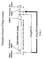
- Figure 1 is a schematic representation of the structure of Nogo receptor-1.
- Human sNogoR310 contains residues 26-310 and sNogoR344 contains residues 26-344.
- Rat sNogoR310 contains residues 27-310 and sNogoR344 contains residues 27-344
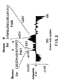
- Figure 2 depicts an antigenicity plot for the Nogo receptor-1 protein using the Vector Nti TM software.
- Rat P-617 is SEQ ID NO: 10 and rat P-618 is SEQ ID NO: 11.
- Figure 3A is a graph depicting the binding activity of anti-Nogo receptor-1 antibody, 7E11. The graph presents the effect of 7E11 concentration on the binding of Nogo66 to Nogo receptor-1.
- Figure 3B depicts the binding activity of anti-Nogo receptor-1 antibody, 1H2. The graph presents the effect of 1H2 concentration on the binding of Nogo66 to sNogoR344-Fc (also referred to herein and in United States patent application 60/402,866 as Fc-sNogoR344 or Ig-sNogoR344).
- Fc-MAG did not compete with Nogo66 for binding to sNogoR344-Fc.
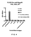
- Figure 4 depicts the results of an ELISA for anti-Nogo-R-1 antibodies 1H2, 3G5 and 2F7.
- the effect of the antibodies on OD 450 in the presence of immobilized antigens was determined.
- the immobilized antigens were sNogoR310-Fc (also referred to herein and in United States patent application 60/402,866 as Fc-sNogoR310 or Ig-sNogoR310), sNogoR344-Fc, p-617, p-618, p-4, p-5 and ovalbumin and BSA.
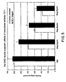
- Figure 5 is a graph depicting the effects of monoclonal antibody, 7E11, on rat DRG neurite outgrowth in the presence of varying amounts of myelin.
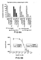
- Figure 6A is a graph depicting the effect of binding of sNogoR310 to 125 I-Nogo66 and 125 I-Nogo40 in the presence of the following competitors: Nogo66, Nogo40 and anti-Nogo receptor-1 monoclonal antibody supernatant.
- Figure 6B depicts the binding activity of 125 I-Nogo66 to sNogoR310.
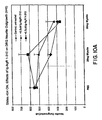
- Figure 7 is a graph depicting the effect of sNogoR310-Fc on 125 I-Nogo40 binding to sNogoR310.
- Figure 8 is a graph depicting the binding activity of sNogoR310-Fc to 125 I-Nogo40.
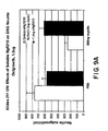
- Figure 9A is a graph of the effect of sNogoR310 on neurite outgrowth/cell in the presence or absence of myelin.
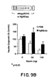
- Figure 9B is a graph of the effect of sNogoR310 on neurite outgrowth in the presence or absence of myelin.
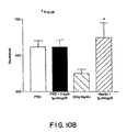
- Figure 10A is a graph depicting the effect of sNogoR310-Fc on P4 rat DRG neurite outgrowth in the presence or absence of increasing amounts of myelin.
- Figure 10B depicts the number of neurites/cell following treatment with PBS, PBS + sNogoR310-Fc, 20ng myelin and myelin + sNogoR310-Fc.
- Figure 11 is a graph depicting the effect of monoclonal antibody 5B10 on DRG neurite outgrowth/cell in the presence of increasing amounts of myelin.
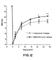
- Figure 12 is a graph depicting the effect of sNogoR310-Fc on the BBB score up to 30 days following induction of injury in a rat spinal cord transection model.
- Figures 13A and 13B report the locomotor BBB score as a function of time after dorsal hemisection in the WT or transgenic mice from Line 08 or Line 01.
- Figure 13C graphs the maximal tolerated inclined plane angle as a function of time after injury for WT and transgenic mice.
- Figure 13D shows hindlimb errors during inclined grid climbing as a function of post-injury time. In all the graphs, means ⁇ s.e.m. from 7-9 mice in each group are reported. The values from transgenic group are statistically different from the WT mice. (double asterisks, P ⁇ 0.01; Student's t-test).
- Figure 14A shows the locomotor BBB score as a function of time after dorsal hemisection in vehicle or sNogoR310-Fc treated animals.
- Figure 14B shows hindlimb errors during grid walking as a function of time after injury.
- Figure 14C shows footprint analysis revealing a shorter stride length and a greater stride width in control rats than uninjured or injured + sNogoR310-Fc rats. In all the graphs, means ⁇ s.e.m. from 7-9 rats in each group are reported.
- the values of sNogoR310-Fc group are statistically different from the control ( Figures 14A-B ).
- the control values are statistically different from no-SCI or SCI + sNogoR310-Fc rats in Figure 14C . (asterisk, p ⁇ 0.05; double asterisks, p ⁇ 0.01; Student's t-test).
- antibody means an intact immunoglobulin, or an antigen-binding fragment thereof.
- Antibodies of this disclosure can be of any isotype or class (e.g ., M, D, G, E and A) or any subclass ( e.g ., G1-4, A1-2) and can have either a kappa ( ⁇ ) or lambda ( ⁇ ) light chain.
- Fc means a portion of the heavy chain constant region of an antibody that is obtainable by papain digestion.
- NogoR fusion protein means a protein comprising a soluble Nogo receptor-1 moiety fused to a heterologous polypeptide.
- humanized antibody means an antibody in which at least a portion of the non-human sequences are replaced with human sequences. Examples of how to make humanized antibodies may be found in United States Patent Nos. 6,054,297 , 5,886,152 and 5,877,293 .
- chimeric antibody means an antibody that contains one or more regions from a first antibody and one or more regions from at least one other antibody.
- the first antibody and the additional antibodies can be from the same or different species.
- Nogo receptor As used herein and in United States patent application 60/402,866 , "Nogo receptor,” “NogoR,” “NogoR-1,” “NgR,” and “NgR-1” each means Nogo receptor-1.
- the present disclosure relates to Nogo receptor-1 polypeptides that are immunogenic.
- the immunogenic polypeptide consists essentially of an amino acid sequence selected from the group consisting of: LDLSDNAQLRVVDPTT (rat) (SEQ ID NO: 1); LDLSDNAQLRSVDPAT (human) (SEQ ID NO: 2); AVASGPFRPFQTNQLTDEELLGLPKCCQPDAADKA (rat) (SEQ ID NO: 3); AVATGPYHPIWTGRATDEEPLGLPKCCQPDAADKA (human)(SEQ ID NO: 4); and CRLGQAGSGA (mouse) (SEQ ID NO: 5).
- the disclosure relates to a nucleic acid encoding a polypeptide of any one of SEQ ID NOs: 1-5.
- the nucleic acid molecule is linked to an expression control sequence (e.g ., pCDNA(I)).
- the present disclosure also relates to a vector comprising a nucleic acid coding for an immunogenic polypeptide of the disclosure.
- Certain vectors provide aspects of the present invention.
- the vector is a cloning vector.
- the vector is an expression vector.
- the vector contains at least one selectable marker.
- the present disclosure also relates to host cells comprising the above-described nucleic acid or vector, e.g. a nucleic acid or vector of the present invention.
- the present disclosure also relates to a method of producing an immunogenic polypeptide of the present disclosure, e.g. an immunogenic polypeptide of the present invention, comprising the step of culturing a host cell.
- Host cells comprising a nucleic acid or vector of the present invention, and methods of culturing said host cells and recovering the immunogenic polypeptide of the present invention, form further aspects of the present invention as set out in the claims.
- the host cell is prokaryotic.
- the host cell is eukaryotic.
- the host cell is yeast.
- the present disclosure further relates to an antibody or an antigen-binding fragment thereof that specifically binds an immunogenic Nogo receptor-1 polypeptide of the disclosure, e.g. an immunogenic Nogo receptor-1 polypeptide of the invention.
- the antibody or antigen-binding fragment binds a polypeptide consisting essentially of an amino acid sequence selected from the group consisting of SEQ ID NOs: 1-5.
- Antibodies and antigen-binding fragments of the present invention are set out in the appended claims.
- the antibody or antigen-binding fragment of the present disclosure e.g. an antibody or antigen-binding fragment of the present invention, may be produced in vivo or in vitro . Production of the antibody or antigen-binding fragment is discussed below.
- An antibody or an antigen-binding fragment thereof according to the present disclosure inhibits the binding of Nogo receptor-1 to a ligand (e.g ., NogoA, NogoB, NogoC, MAG, OM-gp) and decreases myelin-mediated inhibition of neurite outgrowth and sprouting, particularly axonal growth, and attenuates myelin mediated growth cone collapse.
- a ligand e.g ., NogoA, NogoB, NogoC, MAG, OM-gp
- the anti-Nogo receptor-1 antibody or antigen-binding fragment thereof is murine.
- the Nogo receptor-1 is from rat.
- the Nogo receptor-1 is human.
- the anti-Nogo receptor-1 antibody or antigen-binding fragment thereof is recombinant, engineered, humanized and/or chimeric.
- the antibody is selected from the group consisting of: monoclonal 7E11 (ATCC ® accession No. PTA-4587); monoclonal 1H2 (ATCC ® accession No. PTA-4584); monoclonal 2F7 (ATCC ® accession No. PTA-4585); monoclonal 3G5 (ATCC ® accession No. PTA-4586); and monoclonal 5B10 (ATCC ® accession No. PTA-4588).
- the antibody is polyclonal antibody 46.
- antigen-binding fragments are, Fab, Fab', F(ab') 2 , Fv, Fd, dAb, and fragments containing complementarity determining region (CDR) fragments, single-chain antibodies (scFv), chimeric antibodies, diabodies and polypeptides that contain at least a portion of an immunoglobulin that is sufficient to confer specific antigen-binding to the polypeptide (e.g ., immunoadhesins).
- CDR complementarity determining region
- Fd means a fragment that consists of the V H and C H1 domains
- Fv means a fragment that consists of the V L and V H domains of a single arm of an antibody
- dAb means a fragment that consists of a V H domain ( Ward et al., Nature 341:544-546, 1989 ).
- single-chain antibody means an antibody in which a V L region and a V H region are paired to form a monovalent molecules via a synthetic linker that enables them to be made as a single protein chain ( Bird et al., Science 242:423-426, 1988 and Huston et al., Proc. Natl. Acad. Sci.
- diabody means a bispecific antibody in which V H and V L domains are expressed on a single polypeptide chain, but using a linker that is too short to allow for pairing between the two domains on the same chain, thereby forcing the domains to pair with complementary domains of another chain and creating two antigen-binding sites (see e.g., Holliger, P., et al., Proc. Natl. Acad. Sci. USA 90:6444-6448, 1993 , and Poljak, R. J., et al., Structure 2:1121-1123, 1994 ).
- immunoadhesin that specifically binds an antigen of interest means a molecule in which one or more CDRs may be incorporated, either covalently or noncovalently.
- the disclosure provides a subunit polypeptide of a Nogo receptor-1 antibody of the present disclosure, e.g. of a Nogo receptor-1 antibody of the present invention, wherein the subunit polypeptide is selected from the group consisting of: (a) a heavy chain or a variable region thereof; and (b) a light chain or a variable region thereof.
- the disclosure provides a nucleic acid encoding the heavy chain or the variable region thereof, or the light chain or the variable region thereof, of a subunit polypeptide of a Nogo receptor-1 antibody of the present disclosure, e.g. of a Nogo receptor-1 antibody of the present invention.
- the disclosure provides a hypervariable region (CDR) of a Nogo receptor-1 antibody of the present disclosure, e.g. of a Nogo-receptor-1 antibody of the present invention, or a nucleic acid encoding a CDR.
- CDR hypervariable region
- Antibodies of the present disclosure can be generated by immunization of a suitable host (e.g., vertebrates, including humans, mice, rats, sheep, goats, pigs, cattle, horses, reptiles, fishes, amphibians, and in eggs of birds, reptiles and fish).
- a suitable host e.g., vertebrates, including humans, mice, rats, sheep, goats, pigs, cattle, horses, reptiles, fishes, amphibians, and in eggs of birds, reptiles and fish.
- Such antibodies may be polyclonal or monoclonal.
- the host is immunized with an immunogenic Nogo receptor-1 polypeptide of the present disclosure, e.g. with an immunogenic Nogo receptor-1 polypeptide of the invention.
- the host is immunized with Nogo receptor-1 associated with the cell membrane of an intact or disrupted cell and antibodies of the disclosure, e.g. antibodies of the invention, are identified by binding to an immunogenic Nogo receptor-1 polypeptide of the disclosure, e.g. to an immunogenic Nogo receptor-1 polypeptide of the invention.
- Certain methods of producing antibodies by immunizing hosts provide further aspects of the present invention as set out in the appended claims.
- the Nogo receptor-1 antigen is administered with an adjuvant to stimulate the immune response.
- adjuvants often need to be administered in addition to antigen in order to elicit an immune response to the antigen.
- These adjuvants are usually insoluble or undegradable substances that promote nonspecific inflammation, with recruitment of mononuclear phagocytes at the site of immunization.
- adjuvants include, but are not limited to, Freund's adjuvant, RIBI (muramyl dipeptides), ISCOM (immunostimulating complexes) or fragments thereof.
- Monoclonal antibodies of the present disclosure can made by standard procedures as described, e.g ., in Harlow and Lane (1988), supra.
- lymph node and/or splenic B-cells are immortalized by any one of several techniques that are well-known in the art, including but not limited to transformation, such as with EBV or fusion with an immortalized cell line, such as myeloma cells. Thereafter, the cells are clonally separated and the supernatants of each clone tested for production of an antibody specific for an immunogenic Nogo receptor-1 polypeptide of the disclosure, e.g. for an immunogenic Nogo receptor-1 polypeptide of the present invention. Methods of selecting, cloning and expanding hybridomas are well known in the art. Similarly, methods for identifying the nucleotide and amino acid sequence of the immunoglobulin genes are known in the art.
- an antibody of the present disclosure e.g. an antibody of the present invention
- suitable techniques for producing an antibody of the present disclosure involve in vitro exposure of lymphocytes to the Nogo receptor-1 or to an immunogenic polypeptide of the disclosure, e.g. to an immunogenic polypeptide of the invention, or alternatively, selection of libraries of antibodies in phage or similar vectors. See Huse et al., Science, 246, pp. 1275-81 (1989 ).
- Antibodies useful in the present disclosure, e.g., in the present invention may be employed with or without modification.
- Antigens in this case Nogo receptor-1 or an immunogenic polypeptide of the disclosure, e.g. an immunogenic polypeptide of the present invention
- antibodies can be labeled by joining, either covalently or non-covalently, a substance that provides for a detectable signal.
- Suitable labels include, but are not limited to, radionucleotides, enzymes, substrates, cofactors, inhibitors, fluorescent agents, chemiluminescent agents, magnetic particles and the like. Patents teaching the use of such labels include U.S.
- recombinant immunoglobulins may be produced (see U.S. Patent 4,816,567 ).
- an antibody has multiple binding specificities, such as a bifunctional antibody prepared by any one of a number of techniques known to those of skill in the art including the production of hybrid hybridomas, disulfide exchange, chemical crosslinking, addition of peptide linkers between two monoclonal antibodies, the introduction of two sets of immunoglobulin heavy and light chains into a particular cell line, and so forth (see below for more detailed discussion).
- the antibodies of this disclosure may also be human monoclonal antibodies, for example those produced by immortalized human cells, by SCID-hu mice or other non-human animals capable of producing "human” antibodies.
- Anti-Nogo receptor-1 antibodies of this disclosure can be isolated by screening a recombinant combinatorial antibody library.
- Exemplary combinatorial libraries are screened for binding to an immunogenic Nogo receptor-1 polypeptide of the disclosure, e.g. to an immunogenic Nogo receptor-1 polypeptide of the invention, such as a scFv phage display library, prepared using V L and V H cDNAs prepared from mRNA derived from an animal immunized with an immunogenic Nogo receptor-1 polypeptide of the disclosure, e.g. with an immunogenic Nogo receptor-1 polypeptide of the invention.
- Methodologies for preparing and screening such libraries are known in the art.
- phage display libraries There are commercially available methods and materials for generating phage display libraries (e.g. , the Pharmacia Recombinant Phage Antibody System, catalog no. 27-9400-01; the Stratagene SurfZAP TM phage display kit, catalog no. 240612; and others from MorphoSys). There are also other methods and reagents that can be used in generating and screening antibody display libraries (see e.g., Ladner et al. U.S. Pat. No. 5,223,409 ; Kang et al. PCT Publication No. WO 92/18619 ; Dower et al. PCT Publication No. WO 91/17271 ; Winter et al. PCT Publication No.
- the nucleic acid encoding the selected antibody can be recovered from the display package (e.g. , from the phage genome) and subcloned into other expression vectors by standard recombinant DNA techniques. If desired, the nucleic acid can be further manipulated to create other antibody forms of the present disclosure, e.g. other antibody forms of the invention, as described below.
- DNA encoding the antibody heavy chain and light chain or the variable regions thereof is cloned into a recombinant expression vector and introduced into a mammalian host cell, as described above.
- Anti-Nogo receptor-1 antibodies of the disclosure can be of any isotype.
- An antibody of any desired isotype can be produced by class switching.
- nucleic acids encoding V L or V H that do not include any nucleotide sequences encoding C L or C H , are isolated using methods well known in the art.
- the nucleic acids encoding V L or V H are then operatively linked to a nucleotide sequence encoding a C L or C H from a desired class of immunoglobulin molecule. This may be achieved using a vector or nucleic acid that comprises a C L or C H chain, as described above.
- an anti-Nogo receptor-1 antibody of the disclosure e.g. an anti-Nogo receptor-1 antibody of the invention, that was originally IgM may be class switched to an IgG. Further, the class switching may be used to convert one IgG subclass to another, e.g., from IgG1 to IgG2.
- Antibodies or antigen-binding fragments of the present disclosure may be mutated in the variable domains of the heavy and/or light chains to alter a binding property of the antibody.
- a mutation may be made in one or more of the CDR regions to increase or decrease the K d of the antibody for Nogo receptor-1, to increase or decrease K off , or to alter the binding specificity of the antibody.
- Techniques in site-directed mutagenesis are well known in the art. See e.g., Sambrook et al. and Ausubel et al., supra . In a preferred instance, e.g.
- mutations are made at an amino acid residue that is known to be changed compared to the germline in a variable region of an anti-Nogo receptor-1 antibody of the disclosure, e.g. of an anti-Nogo receptor-1 antibody of the invention, as appropriate.
- mutations are made at one or more amino acid residues that are known to be changed compared to the germline in a variable region of an anti-Nogo receptor-1 antibody of the present disclosure, e.g. of an anti-Nogo receptor-1 antibody of the invention, as appropriate.
- a nucleic acid encoding an antibody heavy chain or light chain variable region is mutated in one or more of the framework regions.
- a mutation may be made in a framework region or constant domain to increase the half-life.
- a mutation in a framework region or constant domain also may be made to alter the immunogenicity of the antibody, to provide a site for covalent or non-covalent binding to another molecule, or to alter such properties as complement fixation. Mutations may be made in each of the framework regions, the constant domain and the variable regions in a single mutated antibody. Alternatively, mutations may be made in only one of the framework regions, the variable regions or the constant domain in a single mutated antibody.
- a fusion antibody or immunoadhesin may be made which comprises all or a portion of an anti-Nogo receptor-1 antibody of the present disclosure, e.g. of an anti-Nogo receptor-1 antibody of the present invention, linked to another polypeptide. In some cases, only the variable region of the anti-Nogo receptor-1 antibody is linked to the polypeptide. In other cases, the V H domain of an anti-Nogo receptor-1 antibody of the disclosure, e.g. of an anti-Nogo receptor-1 antibody of this invention, is linked to a first polypeptide, while the V L domain of the antibody is linked to a second polypeptide that associates with the first polypeptide in a manner that permits the V H and V L domains to interact with one another to form an antibody binding site.
- the V H domain is separated from the V L domain by a linker that permits the V H and V L domains to interact with one another (see below under Single Chain Antibodies).
- the V H -linker- V L antibody is then linked to a polypeptide of interest.
- the fusion antibody is useful to directing a polypeptide to a cell or tissue that expresses a Nogo receptor-1 ligand.
- the polypeptide of interest may be a therapeutic agent, such as a toxin, or may be a diagnostic agent, such as an enzyme that may be easily visualized, such as horseradish peroxidase.
- fusion antibodies can be created in which two (or more) single-chain antibodies are linked to one another. This is useful if one wants to create a divalent or polyvalent antibody on a single polypeptide chain, or if one wants to create a bispecific antibody.
- the present disclosure includes a single chain antibody (scFv) that binds an immunogenic Nogo receptor-1 polypeptide of the present disclosure, e.g. an immunogenic Nogo receptor-1 polypeptide of the invention.
- scFv single chain antibody
- V H - and V L -encoding DNA is operatively linked to DNA encoding a flexible linker, e.g., encoding the amino acid sequence (Gly 4 -Ser) 3 (SEQ ID NO:10), such that the V H and V L sequences can be expressed as a contiguous single-chain protein, with the V L and V H regions joined by the flexible linker (see e.g., Bird et al. (1988) Science 242:423-426 ; Huston et al.
- a flexible linker e.g., encoding the amino acid sequence (Gly 4 -Ser) 3 (SEQ ID NO:10)
- the single chain antibody may be monovalent, if only a single V H and V L are used, bivalent, if two V H and V L are used, or polyvalent, if more than two V H and VL are used.
- a bispecific antibody or antigen-binding fragment thereof in which one specificity is for an immunogenic Nogo receptor-1 polypeptide of the disclosure, e.g. for an immunogenic Nogo receptor-1 polypeptide of the invention.
- a chimeric antibody can be generated that specifically binds to an immunogenic Nogo receptor-1 polypeptide of the disclosure, e.g. to an immunogenic Nogo receptor-1 polypeptide of the invention, through one binding domain,and to a second molecule through a second binding domain.
- the chimeric antibody can be produced through recombinant molecular biological techniques, or may be physically conjugated together.
- a single chain antibody containing more than one V H and V L may be generated that binds specifically to an immunogenic polypeptide of the disclosure, e.g. to an immunogenic polypeptide of the invention, and to another molecule that is associated with attenuating myelin mediated growth cone collapse and inhibition of neurite outgrowth and sprouting.
- bispecific antibodies can be generated using techniques that are well known for example, Fanger et al. Immunol Methods 4: 72-81 (1994 ) and Wright and Harris, supra. and in connection with (iii) see e.g., Traunecker et al. Int. J. Cancer (Suppl.) 7: 51-52 (1992 ).
- the chimeric antibodies are prepared using one or more of the variable regions from an antibody of the present disclosure, e.g. from an antibody of the invention. In another case, the chimeric antibody is prepared using one or more CDR regions from said antibody.
- an antibody or an antigen-binding fragment of the present disclosure can be derivatized or linked to another molecule (e.g., another peptide or protein).
- the antibody or antigen-binding fragment is derivatized such that binding to an immunogenic polypeptide of the disclosure, e.g. to an immunogenic polypeptide of the invention, is not affected adversely by the derivatization or labeling.
- an antibody or antibody portion of the present disclosure e.g. an antibody or antibody portion of the invention, can be functionally linked (by chemical coupling, genetic fusion, noncovalent association or otherwise) to one or more other molecular entities, such as another antibody (e.g.
- a bispecific antibody or a diabody a detection agent, a cytotoxic agent, a pharmaceutical agent, and/or a protein or peptide that can mediate association of the antibody or antigen-binding fragment with another molecule (such as a streptavidin core region or a polyhistidine tag).
- a derivatized antibody is produced by crosslinking two or more antibodies (of the same type or of different types, e.g., to create bispecific antibodies).
- Suitable crosslinkers include those that are heterobifunctional, having two distinctly reactive groups separated by an appropriate spacer (e.g., m-maleimidobenzoyl-N-hydroxysuccinimide ester) or homobifunctional ( e.g. , disuccinimidyl suberate).
- spacer e.g., m-maleimidobenzoyl-N-hydroxysuccinimide ester
- homobifunctional e.g. , disuccinimidyl suberate
- the derivatized antibody is a labeled antibody.
- detection agents with which an antibody or antibody portion of the invention may be derivatized are fluorescent compounds, including fluorescein, fluorescein isothiocyanate, rhodamine, 5-dimethylamine-1-napthalenesulfonyl chloride, phycoerythrin, lanthanide phosphors and the like.
- An antibody also may be labeled with enzymes that are useful for detection, such as horseradish peroxidase, ⁇ -galactosidase, luciferase, alkaline phosphatase, glucose oxidase and the like. In instances, e.g.
- the antibody in embodiments that are labeled with a detectable enzyme, is detected by adding additional reagents that the enzyme uses to produce a detectable reaction product. For example, horseradish peroxidase with hydrogen peroxide and diaminobenzidine.
- An antibody also may be labeled with biotin, and detected through indirect measurement of avidin or streptavidin binding.
- An antibody may also be labeled with a predetermined polypeptide epitopes recognized by a secondary reporter (e.g., leucine zipper pair sequences, binding sites for secondary antibodies, metal binding domains, epitope tags) .
- An anti-Nogo receptor-1 antibody or an antigen-fragment thereof also may be labeled with a radio-labeled amino acid.
- the radiolabel may be used for both diagnostic and therapeutic purposes.
- the radio-labeled anti-Nogo receptor-1 antibody may be used diagnostically, for example, for determining Nogo receptor-1 levels in a subject. Further, the radio-labeled anti-Nogo receptor-1 antibody may be used therapeutically for treating spinal cord injury.
- Examples of labels for polypeptides include, but are not limited to, the following radioisotopes or radionucleotides - 3 H, 14 C, 15 N, 35 S, 90 Y, 99 TC, 111 In, 125 I, 131 I.
- An anti-Nogo receptor-1 antibody or an antigen-fragment thereof may also be derivatized with a chemical group such as polyethylene glycol (PEG), a methyl or ethyl group, or a carbohydrate group. These groups may be useful to improve the biological characteristics of the antibody, e.g., to increase serum half-life or to increase tissue binding.
- a chemical group such as polyethylene glycol (PEG), a methyl or ethyl group, or a carbohydrate group.
- the class and subclass of anti-Nogo receptor-1 antibodies may be determined by any method known in the art.
- the class and subclass of an antibody may be determined using antibodies that are specific for a particular class and subclass of antibody. Such antibodies are available commercially.
- the class and subclass can be determined by ELISA, Western Blot as well as other techniques.
- the class and subclass may be determined by sequencing all or a portion of the constant domains of the heavy and/or light chains of the antibodies, comparing their amino acid sequences to the known amino acid sequences of various class and subclasses of immunoglobulins, and determining the class and subclass of the antibodies.
- the binding affinity and dissociation rate of an anti-Nogo receptor-1 antibody of the disclosure e.g. of an anti-Nogo receptor-1 antibody of the invention, to an immunogenic Nogo receptor-1 polypeptide of the disclosure, e.g. to an immunogenic Nogo receptor-1 polypeptide of the invention, may be determined by any method known in the art.
- the binding affinity can be measured by competitive ELISAs, RIAs, BIAcore or KinExA technology.
- the dissociation rate also can be measured by BIAcore or KinExA technology.
- the binding affinity and dissociation rate are measured by surface plasmon resonance using, e.g., a BIAcore.
- the K d of 7E11 and 1H2 were determined to be 1 x 10 -7 M and 2 x 10 -8 M, respectively.
- an anti-Nogo receptor-1 antibody or an antigen-binding fragment of the present disclosure inhibits the binding of Nogo receptor-1 to a ligand.
- the IC 50 of such inhibition can be measured by any method known in the art, e.g., by ELISA, RIA, or Functional Antagonism.
- the IC 50 is between 0.1 and 500 nM.
- the IC 50 is between 10 and 400 nM. In yet other instances, e.g.
- the antibody or portion thereof has an IC 50 of between 60 nM and 400 nM.
- the IC 50 of 7E11 and 1H2 were determined to be 400 nM and 60 nM, respectively, in a binding assay. See also Table 3, infra.
- compositions comprising, and uses of, the antibodies of the present disclosure, e.g. the antibodies of the present invention, are described below.
- Full-length Nogo receptor-1 consists of a signal sequence, a N-terminus region (NT), eight leucine rich repeats (LRR), a LRRCT region (a leucine rich repeat domain C-terminal of the eight leucine rich repeats), a C-terminus region (CT) and a GPI anchor (see Fig. 1 ).
- Soluble Nogo receptor-1 polypeptides of the disclosure comprise an NT domain; 8 LRRs and an LRRCT domain and lack a signal sequence and a functional GPI anchor (i.e. , no GPI anchor or a GPI anchor that lacks the ability to efficiently associate to a cell membrane).
- a soluble Nogo receptor-1 polypeptide comprises a heterologous LRR.
- a soluble Nogo receptor-1 polypeptide comprises 2, 3, 4, 5, 6, 7, or 8 heterologous LRR's.
- a heterologous LRR means an LRR obtained from a protein other than Nogo receptor-1.
- Exemplary proteins from which a heterologous LRR can be obtained are toll-like receptor (TLR1.2); T-cell activation leucine repeat rich protein; deceorin; OM-gp; insulin-like growth factor binding protein acidic labile subunit slit and robo; and toll-like receptor 4.
- the disclosure provides a soluble Nogo receptor-1 polypeptide of 319 amino acids (soluble Nogo receptor-1 344, sNogoR1-344, or sNogoR344) (residues 26-344 of SEQ ID NOs: 6 and 8 or residues 27-344 of SEQ ID NO: 8).
- the disclosure provides a soluble Nogo receptor-1 polypeptide of 285 amino acids (soluble Nogo receptor-1 310, sNogoR1-310, or sNogoR310) (residues 26-310 of SEQ ID NOs: 7 and 9 or residues 27-310 of SEQ ID NO: 9).
- the soluble Nogo receptor-1 polypeptides of the disclosure may be used to inhibit the binding of a ligand to Nogo receptor-1 and act as an antagonist of Nogo receptor-1 ligands.
- the soluble Nogo receptor-1 polypeptides of the disclosure e.g. the soluble Nogo receptor-1 polypeptides of the invention, may be used to decrease inhibition of neurite outgrowth and sprouting in a neuron, such as axonal growth and to inhibit myelin mediated growth cone collapse in a neuron.
- the neuron is a CNS neuron.
- sNogoR310 and sNogoR344 surprisingly, block the binding of NogoA, NogoB, NogoC, MAG and OM-gp to Nogo receptor-1.
- the soluble Nogo receptor-1 polypeptide of the disclosure e.g. the soluble Nogo receptor-1 polypeptide of the invention
- the heterologous polypeptide is an immunoglobulin constant domain.
- the immunoglobulin constant domain is a heavy chain constant domain.
- the heterologous polypeptide is an Fc fragment.
- the Fc is joined to the C-terminal end of the soluble Nogo receptor-1 polypeptide of the disclosure, e.g. of the soluble Nogo receptor-1 polypeptide of the invention.
- the fusion Nogo receptor-1 protein is a dimer. Fusion proteins of the invention are set out in the appended claims.
- the present disclosure provides a nucleic acid that encodes a polypeptide of the disclosure, including the polypeptides of any one of SEQ ID NOs: 1-9.
- Certain nucleic acids of the present disclosure form aspects of the present invention, these being set out in the appended claims.
- the nucleic acid encodes a polypeptide selected from the group consisting of amino acid residues 26-344 of Nogo receptor-1 as shown in SEQ ID NOs: 6 and 8 or amino acid residues 27-344 of Nogo receptor-1 as shown in SEQ ID NO: 8.
- the nucleic acid encodes a polypeptide selected from the group consisting of amino acid residues 26-344 of Nogo receptor-1 as shown in SEQ ID NOs: 6 and 8 or amino acid residues 27-344 of Nogo receptor-1 as shown in SEQ ID NO: 8.
- the nucleic acid molecule encodes a polypeptide selected from the group consisting of amino acid residues 26-310 of Nogo receptor-1 as shown in SEQ ID NOs: 7 and 9 or amino acid residues 27-310 of Nogo receptor-1 as shown in SEQ ID NO: 9.
- nucleic acid means genomic DNA, cDNA, mRNA and antisense molecules, as well as nucleic acids based on alternative backbones or including alternative bases whether derived from natural sources or synthesized. In some instances, e.g.
- the nucleic acid further comprises a transcriptional promoter and optionally a signal sequence each of which is operably linked to the nucleotide sequence encoding the polypeptide of the present disclosure, e.g. the polypeptide of the present invention, as appropriate.
- nucleic acid encoding a Nogo receptor-1 fusion protein of the present disclosure, e.g. a Nogo receptor-1 fusion protein of the invention.
- the nucleic acid encodes a Nogo receptor-1 fusion protein of the invention, including a fusion protein comprising a polypeptide selected from the group consisting of amino acid residues 26-344 of Nogo receptor-1 as shown in SEQ ID NOs: 6 and 8 or amino acid residues 27-344 of SEQ ID NO: 8 and amino acid residues 26-310 of Nogo receptor-1 as shown in SEQ ID NOs: 7 and 9 or amino acid residues 27-310 of SEQ ID NO: 9.
- a polypeptide selected from the group consisting of amino acid residues 26-344 of Nogo receptor-1 as shown in SEQ ID NOs: 6 and 8 or amino acid residues 27-344 of SEQ ID NO: 8 and amino acid residues 26-310 of Nogo receptor-1 as shown in SEQ ID NOs: 7 and 9 or amino acid residues 27-310 of SEQ ID NO: 9.
- the nucleic acid encoding a Nogo receptor-1 fusion protein further comprises a transcriptional promoter and optionally a signal sequence.
- the nucleotide sequence further encodes an immunoglobulin constant region.
- the immunoglobulin constant region is a heavy chain constant region.
- the nucleotide sequence further encodes an immunoglobulin heavy chain constant region joined to a hinge region.
- the nucleic acid further encodes Fc.
- the Nogo receptor-1 fusion proteins comprise an Fc fragment.
- the encoding nucleic acids of the present disclosure may further be modified so as to contain a detectable label for diagnostic and probe purposes.
- a detectable label for diagnostic and probe purposes.
- a variety of such labels are known in the art and can readily be employed with the encoding molecules herein described. Suitable labels include, but are not limited to, biotin, radiolabeled nucleotides and the like. A skilled artisan can employ any of the art known labels to obtain a labeled encoding nucleic acid molecule.
- compositions comprising an immunogenic polypeptide selected from the group consisting of SEQ ID NO: 1, SEQ ID NO: 2, SEQ ID NO: 3, SEQ ID NO: 4 and SEQ ID NO: 5.
- compositions comprising an anti-Nogo receptor-1 antibody or an antigen-binding fragment thereof, or a soluble Nogo receptor-1 polypeptide or fusion protein of the present disclosure, e.g. of the present invention.
- Certain compositions of the disclosure provide a further aspect of the present invention, as set out in the appended claims.
- the compositions may contain suitable pharmaceutically acceptable carriers comprising excipients and auxiliaries which facilitate processing of the active compounds into preparations which can be used pharmaceutically for delivery to the site of action.
- suitable formulations for parenteral administration include aqueous solutions of the active compounds in water-soluble form, for example, water-soluble salts.
- suspensions of the active compounds as appropriate oily injection suspensions may be administered.
- Suitable lipophilic solvents or vehicles include fatty oils, for example, sesame oil, or synthetic fatty acid esters, for example, ethyl oleate or triglycerides.
- Aqueous injection suspensions may contain substances which increase the viscosity of the suspension include, for example, sodium carboxymethyl cellulose, sorbitol and dextran.
- the suspension may also contain stabilizers.
- Liposomes can also be used to encapsulate the molecules of this disclosure, e.g. the molecules of this invention, for delivery into the cell.
- Exemplary "pharmaceutically acceptable carriers" are any and all solvents, dispersion media, coatings, antibacterial and antifungal agents, isotonic and absorption delaying agents, and the like that are physiologically compatible, water, saline, phosphate buffered saline, dextrose, glycerol, ethanol and the like, as well as combinations thereof. In some instances, e.g.
- the compositions comprise isotonic agents, for example, sugars, polyalcohols such as mannitol, sorbitol, or sodium chloride.
- the compositions comprise pharmaceutically acceptable substances such as wetting agents or minor amounts of auxiliary substances such as wetting or emulsifying agents, preservatives or buffers, which enhance the shelf life or effectiveness of the antibodies, antigen-binding fragments, soluble Nogo receptors or fusion proteins of the present disclosure, including those of the invention.
- compositions of the present disclosure may be in a variety of forms, including, for example, liquid, semi-solid and solid dosage forms, such as liquid solutions (e.g., injectable and infusible solutions), dispersions or suspensions.
- liquid solutions e.g., injectable and infusible solutions
- dispersions or suspensions e.g., dispersions or suspensions.
- the preferred form depends on the intended mode of administration and therapeutic application.
- compositions are in the form of injectable or infusible solutions, such as compositions similar to those used for passive immunization of humans with other antibodies.
- compositions can be formulated as a solution, micro emulsion, dispersion, liposome, or other ordered structure suitable to high drug concentration.
- Sterile injectable solutions can be prepared by incorporating the anti-Nogo receptor-1 antibody in the required amount in an appropriate solvent with one or a combination of ingredients enumerated above, as required, followed by filtered sterilization.
- dispersions are prepared by incorporating the active compound into a sterile vehicle that contains a basic dispersion medium and the required other ingredients from those enumerated above.
- the preferred methods of preparation are vacuum drying and freeze-drying that yields a powder of the active ingredient plus any additional desired ingredient from a previously sterile-filtered solution thereof.
- the proper fluidity of a solution can be maintained, for example, by the use of a coating such as lecithin, by the maintenance of the required particle size in the case of dispersion and by the use of surfactants.
- Prolonged absorption of injectable compositions can be brought about by including in the composition an agent that delays absorption, for example, monostearate salts and gelatin.
- the active compound may be prepared with a carrier that will protect the compound against rapid release, such as a controlled release formulation, including implants, transdermal patches, and microencapsulated delivery systems.
- a controlled release formulation including implants, transdermal patches, and microencapsulated delivery systems.
- Biodegradable, biocompatible polymers can be used, such as ethylene vinyl acetate, polyanhydrides, polyglycolic acid, collagen, polyorthoesters, and polylactic acid. Many methods for the preparation of such formulations are patented or generally known to those skilled in the art. See e.g., Sustained and Controlled Release Drug Delivery Systems, J. R. Robinson, ed., Marcel Dekker, Inc., New York, 1978 .
- Nogo receptor-1 antibodies or antigen-binding fragments thereof, or soluble Nogo receptor-1 polypeptides or fusion proteins of the disclosure, including those of the invention are coformulated with and/or coadministered with one or more additional therapeutic agents.
- compositions may include a "therapeutically effective amount” or a “prophylactically effective amount” of an antibody, antigen-binding fragment, polypeptide(s), or fusion protein of the disclosure, e.g. of the invention.
- a “therapeutically effective amount” refers to an amount effective, at dosages and for periods of time necessary, to achieve the desired therapeutic result.
- a therapeutically effective amount of the Nogo receptor-1 antibody or antigen-binding fragment thereof, soluble Nogo receptor-1 polypeptide or Nogo receptor fusion protein may vary according to factors such as the disease state, age, sex, and weight of the individual.
- a therapeutically effective amount is also one in which any toxic or detrimental effects of the antibody, antigen-binding fragment, soluble Nogo receptor-1 polypeptide or Nogo receptor fusion protein are outweighed by the therapeutically beneficial effects.
- a “prophylactically effective amount” refers to an amount effective, at dosages and for periods of time necessary, to achieve the desired prophylactic result. Typically, since a prophylactic dose is used in subjects prior to or at an earlier stage of disease, the prophylactically effective amount will be less than the therapeutically effective amount.
- Dosage regimens may be adjusted to provide the optimum desired response (e.g., a therapeutic or prophylactic response). For example, a single bolus may be administered, several divided doses may be administered over time or the dose may be proportionally reduced or increased as indicated by the exigencies of the therapeutic situation. It is especially advantageous to formulate parenteral compositions in dosage unit form for ease of administration and uniformity of dosage unit form as used herein refers to physically discrete units suited as unitary dosages for the mammalian subjects to be treated, each unit containing a predetermined quantity of active compound calculated to produce the desired therapeutic effect in association with the required pharmaceutical carrier.
- the specification for the dosage unit forms of the present disclosure e.g.
- a therapeutically effective dose range for Nogo receptor-1 antibodies or antigen-binding fragments thereof is 0.1 - 4 mg/Kg per day. In some instances, e.g.
- a therapeutically effective dose range for Nogo receptor-1 antibodies or antigen-binding fragments thereof is 0.2 - 4 mg/Kg per day. In some instances, e.g. in some embodiments, a therapeutically effective dose range for Nogo receptor-1 antibodies or antigen-binding fragments thereof is 0.2 mg/Kg per day.
- the present disclosure provides methods for inhibiting Nogo receptor-1 activity by administering anti-Nogo receptor-1 antibodies, antigen-binding fragments of such antibodies, soluble Nogo receptor-1 polypeptides, or fusion proteins comprising such polypeptides to a mammal in need thereof.
- the disclosure provides a method of inhibiting Nogo receptor-1 binding to a ligand, comprising the step of contacting Nogo receptor-1 with an antibody or antigen-binding fragment of the present disclosure, e.g. of the present invention.
- the ligand is selected from the group consisting of NogoA, NogoB, NogoC, MAG and OM-gp.
- the present disclosure provides a method for inhibiting growth cone collapse in a neuron, comprising the step of contacting the neuron with the antibody or antigen-binding fragment the present disclosure, e.g. of the present invention.
- the disclosure provides a method for decreasing the inhibition of neurite outgrowth or sprouting in a neuron, comprising the step of contacting the neuron with the antibody or antigen-binding fragment of the present disclosure, e.g. of the present invention.
- the neuron is a CNS neuron. In some of these methods, the neurite outgrowth or sprouting is axonal growth.
- the present disclosure provides a method of promoting the survival of a neuron in a mammal, which neuron is at risk of dying, comprising (a) providing a cultured host cell expressing (i) an anti-Nogo receptor-1 antibody or antigen-binding fragment thereof; or (ii) a soluble Nogo receptor-1 polypeptide; and (b) introducing the host cell into the mammal at or near the site of the neuron.
- a cultured host cell expressing (i) an anti-Nogo receptor-1 antibody or antigen-binding fragment thereof; or (ii) a soluble Nogo receptor-1 polypeptide
- introducing the host cell into the mammal at or near the site of the neuron comprising (a) providing a cultured host cell expressing (i) an anti-Nogo receptor-1 antibody or antigen-binding fragment thereof; or (ii) a soluble Nogo receptor-1 polypeptide; and (b) introducing the host cell into the mammal at or near the site of the neuron.
- the present disclosure provides a gene therapy method of promoting survival of a neuron at risk of dying, which neuron is in a mammal, comprising administering at or near the site of the neuron a viral vector comprising a nucleotide sequence that encodes (a) an anti-Nogo receptor-1 antibody or antigen-binding fragment thereof; or (b) a soluble Nogo receptor-1 polypeptide, wherein the anti-Nogo receptor-1 antibody, antigen-binding fragment, or soluble Nogo receptor-1 polypeptide is expressed from the nucleotide sequence in the mammal in an amount sufficient to promote survival of the neuron.
- Viral vectors and methods useful for these embodiments are described in, e.g. , No ⁇ l et al., Human Gene Therapy, 13, 1483-93 (2002 ).
- the disclosure provides a method of inhibiting Nogo receptor-1 binding to a ligand, comprising the step of contacting the ligand with the soluble Nogo receptor-1 polypeptide or the Nogo receptor-1 fusion protein of the present disclosure, e.g. of the present invention.
- the disclosure provides a method of modulating an activity of a Nogo receptor-1 ligand, comprising the step of contacting the Nogo receptor-1 ligand with a soluble Nogo receptor-1 polypeptide or a Nogo receptor-1 fusion protein of the present disclosure e.g. of the present invention.
- the disclosure provides a method for inhibiting growth cone collapse in a neuron, comprising the step of contacting a Nogo receptor-1 ligand with a soluble Nogo receptor-1 polypeptide or a Nogo receptor-1 fusion protein of the present disclosure, e.g. of the present invention.
- the disclosure provides a method for decreasing the inhibition of neurite outgrowth or sprouting in a neuron, comprising the step of contacting a Nogo receptor-1 ligand with the soluble Nogo receptor-1 polypeptide or the Nogo receptor-1 fusion protein of the present disclosure, e.g. of the present invention.
- the neuron is a CNS neuron.
- the ligand is selected from the group consisting of NogoA, NogoB, NogoC, MAG and OM-gp.
- the neurite outgrowth or sprouting is axonal growth.
- the anti-Nogo receptor-1 antibody is a human antibody.
- the mammal is a human patient.
- the antibody or antigen-binding fragment thereof is administered to a non-human mammal expressing a Nogo receptor-1 with which the antibody cross-reacts (e.g. , a primate, cynomologous or rhesus monkey) for veterinary purposes or as an animal model of human disease.
- a non-human mammal expressing a Nogo receptor-1 with which the antibody cross-reacts e.g. , a primate, cynomologous or rhesus monkey
- Such animal models may be useful for evaluating the therapeutic efficacy of antibodies of this disclosure, including the antibodies of this invention.
- administering is used to treat a spinal cord injury to facilitate axonal growth throughout the injured site.
- anti-Nogo receptor-1 antibodies or antigen-binding fragments, or soluble Nogo receptor-1 polypeptides or fusion proteins of the present disclosure can be provided alone, or in combination, or in sequential combination with other agents that modulate a particular pathological process.
- anti-inflammatory agents may be coadministered following stroke as a means for blocking further neuronal damage and inhibition of axonal regeneration.
- the Nogo receptor-1 antibodies, antigen-binding fragments, soluble Nogo receptor-1 and Nogo receptor fusion proteins are said to be administered in combination with one or more additional therapeutic agents when the two are administered simultaneously, consecutively or independently.
- the anti-Nogo receptor-1 antibodies, antigen-binding fragments, soluble Nogo receptor-1 polypeptides, or Nogo receptor-1 fusion proteins of the present disclosure can be administered via parenteral, subcutaneous, intravenous, intramuscular, intraperitoneal, transdermal, inhalational or buccal routes.
- an agent may be administered locally to a site of injury via microinfusion. Typical sites include, but are not limited to, damaged areas of the spinal cord resulting from injury.
- the dosage administered will be dependent upon the age, health, and weight of the recipient, kind of concurrent treatment, if any, frequency of treatment, and the nature of the effect desired.
- the compounds of this disclosure can be utilized in vivo, ordinarily in mammals, such as humans, sheep, horses, cattle, pigs, dogs, cats, rats and mice, or in vitro. Methods and uses in accordance with the present invention are set out in the appended claims.
- rDNA recombinant DNA molecules
- a rDNA molecule is a DNA molecule that has been subjected to molecular manipulation. Methods for generating rDNA molecules are well known in the art, for example, see Sambrook et al., (1989) Molecular Cloning - A Laboratory Manual, Cold Spring Harbor Laboratory Press .
- a coding DNA sequence is operably linked to expression control sequences and vector sequences.
- vectors comprising the nucleic acids encoding the polypeptides of the present disclosure, e.g. the polypeptides of the invention.
- Vectors that form part of the present invention are set out in the appended claims.
- the choice of vector and expression control sequences to which the nucleic acids of this disclosure, e.g. the nucleic acids of this invention, are operably linked depends directly, as is well known in the art, on the functional properties desired (e.g. , protein expression, and the host cell to be transformed).
- a vector of the present disclosure e.g. a vector of the present invention, may be at least capable of directing the replication or insertion into the host chromosome, and preferably also expression, of the structural gene included in the rDNA molecule.
- Expression control elements that are used for regulating the expression of an operably linked protein encoding sequence are known in the art and include, but are not limited to, inducible promoters, constitutive promoters, secretion signals, and other regulatory elements.
- the inducible promoter is readily controlled, such as being responsive to a nutrient in the host cell's medium.
- the vector containing a coding nucleic acid molecule will include a prokaryotic replicon, i.e., a DNA sequence having the ability to direct autonomous replication and maintenance of the recombinant DNA molecule extra-chromosomally in a prokaryotic host cell, such as a bacterial host cell, transformed therewith.
- a prokaryotic replicon i.e., a DNA sequence having the ability to direct autonomous replication and maintenance of the recombinant DNA molecule extra-chromosomally in a prokaryotic host cell, such as a bacterial host cell, transformed therewith.
- a prokaryotic replicon i.e., a DNA sequence having the ability to direct autonomous replication and maintenance of the recombinant DNA molecule extra-chromosomally in a prokaryotic host cell, such as a bacterial host cell, transformed therewith.
- vectors that include a prokaryotic replicon may also include a gene whose expression confer
- Vectors that include a prokaryotic replicon can further include a prokaryotic or bacteriophage promoter capable of directing the expression (transcription and translation) of the coding gene sequences in a bacterial host cell, such as E. coli.
- a promoter is an expression control element formed by a DNA sequence that permits binding of RNA polymerase and transcription to occur.
- Promoter sequences compatible with bacterial hosts are typically provided in plasmid vectors containing convenient restriction sites for insertion of a DNA segment of the present invention. Examples of such vector plasmids are pUC8, pUC9, pBR322 and pBR329 (Bio-Rad ® Laboratories), pPL and pKK223 (Pharmacia). Any suitable prokaryotic host can be used to express a recombinant DNA molecule encoding a protein of the present disclosure, e.g. a protein of the present invention.
- Expression vectors compatible with eukaryotic cells can also be used to form rDNA molecules that contain a coding sequence.
- Eukaryotic cell expression vectors are well known in the art and are available from several commercial sources. Typically, such vectors are provided containing convenient restriction sites for insertion of the desired DNA segment. Examples of such vectors are pSVL and pKSV-10 (Pharmacia), pBPV-1, pML2d (International Biotechnologies), pTDT1 (ATCC ® 31255) and other eukaryotic expression vectors.
- Eukaryotic cell expression vectors used to construct the rDNA molecules of the present disclosure, including rDNA molecules of the present invention, may further include a selectable marker that is effective in an eukaryotic cell, preferably a drug resistance selection marker.
- a preferred drug resistance marker is the gene whose expression results in neomycin resistance, i.e., the neomycin phosphotransferase (neo) gene. ( Southern et al., (1982) J. Mol. Anal. Genet. 1, 327-341 ).
- the selectable marker can be present on a separate plasmid, the two vectors introduced by co-transfection of the host cell, and transfectants selected by culturing in the appropriate drug for the selectable marker.
- DNAs encoding partial or full-length light and heavy chains are inserted into expression vectors such that the genes are operatively linked to transcriptional and translational control sequences.
- Expression vectors include plasmids, retroviruses, cosmids, YACs, EBV-derived episomes, and the like.
- the antibody gene is ligated into a vector such that transcriptional and translational control sequences within the vector serve their intended function of regulating the transcription and translation of the antibody gene.
- the expression vector and expression control sequences are chosen to be compatible with the expression host cell used.
- the antibody light chain gene and the antibody heavy chain gene can be inserted into separate vectors.
- Both genes may, however, be inserted into the same expression vector.
- the antibody genes are inserted into the expression vector by standard methods (e . g ., ligation of complementary restriction sites on the antibody gene fragment and vector, or blunt end ligation if no restriction sites are present).
- a convenient vector is one that encodes a functionally complete human C H or C L immunoglobulin sequence, with appropriate restriction sites engineered so that any V H or V L sequence can be easily inserted and expressed, as described above.
- splicing usually occurs between the splice donor site in the inserted J region and the splice acceptor site preceding the human C region, and also at the splice regions that occur within the human C H exons. Polyadenylation and transcription termination occur at native chromosomal sites downstream of the coding regions.
- the recombinant expression vector can also encode a signal peptide that facilitates secretion of the antibody chain from a host cell.
- the antibody chain gene may be cloned into the vector such that the signal peptide is linked in-frame to the amino terminus of the antibody chain gene.
- the signal peptide can be an immunoglobulin signal peptide or a heterologous signal peptide (i.e., a signal peptide from a non-immunoglobulin protein).
- the recombinant expression vectors that are disclosed herein carry regulatory sequences that control their expression in a host cell. It will be appreciated by those skilled in the art that the design of the expression vector, including the selection of regulatory sequences may depend on such factors as the choice of the host cell to be transformed, the level of expression of protein desired, etc.
- Preferred regulatory sequences for mammalian host cell expression include viral elements that direct high levels of protein expression in mammalian cells, such as promoters and/or enhancers derived from retroviral LTRs, cytomegalovirus (CMV) (such as the CMV promoter/enhancer), Simian Virus 40 (SV40) (such as the SV40 promoter/enhancer), adenovirus, ( e.g. , the adenovirus major late promoter (AdMLP)), polyoma and strong mammalian promoters such as native immunoglobulin and actin promoters.
- CMV cytomegalovirus
- SV40 Simian Virus 40
- AdMLP adenovirus major late promoter
- polyoma and strong mammalian promoters such as native immunoglobulin and actin promoters.
- the recombinant expression vectors of the present disclosure may carry additional sequences, such as sequences that regulate replication of the vector in host cells (e.g. , origins of replication) and selectable marker genes.
- the selectable marker gene facilitates selection of host cells into which the vector has been introduced (see e.g., U.S. Pat. Nos. 4,399,216 , 4,634,665 and 5,179,017 , all by Axel et al.).
- the selectable marker gene confers resistance to drugs, such as G418, hygromycin or methotrexate, on a host cell into which the vector has been introduced.
- Preferred selectable marker genes include the dihydrofolate reductase (DHFR) gene (for use in dhfr - host cells with methotrexate selection/amplification) and the neo gene (for G418 selection).
- DHFR dihydrofolate reductase
- Nucleic acid molecules encoding anti-Nogo receptor-1 antibodies, immunogenic peptides, soluble Nogo receptor-1 polypeptides, or soluble Nogo receptor-1 fusion proteins of this disclosure, e.g. of this invention, and vectors comprising these nucleic acid molecules, can be used for transformation of a suitable host cell. Transformation can be by any known method for introducing polynucleotides into a host cell.
- nucleic acid molecules may be introduced into mammalian cells by viral vectors.
- Transformation of appropriate cell hosts with an rDNA molecule of the present disclosure is accomplished by well known methods that typically depend on the type of vector used and host system employed.
- electroporation and salt treatment methods can be employed (see, for example, Sambrook et al., (1989) Molecular Cloning - A Laboratory Manual, Cold Spring Harbor Laboratory Press ; Cohen et al., (1972) Proc. Natl. Acad. Sci. USA 69, 2110-2114 ).
- cationic lipid or salt treatment methods can be employed (see, for example, Graham et al., (1973) Virology 52, 456-467 ; Wigler et al., (1979) Proc. Natl. Acad. Sci. USA 76, 1373-1376 ).
- Successfully transformed cells i.e., cells that contain an rDNA molecule of the present disclosure, e.g. an rDNA molecule of the present invention, can be identified by well known techniques including the selection for a selectable marker.
- cells resulting from the introduction of an rDNA of the present disclosure, e.g. an rDNA molecule of the present invention can be cloned to produce single colonies. Cells from those colonies can be harvested, lysed and their DNA content examined for the presence of the rDNA using a method such as that described by Southern, (1975) J. Mol. Biol. 98, 503-517 or the proteins produced from the cell may be assayed by an immunological method.
- Mammalian cell lines available as hosts for expression are well known in the art and include many immortalized cell lines available from the American Type Culture Collection (ATCC ® ). These include, inter alia, Chinese hamster ovary (CHO) cells, NSO, SP2 cells, HeLa cells, baby hamster kidney (BHK) cells, monkey kidney cells (COS), human hepatocellular carcinoma cells (e.g., Hep G2), A549 cells, and a number of other cell lines. Cell lines of particular preference are selected through determining which cell lines have high expression levels. Other cell lines that may be used are insect cell lines, such as Sf9 cells.
- recombinant expression vectors encoding the immunogenic polypeptides, Nogo receptor-1 antibodies or antigen-binding fragments, soluble Nogo receptor-1 polypeptides and soluble Nogo receptor-1 fusion proteins of the present disclosure are introduced into mammalian host cells, they are produced by culturing the host cells for a period of time sufficient to allow for expression of the antibody, polypeptide and fusion polypeptide in the host cells or, more preferably, secretion of the immunogenic polypeptides, Nogo receptor-1 antibodies or antigen-binding fragments, soluble Nogo receptor-1 polypeptides and soluble Nogo receptor-1 fusion proteins into the culture medium in which the host cells are grown.
- Immunogenic polypeptides, Nogo receptor-1 antibodies or antigen-binding fragments, soluble Nogo receptor-1 polypeptides and soluble Nogo receptor-1 fusion proteins of the present disclosure, e.g. of the present invention, can be recovered from the culture medium using standard protein purification methods.
- immunogenic polypeptides, Nogo receptor-1 antibodies or antigen-binding fragments, soluble Nogo receptor-1 polypeptides and soluble Nogo receptor-1 fusion proteins of the present disclosure, e.g. of the present invention, (or other moieties therefrom) from production cell lines can be enhanced using a number of known techniques.
- the glutamine synthetase gene expression system (the GS system) is a common approach for enhancing expression under certain conditions.
- the GS system is discussed in whole or part in connection with European Patent Nos. 0 216 846 , 0 256 055 , and 0 323 997 and European Patent Application No. 89303964.4 .
- host cells transformed with a nucleic acid molecule that encodes a Nogo receptor-1 antibody, antigen-binding fragment, soluble Nogo receptor-1 polypeptide and/or soluble Nogo receptor-1 fusion protein of the present disclosure, e.g. a fusion protein of the invention.
- Certain host cells of the present disclosure provide further aspects of the present invention as set out out in the appended claims.
- Host cells can be either prokaryotic or eukaryotic.
- Eukaryotic cells useful for expression of a protein of the present disclosure, e.g. a protein of the present invention are not limited, so long as the cell line is compatible with cell culture methods and compatible with the propagation of the expression vector and expression of the gene product.
- Preferred eukaryotic host cells include, but are not limited to, yeast, insect and mammalian cells, preferably vertebrate cells such as those from a mouse, rat, monkey or human cell line.
- useful eukaryotic host cells include Chinese hamster ovary (CHO) cells available from the ATCC ® as CCL61, NIH Swiss mouse embryo cells NIH-3T3 available from the ATCC as CRL1658, baby hamster kidney cells (BHK), and the like eukaryotic tissue culture cell lines.
- the production of a recombinant form of a protein typically involves the following steps:
- nucleic acid molecule that encodes a protein of the present disclosure, e.g. a protein of the invention. If the encoding sequence is uninterrupted by introns, it is directly suitable for expression in any host.
- the nucleic acid molecule is then optionally placed in operable linkage with suitable control sequences, as described above, to form an expression unit containing the protein open reading frame.
- the expression unit is used to transform a suitable host and the transformed host is cultured under conditions that allow the production of the recombinant protein.
- the recombinant protein is isolated from the medium or from the cells; recovery and purification of the protein may not be necessary in some instances where some impurities may be tolerated.
- the desired coding sequences may be obtained from genomic fragments and used directly in appropriate hosts.
- the construction of expression vectors that are operable in a variety of hosts is accomplished using appropriate replicons and control sequences, as set forth above.
- the control sequences, expression vectors, and transformation methods are dependent on the type of host cell used to express the gene and were discussed in detail earlier.
- Suitable restriction sites can, if not normally available, be added to the ends of the coding sequence so as to provide an excisable gene to insert into these vectors.
- a skilled artisan can readily adapt any host/expression system known in the art for use with the nucleic acid molecules of the present disclosure, e.g. with the nucleic acid molecules of the present invention, to produce recombinant protein.
- Anti-Nogo receptor-1 antibodies that specifically bind an immunogenic Nogo receptor-1 polypeptide of the invention were made using the following methods and procedures.
- the rat Nogo receptor-1 gene (GenBank TM No. AF 462390) was subcloned into the mammalian expression vector pEAG1256 (Biogen ® ) that contained the CMV promotor and geneticin resistance gene for drug selection.
- the recombinant plasmid was transfected into COS-7 cells using Superfect (Qiagen ® ). Transfectants were selected using geneticin (Gibco TM , 2 mg/ml), cloned and verified for surface expression of Nogo receptor-1 protein by FACS.
- COS-7 membranes were prepared from these cells according to procedures as described [ Wang et al., J. Neurochem., 75:1155-1161 (2000 )] with two washings, and stored at 1 mg/ml [protein concentration] in 10% glycerol at -70°C.
- the rat Nogo receptor-1 gene sequence was subjected to antigenicity analyses using Vector NTi TM software ( Fig. 2 ).
- Antigenic peptides,identified in the analyses were conjugated to Keyhole Limpet Hemocyanin (KLH) using standard glutaldehyde procedures.
- KLH Keyhole Limpet Hemocyanin
- mice immunized with antigenic Nogo receptor-1 peptides were screened by ELISA whereas sera from mice immunized with COS-7 cells expressing Nogo receptor-1 were screened by flow cytometry. Mice that were positive for antibodies that specifically bound Nogo receptor-1-COS-7 cells were identified by flow cytometry and were sacrificed.
- Splenocytes were isolated from the mice and fused to the FL653 myeloma (an APRT- derivative of a Ig-/HGPRT- Balb/c mouse myeloma, maintained in DMEM containing 10% FBS, 4500 mg/L glucose, 4 mM L-glutamine, and 20 mg/ml 8-azaguanine) as described ( Kennett et al., 1993. Monoclonal Antibodies: A New Dimension in Biological Analysis. Plenum Press, New York ). Fused cells were plated into 24- or 48-well plates (Corning Glass Works, Corning, NY), and fed with adenine, aminopterin and thymidine containing culture media.
- FL653 myeloma an APRT- derivative of a Ig-/HGPRT- Balb/c mouse myeloma, maintained in DMEM containing 10% FBS, 4500 mg/L glucose, 4 mM L-glutamine, and 20 mg/m
- AAT resistant cultures were screened by ELISA or flow cytometry for binding to either Nogo receptor-1-COS-7 cells or to a Nogo receptor-1 antigenic peptide as described below. Cells in the positive wells were further subcloned by limiting dilution.
- the peptides that were used as immunogens were conjugated to BSA.
- 0.5 ⁇ g of the conjugated peptide in 50 ⁇ l of 0.1 M sodium bicarbonate buffer, pH 9.0 was added to each well of a 96-well MaxiSorp plate (Nunc TM ). The plate was then incubated at 37°C for 1 hour or 4°C for 16 hours and non-specific binding sites were blocked using 25 mM HEPES, pH 7.4 containing 0.1% BSA, 0.1% ovalbumin, 0.1% blotto and 0.001% azide. Hybridoma supernatant was added and incubated at 25°C for 1 hour.
- Antibodies were screened for binding to full length Nogo receptor-1 as follows.
- COS-7 cells were labeled with 0.1 uM CellTracker TM Green CMFDA I (Molecular Probes, Eugene, OR) as described by the vendor.
- Equal volumes of CellTracker TM labeled control cells were mixed with washed Nogo receptor-1-COS-7 cells before incubation with anti-Nogo receptor-1 test sera.
- Fifty microliters of the cell mixture was dispensed into each well of a 96-well V-bottom polystyrene plates (Costar ® 3877, Corning, NY) and 100 ⁇ l of hybridoma supernatant or a control anti-Nogo receptor-1 antibody was added.
- the cells were washed and incubated with 50 ⁇ l of R-phycoerythrin-conjugated affinity pure F(ab')2 fragment goat anti-mouse IgG Fc gamma specific second antibody (1:200, Jackson ImmunoResearch Laboratory, West Grove, PA) in PBS.
- the cells were washed twice with PBS and suspended in 200 ⁇ l of PBS containing 1% FBS, and subjected to FACS analyses.
- Nogo receptor-I-COS-7 cells were mixed with hybridoma supernatant and then treated with R-phycoerythrin-conjugated goat anti-mouse secondary antibody and directly subjected to standard FACS analyses.
- RNA was extracted total RNA using Qiagen ® RNeasy ® mini kit, and generated cDNA from the isolated RNA.
- We amplified the heavy chain sequence by PCR using primers 5'-GGGGATATCCACCATGAAGTTGCCTGTTAGGCTGTTG-3' (SEQ ID NO: 13) and 5'-GGGGATATCCACCATGAGGKCCCCWGCTCAGYTYCTKGGA-3' (SEQ ID NO: 14).
- primers comprise degenerate nucleotides as follows: S represents G or C; M represents A or C, R represents G or A; W represents A or T; K represents G or T; and Y represents T or C.
- S represents G or C
- M represents A or C
- R represents G or A
- W represents A or T
- K represents G or T
- Y represents T or C.
- the light chains of 7E11 and 5B10 are have 94% amino acid sequence identity and the heavy chains have 91% amino acid sequence identity.
- mAbs 7E11, 5B10, and 1H2 are of the IgG1 isotype and mAbs 3G5 and 2F7 are of the IgG2a isotype. Each of these five mAbs has a light chain of the kappa isotype. We analyze the sequence of the other monoclonal antibodies by this approach. Table 2.
- a soluble Nogo receptor-1 fusion protein comprising amino acid residues 26-344 of rat Nogo receptor-1 and the hinge and Fc region of the rat IgG1 molecule (sNogoR344-Fc) produced as described below was immobilized on 250 ⁇ g of protein-A- or wheatgerm agglutinin-conjugated SPA beads (Amersham Pharmacia Biotech) for 2 hours at 25°C.
- AP-ligand conjugates e.g. AP-Nogo66
- alkaline phosphatase activity we also assayed the ability of the mAbs to block binding of the ligands MAG-Fc and AP-OM-gp to Nogo receptor-1.
- Monoclonal antibodies 7E11, 5B10, 1H2, 3G5 and 2F7 all inhibited binding of Nogo66, MAG and OM-gp to sNogoR344-Fc.
- the calculated IC 50 for Nogo66 for 7E11 and 1H2 were 400 nM and 60 nM, respectively.
- the data from ELISAs monitoring mAb-mediated inhibition of binding of the three ligands to Nogo receptor-1 are summarized in Table 3.
- Table 3 mAbs Inhibit Binding of Nogo66, MAG and OM-gp to Nogo receptor-1.
- Anti-Nogo receptor-1 Fab-phage antibodies that specifically bind an immunogenic Nogo receptor-1 polypeptide of the invention were also made by screening a Fab-phage library as follows.
- the MorphoSys Fab-phage library HuCAL ® GOLD was screened against recombinant rat soluble sNogoR310-Fc protein and COS7 cells expressing rat Nogo receptor-1.
- Fab-phages that specifically bound to Nogo receptor-1 were purified and characterized.
- the heavy chain of 14D5 is derived from the V H 2 gene and the light chain is derived from the V K 1 gene.
- the amino acid sequences of the CDRs of the heavy chain and light chain of one of these Fab-phages, 14D5, are shown in Table 4. Table 4.
- 14D5 binds to rat Nogo receptor-1 in both monovalent and bivalent forms. In addition, 14D5 binds to mouse and human Nogo receptor-1 and human Nogo receptor-2 but not mouse Nogo receptor-3.
- 100 ⁇ l lysed cells or 50 ⁇ l PiPLC treated cells were mixed with 400 or 450 ⁇ l extraction buffer [10 mM Tris-HCl, pH 7.2, 0.5% Tween-20 TM , 0.2 mM PMSF] or RIPA buffer, respectively in the presence of 30 ⁇ l Protein A or G and 1-2 ⁇ g antibody.
- extraction buffer 10 mM Tris-HCl, pH 7.2, 0.5% Tween-20 TM , 0.2 mM PMSF
- RIPA buffer RIPA buffer
- sNogoR310-Fc a fusion protein comprising amino acids 26-310 of rat Nogo receptor-1 and a rat Fc fragment
- sNogoR344-Fc see supra
- polypeptide p-617 SEQ ID NO: 1
- polypeptide p-618 a 19-amino acid polypeptide from the LRR7 region of rat Nogo receptor-1; Fig.
- polypeptides p-4 and p-5 polypeptides from the LRR5 and LRRCT regions of Nogo receptor-1, respectively. Ovalbumin and BSA were used as controls.
- mAbs 1H2, 3G5 and 2F7 all specifically bound to sNogoR344-Fc.
- those antibodies also specifically bound a polypeptide consisting of amino acids 310-344 of rat Nogo receptor-1 (SEQ ID NO: 3) and mAbs 7E11 and 5B10 specifically bound polypeptide p-617 (SEQ ID NO: 1).
- DRG's Dorsal root ganglions (DRG's) from P3-4 Sprague Dawley rat pups were dissociated with 1 mg/ml collagenase type 1 (Worthington), triturated with fire-polished Pasteur pipettes pre-plated to enrich in neuronal cells and finally plated at 23,000 cells/well on the pre-coated Lab-Tek ® culture slides.
- the culture medium was F12 containing 5% heat inactivated donor horse serum, 5% heat inactivated fetal bovine serum and 50 ng/ml mNGF and incubated at 37°C and 5% CO 2 for 6 hours. Fifteen ⁇ g/ml of mAb 7E11 was added immediately after plating.
- MAb 7E11 protected DRG neurons from myelin-mediated inhibition of neurite outgrowth. ( Fig. 5 ). Similar results were observed with mAbs 1H2 and 3G5.
- Nogo receptor-1 transfected and non-transfected cells were plated in 8-well Lab-Tek ® culture slides, fixed with 4% paraformaldehyde for 15 minutes, blocked with 10% normal goat serum, 0.1% Triton X-100 in PBS for 1 hour.
- Mab 7E11 was added at 15 ⁇ g/ml and 1.5 ⁇ g/ml in blocking solution and incubated for 2 hours at room temperature; Alexa ® -conjugated secondary antibody anti-mouse (Molecular Probes) was incubated at a 1:300 dilution in blocking solution for 1 hour; DAPI was added at 5 ⁇ g/ml to the secondary antibody to label all nuclei.
- Transfected and non-transfected cells were plated in 8 well Lab-Tek ® culture slides, blocked with FACS buffer (containing 4% donor horse serum) for 30 minutes at 4°C, incubated with 7E11 at 15 ⁇ g/ml and 1.5 ⁇ g/ml in FACS buffer for 1 hour at 4°C, rinsed and incubated with secondary antibody anti-mouse-Alexa ® (1:300 in FACS buffer) for 30 minutes at 4°C.
- FACS buffer containing 4% donor horse serum
- Example 2 To test the effect of anti-Nogo receptor-1 mAbs produced in Example 1 on neurons in vivo, we use a mouse spinal cord contusion injury model.
- mice Female mice (18-22 g) are treated prophylactically with analgesic and antibiotic agents. Mice are anesthetized and placed in a stereotaxic apparatus with vertebral column fixation under a stereomicroscope. Trauma to the spinal cord is introduced by a modified version of the weight-drop method ( M. Li et al., Functional role and therapeutic implications of neuronal caspase-1 and -3 in a mouse model of traumatic spinal cord injury. Neuroscience Vol. 99, pp. 333-342, 2000 ).
- a T9 and T10 laminectomy is made and the vertebral column is stabilized using a pair of mouse transverse clamps supporting the T9-T10 transverse processes bilaterally.
- a stainless steel impact rod with a diameter of 1.4 mm and weight of 2 g, is raised 2.5 cm above the dura and dropped onto the spinal cord at the T10 level.
- mice are kept on a 37°C warming blanket and 1 ml of warmed sterile saline is administered subcutaneously to each mouse after surgery to avoid dehydration.
- the bladder is manually expressed once daily until reflexive bladder control is regained.
- Anti-Nogo receptor-1 antibodies are delivered to the injury site via intrathecal injection for 28 days as described in the rat spinal cord transection model below.
- soluble Nogo receptors Three ⁇ g of soluble Nogo receptors (sNogoR310-Fc and sNogoR344-Fc) were immobilized on 250 ⁇ g WGA-SPA beads and received 0.5 ⁇ L of radioactive ligand (final concentration 0.5 nM) in a final volume of 100 ⁇ L of binding buffer (20 mM HEPES, pH 7.4, 2 mM Ca, 2 mM Mg, 0.1% BSA, 0.1% ovalubmin and protease inhibitors).
- Ligands included 10 ⁇ M Nogo66, 10 ⁇ M 125 I-Nogo40 (amino acids 1-40 of NogoA) and 10 ⁇ L of anti-Nogo receptor-1 antibody supernatant for each ligand set.
- Nogo40-A,-B and -C The three tyrosines on Nogo40 were separately iodinated and designated as Nogo40-A,-B and -C respectively. Mean values of triplicates are presented as normalized % bound radioactivity ( Figs. 6 , 7 and 8 ). Error bars indicate SEM. Bound radioactivity in the absence of inhibitors was taken as 100% and the lowest bound radioactivity in the presence of 10 ⁇ M Nogo40 was taken as the 0% for data normalization.
- a binding assay similar to the binding assay of Example 8 was used to test the ability of two mAbs produced in Example 1 to inhibit 125 I-Nogo66 binding to sNogoR344-Fc. Mabs 2F7 and 3G5 inhibited 125 I-Nogo66 binding to sNogoR344-Fc.
- DRG's Dorsal root ganglions (DRG's) from P3-4 Sprague Dawley rat pups were dissociated with 1 mg/ml collagenase type 1 (Worthington), triturated with fire-polished Pasteur pipettes pre-plated to enrich in neuronal cells and finally plated at 23,000 cells/well on the pre-coated Labtek culture slides.
- the culture medium was F12 containing 5% heat inactivated donor horse serum, 5% heat inactivated fetal bovine serum and 50 ng/ml mNGF and incubated at 37°C and 5% CO 2 for 6 hours.
- sNogoR310 sNogoR310-Fc and mAb 5B10 all protected DRG neurons from myelin-mediated inhibition of neurite outgrowth ( Figs. 9-11 ). sNogoR310 was used in a similar assay using chick neurons and was found to be protective.
- DRG's were dissected from post-natal day 6-7 rat pups (P6-7), dissociated into single cells and plated on 96-well plates pre-coated with poly-D-lysine as described above. In some wells 2 ⁇ g/ml laminin was added for 2-3 hours and rinsed before the cells were plated. After an 18-20 h incubation the plates were fixed with 4% paraformaldehyde, stained with rabbit anti-Beta-III-tubulin antibody diluted 1:500 (Covance ® ) and anti-HuC/D diluted 1:100 (Molecular Probes), and fluorescent secondary antibodies (Molecular Probes) were added at 1:200 dilution.
- the ArrayScan ® II (Cellomics ® ) was used to capture 5x digital images and to quantify neurite outgrowth as average neurite outgrowth/neuron per well, by using the Neurite outgrowth application. Nine 5x images from 3 wells/condition were analyzed.
- Neuroscreen TM a sub-clone of PC12 cells (Neuroscreen TM ) was used (Cellomics ® ).
- the Neuroscreen TM cells were pre-differentiated for 7 days with 200 ng/ml NGF, detached and replated on 96-well plates pre-coated with poly-D-lysine. In some wells 5 ⁇ g/ml laminin was added for 2-3 hours and rinsed before the cells were plated. After 2 days incubation the plates were fixed with 4% paraformaldehyde, stained with rabbit anti-Beta-III-tubulin antibody diluted 1:500 (Covance ® ) and Hoechst (nuclear stain). The ArrayScan ® II was used to quantify neurite outgrowth as in the DRG cells.
- sNogoR344-Fc or rat IgG were added in solution to P6-7 DRG neurons and to differentiated Neuroscreen TM cells at the time of plating.
- the neuro-protective effect of sNogoR344-Fc was observed at 1 ⁇ M and 10 ⁇ M when P6 DRG neurons were grown in the absence of laminin. Quantification of neurite outgrowth showed a dose-dependent increase with the addition of sNogoR344-Fc. Addition of sNogoR344-Fc at the same concentrations to DRG neurons growing on a laminin substrate, did not produce any unusual effect, indicating that sNogoR344-Fc is only active on stressed cells. The neuro-protective effect of sNogoR344-Fc at the same concentrations in the absence of laminin also was seen with Neuroscreen cells.
- a cDNA construct encoding amino acids 1-310 of rat Nogo receptor-1 was fused to rat IgG1 Fc contained in a mammalian expression vector and this vector was electroporated into Chinese hamster ovary (CHO) (DG44) cells.
- CHO Chinese hamster ovary
- Cells were maintained in alpha-MEM, supplemented with 10% dialyzed fetal bovine serum, 2 mM glutamine and antibiotic-antimycotic reagents. Two days after transfection, the conditioned media was collected and analyzed by Western blot under reducing conditions. A protein band about 60 kDa was detected using a polyclonal rabbit anti-Nogo receptor-1 antibody. Cells were expanded and sorted using a R-PE conjugated goat anti-rat IgG antibody.
- CHO cells expressing the sNogoR310-Fc fusion protein were cultured in large scale.
- 1.7 L of concentrated conditioned media was obtained from a 10 L bioreactor run. The pH was raised by addition of one-tenth volume 1.0 M Tris-HCl, pH 8.9. Solid sodium chloride and glycine were added to 3.0 M and 1.5 M respectively.
- a 60 mL protein A-Sepharose TM column equilibrated with 10 mM Tris-HCl, 3 M sodium chloride, 1.5 M glycine, pH 8.9 was prepared. Concentrated conditioned media was applied to the column at 1.5 mL/min using a peristaltic pump.
- the column was washed with 300 mL of 10 mM Tris-HCl, 3 M sodium chloride, 1.5 M glycine, pH 8.9 followed with 120 mL 5 mM Tris-HCl, 3 M sodium chloride, pH 8.9. Protein was eluted with 25 mM sodium phosphate, 100 mM sodium chloride, pH 2.8. 10 mL fractions were collected in tubes containing 1.0 mL of 1.0 M HEPES, pH 8.5. Protein fractions were pooled and dialyzed against 3 x 2 L of 5 mM sodium phosphate, 300 mM NaCl, pH 7.4.
- an sNogoR-1 fusion protein was tested in a rat spinal cord transection assay.
- Alzet ® osmotic pumps were loaded with test solution (sNogoR310-Fc in PBS) made up freshly on the day of use. The loading concentration was calculated to be 5 and 50 ⁇ M. Pumps were primed for >40 hours at 37°C prior to implantation into animals. Female Long Evans rats were given pre-operative analgesia and tranquilizer and anesthetized using isoflurane (3% in O 2 ).
- Rats were allowed to recover and survive up to 28 days after surgery. Behavioral scoring using the BBB system was recorded up to 28 days after induction of injury, just prior to termination of the in-life phase of the study. Following perfusion and fixation, spinal cords were removed, cryoprotected, sectioned, stained and axonal counts performed.
- the Basso-Beattie-Bresnahan (BBB) locomotor rating scale ( Basso et al., 1996, Neurotrauma 13, 343-359 ), the inclined plane test and the inclined grid walking test ( Li and Strittmatter, 2003, J Neurosci. 2003, 23, 4219-27 ) were monitored in rats and mice after injury.
- BBB Basso-Beattie-Bresnahan locomotor rating scale
- the inclined plane test we measured the maximal angle to which a 50 cm x 60 cm board could be angled for 5 sec without the mouse sliding off.
- For inclined grid walking the mice were trained to climb a wire grid (35 cm long with 2.54 cm squares) at a slope of 45 degrees. The number of instances in which the hindpaw dropped below the grid plane was scored for each excursion from bottom to top.
- rat behavioral testing BBB locomotor scale, grid walking and footprint analysis were performed.
- grid walking the rats were trained to walk on a wire grid (70 cm long with 2.54 cm squares), and the number of instances in which the hindpaw dropped below the grid plane was counted.
- footprint analysis the walking patterns of rat hindpaws were recorded with ink during a continuous locomotion across a 90 cm runway, and stride length on each side and stride width were calculated ( Metz et al., 2000, Brain Res., 883, 165-177 ). All of these behavioral tests were performed by at least two individuals. Throughout the surgery, behavioral testing and histologic analysis, researchers were blind to the identity of the compound in the minipump.
- mice Female hooded Long Evans rats (170-190 g) are treated prophylactically with analgesic and antibiotic agents. Ten minutes before surgery, animals are tranquilized with 2.5 mg/kg Midazolam i.p. and anesthetized in 2-3% isoflurane in O 2 . Rats are then shaved, wiped down with alcohol and betadine, and ocular lubricant applied to their eyes. Next, an incision is made down the midline and the T7 to T12 vertebrae exposed.
- a dorsal laminectomy is performed at T9 1/2 and T10 to expose the cord.
- the rat is mounted on the Impactor.
- T7 and T8 segments are first clamped and then the T11 and T12 segments are attached to the caudal clamp.
- a soft material is placed underneath the chest of the rat.
- the Impactor rod is set to the zero position and the electrical ground clip is attached to the wound edge.
- the Impactor rod is then raised to 25.0 mm and appropriately adjusted to a position directly above the exposed spinal cord. Next, the Impactor rod is released to hit the exposed cord and the Impactor rod is immediately lifted.
- the rat is then dismounted, and Gelfoam ® placed on the wound.
- the muscle over the wound is sutured, and the incision is surgically stapled.
- Animals are placed in an incubator until they recover from anesthesia. Rats are given antibiotics, analgesics, and saline as required. Bladders are expressed every morning and evening thereafter until function is recovered.
- Soluble Nogo receptor-1 fusion protein (e.g., sNogoR310-Fc) is administered intrathecally as described in the rat spinal cord transection model above. BBB scoring is performed one-day after surgery, then every week thereafter until 4 to 6 weeks.
- sNogoR310 cDNA corresponding to amino acids 1-310 of the Nogo receptor-1 into the NotI site of the C-3123 vector.
- sNogoR310 expression is under the control of the glial fibrillary acidic protein (gfap) gene regulatory elements, which allow high level expression with enhanced secretion from reactive astrocytes at site of injury.
- gfap glial fibrillary acidic protein
- mice used for western blot analysis the spinal cord at a level between T3 and L3 was collected without perfusion 14 days after injury.
- Mice used for Nogo receptor-1 immunohistochemical staining were perfused with 4% paraformaldehyde 10 days after hemisection, and the injured spinal cord was removed for sectioning.
- a cortex stab injury was performed with a number 11 scalpel blade held in a stereotaxic apparatus (David Kopf, Tujunga, CA). A 4 mm parasagittal cut was made, 0.5 mm posterior to Bregma, 1.5 mm laterally from midline and 3.5 mm deep.
- CST corticospinal tracts
- Transverse sections were collected from the spinal cord 11-16 mm rostral to and 11-16 mm caudal to the injury site. We incubated the sections with avidin-biotin-peroxidase complex and visualized the BDA tracer by nickel-enhanced diaminobenzidine HRP reaction ( Grandpre, 2002, Nature, 417, 547-551 ). We processed some sections for serotonin immunohistochemistry (anti-5-HT antibody) by indirect immunofluoresence. To visualize the lesion area, we double-stained some sections with antibodies directed against GFAP (Sigma ® , St. Louis, MO). We mounted, dehydrated and covered the sections with mounting medium.
- Consecutive parasaggital sections across the injury site drawn in camera lucida display the overall distribution pattern of the regenerating CST fibers a few millimeters from the lesion. Sections from WT mice show no CST fibers extending beyond the injury site. Similar sections from sNogoR310 transgenic mice display numerous CST fibers that cross the transection area and project into the distal gray and white matter areas in a highly branched pattern. Immediately rostral to hemisection, a high density of BDA-labeled CST sprouting originated from prominent dCST projects into the lesion area, but most CST sprouts failed to pass the transection area where scar formation and tissue cavitation are prominent.
- a small but highly significant fraction of the regenerating axons bypass the lesion site through the remaining tissue bridges of the ventral and ventrolateral gray and white matter.
- a few CST fibers appear to cross the transection area itself via the lesioned dorsal and dorsolateral spinal cord into distal regions.
- the course of regenerating fibers was typically tortuous and quite distinct from the normal straight fibers in the rostral CST. Collaterals and arborized fibers are most frequently seen in gray matter area of distal spinal cord.
- the reconstructions demonstrate 5-15 BDA-labeled regenerating fibers coursing in the rostral-caudal axis at any level 1-4 mm caudal to the lesion in each transgenic mouse.
- BDA-labeled CST axons are seen in both the gray matter and white matter areas in each transgenic mouse.
- the fiber counts for the transgenic mice indicate approximately a similar number of BDA-labeled CST fibers to the proximal levels in the sagittal sections.
- the other descending tracts such as raphespinal fibers
- the transection injures a majority of the serotonergic fibers, decreasing the density of these fibers by approximately 80% in the ventral horn.
- Analysis of total length of serotonin fibers in the ventral horn of caudal spinal cord indicates a much greater number of these fibers in transgenic mice than WT group, indicating that the growth-promoting effects of sNogoR310 in transgenic mice are not limited to one axon descending pathway.
- the CST axon tracing and serotonergic fiber analysis indicate that the sNogoR310 released from astrocytes in transgenic mice stimulates extensive anatomical regeneration of injured descending axons in the spinal cord.
- the WT mice partially recover locomotor function during a 4-week period of survival.
- most WT mice recover a level characterized by consistent plantar stepping with consistent weight support, but they exhibit only occasional to frequent forelimb-hindlimb coordination, with a rotation of predominant paw position when making initial contact with surface.
- the BBB scores of sNogoR310 transgenic mice from both lines Tg08 and Tg 01 are significantly higher than control group throughout the 7-28 day observation period ( Figs. 13A and 13B ).
- At 28 days after injury most transgenic mice show consistent forelimb-hindlimb coordination, and the predominant paw position is parallel to the body.
- mice were employed two more behavioral tests to further characterize the performance of sNogoR310 transgenic mice.
- mice climbed a grid placed at a 45 degree angle to vertical and excursions of the hindlimbs below the plane of the grid were counted (Metz et al., 2000). No mice made errors on this test during the pre-injury training.
- transgenic mice secreting sNogoR310 from astrocytes exhibit CST regeneration, raphespinal sprouting and improved motor function after thoracic spinal hemisection.
- 1.2 mg sNogoR310-Fc protein was locally administered in each rat.
- rats receiving the vehicle treatment 1.2 mg rat IgG
- sections rostral to hemisection display the tightly bundled prominent dorsal CST and very few ectopic BDA-labeled CST fibers above the lesion site.
- Sections rostral to lesion from injured rats receiving sNogoR310-Fc protein exhibit a quite different pattern of labeling. Numerous ectopic fibers sprouting from the BDA-labeled CST are observed from transverse and parasagittal sections.
- projections cross from the dCST area near the midline to the circumference of the cord, becoming intermingled with the dorsolateral CST.
- the sprouting axons extend through gray matter to a greater extent than white matter.
- a measure of ectopic sprouting fibers ( ⁇ 100 ⁇ m in transverse sections, ⁇ 200 ⁇ m in sagittal sections) adjacent to the dCST reveals a greater increase in the sNogoR310-Fc-treated rats.
- An osmotic minipump (Alzet ® 2ML4, 2 ml volume, 2.5 ⁇ l/h, 28 day delivery), which was filled with 1.2 mg rat IgG in PBS or 1.2 mg sNogoR310-Fc fusion protein in PBS, was sutured to muscles under the skin on the back of the animals.
- a catheter connected to the outlet of the minipump was inserted into the intrathecal space of the spinal cord at the T7-8 level through a small hole in the dura.
- Nogo receptor-1 antagonist protein infusion induced extensive sprouting rostral to a rat hemisection but a more critical issue is whether the sprouting CST fibers project to distal spinal cord and contribute to locomotor recovery.
- Longitudinal sections across lesion site from vehicle-treated rats display no detectable or a very small number of BDA-labeled ventral CST fibers below the lesion level (GrandPre et al., 2002; Weidner et al., 2001, Proc. Natl. Acad. Sci. USA, 98, 3513-3518 ).
- Counts of CST fibers from sagittal sections indicate approximately 20 BDA-labeled axons at 1-2 mm caudal to lesion and 15 traced axons at 7-8 mm distal to lesion from each sNogoR310-Fc-treated rat.
- the branching pattern of these fibers is similar to that observed from local NEP 1-40 peptide treated animals, but more collateral branching in each sprout is seen from the sections treated with sNogoR310-Fc protein.
- a measure of the sprouts from distal spinal cord demonstrates that the total collateral length of each sprout in sNogoR310-Fc-treated rats is twice as great as that from NEP 1-40-treated animals.
- Regenerating CST axons are observed in transverse sections of spinal cord 11-15 mm caudal to hemisection in rats receiving sNogoR310-Fc treatment. These fibers are detected in both gray matter and white matter of the spinal cord. The fibers detected in gray matter often exhibit more collateral branching than in white matter area. In contrast, in transverse sections from vehicle-treated group, only occasional BDA-labeled are seen in the ventral white matter area, consistent with the uninjured ventral CST axons. At this level of distal spinal cord, the average number of BDA-labeled CST fibers from both Nogo receptor-1 antagonist-treated groups [sNogoR310-Fc and NEP 1-40] are approximately 20-fold greater than vehicle-treated rats.
- a burr hole was made on each side of the skull overlying the sensorimotor cortex of the lower limbs to trace CST fibers.
- the anterograde neuronal tracer BDA (10% in PBS, 3.5 ⁇ l per cortex) was applied at seven injection sites at a depth of 1.5 mm from dura on each side (Grandpre, 2002).
- the tracer BDA (1 ⁇ l; MW 10,000; 10% in PBS) was injected into red nucleus on the left side (5.8 mm posterior to bregma, 0.7 mm lateral, 7.0 mm ventral to the skull surface).
- Two weeks after BDA injection these animals were perfused with PBS, followed by 4% paraformadehyde, and tissue was collected for histology.
- RST rubrospinal tract
- RST fibers are normally located in dorsolateral white matter area of spinal cord, and are transected by the dorsal hemisections of this study.
- transverse sections 11-15 mm rostral to lesion from control rats a small number of short BDA-labeled fibers are seen between the prominent RST and dorsal horn gray matter.
- Sections at same level treated with sNogoR310-Fc exhibit many linking fibers between the main RST and dorsal horn gray matter.
- Transverse sections 11-15 mm distal to SCI no BDA-labeled RST fibers in vehicle-treated rats.
- sections at the same level receiving sNogoR310-Fc treatment display many BDA-labeled RST fibers in both gray and white matter contralateral to tracer injection. Some sprouts with a branching pattern are seen in the gray matter ipsilateral to BDA injection.
- Intrathecal administration of sNogoR310-Fc protein stimulates axon regeneration in several descending pathways after traumatic spinal cord injury. We tested whether the protein also improves functional recovery in the injured spinal cord.
- the locomotor BBB score in vehicle-treated rats reaches a stable level of 12 ( Fig. 14A ).
- most of controls (6 out of 7) have frequent-consistent weight-supported plantar steps and frequent-consistent forelimb-hindlimb coordination, but they have a rotation of predominant paw position when making initial contact with surface.
- the locomotor score continues to improve between 2-4 weeks post-trauma.
- all 9 of the sNogoR310-Fc treated animals had consistent forelimb-hindlimb coordination and a parallel paw position at initial contact with the testing surface.
- Grid walking has been used to assess the deficits in descending fine motor control after spinal cord injury (Metz et al., 2000). This performance requires forelimb-hindlimb coordination and voluntary movement control mediated by ventrolateral, corticospinal and rubrospinal fibers. During the pre-injury training, all the rats accurately place their hindlimbs on the grid bars. At 2-4 weeks post-injury, control rats make 8-9 errors per session with only minimal improvement over time. In contrast, the rats treated with sNogoR310-Fc exhibit a progressive improvement on grid walking and make significant fewer errors (4-7/session on average). The majority of the improvement occurs at 2-3 weeks after injury.
- Hybridomas HB 7E11 (ATCC ® accession No. PTA-4587), HB 1H2 (ATCC ® accession No. PTA-4584), HB 3G5 (ATCC ® accession No. PTA-4586), HB 5B10 (ATCC ® accession No. PTA-4588) and HB 2F7 (ATCC ® accession No. PTA-4585) were deposited with the American Type Culture Collection ("ATCC ® "), 10801 University Boulevard, Manassas, VA 20110-2209, USA, on August 9, 2002.
- ATCC ® " American Type Culture Collection
Landscapes
- Health & Medical Sciences (AREA)
- Chemical & Material Sciences (AREA)
- Life Sciences & Earth Sciences (AREA)
- Organic Chemistry (AREA)
- Immunology (AREA)
- Medicinal Chemistry (AREA)
- General Health & Medical Sciences (AREA)
- Genetics & Genomics (AREA)
- Biophysics (AREA)
- Molecular Biology (AREA)
- Proteomics, Peptides & Aminoacids (AREA)
- Biochemistry (AREA)
- Cell Biology (AREA)
- Toxicology (AREA)
- Zoology (AREA)
- Gastroenterology & Hepatology (AREA)
- Peptides Or Proteins (AREA)
- Micro-Organisms Or Cultivation Processes Thereof (AREA)
- Preparation Of Compounds By Using Micro-Organisms (AREA)
Claims (55)
- Polypeptid, welches aus der Gruppe ausgewählt ist, die aus SEQ ID Nr.: 1, SEQ ID Nr.: 2, SEQ ID Nr.: 3, SEQ ID Nr.: 4 und SEQ ID Nr.: 5 besteht.
- Nukleinsäuremolekül, welches ein Polypeptid gemäß Anspruch 1 kodiert.
- Nukleinsäuremolekül gemäß Anspruch 2, welches mit einer Expressionskontrollsequenz funktionell verbunden ist.
- Vektor, umfassend das Nukleinsäuremolekül gemäß Anspruch 2 oder 3.
- Wirtszelle, umfassend das Nukleinsäuremolekül gemäß Anspruch 2 oder 3 oder umfassend den Vektor gemäß Anspruch 4.
- Verfahren zur Herstellung des Polypeptids gemäß Anspruch 1, umfassend die Schritte von:(a) Kultivieren der Wirtszelle gemäß Anspruch 5; und(b) Gewinnen des Polypeptids aus der Wirtszelle oder dem Kulturmedium.
- Verfahren zur Herstellung eines Antikörpers, umfassend die Schritte von:(a) Immunisieren eines Wirts mit einem Polypeptid gemäß Anspruch 1 oder einer Wirtszelle gemäß Anspruch 5; und(b) Gewinnen des Antikörpers.
- Antikörper oder Antigen-bindendes Fragment davon, welche spezifisch an ein Polypeptid gemäß Anspruch 1 binden oder durch das Verfahren gemäß Anspruch 7 hergestellt wurden, wobei der Antikörper oder das Antigen-bindende Fragment davon:(a) einen Wachstumskegelzusammenbruch eines Neurons inhibieren;(b) die Inhibierung von Neuritauswuchs und -aussprossung in einem Neuron erniedrigen; oder(c) eine Nogo-Rezeptor-1-Bindung an einen Liganden inhibieren.
- Antikörper oder Antigen-bindendes Fragment gemäß Anspruch 8, wobei der bzw. die Neuritauswuchs und -aussprossung axonales Wachstum sind.
- Antikörper oder Antigen-bindendes Fragment gemäß Anspruch 8 oder 9, wobei das Neuron ein Zentralnervensystemneuron ist.
- Antikörper oder Antigen-bindendes Fragment gemäß einem der Ansprüche 8 bis 10, wobei:(a) der Antikörper ein monoklonaler Antikörper ist;(b) der Antikörper ein muriner Antikörper ist; oder(c) der Antikörper aus der Gruppe ausgewählt ist, die aus einem humanisierten Antikörper, einem chimären Antikörper und einem Einzelketten-Antikörper besteht.
- Hybridomzelllinie, welche aus der Gruppe ausgewählt ist, die besteht aus: HB 7E11 wie hinterlegt bei der ATCC unter der Zugangsnr. PTA-4587, HB 1H2 wie hinterlegt bei der ATCC unter der Zugangsnr. PTA-4584, HB 3G5 wie hinterlegt bei der ATCC unter der Zugangsnr. PTA-4586, HB 5B10 wie hinterlegt bei der ATCC unter der Zugangsnr. PTA-4588 und HB 2F7 wie hinterlegt bei der ATCC unter der Zugangsnr. PTA-4585.
- Antikörper oder Antigen-bindendes Fragment davon, welche durch die Hybridomzelllinie gemäß Anspruch 12 hergestellt wurden.
- Antikörper oder Antigen-bindendes Fragment davon, welche die Bindung eines Antikörpers gemäß Anspruch 13 an dem Polypeptid gemäß Anspruch 1 oder an einem Nogo-Rezeptor-1 kompetitiv inhibieren, wobei der Antikörper oder das Antigen-bindende Fragment:(a) einen Wachstumskegelzusammenbruch eines Neurons inhibieren;(b) die Inhibierung von Neuritauswuchs und -aussprossung in einem Neuron erniedrigen; oder(c) eine Nogo-Rezeptor-1-Bindung an einen Liganden inhibieren.
- Antikörper oder Antigen-bindendes Fragment davon, welche einen Nogo-Rezeptor-1 spezifisch binden, wobei der Antikörper oder das Fragment eine leichte Kette umfassen, umfassend eine Aminosäuresequenz, welche aus der Gruppe ausgewählt ist, die besteht aus:(i) der Aminosäuresequenz SEQ ID Nr.: 15;(ii) der Aminosäuresequenz SEQ ID Nr.: 16; und(iii) einer Aminosäuresequenz, welche die CDR1-, CDR2- und CDR3-Aminosäuresequenzen von SEQ ID Nr.: 22, 23 und 24 umfasst; undwobei der Antikörper oder das Antigen-bindende Fragment:(a) einen Wachstumskegelzusammenbruch eines Neurons inhibieren;(b) die Inhibierung von Neuritauswuchs und -aussprossung in einem Neuron erniedrigen; oder(c) eine Nogo-Rezeptor-1-Bindung an einen Liganden inhibieren.
- Antikörper oder Antigen-bindendes Fragment nach Anspruch 15, ferner umfassend eine schwere Kette, umfassend eine Aminosäuresequenz, welche aus der Gruppe ausgewählt ist, die besteht aus:(i) der Aminosäuresequenz SEQ ID Nr.: 17;(ii) der Aminosäuresequenz SEQ ID Nr.: 18; und(iii) einer Aminosäuresequenz, welche die CDR1-, CDR2- und CDR3-Aminosäuresequenzen von SEQ ID Nr.: 19, 20 und 21 umfasst.
- Antikörper oder Antigen-bindendes Fragment davon, welche einen Nogo-Rezeptor-1 spezifisch binden, wobei der Antikörper oder das Fragment eine schwere Kette umfassen, umfassend eine Aminosäuresequenz, welche aus der Gruppe ausgewählt ist, die besteht aus:(i) der Aminosäuresequenz SEQ ID Nr.: 17;(ii) der Aminosäuresequenz SEQ ID Nr.: 18; und(iii) einer Aminosäuresequenz, welche die CDR1-, CDR2- und CDR3-Aminosäuresequenzen von SEQ ID Nr.: 19, 20 und 21 umfasst; undwobei der Antikörper oder das Antigen-bindende Fragment:(a) einen Wachstumskegelzusammenbruch eines Neurons inhibieren;(b) die Inhibierung von Neuritauswuchs und -aussprossung in einem Neuron erniedrigen; oder(c) eine Nogo-Rezeptor-1-Bindung an einen Liganden inhibieren.
- Antikörper oder Antigen-bindendes Fragment nach Anspruch 17, ferner umfassend eine leichte Kette, umfassend eine Aminosäuresequenz, welche aus der Gruppe ausgewählt ist, die besteht aus:(i) der Aminosäuresequenz SEQ ID Nr.: 15;(ii) der Aminosäuresequenz SEQ ID Nr.: 16; und(iii) einer Aminosäuresequenz, welche die CDR1-, CDR2- und CDR3-Aminosäuresequenzen von SEQ ID Nr.: 22, 23 und 24 umfasst.
- Lösliches Nogo-Rezeptor-1-Polypeptid, welches aus der Gruppe ausgewählt ist, die besteht aus: Aminosäureresten 26-344 von SEQ ID Nr.: 6; Aminosäureresten 26-310 von SEQ ID Nr.: 7; Aminosäureresten 26-344 von SEQ ID Nr.: 8; Aminosäureresten 26-310 von SEQ ID Nr.: 9; Aminosäureresten 27-344 von SEQ ID Nr.: 8; und Aminosäureresten 27-310 von SEQ ID Nr.: 9.
- Lösliches Nogo-Rezeptor-1-Polypeptid gemäß Anspruch 19, welches an eine Signalsequenz gebunden ist.
- Nukleinsäuremolekül, welches das lösliche Nogo-Rezeptor-1-Polypeptid gemäß einem der Ansprüche 19 bis 20 kodiert.
- Nukleinsäuremolekül gemäß Anspruch 21, welches mit einer Expressionskontrollsequenz funktionell verbunden ist.
- Vektor, umfassend das Nukleinsäuremolekül gemäß Anspruch 21 oder 22.
- Wirtszelle, umfassend das Nukleinsäuremolekül gemäß Anspruch 21 oder 22 oder umfassend den Vektor gemäß Anspruch 23.
- Verfahren zur Herstellung des löslichen Nogo-Rezeptor-1-Polypeptids gemäß einem der Ansprüche 19 bis 20, umfassend die Schritte von:(a) Kultivieren der Wirtszelle gemäß Anspruch 24; und(b) Gewinnen des Polypeptids aus der Wirtszelle oder dem Kulturmedium.
- Nogo-Rezeptor-1-Fusionsprotein, umfassend:(a) Aminosäurereste 26-344 von SEQ ID Nr.: 6; Aminosäurereste 26-310 von SEQ ID Nr.: 7; Aminosäurereste 26-344 von SEQ ID Nr.: 8; Aminosäurereste 26-310 von SEQ ID Nr.: 9; Aminosäurereste 27-344 von SEQ ID Nr.: 8; und Aminosäurereste 27-310 von SEQ ID Nr.: 9; und(b) ein heterologes Polypeptid, umfassend:(i) eine konstante Immunglobulinregion; oder(ii) eine Fc-Region.
- Nogo-Rezeptor-1-Fusionsprotein gemäß Anspruch 26, welches ein Dimer ist.
- Nogo-Rezeptor-1-Fusionsprotein gemäß Anspruch 26 oder 27, wobei die konstante Immunglobulinregion eine konstante Region einer schweren Kette eines Immunglobulins ist.
- Nogo-Rezeptor-1-Fusionsprotein gemäß Anspruch 28, wobei die konstante Region einer schweren Kette eines Immunglobulins eine konstante Region einer schweren Kette von IgG ist.
- Nogo-Rezeptor-1-Fusionsprotein gemäß einem der Ansprüche 26 bis 29, ferner umfassend eine Signalsequenz.
- Nukleinsäuremolekül, welches das Nogo-Rezeptor-1-Fusionsprotein gemäß einem der Ansprüche 26 bis 30 kodiert.
- Nukleinsäuremolekül gemäß Anspruch 31, welches mit einer Expressionskontrollsequenz funktionell verbunden ist.
- Vektor, umfassend das Nukleinsäuremolekül gemäß Anspruch 31 oder 32.
- Wirtszelle, umfassend das Nukleinsäuremolekül gemäß Anspruch 31 oder 32 oder umfassend den Vektor gemäß Anspruch 33.
- Verfahren zur Herstellung des Nogo-Rezeptor-1-Fusionsproteins gemäß einem der Ansprüche 26 bis 30, umfassend die Schritte von:(a) Kultivieren der Wirtszelle gemäß Anspruch 34; und(b) Gewinnen des Nogo-Rezeptor-1-Fusionsproteins aus der Wirtszelle oder dem Kulturmedium.
- Verfahren zur Herstellung eines Nogo-Rezeptor-1-spezifischen Antikörpers, umfassend die Schritte von:(a) Immunisieren eines Wirts mit einem löslichen Nogo-Rezeptor-1-Polypeptid gemäß einem der Ansprüche 19 bis 20, einem Nogo-Rezeptor-1-Fusionsprotein gemäß einem der Ansprüche 26 bis 30 oder einer Wirtszelle gemäß Anspruch 24 oder 34; und(b) Gewinnen des Antikörpers.
- Antikörper oder Antigen-bindendes Fragment davon, welche an ein lösliches Nogo-Rezeptor-1-Polypeptid gemäß einem der Ansprüche 19 bis 20 spezifisch binden oder durch das Verfahren gemäß Anspruch 36 hergestellt wurden, wobei der Antikörper oder das Antigen-bindende Fragment:(a) einen Wachstumskegelzusammenbruch eines Neurons inhibieren;(b) die Inhibierung von Neuritauswuchs und -aussprossung in einem Neuron erniedrigen; oder(c) eine Nogo-Rezeptor-1-Bindung an einen Liganden inhibieren.
- In vitro-Verfahren zur Inhibierung von Nogo-Rezeptor-1-Bindung an einen Liganden, umfassend den Schritt von:(a) Inkontaktbringen eines Nogo-Rezeptor-1 mit einem Antikörper oder Antigen-bindenden Fragment gemäß einem der Ansprüche 8 bis 11 und 13 bis 18; oder(b) Inkontaktbringen des Liganden mit dem löslichen Nogo-Rezeptor-1-Polypeptid gemäß einem der Ansprüche 19 bis 20 oder dem Nogo-Rezeptor-1-Fusionsprotein gemäß einem der Ansprüche 26 bis 30.
- Verwendung eines Antikörpers oder Antigen-bindenden Fragments gemäß einem der Ansprüche 8 bis 11 und 13 bis 18 oder des löslichen Nogo-Rezeptor-1-Polypeptids gemäß einem der Ansprüche 19 bis 20 oder des Nogo-Rezeptor-1-Fusionsproteins gemäß einem der Ansprüche 26 bis 30 zur Herstellung einer pharmazeutischen Zusammensetzung zur Behandlung eines Säugers, welcher Anzeichen oder Symptome von Multipler Sklerose, ALS, Huntington-Krankheit, Alzheimer-Krankheit, Parkinson-Krankheit, diabetischer Neuropathie, Schlaganfall, traumatischen Gehirnverletzungen oder Rückenmarkverletzung zeigt.
- In vivo-Verfahren zum Modulieren einer Aktivität eines Nogo-Rezeptor-1-Liganden, umfassend den Schritt von Inkontaktbringen des Nogo-Rezeptor-1-Liganden mit dem Polypeptid gemäß einem der Ansprüche 19 bis 20 oder dem Nogo-Rezeptor-1-Fusionsprotein gemäß einem der Ansprüche 26 bis 30.
- Verwendung des Polypeptids gemäß einem der Ansprüche 19 bis 20 oder des Nogo-Rezeptor-1-Fusionsproteins gemäß einem der Ansprüche 26 bis 30 zur Herstellung einer pharmazeutischen Zusammensetzung zur Behandlung eines Säugers, welcher Anzeichen oder Symptome von Multipler Sklerose, ALS, Huntington-Krankheit, Alzheimer-Krankheit, Parkinson-Krankheit, diabetischer Neuropathie, Schlaganfall, traumatischen Gehirnverletzungen oder Rückenmarkverletzung zeigt.
- In vitro-Verfahren zur Inhibierung eines Wachstumskegelzusammenbruchs in einem Neuron, umfassend den Schritt von:(a) Inkontaktbringen des Neurons mit dem Antikörper oder Antigen-bindenden Fragment davon gemäß einem der Ansprüche 8 bis 11 und 13 bis 18; oder(b) Inkontaktbringen eines Nogo-Rezeptor-1-Liganden mit dem Polypeptid gemäß einem der Ansprüche 19 bis 20 oder dem Nogo-Rezeptor-1-Fusionsprotein gemäß einem der Ansprüche 26 bis 30.
- In vitro-Verfahren zur Erniedrigung der Inhibierung von Neuritauswuchs oder -aussprossung in einem Neuron, umfassend den Schritt von:(a) Inkontaktbringen des Neurons mit dem Antikörper oder Antigen-bindenden Fragment davon gemäß einem der Ansprüche 8 bis 11 und 13 bis 18; oder(b) Inkontaktbringen eines Nogo-Rezeptor-1-Liganden mit dem Polypeptid gemäß einem der Ansprüche 19 bis 20 oder dem Nogo-Rezeptor-1-Fusionsprotein gemäß einem der Ansprüche 26 bis 30.
- Verfahren gemäß einem der Ansprüche 38, 40, 42 und 43, wobei der Ligand aus der Gruppe ausgewählt ist, die aus NogoA, NogoB, NogoC, MAG und OM-gp besteht.
- Verfahren gemäß Anspruch 43, wobei der bzw. die Neuritauswuchs oder -aussprossung axonales Wachstum sind.
- Verfahren gemäß einem der Ansprüche 42, 43 und 45, wobei das Neuron ein ZNS-Neuron ist.
- Zusammensetzung, umfassend einen pharmazeutisch verträglichen Träger und eine Komponente, welche ausgewählt ist aus:(a) dem Antikörper oder einem Antigen-bindenden Fragment gemäß einem der Ansprüche 8 bis 11, 13 bis 18 und 37;(b) dem löslichen Nogo-Rezeptor-1-Polypeptid gemäß einem der Ansprüche 19 bis 20; und(c) dem Fusionsprotein gemäß einem der Ansprüche 26 bis 30.
- Zusammensetzung gemäß Anspruch 47, ferner umfassend ein oder mehrere zusätzliche therapeutische Mittel.
- In vitro-Verfahren zur Förderung des Überlebens eines Neurons, welches abzusterben droht, umfassend das Inkontaktbringen des Neurons mit einer wirksamen Menge von:(a) einem anti-Nogo-Rezeptor-1-Antikörper oder Antigen-bindenden Fragment gemäß einem der Ansprüche 8 bis 11 und 13 bis 18; oder(b) einem löslichen Nogo-Rezeptor-1-Polypeptid gemäß einem der Ansprüche 19 bis 20; oder(c) einem Nogo-Rezeptor-1-Fusionsprotein gemäß einem der Ansprüche 26 bis 30.
- Verfahren nach Anspruch 49 oder Verwendung nach Anspruch 39 oder 41, wobei das Fusionsprotein ein Fc-Fusionsprotein ist.
- Verfahren oder Verwendung nach Anspruch 50, wobei das Fc-Fusionsprotein Aminosäurereste 26-344 von SEQ ID Nr.: 8 und die Hinge- und Fc-Region des Ratte-IgG1-Moleküls (Ig-sNogoR344) ist.
- Verfahren nach einem der Ansprüche 49 bis 51, wobei das Neuron von einem Säuger stammt.
- Verfahren nach Anspruch 52, wobei der Säuger Anzeichen oder Symptome von Multipler Sklerose, ALS, Huntington-Krankheit, Alzheimer-Krankheit, Parkinson-Krankheit, diabetischer Neuropathie, Schlaganfall, traumatischen Gehirnverletzungen oder Rückenmarkverletzung zeigt.
- Verwendung einer kultivierten Wirtszelle, welche exprimiert:(a) einen anti-Nogo-Rezeptor-1-Antikörper oder ein Antigen-bindendes Fragment davon gemäß einem der Ansprüche 8 bis 11 und 13 bis 18; oder(b) ein lösliches Nogo-Rezeptor-1-Polypeptid gemäß einem der Ansprüche 19 bis 20; oder(c) ein Nogo-Rezeptor-1-Fusionsprotein gemäß einem der Ansprüche 26 bis 30; zur Herstellung einer pharmazeutischen Zusammensetzung zur Behandlung eines Säugers, welcher Anzeichen oder Symptome von Multipler Sklerose, ALS, Huntington-Krankheit, Alzheimer-Krankheit, Parkinson-Krankheit, diabetischer Neuropathie, Schlaganfall, traumatischen Gehirnverletzungen oder Rückenmarkverletzung zeigt, wobei die Wirtszelle in den Säuger an oder nahe der Stelle eines Neurons eingebracht wird, welches abzusterben droht.
- Verwendung eines viralen Vektors, umfassend eine Nukleotidsequenz, welche kodiert:(a) einen anti-Nogo-Rezeptor-1-Antikörper oder ein Antigen-bindendes Fragment davon gemäß einem der Ansprüche 8 bis 11 und 13 bis 18; oder(b) ein lösliches Nogo-Rezeptor-1-Polypeptid gemäß einem der Ansprüche 19 bis 20; oder(c) ein Nogo-Rezeptor-1-Fusionsprotein gemäß einem der Ansprüche 26 bis 30; zur Herstellung einer pharmazeutischen Zusammensetzung zur Behandlung eines Säugers, welcher Anzeichen oder Symptome von Multipler Sklerose, ALS, Huntington-Krankheit, Alzheimer-Krankheit, Parkinson-Krankheit, diabetischer Neuropathie, Schlaganfall, traumatischen Gehirnverletzungen oder Rückenmarkverletzung zeigt, wobei der anti-Nogo-Rezeptor-1-Antikörper, das Antigen-bindende Fragment, das Nogo-Rezeptor-1-Fusionsprotein oder das lösliche Nogo-Rezeptor-1-Polypeptid von der Nukleotidsequenz in dem Säuger in einer Menge exprimiert werden, welche ausreichend ist, das Überleben eines Neurons zu fördern, welches abzusterben droht.
Priority Applications (2)
| Application Number | Priority Date | Filing Date | Title |
|---|---|---|---|
| SI200331860T SI1534736T1 (sl) | 2002-08-10 | 2003-08-07 | Antagonisti Nogo receptorja |
| EP03785123A EP1534736B1 (de) | 2002-08-10 | 2003-08-07 | Antagonisten des nogo-rezeptors |
Applications Claiming Priority (4)
| Application Number | Priority Date | Filing Date | Title |
|---|---|---|---|
| US40286602P | 2002-08-10 | 2002-08-10 | |
| US402866P | 2002-08-10 | ||
| PCT/US2003/025004 WO2004014311A2 (en) | 2002-08-10 | 2003-08-07 | Nogo receptor antagonists |
| EP03785123A EP1534736B1 (de) | 2002-08-10 | 2003-08-07 | Antagonisten des nogo-rezeptors |
Publications (3)
| Publication Number | Publication Date |
|---|---|
| EP1534736A2 EP1534736A2 (de) | 2005-06-01 |
| EP1534736A4 EP1534736A4 (de) | 2006-11-02 |
| EP1534736B1 true EP1534736B1 (de) | 2010-06-02 |
Family
ID=43034208
Family Applications (1)
| Application Number | Title | Priority Date | Filing Date |
|---|---|---|---|
| EP03785123A Expired - Lifetime EP1534736B1 (de) | 2002-08-10 | 2003-08-07 | Antagonisten des nogo-rezeptors |
Country Status (2)
| Country | Link |
|---|---|
| EP (1) | EP1534736B1 (de) |
| SI (1) | SI1534736T1 (de) |
Family Cites Families (2)
| Publication number | Priority date | Publication date | Assignee | Title |
|---|---|---|---|---|
| US20020072493A1 (en) * | 1998-05-19 | 2002-06-13 | Yeda Research And Development Co. Ltd. | Activated T cells, nervous system-specific antigens and their uses |
| SK9992002A3 (en) * | 2000-01-12 | 2003-05-02 | Univ Yale | Nogo receptor-mediated blockade of axonal growth |
-
2003
- 2003-08-07 EP EP03785123A patent/EP1534736B1/de not_active Expired - Lifetime
- 2003-08-07 SI SI200331860T patent/SI1534736T1/sl unknown
Also Published As
| Publication number | Publication date |
|---|---|
| EP1534736A2 (de) | 2005-06-01 |
| SI1534736T1 (sl) | 2010-10-29 |
| EP1534736A4 (de) | 2006-11-02 |
Similar Documents
| Publication | Publication Date | Title |
|---|---|---|
| US8030456B2 (en) | Nogo receptor antagonists | |
| CA2535007A1 (en) | Nogo receptor antagonists | |
| CA2640423C (en) | Nogo receptor antagonists | |
| KR101245462B1 (ko) | Sp35 항체 및 그의 용도 | |
| US20220251157A1 (en) | IgM-MEDIATED RECEPTOR CLUSTERING AND CELL MODULATION | |
| JP2007501612A5 (de) | ||
| US20080274112A1 (en) | Nogo Receptor Antagonists | |
| EP1534736B1 (de) | Antagonisten des nogo-rezeptors | |
| KR20060072122A (ko) | Nogo 수용체 길항제 | |
| HK1137460B (en) | Sp35 antibodies and uses thereof |
Legal Events
| Date | Code | Title | Description |
|---|---|---|---|
| PUAI | Public reference made under article 153(3) epc to a published international application that has entered the european phase |
Free format text: ORIGINAL CODE: 0009012 |
|
| 17P | Request for examination filed |
Effective date: 20050310 |
|
| AK | Designated contracting states |
Kind code of ref document: A2 Designated state(s): AT BE BG CH CY CZ DE DK EE ES FI FR GB GR HU IE IT LI LU MC NL PT RO SE SI SK TR |
|
| AX | Request for extension of the european patent |
Extension state: AL LT LV MK |
|
| REG | Reference to a national code |
Ref country code: HK Ref legal event code: DE Ref document number: 1076289 Country of ref document: HK |
|
| A4 | Supplementary search report drawn up and despatched |
Effective date: 20061005 |
|
| 17Q | First examination report despatched |
Effective date: 20070702 |
|
| GRAP | Despatch of communication of intention to grant a patent |
Free format text: ORIGINAL CODE: EPIDOSNIGR1 |
|
| GRAJ | Information related to disapproval of communication of intention to grant by the applicant or resumption of examination proceedings by the epo deleted |
Free format text: ORIGINAL CODE: EPIDOSDIGR1 |
|
| GRAP | Despatch of communication of intention to grant a patent |
Free format text: ORIGINAL CODE: EPIDOSNIGR1 |
|
| GRAS | Grant fee paid |
Free format text: ORIGINAL CODE: EPIDOSNIGR3 |
|
| GRAA | (expected) grant |
Free format text: ORIGINAL CODE: 0009210 |
|
| AK | Designated contracting states |
Kind code of ref document: B1 Designated state(s): AT BE BG CH CY CZ DE DK EE ES FI FR GB GR HU IE IT LI LU MC NL PT RO SE SI SK TR |
|
| AX | Request for extension of the european patent |
Extension state: AL LT LV MK |
|
| REG | Reference to a national code |
Ref country code: GB Ref legal event code: FG4D |
|
| REG | Reference to a national code |
Ref country code: CH Ref legal event code: EP |
|
| REG | Reference to a national code |
Ref country code: IE Ref legal event code: FG4D |
|
| REF | Corresponds to: |
Ref document number: 60332842 Country of ref document: DE Date of ref document: 20100715 Kind code of ref document: P |
|
| REG | Reference to a national code |
Ref country code: CH Ref legal event code: NV Representative=s name: ISLER & PEDRAZZINI AG |
|
| REG | Reference to a national code |
Ref country code: RO Ref legal event code: EPE |
|
| REG | Reference to a national code |
Ref country code: PT Ref legal event code: SC4A Free format text: AVAILABILITY OF NATIONAL TRANSLATION Effective date: 20100830 |
|
| REG | Reference to a national code |
Ref country code: NL Ref legal event code: T3 |
|
| REG | Reference to a national code |
Ref country code: DK Ref legal event code: T3 |
|
| REG | Reference to a national code |
Ref country code: SE Ref legal event code: TRGR |
|
| REG | Reference to a national code |
Ref country code: GR Ref legal event code: EP Ref document number: 20100402054 Country of ref document: GR Ref country code: ES Ref legal event code: FG2A Ref document number: 2346868 Country of ref document: ES Kind code of ref document: T3 |
|
| PGFP | Annual fee paid to national office [announced via postgrant information from national office to epo] |
Ref country code: HU Payment date: 20100726 Year of fee payment: 8 Ref country code: MC Payment date: 20100721 Year of fee payment: 8 |
|
| REG | Reference to a national code |
Ref country code: SK Ref legal event code: T3 Ref document number: E 7784 Country of ref document: SK |
|
| PGFP | Annual fee paid to national office [announced via postgrant information from national office to epo] |
Ref country code: AT Payment date: 20100721 Year of fee payment: 8 Ref country code: EE Payment date: 20100802 Year of fee payment: 8 Ref country code: FI Payment date: 20100827 Year of fee payment: 8 Ref country code: LU Payment date: 20100831 Year of fee payment: 8 Ref country code: SI Payment date: 20100909 Year of fee payment: 8 Ref country code: SK Payment date: 20100722 Year of fee payment: 8 Ref country code: TR Payment date: 20100729 Year of fee payment: 8 |
|
| PG25 | Lapsed in a contracting state [announced via postgrant information from national office to epo] |
Ref country code: CY Free format text: LAPSE BECAUSE OF FAILURE TO SUBMIT A TRANSLATION OF THE DESCRIPTION OR TO PAY THE FEE WITHIN THE PRESCRIBED TIME-LIMIT Effective date: 20100602 |
|
| REG | Reference to a national code |
Ref country code: HU Ref legal event code: AG4A Ref document number: E008747 Country of ref document: HU |
|
| PGFP | Annual fee paid to national office [announced via postgrant information from national office to epo] |
Ref country code: DK Payment date: 20100826 Year of fee payment: 8 Ref country code: PT Payment date: 20100923 Year of fee payment: 8 |
|
| PGFP | Annual fee paid to national office [announced via postgrant information from national office to epo] |
Ref country code: BE Payment date: 20100825 Year of fee payment: 8 Ref country code: CZ Payment date: 20100721 Year of fee payment: 8 Ref country code: RO Payment date: 20100726 Year of fee payment: 8 |
|
| PGFP | Annual fee paid to national office [announced via postgrant information from national office to epo] |
Ref country code: GR Payment date: 20101119 Year of fee payment: 8 |
|
| PLBE | No opposition filed within time limit |
Free format text: ORIGINAL CODE: 0009261 |
|
| STAA | Information on the status of an ep patent application or granted ep patent |
Free format text: STATUS: NO OPPOSITION FILED WITHIN TIME LIMIT |
|
| 26N | No opposition filed |
Effective date: 20110303 |
|
| REG | Reference to a national code |
Ref country code: DE Ref legal event code: R097 Ref document number: 60332842 Country of ref document: DE Effective date: 20110302 |
|
| BERE | Be: lapsed |
Owner name: YALE UNIVERSITY Effective date: 20110831 Owner name: BIOGEN IDEC MA INC. Effective date: 20110831 |
|
| REG | Reference to a national code |
Ref country code: HK Ref legal event code: WD Ref document number: 1076289 Country of ref document: HK |
|
| LTLA | Lt: lapse of european patent or patent extension |
Effective date: 20110807 |
|
| PG25 | Lapsed in a contracting state [announced via postgrant information from national office to epo] |
Ref country code: MC Free format text: LAPSE BECAUSE OF NON-PAYMENT OF DUE FEES Effective date: 20110831 |
|
| REG | Reference to a national code |
Ref country code: EE Ref legal event code: MM4A Ref document number: E004570 Country of ref document: EE Effective date: 20110831 |
|
| REG | Reference to a national code |
Ref country code: GR Ref legal event code: ML Ref document number: 20100402054 Country of ref document: GR Effective date: 20120302 |
|
| PG25 | Lapsed in a contracting state [announced via postgrant information from national office to epo] |
Ref country code: HU Free format text: LAPSE BECAUSE OF NON-PAYMENT OF DUE FEES Effective date: 20110808 Ref country code: SK Free format text: LAPSE BECAUSE OF NON-PAYMENT OF DUE FEES Effective date: 20110807 Ref country code: CZ Free format text: LAPSE BECAUSE OF NON-PAYMENT OF DUE FEES Effective date: 20110807 |
|
| REG | Reference to a national code |
Ref country code: SI Ref legal event code: KO00 Effective date: 20120309 Ref country code: DK Ref legal event code: EBP |
|
| REG | Reference to a national code |
Ref country code: SK Ref legal event code: MM4A Ref document number: E 7784 Country of ref document: SK Effective date: 20110807 |
|
| REG | Reference to a national code |
Ref country code: PT Ref legal event code: MM4A Free format text: LAPSE DUE TO NON-PAYMENT OF FEES Effective date: 20120507 |
|
| PG25 | Lapsed in a contracting state [announced via postgrant information from national office to epo] |
Ref country code: GR Free format text: LAPSE BECAUSE OF NON-PAYMENT OF DUE FEES Effective date: 20120302 Ref country code: EE Free format text: LAPSE BECAUSE OF NON-PAYMENT OF DUE FEES Effective date: 20110831 Ref country code: FI Free format text: LAPSE BECAUSE OF NON-PAYMENT OF DUE FEES Effective date: 20110807 Ref country code: SI Free format text: LAPSE BECAUSE OF NON-PAYMENT OF DUE FEES Effective date: 20110808 Ref country code: BE Free format text: LAPSE BECAUSE OF NON-PAYMENT OF DUE FEES Effective date: 20110831 |
|
| PG25 | Lapsed in a contracting state [announced via postgrant information from national office to epo] |
Ref country code: DK Free format text: LAPSE BECAUSE OF NON-PAYMENT OF DUE FEES Effective date: 20110831 |
|
| PG25 | Lapsed in a contracting state [announced via postgrant information from national office to epo] |
Ref country code: RO Free format text: LAPSE BECAUSE OF NON-PAYMENT OF DUE FEES Effective date: 20110807 Ref country code: PT Free format text: LAPSE BECAUSE OF NON-PAYMENT OF DUE FEES Effective date: 20120507 |
|
| PG25 | Lapsed in a contracting state [announced via postgrant information from national office to epo] |
Ref country code: BG Free format text: LAPSE BECAUSE OF FAILURE TO SUBMIT A TRANSLATION OF THE DESCRIPTION OR TO PAY THE FEE WITHIN THE PRESCRIBED TIME-LIMIT Effective date: 20100602 |
|
| REG | Reference to a national code |
Ref country code: AT Ref legal event code: MM01 Ref document number: 469913 Country of ref document: AT Kind code of ref document: T Effective date: 20110807 |
|
| PG25 | Lapsed in a contracting state [announced via postgrant information from national office to epo] |
Ref country code: AT Free format text: LAPSE BECAUSE OF NON-PAYMENT OF DUE FEES Effective date: 20110807 |
|
| PG25 | Lapsed in a contracting state [announced via postgrant information from national office to epo] |
Ref country code: LU Free format text: LAPSE BECAUSE OF NON-PAYMENT OF DUE FEES Effective date: 20110807 |
|
| PG25 | Lapsed in a contracting state [announced via postgrant information from national office to epo] |
Ref country code: BG Free format text: LAPSE BECAUSE OF FAILURE TO SUBMIT A TRANSLATION OF THE DESCRIPTION OR TO PAY THE FEE WITHIN THE PRESCRIBED TIME-LIMIT Effective date: 20100902 |
|
| PG25 | Lapsed in a contracting state [announced via postgrant information from national office to epo] |
Ref country code: TR Free format text: LAPSE BECAUSE OF NON-PAYMENT OF DUE FEES Effective date: 20110807 |
|
| REG | Reference to a national code |
Ref country code: CH Ref legal event code: PFA Owner name: YALE UNIVERSITY, US Free format text: FORMER OWNER: YALE UNIVERSITY, US |
|
| REG | Reference to a national code |
Ref country code: FR Ref legal event code: PLFP Year of fee payment: 13 |
|
| REG | Reference to a national code |
Ref country code: FR Ref legal event code: CD Owner name: YALE UNIVERSITY, US Effective date: 20150819 Ref country code: FR Ref legal event code: CD Owner name: BIOGEN MA INC., US Effective date: 20150819 Ref country code: FR Ref legal event code: CA Effective date: 20150819 |
|
| REG | Reference to a national code |
Ref country code: ES Ref legal event code: PC2A Owner name: BIOGEN MA INC Effective date: 20151209 |
|
| REG | Reference to a national code |
Ref country code: DE Ref legal event code: R082 Ref document number: 60332842 Country of ref document: DE Representative=s name: SSM SANDMAIR PATENTANWAELTE RECHTSANWALT PARTN, DE Ref country code: DE Ref legal event code: R082 Ref document number: 60332842 Country of ref document: DE Representative=s name: SCHWABE SANDMAIR MARX, DE Ref country code: DE Ref legal event code: R081 Ref document number: 60332842 Country of ref document: DE Owner name: BIOGEN MA INC., CAMBRIDGE, US Free format text: FORMER OWNERS: YALE UNIVERSITY, NEW HAVEN, CONN., US; BIOGEN IDEC MA INC., CAMBRIDGE, MASS., US Ref country code: DE Ref legal event code: R081 Ref document number: 60332842 Country of ref document: DE Owner name: YALE UNIVERSITY, NEW HAVEN, US Free format text: FORMER OWNERS: YALE UNIVERSITY, NEW HAVEN, CONN., US; BIOGEN IDEC MA INC., CAMBRIDGE, MASS., US Ref country code: DE Ref legal event code: R082 Ref document number: 60332842 Country of ref document: DE Representative=s name: SCHWABE SANDMAIR MARX PATENTANWAELTE RECHTSANW, DE |
|
| REG | Reference to a national code |
Ref country code: FR Ref legal event code: PLFP Year of fee payment: 14 |
|
| REG | Reference to a national code |
Ref country code: FR Ref legal event code: PLFP Year of fee payment: 15 |
|
| REG | Reference to a national code |
Ref country code: FR Ref legal event code: PLFP Year of fee payment: 16 |
|
| PGFP | Annual fee paid to national office [announced via postgrant information from national office to epo] |
Ref country code: NL Payment date: 20180826 Year of fee payment: 16 Ref country code: ES Payment date: 20180904 Year of fee payment: 16 Ref country code: DE Payment date: 20180829 Year of fee payment: 16 Ref country code: IE Payment date: 20180827 Year of fee payment: 16 Ref country code: FR Payment date: 20180827 Year of fee payment: 16 Ref country code: IT Payment date: 20180822 Year of fee payment: 16 |
|
| PGFP | Annual fee paid to national office [announced via postgrant information from national office to epo] |
Ref country code: CH Payment date: 20180905 Year of fee payment: 16 Ref country code: SE Payment date: 20180829 Year of fee payment: 16 Ref country code: GB Payment date: 20180828 Year of fee payment: 16 |
|
| REG | Reference to a national code |
Ref country code: DE Ref legal event code: R119 Ref document number: 60332842 Country of ref document: DE |
|
| REG | Reference to a national code |
Ref country code: NL Ref legal event code: MM Effective date: 20190901 |
|
| REG | Reference to a national code |
Ref country code: SE Ref legal event code: EUG |
|
| GBPC | Gb: european patent ceased through non-payment of renewal fee |
Effective date: 20190807 |
|
| PG25 | Lapsed in a contracting state [announced via postgrant information from national office to epo] |
Ref country code: SE Free format text: LAPSE BECAUSE OF NON-PAYMENT OF DUE FEES Effective date: 20190808 |
|
| PG25 | Lapsed in a contracting state [announced via postgrant information from national office to epo] |
Ref country code: CH Free format text: LAPSE BECAUSE OF NON-PAYMENT OF DUE FEES Effective date: 20190831 Ref country code: LI Free format text: LAPSE BECAUSE OF NON-PAYMENT OF DUE FEES Effective date: 20190831 |
|
| PG25 | Lapsed in a contracting state [announced via postgrant information from national office to epo] |
Ref country code: IE Free format text: LAPSE BECAUSE OF NON-PAYMENT OF DUE FEES Effective date: 20190807 Ref country code: DE Free format text: LAPSE BECAUSE OF NON-PAYMENT OF DUE FEES Effective date: 20200303 Ref country code: NL Free format text: LAPSE BECAUSE OF NON-PAYMENT OF DUE FEES Effective date: 20190901 Ref country code: FR Free format text: LAPSE BECAUSE OF NON-PAYMENT OF DUE FEES Effective date: 20190831 |
|
| PG25 | Lapsed in a contracting state [announced via postgrant information from national office to epo] |
Ref country code: GB Free format text: LAPSE BECAUSE OF NON-PAYMENT OF DUE FEES Effective date: 20190807 Ref country code: IT Free format text: LAPSE BECAUSE OF NON-PAYMENT OF DUE FEES Effective date: 20190807 |
|
| REG | Reference to a national code |
Ref country code: ES Ref legal event code: FD2A Effective date: 20210105 |
|
| PG25 | Lapsed in a contracting state [announced via postgrant information from national office to epo] |
Ref country code: ES Free format text: LAPSE BECAUSE OF NON-PAYMENT OF DUE FEES Effective date: 20190808 |