EP1534331B1 - Membrane associated tumor endothelium markers - Google Patents
Membrane associated tumor endothelium markers Download PDFInfo
- Publication number
- EP1534331B1 EP1534331B1 EP03742108.8A EP03742108A EP1534331B1 EP 1534331 B1 EP1534331 B1 EP 1534331B1 EP 03742108 A EP03742108 A EP 03742108A EP 1534331 B1 EP1534331 B1 EP 1534331B1
- Authority
- EP
- European Patent Office
- Prior art keywords
- protein
- yes
- cells
- tumor
- cell
- Prior art date
- Legal status (The legal status is an assumption and is not a legal conclusion. Google has not performed a legal analysis and makes no representation as to the accuracy of the status listed.)
- Expired - Lifetime
Links
- 206010028980 Neoplasm Diseases 0.000 title claims description 57
- 210000003038 endothelium Anatomy 0.000 title description 30
- 239000012528 membrane Substances 0.000 title description 6
- 108090000623 proteins and genes Proteins 0.000 claims description 73
- 210000002889 endothelial cell Anatomy 0.000 claims description 48
- 210000004027 cell Anatomy 0.000 claims description 35
- 238000000034 method Methods 0.000 claims description 31
- 108020004999 messenger RNA Proteins 0.000 claims description 18
- 239000002299 complementary DNA Substances 0.000 claims description 16
- 239000000523 sample Substances 0.000 claims description 7
- 102000003688 G-Protein-Coupled Receptors Human genes 0.000 claims description 5
- 108090000045 G-Protein-Coupled Receptors Proteins 0.000 claims description 5
- 238000007899 nucleic acid hybridization Methods 0.000 claims description 4
- 230000000295 complement effect Effects 0.000 claims description 3
- 238000001514 detection method Methods 0.000 claims description 2
- 241000282414 Homo sapiens Species 0.000 description 48
- 235000018102 proteins Nutrition 0.000 description 47
- 102000004169 proteins and genes Human genes 0.000 description 47
- 230000014509 gene expression Effects 0.000 description 20
- 230000003511 endothelial effect Effects 0.000 description 18
- 150000007523 nucleic acids Chemical class 0.000 description 18
- 102000039446 nucleic acids Human genes 0.000 description 17
- 108020004707 nucleic acids Proteins 0.000 description 17
- 230000012010 growth Effects 0.000 description 14
- 238000003196 serial analysis of gene expression Methods 0.000 description 14
- 210000001519 tissue Anatomy 0.000 description 14
- 238000012360 testing method Methods 0.000 description 13
- 239000003814 drug Substances 0.000 description 12
- 210000005166 vasculature Anatomy 0.000 description 11
- 238000004458 analytical method Methods 0.000 description 10
- 208000001333 Colorectal Neoplasms Diseases 0.000 description 9
- 230000033115 angiogenesis Effects 0.000 description 9
- 229940079593 drug Drugs 0.000 description 9
- 241000699670 Mus sp. Species 0.000 description 8
- 239000000427 antigen Substances 0.000 description 8
- 108091007433 antigens Proteins 0.000 description 8
- 102000036639 antigens Human genes 0.000 description 8
- 239000003446 ligand Substances 0.000 description 8
- 230000001225 therapeutic effect Effects 0.000 description 8
- 108010052285 Membrane Proteins Proteins 0.000 description 7
- 230000002401 inhibitory effect Effects 0.000 description 7
- 238000011830 transgenic mouse model Methods 0.000 description 7
- 108020004414 DNA Proteins 0.000 description 6
- 102000018697 Membrane Proteins Human genes 0.000 description 6
- 241000699660 Mus musculus Species 0.000 description 6
- 239000003795 chemical substances by application Substances 0.000 description 6
- 230000000694 effects Effects 0.000 description 6
- 102000005962 receptors Human genes 0.000 description 6
- 108020003175 receptors Proteins 0.000 description 6
- 239000000126 substance Substances 0.000 description 6
- 230000004614 tumor growth Effects 0.000 description 6
- 241000699666 Mus <mouse, genus> Species 0.000 description 5
- 238000011161 development Methods 0.000 description 5
- 230000018109 developmental process Effects 0.000 description 5
- 230000003834 intracellular effect Effects 0.000 description 5
- 239000003550 marker Substances 0.000 description 5
- 210000004379 membrane Anatomy 0.000 description 5
- 239000000203 mixture Substances 0.000 description 5
- 238000012216 screening Methods 0.000 description 5
- 206010009944 Colon cancer Diseases 0.000 description 4
- AOJJSUZBOXZQNB-TZSSRYMLSA-N Doxorubicin Chemical compound O([C@H]1C[C@@](O)(CC=2C(O)=C3C(=O)C=4C=CC=C(C=4C(=O)C3=C(O)C=21)OC)C(=O)CO)[C@H]1C[C@H](N)[C@H](O)[C@H](C)O1 AOJJSUZBOXZQNB-TZSSRYMLSA-N 0.000 description 4
- 108060003951 Immunoglobulin Proteins 0.000 description 4
- 102000003959 Lymphotoxin-beta Human genes 0.000 description 4
- 108090000362 Lymphotoxin-beta Proteins 0.000 description 4
- 241000283973 Oryctolagus cuniculus Species 0.000 description 4
- 102100024616 Platelet endothelial cell adhesion molecule Human genes 0.000 description 4
- 239000004037 angiogenesis inhibitor Substances 0.000 description 4
- 239000002246 antineoplastic agent Substances 0.000 description 4
- 238000003556 assay Methods 0.000 description 4
- 230000015572 biosynthetic process Effects 0.000 description 4
- 208000029742 colonic neoplasm Diseases 0.000 description 4
- 229940000406 drug candidate Drugs 0.000 description 4
- 230000003394 haemopoietic effect Effects 0.000 description 4
- 238000009396 hybridization Methods 0.000 description 4
- 230000002163 immunogen Effects 0.000 description 4
- 102000018358 immunoglobulin Human genes 0.000 description 4
- 238000004519 manufacturing process Methods 0.000 description 4
- ITZMJCSORYKOSI-AJNGGQMLSA-N APGPR Enterostatin Chemical compound C[C@H](N)C(=O)N1CCC[C@H]1C(=O)NCC(=O)N1[C@H](C(=O)N[C@@H](CCCN=C(N)N)C(O)=O)CCC1 ITZMJCSORYKOSI-AJNGGQMLSA-N 0.000 description 3
- 102000002260 Alkaline Phosphatase Human genes 0.000 description 3
- 108020004774 Alkaline Phosphatase Proteins 0.000 description 3
- 102100023126 Cell surface glycoprotein MUC18 Human genes 0.000 description 3
- 241000282412 Homo Species 0.000 description 3
- 108010001336 Horseradish Peroxidase Proteins 0.000 description 3
- 108091005461 Nucleic proteins Proteins 0.000 description 3
- 241000700159 Rattus Species 0.000 description 3
- 108090000829 Ribosome Inactivating Proteins Proteins 0.000 description 3
- 108010039491 Ricin Proteins 0.000 description 3
- 108090000054 Syndecan-2 Proteins 0.000 description 3
- 102000003711 Syndecan-2 Human genes 0.000 description 3
- 206010052428 Wound Diseases 0.000 description 3
- 208000027418 Wounds and injury Diseases 0.000 description 3
- 230000003321 amplification Effects 0.000 description 3
- 239000005557 antagonist Substances 0.000 description 3
- 239000011324 bead Substances 0.000 description 3
- 230000008901 benefit Effects 0.000 description 3
- 210000001072 colon Anatomy 0.000 description 3
- 239000012634 fragment Substances 0.000 description 3
- 210000003958 hematopoietic stem cell Anatomy 0.000 description 3
- 230000005764 inhibitory process Effects 0.000 description 3
- 210000000265 leukocyte Anatomy 0.000 description 3
- 150000002632 lipids Chemical class 0.000 description 3
- 239000000463 material Substances 0.000 description 3
- 210000004877 mucosa Anatomy 0.000 description 3
- 238000003199 nucleic acid amplification method Methods 0.000 description 3
- 229920001184 polypeptide Polymers 0.000 description 3
- 238000002360 preparation method Methods 0.000 description 3
- 102000004196 processed proteins & peptides Human genes 0.000 description 3
- 108090000765 processed proteins & peptides Proteins 0.000 description 3
- 210000000130 stem cell Anatomy 0.000 description 3
- 239000003053 toxin Substances 0.000 description 3
- 231100000765 toxin Toxicity 0.000 description 3
- 108700012359 toxins Proteins 0.000 description 3
- 230000005747 tumor angiogenesis Effects 0.000 description 3
- 239000013598 vector Substances 0.000 description 3
- 239000013603 viral vector Substances 0.000 description 3
- WUAPFZMCVAUBPE-NJFSPNSNSA-N 188Re Chemical compound [188Re] WUAPFZMCVAUBPE-NJFSPNSNSA-N 0.000 description 2
- VZWXNOBHWODXCW-ZOBUZTSGSA-N 5-[(3as,4s,6ar)-2-oxo-1,3,3a,4,6,6a-hexahydrothieno[3,4-d]imidazol-4-yl]-n-[2-(4-hydroxyphenyl)ethyl]pentanamide Chemical group C1=CC(O)=CC=C1CCNC(=O)CCCC[C@H]1[C@H]2NC(=O)N[C@H]2CS1 VZWXNOBHWODXCW-ZOBUZTSGSA-N 0.000 description 2
- STQGQHZAVUOBTE-UHFFFAOYSA-N 7-Cyan-hept-2t-en-4,6-diinsaeure Natural products C1=2C(O)=C3C(=O)C=4C(OC)=CC=CC=4C(=O)C3=C(O)C=2CC(O)(C(C)=O)CC1OC1CC(N)C(O)C(C)O1 STQGQHZAVUOBTE-UHFFFAOYSA-N 0.000 description 2
- 102100021240 ATP-sensitive inward rectifier potassium channel 8 Human genes 0.000 description 2
- 208000024827 Alzheimer disease Diseases 0.000 description 2
- 108010079054 Amyloid beta-Protein Precursor Proteins 0.000 description 2
- 102000014303 Amyloid beta-Protein Precursor Human genes 0.000 description 2
- 102100022704 Amyloid-beta precursor protein Human genes 0.000 description 2
- 101710151993 Amyloid-beta precursor protein Proteins 0.000 description 2
- 102100027468 Anion exchange protein 2 Human genes 0.000 description 2
- 101000640362 Arabidopsis thaliana (+)-neomenthol dehydrogenase Proteins 0.000 description 2
- 102100035730 B-cell receptor-associated protein 31 Human genes 0.000 description 2
- 101710113110 B-cell receptor-associated protein 31 Proteins 0.000 description 2
- 102100025250 C-X-C motif chemokine 14 Human genes 0.000 description 2
- 108010025714 CD146 Antigen Proteins 0.000 description 2
- 102100035893 CD151 antigen Human genes 0.000 description 2
- 102100038078 CD276 antigen Human genes 0.000 description 2
- 101710185679 CD276 antigen Proteins 0.000 description 2
- 101710193358 Calsyntenin-1 Proteins 0.000 description 2
- 102100028801 Calsyntenin-1 Human genes 0.000 description 2
- 102100028757 Chondroitin sulfate proteoglycan 4 Human genes 0.000 description 2
- 102100032584 Cleft lip and palate transmembrane protein 1-like protein Human genes 0.000 description 2
- 102100023949 Cytochrome c oxidase subunit NDUFA4 Human genes 0.000 description 2
- 102000004127 Cytokines Human genes 0.000 description 2
- 108090000695 Cytokines Proteins 0.000 description 2
- 102100039104 Dolichyl-diphosphooligosaccharide-protein glycosyltransferase subunit DAD1 Human genes 0.000 description 2
- 101710178850 Dolichyl-diphosphooligosaccharide-protein glycosyltransferase subunit DAD1 Proteins 0.000 description 2
- 241000255581 Drosophila <fruit fly, genus> Species 0.000 description 2
- 102100039627 E3 ubiquitin-protein ligase RNF167 Human genes 0.000 description 2
- 238000002965 ELISA Methods 0.000 description 2
- 241000196324 Embryophyta Species 0.000 description 2
- 102100038083 Endosialin Human genes 0.000 description 2
- 102100040611 Endothelin receptor type B Human genes 0.000 description 2
- 101710194572 Endothelin receptor type B Proteins 0.000 description 2
- 102100033045 G-protein coupled receptor 4 Human genes 0.000 description 2
- 101710198859 G-protein coupled receptor 4 Proteins 0.000 description 2
- 102100030525 Gap junction alpha-4 protein Human genes 0.000 description 2
- WZUVPPKBWHMQCE-UHFFFAOYSA-N Haematoxylin Chemical compound C12=CC(O)=C(O)C=C2CC2(O)C1C1=CC=C(O)C(O)=C1OC2 WZUVPPKBWHMQCE-UHFFFAOYSA-N 0.000 description 2
- 102000052396 Hephaestin Human genes 0.000 description 2
- 108700038053 Hephaestin Proteins 0.000 description 2
- 101000614717 Homo sapiens ATP-sensitive inward rectifier potassium channel 8 Proteins 0.000 description 2
- 101000936546 Homo sapiens Anion exchange protein 2 Proteins 0.000 description 2
- 101000858068 Homo sapiens C-X-C motif chemokine 14 Proteins 0.000 description 2
- 101000916489 Homo sapiens Chondroitin sulfate proteoglycan 4 Proteins 0.000 description 2
- 101000942452 Homo sapiens Cleft lip and palate transmembrane protein 1-like protein Proteins 0.000 description 2
- 101001111225 Homo sapiens Cytochrome c oxidase subunit NDUFA4 Proteins 0.000 description 2
- 101000670535 Homo sapiens E3 ubiquitin-protein ligase RNF167 Proteins 0.000 description 2
- 101000726582 Homo sapiens Gap junction alpha-4 protein Proteins 0.000 description 2
- 101000961145 Homo sapiens Immunoglobulin heavy constant gamma 3 Proteins 0.000 description 2
- 101001078151 Homo sapiens Integrin alpha-11 Proteins 0.000 description 2
- 101001046677 Homo sapiens Integrin alpha-V Proteins 0.000 description 2
- 101000579789 Homo sapiens Leucine-rich repeat-containing protein 59 Proteins 0.000 description 2
- 101000577202 Homo sapiens Neurogenic locus notch homolog protein 3 Proteins 0.000 description 2
- 101000604054 Homo sapiens Neuroplastin Proteins 0.000 description 2
- 101001126417 Homo sapiens Platelet-derived growth factor receptor alpha Proteins 0.000 description 2
- 101000692455 Homo sapiens Platelet-derived growth factor receptor beta Proteins 0.000 description 2
- 101001098872 Homo sapiens Proprotein convertase subtilisin/kexin type 7 Proteins 0.000 description 2
- 101001062776 Homo sapiens Protein FAM234A Proteins 0.000 description 2
- 101000620365 Homo sapiens Protein TMEPAI Proteins 0.000 description 2
- 101000919288 Homo sapiens Protein disulfide isomerase CRELD1 Proteins 0.000 description 2
- 101000629617 Homo sapiens Protein sprouty homolog 4 Proteins 0.000 description 2
- 101000845257 Homo sapiens Protein tweety homolog 2 Proteins 0.000 description 2
- 101000824318 Homo sapiens Protocadherin Fat 1 Proteins 0.000 description 2
- 101000821962 Homo sapiens Putative sodium-coupled neutral amino acid transporter 10 Proteins 0.000 description 2
- 101000806155 Homo sapiens Short-chain dehydrogenase/reductase 3 Proteins 0.000 description 2
- 101001133085 Homo sapiens Sialomucin core protein 24 Proteins 0.000 description 2
- 101000829012 Homo sapiens Signal peptidase complex subunit 2 Proteins 0.000 description 2
- 101000713169 Homo sapiens Solute carrier family 52, riboflavin transporter, member 2 Proteins 0.000 description 2
- 101000692109 Homo sapiens Syndecan-2 Proteins 0.000 description 2
- 101000852857 Homo sapiens Transmembrane protein 109 Proteins 0.000 description 2
- 101000671638 Homo sapiens Vesicle transport protein USE1 Proteins 0.000 description 2
- 102100039348 Immunoglobulin heavy constant gamma 3 Human genes 0.000 description 2
- 102100036344 Inositol 1,4,5-triphosphate receptor associated 1 Human genes 0.000 description 2
- 101710135842 Inositol 1,4,5-triphosphate receptor associated 1 Proteins 0.000 description 2
- 102100025320 Integrin alpha-11 Human genes 0.000 description 2
- 102100022337 Integrin alpha-V Human genes 0.000 description 2
- 102100027354 Interferon alpha-inducible protein 6 Human genes 0.000 description 2
- 102000000588 Interleukin-2 Human genes 0.000 description 2
- 108010002350 Interleukin-2 Proteins 0.000 description 2
- FBOZXECLQNJBKD-ZDUSSCGKSA-N L-methotrexate Chemical compound C=1N=C2N=C(N)N=C(N)C2=NC=1CN(C)C1=CC=C(C(=O)N[C@@H](CCC(O)=O)C(O)=O)C=C1 FBOZXECLQNJBKD-ZDUSSCGKSA-N 0.000 description 2
- 102100024116 LHFPL tetraspan subfamily member 6 protein Human genes 0.000 description 2
- 101710201266 LHFPL tetraspan subfamily member 6 protein Proteins 0.000 description 2
- 102100028206 Leucine-rich repeat-containing protein 59 Human genes 0.000 description 2
- 102100024131 Matrix metalloproteinase-25 Human genes 0.000 description 2
- 241001465754 Metazoa Species 0.000 description 2
- 102000002023 NADH:ubiquinone oxidoreductases Human genes 0.000 description 2
- 108050009313 NADH:ubiquinone oxidoreductases Proteins 0.000 description 2
- 241000272144 Naja atra Species 0.000 description 2
- 206010029113 Neovascularisation Diseases 0.000 description 2
- 102100025247 Neurogenic locus notch homolog protein 3 Human genes 0.000 description 2
- 102100038434 Neuroplastin Human genes 0.000 description 2
- 108700026244 Open Reading Frames Proteins 0.000 description 2
- 101000640360 Papaver somniferum Noscapine synthase SDR1 Proteins 0.000 description 2
- 102100030485 Platelet-derived growth factor receptor alpha Human genes 0.000 description 2
- 102100026547 Platelet-derived growth factor receptor beta Human genes 0.000 description 2
- 102100023832 Prolyl endopeptidase FAP Human genes 0.000 description 2
- 102100038950 Proprotein convertase subtilisin/kexin type 7 Human genes 0.000 description 2
- 102100026476 Prostacyclin receptor Human genes 0.000 description 2
- 101710170814 Prostacyclin receptor Proteins 0.000 description 2
- 102100036735 Prostate stem cell antigen Human genes 0.000 description 2
- 101710120463 Prostate stem cell antigen Proteins 0.000 description 2
- 102100030560 Protein FAM234A Human genes 0.000 description 2
- 108010076504 Protein Sorting Signals Proteins 0.000 description 2
- 102100022429 Protein TMEPAI Human genes 0.000 description 2
- 102100029371 Protein disulfide isomerase CRELD1 Human genes 0.000 description 2
- 102100030908 Protein shisa-5 Human genes 0.000 description 2
- 101710205495 Protein shisa-5 Proteins 0.000 description 2
- 102100026845 Protein sprouty homolog 4 Human genes 0.000 description 2
- 102100031076 Protein tweety homolog 2 Human genes 0.000 description 2
- 102100022095 Protocadherin Fat 1 Human genes 0.000 description 2
- 102100040144 Protocadherin beta-9 Human genes 0.000 description 2
- 101710107693 Protocadherin beta-9 Proteins 0.000 description 2
- 101710097451 Putative G-protein coupled receptor Proteins 0.000 description 2
- 102100021483 Putative sodium-coupled neutral amino acid transporter 10 Human genes 0.000 description 2
- 102100039117 Putative vomeronasal receptor-like protein 4 Human genes 0.000 description 2
- 102100034258 Sialomucin core protein 24 Human genes 0.000 description 2
- 102100036862 Solute carrier family 52, riboflavin transporter, member 2 Human genes 0.000 description 2
- 102100026087 Syndecan-2 Human genes 0.000 description 2
- 108010077677 Tetraspanin 24 Proteins 0.000 description 2
- 102100036708 Transmembrane protein 109 Human genes 0.000 description 2
- 108060008682 Tumor Necrosis Factor Proteins 0.000 description 2
- 108010000134 Vascular Cell Adhesion Molecule-1 Proteins 0.000 description 2
- 102100023543 Vascular cell adhesion protein 1 Human genes 0.000 description 2
- 102100040106 Vesicle transport protein USE1 Human genes 0.000 description 2
- VWQVUPCCIRVNHF-OUBTZVSYSA-N Yttrium-90 Chemical compound [90Y] VWQVUPCCIRVNHF-OUBTZVSYSA-N 0.000 description 2
- DZHSAHHDTRWUTF-SIQRNXPUSA-N amyloid-beta polypeptide 42 Chemical compound C([C@@H](C(=O)N[C@@H](C)C(=O)N[C@@H](CCC(O)=O)C(=O)N[C@@H](CC(O)=O)C(=O)N[C@H](C(=O)NCC(=O)N[C@@H](CO)C(=O)N[C@@H](CC(N)=O)C(=O)N[C@@H](CCCCN)C(=O)NCC(=O)N[C@@H](C)C(=O)N[C@H](C(=O)N[C@@H]([C@@H](C)CC)C(=O)NCC(=O)N[C@@H](CC(C)C)C(=O)N[C@@H](CCSC)C(=O)N[C@@H](C(C)C)C(=O)NCC(=O)NCC(=O)N[C@@H](C(C)C)C(=O)N[C@@H](C(C)C)C(=O)N[C@@H]([C@@H](C)CC)C(=O)N[C@@H](C)C(O)=O)[C@@H](C)CC)C(C)C)NC(=O)[C@H](CC=1C=CC=CC=1)NC(=O)[C@@H](NC(=O)[C@H](CC(C)C)NC(=O)[C@H](CCCCN)NC(=O)[C@H](CCC(N)=O)NC(=O)[C@H](CC=1N=CNC=1)NC(=O)[C@H](CC=1N=CNC=1)NC(=O)[C@@H](NC(=O)[C@H](CCC(O)=O)NC(=O)[C@H](CC=1C=CC(O)=CC=1)NC(=O)CNC(=O)[C@H](CO)NC(=O)[C@H](CC(O)=O)NC(=O)[C@H](CC=1N=CNC=1)NC(=O)[C@H](CCCNC(N)=N)NC(=O)[C@H](CC=1C=CC=CC=1)NC(=O)[C@H](CCC(O)=O)NC(=O)[C@H](C)NC(=O)[C@@H](N)CC(O)=O)C(C)C)C(C)C)C1=CC=CC=C1 DZHSAHHDTRWUTF-SIQRNXPUSA-N 0.000 description 2
- 230000002491 angiogenic effect Effects 0.000 description 2
- 230000001772 anti-angiogenic effect Effects 0.000 description 2
- 230000001745 anti-biotin effect Effects 0.000 description 2
- 230000000692 anti-sense effect Effects 0.000 description 2
- 238000013459 approach Methods 0.000 description 2
- 210000001106 artificial yeast chromosome Anatomy 0.000 description 2
- 210000003719 b-lymphocyte Anatomy 0.000 description 2
- JCXGWMGPZLAOME-AKLPVKDBSA-N bismuth-212 Chemical compound [212Bi] JCXGWMGPZLAOME-AKLPVKDBSA-N 0.000 description 2
- 210000001124 body fluid Anatomy 0.000 description 2
- 201000011510 cancer Diseases 0.000 description 2
- 238000012512 characterization method Methods 0.000 description 2
- 238000010367 cloning Methods 0.000 description 2
- 210000004246 corpus luteum Anatomy 0.000 description 2
- 231100000599 cytotoxic agent Toxicity 0.000 description 2
- STQGQHZAVUOBTE-VGBVRHCVSA-N daunorubicin Chemical compound O([C@H]1C[C@@](O)(CC=2C(O)=C3C(=O)C=4C=CC=C(C=4C(=O)C3=C(O)C=21)OC)C(C)=O)[C@H]1C[C@H](N)[C@H](O)[C@H](C)O1 STQGQHZAVUOBTE-VGBVRHCVSA-N 0.000 description 2
- 230000003412 degenerative effect Effects 0.000 description 2
- 238000012217 deletion Methods 0.000 description 2
- 230000037430 deletion Effects 0.000 description 2
- 230000001419 dependent effect Effects 0.000 description 2
- 210000001671 embryonic stem cell Anatomy 0.000 description 2
- 210000002919 epithelial cell Anatomy 0.000 description 2
- 230000006870 function Effects 0.000 description 2
- 230000001900 immune effect Effects 0.000 description 2
- 238000002955 isolation Methods 0.000 description 2
- 239000002502 liposome Substances 0.000 description 2
- 210000002540 macrophage Anatomy 0.000 description 2
- 108090000440 matrix metalloproteinase 25 Proteins 0.000 description 2
- 229960000485 methotrexate Drugs 0.000 description 2
- 238000002493 microarray Methods 0.000 description 2
- 238000013508 migration Methods 0.000 description 2
- 230000005012 migration Effects 0.000 description 2
- 230000003278 mimic effect Effects 0.000 description 2
- 230000001613 neoplastic effect Effects 0.000 description 2
- IEQIEDJGQAUEQZ-UHFFFAOYSA-N phthalocyanine Chemical compound N1C(N=C2C3=CC=CC=C3C(N=C3C4=CC=CC=C4C(=N4)N3)=N2)=C(C=CC=C2)C2=C1N=C1C2=CC=CC=C2C4=N1 IEQIEDJGQAUEQZ-UHFFFAOYSA-N 0.000 description 2
- 208000030761 polycystic kidney disease Diseases 0.000 description 2
- 230000035755 proliferation Effects 0.000 description 2
- 238000011160 research Methods 0.000 description 2
- 108091008146 restriction endonucleases Proteins 0.000 description 2
- 238000003757 reverse transcription PCR Methods 0.000 description 2
- WUAPFZMCVAUBPE-IGMARMGPSA-N rhenium-186 Chemical compound [186Re] WUAPFZMCVAUBPE-IGMARMGPSA-N 0.000 description 2
- 241000894007 species Species 0.000 description 2
- 102000003390 tumor necrosis factor Human genes 0.000 description 2
- 239000003981 vehicle Substances 0.000 description 2
- 230000003612 virological effect Effects 0.000 description 2
- 108010047303 von Willebrand Factor Proteins 0.000 description 2
- 102100036537 von Willebrand factor Human genes 0.000 description 2
- 229960001134 von willebrand factor Drugs 0.000 description 2
- 230000029663 wound healing Effects 0.000 description 2
- 108091032973 (ribonucleotides)n+m Proteins 0.000 description 1
- PNDPGZBMCMUPRI-HVTJNCQCSA-N 10043-66-0 Chemical compound [131I][131I] PNDPGZBMCMUPRI-HVTJNCQCSA-N 0.000 description 1
- CXNVOWPRHWWCQR-UHFFFAOYSA-N 4-Chloro-ortho-toluidine Chemical compound CC1=CC(Cl)=CC=C1N CXNVOWPRHWWCQR-UHFFFAOYSA-N 0.000 description 1
- 101150005096 AKR1 gene Proteins 0.000 description 1
- 108010066676 Abrin Proteins 0.000 description 1
- 108010049777 Ankyrins Proteins 0.000 description 1
- 102000008102 Ankyrins Human genes 0.000 description 1
- 108020000948 Antisense Oligonucleotides Proteins 0.000 description 1
- 241000228254 Aspergillus restrictus Species 0.000 description 1
- 101000669426 Aspergillus restrictus Ribonuclease mitogillin Proteins 0.000 description 1
- 241000894006 Bacteria Species 0.000 description 1
- 241000283690 Bos taurus Species 0.000 description 1
- 206010006187 Breast cancer Diseases 0.000 description 1
- 208000026310 Breast neoplasm Diseases 0.000 description 1
- 102100025351 C-type mannose receptor 2 Human genes 0.000 description 1
- UJKPHYRXOLRVJJ-MLSVHJFASA-N CC(O)C1=C(C)/C2=C/C3=N/C(=C\C4=C(CCC(O)=O)C(C)=C(N4)/C=C4\N=C(\C=C\1/N\2)C(C)=C4C(C)O)/C(CCC(O)=O)=C3C Chemical compound CC(O)C1=C(C)/C2=C/C3=N/C(=C\C4=C(CCC(O)=O)C(C)=C(N4)/C=C4\N=C(\C=C\1/N\2)C(C)=C4C(C)O)/C(CCC(O)=O)=C3C UJKPHYRXOLRVJJ-MLSVHJFASA-N 0.000 description 1
- 241000282472 Canis lupus familiaris Species 0.000 description 1
- 241000283707 Capra Species 0.000 description 1
- 108090000994 Catalytic RNA Proteins 0.000 description 1
- 102000053642 Catalytic RNA Human genes 0.000 description 1
- 108091005462 Cation channels Proteins 0.000 description 1
- 241000700198 Cavia Species 0.000 description 1
- 241000282693 Cercopithecidae Species 0.000 description 1
- 108091026890 Coding region Proteins 0.000 description 1
- 102000029816 Collagenase Human genes 0.000 description 1
- 108060005980 Collagenase Proteins 0.000 description 1
- 102100040132 Complement factor H-related protein 1 Human genes 0.000 description 1
- 241000699800 Cricetinae Species 0.000 description 1
- 101710112752 Cytotoxin Proteins 0.000 description 1
- WEAHRLBPCANXCN-UHFFFAOYSA-N Daunomycin Natural products CCC1(O)CC(OC2CC(N)C(O)C(C)O2)c3cc4C(=O)c5c(OC)cccc5C(=O)c4c(O)c3C1 WEAHRLBPCANXCN-UHFFFAOYSA-N 0.000 description 1
- 206010012689 Diabetic retinopathy Diseases 0.000 description 1
- 108010053187 Diphtheria Toxin Proteins 0.000 description 1
- 102000016607 Diphtheria Toxin Human genes 0.000 description 1
- 241000251948 Dolophilodes major Species 0.000 description 1
- 102000001301 EGF receptor Human genes 0.000 description 1
- 108060006698 EGF receptor Proteins 0.000 description 1
- 108010089760 Electron Transport Complex I Proteins 0.000 description 1
- 102000008013 Electron Transport Complex I Human genes 0.000 description 1
- 101710144543 Endosialin Proteins 0.000 description 1
- 102000004190 Enzymes Human genes 0.000 description 1
- 108090000790 Enzymes Proteins 0.000 description 1
- 241000282326 Felis catus Species 0.000 description 1
- 241000233866 Fungi Species 0.000 description 1
- 108700004714 Gelonium multiflorum GEL Proteins 0.000 description 1
- 102100030386 Granzyme A Human genes 0.000 description 1
- 102100028972 HLA class I histocompatibility antigen, A alpha chain Human genes 0.000 description 1
- 102100031573 Hematopoietic progenitor cell antigen CD34 Human genes 0.000 description 1
- 102100024025 Heparanase Human genes 0.000 description 1
- 241001272567 Hominoidea Species 0.000 description 1
- 101000576898 Homo sapiens C-type mannose receptor 2 Proteins 0.000 description 1
- 101000623903 Homo sapiens Cell surface glycoprotein MUC18 Proteins 0.000 description 1
- 101000890732 Homo sapiens Complement factor H-related protein 1 Proteins 0.000 description 1
- 101000884275 Homo sapiens Endosialin Proteins 0.000 description 1
- 101001009599 Homo sapiens Granzyme A Proteins 0.000 description 1
- 101000986086 Homo sapiens HLA class I histocompatibility antigen, A alpha chain Proteins 0.000 description 1
- 101000777663 Homo sapiens Hematopoietic progenitor cell antigen CD34 Proteins 0.000 description 1
- 101001082070 Homo sapiens Interferon alpha-inducible protein 6 Proteins 0.000 description 1
- 101001011906 Homo sapiens Matrix metalloproteinase-14 Proteins 0.000 description 1
- 101000601616 Homo sapiens NADH dehydrogenase [ubiquinone] 1 alpha subcomplex subunit 1 Proteins 0.000 description 1
- 101001094043 Homo sapiens Solute carrier family 26 member 6 Proteins 0.000 description 1
- 101000800116 Homo sapiens Thy-1 membrane glycoprotein Proteins 0.000 description 1
- 101000633089 Homo sapiens Transient receptor potential cation channel subfamily V member 2 Proteins 0.000 description 1
- 102000008394 Immunoglobulin Fragments Human genes 0.000 description 1
- 108010021625 Immunoglobulin Fragments Proteins 0.000 description 1
- 108700005091 Immunoglobulin Genes Proteins 0.000 description 1
- 102000006496 Immunoglobulin Heavy Chains Human genes 0.000 description 1
- 108010019476 Immunoglobulin Heavy Chains Proteins 0.000 description 1
- 102100039904 Integrin alpha-D Human genes 0.000 description 1
- 101710122981 Integrin alpha-D Proteins 0.000 description 1
- 101710169201 Interferon alpha-inducible protein 6 Proteins 0.000 description 1
- 102000014150 Interferons Human genes 0.000 description 1
- 108010050904 Interferons Proteins 0.000 description 1
- 101150008942 J gene Proteins 0.000 description 1
- 108010092694 L-Selectin Proteins 0.000 description 1
- 102100033467 L-selectin Human genes 0.000 description 1
- 108700018351 Major Histocompatibility Complex Proteins 0.000 description 1
- 102100030216 Matrix metalloproteinase-14 Human genes 0.000 description 1
- 101150094768 Mcam gene Proteins 0.000 description 1
- 102000008166 Member 25 Tumor Necrosis Factor Receptors Human genes 0.000 description 1
- 108010060408 Member 25 Tumor Necrosis Factor Receptors Proteins 0.000 description 1
- 108010006035 Metalloproteases Proteins 0.000 description 1
- 102000005741 Metalloproteases Human genes 0.000 description 1
- 102100034256 Mucin-1 Human genes 0.000 description 1
- 101100339600 Mus musculus Hprt1 gene Proteins 0.000 description 1
- 102100037508 NADH dehydrogenase [ubiquinone] 1 alpha subcomplex subunit 1 Human genes 0.000 description 1
- 101710204212 Neocarzinostatin Proteins 0.000 description 1
- 101100215778 Neurospora crassa (strain ATCC 24698 / 74-OR23-1A / CBS 708.71 / DSM 1257 / FGSC 987) ptr-1 gene Proteins 0.000 description 1
- 238000000636 Northern blotting Methods 0.000 description 1
- 108020004711 Nucleic Acid Probes Proteins 0.000 description 1
- 108091028043 Nucleic acid sequence Proteins 0.000 description 1
- 101710157676 Palmitoyltransferase AKR1 Proteins 0.000 description 1
- 241000282579 Pan Species 0.000 description 1
- 206010061902 Pancreatic neoplasm Diseases 0.000 description 1
- 229930040373 Paraformaldehyde Natural products 0.000 description 1
- 208000037273 Pathologic Processes Diseases 0.000 description 1
- 241001494479 Pecora Species 0.000 description 1
- 102000057297 Pepsin A Human genes 0.000 description 1
- 108090000284 Pepsin A Proteins 0.000 description 1
- 231100000742 Plant toxin Toxicity 0.000 description 1
- 108010038512 Platelet-Derived Growth Factor Proteins 0.000 description 1
- 102000010780 Platelet-Derived Growth Factor Human genes 0.000 description 1
- 108700033844 Pseudomonas aeruginosa toxA Proteins 0.000 description 1
- 201000004681 Psoriasis Diseases 0.000 description 1
- 108010083644 Ribonucleases Proteins 0.000 description 1
- 102000006382 Ribonucleases Human genes 0.000 description 1
- 108091006207 SLC-Transporter Proteins 0.000 description 1
- 102000037054 SLC-Transporter Human genes 0.000 description 1
- 240000004808 Saccharomyces cerevisiae Species 0.000 description 1
- 240000003946 Saponaria officinalis Species 0.000 description 1
- 108010084592 Saporins Proteins 0.000 description 1
- 241000710960 Sindbis virus Species 0.000 description 1
- 102100035281 Solute carrier family 26 member 6 Human genes 0.000 description 1
- 210000001744 T-lymphocyte Anatomy 0.000 description 1
- GKLVYJBZJHMRIY-OUBTZVSYSA-N Technetium-99 Chemical compound [99Tc] GKLVYJBZJHMRIY-OUBTZVSYSA-N 0.000 description 1
- 108091036066 Three prime untranslated region Proteins 0.000 description 1
- 102100033523 Thy-1 membrane glycoprotein Human genes 0.000 description 1
- 102400001320 Transforming growth factor alpha Human genes 0.000 description 1
- 101800004564 Transforming growth factor alpha Proteins 0.000 description 1
- 102100029621 Transient receptor potential cation channel subfamily V member 2 Human genes 0.000 description 1
- 108060008683 Tumor Necrosis Factor Receptor Proteins 0.000 description 1
- 108091023045 Untranslated Region Proteins 0.000 description 1
- 102000008790 VE-cadherin Human genes 0.000 description 1
- 238000012452 Xenomouse strains Methods 0.000 description 1
- 230000002159 abnormal effect Effects 0.000 description 1
- 239000002253 acid Substances 0.000 description 1
- 150000007513 acids Chemical class 0.000 description 1
- 229940009456 adriamycin Drugs 0.000 description 1
- 238000012382 advanced drug delivery Methods 0.000 description 1
- 238000001261 affinity purification Methods 0.000 description 1
- 239000002776 alpha toxin Substances 0.000 description 1
- REDXJYDRNCIFBQ-UHFFFAOYSA-N aluminium(3+) Chemical compound [Al+3] REDXJYDRNCIFBQ-UHFFFAOYSA-N 0.000 description 1
- 239000003242 anti bacterial agent Substances 0.000 description 1
- 230000003527 anti-angiogenesis Effects 0.000 description 1
- 238000011122 anti-angiogenic therapy Methods 0.000 description 1
- 230000003497 anti-pneumococcal effect Effects 0.000 description 1
- 230000000259 anti-tumor effect Effects 0.000 description 1
- 229940088710 antibiotic agent Drugs 0.000 description 1
- 229940034982 antineoplastic agent Drugs 0.000 description 1
- 239000000074 antisense oligonucleotide Substances 0.000 description 1
- 238000012230 antisense oligonucleotides Methods 0.000 description 1
- 230000005784 autoimmunity Effects 0.000 description 1
- 230000001580 bacterial effect Effects 0.000 description 1
- 230000005540 biological transmission Effects 0.000 description 1
- JCXGWMGPZLAOME-RNFDNDRNSA-N bismuth-213 Chemical compound [213Bi] JCXGWMGPZLAOME-RNFDNDRNSA-N 0.000 description 1
- 230000000903 blocking effect Effects 0.000 description 1
- 210000004369 blood Anatomy 0.000 description 1
- 239000008280 blood Substances 0.000 description 1
- 230000036770 blood supply Effects 0.000 description 1
- 210000000481 breast Anatomy 0.000 description 1
- 108010018828 cadherin 5 Proteins 0.000 description 1
- 229960004562 carboplatin Drugs 0.000 description 1
- 190000008236 carboplatin Chemical compound 0.000 description 1
- 230000021164 cell adhesion Effects 0.000 description 1
- 230000008568 cell cell communication Effects 0.000 description 1
- 238000004113 cell culture Methods 0.000 description 1
- 239000013592 cell lysate Substances 0.000 description 1
- 230000001413 cellular effect Effects 0.000 description 1
- 230000030570 cellular localization Effects 0.000 description 1
- 230000036755 cellular response Effects 0.000 description 1
- 238000006243 chemical reaction Methods 0.000 description 1
- 238000002512 chemotherapy Methods 0.000 description 1
- 229940044683 chemotherapy drug Drugs 0.000 description 1
- 238000003776 cleavage reaction Methods 0.000 description 1
- 229960002424 collagenase Drugs 0.000 description 1
- 230000000112 colonic effect Effects 0.000 description 1
- 150000001875 compounds Chemical class 0.000 description 1
- 238000004590 computer program Methods 0.000 description 1
- 239000000470 constituent Substances 0.000 description 1
- 238000011109 contamination Methods 0.000 description 1
- 238000010924 continuous production Methods 0.000 description 1
- 230000008878 coupling Effects 0.000 description 1
- 238000010168 coupling process Methods 0.000 description 1
- 238000005859 coupling reaction Methods 0.000 description 1
- XUJNEKJLAYXESH-UHFFFAOYSA-N cysteine Natural products SCC(N)C(O)=O XUJNEKJLAYXESH-UHFFFAOYSA-N 0.000 description 1
- 230000009089 cytolysis Effects 0.000 description 1
- 230000001086 cytosolic effect Effects 0.000 description 1
- 231100000433 cytotoxic Toxicity 0.000 description 1
- 229940127089 cytotoxic agent Drugs 0.000 description 1
- 239000002254 cytotoxic agent Substances 0.000 description 1
- 230000001472 cytotoxic effect Effects 0.000 description 1
- 239000002619 cytotoxin Substances 0.000 description 1
- 229960000975 daunorubicin Drugs 0.000 description 1
- 230000002950 deficient Effects 0.000 description 1
- 210000004443 dendritic cell Anatomy 0.000 description 1
- 230000008021 deposition Effects 0.000 description 1
- 239000000032 diagnostic agent Substances 0.000 description 1
- 229940039227 diagnostic agent Drugs 0.000 description 1
- 238000012631 diagnostic technique Methods 0.000 description 1
- 230000004069 differentiation Effects 0.000 description 1
- 201000010099 disease Diseases 0.000 description 1
- 208000037265 diseases, disorders, signs and symptoms Diseases 0.000 description 1
- 229960004679 doxorubicin Drugs 0.000 description 1
- 230000002121 endocytic effect Effects 0.000 description 1
- 238000005516 engineering process Methods 0.000 description 1
- 231100000655 enterotoxin Toxicity 0.000 description 1
- 229940088598 enzyme Drugs 0.000 description 1
- 230000008029 eradication Effects 0.000 description 1
- 210000003743 erythrocyte Anatomy 0.000 description 1
- 238000010195 expression analysis Methods 0.000 description 1
- 238000001943 fluorescence-activated cell sorting Methods 0.000 description 1
- 108020001507 fusion proteins Proteins 0.000 description 1
- 102000037865 fusion proteins Human genes 0.000 description 1
- 238000010353 genetic engineering Methods 0.000 description 1
- 210000004602 germ cell Anatomy 0.000 description 1
- 208000005017 glioblastoma Diseases 0.000 description 1
- 230000035876 healing Effects 0.000 description 1
- 229960003569 hematoporphyrin Drugs 0.000 description 1
- 108010037536 heparanase Proteins 0.000 description 1
- 210000005260 human cell Anatomy 0.000 description 1
- 210000004408 hybridoma Anatomy 0.000 description 1
- 230000028993 immune response Effects 0.000 description 1
- 210000000987 immune system Anatomy 0.000 description 1
- 230000036039 immunity Effects 0.000 description 1
- 238000002649 immunization Methods 0.000 description 1
- 230000003053 immunization Effects 0.000 description 1
- 238000003119 immunoblot Methods 0.000 description 1
- 238000003125 immunofluorescent labeling Methods 0.000 description 1
- 230000016784 immunoglobulin production Effects 0.000 description 1
- 238000003364 immunohistochemistry Methods 0.000 description 1
- 238000001114 immunoprecipitation Methods 0.000 description 1
- 230000001771 impaired effect Effects 0.000 description 1
- 238000007901 in situ hybridization Methods 0.000 description 1
- 238000001727 in vivo Methods 0.000 description 1
- 230000002779 inactivation Effects 0.000 description 1
- 238000011534 incubation Methods 0.000 description 1
- 230000001939 inductive effect Effects 0.000 description 1
- 208000027866 inflammatory disease Diseases 0.000 description 1
- 239000003112 inhibitor Substances 0.000 description 1
- 238000003780 insertion Methods 0.000 description 1
- 230000037431 insertion Effects 0.000 description 1
- 229940079322 interferon Drugs 0.000 description 1
- 229940096397 interleukin-8 Drugs 0.000 description 1
- 230000008606 intracellular interaction Effects 0.000 description 1
- 238000007918 intramuscular administration Methods 0.000 description 1
- 238000007912 intraperitoneal administration Methods 0.000 description 1
- 238000001990 intravenous administration Methods 0.000 description 1
- 238000005304 joining Methods 0.000 description 1
- 210000002510 keratinocyte Anatomy 0.000 description 1
- 210000001985 kidney epithelial cell Anatomy 0.000 description 1
- 210000004072 lung Anatomy 0.000 description 1
- 208000020816 lung neoplasm Diseases 0.000 description 1
- 210000004698 lymphocyte Anatomy 0.000 description 1
- 239000006166 lysate Substances 0.000 description 1
- 239000012139 lysis buffer Substances 0.000 description 1
- 108010082117 matrigel Proteins 0.000 description 1
- 239000011159 matrix material Substances 0.000 description 1
- 238000005259 measurement Methods 0.000 description 1
- 201000001441 melanoma Diseases 0.000 description 1
- 210000004088 microvessel Anatomy 0.000 description 1
- 238000012986 modification Methods 0.000 description 1
- 230000004048 modification Effects 0.000 description 1
- 238000012544 monitoring process Methods 0.000 description 1
- 210000001616 monocyte Anatomy 0.000 description 1
- 230000004899 motility Effects 0.000 description 1
- 210000004897 n-terminal region Anatomy 0.000 description 1
- 239000002077 nanosphere Substances 0.000 description 1
- 229930014626 natural product Natural products 0.000 description 1
- 230000001338 necrotic effect Effects 0.000 description 1
- QZGIWPZCWHMVQL-UIYAJPBUSA-N neocarzinostatin chromophore Chemical compound O1[C@H](C)[C@H](O)[C@H](O)[C@@H](NC)[C@H]1O[C@@H]1C/2=C/C#C[C@H]3O[C@@]3([C@@H]3OC(=O)OC3)C#CC\2=C[C@H]1OC(=O)C1=C(O)C=CC2=C(C)C=C(OC)C=C12 QZGIWPZCWHMVQL-UIYAJPBUSA-N 0.000 description 1
- 210000005170 neoplastic cell Anatomy 0.000 description 1
- 239000002853 nucleic acid probe Substances 0.000 description 1
- 210000004940 nucleus Anatomy 0.000 description 1
- 210000000056 organ Anatomy 0.000 description 1
- 229920002866 paraformaldehyde Polymers 0.000 description 1
- 230000001575 pathological effect Effects 0.000 description 1
- 230000009054 pathological process Effects 0.000 description 1
- 230000007170 pathology Effects 0.000 description 1
- 239000008188 pellet Substances 0.000 description 1
- 229940111202 pepsin Drugs 0.000 description 1
- 238000002823 phage display Methods 0.000 description 1
- 239000008177 pharmaceutical agent Substances 0.000 description 1
- 238000002428 photodynamic therapy Methods 0.000 description 1
- 238000002047 photoemission electron microscopy Methods 0.000 description 1
- 239000003504 photosensitizing agent Substances 0.000 description 1
- 230000035790 physiological processes and functions Effects 0.000 description 1
- 239000003123 plant toxin Substances 0.000 description 1
- 239000013612 plasmid Substances 0.000 description 1
- 229920001483 poly(ethyl methacrylate) polymer Polymers 0.000 description 1
- 230000001023 pro-angiogenic effect Effects 0.000 description 1
- 230000008569 process Effects 0.000 description 1
- 230000001681 protective effect Effects 0.000 description 1
- 230000004952 protein activity Effects 0.000 description 1
- 238000000746 purification Methods 0.000 description 1
- 150000003834 purine nucleoside derivatives Chemical class 0.000 description 1
- 238000004451 qualitative analysis Methods 0.000 description 1
- 238000011002 quantification Methods 0.000 description 1
- 238000004445 quantitative analysis Methods 0.000 description 1
- 230000002285 radioactive effect Effects 0.000 description 1
- 238000003127 radioimmunoassay Methods 0.000 description 1
- 230000027174 regulation of B cell differentiation Effects 0.000 description 1
- 230000004044 response Effects 0.000 description 1
- 230000000250 revascularization Effects 0.000 description 1
- 238000012552 review Methods 0.000 description 1
- 206010039073 rheumatoid arthritis Diseases 0.000 description 1
- 108091092562 ribozyme Proteins 0.000 description 1
- 238000005096 rolling process Methods 0.000 description 1
- 230000007017 scission Effects 0.000 description 1
- 150000003384 small molecules Chemical class 0.000 description 1
- 238000010186 staining Methods 0.000 description 1
- 238000010561 standard procedure Methods 0.000 description 1
- 230000004936 stimulating effect Effects 0.000 description 1
- 230000000638 stimulation Effects 0.000 description 1
- 210000002536 stromal cell Anatomy 0.000 description 1
- 239000000758 substrate Substances 0.000 description 1
- 230000020382 suppression by virus of host antigen processing and presentation of peptide antigen via MHC class I Effects 0.000 description 1
- 230000004083 survival effect Effects 0.000 description 1
- 230000009897 systematic effect Effects 0.000 description 1
- 229940056501 technetium 99m Drugs 0.000 description 1
- 229940124597 therapeutic agent Drugs 0.000 description 1
- 238000002560 therapeutic procedure Methods 0.000 description 1
- 230000000699 topical effect Effects 0.000 description 1
- 238000013518 transcription Methods 0.000 description 1
- 230000035897 transcription Effects 0.000 description 1
- 230000001052 transient effect Effects 0.000 description 1
- 102000035160 transmembrane proteins Human genes 0.000 description 1
- 108091005703 transmembrane proteins Proteins 0.000 description 1
- 102000027257 transmembrane receptors Human genes 0.000 description 1
- 108091008578 transmembrane receptors Proteins 0.000 description 1
- 238000011282 treatment Methods 0.000 description 1
- 102000003298 tumor necrosis factor receptor Human genes 0.000 description 1
- 229940121358 tyrosine kinase inhibitor Drugs 0.000 description 1
- 239000005483 tyrosine kinase inhibitor Substances 0.000 description 1
- 241000701161 unidentified adenovirus Species 0.000 description 1
- 241001430294 unidentified retrovirus Species 0.000 description 1
- 229960005486 vaccine Drugs 0.000 description 1
- 210000004509 vascular smooth muscle cell Anatomy 0.000 description 1
- 230000032665 vasculature development Effects 0.000 description 1
- 238000012800 visualization Methods 0.000 description 1
- 238000001262 western blot Methods 0.000 description 1
- 229950009268 zinostatin Drugs 0.000 description 1
Classifications
-
- C—CHEMISTRY; METALLURGY
- C07—ORGANIC CHEMISTRY
- C07K—PEPTIDES
- C07K14/00—Peptides having more than 20 amino acids; Gastrins; Somatostatins; Melanotropins; Derivatives thereof
- C07K14/435—Peptides having more than 20 amino acids; Gastrins; Somatostatins; Melanotropins; Derivatives thereof from animals; from humans
- C07K14/705—Receptors; Cell surface antigens; Cell surface determinants
-
- A—HUMAN NECESSITIES
- A61—MEDICAL OR VETERINARY SCIENCE; HYGIENE
- A61P—SPECIFIC THERAPEUTIC ACTIVITY OF CHEMICAL COMPOUNDS OR MEDICINAL PREPARATIONS
- A61P13/00—Drugs for disorders of the urinary system
- A61P13/12—Drugs for disorders of the urinary system of the kidneys
-
- A—HUMAN NECESSITIES
- A61—MEDICAL OR VETERINARY SCIENCE; HYGIENE
- A61P—SPECIFIC THERAPEUTIC ACTIVITY OF CHEMICAL COMPOUNDS OR MEDICINAL PREPARATIONS
- A61P17/00—Drugs for dermatological disorders
- A61P17/02—Drugs for dermatological disorders for treating wounds, ulcers, burns, scars, keloids, or the like
-
- A—HUMAN NECESSITIES
- A61—MEDICAL OR VETERINARY SCIENCE; HYGIENE
- A61P—SPECIFIC THERAPEUTIC ACTIVITY OF CHEMICAL COMPOUNDS OR MEDICINAL PREPARATIONS
- A61P17/00—Drugs for dermatological disorders
- A61P17/06—Antipsoriatics
-
- A—HUMAN NECESSITIES
- A61—MEDICAL OR VETERINARY SCIENCE; HYGIENE
- A61P—SPECIFIC THERAPEUTIC ACTIVITY OF CHEMICAL COMPOUNDS OR MEDICINAL PREPARATIONS
- A61P19/00—Drugs for skeletal disorders
- A61P19/02—Drugs for skeletal disorders for joint disorders, e.g. arthritis, arthrosis
-
- A—HUMAN NECESSITIES
- A61—MEDICAL OR VETERINARY SCIENCE; HYGIENE
- A61P—SPECIFIC THERAPEUTIC ACTIVITY OF CHEMICAL COMPOUNDS OR MEDICINAL PREPARATIONS
- A61P27/00—Drugs for disorders of the senses
-
- A—HUMAN NECESSITIES
- A61—MEDICAL OR VETERINARY SCIENCE; HYGIENE
- A61P—SPECIFIC THERAPEUTIC ACTIVITY OF CHEMICAL COMPOUNDS OR MEDICINAL PREPARATIONS
- A61P27/00—Drugs for disorders of the senses
- A61P27/02—Ophthalmic agents
-
- A—HUMAN NECESSITIES
- A61—MEDICAL OR VETERINARY SCIENCE; HYGIENE
- A61P—SPECIFIC THERAPEUTIC ACTIVITY OF CHEMICAL COMPOUNDS OR MEDICINAL PREPARATIONS
- A61P35/00—Antineoplastic agents
-
- A—HUMAN NECESSITIES
- A61—MEDICAL OR VETERINARY SCIENCE; HYGIENE
- A61P—SPECIFIC THERAPEUTIC ACTIVITY OF CHEMICAL COMPOUNDS OR MEDICINAL PREPARATIONS
- A61P43/00—Drugs for specific purposes, not provided for in groups A61P1/00-A61P41/00
-
- A—HUMAN NECESSITIES
- A61—MEDICAL OR VETERINARY SCIENCE; HYGIENE
- A61P—SPECIFIC THERAPEUTIC ACTIVITY OF CHEMICAL COMPOUNDS OR MEDICINAL PREPARATIONS
- A61P9/00—Drugs for disorders of the cardiovascular system
-
- C—CHEMISTRY; METALLURGY
- C12—BIOCHEMISTRY; BEER; SPIRITS; WINE; VINEGAR; MICROBIOLOGY; ENZYMOLOGY; MUTATION OR GENETIC ENGINEERING
- C12Q—MEASURING OR TESTING PROCESSES INVOLVING ENZYMES, NUCLEIC ACIDS OR MICROORGANISMS; COMPOSITIONS OR TEST PAPERS THEREFOR; PROCESSES OF PREPARING SUCH COMPOSITIONS; CONDITION-RESPONSIVE CONTROL IN MICROBIOLOGICAL OR ENZYMOLOGICAL PROCESSES
- C12Q1/00—Measuring or testing processes involving enzymes, nucleic acids or microorganisms; Compositions therefor; Processes of preparing such compositions
- C12Q1/68—Measuring or testing processes involving enzymes, nucleic acids or microorganisms; Compositions therefor; Processes of preparing such compositions involving nucleic acids
- C12Q1/6876—Nucleic acid products used in the analysis of nucleic acids, e.g. primers or probes
- C12Q1/6883—Nucleic acid products used in the analysis of nucleic acids, e.g. primers or probes for diseases caused by alterations of genetic material
- C12Q1/6886—Nucleic acid products used in the analysis of nucleic acids, e.g. primers or probes for diseases caused by alterations of genetic material for cancer
-
- A—HUMAN NECESSITIES
- A61—MEDICAL OR VETERINARY SCIENCE; HYGIENE
- A61K—PREPARATIONS FOR MEDICAL, DENTAL OR TOILETRY PURPOSES
- A61K39/00—Medicinal preparations containing antigens or antibodies
- A61K2039/505—Medicinal preparations containing antigens or antibodies comprising antibodies
-
- A—HUMAN NECESSITIES
- A61—MEDICAL OR VETERINARY SCIENCE; HYGIENE
- A61K—PREPARATIONS FOR MEDICAL, DENTAL OR TOILETRY PURPOSES
- A61K38/00—Medicinal preparations containing peptides
-
- C—CHEMISTRY; METALLURGY
- C12—BIOCHEMISTRY; BEER; SPIRITS; WINE; VINEGAR; MICROBIOLOGY; ENZYMOLOGY; MUTATION OR GENETIC ENGINEERING
- C12Q—MEASURING OR TESTING PROCESSES INVOLVING ENZYMES, NUCLEIC ACIDS OR MICROORGANISMS; COMPOSITIONS OR TEST PAPERS THEREFOR; PROCESSES OF PREPARING SUCH COMPOSITIONS; CONDITION-RESPONSIVE CONTROL IN MICROBIOLOGICAL OR ENZYMOLOGICAL PROCESSES
- C12Q2600/00—Oligonucleotides characterized by their use
- C12Q2600/136—Screening for pharmacological compounds
-
- C—CHEMISTRY; METALLURGY
- C12—BIOCHEMISTRY; BEER; SPIRITS; WINE; VINEGAR; MICROBIOLOGY; ENZYMOLOGY; MUTATION OR GENETIC ENGINEERING
- C12Q—MEASURING OR TESTING PROCESSES INVOLVING ENZYMES, NUCLEIC ACIDS OR MICROORGANISMS; COMPOSITIONS OR TEST PAPERS THEREFOR; PROCESSES OF PREPARING SUCH COMPOSITIONS; CONDITION-RESPONSIVE CONTROL IN MICROBIOLOGICAL OR ENZYMOLOGICAL PROCESSES
- C12Q2600/00—Oligonucleotides characterized by their use
- C12Q2600/158—Expression markers
Definitions
- This invention is related to the area of angiogenesis and anti-angiogenesis. In particular, it relates to a method for identifying tumour endothelial cells in a patient.
- tumors require a blood supply for expansive growth. This recognition has stimulated a profusion of research on tumor angiogenesis, based on the idea that the vasculature in tumors represents a potential therapeutic target.
- tumor endothelium remains unanswered. For example, are vessels of tumors qualitatively different from normal vessels of the same tissue? What is the relationship of tumor endothelium to endothelium of healing wounds or other physiological or pathological forms of angiogenesis? The answers to these questions critically impact on the potential for new therapeutic approaches to inhibit angiogenesis in a specific manner.
- SAGE s erial a nalysis of gene e xpression
- SAGE is particularly suited to the characterization of genes associated with vasculature stimulation or inhibition because it is capable of detecting rare sequences, evaluating large numbers of sequences at one time, and to provide a basis for the identification of previously unknown genes.
- the invention is a method for identifying tumour endothelial cells in a patient.
- One or more nucleic acid hybridization probes which are complementary to a cDNA, or mRNA for a gene encoding a G protein-coupled receptor having the sequence shown in SEQ ID NO. 4 is contacted with cDNA or mRNA of a population of cells obtained from the patient.
- cDNA or mRNA which have specifically hybridized to said nucleic acid hybridization probes are detected.
- Cells wherein the detection of higher than normal levels of the cDNA or mRNA is observed are identified as tumour endothelial cells.
- tumor endothelium are highly related, sharing many endothelial cell specific markers. It is equally clear that the endothelium derived from tumors is qualitatively different from that derived from normal tissues of the same type and is also different from primary endothelial cultures. These genes are characteristically expressed in tumors derived from several different tissue types, documenting that tumor endothelium, in general, is different from normal endothelium. The genes expressed differentially in tumor endothelium are also expressed during other angiogenic processes such as corpus luteum formation and wound healing. It is therefore more appropriate to regard the formation of new vessels in tumors as "neoangiogenesis" rather than "tumor angiogenesis” per se.
- Membrane associated TEM proteins have been identified which contain transmembrane regions. These include potassium inwardly-rectifying channel, subfamily J, member 8; vascular cell adhesion molecule 1; NADH:ubiquinone oxidoreductase MLRQ subunit homolog; hypothetical protein MGC5508; syndecan 2 (heparan sulfate proteoglycan 1, cell surface-associated, fibroglycan); hypothetical protein BC002942; uncharacterized hematopoietic; stem/progenitor cells protein MDS032; FAT tumor suppressor homolog 1 (Drosophila); G protein-coupled receptor 4; amyloid beta (A4) precursor protein (protease nexin-II, Alzheimer disease); tumor necrosis factor receptor superfamily, member 25 (translocating chain-association membrane protein); major histocompatibility complex, class I, A; degenerative spermatocyte homolog, lipid desaturase (D
- ECs represent only a minor fraction of the total cells within normal or tumor tissues, and only those EC transcripts expressed at the highest levels would be expected to be represented in libraries constructed from unfractionated tissues.
- the genes described in the current study should therefore provide a valuable resource for basic and clinical studies of human angiogenesis in the future. Nucleic acids and/or proteins corresponding to each of these genes are identified in Unigene, OMIM, and/or protein databases as indicated in Table 1.
- Isolated and purified nucleic acids, described herein are those which are not linked to those genes to which they are linked in the human genome. Moreover, they are not present in a mixture such as a library containing a multitude of distinct sequences from distinct genes. They may be, however, linked to other genes such as vector sequences or sequences of other genes to which they are not naturally adjacent.
- Tags disclosed herein because of the way that they were made, represent sequences which are 3' of the 3' most restriction enzyme recognition site for the tagging enzyme used to generate the SAGE tags. In this case, the tags are 3' of the most 3' most N1aIII site in the cDNA molecules corresponding to mRNA.
- Nucleic acids corresponding to tags may be RNA, cDNA, or genomic DNA, for example. Such corresponding nucleic acids can be determined by comparison to sequence databases to determine sequence identities. Sequence comparisons can be done using any available technique, such as BLAST, available from the National Library of Medicine, National Center for Biotechnology Information. Tags can also be used as hybridization probes to libraries of genomic or cDNA to identify the genes from which they derive. Thus, using sequence comparisons or cloning, or combinations of these methods, one skilled in the art can obtain full-length nucleic acid sequences.
- Genes corresponding to tags will contain the sequence of the tag at the 3' end of the coding sequence or of the 3' untranslated region (UTR), 3' of the 3' most recognition site in the cDNA for the restriction endonuclease which was used to make the tags.
- the nucleic acids may represent either the sense or the anti-sense strand.
- Nucleic acids and proteins although disclosed herein with sequence particularity, may be derived from a single individual. Allelic variants which occur in the population of humans are included within the scope of such nucleic acids and proteins. Those of skill in the art are well able to identify allelic variants as being the same gene or protein.
- Proteins comprising such polypeptides can be the naturally occurring proteins, fusion proteins comprising exogenous sequences from other genes from humans or other species, epitope tagged polypeptides, etc. Isolated and purified proteins are not in a cell, and are separated from the normal cellular constituents, such as nucleic acids, lipids, etc. Typically the protein is purified to such an extent that it comprises the predominant species of protein in the composition, such as greater than 50, 60 70, 80, 90, or even 95% of the proteins present.
- antibodies which specifically bind to the proteins.
- Such antibodies can be monoclonal or polyclonal. They can be chimeric, humanized, or totally human. Any functional fragment or derivative of an antibody can be used including Fab, Fab', Fab2, Fab'2, and single chain variable regions. So long as the fragment or derivative retains specificity of binding for the endothelial marker protein it can be used.
- Antibodies can be tested for specificity of binding by comparing binding to appropriate antigen to binding to irrelevant antigen or antigen mixture under a given set of conditions. If the antibody binds to the appropriate antigen at least 2, 5, 7, and preferably 10 times more than to irrelevant antigen or antigen mixture then it is considered to be specific.
- Fully human antibody sequences may be made in a transgenic mouse which has been engineered to express human heavy and light chain antibody genes. Multiple strains of such transgenic mice have been made which can produce different classes of antibodies. B cells from transgenic mice which are producing a desirable antibody can be fused to make hybridoma cell lines for continuous production of the desired antibody. See for example, Nina D. Russel, Jose R. F. Corvalan, Michael L. Gallo, C. Geoffrey Davis, Liise-Anne Pirofski. Production of Protective Human Antipneumococcal Antibodies by Transgenic Mice with Human Immunoglobulin Loci Inflection and Immunity April 2000, p.
- mice engineered with human immunoglobulin YACs A new technology for production of fully human antibodies for autoimmunity therapy. Weir's Handbook of Experimental Immunology, The Integrated Immune System Vol. IV, pp: 194.1-194.7 (1996 ) ; Jakobovits A. Production of fully human antibodies by transgenic mice. Current Opinion in Biotechnology Vol. 6, No. 5, pp: 561-566 (1995 ) ; Mendez M, Abderrahim H, Noguchi M, David N, Hardy M, Green L, Tsuda H, Yoast S, Maynard-Currie C, Garza D, Gemmill R, Jakobovits A, Klapholz S.
- YACs comprising human immunoglobulin genes in yeast and in embryonic stem cells.
- Antibodies can also be made using phage display techniques. Such techniques can be used to isolate an initial antibody or to generate variants with altered specificity or avidity characteristics. Single chain Fv can also be used as is convenient. They can be made from vaccinated transgenic mice, if desired. Antibodies can be produced in cell culture, in phage, or in various animals, including but not limited to cows, rabbits, goats, mice, rats, hamsters, guinea pigs, sheep, dogs, cats, monkeys, chimpanzees, apes.
- Antibodies can be labeled with a detectable moiety such as a radioactive atom, a chromophore, a fluorophore, or the like. Such labeled antibodies can be used for diagnostic techniques, either in vivo, or in an isolated test sample. Antibodies can also be conjugated, for example, to a pharmaceutical agent, such as chemotherapeutic drug or a toxin. They can be linked to a cytokine, to a ligand, to another antibody.
- a detectable moiety such as a radioactive atom, a chromophore, a fluorophore, or the like.
- Such labeled antibodies can be used for diagnostic techniques, either in vivo, or in an isolated test sample.
- Antibodies can also be conjugated, for example, to a pharmaceutical agent, such as chemotherapeutic drug or a toxin. They can be linked to a cytokine, to a ligand, to another antibody.
- Suitable agents for coupling to antibodies to achieve an anti-tumor effect include cytokines, such as interleukin 2 (IL-2) and Tumor Necrosis Factor (TNF); photosensitizers, for use in photodynamic therapy, including aluminum (III) phthalocyanine tetrasulfonate, hematoporphyrin, and phthalocyanine; radionuclides, such as iodine-131 ( 131 I), yttrium-90 ( 90 Y), bismuth-212 ( 212 Bi), bismuth-213 ( 213 Bi), technetium-99m ( 99m Tc), rhenium-186 ( 186 Re), and rhenium-188 ( 188 Re); antibiotics, such as doxorubicin, adriamycin, daunorubicin, methotrexate, daunomycin, neocarzinostatin, and carboplatin; bacterial, plant, and other toxins, such as diphtheria
- the antibodies may be cytotoxic on their own, or they may be used to deliver cytotoxic agents to particular locations in the body.
- the antibodies can be administered to individuals in need thereof as a form of passive immunization.
- Characterization of extracellular regions for the cell surface and secreted proteins from the protein sequence is based on the prediction of signal sequence, transmembrane domains and functional domains.
- Antibodies are preferably specifically immunoreactive with membrane associated proteins, particularly to extracellular domains of such proteins or to secreted proteins. Such targets are readily accessible to antibodies, which typically do not have access to the interior of cells or nuclei. However, in some applications, antibodies directed to intracellular proteins or epitopes may be useful as well. Moreover, for diagnostic purposes, an intracellular protein or epitope may be an equally good target since cell lysates may be used rather than a whole cell assay.
- Computer programs can be used to identify extracellular domains of proteins whose sequences are known. Such programs include SMART software ( Schultz et al., Proc. Natl. Acad. Sci. USA 95: 5857-5864,1998 ) and Pfam software ( Bateman et al., Nucleic acids Res. 28: 263-266, 2000 ) as well as PSORTII. Typically such programs identify transmembrane domains; the extracellular domains are identified as immediately adjacent to the transmembrane domains. Prediction of extracellular regions and the signal cleavage sites are only approximate. It may have a margin of error + or - 5 residues.
- TM domains can be identified by multiple prediction methods.
- Putative functions or functional domains of novel proteins can be inferred from homologous regions in the database identified by BLAST searches ( Altschul et. al. Nucleic Acid Res. 25: 3389-3402, 1997 ) and/or from a conserved domain database such as Pfam ( Bateman et.al, Nucleic Acids Res. 27:260-262 1999 ) BLOCKS ( Henikoff, et. al, Nucl. Acids Res. 28:228-230, 2000 ) and SMART ( Ponting, et. al, Nucleic Acid Res. 27,229-232, 1999 ).
- Extracellular domains include regions adjacent to a transmembrane domain in a single transmembrane domain protein (out-in or type I class).
- the extracellular domain also includes those regions between two adjacent transmembrane domains (in-out and out-in).
- regions following the transmembrane domain is generally extracellular.
- Secreted proteins on the other hand do not have a transmembrane domain and hence the whole protein is considered as extracellular.
- Membrane associated proteins can be engineered using standard techniques to delete the transmembrane domains, thus leaving the extracellular portions which can bind to ligands.
- Such soluble forms of transmembrane receptor proteins can be used to compete with natural forms for binding to ligand. Thus such soluble forms act as inhibitors. and can be used therapeutically as anti-angiogenic agents, as diagnostic tools for the quantification of natural ligands, and in assays for the identification of small molecules which modulate or mimic the activity of a TEM:ligand complex.
- the endothelial markers themselves can be used as vaccines to raise an immune response in the vaccinated animal or human.
- a protein, or immunogenic fragment of such protein corresponding to the intracellular, extracellular or secreted TEM of interest is administered to a subject.
- the immogenic agent may be provided as a purified preparation or in an appropriately expressing cell.
- the administration may be direct, by the delivery of the immunogenic agent to the subject, or indirect, through the delivery of a nucleic acid encoding the immunogenic agent under conditions resulting in the expression of the immunogenic agent of interest in the subject.
- the TEM of interest may be delivered in an expressing cell, such as a purified population of tumor endothelial cells or a populations of fused tumor endothelial and dendritic cells.
- Nucleic acids encoding the TEM of interest may be delivered in a viral or non-viral delivery vector or vehicle.
- Non-human sequences encoding the human TEM of interest or other mammalian homolog can be used to induce the desired immunologic response in a human subject.
- mouse, rat or other ortholog sequences can be obtained from the literature or using techniques well within the skill of the art.
- Endothelial cells can be identified using the markers which are disclosed herein as being endothelial cell specific. These include the 76 human markers identified herein, i.e., the tumor endothelial markers. Antibodies specific for such markers can be used to identify such cells, by contacting the antibodies with a population of cells containing some endothelial cells. The presence of cross-reactive material with the antibodies identifies particular cells as endothelial. Similarly, lysates of cells can be tested for the presence of cross-reactive material. Any known format or technique for detecting cross-reactive material can be used including, immunoblots, radioimmunoassay, ELISA, immunoprecipitation, and immunohistochemistry. In addition, nucleic acid probes for these markers can also be used to identify endothelial cells. Any hybridization technique known in the art including Northern blotting, RT-PCR, microarray hybridization, and in situ hybridization can be used.
- Endothelial cells can also be made using the antibodies to endothelial markers of the invention.
- the antibodies can be used to purify cell populations according to any technique known in the art, including but not limited to fluorescence activated cell sorting. Such techniques permit the isolation of populations which are at least 50, 60, 70, 80, 90, 92, 94, 95, 96, 97, 98, and even 99 % the type of endothelial cell desired, whether normal, tumor, or pan-endothelial.
- Antibodies can be used to both positively select and negatively select such populations. Preferably at least 1, 5, 10, 15, 20, or 25 of the appropriate markers are expressed by the endothelial cell population.
- Populations of endothelial cells made as described herein, can be used for screening drugs to identify those suitable for inhibiting the growth of tumors by virtue of inhibiting the growth of the tumor vasculature.
- Endothelial cells made as described herein can be used for screening candidate drugs to identify those suitable for modulating angiogenesis, such as for inhibiting the growth of tumors by virtue of inhibiting the growth of endothelial cells, such as inhibiting the growth of the tumor or other undesired vasculature, or alternatively, to promote the growth of endothelial cells and thus stimulate the growth of new or additional large vessel or microvasculature.
- Inhibiting the growth of endothelial cells means either regression of vasculature which is already present, or the slowing or the absence of the development of new vascularization in a treated system as compared with a control system.
- By stimulating the growth of endothelial cells one can influence development of new (neovascularization) or additional vasculature development (revascularization).
- a variety of model screen systems are available in which to test the angiogenic and/or anti-angiogenic properties of a given candidate drug. Typical tests involve assays measuring the endothelial cell response, such as proliferation, migration, differentiation and/or intracellular interaction of a given candidate drug. By such tests, one can study the signals and effects of the test stimuli.
- Some common screens involve measurement of the inhibition of heparanase, endothelial tube formation on Matrigel, scratch induced motility of endothelial cells, platelet-derived growth factor driven proliferation of vascular smooth muscle cells, and the rat aortic ring assay (which provides an advantage of capillary formation rather than just one cell type).
- Drugs can be screened for the ability to mimic or modulate, inhibit or stimulate, growth of tumor endothelium cells and/or normal endothelial cells. Drugs can be screened for the ability to inhibit tumor endothelium growth but not normal endothelium growth or survival. Similarly, human cell populations, such as normal endothelium populations or tumor endothelial cell populations, can be contacted with test substances and the expression of tumor endothelial markers determined. Test substances which decrease the expression of tumor endothelial markers (TEMs) are candidates for inhibiting angiogenesis and the growth of tumors. In cases where the activity of a TEM is known, agents can be screened for their ability to decrease or increase the activity.
- TEMs tumor endothelial markers
- Drug candidates capable of binding to TEM receptors found at the cell surface can be identified. For some applications, the identification of drug candidates capable of blocking the TEM receptor from its native ligand will be desired. For some applications, the identification of a drug candidate capable of binding to the TEM receptor may be used as a means to deliver a therapeutic or diagnostic agent. For other applications, the identification of drug candidates capable of mimicking the activity of the native ligand will be desired. Thus, by manipulating the binding of a transmembrane TEM receptor:ligand complex, one may be able to promote or inhibit further development of endothelial cells and hence, vascularization.
- Expression can be monitored according to any convenient method. Protein or mRNA can be monitored. Any technique known in the art for monitoring specific genes' expression can be used, including but not limited to ELISAs, SAGE, microarray hybridization, Western blots. Changes in expression of a single marker may be used as a criterion for significant effect as a potential pro-angiogenic, anti-angiogenic or anti-tumor agent. However, it also may be desirable to screen for test substances which are able to modulate the expression of at least 5, 10, 15, or 20 of the relevant markers, such as the tumor or normal endothelial markers. Inhibition of TEM protein activity can also be used as a drug screen. Human and mouse TEMS can be used for this purpose.
- Test substances for screening can come from any source. They can be libraries of natural products, combinatorial chemical libraries, biological products made by recombinant libraries, etc.
- the source of the test substances is not critical to the invention.
- the present invention provides means for screening compounds and compositions which may previously have been overlooked in other screening schemes.
- Nucleic acids and the corresponding encoded proteins of the markers of the present invention can be used therapeutically in a variety of modes.
- TEMs can be used to stimulate the growth of vasculature, such as for wound healing or to circumvent a blocked vessel.
- the nucleic acids and encoded proteins can be administered by any means known in the art. Such methods include, using liposomes, nanospheres, viral vectors, non-viral vectors comprising polycations, etc.
- Suitable viral vectors include adenovirus, retroviruses, and Sindbis virus.
- Administration modes can be any known in the art, including parenteral, intravenous, intramuscular, intraperitoneal, topical, intranasal, intrarectal, intrabronchial, etc.
- Specific biological antagonists of TEMs can also be used to therapeutic benefit.
- antibodies, T cells specific for a TEM, antisense to a TEM, and ribozymes specific for a TEM can be used to restrict, inhibit, reduce, and/or diminish tumor or other abnormal or undesirable vasculature growth.
- Such antagonists can be administered as is known in the art for these classes of antagonists generally.
- Anti-angiogenic drugs and agents can be used to inhibit tumor growth, as well as to treat diabetic retinopathy, rheumatoid arthritis, psoriasis, polycystic kidney disease (PKD), and other diseases requiring angiogenesis for their pathologies.
- PPD polycystic kidney disease
- the endothelium of human colorectal cancer was chosen to address the issues of tumor angiogenesis, based on the high incidence, relatively slow growth, and resistance to anti-neoplastic agents of these cancers. While certain less common tumor types, such as glioblastomas, are highly vascularized and are regarded as good targets for anti-angiogenic therapy, the importance of angiogenesis for the growth of human colorectal cancers and other common solid tumor types is less well documented.
- SAGE Serial Analysis of Gene Expression
- a library of ⁇ 100,000 tags from the purified ECs of a colorectal cancer, and a similar library from the ECs of normal colonic mucosa from the same patient were generated. These ⁇ 193,000 tags corresponded to over 32,500 unique transcripts. Examination of the expression pattern of hematopoietic, epithelial and endothelial markers confirmed the purity of the preparations.
- transcripts that were differentially expressed in endothelium derived from normal or neoplastic tissues were identified that were expressed at 2-fold or higher levels in tumor vessels. Those transcripts expressed at higher levels in tumor endothelium are most likely to be useful in the future for diagnostic and therapeutic purposes.
Landscapes
- Health & Medical Sciences (AREA)
- Chemical & Material Sciences (AREA)
- Life Sciences & Earth Sciences (AREA)
- Organic Chemistry (AREA)
- General Health & Medical Sciences (AREA)
- Engineering & Computer Science (AREA)
- Medicinal Chemistry (AREA)
- Bioinformatics & Cheminformatics (AREA)
- General Chemical & Material Sciences (AREA)
- Nuclear Medicine, Radiotherapy & Molecular Imaging (AREA)
- Chemical Kinetics & Catalysis (AREA)
- Pharmacology & Pharmacy (AREA)
- Animal Behavior & Ethology (AREA)
- Public Health (AREA)
- Veterinary Medicine (AREA)
- Proteomics, Peptides & Aminoacids (AREA)
- Immunology (AREA)
- Zoology (AREA)
- Genetics & Genomics (AREA)
- Analytical Chemistry (AREA)
- Biochemistry (AREA)
- Wood Science & Technology (AREA)
- Pathology (AREA)
- Molecular Biology (AREA)
- Biophysics (AREA)
- Oncology (AREA)
- General Engineering & Computer Science (AREA)
- Gastroenterology & Hepatology (AREA)
- Toxicology (AREA)
- Cell Biology (AREA)
- Microbiology (AREA)
- Dermatology (AREA)
- Biotechnology (AREA)
- Hospice & Palliative Care (AREA)
- Physics & Mathematics (AREA)
- Orthopedic Medicine & Surgery (AREA)
- Cardiology (AREA)
- Rheumatology (AREA)
- Heart & Thoracic Surgery (AREA)
- Ophthalmology & Optometry (AREA)
Description
- This invention is related to the area of angiogenesis and anti-angiogenesis. In particular, it relates to a method for identifying tumour endothelial cells in a patient.
- It is now widely recognized that tumors require a blood supply for expansive growth. This recognition has stimulated a profusion of research on tumor angiogenesis, based on the idea that the vasculature in tumors represents a potential therapeutic target. However, several basic questions about tumor endothelium remain unanswered. For example, are vessels of tumors qualitatively different from normal vessels of the same tissue? What is the relationship of tumor endothelium to endothelium of healing wounds or other physiological or pathological forms of angiogenesis? The answers to these questions critically impact on the potential for new therapeutic approaches to inhibit angiogenesis in a specific manner.
- There is a continuing need in the art to characterize the vasculature of tumors relative to normal vasculature so that any differences can be exploited for therapeutic and diagnostic benefits.
- One technique which can be used to characterize gene expression, or more precisely gene transcription, is termed serial analysis of gene expression (SAGE). Briefly, the SAGE approach is a method for the rapid quantitative and qualitative analysis of mRNA transcripts based upon the isolation and analysis of short defined sequence tags (SAGE Tags) corresponding to expressed genes. Each Tag is a short nucleotide sequences (9-17 base pairs in length) from a defined position in the transcript. In the SAGE method, the Tags are dimerized to reduce bias inherent in cloning or amplification reactions. (See,
US Patent 5,695,937 .) SAGE is particularly suited to the characterization of genes associated with vasculature stimulation or inhibition because it is capable of detecting rare sequences, evaluating large numbers of sequences at one time, and to provide a basis for the identification of previously unknown genes. - St. Croix et al (Science, 2000: 289, 1197-1202) discloses genes that are over-expressed in endothelial cells associated with colon cancer and the use of these genes in a method of diagnosing colorectal cancer. There is no disclosure of a G-protein-coupled receptor or a method in accordance with the present invention.
- The invention is a method for identifying tumour endothelial cells in a patient. One or more nucleic acid hybridization probes which are complementary to a cDNA, or mRNA for a gene encoding a G protein-coupled receptor having the sequence shown in SEQ ID NO. 4 is contacted with cDNA or mRNA of a population of cells obtained from the patient. cDNA or mRNA which have specifically hybridized to said nucleic acid hybridization probes are detected. Cells wherein the detection of higher than normal levels of the cDNA or mRNA is observed are identified as tumour endothelial cells.
- We identified 76 human genes that are expressed at significantly higher levels (≥ 2-fold) in tumor endothelium than in normal endothelium and that encode membrane proteins. See Table 1. Most of these genes were either not expressed or expressed at relatively low levels in Endothelial Cells (ECs) maintained in culture. Interestingly, the tumor endothelium genes were expressed in all tumors tested, regardless of its tissue or organ source. Most tumor endothelium genes were also expressed in corpus luteum and wounds.
- It is clear that normal and tumor endothelium are highly related, sharing many endothelial cell specific markers. It is equally clear that the endothelium derived from tumors is qualitatively different from that derived from normal tissues of the same type and is also different from primary endothelial cultures. These genes are characteristically expressed in tumors derived from several different tissue types, documenting that tumor endothelium, in general, is different from normal endothelium. The genes expressed differentially in tumor endothelium are also expressed during other angiogenic processes such as corpus luteum formation and wound healing. It is therefore more appropriate to regard the formation of new vessels in tumors as "neoangiogenesis" rather than "tumor angiogenesis" per se. This distinction is important from a variety of perspectives, and is consistent with the idea that tumors recruit vasculature using much of, or basically the same signals elaborated during other physiologic or pathological processes. That tumors represent "unhealed wounds" is one of the oldest ideas in cancer biology.
- Sequence and literature study has permitted the following identifications to be made among the family of TEM proteins. Membrane associated TEM proteins have been identified which contain transmembrane regions. These include potassium inwardly-rectifying channel, subfamily J, member 8; vascular cell adhesion molecule 1; NADH:ubiquinone oxidoreductase MLRQ subunit homolog; hypothetical protein MGC5508; syndecan 2 (heparan sulfate proteoglycan 1, cell surface-associated, fibroglycan); hypothetical protein BC002942; uncharacterized hematopoietic; stem/progenitor cells protein MDS032; FAT tumor suppressor homolog 1 (Drosophila); G protein-coupled receptor 4; amyloid beta (A4) precursor protein (protease nexin-II, Alzheimer disease); tumor necrosis factor receptor superfamily, member 25 (translocating chain-association membrane protein); major histocompatibility complex, class I, A; degenerative spermatocyte homolog, lipid desaturase (Drosophila); matrix metalloproteinase 25; prostate stem cell antigen; melanoma cell; adhesion molecule; G protein-coupled receptor; protocadherin beta 9; matrix; metalloproteinase 14 (membrane-inserted); scotin; chemokine (C-X-C motif) ligand 14; murine retrovirus integration site 1 homolog; integrin, alpha 11; interferon, alpha-; inducible protein (clone IFI-6-16); CLST 11240 protein; H factor (complement)-like; tweety homolog 2 (Drosophila); transient receptor potential; cation channel, subfamily V, member 2; hypothetical protein PRO1855; sprouty homolog 4 (Drosophila); accessory protein BAP31; integrin, alpha V (vitronectin receptor, alpha polypeptide, antigen CD51); gap junction protein, alpha 4, 37kDa (connexin 37); calsyntenin 1; solute carrier family 26, member 6; family with sequence similarity 3, member C; immunoglobulin heavy constant gamma 3 (G3m marker); hephaestin; hypothetical protein DKFZp761D0211; cisplatin resistance related protein CRR9p; hypothetical protein IMAGE3455200; Homo sapiens mRNA full length insert cDNA clone EUROIMAGE881791; hypothetical protein MGC15523; prostaglandin I2 (prostacyclin) receptor (IP); CD164 antigen, sialomucin; putative G-protein coupled receptor GPCR41; DKFZP566H073 protein; platelet-derived growth factor receptor, alpha polypeptide; NADH dehydrogenase (ubiquinone) 1 alpha subcomplex, 1, 7.5kDa; CD151 antigen; platelet-derived growth factor receptor, beta polypeptide; KIAA0102 gene product; B7 homolog 3; solute carrier family 4, anion exchanger, member 2 (erythrocyte membrane protein band 3-like 1); endothelin receptor type B; defender against cell death 1; transmembrane, prostate androgen induced RNA; Notch homolog 3 (Drosophila); lymphotoxin beta (TNF superfamily, member 3) chondroitin sulfate proteoglycan 4 (melanoma-associated); lipoma HMGIC fusion partner; hypothetical protein similar to ankyrin repeat-containing protein AKR1; SDR1 short-chain dehydrogenase/reductase 1; PCSK7 proprotein convertase subtilisin/kexin type 7; Homo sapiens mRNA, cDNA DKFZp686D0720 (from clone DKFZp686D0720); FAP fibroblast activation protein, alpha; MCAM melanoma cell adhesion molecule; and CRELD 1 cysteine-rich with EGF-like domains 1.
- ECs represent only a minor fraction of the total cells within normal or tumor tissues, and only those EC transcripts expressed at the highest levels would be expected to be represented in libraries constructed from unfractionated tissues. The genes described in the current study should therefore provide a valuable resource for basic and clinical studies of human angiogenesis in the future. Nucleic acids and/or proteins corresponding to each of these genes are identified in Unigene, OMIM, and/or protein databases as indicated in Table 1.
- Isolated and purified nucleic acids, described herein are those which are not linked to those genes to which they are linked in the human genome. Moreover, they are not present in a mixture such as a library containing a multitude of distinct sequences from distinct genes. They may be, however, linked to other genes such as vector sequences or sequences of other genes to which they are not naturally adjacent. Tags disclosed herein, because of the way that they were made, represent sequences which are 3' of the 3' most restriction enzyme recognition site for the tagging enzyme used to generate the SAGE tags. In this case, the tags are 3' of the most 3' most N1aIII site in the cDNA molecules corresponding to mRNA. Nucleic acids corresponding to tags may be RNA, cDNA, or genomic DNA, for example. Such corresponding nucleic acids can be determined by comparison to sequence databases to determine sequence identities. Sequence comparisons can be done using any available technique, such as BLAST, available from the National Library of Medicine, National Center for Biotechnology Information. Tags can also be used as hybridization probes to libraries of genomic or cDNA to identify the genes from which they derive. Thus, using sequence comparisons or cloning, or combinations of these methods, one skilled in the art can obtain full-length nucleic acid sequences. Genes corresponding to tags will contain the sequence of the tag at the 3' end of the coding sequence or of the 3' untranslated region (UTR), 3' of the 3' most recognition site in the cDNA for the restriction endonuclease which was used to make the tags. The nucleic acids may represent either the sense or the anti-sense strand. Nucleic acids and proteins although disclosed herein with sequence particularity, may be derived from a single individual. Allelic variants which occur in the population of humans are included within the scope of such nucleic acids and proteins. Those of skill in the art are well able to identify allelic variants as being the same gene or protein. Given a nucleic acid, one of ordinary skill in the art can readily determine an open reading frame present, and consequently the sequence of a polypeptide encoded by the open reading frame and, using techniques well known in the art, express such protein in a suitable host. Proteins comprising such polypeptides can be the naturally occurring proteins, fusion proteins comprising exogenous sequences from other genes from humans or other species, epitope tagged polypeptides, etc. Isolated and purified proteins are not in a cell, and are separated from the normal cellular constituents, such as nucleic acids, lipids, etc. Typically the protein is purified to such an extent that it comprises the predominant species of protein in the composition, such as greater than 50, 60 70, 80, 90, or even 95% of the proteins present.
- Using the proteins disclosed herein, one of ordinary skill in the art can readily generate antibodies which specifically bind to the proteins. Such antibodies can be monoclonal or polyclonal. They can be chimeric, humanized, or totally human. Any functional fragment or derivative of an antibody can be used including Fab, Fab', Fab2, Fab'2, and single chain variable regions. So long as the fragment or derivative retains specificity of binding for the endothelial marker protein it can be used. Antibodies can be tested for specificity of binding by comparing binding to appropriate antigen to binding to irrelevant antigen or antigen mixture under a given set of conditions. If the antibody binds to the appropriate antigen at least 2, 5, 7, and preferably 10 times more than to irrelevant antigen or antigen mixture then it is considered to be specific.
- Techniques for making such partially to fully human antibodies are known in the art and any such techniques can be used. Fully human antibody sequences may be made in a transgenic mouse which has been engineered to express human heavy and light chain antibody genes. Multiple strains of such transgenic mice have been made which can produce different classes of antibodies. B cells from transgenic mice which are producing a desirable antibody can be fused to make hybridoma cell lines for continuous production of the desired antibody. See for example, Nina D. Russel, Jose R. F. Corvalan, Michael L. Gallo, C. Geoffrey Davis, Liise-Anne Pirofski. Production of Protective Human Antipneumococcal Antibodies by Transgenic Mice with Human Immunoglobulin Loci Inflection and Immunity April 2000, p. 1820-1826; Michael L. Gallo, Vladimir E. Ivanov, Aya Jakobovits, and C. Geoffrey Davis. The human immunoglobulin loci introduced into mice: V (D) and J gene segment usage similar to that of adult humans European Journal of Immunology 30: S34-540, 2000; Larry L. Green. Antibody engineering via genetic engineering of the mouse: XenoMouse strains are a vehicle for the facile generation of therapeutic human monoclonal antibodies Journal of Immunological Methods 231 11-23, 1999; Yang X-D, Corvalan JRF, Wang P, Roy CM-N and Davis CG. Fully Human Anti-interleukin-8 Monoclonal Antibodies: Potential Therapeutics for the Treatment of Inflammatory Disease States. Journal of Leukocyte Biology Vol. 66, pp401-410 (1999); Yang X-D, Jia X-C, Corvalan JRF, Wang P, CG Davis and Jakobovits A. Eradication of Established Tumors by a Fully Human Monoclonal Antibody to the Epidermal Growth Factor Receptor without Concomitant Chemotherapy. Cancer Research Vol. 59, Number 6, pp1236-1243 (1999); Jakobovits A. Production and selection of antigen-specific fully human monoclonal antibodies from mice engineered with human Ig loci. Advanced Drug Delivery Reviews Vol. 31, pp: 33-42 (1998); Green L and Jakobovits A. Regulation of B cell development by variable gene complexity in mice reconstituted with human immunoglobulin yeast artificial chromosomes. J. Exp. Med. Vol. 188, Number 3, pp: 483-495 (1998); Jakobovits A. The long-awaited magic bullets: therapeutic human monoclonal antibodies from transgenic mice. Exp. Opin. Invest. Drugs Vol. 7(4), pp : 607-614 (1998); Tsuda H, Maynard-Currie K, Reid L, Yoshida T, Edamura K, Maeda N, Smithies O, Jakobovits A. Inactivation of Mouse HPRT locus by a 203-bp retrotransposon insertion and a 55-kb gene-targeted deletion: establishment of new HPRT-Deficient mouse embryonic stem cell lines. Genomics Vol. 42, pp: 413-421 (1997); Sherman-Gold, R. Monoclonal Antibodies: The Evolution from '80s Magic Bullets To Mature, Mainstream Applications as Clinical Therapeutics. Genetic Engineering News Vol. 17, Number 14 (August 1997); Mendez M, Green L, Corvalan J, Jia X-C, Maynard-Currie C, Yang X-d, Gallo M, Louie D, Lee D, Erickson K, Luna J, Roy C, Abderrahim H, Kirschenbaum F, Noguchi M, Smith D, Fukushima A, Hales J, Finer M, Davis C, Zsebo K, Jakobovits A. Functional transplant of megabase human immunoglobulin loci recapitulates human antibody response in mice. Nature Genetics Vol. 15, pp: 146-156 (1997); Jakobovits A. Mice engineered with human immunoglobulin YACs: A new technology for production of fully human antibodies for autoimmunity therapy. Weir's Handbook of Experimental Immunology, The Integrated Immune System Vol. IV, pp: 194.1-194.7 (1996) ; Jakobovits A. Production of fully human antibodies by transgenic mice. Current Opinion in Biotechnology Vol. 6, No. 5, pp: 561-566 (1995) ; Mendez M, Abderrahim H, Noguchi M, David N, Hardy M, Green L, Tsuda H, Yoast S, Maynard-Currie C, Garza D, Gemmill R, Jakobovits A, Klapholz S. Analysis of the structural integrity of YACs comprising human immunoglobulin genes in yeast and in embryonic stem cells. Genomics Vol. 26, pp: 294-307 (1995); Jakobovits A. YAC Vectors: Humanizing the mouse genome. Current Biology Vol. 4, No. 8, pp: 761-763 (1994); Arbones M, Ord D, Ley K, Ratech H, Maynard-Curry K, Otten G, Capon D, Tedder T. Lymphocyte homing and leukocyte rolling and migration are impaired in L-selectin-deficient mice. Immunity Vol. 1, No. 4, pp: 247-260 (1994); Green L, Hardy M, Maynard-Curry K, Tsuda H, Louie D, Mendez M, Abderrahim H, Noguchi M, Smith D, Zeng Y, et. al. Antigen-specific human monoclonal antibodies from mice engineered with human Ig heavy and light chain YACs. Nature Genetics Vol. 7, No. 1, pp: 13-21 (1994); Jakobovits A, Moore A, Green L, Vergara G, Maynard-Curry K, Austin H, Klapholz S. Germ-line transmission and expression of a human-derived yeast artificial chromosome. Nature Vol. 362, No. 6417, pp: 255-258 (1993) ; Jakobovits A, Vergara G, Kennedy J, Hales J, McGuinness R, Casentini-Borocz D, Brenner D, Otten G. Analysis of homozygous mutant chimeric mice: deletion of the immunoglobulin heavy-chain joining region blocks B-cell development and antibody production. Proceedings of the National Academy of Sciences USA Vol. 90, No. 6, pp: 2551-2555 (1993);
Kucherlapati et al., U.S. 6,1075,181 - Antibodies can also be made using phage display techniques. Such techniques can be used to isolate an initial antibody or to generate variants with altered specificity or avidity characteristics. Single chain Fv can also be used as is convenient. They can be made from vaccinated transgenic mice, if desired. Antibodies can be produced in cell culture, in phage, or in various animals, including but not limited to cows, rabbits, goats, mice, rats, hamsters, guinea pigs, sheep, dogs, cats, monkeys, chimpanzees, apes.
- Antibodies can be labeled with a detectable moiety such as a radioactive atom, a chromophore, a fluorophore, or the like. Such labeled antibodies can be used for diagnostic techniques, either in vivo, or in an isolated test sample. Antibodies can also be conjugated, for example, to a pharmaceutical agent, such as chemotherapeutic drug or a toxin. They can be linked to a cytokine, to a ligand, to another antibody. Suitable agents for coupling to antibodies to achieve an anti-tumor effect include cytokines, such as interleukin 2 (IL-2) and Tumor Necrosis Factor (TNF); photosensitizers, for use in photodynamic therapy, including aluminum (III) phthalocyanine tetrasulfonate, hematoporphyrin, and phthalocyanine; radionuclides, such as iodine-131 (131I), yttrium-90 (90Y), bismuth-212 (212Bi), bismuth-213 (213Bi), technetium-99m (99mTc), rhenium-186 (186Re), and rhenium-188 (188Re); antibiotics, such as doxorubicin, adriamycin, daunorubicin, methotrexate, daunomycin, neocarzinostatin, and carboplatin; bacterial, plant, and other toxins, such as diphtheria toxin, pseudomonas exotoxin A, staphylococcal enterotoxin A, abrin-A toxin, ricin A (deglycosylated ricin A and native ricin A), TGF-alpha toxin, cytotoxin from chinese cobra (najanaja atra), and gelonin (a plant toxin); ribosome inactivating proteins from plants, bacteria and fungi, such as restrictocin (a ribosome inactivating protein produced by Aspergillus restrictus), saporin (a ribosome inactivating protein from Saponaria officinalis), and RNase; tyrosine kinase inhibitors; ly207702 (a difluorinated purine nucleoside); liposomes containing antitumor agents (e.g., antisense oligonucleotides, plasmids which encode for toxins, methotrexate, etc.); and other antibodies or antibody fragments, such as F(ab).
- Those of skill in the art will readily understand and be able to make such antibody derivatives, as they are well known in the art. The antibodies may be cytotoxic on their own, or they may be used to deliver cytotoxic agents to particular locations in the body. The antibodies can be administered to individuals in need thereof as a form of passive immunization.
- Characterization of extracellular regions for the cell surface and secreted proteins from the protein sequence is based on the prediction of signal sequence, transmembrane domains and functional domains. Antibodies are preferably specifically immunoreactive with membrane associated proteins, particularly to extracellular domains of such proteins or to secreted proteins. Such targets are readily accessible to antibodies, which typically do not have access to the interior of cells or nuclei. However, in some applications, antibodies directed to intracellular proteins or epitopes may be useful as well. Moreover, for diagnostic purposes, an intracellular protein or epitope may be an equally good target since cell lysates may be used rather than a whole cell assay.
- Computer programs can be used to identify extracellular domains of proteins whose sequences are known. Such programs include SMART software (Schultz et al., Proc. Natl. Acad. Sci. USA 95: 5857-5864,1998) and Pfam software (Bateman et al., Nucleic acids Res. 28: 263-266, 2000) as well as PSORTII. Typically such programs identify transmembrane domains; the extracellular domains are identified as immediately adjacent to the transmembrane domains. Prediction of extracellular regions and the signal cleavage sites are only approximate. It may have a margin of error + or - 5 residues. Signal sequence can be predicted using three different methods (Nielsen et al, Protein Engineering 10: 1-6,1997, Jagla et. al, Bioinformatics 16: 245-250,2000, Nakai, K and Horton, P. Trends in Biochem. Sci. 24:34-35, 1999) for greater accuracy. Similarly transmembrane (TM) domains can be identified by multiple prediction methods. (Pasquier, et. al, Protein Eng. 12:381-385, 1999, Sonnhammer et al., In Proc. of Sixth Int. Conf. on Intelligent Systems for Molecular Biology, p. 175-182, Ed J. Glasgow, T. Littlejohn, F. Major, R. Lathrop, D. Sankoff, and C. Sensen Menlo Park, CA: AAAI Press, 1998 , Klein, et.al, Biochim. Biophys. Acta, 815:468, 1985, Nakai and Kanehisa Genomics, 14: 897-911 , 1992). In ambiguous cases, locations of functional domains in well characterized proteins are used as a guide to assign a cellular localization.
- Putative functions or functional domains of novel proteins can be inferred from homologous regions in the database identified by BLAST searches (Altschul et. al. Nucleic Acid Res. 25: 3389-3402, 1997) and/or from a conserved domain database such as Pfam (Bateman et.al, Nucleic Acids Res. 27:260-262 1999) BLOCKS (Henikoff, et. al, Nucl. Acids Res. 28:228-230, 2000) and SMART (Ponting, et. al, Nucleic Acid Res. 27,229-232, 1999). Extracellular domains include regions adjacent to a transmembrane domain in a single transmembrane domain protein (out-in or type I class). For multiple transmembrane domains proteins, the extracellular domain also includes those regions between two adjacent transmembrane domains (in-out and out-in). For type II transmembrane domain proteins, for which the N-terminal region is cytoplasmic, regions following the transmembrane domain is generally extracellular. Secreted proteins on the other hand do not have a transmembrane domain and hence the whole protein is considered as extracellular.
- Membrane associated proteins can be engineered using standard techniques to delete the transmembrane domains, thus leaving the extracellular portions which can bind to ligands. Such soluble forms of transmembrane receptor proteins can be used to compete with natural forms for binding to ligand. Thus such soluble forms act as inhibitors. and can be used therapeutically as anti-angiogenic agents, as diagnostic tools for the quantification of natural ligands, and in assays for the identification of small molecules which modulate or mimic the activity of a TEM:ligand complex.
- Alternatively, the endothelial markers themselves can be used as vaccines to raise an immune response in the vaccinated animal or human. For such uses, a protein, or immunogenic fragment of such protein, corresponding to the intracellular, extracellular or secreted TEM of interest is administered to a subject. The immogenic agent may be provided as a purified preparation or in an appropriately expressing cell. The administration may be direct, by the delivery of the immunogenic agent to the subject, or indirect, through the delivery of a nucleic acid encoding the immunogenic agent under conditions resulting in the expression of the immunogenic agent of interest in the subject. The TEM of interest may be delivered in an expressing cell, such as a purified population of tumor endothelial cells or a populations of fused tumor endothelial and dendritic cells. Nucleic acids encoding the TEM of interest may be delivered in a viral or non-viral delivery vector or vehicle. Non-human sequences encoding the human TEM of interest or other mammalian homolog can be used to induce the desired immunologic response in a human subject. For several of the TEMs of the present invention, mouse, rat or other ortholog sequences can be obtained from the literature or using techniques well within the skill of the art.
- Endothelial cells can be identified using the markers which are disclosed herein as being endothelial cell specific. These include the 76 human markers identified herein, i.e., the tumor endothelial markers. Antibodies specific for such markers can be used to identify such cells, by contacting the antibodies with a population of cells containing some endothelial cells. The presence of cross-reactive material with the antibodies identifies particular cells as endothelial. Similarly, lysates of cells can be tested for the presence of cross-reactive material. Any known format or technique for detecting cross-reactive material can be used including, immunoblots, radioimmunoassay, ELISA, immunoprecipitation, and immunohistochemistry. In addition, nucleic acid probes for these markers can also be used to identify endothelial cells. Any hybridization technique known in the art including Northern blotting, RT-PCR, microarray hybridization, and in situ hybridization can be used.
- One can identify tumor endothelial cells for diagnostic purposes, testing cells suspected of containing one or more TEMs. One can test both tissues and bodily fluids of a subject. For example, one can test a patient's blood for evidence of intracellular and membrane associated TEMs, as well as for secreted TEMs. Intracellular and/or membrane associated TEMs may be present in bodily fluids as the result of high levels of expression of these factors and/or through lysis of cells expressing the TEMs.
- Populations of various types of endothelial cells can also be made using the antibodies to endothelial markers of the invention. The antibodies can be used to purify cell populations according to any technique known in the art, including but not limited to fluorescence activated cell sorting. Such techniques permit the isolation of populations which are at least 50, 60, 70, 80, 90, 92, 94, 95, 96, 97, 98, and even 99 % the type of endothelial cell desired, whether normal, tumor, or pan-endothelial. Antibodies can be used to both positively select and negatively select such populations. Preferably at least 1, 5, 10, 15, 20, or 25 of the appropriate markers are expressed by the endothelial cell population.
- Populations of endothelial cells made as described herein, can be used for screening drugs to identify those suitable for inhibiting the growth of tumors by virtue of inhibiting the growth of the tumor vasculature.
- Populations of endothelial cells made as described herein, can be used for screening candidate drugs to identify those suitable for modulating angiogenesis, such as for inhibiting the growth of tumors by virtue of inhibiting the growth of endothelial cells, such as inhibiting the growth of the tumor or other undesired vasculature, or alternatively, to promote the growth of endothelial cells and thus stimulate the growth of new or additional large vessel or microvasculature.
- Inhibiting the growth of endothelial cells means either regression of vasculature which is already present, or the slowing or the absence of the development of new vascularization in a treated system as compared with a control system. By stimulating the growth of endothelial cells, one can influence development of new (neovascularization) or additional vasculature development (revascularization). A variety of model screen systems are available in which to test the angiogenic and/or anti-angiogenic properties of a given candidate drug. Typical tests involve assays measuring the endothelial cell response, such as proliferation, migration, differentiation and/or intracellular interaction of a given candidate drug. By such tests, one can study the signals and effects of the test stimuli. Some common screens involve measurement of the inhibition of heparanase, endothelial tube formation on Matrigel, scratch induced motility of endothelial cells, platelet-derived growth factor driven proliferation of vascular smooth muscle cells, and the rat aortic ring assay (which provides an advantage of capillary formation rather than just one cell type).
- Drugs can be screened for the ability to mimic or modulate, inhibit or stimulate, growth of tumor endothelium cells and/or normal endothelial cells. Drugs can be screened for the ability to inhibit tumor endothelium growth but not normal endothelium growth or survival. Similarly, human cell populations, such as normal endothelium populations or tumor endothelial cell populations, can be contacted with test substances and the expression of tumor endothelial markers determined. Test substances which decrease the expression of tumor endothelial markers (TEMs) are candidates for inhibiting angiogenesis and the growth of tumors. In cases where the activity of a TEM is known, agents can be screened for their ability to decrease or increase the activity.
- Drug candidates capable of binding to TEM receptors found at the cell surface can be identified. For some applications, the identification of drug candidates capable of blocking the TEM receptor from its native ligand will be desired. For some applications, the identification of a drug candidate capable of binding to the TEM receptor may be used as a means to deliver a therapeutic or diagnostic agent. For other applications, the identification of drug candidates capable of mimicking the activity of the native ligand will be desired. Thus, by manipulating the binding of a transmembrane TEM receptor:ligand complex, one may be able to promote or inhibit further development of endothelial cells and hence, vascularization.
- Expression can be monitored according to any convenient method. Protein or mRNA can be monitored. Any technique known in the art for monitoring specific genes' expression can be used, including but not limited to ELISAs, SAGE, microarray hybridization, Western blots. Changes in expression of a single marker may be used as a criterion for significant effect as a potential pro-angiogenic, anti-angiogenic or anti-tumor agent. However, it also may be desirable to screen for test substances which are able to modulate the expression of at least 5, 10, 15, or 20 of the relevant markers, such as the tumor or normal endothelial markers. Inhibition of TEM protein activity can also be used as a drug screen. Human and mouse TEMS can be used for this purpose.
- Test substances for screening can come from any source. They can be libraries of natural products, combinatorial chemical libraries, biological products made by recombinant libraries, etc. The source of the test substances is not critical to the invention. The present invention provides means for screening compounds and compositions which may previously have been overlooked in other screening schemes. Nucleic acids and the corresponding encoded proteins of the markers of the present invention can be used therapeutically in a variety of modes. TEMs can be used to stimulate the growth of vasculature, such as for wound healing or to circumvent a blocked vessel. The nucleic acids and encoded proteins can be administered by any means known in the art. Such methods include, using liposomes, nanospheres, viral vectors, non-viral vectors comprising polycations, etc. Suitable viral vectors include adenovirus, retroviruses, and sindbis virus. Administration modes can be any known in the art, including parenteral, intravenous, intramuscular, intraperitoneal, topical, intranasal, intrarectal, intrabronchial, etc.
- Specific biological antagonists of TEMs can also be used to therapeutic benefit. For example, antibodies, T cells specific for a TEM, antisense to a TEM, and ribozymes specific for a TEM can be used to restrict, inhibit, reduce, and/or diminish tumor or other abnormal or undesirable vasculature growth. Such antagonists can be administered as is known in the art for these classes of antagonists generally. Anti-angiogenic drugs and agents can be used to inhibit tumor growth, as well as to treat diabetic retinopathy, rheumatoid arthritis, psoriasis, polycystic kidney disease (PKD), and other diseases requiring angiogenesis for their pathologies.
- The endothelium of human colorectal cancer was chosen to address the issues of tumor angiogenesis, based on the high incidence, relatively slow growth, and resistance to anti-neoplastic agents of these cancers. While certain less common tumor types, such as glioblastomas, are highly vascularized and are regarded as good targets for anti-angiogenic therapy, the importance of angiogenesis for the growth of human colorectal cancers and other common solid tumor types is less well documented.
- We began by staining vessels in colorectal cancers using von Willebrand Factor (vWF) as a marker. In each of 6 colorectal tumors, this examination revealed a high density of vessels throughout the tumor parenchyma. Interestingly, these analyses also substantiated the importance of these vessels for tumor growth, as endothelium was often surrounded by a perivascular cuff of viable cells, with a ring of necrotic cells evident at the periphery. Although these preliminary studies suggested that colon tumors are angiogenesis-dependent, reliable markers that could distinguish vessels in colon cancers from the vessels in normal colon are currently lacking. One way to determine if such markers exist is by analyzing gene expression profiles in endothelium derived from normal and neoplastic tissue.
- Global systematic analysis of gene expression in tumor and normal endothelium has been hampered by at least three experimental obstacles. First, endothelium is enmeshed in a complex tissue consisting of vessel wall components, stromal cells, and neoplastic cells, requiring highly selective means of purifying ECs for analysis. Second, techniques for defining global gene expression profiles were not available until recently. And third, only a small fraction of the cells within a tumor are endothelial, mandating the development of methods that are suitable for the analysis of global expression profiles from relatively few cells.
- To overcome the first obstacle, we initially attempted to purify ECs from dispersed human colorectal tissue using CD31, an endothelial marker commonly used for this purpose. This resulted in a substantial enrichment of ECs but also resulted in contamination of the preparations by hematopoietic cells, most likely due to expression of CD31 by macrophages. We therefore developed a new method for purifying ECs from human tissues using P1H12, a recently described marker for ECs. Unlike CD31, P1H12 was specifically expressed on the ECs of both colorectal tumors and normal colorectal mucosa. Moreover, immunofluorescence staining of normal and cancerous colon with a panel of known cell surface endothelial markers (e.g. VE-cadherin, CD31 and CD34) revealed that P1H12 was unique in that it stained all vessels including microvessels. In addition to selection with P1H12, it was necessary to optimize the detachment of ECs from their neighbors without destroying their cell surface proteins as well as to employ positive and negative affinity purifications using a cocktail of antibodies. The ECs purified from normal colorectal mucosa and colorectal cancers were essentially free of epithelial and hematopoietic cells as judged by RT-PCR and subsequent gene expression analysis (see below).
- To overcome the remaining obstacles, a modification of the Serial Analysis of Gene Expression (SAGE) technique was used. SAGE associates individual mRNA transcripts with 14 base pair tags derived from a specific position near their 3' termini. The abundance of each tag provides a quantitative measure of the transcript level present within the mRNA population studied. SAGE is not dependent on pre-existing databases of expressed genes, and therefore provides an unbiased view of gene expression profiles. This feature is particularly important in the analysis of cells that constitute only a small fraction of the tissue under study, as transcripts from these cells are unlikely to be well represented in extant EST databases. We adapted the SAGE protocol so that it could be used on small numbers of purified ECs. A library of ∼100,000 tags from the purified ECs of a colorectal cancer, and a similar library from the ECs of normal colonic mucosa from the same patient were generated. These ∼193,000 tags corresponded to over 32,500 unique transcripts. Examination of the expression pattern of hematopoietic, epithelial and endothelial markers confirmed the purity of the preparations.
- We next attempted to identify transcripts that were differentially expressed in endothelium derived from normal or neoplastic tissues. Forty-seven tags encoding transmembrane proteins were identified that were expressed at 2-fold or higher levels in tumor vessels. Those transcripts expressed at higher levels in tumor endothelium are most likely to be useful in the future for diagnostic and therapeutic purposes.
- The disclosure of each reference cited is expressly incorporated herein.
- 1. J. Folkman, in Cancel Medicine J. Holland, Bast Jr, RC, Morton DL, Frei III, E, Kufe, DW, Weichselbaum, RR, Ed. (Williams & Wilkins, Baltimore, 1997) pp. 181.
- 2. R. S. Kerbel, Carcinogenesis 21, 505 (2000).
- 3. P. Wesseling, D. J. Ruiter, P. C. Burger, J Neurooncol 32, 253 (1997).
- 4. Q. G. Dong, et al., Arterioscler Thromb Vasc Biol 17, 1599 (1997).
- 5. P. W. Hewett, J. C. Murray, In Vitro Cell Dev Biol Anim 32, 462 (1996).
- 6. M. A. Hull, P. W. Hewett, J. L. Brough, C. J. Hawkey, Gastroenterology 111, 1230 (1996).
- 7. G. Haraldsen, et al., Gut 37, 225 (1995).
- 8. The original EC isolation protocol was the same as that shown in Fig. 2B except that dispersed cells were stained with anti-CD31 antibodies instead of anti-P1H12, and magnetic beads against CD64 and CD 14 were not included in the negative selection. After generating 120,000 SAGE tags from these two EC preparations, careful analysis of the SAGE data revealed that, in addition to endothelial-specific markers, several macrophage-specific markers were also present.
- 9. A. Solovey, et al., N Engl J Med 337, 1584 (1997).
- 10. V. E. Velculescu, L. Zhang, B. Vogelstein, K. W. Kinzler, Science 270 , 484-487 (1995).
- 11. In order to reduce the minimum amount of starting material required from ∼50 million cells to ∼50,000 cells (i.e. ∼1000-fold less) we and others (38) have introduced several modifications to the original SAGE protocol. A detailed version of our modified "MicroSAGE" protocol is available from the authors upon request.
- 12. 96,694 and 96,588 SAGE tags were analyzed.from normal and tumor derived ECs, respectively, and represented 50,298 unique tags. A conservative estimate of 32,703 unique transcripts was derived by considering only those tags observed more than once in the current data set or in the 134,000 transcripts previously identified in human transcriptomes (39).
- 13. To identify endothelial specific transcripts, we normalized the number of tags analyzed in each group to 100,000, and limited our analysis to transcripts that were expressed at levels at least 20-fold higher in ECs than in non-endothelial cell lines in culture and present at fewer than 5 copies per 100,000 transcripts in non-endothelial cell lines and the hematopoietic fraction (∼57,000 tags)(41). Non-endothelial cell lines consisted of 1.8x10⁶ tags derived from a total of 14 different cancer cell lines including colon, breast, lung, and pancreatic cancers, as well as one non-transformed keratinocyte cell line, two kidney epithelial cell lines, and normal monocytes. A complete list of PEMs is available at www.sagenet.org\angio\table1.htm.
- 14. M. Tucci, et al., J Endocrinol 157, 13 (1998).
- 15. T. Oono, et al., J Invest Dermatol 100, 329 (1993).
- 16. K. Motamed, Int J Biochem Cell Biol 31, 1363 (1999).
- 17. N. Bardin, et al., Tissue Antigens 48, 531 (1996).
- 18. D. M. Bradham, A. Igarashi, R. L. Potter, G. R. Grotendorst, J Cell Biol 114, 1285 (1991).
- 19. K. Akaogi, et al., Proc Natl Acad Sci U S A 93, 8384 (1996).
- 20. Y. Muragaki, et al., Proc Natl Acad Sci U S A 92, 8763 (1995).
- 21. M. L. Iruela-Arispe, C. A. Diglio, E. H. Sage, Arterioscler Thromb 11 , 805 (1991).
- 22. J. P. Girard, T. A. Springer, Immunity 2, 113 (1995).
- 23. E. A. Jaffe, et al., J Immunol 143, 3961 (1989).
- 24. J. P. Girard, et al., Am J Pathol 155, 2043 (1999).
- 25. H. Ohtani, N. Sasano, J Electron Microsc 36, 204 (1987).
- 26. For non-radioactive in situ hybridization, digoxigenin (DIG)-labelled sense and anti-sense riboprobes were generated through PCR by amplifying 500-600 bp products and incorporating a T7 promoter into the anti-sense primer. In vitro transcription was performed using DIG RNA labelling reagents and T7 RNA polymerase (Roche, Indianapolis, IN). Frozen tissue sections were fixed with 4 % paraformaldehyde, permeabilized with pepsin, and incubated with 200 ng/ml of riboprobe overnight at 55oC. For signal amplification, a horseradish peroxidase (HRP) rabbit anti-DIG antibody (DAKO, Carpinteria, CA) was used to catalyse the deposition of Biotin-Tyramide (from GenPoint kit, DAKO). Further amplification was achieved by adding HRP rabbit anti-biotin (DAKO), biotin-tyramide, and then alkaline-phosphatase (AP) rabbit anti-biotin (DAKO). Signal was detected using the AP substrate Fast Red TR/Napthol AS-MX (Sigma, St. Louis, MO), and cells were counterstained with hematoxylin unless otherwise indicated. A detailed protocol including the list of primers used to generate the probes can be obtained from the authors upon request.
- 27. Transcript copies per cell were calculated assuming an average cell contains 300,000 transcripts.
- 28. R. S. Warren, H. Yuan, M. R. Matli, N. A. Gillett, N. Ferrara, J Clin Invest 95, 1789 (1995).
- 29. Y. Takahashi, Y. Kitadai, C. D. Bucana, K. R. Cleary, L. M. Ellis, Cancer Res 55, 3964(1995).
- 30. L. F. Brown, et al., Cancer Res 53, 4727 (1993).
- 31. Endothelial-specific transcripts were defined as those expressed at levels at least 5-fold higher in ECs in vivo than in non-endothelial cell lines in culture (13), and present at no more than 5 copies per 100,000 transcripts in non-endothelial cell lines and the hematopoietic cell fraction (41). Transcripts showing statistically different levels of expression (P <0.05) were then identified using Monte Carlo analysis as previously described (40). Transcripts preferentially expressed in normal endothelium were then defined as those expressed at levels at least 10-fold higher in normal endothelium than in tumor endothelium. Conversely, tumor endothelial transcripts were at least 10-fold higher in tumor versus normal endothelium. See www.sagenet.org\angio\table2.htm and www.sagenet.org\angio\table3.htm for a complete list of differentially expressed genes.
- 32. M. Iurlaro, et al., Eur J Clin Invest 29, 793 (1999).
- 33. W. S. Lee, et al., Circ Res 82, 845 (1998).
- 34. J. Niquet, A. Represa, Brain Res Dev Brain Res 95, 227 (1996).
- 35. L. Fouser, L. Iruela-Arispe, P. Bornstein, E. H. Sage, J Biol Chem 266 , 18345 (1991).
- 36. M. L. Iruela-Arispe, P. Hasselaar, H. Sage, Lab Invest 64, 174 (1991).
- 37. H. F. Dvorak, N Engl J Med 315, 1650 (1986).
- 38. B. Virlon, et al., Proc Natl Acad Sci U S A 96, 15286 (1999).
- 39. V. E. Velculescu, et al., Nat Genet 23, 387 (1999).
- 40. L. Zhang, et al., Science 276, 1268 (1997).
- 41. Human colon tissues were obtained within ½ hour after surgical removal from patients. Sheets of epithelial cells were peeled away from normal tissues with a glass slide following treatment with 5 mM DDT, then 10 mM EDTA, leaving the lamina propria intact. After a 2h incubation in collagenase at 37 oC, cells were filtered sequentially through 400 um, 100 um, 50 um and 25 um mesh, and spun through a 30 % pre-formed Percoll gradient to pellet RBCs. Epithelial cells (Epithelial Fraction), which were found to non-specifically bind magnetic beads, were removed using Dynabeads coupled to BerEP4 (Dynal, Lake Success, NY). Subsequently, macrophages and other leukocytes (Hematopoietic Fraction) were removed using a cocktail of beads coupled to anti-CD45, anti-CD14 and anti-CD64 (Dynal). The remaining cells were stained with P1H12 antibody, purified with anti-mouse IgG-coupled magnetic beads, and lysed in mRNA lysis buffer. A detailed protocol can be obtained from the authors upon request. 42. H. Sheikh, H. Yarwood, A. Ashworth, C. M. Isacke, J Cell Sci 113, 1021-32 (2000).
-
- <110> St. Croix, Brad Kinzler, Kenneth W. Vogelstein, Bert
- <120> MEMBRANE ASSOCIATED TUMOR ENDOTHELIUM
MARKERS - <130> 001107.00358
- <150>
60/390,187
<151> 2002-06-21 - <160> 10
- <170> FastSEQ for Windows Version 4.0
- <210> 1
<211> 1909
<212> DNA
<213> Homo sapiens - <400> 1


- <210> 2
<211> 83
<212> PRT
<213> Homo sapiens - <400> 2

- <210> 3
<211> 2064
<212> DNA
<213> Homo sapiens - <400> 3


- <210> 4
<211> 362
<212> PRT
<213> Homo sapiens - <400> 4


- <210> 5
<211> 4286
<212> DNA
<213> Homo sapiens - <400> 5


- <210> 6
<211> 436
<212> PRT
<213> Homo sapiens - <400> 6
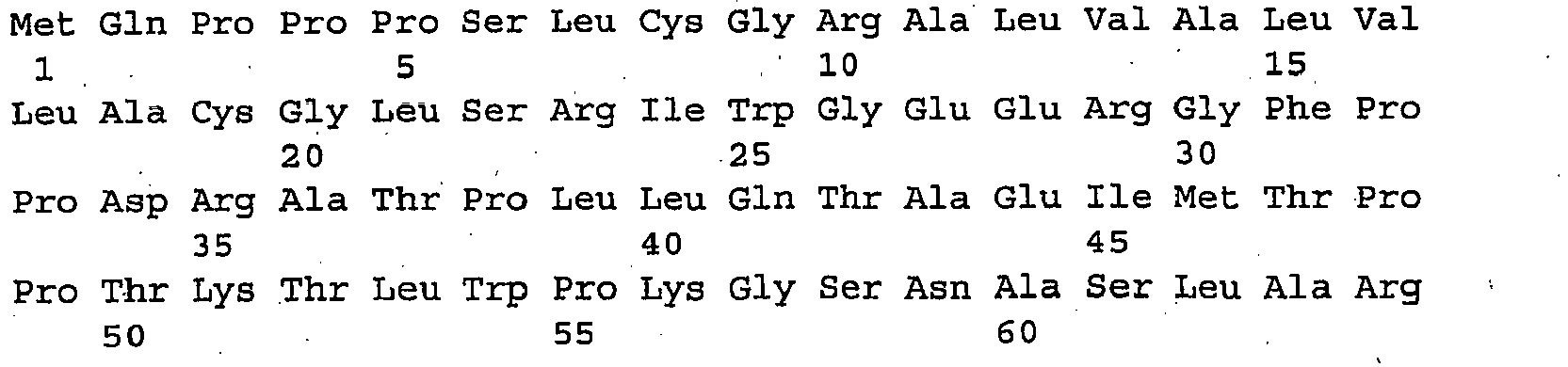
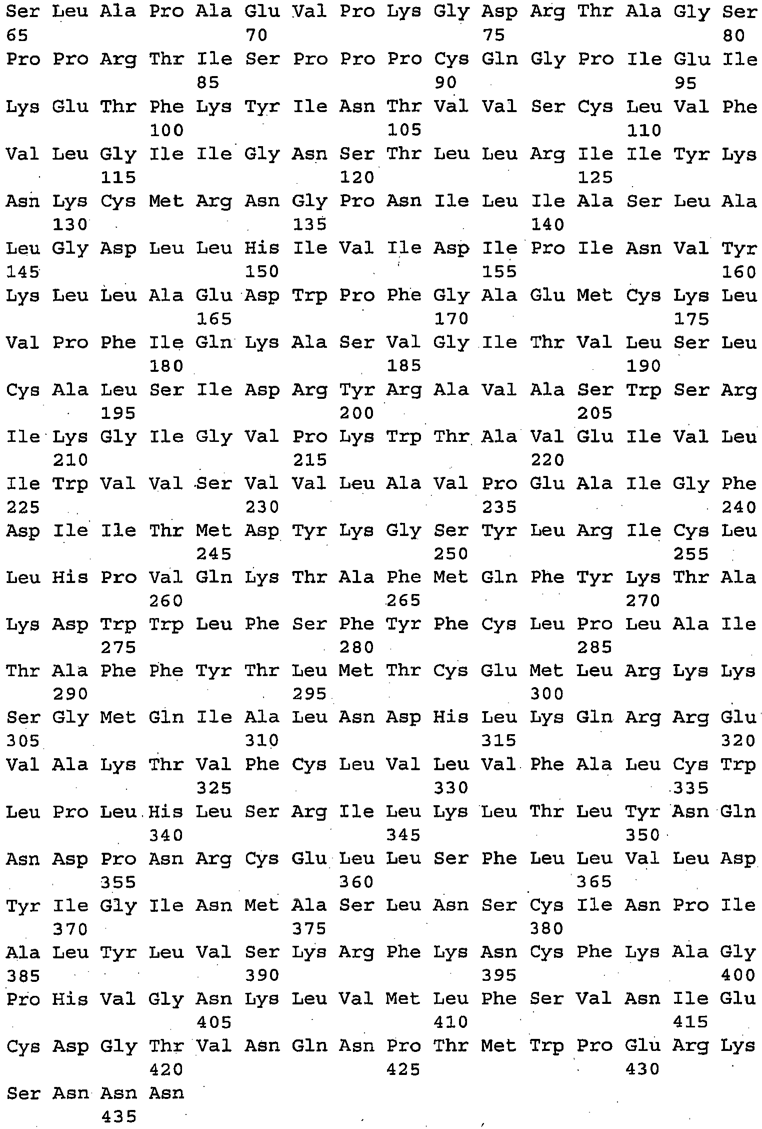
- <210> 7
<211> 8091
<212> DNA
<213> Homo sapiens - <400> 7


- <210> 8
<211> 2321
<212> PRT
<213> Homo sapiens - <400> 8


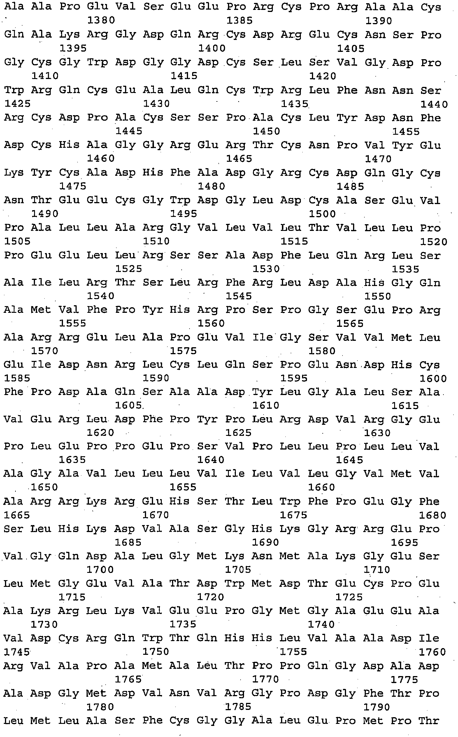

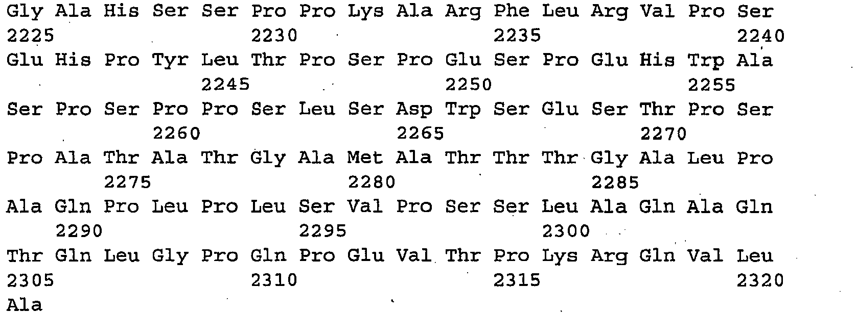
- <210> 9
<211> 1638
<212> DNA
<213> Homo sapiens - <400> 9

- <210> 10
<211> 417
<212> PRT
<213> Homo sapiens - <400> 10


| Unigene ID | Function | OMIMID | Signal Seq | Protein | TM Location | Orientation | SEQ ID NO |
| Hs.102308 | potassium inwardly-rectifying channel, subfamily J, member 8 | 600935 | no | NP_004973 | 73-95,156-178 | IN | |
| Hs.109225 | Vascular cell adhesion molecule 1 | 192225 | yes | NP_001069 | 699-721 | Unsure | |
| Hs.110024 | NADH:ubiquinone oxidoreductase MLRQ subunit homolog | yes | NP_064527 | 20-42 | Unsure | ||
| Hs.125036 | TEM17 | 606826 | yes | NP 065138 | 425-447 | OUT | |
| Hs.125359 | TEM13, Thy-1 cell surface antigen | 188230 | yes | NP_006279 | 140-161 | Unsure | |
| Hs.13662 I | hypothetical protein MGC5508 | yes | NP_076997 | 84-106,130-152,159-176.189-205 | Unsure | ||
| Hs.1501 | syndecan 2 (heparan sulfate proteoglycan 1, cell surface-associated fibroglycan) | 142460 | yes | AAA52701 | 147-169 | Unsure | |
| Hs.150540 | hypothetical protein BC002942 | yes | NP_149977 | 367-389,314-336,79-101,256-278,108-130,401-423,639-661,131-152,13-35 226-248 | Unsure | ||
| Hs.155071 | TEM44, hypothetical protein FLJ11990 | no | NP_060824 | 121-143,177-199 | Unsure | ||
| His.16187 | uncharacterized hematopoietic stem/progenitor cells protein MDS032 | no | NP_060937 | 232-254 | OUT | ||
| Hs.166994 | FAT tumor suppressor homolog 1 (Drosophila) | 600976 | yes | NP_005236 | 4181-4203 | Unsure | |
| Hs.17170 | G protein-coupled receptor 4 | 600551 | no | NP_005273 | 55-77,92-113,20-42,225-244,183-205 | OUT | |
| Hs.17270 | TEM9 | 606823 | yes | NP_116166 | 921-943,764-786,1041-1060,878-900,799-821, 1012-1034 | Unsure | |
| Hs.177486 | amyloid beta (A4) precursor protein (protease nexin-II, Alzheimer disease) | 104760 | yes | NP_000475 | 701-723 | Unsure | |
| Hs.180338 | tumor necrosis factor receptor superfamily, formerly member 12, now member 25 (translocating chain-association membrane protein) | 603366 | yes | NP_683869 | 200-222 | IN | 9, 10 |
| Hs.181244 | major histocompatibility complex. class I. A | 142800 | yes | NP_002107 | 305-327 | OUT | |
| Hs.185973 | degenerative spermatocyte homolog, lipid desaturase (Drosophila) | yes | NP_003667 | 43-61,160-177 | Unsure | ||
| Hs.195727 | TEM1, endosialin | 606064 | yes | NP 065137 | 686-708 | Unsure | |
| Hs.198265 | matrix metalloproteinase 25 | yes | NP_071913 | 541-562 | Unsure | ||
| Hs.20166 | prostate stem cell antigen | 602470 | yes | NP_005663 | 100-122 | Unsure | |
| Hs.211579 | melanoma cell adhesion molecule | 155735 | yes | NP_006491 | 560-582 | OUT | |
| Hs.23016 | G protein-coupled receptor | yes | 47-69,297-319,82-104,214-236,119-140,160-182 255-277 | OUT | 3, 4 | ||
| Hs.231119 | protocadherin beta 9 | 606335 | yes | NP_061992 | 689-711,13-35 | IN | |
| Hs.2399 | matrix metalloproteinase 14 (membrane-inserted) | 600754 | yes | NP_004986 | 540-562 | Unsure | |
| Hs.24220 | Scotin | 607290 | yes | NP 057563 | 110-132 | OUT | |
| Hs.24395 | chemokine (C-X-C motif) ligand 14 | 604186 | no | NP_004878 | 31-Oct | OUT | |
| Hs.251385 | murine retrovirus integration site 1 homolog | 604673 | no | NP_569056 | 830-852 | Unsure | |
| Hs.256297 | integrin, alpha 11 | 604789 | yes | NP 036343 | 1143-1165 | OUT | |
| Hs.265827 | interferon, alpha-inducible protein (clone IFI-6-16) | 147572 | yes | NP_075011 | 5-24,44-66 | IN | |
| Hs.274127 | CLST 11240 protein | no | NP_057522 | 62-84,30-47 | IN | ||
| Hs.274368 | TEM42, MSTP032 rev str: | no | NP_079502 | 47-69 | OUT | 1, 2 | |
| Hs.278568 | H factor (complement)-like 1 | 134371 | yes | NP_002104 | 23-Jan | Unsure | |
| Hs.27935 | tweety homolog 2 (Drosophila) | yes | NP_116035 | 242-264,89-111,390-412,215-237,47-69 | IN | ||
| Hs.279746 | transient receptor potential cation channel, subfamily V, member 2 | 606676 | no | NP_057197 | 535-557,391-413,492-514,433-455,622-R44 460-482 | Unsure | |
| Hs.283558 | Hypothetical protein PRO1855 | no | NP_060979 | 246-268 | IN | ||
| Hs.285814 | sprouty homolog 4 (Drosophila) | no | AAK00653 | 236-258 | OUT | ||
| Hs.291904 | accessory protein BAP31 | 300398 | yes | NP_005736 | 44-63,102-121 | IN | |
| Hs.295726 | integrin, alpha V (vitronectin receptor, alpha polypeptide, antigen CD51) | 193210 | yes | NP_002201 | 994-1016 | OUT | |
| Hs.296310 | gap junction protein, alpha 4, 37kDa (connexin 37) | 121012 | no | NP_002051 | 207-229,20-39,76-98 | IN | |
| Hs.29665 | calsyntenin 1 | yes | NP_055759 | 860-882 | Unsure | ||
| Hs.298476 | solute carrier family 26, | no | NP_599025 | 380-402,187-209,115-137,475-506,417-436,264-283,346-368,141-163,295-314 443 460 | OUT | ||
| Hs.29882 | family with sequence similarity 3. member C | yes | NP_055703 | 29-Jul | IN | ||
| Hs.300697 | immunoglobulin heavy constant gamma 3 (G3m marker) | 147120 | yes | 547-569 | OUT | ||
| Hs.31720 | Hephaestin | 300167 | yes | NP_620074 | 1108-1130 | OUT | |
| Hs.322456 | Hypothetical protein DKFZp761D0211 | no | NP_114428 | 49-71 | IN | ||
| Hs.323769 | cisplatin resistance related protein CRR9p | yes | NP_110409 | 15-36,401-423,285-307,431-453 ,345-362,318-340 | IN | ||
| Hs.324844 | Hypothetical protein IMAGE3455200 | yes | NP_076869 | 75-97,101-123,116-138 | Unsure | ||
| Hs.34665 | Homo sapiens mRNA full length insert cDNA clone EUROIMAGE88179 1 | no | 456-478 | OUT | |||
| Hs.381200 | Hypothetical protein MGC15523 | yes | NP_612637 | 378-397,83-105,120-142,230-252,323-340,149-171,344-366,272-294,36-58 | IN | ||
| Hs.393 | Prostaglandin I2 (prostacyclin) receptor (IP) | 600022 | no | NP_000951 | 188-210,49-71,93-115,136-158,238-260,15-37 | OUT | |
| Hs.43910 | CD164 antigen, sialomucin | 603356 | yes | NP_006007 | 164-186 | Unsure | |
| Hs.6459 | Putative G-protein coupled receptor GPCR41 | yes | NP_078807 | 196-218,46-68,369-391,81-103,113-135,404-426,147-169,325-347,337-359,9-31 276- 298 | IN | ||
| Hs.7158 | DKFZP566H073 protein | yes | NP_056343 | 172-194 | Unsure | ||
| Hs.74615 | platelet-derived growth factor receptor, alpha polypeptide | 173490 | yes | NP_006197 | 527-549,7-29 | IN | |
| Hs.74823 | NADH dehydrogenase (ubiquinone) 1 alpha subcomplex, 1 75kDa | 300078 | yes | NP_004532 | 27-May | OUT | |
| Hs.75564 | CD151 antigen | 602243 | yes | 57-79,92-114,222-244 | IN | ||
| Hs.76144 | platelet-derived growth factor receptor, beta polypeptide | 173410 | yes | NP_002600 | 534-556 | Unsure | |
| Hs.77665 | KIAA0102 gene product | no | NP_055567 | 80-102,112-134 | IN | ||
| Hs.77873 | B7 homolog 3 | 605715 | yes | 466-488 | IN | ||
| Hs.7835 | TEM22, endocytic receptor (mannose receptor, C type 2); involved in cell-cell communication, cell adhesion | yes | NP_006030 | 1412-1434 | OUT | ||
| Hs.79410 | solute carrier family 4, anion exchanger, member 2 (erythrocyte membrane protein band 3-like 1) | 109280 | no | NP_003031 | 794-816,1031-1053,901-918,709-731,988-1010,752-774,818-840,931-950,1114-1136,1175-1197,1188-1210, 1101-1122 | OUT | |
| Hs.82002 | endothelin receptor type B | 131244 | yes | NP_000106 | 367-389,104-126,217-239,138-160,325-347,175-197, 275-297 | Unsure | 5, 6 |
| Hs.82890 | defender against cell death 1 | 600243 | yes | NP_001335 | 29-51,56-78,93-112 | OUT | |
| Hs.83883 | transmembrane, prostate androgen induced RNA | 606564 | yes | NP_064567 | 41-63 | OUT | |
| Hs.8546 | Notch homolog 3 (Drosophila) | 600276 | yes | NP_000426 | 1641-1663,1496-1518.20-42 | Unsure | 7,8 |
| Hs.890 | lymphotoxin beta (TNF superfamily, member 3) | 600978 | yes | NP_002332 | 21-43 | IN | |
| Hs.8966 | TEM19 var1 (long); cell-surface protein, domain homology with leukointegrin (integrin alpha-D); ATR | 606410 | yes | NP_115584 | 321-343 | IN | |
| Hs.9004 | chondroitin sulfate proteoglycan 4 (melanoma-associated) | 601172 | yes | NP_001888 | 2224-2246 | Unsure | |
| Hs.93765 | lipoma HMGIC fusion partner | 606710 | yes | NP_005771 | 87-109,121-143,12-34,166-188 | Unsure | |
| Hs.95744 | hypothetical protein similar to ankyrin repeat-containing priotein AKR1 | no | NP_061901 | 472-494,289-311,318-340,347-369,374-395, 505-528 | OUT | ||
| Hs.17144 | short-chain dehydrogenase/red uctase 1 SDR1 | yes | NP_004744 | ||||
| Hs.32978 | proprotein convertase subtilisin/kexin type 7 PCSK7 | 604872 | yes | NP_004707 | |||
| Hs.289770 | Homo sapiens mRNA; cDNA DKF2p686D0720 | no | |||||
| Hs.418 | fibroblast activation protein, alpha FAP | 600403 | yes | NP_004451 | |||
| Hs.211579 | melanoma cell adhesion molecule mCAM | 155735 | yes | NP_006491 | |||
| Hs.9383 | cystein-rich with EFG-like domains 1 CRELD1 | 607170 | yes | NP_056328 |
Claims (1)
- A method for identifying tumour endothelial cells in a patient, comprising:contacting cDNA or mRNA obtained from a population of cells obtained from the patient with one or more nucleic acid hybridization probes which are complementary to a cDNA or mRNA for a gene encoding a G protein-coupled receptor having the sequence shown in SEQ ID NO. 4; anddetecting cDNA or mRNA which have specifically hybridized to said nucleic acid hybridization probes,wherein the detection of higher than normal levels of the cDNA or mRNA indicates the presence of tumour endothelial cells in the population.
Priority Applications (1)
| Application Number | Priority Date | Filing Date | Title |
|---|---|---|---|
| EP11159411.5A EP2365004B1 (en) | 2002-06-21 | 2003-06-23 | Membrane associated tumor endothelium markers |
Applications Claiming Priority (5)
| Application Number | Priority Date | Filing Date | Title |
|---|---|---|---|
| US39018702P | 2002-06-21 | 2002-06-21 | |
| US390187P | 2002-06-21 | ||
| US45895903P | 2003-04-01 | 2003-04-01 | |
| US458959P | 2003-04-01 | ||
| PCT/US2003/019544 WO2004001004A2 (en) | 2002-06-21 | 2003-06-23 | Membrane associated tumor endothelium markers |
Related Child Applications (2)
| Application Number | Title | Priority Date | Filing Date |
|---|---|---|---|
| EP11159411.5A Division-Into EP2365004B1 (en) | 2002-06-21 | 2003-06-23 | Membrane associated tumor endothelium markers |
| EP11159411.5A Division EP2365004B1 (en) | 2002-06-21 | 2003-06-23 | Membrane associated tumor endothelium markers |
Publications (3)
| Publication Number | Publication Date |
|---|---|
| EP1534331A2 EP1534331A2 (en) | 2005-06-01 |
| EP1534331A4 EP1534331A4 (en) | 2006-12-13 |
| EP1534331B1 true EP1534331B1 (en) | 2014-10-29 |
Family
ID=30003143
Family Applications (2)
| Application Number | Title | Priority Date | Filing Date |
|---|---|---|---|
| EP11159411.5A Expired - Lifetime EP2365004B1 (en) | 2002-06-21 | 2003-06-23 | Membrane associated tumor endothelium markers |
| EP03742108.8A Expired - Lifetime EP1534331B1 (en) | 2002-06-21 | 2003-06-23 | Membrane associated tumor endothelium markers |
Family Applications Before (1)
| Application Number | Title | Priority Date | Filing Date |
|---|---|---|---|
| EP11159411.5A Expired - Lifetime EP2365004B1 (en) | 2002-06-21 | 2003-06-23 | Membrane associated tumor endothelium markers |
Country Status (5)
| Country | Link |
|---|---|
| US (2) | US20060275287A1 (en) |
| EP (2) | EP2365004B1 (en) |
| JP (1) | JP2005530856A (en) |
| AU (1) | AU2003278161A1 (en) |
| WO (1) | WO2004001004A2 (en) |
Families Citing this family (120)
| Publication number | Priority date | Publication date | Assignee | Title |
|---|---|---|---|---|
| US6136311A (en) | 1996-05-06 | 2000-10-24 | Cornell Research Foundation, Inc. | Treatment and diagnosis of cancer |
| US20030055231A1 (en) * | 1998-10-28 | 2003-03-20 | Jian Ni | 12 human secreted proteins |
| SE9902056D0 (en) * | 1999-06-03 | 1999-06-03 | Active Biotech Ab | An integrin heterodimer and an alpha subunit thereof |
| US6984522B2 (en) | 2000-08-03 | 2006-01-10 | Regents Of The University Of Michigan | Isolation and use of solid tumor stem cells |
| ATE488530T1 (en) * | 2002-06-19 | 2010-12-15 | Raven Biotechnologies Inc | INTERNALIZING ANTIBODIES SPECIFIC TO THE RAAG10 CELL SURFACE TARGET |
| SE0301087D0 (en) * | 2003-04-14 | 2003-04-14 | Cartela Ab | New monoclonal antibody |
| EP2478912B1 (en) | 2003-11-06 | 2016-08-31 | Seattle Genetics, Inc. | Auristatin conjugates with anti-HER2 or anti-CD22 antibodies and their use in therapy |
| WO2005095594A1 (en) * | 2004-03-31 | 2005-10-13 | Universite De La Mediterranee | Glycopeptides derived from pancreatic structures, antibodies and applications thereof in diagnostics and therapeutics |
| NZ579482A (en) | 2004-06-01 | 2011-02-25 | Genentech Inc | Antibody drug conjugates and methods |
| DK1791565T3 (en) | 2004-09-23 | 2016-08-01 | Genentech Inc | Cysteingensplejsede antibodies and conjugates |
| US20100111856A1 (en) | 2004-09-23 | 2010-05-06 | Herman Gill | Zirconium-radiolabeled, cysteine engineered antibody conjugates |
| WO2006078780A2 (en) * | 2005-01-19 | 2006-07-27 | Genzyme Corporation | Rdc1 antibodies for the diagnosis of nsclc |
| BRPI0520524A2 (en) | 2005-09-13 | 2009-05-12 | Sca Hygiene Prod Ab | absorbent articles and laminates containing a binding pattern |
| US7723477B2 (en) | 2005-10-31 | 2010-05-25 | Oncomed Pharmaceuticals, Inc. | Compositions and methods for inhibiting Wnt-dependent solid tumor cell growth |
| CA2628221A1 (en) | 2005-10-31 | 2007-05-10 | Oncomed Pharmaceuticals, Inc. | Anti-frizzled receptor antibodies for treating cancer |
| CA2646329C (en) | 2006-03-20 | 2018-07-03 | The Regents Of The University Of California | Engineered anti-prostate stem cell antigen (psca) antibodies for cancer targeting |
| ES2413087T3 (en) | 2006-06-13 | 2013-07-15 | Oncomed Pharmaceuticals, Inc. | Compositions and methods for the diagnosis and treatment of cancer |
| JP2009292725A (en) * | 2006-09-01 | 2009-12-17 | Stelic Institute Of Regenerative Medicine | Kidney disease-improving agent |
| JPWO2008032868A1 (en) | 2006-09-15 | 2010-01-28 | 株式会社島津製作所 | Tumor marker for renal cancer and method for identifying morbidity of renal cancer |
| AU2007325872B2 (en) * | 2006-11-08 | 2012-12-13 | Macrogenics West, Inc. | TES7 and antibodies that bind thereto |
| US20100028256A1 (en) | 2006-11-09 | 2010-02-04 | St Croix Brad | Differential gene expression in physiological and pathological angiogenesis |
| AU2014200453B2 (en) * | 2006-11-09 | 2015-06-18 | The Government Of The United States Of America As Represented By The Secretary Of The Department Of Health And Human Services | Differential gene expression in physiological and pathological angiogenesis |
| WO2008091641A2 (en) | 2007-01-24 | 2008-07-31 | Oncomed Pharmaceuticals, Inc. | Compositions and methods for diagnosing and treating cancer |
| EA200700940A1 (en) * | 2007-04-27 | 2007-12-28 | Петр Генриевич ЛОХОВ | METHOD FOR OBTAINING ANTITUMENT VACCINE ON THE BASIS OF SURFACE ANTIGENES OF ENDOTHELIAL CELLS |
| EP2176295B1 (en) | 2007-07-16 | 2014-11-19 | Genentech, Inc. | Humanized anti-cd79b antibodies and immunoconjugates and methods of use |
| PE20090943A1 (en) | 2007-07-16 | 2009-08-05 | Genentech Inc | ANTI-CD79B ANTIBODIES AND IMMUNOCONJUGATES |
| WO2009032949A2 (en) | 2007-09-04 | 2009-03-12 | The Regents Of The University Of California | High affinity anti-prostate stem cell antigen (psca) antibodies for cancer targeting and detection |
| CA2700410C (en) * | 2007-10-03 | 2020-10-06 | Cornell University | Treatment of proliferative disorders using antibodies to psma |
| KR101607346B1 (en) | 2008-01-31 | 2016-03-29 | 제넨테크, 인크. | Anti-cd79b antibodies and immunoconjugates and methods of use |
| US9132189B2 (en) | 2008-07-08 | 2015-09-15 | Oncomed Pharmaceuticals, Inc. | Notch1 binding agents and methods of use thereof |
| CN102112490B (en) | 2008-07-08 | 2014-10-22 | 昂考梅德药品有限公司 | Notch1 receptor binding agents and methods of use thereof |
| SG190568A1 (en) | 2008-09-26 | 2013-06-28 | Oncomed Pharm Inc | Frizzled-binding agents and uses thereof |
| EP2216399A1 (en) * | 2009-01-30 | 2010-08-11 | Université de la Méditerranée | Human soluble CD146, preparation and uses thereof |
| ES2712732T3 (en) | 2009-02-17 | 2019-05-14 | Cornell Res Foundation Inc | Methods and kits for the diagnosis of cancer and the prediction of therapeutic value |
| CA2769822C (en) | 2009-08-13 | 2019-02-19 | The Johns Hopkins University | Methods of modulating immune function |
| JP2013504585A (en) | 2009-09-09 | 2013-02-07 | セントローズ, エルエルシー | Extracellular targeted drug complex |
| CN101659703B (en) * | 2009-10-09 | 2012-12-05 | 天津医科大学附属肿瘤医院 | Monoclonal antibody of anti-human BAP31 protein, preparation method and application thereof |
| AU2010325969B2 (en) | 2009-12-02 | 2016-10-20 | Imaginab, Inc. | J591 minibodies and cys-diabodies for targeting human prostate specific membrane antigen |
| TWI535445B (en) | 2010-01-12 | 2016-06-01 | 安可美德藥物股份有限公司 | Wnt antagonists and methods of treatment and screening |
| MX2012008085A (en) | 2010-01-13 | 2012-09-12 | Oncomed Pharm Inc | Notch1 binding agents and methods of use thereof. |
| CA2794674A1 (en) | 2010-04-01 | 2011-10-06 | Oncomed Pharmaceuticals, Inc. | Frizzled-binding agents and uses thereof |
| SI2528625T1 (en) | 2010-04-15 | 2013-11-29 | Spirogen Sarl | Pyrrolobenzodiazepines and conjugates thereof |
| CA2799540A1 (en) | 2010-06-08 | 2011-12-15 | Genentech, Inc. | Cysteine engineered antibodies and conjugates |
| US20120121615A1 (en) | 2010-11-17 | 2012-05-17 | Flygare John A | Alaninyl maytansinol antibody conjugates |
| US8679767B2 (en) | 2011-05-12 | 2014-03-25 | Genentech, Inc. | Multiple reaction monitoring LC-MS/MS method to detect therapeutic antibodies in animal samples using framework signature peptides |
| BR112014009050B1 (en) | 2011-10-14 | 2022-06-21 | Medimmune Limited | Pyrrolbenzodiazepine antibody-drug conjugate, pharmaceutical composition comprising the same, as well as pyrrolebenzodiazepine compounds |
| WO2013102878A2 (en) * | 2012-01-05 | 2013-07-11 | Department Of Biotechnology (Dbt) | Fat1 gene in cancer and inflammation |
| WO2013130093A1 (en) | 2012-03-02 | 2013-09-06 | Genentech, Inc. | Biomarkers for treatment with anti-tubulin chemotherapeutic compounds |
| TWI595007B (en) * | 2012-09-10 | 2017-08-11 | Neotope Biosciences Ltd | Anti-mcam antibodies and associated methods of use |
| RS57694B1 (en) | 2012-10-12 | 2018-11-30 | Adc Therapeutics Sa | Pyrrolobenzodiazepine - anti-psma antibody conjugates |
| EP2906296B1 (en) | 2012-10-12 | 2018-03-21 | ADC Therapeutics SA | Pyrrolobenzodiazepine-antibody conjugates |
| HRP20182129T1 (en) | 2012-10-12 | 2019-02-08 | Adc Therapeutics Sa | CONJUGATES ANTIBODY - PIROLOBENZODIAZEPINE |
| JP6392765B2 (en) | 2012-10-12 | 2018-09-19 | エイディーシー・セラピューティクス・エス・アーAdc Therapeutics Sa | Pyrrolobenzodiazepine-antibody conjugate |
| EP2906297B1 (en) | 2012-10-12 | 2017-12-06 | ADC Therapeutics SA | Pyrrolobenzodiazepine-antibody conjugates |
| MX338711B (en) | 2012-10-12 | 2016-04-28 | Medimmune Ltd | Pyrrolobenzodiazepines and conjugates thereof. |
| ES2680153T3 (en) | 2012-10-12 | 2018-09-04 | Adc Therapeutics Sa | Anti-PSMA-pyrrolobenzodiazepine antibody conjugates |
| AU2013334790A1 (en) | 2012-10-23 | 2015-04-30 | Oncomed Pharmaceuticals, Inc. | Methods of treating neuroendocrine tumors using Wnt pathway-binding agents |
| CA2890571A1 (en) | 2012-11-07 | 2014-05-15 | Kenneth G. Geles | Anti-notch3 antibodies and antibody-drug conjugates |
| CN110627797A (en) | 2012-12-21 | 2019-12-31 | 麦迪穆有限责任公司 | Asymmetric pyrrolobenzodiazepine dimers for the treatment of proliferative and autoimmune diseases |
| CN105189507A (en) | 2012-12-21 | 2015-12-23 | 斯皮罗根有限公司 | Pyrrolobenzodiazepines and conjugates thereof |
| AU2014212081A1 (en) | 2013-02-04 | 2015-08-13 | Oncomed Pharmaceuticals, Inc. | Methods and monitoring of treatment with a Wnt pathway inhibitor |
| AU2014229529B2 (en) | 2013-03-13 | 2018-02-15 | Medimmune Limited | Pyrrolobenzodiazepines and conjugates thereof |
| TWI680766B (en) | 2013-03-13 | 2020-01-01 | 英商梅迪繆思有限公司 | Pyrrolobenzodiazepines and conjugates thereof |
| MX362970B (en) | 2013-03-13 | 2019-02-28 | Medimmune Ltd | Pyrrolobenzodiazepines and conjugates thereof. |
| US9168300B2 (en) | 2013-03-14 | 2015-10-27 | Oncomed Pharmaceuticals, Inc. | MET-binding agents and uses thereof |
| BR112016002829A2 (en) | 2013-08-12 | 2017-09-19 | Genentech Inc | COMPOUND AND PROCESS FOR PREPARING ANTIBODY-DRUG CONJUGATE COMPOUND, PHARMACEUTICAL COMPOSITION, CANCER TREATMENT METHOD, CANCER TREATMENT KIT, DRUG LINKER INTERMEDIATE, CBI DIMER DRUG MOUNT AND COMPOUND |
| KR101535717B1 (en) * | 2013-09-11 | 2015-07-09 | 연세대학교 산학협력단 | Composition for pancreatic cancer diagnosis comprising complememt factor i-specific binding polypeptide or antibody |
| GB201317982D0 (en) | 2013-10-11 | 2013-11-27 | Spirogen Sarl | Pyrrolobenzodiazepines and conjugates thereof |
| EP3054983B1 (en) | 2013-10-11 | 2019-03-20 | Medimmune Limited | Pyrrolobenzodiazepine-antibody conjugates |
| WO2015052534A1 (en) | 2013-10-11 | 2015-04-16 | Spirogen Sàrl | Pyrrolobenzodiazepine-antibody conjugates |
| WO2015052535A1 (en) | 2013-10-11 | 2015-04-16 | Spirogen Sàrl | Pyrrolobenzodiazepine-antibody conjugates |
| EP3082876B1 (en) | 2013-12-16 | 2018-01-17 | Genentech, Inc. | 1-(chloromethyl)-2,3-dihydro-1h-benzo[e]indole dimer antibody-drug conjugate compounds, and methods of use and treatment |
| JP6671292B2 (en) | 2013-12-16 | 2020-03-25 | ジェネンテック, インコーポレイテッド | Peptidomimetic compounds and antibody-drug conjugates thereof |
| WO2015095227A2 (en) | 2013-12-16 | 2015-06-25 | Genentech, Inc. | Peptidomimetic compounds and antibody-drug conjugates thereof |
| EP3193940A1 (en) | 2014-09-10 | 2017-07-26 | Medimmune Limited | Pyrrolobenzodiazepines and conjugates thereof |
| GB201416112D0 (en) | 2014-09-12 | 2014-10-29 | Medimmune Ltd | Pyrrolobenzodiazepines and conjugates thereof |
| WO2016040825A1 (en) | 2014-09-12 | 2016-03-17 | Genentech, Inc. | Anthracycline disulfide intermediates, antibody-drug conjugates and methods |
| SG11201701128YA (en) | 2014-09-12 | 2017-03-30 | Genentech Inc | Cysteine engineered antibodies and conjugates |
| CN107073136A (en) | 2014-09-17 | 2017-08-18 | 健泰科生物技术公司 | Pyrrolobenzodiazepines Zhuo and its antibody disulphide conjugate |
| HUE049175T2 (en) | 2014-09-23 | 2020-09-28 | Hoffmann La Roche | Method for using anti-CD79b immunoconjugates |
| CN107148285B (en) | 2014-11-25 | 2022-01-04 | Adc治疗股份有限公司 | Pyrrolobenzodiazepine-antibody conjugates |
| AU2015358532C1 (en) | 2014-12-03 | 2020-10-29 | Genentech, Inc. | Quaternary amine compounds and antibody-drug conjugates thereof |
| GB201506402D0 (en) | 2015-04-15 | 2015-05-27 | Berkel Patricius H C Van And Howard Philip W | Site-specific antibody-drug conjugates |
| GB201506411D0 (en) | 2015-04-15 | 2015-05-27 | Bergenbio As | Humanized anti-axl antibodies |
| US11254744B2 (en) | 2015-08-07 | 2022-02-22 | Imaginab, Inc. | Antigen binding constructs to target molecules |
| CR20180243A (en) | 2015-10-02 | 2018-07-31 | Genentech Inc | PIRROLOBENZODIAZEPIN ANTIBODY-DRUG CONJUGATES AND METHODS OF USE |
| MA43354A (en) | 2015-10-16 | 2018-08-22 | Genentech Inc | CONJUGATE DRUG CONJUGATES WITH CLOUDY DISULPHIDE |
| EP3365025B1 (en) | 2015-10-20 | 2020-07-15 | Genentech, Inc. | Calicheamicin-antibody-drug conjugates and methods of use |
| GB201601431D0 (en) | 2016-01-26 | 2016-03-09 | Medimmune Ltd | Pyrrolobenzodiazepines |
| GB201602359D0 (en) | 2016-02-10 | 2016-03-23 | Medimmune Ltd | Pyrrolobenzodiazepine Conjugates |
| GB201602356D0 (en) | 2016-02-10 | 2016-03-23 | Medimmune Ltd | Pyrrolobenzodiazepine Conjugates |
| CN108700598A (en) | 2016-03-25 | 2018-10-23 | 豪夫迈·罗氏有限公司 | The drug of the total antibody of multichannel and antibody conjugate quantifies measuring method |
| GB201607478D0 (en) | 2016-04-29 | 2016-06-15 | Medimmune Ltd | Pyrrolobenzodiazepine Conjugates |
| CN118436801A (en) | 2016-05-20 | 2024-08-06 | 豪夫迈·罗氏有限公司 | PROTAC antibody conjugates and methods of use thereof |
| EP3465221B1 (en) | 2016-05-27 | 2020-07-22 | H. Hoffnabb-La Roche Ag | Bioanalytical method for the characterization of site-specific antibody-drug conjugates |
| WO2017214024A1 (en) | 2016-06-06 | 2017-12-14 | Genentech, Inc. | Silvestrol antibody-drug conjugates and methods of use |
| CN109689111B (en) | 2016-08-11 | 2024-04-05 | 基因泰克公司 | Pyrrolobenzodiazepine prodrugs and antibody conjugates thereof |
| JP7050770B2 (en) | 2016-10-05 | 2022-04-08 | エフ・ホフマン-ラ・ロシュ・アクチェンゲゼルシャフト | Method for preparing antibody drug conjugate |
| GB201617466D0 (en) | 2016-10-14 | 2016-11-30 | Medimmune Ltd | Pyrrolobenzodiazepine conjugates |
| US11266745B2 (en) | 2017-02-08 | 2022-03-08 | Imaginab, Inc. | Extension sequences for diabodies |
| GB201702031D0 (en) | 2017-02-08 | 2017-03-22 | Medlmmune Ltd | Pyrrolobenzodiazepine-antibody conjugates |
| RS61795B1 (en) | 2017-02-08 | 2021-06-30 | Adc Therapeutics Sa | Pyrrolobenzodiazepine-antibody conjugates |
| CN110582505B (en) | 2017-04-18 | 2021-04-02 | 免疫医疗有限公司 | Pyrrolobenzodiazepine* conjugates |
| WO2018193102A1 (en) | 2017-04-20 | 2018-10-25 | Adc Therapeutics Sa | Combination therapy with an anti-axl antibody-drug conjugate |
| CA3064804A1 (en) | 2017-06-14 | 2018-12-20 | Adc Therapeutics Sa | Dosage regimes for the administration of an anti-cd19 adc |
| KR102270107B1 (en) | 2017-08-18 | 2021-06-30 | 메디뮨 리미티드 | pyrrolobenzodiazepine conjugate |
| EP3684773B1 (en) | 2017-09-20 | 2026-04-29 | pH Pharma Co., Ltd. | Thailanstatin analogs |
| GB201803342D0 (en) | 2018-03-01 | 2018-04-18 | Medimmune Ltd | Methods |
| GB201806022D0 (en) | 2018-04-12 | 2018-05-30 | Medimmune Ltd | Pyrrolobenzodiazepines and conjugates thereof |
| GB201814281D0 (en) | 2018-09-03 | 2018-10-17 | Femtogenix Ltd | Cytotoxic agents |
| JP7708662B2 (en) | 2018-10-24 | 2025-07-15 | エフ・ホフマン-ラ・ロシュ・アクチェンゲゼルシャフト | Conjugated chemical degraders and methods of use |
| JP2022513198A (en) | 2018-12-10 | 2022-02-07 | ジェネンテック, インコーポレイテッド | Photocrosslinkable peptide for site-specific conjugation to Fc-containing proteins |
| GB201901197D0 (en) | 2019-01-29 | 2019-03-20 | Femtogenix Ltd | G-A Crosslinking cytotoxic agents |
| CN113631560B (en) | 2019-03-15 | 2025-02-18 | 麦迪穆有限责任公司 | Azetidine benzodiazepine dimers and conjugates containing them for treating cancer |
| GB2597532A (en) | 2020-07-28 | 2022-02-02 | Femtogenix Ltd | Cytotoxic compounds |
| EP4426727A2 (en) | 2021-11-03 | 2024-09-11 | Hangzhou Dac Biotech Co., Ltd. | Specific conjugation of an antibody |
| WO2024138128A2 (en) | 2022-12-23 | 2024-06-27 | Genentech, Inc. | Cereblon degrader conjugates, and uses thereof |
| KR20260012304A (en) | 2023-04-17 | 2026-01-26 | 피크 바이오, 인크. | Antibodies and antibody-drug conjugates and methods of use and synthetic processes and intermediates |
| WO2026006689A2 (en) | 2024-06-28 | 2026-01-02 | Firefly Bio, Inc. | Bcl-xl degrader antibody conjugates and uses thereof |
| WO2026006688A2 (en) | 2024-06-28 | 2026-01-02 | Firefly Bio, Inc. | Degrader antibody conjugates and uses thereof |
Family Cites Families (12)
| Publication number | Priority date | Publication date | Assignee | Title |
|---|---|---|---|---|
| US6075181A (en) | 1990-01-12 | 2000-06-13 | Abgenix, Inc. | Human antibodies derived from immunized xenomice |
| US5508384A (en) * | 1992-09-10 | 1996-04-16 | New York University | Polypeptide derived from a popamine receptor, and compositions and methods thereof |
| US5342757A (en) * | 1992-11-13 | 1994-08-30 | Ludwig Institute For Cancer Research | Monoclonal antibodies which specifically binds to endosialin, a 165 Kd glycoprotein found on tumor vascular endothelium, and uses thereof |
| US5587299A (en) * | 1994-04-20 | 1996-12-24 | Memorial Sloan-Kettering Cancer Center | Isolated nucleic acid molecule coding for fibroblast activation protein alpha and uses thereof |
| US5695937A (en) | 1995-09-12 | 1997-12-09 | The Johns Hopkins University School Of Medicine | Method for serial analysis of gene expression |
| EP0926238A3 (en) * | 1997-12-08 | 2000-11-22 | Smithkline Beecham Laboratoires Pharmaceutiques | Putative Adrenomedullin receptor polypeptide |
| US6455677B1 (en) * | 1998-04-30 | 2002-09-24 | Boehringer Ingelheim International Gmbh | FAPα-specific antibody with improved producibility |
| EP0953639A1 (en) * | 1998-04-30 | 1999-11-03 | Boehringer Ingelheim International GmbH | FAPalpha-specific antibody with improved producibility |
| US6559128B1 (en) * | 2000-01-21 | 2003-05-06 | Northwestern University | Inhibitors of G protein-mediated signaling, methods of making them, and uses thereof |
| JP2004527210A (en) * | 2000-08-02 | 2004-09-09 | ザ ジョンズ ホプキンス ユニバーシティー | Evaluation method of endothelial cell expression pattern |
| US6599128B2 (en) * | 2001-08-21 | 2003-07-29 | Ronald J. Roberts | Game for training of managers |
| WO2003042661A2 (en) * | 2001-11-13 | 2003-05-22 | Protein Design Labs, Inc. | Methods of diagnosis of cancer, compositions and methods of screening for modulators of cancer |
-
2003
- 2003-06-23 EP EP11159411.5A patent/EP2365004B1/en not_active Expired - Lifetime
- 2003-06-23 US US10/518,751 patent/US20060275287A1/en not_active Abandoned
- 2003-06-23 JP JP2004530961A patent/JP2005530856A/en active Pending
- 2003-06-23 AU AU2003278161A patent/AU2003278161A1/en not_active Abandoned
- 2003-06-23 EP EP03742108.8A patent/EP1534331B1/en not_active Expired - Lifetime
- 2003-06-23 WO PCT/US2003/019544 patent/WO2004001004A2/en not_active Ceased
-
2009
- 2009-05-06 US US12/436,444 patent/US20110104059A1/en not_active Abandoned
Non-Patent Citations (7)
| Title |
|---|
| GREENBAUM D ET AL: "COMPARING PROTEIN ABUNDANCE AND MRNA EXPRESSION LEVELS ON A GENOMIC SCALE", GENOME BIOLOGY (ONLINE), BIOMED CENTRAL LTD, GB, vol. 40, no. 9, 1 January 2003 (2003-01-01), pages 117.01 - 117.08, XP008036618, ISSN: 1465-6914 * |
| KOLLMAR OTTO ET AL: "CXCR4 and CXCR7 regulate angiogenesis and CT26.WT tumor growth independent from SDF-1.", INTERNATIONAL JOURNAL OF CANCER. JOURNAL INTERNATIONAL DU CANCER 15 MAR 2010, vol. 126, no. 6, 15 March 2010 (2010-03-15), pages 1302 - 1315, ISSN: 1097-0215 * |
| MAUSSANG DAVID ET AL: "Llama-derived single variable domains (nanobodies) directed against chemokine receptor CXCR7 reduce head and neck cancer cell growth in vivo.", THE JOURNAL OF BIOLOGICAL CHEMISTRY 11 OCT 2013, vol. 288, no. 41, 11 October 2013 (2013-10-11), pages 29562 - 29572, ISSN: 1083-351X * |
| MIAO ZHENHUA ET AL: "CXCR7 (RDC1) promotes breast and lung tumor growth in vivo and is expressed on tumor-associated vasculature.", PROCEEDINGS OF THE NATIONAL ACADEMY OF SCIENCES OF THE UNITED STATES OF AMERICA 2 OCT 2007, vol. 104, no. 40, 2 October 2007 (2007-10-02), pages 15735 - 15740, ISSN: 0027-8424 * |
| SCHUTYSER EVEMIE ET AL: "Hypoxia enhances CXCR4 expression in human microvascular endothelial cells and human melanoma cells.", EUROPEAN CYTOKINE NETWORK JUN 2007, vol. 18, no. 2, June 2007 (2007-06-01), pages 59 - 70, ISSN: 1148-5493 * |
| WANG JIANHUA ET AL: "The role of CXCR7/RDC1 as a chemokine receptor for CXCL12/SDF-1 in prostate cancer.", THE JOURNAL OF BIOLOGICAL CHEMISTRY 15 FEB 2008, vol. 283, no. 7, 15 February 2008 (2008-02-15), pages 4283 - 4294, ISSN: 0021-9258 * |
| ZABEL BRIAN A ET AL: "The novel chemokine receptor CXCR7 regulates trans-endothelial migration of cancer cells.", MOLECULAR CANCER 2011, vol. 10, 2011, pages 73, ISSN: 1476-4598 * |
Also Published As
| Publication number | Publication date |
|---|---|
| AU2003278161A8 (en) | 2004-01-06 |
| US20110104059A1 (en) | 2011-05-05 |
| EP2365004A3 (en) | 2012-03-07 |
| AU2003278161A1 (en) | 2004-01-06 |
| EP2365004A2 (en) | 2011-09-14 |
| WO2004001004A2 (en) | 2003-12-31 |
| US20060275287A1 (en) | 2006-12-07 |
| JP2005530856A (en) | 2005-10-13 |
| EP2365004B1 (en) | 2016-01-06 |
| EP1534331A2 (en) | 2005-06-01 |
| EP1534331A4 (en) | 2006-12-13 |
| WO2004001004A3 (en) | 2004-04-08 |
Similar Documents
| Publication | Publication Date | Title |
|---|---|---|
| EP1534331B1 (en) | Membrane associated tumor endothelium markers | |
| US7358351B2 (en) | Endothelial cell expression patterns | |
| US8568985B2 (en) | Breast endothelial cell expression patterns | |
| JP2002533056A (en) | Compounds and methods for treatment and diagnosis of lung cancer | |
| JP2002507387A (en) | Compounds for immunotherapy and diagnosis of breast cancer and methods for their use | |
| AU2002258543B2 (en) | Endothelial cell expression patterns | |
| WO2004005883A2 (en) | Secreted and cytoplasmic tumor endothelial markers | |
| JP2002541805A (en) | Compounds for immunotherapy and diagnosis of breast cancer and methods for their use | |
| AU2002258543A1 (en) | Endothelial cell expression patterns | |
| US20090233270A9 (en) | Secreted and cytoplasmic tumor endothelial markers | |
| US7776519B2 (en) | Use of the slug gene, or of the transcription or expression products thereof in the detection and/or treatment of cancerous cells | |
| HK1154598A (en) | Differential in tumour gene products and use of same | |
| HK1153489A (en) | Differential in tumour gene products and use of same | |
| HK1153487A (en) | Differential in tumour gene products and use of same | |
| HK1153752A (en) | Differential in tumour gene products and use of same |
Legal Events
| Date | Code | Title | Description |
|---|---|---|---|
| PUAI | Public reference made under article 153(3) epc to a published international application that has entered the european phase |
Free format text: ORIGINAL CODE: 0009012 |
|
| 17P | Request for examination filed |
Effective date: 20050121 |
|
| AK | Designated contracting states |
Kind code of ref document: A2 Designated state(s): AT BE BG CH CY CZ DE DK EE ES FI FR GB GR HU IE IT LI LU MC NL PT RO SE SI SK TR |
|
| AX | Request for extension of the european patent |
Extension state: AL LT LV MK |
|
| DAX | Request for extension of the european patent (deleted) | ||
| A4 | Supplementary search report drawn up and despatched |
Effective date: 20061113 |
|
| RIC1 | Information provided on ipc code assigned before grant |
Ipc: G01N 33/68 20060101ALI20061107BHEP Ipc: A61K 39/395 20060101ALI20061107BHEP Ipc: C07K 14/705 20060101ALI20061107BHEP Ipc: C07K 16/28 20060101AFI20061107BHEP Ipc: A61K 38/17 20060101ALI20061107BHEP |
|
| 17Q | First examination report despatched |
Effective date: 20090420 |
|
| GRAP | Despatch of communication of intention to grant a patent |
Free format text: ORIGINAL CODE: EPIDOSNIGR1 |
|
| INTG | Intention to grant announced |
Effective date: 20140523 |
|
| GRAS | Grant fee paid |
Free format text: ORIGINAL CODE: EPIDOSNIGR3 |
|
| GRAA | (expected) grant |
Free format text: ORIGINAL CODE: 0009210 |
|
| AK | Designated contracting states |
Kind code of ref document: B1 Designated state(s): AT BE BG CH CY CZ DE DK EE ES FI FR GB GR HU IE IT LI LU MC NL PT RO SE SI SK TR |
|
| REG | Reference to a national code |
Ref country code: GB Ref legal event code: FG4D |
|
| REG | Reference to a national code |
Ref country code: CH Ref legal event code: EP |
|
| REG | Reference to a national code |
Ref country code: AT Ref legal event code: REF Ref document number: 693559 Country of ref document: AT Kind code of ref document: T Effective date: 20141115 |
|
| REG | Reference to a national code |
Ref country code: IE Ref legal event code: FG4D |
|
| REG | Reference to a national code |
Ref country code: DE Ref legal event code: R096 Ref document number: 60346920 Country of ref document: DE Effective date: 20141204 |
|
| REG | Reference to a national code |
Ref country code: AT Ref legal event code: MK05 Ref document number: 693559 Country of ref document: AT Kind code of ref document: T Effective date: 20141029 |
|
| REG | Reference to a national code |
Ref country code: NL Ref legal event code: VDEP Effective date: 20141029 |
|
| PG25 | Lapsed in a contracting state [announced via postgrant information from national office to epo] |
Ref country code: PT Free format text: LAPSE BECAUSE OF FAILURE TO SUBMIT A TRANSLATION OF THE DESCRIPTION OR TO PAY THE FEE WITHIN THE PRESCRIBED TIME-LIMIT Effective date: 20150302 Ref country code: ES Free format text: LAPSE BECAUSE OF FAILURE TO SUBMIT A TRANSLATION OF THE DESCRIPTION OR TO PAY THE FEE WITHIN THE PRESCRIBED TIME-LIMIT Effective date: 20141029 Ref country code: NL Free format text: LAPSE BECAUSE OF FAILURE TO SUBMIT A TRANSLATION OF THE DESCRIPTION OR TO PAY THE FEE WITHIN THE PRESCRIBED TIME-LIMIT Effective date: 20141029 Ref country code: FI Free format text: LAPSE BECAUSE OF FAILURE TO SUBMIT A TRANSLATION OF THE DESCRIPTION OR TO PAY THE FEE WITHIN THE PRESCRIBED TIME-LIMIT Effective date: 20141029 |
|
| PG25 | Lapsed in a contracting state [announced via postgrant information from national office to epo] |
Ref country code: AT Free format text: LAPSE BECAUSE OF FAILURE TO SUBMIT A TRANSLATION OF THE DESCRIPTION OR TO PAY THE FEE WITHIN THE PRESCRIBED TIME-LIMIT Effective date: 20141029 Ref country code: CY Free format text: LAPSE BECAUSE OF FAILURE TO SUBMIT A TRANSLATION OF THE DESCRIPTION OR TO PAY THE FEE WITHIN THE PRESCRIBED TIME-LIMIT Effective date: 20141029 Ref country code: GR Free format text: LAPSE BECAUSE OF FAILURE TO SUBMIT A TRANSLATION OF THE DESCRIPTION OR TO PAY THE FEE WITHIN THE PRESCRIBED TIME-LIMIT Effective date: 20150130 Ref country code: SE Free format text: LAPSE BECAUSE OF FAILURE TO SUBMIT A TRANSLATION OF THE DESCRIPTION OR TO PAY THE FEE WITHIN THE PRESCRIBED TIME-LIMIT Effective date: 20141029 |
|
| REG | Reference to a national code |
Ref country code: FR Ref legal event code: PLFP Year of fee payment: 13 |
|
| REG | Reference to a national code |
Ref country code: DE Ref legal event code: R097 Ref document number: 60346920 Country of ref document: DE |
|
| PG25 | Lapsed in a contracting state [announced via postgrant information from national office to epo] |
Ref country code: EE Free format text: LAPSE BECAUSE OF FAILURE TO SUBMIT A TRANSLATION OF THE DESCRIPTION OR TO PAY THE FEE WITHIN THE PRESCRIBED TIME-LIMIT Effective date: 20141029 Ref country code: RO Free format text: LAPSE BECAUSE OF FAILURE TO SUBMIT A TRANSLATION OF THE DESCRIPTION OR TO PAY THE FEE WITHIN THE PRESCRIBED TIME-LIMIT Effective date: 20141029 Ref country code: DK Free format text: LAPSE BECAUSE OF FAILURE TO SUBMIT A TRANSLATION OF THE DESCRIPTION OR TO PAY THE FEE WITHIN THE PRESCRIBED TIME-LIMIT Effective date: 20141029 Ref country code: CZ Free format text: LAPSE BECAUSE OF FAILURE TO SUBMIT A TRANSLATION OF THE DESCRIPTION OR TO PAY THE FEE WITHIN THE PRESCRIBED TIME-LIMIT Effective date: 20141029 Ref country code: SK Free format text: LAPSE BECAUSE OF FAILURE TO SUBMIT A TRANSLATION OF THE DESCRIPTION OR TO PAY THE FEE WITHIN THE PRESCRIBED TIME-LIMIT Effective date: 20141029 |
|
| PG25 | Lapsed in a contracting state [announced via postgrant information from national office to epo] |
Ref country code: IT Free format text: LAPSE BECAUSE OF FAILURE TO SUBMIT A TRANSLATION OF THE DESCRIPTION OR TO PAY THE FEE WITHIN THE PRESCRIBED TIME-LIMIT Effective date: 20141029 |
|
| PLBE | No opposition filed within time limit |
Free format text: ORIGINAL CODE: 0009261 |
|
| STAA | Information on the status of an ep patent application or granted ep patent |
Free format text: STATUS: NO OPPOSITION FILED WITHIN TIME LIMIT |
|
| 26N | No opposition filed |
Effective date: 20150730 |
|
| PG25 | Lapsed in a contracting state [announced via postgrant information from national office to epo] |
Ref country code: MC Free format text: LAPSE BECAUSE OF FAILURE TO SUBMIT A TRANSLATION OF THE DESCRIPTION OR TO PAY THE FEE WITHIN THE PRESCRIBED TIME-LIMIT Effective date: 20141029 |
|
| REG | Reference to a national code |
Ref country code: CH Ref legal event code: PL |
|
| PG25 | Lapsed in a contracting state [announced via postgrant information from national office to epo] |
Ref country code: SI Free format text: LAPSE BECAUSE OF FAILURE TO SUBMIT A TRANSLATION OF THE DESCRIPTION OR TO PAY THE FEE WITHIN THE PRESCRIBED TIME-LIMIT Effective date: 20141029 Ref country code: LU Free format text: LAPSE BECAUSE OF FAILURE TO SUBMIT A TRANSLATION OF THE DESCRIPTION OR TO PAY THE FEE WITHIN THE PRESCRIBED TIME-LIMIT Effective date: 20150623 |
|
| REG | Reference to a national code |
Ref country code: IE Ref legal event code: MM4A |
|
| PG25 | Lapsed in a contracting state [announced via postgrant information from national office to epo] |
Ref country code: LI Free format text: LAPSE BECAUSE OF NON-PAYMENT OF DUE FEES Effective date: 20150630 Ref country code: IE Free format text: LAPSE BECAUSE OF NON-PAYMENT OF DUE FEES Effective date: 20150623 Ref country code: CH Free format text: LAPSE BECAUSE OF NON-PAYMENT OF DUE FEES Effective date: 20150630 |
|
| REG | Reference to a national code |
Ref country code: FR Ref legal event code: PLFP Year of fee payment: 14 |
|
| PG25 | Lapsed in a contracting state [announced via postgrant information from national office to epo] |
Ref country code: BG Free format text: LAPSE BECAUSE OF FAILURE TO SUBMIT A TRANSLATION OF THE DESCRIPTION OR TO PAY THE FEE WITHIN THE PRESCRIBED TIME-LIMIT Effective date: 20141029 Ref country code: HU Free format text: LAPSE BECAUSE OF FAILURE TO SUBMIT A TRANSLATION OF THE DESCRIPTION OR TO PAY THE FEE WITHIN THE PRESCRIBED TIME-LIMIT; INVALID AB INITIO Effective date: 20030623 |
|
| REG | Reference to a national code |
Ref country code: FR Ref legal event code: PLFP Year of fee payment: 15 |
|
| PG25 | Lapsed in a contracting state [announced via postgrant information from national office to epo] |
Ref country code: TR Free format text: LAPSE BECAUSE OF FAILURE TO SUBMIT A TRANSLATION OF THE DESCRIPTION OR TO PAY THE FEE WITHIN THE PRESCRIBED TIME-LIMIT Effective date: 20141029 |
|
| PG25 | Lapsed in a contracting state [announced via postgrant information from national office to epo] |
Ref country code: BE Free format text: LAPSE BECAUSE OF FAILURE TO SUBMIT A TRANSLATION OF THE DESCRIPTION OR TO PAY THE FEE WITHIN THE PRESCRIBED TIME-LIMIT Effective date: 20141029 |
|
| REG | Reference to a national code |
Ref country code: FR Ref legal event code: PLFP Year of fee payment: 16 |
|
| PGFP | Annual fee paid to national office [announced via postgrant information from national office to epo] |
Ref country code: GB Payment date: 20220628 Year of fee payment: 20 |
|
| PGFP | Annual fee paid to national office [announced via postgrant information from national office to epo] |
Ref country code: FR Payment date: 20220627 Year of fee payment: 20 |
|
| PGFP | Annual fee paid to national office [announced via postgrant information from national office to epo] |
Ref country code: DE Payment date: 20220629 Year of fee payment: 20 |
|
| REG | Reference to a national code |
Ref country code: DE Ref legal event code: R071 Ref document number: 60346920 Country of ref document: DE |
|
| REG | Reference to a national code |
Ref country code: GB Ref legal event code: PE20 Expiry date: 20230622 |
|
| PG25 | Lapsed in a contracting state [announced via postgrant information from national office to epo] |
Ref country code: GB Free format text: LAPSE BECAUSE OF EXPIRATION OF PROTECTION Effective date: 20230622 |