EP1526184A1 - Promoter sequences specific for cells of the B lymphoid lineage and pharmaceutical compositions containing the same - Google Patents

Promoter sequences specific for cells of the B lymphoid lineage and pharmaceutical compositions containing the same Download PDF

Info

Publication number
EP1526184A1
EP1526184A1 EP03292663A EP03292663A EP1526184A1 EP 1526184 A1 EP1526184 A1 EP 1526184A1 EP 03292663 A EP03292663 A EP 03292663A EP 03292663 A EP03292663 A EP 03292663A EP 1526184 A1 EP1526184 A1 EP 1526184A1
Authority
EP
European Patent Office
Prior art keywords
sequence
cells
gene
gfp
nucleic acid
Prior art date
Legal status (The legal status is an assumption and is not a legal conclusion. Google has not performed a legal analysis and makes no representation as to the accuracy of the status listed.)
Withdrawn
Application number
EP03292663A
Other languages
German (de)
French (fr)
Inventor
Cécile Tonelle
Thomas Moreau
Christian Chabannon
Jean Imbert
Current Assignee (The listed assignees may be inaccurate. Google has not performed a legal analysis and makes no representation or warranty as to the accuracy of the list.)
Institut National de la Santé et de la Recherche Médicale
Original Assignee
Institut National de la Santé et de la Recherche Médicale
Priority date (The priority date is an assumption and is not a legal conclusion. Google has not performed a legal analysis and makes no representation as to the accuracy of the date listed.)
Filing date
Publication date
Application filed by Institut National de la Santé et de la Recherche Médicale filed Critical Institut National de la Santé et de la Recherche Médicale
Priority to EP03292663A priority Critical patent/EP1526184A1/en
Priority to PCT/EP2004/011814 priority patent/WO2005040383A1/en
Publication of EP1526184A1 publication Critical patent/EP1526184A1/en
Withdrawn legal-status Critical Current

Links

Images

Classifications

    • C—CHEMISTRY; METALLURGY
    • C07—ORGANIC CHEMISTRY
    • C07K—PEPTIDES
    • C07K14/00—Peptides having more than 20 amino acids; Gastrins; Somatostatins; Melanotropins; Derivatives thereof
    • C07K14/435—Peptides having more than 20 amino acids; Gastrins; Somatostatins; Melanotropins; Derivatives thereof from animals; from humans
    • C07K14/705—Receptors; Cell surface antigens; Cell surface determinants
    • C07K14/70503—Immunoglobulin superfamily
    • A—HUMAN NECESSITIES
    • A01—AGRICULTURE; FORESTRY; ANIMAL HUSBANDRY; HUNTING; TRAPPING; FISHING
    • A01K—ANIMAL HUSBANDRY; AVICULTURE; APICULTURE; PISCICULTURE; FISHING; REARING OR BREEDING ANIMALS, NOT OTHERWISE PROVIDED FOR; NEW BREEDS OF ANIMALS
    • A01K67/00—Rearing or breeding animals, not otherwise provided for; New or modified breeds of animals
    • A01K67/027—New or modified breeds of vertebrates
    • A01K67/0271—Chimeric vertebrates, e.g. comprising exogenous cells
    • C—CHEMISTRY; METALLURGY
    • C12—BIOCHEMISTRY; BEER; SPIRITS; WINE; VINEGAR; MICROBIOLOGY; ENZYMOLOGY; MUTATION OR GENETIC ENGINEERING
    • C12N—MICROORGANISMS OR ENZYMES; COMPOSITIONS THEREOF; PROPAGATING, PRESERVING, OR MAINTAINING MICROORGANISMS; MUTATION OR GENETIC ENGINEERING; CULTURE MEDIA
    • C12N15/00—Mutation or genetic engineering; DNA or RNA concerning genetic engineering, vectors, e.g. plasmids, or their isolation, preparation or purification; Use of hosts therefor
    • C12N15/09—Recombinant DNA-technology
    • C12N15/63—Introduction of foreign genetic material using vectors; Vectors; Use of hosts therefor; Regulation of expression
    • C12N15/79—Vectors or expression systems specially adapted for eukaryotic hosts
    • C12N15/85—Vectors or expression systems specially adapted for eukaryotic hosts for animal cells
    • A—HUMAN NECESSITIES
    • A01—AGRICULTURE; FORESTRY; ANIMAL HUSBANDRY; HUNTING; TRAPPING; FISHING
    • A01K—ANIMAL HUSBANDRY; AVICULTURE; APICULTURE; PISCICULTURE; FISHING; REARING OR BREEDING ANIMALS, NOT OTHERWISE PROVIDED FOR; NEW BREEDS OF ANIMALS
    • A01K2267/00—Animals characterised by purpose
    • A01K2267/03—Animal model, e.g. for test or diseases
    • A01K2267/0393—Animal model comprising a reporter system for screening tests
    • A—HUMAN NECESSITIES
    • A61—MEDICAL OR VETERINARY SCIENCE; HYGIENE
    • A61K—PREPARATIONS FOR MEDICAL, DENTAL OR TOILETRY PURPOSES
    • A61K48/00—Medicinal preparations containing genetic material which is inserted into cells of the living body to treat genetic diseases; Gene therapy
    • C—CHEMISTRY; METALLURGY
    • C12—BIOCHEMISTRY; BEER; SPIRITS; WINE; VINEGAR; MICROBIOLOGY; ENZYMOLOGY; MUTATION OR GENETIC ENGINEERING
    • C12N—MICROORGANISMS OR ENZYMES; COMPOSITIONS THEREOF; PROPAGATING, PRESERVING, OR MAINTAINING MICROORGANISMS; MUTATION OR GENETIC ENGINEERING; CULTURE MEDIA
    • C12N2799/00—Uses of viruses
    • C12N2799/02—Uses of viruses as vector
    • C12N2799/021—Uses of viruses as vector for the expression of a heterologous nucleic acid
    • C12N2799/027—Uses of viruses as vector for the expression of a heterologous nucleic acid where the vector is derived from a retrovirus
    • C—CHEMISTRY; METALLURGY
    • C12—BIOCHEMISTRY; BEER; SPIRITS; WINE; VINEGAR; MICROBIOLOGY; ENZYMOLOGY; MUTATION OR GENETIC ENGINEERING
    • C12N—MICROORGANISMS OR ENZYMES; COMPOSITIONS THEREOF; PROPAGATING, PRESERVING, OR MAINTAINING MICROORGANISMS; MUTATION OR GENETIC ENGINEERING; CULTURE MEDIA
    • C12N2830/00—Vector systems having a special element relevant for transcription
    • C12N2830/008—Vector systems having a special element relevant for transcription cell type or tissue specific enhancer/promoter combination
    • C—CHEMISTRY; METALLURGY
    • C12—BIOCHEMISTRY; BEER; SPIRITS; WINE; VINEGAR; MICROBIOLOGY; ENZYMOLOGY; MUTATION OR GENETIC ENGINEERING
    • C12N—MICROORGANISMS OR ENZYMES; COMPOSITIONS THEREOF; PROPAGATING, PRESERVING, OR MAINTAINING MICROORGANISMS; MUTATION OR GENETIC ENGINEERING; CULTURE MEDIA
    • C12N2830/00—Vector systems having a special element relevant for transcription
    • C12N2830/80—Vector systems having a special element relevant for transcription from vertebrates
    • C12N2830/85—Vector systems having a special element relevant for transcription from vertebrates mammalian

Definitions

  • the present invention particularly relates to B lymphoid lineage specific promoter sequences and to pharmaceutical compositions containing said sequences.
  • hematopoietic stem cells remains a major objective and challenge in medical science, particularly for gene or cell therapy.
  • the field still faces many technological obstacles, including the difficulty to transduce mostly non-dividing cells, the risk of mutational insertion, and the difficulty to control transgene expression.
  • lentiviruses have been increasingly used and substituted to onco-retroviruses, because of their ability to integrate the genome of quiescent cells, and the reduced need to use exogenous cytokines to induce cell cycling 1;2 .
  • Efficient transduction of human hematopoietic progenitor cell populations, including CD34 + cells 3 , CD34 + /CD38- cells and SCID-repopulating cells (SRC) 4 has been documented with lentiviral vectors.
  • Lentiviruses also offer advantages over onco-retroviruses, in that they allow for the more efficient integration of regulatory sequences that target the transgene expression to a defined lineage.
  • Several examples have been reported, both with onco-retroviral and lentiviral vectors, that limit the expression of a marker gene to one or several lineages with various efficacies. These include the transcriptional targeting of T-cells 5; 6 , antigen presenting cells (APC) cells 7 , monocytes/macrophages 8 and erythroid cells 9 ; 10 .
  • APC antigen presenting cells
  • the CD19 antigen under the control of the BSAP (Pax5) transcription factor, is a specific marker of B cells, appears at the pre-B stage, and remains expressed throughout B-cell differentiation and maturation, to be lost at the final stage when B-lymphocytes maturate into plasma cells 11 .
  • CD19 takes part to the CD21, CD81 and Leu-13 complex which regulates thresholds of intracellular signals 12 .
  • an object of the present invention is to provide new fragments derived from the CD19 promoter sequence, which are smaller than the large fragments used in transgenic mice and/or more B lineage specific than those of the prior art.
  • the present invention relates to a nucleic acid sequence comprising or consisting of:
  • the expression "per block of 150 to 200 contiguous nucleotides” means that the sequence which is homologous to the S sequence comprises at least one fragment of 150 to 200 contiguous nucleotides presenting a homology of at least 60% with the corresponding fragment of the S sequence, provided that said sequence is capable of directing the specific expression of a gene in cells of the B lymphoid lineage.
  • the invention also relates to a nucleic acid sequence as defined above, comprising or consisting of a sequence homologous to the S sequence, said homologous sequence comprising two fragments presenting a homology of at least 73% and 63% respectively with the corresponding fragments of the S sequence, said fragments corresponding respectively to the contiguous nucleotides ranging from position -193 to position -1 (CR1) and from position -1260 to position -1062 (CR2) of the promoter sequence of the human CD19 gene.
  • CR1 position -1
  • CR2 position -1
  • CR2 position -1260 to position -1062
  • the invention also relates to a nucleic acid sequence as defined above, comprising or consisting of a sequence presenting an homology of at least 80%, in particular 90% with the above defined S sequence, provided that said sequence is capable of directing the specific expression of a gene in cells of the B lymphoid lineage.
  • the expression "capable of directing the specific expression of a gene” means that the corresponding sequence can activate the transcription of sequences which are situated on the 3' side of said sequence, and preferably of sequences, the initiation codon of which is linked to the 3' end of said sequence. Such a sequence corresponds in particular to a promoter sequence.
  • the S sequence consists of:
  • the 3' end is linked to the 5' end via a phosphoester linkage.
  • X -1274
  • Y -1042
  • Z -231
  • the invention relates to a nucleic acid sequence comprising or consisting of a S sequence, said S sequence consisting of:
  • SEQ ID NO: 3 is hereafter referred to as M.CD19.
  • SEQ ID NO: 3 comprises two elements hereafter referred to as CR1 (position -1 to position -193 of the promoter sequence of the human CD19 gene) and CR2 (position -1062 to position -1260 of the promoter sequence of the human CD19 gene), said elements providing a potent and specific promoter activity in cells of the B lineage.
  • X is equal to -1274
  • SEQ ID NO: 4 is hereafter referred to as L.CD19.
  • SEQ ID NO: 4 and SEQ ID NO: 5 comprise two elements hereafter referred to as CR1 (position -1 to position -193 of the promoter sequence of the human CD19 gene) and CR2 (position -1062 to position -1260 of the promoter sequence of the human CD19 gene), said elements providing a potent and specific promoter activity in cells of the B lineage.
  • the invention also relates to an expression sequence, characterized in that it comprises a nucleic acid sequence such as defined above, linked to a protein encoding nucleic acid sequence, in particular a protein encoding nucleic acid sequence coding for a protein expressed specifically in cells of the B lymphoid lineage, such as a sequence encoding Btk, Ig ⁇ , surrogate light chains, BLNK, Igb, or to a hairpin RNA encoding nucleic acid sequence, or to a suicide gene such as the HSVTK gene (Herpes simplex virus thymidine kinase), or to a marker gene such as the GFP gene (green fluorescent protein) or the LacZ gene.
  • a protein encoding nucleic acid sequence in particular a protein encoding nucleic acid sequence coding for a protein expressed specifically in cells of the B lymphoid lineage, such as a sequence encoding Btk, Ig ⁇ , surrogate light chains, BLNK, Igb, or to
  • Cells of the B lymphoid lineage notably comprise proB cells, preB cells, immature B cells, mature B cells, memory B cells, or cells of the Nalm6, Ramos, and Daudi JEA2 cell lines for example.
  • suicide gene refers to a gene which induces cell death in response to a drug, for cells in which it is expressed.
  • the HSVTK gene thus induces ganciclovir-mediated cell death.
  • marker gene refers to a gene which codes for a protein which is liable to be detected according to methods well known to the man skilled in the art, such as microscopy, in particular fluorescent microscopy. Such a gene notably comprises genes encoding for fluorescent proteins well known to the man skilled in the art, such as GFP and GFP derivatives.
  • Btk (Bruton tyrosine kinase), Ig ⁇ , surrogate light chains, BLNK (B cell Linker protein), Igb are notably described in Ballow (2002) J . Allergy Clin. Immunol. 109: 581, Conley and Cooper (1998) Curr. Opin. Immunol. 10: 399 and Gauld et al. (2002) Science 296: 1641.
  • the abovementioned expression sequences are expressed specifically in cells of the B lymphoid lineage.
  • the abovementioned nucleic acid sequence of said expression sequence consists of SEQ ID NO: 3, 4 or 5.
  • the invention also relates to a vector, in particular an expression vector, characterized in that it comprises a nucleic acid sequence or an expression sequence such as defined above.
  • Vectors according to the invention comprise in particular plasmidic vectors such as the pRSET vector or the pcDNA vector, or lentiviral vectors, such as the pTRIP and pHPT vectors.
  • such vectors are well suited to express RNA and/or proteins in cells of the B lymphoid lineage.
  • the abovementioned nucleic acid sequence of said vector consists of SEQ ID NO: 3, 4 or 5, such as for the lentiviral vectors corresponding respectively to SEQ ID NO: 6, 7 and 8.
  • the invention also relates to a cell, in particular a cell of the B lymphoid lineage, comprising a nucleic acid sequence such as defined above, or an expression sequence such as defined above, or a vector such as defined above.
  • Cells according to the invention comprise in particular stem cells, hematopoietic stem cells, embryonic stem cells and precursor cells.
  • the abovementioned cell comprises a nucleic acid sequence consisting of SEQ ID NO: 3, 4 or 5.
  • the invention also relates to a nonhuman transgenic animal, characterized in that all or some of its cells comprise a nucleic acid sequence such as defined above, or an expression sequence such as defined above, or a vector such as defined above, said animal being in particular a mammal, such as a mouse or a rat.
  • Such animals can be prepared according to methods well known to the man skilled in the art such as described for example in Maas et al. (1999) J . Immunol. 162 :6526 and Lois et al. (2002) Science 295 :868.
  • the invention also relates to a pharmaceutical composition
  • a pharmaceutical composition comprising as active substance a nucleic acid sequence such as defined above, or an expression sequence such as defined above, or a vector such as defined above, or a cell such as defined above, in association with a pharmaceutically acceptable vehicle.
  • the invention also relates to the use of a nucleic acid sequence such as defined above, or an expression sequence such as defined above, or a vector such as defined above, or a cell such as defined above, for the manufacture of a medicament intended to treat pathologies in which cells of the B lymphoid lineage are involved, such as autoimmune diseases, B lymphomas, B leukemias, antibody deficiencies, in particular X linked agammaglobulinemia (Bruton's disease), or diseases resulting from a deficient preB cell receptor complex, such as a Ig ⁇ defect, a surrogate light chains defect, or a BLNK defect.
  • autoimmune diseases such as defined above, or an expression sequence such as defined above, or a vector such as defined above, or a cell such as defined above
  • a cell such as defined above
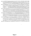
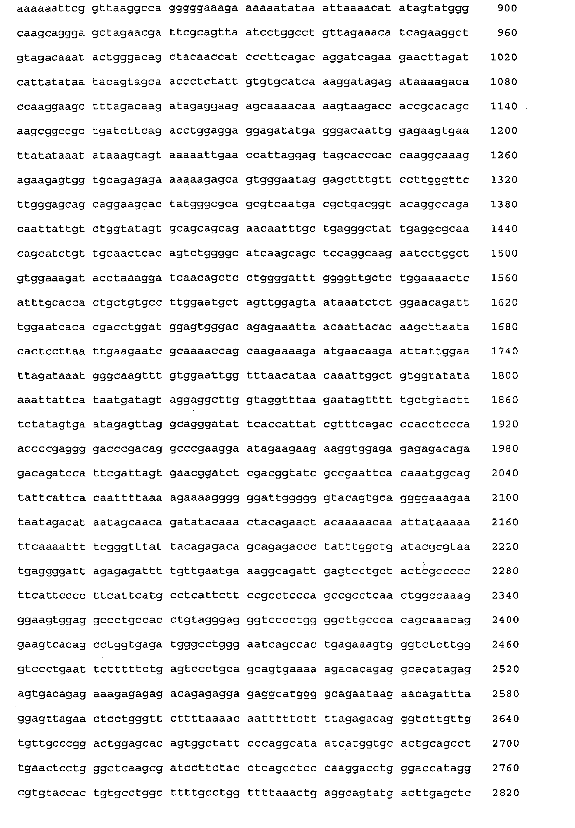
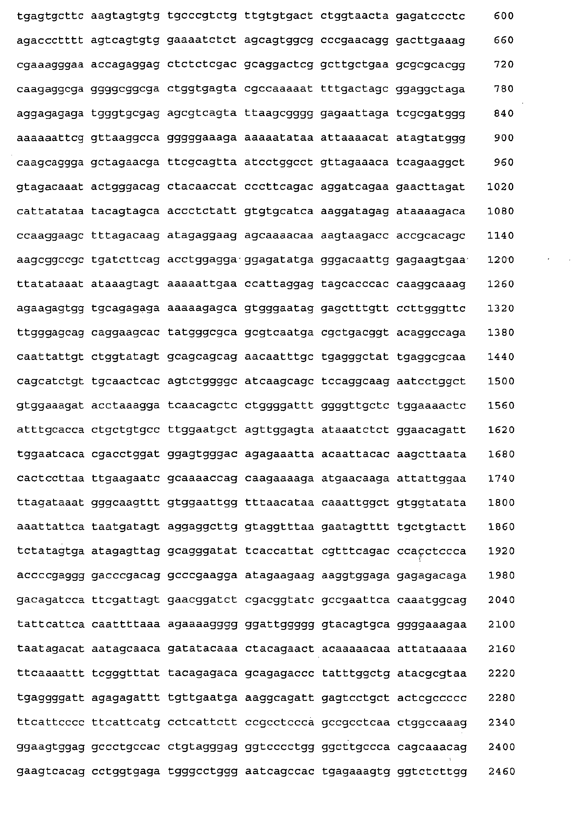
  • Figure 1 represents the upstream nucleic acid sequence of the human CD19 gene.
  • the 5' end corresponds to the nucleotide in position -4339 relative to the A nucleotide of the translation initiation codon (AUG) in position +1, and the 3' end corresponds to the nucleotide in position -1. Nucleotide positions are indicated on the left.
  • Figure 2 represents the graphical results of the computational analysis of the upstream region of the mouse and human CD 19 genes based on their comparison.
  • the graph abscissae represent the 1274bp of human gene and the ordinates the 1529bp of the murine one.
  • Two conserved regions were identified and schematically drawn: a proximal sequence (CR1) and a distal one (CR2).
  • Putative fixation sites for transcription factors were identified, as indicated by boxes on a horizontal axis for the human gene and on a vertical axis for the murine one.
  • Several restriction sites as well as repetitive sequences are also shown.
  • Figure 3 represents the plasmid form of the recombinant lentiviral genomes of the invention.
  • the ubiquitous EF1 ⁇ promoter (235bp) of pTRIP ⁇ U3-EF1 ⁇ -GFP vector plasmid was substituted by the S.CD19 (230bp), M.CD19 (464bp) or L.CD19 (1274bp) sequence by cloning between the Mlu I (black diamond shape) and Bam HI(star) sites.
  • New plasmids were respectively called pTRIP ⁇ U3-S.CD19-GFP, pTRIP ⁇ U3-M.CD19-GFP, pTRIP ⁇ U3-L.CD19-GFP and gave rise to the S.CD19-GFP, M.CD19-GFP and L.CD19-GFP lentiviral vectors.
  • LTR Long Terminal Repeat
  • SD/SA Splice Donor / Splice Acceptor
  • ⁇ encapsidation signal (on truncated GAG sequence)
  • RRE Rev Responsive Element
  • Triplex Central Termination Sequence (CTS) and central PolyPurine Tract (cPPT) of HIV-1 virus
  • ⁇ U3 deletion of 400bp in the 3'LTR U3 region (Self Inactivated vector).
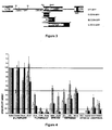
  • Figure 4 represents the specificity index (vertical axis) of GFP expression by cell lines (horizontal axis) transduced by the S.CD19-GFP (white bars), M.CD19-GFP (hatched bars) and L.CD19-GFP (black bars) vectors, relative to the GFP expression in the Nalm6 cell line.
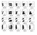
  • Figures 5A to 5E represent the flow cytometry profile of GFP expression (horizontal axis, fluorescence intensity) by human peripheral T-lymphoid cells, identified by an anti-CD3 antibody (vertical axis, fluorescence intensity), 7 days after transduction in parallel with lentiviral vectors EF1-GFP (Figure 5B), S.CD19-GFP ( Figure 5C), M.CD19-GFP ( Figure 5D) and L.CD19-GFP ( Figure 5E), non-tranduced cells were used as negative control (Figure 5A).
  • the box in the center of each flow cytometry diagram (R3) delimits GFP positive CD3+ cells.
  • Figures 5F to 51 represent the quantitative flow cytometry profile of GFP expression (horizontal axis, fluorescence intensity) by human cord blood CD34+ cells (vertical axis, number of cells), 7 days after transduction in parallel with lentiviral vectors EF1-GFP (Figure 5F), S.CD19-GFP ( Figure 5G), M.CD19-GFP ( Figure 5H) and L.CD19-GFP ( Figure 5I); negative control for GFP expression is shown on each diagram by dotted lines.
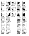
  • Figures 6A to 6T represent flow cytometry dot blots of GFP expression (horizontal axis, intensity of fluorescence) by cells deriving from transduced and un-transduced CD34+ placental progenitor cells submitted to in vitro differentiation by co-culture with the MS.5 cell line (B-lymphoid, myeloid and NK differentiation) or in liquid culture (erythroid differentiation).
  • Figures 6A to 6E relate to CD19+ cells (vertical axis, intensity of fluorescence), that is B lymphoid cells, deriving from CD34+ progenitor cells untransduced (Figure 6A) or transduced by EF1-GFP ( Figure 6B), S.CD19-GFP ( Figure 6C), M.CD19-GFP ( Figure 6D) and L.CD19-GFP ( Figure 6E).
  • Figures 6F to 6J relate to CD33+ cells (vertical axis, intensity of fluorescence), that is myeloid cells, deriving from CD34+ progenitor cells untransduced (Figure 6F) or transduced by EF1-GFP ( Figure 6G), S.CD19-GFP ( Figure 6H), M.CD19-GFP ( Figure 6I) and L.CD19-GFP ( Figure 6J).
  • Figures 6K to 60 relate to CD56+ cells (vertical axis, intensity of fluorescence), that is NK cells, deriving from CD34+ progenitor cells untransduced (Figure 6K) or transduced by EF1-GFP ( Figure 6L), S.CD19-GFP (Figure 6M), M.CD19-GFP ( Figure 6N) and L.CD19-GFP ( Figure 6O).
  • Figures 6P to 6T relate to GP-A+ cells (vertical axis, intensity of fluorescence), that is erythroid cells, deriving from CD34+ progenitor cells untransduced (Figure 6P) or transduced by EF1-GFP (Figure 6Q), S.CD19-GFP ( Figure 6R), M.CD19-GFP ( Figure 6S) and L.CD19-GFP ( Figure 6T). Analyses were done inside the ToPRO-3 neg /hCD45+ gate; the box in the center of each flow cytometry diagram delimits lineage+/GFP+ cells.
  • Figure 7 represents the specificity index (vertical axis) of the expression of GFP by CD19+, CD33+ and CD56+ cells (horizontal axis) derived ex vivo from CD34+ progenitor cells transduced by the EF1-GFP (black bars), S.CD19-GFP (white bars), M.CD19-GFP (largely hatched bars) and L.CD19-GFP (closely hatched bars) vectors, relative to the GFP expression in CD 19+ cells.
  • Figure 8A to 8ZA represent flow cytometry diagrams of GFP expression (horizontal axis, intensity of fluorescence) by cells deriving from transduced and un-transduced CD34+ placental progenitor cells submitted to in vivo differentiation in the NOD/SCID murine xenograft model.
  • B-lymphoid CD10, CD19, IgM
  • myeloid CD33
  • progenitor cells CD34
  • Figures 8A to 8E relate to CD 19+ cells (vertical axis, intensity of fluorescence), taken from bone marrow, deriving from CD34+ progenitor cells untransduced (Figure 8A) or transduced by EF1-GFP (Figure 8B), S.CD19-GFP (Figure 8C), M.CD19-GFP ( Figure 8D) and L.CD19-GFP ( Figure 8E).
  • Figures 8F to 8J relate to CD33+ cells (vertical axis, intensity of fluorescence), taken from bone marrow, deriving from CD34+ progenitor cells untransduced (Figure 8F) or transduced by EF1-GFP (Figure 8G), S.CD19-GFP ( Figure 8H), M.CD19-GFP ( Figure 8I) and L.CD19-GFP ( Figure 8J).
  • Figures 8K to 80 relate to CD34+ cells (vertical axis, intensity of fluorescence), taken from bone marrow, deriving from CD34+ progenitor cells untransduced (Figure 8K) or transduced by EF1-GFP ( Figure 8L), S.CD19-GFP (Figure 8M), M.CD19-GFP ( Figure 8N) and L.CD19-GFP ( Figure 8O).
  • Figures 8P to 8S relate to IgM+ cells (vertical axis, intensity of fluorescence), taken from the spleen, deriving from CD34+ progenitor cells untransduced (Figure 8P) or transduced by EF1-GFP (Figure 8Q), M.CD19-GFP ( Figure 8R) and L.CD19-GFP ( Figure 8S).
  • Figures 8T to 8W relate to CD19+ cells (vertical axis, intensity of fluorescence), taken from the spleen, deriving from CD34+ progenitor cells untransduced (Figure 8T) or transduced by EF1-GFP ( Figure 8U), M.CD19-GFP (Figure 8V) and L.CD19-GFP ( Figure 8W).
  • Figures 8X to 8ZA relate to CD33+ cells (vertical axis, intensity of fluorescence), taken from the spleen, deriving from CD34+ progenitor cells untransduced (Figure 8X) or transduced by EF1-GFP (Figure 8Y), M.CD19-GFP ( Figure 8Z) and L.CD19-GFP ( Figure 8ZA).
  • Flow cytometry analysis were performed after 12 weeks on bone marrow and spleen gating in ToPRO-3 neg /hCD45+ cells; boxes on dot plots delimit lineage+/GFP+ cells
  • Figure 9A represents the specificity index (vertical axis) of the expression of GFP by CD19+, CD10+, CD33+ and CD34+ bone marrow cells (horizontal axis) derived in vivo from CD34+ progenitor cells transduced by the EF1-GFP (black bars), S.CD19-GFP (white bars), M.CD19-GFP (largely hatched bars) and L.CD19-GFP (closely hatched bars) vectors, relative to the GFP expression in CD 19+ cells.
  • Figure 9B represents the specificity index (vertical axis) of the expression of GFP by CD19+, IgM+ and CD33+ splenic cells (horizontal axis) derived in vivo from CD34+ progenitor cells transduced by the EF1-GFP (black bars), M.CD19-GFP (largely hatched bars) and L.CD19-GFP (closely hatched bars) vectors, relative to the GFP expression in CD19+ cells.
  • Figure 10A represents the flow cytometry analysis of GFP expression by cells deriving from CD34+ progenitor cells engrafted in NOD/SCID mice.
  • the flow cytometry dot blot on the left shows a representative experiment of CD34 expression (vertical axis, intensity of fluorescence) and of CD 19 expression (horizontal axis, intensity of fluorescence) of cells taken from mice engrafted with EF1, M.CD19 or L.CD19-GFP transduced CD34+ progenitor cells.
  • the boxes delimit non-B progenitors (CD34+CD19-) (R3), B-progenitors (CD34+CD19+) (R4) and more mature B-cells (CD34-CD19+) (R5).
  • the histograms on the right represent the GFP expression profile (horizontal axis, fluorescence intensity) corresponding to the abovementioned CD34+CD19- cells (upper histograms), CD34+CD19+ cells (middle histograms) and CD34-CD19+ cells (lower histograms) deriving from CD34+ progenitor cells transduced by EF1-GFP, M.CD19-GFP, L.CD19-GFP.
  • the percentage of GFP expressing cells is indicated in bold for each histogram, background levels are indicated by dotted lines. From top to bottom the histograms are representative of a more advanced B cell commitment stage.
  • Figure 10B shows a histogram representing the percentage of mean GFP expression ⁇ SEM of the CD34+CD19- cells, CD34+CD19+ cells and CD34-CD19+ cells described in figure 10A and deriving from CD34+ progenitor cells transduced by EF1-GFP (black bars), M.CD19-GFP (largely hatched bars) and L.CD19-GFP (closely hatched bars).
  • Figures 11A to 11D represent the flow cytometry expression profile of GFP expression (horizontal axis, fluorescence intensity) by human CD19+ (vertical axis, fluorescence intensity) ( Figure 11A), CD3+ (vertical axis, fluorescence intensity) ( Figure 11B), CD4+ (vertical axis, fluorescence intensity) ( Figure 11C) and CD8+ (vertical axis, fluorescence intensity) ( Figure 11D) thymic cells issued from NOD/SCID mouse engrafted by M.CD19-GFP transduced CD34+ cells. Flow cytometry analysis was performed after 12 weeks, gating in ToPRO-3 neg /hCD45+ cells; the box in the center of each flow cytometry diagram delimits lineage+/GFP+ cells.
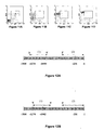
  • Figure 12A and 12B represent fragments (arrows) of the 1500 nucleotides upstream of the translation initiation codon of the human CD19 gene (grey box) as defined by their nucleotide positions.
  • Figure 12A represents the two sequences (1 and 2) constituting the S sequence, wherein X is comprised from positions -1500 to -1000, Y is such that the length of sequence (i) is at least 10 nucleotide long and Z is comprised from positions Y+1 to -231.
  • Figure 12B represents the two sequences (i and ii) constituting the S sequence, wherein X is equal to -1274, Y is greater or equal to -1042 and Z is comprised from positions Y+1 to -231.
  • CR1 and CR2 The two conserved regions, hereafter named CR1 and CR2, were contained in a contiguous 1274 bp fragment of the human CD19 gene ( Figure 2) .
  • CR2 (198 bp) was located -1 Kb upstream of human CD19 gene initiation site, and at a not yet localized upstream position in the mouse gene, since the sequence of contig AC 125169 is not yet entirely known.
  • a 1529 bp fragment for the mouse CD19 gene was created, where CR2 was inserted 5' upstream and artificially separated with 100 Ns (undefined nucleotides) from the sequenced mouse CD19 promoter.
  • the CD19 promoter contains several well characterized regulatory elements required for B-cell specific expression such as the binding sites for the B-cell specific transcription factor BSAP/Pax5 16; 31-33 . Both human and mouse CD19 upstream regions were further screened for putative regulatory elements using the TESS software (TESS: Transcription Element Search Software available at http://www.cbil.upenn.edu/tess, Jonathan Schug and G. Christian Overton, Technical Report CBIL-TR-1997-1001-v0.0 Computational Biology and Informatics Laboratory School of Medicine University of Pennsylvania, 1997) and the Matlnspector software 20 (available at Genomatix: http://www.genomatix.de) 21 ( Figure 2 ).
  • TESS Transcription Element Search Software available at http://www.cbil.upenn.edu/tess, Jonathan Schug and G. Christian Overton, Technical Report CBIL-TR-1997-1001-v0.0 Computational Biology and Informatics Laboratory School of Medicine University of Pennsylvania, 1997) and the Matln
  • CD19 sequences were produced by PCR methods from human primary cell genomic DNA.
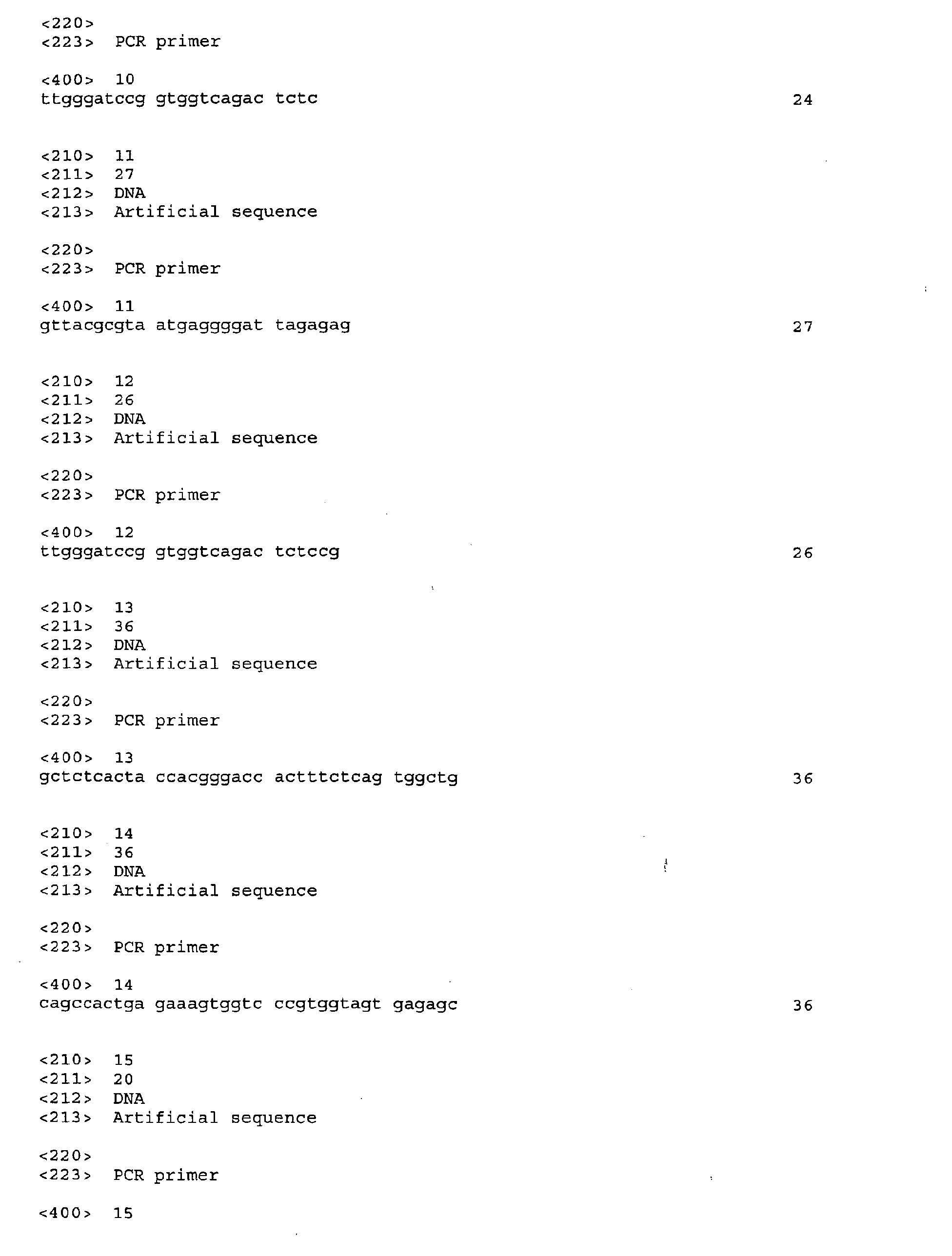
  • oligonucleotides were used as primers: S.CD19(+) 5' GTT ACG CGT CCC GTG GTA GTG AGA (SEQ ID NO: 9) and S.CD19(-) 5' TTG GGA TCC GGT GGT CAG ACT CTC (SEQ ID NO: 10) for the S.CD19 sequence; L.CD19(+) 5' GTT ACG CGT AAT GAG GGG ATT AGA GAG (SEQ ID NO: 11) and L.CD19(-) 5' TTG GGA TCC GGT GGT CAG ACT CTC CG (SEQ ID NO: 12) for the L.CD19 sequence (underlined sequences are restriction sites for Ml uI and Bam HI).
  • the generation of the M.CD19 sequence required two PCR steps, in order to juxtapose two distant regions of the native genome; first, two PCR reactions were carried out with the following two primer pairs: L.CD19(+) and M.CD19(-) 5' GCT CTC ACT ACC ACG GGA CCA CTT TCT CAG TGG CTG (SEQ ID NO: 13); M.CD19(+) 5' CAG CCA CTG AGA AAG TGG TCC CGT GGT AGT GAG AGC (SEQ ID NO: 14) and L.CD19(-); purified products were then joined end to end in a single PCR reaction carried out with the L.CD19(+) and L.CD19(-) primer pair.
  • Amplifications were realized in presence of Platinum HIFI Taq polymerase (Invitrogen), with the following program: 2' at 94°C followed by 10 cycles with 45s at 94°C, 45s at 58°C and 2' at 68°C, then 25 cycles with 45s at 94°C, 45s at 66°C and 2' at 68°C; the last cycle ended with an extension phase of 7'.
  • Platinum HIFI Taq polymerase Invitrogen
  • the S.CD19 sequence contains the proximal region of homology (CR1) for a total length of 230bp.
  • the composite M.CD19 (SEQ ID NO: 3) sequence of 464bp length results from the artificial juxtaposition of the two fragments containing the proximal and distal highlighted homologous regions (CR1 and CR2).
  • the last sequence named L.CD19 (SEQ ID NO: 4) corresponds to the entire native CD19 sequence from the initiation site to the CR2 region, and is 1274bp long.
  • the inventors then designed three new lentiviral vectors, in which the GFP gene was placed under the control of human CD 19 gene promoter fragments.
  • the inventors cloned the three abovementioned CD19 sequences upstream a GFP marker gene between Mlu I and Bam HI restriction sites of the pTRIP ⁇ U3-EF1-GFP plasmid in place of the ubiquitous EFI ⁇ promoter (235bp): the subsequent recombinant lentiviral vectors were respectively called S.CD19-GFP, M.CD19-GFP and L.CD19-GFP ( Figure 3).
  • the S.CD19, M.CD19 and L.CD19 sequences were introduced in the pTRIP ⁇ U3-EF1 ⁇ -GFP vector plasmid ( N.B. all lentiviral constructions were derived from the initial vector backbone pTRIP ⁇ U3-EF1 ⁇ -GFP [gift from Dr P. Charneau, Institut Pasteur, Paris, France 3 ]), in substitution to the EF1 ⁇ ubiquitous promoter between Mlu I and Bam HI sites to obtain the pTRIP ⁇ U3-S.CD19-GFP, the pTRIP ⁇ U3-M.CD19-GFP and the pTRIP ⁇ U3-L.CD19-GFP vector plasmids respectively ( Figure 3).
  • sequences of the lentiviral vectors comprising the M.CD19, L.CD19 and the deleted L.CD19 promoters, without the GFP gene, are respectively presented in SEQ ID NO: 6, 7 and 8.
  • Lentiviral particles were then produced by transient co-transfection of three plasmids into 293T cells: the plasmid containing the recombinant vector sequence, the packaging plasmid (p8.91), and the plasmid coding for the VSV-G (Vesicular Stomatitis Virus) envelop protein (pMDG), that confers amphotropic properties to recombinant lentiviral vectors; transfection was done by calcium phosphate co-precipitation, using the Calphos Mammalian transfection kit (Clontech laboratories, Ozyme, Montigny le Bretonneux, France).
  • Titers of different vectors usually ranged from 1x10 8 and 1x10 9 TU/mL (Transduction Unit), as measured by GFP expression on day 7 in the human pre-B cell line Nalm6, transduced in triplicate with serial dilution of concentrated lentiviruses.
  • TU/mL Transduction Unit
  • the promoter activity of the three CD19 sequences, in the context of lentiviral vectors, was first tested on a panel of hematopoietic and non-hematopoietic human cell lines, comprising:
  • All cells were transduced in parallel with the three different CD 19-GFP vectors and the EF1-GFP vector as positive control; we used a MOI between 0.5 and 0.7 that allowed 20 to 50% of positive control cells to express GFP for all cell types.
  • Cell lines were transduced without stimulation in their initial medium at a cell density of 0.05 to 0.3x10 6 cells per well (24 well plate) in a total volume of 0.5 mL of their respective culture medium, and in the presence of polybren (4 ⁇ g/mL, Sigma Saint Quentin Fallavier, France). Cells were maintained at 37°C for 24 hours before washing with PBS.
  • Transgene expression was measured by flow cytometry 7 days post-transduction, and the expression specificity index (SI) for CD19 sequences calculated and normalized for each experiments by values obtained in parallel with the pre-B cell line Nalm6.
  • the antibodies used in were murine mAb IgG 1 -PC5 (Phycoerythrocyanin 5), IgG 1 -PE (Phycoerythrin), hCD45-PC5, hCD3-PE, hCD4-PE, hCD8-PE, hCD10-PE, hCD19-PE, hCD33-PE, hCD34-PE, hCD34-APC (Allophycocyanin) (Beckman Coulter/Immunotech, Marseille, France), hCD56-PE, hIgM-PE and GP-A-PE (BD/Bioscience, Le Pont de Claix, France).
  • CD19 sequences actually showed promoter activity in this lentiviral context.
  • GFP expression driven by any of the CD19 sequences was found preferentially in cell lines of B-lymphoid origin that express CD19 (Nalm6, Ramos, Daudi, JEA2). More precisely, the M and L.CD19 promoters presented a strong activity in all four B-cell lines (0.7>SI>1.09), whereas S.CD19 one appeared more variable (0.35>SI>1).
  • the L.CD19 sequence presented a more marked B-specific promoter activity than the S or M.CD19 ones that showed respectively low promoter activity in CD19+ B-cell lines and a quite high nonspecific activity in non-B-lymphoid cells.
  • PCR experiments on genomic DNA of primary human T-lymphoid and progenitor cells revealed a similar integration of the viral genome after transduction with S, M or L.CD19-GFP vectors as compared to EF1-GFP.
  • PCR reactions were performed on 75 ng of purified genomic DNA (Nucleospin Tissue, Macherey Nagel, France) isolated 7 to 14 days after transduction.
  • Thermal cycling conditions have been adjusted to the following parameters: 2 min at 50°C and 10 min at 95°C, then 40 cycles of 1 min at 95°C and 1 min at 66°C.
  • raw data were analyzed with the software of ABI prism 7700.
  • Human primary T cells were positively selected from peripheral blood mononuclear fractions by panning, using mouse anti-hCD4 and anti-hCD8 monoclonal antibodies (Baxter), as previously described 18 .
  • human CD19 + cells were positively selected from the mononuclear fraction of peripheral blood by magnetic immunoselection (Miltenyi Biotec).
  • CD34 + cells were enriched from CB mononuclear fractions, using immunoselection with the M.A.C.S. technology, according to the manufacturer's recommendations (Miltenyi Biotec, Bergish-Gladbach, Germany); purity was usually above 95% of CD34+ cells. Sorted cells were frozen in FCS (Gibco/Invitrogen, Cergy Pontoise, France) and stored in liquid nitrogen until use.
  • T cells were activated in IMDM medium (Biowhittaker), 10% human AB serum (Institut Jacques Boy, Reims, France), 1% PS, supplemented with rhIL-2 (40ng/ml, Chiron Suresne, France) and 7 ⁇ l of anti-CD3/CD28 microbeads (Dynabeads CD3/CD28 T cell expander, Dynal Biotech, Compiègne, France) per 1x10 6 cells for 4 to 6 days before transduction.
  • Primary cells were transduced at a cell density of 0.3 to 0.9x10 6 cells per well (24 well plate) in a total volume of 0.5 mL of their respective culture medium, and in the presence of protamine sulfate (4 ⁇ g/mL, Sigma Saint Quentin Fallavier, France). The lentiviral supernatant was added on the basis of 2 MOI for primary T-cells.
  • irradiated (75Gy) L-cells murine fibroblasts expressing CD40-L, kindly provided by J Banchereau 23
  • LL-cells murine fibroblasts expressing CD40-L and LIGHT; kind gift of Costello R., Marseille, France
  • rhIL-2 (20 to 50ng/mL
  • rhIL-4 (20 to 40ng/mL
  • Peproteck/Tebu Peproteck/Tebu
  • rhIL-10 20ng/mL, Peproteck/Tebu
  • Lentiviral transduction was done with the EF1-GFP vector at 50 MOI in 96 well plates coated or not with recombinant fibronectin (Chemicon/Euromedex, Mundolsheim, France), or prepared with L/LL-cells layers. GFP expression was assessed by flow cytometry 3 to 7 days after transduction.
  • cord blood CD34 + cells were thawed, and first stimulated for 24 hours in serum-free BIT medium, 1% PS, supplemented with a combination of cytokines that includes rhFlt3-L (100 U/mL Peproteck/Tebu), rhKit-L/SCF (100 ng/mL, a kind gift of Amgen,) and rhTPO (20 ng/mL, Stem Cell Technologies).
  • cytokines that includes rhFlt3-L (100 U/mL Peproteck/Tebu), rhKit-L/SCF (100 ng/mL, a kind gift of Amgen,) and rhTPO (20 ng/mL, Stem Cell Technologies).
  • CD34 + progenitors were then transduced with a MOI of 50.
  • Cord blood CD34 + transduced cells were submitted to in vitro differentiation in order to follow GFP expression in different hematopoietic lineages.
  • rhEPO human hematopoietic growth factors
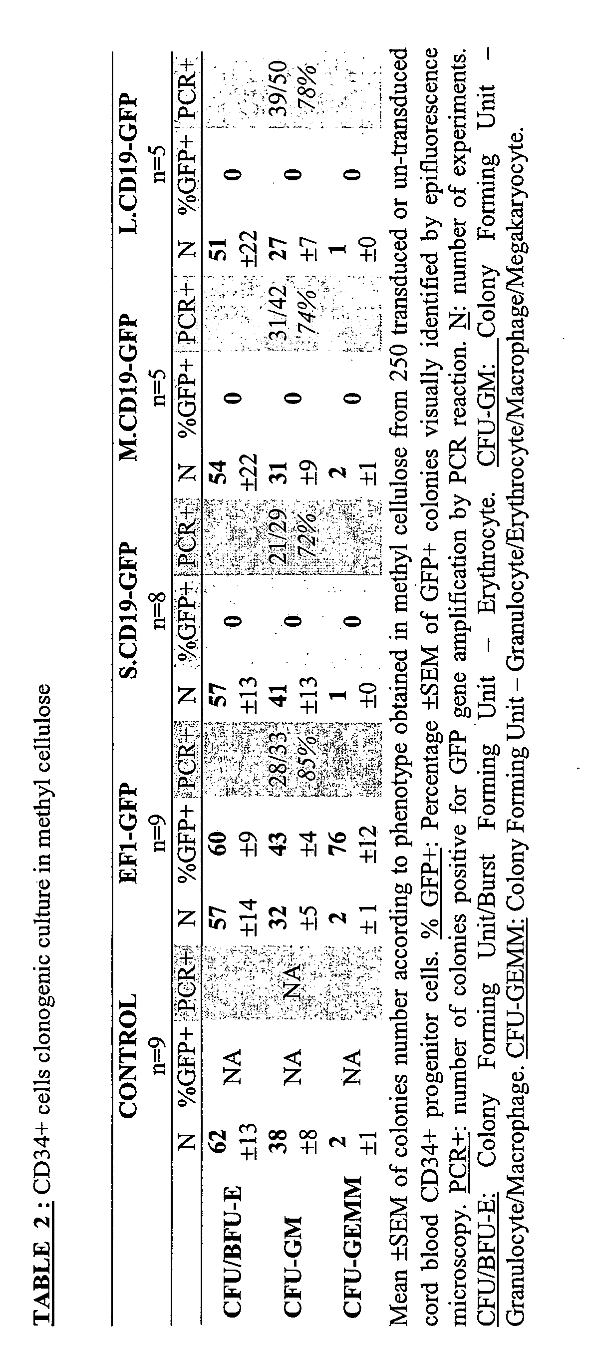
  • Colony-Forming Unit Granulocyte-Macrophage (CFU-GM) and Burst-Forming Unit Erythrocyte (BFU-E) were counted at day 14 according to published criteria.
  • Colony-Forming Cell (CFC)-derived cells were plucked, re-suspended in phosphate buffer saline, and examined for GFP expression by flow cytometry. Single colonies were also analyzed by PCR for lentiviral transduction after pronase (Roche Applied Science) digestion (1mg/mL in 20 ⁇ L Tris 50mM).
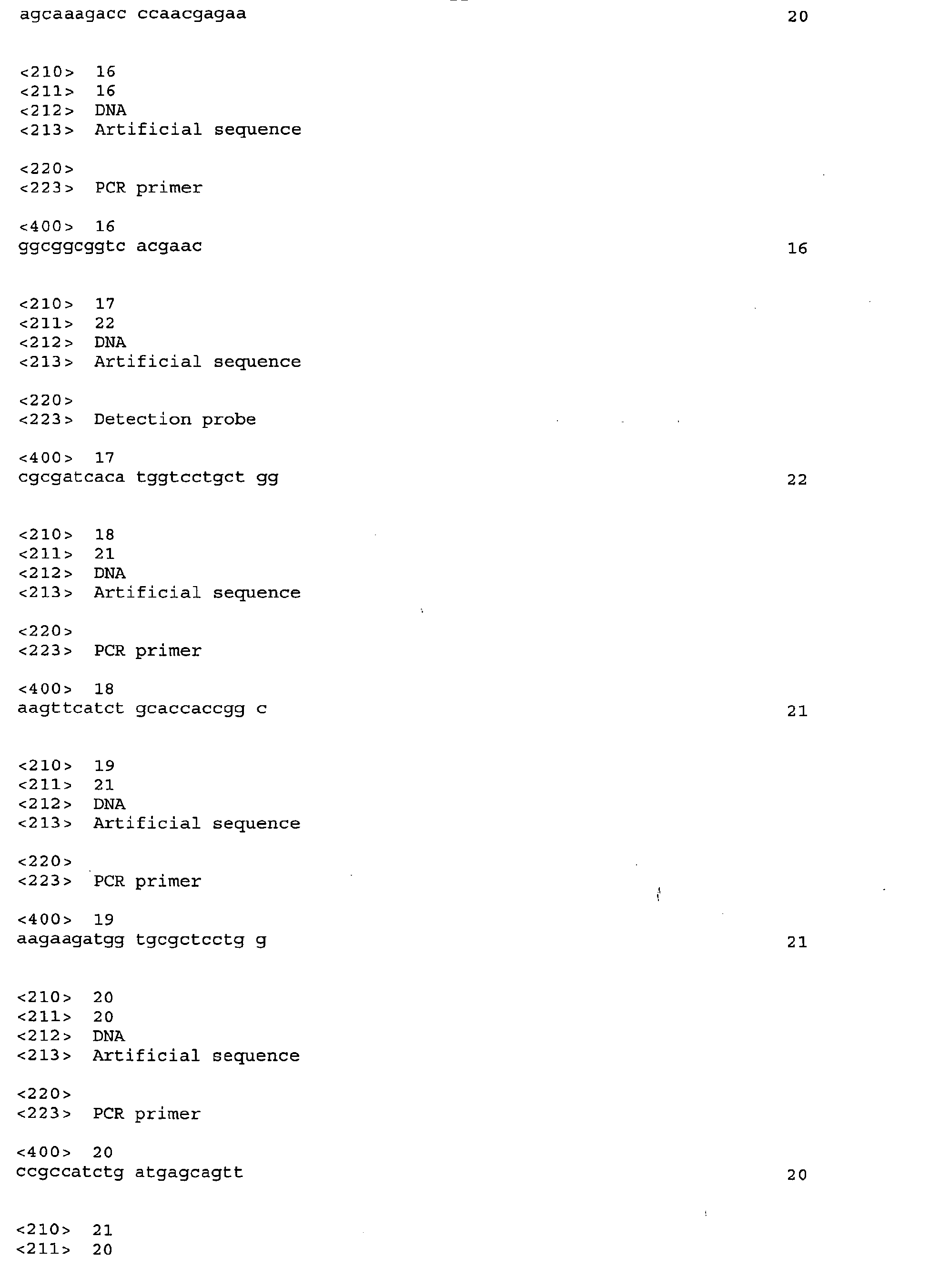
  • Amplification was performed with two pairs of primers in order to identify the GFP transgene and the C-Kappa endogene in the same sample: GFP sens 5' AAG TTC ATC TGC ACC ACC GGC (SEQ ID NO: 18) and antisens 5' AAG AAG ATG GTG CGC TCC TGG (SEQ ID NO: 19) (166bp amplification); CK sens 5' CCG CCA TCT GAT GAG CAG TT (SEQ ID NO: 20) and antisens 5' TGA AGC TCT TTG TGA CGG GC (SEQ ID NO: 21) (273bp amplification).
  • the PCR reaction was done at 94°C for 3' followed by 40 cycles of 45s at 94°C, 1' at 60°C, 1' at 72°C, and completed by 4' at 72°C.
  • MS-5 cells 25 were cultured in IMDM 10% FCS, 1% PS, in 96 well plates until they reached confluence.
  • Transduced CD34 + cells were then added at 3x10 4 cells/well in IMDM supplemented with 5% human AB serum, 5% FCS (Stem Cell Technologies), 1 % PS, for B-lymphoid/myelo-monocytic differentiation.
  • NK cell differentiation 1.5x10 4 CD34 + cells were cultured in IMDM, 15% FCS, 1% PS supplemented with rhIL-15 (20 ng/mL, R&D system, Abingdon, UK) and rhSCF (50 ng/mL). Cells were maintained at 37°C in a fully humidified incubator, with 5% CO 2 and fed weekly by half medium change for 3 to 5 weeks.
  • 2x10 4 transduced CD34+ cells were cultured in 24 well plates in BIT medium, 1% PS, supplemented with rhIL-3 (10ng/mL), rhEPO (2U/mL) and rhSCF (100ng/mL), and maintained during 10 days at 37°C/5%CO 2 .
  • Cells were then collected for GFP and phenotypic marker expression studies with flow cytometry, using Phycoerythrin (PE) conjugated murine monoclonal antibodies (mAb) directed against human CD19, CD33, CD56 and GP-A.
  • PE Phycoerythrin
  • mAb murine monoclonal antibodies
  • CD34+ cells transduced with the EF1-GFP control vector gave an expression of GFP in all differentiated lineages - 76.2 ⁇ 6.6% of B-cells (CD19+), 62.3 ⁇ 4.9% of myeloid cells (CD33+), 36.7 ⁇ 6.1% of NK cells (CD56+) and 84 ⁇ 7.2% of erythroid cells (GPA+) - GFP expression was significant only in B-cells when differentiation came from M and L.CD19-GFP transduced progenitors: 44 ⁇ 4.9% and 36.9 ⁇ 3.7% of CD19+ cells respectively.
  • mice 27 were obtained from Charles River Laboratories (France). Founders were bred at the Inventors' animal facility. All animals were maintained in germ-free conditions. Seven to 8 week old animals were used for xeno-transplantation. Recipients were prepared with total body gamma irradiation at a dose of 3.25 Gy. As described by Kerre et al. 28 , mice were injected within 24 hours of irradiation, intraperitoneally, with 0.2mg/mouse of anti CD122 monoclonal antibody (TM ⁇ 1 29 , kindly provided by Dr T Tanaka, Tokyo, Japan).
  • TM ⁇ 1 29 anti CD122 monoclonal antibody
  • thymus engraftment was a rare event (10/34 bone marrow engrafted tested mice) and weakly pronounced (hCD45+ cells ⁇ 1% of total).
  • human cells were identified, there was not systematically presence of CD3+ T-lymphoid cells.
  • Figure 11A-11D where appeared CD3, CD4 and CD8+ cells in a mouse engrafted with M.CD19-GFP transduced progenitor cells, only background expression of marker was observed on former T-lymphoid cells and GFP expressing cells were mainly found in CD19+ population.

Landscapes

  • Health & Medical Sciences (AREA)
  • Life Sciences & Earth Sciences (AREA)
  • Genetics & Genomics (AREA)
  • Chemical & Material Sciences (AREA)
  • Organic Chemistry (AREA)
  • Engineering & Computer Science (AREA)
  • Zoology (AREA)
  • Biotechnology (AREA)
  • General Engineering & Computer Science (AREA)
  • Bioinformatics & Cheminformatics (AREA)
  • Wood Science & Technology (AREA)
  • Biomedical Technology (AREA)
  • Biochemistry (AREA)
  • Molecular Biology (AREA)
  • General Health & Medical Sciences (AREA)
  • Biophysics (AREA)
  • Physics & Mathematics (AREA)
  • Microbiology (AREA)
  • Plant Pathology (AREA)
  • Immunology (AREA)
  • Cell Biology (AREA)
  • Environmental Sciences (AREA)
  • Medicinal Chemistry (AREA)
  • Gastroenterology & Hepatology (AREA)
  • Proteomics, Peptides & Aminoacids (AREA)
  • Animal Behavior & Ethology (AREA)
  • Animal Husbandry (AREA)
  • Biodiversity & Conservation Biology (AREA)
  • Toxicology (AREA)
  • Micro-Organisms Or Cultivation Processes Thereof (AREA)

Abstract

The present invention relates to a nucleic acid sequence comprising or consisting of a S sequence, said S sequence comprising or consisting of:
  • (1) a first sequence corresponding to a fragment of at least 10 nucleotides of the promoter sequence of the human CD19 gene, such as defined in Figure 1, the ends of which being comprised from the nucleotide at position -1500 to the nucleotide at position -1000 of the promoter sequence of the human CD19 gene,
  • (2) a second sequence consisting of the contiguous nucleotides ranging from position -231 to position -1 (SEQ ID NO : 1) of the promoter sequence of the human CD19 gene.
  • Description

    • The present invention particularly relates to B lymphoid lineage specific promoter sequences and to pharmaceutical compositions containing said sequences.
    • Gene transfer into hematopoietic stem cells remains a major objective and challenge in medical science, particularly for gene or cell therapy. The field still faces many technological obstacles, including the difficulty to transduce mostly non-dividing cells, the risk of mutational insertion, and the difficulty to control transgene expression. Over the last few years, lentiviruses have been increasingly used and substituted to onco-retroviruses, because of their ability to integrate the genome of quiescent cells, and the reduced need to use exogenous cytokines to induce cell cycling 1;2. Efficient transduction of human hematopoietic progenitor cell populations, including CD34+ cells3, CD34+/CD38- cells and SCID-repopulating cells (SRC) 4 has been documented with lentiviral vectors.
    • Lentiviruses also offer advantages over onco-retroviruses, in that they allow for the more efficient integration of regulatory sequences that target the transgene expression to a defined lineage. Several examples have been reported, both with onco-retroviral and lentiviral vectors, that limit the expression of a marker gene to one or several lineages with various efficacies. These include the transcriptional targeting of T-cells5; 6, antigen presenting cells (APC) cells7, monocytes/macrophages 8 and erythroid cells9; 10.
    • The possibility to limit transgene expression to the B-cell compartment, following transduction of stem and progenitor cells has rarely been reported so far. In one instance, a sequence from the immunoglobulin heavy chain enhancer in a lentiviral vector was described to increase marker gene expression in B cells 17. Besides, several reports mentioned the promising use of the human CD19 promoter to target gene expression to B cells in different experimental systems.
    • The CD19 antigen, under the control of the BSAP (Pax5) transcription factor, is a specific marker of B cells, appears at the pre-B stage, and remains expressed throughout B-cell differentiation and maturation, to be lost at the final stage when B-lymphocytes maturate into plasma cells11. CD19 takes part to the CD21, CD81 and Leu-13 complex which regulates thresholds of intracellular signals12.
    • Large regions (6.3 to 4.2kb) that flank the human CD19 gene drove expression of the transgene in the B cell lineage in transgenic mice 13; 14; 15. However, the large fragments of the CD19 promoter described to date are ill-suited for an efficient insertion into a vector, such as a lentiviral vector, because of their size.
    • In transfection assays, a short region (200bp) was identified in the proximal region of the human CD 19 gene promoter, that produced high levels of reporter activity in lymphoid cell lines16. But its B lineage-specific promoter activity has never been soundly assessed.
    • Thus, an object of the present invention is to provide new fragments derived from the CD19 promoter sequence, which are smaller than the large fragments used in transgenic mice and/or more B lineage specific than those of the prior art.
    • The present invention relates to a nucleic acid sequence comprising or consisting of:
      • a S sequence, said S sequence comprising or consisting of:
      • (1) a first sequence corresponding to a fragment of at least 10 nucleotides of the promoter sequence of the human CD19 gene, such as defined in Figure 1, the ends of which being comprised from the nucleotide at position -1500 to the nucleotide at position -1000 of the promoter sequence of the human CD19 gene,
      • (2) a second sequence consisting of the contiguous nucleotides ranging from position -231 to position -1 (SEQ ID NO :1) of the promoter sequence of the human CD 19 gene; or
      • a sequence derived from the above defined S sequence by insertion, deletion or mutation of at least one nucleotide, or presenting an homology of at least 60 % per block of 150 to 200 contiguous nucleotides with the above defined S sequence, provided that said sequence is capable of directing the specific expression of a gene in cells of the B lymphoid lineage;
      provided that when said S sequence consists of the contiguous nucleotides of the promoter sequence of the human CD19 gene ranging from the 5' end of the first sequence to position -1, then, if present, the 5' contiguous nucleotides flanking the 5'end of the first sequence in said nucleic acid sequence are different from the 5' contiguous nucleotides flanking the 5' end of the sequence corresponding to said first sequence in the promoter sequence of the human CD 19 gene.
    • The expression "per block of 150 to 200 contiguous nucleotides" means that the sequence which is homologous to the S sequence comprises at least one fragment of 150 to 200 contiguous nucleotides presenting a homology of at least 60% with the corresponding fragment of the S sequence, provided that said sequence is capable of directing the specific expression of a gene in cells of the B lymphoid lineage.
    • According to a preferred embodiment, the invention also relates to a nucleic acid sequence as defined above, comprising or consisting of a sequence homologous to the S sequence, said homologous sequence comprising two fragments presenting a homology of at least 73% and 63% respectively with the corresponding fragments of the S sequence, said fragments corresponding respectively to the contiguous nucleotides ranging from position -193 to position -1 (CR1) and from position -1260 to position -1062 (CR2) of the promoter sequence of the human CD19 gene.
    • In an advantageous embodiment, the invention also relates to a nucleic acid sequence as defined above, comprising or consisting of a sequence presenting an homology of at least 80%, in particular 90% with the above defined S sequence, provided that said sequence is capable of directing the specific expression of a gene in cells of the B lymphoid lineage.
    • The expression "capable of directing the specific expression of a gene" means that the corresponding sequence can activate the transcription of sequences which are situated on the 3' side of said sequence, and preferably of sequences, the initiation codon of which is linked to the 3' end of said sequence. Such a sequence corresponds in particular to a promoter sequence.
    • According to a particular embodiment, the S sequence consists of:
    • (1) a first sequence consisting of the contiguous nucleotides ranging from position X to position Y of the promoter sequence of the human CD19 gene, X and Y being negative integer numbers, and
    • (2) a second sequence consisting of contiguous nucleotides ranging from position Z to position -1 of the promoter sequence of the human CD19 gene, Z being a negative integer number,
    • wherein X, Y and Z are such, that:
      • X ranges from -1500 to -1000,
      • Y ranges from X+9 to Z-1, and
      • Z ranges from Y+1 to -231,
      the 3' end of said first sequence being covalently linked to the 5' end of said second sequence.
    • For clarity sake the above defined S sequence is represented in Figure 12A. Preferably the 3' end is linked to the 5' end via a phosphoester linkage.
    • According to a more particular embodiment,
      X=-1274
      Y ranges from -1042 to Z-1, and
      Z ranges from Y+1 to -231.
    • For clarity sake the above defined S sequence is represented in Figure 12B.
    • According to a preferred embodiment,
      X = -1274,
      Y = -1042, and
      Z = -231,
      that is, the invention relates to a nucleic acid sequence comprising or consisting of a S sequence, said S sequence consisting of:
    • (1) a first sequence consisting of the contiguous nucleotides ranging from position -1274 to position -1042 (SEQ ID NO: 2) of the promoter sequence of the human CD19 gene, and
    • (2) a second sequence consisting of the contiguous nucleotide ranging from position -231 to position -1 (SEQ ID NO: 1) of the promoter sequence of the human CD19 gene,
    • the 3' end of said first sequence being covalently linked to the 5' end of said second sequence, said S sequence corresponding to SEQ ID NO: 3.
    • SEQ ID NO: 3 is hereafter referred to as M.CD19.
    • Advantageously, SEQ ID NO: 3 comprises two elements hereafter referred to as CR1 (position -1 to position -193 of the promoter sequence of the human CD19 gene) and CR2 (position -1062 to position -1260 of the promoter sequence of the human CD19 gene), said elements providing a potent and specific promoter activity in cells of the B lineage.
    • According to another preferred embodiment,
      X is equal to -1274, and
      Y = Z-1,
      that is, the invention relates to a nucleic acid sequence comprising or consisting of a S sequence, said S sequence consisting of the contiguous nucleotides ranging from position -1274 to position -1 (SEQ ID NO: 4) of the promoter sequence of the human CD19 gene, or of a sequence (SEQ ID NO: 5) derived from said S sequence by the deletion of one nucleotide selected from the contiguous nucleotides ranging from position -541 to position -530 of the promoter sequence of the human CD19 gene, provided that, if present, the 5' contiguous nucleotides flanking the 5'end of the S sequence in said nucleic acid sequence are different from the 5' contiguous nucleotides flanking the nucleotide at position -1274 in the promoter sequence of the human CD 19 gene.
    • SEQ ID NO: 4 is hereafter referred to as L.CD19.
    • Advantageously, SEQ ID NO: 4 and SEQ ID NO: 5 comprise two elements hereafter referred to as CR1 (position -1 to position -193 of the promoter sequence of the human CD19 gene) and CR2 (position -1062 to position -1260 of the promoter sequence of the human CD19 gene), said elements providing a potent and specific promoter activity in cells of the B lineage.
    • The invention also relates to an expression sequence, characterized in that it comprises a nucleic acid sequence such as defined above, linked to a protein encoding nucleic acid sequence, in particular a protein encoding nucleic acid sequence coding for a protein expressed specifically in cells of the B lymphoid lineage, such as a sequence encoding Btk, Igα, surrogate light chains, BLNK, Igb, or to a hairpin RNA encoding nucleic acid sequence, or to a suicide gene such as the HSVTK gene (Herpes simplex virus thymidine kinase), or to a marker gene such as the GFP gene (green fluorescent protein) or the LacZ gene.
    • Cells of the B lymphoid lineage notably comprise proB cells, preB cells, immature B cells, mature B cells, memory B cells, or cells of the Nalm6, Ramos, and Daudi JEA2 cell lines for example.
    • The expression "suicide gene" refers to a gene which induces cell death in response to a drug, for cells in which it is expressed. The HSVTK gene thus induces ganciclovir-mediated cell death.
    • The expression "marker gene" refers to a gene which codes for a protein which is liable to be detected according to methods well known to the man skilled in the art, such as microscopy, in particular fluorescent microscopy. Such a gene notably comprises genes encoding for fluorescent proteins well known to the man skilled in the art, such as GFP and GFP derivatives.
    • Btk (Bruton tyrosine kinase), Igα, surrogate light chains, BLNK (B cell Linker protein), Igb are notably described in Ballow (2002) J. Allergy Clin. Immunol. 109:581, Conley and Cooper (1998) Curr. Opin. Immunol. 10:399 and Gauld et al. (2002) Science 296:1641.
    • Advantageously the abovementioned expression sequences are expressed specifically in cells of the B lymphoid lineage.
    • According to a preferred embodiment, the abovementioned nucleic acid sequence of said expression sequence consists of SEQ ID NO: 3, 4 or 5.
    • The invention also relates to a vector, in particular an expression vector, characterized in that it comprises a nucleic acid sequence or an expression sequence such as defined above.
    • Vectors according to the invention comprise in particular plasmidic vectors such as the pRSET vector or the pcDNA vector, or lentiviral vectors, such as the pTRIP and pHPT vectors.
    • Advantageously, such vectors are well suited to express RNA and/or proteins in cells of the B lymphoid lineage.
    • According to a preferred embodiment, the abovementioned nucleic acid sequence of said vector consists of SEQ ID NO: 3, 4 or 5, such as for the lentiviral vectors corresponding respectively to SEQ ID NO: 6, 7 and 8.
    • The invention also relates to a cell, in particular a cell of the B lymphoid lineage, comprising a nucleic acid sequence such as defined above, or an expression sequence such as defined above, or a vector such as defined above.
    • Cells according to the invention comprise in particular stem cells, hematopoietic stem cells, embryonic stem cells and precursor cells.
    • According to a preferred embodiment, the abovementioned cell comprises a nucleic acid sequence consisting of SEQ ID NO: 3, 4 or 5.
    • The invention also relates to a nonhuman transgenic animal, characterized in that all or some of its cells comprise a nucleic acid sequence such as defined above, or an expression sequence such as defined above, or a vector such as defined above, said animal being in particular a mammal, such as a mouse or a rat.
    • Such animals can be prepared according to methods well known to the man skilled in the art such as described for example in Maas et al. (1999) J. Immunol. 162:6526 and Lois et al. (2002) Science 295:868.
    • The invention also relates to a pharmaceutical composition comprising as active substance a nucleic acid sequence such as defined above, or an expression sequence such as defined above, or a vector such as defined above, or a cell such as defined above, in association with a pharmaceutically acceptable vehicle.
    • The invention also relates to the use of a nucleic acid sequence such as defined above, or an expression sequence such as defined above, or a vector such as defined above, or a cell such as defined above, for the manufacture of a medicament intended to treat pathologies in which cells of the B lymphoid lineage are involved, such as autoimmune diseases, B lymphomas, B leukemias, antibody deficiencies, in particular X linked agammaglobulinemia (Bruton's disease), or diseases resulting from a deficient preB cell receptor complex, such as a Igα defect, a surrogate light chains defect, or a BLNK defect.
    • Such diseases are notably described in Conley and Cooper (1998) Current Opinion in immunology 10:399-406 ; Gauld et al. (2002) Science 296:1641-1642; and Ballow (2002) J. Allergy Clin. Immunol. 109:581-591.
    • DESCRIPTION OF THE FIGURES Figure 1
    • Figure 1 represents the upstream nucleic acid sequence of the human CD19 gene. The 5' end corresponds to the nucleotide in position -4339 relative to the A nucleotide of the translation initiation codon (AUG) in position +1, and the 3' end corresponds to the nucleotide in position -1. Nucleotide positions are indicated on the left.
    • Figure 2
    • Figure 2 represents the graphical results of the computational analysis of the upstream region of the mouse and human CD 19 genes based on their comparison. The graph abscissae represent the 1274bp of human gene and the ordinates the 1529bp of the murine one. Two conserved regions were identified and schematically drawn: a proximal sequence (CR1) and a distal one (CR2). Putative fixation sites for transcription factors were identified, as indicated by boxes on a horizontal axis for the human gene and on a vertical axis for the murine one. Several restriction sites as well as repetitive sequences (grey bars corresponding to the grey areas on the graph) are also shown.
    • Figure 3
    • Figure 3 represents the plasmid form of the recombinant lentiviral genomes of the invention. The ubiquitous EF1α promoter (235bp) of pTRIPΔU3-EF1α-GFP vector plasmid was substituted by the S.CD19 (230bp), M.CD19 (464bp) or L.CD19 (1274bp) sequence by cloning between the MluI (black diamond shape) and BamHI(star) sites. New plasmids were respectively called pTRIPΔU3-S.CD19-GFP, pTRIPΔU3-M.CD19-GFP, pTRIPΔU3-L.CD19-GFP and gave rise to the S.CD19-GFP, M.CD19-GFP and L.CD19-GFP lentiviral vectors. LTR: Long Terminal Repeat; SD/SA: Splice Donor / Splice Acceptor; Ψ: encapsidation signal (on truncated GAG sequence); RRE: Rev Responsive Element; Triplex: Central Termination Sequence (CTS) and central PolyPurine Tract (cPPT) of HIV-1 virus; ΔU3: deletion of 400bp in the 3'LTR U3 region (Self Inactivated vector).
    • Figure 4
    • Figure 4 represents the specificity index (vertical axis) of GFP expression by cell lines (horizontal axis) transduced by the S.CD19-GFP (white bars), M.CD19-GFP (hatched bars) and L.CD19-GFP (black bars) vectors, relative to the GFP expression in the Nalm6 cell line.
    • Figures 5A-5I
    • Figures 5A to 5E represent the flow cytometry profile of GFP expression (horizontal axis, fluorescence intensity) by human peripheral T-lymphoid cells, identified by an anti-CD3 antibody (vertical axis, fluorescence intensity), 7 days after transduction in parallel with lentiviral vectors EF1-GFP (Figure 5B), S.CD19-GFP (Figure 5C), M.CD19-GFP (Figure 5D) and L.CD19-GFP (Figure 5E), non-tranduced cells were used as negative control (Figure 5A). The box in the center of each flow cytometry diagram (R3) delimits GFP positive CD3+ cells. Figures 5F to 51 represent the quantitative flow cytometry profile of GFP expression (horizontal axis, fluorescence intensity) by human cord blood CD34+ cells (vertical axis, number of cells), 7 days after transduction in parallel with lentiviral vectors EF1-GFP (Figure 5F), S.CD19-GFP (Figure 5G), M.CD19-GFP (Figure 5H) and L.CD19-GFP (Figure 5I); negative control for GFP expression is shown on each diagram by dotted lines.
    • Figures 6A-6T
    • Figures 6A to 6T represent flow cytometry dot blots of GFP expression (horizontal axis, intensity of fluorescence) by cells deriving from transduced and un-transduced CD34+ placental progenitor cells submitted to in vitro differentiation by co-culture with the MS.5 cell line (B-lymphoid, myeloid and NK differentiation) or in liquid culture (erythroid differentiation).
      Figures 6A to 6E relate to CD19+ cells (vertical axis, intensity of fluorescence), that is B lymphoid cells, deriving from CD34+ progenitor cells untransduced (Figure 6A) or transduced by EF1-GFP (Figure 6B), S.CD19-GFP (Figure 6C), M.CD19-GFP (Figure 6D) and L.CD19-GFP (Figure 6E).
      Figures 6F to 6J relate to CD33+ cells (vertical axis, intensity of fluorescence), that is myeloid cells, deriving from CD34+ progenitor cells untransduced (Figure 6F) or transduced by EF1-GFP (Figure 6G), S.CD19-GFP (Figure 6H), M.CD19-GFP (Figure 6I) and L.CD19-GFP (Figure 6J).
      Figures 6K to 60 relate to CD56+ cells (vertical axis, intensity of fluorescence), that is NK cells, deriving from CD34+ progenitor cells untransduced (Figure 6K) or transduced by EF1-GFP (Figure 6L), S.CD19-GFP (Figure 6M), M.CD19-GFP (Figure 6N) and L.CD19-GFP (Figure 6O).
      Figures 6P to 6T relate to GP-A+ cells (vertical axis, intensity of fluorescence), that is erythroid cells, deriving from CD34+ progenitor cells untransduced (Figure 6P) or transduced by EF1-GFP (Figure 6Q), S.CD19-GFP (Figure 6R), M.CD19-GFP (Figure 6S) and L.CD19-GFP (Figure 6T).
      Analyses were done inside the ToPRO-3neg/hCD45+ gate; the box in the center of each flow cytometry diagram delimits lineage+/GFP+ cells.
    • Figure 7
    • Figure 7 represents the specificity index (vertical axis) of the expression of GFP by CD19+, CD33+ and CD56+ cells (horizontal axis) derived ex vivo from CD34+ progenitor cells transduced by the EF1-GFP (black bars), S.CD19-GFP (white bars), M.CD19-GFP (largely hatched bars) and L.CD19-GFP (closely hatched bars) vectors, relative to the GFP expression in CD 19+ cells.
    • Figures 8A-8ZA
    • Figure 8A to 8ZA represent flow cytometry diagrams of GFP expression (horizontal axis, intensity of fluorescence) by cells deriving from transduced and un-transduced CD34+ placental progenitor cells submitted to in vivo differentiation in the NOD/SCID murine xenograft model. B-lymphoid (CD10, CD19, IgM), myeloid (CD33) and progenitor cells (CD34) compartments were analyzed.
      Figures 8A to 8E relate to CD 19+ cells (vertical axis, intensity of fluorescence), taken from bone marrow, deriving from CD34+ progenitor cells untransduced (Figure 8A) or transduced by EF1-GFP (Figure 8B), S.CD19-GFP (Figure 8C), M.CD19-GFP (Figure 8D) and L.CD19-GFP (Figure 8E).
      Figures 8F to 8J relate to CD33+ cells (vertical axis, intensity of fluorescence), taken from bone marrow, deriving from CD34+ progenitor cells untransduced (Figure 8F) or transduced by EF1-GFP (Figure 8G), S.CD19-GFP (Figure 8H), M.CD19-GFP (Figure 8I) and L.CD19-GFP (Figure 8J).
      Figures 8K to 80 relate to CD34+ cells (vertical axis, intensity of fluorescence), taken from bone marrow, deriving from CD34+ progenitor cells untransduced (Figure 8K) or transduced by EF1-GFP (Figure 8L), S.CD19-GFP (Figure 8M), M.CD19-GFP (Figure 8N) and L.CD19-GFP (Figure 8O).
      Figures 8P to 8S relate to IgM+ cells (vertical axis, intensity of fluorescence), taken from the spleen, deriving from CD34+ progenitor cells untransduced (Figure 8P) or transduced by EF1-GFP (Figure 8Q), M.CD19-GFP (Figure 8R) and L.CD19-GFP (Figure 8S).
      Figures 8T to 8W relate to CD19+ cells (vertical axis, intensity of fluorescence), taken from the spleen, deriving from CD34+ progenitor cells untransduced (Figure 8T) or transduced by EF1-GFP (Figure 8U), M.CD19-GFP (Figure 8V) and L.CD19-GFP (Figure 8W).
      Figures 8X to 8ZA relate to CD33+ cells (vertical axis, intensity of fluorescence), taken from the spleen, deriving from CD34+ progenitor cells untransduced (Figure 8X) or transduced by EF1-GFP (Figure 8Y), M.CD19-GFP (Figure 8Z) and L.CD19-GFP (Figure 8ZA).
      Flow cytometry analysis were performed after 12 weeks on bone marrow and spleen gating in ToPRO-3neg/hCD45+ cells; boxes on dot plots delimit lineage+/GFP+ cells
    • Figure 9A and Figure 9B
    • Figure 9A represents the specificity index (vertical axis) of the expression of GFP by CD19+, CD10+, CD33+ and CD34+ bone marrow cells (horizontal axis) derived in vivo from CD34+ progenitor cells transduced by the EF1-GFP (black bars), S.CD19-GFP (white bars), M.CD19-GFP (largely hatched bars) and L.CD19-GFP (closely hatched bars) vectors, relative to the GFP expression in CD 19+ cells.
      Figure 9B represents the specificity index (vertical axis) of the expression of GFP by CD19+, IgM+ and CD33+ splenic cells (horizontal axis) derived in vivo from CD34+ progenitor cells transduced by the EF1-GFP (black bars), M.CD19-GFP (largely hatched bars) and L.CD19-GFP (closely hatched bars) vectors, relative to the GFP expression in CD19+ cells.
    • Figure 10A and Figure 10B
    • Figure 10A represents the flow cytometry analysis of GFP expression by cells deriving from CD34+ progenitor cells engrafted in NOD/SCID mice.
      The flow cytometry dot blot on the left shows a representative experiment of CD34 expression (vertical axis, intensity of fluorescence) and of CD 19 expression (horizontal axis, intensity of fluorescence) of cells taken from mice engrafted with EF1, M.CD19 or L.CD19-GFP transduced CD34+ progenitor cells. The boxes delimit non-B progenitors (CD34+CD19-) (R3), B-progenitors (CD34+CD19+) (R4) and more mature B-cells (CD34-CD19+) (R5).
      The histograms on the right represent the GFP expression profile (horizontal axis, fluorescence intensity) corresponding to the abovementioned CD34+CD19- cells (upper histograms), CD34+CD19+ cells (middle histograms) and CD34-CD19+ cells (lower histograms) deriving from CD34+ progenitor cells transduced by EF1-GFP, M.CD19-GFP, L.CD19-GFP.
      The percentage of GFP expressing cells is indicated in bold for each histogram, background levels are indicated by dotted lines.
      From top to bottom the histograms are representative of a more advanced B cell commitment stage.
      Figure 10B shows a histogram representing the percentage of mean GFP expression ± SEM of the CD34+CD19- cells, CD34+CD19+ cells and CD34-CD19+ cells described in figure 10A and deriving from CD34+ progenitor cells transduced by EF1-GFP (black bars), M.CD19-GFP (largely hatched bars) and L.CD19-GFP (closely hatched bars).
    • Figure 11A, Figure 11B, Figure 11C and Figure 11D
    • Figures 11A to 11D represent the flow cytometry expression profile of GFP expression (horizontal axis, fluorescence intensity) by human CD19+ (vertical axis, fluorescence intensity) (Figure 11A), CD3+ (vertical axis, fluorescence intensity) (Figure 11B), CD4+ (vertical axis, fluorescence intensity) (Figure 11C) and CD8+ (vertical axis, fluorescence intensity) (Figure 11D) thymic cells issued from NOD/SCID mouse engrafted by M.CD19-GFP transduced CD34+ cells.
      Flow cytometry analysis was performed after 12 weeks, gating in ToPRO-3neg/hCD45+ cells; the box in the center of each flow cytometry diagram delimits lineage+/GFP+ cells.
    • Figure 12A and Figure 12B
    • Figure 12A and 12B represent fragments (arrows) of the 1500 nucleotides upstream of the translation initiation codon of the human CD19 gene (grey box) as defined by their nucleotide positions.
      Figure 12A represents the two sequences (1 and 2) constituting the S sequence, wherein X is comprised from positions -1500 to -1000, Y is such that the length of sequence (i) is at least 10 nucleotide long and Z is comprised from positions Y+1 to -231.
      Figure 12B represents the two sequences (i and ii) constituting the S sequence, wherein X is equal to -1274, Y is greater or equal to -1042 and Z is comprised from positions Y+1 to -231.
    • EXAMPLES EXAMPLE 1 Design of CD1 9-GFP lentiviral vectors and GFP expression in B cell lines transduced by said vectors 1. Selection of CD19 B-cell specific regulatory sequences
    • In order to determine the minimal sequences that are functionally important, and may thus confer B-cell specificity when inserted in a lentiviral vector, a computational sequence analysis approach30 was used to study the CD19 gene regulatory region. Contigs containing the human and mouse CD19 genes were first obtained from the Ensembl genome database (Sanger Institute/EMBL-EBI) (www.ensembl.org)19.
    • First, using the RepeatMasker software (available at http://ftp.genome.washington.edu/RM/RepeatMasker.html) (Smit, AFA & Green, P RepeatMasker), repetitive sequences were identified and masked in the human contig AC 109460 containing the CD 19 gene.
    • Second, comparison with the mouse contig AC125169 was carried out, using the Pipmaker software (Schwartz et al. (2000) Genome Research 10:577-586); two conserved regions were identified in the human and mouse CD19 gene promoters 31, including a previously non identified 5' upstream fragment.
    • The two conserved regions, hereafter named CR1 and CR2, were contained in a contiguous 1274 bp fragment of the human CD19 gene (Figure 2) . CR2 (198 bp) was located -1 Kb upstream of human CD19 gene initiation site, and at a not yet localized upstream position in the mouse gene, since the sequence of contig AC 125169 is not yet entirely known. To allow for a more precise comparison using Pipmaker, a 1529 bp fragment for the mouse CD19 gene was created, where CR2 was inserted 5' upstream and artificially separated with 100 Ns (undefined nucleotides) from the sequenced mouse CD19 promoter. The CD19 promoter contains several well characterized regulatory elements required for B-cell specific expression such as the binding sites for the B-cell specific transcription factor BSAP/Pax5 16; 31-33 . Both human and mouse CD19 upstream regions were further screened for putative regulatory elements using the TESS software (TESS: Transcription Element Search Software available at http://www.cbil.upenn.edu/tess, Jonathan Schug and G. Christian Overton, Technical Report CBIL-TR-1997-1001-v0.0 Computational Biology and Informatics Laboratory School of Medicine University of Pennsylvania, 1997) and the Matlnspector software20 (available at Genomatix: http://www.genomatix.de)21 (Figure 2).
    • Based upon this computational approach, it was hypothesized that the two conserved regions CR1 and CR2, which present respectively 73% and 63% of homology with the corresponding mouse nucleotide sequence, might be functionally important to promote CD19 expression. All CD19 sequences were produced by PCR methods from human primary cell genomic DNA.
    • Briefly, three sequences based on the sequence of the CD 19 promoter were generated byPCR (S.CD19, M.CD19 and L.CD19).
    • The following oligonucleotides were used as primers: S.CD19(+) 5'GTT ACG CGT CCC GTG GTA GTG AGA (SEQ ID NO: 9) and S.CD19(-) 5'TTG GGA TCC GGT GGT CAG ACT CTC (SEQ ID NO: 10) for the S.CD19 sequence; L.CD19(+) 5'GTT ACG CGT AAT GAG GGG ATT AGA GAG (SEQ ID NO: 11) and L.CD19(-) 5'TTG GGA TCC GGT GGT CAG ACT CTC CG (SEQ ID NO: 12) for the L.CD19 sequence (underlined sequences are restriction sites for MluI and BamHI). The generation of the M.CD19 sequence required two PCR steps, in order to juxtapose two distant regions of the native genome; first, two PCR reactions were carried out with the following two primer pairs: L.CD19(+) and M.CD19(-) 5'GCT CTC ACT ACC ACG GGA CCA CTT TCT CAG TGG CTG (SEQ ID NO: 13); M.CD19(+) 5'CAG CCA CTG AGA AAG TGG TCC CGT GGT AGT GAG AGC (SEQ ID NO: 14) and L.CD19(-); purified products were then joined end to end in a single PCR reaction carried out with the L.CD19(+) and L.CD19(-) primer pair. Amplifications were realized in presence of Platinum HIFI Taq polymerase (Invitrogen), with the following program: 2' at 94°C followed by 10 cycles with 45s at 94°C, 45s at 58°C and 2' at 68°C, then 25 cycles with 45s at 94°C, 45s at 66°C and 2' at 68°C; the last cycle ended with an extension phase of 7'.
    • The S.CD19 sequence contains the proximal region of homology (CR1) for a total length of 230bp. The composite M.CD19 (SEQ ID NO: 3) sequence of 464bp length results from the artificial juxtaposition of the two fragments containing the proximal and distal highlighted homologous regions (CR1 and CR2). The last sequence named L.CD19 (SEQ ID NO: 4) corresponds to the entire native CD19 sequence from the initiation site to the CR2 region, and is 1274bp long.
    • The inventors then designed three new lentiviral vectors, in which the GFP gene was placed under the control of human CD 19 gene promoter fragments.
    • 2. Design of CD19-GFP lentiviral vectors
    • In order to produce B-specific lentiviral vectors, the inventors cloned the three abovementioned CD19 sequences upstream a GFP marker gene between MluI and BamHI restriction sites of the pTRIPΔU3-EF1-GFP plasmid in place of the ubiquitous EFIα promoter (235bp): the subsequent recombinant lentiviral vectors were respectively called S.CD19-GFP, M.CD19-GFP and L.CD19-GFP (Figure 3).
    • Briefly, the S.CD19, M.CD19 and L.CD19 sequences were introduced in the pTRIPΔU3-EF1α-GFP vector plasmid (N.B. all lentiviral constructions were derived from the initial vector backbone pTRIPΔU3-EF1α-GFP [gift from Dr P. Charneau, Institut Pasteur, Paris, France3]), in substitution to the EF1α ubiquitous promoter between MluI and BamHI sites to obtain the pTRIPΔU3-S.CD19-GFP, the pTRIPΔU3-M.CD19-GFP and the pTRIPΔU3-L.CD19-GFP vector plasmids respectively (Figure 3).
    • Each construction was sequenced in the promoter region to confirm the identity of the CD19 sequence fragment with the genomic CD19 sequence (Genome express, Meylan, France); of note, a single T deletion was observed in a thymidine-rich region (12 repetitions, positions -541 to -530) in the L.CD19 sequence used (SEQ ID NO: 5).
    • The sequences of the lentiviral vectors comprising the M.CD19, L.CD19 and the deleted L.CD19 promoters, without the GFP gene, are respectively presented in SEQ ID NO: 6, 7 and 8.
    • Lentiviral particles were then produced by transient co-transfection of three plasmids into 293T cells: the plasmid containing the recombinant vector sequence, the packaging plasmid (p8.91), and the plasmid coding for the VSV-G (Vesicular Stomatitis Virus) envelop protein (pMDG), that confers amphotropic properties to recombinant lentiviral vectors; transfection was done by calcium phosphate co-precipitation, using the Calphos Mammalian transfection kit (Clontech laboratories, Ozyme, Montigny le Bretonneux, France). Supernatants were collected 24, 48 and 72 hours post-transfection, 0.45µm filtered, treated with DNAseI (1µg/mL, Roche Applied Science, Meylan, France) in the presence of MgCl2 (1nM) for 15'at 37°C and stored at -80°C; samples were tested for the absence of replication competent lentivirus (RCL) by monitoring p24 and Tat protein production in transduced HelaP4 cell cultures22. Finally, lentiviral vector productions were concentrated by ultracentrifugation, aliquoted in serum-free BIT medium (Stem Cell Technologies, Meylan, France) and stored at -80°C until use. Titers of different vectors usually ranged from 1x10⁸ and 1x10⁹ TU/mL (Transduction Unit), as measured by GFP expression on day 7 in the human pre-B cell line Nalm6, transduced in triplicate with serial dilution of concentrated lentiviruses.
    • 3. GFP expression in B cell lines transduced with CD19-GFP vectors
    • The promoter activity of the three CD19 sequences, in the context of lentiviral vectors, was first tested on a panel of hematopoietic and non-hematopoietic human cell lines, comprising:
      • human hematopoietic cell lines: B-lymphoid cells (Nalm6, Ramos, Daudi and JEA2), T-lymphoid cells (Jurkat or JA16, CEM and Molt4), myeloid cells (U937, HL60) and erythroleukemic cells (HEL, MO7e, TF1), which were all cultured in RPMI (Biowhittaker, Emerainville, France), 10% FCS (Foetal Calf Serum) (Gibco/Invitrogen), in the presence of antibiotics: 1%PS (100 U/mL Penicillin and 100 U/mL streptomycin, Biowhittaker). For TF1 and MO7e cell lines, culture medium was supplemented by rhGM-CSF (10 ng/mL, Peproteck/Tebu, Le Perray-en Yvelines, France) and rhGM-CSF (20 ng/mL) plus rhSCF (20 ng/mL, Amgen, Thousand Oaks, CA, USA) respectively.
      • epithelial cell lines (293T, HelaP4), which were cultured in DMEM (Biowhittaker) 10% FCS, 1% PS; and
      • endothelial cell lines (EAHY, HBMEC) in DMEM 20% FCS, 1% PS and DMEM/1% Glucose, 5% FCS, 1% PS, folic acid (1µg/mL), HEPES (10mM), glutamine (3mM) respectively.
    • All cells were transduced in parallel with the three different CD 19-GFP vectors and the EF1-GFP vector as positive control; we used a MOI between 0.5 and 0.7 that allowed 20 to 50% of positive control cells to express GFP for all cell types.
    • Cell lines were transduced without stimulation in their initial medium at a cell density of 0.05 to 0.3x10⁶ cells per well (24 well plate) in a total volume of 0.5 mL of their respective culture medium, and in the presence of polybren (4 µg/mL, Sigma Saint Quentin Fallavier, France). Cells were maintained at 37°C for 24 hours before washing with PBS.
    • Transgene expression was measured by flow cytometry 7 days post-transduction, and the expression specificity index (SI) for CD19 sequences calculated and normalized for each experiments by values obtained in parallel with the pre-B cell line Nalm6.
    • Briefly, for experiments on cell lines, where EF1-GFP positive control vector was systematically used in parallel of CD19-GFP vectors, the SI was calculated for each cell line as followed: SI= (%GFP+) CD19 vector/(%GFP+)EF1 vector ; then, individual SI values were normalized by SI value obtained in parallel with the Nalm6 pre-B cell line. For in vitro and in vivo differentiation experiments, SI was calculated for each lineage as followed: SI=(%GFP+)lineage/(%GFP+)CD19+lineage (Figure 4).
    • For the present and subsequent cytometry flow analyses, the antibodies used in were murine mAb IgG1-PC5 (Phycoerythrocyanin 5), IgG1-PE (Phycoerythrin), hCD45-PC5, hCD3-PE, hCD4-PE, hCD8-PE, hCD10-PE, hCD19-PE, hCD33-PE, hCD34-PE, hCD34-APC (Allophycocyanin) (Beckman Coulter/Immunotech, Marseille, France), hCD56-PE, hIgM-PE and GP-A-PE (BD/Bioscience, Le Pont de Claix, France). Cells were analyzed with a FACScalibur (Becton-Dickinson Immunocytometry systems, BDIS, San Jose, CA, USA). Dead cells were excluded for further analysis, by gating out TO-PRO-3 iodide positive cells at 630 nm, in 0.5 µM concentration (Molecular Probes, Interchim, Montluçon, France).
    • First, it appeared that CD19 sequences actually showed promoter activity in this lentiviral context. Second, GFP expression driven by any of the CD19 sequences was found preferentially in cell lines of B-lymphoid origin that express CD19 (Nalm6, Ramos, Daudi, JEA2). More precisely, the M and L.CD19 promoters presented a strong activity in all four B-cell lines (0.7>SI>1.09), whereas S.CD19 one appeared more variable (0.35>SI>1). Intensity of GFP expression was quite similar for all CD19 sequences (MFI=20-30), although slightly higher with the M and L.CD19 ones, and still weak in comparison with positive control cells (MFI ~ 100); of note, the MFI varied significantly between experiments, for CD19 promoters as for EF1 promoter. Considering GFP expression in non-B cell lines, experiments showed that the S and L.CD19 sequences had very low promoter activity (0>IS>0.22) in T-lymphoid (JA16, CEM, Molt4), myeloid (U937, HL60), epithelial (293T, HelaP4) and endothelial (EAHY, HBMEC) cells; these two sequences were slightly more active (0.22>IS>0.45) in erythroleukemic cells (TF1, HEL, MO7e). Analysis of the M.CD19 promoter behavior showed a more pronounced aspecific promoter activity in non-B cell lines (0>IS>0.64), especially in myeloid and erythroleukemic cells. To summarize, the L.CD19 sequence presented a more marked B-specific promoter activity than the S or M.CD19 ones that showed respectively low promoter activity in CD19+ B-cell lines and a quite high nonspecific activity in non-B-lymphoid cells.
    • 4. Transduction efficiency of the native and CD19 lentiviral vectors .
    • PCR experiments on genomic DNA of primary human T-lymphoid and progenitor cells revealed a similar integration of the viral genome after transduction with S, M or L.CD19-GFP vectors as compared to EF1-GFP.
    • Briefly, PCR reactions were performed on 75 ng of purified genomic DNA (Nucleospin Tissue, Macherey Nagel, France) isolated 7 to 14 days after transduction.
    • Transduction efficiency was measured with real time quantitative PCR, using TaqMan Master mix with the TaqMan® apparatus (Perkin Elmer Applied Biosystems, Courtaboeuf, France). GFP specific pair of primers (Genset) and fluorogenic probe (Perkin-Elmer Applied Biosystems) were designed using Primer Express® software (Perkin-Elmer): GFP-F 5'AGC AAA GAC CCC AAC GAG AA (SEQ ID NO: 15) and GFP-R 5'GGC GGC GGT CAC GAA C (SEQ ID NO: 16) were used at 650nM and the probe CGCGATCACATGGTCCTGCTGG (SEQ ID NO: 17) was at 200nM in a final reaction volume of 25µL. Thermal cycling conditions have been adjusted to the following parameters: 2 min at 50°C and 10 min at 95°C, then 40 cycles of 1 min at 95°C and 1 min at 66°C. In order to determine the lentiviral sequence copy number in each sample, raw data were analyzed with the software of ABI prism 7700.
    • EXAMPLE 2 GFP expression in primary human hematopoietic cells.
    • Human primary T cells were positively selected from peripheral blood mononuclear fractions by panning, using mouse anti-hCD4 and anti-hCD8 monoclonal antibodies (Baxter), as previously described 18.
    • For primary B-cell transduction experiments, human CD19+ cells were positively selected from the mononuclear fraction of peripheral blood by magnetic immunoselection (Miltenyi Biotec).
    • Cord blood (CB) samples were obtained after informed consent. CD34+ cells were enriched from CB mononuclear fractions, using immunoselection with the M.A.C.S. technology, according to the manufacturer's recommendations (Miltenyi Biotec, Bergish-Gladbach, Germany); purity was usually above 95% of CD34+ cells. Sorted cells were frozen in FCS (Gibco/Invitrogen, Cergy Pontoise, France) and stored in liquid nitrogen until use.
    • Human primary T cells isolated from peripheral (n=2) or cord blood (n=3) samples were CD3/CD28 activated for 4 to 6 days, before parallel transduction (MOI=2) with CD19-GFP vectors and EF1-GFP control vector.
    • Primary T cells were activated in IMDM medium (Biowhittaker), 10% human AB serum (Institut Jacques Boy, Reims, France), 1% PS, supplemented with rhIL-2 (40ng/ml, Chiron Suresne, France) and 7µl of anti-CD3/CD28 microbeads (Dynabeads CD3/CD28 T cell expander, Dynal Biotech, Compiègne, France) per 1x10⁶ cells for 4 to 6 days before transduction.
    • Primary cells were transduced at a cell density of 0.3 to 0.9x10⁶ cells per well (24 well plate) in a total volume of 0.5 mL of their respective culture medium, and in the presence of protamine sulfate (4 µg/mL, Sigma Saint Quentin Fallavier, France). The lentiviral supernatant was added on the basis of 2 MOI for primary T-cells.
    • For primary B cells, 5 to 0.5x10⁴ CD19+ cells were cultured 5 to 7 days in 96 well plates on irradiated (75Gy) L-cells (murine fibroblasts expressing CD40-L, kindly provided by J Banchereau 23) or LL-cells (murine fibroblasts expressing CD40-L and LIGHT; kind gift of Costello R., Marseille, France) layers in IMDM 10% human AB serum, 1% PS, supplemented with different cytokine combinations at various concentrations: rhIL-2 (20 to 50ng/mL), rhIL-4 (20 to 40ng/mL, Peproteck/Tebu) and rhIL-10 (20ng/mL, Peproteck/Tebu). Lentiviral transduction was done with the EF1-GFP vector at 50 MOI in 96 well plates coated or not with recombinant fibronectin (Chemicon/Euromedex, Mundolsheim, France), or prepared with L/LL-cells layers. GFP expression was assessed by flow cytometry 3 to 7 days after transduction.
    • For transduction, cord blood CD34+ cells were thawed, and first stimulated for 24 hours in serum-free BIT medium, 1% PS, supplemented with a combination of cytokines that includes rhFlt3-L (100 U/mL Peproteck/Tebu), rhKit-L/SCF (100 ng/mL, a kind gift of Amgen,) and rhTPO (20 ng/mL, Stem Cell Technologies). CD34+ progenitors were then transduced with a MOI of 50.
    • On day 7, 36.7 ±8% of CD3+ control cells expressed GFP, whereas only 4 ±0.8% and 2.8 ±1.3% of respectively S.CD19-GFP and L.CD19-GFP transduced cells showed transgene expression; GFP expression was found in a slightly higher proportion (8.9 ±1.2%) of CD3+ cells transduced with the M.CD19-GFP vector (Figures 5A.5E and Table 1). Therefore, the promoter activity of CD 19 sequences in primary T-cells appeared low. Similar results were observed with human cord blood CD34+ progenitor cells. After a parallel transduction (MOI=50), 80.5 ±2.1% of EF1-GFP transduced precursors expressed GFP on day 7; in contrast, transgene expression was only observed in 3.7 to 10.9% of cells when GFP was under CD19 sequences control (Figures 5F-5I and Table 1): once again, the S and L.CD19 promoters appeared more specific when compared with M.CD19 that displayed a weak nonspecific activity.
    • Of interest, the Inventors were unable to efficiently express GFP transgene in human CD19+ primary B-cells. Transduction experiments were done with the EF1-GFP vector (MOI=50), using different conditions of in vitro activation (see material and methods for details): a maximum of 5% of GFP expressing cells was reached in any case, and did not permit a robust analysis of promoter activity in these cells. Further experiments are in progress to improve primary B-cells transduction and transgene expression efficiency, for example test of co-culture systems with murine T-helper cells (EL4.B5)34.
    • EXAMPLE 3 GFP expression after in vitro differentiation of transduced CD34+ cells
    • Cord blood CD34+ transduced cells were submitted to in vitro differentiation in order to follow GFP expression in different hematopoietic lineages.
    • 1. Clonogenic assays.
    • Two hundred fifty transduced CD34+ cells were plated in triplicate in 0.5 mL of methylcellulose (methocult 4230, Stem Cell Technologies) supplemented with recombinant human hematopoietic growth factors : rhEPO (2 U/mL, Eprex, Janssen-Cilag, Issy les Moulineaux, France), rhIL-3 (10 ng/mL, Peproteck/Tebu), rhIL-6 (10 ng/mL, Peproteck/Tebu), rhG-CSF (10 ng/mL, Amgen), rhGM-CSF (10ng/mL) and rhSCF (100 ng/mL). Colony-Forming Unit Granulocyte-Macrophage (CFU-GM) and Burst-Forming Unit Erythrocyte (BFU-E) were counted at day 14 according to published criteria. In some experiments, Colony-Forming Cell (CFC)-derived cells were plucked, re-suspended in phosphate buffer saline, and examined for GFP expression by flow cytometry. Single colonies were also analyzed by PCR for lentiviral transduction after pronase (Roche Applied Science) digestion (1mg/mL in 20µL Tris 50mM). Amplification was performed with two pairs of primers in order to identify the GFP transgene and the C-Kappa endogene in the same sample: GFP sens 5'AAG TTC ATC TGC ACC ACC GGC (SEQ ID NO: 18) and antisens 5'AAG AAG ATG GTG CGC TCC TGG (SEQ ID NO: 19) (166bp amplification); CK sens 5'CCG CCA TCT GAT GAG CAG TT (SEQ ID NO: 20) and antisens 5'TGA AGC TCT TTG TGA CGG GC (SEQ ID NO: 21) (273bp amplification). The PCR reaction was done at 94°C for 3' followed by 40 cycles of 45s at 94°C, 1' at 60°C, 1' at 72°C, and completed by 4' at 72°C.
    • An epi-fluorescence microscopic analysis of clonogenic assays revealed no expression, or at a very low frequency (<0.3%), of GFP in CFU/BFU-E, CFU-GM nor CFU-GEMM coming from progenitors transduced with S, M or L.CD19-GFP lentiviral vectors; in contrast, green colonies represented 60 ±9%, 43 ±4% and 76 ±12% of all CFU/BFU-E, CFU-GM and CFU-GEMM respectively, following transduction with the EF1-GFP control vector (Table 2). In order to detect possible low GFP expression in colonies that came from CD19-GFP transduced precursor cells, flow cytometry analysis of randomly picked up isolated colonies was performed: once again, no GFP expression could be observed in tested colonies (data not shown). PCR experiments carried on individual colonies showed similar level of CD34+ progenitors transduction whatever lentiviral vectors used (Table 2), and confirmed efficiency of the parallel transduction protocol.
    • 2. Ex vivo differentiation of transduced placental progenitors
    • Co-cultures of human hematopoietic progenitors with the murine stromal cell line MS-5 were performed as previously described 24. Briefly, MS-5 cells 25 were cultured in IMDM 10% FCS, 1% PS, in 96 well plates until they reached confluence. Transduced CD34+ cells were then added at 3x10⁴ cells/well in IMDM supplemented with 5% human AB serum, 5% FCS (Stem Cell Technologies), 1 % PS, for B-lymphoid/myelo-monocytic differentiation. For NK cell differentiation, 1.5x10⁴ CD34+ cells were cultured in IMDM, 15% FCS, 1% PS supplemented with rhIL-15 (20 ng/mL, R&D system, Abingdon, UK) and rhSCF (50 ng/mL). Cells were maintained at 37°C in a fully humidified incubator, with 5% CO2 and fed weekly by half medium change for 3 to 5 weeks. For erythroid differentiation, 2x10⁴ transduced CD34+ cells were cultured in 24 well plates in BIT medium, 1% PS, supplemented with rhIL-3 (10ng/mL), rhEPO (2U/mL) and rhSCF (100ng/mL), and maintained during 10 days at 37°C/5%CO2. Cells were then collected for GFP and phenotypic marker expression studies with flow cytometry, using Phycoerythrin (PE) conjugated murine monoclonal antibodies (mAb) directed against human CD19, CD33, CD56 and GP-A.
    • Results of in vitro differentiation tests on MS5 cell line or in liquid culture are summarized in Table 3, representative assays are presented in Figures 6A-6T and specificity indexes are shown in Figure 7.
    • While CD34+ cells transduced with the EF1-GFP control vector gave an expression of GFP in all differentiated lineages - 76.2 ±6.6% of B-cells (CD19+), 62.3 ±4.9% of myeloid cells (CD33+), 36.7 ±6.1% of NK cells (CD56+) and 84 ±7.2% of erythroid cells (GPA+) - GFP expression was significant only in B-cells when differentiation came from M and L.CD19-GFP transduced progenitors: 44 ±4.9% and 36.9 ±3.7% of CD19+ cells respectively. We have to note the very low level of GFP expression when under S.CD19 sequence control, even in B-cells (4.7 ±0.8%). A relative high percentage of GFP expressing cells was found in erythroblasts (9.7 ±5.8%) when M.CD19-GFP vector was used to transduce CD34+ cell. The specificity index analysis (as described above) showed that the M and L.CD19 promoter sequences had the property to restrict GFP expression to the B-lymphoid progeny of CD34+ progenitor cells; the S.CD19 promoter appeared also relatively specific, but remained poorly efficient according to very low levels of GFP expressing B-cells. Promoter activity of CD19 sequences seemed weak even in CD19+ cells considering GFP expression intensities about 4 to 5-fold lower (MFI=30 vs. 120-150) than when the EF1 promoter was used. Of note, transduction with any of the recombinant lentiviral vectors did not appear to perturb differentiation capabilities of CD34+ progenitor cells.
    • EXAMPLE 4 GFP expression after in vivo differentiation of transduced CD34+ cells in NOD/SCID mice
    • Conditions were reproduced from previously published work26 . Briefly, NOD/SCID mice 27 were obtained from Charles River Laboratories (France). Founders were bred at the Inventors' animal facility. All animals were maintained in germ-free conditions. Seven to 8 week old animals were used for xeno-transplantation. Recipients were prepared with total body gamma irradiation at a dose of 3.25 Gy. As described by Kerre et al.28, mice were injected within 24 hours of irradiation, intraperitoneally, with 0.2mg/mouse of anti CD122 monoclonal antibody (TMβ129, kindly provided by Dr T Tanaka, Tokyo, Japan). Twenty hours later, 0.05 to 0.1x10⁶ transduced or un-transduced CD34+ cells were injected via the tail vein. Ten to 12 weeks later, animals were sacrificed; bone marrow (femurs and tibia), spleen and thymus were analyzed by flow cytometry for the expression of human antigens. The presence of human cells was detected with a PE-cyanin5 (PC5) conjugated murine mAb against human CD45; cells were simultaneously stained with PE-conjugated murine mAbs that recognize different human hematopoietic differentiation antigens (CD3, CD4, CD8, CD10, CD19, CD33, CD34, IgM), and evaluated for GFP expression.
    • All results are given from the human To-PRO-3neg and CD45+ gated cells. The expression of marker was up to 76% in any types of analyzed cells in bone marrow or spleen of mice engrafted by EF1-GFP transduced progenitor cells with a strong expression of GFP (MFI∼ 1-2x10³) (Figures 8A-8ZA, Table 4). In bone marrow, with S.CD19-GFP transduced cells, the expression of marker gene appeared similar in all differentiated human lineages (about 20% of GFP expressing cells) (Table 4). Detailed analysis of S.CD19 sequence behavior in vivo showed that global GFP expression (varying from1 to 81.7% in hCD45+ cells) as specificity of GFP expression (0<SI<6.07 for CD33+ cells) was strongly variable between animals. In contrast, transgene expression was found preferentially in B-lymphoid progeny of M or L.CD9-GFP transduced progenitor cells. In bone marrow, 65.0 ±7.6% and 70.2 ±5.0% of CD19+ cells expressed GFP to only 30.8 ±4.9% and 15.9 ±4.1% of CD33+ cells for M and L.CD19-GFP transduced cells respectively (Figures 8A-8ZA, Table 4); to notice, CD10+ B-cell compartment followed CD19+ cells GFP expression profile. In spleen, a specific GFP expression was observed in CD19+ and more mature IgM+ B-cells compared with CD33+ myeloid cells when the M and L.CD19 sequences were used (Figures 8A-8ZA, Table 4) . All CD19 promoters gave similar GFP expression intensities in bone marrow or spleen B-cell progeny (MFI ~ 150). Marker gene expression under control of the different used promoters in human differentiated cells was summarize in the specificity index histograms: once more, the L.CD19 promoter sequence showed better specificity for B-cell compartment than the M.CD19 one (0.23 ±0.05% vs. 0.49 ±0.05% for bone marrow CD33+ SI; SI values were similar in spleen) (Figures 9A, 9B) ; interestingly, less variations of GFP positive B-cells percentages were observed between animals when L.CD19 sequence was used that suggested a better promoter functionality for this sequence. First analysis of global bone marrow CD34+ population showed GFP expression in a large part of these cells even when they came from M or L.CD19-GFP transduced progenitors (Figures 8A-8ZA, Table 4). However, a more precise study of this progenitor pool revealed that transgene expression was in majority found in CD34+/CD19+ immature B-cells and only weakly in CD34+/CD19-non-B progenitor-cells (Figure 10, Table 5 ): this was an obvious result for L.CD19-GFP transduced cells (62 ±8.8% vs. 17.2 ±5.4% GFP expressing cells respectively) whereas specificity appeared weaker in M.CD19-GFP transduced cells (52.5 ±12.4% vs. 48.6 ±9.7% GFP expressing cells respectively).
    • Of note, thymus engraftment was a rare event (10/34 bone marrow engrafted tested mice) and weakly pronounced (hCD45+ cells<1% of total). Moreover, when human cells were identified, there was not systematically presence of CD3+ T-lymphoid cells. In the case reported Figure 11A-11D. where appeared CD3, CD4 and CD8+ cells in a mouse engrafted with M.CD19-GFP transduced progenitor cells, only background expression of marker was observed on former T-lymphoid cells and GFP expressing cells were mainly found in CD19+ population.
      Figure 00240001
      Figure 00250001
      Figure 00260001
      Figure 00270001
      Figure 00280001
      Figure 00290001
    • REFERENCES
    • 1. Naldini L, Blomer U, Gallay P, Ory D, Mulligan R, Gage FH, Verma IM, Trono D: In vivo gene delivery and stable transduction of nondividing cells by a lentiviral vector. Science 272:263, 1996
    • 2. Vigna E, Naldini L: Lentiviral vectors: excellent tools for experimental gene transfer and promising candidates for gene therapy. J Gene Med 2:308, 2000
    • 3. Sirven A, Ravet E, Charneau P, Zennou V, Coulombel L, Guetard D, Pflumio F, Dubart-Kupperschmitt A: Enhanced transgene expression in cord blood cd34(+)-derived hematopoietic cells, including developing t cells and nod/scid mouse repopulating cells, following transduction with modified trip lentiviral vectors. Mol Ther 3:438, 2001
    • 4. Guenechea G, Gan OI, Inamitsu T, Dorrell C, Pereira DS, Kelly M, Naldini L, Dick JE: Transduction of human CD34+ CD38- bone marrow and cord blood-derived SCID-repopulating cells with third-generation lentiviral vectors. Mol Ther 1:566, 2000
    • 5. Indraccolo S, Minuzzo S, Roccaforte F, Zamarchi R, Habeler W, Stievano L, Tosello V, Klein D, Gunzburg WH, Basso G, Chieco-Bianchi L, Amadori A: Effects of CD2 locus control region sequences on gene expression by retroviral and lentiviral vectors. Blood 98:3607, 2001
    • 6. Marodon G, Mouly E, Blair EJ, Frisen C, Lemoine FM, Klatzmann D: Specific transgene expression in human and mouse CD4+ cells using lentiviral vectors with regulatory sequences from the CD4 gene. Blood 101:3416, 2003
    • 7. Cui Y, Golob J, Kelleher E, Ye Z, Pardoll D, Cheng L: Targeting transgene expression to antigen-presenting cells derived from lentivirus-transduced engrafting human hematopoietic stem/progenitor cells. Blood 99:399, 2002
    • 8. Gough PJ, Raines EW: Gene therapy of apolipoprotein E-deficient mice using a novel macrophage-specific retroviral vector. Blood 101:485, 2003
    • 9. Moreau-Gaudry F, Xia P, Jiang G, Perelman NP, Bauer G, Ellis J, Surinya KH, Mavilio F, Shen CK, Malik P : High-level erythroid-specific gene expression in primary human and murine hematopoietic cells with self-inactivating lentiviral vectors. Blood 98 : 2664, 2001
    • 10. Imren S, Payen E, Westerman KA, Pawliuk R, Fabry ME, Eaves CJ, Cavilla B, Wadsworth LD, Beuzard Y, Bouhassira EE, Russell R, London IM, Nagel RL, Leboulch P, Humphries RK: Permanent and panerythroid correction of murine beta thalassemia by multiple lentiviral integration in hematopoietic stem cells. PNAS 99 : 14380,2002
    • 11. Kozmik Z, Wang S, Dorfler P, Adams B, Busslinger M: The promoter of the CD19 gene is a target for the B-cell-specific transcription factor BSAP. Mol.Cell Biol. 12:2662, 1992
    • 12. Tedder TF, Inaoki M, Sato S: The CD19-CD21 complex regulates signal transduction thresholds governing humoral immunity and autoimmunity. Immunity 6:107, 1997
    • 13. Zhou LJ, Smith HM, Waldschmidt TJ, Schwarting R, Daley J, Tedder TF: Tissue-specific expression of the human CD 19 gene in transgenic mice inhibits antigen-independent B-lymphocyte development. Mol Cell Biol 14:3884, 1994
    • 14. Maas A, Dingjan GM, Grosveld F, Hendriks RW: Early arrest in B cell development in transgenic mice that express the E41K Bruton's tyrosine kinase mutant under the control of the CD19 promoter region. J.Immunol. 162:6526, 1999
    • 15. Kleindienst P, Chretien I, Winkler T, Brocker T: Functional comparison of thymic B cells and dendritic cells in vivo. Blood 95:2610, 2000
    • 16. Riva A, Wilson GL, Kehrl JH: In vivo footprinting and mutational analysis of the proximal CD19 promoter reveal important roles for an SP1/Egr-1 binding site and a novel site termed the PyG box. J.Immunol. 159:1284, 1997
    • 17. Lutzko C, Senadheera D, Skelton D, Petersen D, Kohn DB: Lentivirus vectors incorporating the immunoglobulin heavy chain enhancer and matrix attachment regions provide position-independent expression in B lymphocytes. J Virol 77:7341, 2003
    • 18. Defrance T, Vanbervliet B, Durand I, Briolay J, Banchereau J: Proliferation and differentiation of human CD5+ and CD5- B cell subsets activated through their antigen receptors or CD40 antigens. Eur.J.Immunol. 22:2831, 1992
    • 19. Hubbard T, Barker D, Birney E, Cameron G, Chen Y, Clark L, Cox T, Cuff J, Curwen V, Down T, Durbin R, Eyras E, Gilbert J, Hammond M, Huminiecki L, Kasprzyk A, Lehvaslaiho H, Lijnzaad P, Melsopp C, Mongin E, Pettett R, Pocock M, Potter S, Rust A, Schmidt E, Searle S, Slater G, Smith J, Spooner W, Stabenau A, Stalker J, Stupka E, Ureta-Vidal A, Vastrik I, Clamp M: The Ensembl genome database project. Nucleic Acids Res 30:38, 2002
    • 20. Quandt K, Frech K, Karas H, Wingender E, Werner T: MatInd and MatInspector: new fast and versatile tools for detection of consensus matches in nucleotide sequence data. Nucleic Acids Res 23:4878, 1995
    • 21. Schwartz S, Zhang Z, Frazer KA, Smit A, Riemer C, Bouck J, Gibbs R, Hardison R, Miller W: PipMaker--a web server for aligning two genomic DNA sequences. Genome Res 10:577, 2000
    • 22. Rocancourt D, Bonnerot C, Jouin H, Emerman M, Nicolas JF: Activation of a beta-galactosidase recombinant provirus: application to titration of human immunodeficiency virus (HIV) and HIV-infected cells. J Virol 64:2660, 1990
    • 23. Garrone P, Neidhardt EM, Garcia E, Galibert L, Van Kooten C, Banchereau J: Fas ligation induces apoptosis of CD40-activated human B lymphocytes. J.Exp.Med. 182:1265, 1995
    • 24. Hacein-Bey S, Gross F, Nusbaum P, Hue C, Hamel Y, Fischer A, Cavazzana-Calvo M: Optimization of retroviral gene transfer protocol to maintain the lymphoid potential of progenitor cells. Hum Gene Ther 12:291, 2001
    • 25. Itoh K, Tezuka H, Sakoda H, Konno M, Nagata K, Uchiyama T, Uchino H, Mori KJ: Reproducible establishment of hemopoietic supportive stromal cell lines from murine bone marrow. Exp Hematol 17:145, 1989
    • 26. Tonnelle C, Bardin F, Maroc C, Imbert AM, Campa F, Dalloul A, Schmitt C, Chabannon C: Forced expression of the Ikaros 6 isoform in human placental blood CD34(+) cells impairs their ability to differentiate toward the B-lymphoid lineage. Blood 98:2673, 2001
    • 27. Shultz LD, Schweitzer PA, Christianson SW, Gott B, Schweitzer IB, Tennent B, McKenna S, Mobraaten L, Rajan TV, Greiner DL: Multiple defects in innate and adaptive immunologic function in NOD/LtSz-scid mice. J.Immunol. 154:180, 1995
    • 28. Kerre TC, De Smet G, De Smedt M, Zippelius A, Pittet MJ, Langerak AW, De Bosscher J, Offner F, Vandekerckhove B, Plum J: Adapted NOD/SCID model supports development of phenotypically and functionally mature T cells from human umbilical cord blood CD34(+) cells. Blood 99:1620, 2002
    • 29. Tanaka T, Kitamura F, Nagasaka Y, Kuida K, Suwa H, Miyasaka M: Selective long-term elimination of natural killer cells in vivo by an anti-interleukin 2 receptor beta chain monoclonal antibody in mice. J Exp Med 178:1103, 1993
    • 30. Pennacchio LA, Rubin EM: Genomic strategies to identify mammalian regulatory sequences. Nat Rev Genet 2:100, 2001
    • 31. Kozmik Z, Wang S, Dorfler P, Adams B, Busslinger M: The promoter of the CD19 gene is a target for the B-cell-specific transcription factor BSAP. Mol.Cell.Biol. 12:2662, 1992
    • 32. Gisler R, Akerblad P, Sigvardsson M: A human early B-cell factor-like protein participates in the regulation of the human CD19 promoter. Mol Immunol 36:1067, 1999
    • 33. Kehrl JH, Riva A, Wilson GL, Thévenin C: Molecular mechanisms regulating CD19, CD20 and CD22 gene expression. Immunol.Today 15:432, 1994
    • 34. Bovia F, Salmon P, Matthes T, Kvell K, Nguyen TH, Werner-Favre C, Barnet M, Nagy M, Leuba F, Arrighi JF, Piguet V, Trono D, Zubler RH: Efficient transduction of primary human B lymphocytes and nondividing myeloma B cells with HIV-1-derived lentiviral vectors. Blood 101:1727,2003
      Figure 00330001
      Figure 00340001
      Figure 00350001
      Figure 00360001
      Figure 00370001
      Figure 00380001
      Figure 00390001
      Figure 00400001
      Figure 00410001
      Figure 00420001
      Figure 00430001
      Figure 00440001

    Claims (14)

    1. A nucleic acid sequence comprising or consisting of:
      a S sequence, said S sequence comprising or consisting of:
      (1) a first sequence corresponding to a fragment of at least 10 nucleotides of the promoter sequence of the human CD19 gene, such as defined in Figure 1, the ends of which being comprised from the nucleotide at position -1500 to the nucleotide at position -1000 of the promoter sequence of the human CD19 gene,
      (2) a second sequence consisting of the contiguous nucleotides ranging from position -231 to position -1 (SEQ ID NO :1) of the promoter sequence of the human CD 19 gene; or
      a sequence derived from the above defined S sequence by insertion, deletion or mutation of at least one nucleotide, or presenting an homology of at least 60 % per block of 150 to 200 contiguous nucleotides with the above defined S sequence, provided that said sequence is capable of directing the specific expression of a gene in cells of the B lymphoid lineage;
      provided that when said S sequence consists of the contiguous nucleotides of the promoter sequence of the human CD19 gene ranging from the 5' end of the first sequence to position -1, then, if present, the 5' contiguous nucleotides flanking the 5'end of the first sequence in said nucleic acid sequence are different from the 5' contiguous nucleotides flanking the 5' end of the sequence corresponding to said first sequence in the promoter sequence of the human CD 19 gene.
    2. A nucleic acid sequence according to claim 1, characterised in that the S sequence consists of:
      (1) a first sequence consisting of the contiguous nucleotides ranging from position X to position Y of the promoter sequence of the human CD19 gene, X and Y being negative integer numbers, and
      (2) a second sequence consisting of contiguous nucleotides ranging from position Z to position -1 of the promoter sequence of the human CD19 gene, Z being a negative integer number,
      wherein X, Y and Z are such, that:
      X ranges from -1500 to -1000,
      Y ranges from X+9 to Z-1, and
      Z ranges from Y+1 to -231,
      the 3' end of said first sequence being covalently linked to the 5' end of said second sequence.
    3. A nucleic acid sequence according to claim 2, characterized in that
      X = -1274
      Y ranges from -1042 to Z-1, and
      Z ranges from Y+1 to -231.
    4. A nucleic acid sequence according to claim 3, characterized in that
      X = -1274,
      Y = -1042, and
      Z = -231,
      that is, a nucleic acid sequence comprising or consisting of a S sequence, said S sequence consisting of:
      (1) a first sequence consisting of the contiguous nucleotides ranging from position -1274 to position -1042 (SEQ ID NO: 2) of the promoter sequence of the human CD19 gene, and
      (2) a second sequence consisting of the contiguous nucleotide ranging from position -231 to position -1 (SEQ ID NO: 1) of the promoter sequence of the human CD19 gene,
      the 3' end of said first sequence being covalently linked to the 5' end of said second sequence, said S sequence corresponding to SEQ ID NO: 3.
    5. A nucleic acid sequence according to claim 2 or 3, characterized in that
      X is equal to -1274, and
      Y = Z-1,
      that is, a nucleic acid sequence comprising or consisting of a S sequence, said S sequence consisting of the contiguous nucleotides ranging from position -1274 to position -1 (SEQ ID NO: 4) of the promoter sequence of the human CD19 gene, or of a sequence (SEQ ID NO: 5) derived from said S sequence by the deletion of one nucleotide selected from the contiguous nucleotides ranging from position -541 to position -530 of the promoter sequence of the human CD19 gene, provided that, if present, the 5' contiguous nucleotides flanking the 5'end of the S sequence in said nucleic acid sequence are different from the 5' contiguous nucleotides flanking the nucleotide at position -1274 in the promoter sequence of the human CD 19 gene.
    6. An expression sequence, characterized in that it comprises a nucleic acid sequence according to any of claims 1 to 5, linked to a protein encoding nucleic acid sequence, in particular a protein encoding nucleic acid sequence coding for a protein expressed specifically in cells of the B lymphoid lineage, such as a sequence encoding Btk, Igα, surrogate light chains, BLNK, Igb, or to a hairpin RNA encoding nucleic acid sequence, or to a suicide gene such as the HSVTK gene (Herpes simplex virus thymidine kinase), or to a marker gene such as the GFP gene (Green fluorescent protein) or the LacZ gene.
    7. An expression sequence according to claim 6, characterized in that the nucleic acid sequence consists of SEQ ID NO: 3, 4 or 5.
    8. A vector, in particular an expression vector, characterized in that it comprises a nucleic acid sequence or an expression sequence according to any of claims 1 to 7.
    9. A vector, in particular an expression vector, according to claim 8, characterized in that the nucleic acid sequence consists of SEQ ID NO: 3, 4 or 5, such as the lentiviral vectors corresponding to SEQ ID NO: 6, 7 and 8.
    10. A cell, in particular a cell of the B lymphoid lineage, comprising a nucleic acid sequence according to any of claims 1 to 5, or an expression sequence according to claim 6 or 7, or a vector according to claim 8 or 9.
    11. A cell, in particular a cell of the B lymphoid lineage, according to claim 10, characterized in that it comprises a nucleic acid sequence consisting of SEQ ID NO: 3, 4 or 5.
    12. A nonhuman transgenic animal, characterized in that all or some of its cells comprise a nucleic acid sequence according to any of claims 1 to 5, or an expression sequence according to claim 6 or 7, or a vector according to claim 8 or 9, said animal being in particular a mammal, such as a mouse or a rat.
    13. A pharmaceutical composition comprising as active substance a nucleic acid sequence according to any of claims 1 to 5, or an expression sequence according to claim 6 or 7, or a vector according to claim 8 or 9, or a cell according to claim 10 or 11, in association with a pharmaceutically acceptable vehicle.
    14. The use of a nucleic acid sequence according to any of claims 1 to 5, or an expression sequence according to claim 6 or 7, or a vector according to claim 8 or 9, or a cell according to claim 10 or 11, for the manufacture of a medicament intended to treat pathologies in which cells of the B lymphoid lineage are involved, such as autoimmune diseases, B lymphomas, B leukemias, antibody deficiencies, in particular X linked agammaglobulinemia (Bruton's disease), or diseases resulting from a deficient preB cell receptor complex, such as a Igα defect, a surrogate light chains defect, or a BLNK defect.
    EP03292663A 2003-10-24 2003-10-24 Promoter sequences specific for cells of the B lymphoid lineage and pharmaceutical compositions containing the same Withdrawn EP1526184A1 (en)

    Priority Applications (2)

    Application Number Priority Date Filing Date Title
    EP03292663A EP1526184A1 (en) 2003-10-24 2003-10-24 Promoter sequences specific for cells of the B lymphoid lineage and pharmaceutical compositions containing the same
    PCT/EP2004/011814 WO2005040383A1 (en) 2003-10-24 2004-10-19 B lymphoid lineage specific promoter sequences and pharmaceutical compositions containing the same

    Applications Claiming Priority (1)

    Application Number Priority Date Filing Date Title
    EP03292663A EP1526184A1 (en) 2003-10-24 2003-10-24 Promoter sequences specific for cells of the B lymphoid lineage and pharmaceutical compositions containing the same

    Publications (1)

    Publication Number Publication Date
    EP1526184A1 true EP1526184A1 (en) 2005-04-27

    Family

    ID=34384719

    Family Applications (1)

    Application Number Title Priority Date Filing Date
    EP03292663A Withdrawn EP1526184A1 (en) 2003-10-24 2003-10-24 Promoter sequences specific for cells of the B lymphoid lineage and pharmaceutical compositions containing the same

    Country Status (2)

    Country Link
    EP (1) EP1526184A1 (en)
    WO (1) WO2005040383A1 (en)

    Cited By (1)

    * Cited by examiner, † Cited by third party
    Publication number Priority date Publication date Assignee Title
    CN114380904A (en) * 2021-12-03 2022-04-22 河南省华隆生物技术有限公司 Fluorescent fusion protein for detecting CAR-T cell CAR positive expression rate and application thereof

    Families Citing this family (1)

    * Cited by examiner, † Cited by third party
    Publication number Priority date Publication date Assignee Title
    US11464872B2 (en) 2020-12-07 2022-10-11 Noga Therapeutics Ltd. Lentiviral vectors for therapeutic expression of BTK in haematopoietic cells

    Citations (1)

    * Cited by examiner, † Cited by third party
    Publication number Priority date Publication date Assignee Title
    WO2002079418A2 (en) * 2001-03-28 2002-10-10 Gendaq Limited Gene regulation ii

    Patent Citations (1)

    * Cited by examiner, † Cited by third party
    Publication number Priority date Publication date Assignee Title
    WO2002079418A2 (en) * 2001-03-28 2002-10-10 Gendaq Limited Gene regulation ii

    Non-Patent Citations (6)

    * Cited by examiner, † Cited by third party
    Title
    GISLER RAMIRO ET AL: "A human early B-cell factor-like protein participates in the regulation of the human CD19 promoter", MOLECULAR IMMUNOLOGY, vol. 36, no. 15-16, October 1999 (1999-10-01), pages 1067 - 1077, XP002274086, ISSN: 0161-5890 *
    KOZMIK Z ET AL: "THE PROMOTER OF THE CD19 GENE IS A TARGET FOR THE B-CELL-SPECIFIC TRANSCRIPTION FACTOR BSAP", MOLECULAR AND CELLULAR BIOLOGY, vol. 12, no. 6, June 1992 (1992-06-01), pages 2662 - 2672, XP001117417, ISSN: 0270-7306 *
    MOREAU T (REPRINT) ET AL: "Restriction of EGFP expression in the B-cell lineage, using a lentiviral vector with a sequence derived from the human CD19 promoter", EXPERIMENTAL HEMATOLOGY, (JUL 2003) VOL. 31, NO. 7, SUPP. [1], PP. 95-95. MA 105. PUBLISHER: ELSEVIER SCIENCE INC, 360 PARK AVE SOUTH, NEW YORK, NY 10010-1710 USA. ISSN: 0301-472X., Inst J Paoli I Calmettes, F-13009 Marseille, France, XP001180232 *
    MOREAU T (REPRINT) ET AL: "Restriction of EGFP expression to B-lymphoid progeny from human CD34(+) cells using a lentiviral vector with sequences derived from the human CD19 promoter.", BLOOD (16 NOV 2003) VOL. 102, NO. 11, PART 2, PP. 491B-491B. MA 5694. PUBLISHER: AMER SOC HEMATOLOGY, 1900 M STREET. NW SUITE 200, WASHINGTON, DC 20036 USA. ISSN: 0006-4971., Inst J Paoli I Calmettes, Ctr Therapie Cellulaire & Genet, F-13009 Marseille, France;INSERM, U119, F-13258 Marseille, France, XP009027672 *
    MOREAU T (REPRINT) ET AL: "Specificity of EGFP expression in the B-cell lineage using a lentiviral vector with a sequence derived from the human CD19 promoter", MOLECULAR THERAPY, (MAY 2003) VOL. 7, NO. 5, PART 2, PP. S437-S437. MA 1131. PUBLISHER: ACADEMIC PRESS INC ELSEVIER SCIENCE, 525 B ST, STE 1900, SAN DIEGO, CA 92101-4495 USA. ISSN: 1525-0016., Inst J Paoli I Calmettes, Ctr Therapie Cellulaire & Gen, F-13009 Marseille, France, XP001180234 *
    RIVA A ET AL: "In vivo footprinting and mutational analysis of the proximal CD19 promoter reveal important roles for an SP1/Egr-1 binding site and a novel site termed the PyG box.", JOURNAL OF IMMUNOLOGY (BALTIMORE, MD.: 1950) UNITED STATES 1 AUG 1997, vol. 159, no. 3, 1 August 1997 (1997-08-01), pages 1284 - 1292, XP002274087, ISSN: 0022-1767 *

    Cited By (1)

    * Cited by examiner, † Cited by third party
    Publication number Priority date Publication date Assignee Title
    CN114380904A (en) * 2021-12-03 2022-04-22 河南省华隆生物技术有限公司 Fluorescent fusion protein for detecting CAR-T cell CAR positive expression rate and application thereof

    Also Published As

    Publication number Publication date
    WO2005040383A1 (en) 2005-05-06

    Similar Documents

    Publication Publication Date Title
    JP7341110B2 (en) Lentiviral vectors for controlled expression of chimeric antigen receptor molecules
    JP7399866B2 (en) CARTyrin composition and its use
    Hennemann et al. Optimization of retroviral-mediated gene transfer to human NOD/SCID mouse repopulating cord blood cells through a systematic analysis of protocol variables
    Hanawa et al. Efficient gene transfer into rhesus repopulating hematopoietic stem cells using a simian immunodeficiency virus–based lentiviral vector system
    US6682907B1 (en) Use of triplex structure DNA in transferring nucleotide sequences
    Thorsteinsdottir et al. Overexpression of HOXA10 in murine hematopoietic cells perturbs both myeloid and lymphoid differentiation and leads to acute myeloid leukemia
    KR100506569B1 (en) Methods of Enhancing Virus-Mediated DNA Delivery Using Molecules Having Virus- and Cell-Binding Domains
    KR20180021137A (en) Chimeric antigen receptor (CAR), compositions and methods for their use
    WO1997011604A9 (en) Methods for enhanced virus-mediated dna transfer using molecules with virus- and cell-binding domains
    Bellon et al. Granulocytic differentiation of normal hematopoietic precursor cells induced by transcription factor PU. 1 correlates with negative regulation of the c-myb promoter
    JP7848138B2 (en) Modified epidermal growth factor receptor and its use in cell tracking
    CN116348496A (en) BCMA Chimeric Antigen Receptor
    JPH09501569A (en) A method for marking eukaryotic cells by using cell surface receptors as markers
    Moreau et al. Restriction of transgene expression to the B-lymphoid progeny of human lentivirally transduced CD34+ cells
    US8623815B2 (en) TRIM5alpha mutants and uses thereof
    US5834589A (en) Chimeric viral receptor polypeptides
    KR20220107233A (en) Method of obtaining CAR-NK cells
    WO2005040383A1 (en) B lymphoid lineage specific promoter sequences and pharmaceutical compositions containing the same
    WO1993025682A2 (en) Chimeric receptor polypeptides, human h13 proteins and uses thereof
    WO1993025682A9 (en) Chimeric receptor polypeptides, human h13 proteins and uses thereof
    US7071301B1 (en) Chimeric viral receptor polypeptides, human viral receptor polypeptides and uses thereof
    Nagashima et al. New selective amplifier genes containing c-Mpl for hematopoietic cell expansion
    WO2008125720A1 (en) Expression vectors which include the human pklr gene promoter and use thereof for preparing pharmaceutical compositions for somatic genetic therapy with specific expression in erythroid cells
    RU2774895C2 (en) T-cells modified with chimeric antigen receptor targeted to cs1 for treatment of amyloidosis al
    AU2007216712B2 (en) Use of Triplex Structure DNA Sequences for Transferring Nucleotide Sequences

    Legal Events

    Date Code Title Description
    PUAI Public reference made under article 153(3) epc to a published international application that has entered the european phase

    Free format text: ORIGINAL CODE: 0009012

    AK Designated contracting states

    Kind code of ref document: A1

    Designated state(s): AT BE BG CH CY CZ DE DK EE ES FI FR GB GR HU IE IT LI LU MC NL PT RO SE SI SK TR

    AX Request for extension of the european patent

    Extension state: AL LT LV MK

    AKX Designation fees paid

    Designated state(s): FR

    REG Reference to a national code

    Ref country code: DE

    Ref legal event code: 8566

    STAA Information on the status of an ep patent application or granted ep patent

    Free format text: STATUS: THE APPLICATION IS DEEMED TO BE WITHDRAWN

    18D Application deemed to be withdrawn

    Effective date: 20051115