TECHNICAL FIELD
The present invention relates to a method for
measuring β-amyloid, which is considered to be one of the causes
of Alzheimer's disease. The invention also relates to a method
for screening a substance which affects the production of
β-amyloid.
BACKGROUND OF THE INVENTION
Alzheimer's-type dementia is a progressive
neurodegenerative disease, and is characterized by cognitive
dysfunction typified by recent-memory disturbance and
pathological changes (a senile plaque, neurofibrillary
degeneration, and neurocyte death).
β-amyloid is a polypeptide composed of about 40-amino
acid residues, and is accepted as a principal component of the
senile plaque that is a pathological feature of Alzheimer' s-type
dementia. As currently known, accumulation of β-amyloid in the
brain is observed as a pathological change in the early stages
of this disease.
β-amyloid is considered to be one of the causes for
the onset of this disorder for the following reasons: β-amyloid
itself is toxic to neurocytes, there exists a family with genetic
mutation in a precursor of a β-amyloid, i.e., aβ-amyloid precursor
protein (APP) and the family develops Alzheimer's-type dementia
with high frequency, and, in a transgenic mouse to which the mutant
gene was introduced, a large amount of β-amyloid was accumulated
in the brain.
APP is classified into a type I membrane protein by
structure. In the living body, there are various kinds of
molecular species distinguished by the existence or absence of
the Kunitz-Type protease inhibitor domain. Although APP695,
which is composed of 695 amino acid residues, is a principal
molecule in the nervous system, the physiological function of
APP 695 is almost unknown.
As shown in Fig. 1, most APPs are subjected to
α-secretase processing at the β-amyloid central site (the 16th
amino acid residue from N-terminus), i.e., at C terminal side
of a lysine residue at position 612 of APP 695. In this case,
the N-terminal sAPP-α and the C-terminal fragment (C83) composed
of 83 residues are generated and β-amyloids are not produced.
Further, a P3 peptide is generated from C83 by γ-secretase.
However, the P3 peptide differs from β-Amyloid in its properties,
and thus it is incohesive and easily-degradable (β-amyloid
non-production system).
As a minor processing system for APP, there exists an
amyloidgenic system which produces β-amyloid. This processing
is a system in which β-secretase acts on APP 695 at the C terminal
side of a methionine residue at position 596 of APP695 to generate
sAPPβ and a C-terminal fragment of 99 residues (C99). C99 is
further cleaved by γ-secretase to generate β-amyloid, which is
considered to be a cause of Alzheimer's disease. This pathway
may be 10% or less of the entire processing of APP (β-amyloid
production system).
The α-secretase used herein denotes an enzyme which
acts on the α-secretase cleavage site in APP to cleave a peptide
bond.
In the early stage of the above-described APP
processing, α- and β-secretases act in a competitive manner
(Journal of Molecular Neuroscience 17, 157-180, 2001).
At present, some attempts are being made to prevent
and/or treat Alzheimer's disease by inhibiting β-amyloid
production, and β-secretase is being studied as one of the targeted
candidate molecules. β-secretase has already been identified
as BACE1. It is reported that mice deficient in BACE1 have a
normal phenotype based on knowledge obtained from BACE1 knockout
mice (Nature Neurosci., 4, 231, 2001 and Nature Neurosci., 4,
233, 2001). Therefore, the suppression of β-amyloid production
by inhibiting β-secretase activity is expected to be effective
in a prophylactic and/or therapeutic agent for Alzheimer's disease.
A substance for improving β-secretase activity is useful for
elucidating the onset mechanism of Alzheimer's disease, the
production of a disease model, and proof of an amyloid hypothesis.
A method for screening a compound which has β-secretase
inhibition activity is practically examined. The method
comprises expressing in a cell a protein created by fusing the
C terminus of a wild type APP into the C terminus of the secretory
form of alkaline phosphatase (SEAP), and determining SEAP activity
secreted in the culture supernatant (CIS Diagnostic Co., Ltd.).
However, since α- and β- secretases act in a competitive
manner as described above, and α-secretase processing makes up
90% of all processing, β-amyloid, production efficiency is
extremely low. As is clear from the above, it is unlikely that
the screening system using the fusion protein will be able to
efficiently screen substances which affect the β-amyloid
production.
DISCLOSURE OF THE INVENTION
A primary object of the invention is to provide a
material and a process for efficiently measuring the production
of β-Amyloid related to the onset of Alzheimer-type dementia.
Another primary object of the invention is to provide a process
for efficiently screening substances which affect the production
of β-Amyloid.
The inventors conducted intensive research on a
material and a process for efficiently measuring the production
of β-amyloid and found that a specific amino acid substitution
in APP eliminates the α-secretase cleavage without affecting
β-secretase cleavage. The inventors also found that a substance
which affects β-amyloid production can be efficiently screened
by fusing the APP-modified protein into a detectable secretory
protein and stably expressing the same in a cell. The inventors
conducted further research and accomplished the present invention
based on these findings.
The present invention includes the following items:
Hereinafter, the present invention is described inmore
detail.
β-amyloid precursor protein (APP) modified protein
The β-amyloid precursor protein (APP) modified protein
of the invention is a protein which is characterized by containing
a β-secretase cleavage site in APP and which is modified by an
amino acid mutation or mutations which do not affect β-secretase
cleavage.
Here, using APP695 as an example, the β-secretase
cleavage site is a site between a methionine residue at the 596
position and an aspartic acid residue at position 597 (see Fig.
1). Again, using APP695 as an example, the α-secretase cleavage
site is a site between a lysine residue at position 612 and a
leucine residue at 613 position.
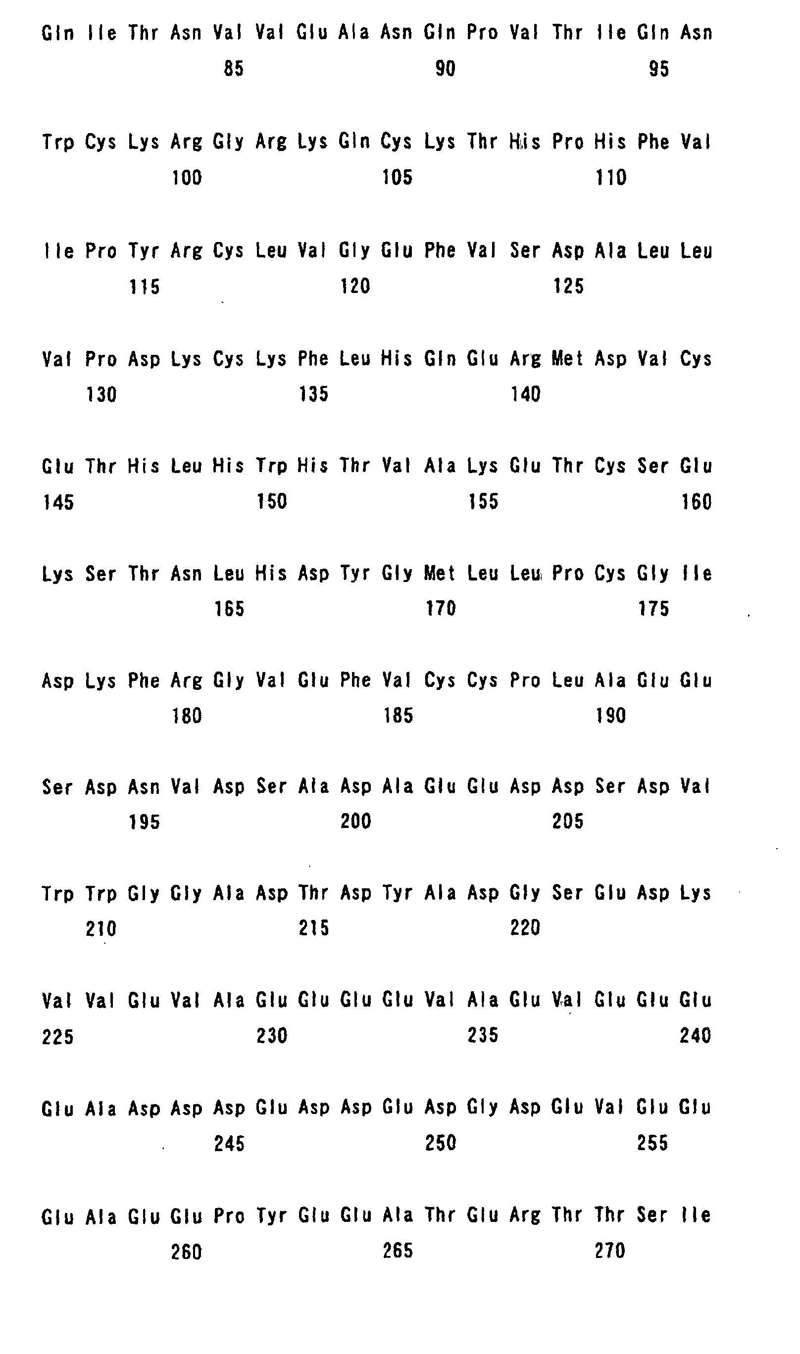
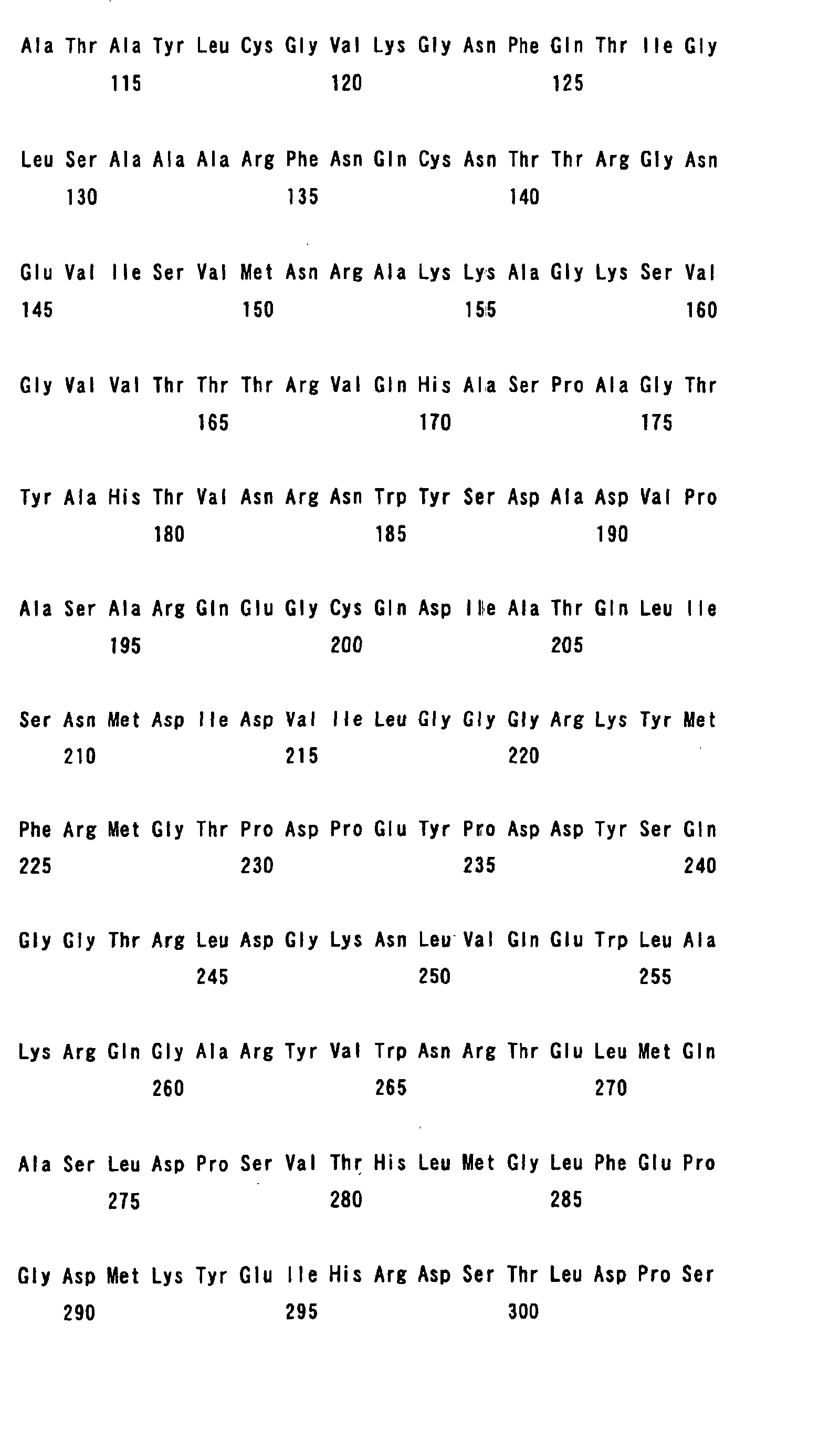
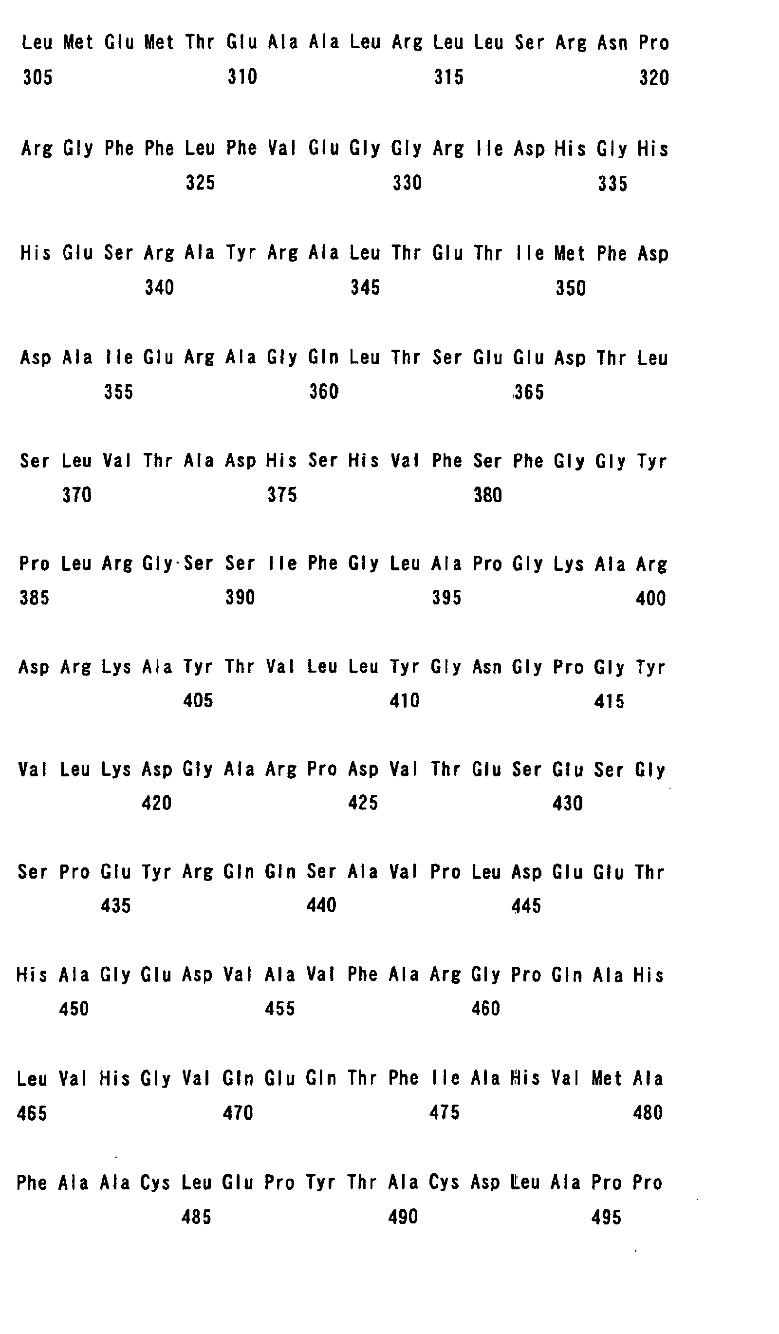
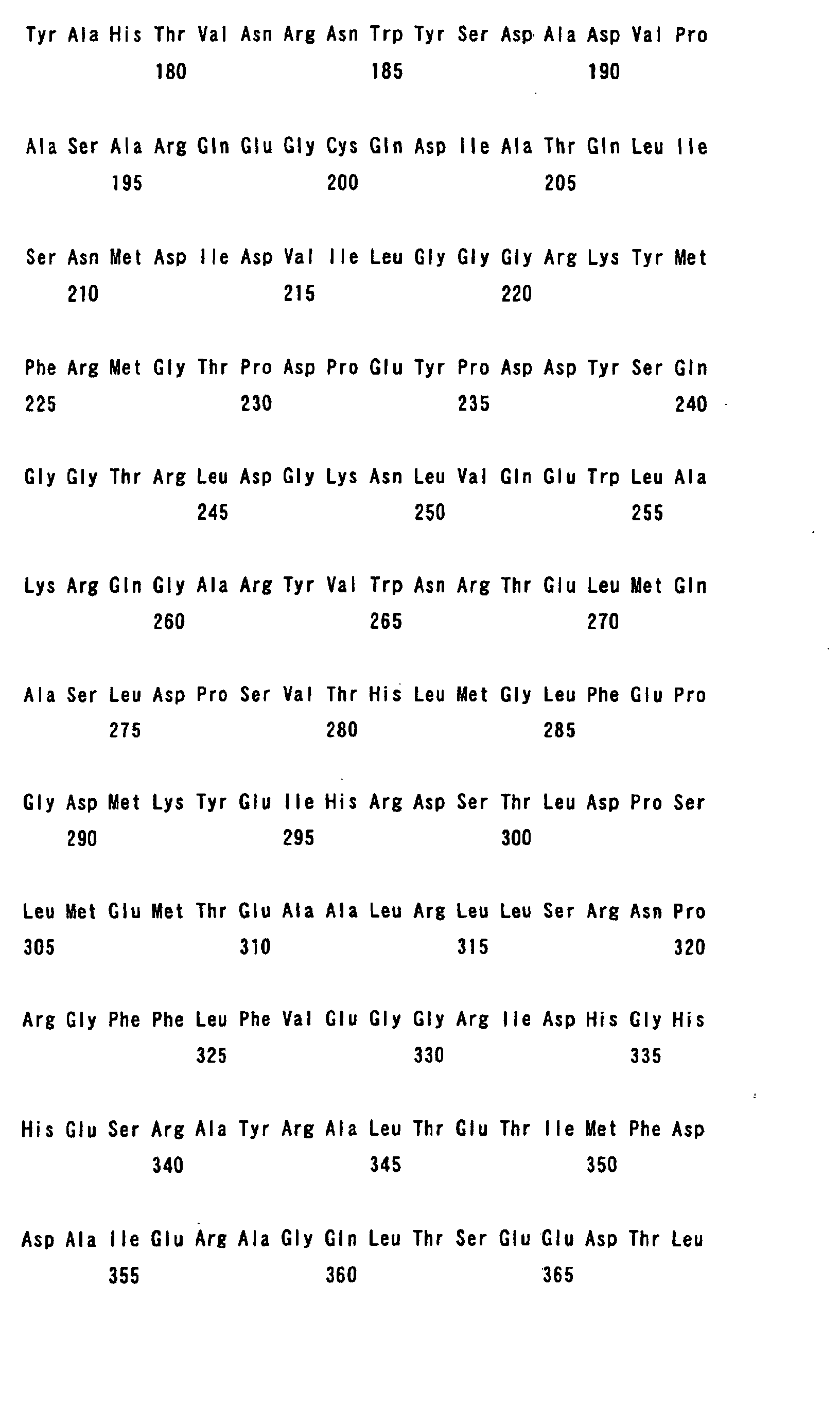
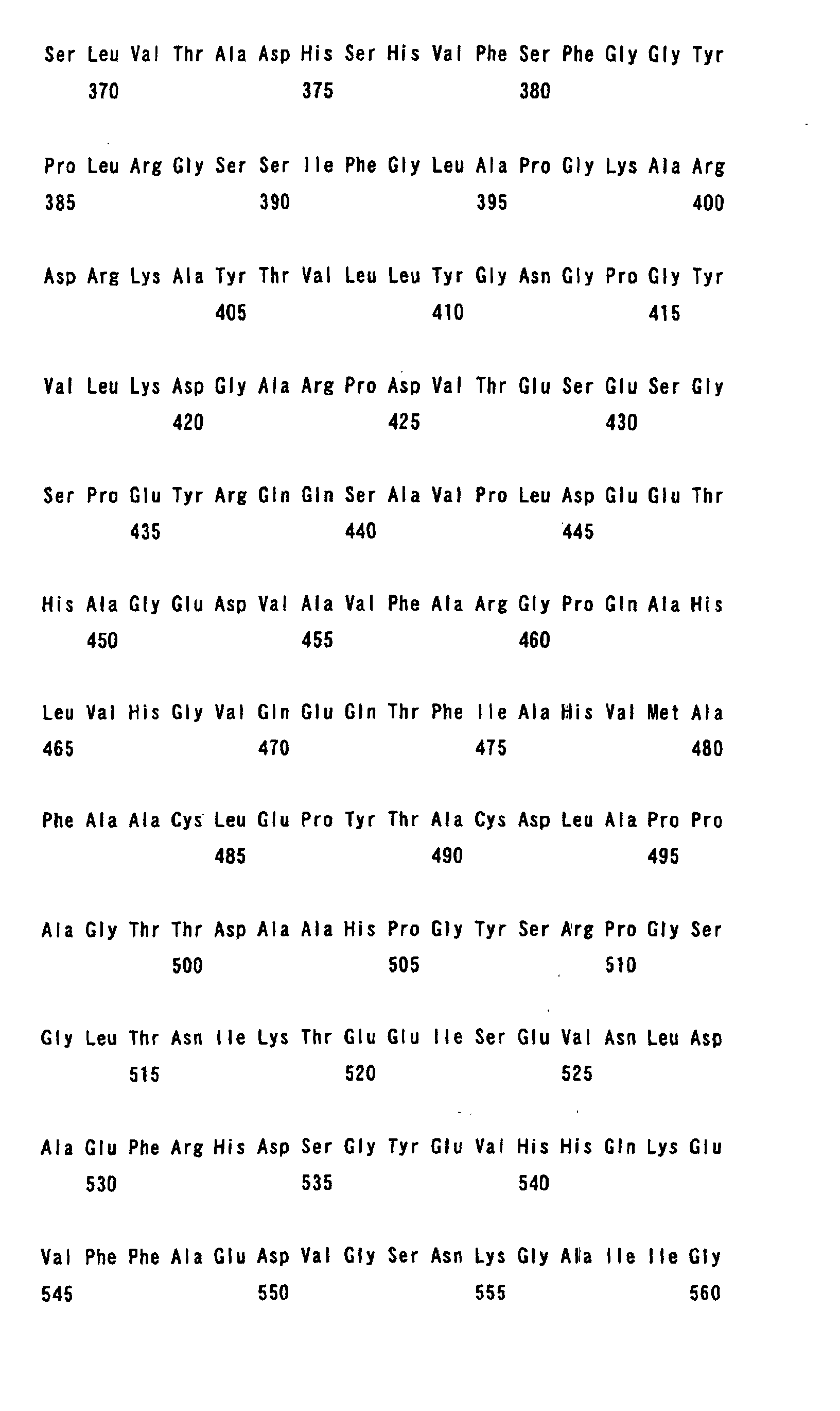
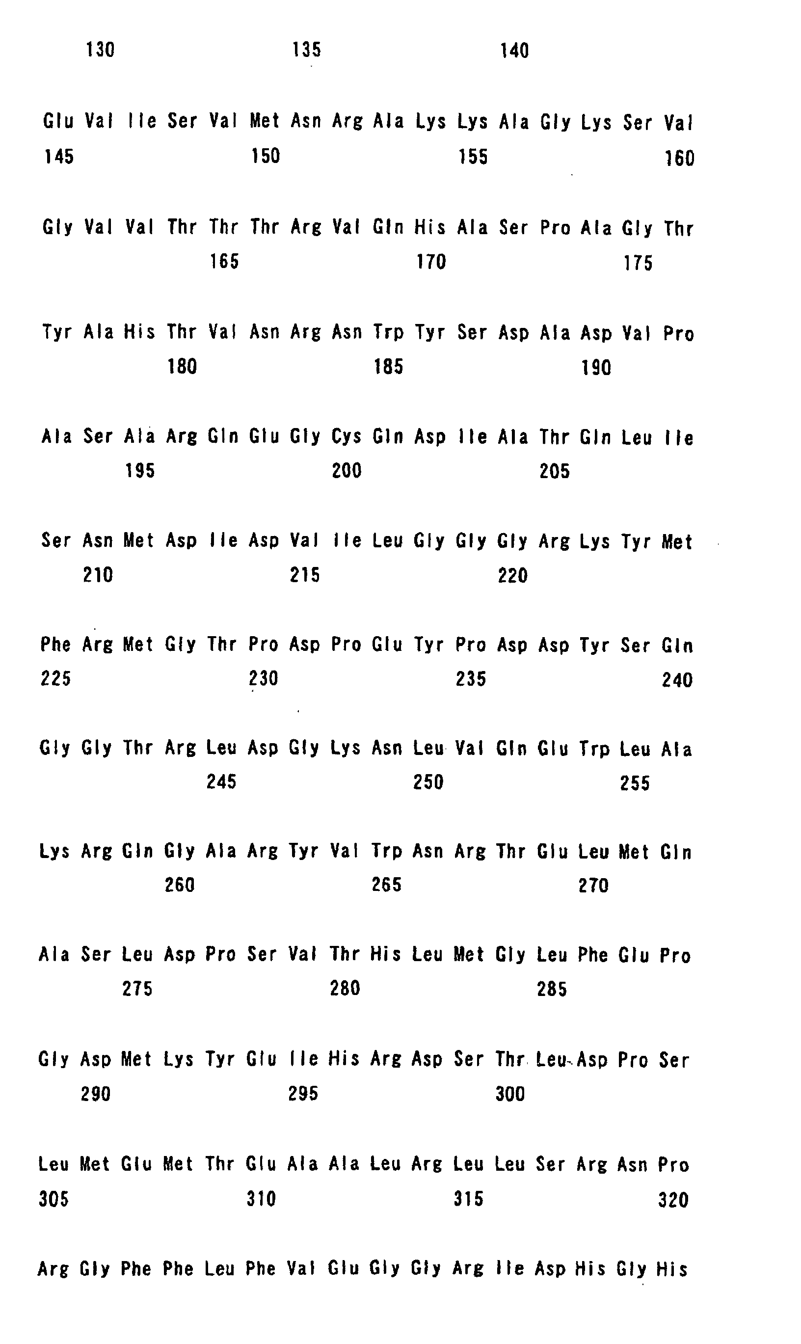
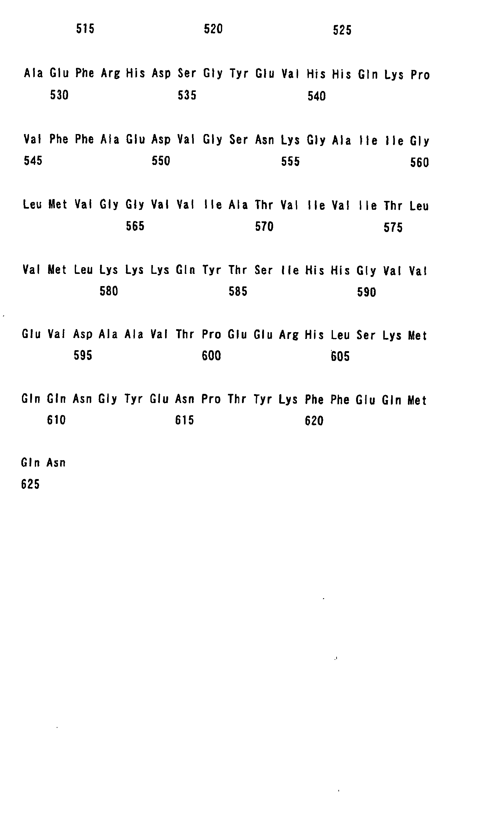
The amino acid sequence of APP695 is shown in SEQ ID
No. 1 of Fig. 2 and a sequence list. The representation of amino
acids, peptides, nucleotide sequences, nucleic acids and the like
by abbreviations in this description is in conformity with the
rules recommended by the IUPAC-IUB, "Guidelines for Writing
Descriptions Containing Nucleotide Sequences or Amino Acid
Sequences" (edited by the Japanese Patent Office), and the
conventions relating to the use of codes or symbols in the art.
A number of molecular species of APP in addition to
APP 695, such as APP751 and APP770, are known to exist. They
are identical to each other in the α-secretase and β-secretase
cleavage sites, and thus APP695 is used to describe the invention.
The APP used for the APP modified protein of the invention, however,
is not limited to APP695.
In the APP modified protein of the invention, there
is no limitation to the length of the amino acid sequence at the
N-terminus to the extent that a region required for exhibiting
the above characteristics is contained and the β-secretase
cleavage is not affected. All of the amino acid sequences at
the C terminus are not necessarily required to the extent that
the β-amyloid production caused by γ-secretase generated after
the β-secretase region is cleaved is not affected.
Taking APP695 as an example, the region of positions
593 to 648 of the APP695 is required to demonstrate the above
characteristics.
596 and 597 positions as a β-secretase recognition site
need to be contained in the region. Since it has been reported
that about 4 residues at the N terminus are necessary for the
β-secretase to recognize APP695 as a substrate (Neuron, 14, 661-670
(1995)), the N terminus should start with at least position 593.
612 and 613 positions as the α-secretase site are also necessary.
When the β-amyloid region (from 597 to 638) for confirming the
production amount of the β-Amyloid is included, positions 593
to 638 are required. Since the APP is a type I membrane protein,
positions 625 to 648 of a transmembrane region are required so
that the APP may be normally fixed to a membrane and maybe undergo
processing in the fixed state. This is why positions 593 to 648,
which include these positions, are mentioned as an example.
Specific examples of proteins containing such a region
include a protein having the C terminal region containing posit ions
581 to 695 of the amino acid sequence of APP695.
In the modified protein of the invention, an amino acid
mutation or mutations which do not affect β-secretase cleavage
and do not cause α-secretase cleavage is referred to as an amino
acid mutation or mutations which do not cause α-secretase cleavage,
and such an amino acid mutation or mutations do not eliminate
nor reduce β-secretase cleavage activity.
Such an amino acid mutation or mutations are caused
by deletion, substitution, addition, etc., of the amino acid at,
for example, the α-secretase cleavage site.
There is no limitation to such an amino acid mutation
or mutations to the extent that the mutation or mutations do not
affect β-secretase cleavage nor cause α-secretase cleavage.
Particularly preferable examples of amino acid
mutations include an amino acid mutation in which the leucine
residue at position 17 counting from the amino acid at the N terminal
β-amyloid is substituted by glutamic acid, proline, alanine, or
lysine, or an amino acid mutation in which the lysine residue
at position 16 is substituted by tryptophan.
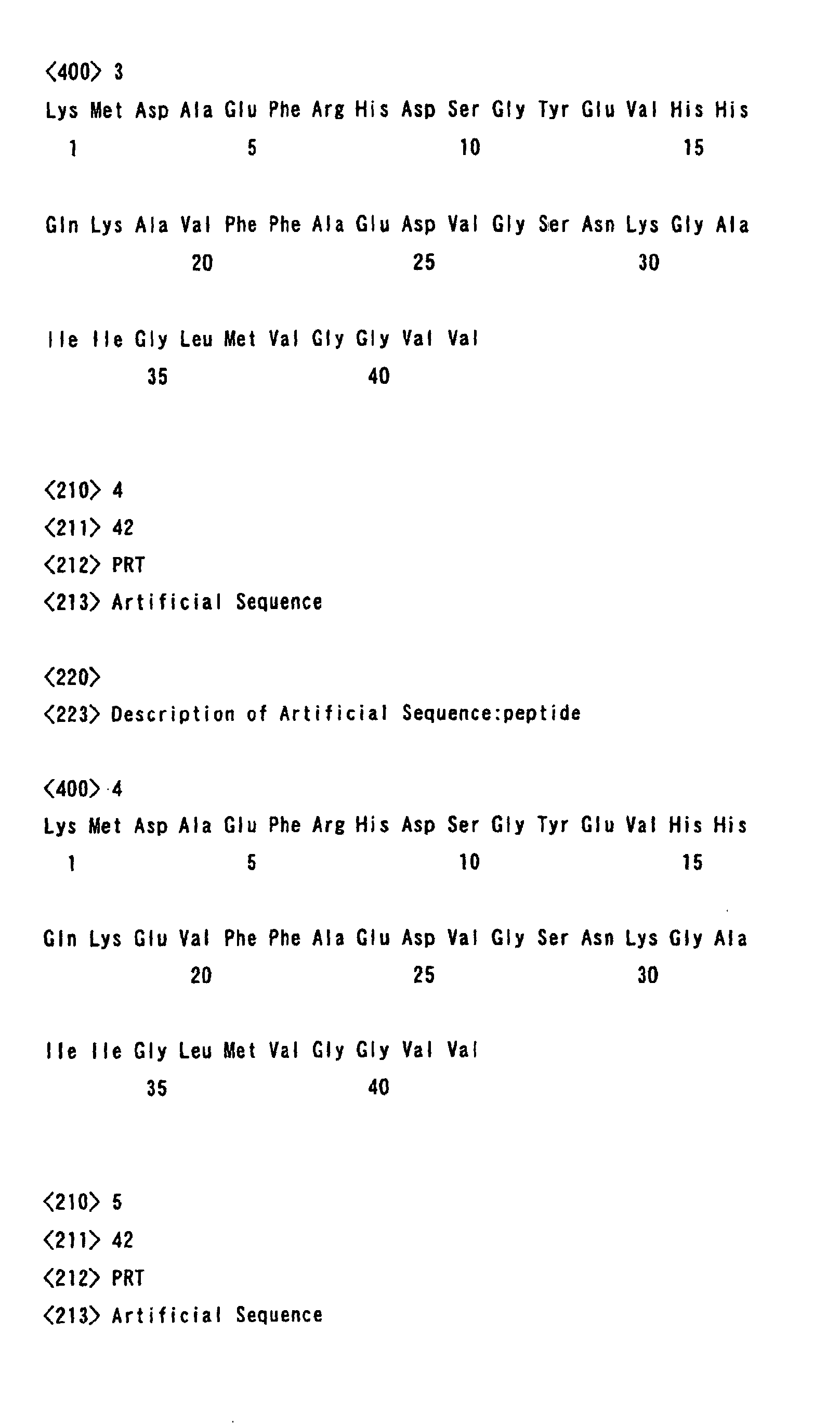
Specific examples of proteins having the above amino
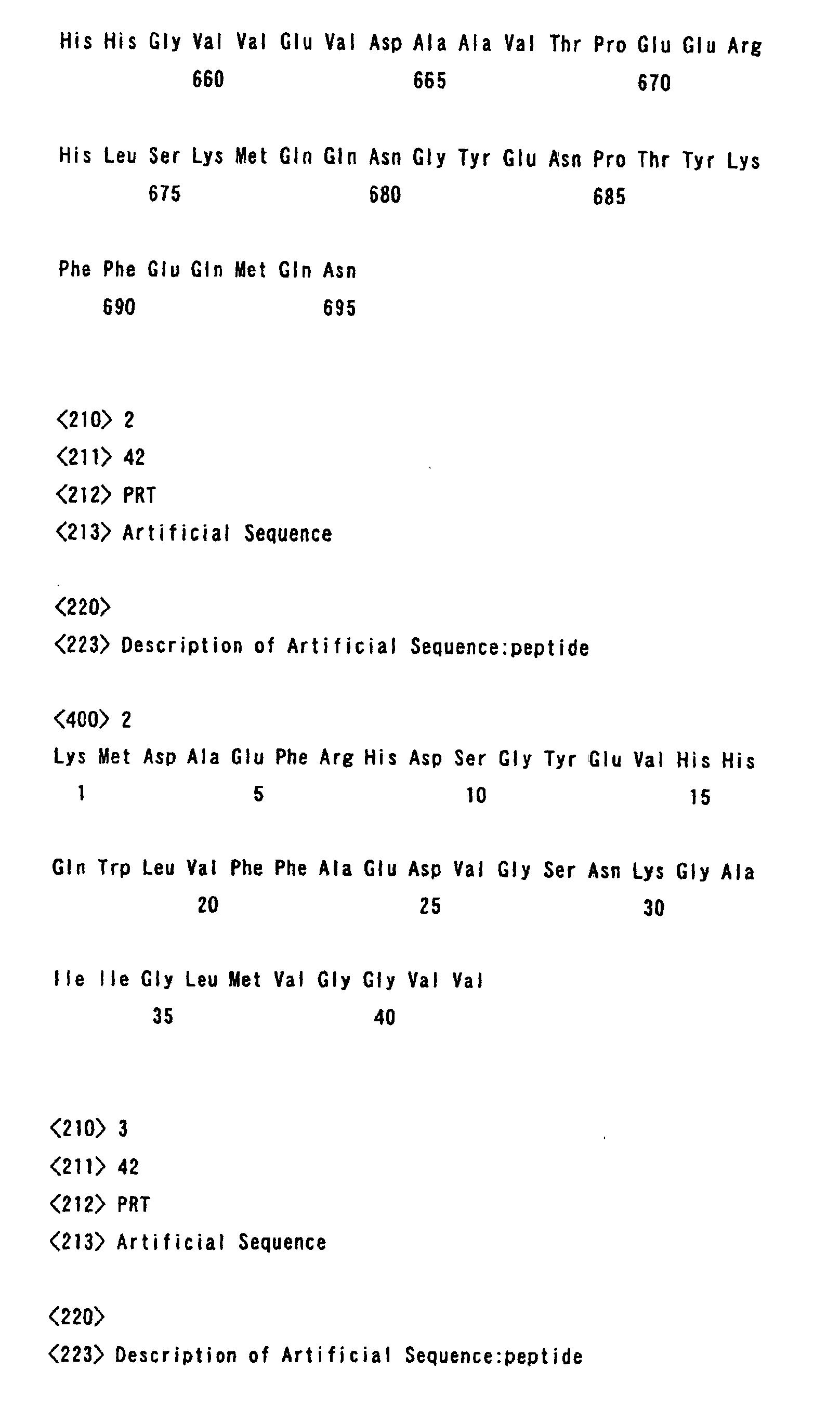
acid mutation include an APP modified protein which has an amino
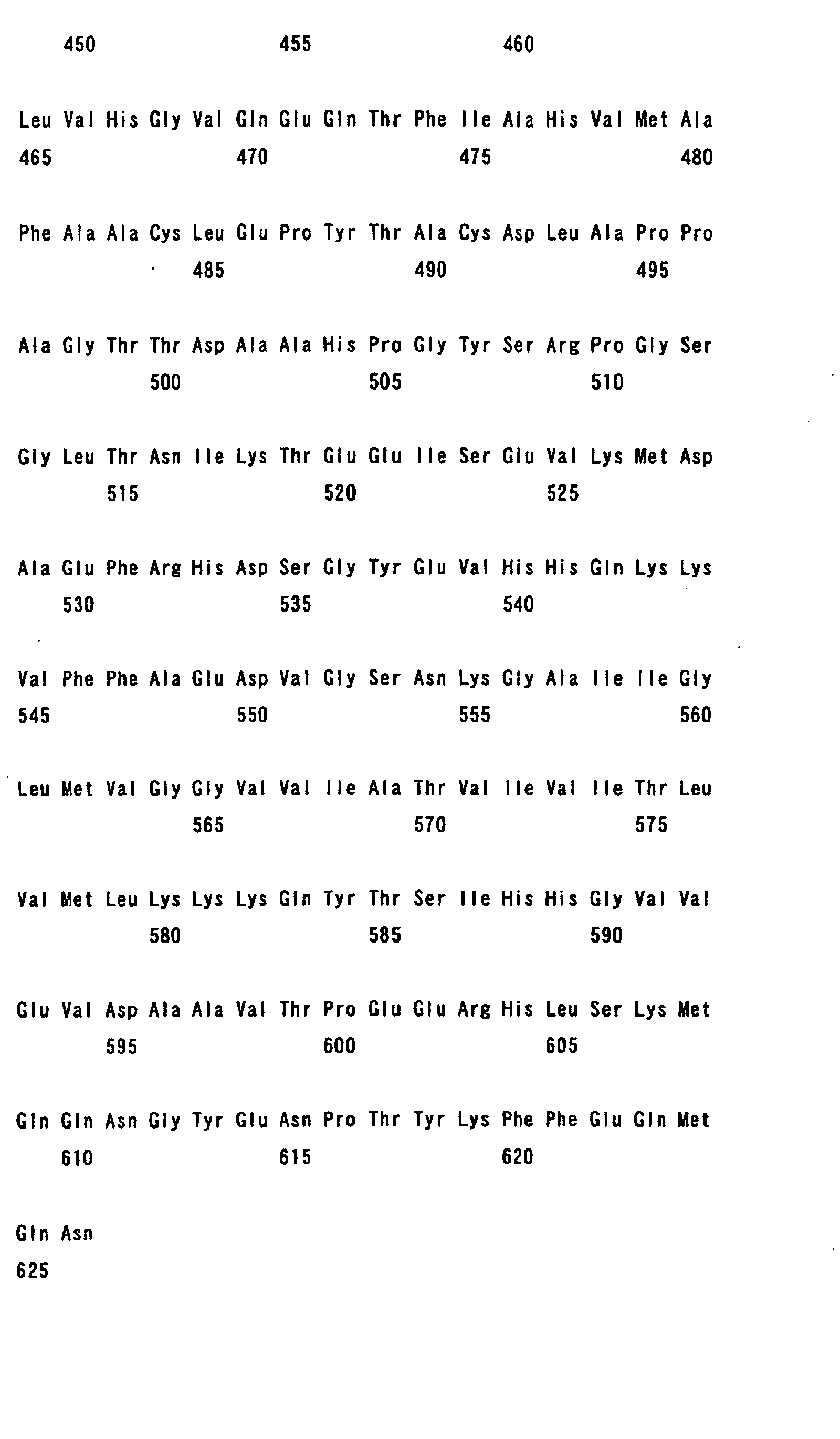
acid sequence of any one of SEQ ID Nos. 2 to 6.
Further, in order to make an attack of β-secretase easier,
the generally-known APP amino acid mutation or mutations in the
hereditary Alzheimer's disease may be introduced into the modified
protein of the invention. Specific examples of the APP amino
acid mutation or mutations in the hereditary Alzheimer' s disease
include a Swedish familial Alzheimer's disease APP mutations.
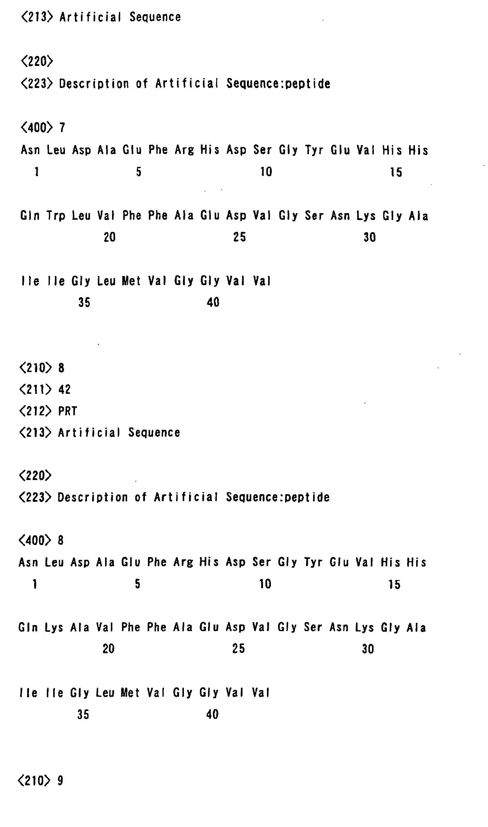
Specific examples of proteins into which such a mutation
or mutations are introduced include modified proteins having an
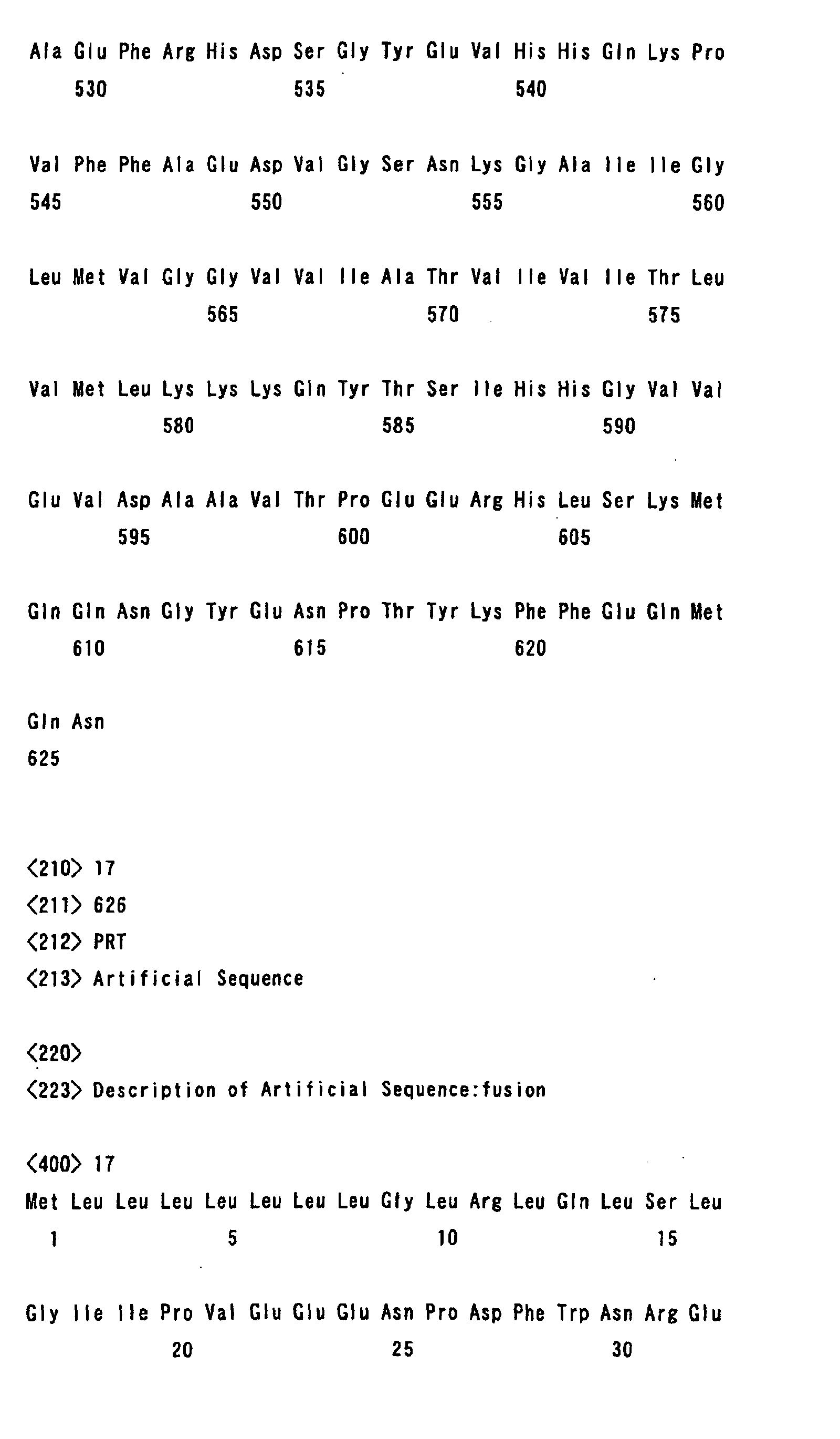
amino acid sequence of any one of SEQ ID Nos. 7 to 11.
The APP modified protein of the invention may be
obtained by modifying a gene encoding an APP protein to a DNA
sequence that encodes the amino acid sequence of the modified
protein, introducing the mutated gene into a suitable expression
vector, to be expressed in a host cell. Subcloning, etc. may
be appropriately performed to acquire the target gene in each
step. For example, an insertion of the target gene into a suitable
cloning vector, and a screening of the target gene by a routine
procedure may be conducted to increase the amount of the gene
itself.
The selection of codons in the DNA molecule
corresponding to the amino acid of an APP modified region is not
limited, and any combination can be accepted to the extent that
the DNA sequence encodes the amino acid sequence. The codons
can be selected in a routine manner. For example, codons can
be selected with reference to the codon usage frequencies in the
host to be employed.
The target DNA molecular arrangement may be acquired
by the DNA amplification method using PCR, for example. The
primers used when PCR is employed can be suitably determined based
on the sequence information of the target amino acid mutation
or mutations, and can be synthesized according to a routine method.
The amplified DNA fragments can be isolated and purified
by a routine method, such as gel electrophoresis, molecular sieving,
etc.
The base sequence of the nucleic acid molecule of the
invention can be determined according to the dideoxy method, the
Maxam-Gilbert method, etc., or determined simply by using a
commercially-available sequencing kit, etc.
The protein is produced based on the sequence
information of the DNA molecule provided by the above DNA molecule
according to a common gene recombination technique (see Sambrook,
J., Frisch, E.F., and Maniatis, T., Molecular Cloning, A Laboratory
Manual, 2nd Edition, Cold Spring Harbor Laboratory Press, 1989;
Current Protocols in Molecular Biology, John Wiley Sons, Inc.
1998., etc.)
More specifically, the protein is produced by preparing
a recombinant DNA (expression vector) in which a DNA molecule
coding the desired protein can be expressed in a host cell,
introducing the recombinant DNA into the host cell to be
transformed, culturing the transformant, and subsequently
collecting such proteins from the obtained culture.
Specific examples of host cells include cells derived
from prokaryotes, cells derived from eukaryotes, etc.
Specific examples of those derived from prokaryotes
include those derived from colibacillus, Bacillus subtilis, etc.
Preferable examples of colibacillus are those belonging to the
genus Escherichia, such as Escherichia coli K12 strain.
Preferable examples of Bacillus subtilis are Bacillus strains,
such as Bacillus subtilis Marburg 168.
When prokaryotic cells are used as a host cell, a
vector which is replicable in the host cell is used, andapromoter,
SD (Shine and Dalgarno) base sequence, and an initiation codon
(e.g., ATG), which is necessary for initiation of the protein
synthesis, are provided upstream of the gene in the vector,
so that the DNA molecule may express.
Various known vectors can be used as the vector
mentioned above, and plasmids derived from Escherichia coli,
such as pBR322, pBR325, pUC12, pUC13, etc., are generally used.
Examples of the vectors for use in expression systems using
E. coli include pGEX-4T (Amersham Pharmacia Biotech), pMAL-C2.
pMAL-P2 (New England Biolabs), pET21, pET21/lacq (Invitrogen)
and pBAD/His (Invitrogen).
There is no limitation to usable promoters. Any
promoter can be used. When a strain of the genus Escherichia
is used as the host, tryptophan (trp) promoters, lpp promoters,
lac promoters, recA promoters, PL/PR promoters, etc., are
preferably used. When the host is a strain of the genus Bacillus ,
SP01 promoters, penP promoters, etc., are preferably used.
Usable cells derived from eukaryotes are cells
derived from mammal, for example, and specific examples of such
cells include the monkey cell line COS [Cell, 23: 175 (1981)],
Chinese hamster ovary cells and a dihydrofolate
reductase-defective cell line thereof [J.Exp.Med., 108: 945
(1958). Proc. Natl. Acad. Sci., 77:4216 (1980), NIH3T3 cells
(J.Viol., 4:549 (1969)), Jurkat cells (J. Immunol., 133:123
(1984), etc. Cells of yeast belonging to the genus
Saccharomyces, etc., are preferably used.
When cells derived from eukaryotes are used as the
host cell, vectors having a promoter upstream of the DNA molecule
to be expressed, an RNA splice site, a polyadenylation site
and a transcription termination sequence are usually used as
the expression vector. This vector may also have a replication
origin as required. A specific example of the expression vector
includes pSV2dhfr harboring an early promoter of SV40 [Mol.
Cell. Biol., 1: 854 (1981)]. In addition to the above, various
known commercially available vectors may be used. Examples
of commercial vectors which are used in expression systems using
animal cells include vectors for animal cells, such as pCI
(Promega), pIND (Invitrogen), pcDNA3.1/His (Invitrogen),
pEF1/V5-His (Invitrogen), etc., and vectors for insect cells,
such as pFastBac HT (GibciBRL), pAcGHLT (PharMingen),
pAcS/V5-His, pMT/V5-His and pMT/Bip/V5-his (all Invitrogen),
etc.
Examples of preferable promoters for use when animal
cells are used as the host cell include SV40-derived promoters,
retrovirus promoters, metallothionein promoters, heat shock
promoters, cytomegalovirus promoters, and SRα promoters,
adenovirus promoters, the elongation factor promoters, etc.
When a yeast is used as the host, pH05 promoters, PGK promoters,
GAP promoters, ADH promoters, etc., are preferably used.
Introduction of expression vectors into the host cell
and transformation caused by the introduction can be carried
out without limitation in accordance with various general
methods, such as an electroporation, liposome method, calcium
phosphate method, DEAE dextran method, etc.
The protein of the invention can be obtained by
culturing the host cells, and appropriately isolating and
purifying the produced proteins.
The protein thus obtained can be used per se for
screening a compound affecting β-secretase.
When the protein is produced using cells derived from
eukaryotes, isolated proteins or the whole cell may be used.
In the coexistence of the test substance and the
protein (or cells expressing the protein), β-secretase is acted
and its influence is measured, thereby screening a substance
which affects β-secretase or the production of β-amyloid.
Proteins can be detected by Western blotting, ELISA
method, or the like, using an antibody which recognizes the
amino acid sequence of APP, for example.
Fusion protein
The fusion protein of the invention denotes a protein
obtained by fusing proteins other than the APP modified protein,
e.g., a detectable secretory protein, into the N-terminal
protein of the above-described APP modified protein of the
invention.
Here, the detectable secretory protein denotes a
protein or a peptide which does not contain the localization
sequence for each intracellular tissue in the amino acid
sequence, and which is released or emitted outside the cell
to demonstrate this function. More specifically, this refers
to a protein which is cleaved at the C terminal region of the
protein by β-secretase and secreted outside the cell.
Preferable examples of detectable secretory proteins
include a protein which can be easily detected using a substrate
like an enzyme; a protein which has physical properties and
thus the amount secreted outside the cell or into the culture
supernatant can be quantatively measured using an instrument,
a reagent, etc.: or a protein which demonstrates a bioactivity
outside the cell. In addition to the above, usable proteins
include those in which the bioactivity resulting from the
physiologically active protein in the culture supernatant can
be determined, or in which the amount of the secretory protein
in the culture supernatant can be generally determined using
the antibody against the secretory protein with ELISA or Western
blotting, for example.
More specifically, secretory alkaline phosphatase,
β-galactosidase, peroxidase, luciferase, etc., can be
mentioned. These can be quantified as the amount of enzyme
activity by, for example, adding an enzyme substrate in the
culture supernatant.
Other examples include a green fluorescence protein
(GFP), aequorin, immunoglobulin, growth hormone, histidine tag,
c-myc tag, Flag tag, hemagglutinin (HA) tag, glutathione S
transferase (GSP) tag, T7 tag, V5 epitope tag, X-press tag,
BE11 tag, etc.
Furthermore, a physiologically activating protein
group belonging to cytokine, for example, interleukin 2, can
be mentioned. The protein released outside the cell can be
detected by measuring the bioactivity in a culture medium or
by measuring with a commercially-available ELISA kit.
Although these secretory proteins are desirably
produced and secreted as a secretory-type protein having an
original signal peptide, they can be secreted using a signal
peptide of the secretory protein of APP and other secretory
proteins.
These secretory proteins can be partially modified,
deleted, and inserted to the extent that they do not
significantly affect the detected enzyme activities,
immunoreactivity, bioactivity, etc.
The APP modified protein encompassed in the fusion
protein of the invention is a protein subjected to the
above-described amino acid mutation or mutations such that the
β-secretase cleavage site is contained, the β-secretase
cleavage is not affected, and the α-secretase cleavage does
not arise.
There is no limitation to the amino acid mutation
or mutations to the extent that the amino acid mutation or
mutations do not affect the β-secretase cleavage nor cause the
α-secretase cleavage. Particularly preferable is an amino
acid mutation in which the leucine residue at position 17
counting from the N terminal amino acid of β-amyloid is
substituted by glutamic acid, proline, alanine, or lysine, or
the lysine residue at position 16 is substituted by tryptophan.
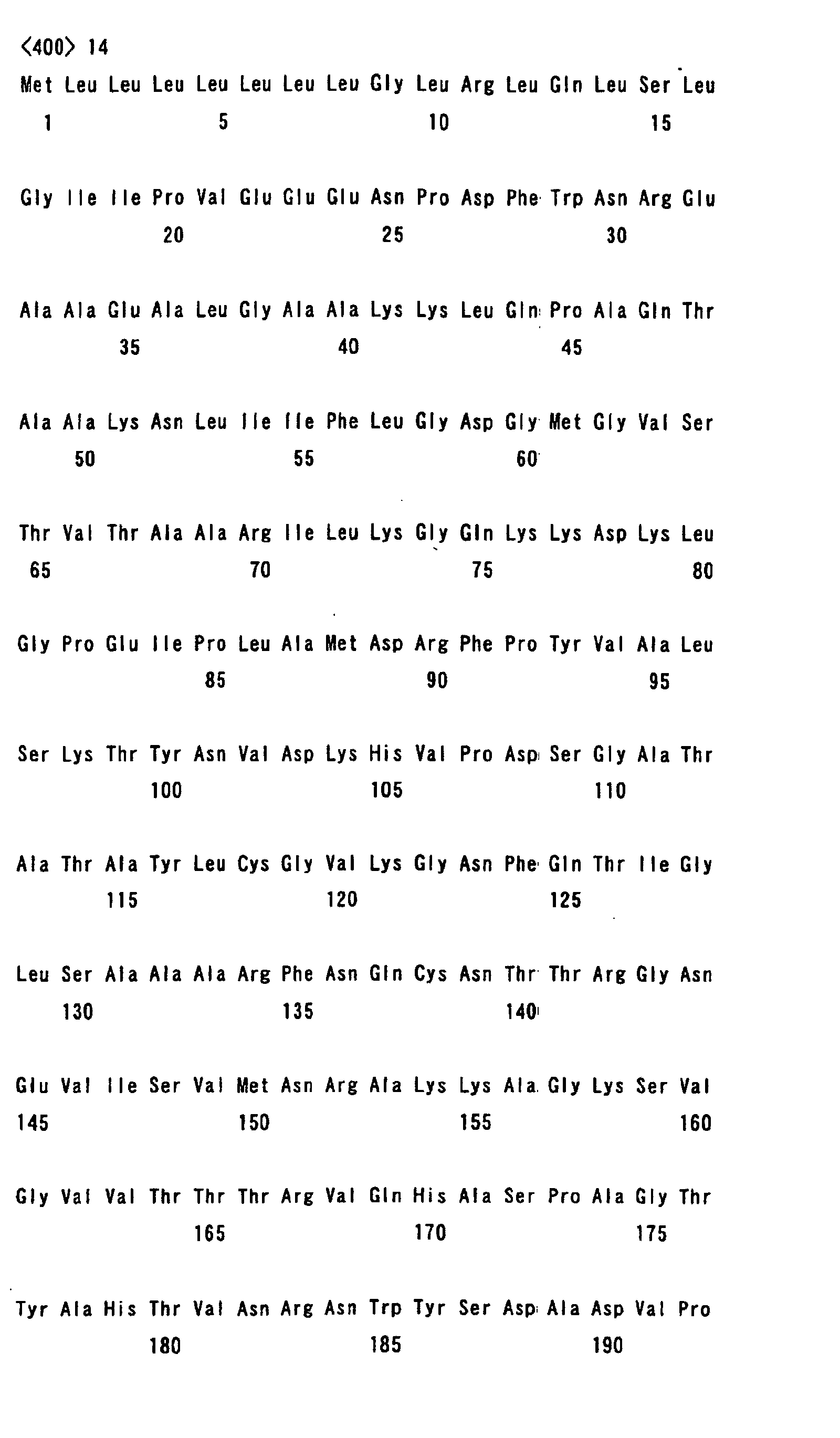
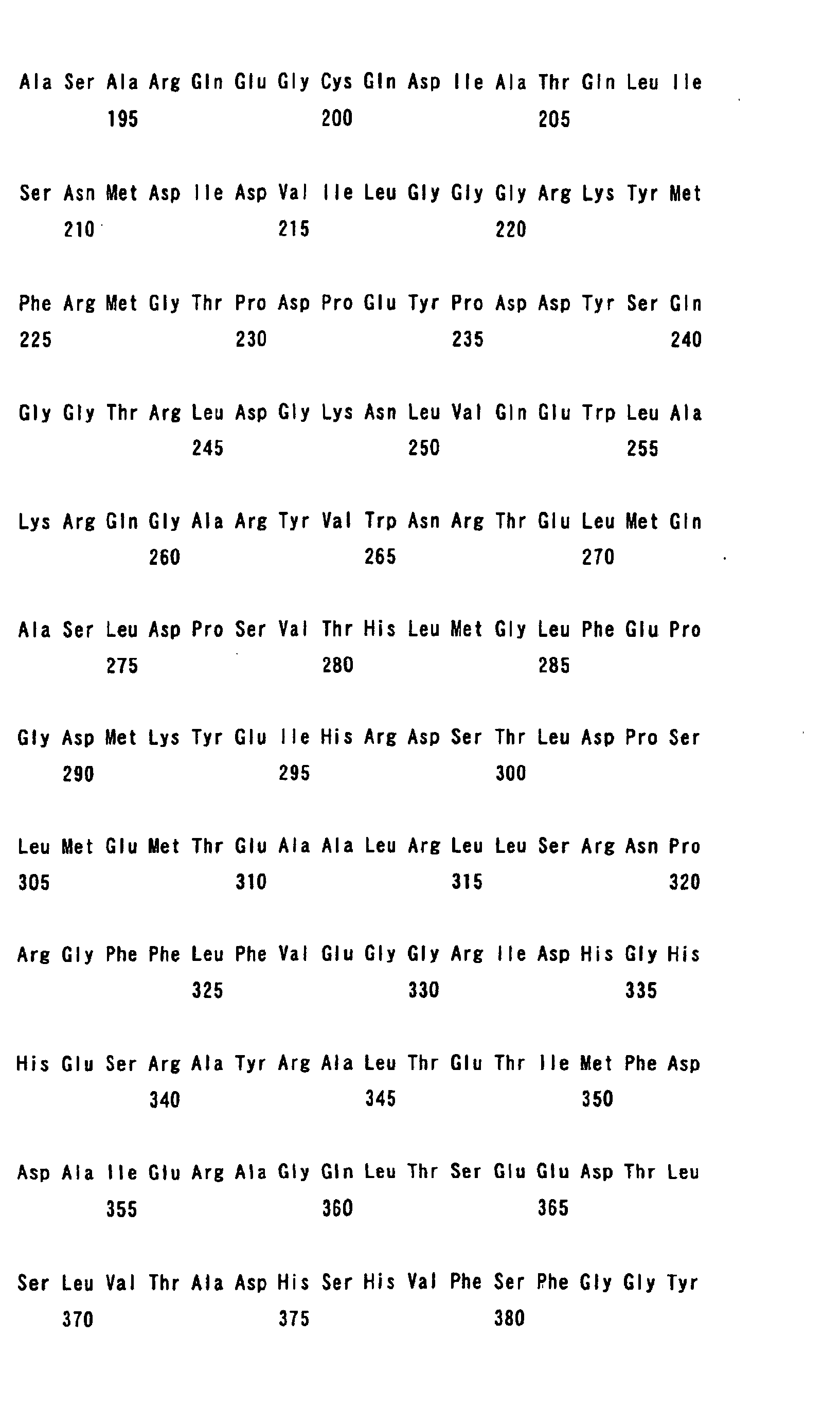
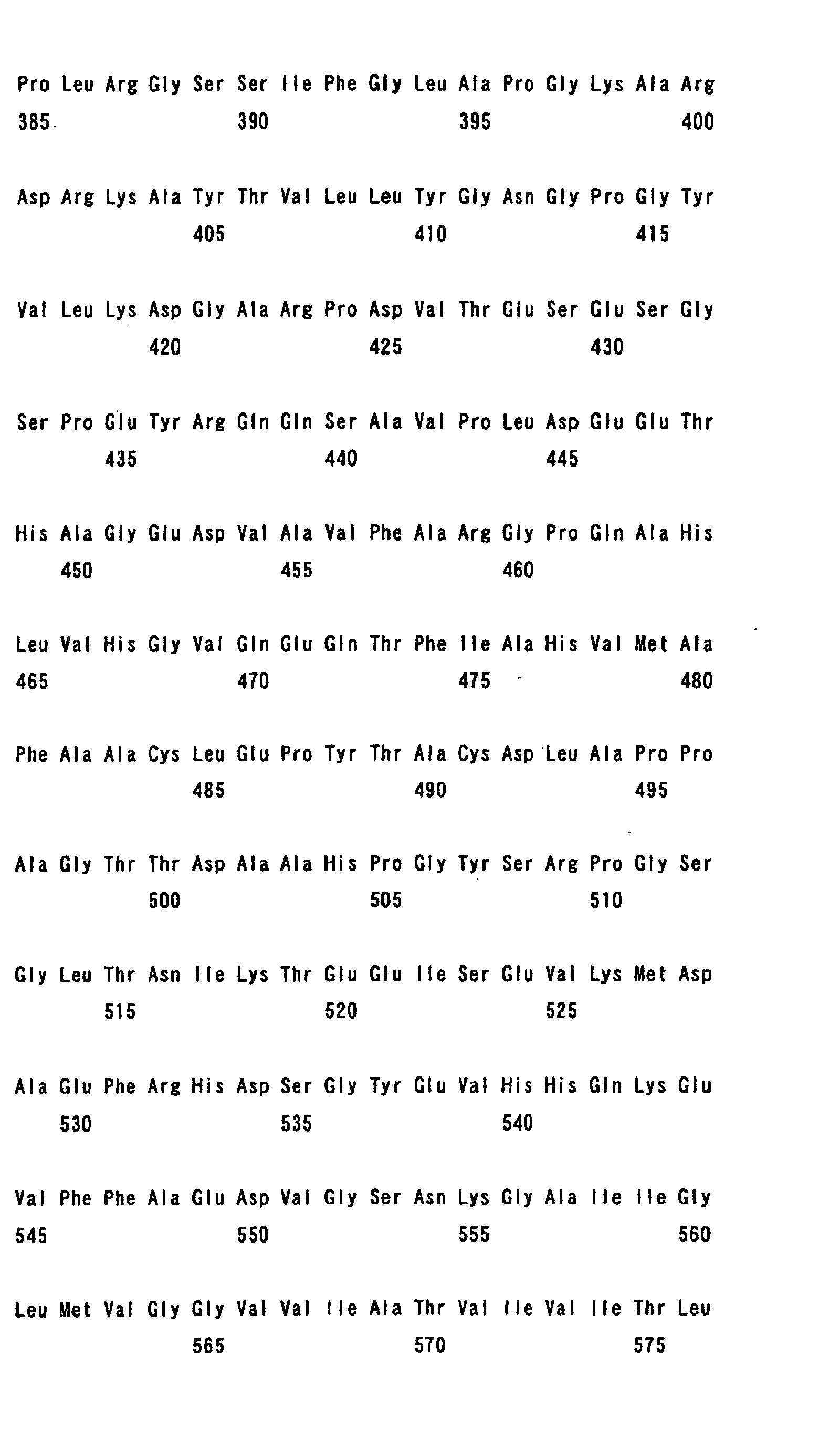
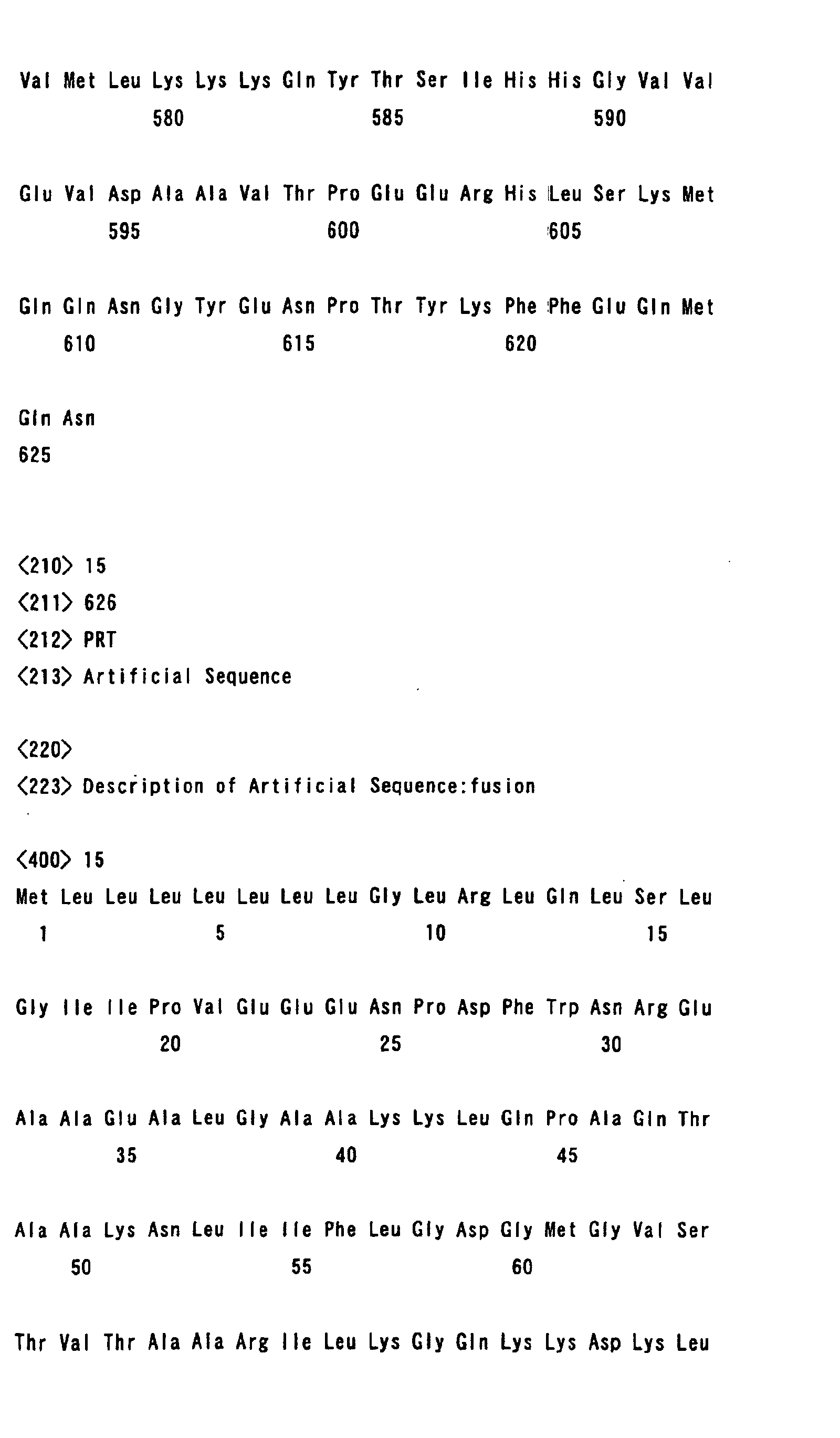
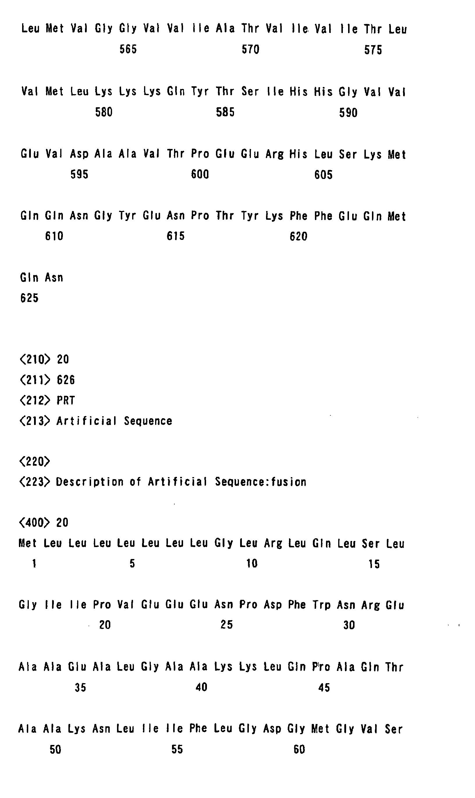
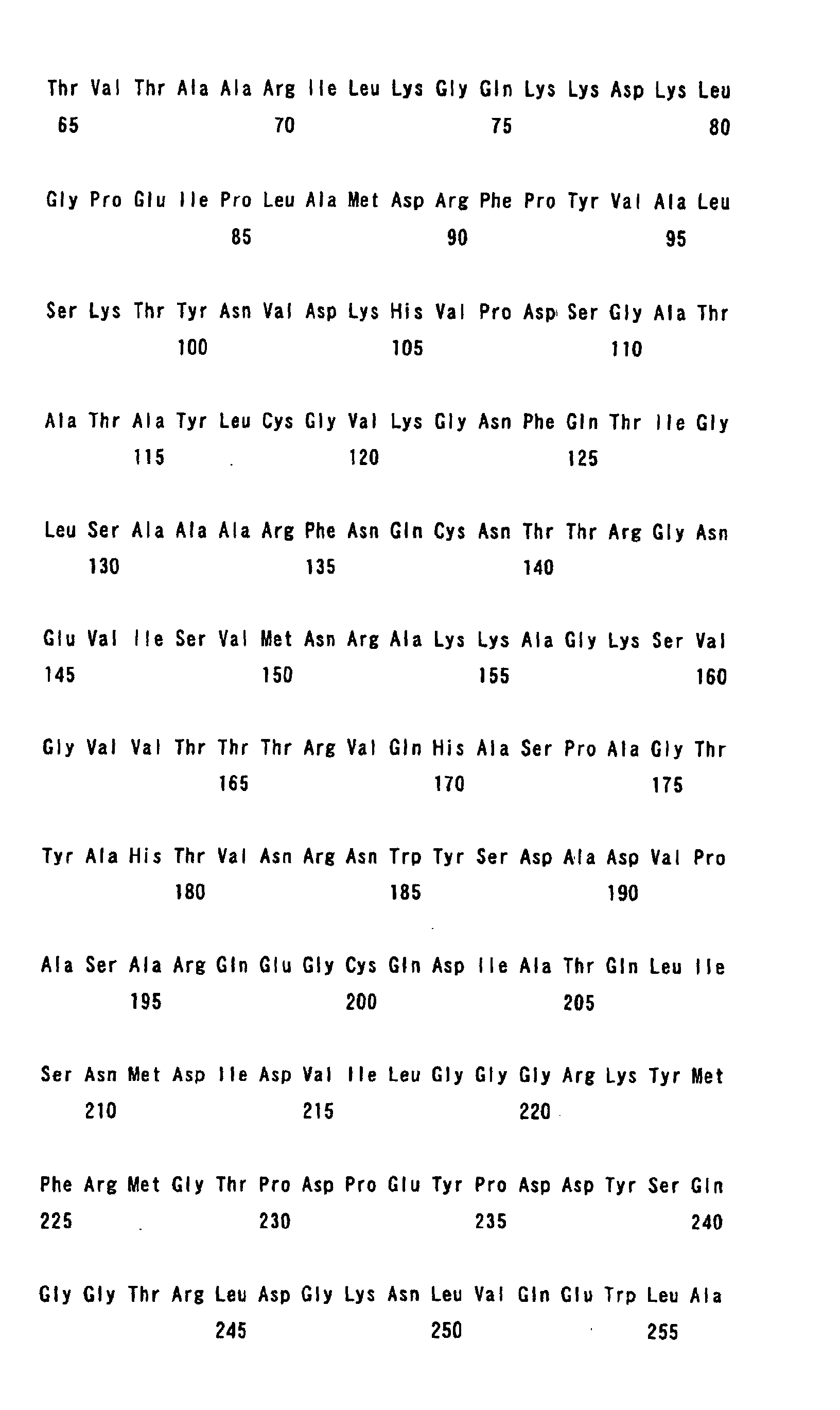
Specific examples of fusion proteins of the invention
include those having an amino acid sequence of any one of SEQ
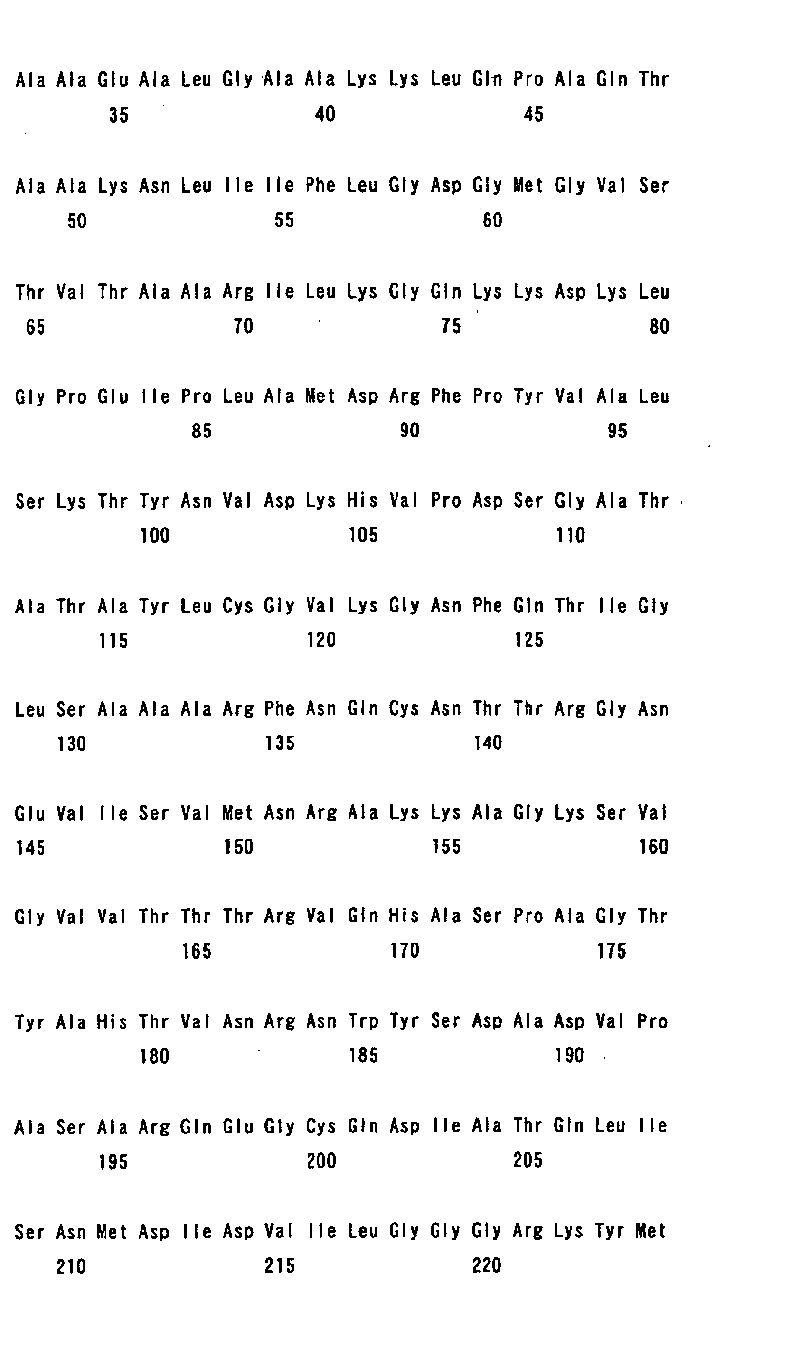
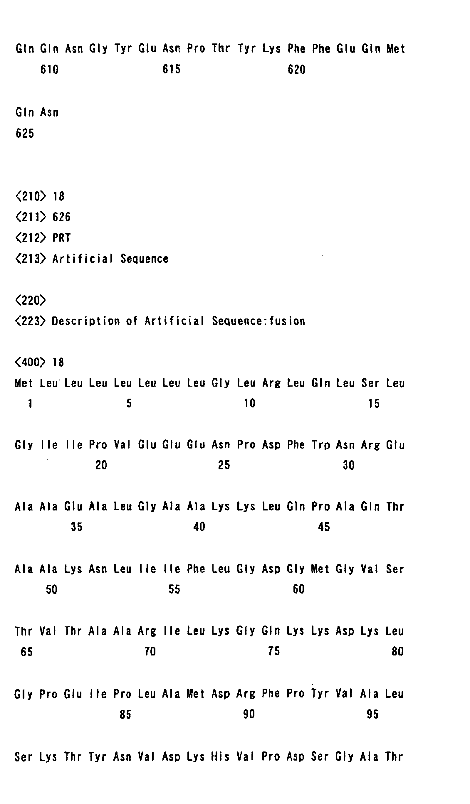
ID Nos. 12 to 16.
Further, in order to make the β-secretase attack easier,
a generally-known APP amino acid mutation or mutations in
hereditary Alzheimer's disease, such as a Swedish familiar
Alzheimer's disease APP mutations, may be introduced into the
modified protein of the invention.
Specific examples of fusion proteins include those
having an amino acid sequence of any one of SEQ ID Nos. 17 to
21.
In the fusion protein, the N terminal region of the
APP modified protein may be extended, or a spacer may be suitably
inserted between a secretory protein and an APP modified protein,
as required, so that the β-secretase cleavage site in the APP
does not have a steric hindrance that affects the attack of
the β-secretase.
The fusion protein can be produced from a gene which
encodes the above-described APP modified protein of the
invention and a gene which encodes secretable protein according
to the genetic recombination procedure in the same manner as
in the above-described production process for the APP modified
protein of the invention.
Test Cell
Hereinafter, the test cell of the invention which
stably expresses the above described APP modified protein or
fusion protein of the invention is described.
The test cell used in the invention denotes a cell
used for a test to measure the effect, toxicity, etc., of a
compound and to evaluate the measurement results.
The test cell can be produced using the same genetic
recombination procedure and materials (expression vector) as
previously described.
In the cell used as the test cell, it is preferable
that the gene expression mechanism, protein expression
mechanism, and secretion mechanism are similar to human
components or human mechanisms. As the cell expressing a fusion
protein, it is preferable to use, for example, a cell with a
mechanism such that secretory proteins are released outside
the cell after cleavage at the APP site. The cell which has
a rapid growth rate and maintains the stability even after
subculturing is preferable. These include, for example,
human-derived cells or mammalian-derived cells such as CHO-K1
cell (J. Exp.Med., 108: 945 (1958), a Hela cell (Cancer Res.,
12: 264 (1952)), a NIH3T3 cell (J. Viol. , 108: (1958)), a Jurkat
cell (J. Immunol., 133: 123 (1984)), a 293 cell (J. Gen.Viol.,
36: 59 (1977)), etc.
The following kind of procedure may be applied to
obtain a cell strain with stable expression as a test cell.
For example, usable is a method comprising integrating a drug
resistance gene, such as aminoglycoside phosphotransferase
(Th5neo), hygromycin B phosphotransferase (hygr), xanthin
guanine phosphoribosyl transferase (Eco-gpt), dihydrofolate
reductase (dhfr), etc., into an expression vector, introducing
the target gene-containing expression vector into a cell, and
cultivating it in a medium containing the selective drug, such
as G418, hygromycin, a HAT, and a methotrexate. For example,
pSV2neo, pRSVneo, pHyg, pSV2gpt, or pSV2dhfr is used as an
expression vector.
In order to screen a highly expressive cell strain,
secreted proteins in the culture supernatant or fusion proteins
may be measured. For example, the enzyme activity in the culture
supernatant may be measured. Detection using the antibody
recognizing the C terminal region of APP is performed according
to Western blotting or ELISA. Measurement by the Northern
blotting method or RT-PCR method is performed at the gene
expression level.
Measurement method
The measurement method of the invention comprises
processing the above-described test cell with a subject
substance, and measuring detectable secretory proteins which
are cleaved at the β-secretase cleavage site and are secreted
outside the cell.
The following processes can be adopted. The test
cells are initially suspended in a culture medium, and are then
seeded on a suitable culture plate. The seeded cells are
incubated, as required, for a given period of time until they
are stabilized. Any plate can be used by considering the number
of seeded cells and the measurement sensitivity of the secretory
proteins. For example, a 96-well plate is suitable.
The subject substance is then added to an appropriate
solvent and dissolved, and the solution is added to a plate
as a fixed concentration. In this case, amphiphilic solvents,
such as DMSO, may be used when the subject substance is a
fat-soluble compound or an insoluble compound. Water-soluble
solvents, such as physiological saline, can be used when the
subject substance is a hydrophilic compound. The concentration
of the solvent used is preferably adjusted to a degree such
that the cells are not adversely affected. After the subject
substance is added, incubation is conducted for a
given-period-of-time. The incubation period can be determined
without limitation according to a test system to the extent
that the subject substance is incorporated into a cell and
sufficiently reacted with a target molecule.
After the incubation is complete, a given quantity
of the culture supernatant is taken, and the quantity of the
secretory proteins in the supernatant is measured according
to the properties. When the secretory protein is, for example,
an enzyme, a substrate is added to measure the enzyme activity.
When the secretory proteins have characteristic physical
properties, such as a specific absorption spectrum, an
absorbance meter is used for measurement. The target proteins
may be quantified by monitoring them through a separation
operation using HPLC, etc., as required. When a secretory
protein is a physiologically active protein, such as cytokine,
the bioactivity resulting from the proteins in the culture
supernatant may be measured. The measurement and
quantification may be made using the commercially-available
ELISA kit, etc.
The existence form of a fusion protein expressing
in a cell may be monitored. More specifically, it can be directly
confirmed whether or not a fusion protein is cleaved at the
β-secretase cleavage site by measuring the amount of 99-amino
acid C-terminal fragment (C99) which is produced by the
β-secretase cleavage. For example, after lysis of cell, C99
can be detected and quantified using an antibody which
recognizes C99 according to Western blotting or ELISA. The
above-described measurement method of C99 is applicable not
only to the expression cell of a fusion protein but to the
expression cell of an APP modified protein. Therefore, the
cell can be used for screening a compound affecting β-secretase
activity, a compound affecting β-amyloid production, or a
substance usable as the active ingredient for a therapeutic
or prophylactic agent of Alzheimer's-type dementia.
The effect of the subject substance on the β-secretase
activity or the β-amyloid production can be determined based
on a comparison between the measurement value obtained when
the subject substance is added according to the above-described
measurement method using the modified protein of the invention
and the base value obtained when the subject substance is not
added (solvent only). More specifically, when the measurement
value is lower than the control value, the β-amyloid production
is judged to be suppressed. In contrast thereto, when the
measurement value is higher than the control value, the β-amyloid
production is judged to be promoted.
Whether or not the subject substance affects the
β-amyloid production is judged by performing statistical
analysis, i.e., a significant difference test.
Whether or not the subject substance affects the
β-secretase activity can be examined by the above-described
procedure. Further, it is possible to screen the substance
as a candidate for the active ingredient for a therapeutic or
prophylactic agent of Alzheimer's-type dementia.
The measurement method of the invention may further
comprise a step of examining whether or not the subject substance
shows toxicity to a cell.
Whether or not the subject substance has toxicity
to a cell can be judged by culturing the test cell with the
subject substance, measuring the detectable secretory protein,
and then measuring the survival degree of the cultured test
cells. The number of survival cells can be determined by
measuring the number of cells dyed by, for example, calcein
AM. The number of dead cells can be determined by measuring
the number of cells dyed by, for example, propidium iodide.
Alternatively, the number of dead cells can be measured by
counting the number of cells dyed by trypan blue. Alternatively,
the number of survival cells may be determined by colorimetry
measuring the formazan dye of the tetrazolium salt (MTT, MTS,
WST-1, WST-8, etc.) produced by the survival cells. The number
of dead cells may be determined by measuring the lactate
dehydrogenase (LDH) which has flowed into the culture
supernatant due to cell death, such as apotosis. Of course,
these determination methods are not limited to the above.
BRIEF DESCRIPTION OF DRAWINGS
Fig. 1 schematically illustrates an APP structure
and APP processing.
Fig. 2 shows an amino acid sequence of APP 695. The
underlined portion shows the amino acid sequence of β-Amyloid
(1-42). The shaded portions, MD and KL, denote the sites to
be cleaved by β-secretase and α-secretase, respectively.
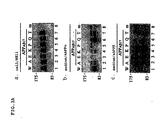
Fig. 3A shows the result of Western blotting. This
shows that the amino acid substitution at the α-secretase
cleavage site (position 17) in APP affects the β-secretase
cleavage.
Fig. 3A(A) shows the result of a process comprising
carrying out electrophoresis of 8% SDS-PAGE after cytolysis,
and conducting Western blotting. A full-length APP was
detected by a monoclonal antibody against a BE11 epitope tag.
Fig. 3A(B) shows the result of a process comprising
electrophoresing a culture supernatant, and conducting Western
blotting in the same manner as in the above A. A monoclonal
antibody (anti-sAPPα antibody) which recognizes the amino acids
of the region between the α-secretase and β-secretase cleavage
sites was used for detection.
Fig. 3A(C) shows the result of a process comprising
electrophoresing a culture supernatant, and conducting Western
blotting in the same manner as in the above B. An antibody
(anti-sAPPβ antibody) which recognizes the APP N terminal region
from N terminus to position 596 was used for detection.
Fig. 3B schematically illustrates an antibody
recognition site.
Fig. 4A shows the alkaline phosphatase activity of
the culture supernatant of a CHO K1 cell line stably expressing
SEAP-APPsw(CHO/SW).
Fig. 4B shows cell proliferation of a CHO K1 cell
line stably expressing SEAP-APPsw (CHO/SW) by MTT assay.
Figs. 4C and 4D show the production amount of β-amyloid
in the culture supernatant.
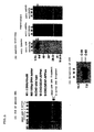
Fig. 5 (A) shows confirmation by PCR of whether or
not the gene which encodes the fusion protein of the invention
is introduced into the cell in the selected clone.
Fig. 5(B) shows confirmation by Western blotting of
whether or not fusion proteins are actually synthesized, and
are then cleaved only at the target site to be secreted in the
culture supernatant in the selected clone.
Fig. 5(C) is a view showing the result of confirming
an intracellular C99 by Western blotting. The obtained result
shows whether or not fusion proteins are actually synthesized,
and are then cleaved at the target site in the selected clone.
Fig. 6 shows examples the result of the screening
in accordance with the invention.
Fig. 6 (a) shows the result of determining the alkaline
phosphatase activity in the culture supernatant on 650 nm by
colorimetry.
Fig. 6 (b) shows the measurement result of the number
of cells by MTT assay.
Fig. 6 (c) shows the evaluated alkaline phosphatase
activity in numerical values for the case in which the subject
substance-free condition is set to 100.
Fig. 6 (d) shows the evaluated number of cells by
MTT assay in numerical values for the case in which the subject
substance-free condition is set to 100.
BEST MODE FOR CARRYING OUT THE INVENTION
Hereinafter, the present invention is described with
reference to Examples, however it is not limited thereto.
Example 1: Construction and expression of APP modified protein
gene
BE11-hAPP695 was used as a starting material which
was produced by adding a BE11 epitope tag immediately after
a signal peptide of the N-terminus of a human-type APP gene
(Fig. 2) and incorporating it into a vector. With respect to
the amino acid at the N-treminus of the α-secretase cleavage
site of APP695 (lysine at position 16 of β-amyloid) or the amino
acid at the C-terminus (leucine at position 17 of β-amyloid),
tryptophan was introduced into position 16 and alanine, glutamic
acid, lysine, proline, glutamine, and threonine were introduced
into position 17 by the site-directed mutagenesis (QuickChange,
Stratagene). A construct was produced in which these mutated
APPs and wild-type APPs are respectively incorporated into a
pCIneo mammalian expression vector (Promega), and the construct
was then transfected to a COS7 cell by a liposome method (DOTAP,
Boeringer Manheim), to achieve transient expression.
Forty-eight hours after transfection, the cell and culture
supernatant were collected, and the expressed proteins and the
processing were analyzed by 8% SDS-PAGE/Western blotting. The
expression of a cell membrane-associated full-length APP
confirmed using the monoclonal antibody (Mab BE11) against BE11.
The expression level was standardized, and then α-secretase
cleavage products (sAPPα) and β-secretase cleavage products
(sAPPβ) which were secreted in the culture supernatant (medium)
were detected and quantified using a specific antibody against
the respective products. The results are shown in Fig. 3 and
Table 1.
| | α-cleavage | β-cleavage |
| Trp (position 16) | ↓↓ | → |
| Ala (position 17) | ↓ | → |
| Glu (position 17) | 0 | → |
| Pro (position 17) | 0 | → |
| Lys (position 17) | ↓↓ | ↑ |
In Table 1, " ↓ " indicates a reduced amount of
secretase cleavage product, "→" indicates an unchanged amount
of each secretase cleavage product, " ↑ " indicates an increased
amount of secretase cleavage product, and "0" indicates no
detection of each secretase cleavage product.
In Fig. 3, the reference characters, A, E, K, P, Q,
and T denote amino acid mutated to alanine, glutamicacid, lysine,
proline, glutamine, and threonine, respectively. The left end
"W" denotes a wild type and the right end "m" means "mock",
i.e., a vector only.
As can be seen from the result shown in Fig. 3 or
Table 1, the amount of α-secretase cleavage product decreased
when position 17 was replaced with alanine. The amount of
α-secretase cleavage product sharply decreased when position
16 was replaced with tryptophan. No α-secretase cleavage
product was detected when position 17 was replaced with glutamic
acid or proline.
The β-secretase cleavage was not affected by these
mutations.
When position 17 was replaced with lysine, the amount
of α-secretase cleavage product decreased and the amount of
β-secretase cleavage product slightly increased.
It was confirmed based on the above results that the
protein introducing these mutations of the invention was
effectively used for efficiently screening substances which
affect β-Amyloid production.
Example 2: Construction of a fusion protein gene and formation
of a test cell
(1) Construction of a fusion protein gene
A fusion protein was constructed using an APP modified
protein which was mutated by replacing position 17 of β-amyloid
with glutamic acid.
cDNA (pSEAP2-Basic (Clontech, Cat.No.6049-1)),
which encodes secretory alkaline phosphatase (SEAP), was
treated with EcoRI and XbaI to produce an EcoRI-XbaI fragment.
However, the region corresponding to the last ten amino acids
of the SEAP coding region was excluded.
Subsequently, EcoRI-XbaI fragment was isolated by
PCR using a human APP modified protein cDNA fragment, i.e.,
a C terminal region (581 to 695 amino acids from the N-terminus
of APP). The following primers were used:
A gene encoding the APP modified protein of the
invention which was mutated by replacing position 17 of β-amyloid
with glutamic acid was used as a template DNA. As a reaction
solution, 25 µl of a solution of the following composition was
used (20 mM Tris-HCl with pH 8.0 containing 10 mM KCl, 10
mM(NH4)2SO4, 2 mM MgSO4, 0.1% Triton X-100, 400 µM NTP, 0.5 µM
Primer (each sense and antisense), 10 ng template DNA, 0.01%
BSA, and 0.25 unit Ventr'q DNA polymerase (NEB, Cat.No.254L)).
Amplification was conducted under the reaction condition at
94°C for 30 seconds, 55°C for 1 minute, and 72°C for 2 minutes,
and 25 cycles were repeated. The amplified PCR fragments were
cloned using pT7 blue-3 (Novagen Inc., Cat. No. 70029-3).
E.coli DH5a (Toyobo, Cat. No. DNA-901) was used for the
transformation. The above-described process produces pT7
blue-3/cAPPE.
Subsequently, the following processes were
conducted to insert Swedish type mutations. More specifically,
a DNA adapter containing BglII and an EcoRI site was initially
produced. The following sequences were used as oligonucleotide
sequences:
The DNA adapter was inserted into pT7 blue-3/cAPPE,
which was previously digested by BgIII and EcoRI, and subcloning
was performed according to the routine procedure, producing
pT7 blue-3/APPE.
Subsequently, pT7 blue-3/APPE was processed by XbaI
and NotI according to the routine procedure, and the XbaI-NotI
fragment obtained was used to produce a fusion protein.
With respect to SEAP, pSEAP2-Basic was processed by
EcoRI and XbaI, producing an SEAP EcoRI-XbaI DNA fragment.
The two types of DNA fragments, i.e., the pT7
blue-3/APPE XbaI-NotI DNA fragment and the SEAP EcoRI-XbaI DNA
fragment were ligated into expression vector pEF1/V5-His which
was previously processed by EcoRI and NotI, and the construction
of a fusion protein expression gene was completed.
(2) Formation of a test cell line for a fusion protein stable
expression
The expression vector was transfected using a
transfection reagent of Polyfect™ (Qiagen, Cat. No. 301105)
to Chinese hamster ovary cells (CHO-K1, ATCC CCL61), which was
cultivated on a 60 mm-culture dish. The culture was carried
out using a culture medium which was obtained by adding cow
fetus blood serum (FBS) (JRH Bioscience, Cat. No. 12103-789,
Lot. No. 9K2087) to a DMEM/F12 culture medium (Sigma Chemical
Co., Cat. No. D8062) to bring the final concentration to 10%.
Forty-eight hours after culture initiation, the processed cells
were suspended in the above-described culture medium containing
Geneticin (500 µg/ml), and thereafter the resultant cells were
seeded to a 96-well microplate at ratios of 1, 10, and 100
cells/well. Two weeks later, the wells containing a positive
clone were screened. The alkaline phosphatase activity of the
culture supernatant was determined using the alkaline
phosphatase substrate (Blue Phos™, KPL, Cat. No. 50-88-00).
Clones showing stable expression were screened based on the
determined alkaline phosphatase activity.
It was confirmed in the obtained clone (CHO/sw) that
cells proliferated with time (Fig. 4B), and the alkaline
phosphatase activity in the culture supernatant increased with
time (Fig. 4A).
The fusion protein expression cell (CHO/sw)
established thus secreted alkaline phosphatase as a β-secretase
cleavage product in the culture supernatant as the cell
proliferation and culture period proceeded. It was clarified
that the activity of the secreted alkaline phosphatase
correlated with the production amount of β-amyloid secreted.
It was confirmed based on the above result that a target cell
line which allows detection of the β-amyloid production amount
by determining the activity of secreted alkaline phosphatase
was successfully produced.
Further, it was confirmed that the production amount
of β-amyloid correlated with the alkaline phosphatase activity
by measuring the β-amyloid in the culture supernatant by ELISA
(Fig. 4 C and D). The alkaline phosphatase activity and cell
proliferation by MTT were determined by a procedure that is
described later. β-amyloids (1-40) and (1-42) were determined
using a commercially available kit (Immuno-Biological
Laboratories Co., Ltd., Code Nos. 17714 and 17712).
(3) Confirmation of a gene introduced into a fusion protein
Whether or not a gene was actually transferred was
further confirmed. The gene-introduced CHO-K1 cell, which was
cultured to be confluent state on a 100-mm dish was suspended
in 500 µl of a digestion buffer (10 mM Tris-HCl with pH8.0
containing 100 mM NaCl, 25 mM EDTA, 0.5% SDS, 0.1 mg/ml
protein-kinase K (proteinase K)) and was incubated at 50°C
overnight. The cell lysate was then extracted with phenol
chloroform (1:1), isopropanol was added in equal proportions
to an aqueous layer, and DNA was collected by centrifugation
at 1,700 × g for 2 minutes. Subsequently, DNA was suspended
in 0.5 mg/ml of TE buffer solution (Tris-HCl with pH8.0
containing 1 mM EDTA), and was completely dissolved at 60°C.
PCR was conducted using the prepared DNA (2.5 µg) as a template
using Takara Ex Taq (Takara, Cat. No. RR001A) and 0.5 µM of
each primer described later. The PCR was conducted at 94°C
for 30 seconds, 60°C for 1 minute, and 72°C for 2 minutes, and
30 cycles were repeated. Primers for the fusion protein are
as follows:


Primers for β-Actin are as follows:
The β-Actin gene was used as a control gene.
The results are shown in Fig. 5(A). In Fig. 5(A),
the reference character "S" denotes a cell line with a fusion
protein (SEAP-APPsw) gene to which Swedish mutations were added,
"W" denotes a cell line with a fusion protein (SEAP-APPwt) gene
to which no Swedish mutations were added, and "H" denotes a
non-transgenic CHO cell line.
As can be seen from the result shown in Fig. 5(A),
it was confirmed that the fusion protein gene (SEAP-APP) was
introduced in the fusion protein expression cell line (S and
W in the drawings).
Next, it was confirmed by Western blotting whether
or not the obtained clone expressed the target fusion protein.
A CHO-K1 cell cultured to be a confluent state on
a 100-mm dish was suspended in 1 ml of a cytolysis buffer (50
mM Tris-HCl with pH8.0 containing 150 mM NaCl, 0.1% SDS, 1%
NP-40, 0.5% Chaps, 10 mg/ml aprotinin, 10 mg/ml leupeptine),
and the suspension was intermittently vortexed for 30 minutes
on ice. Cytolysis was centrifuged at 4°C for 10 minutes at
14,000 rpm, and the supernatant was used as a sample for Western
blotting. Subsequently, a cell extract and 16 µl of a culture
supernatant were mixed into a sample preparation buffer (0.624
MTris-HCl with pH6.8 containing 10mMEDTA, 10%SDS, 25% glycerol,
0.1% BPB, and 1% 2-mercaptoethanol), and the mixture was
electrophoresed with 13% polyacrylamide gel electrophoresis
(10×10cm slab gel). After the electrophoresis, the protein
was transferred to a nitrocellulose membrane (Hybond™-ECL,
Amersham Pharmacia Biotech, Cat. No. RPN3032D) using a semidry
blotter (Trans-Blot SD, Bio Rad, Cat. No. 170-3940).
Subsequently, the nitrocellulose membrane was blocked at room
temperature for 1 hour in a Blotto-B buffer (PBS containing
1% BSA, 1% skim milk, and 0.05% tween-20), and was treated with
the APP recognition antibody solution (at room temperature for
2 hours). The nitrocellulose membrane was sufficiently washed
with a washing solution (PBS containing 0.05% Tween20), and
treated with the secondary antibody (Peroxidase-conjugated
anti-mouse IgG (Santa Cruz Biotechnology Inc. Cat. No. SC-2030)
and Peroxidase-conjugated anti-rabbit IgG (Santa Cruz
Biotechnology Inc. Cat. No. SC-2005)) at room temperature for
1 hour. A chemiluminescence detection kit (Super Signal'q,
Pierce Co., Cat. No. 34080) was used for the detection.
The anti-human APP antibody (C terminal portion
recognition antibody, Sigma Chemical Co., Cat. No. A-8717),
the anti-sAPPα antibody (Otsuka Pharmaceutical Co., Ltd.), and
the anti-sAPPβ antibody (Immuno-Biological laboratories Co.,
Ltd.) were used as primary antibodies.
It was confirmed by the result of Western blotting
for cell lysate (Fig. 5(B), left photograph) that APP was
expressed at the desired position as indicated by the arrows.
The upper band shows endogenous APP and the lower band shows
a fusion protein. It was confirmed that a fusion protein was
expressed in the fusion protein expression cell line with
Swedish mutations (S) and in the fusion protein transgenic cell
line with no Swedish mutations (W).
The result of Western blotting for the culture
supernatant shows that no α-cleavage product was detected in
any culture supernatant (Fig. 5(B), middle photograph).
In contrast, a β-secretase cleavage product was
detected only in the fusion protein expression cell line (S)
(Fig. 5(B), right photograph).
It was confirmed by the above result that the fusion
protein expression cell line expressed the desired fusion
protein at a protein level, and the fusion protein was subjected
to preferential β-cleavage to be secreted outside the cell,
as intended.
The C terminal region produced by the cleavage of
APP was also confirmed. The Western-blotting sample
(supernatant of a cell lysate) prepared as described above was
concentrated using acetone. Specifically, to 167 µl of the
Western-blotting sample was added cold acetone in the amount
of 8 times the sample, the mixture was stirred, allowed to stand
overnight at -20°C, centrifuged at 4°C at 14,000 rpm for 10
minutes, and an anti-human APP C terminus antibody (Sigma
Chemical Co., Cat. N.A-8717) was detected as a primary antibody.
The fusion protein expression cell line (S) was
subjected to preferential β-cleavage, and it was observed, as
shown in Fig. 5 (C), the β-cleavage product, i.e., the C terminus
99 residue fragment (C99), increased in the cell. Since the
α-cleavage product, C83, which was simultaneously observed,
was similarly observed in the host cell, it was presumed to
be a molecule produced by the processing of an endogenous APP.
As described above, it was confirmed that the test
cell of the invention contained a β-secretase cleavage site;
expressed a fusion protein containing a β-amyloid precursor
protein (APP) modified protein which was subjected to an amino
acid mutation without affecting the β-secretase cleavage and
without causing α-secretase cleavage at the α-secretase
cleavage site; and was subjected to the target processing
(β-secretase cleavage) to secrete a detectable secretory
protein. Further, it was clarified that the APP modified
protein into which Swedish type mutations were introduced
exhibited a high substrate specificity of β-secretase, and
produced 5 to 10 times as much β-amyloid.
Example 3: Screening of a compound using a test cell
(1) Determination of effects on β-amyloid production
The cell obtained in Example 2, i.e., a stable cell
line with a gene of fusion protein (SEAP-APPsw), was seeded
to a 96-well microplate at 5,000 cells/well, a subject substance
previously dissolved in DMSO was added thereto so that the final
concentration became 5 µl/ml, and incubated under 5% CO2
overnight (18 to 24 hours). Thereafter, 50 µl of the culture
supernatant was transferred to a microplate for measuring the
alkaline phosphatase activity, 50 µl of an alkaline phosphatase
substrate (Blue Phos™, KPL, Cat. No. 50-88-00) was added and
reacted for 1 hour, a reaction stop solution (Blue Phos™, KPL,
Cat. No. 50-89-01) 100 µl was added, and 650-nm absorption was
measured using a microplate reader (Labsystems Multiskan'q
Multisoft) (Figs. 6a and 6c).
A subject substance which shows a significant
difference in absorption between when it was added and when
it was not added was regarded as a candidate substance for
affecting β-amyloid production.
(2) Determination of toxicity of a subject substance to cells
50 µl (1 mg/ml culture medium) of an MTT solution
was added to each well of the microplate onto which the
above-described test cells were seeded, and the reaction was
conducted in a 5% CO2 incubator for 4 hours. After the reaction,
the supernatant was discarded, 100 µl of DMSO was added to each
well and stirred, and 575-nm absorption was measured using a
microplate reader (Labsystems Multiskan'q Multisoft) (Figs.
6b and 6d). A subject substance which showed absorption
approximately equivalent to absorption when it was not added
was judged to have no cytotoxicity.
The measurement results of Example 3 are shown in
Fig.6. Figs. 6a and 6b are photographs of the actual microplates
that were used. Fig. 6a shows the result of measuring alkaline
phosphatase activity, and Fig. 6b shows the result of
cytotoxicity by MTT assay. The first column of the plate shows
a blank which does not contain any cells. The 12th column shows
a control solution which contains a cell but does not contain
any subject substance. The 2nd to 11th columns show the cases
where the subject substance was added.
The values in Figs. 6C and 6d show percentages of
the absorbance obtained relative to the absorbance of the
control. The value in each cell corresponds to that of the
2nd to 11th columns in the left-side photograph.
Based on the results, the decrease in alkaline
phosphatase activity was observed in 6D, 10G, and 11B.
Cytotoxicity was observed in 5G, 8C, 8D, 10G, and 11B.
Considering these results overall, the 6D compound was screened
as a compound which affects β-amyloid production, and has little
cytotoxicity.
INDUSTRIAL APPLICABILITY
The present invention provides a substance which can
efficiently monitor β-amyloid production. Specifically, the
present invention provides a β-amyloid precursor protein (APP)
modified protein, a fusion protein containing the modified
protein, a test cell expressing those proteins, etc. The use
of such material allows the efficient screening of substances
affecting β-amyloid production. Substances obtained by the
screening serve as active ingredients for therapeutic or
prophylactic agents of Alzheimer's disease.
As described above, the present invention
particularly provides an excellent means for use in techniques
for the prevention or treatment of Alzheimer's disease.