EP1497453B1 - Methods for measuring protein kinase and phosphatase activity - Google Patents
Methods for measuring protein kinase and phosphatase activity Download PDFInfo
- Publication number
- EP1497453B1 EP1497453B1 EP03725332A EP03725332A EP1497453B1 EP 1497453 B1 EP1497453 B1 EP 1497453B1 EP 03725332 A EP03725332 A EP 03725332A EP 03725332 A EP03725332 A EP 03725332A EP 1497453 B1 EP1497453 B1 EP 1497453B1
- Authority
- EP
- European Patent Office
- Prior art keywords
- substrate
- enzyme
- phosphorylated
- labelled
- group
- Prior art date
- Legal status (The legal status is an assumption and is not a legal conclusion. Google has not performed a legal analysis and makes no representation as to the accuracy of the status listed.)
- Expired - Lifetime
Links
- 238000000034 method Methods 0.000 title claims abstract description 112
- 230000000694 effects Effects 0.000 title claims abstract description 68
- 102000001253 Protein Kinase Human genes 0.000 title description 14
- 108060006633 protein kinase Proteins 0.000 title description 14
- 108700019535 Phosphoprotein Phosphatases Proteins 0.000 title description 8
- 102000045595 Phosphoprotein Phosphatases Human genes 0.000 title description 3
- 239000000758 substrate Substances 0.000 claims abstract description 205
- 102000004190 Enzymes Human genes 0.000 claims abstract description 118
- 108090000790 Enzymes Proteins 0.000 claims abstract description 118
- 239000007850 fluorescent dye Substances 0.000 claims abstract description 64
- 239000003795 chemical substances by application Substances 0.000 claims abstract description 33
- 239000000975 dye Substances 0.000 claims abstract description 31
- 238000012360 testing method Methods 0.000 claims abstract description 29
- 230000026731 phosphorylation Effects 0.000 claims abstract description 28
- 238000006366 phosphorylation reaction Methods 0.000 claims abstract description 28
- 230000000865 phosphorylative effect Effects 0.000 claims abstract description 25
- FZEYVTFCMJSGMP-UHFFFAOYSA-N acridone Chemical compound C1=CC=C2C(=O)C3=CC=CC=C3NC2=C1 FZEYVTFCMJSGMP-UHFFFAOYSA-N 0.000 claims abstract description 19
- GDALETGZDYOOGB-UHFFFAOYSA-N Acridone Natural products C1=C(O)C=C2N(C)C3=CC=CC=C3C(=O)C2=C1O GDALETGZDYOOGB-UHFFFAOYSA-N 0.000 claims abstract description 18
- NRCMAYZCPIVABH-UHFFFAOYSA-N Quinacridone Chemical compound N1C2=CC=CC=C2C(=O)C2=C1C=C1C(=O)C3=CC=CC=C3NC1=C2 NRCMAYZCPIVABH-UHFFFAOYSA-N 0.000 claims abstract description 16
- 230000008859 change Effects 0.000 claims abstract description 13
- 238000006209 dephosphorylation reaction Methods 0.000 claims abstract description 10
- 230000030609 dephosphorylation Effects 0.000 claims abstract description 9
- 238000012216 screening Methods 0.000 claims abstract description 9
- 108090000765 processed proteins & peptides Proteins 0.000 claims description 83
- 108091000080 Phosphotransferase Proteins 0.000 claims description 34
- 102000020233 phosphotransferase Human genes 0.000 claims description 34
- 108090000623 proteins and genes Proteins 0.000 claims description 32
- 102000004169 proteins and genes Human genes 0.000 claims description 31
- IJGRMHOSHXDMSA-UHFFFAOYSA-N Atomic nitrogen Chemical compound N#N IJGRMHOSHXDMSA-UHFFFAOYSA-N 0.000 claims description 28
- 125000004429 atom Chemical group 0.000 claims description 28
- 229910019142 PO4 Inorganic materials 0.000 claims description 25
- 239000010452 phosphate Substances 0.000 claims description 25
- NBIIXXVUZAFLBC-UHFFFAOYSA-K phosphate Chemical compound [O-]P([O-])([O-])=O NBIIXXVUZAFLBC-UHFFFAOYSA-K 0.000 claims description 25
- -1 cyano, amino Chemical group 0.000 claims description 20
- 102000004160 Phosphoric Monoester Hydrolases Human genes 0.000 claims description 19
- 108090000412 Protein-Tyrosine Kinases Proteins 0.000 claims description 17
- 102000004022 Protein-Tyrosine Kinases Human genes 0.000 claims description 17
- 150000001413 amino acids Chemical class 0.000 claims description 17
- 102000004196 processed proteins & peptides Human genes 0.000 claims description 17
- 108090000608 Phosphoric Monoester Hydrolases Proteins 0.000 claims description 14
- NINIDFKCEFEMDL-UHFFFAOYSA-N Sulfur Chemical compound [S] NINIDFKCEFEMDL-UHFFFAOYSA-N 0.000 claims description 14
- 239000005864 Sulphur Substances 0.000 claims description 14
- 125000003118 aryl group Chemical group 0.000 claims description 14
- QVGXLLKOCUKJST-UHFFFAOYSA-N atomic oxygen Chemical compound [O] QVGXLLKOCUKJST-UHFFFAOYSA-N 0.000 claims description 14
- 125000001072 heteroaryl group Chemical group 0.000 claims description 14
- 238000005259 measurement Methods 0.000 claims description 14
- 239000000203 mixture Substances 0.000 claims description 14
- 229910052757 nitrogen Inorganic materials 0.000 claims description 14
- 229910052760 oxygen Inorganic materials 0.000 claims description 14
- 239000001301 oxygen Substances 0.000 claims description 14
- 125000003178 carboxy group Chemical class [H]OC(*)=O 0.000 claims description 13
- 230000001413 cellular effect Effects 0.000 claims description 12
- 125000005647 linker group Chemical group 0.000 claims description 12
- 230000007423 decrease Effects 0.000 claims description 10
- 125000001493 tyrosinyl group Chemical group [H]OC1=C([H])C([H])=C(C([H])=C1[H])C([H])([H])C([H])(N([H])[H])C(*)=O 0.000 claims description 10
- 150000001408 amides Chemical class 0.000 claims description 9
- 125000002887 hydroxy group Chemical group [H]O* 0.000 claims description 9
- 125000002915 carbonyl group Chemical class [*:2]C([*:1])=O 0.000 claims description 8
- 125000003837 (C1-C20) alkyl group Chemical class 0.000 claims description 7
- 125000004191 (C1-C6) alkoxy group Chemical class 0.000 claims description 7
- OKTJSMMVPCPJKN-UHFFFAOYSA-N Carbon Chemical compound [C] OKTJSMMVPCPJKN-UHFFFAOYSA-N 0.000 claims description 7
- QAOWNCQODCNURD-UHFFFAOYSA-L Sulfate Chemical group [O-]S([O-])(=O)=O QAOWNCQODCNURD-UHFFFAOYSA-L 0.000 claims description 7
- 125000003710 aryl alkyl group Chemical class 0.000 claims description 7
- 229910052799 carbon Inorganic materials 0.000 claims description 7
- 125000004432 carbon atom Chemical group C* 0.000 claims description 7
- KAKKHKRHCKCAGH-UHFFFAOYSA-L disodium;(4-nitrophenyl) phosphate;hexahydrate Chemical compound O.O.O.O.O.O.[Na+].[Na+].[O-][N+](=O)C1=CC=C(OP([O-])([O-])=O)C=C1 KAKKHKRHCKCAGH-UHFFFAOYSA-L 0.000 claims description 7
- 229910052736 halogen Inorganic materials 0.000 claims description 7
- 150000002367 halogens Chemical class 0.000 claims description 7
- 229910052739 hydrogen Inorganic materials 0.000 claims description 7
- 239000001257 hydrogen Substances 0.000 claims description 7
- 125000004435 hydrogen atom Chemical class [H]* 0.000 claims description 7
- UEZVMMHDMIWARA-UHFFFAOYSA-M phosphonate Chemical group [O-]P(=O)=O UEZVMMHDMIWARA-UHFFFAOYSA-M 0.000 claims description 7
- 125000004437 phosphorous atom Chemical group 0.000 claims description 7
- 125000001453 quaternary ammonium group Chemical group 0.000 claims description 7
- 125000006850 spacer group Chemical group 0.000 claims description 7
- 125000001273 sulfonato group Chemical group [O-]S(*)(=O)=O 0.000 claims description 7
- 229910021653 sulphate ion Inorganic materials 0.000 claims description 7
- MTCFGRXMJLQNBG-REOHCLBHSA-N (2S)-2-Amino-3-hydroxypropansäure Chemical compound OC[C@H](N)C(O)=O MTCFGRXMJLQNBG-REOHCLBHSA-N 0.000 claims description 5
- AYFVYJQAPQTCCC-GBXIJSLDSA-N L-threonine Chemical compound C[C@@H](O)[C@H](N)C(O)=O AYFVYJQAPQTCCC-GBXIJSLDSA-N 0.000 claims description 5
- MTCFGRXMJLQNBG-UHFFFAOYSA-N Serine Natural products OCC(N)C(O)=O MTCFGRXMJLQNBG-UHFFFAOYSA-N 0.000 claims description 5
- AYFVYJQAPQTCCC-UHFFFAOYSA-N Threonine Natural products CC(O)C(N)C(O)=O AYFVYJQAPQTCCC-UHFFFAOYSA-N 0.000 claims description 5
- 239000004473 Threonine Substances 0.000 claims description 5
- 102000003886 Glycoproteins Human genes 0.000 claims description 4
- 108090000288 Glycoproteins Proteins 0.000 claims description 4
- HNDVDQJCIGZPNO-YFKPBYRVSA-N L-histidine Chemical compound OC(=O)[C@@H](N)CC1=CN=CN1 HNDVDQJCIGZPNO-YFKPBYRVSA-N 0.000 claims description 4
- 102000004895 Lipoproteins Human genes 0.000 claims description 4
- 108090001030 Lipoproteins Proteins 0.000 claims description 4
- HNDVDQJCIGZPNO-UHFFFAOYSA-N histidine Natural products OC(=O)C(N)CC1=CN=CN1 HNDVDQJCIGZPNO-UHFFFAOYSA-N 0.000 claims description 4
- 125000002467 phosphate group Chemical group [H]OP(=O)(O[H])O[*] 0.000 claims description 4
- DCWXELXMIBXGTH-QMMMGPOBSA-N phosphonotyrosine Chemical group OC(=O)[C@@H](N)CC1=CC=C(OP(O)(O)=O)C=C1 DCWXELXMIBXGTH-QMMMGPOBSA-N 0.000 claims description 4
- 108091005626 post-translationally modified proteins Proteins 0.000 claims description 4
- 102000035123 post-translationally modified proteins Human genes 0.000 claims description 4
- 108010072039 Histidine kinase Proteins 0.000 claims description 3
- 101001059454 Homo sapiens Serine/threonine-protein kinase MARK2 Proteins 0.000 claims description 3
- 102100028904 Serine/threonine-protein kinase MARK2 Human genes 0.000 claims description 3
- 239000007787 solid Substances 0.000 claims description 3
- OUYCCCASQSFEME-QMMMGPOBSA-N L-tyrosine Chemical compound OC(=O)[C@@H](N)CC1=CC=C(O)C=C1 OUYCCCASQSFEME-QMMMGPOBSA-N 0.000 claims description 2
- 239000013592 cell lysate Substances 0.000 claims description 2
- 239000012530 fluid Substances 0.000 claims description 2
- OUYCCCASQSFEME-UHFFFAOYSA-N tyrosine Natural products OC(=O)C(N)CC1=CC=C(O)C=C1 OUYCCCASQSFEME-UHFFFAOYSA-N 0.000 claims description 2
- 150000003668 tyrosines Chemical class 0.000 claims 1
- 238000003556 assay Methods 0.000 abstract description 29
- 230000001747 exhibiting effect Effects 0.000 abstract description 2
- 238000006243 chemical reaction Methods 0.000 description 33
- 235000018102 proteins Nutrition 0.000 description 26
- 238000002372 labelling Methods 0.000 description 24
- IAZDPXIOMUYVGZ-UHFFFAOYSA-N Dimethylsulphoxide Chemical compound CS(C)=O IAZDPXIOMUYVGZ-UHFFFAOYSA-N 0.000 description 23
- FUZZWVXGSFPDMH-UHFFFAOYSA-N hexanoic acid Chemical compound CCCCCC(O)=O FUZZWVXGSFPDMH-UHFFFAOYSA-N 0.000 description 23
- TWRXJAOTZQYOKJ-UHFFFAOYSA-L Magnesium chloride Chemical compound [Mg+2].[Cl-].[Cl-] TWRXJAOTZQYOKJ-UHFFFAOYSA-L 0.000 description 22
- KISWVXRQTGLFGD-UHFFFAOYSA-N 2-[[2-[[6-amino-2-[[2-[[2-[[5-amino-2-[[2-[[1-[2-[[6-amino-2-[(2,5-diamino-5-oxopentanoyl)amino]hexanoyl]amino]-5-(diaminomethylideneamino)pentanoyl]pyrrolidine-2-carbonyl]amino]-3-hydroxypropanoyl]amino]-5-oxopentanoyl]amino]-5-(diaminomethylideneamino)p Chemical compound C1CCN(C(=O)C(CCCN=C(N)N)NC(=O)C(CCCCN)NC(=O)C(N)CCC(N)=O)C1C(=O)NC(CO)C(=O)NC(CCC(N)=O)C(=O)NC(CCCN=C(N)N)C(=O)NC(CO)C(=O)NC(CCCCN)C(=O)NC(C(=O)NC(CC(C)C)C(O)=O)CC1=CC=C(O)C=C1 KISWVXRQTGLFGD-UHFFFAOYSA-N 0.000 description 20
- 210000004027 cell Anatomy 0.000 description 19
- 102000047918 Myelin Basic Human genes 0.000 description 17
- 101710107068 Myelin basic protein Proteins 0.000 description 17
- 230000015572 biosynthetic process Effects 0.000 description 14
- 238000003786 synthesis reaction Methods 0.000 description 13
- DTQVDTLACAAQTR-UHFFFAOYSA-N Trifluoroacetic acid Chemical compound OC(=O)C(F)(F)F DTQVDTLACAAQTR-UHFFFAOYSA-N 0.000 description 12
- 239000007993 MOPS buffer Substances 0.000 description 11
- 239000003153 chemical reaction reagent Substances 0.000 description 11
- 229910001629 magnesium chloride Inorganic materials 0.000 description 11
- WEVYAHXRMPXWCK-UHFFFAOYSA-N Acetonitrile Chemical compound CC#N WEVYAHXRMPXWCK-UHFFFAOYSA-N 0.000 description 9
- 238000001514 detection method Methods 0.000 description 9
- HKSZLNNOFSGOKW-UHFFFAOYSA-N ent-staurosporine Natural products C12=C3N4C5=CC=CC=C5C3=C3CNC(=O)C3=C2C2=CC=CC=C2N1C1CC(NC)C(OC)C4(C)O1 HKSZLNNOFSGOKW-UHFFFAOYSA-N 0.000 description 9
- 239000011541 reaction mixture Substances 0.000 description 9
- 239000007790 solid phase Substances 0.000 description 9
- HKSZLNNOFSGOKW-FYTWVXJKSA-N staurosporine Chemical compound C12=C3N4C5=CC=CC=C5C3=C3CNC(=O)C3=C2C2=CC=CC=C2N1[C@H]1C[C@@H](NC)[C@@H](OC)[C@]4(C)O1 HKSZLNNOFSGOKW-FYTWVXJKSA-N 0.000 description 9
- CGPUWJWCVCFERF-UHFFFAOYSA-N staurosporine Natural products C12=C3N4C5=CC=CC=C5C3=C3CNC(=O)C3=C2C2=CC=CC=C2N1C1CC(NC)C(OC)C4(OC)O1 CGPUWJWCVCFERF-UHFFFAOYSA-N 0.000 description 9
- XLYOFNOQVPJJNP-UHFFFAOYSA-N water Substances O XLYOFNOQVPJJNP-UHFFFAOYSA-N 0.000 description 8
- 229910018828 PO3H2 Inorganic materials 0.000 description 7
- 239000007983 Tris buffer Substances 0.000 description 7
- YMWUJEATGCHHMB-UHFFFAOYSA-N Dichloromethane Chemical compound ClCCl YMWUJEATGCHHMB-UHFFFAOYSA-N 0.000 description 6
- RTZKZFJDLAIYFH-UHFFFAOYSA-N Diethyl ether Chemical compound CCOCC RTZKZFJDLAIYFH-UHFFFAOYSA-N 0.000 description 6
- OKKJLVBELUTLKV-UHFFFAOYSA-N Methanol Chemical compound OC OKKJLVBELUTLKV-UHFFFAOYSA-N 0.000 description 6
- 125000000524 functional group Chemical group 0.000 description 6
- 239000000243 solution Substances 0.000 description 6
- 102000003923 Protein Kinase C Human genes 0.000 description 5
- 108090000315 Protein Kinase C Proteins 0.000 description 5
- 239000000872 buffer Substances 0.000 description 5
- 238000006911 enzymatic reaction Methods 0.000 description 5
- 230000005764 inhibitory process Effects 0.000 description 5
- 238000000021 kinase assay Methods 0.000 description 5
- FAPWRFPIFSIZLT-UHFFFAOYSA-M sodium chloride Inorganic materials [Na+].[Cl-] FAPWRFPIFSIZLT-UHFFFAOYSA-M 0.000 description 5
- 229910021380 Manganese Chloride Inorganic materials 0.000 description 4
- GLFNIEUTAYBVOC-UHFFFAOYSA-L Manganese chloride Chemical compound Cl[Mn]Cl GLFNIEUTAYBVOC-UHFFFAOYSA-L 0.000 description 4
- JGFZNNIVVJXRND-UHFFFAOYSA-N N,N-Diisopropylethylamine (DIPEA) Chemical compound CCN(C(C)C)C(C)C JGFZNNIVVJXRND-UHFFFAOYSA-N 0.000 description 4
- WCUXLLCKKVVCTQ-UHFFFAOYSA-M Potassium chloride Chemical compound [Cl-].[K+] WCUXLLCKKVVCTQ-UHFFFAOYSA-M 0.000 description 4
- 230000008901 benefit Effects 0.000 description 4
- 230000000875 corresponding effect Effects 0.000 description 4
- 230000001419 dependent effect Effects 0.000 description 4
- 238000010511 deprotection reaction Methods 0.000 description 4
- 239000011565 manganese chloride Substances 0.000 description 4
- 230000008569 process Effects 0.000 description 4
- 230000002285 radioactive effect Effects 0.000 description 4
- 239000011347 resin Substances 0.000 description 4
- 229920005989 resin Polymers 0.000 description 4
- 238000002877 time resolved fluorescence resonance energy transfer Methods 0.000 description 4
- JKMHFZQWWAIEOD-UHFFFAOYSA-N 2-[4-(2-hydroxyethyl)piperazin-1-yl]ethanesulfonic acid Chemical compound OCC[NH+]1CCN(CCS([O-])(=O)=O)CC1 JKMHFZQWWAIEOD-UHFFFAOYSA-N 0.000 description 3
- 0 CCC(C(C(C)=C(*1*)I)=O)=C1IC Chemical compound CCC(C(C(C)=C(*1*)I)=O)=C1IC 0.000 description 3
- KCXVZYZYPLLWCC-UHFFFAOYSA-N EDTA Chemical group OC(=O)CN(CC(O)=O)CCN(CC(O)=O)CC(O)=O KCXVZYZYPLLWCC-UHFFFAOYSA-N 0.000 description 3
- 108010001483 Glycogen Synthase Proteins 0.000 description 3
- 239000007995 HEPES buffer Substances 0.000 description 3
- 102000036243 Lymphocyte Specific Protein Tyrosine Kinase p56(lck) Human genes 0.000 description 3
- 108010002481 Lymphocyte Specific Protein Tyrosine Kinase p56(lck) Proteins 0.000 description 3
- 102100025912 Melanopsin Human genes 0.000 description 3
- 108010064071 Phosphorylase Kinase Proteins 0.000 description 3
- 102000014750 Phosphorylase Kinase Human genes 0.000 description 3
- 102000002727 Protein Tyrosine Phosphatase Human genes 0.000 description 3
- 239000012131 assay buffer Substances 0.000 description 3
- 239000007979 citrate buffer Substances 0.000 description 3
- 150000001875 compounds Chemical class 0.000 description 3
- 238000004128 high performance liquid chromatography Methods 0.000 description 3
- RAXXELZNTBOGNW-UHFFFAOYSA-N imidazole Natural products C1=CNC=N1 RAXXELZNTBOGNW-UHFFFAOYSA-N 0.000 description 3
- 238000000338 in vitro Methods 0.000 description 3
- 238000011534 incubation Methods 0.000 description 3
- 238000001840 matrix-assisted laser desorption--ionisation time-of-flight mass spectrometry Methods 0.000 description 3
- 239000012528 membrane Substances 0.000 description 3
- 239000013213 metal-organic polyhedra Substances 0.000 description 3
- 238000012011 method of payment Methods 0.000 description 3
- 229920001184 polypeptide Polymers 0.000 description 3
- 108020000494 protein-tyrosine phosphatase Proteins 0.000 description 3
- 238000000746 purification Methods 0.000 description 3
- 238000004007 reversed phase HPLC Methods 0.000 description 3
- 238000012552 review Methods 0.000 description 3
- 238000000926 separation method Methods 0.000 description 3
- 239000011780 sodium chloride Substances 0.000 description 3
- 230000036962 time dependent Effects 0.000 description 3
- 210000001519 tissue Anatomy 0.000 description 3
- ZGYICYBLPGRURT-UHFFFAOYSA-N tri(propan-2-yl)silicon Chemical compound CC(C)[Si](C(C)C)C(C)C ZGYICYBLPGRURT-UHFFFAOYSA-N 0.000 description 3
- LENZDBCJOHFCAS-UHFFFAOYSA-N tris Chemical compound OCC(N)(CO)CO LENZDBCJOHFCAS-UHFFFAOYSA-N 0.000 description 3
- QKNYBSVHEMOAJP-UHFFFAOYSA-N 2-amino-2-(hydroxymethyl)propane-1,3-diol;hydron;chloride Chemical compound Cl.OCC(N)(CO)CO QKNYBSVHEMOAJP-UHFFFAOYSA-N 0.000 description 2
- FZTIWOBQQYPTCJ-UHFFFAOYSA-N 4-[4-(4-carboxyphenyl)phenyl]benzoic acid Chemical compound C1=CC(C(=O)O)=CC=C1C1=CC=C(C=2C=CC(=CC=2)C(O)=O)C=C1 FZTIWOBQQYPTCJ-UHFFFAOYSA-N 0.000 description 2
- 102000008130 Cyclic AMP-Dependent Protein Kinases Human genes 0.000 description 2
- 108010049894 Cyclic AMP-Dependent Protein Kinases Proteins 0.000 description 2
- 108010033040 Histones Proteins 0.000 description 2
- UIIMBOGNXHQVGW-UHFFFAOYSA-M Sodium bicarbonate Chemical compound [Na+].OC([O-])=O UIIMBOGNXHQVGW-UHFFFAOYSA-M 0.000 description 2
- 125000002777 acetyl group Chemical group [H]C([H])([H])C(*)=O 0.000 description 2
- 230000001464 adherent effect Effects 0.000 description 2
- 230000003197 catalytic effect Effects 0.000 description 2
- 239000006143 cell culture medium Substances 0.000 description 2
- 238000010168 coupling process Methods 0.000 description 2
- 239000013058 crude material Substances 0.000 description 2
- 238000011161 development Methods 0.000 description 2
- 229960001760 dimethyl sulfoxide Drugs 0.000 description 2
- VHJLVAABSRFDPM-QWWZWVQMSA-N dithiothreitol Chemical compound SC[C@@H](O)[C@H](O)CS VHJLVAABSRFDPM-QWWZWVQMSA-N 0.000 description 2
- 238000005516 engineering process Methods 0.000 description 2
- 150000002148 esters Chemical class 0.000 description 2
- DEFVIWRASFVYLL-UHFFFAOYSA-N ethylene glycol bis(2-aminoethyl)tetraacetic acid Chemical compound OC(=O)CN(CC(O)=O)CCOCCOCCN(CC(O)=O)CC(O)=O DEFVIWRASFVYLL-UHFFFAOYSA-N 0.000 description 2
- 238000001917 fluorescence detection Methods 0.000 description 2
- 238000002875 fluorescence polarization Methods 0.000 description 2
- 238000001506 fluorescence spectroscopy Methods 0.000 description 2
- 150000004676 glycans Polymers 0.000 description 2
- 238000012188 high-throughput screening assay Methods 0.000 description 2
- 238000003384 imaging method Methods 0.000 description 2
- 238000001727 in vivo Methods 0.000 description 2
- 238000010348 incorporation Methods 0.000 description 2
- 239000003112 inhibitor Substances 0.000 description 2
- 239000003999 initiator Substances 0.000 description 2
- 239000002609 medium Substances 0.000 description 2
- 230000004060 metabolic process Effects 0.000 description 2
- 150000002772 monosaccharides Chemical class 0.000 description 2
- 238000010647 peptide synthesis reaction Methods 0.000 description 2
- 108091005981 phosphorylated proteins Proteins 0.000 description 2
- 239000005017 polysaccharide Substances 0.000 description 2
- 150000004804 polysaccharides Polymers 0.000 description 2
- 239000001103 potassium chloride Substances 0.000 description 2
- 235000011164 potassium chloride Nutrition 0.000 description 2
- 238000001556 precipitation Methods 0.000 description 2
- 238000002731 protein assay Methods 0.000 description 2
- 230000009822 protein phosphorylation Effects 0.000 description 2
- 239000011535 reaction buffer Substances 0.000 description 2
- 230000001105 regulatory effect Effects 0.000 description 2
- 150000003839 salts Chemical class 0.000 description 2
- 241000894007 species Species 0.000 description 2
- 239000000126 substance Substances 0.000 description 2
- WGJUFIXHTBAMBX-BYPYZUCNSA-N (2s)-2,6,6-triaminohexanoic acid Chemical group NC(N)CCC[C@H](N)C(O)=O WGJUFIXHTBAMBX-BYPYZUCNSA-N 0.000 description 1
- IEQAICDLOKRSRL-UHFFFAOYSA-N 2-[2-[2-[2-[2-[2-[2-[2-[2-[2-[2-[2-[2-[2-[2-[2-[2-[2-[2-[2-[2-[2-(2-dodecoxyethoxy)ethoxy]ethoxy]ethoxy]ethoxy]ethoxy]ethoxy]ethoxy]ethoxy]ethoxy]ethoxy]ethoxy]ethoxy]ethoxy]ethoxy]ethoxy]ethoxy]ethoxy]ethoxy]ethoxy]ethoxy]ethoxy]ethanol Chemical compound CCCCCCCCCCCCOCCOCCOCCOCCOCCOCCOCCOCCOCCOCCOCCOCCOCCOCCOCCOCCOCCOCCOCCOCCOCCOCCOCCO IEQAICDLOKRSRL-UHFFFAOYSA-N 0.000 description 1
- IHDBZCJYSHDCKF-UHFFFAOYSA-N 4,6-dichlorotriazine Chemical compound ClC1=CC(Cl)=NN=N1 IHDBZCJYSHDCKF-UHFFFAOYSA-N 0.000 description 1
- 102000014156 AMP-Activated Protein Kinases Human genes 0.000 description 1
- 108010011376 AMP-Activated Protein Kinases Proteins 0.000 description 1
- QQACQIHVWCVBBR-GVARAGBVSA-N Ala-Ile-Tyr Chemical compound [H]N[C@@H](C)C(=O)N[C@@H]([C@@H](C)CC)C(=O)N[C@@H](CC1=CC=C(O)C=C1)C(O)=O QQACQIHVWCVBBR-GVARAGBVSA-N 0.000 description 1
- 101150012716 CDK1 gene Proteins 0.000 description 1
- 102000019025 Calcium-Calmodulin-Dependent Protein Kinases Human genes 0.000 description 1
- 108010026870 Calcium-Calmodulin-Dependent Protein Kinases Proteins 0.000 description 1
- BVKZGUZCCUSVTD-UHFFFAOYSA-L Carbonate Chemical compound [O-]C([O-])=O BVKZGUZCCUSVTD-UHFFFAOYSA-L 0.000 description 1
- 102000008122 Casein Kinase I Human genes 0.000 description 1
- 108010049812 Casein Kinase I Proteins 0.000 description 1
- 102000052052 Casein Kinase II Human genes 0.000 description 1
- 108010010919 Casein Kinase II Proteins 0.000 description 1
- 102000011632 Caseins Human genes 0.000 description 1
- 108010076119 Caseins Proteins 0.000 description 1
- 102000004654 Cyclic GMP-Dependent Protein Kinases Human genes 0.000 description 1
- 108010003591 Cyclic GMP-Dependent Protein Kinases Proteins 0.000 description 1
- IVOMOUWHDPKRLL-KQYNXXCUSA-N Cyclic adenosine monophosphate Chemical compound C([C@H]1O2)OP(O)(=O)O[C@H]1[C@@H](O)[C@@H]2N1C(N=CN=C2N)=C2N=C1 IVOMOUWHDPKRLL-KQYNXXCUSA-N 0.000 description 1
- 102000001301 EGF receptor Human genes 0.000 description 1
- 108060006698 EGF receptor Proteins 0.000 description 1
- 101100059559 Emericella nidulans (strain FGSC A4 / ATCC 38163 / CBS 112.46 / NRRL 194 / M139) nimX gene Proteins 0.000 description 1
- 102000008946 Fibrinogen Human genes 0.000 description 1
- 108010049003 Fibrinogen Proteins 0.000 description 1
- 108091007911 GSKs Proteins 0.000 description 1
- 102000004103 Glycogen Synthase Kinases Human genes 0.000 description 1
- 102000006947 Histones Human genes 0.000 description 1
- DGAQECJNVWCQMB-PUAWFVPOSA-M Ilexoside XXIX Chemical compound C[C@@H]1CC[C@@]2(CC[C@@]3(C(=CC[C@H]4[C@]3(CC[C@@H]5[C@@]4(CC[C@@H](C5(C)C)OS(=O)(=O)[O-])C)C)[C@@H]2[C@]1(C)O)C)C(=O)O[C@H]6[C@@H]([C@H]([C@@H]([C@H](O6)CO)O)O)O.[Na+] DGAQECJNVWCQMB-PUAWFVPOSA-M 0.000 description 1
- WHUUTDBJXJRKMK-VKHMYHEASA-N L-glutamic acid Chemical compound OC(=O)[C@@H](N)CCC(O)=O WHUUTDBJXJRKMK-VKHMYHEASA-N 0.000 description 1
- KDXKERNSBIXSRK-UHFFFAOYSA-N Lysine Natural products NCCCCC(N)C(O)=O KDXKERNSBIXSRK-UHFFFAOYSA-N 0.000 description 1
- 239000004472 Lysine Substances 0.000 description 1
- 101150018665 MAPK3 gene Proteins 0.000 description 1
- PEEHTFAAVSWFBL-UHFFFAOYSA-N Maleimide Chemical compound O=C1NC(=O)C=C1 PEEHTFAAVSWFBL-UHFFFAOYSA-N 0.000 description 1
- BZQFBWGGLXLEPQ-UHFFFAOYSA-N O-phosphoryl-L-serine Natural products OC(=O)C(N)COP(O)(O)=O BZQFBWGGLXLEPQ-UHFFFAOYSA-N 0.000 description 1
- 108010065084 Phosphorylase a Proteins 0.000 description 1
- 108010065081 Phosphorylase b Proteins 0.000 description 1
- 108010073135 Phosphorylases Proteins 0.000 description 1
- 102000009097 Phosphorylases Human genes 0.000 description 1
- 102000013009 Pyruvate Kinase Human genes 0.000 description 1
- 108020005115 Pyruvate Kinase Proteins 0.000 description 1
- 241000283984 Rodentia Species 0.000 description 1
- 101710103851 Tyrosine-protein kinase transforming protein Abl Proteins 0.000 description 1
- IVOMOUWHDPKRLL-UHFFFAOYSA-N UNPD107823 Natural products O1C2COP(O)(=O)OC2C(O)C1N1C(N=CN=C2N)=C2N=C1 IVOMOUWHDPKRLL-UHFFFAOYSA-N 0.000 description 1
- 101100273808 Xenopus laevis cdk1-b gene Proteins 0.000 description 1
- 239000002253 acid Substances 0.000 description 1
- 230000006978 adaptation Effects 0.000 description 1
- 210000004102 animal cell Anatomy 0.000 description 1
- 239000012298 atmosphere Substances 0.000 description 1
- 230000001580 bacterial effect Effects 0.000 description 1
- 230000004071 biological effect Effects 0.000 description 1
- 239000013060 biological fluid Substances 0.000 description 1
- 125000005340 bisphosphate group Chemical group 0.000 description 1
- 210000004556 brain Anatomy 0.000 description 1
- 239000008366 buffered solution Substances 0.000 description 1
- 101710185079 cAMP-dependent protein kinase regulatory subunit Proteins 0.000 description 1
- 244000309466 calf Species 0.000 description 1
- 150000001718 carbodiimides Chemical class 0.000 description 1
- 238000000423 cell based assay Methods 0.000 description 1
- 238000012832 cell culture technique Methods 0.000 description 1
- 230000032823 cell division Effects 0.000 description 1
- 230000003915 cell function Effects 0.000 description 1
- 230000010261 cell growth Effects 0.000 description 1
- 238000012412 chemical coupling Methods 0.000 description 1
- 238000007385 chemical modification Methods 0.000 description 1
- 238000003776 cleavage reaction Methods 0.000 description 1
- 238000002967 competitive immunoassay Methods 0.000 description 1
- 230000008878 coupling Effects 0.000 description 1
- 238000005859 coupling reaction Methods 0.000 description 1
- 210000004748 cultured cell Anatomy 0.000 description 1
- 229940095074 cyclic amp Drugs 0.000 description 1
- 229950006137 dexfosfoserine Drugs 0.000 description 1
- 238000000502 dialysis Methods 0.000 description 1
- SLPJGDQJLTYWCI-UHFFFAOYSA-N dimethyl-(4,5,6,7-tetrabromo-1h-benzoimidazol-2-yl)-amine Chemical compound BrC1=C(Br)C(Br)=C2NC(N(C)C)=NC2=C1Br SLPJGDQJLTYWCI-UHFFFAOYSA-N 0.000 description 1
- 201000010099 disease Diseases 0.000 description 1
- 208000037265 diseases, disorders, signs and symptoms Diseases 0.000 description 1
- AFOSIXZFDONLBT-UHFFFAOYSA-N divinyl sulfone Chemical compound C=CS(=O)(=O)C=C AFOSIXZFDONLBT-UHFFFAOYSA-N 0.000 description 1
- 231100000673 dose–response relationship Toxicity 0.000 description 1
- 239000003814 drug Substances 0.000 description 1
- 229940079593 drug Drugs 0.000 description 1
- 230000009977 dual effect Effects 0.000 description 1
- 230000004064 dysfunction Effects 0.000 description 1
- 230000003511 endothelial effect Effects 0.000 description 1
- 230000002255 enzymatic effect Effects 0.000 description 1
- 239000002532 enzyme inhibitor Substances 0.000 description 1
- 210000003527 eukaryotic cell Anatomy 0.000 description 1
- 230000005284 excitation Effects 0.000 description 1
- 230000005281 excited state Effects 0.000 description 1
- 238000002270 exclusion chromatography Methods 0.000 description 1
- 229940012952 fibrinogen Drugs 0.000 description 1
- 238000002060 fluorescence correlation spectroscopy Methods 0.000 description 1
- 238000000799 fluorescence microscopy Methods 0.000 description 1
- 238000001215 fluorescent labelling Methods 0.000 description 1
- 125000002485 formyl group Chemical class [H]C(*)=O 0.000 description 1
- 230000002538 fungal effect Effects 0.000 description 1
- 239000001963 growth medium Substances 0.000 description 1
- 150000004820 halides Chemical class 0.000 description 1
- 210000002216 heart Anatomy 0.000 description 1
- 238000000099 in vitro assay Methods 0.000 description 1
- 230000000977 initiatory effect Effects 0.000 description 1
- 238000011835 investigation Methods 0.000 description 1
- 150000002540 isothiocyanates Chemical class 0.000 description 1
- 150000002576 ketones Chemical class 0.000 description 1
- 230000006528 key intracellular process Effects 0.000 description 1
- 210000003734 kidney Anatomy 0.000 description 1
- 150000002632 lipids Chemical class 0.000 description 1
- 210000004185 liver Anatomy 0.000 description 1
- 210000004072 lung Anatomy 0.000 description 1
- 238000012423 maintenance Methods 0.000 description 1
- 125000005439 maleimidyl group Chemical group C1(C=CC(N1*)=O)=O 0.000 description 1
- 230000007246 mechanism Effects 0.000 description 1
- 229910021645 metal ion Inorganic materials 0.000 description 1
- 238000002156 mixing Methods 0.000 description 1
- 238000012544 monitoring process Methods 0.000 description 1
- 238000007837 multiplex assay Methods 0.000 description 1
- 210000003205 muscle Anatomy 0.000 description 1
- UPSFMJHZUCSEHU-JYGUBCOQSA-N n-[(2s,3r,4r,5s,6r)-2-[(2r,3s,4r,5r,6s)-5-acetamido-4-hydroxy-2-(hydroxymethyl)-6-(4-methyl-2-oxochromen-7-yl)oxyoxan-3-yl]oxy-4,5-dihydroxy-6-(hydroxymethyl)oxan-3-yl]acetamide Chemical compound CC(=O)N[C@@H]1[C@@H](O)[C@H](O)[C@@H](CO)O[C@H]1O[C@H]1[C@H](O)[C@@H](NC(C)=O)[C@H](OC=2C=C3OC(=O)C=C(C)C3=CC=2)O[C@@H]1CO UPSFMJHZUCSEHU-JYGUBCOQSA-N 0.000 description 1
- 239000006225 natural substrate Substances 0.000 description 1
- 239000008363 phosphate buffer Substances 0.000 description 1
- 125000001095 phosphatidyl group Chemical group 0.000 description 1
- FDIKHVQUPVCJFA-UHFFFAOYSA-N phosphohistidine Chemical compound OP(=O)(O)NC(C(=O)O)CC1=CN=CN1 FDIKHVQUPVCJFA-UHFFFAOYSA-N 0.000 description 1
- 150000003906 phosphoinositides Chemical class 0.000 description 1
- 229940067631 phospholipid Drugs 0.000 description 1
- 150000003904 phospholipids Chemical class 0.000 description 1
- 150000008300 phosphoramidites Chemical class 0.000 description 1
- BZQFBWGGLXLEPQ-REOHCLBHSA-N phosphoserine Chemical compound OC(=O)[C@@H](N)COP(O)(O)=O BZQFBWGGLXLEPQ-REOHCLBHSA-N 0.000 description 1
- USRGIUJOYOXOQJ-GBXIJSLDSA-N phosphothreonine Chemical compound OP(=O)(O)O[C@H](C)[C@H](N)C(O)=O USRGIUJOYOXOQJ-GBXIJSLDSA-N 0.000 description 1
- DCWXELXMIBXGTH-UHFFFAOYSA-N phosphotyrosine Chemical compound OC(=O)C(N)CC1=CC=C(OP(O)(O)=O)C=C1 DCWXELXMIBXGTH-UHFFFAOYSA-N 0.000 description 1
- 238000002360 preparation method Methods 0.000 description 1
- 125000002924 primary amino group Chemical group [H]N([H])* 0.000 description 1
- 238000010791 quenching Methods 0.000 description 1
- 230000000171 quenching effect Effects 0.000 description 1
- 238000007420 radioactive assay Methods 0.000 description 1
- 238000000163 radioactive labelling Methods 0.000 description 1
- 239000012070 reactive reagent Substances 0.000 description 1
- 230000008844 regulatory mechanism Effects 0.000 description 1
- 230000002441 reversible effect Effects 0.000 description 1
- 230000035945 sensitivity Effects 0.000 description 1
- 125000003607 serino group Chemical group [H]N([H])[C@]([H])(C(=O)[*])C(O[H])([H])[H] 0.000 description 1
- 210000002966 serum Anatomy 0.000 description 1
- 239000004017 serum-free culture medium Substances 0.000 description 1
- 230000019491 signal transduction Effects 0.000 description 1
- 229910000030 sodium bicarbonate Inorganic materials 0.000 description 1
- 239000013077 target material Substances 0.000 description 1
- 230000002123 temporal effect Effects 0.000 description 1
- 229940126585 therapeutic drug Drugs 0.000 description 1
- 125000003396 thiol group Chemical group [H]S* 0.000 description 1
- 150000003573 thiols Chemical class 0.000 description 1
- RYYWUUFWQRZTIU-UHFFFAOYSA-K thiophosphate Chemical compound [O-]P([O-])([O-])=S RYYWUUFWQRZTIU-UHFFFAOYSA-K 0.000 description 1
- 238000001161 time-correlated single photon counting Methods 0.000 description 1
- 231100000027 toxicology Toxicity 0.000 description 1
- 230000035897 transcription Effects 0.000 description 1
- 238000013518 transcription Methods 0.000 description 1
- 238000013519 translation Methods 0.000 description 1
- 230000014616 translation Effects 0.000 description 1
- 238000011282 treatment Methods 0.000 description 1
- 239000003656 tris buffered saline Substances 0.000 description 1
- 230000003612 virological effect Effects 0.000 description 1
- 235000021249 α-casein Nutrition 0.000 description 1
Images


Classifications
-
- C—CHEMISTRY; METALLURGY
- C12—BIOCHEMISTRY; BEER; SPIRITS; WINE; VINEGAR; MICROBIOLOGY; ENZYMOLOGY; MUTATION OR GENETIC ENGINEERING
- C12Q—MEASURING OR TESTING PROCESSES INVOLVING ENZYMES, NUCLEIC ACIDS OR MICROORGANISMS; COMPOSITIONS OR TEST PAPERS THEREFOR; PROCESSES OF PREPARING SUCH COMPOSITIONS; CONDITION-RESPONSIVE CONTROL IN MICROBIOLOGICAL OR ENZYMOLOGICAL PROCESSES
- C12Q1/00—Measuring or testing processes involving enzymes, nucleic acids or microorganisms; Compositions therefor; Processes of preparing such compositions
- C12Q1/48—Measuring or testing processes involving enzymes, nucleic acids or microorganisms; Compositions therefor; Processes of preparing such compositions involving transferase
- C12Q1/485—Measuring or testing processes involving enzymes, nucleic acids or microorganisms; Compositions therefor; Processes of preparing such compositions involving transferase involving kinase
-
- C—CHEMISTRY; METALLURGY
- C12—BIOCHEMISTRY; BEER; SPIRITS; WINE; VINEGAR; MICROBIOLOGY; ENZYMOLOGY; MUTATION OR GENETIC ENGINEERING
- C12Q—MEASURING OR TESTING PROCESSES INVOLVING ENZYMES, NUCLEIC ACIDS OR MICROORGANISMS; COMPOSITIONS OR TEST PAPERS THEREFOR; PROCESSES OF PREPARING SUCH COMPOSITIONS; CONDITION-RESPONSIVE CONTROL IN MICROBIOLOGICAL OR ENZYMOLOGICAL PROCESSES
- C12Q1/00—Measuring or testing processes involving enzymes, nucleic acids or microorganisms; Compositions therefor; Processes of preparing such compositions
- C12Q1/34—Measuring or testing processes involving enzymes, nucleic acids or microorganisms; Compositions therefor; Processes of preparing such compositions involving hydrolase
- C12Q1/42—Measuring or testing processes involving enzymes, nucleic acids or microorganisms; Compositions therefor; Processes of preparing such compositions involving hydrolase involving phosphatase
Definitions
- the present invention relates to fluorescence-based assays for measuring protein kinase and phosphatase activity.
- Phosphorylation and dephosphorylation of proteins are key intracellular processes that regulate cellular functions in eukaryotic cells.
- the reversible phosphorylation of serine, threonine and tyrosine residues in proteins is a highly effective means for regulating the biological properties of proteins so as to modulate such diverse processes as metabolism, cell division, transcription and translation of genes, and signal transduction mechanisms in cells.
- protein phosphorylation and dephosphorylation are significant events in an organism's maintenance, adaptation, and susceptibility to disease.
- Dysfunction in protein phosphorylation and dephosphorylation processes can have serious consequences for cellular regulatory mechanisms and for this reason, protein kinases and phosphatases are suitable targets for the development of high throughput screening assays of importance in the development of new therapeutic drug treatments.
- assays involving non-radioactive detection have been employed, for example utilising antibodies to detect phosphorylated proteins and peptides.
- Detection modalities include fluorescence polarisation and time resolved fluorescence resonance energy transfer.
- WO 99/29894 (Epps et al ) relates to high throughput screening assays for protein kinases and phosphatases employing fluorescence detection.
- the methods utilize a competitive immunoassay procedure to determine the amount of substrate that is phosphorylated (or dephosphorylated) by the enzyme. Determination of enzyme activity is achieved by measuring fluorescence polarisation of a labelled antibody-product conjugate. Alternatively, fluorescence quenching or fluorescence correlation spectroscopy may be used.
- WO 00/75167 Sportsman et al relates to methods for detecting the addition or removal of a phosphate group to or from a substrate by contacting a luminescent peptide with a binding partner that specifically binds to a phosphorylated peptide.
- TR-FRET time resolved fluorescence resonance energy transfer
- a method for determining the phosphorylating activity of an enzyme acting on a substrate molecule said substrate comprising at least one moiety that is capable of being phosphorylated by said enzyme to yield a phosphorylated product and wherein said substrate is labelled with a fluorescent dye, said method comprising the steps of:
- the substrate is labelled with a fluorescent dye selected from the acridone and the quinacridone classes of dyes.
- a method for determining the phosphorylating activity of an enzyme acting on a substrate molecule said substrate comprising at least one moiety that is capable of being phosphorylated by said enzyme to yield a phosphorylated product and wherein said substrate is labelled with a fluorescent dye as defined in claim 3, said method comprising the steps of:
- the substrate molecule may be selected from natural proteins, (including post-translationally modified proteins such as glyco-proteins and lipo-proteins); and synthetic peptides.
- the substrate molecule comprises at least one amino acid that is capable of being phosphorylated.
- the substrate may be selected from a lipid, such as an inositol lipid including phosphatidyl mono- or bis-phosphate; or may be a mono- or poly-saccharide, in which case, the substrate molecule comprises at least one hydroxyl, group that is capable of being phosphorylated.
- a lipid such as an inositol lipid including phosphatidyl mono- or bis-phosphate; or may be a mono- or poly-saccharide, in which case, the substrate molecule comprises at least one hydroxyl, group that is capable of being phosphorylated.
- the substrate is a protein or a peptide and the amino acid that is capable of being phosphorylated is selected from tyrosine, serine, threonine and histidine.
- the substrate is capable of being phosphorylated by a protein kinase, for example, one or more of the enzymes selected from tyrosine kinase, serine/threonine kinase and histidine kinase.
- the phosphorylated product comprises at least one phospho-tyrosine residue.
- the measuring steps i) and iii) may additionally comprise measurement of the fluorescence lifetimes of the fluorescent label wherein an increase in fluorescence lifetime is used to measure the concentration of phosphorylated product relative to the concentration of non-phosphorylated substrate.
- the method may be used for continuous recording in real time, of the amount of substrate that is converted during the course of the enzymatic reaction and the corresponding increase in the amount of phosphorylated product produced thereby.
- the substrate is phosphorylated at a residue selected from serine, threonine and histidine.
- a method of screening for a test agent whose effect on the phosphorylating activity of an enzyme is to be determined comprises the steps of: (a) performing the method according to the first or second aspects in the presence and in the absence of said agent; and (b) determining the phosphorylating activity of the enzyme in the presence and in the absence of said agent; wherein a difference between the phosphorylating activity of said enzyme in the presence and in the absence of said agent is indicative of the effect of said test agent on the phosphorylating activity of said enzyme.
- the screening can be done by performing the method in the presence of a test agent and comparing the value of the phosphorylating activity of the enzyme with a control value for the enzyme activity in the absence of the test agent.
- the control value may conveniently be stored electronically in a database or other electronic format.
- phosphate donor is intended to mean high energy phosphate donors such as ATP and GTP.
- a method for determining the dephosphorylating activity of an enzyme acting on a substrate molecule said substrate comprising at least one phosphorylated moiety that is capable of being de-phosphorylated by said enzyme to yield a product and wherein said substrate is labelled with a fluorescent dye, said method comprising the steps of:
- the substrate is labelled with a fluorescent dye selected from the acridone and the quinacridone classes of dyes.
- a method for determining the dephosphorylating activity of an enzyme acting on a substrate molecule said substrate comprising at least one phosphorylated moiety that is capable of being de-phosphorylated by said enzyme to yield a product and wherein said substrate is labelled with a fluorescent dye as defined in claim 3 said method comprising the steps of:
- the enzyme according to the third and fourth aspects is a phosphatase.
- the substrate molecule may be selected from natural proteins, (including post-translationally modified proteins such as glyco-proteins and lipo-proteins); and synthetic peptides.
- the substrate molecule comprises at least one phosphorylated amino acid that is capable of being dephosphorylated.
- the substrate may be selected from a phospho-lipid (such as a phospho-inositide), or may be a phosphorylated derivative of a mono- or poly-saccharide.
- the substrate molecule comprises at least one phosphorylated hydroxyl group that is capable of being dephosphorylated.
- the substrate is a phosphorylated protein or a phosphorylated peptide and the phosphorylated amino acid that is dephosphorylated may be selected from phospho-tyrosine, phospho-serine, phospho-threonine and phospho-histidine and a phosphatase enzymatically removes a phosphate group from said phosphorylated amino acid.
- the substrate comprises a phosphorylated tyrosine residue and the enzyme removes a phosphate group from a phospho-tyrosine residue in the substrate.
- the measuring steps i) and iii) may additionally comprise measurement of the fluorescence lifetimes of the fluorescent label wherein a decrease in fluorescence lifetime is used to measure the concentration of phosphorylated substrate relative to the concentration of product.
- the method may be used for continuous recording in real time, of the amount of phosphorylated substrate that is converted during the course of the enzymatic reaction and the corresponding increase in the amount of product produced thereby.
- a method of screening for a test agent whose effect on the dephosphorylating activity of an enzyme is to be determined comprises the steps of: (a) performing the method according to the third or fourth aspects in the presence and in the absence of said agent; and (b) determining the dephosphorylating activity of said enzyme in the presence and in the absence of said agent; wherein a difference between the dephosphorylating activity of said enzyme in the presence and in the absence of said agent is indicative of the effect of said test agent on the dephosphorylating activity of said enzyme.
- the screening can be done by performing the method in the presence of a test agent and comparing the value of the dephosphorylating activity of the enzyme with a control value for the enzyme activity in the absence of the test agent.
- the control value may be conveniently stored electronically in a database or other electronic format.
- the kinase or the phosphatase substrate may be linked by a linker group to a solid support.
- assays for determining the phosphorylating or the dephosphorylating activity of an enzyme may be performed in the solid phase.
- the kinase or the phosphatase substrate may be conjugated (or fused) to a second peptide or protein, such as a carrier or transport peptide as described in US Patent No.5807746 ; WO 99/64455 ; WO 97/12912 ; WO 99/05302 . See also, Rojas et al, Nature Biotechnology, (1998), 16,370-375 ; Hawiger et al, Curr. Opinion Chem. Biol., (1999), 89-94 ).
- the carrier peptide may be employed to transport the kinase or phosphatase substrate across a cellular membrane and into a cell so as to enable the study of the phosphorylation status of a substrate in a cellular environment.
- a natural protein kinase substrate will depend upon the particular kinase to be assayed.
- Substrate specificity of protein kinases varies considerably and it is known that the local sequence adjacent to the phosphorylation site plays a critical role in the recognition of the substrate by protein kinases.
- the selection of a particular substrate for a kinase assay will depend on the phosphorylation site motifs present in the sequence. Table 1 lists some known protein kinase substrates and corresponding kinases that are suitable for use in the assay according to the invention.
- Suitable synthetic peptide substrates may be prepared by methods that are well known to the skilled person, for example by means of solid phase peptide synthesis methods by the sequential addition of protected amino acids linked (optionally through a linker group) to a solid phase support, as described in " Solid Phase Peptide Synthesis", Atherton, E. and Sheppard, R.C., IRL Press (1989) .
- Phosphatases in vitro generally display broad substrate specificity (unlike kinases), see Helps, N.R. et al, Biochem. J., (2000), 349, 509 - 518 ; Majeti, R. and Weiss, A., Chem. Rev. (2001), 101, 2441 - 2448 ; Cohen, P., J. Cell Science, (2002), 115, 241- 256 .
- the phosphatase catalytic domain is associated with and targeted by a regulatory subunit. This combination means that phosphatase substrate specificity in vivo may be very specific. However, in in vitro assays, only the catalytic domain is assayed. Consequently, phosphatases in vitro are able to act on a wide range of both peptide and protein substrates.
- the substrate used in kinase and phosphatase assays according to the invention is labelled with a fluorescent dye that is capable of exhibiting a change in fluorescence intensity and in fluorescence lifetime upon phosphorylation of the dye-labelled substrate, or upon dephosphorylation of a dye-labelled phosphorylated substrate.
- a fluorescent dye that is capable of exhibiting a change in fluorescence intensity and in fluorescence lifetime upon phosphorylation of the dye-labelled substrate, or upon dephosphorylation of a dye-labelled phosphorylated substrate.
- Dyes suitable for use in the present invention are fluorescence lifetime dyes.
- the term "lifetime dye” is intended to mean a dye having a measurable fluorescence lifetime, defined as the average amount of time that the dye remains in its excited state following excitation (Lackowicz, J.R., Principles of Fluorescence Spectroscopy, Kluwer Academic/Plenum Publishers, New York, (1999)).
- the fluorescent dye may be selected from the acridone and the quinacridone classes of dyes. Acridone and quinacridone derivatives for use as labels for fluorescence detection of target materials are described in, respectively, WO 02/099424 A2 and WO 021099432 A2 .
- Acridone dyes for use in the method of the invention are those having the general formula (I): wherein:
- Quinacridone dyes for use in the method of the invention are those having the general formula (II): wherein:
- the target bonding group F is a reactive or functional group.
- a reactive group of the fluorescent dyes according to formula (I) and formula (II) can react under suitable conditions with a functional group of the substrate; a functional group of a compound according to formula (I) and formula (II) can react under suitable conditions with a reactive group of the substrate.
- the fluorescent dyes according to formula (I) and formula (II) may be reacted with and covalently bond to the substrate, such that the substrate becomes labelled with the fluorescent dye.
- F when F is a reactive group, it is selected from succinimidyl ester, sulpho-succinimidyl ester, isothiocyanate, maleimide, haloacetamide, acid halide, vinylsulphone, dichlorotriazine, carbodiimide, hydrazide and phosphoramidite.
- F when F is a functional group, it is selected from hydroxy, amino, sulphydryl, imidazole, carbonyl including aldehyde and ketone, phosphate and thiophosphate.
- acridone and quinacridone dyes are shown as compounds (III), (IV), (V), (VI) and (VII) in Table 2.
- Table 2 O-(N-Succinimidyl)-6-(9-oxo-9H-acridin-4-carboxamido)hexanoate (III) O-(N-Succinimidyl)-6-(2-bromo-9-oxo-9H-acridin-10-yl)hexanoate (IV) O-(N-Succinimidl)-6-(9-oxo-9H-acridin-10-yl)hexanoate M O-(N-Succinimidyl)-6-(2-acetamido-9-oxo-9H-acridin-10-yl)hexanoate (VI) O-(N-Succinimidyl)-6-(12-ethyl-7,14-diox
- the dye-labelled protein and peptide substrate may be prepared by direct chemical coupling of a reactive fluorescent dye derivative to the protein or peptide by techniques that are well known to the skilled person.
- An alternative labelling strategy may involve coupling (or ligating) the substrate to a polypeptide label, such as a fluorescent polypeptide.
- Peptide and protein substrates for use in the invention may be labelled at a terminal amino acid position, or alternatively at one or more internal amino acid positions.
- labelling protein and peptides with fluorescent dyes are well documented and a variety of chemistries is available for the chemical modification of peptides.
- the choice of labelling reagent will be determined by the amino acid composition of the protein or peptide to be labelled. Particularly preferred are amine-reactive and thiol-reactive fluorescent labelling dyes.
- the functional group for labelling is a primary amino group, which may be derived from the ⁇ -amino group of lysine, or alternatively the amino-terminus of the peptide or protein.
- reactive groups for labelling ⁇ -amino lysine residues include the isothiocyanato- and N-hydroxysuccinimidyl (NHS) ester derivatives of a fluorescent dye.
- NHS N-hydroxysuccinimidyl
- thiol labelling procedures have proved very useful for labelling proteins and peptides, using thiol-reactive reagents, for example, iodoacetyl and maleimidyl derivatives of fluorescent dyes.
- thiol-reactive reagents for example, iodoacetyl and maleimidyl derivatives of fluorescent dyes.
- N-hydroxy succinimidyl (NHS) esters of the acridone dyes may be linked to polypeptides in a weak carbonate buffer at pH 9. The reaction is allowed to proceed for a suitable time, typically in the range 30 to 60 minutes. Unreacted or free dye may be removed by gel exclusion chromatography or by dialysis.
- Site specific fluorescent dye labelling of the substrate may be obtained during synthesis of the peptide, either by the use of a labelled amino acid in the synthesis process, or by the specific deprotection and labelling of the residue of interest before deprotection of other potentially reactive residues at the completion of the synthesis.
- the assay methods according to the present invention are suitably performed in the wells of a multiwell plate, e.g. a microtitre plate having 24, 96, 384 or higher densities of wells, e.g. 1536 wells.
- the assays may be conducted in assay tubes or in the microchannels of a microfluidic device.
- a kinase assay according to the first aspect a kinase is contacted with a substrate molecule in the presence of a high-energy phosphate donor such as ATP or GTP.
- the reaction may be performed with the substrate and phosphate donor initially present in an aqueous assay buffer, suitably, 10mM MOPs, 50mM Tris or 50mM HEPES, containing 5 mM MgCl 2 .
- the assay may be performed either in the presence of, or the absence of a sample of a test agent
- the components of the reaction mixture, minus the initiator are pre-dispensed into the wells of a microtitre plate.
- the reaction is then initiated by the addition of the enzyme.
- a reaction mixture may be prepared containing enzyme and substrate in a suitable buffered solution. In this case, the reaction is initiated by the addition of the phosphate donor.
- kinase assays are performed under "stopped” conditions.
- the reaction is allowed to proceed for a predetermined time and then the reaction is terminated with a stop reagent, normally an inhibitor of the enzyme activity, which is often non-specific.
- a stop reagent is EDTA, which is used to sequester metal ions that are normally required for enzymatic activity.
- Measurements of fluorescence intensity and fluorescence lifetime may be made using instruments incorporating photo-multiplier tubes as detectors.
- methods for the measurement of fluorescence lifetime may be based on: i) time correlated single photon counting, or ii) frequency domain (as described in Principles of Fluorescence Spectroscopy by J R Lakowicz, 2nd Ed,1999, Chapters 4 and 5 Kluwer/Academic Press , New York), or on time gating (see for example, Sanders, et al, Analytical Biochemistry, (1995), 227(2 ), 302-308).
- Changes in fluorescence intensity may be measured by means of a charge coupled device (CCD) imager (such as a scanning imager or an area imager) to image all of the wells of a microtitre plate.
- CCD charge coupled device
- the LEADseeker TM system features a CCD camera allowing fluorescence imaging of high density microtitre plates in a single pass. Imaging is quantitative and fast, and instrumentation suitable for imaging applications can now simultaneously image the whole of a multiwell plate.
- the assay may be performed under continuous measurement of the fluorescence of the substrate.
- the intensity of the fluorescent labelled substrate changes continuously.
- the labelled substrate does not need separation from the product of the enzymatic reaction and thus, a time-course of the reaction may be obtained, allowing kinetic studies to be performed in real time.
- the enzyme is a tyrosine kinase and the substrate is a peptide substrate specific for that enzyme, such as Abl peptide.
- the assay may be performed in a microtitre plate in aqueous conditions, using either HEPES, Tris or MOPs buffer at a pH of 7-8. Typically, the buffer concentration will be about 50mM.
- salts such as sodium or potassium chloride may be added.
- the preferred phosphate donor is ATP, which is typically present at a concentration of 2 x K m , i.e. in the range 30 - 200 ⁇ M.
- the enzyme substrate should be present at an optimal concentration of at or below K m , which will typically be in the range 1-100 ⁇ M. Additional cofactors, such as Mg 2+ ions may also be present at a suitable concentration for the given enzyme, typically in the range of 1-90mM.
- the substrate is labelled with a dye chosen from the acridone class of dyes, preferably O-(N-succinimidyl)-6-(9-oxo-9H-acridin-10yl) hexanoate.
- the non-phosphorylated peptide substrate may be distinguished from the phosphorylated form of the substrate by detecting and measuring differences between the lifetime of the dye attached to the non-phosphorylated substrate and the lifetime of the dye attached to the phosphorylated substrate. Thus, changes, both in the intensity and the lifetime, can be monitored simultaneously, thereby allowing a dual parameter measurement of the assay. This gives a number of advantages.
- the biology of the assays can be confirmed by the appearance of the lifetime, characteristic for the product of the enzymatic reaction and the intensity of the product can be simultaneously monitored.
- the removal of the substrate can be monitored by its characteristic fluorescence lifetime and the fluorescence intensity of the dye-labelled substrate can be observed to decline.
- a phosphorylated substrate is first prepared.
- a peptide substrate may be synthesised and then phosphorylated, or alternatively, one or more phosphorylated amino acids may be incorporated into the peptide chain during synthesis.
- Phosphorylated amino acids suitable for incorporation into chemically synthesised peptides may be obtained commercially, for example from Bachem A.G.
- Fluorescent dye labelling of a phosphorylated peptide may be carried out as above during the synthesis of the peptide, either by the use of a labelled amino acid in the synthesis process, or by the specific deprotection and labelling of the residue of interest before deprotection of other potentially reactive residues at the completion of the synthesis.
- Protein phosphatase substrates must first be in, or converted into, a suitable phosphorylated form, for use in the assays according to the invention.
- phosphorylase b de-phosphorylated form
- the phosphorylated substrate is then labelled with a fluorescent dye labelling reagent, preferably, an acridone or a quinacridone.
- Assay methods for phosphatases may be performed in an aqueous buffered medium, where the buffer is typically in the range 10 - 200mM at pH 7 - 8, for example 50mM Tris pH 7.2 (or alternatively, HEPES or MOPS). Additionally, salts such as sodium chloride may be added in the range 10 - 100mM. Other factors such as DTT ,1 - 5mM, EDTA, 100 ⁇ M - 2mM, and 0.05 - 0.1% Brij TM may also be included in the assay mix. The dye-labelled phosphorylated substrate is present in the range from 10 - 200 ⁇ M. Reactions are initiated by the addition of phosphatase. Generally reactions are incubated for 30 - 60 minutes at 30°C and then stopped by addition of a stop reagent as before.
- the methods according to the present invention may also be employed to measure the phosphorylation status of a kinase and phosphatase substrate present in a cellular environment by means of cell-based assays.
- a method for determining the phosphorylation status of a substrate in a cellular environment comprising at least one moiety that is capable of being phosphorylated or dephosphorylated by a cellular enzyme to yield a product, and wherein said substrate is labelled with a fluorescent dye, said method comprising the steps of:
- the substrate according to the fifth aspect is a kinase substrate or a phosphatase substrate.
- the fluorescent dye is selected from the acridone and the quinacridone classes of dyes as described hereinbefore.
- the substrate is conjugated (or fused) to a second peptide or protein, such as a carrier or transport peptide, which facilitates transport of the substrate across the cellular membrane and into the cell.
- a second peptide or protein such as a carrier or transport peptide
- cultured cells are incubated with the conjugate at a concentration of 0.1 to 100 ⁇ M in a suitable cell culture medium under conditions suitable for cell growth and for a time that may range from 0.5 to 24 hours.
- Cells are cultured according to standard cell culture techniques, e.g. cells are cultured in a suitable vessel in a sterile environment at 37°C in an incubator containing a humidified 95% air/5% CO 2 atmosphere. Vessels may contain stirred or stationary cultures.
- Various cell culture media may be used including media containing undefined biological fluids such as foetal calf serum, as well as media which is fully defined, such as 293 SFM II serum free media (Life Technologies Ltd., Paisley, UK).
- media containing undefined biological fluids such as foetal calf serum, as well as media which is fully defined, such as 293 SFM II serum free media (Life Technologies Ltd., Paisley, UK).
- the method of the invention may be used with any adherent or non-adherent cell type that can be cultured in standard tissue culture plastic-ware.
- Such cell types include all normal and transformed cells derived from any recognised source, for example mammalian, plant, bacterial, viral or fungal, and with respect to species (e.g. human, rodent, simian), tissue source (e.g. brain, liver, lung, heart, kidney skin, muscle) and cell type (e.g. epithelial, endothelial).
- tissue source e.g. brain, liver, lung, heart, kidney skin, muscle
- cell type e.g. epithelial, endothelial
- the cells may be contacted with the conjugate in the presence of a substance whose effect on the phosphorylation status of the substrate is to be determined.
- the detection step provides a measurement of the effect of the test substance on the phosphorylation status and may be applied to a compound whose metabolism and toxicology towards a particular cell type is under investigation, e.g. drugs, enzyme inhibitors, and the like.
- the fluorescent dyes of each of the acridone and quinacridone classes of dyes may be distinguished one from the other by virtue of their different lifetimes.
- two or more different substrates, each one labelled with a different one of a set of acridone dyes (or with a different one of a set of quinacridone dyes) may be used in multiplex assays in which the measurement of the kinase activities of different enzymes may be performed simultaneously.
- the fluorescent dyes in said set are selected from the acridone and the quinacridone classes of dyes as described hereinbefore.
- the method may be performed in vitro, either using isolated enzymes, or alternatively, one or more of said enzymes may be a component of a cell lysate.
- the method may be performed in a cellular environment.
- each of the different substrates is conjugated (or fused) to a second peptide or protein, such as a carrier or transport peptide, which facilitates transport of the substrate across the cellular membrane and into the cell as described.
- the measurement of kinase whose activity is being measured is a tyrosine kinase.
- composition comprising:
- test kit for measuring the kinase activity of at least one enzyme comprising:
- test kit for measuring the phosphatase activity of at least one enzyme comprising:
- the present invention simplifies conventional assay methodologies by reducing the number of steps required. Furthermore, in conventional (radioactive based) kinase assays, the concentration of the phosphate donor cannot be utilised at an optimal concentration. For example, in radioactive assays the ATP concentration can often be at 1/10 of K m (this will typically be 1-10 ⁇ M), whereas it is usually recommended to perform kinase assays at 2 fold or higher K m for ATP. In the present invention, the concentration of ATP does not affect the read-out and can therefore be utilised at optimal levels for the assay, independent of the fluorescent read-out technology.
- a reaction mixture may be prepared omitting ATP, but including all other components of the reaction. Prior to adding the ATP (initiator), the reaction can then be monitored for non-specific effects. It is also possible to construct mixture with no enzyme for further controls. Due to the nature of the reactions, it is then possible to add the final component and monitor changes either in real time or by stopping the reaction following the initiation step.
- fluorescence lifetime measurements that may be utilised in the present invention can offer significant advantages over conventional fluorescence techniques that are based solely on quantifying fluorescence intensity. Fluorescence lifetime is determined from the same spectrally resolved intensity signal, but is additionally resolved in the temporal domain. Fluorescence lifetime techniques provide greater sensitivity because the signal is largely unaffected by 'background noise'. A further advantage with this technique is that several different events can be measured simultaneously by selecting labels having distinguishable lifetimes, thus enabling multiplexing. In addition, measurements of fluorescence lifetime are unaffected by concentration effects and photobleaching.
- H-Glu-Ala-Ile-Tyr Ala-Ala-Pro-Phe-Ata-Lys-Lys-LysrRink Amide resin and H-Glu Ala-Ile-Tyr(PO 3 H 2 ) Ala- Ala-Pro-Phe-Ala-Lys-Lys-Lys Rink Amide resin were synthesised using a commercially available Applied Biosystems Model 433A automated peptide synthesiser and FastMoc TM chemistry, following the instrument manufacturer's recommended procedures throughout. Both syntheses were performed on a 0.25 millimolar scale.
- Crude peptide was deprotected and cleaved from the solid phase using a mixture of 95% trifluoroacetic acid : 2.5% water: 2.5% triisopropylsilane.
- the crude peptide obtained from the cleavage reaction was purified by conventional C-18 reverse phase HPLC using a linear gradient of water/acetonitrile (both containing 0.1% trifluoroacetic acid). After purification, the peptide was lyophilised and characterised by Maldi TOF mass spectroscopy and HPLC.
- the labelled peptide was deprotected and cleaved from the solid phase using a mixture of 95% trifluoroacetic acid : 2.5% water: 2.5% triisopropylsilane.
- the crude material isolated by precipitation from cold diethyl ether and purified by C-18 reverse phase HPLC using a linear gradient of water/acetonitrile (both containing 0.1 % trifluoroacetic acid). After purification, the mono-labelled peptide was lyophilised and characterised by Maldi TOF mass spectroscopy, UV and HPLC.
- MBP 100mg of MBP (100mg; 7mg/ml) was dialysed overnight at +4°C in 0.1m NaHCO 3 solution, O-(N-Succinimidyl)-6-(9-oxo-9H-acridin-10yl) hexanoate (10mg) was dissolved in 1ml DMSO (Aldrich) and added to the sample of dialysed MBP. The resulting mixture was stirred for 45 minutes. 10 PD10 columns (Amersham Biosciences) were equilibrated with 10ml of 10mM MOPS pH 7.2. Labelled MBP (1.3ml aliquots) was added to each column and the columns were washed with 10mM MOPS (1ml).
- Each column was eluted with 3ml of 10mM MOPS and the eluants pooled.
- the final protein concentration was determined using Biorad Protein Assay (500-006), with BSA as a standard (0.1 mg/ml).
- the final concentration of MBP was 1.44mg/ml in a total volume of 30ml.
- the labelled MBP was concentrated using Amicon Centripreps YM-10 (10,000 NMWL) (spun for 15mins). Subsequent protein determination gave a concentration of 4.5mg/ml (7.1ml).
- the acridone labelled MBP was diluted with 500mM MOPS pH 7.2, 50mM MgCl 2 , 1mM ATP and PF H 2 O to a concentration of 16.6 ⁇ M.
- MBP (100mg; 7mg/ml) was dialysed overnight at +4°C in a PBS solution (0.01M phosphate buffer, 0.0027 M potassium chloride and 0.137 M sodium chloride, pH 7.4) Sigma P-4417.
- the dye labelled MBP was diluted with 500mM MOPS pH 7.2, 50mM MgCl 2 , 1mM ATP and PF H 2 O to a concentration of 16.6 ⁇ M.
- H-Glu-Pro-Glu-Gly-Ile-Tyr-Gly-Val-Leu-Phe-Rink Amide resin and H-Glu-Pro-Glu-Gly-lIe-Tyr(PO 3 H 2 )-Gly-Val-leu-Phe-Rink Amide resin were synthesised using a commercially available Applied Biosystems Model 433A automated peptide synthesiser and FastMoc TM chemistry, following the instrument manufacturer's recommended procedures throughout The synthesises were performed on a 0.25 millimolar scale.
- H-Glu-Pro-Glu-Gly-ile-Tyr-Gly-Val-Leu-Phe-Rink Amide resin was labelled at the N-terminus on solid phase, with O-(N-succinimidyl)-6-(9-oxo-9H-acridin-10yl) hexanoate (1.5eq) (Amersham Biosciences) in dimethylsulphoxide and diisopropylethylamine (4% by volume), overnight at room temperature. The resin was washed with DMSO, followed by methanol and finally with dichloromethane and then dried in vacuo.
- the labelled peptide was deprotected and cleaved from the solid phase using a mixture of 95% trifluoroacetic acid : 2.5% water: 2.5% triisopropylsilane.
- the crude material isolated by precipitation from cold diethyl ether and purified by C-18 reverse phase HPLC using a linear gradient of water/acetonitrile (both containing 0.1 % trifluoroacetic acid). After purification, the mono-labelled peptide was lyophilised and characterised by Maldi TOF mass spectroscopy, UV and HPLC.
- An Abl reaction mixture was prepared by mixing 1ml of reaction buffer (50mM Tris-HCl, 10mM MgCl 2 1mM EGTA 2mM dithiothreitol (pH 7.5 at 25°C)), 10 ⁇ l of 10mM ATP, 2 ⁇ l of 6-(9-ox ⁇ r9H-acridin-10yl) hexanoate-labelled Abl Peptide substrate (non-phosphorylated) (500 ⁇ M in DMSO). 100 ⁇ l of this mixture was placed into the wells of a black flat bottomed microtitre plate.
- reaction buffer 50mM Tris-HCl, 10mM MgCl 2 1mM EGTA 2mM dithiothreitol (pH 7.5 at 25°C)
- 10 ⁇ l of 10mM ATP 2 ⁇ l of 6-(9-ox ⁇ r9H-acridin-10yl) hexanoate-labelled Abl Peptide substrate (non-phosphorylated) (
- reaction was initiated by the addition of 10 ⁇ l of Abl kinase (New England Biolabs, Code P6050L Lot 5), (100,000 units/ml or 100 units/ ⁇ l) which had been diluted in reaction buffer (50mM Tris-HCl, 10mM MgCl 2 , 1mM EGTA, 2mM dithiothreitol, 0.01 % Brij 35 (pH 7.5 at 25°C) to a concentration of 100 units per 10 ⁇ l.
- reaction buffer 50mM Tris-HCl, 10mM MgCl 2 , 1mM EGTA, 2mM dithiothreitol, 0.01 % Brij 35 (pH 7.5 at 25°C)
- the reaction was monitored at 30 second intervals for both lifetime and intensity changes characteristic of the product and substrate.
- the progress of the reaction may be monitored in real time.
- changes in the product intensity are greater than those of the substrate, as would be predicted from the study of the isolated peptides.
- MBP Myelin Basic Protein
- O-(N-succinimidyl)-6-(9-oxo-9H-acridin-10yl) hexanoate prepared according to Example 2, Method 1 (1ml) was diluted to 10ml with water, giving a solution containing 50mM MOPS, pH 7.2, 5nM MgCl 2 , 100 ⁇ M ATP and 1.66 ⁇ M MBP. Seven aliquots of 200 ⁇ l of reaction mixture were each mixed with 5 ⁇ l of Erk1 kinase (1.8mg/ml). The reactions were incubated at room temperature for various times, 100 ⁇ l aliquots were withdrawn and the fluorescence of each aliquot was measured.
- MBP Myelin Basic Protein
- O-(N-succinimidyl)-6-(9-oxo-9H-acridin-10yl) hexanoate prepared according to Example 2, Method 1 (1ml) was diluted to 10ml with water, giving a solution containing 50mM MOPS, pH 7.2, 5nM MgCl 2 , 100 ⁇ m ATP and 1.66 ⁇ M MBP. Seven 100 ⁇ l aliquots were made up to a concentration of 0 - 100 ⁇ M with respect to staurosporine. Finally, 2 ⁇ l of Erk 1 kinase (1.8mg/ml) were added to each reaction mixture. The reaction mixtures were incubated at room temperature for 3 hours, after which time 100 ⁇ l of 100mM EDTA were added to stop the reactions. Fluorescence intensities of the reactions were then determined.
- the enzyme reaction is inhibited in a dose dependent manner by staurosporine as shown in Figure 4.
- Staurosporine (Sigma, Code S4400, 1 mM in DMSO) was diluted with 10%(v/v) DMSO in assay buffer (50mM TRIS/10mM MgCl 2 /2.5mM MnCl 2 , pH 7.2) to prepare the following concentrations of staurosporine - 100 ⁇ M, 10 ⁇ M, 1 ⁇ M, 100nM, 10nM and 1nM.
- Reaction mixture was prepared: 10ml assay buffer + 2.5 ⁇ l N-(6-(9-oxo-9H-acridin-10-yl)-hexanoyl)-Glu-Pro-Glu-Gly-Ile-Tyr-Gly-Val-Leu-Phe amide (1mM) + 20 ⁇ l ATP (10mM).
- Five replicates (10 ⁇ l) of each inhibitor concentration were pipetted into the wells of a black 96-well microplate (Costar, Code 3650). Reaction mixture (100 ⁇ l) was added to each well. The reactions were initiated by the addition of 12.7 milli-units of Lck enzyme (Upstate Biotechnology, Code 14-379) in a volume of 10 ⁇ l. After incubation at room temperature for 60 minutes, the reactions were stopped by addition of 0.1 M citrate buffer pH 3.0 (20 ⁇ l) to each well. The reactions were monitored for both lifetime and intensity changes characteristic of both substrate and product.
- Figure 9 is a plot showing the dephosphorylation of the substrate by this phosphatase. The appearance of the dephosphorylated product is also monitored.
Landscapes
- Chemical & Material Sciences (AREA)
- Organic Chemistry (AREA)
- Life Sciences & Earth Sciences (AREA)
- Health & Medical Sciences (AREA)
- Wood Science & Technology (AREA)
- Proteomics, Peptides & Aminoacids (AREA)
- Zoology (AREA)
- Engineering & Computer Science (AREA)
- Immunology (AREA)
- Biochemistry (AREA)
- Microbiology (AREA)
- Molecular Biology (AREA)
- Analytical Chemistry (AREA)
- Biotechnology (AREA)
- Physics & Mathematics (AREA)
- Biophysics (AREA)
- Bioinformatics & Cheminformatics (AREA)
- General Engineering & Computer Science (AREA)
- General Health & Medical Sciences (AREA)
- Genetics & Genomics (AREA)
- Measuring Or Testing Involving Enzymes Or Micro-Organisms (AREA)
- Investigating Or Analysing Materials By The Use Of Chemical Reactions (AREA)
- Investigating Or Analysing Biological Materials (AREA)
- Enzymes And Modification Thereof (AREA)
Abstract
Description
- The present invention relates to fluorescence-based assays for measuring protein kinase and phosphatase activity.
- Phosphorylation and dephosphorylation of proteins, catalysed respectively by protein kinases and protein phosphatases, are key intracellular processes that regulate cellular functions in eukaryotic cells. The reversible phosphorylation of serine, threonine and tyrosine residues in proteins is a highly effective means for regulating the biological properties of proteins so as to modulate such diverse processes as metabolism, cell division, transcription and translation of genes, and signal transduction mechanisms in cells. For reviews, see Hunter, T., Cell, (1995), 80, 225-236; Karin, M., Curr. Opin. Cell Biol., (1991), 3, 467-473. Thus, protein phosphorylation and dephosphorylation are significant events in an organism's maintenance, adaptation, and susceptibility to disease. Dysfunction in protein phosphorylation and dephosphorylation processes can have serious consequences for cellular regulatory mechanisms and for this reason, protein kinases and phosphatases are suitable targets for the development of high throughput screening assays of importance in the development of new therapeutic drug treatments.
- Conventional assays for the detection and measurement of kinase activity include those based on radioactive detection methods using [32P]- or [33P]-labelled ATP as a phosphate source for incorporation of phosphate into a target substrate. Methods employing radioactive isotopes typically rely on a separation step to isolate the labelled product, prior to counting in a scintillation counter (Songyang, Z. et al, Nature, (1995), 373, 536-539).
- Alternatively, assays involving non-radioactive detection have been employed, for example utilising antibodies to detect phosphorylated proteins and peptides. Detection modalities include fluorescence polarisation and time resolved fluorescence resonance energy transfer.
-
WO 99/29894 (Epps et al -
WO 00/75167 (Sportsman et al - An alternative fluorescent assay method for protein kinase C (PKC) without antibody selection has been reported by Mcllroy et al (Analytical Biochemistry, (1991), 195, 148-152). In this assay, an acrytodan-labelled 25-amino acid synthetic peptide incorporating a PKC phosphorylation site is reported to undergo a 20% fluorescence decrease upon phosphorylation. The assay allows detection of PKC to a level of 0.2nM, while similar concentrations of cyclic AMP-dependent or type II calmodulin dependent protein kinases produced no change in peptide fluorescence.
JP 2001-19700-A - In those assays that employ time resolved fluorescence resonance energy transfer (TR-FRET), more than one detection reagent is required. The preparation and addition of such reagents often requires considerable time, effort and expense. Many of the assays are not truly homogeneous in that they require addition of reagents after the reaction has been initiated. The dependence of TR-FRET on the distance between the detection reagents means that substrates must be engineered to meet that need. Furthermore, many of the assays will require multiple labels. It is not possible in most cases to use the natural substrate, even if desired, because the need to biotinylate proteins and add antibodies precludes their use.
- Thus, there is a requirement in the art for novel, convenient, rapid and sensitive methods for the determination of kinase and phosphatase activities that are simple to perform, can be carried out with little technical intervention and are fully amenable to robotic automation.
- In a first aspect there is provided a method for determining the phosphorylating activity of an enzyme acting on a substrate molecule, said substrate comprising at least one moiety that is capable of being phosphorylated by said enzyme to yield a phosphorylated product and wherein said substrate is labelled with a fluorescent dye, said method comprising the steps of:
- i) measuring the fluorescence intensity and the fluorescence lifetime of the fluorescently labelled substrate;
- ii) combining said enzyme with said substrate molecule in the presence of a phosphate donor; and
- iii) measuring a change in fluorescence intensity and in fluorescence lifetime of the fluorescent label following the combination of step ii);
- Preferably, the substrate is labelled with a fluorescent dye selected from the acridone and the quinacridone classes of dyes.
- In a second aspect of the present invention, there is provided a method for determining the phosphorylating activity of an enzyme acting on a substrate molecule, said substrate comprising at least one moiety that is capable of being phosphorylated by said enzyme to yield a phosphorylated product and wherein said substrate is labelled with a fluorescent dye as defined in
claim 3, said method comprising the steps of: - i) measuring the fluorescence intensity of the fluorescently labelled substrate;
- ii) combining said enzyme with said substrate molecule in the presence of a phosphate donor, and
- iii) measuring an increase in fluorescence intensity of the fluorescent label following the combination of step ii);
- Suitably, according to the first and second aspects, the substrate molecule may be selected from natural proteins, (including post-translationally modified proteins such as glyco-proteins and lipo-proteins); and synthetic peptides. In these embodiments, the substrate molecule comprises at least one amino acid that is capable of being phosphorylated.
- Alternatively, the substrate may be selected from a lipid, such as an inositol lipid including phosphatidyl mono- or bis-phosphate; or may be a mono- or poly-saccharide, in which case, the substrate molecule comprises at least one hydroxyl, group that is capable of being phosphorylated.
- In preferred embodiments, the substrate is a protein or a peptide and the amino acid that is capable of being phosphorylated is selected from tyrosine, serine, threonine and histidine. In these embodiments, the substrate is capable of being phosphorylated by a protein kinase, for example, one or more of the enzymes selected from tyrosine kinase, serine/threonine kinase and histidine kinase.
- In an embodiment according to the second aspect wherein the substrate is phosphorylated at a tyrosine residue by a tyrosine kinase, the phosphorylated product comprises at least one phospho-tyrosine residue. In this embodiment, the measuring steps i) and iii) may additionally comprise measurement of the fluorescence lifetimes of the fluorescent label wherein an increase in fluorescence lifetime is used to measure the concentration of phosphorylated product relative to the concentration of non-phosphorylated substrate. Thus, the method may be used for continuous recording in real time, of the amount of substrate that is converted during the course of the enzymatic reaction and the corresponding increase in the amount of phosphorylated product produced thereby.
In another embodiment, the substrate is phosphorylated at a residue selected from serine, threonine and histidine. - In preferred embodiments according to the first or second aspects, there is provided a method of screening for a test agent whose effect on the phosphorylating activity of an enzyme is to be determined. The method comprises the steps of: (a) performing the method according to the first or second aspects in the presence and in the absence of said agent; and (b) determining the phosphorylating activity of the enzyme in the presence and in the absence of said agent; wherein a difference between the phosphorylating activity of said enzyme in the presence and in the absence of said agent is indicative of the effect of said test agent on the phosphorylating activity of said enzyme. Alternatively, the screening can be done by performing the method in the presence of a test agent and comparing the value of the phosphorylating activity of the enzyme with a control value for the enzyme activity in the absence of the test agent. The control value may conveniently be stored electronically in a database or other electronic format.
- The term "phosphate donor" is intended to mean high energy phosphate donors such as ATP and GTP.
- In a third aspect, there is provided a method for determining the dephosphorylating activity of an enzyme acting on a substrate molecule, said substrate comprising at least one phosphorylated moiety that is capable of being de-phosphorylated by said enzyme to yield a product and wherein said substrate is labelled with a fluorescent dye, said method comprising the steps of:
- i) measuring the fluorescence intensity and the fluorescence lifetime of the fluorescently labelled substrate;
- ii) combining said enzyme with said substrate molecule to yield a product; and
- iii) measuring a change in fluorescence intensity and in fluorescence lifetime following the combination of step ii);
- Preferably, the substrate is labelled with a fluorescent dye selected from the acridone and the quinacridone classes of dyes.
- In a fourth aspect of the present invention, there is provided a method for determining the dephosphorylating activity of an enzyme acting on a substrate molecule, said substrate comprising at least one phosphorylated moiety that is capable of being de-phosphorylated by said enzyme to yield a product and wherein said substrate is labelled with a fluorescent dye as defined in
claim 3 said method comprising the steps of: - i) measuring the fluorescence intensity of the fluorescently labelled substrate;
- ii) combining said enzyme with said substrate molecule to yield a product; and
- iii) measuring a decrease in fluorescence intensity following the combination of step ii);
- Suitably, the enzyme according to the third and fourth aspects is a phosphatase.
- Suitably, according to the third and fourth aspects, the substrate molecule may be selected from natural proteins, (including post-translationally modified proteins such as glyco-proteins and lipo-proteins); and synthetic peptides. In these embodiments, the substrate molecule comprises at least one phosphorylated amino acid that is capable of being dephosphorylated.
- Alternatively, the substrate may be selected from a phospho-lipid (such as a phospho-inositide), or may be a phosphorylated derivative of a mono- or poly-saccharide. In this embodiment, the substrate molecule comprises at least one phosphorylated hydroxyl group that is capable of being dephosphorylated.
- In preferred embodiments, the substrate is a phosphorylated protein or a phosphorylated peptide and the phosphorylated amino acid that is dephosphorylated may be selected from phospho-tyrosine, phospho-serine, phospho-threonine and phospho-histidine and a phosphatase enzymatically removes a phosphate group from said phosphorylated amino acid.
- In a particular embodiment of the fourth aspect, the substrate comprises a phosphorylated tyrosine residue and the enzyme removes a phosphate group from a phospho-tyrosine residue in the substrate. In this embodiment, the measuring steps i) and iii) may additionally comprise measurement of the fluorescence lifetimes of the fluorescent label wherein a decrease in fluorescence lifetime is used to measure the concentration of phosphorylated substrate relative to the concentration of product. Thus, the method may be used for continuous recording in real time, of the amount of phosphorylated substrate that is converted during the course of the enzymatic reaction and the corresponding increase in the amount of product produced thereby.
- In preferred embodiments, there is provided a method of screening for a test agent whose effect on the dephosphorylating activity of an enzyme is to be determined. The method comprises the steps of: (a) performing the method according to the third or fourth aspects in the presence and in the absence of said agent; and (b) determining the dephosphorylating activity of said enzyme in the presence and in the absence of said agent; wherein a difference between the dephosphorylating activity of said enzyme in the presence and in the absence of said agent is indicative of the effect of said test agent on the dephosphorylating activity of said enzyme. Alternatively, the screening can be done by performing the method in the presence of a test agent and comparing the value of the dephosphorylating activity of the enzyme with a control value for the enzyme activity in the absence of the test agent. The control value may be conveniently stored electronically in a database or other electronic format.
- In embodiments according to the invention, the kinase or the phosphatase substrate may be linked by a linker group to a solid support. In these embodiments, assays for determining the phosphorylating or the dephosphorylating activity of an enzyme may be performed in the solid phase.
- In other embodiments, the kinase or the phosphatase substrate may be conjugated (or fused) to a second peptide or protein, such as a carrier or transport peptide as described in
US Patent No.5807746 ;WO 99/64455 WO 97/12912 WO 99/05302 - Where the activity of a kinase is to be determined, the choice of a natural protein kinase substrate will depend upon the particular kinase to be assayed. Substrate specificity of protein kinases varies considerably and it is known that the local sequence adjacent to the phosphorylation site plays a critical role in the recognition of the substrate by protein kinases. Thus, the selection of a particular substrate for a kinase assay will depend on the phosphorylation site motifs present in the sequence. Table 1 lists some known protein kinase substrates and corresponding kinases that are suitable for use in the assay according to the invention.
Table 1: Protein Kinase Substrates Kinase Protein Kinase Substrate cAMP dependent protein kinase Pyruvate kinase Phosphorylase kinase Histone H1 Casein kinase I Glycogen synthase α-Casein Casein kinase II PKA regulatory subunit p34 cdc2 Acetyl co-enzyme A carboxylase Protein kinase C EGF receptor Fibrinogen Glycogen synthase Myelin basic protein cGMP dependent protein kinase Histone Phosphorylase kinase Phosphorylase kinase Phosphorylase V-Abl pp60src AMP-activated protein kinase Acetyl Co-enzyme A carboxylase Glycogen synthase Glycogen synthase kinase - Advances in knowledge relating to kinase substrate specificity has made it possible to identify potential enzyme recognition sites in newly sequenced proteins, as well as to construct synthetic peptide model substrates. For reviews in this field, see Kennelly, P.J. and Krebs, E.G., J. Biol. Chem., (1991), 266, 15555-58; Kemp, B.E. and Pearson, R.B., Trends in Biochemical Sciences, (1990), 343. Suitable synthetic peptide substrates may be prepared by methods that are well known to the skilled person, for example by means of solid phase peptide synthesis methods by the sequential addition of protected amino acids linked (optionally through a linker group) to a solid phase support, as described in "Solid Phase Peptide Synthesis", Atherton, E. and Sheppard, R.C., IRL Press (1989).
- Phosphatases in vitro generally display broad substrate specificity (unlike kinases), see Helps, N.R. et al, Biochem. J., (2000), 349, 509 - 518; Majeti, R. and Weiss, A., Chem. Rev. (2001), 101, 2441 - 2448; Cohen, P., J. Cell Science, (2002), 115, 241- 256. In vivo, the phosphatase catalytic domain is associated with and targeted by a regulatory subunit. This combination means that phosphatase substrate specificity in vivo may be very specific. However, in in vitro assays, only the catalytic domain is assayed. Consequently, phosphatases in vitro are able to act on a wide range of both peptide and protein substrates.
- The substrate used in kinase and phosphatase assays according to the invention is labelled with a fluorescent dye that is capable of exhibiting a change in fluorescence intensity and in fluorescence lifetime upon phosphorylation of the dye-labelled substrate, or upon dephosphorylation of a dye-labelled phosphorylated substrate. Thus, there may be an increase in fluorescence intensity and in fluorescence lifetime upon phosphorylation of the dye-labelled substrate. Alternatively, there may be a decrease in fluorescence intensity and in fluorescence lifetime upon dephosphorylation of the dye-labelled phosphorylated substrate. Dyes suitable for use in the present invention are fluorescence lifetime dyes. In the context of the present invention, the term "lifetime dye" is intended to mean a dye having a measurable fluorescence lifetime, defined as the average amount of time that the dye remains in its excited state following excitation (Lackowicz, J.R., Principles of Fluorescence Spectroscopy, Kluwer Academic/Plenum Publishers, New York, (1999)). Suitably, the fluorescent dye may be selected from the acridone and the quinacridone classes of dyes. Acridone and quinacridone derivatives for use as labels for fluorescence detection of target materials are described in, respectively,
WO 02/099424 A2 WO 021099432 A2 - Acridone dyes for use in the method of the invention are those having the general formula (I):

- groups R2 and R3 are attached to the Z1 ring structure and groups R4 and R5 are attached to the Z2 ring structure;
- Z1 and Z2 independently represent the atoms necessary to complete one or two fused ring aromatic or heteroaromatic systems, each ring having five or six atoms selected from carbon atoms and optionally no more than two atoms selected from oxygen, nitrogen and sulphur,
- R1, R2, R3, R4 and R5 are independently selected from hydrogen, halogen, amide, hydroxyl, cyano, amino, mono- or di-C1-C4 alkyl-substituted amino, sulphydryl, carbonyl, C1-C6 alkoxy, aryl, heteroaryl, C1-C20 alkyl, aralkyl; the group -E-F where E is a spacer group having a chain from 1-60 atoms selected from the group consisting of carbon, nitrogen, oxygen, sulphur and phosphorus atoms and F is a target bonding group; and the group -(CH2-)nY where Y is selected from sulphonate, sulphate, phosphonate, phosphate, quaternary ammonium and carboxyl and n is zero or an integer from 1 to 6.
- Quinacridone dyes for use in the method of the invention are those having the general formula (II):
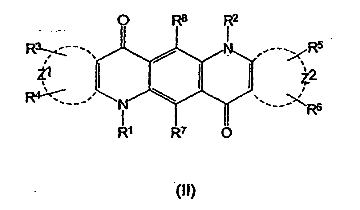
- groups R3 and R4 are attached to the Z1 ring structure and groups R5 and R6 are attached to the Z2 ring structure;
- Z1 and Z2 independently represent the atoms necessary to complete one or two fused ring aromatic or heteroaromatic systems, each ring having five or six atoms selected from carbon atoms and optionally no more than two atoms selected from oxygen, nitrogen and sulphur,
- R1, R2, R3, R4, R5, R6, R7 and R8 are independently selected from hydrogen, halogen, amide, hydroxyl, cyano, amino, mono- or di-C1-C4 alkyl-substituted amino, sulphydryl, carbonyl, carboxyl, C1-C6 alkoxy, aryl, heteroaryl, C1-C20 alkyl, aralkyl; the group -E-F where E is a spacer group having a chain from 1-60 atoms selected from the group consisting of carbon, nitrogen, oxygen, sulphur and phosphorus atoms and F is a target bonding group; and the group -(CH2-)nY where Y is selected from sulphonate, sulphate, phosphonate, phosphate, quaternary ammonium and carboxyl and n is zero or an integer from 1 to 6.
- Suitably, the target bonding group F is a reactive or functional group. A reactive group of the fluorescent dyes according to formula (I) and formula (II) can react under suitable conditions with a functional group of the substrate; a functional group of a compound according to formula (I) and formula (II) can react under suitable conditions with a reactive group of the substrate. By virtue of these reactive and functional groups, the fluorescent dyes according to formula (I) and formula (II) may be reacted with and covalently bond to the substrate, such that the substrate becomes labelled with the fluorescent dye.
- Preferably, when F is a reactive group, it is selected from succinimidyl ester, sulpho-succinimidyl ester, isothiocyanate, maleimide, haloacetamide, acid halide, vinylsulphone, dichlorotriazine, carbodiimide, hydrazide and phosphoramidite. Preferably, when F is a functional group, it is selected from hydroxy, amino, sulphydryl, imidazole, carbonyl including aldehyde and ketone, phosphate and thiophosphate.
- Preferred examples of acridone and quinacridone dyes (and their corresponding lifetimes (nsecs)) are shown as compounds (III), (IV), (V), (VI) and (VII) in Table 2.
Table 2 

O-(N-Succinimidyl)-6-(9-oxo-9H-acridin-4-carboxamido)hexanoate (III) O-(N-Succinimidyl)-6-(2-bromo-9-oxo-9H-acridin-10-yl)hexanoate (IV) 

O-(N-Succinimidl)-6-(9-oxo-9H-acridin-10-yl)hexanoate M O-(N-Succinimidyl)-6-(2-acetamido-9-oxo-9H-acridin-10-yl)hexanoate (VI) 
O-(N-Succinimidyl)-6-(12-ethyl-7,14-dioxo-2,9-disulpho-7,14-dihvdroctuino[2,3-b]acridin-5(12H)-yl)hexanoate (VII) - The dye-labelled protein and peptide substrate may be prepared by direct chemical coupling of a reactive fluorescent dye derivative to the protein or peptide by techniques that are well known to the skilled person. An alternative labelling strategy may involve coupling (or ligating) the substrate to a polypeptide label, such as a fluorescent polypeptide. Peptide and protein substrates for use in the invention may be labelled at a terminal amino acid position, or alternatively at one or more internal amino acid positions.
- The chemistry of labelling protein and peptides with fluorescent dyes is well documented and a variety of chemistries is available for the chemical modification of peptides. Generally, the choice of labelling reagent will be determined by the amino acid composition of the protein or peptide to be labelled. Particularly preferred are amine-reactive and thiol-reactive fluorescent labelling dyes. In the first case, the functional group for labelling is a primary amino group, which may be derived from the ε-amino group of lysine, or alternatively the amino-terminus of the peptide or protein. Particular examples of reactive groups for labelling ε-amino lysine residues include the isothiocyanato- and N-hydroxysuccinimidyl (NHS) ester derivatives of a fluorescent dye. Although relatively few proteins and peptides have free thiol groups (they generally exist as disulphide groups), thiol labelling procedures have proved very useful for labelling proteins and peptides, using thiol-reactive reagents, for example, iodoacetyl and maleimidyl derivatives of fluorescent dyes. For reviews and examples of protein labelling using fluorescent dye labelling reagents, see "Non-Radioactive Labelling, a Practical Introduction", Garman, A.J. Academic Press,1997; "Bioconjugation - Protein Coupling Techniques for the Biomedical Sciences", Aslam, M. and Dent, A., Macmillan Reference Ltd, (1998). Protocols are available to obtain site specific labelling in a synthesised peptide, for example, see Hermanson, G.T., Bioconjugate Techniques, Academic Press (1996). In a typical example, N-hydroxy succinimidyl (NHS) esters of the acridone dyes, may be linked to polypeptides in a weak carbonate buffer at
pH 9. The reaction is allowed to proceed for a suitable time, typically in therange 30 to 60 minutes. Unreacted or free dye may be removed by gel exclusion chromatography or by dialysis. Site specific fluorescent dye labelling of the substrate may be obtained during synthesis of the peptide, either by the use of a labelled amino acid in the synthesis process, or by the specific deprotection and labelling of the residue of interest before deprotection of other potentially reactive residues at the completion of the synthesis. - The assay methods according to the present invention are suitably performed in the wells of a multiwell plate, e.g. a microtitre plate having 24, 96, 384 or higher densities of wells, e.g. 1536 wells. Alternatively, the assays may be conducted in assay tubes or in the microchannels of a microfluidic device. In a typical kinase assay according to the first aspect, a kinase is contacted with a substrate molecule in the presence of a high-energy phosphate donor such as ATP or GTP. The reaction may be performed with the substrate and phosphate donor initially present in an aqueous assay buffer, suitably, 10mM MOPs, 50mM Tris or 50mM HEPES, containing 5 mM MgCl2. The assay may be performed either in the presence of, or the absence of a sample of a test agent Suitably, the components of the reaction mixture, minus the initiator, are pre-dispensed into the wells of a microtitre plate. The reaction is then initiated by the addition of the enzyme. Alternatively, a reaction mixture may be prepared containing enzyme and substrate in a suitable buffered solution. In this case, the reaction is initiated by the addition of the phosphate donor.
- Typically, kinase assays are performed under "stopped" conditions. Thus, the reaction is allowed to proceed for a predetermined time and then the reaction is terminated with a stop reagent, normally an inhibitor of the enzyme activity, which is often non-specific. An example of a stop reagent is EDTA, which is used to sequester metal ions that are normally required for enzymatic activity.
- Measurements of fluorescence intensity and fluorescence lifetime may be made using instruments incorporating photo-multiplier tubes as detectors. Typically, methods for the measurement of fluorescence lifetime may be based on: i) time correlated single photon counting, or ii) frequency domain (as described in Principles of Fluorescence Spectroscopy by J R Lakowicz, 2nd Ed,1999, , New York), or on time gating (see for example, Sanders, et al, Analytical Biochemistry, (1995), 227(2), 302-308). Changes in fluorescence intensity may be measured by means of a charge coupled device (CCD) imager (such as a scanning imager or an area imager) to image all of the wells of a microtitre plate. The LEADseeker™ system features a CCD camera allowing fluorescence imaging of high density microtitre plates in a single pass. Imaging is quantitative and fast, and instrumentation suitable for imaging applications can now simultaneously image the whole of a multiwell plate.
- Where an assay is to be formatted for the determination of the activity of a test agent on kinase activity, the assay may be performed under continuous measurement of the fluorescence of the substrate. In this format, the intensity of the fluorescent labelled substrate changes continuously. The labelled substrate does not need separation from the product of the enzymatic reaction and thus, a time-course of the reaction may be obtained, allowing kinetic studies to be performed in real time.
- In a particular embodiment of the first or second aspects, the enzyme is a tyrosine kinase and the substrate is a peptide substrate specific for that enzyme, such as Abl peptide. The assay may be performed in a microtitre plate in aqueous conditions, using either HEPES, Tris or MOPs buffer at a pH of 7-8. Typically, the buffer concentration will be about 50mM. Depending on the enzyme whose activity is to be measured, salts such as sodium or potassium chloride may be added. The preferred phosphate donor is ATP, which is typically present at a concentration of 2 x Km, i.e. in the range 30 - 200µM. The enzyme substrate should be present at an optimal concentration of at or below Km, which will typically be in the range 1-100µM. Additional cofactors, such as Mg2+ ions may also be present at a suitable concentration for the given enzyme, typically in the range of 1-90mM.
- The substrate is labelled with a dye chosen from the acridone class of dyes, preferably O-(N-succinimidyl)-6-(9-oxo-9H-acridin-10yl) hexanoate. The non-phosphorylated peptide substrate may be distinguished from the phosphorylated form of the substrate by detecting and measuring differences between the lifetime of the dye attached to the non-phosphorylated substrate and the lifetime of the dye attached to the phosphorylated substrate. Thus, changes, both in the intensity and the lifetime, can be monitored simultaneously, thereby allowing a dual parameter measurement of the assay. This gives a number of advantages. Firstly, the biology of the assays can be confirmed by the appearance of the lifetime, characteristic for the product of the enzymatic reaction and the intensity of the product can be simultaneously monitored. Secondly, the removal of the substrate can be monitored by its characteristic fluorescence lifetime and the fluorescence intensity of the dye-labelled substrate can be observed to decline. Thirdly, it will be possible to determine a quantitative relationship between the intensity of each species; this can be converted directly into concentration units for on-line, real-time monitoring of the reaction.
- Where the activity of a phosphatase is to be determined, a phosphorylated substrate is first prepared. A peptide substrate may be synthesised and then phosphorylated, or alternatively, one or more phosphorylated amino acids may be incorporated into the peptide chain during synthesis. Phosphorylated amino acids suitable for incorporation into chemically synthesised peptides may be obtained commercially, for example from Bachem A.G. Fluorescent dye labelling of a phosphorylated peptide may be carried out as above during the synthesis of the peptide, either by the use of a labelled amino acid in the synthesis process, or by the specific deprotection and labelling of the residue of interest before deprotection of other potentially reactive residues at the completion of the synthesis. Protein phosphatase substrates must first be in, or converted into, a suitable phosphorylated form, for use in the assays according to the invention. For example, phosphorylase b (de-phosphorylated form) may be phosphorylated at a single serine residue using phosphorylase kinase to obtain phosphorylase a. The phosphorylated substrate is then labelled with a fluorescent dye labelling reagent, preferably, an acridone or a quinacridone.
- Assay methods for phosphatases may be performed in an aqueous buffered medium, where the buffer is typically in the range 10 - 200mM at pH 7 - 8, for example 50mM Tris pH 7.2 (or alternatively, HEPES or MOPS). Additionally, salts such as sodium chloride may be added in the range 10 - 100mM. Other factors such as DTT ,1 - 5mM, EDTA, 100µM - 2mM, and 0.05 - 0.1% Brij™ may also be included in the assay mix. The dye-labelled phosphorylated substrate is present in the range from 10 - 200µM. Reactions are initiated by the addition of phosphatase. Generally reactions are incubated for 30 - 60 minutes at 30°C and then stopped by addition of a stop reagent as before.
- The methods according to the present invention may also be employed to measure the phosphorylation status of a kinase and phosphatase substrate present in a cellular environment by means of cell-based assays.
- Thus, in a fifth aspect of the present invention there is provided a method for determining the phosphorylation status of a substrate in a cellular environment, the substrate comprising at least one moiety that is capable of being phosphorylated or dephosphorylated by a cellular enzyme to yield a product, and wherein said substrate is labelled with a fluorescent dye, said method comprising the steps of:
- i) measuring the fluorescence intensity and the fluorescence lifetime of the fluorescently labelled substrate in a celt-free environment;
- ii) adding the substrate to one or more cells in a fluid medium: and
- iii) measuring the fluorescence intensity and the fluorescence lifetime of the fluorescent label following step ii);
- Suitably, the substrate according to the fifth aspect is a kinase substrate or a phosphatase substrate.
- Preferably, the fluorescent dye is selected from the acridone and the quinacridone classes of dyes as described hereinbefore.
- Suitably, the substrate is conjugated (or fused) to a second peptide or protein, such as a carrier or transport peptide, which facilitates transport of the substrate across the cellular membrane and into the cell.
- Typically, cultured cells are incubated with the conjugate at a concentration of 0.1 to 100µM in a suitable cell culture medium under conditions suitable for cell growth and for a time that may range from 0.5 to 24 hours. Cells are cultured according to standard cell culture techniques, e.g. cells are cultured in a suitable vessel in a sterile environment at 37°C in an incubator containing a humidified 95% air/5% CO2 atmosphere. Vessels may contain stirred or stationary cultures. Various cell culture media may be used including media containing undefined biological fluids such as foetal calf serum, as well as media which is fully defined, such as 293 SFM II serum free media (Life Technologies Ltd., Paisley, UK). There are established protocols available for the culture of diverse cell types. (See for example, Freshney, R.1., Culture of Animal Cells: A Manual of Basic Technique, 2nd Edition, Alan R.Liss Inc. 1987).
- The method of the invention may be used with any adherent or non-adherent cell type that can be cultured in standard tissue culture plastic-ware. Such cell types include all normal and transformed cells derived from any recognised source, for example mammalian, plant, bacterial, viral or fungal, and with respect to species (e.g. human, rodent, simian), tissue source (e.g. brain, liver, lung, heart, kidney skin, muscle) and cell type (e.g. epithelial, endothelial). When the substrate is required to be delivered to cells grown in cell or tissue culture, the conjugate is simply added to the culture medium.
- In a particular embodiment of the fifth aspect, the cells may be contacted with the conjugate in the presence of a substance whose effect on the phosphorylation status of the substrate is to be determined. In this embodiment, the detection step provides a measurement of the effect of the test substance on the phosphorylation status and may be applied to a compound whose metabolism and toxicology towards a particular cell type is under investigation, e.g. drugs, enzyme inhibitors, and the like.
- The fluorescent dyes of each of the acridone and quinacridone classes of dyes may be distinguished one from the other by virtue of their different lifetimes. Thus, two or more different substrates, each one labelled with a different one of a set of acridone dyes (or with a different one of a set of quinacridone dyes) may be used in multiplex assays in which the measurement of the kinase activities of different enzymes may be performed simultaneously.
- In a sixth aspect of the present invention, there is provided a method of simultaneously measuring the kinase activities of two or more different enzymes, each enzyme specific for a different substrate, wherein each substrate comprises at least one moiety that is capable of being phosphorylated by an enzyme to yield a product, and wherein each said substrate is labelled with a different one of a set of fluorescent dyes, said method comprising the steps of:
- i) measuring the fluorescence intensity and the fluorescence lifetime of each of the fluorescently labelled substrates;
- ii) combining a mixture of said enzymes with each of said substrate molecules in the presence of a phosphate source; and
- iii) measuring an increase in fluorescence intensity and in fluorescence lifetime of each of the fluorescent labels following the combination of step ii);
- Preferably, the fluorescent dyes in said set are selected from the acridone and the quinacridone classes of dyes as described hereinbefore.
- In one embodiment according to the sixth aspect, the method may be performed in vitro, either using isolated enzymes, or alternatively, one or more of said enzymes may be a component of a cell lysate.
- In another embodiment according to the sixth aspect, the method may be performed in a cellular environment. In this embodiment, each of the different substrates is conjugated (or fused) to a second peptide or protein, such as a carrier or transport peptide, which facilitates transport of the substrate across the cellular membrane and into the cell as described.
- Preferably, the measurement of kinase whose activity is being measured is a tyrosine kinase.
- In a seventh aspect, there is provided a composition comprising:
- i) a substrate for a kinase enzyme said substrate comprising at least one moiety that is capable of being of phosphorylated by said enzyme, to yield a phosphorylated product and wherein said substrate is labelled with a fluorescent dye as defined in
claim 3; and - ii) a component selected from a phosphate donor and said kinase.
- In a eighth aspect, there is provided a test kit for measuring the kinase activity of at least one enzyme, the test kit comprising:
- i) one or more different substrates wherein each of said substrates comprises a moiety that is capable of being of phosphorylated to yield a product and wherein each of said substrates is labelled with different one of a set of fluorescent dyes as defined in
claim 3; and optionally, - ii) one or more different enzymes each enzyme specific for a different substrate.
- In a ninth aspect, there is provided a test kit for measuring the phosphatase activity of at least one enzyme, the test kit comprising:
- i) one or more different substrates wherein each of said substrates comprises a phosphorylated moiety that is capable of being of dephosphorylated to yield a product and wherein each of said substrates is labelled with different one of a set of fluorescent dyes as defined in
claim 3; and optionally, - ii) one or more different enzymes each enzyme specific for a different substrate.
- The present invention simplifies conventional assay methodologies by reducing the number of steps required. Furthermore, in conventional (radioactive based) kinase assays, the concentration of the phosphate donor cannot be utilised at an optimal concentration. For example, in radioactive assays the ATP concentration can often be at 1/10 of Km (this will typically be 1-10µM), whereas it is usually recommended to perform kinase assays at 2 fold or higher Km for ATP. In the present invention, the concentration of ATP does not affect the read-out and can therefore be utilised at optimal levels for the assay, independent of the fluorescent read-out technology.
- According to the method of the present invention, it is possible to add any of the reagents to the mix in any order, while omitting a critical component Thus, a reaction mixture may be prepared omitting ATP, but including all other components of the reaction. Prior to adding the ATP (initiator), the reaction can then be monitored for non-specific effects. It is also possible to construct mixture with no enzyme for further controls. Due to the nature of the reactions, it is then possible to add the final component and monitor changes either in real time or by stopping the reaction following the initiation step.
- Furthermore, fluorescence lifetime measurements that may be utilised in the present invention can offer significant advantages over conventional fluorescence techniques that are based solely on quantifying fluorescence intensity. Fluorescence lifetime is determined from the same spectrally resolved intensity signal, but is additionally resolved in the temporal domain. Fluorescence lifetime techniques provide greater sensitivity because the signal is largely unaffected by 'background noise'. A further advantage with this technique is that several different events can be measured simultaneously by selecting labels having distinguishable lifetimes, thus enabling multiplexing. In addition, measurements of fluorescence lifetime are unaffected by concentration effects and photobleaching.
- The present invention is further illustrated with reference to the following Figures and Examples in which:
- Figure 1 illustrates the differences in lifetimes and intensities observed upon phosphorylation of Abl peptide according to Example 5;
- Figure 2 illustrates the phosphorylation of a peptide by the protein tyrosine kinase Abl according to Example 6;
- Figure 3 is a plot showing the time dependent phosphorylation of Myelin Basic Protein by Erk kinase according to Example 7;
- Figure 4 is a plot showing inhibition of Erk kinase by staurosporine according to Example 8;
- Figure 5 is a time-course for phosphorylation by the tyrosine kinase Lck according to Example 9;
- Figure 6 is a plot illustrating ATP dependence of phosphorylation by the tyrosine kinase Lck according to Example 10;
- Figure 7 is a plot showing enzyme dependence of phosphorylation by the tyrosine kinase Lck according to Example 11;
- Figure 8 is a plot showing inhibition of Lck kinase activity by staurosporine according to Example 12;
- Figure 9 is a time-course for a dephosphorylation reaction by protein-tyrosine phosphatase according to Example 13.
- H-Glu-Ala-Ile-Tyr Ala-Ala-Pro-Phe-Ata-Lys-Lys-LysrRink Amide resin and H-Glu Ala-Ile-Tyr(PO3H2) Ala- Ala-Pro-Phe-Ala-Lys-Lys-Lys Rink Amide resin were synthesised using a commercially available Applied Biosystems Model 433A automated peptide synthesiser and FastMoc™ chemistry, following the instrument manufacturer's recommended procedures throughout. Both syntheses were performed on a 0.25 millimolar scale.
- Crude peptide was deprotected and cleaved from the solid phase using a mixture of 95% trifluoroacetic acid : 2.5% water: 2.5% triisopropylsilane. The crude peptide obtained from the cleavage reaction was purified by conventional C-18 reverse phase HPLC using a linear gradient of water/acetonitrile (both containing 0.1% trifluoroacetic acid). After purification, the peptide was lyophilised and characterised by Maldi TOF mass spectroscopy and HPLC.
- This was prepared as in 1.2 above, using H-Glu-Ala-Ile-Tyr(PO3H2)-Ala-Ala-Pro-Phe-Ala-Lys-Lys-Lys Rink Amide Resin.
- H-Glu-Ala-Ile-Tyr-Ala-Ala-Pro-Phe-Ala-Lys-Lys-Lys-Rink Amide resin was labelled at the N-terminus on solid phase, with O-(N-succinimidyl)-6-(9-oxo-9H-acridin-10yl) hexanoate (1.5eq) (Amersham Biosciences) in dimethylsulphoxide and diisopropylethylamine (4% by volume), overnight at room temperature. The resin was washed with DMSO, followed by methanol and finally with dichloromethane and then dried in vacuo. The labelled peptide was deprotected and cleaved from the solid phase using a mixture of 95% trifluoroacetic acid : 2.5% water: 2.5% triisopropylsilane. The crude material isolated by precipitation from cold diethyl ether and purified by C-18 reverse phase HPLC using a linear gradient of water/acetonitrile (both containing 0.1 % trifluoroacetic acid). After purification, the mono-labelled peptide was lyophilised and characterised by Maldi TOF mass spectroscopy, UV and HPLC.
- This was prepared as in 1.4 above, except that H-Glu-Ala-Ile-Tyr(PO3H2)-Ala-Ala-Pro-Phe-Ala-Lys-Lys-Lys-Rink Amide resin was used.
- 100mg of MBP (100mg; 7mg/ml) was dialysed overnight at +4°C in 0.1m NaHCO3 solution, O-(N-Succinimidyl)-6-(9-oxo-9H-acridin-10yl) hexanoate (10mg) was dissolved in 1ml DMSO (Aldrich) and added to the sample of dialysed MBP. The resulting mixture was stirred for 45 minutes. 10 PD10 columns (Amersham Biosciences) were equilibrated with 10ml of 10mM MOPS pH 7.2. Labelled MBP (1.3ml aliquots) was added to each column and the columns were washed with 10mM MOPS (1ml). Each column was eluted with 3ml of 10mM MOPS and the eluants pooled. The final protein concentration was determined using Biorad Protein Assay (500-006), with BSA as a standard (0.1 mg/ml). The final concentration of MBP was 1.44mg/ml in a total volume of 30ml. The labelled MBP was concentrated using Amicon Centripreps YM-10 (10,000 NMWL) (spun for 15mins). Subsequent protein determination gave a concentration of 4.5mg/ml (7.1ml). The acridone labelled MBP was diluted with 500mM MOPS pH 7.2, 50mM MgCl2, 1mM ATP and PF H2O to a concentration of 16.6µM.
- MBP (100mg; 7mg/ml) was dialysed overnight at +4°C in a PBS solution (0.01M phosphate buffer, 0.0027 M potassium chloride and 0.137 M sodium chloride, pH 7.4) Sigma P-4417. O-(N-Succinimidyl)-6-(9-oxo-9H-acridin-10y1) hexanoate (10mg) was dissolved in 1ml DMSO (Aldrich) and added to the dialysed MBP. The mixture was roller-mixed overnight at +4°C. 10 PD10 columns (Amersham Biosciences) were equilibrated with 10ml of 10mM MOPS pH 7.2. Aliquots of 1.3ml of labelled MBP were added to each column, each column was washed with a further 1ml of 10mM MOPS. Each column was then eluted with 3ml of 10mM MOPS and the eluants pooled. The protein concentration was determined using a Biorad Protein Assay (500-006) with BSA as a standard at 0.1mg/ml. The concentration of the labelled MBP was found to be 1.4mg/ml in a total volume of 28ml. The labelled MBP was re-concentrated using Amicon Centripreps YM-10 (10,000 NMWL) (spun for 10mins). Subsequent protein determination gave a concentration of 3.25mg/ml in a total volume of 9.7ml. The dye labelled MBP was diluted with 500mM MOPS pH 7.2, 50mM MgCl2, 1mM ATP and PF H2O to a concentration of 16.6µM.
- H-Glu-Pro-Glu-Gly-Ile-Tyr-Gly-Val-Leu-Phe-Rink Amide resin and H-Glu-Pro-Glu-Gly-lIe-Tyr(PO3H2)-Gly-Val-leu-Phe-Rink Amide resin were synthesised using a commercially available Applied Biosystems Model 433A automated peptide synthesiser and FastMoc™ chemistry, following the instrument manufacturer's recommended procedures throughout The synthesises were performed on a 0.25 millimolar scale.
- H-Glu-Pro-Glu-Gly-ile-Tyr-Gly-Val-Leu-Phe-Rink Amide resin was labelled at the N-terminus on solid phase, with O-(N-succinimidyl)-6-(9-oxo-9H-acridin-10yl) hexanoate (1.5eq) (Amersham Biosciences) in dimethylsulphoxide and diisopropylethylamine (4% by volume), overnight at room temperature. The resin was washed with DMSO, followed by methanol and finally with dichloromethane and then dried in vacuo. The labelled peptide was deprotected and cleaved from the solid phase using a mixture of 95% trifluoroacetic acid : 2.5% water: 2.5% triisopropylsilane. The crude material isolated by precipitation from cold diethyl ether and purified by C-18 reverse phase HPLC using a linear gradient of water/acetonitrile (both containing 0.1 % trifluoroacetic acid). After purification, the mono-labelled peptide was lyophilised and characterised by Maldi TOF mass spectroscopy, UV and HPLC.
- This was prepared as in 3.2 above, except that H-Glu-Pro-Glu-Gly-Ile-Tyr(PO3H2)-Gly-Val-Leu-Phe-Rink Amide Resin was used.
- These syntheses were performed as described in Example 3.2, except that O-(N-succinimidyl)-6-(2-acetamido-9-oxo-9H-acridin-10yl) hexanoate (Ace-17) was used in place of O-(N-succinimidyl)-6-(9-oxo-9H-acridin-10yl) hexanoate (Ace-14).
- This synthesis was performed as described in Example 3.2, except that H-Thr-Arg-Asp-Ile-Tyr(PO3H2)-Glu-Thr-Asp-Rink amide resin was used. The non-phosphorylated peptide was synthesised in a similar way.
- 1µM solutions of the phosphorylated and non-phosphorylated forms of the Abl peptide were prepared in 10mM MOPs buffer pH 7.2. The fluorescence intensities and lifetimes of the solutions were compared.
- Clear differences can be seen between the two peptides. The intensity of the non-phosphorylated peptide was approximately 7 x 10⁴ rfu, while that of the phosphorylated peptide was approximately 9 x 10⁴ rfu. The lifetime of the non-phosphorylated peptide was about 10 nsec, whereas that of the phosphorylated peptide was 15nsec. See Figure 1.
- An Abl reaction mixture was prepared by mixing 1ml of reaction buffer (50mM Tris-HCl, 10mM MgCl2 1mM EGTA 2mM dithiothreitol (pH 7.5 at 25°C)), 10µl of 10mM ATP, 2µl of 6-(9-ox<r9H-acridin-10yl) hexanoate-labelled Abl Peptide substrate (non-phosphorylated) (500µM in DMSO). 100µl of this mixture was placed into the wells of a black flat bottomed microtitre plate. The reaction was initiated by the addition of 10µl of Abl kinase (New England Biolabs, Code P6050L Lot 5), (100,000 units/ml or 100 units/µl) which had been diluted in reaction buffer (50mM Tris-HCl, 10mM MgCl2, 1mM EGTA, 2mM dithiothreitol, 0.01 % Brij 35 (pH 7.5 at 25°C) to a concentration of 100 units per 10µl.
- The reaction was monitored at 30 second intervals for both lifetime and intensity changes characteristic of the product and substrate.
- As shown in Figure 2, the progress of the reaction may be monitored in real time. In particular, changes in the product intensity are greater than those of the substrate, as would be predicted from the study of the isolated peptides. Furthermore, it is possible to carry out this reaction with no separation step involved, and to distinguish the substrate and product on the basis of their respective fluorescent lifetimes.
- Myelin Basic Protein (MBP) labelled with O-(N-succinimidyl)-6-(9-oxo-9H-acridin-10yl) hexanoate (prepared according to Example 2, Method 1) (1ml) was diluted to 10ml with water, giving a solution containing 50mM MOPS, pH 7.2, 5nM MgCl2, 100 µM ATP and 1.66µM MBP. Seven aliquots of 200µl of reaction mixture were each mixed with 5µl of Erk1 kinase (1.8mg/ml). The reactions were incubated at room temperature for various times, 100µl aliquots were withdrawn and the fluorescence of each aliquot was measured.
- The results are shown in Figure 3 and indicate a time-dependent increase in fluorescence.
- Myelin Basic Protein (MBP) labelled with O-(N-succinimidyl)-6-(9-oxo-9H-acridin-10yl) hexanoate (prepared according to Example 2, Method 1) (1ml) was diluted to 10ml with water, giving a solution containing 50mM MOPS, pH 7.2, 5nM MgCl2, 100 µm ATP and 1.66µM MBP. Seven 100µl aliquots were made up to a concentration of 0 - 100µM with respect to staurosporine. Finally, 2µl of
Erk 1 kinase (1.8mg/ml) were added to each reaction mixture. The reaction mixtures were incubated at room temperature for 3 hours, after whichtime 100 µl of 100mM EDTA were added to stop the reactions. Fluorescence intensities of the reactions were then determined. - The enzyme reaction is inhibited in a dose dependent manner by staurosporine as shown in Figure 4.
- Replicates of N-(6-(9-oxo-9H-acridin-10-yl)-hexanoyl)-Glu-Pro-Glu-Gly-Ile-Tyr-Gly-Val-Leu-Phe amide (500nM) in 50mM TRIS/10mM MgCl2/2.5mM MnCl2, pH 7.2 in the presence of 50µM ATP in a volume of 100µl were pipetted into the wells of a black 96-well microplate (Costar, Code 3650). The time-course was initiated by the addition of 12.7 milli-units of Lck enzyme (Upstate Biotechnology, Code 14-379) in a volume of 10µl. The reaction was monitored at one minute intervals at ambient temperature for both lifetime and intensity changes characteristic of both substrate and product.
- The results as shown in Figure 5 indicate that the formation of product is dependent on time.
- Replicates of N-(6-(2-acetamido-9-oxo-9H-acridin-10-yl)-hexanoyl)-Glu-Pro-Glu-Gly-Ile-Tyr-Gly-Val-Leu-Phe amide (500nM) in 50mM TRIS/10mM MgCl2/2.5mM MnCl2, pH 7.2 and in the presence of various concentrations of ATP in a volume of 100µl were pipetted into the wells of a black 96-well microplate (Costar, Code 3650). The reactions were initiated by the addition of 12.7 milli-units of Lck enzyme (Upstate Biotechnology, Code 14-379) in a volume of 10µl. After incubation at room temperature for 60 minutes, the reactions were stopped by addition of 0.1 M citrate buffer pH 3.0 (20µl) to each well. The reactions was monitored for both lifetime and intensity changes characteristic of both substrate and product.
- The results are shown in Figure 6, which indicates that the formation of product is dependent on the ATP concentration.
- Replicates of N-(6-(9-oxo-9H-acridin-10-yl)-hexanoyl)-Glu-Pro-Glu-Ile-Tyr-Gly-Val-Leu-Phe amide (500nM) in 50mM TRIS/10mM MgCl2/2.5mM MnCl2. pH 7.2 and presence of 50µM ATP in a volume of 100µl were pipetted into the wells of a black 96-well microplate (Costar, Code 3650). The reactions were initiated by the addition of quantities of Lck enzyme (Upstate Biotechnology, Code 14-379) in a volume of 10µl. After incubation at room temperature for 90 minutes, the reactions were stopped by addition of 0.1 M citrate buffer pH 3.0 (20µl) to each well. The reactions was monitored for both lifetime and intensity changes characteristic of both substrate and product.
- The results in Figure 7 show that the formation of product is dependent on the enzyme concentration.
- Staurosporine (Sigma, Code S4400, 1 mM in DMSO) was diluted with 10%(v/v) DMSO in assay buffer (50mM TRIS/10mM MgCl2/2.5mM MnCl2, pH 7.2) to prepare the following concentrations of staurosporine - 100µM, 10µM, 1µM, 100nM, 10nM and 1nM. Reaction mixture was prepared: 10ml assay buffer + 2.5µl N-(6-(9-oxo-9H-acridin-10-yl)-hexanoyl)-Glu-Pro-Glu-Gly-Ile-Tyr-Gly-Val-Leu-Phe amide (1mM) + 20µl ATP (10mM). Five replicates (10µl) of each inhibitor concentration were pipetted into the wells of a black 96-well microplate (Costar, Code 3650). Reaction mixture (100µl) was added to each well. The reactions were initiated by the addition of 12.7 milli-units of Lck enzyme (Upstate Biotechnology, Code 14-379) in a volume of 10µl. After incubation at room temperature for 60 minutes, the reactions were stopped by addition of 0.1 M citrate buffer pH 3.0 (20µl) to each well. The reactions were monitored for both lifetime and intensity changes characteristic of both substrate and product.
- The results in Figure 8 show the inhibition of Lck kinase activity by staurosporine; the IC50 value is 16nM. Park et al (Anal. Biochem., (1999), 269, 94-104) reported an IC50 value of about 10nM using ATP at 2µM final concentration and a similar peptide sequence in a time-resolved fluorescence format assay.
- Replicates of N-(6-(9-oxo-9H-acridin-10-yl)-hexanoyl)-Thr-Arg-Asp-Ile-Tyr(PO3H2)-Glu-Thr-Asp-NH2 (1µM) in TRIS buffered saline, pH 7.6 in a volume of 100µl were pipetted into the wells of a black 96-well microplate (Costar, Code 3650). The time-course was initiated by the addition of 88 units of protein-tyrosine phosphatase enzyme (Sigma, Code P9864) in a volume of 10µl. The reaction was monitored at one minute intervals at ambient temperature for both lifetime and intensity changes characteristic of both substrate and product
- Figure 9 is a plot showing the dephosphorylation of the substrate by this phosphatase. The appearance of the dephosphorylated product is also monitored.
-
- <110> Amersham Biosciences UK Ltd
- <120> Methods for Measuring Protein Kinase and Phosphatase Activity
- <130> PA0221 -PCT
- <140> PCT
GB/03/01683 - <141> 2003-04-17
- <160> 6
- <170> PatentIn version 3.1
- <210> 1
<211> 12
<212> PRT
<213> Artificial Sequence - <220>
<223> Synthetic peptide - <400> 1

- <210> 2
<211> 12
<212> PRT
<213> Artificial sequence - <220>
<223> synthetic peptide - <220>
<221> MOD_RES
<222> (4).. (4)
<223> Tyr residue is phosphorylated - <400> 2

- <210> 3
<211> 10
<212> PRT
<213> Artificial Sequence - <220>
<223> Synthetic peptide - <400> 3

- <210> 4
<211> 10
<212> PRT
<213> Artificial Sequence - <220>
<223> Synthetic peptide - <220>
<221> MOD_RES
<222> (6)..(6)
<223> Tyr residue is phosphorylated - <400> 4

- <210> 5
<211> 8
<212> PRT
<213> Artificial sequence - <220>
<223> Synthetic peptide - <220>
<221> MOD_RES
<222> (5)..(5)
<223> Tyr residue is phosphorylated - <400> 5

- <210> 6
<211> 8
<212> PRT
<213> Artificial sequence - <220>
<223> synthetic peptide - <400> 6

-
- <110> Amersham Biosciences UK Ltd
- <120> Methods for Measuring Protein Kinase and Phosphatase Activity
- <130> PA0221 -PCT
- <140>
PCT GB/03/01683 - <141> 2003-04-17
- <160> 6
- <170> Patent In version 3.1
- <210> 1
<211> 12
<212> PRT
<213> Artificial Sequence - <220>
<223> Synthetic peptide - <400> 1

- <210> 2
<211> 12
<212> PRT
<213> Artificial sequence - <220>
<223> synthetic peptide - <220>
<221> MOD_RES
<222> (4)..(4)
<223> Tyr residue is phosphorylated - <400> 2

- <210> 3
<211> 10
<212> PRT
<213> Artificial Sequence - <220>
<223> Synthetic peptide - <400> 3

- <210> 4
<211> 10
<212> PRT
<213> Artificial Sequence - <220>
<223> Synthetic peptide - <220>
<221> MOD_RES
<222> (6) .. (6)
<223> Tyr residue is phosphorylated - <400> 4

- <210> 5
<211> 8
<212> PRT
<213> Artificial sequence - <220>
<223> Synthetic peptide - <220>
<221> MOD_RES
<222> (5)..(5)
<223> Tyr residue is phosphorylated - <400> 5

- <210> 6
<211> 8
<212> PRT
<213> Artificial sequence - <220>
<223> synthetic peptide - <400> 6

Claims (42)
- A method for determining the phosphorylating activity of an enzyme acting on a substrate molecule, said substrate comprising at least one moiety that is capable of being phosphorylated by said enzyme to yield a phosphorylated product and wherein said substrate is labelled with a fluorescent dye, said method comprising the steps of:i) measuring the fluorescence intensity and the fluorescence lifetime of the fluorescently labelled substrate;ii) combining said enzyme with said substrate molecule in the presence of a phosphate donor, andiii) measuring a change in fluorescence intensity and in fluorescence lifetime of the fluorescent label following the combination of step ii);wherein said change in fluorescence intensity and in fluorescence lifetime of the fluorescent label is used to determine the phosphorylating activity of said enzyme.
- A method according to claim 1 wherein said substrate is labelled with a fluorescent dye selected from the acridone and the quinacridone classes of dyes.
- A method for determining the phosphorylating activity of an enzyme acting on a substrate molecule, said substrate comprising at least one moiety that is capable of being phosphorylated by said enzyme to yield a phosphorylated product and wherein said substrate is labelled with a fluorescent dye of formula:

 Z1 and Z2 independently represent the atoms necessary to complete one or two fused ring aromatic or heteroaromatic systems, each ring having five or six atoms selected from carbon atoms and optionally no more than two atoms selected from oxygen, nitrogen and sulphur,R1, R2, R3, R4, R5, (and R6, and R8 if present) are independently selected from hydrogen, halogen, amide, hydroxyl, cyano, amino, mono- or di-C1-C4 alkyl-substituted amino, sulphydryl, carbonyl, carboxyl, C1-C6 alkoxy, aryl, heteroaryl, C1-C20 alkyl, aralkyl; the group -E-F where E is a spacer group having a chain from 1-60 atoms selected from the group consisting of carbon, nitrogen, oxygen, sulphur and phosphorus atoms and F is a target bonding group; and the group -(CH2-)nY where Y is selected from sulphonate, sulphate, phosphonate, phosphate, quaternary ammonium and carboxyl and n is zero or an integer from 1 to 6;said method comprising the steps of:i) measuring the fluorescence intensity of the fluorescently labelled substrate;ii) combining said enzyme with said substrate molecule in the presence of a phosphate donor, andiii) measuring an increase in fluorescence intensity of the fluorescent label following the combination of step ii);wherein said increase in fluorescence intensity of the fluorescent label is used to determine the phosphorylating activity of said enzyme.
 - The method according to claim 3, wherein said measuring steps i) and iii) additionally comprise measurement of the fluorescence lifetimes of the fluorescent label; wherein an increase in fluorescence intensity and in fluorescence lifetime upon phosphorylation of the dye-labelled substrate is used to determine the phosphorylating activity of said enzyme.
- The method according to any of claims 1 to 4, wherein the substrate molecule is selected from natural proteins, (including post-translationally modified proteins such as glyco-proteins and lipo-proteins); and synthetic peptides.
- The method according to any of claims 1 to 5, wherein the substrate molecule comprises at least one amino acid that is capable of being phosphorylated.
- The method according to claim 6, wherein said amino acid that is capable of being phosphorylated is selected from tyrosine, serine, threonine and histidine.
- The method according to any of claims 1 to 7, wherein said substrate is capable of being phosphorylated by one or more of the enzymes selected from: tyrosine kinase, serine/threonine kinase and histidine kinase.
- The method according to any of claims 1 to 8, wherein said phosphorylated product comprises at least one phospho-tyrosine residue.
- The method according to any of claims 4 to 9, wherein the substrate is phosphorylated at a tyrosine residue by a tyrosine kinase and wherein an increase in fluorescence lifetime of the fluorescent label is used to measure the concentration of phosphorylated product relative to the concentration of non-phosphorylated substrate.
- The method according to any of claims 1 to 10, wherein said substrate is linked by a linker group to a solid support.
- The method according to any of claims 1 to 10, wherein said substrate is conjugated to a.second peptide or protein.
- A method of screening for a test agent whose effect on the phosphorylating activity of an enzyme is to be determined the method comprising the steps of:(a) performing the method of any of claims 1 to 12 in the presence and in the absence of said agent; and(b) determining the phosphorylating activity of said enzyme in the presence and in the absence of said agent;wherein a difference between the phosphorylating activity of said enzyme in the presence and in the absence of said agent is indicative of the effect of said test agent on the phosphorylating activity of said enzyme.
- A method of screening for a test agent whose effect on the phosphorylating activity of an enzyme is to be determined the method comprising th e steps of:(a) performing the method of any of claims 1 to 12 in the presence of said test agent; and(b) comparing the value of the phosphorylating activity of the enzyme with a control value for the enzyme activity in the absence of the test agent; wherein said control value is stored in an electronic format.
- The method according to claim 13 or claim 14, wherein said enzyme is selected from tyrosine kinase, serine/threonine kinase and histidine kinase.
- A method for determining the dephosphorylating activity of an enzyme acting on a substrate molecule, said substrate comprising at least one phosphorylated moiety that is capable of being de-phosphorylated by said enzyme to yield a product and wherein said substrate is labelled with a fluorescent dye, said method comprising the steps of:i) measuring the fluorescence intensity and the fluorescence lifetime of the fluorescently labelled substrate;ii) combining said enzyme with said substrate molecule to yield a product; andiii) measuring a change in fluorescence intensity and in fluorescence lifetime following the combination of step ii):wherein said change in fluorescence intensity and in fluorescence lifetime of the fluorescent label is used to determine the dephosphorylating activity of said enzyme.
- A method according to claim 16, wherein said substrate is labelled with a fluorescent dye selected from the acridone and the quinacridone classes of dyes.
- A method for determining the dephosphorylating activity of an enzyme acting on a substrate molecule, said substrate comprising at least one phosphorylated moiety that is capable of being de-phosphorylated by said enzyme to yield a product and wherein said substrate is labelled with a fluorescent dye of formula:

 Z1 and Z2 independently represent the atoms necessary to complete one or two fused ring aromatic or heteroaromatic systems, each ring having five or six atoms selected from carbon atoms and optionally no more than two atoms selected from oxygen, nitrogen and sulphur,R1, R2, R3, R4. R5, (and R6, R7 and R8 if present) are independently selected from hydrogen, halogen, amide, hydroxyl, cyano, amino, mono- or di-C1-C4 alkyl-substituted amino, sulphydryl, carbonyl, carboxyl, C1-C6 alkoxy, aryl, heteroaryl, C1-C20 alkyl, aralkyl; the group -E-F where E is a spacer group having a chain from 1-60 atoms selected from the group consisting of carbon, nitrogen, oxygen, sulphur and phosphorus atoms and F is a target bonding group; and the group -(CH2-)nY where Y is selected from sulphonate, sulphate, phosphonate, phosphate, quaternary ammonium and carboxyl and n is zero or an integer from 1 to 6;said method comprising the steps of:i) measuring the fluorescence intensity of the fluorescently labelled substrate;ii) combining said enzyme with said substrate molecule to yield a product; andiii) measuring a decrease in fluorescence intensity following the combination of step ii);wherein said decrease in fluorescence intensity of the fluorescent label is used to determine the dephosphorylating activity of said enzyme.
 - The method according to claim 18, wherein said measuring steps i) and iii) additionally comprise measurement of the fluorescence lifetimes of the fluorescent label; wherein a decrease in fluorescence intensity and in fluorescence lifetime upon dephosphorylation of the dye-labelled phosphorylated substrate is used to determine the dephosphorylating activity of said enzyme.
- The method according to any of claims 16 to 19, wherein the substrate molecule is selected from natural proteins, (including post-translationally modified proteins such as glyco-proteins and lipo-proteins); and synthetic peptides.
- The method according to any of claims 16 to 20, wherein the substrate molecule comprises at least one phosphorylated amino acid that is capable of being dephosphorylated.
- The method according to claim 21, wherein said phosphorylated amino acid is selected from the phosphorylated derivatives of tyrosine, serine, threonine and histidine.
- The method according to any of claims 19 to 22, wherein said enzyme removes a phosphate group from a phospho-tyrosine residue in said substrate and wherein a decrease in fluorescence lifetime of the fluorescent label is used to measure the concentration of phosphorylated substrate relative to the concentration of product.
- The method according to any of claims 16 to 23, wherein said substrate is linked by a linker group to a solid support.
- The method according to any of claims 16 to 23, wherein said substrate is fused to a second peptide or protein.
- A method of screening for a test agent whose effect on the dephosphorylating activity of an enzyme is to be determined the method comprising the steps of:(a) performing the method of any of claims 16 to 25 in the presence and in the absence of said agent; and(b) determining the dephosphorylating activity of said enzyme in the presence and in the absence of said agent;wherein a difference between the dephosphorylating activity of said enzyme in the presence and in the absence of said agent is indicative of the effect of said test agent on the dephosphorylating activity of said enzyme.
- A method of screening for a test agent whose effect on the dephosphorylating activity of an enzyme is to be determined the method comprising the steps of:(a) performing the method of any of claims 16 to 25 in the presence of said agent; and(b) comparing the value of the dephosphorylating activity of the enzyme with a control value for the enzyme activity in the absence of the test agent; wherein said control value is stored in an electronic format.
- The method according to any of claims 16 to 27, wherein said enzyme is a phosphatase.
- A method for determining the phosphorylation status of a substrate in a cellular environment, the substrate comprising at least one moiety that is capable of being phosphorylated or dephosphorylated by a cellular enzyme to yield a product, and wherein said substrate is labelled with a fluorescent dye, said method comprising the steps of:i) measuring the fluorescence intensity and the fluorescence lifetime of the fluorescently labelled substrate in a cell-free environment;ii) adding the substrate to one or more cells in a fluid medium; andiii) measuring the fluorescence intensity and the fluorescence lifetime of the fluorescent label following step ii);wherein a change in the fluorescent intensity and the fluorescence lifetime is used to indicate the phosphorylation status of said substrate.
- The method according to claim 29, wherein the substrate is a kinase substrate or a phosphatase substrate.
- The method according to claim 29 or claim 30, wherein the fluorescent dye is selected from the acridone and the quinacridone classes of dyes.
- The method according to any of claims 29 to 31, wherein the substrate is conjugated to a second peptide or protein.
- The method according to claim 32, wherein the substrate is conjugated to a carrier or transport peptide.
- A method of simultaneously measuring the kinase activities of two or more different enzymes each enzyme specific for a different substrate, wherein each substrate comprises at least one moiety that is capable of being phosphorylated by an enzyme to yield a product, and wherein each said substrate is labelled with a different one of a set of fluorescent dyes, said method comprising the steps of:i) measuring the fluorescence intensity and the fluorescence lifetime of each of the fluorescently labelled substrates;ii) combining a mixture of said enzymes with each of said substrate molecules in the presence of a phosphate source; andiii) measuring an increase in fluorescence intensity and in fluorescence lifetime of each of the fluorescent labels following the combination of step ii);wherein said increase in fluorescence intensity and in fluorescence lifetime of each of the fluorescent labels is used to determine the phosphorylating activity of each of said enzymes.
- The method according to claim 34, wherein the fluorescent dyes in said set are selected from the acridone and the quinacridone classes of dyes.
- The method according to claim 34 or claim 35, wherein said enzyme is an isolated enzyme.
- The method according to claim 34 or claim 35, wherein one or more of said enzymes is a component of a cell lysate.
- The method according to claim 34, wherein the combination of step ii) is performed in a cellular environment and wherein each of the different substrates is conjugated to a carrier or transport peptide.
- The method according to any of claims 34 to 38, wherein the kinase activities that are being measured are tyrosine kinases.
- A composition comprising:i) a substrate for a kinase enzyme said substrate comprising at least one moiety that is capable of being of phosphorylated by said enzyme, to yield a phosphorylated product and wherein said substrate is labelled with a fluorescent dye of formula:

 Z1 and Z2 independently represent the atoms necessary to complete one or two fused ring aromatic or heteroaromatic systems, each ring having five or six atoms selected from carbon atoms and optionally no more than two atoms selected from oxygen, nitrogen and sulphur,R1, R2, R3, R4, R5, (and R6, R7 and R8 if present) are independently selected from hydrogen, halogen, amide, hydroxyl, cyano, amino, mono- or di-C1-C4 alkyl-substituted amino, sulphydryl, carbonyl, carboxyl, C1-C6 alkoxy, aryl, heteroaryl. C1-C20 alkyl, aralkyl; the group -E-F where E is a spacer group having a chain from 1-60 atoms selected from the group consisting of carbon, nitrogen, oxygen, sulphur and phosphorus atoms and F is a target bonding group; and the group -(CH2-)nY where Y is selected from sulphonate, sulphate, phosphonate, phosphate, quaternary ammonium and carboxyl and n is zero or an integer from 1 to 6; andii) a component selected from a phosphate donor and said kinase.
 - A test kit for measuring the kinase activity of at least one enzyme, the test kit comprising:i) one or more different substrates wherein each substrate comprises a moiety that is capable of being of phosphorylated to yield a product and wherein each said substrate is labelled with different one of a set of fluorescent dyes selected from dyes of formula:

 Z1 and Z2 independently represent the atoms necessary to complete one or two fused ring aromatic or heteroaromatic systems, each ring having five or six atoms selected from carbon atoms and optionally no more than two atoms selected from oxygen, nitrogen and sulphur,R1, R2, R3, R4, R5, (and R6, R7 and R8 if present) are independently selected from hydrogen, halogen, amide, hydroxyl, cyano, amino, mono- or di-C1-C4 alkyl-substituted amino, sulphydryl, carbonyl, carboxyl, C1-C6 alkoxy, aryl, heteroaryl, C1-C20 alkyl, aralkyl; the group -E-F where E is a spacer group having a chain from 1-60 atoms selected from the group consisting of carbon, nitrogen, oxygen, sulphur and phosphorus atoms and F is a target bonding group; and the group - (CH2-) n Y where Y is selected from sulphonate, sulphate, phosphonate, phosphate, quaternary ammonium and carboxyl and n is zero or an integer from 1 to 6; and optionally,ii) one or more different enzymes each enzyme specific for a different substrate.
 - A test kit for measuring the phosphatase activity of at least one enzyme, the test kit comprising:i) one or more different substrates wherein each of said substrates comprises a phosphorylated moiety that is capable of being of dephosphorylated to yield a product and wherein each of said substrates is labelled with different one of a set of fluorescent dyes selected from dyes of formula:

 Z1 and Z2 independently represent the atoms necessary to complete one or two fused ring aromatic or heteroaromatic systems, each ring having five or six atoms selected from carbon atoms and optionally no more than two atoms selected from oxygen, nitrogen and sulphur,R1, R2, R3, R4, R5, (and R6, R7 and R8 if present) are independently selected from hydrogen, halogen, amide, hydroxyl, cyano, amino, mono- or di-C1-C4 alkyl-substituted amino, sulphydryl, carbonyl, carboxyl, C1-C6 alkoxy, aryl, heteroaryl, C1-C20 alkyl, aralkyl; the group -E-F where E is a spacer group having a chain from 1-60 atoms selected from the group consisting of carbon, nitrogen, oxygen, sulphur and phosphorus atoms and F is a target bonding group; and the group - (CH2-)nY where Y is selected from sulphonate, sulphate, phosphonate, phosphate, quaternary ammonium and carboxyl and n is zero or an integer from 1 to 6; and optionally,ii) one or more different enzymes each enzyme specific for a different substrate.
Applications Claiming Priority (3)
| Application Number | Priority Date | Filing Date | Title |
|---|---|---|---|
| GBGB0208987.8A GB0208987D0 (en) | 2002-04-19 | 2002-04-19 | Methods for measuring protein kinase and phosphatase activity |
| GB0208987 | 2002-04-19 | ||
| PCT/GB2003/001683 WO2003089665A1 (en) | 2002-04-19 | 2003-04-17 | Methods for measuring protein kinase and phosphatase activity |
Publications (2)
| Publication Number | Publication Date |
|---|---|
| EP1497453A1 EP1497453A1 (en) | 2005-01-19 |
| EP1497453B1 true EP1497453B1 (en) | 2007-06-13 |
Family
ID=9935150
Family Applications (1)
| Application Number | Title | Priority Date | Filing Date |
|---|---|---|---|
| EP03725332A Expired - Lifetime EP1497453B1 (en) | 2002-04-19 | 2003-04-17 | Methods for measuring protein kinase and phosphatase activity |
Country Status (10)
| Country | Link |
|---|---|
| US (1) | US7125682B2 (en) |
| EP (1) | EP1497453B1 (en) |
| JP (1) | JP4615867B2 (en) |
| AT (1) | ATE364715T1 (en) |
| AU (1) | AU2003227872A1 (en) |
| CA (1) | CA2484397C (en) |
| DE (1) | DE60314395T2 (en) |
| GB (2) | GB0208987D0 (en) |
| IL (1) | IL164543A0 (en) |
| WO (1) | WO2003089665A1 (en) |
Families Citing this family (10)
| Publication number | Priority date | Publication date | Assignee | Title |
|---|---|---|---|---|
| US20090118139A1 (en) * | 2000-11-07 | 2009-05-07 | Caliper Life Sciences, Inc. | Microfluidic method and system for enzyme inhibition activity screening |
| ATE393915T1 (en) * | 2003-04-10 | 2008-05-15 | Tecan Trading Ag | METHOD FOR MEASURING CHEMICAL GROUPS BONDED TO BIOLOGICAL MOLECULES |
| JP2007509317A (en) * | 2003-10-18 | 2007-04-12 | バイエル・ヘルスケア・アクチェンゲゼルシャフト | Direct observation of molecular modifications by measuring fluorescence lifetime in biological test systems |
| WO2006132657A2 (en) * | 2004-09-22 | 2006-12-14 | Applera Corporation | Enzyme assay with nanowire sensor |
| CA2627628A1 (en) * | 2005-10-28 | 2007-05-03 | Iti Scotland Limited | Novel fluorescent dyes and uses thereof |
| GB0712109D0 (en) * | 2007-06-22 | 2007-08-01 | Edinburgh Instr | Fluorescence lifetime and fluorescence assays |
| FI20070508A0 (en) * | 2007-06-27 | 2007-06-27 | Turun Yliopisto | Peptide substrates and their use |
| DE102008011578A1 (en) * | 2008-02-28 | 2009-09-03 | Endress + Hauser Conducta Gesellschaft für Mess- und Regeltechnik mbH + Co. KG | Photo-luminescence sensor for determining concentration of analyte in medium, has coloring carrier with photoluminescence coloring, where light source is provided for stimulating photoluminescence |
| GB201122099D0 (en) | 2011-12-22 | 2012-02-01 | Almac Sciences Scotland Ltd | Dyes |
| US20240159672A1 (en) * | 2021-03-14 | 2024-05-16 | Agilent Technologies, Inc. | System and Method For Analyzing Biological Material |
Family Cites Families (8)
| Publication number | Priority date | Publication date | Assignee | Title |
|---|---|---|---|---|
| US5032381A (en) * | 1988-12-20 | 1991-07-16 | Tropix, Inc. | Chemiluminescence-based static and flow cytometry |
| US6803188B1 (en) * | 1996-01-31 | 2004-10-12 | The Regents Of The University Of California | Tandem fluorescent protein constructs |
| AU3801997A (en) | 1996-07-16 | 1998-02-09 | Regents Of The University Of California, The | Assays for protein kinases using fluorescent protein substrates |
| JP3815051B2 (en) | 1997-04-30 | 2006-08-30 | 和光純薬工業株式会社 | Novel peptide derivatives |
| PT1036192E (en) * | 1997-12-05 | 2003-06-30 | Upjohn Co | HIGH-PERFORMANCE RESEARCH ASSAYS BASED ON FLUORESCENCE FOR PROTEIN-KINASES AND PHOSPHATES |
| JP2001019700A (en) | 1999-07-07 | 2001-01-23 | Mitsubishi Chemicals Corp | Fluorescently labeled peptide |
| JP3819294B2 (en) * | 1999-07-30 | 2006-09-06 | バイエル コーポレイション | Chemiluminescent substrate for hydrolyzing enzymes such as phosphate enzymes |
| EP1264897A3 (en) | 2001-06-06 | 2003-11-12 | Europäisches Laboratorium Für Molekularbiologie (Embl) | Synthetic sensor peptide for kinase or phosphatase assays |
-
2002
- 2002-04-19 GB GBGB0208987.8A patent/GB0208987D0/en not_active Ceased
-
2003
- 2003-04-17 AU AU2003227872A patent/AU2003227872A1/en not_active Abandoned
- 2003-04-17 US US10/417,545 patent/US7125682B2/en not_active Expired - Lifetime
- 2003-04-17 EP EP03725332A patent/EP1497453B1/en not_active Expired - Lifetime
- 2003-04-17 AT AT03725332T patent/ATE364715T1/en not_active IP Right Cessation
- 2003-04-17 GB GB0308903A patent/GB2387905B/en not_active Expired - Lifetime
- 2003-04-17 CA CA2484397A patent/CA2484397C/en not_active Expired - Fee Related
- 2003-04-17 JP JP2003586374A patent/JP4615867B2/en not_active Expired - Fee Related
- 2003-04-17 DE DE60314395T patent/DE60314395T2/en not_active Expired - Lifetime
- 2003-04-17 WO PCT/GB2003/001683 patent/WO2003089665A1/en not_active Ceased
-
2004
- 2004-10-13 IL IL16454304A patent/IL164543A0/en unknown
Non-Patent Citations (1)
| Title |
|---|
| None * |
Also Published As
| Publication number | Publication date |
|---|---|
| JP2005523024A (en) | 2005-08-04 |
| AU2003227872A1 (en) | 2003-11-03 |
| GB0208987D0 (en) | 2002-05-29 |
| EP1497453A1 (en) | 2005-01-19 |
| GB2387905B (en) | 2005-01-12 |
| CA2484397A1 (en) | 2003-10-30 |
| GB2387905A (en) | 2003-10-29 |
| US7125682B2 (en) | 2006-10-24 |
| WO2003089665A1 (en) | 2003-10-30 |
| CA2484397C (en) | 2014-06-17 |
| ATE364715T1 (en) | 2007-07-15 |
| DE60314395D1 (en) | 2007-07-26 |
| US20030228646A1 (en) | 2003-12-11 |
| WO2003089665A8 (en) | 2004-11-18 |
| JP4615867B2 (en) | 2011-01-19 |
| GB0308903D0 (en) | 2003-05-21 |
| IL164543A0 (en) | 2005-12-18 |
| DE60314395T2 (en) | 2008-02-21 |
Similar Documents
| Publication | Publication Date | Title |
|---|---|---|
| US8790886B2 (en) | Methods to assay kinase activity | |
| US20040248323A1 (en) | Methods for conducting assays for enzyme activity on protein microarrays | |
| US20090035796A1 (en) | Enzyme sensors including environmentally sensitive or fluorescent labels and uses thereof | |
| US7125682B2 (en) | Methods for measuring protein kinase and phosphatase activity | |
| US7901901B2 (en) | Assays for measuring phosphate modification enzyme activity | |
| WO2009000965A1 (en) | Homogeneous assay for kinase and phosphatase activity | |
| US20070238143A1 (en) | Metal ion mediated fluorescence superquenching assays, kits and reagents | |
| RU2395813C2 (en) | Application of biotin-conjugated polypeptide for protein phosphorylating enzyme analysis | |
| US20060046277A1 (en) | Protein kinase and phosphatase substrates and multiplex assays for identifying their activities | |
| US20110165603A1 (en) | Small molecule fluorescent sensors for detection of post-translationalmodifications and protein interactions in bioassays | |
| JP6770750B2 (en) | Method for measuring tyrosine phosphatase and tyrosine kinase activity | |
| CA2492178A1 (en) | Exopeptidase-mediated unquenching of reporter substrates in assays for kinase and phosphatase activity | |
| EP1217078A1 (en) | Phosphatase immunoassay using antibodies against bisphosphorylated substrates |
Legal Events
| Date | Code | Title | Description |
|---|---|---|---|
| PUAI | Public reference made under article 153(3) epc to a published international application that has entered the european phase |
Free format text: ORIGINAL CODE: 0009012 |
|
| 17P | Request for examination filed |
Effective date: 20040928 |
|
| AK | Designated contracting states |
Kind code of ref document: A1 Designated state(s): AT BE BG CH CY CZ DE DK EE ES FI FR GB GR HU IE IT LI LU MC NL PT RO SE SI SK TR |
|
| AX | Request for extension of the european patent |
Extension state: AL LT LV MK |
|
| 17Q | First examination report despatched |
Effective date: 20050418 |
|
| GRAP | Despatch of communication of intention to grant a patent |
Free format text: ORIGINAL CODE: EPIDOSNIGR1 |
|
| RAP1 | Party data changed (applicant data changed or rights of an application transferred) |
Owner name: GE HEALTHCARE UK LIMITED |
|
| GRAS | Grant fee paid |
Free format text: ORIGINAL CODE: EPIDOSNIGR3 |
|
| GRAJ | Information related to disapproval of communication of intention to grant by the applicant or resumption of examination proceedings by the epo deleted |
Free format text: ORIGINAL CODE: EPIDOSDIGR1 |
|
| GRAL | Information related to payment of fee for publishing/printing deleted |
Free format text: ORIGINAL CODE: EPIDOSDIGR3 |
|
| GRAS | Grant fee paid |
Free format text: ORIGINAL CODE: EPIDOSNIGR3 |
|
| GRAP | Despatch of communication of intention to grant a patent |
Free format text: ORIGINAL CODE: EPIDOSNIGR1 |
|
| GRAS | Grant fee paid |
Free format text: ORIGINAL CODE: EPIDOSNIGR3 |
|
| GRAA | (expected) grant |
Free format text: ORIGINAL CODE: 0009210 |
|
| AK | Designated contracting states |
Kind code of ref document: B1 Designated state(s): AT BE BG CH CY CZ DE DK EE ES FI FR GB GR HU IE IT LI LU MC NL PT RO SE SI SK TR |
|
| REG | Reference to a national code |
Ref country code: GB Ref legal event code: FG4D |
|
| REG | Reference to a national code |
Ref country code: CH Ref legal event code: EP |
|
| REG | Reference to a national code |
Ref country code: IE Ref legal event code: FG4D |
|
| REF | Corresponds to: |
Ref document number: 60314395 Country of ref document: DE Date of ref document: 20070726 Kind code of ref document: P |
|
| PG25 | Lapsed in a contracting state [announced via postgrant information from national office to epo] |
Ref country code: SE Free format text: LAPSE BECAUSE OF FAILURE TO SUBMIT A TRANSLATION OF THE DESCRIPTION OR TO PAY THE FEE WITHIN THE PRESCRIBED TIME-LIMIT Effective date: 20070913 |
|
| REG | Reference to a national code |
Ref country code: CH Ref legal event code: NV Representative=s name: ISLER & PEDRAZZINI AG |
|
| REG | Reference to a national code |
Ref country code: CH Ref legal event code: PCAR Free format text: ISLER & PEDRAZZINI AG;POSTFACH 1772;8027 ZUERICH (CH) |
|
| ET | Fr: translation filed | ||
| PG25 | Lapsed in a contracting state [announced via postgrant information from national office to epo] |
Ref country code: AT Free format text: LAPSE BECAUSE OF FAILURE TO SUBMIT A TRANSLATION OF THE DESCRIPTION OR TO PAY THE FEE WITHIN THE PRESCRIBED TIME-LIMIT Effective date: 20070613 |
|
| NLV1 | Nl: lapsed or annulled due to failure to fulfill the requirements of art. 29p and 29m of the patents act | ||
| PG25 | Lapsed in a contracting state [announced via postgrant information from national office to epo] |
Ref country code: BE Free format text: LAPSE BECAUSE OF FAILURE TO SUBMIT A TRANSLATION OF THE DESCRIPTION OR TO PAY THE FEE WITHIN THE PRESCRIBED TIME-LIMIT Effective date: 20070613 |
|
| PG25 | Lapsed in a contracting state [announced via postgrant information from national office to epo] |
Ref country code: PT Free format text: LAPSE BECAUSE OF FAILURE TO SUBMIT A TRANSLATION OF THE DESCRIPTION OR TO PAY THE FEE WITHIN THE PRESCRIBED TIME-LIMIT Effective date: 20071113 Ref country code: BG Free format text: LAPSE BECAUSE OF FAILURE TO SUBMIT A TRANSLATION OF THE DESCRIPTION OR TO PAY THE FEE WITHIN THE PRESCRIBED TIME-LIMIT Effective date: 20070913 Ref country code: SI Free format text: LAPSE BECAUSE OF FAILURE TO SUBMIT A TRANSLATION OF THE DESCRIPTION OR TO PAY THE FEE WITHIN THE PRESCRIBED TIME-LIMIT Effective date: 20070613 Ref country code: NL Free format text: LAPSE BECAUSE OF FAILURE TO SUBMIT A TRANSLATION OF THE DESCRIPTION OR TO PAY THE FEE WITHIN THE PRESCRIBED TIME-LIMIT Effective date: 20070613 Ref country code: CZ Free format text: LAPSE BECAUSE OF FAILURE TO SUBMIT A TRANSLATION OF THE DESCRIPTION OR TO PAY THE FEE WITHIN THE PRESCRIBED TIME-LIMIT Effective date: 20070613 Ref country code: ES Free format text: LAPSE BECAUSE OF FAILURE TO SUBMIT A TRANSLATION OF THE DESCRIPTION OR TO PAY THE FEE WITHIN THE PRESCRIBED TIME-LIMIT Effective date: 20070924 |
|
| PG25 | Lapsed in a contracting state [announced via postgrant information from national office to epo] |
Ref country code: SK Free format text: LAPSE BECAUSE OF FAILURE TO SUBMIT A TRANSLATION OF THE DESCRIPTION OR TO PAY THE FEE WITHIN THE PRESCRIBED TIME-LIMIT Effective date: 20070613 |
|
| PLBE | No opposition filed within time limit |
Free format text: ORIGINAL CODE: 0009261 |
|
| STAA | Information on the status of an ep patent application or granted ep patent |
Free format text: STATUS: NO OPPOSITION FILED WITHIN TIME LIMIT |
|
| PG25 | Lapsed in a contracting state [announced via postgrant information from national office to epo] |
Ref country code: GR Free format text: LAPSE BECAUSE OF FAILURE TO SUBMIT A TRANSLATION OF THE DESCRIPTION OR TO PAY THE FEE WITHIN THE PRESCRIBED TIME-LIMIT Effective date: 20070914 Ref country code: IT Free format text: LAPSE BECAUSE OF FAILURE TO SUBMIT A TRANSLATION OF THE DESCRIPTION OR TO PAY THE FEE WITHIN THE PRESCRIBED TIME-LIMIT Effective date: 20070613 Ref country code: DK Free format text: LAPSE BECAUSE OF FAILURE TO SUBMIT A TRANSLATION OF THE DESCRIPTION OR TO PAY THE FEE WITHIN THE PRESCRIBED TIME-LIMIT Effective date: 20070613 |
|
| 26N | No opposition filed |
Effective date: 20080314 |
|
| PG25 | Lapsed in a contracting state [announced via postgrant information from national office to epo] |
Ref country code: RO Free format text: LAPSE BECAUSE OF FAILURE TO SUBMIT A TRANSLATION OF THE DESCRIPTION OR TO PAY THE FEE WITHIN THE PRESCRIBED TIME-LIMIT Effective date: 20070613 |
|
| PG25 | Lapsed in a contracting state [announced via postgrant information from national office to epo] |
Ref country code: MC Free format text: LAPSE BECAUSE OF NON-PAYMENT OF DUE FEES Effective date: 20080430 |
|
| PG25 | Lapsed in a contracting state [announced via postgrant information from national office to epo] |
Ref country code: EE Free format text: LAPSE BECAUSE OF FAILURE TO SUBMIT A TRANSLATION OF THE DESCRIPTION OR TO PAY THE FEE WITHIN THE PRESCRIBED TIME-LIMIT Effective date: 20070613 |
|
| PG25 | Lapsed in a contracting state [announced via postgrant information from national office to epo] |
Ref country code: FI Free format text: LAPSE BECAUSE OF FAILURE TO SUBMIT A TRANSLATION OF THE DESCRIPTION OR TO PAY THE FEE WITHIN THE PRESCRIBED TIME-LIMIT Effective date: 20070613 |
|
| PG25 | Lapsed in a contracting state [announced via postgrant information from national office to epo] |
Ref country code: IE Free format text: LAPSE BECAUSE OF NON-PAYMENT OF DUE FEES Effective date: 20080417 |
|
| PG25 | Lapsed in a contracting state [announced via postgrant information from national office to epo] |
Ref country code: CY Free format text: LAPSE BECAUSE OF FAILURE TO SUBMIT A TRANSLATION OF THE DESCRIPTION OR TO PAY THE FEE WITHIN THE PRESCRIBED TIME-LIMIT Effective date: 20070613 |
|
| PG25 | Lapsed in a contracting state [announced via postgrant information from national office to epo] |
Ref country code: LU Free format text: LAPSE BECAUSE OF NON-PAYMENT OF DUE FEES Effective date: 20080417 Ref country code: HU Free format text: LAPSE BECAUSE OF FAILURE TO SUBMIT A TRANSLATION OF THE DESCRIPTION OR TO PAY THE FEE WITHIN THE PRESCRIBED TIME-LIMIT Effective date: 20071214 |
|
| PG25 | Lapsed in a contracting state [announced via postgrant information from national office to epo] |
Ref country code: TR Free format text: LAPSE BECAUSE OF FAILURE TO SUBMIT A TRANSLATION OF THE DESCRIPTION OR TO PAY THE FEE WITHIN THE PRESCRIBED TIME-LIMIT Effective date: 20070613 |
|
| REG | Reference to a national code |
Ref country code: DE Ref legal event code: R082 Ref document number: 60314395 Country of ref document: DE Representative=s name: J D REYNOLDS & CO., GB Ref country code: DE Ref legal event code: R082 Ref document number: 60314395 Country of ref document: DE Representative=s name: FLEUCHAUS & GALLO PARTNERSCHAFT MBB, DE |
|
| REG | Reference to a national code |
Ref country code: FR Ref legal event code: PLFP Year of fee payment: 14 |
|
| REG | Reference to a national code |
Ref country code: DE Ref legal event code: R082 Ref document number: 60314395 Country of ref document: DE Representative=s name: WITHERS & ROGERS LLP, DE Ref country code: DE Ref legal event code: R082 Ref document number: 60314395 Country of ref document: DE Representative=s name: FLEUCHAUS & GALLO PARTNERSCHAFT MBB, DE Ref country code: DE Ref legal event code: R081 Ref document number: 60314395 Country of ref document: DE Owner name: TTP LABTECH LTD., MELBOURN, GB Free format text: FORMER OWNER: GE HEALTHCARE UK LTD., LITTLE CHALFONT, BUCKINGHAMSHIRE, GB |
|
| REG | Reference to a national code |
Ref country code: CH Ref legal event code: PUE Owner name: TTP LABTECH LIMITED, GB Free format text: FORMER OWNER: GE HEALTHCARE UK LIMITED, GB Ref country code: CH Ref legal event code: NV Representative=s name: AMMANN PATENTANWAELTE AG BERN, CH |
|
| REG | Reference to a national code |
Ref country code: GB Ref legal event code: 732E Free format text: REGISTERED BETWEEN 20170105 AND 20170111 |
|
| REG | Reference to a national code |
Ref country code: FR Ref legal event code: PLFP Year of fee payment: 15 |
|
| REG | Reference to a national code |
Ref country code: FR Ref legal event code: TP Owner name: TTP LABTECH LIMITED, GB Effective date: 20170516 |
|
| REG | Reference to a national code |
Ref country code: FR Ref legal event code: PLFP Year of fee payment: 16 |
|
| REG | Reference to a national code |
Ref country code: DE Ref legal event code: R082 Ref document number: 60314395 Country of ref document: DE Representative=s name: WITHERS & ROGERS LLP, DE |
|
| REG | Reference to a national code |
Ref country code: DE Ref legal event code: R082 Ref document number: 60314395 Country of ref document: DE Representative=s name: WITHERS & ROGERS LLP, DE |
|
| PGFP | Annual fee paid to national office [announced via postgrant information from national office to epo] |
Ref country code: DE Payment date: 20190418 Year of fee payment: 17 |
|
| PGFP | Annual fee paid to national office [announced via postgrant information from national office to epo] |
Ref country code: FR Payment date: 20190418 Year of fee payment: 17 |
|
| PGFP | Annual fee paid to national office [announced via postgrant information from national office to epo] |
Ref country code: CH Payment date: 20190418 Year of fee payment: 17 |
|
| PGFP | Annual fee paid to national office [announced via postgrant information from national office to epo] |
Ref country code: GB Payment date: 20190416 Year of fee payment: 17 |
|
| REG | Reference to a national code |
Ref country code: DE Ref legal event code: R119 Ref document number: 60314395 Country of ref document: DE |
|
| REG | Reference to a national code |
Ref country code: CH Ref legal event code: PL |
|
| PG25 | Lapsed in a contracting state [announced via postgrant information from national office to epo] |
Ref country code: DE Free format text: LAPSE BECAUSE OF NON-PAYMENT OF DUE FEES Effective date: 20201103 Ref country code: LI Free format text: LAPSE BECAUSE OF NON-PAYMENT OF DUE FEES Effective date: 20200430 Ref country code: FR Free format text: LAPSE BECAUSE OF NON-PAYMENT OF DUE FEES Effective date: 20200430 Ref country code: CH Free format text: LAPSE BECAUSE OF NON-PAYMENT OF DUE FEES Effective date: 20200430 |
|
| GBPC | Gb: european patent ceased through non-payment of renewal fee |
Effective date: 20200417 |
|
| PG25 | Lapsed in a contracting state [announced via postgrant information from national office to epo] |
Ref country code: GB Free format text: LAPSE BECAUSE OF NON-PAYMENT OF DUE FEES Effective date: 20200417 |