EP1494026B1 - Difference gel electrophoresis using matched multiple dyes - Google Patents
Difference gel electrophoresis using matched multiple dyes Download PDFInfo
- Publication number
- EP1494026B1 EP1494026B1 EP04023563A EP04023563A EP1494026B1 EP 1494026 B1 EP1494026 B1 EP 1494026B1 EP 04023563 A EP04023563 A EP 04023563A EP 04023563 A EP04023563 A EP 04023563A EP 1494026 B1 EP1494026 B1 EP 1494026B1
- Authority
- EP
- European Patent Office
- Prior art keywords
- dye
- protein
- images
- image
- different
- Prior art date
- Legal status (The legal status is an assumption and is not a legal conclusion. Google has not performed a legal analysis and makes no representation as to the accuracy of the status listed.)
- Expired - Lifetime
Links
- 239000000975 dye Substances 0.000 title claims abstract description 120
- 238000002349 difference gel electrophoresis Methods 0.000 title 1
- 102000004169 proteins and genes Human genes 0.000 claims abstract description 130
- 108090000623 proteins and genes Proteins 0.000 claims abstract description 130
- 239000000284 extract Substances 0.000 claims abstract description 48
- 238000000034 method Methods 0.000 claims abstract description 46
- 235000018102 proteins Nutrition 0.000 claims description 123
- 238000012545 processing Methods 0.000 claims description 11
- 239000000203 mixture Substances 0.000 claims description 10
- 238000013508 migration Methods 0.000 claims description 3
- 230000005012 migration Effects 0.000 claims description 3
- 238000004020 luminiscence type Methods 0.000 claims description 2
- 235000004252 protein component Nutrition 0.000 claims 3
- 230000008569 process Effects 0.000 abstract description 2
- 238000000504 luminescence detection Methods 0.000 abstract 1
- 239000000499 gel Substances 0.000 description 56
- 210000004027 cell Anatomy 0.000 description 49
- ANRHNWWPFJCPAZ-UHFFFAOYSA-M thionine Chemical compound [Cl-].C1=CC(N)=CC2=[S+]C3=CC(N)=CC=C3N=C21 ANRHNWWPFJCPAZ-UHFFFAOYSA-M 0.000 description 22
- -1 propyl Cyanine Chemical compound 0.000 description 21
- 239000000523 sample Substances 0.000 description 20
- 125000002496 methyl group Chemical group [H]C([H])([H])* 0.000 description 17
- 238000001962 electrophoresis Methods 0.000 description 15
- 230000001580 bacterial effect Effects 0.000 description 14
- 238000002372 labelling Methods 0.000 description 13
- 238000000539 two dimensional gel electrophoresis Methods 0.000 description 12
- 235000018977 lysine Nutrition 0.000 description 11
- 238000001155 isoelectric focusing Methods 0.000 description 10
- 125000005647 linker group Chemical group 0.000 description 10
- KDXKERNSBIXSRK-UHFFFAOYSA-N Lysine Natural products NCCCCC(N)C(O)=O KDXKERNSBIXSRK-UHFFFAOYSA-N 0.000 description 9
- 239000004472 Lysine Substances 0.000 description 9
- 230000015572 biosynthetic process Effects 0.000 description 9
- DBMJMQXJHONAFJ-UHFFFAOYSA-M Sodium laurylsulphate Chemical compound [Na+].CCCCCCCCCCCCOS([O-])(=O)=O DBMJMQXJHONAFJ-UHFFFAOYSA-M 0.000 description 7
- 238000001419 two-dimensional polyacrylamide gel electrophoresis Methods 0.000 description 7
- 241000894006 Bacteria Species 0.000 description 6
- OKKJLVBELUTLKV-UHFFFAOYSA-N Methanol Chemical compound OC OKKJLVBELUTLKV-UHFFFAOYSA-N 0.000 description 6
- 235000001014 amino acid Nutrition 0.000 description 6
- 150000001413 amino acids Chemical class 0.000 description 6
- 238000004458 analytical method Methods 0.000 description 6
- 230000005284 excitation Effects 0.000 description 6
- 108020004707 nucleic acids Proteins 0.000 description 6
- 150000007523 nucleic acids Chemical class 0.000 description 6
- 102000039446 nucleic acids Human genes 0.000 description 6
- 238000003786 synthesis reaction Methods 0.000 description 6
- QTBSBXVTEAMEQO-UHFFFAOYSA-N Acetic acid Chemical compound CC(O)=O QTBSBXVTEAMEQO-UHFFFAOYSA-N 0.000 description 5
- 102000003846 Carbonic anhydrases Human genes 0.000 description 5
- 108090000209 Carbonic anhydrases Proteins 0.000 description 5
- 239000000047 product Substances 0.000 description 5
- RTZKZFJDLAIYFH-UHFFFAOYSA-N Diethyl ether Chemical compound CCOCC RTZKZFJDLAIYFH-UHFFFAOYSA-N 0.000 description 4
- SIKJAQJRHWYJAI-UHFFFAOYSA-N Indole Chemical compound C1=CC=C2NC=CC2=C1 SIKJAQJRHWYJAI-UHFFFAOYSA-N 0.000 description 4
- 238000006243 chemical reaction Methods 0.000 description 4
- 238000011161 development Methods 0.000 description 4
- 230000018109 developmental process Effects 0.000 description 4
- 150000002148 esters Chemical class 0.000 description 4
- 238000003384 imaging method Methods 0.000 description 4
- 239000000463 material Substances 0.000 description 4
- 230000003595 spectral effect Effects 0.000 description 4
- 229910052717 sulfur Inorganic materials 0.000 description 4
- 125000003396 thiol group Chemical group [H]S* 0.000 description 4
- RFFLAFLAYFXFSW-UHFFFAOYSA-N 1,2-dichlorobenzene Chemical compound ClC1=CC=CC=C1Cl RFFLAFLAYFXFSW-UHFFFAOYSA-N 0.000 description 3
- HJENUMMIFDMCEN-UHFFFAOYSA-N 2-propyl-1h-indole Chemical compound C1=CC=C2NC(CCC)=CC2=C1 HJENUMMIFDMCEN-UHFFFAOYSA-N 0.000 description 3
- IJGRMHOSHXDMSA-UHFFFAOYSA-N Atomic nitrogen Chemical compound N#N IJGRMHOSHXDMSA-UHFFFAOYSA-N 0.000 description 3
- 108020004414 DNA Proteins 0.000 description 3
- YMWUJEATGCHHMB-UHFFFAOYSA-N Dichloromethane Chemical compound ClCCl YMWUJEATGCHHMB-UHFFFAOYSA-N 0.000 description 3
- PEDCQBHIVMGVHV-UHFFFAOYSA-N Glycerine Chemical compound OCC(O)CO PEDCQBHIVMGVHV-UHFFFAOYSA-N 0.000 description 3
- ZMXDDKWLCZADIW-UHFFFAOYSA-N N,N-Dimethylformamide Chemical compound CN(C)C=O ZMXDDKWLCZADIW-UHFFFAOYSA-N 0.000 description 3
- BQCADISMDOOEFD-UHFFFAOYSA-N Silver Chemical compound [Ag] BQCADISMDOOEFD-UHFFFAOYSA-N 0.000 description 3
- ZMANZCXQSJIPKH-UHFFFAOYSA-N Triethylamine Chemical compound CCN(CC)CC ZMANZCXQSJIPKH-UHFFFAOYSA-N 0.000 description 3
- 239000002253 acid Chemical class 0.000 description 3
- 125000000539 amino acid group Chemical group 0.000 description 3
- 125000004432 carbon atom Chemical group C* 0.000 description 3
- 239000003153 chemical reaction reagent Substances 0.000 description 3
- 230000000694 effects Effects 0.000 description 3
- 239000007850 fluorescent dye Substances 0.000 description 3
- 238000001502 gel electrophoresis Methods 0.000 description 3
- 229910052736 halogen Inorganic materials 0.000 description 3
- 238000011534 incubation Methods 0.000 description 3
- 125000001041 indolyl group Chemical group 0.000 description 3
- 125000003588 lysine group Chemical group [H]N([H])C([H])([H])C([H])([H])C([H])([H])C([H])([H])C([H])(N([H])[H])C(*)=O 0.000 description 3
- 229910052760 oxygen Inorganic materials 0.000 description 3
- 229920002401 polyacrylamide Polymers 0.000 description 3
- JUJWROOIHBZHMG-UHFFFAOYSA-N pyridine Substances C1=CC=NC=C1 JUJWROOIHBZHMG-UHFFFAOYSA-N 0.000 description 3
- 229910052709 silver Inorganic materials 0.000 description 3
- 239000004332 silver Substances 0.000 description 3
- 238000000527 sonication Methods 0.000 description 3
- 238000010186 staining Methods 0.000 description 3
- 125000004042 4-aminobutyl group Chemical group [H]C([*])([H])C([H])([H])C([H])([H])C([H])([H])N([H])[H] 0.000 description 2
- WFDIJRYMOXRFFG-UHFFFAOYSA-N Acetic anhydride Chemical compound CC(=O)OC(C)=O WFDIJRYMOXRFFG-UHFFFAOYSA-N 0.000 description 2
- 108010077805 Bacterial Proteins Proteins 0.000 description 2
- 102000016911 Deoxyribonucleases Human genes 0.000 description 2
- 108010053770 Deoxyribonucleases Proteins 0.000 description 2
- 241000255601 Drosophila melanogaster Species 0.000 description 2
- LFQSCWFLJHTTHZ-UHFFFAOYSA-N Ethanol Chemical compound CCO LFQSCWFLJHTTHZ-UHFFFAOYSA-N 0.000 description 2
- OAKJQQAXSVQMHS-UHFFFAOYSA-N Hydrazine Chemical compound NN OAKJQQAXSVQMHS-UHFFFAOYSA-N 0.000 description 2
- 102000006382 Ribonucleases Human genes 0.000 description 2
- 108010083644 Ribonucleases Proteins 0.000 description 2
- 229960000583 acetic acid Drugs 0.000 description 2
- 230000004913 activation Effects 0.000 description 2
- 102000005936 beta-Galactosidase Human genes 0.000 description 2
- 108010005774 beta-Galactosidase Proteins 0.000 description 2
- PFYXSUNOLOJMDX-UHFFFAOYSA-N bis(2,5-dioxopyrrolidin-1-yl) carbonate Chemical compound O=C1CCC(=O)N1OC(=O)ON1C(=O)CCC1=O PFYXSUNOLOJMDX-UHFFFAOYSA-N 0.000 description 2
- 239000000872 buffer Substances 0.000 description 2
- 125000002843 carboxylic acid group Chemical group 0.000 description 2
- 239000003086 colorant Substances 0.000 description 2
- 150000001875 compounds Chemical class 0.000 description 2
- 230000008878 coupling Effects 0.000 description 2
- 238000010168 coupling process Methods 0.000 description 2
- 238000005859 coupling reaction Methods 0.000 description 2
- 238000013461 design Methods 0.000 description 2
- 238000001514 detection method Methods 0.000 description 2
- 238000010586 diagram Methods 0.000 description 2
- 238000005516 engineering process Methods 0.000 description 2
- GNBHRKFJIUUOQI-UHFFFAOYSA-N fluorescein Chemical compound O1C(=O)C2=CC=CC=C2C21C1=CC=C(O)C=C1OC1=CC(O)=CC=C21 GNBHRKFJIUUOQI-UHFFFAOYSA-N 0.000 description 2
- 239000012634 fragment Substances 0.000 description 2
- LEQAOMBKQFMDFZ-UHFFFAOYSA-N glyoxal Chemical compound O=CC=O LEQAOMBKQFMDFZ-UHFFFAOYSA-N 0.000 description 2
- PZOUSPYUWWUPPK-UHFFFAOYSA-N indole Natural products CC1=CC=CC2=C1C=CN2 PZOUSPYUWWUPPK-UHFFFAOYSA-N 0.000 description 2
- RKJUIXBNRJVNHR-UHFFFAOYSA-N indolenine Natural products C1=CC=C2CC=NC2=C1 RKJUIXBNRJVNHR-UHFFFAOYSA-N 0.000 description 2
- 238000012986 modification Methods 0.000 description 2
- 230000004048 modification Effects 0.000 description 2
- DSWNRHCOGVRDOE-UHFFFAOYSA-N n,n-dimethylmethanimidamide Chemical compound CN(C)C=N DSWNRHCOGVRDOE-UHFFFAOYSA-N 0.000 description 2
- 239000008188 pellet Substances 0.000 description 2
- 239000012071 phase Substances 0.000 description 2
- 238000002360 preparation method Methods 0.000 description 2
- 150000003141 primary amines Chemical class 0.000 description 2
- 125000001436 propyl group Chemical group [H]C([*])([H])C([H])([H])C([H])([H])[H] 0.000 description 2
- 238000000164 protein isolation Methods 0.000 description 2
- UMJSCPRVCHMLSP-UHFFFAOYSA-N pyridine Natural products COC1=CC=CN=C1 UMJSCPRVCHMLSP-UHFFFAOYSA-N 0.000 description 2
- 238000000926 separation method Methods 0.000 description 2
- 238000002415 sodium dodecyl sulfate polyacrylamide gel electrophoresis Methods 0.000 description 2
- 239000000243 solution Substances 0.000 description 2
- 239000002904 solvent Substances 0.000 description 2
- 241000894007 species Species 0.000 description 2
- ZTUKGBOUHWYFGC-UHFFFAOYSA-N 1,3,3-trimethyl-2-methylideneindole Chemical compound C1=CC=C2N(C)C(=C)C(C)(C)C2=C1 ZTUKGBOUHWYFGC-UHFFFAOYSA-N 0.000 description 1
- CYNYIHKIEHGYOZ-UHFFFAOYSA-N 1-bromopropane Chemical compound CCCBr CYNYIHKIEHGYOZ-UHFFFAOYSA-N 0.000 description 1
- FLHJIAFUWHPJRT-UHFFFAOYSA-N 2,3,3-trimethylindole Chemical compound C1=CC=C2C(C)(C)C(C)=NC2=C1 FLHJIAFUWHPJRT-UHFFFAOYSA-N 0.000 description 1
- JKMHFZQWWAIEOD-UHFFFAOYSA-N 2-[4-(2-hydroxyethyl)piperazin-1-yl]ethanesulfonic acid Chemical compound OCC[NH+]1CCN(CCS([O-])(=O)=O)CC1 JKMHFZQWWAIEOD-UHFFFAOYSA-N 0.000 description 1
- YJLUBHOZZTYQIP-UHFFFAOYSA-N 2-[5-[2-(2,3-dihydro-1H-inden-2-ylamino)pyrimidin-5-yl]-1,3,4-oxadiazol-2-yl]-1-(2,4,6,7-tetrahydrotriazolo[4,5-c]pyridin-5-yl)ethanone Chemical compound C1C(CC2=CC=CC=C12)NC1=NC=C(C=N1)C1=NN=C(O1)CC(=O)N1CC2=C(CC1)NN=N2 YJLUBHOZZTYQIP-UHFFFAOYSA-N 0.000 description 1
- QKNYBSVHEMOAJP-UHFFFAOYSA-N 2-amino-2-(hydroxymethyl)propane-1,3-diol;hydron;chloride Chemical compound Cl.OCC(N)(CO)CO QKNYBSVHEMOAJP-UHFFFAOYSA-N 0.000 description 1
- BHNHHSOHWZKFOX-UHFFFAOYSA-N 2-methyl-1H-indole Chemical compound C1=CC=C2NC(C)=CC2=C1 BHNHHSOHWZKFOX-UHFFFAOYSA-N 0.000 description 1
- UMCMPZBLKLEWAF-BCTGSCMUSA-N 3-[(3-cholamidopropyl)dimethylammonio]propane-1-sulfonate Chemical compound C([C@H]1C[C@H]2O)[C@H](O)CC[C@]1(C)[C@@H]1[C@@H]2[C@@H]2CC[C@H]([C@@H](CCC(=O)NCCC[N+](C)(C)CCCS([O-])(=O)=O)C)[C@@]2(C)[C@@H](O)C1 UMCMPZBLKLEWAF-BCTGSCMUSA-N 0.000 description 1
- IHDBZCJYSHDCKF-UHFFFAOYSA-N 4,6-dichlorotriazine Chemical compound ClC1=CC(Cl)=NN=N1 IHDBZCJYSHDCKF-UHFFFAOYSA-N 0.000 description 1
- ORLGPUVJERIKLW-UHFFFAOYSA-N 5-chlorotriazine Chemical compound ClC1=CN=NN=C1 ORLGPUVJERIKLW-UHFFFAOYSA-N 0.000 description 1
- NVRVNSHHLPQGCU-UHFFFAOYSA-N 6-bromohexanoic acid Chemical compound OC(=O)CCCCCBr NVRVNSHHLPQGCU-UHFFFAOYSA-N 0.000 description 1
- NOWKCMXCCJGMRR-UHFFFAOYSA-N Aziridine Chemical class C1CN1 NOWKCMXCCJGMRR-UHFFFAOYSA-N 0.000 description 1
- WWZKQHOCKIZLMA-UHFFFAOYSA-N Caprylic acid Natural products CCCCCCCC(O)=O WWZKQHOCKIZLMA-UHFFFAOYSA-N 0.000 description 1
- 241000255581 Drosophila <fruit fly, genus> Species 0.000 description 1
- 241000588724 Escherichia coli Species 0.000 description 1
- 102100039556 Galectin-4 Human genes 0.000 description 1
- 108010068250 Herpes Simplex Virus Protein Vmw65 Proteins 0.000 description 1
- 101000608765 Homo sapiens Galectin-4 Proteins 0.000 description 1
- GUBGYTABKSRVRQ-QKKXKWKRSA-N Lactose Natural products OC[C@H]1O[C@@H](O[C@H]2[C@H](O)[C@@H](O)C(O)O[C@@H]2CO)[C@H](O)[C@@H](O)[C@H]1O GUBGYTABKSRVRQ-QKKXKWKRSA-N 0.000 description 1
- PEEHTFAAVSWFBL-UHFFFAOYSA-N Maleimide Chemical class O=C1NC(=O)C=C1 PEEHTFAAVSWFBL-UHFFFAOYSA-N 0.000 description 1
- YGYAWVDWMABLBF-UHFFFAOYSA-N Phosgene Chemical compound ClC(Cl)=O YGYAWVDWMABLBF-UHFFFAOYSA-N 0.000 description 1
- 240000004808 Saccharomyces cerevisiae Species 0.000 description 1
- VYPSYNLAJGMNEJ-UHFFFAOYSA-N Silicium dioxide Chemical compound O=[Si]=O VYPSYNLAJGMNEJ-UHFFFAOYSA-N 0.000 description 1
- XUIMIQQOPSSXEZ-UHFFFAOYSA-N Silicon Chemical compound [Si] XUIMIQQOPSSXEZ-UHFFFAOYSA-N 0.000 description 1
- NINIDFKCEFEMDL-UHFFFAOYSA-N Sulfur Chemical compound [S] NINIDFKCEFEMDL-UHFFFAOYSA-N 0.000 description 1
- XSQUKJJJFZCRTK-UHFFFAOYSA-N Urea Chemical compound NC(N)=O XSQUKJJJFZCRTK-UHFFFAOYSA-N 0.000 description 1
- 150000001299 aldehydes Chemical class 0.000 description 1
- 125000001931 aliphatic group Chemical group 0.000 description 1
- XAGFODPZIPBFFR-UHFFFAOYSA-N aluminium Chemical compound [Al] XAGFODPZIPBFFR-UHFFFAOYSA-N 0.000 description 1
- 229910052782 aluminium Inorganic materials 0.000 description 1
- AVKUERGKIZMTKX-NJBDSQKTSA-N ampicillin Chemical compound C1([C@@H](N)C(=O)N[C@H]2[C@H]3SC([C@@H](N3C2=O)C(O)=O)(C)C)=CC=CC=C1 AVKUERGKIZMTKX-NJBDSQKTSA-N 0.000 description 1
- 229960000723 ampicillin Drugs 0.000 description 1
- 230000003466 anti-cipated effect Effects 0.000 description 1
- 229940054051 antipsychotic indole derivative Drugs 0.000 description 1
- 238000003491 array Methods 0.000 description 1
- 125000005228 aryl sulfonate group Chemical group 0.000 description 1
- 125000004429 atom Chemical group 0.000 description 1
- 150000001540 azides Chemical class 0.000 description 1
- 239000011324 bead Substances 0.000 description 1
- GONOPSZTUGRENK-UHFFFAOYSA-N benzyl(trichloro)silane Chemical compound Cl[Si](Cl)(Cl)CC1=CC=CC=C1 GONOPSZTUGRENK-UHFFFAOYSA-N 0.000 description 1
- 239000013060 biological fluid Substances 0.000 description 1
- 238000009835 boiling Methods 0.000 description 1
- UDSAIICHUKSCKT-UHFFFAOYSA-N bromophenol blue Chemical compound C1=C(Br)C(O)=C(Br)C=C1C1(C=2C=C(Br)C(O)=C(Br)C=2)C2=CC=CC=C2S(=O)(=O)O1 UDSAIICHUKSCKT-UHFFFAOYSA-N 0.000 description 1
- 239000006227 byproduct Substances 0.000 description 1
- 239000004202 carbamide Substances 0.000 description 1
- 150000001718 carbodiimides Chemical class 0.000 description 1
- 125000003178 carboxy group Chemical group [H]OC(*)=O 0.000 description 1
- 150000001732 carboxylic acid derivatives Chemical group 0.000 description 1
- 230000022131 cell cycle Effects 0.000 description 1
- 230000011712 cell development Effects 0.000 description 1
- 238000005119 centrifugation Methods 0.000 description 1
- 238000003776 cleavage reaction Methods 0.000 description 1
- 230000008602 contraction Effects 0.000 description 1
- 238000007796 conventional method Methods 0.000 description 1
- 238000012937 correction Methods 0.000 description 1
- 238000004163 cytometry Methods 0.000 description 1
- 150000004891 diazines Chemical class 0.000 description 1
- OKZIUSOJQLYFSE-UHFFFAOYSA-N difluoroboron Chemical compound F[B]F OKZIUSOJQLYFSE-UHFFFAOYSA-N 0.000 description 1
- 229910001873 dinitrogen Inorganic materials 0.000 description 1
- OVTCUIZCVUGJHS-UHFFFAOYSA-N dipyrrin Chemical compound C=1C=CNC=1C=C1C=CC=N1 OVTCUIZCVUGJHS-UHFFFAOYSA-N 0.000 description 1
- 229940079593 drug Drugs 0.000 description 1
- 239000003814 drug Substances 0.000 description 1
- 230000007717 exclusion Effects 0.000 description 1
- 230000001747 exhibiting effect Effects 0.000 description 1
- 238000003818 flash chromatography Methods 0.000 description 1
- 239000012530 fluid Substances 0.000 description 1
- 238000001215 fluorescent labelling Methods 0.000 description 1
- 125000000524 functional group Chemical group 0.000 description 1
- 239000012362 glacial acetic acid Substances 0.000 description 1
- 229940015043 glyoxal Drugs 0.000 description 1
- 150000004820 halides Chemical class 0.000 description 1
- 150000002367 halogens Chemical class 0.000 description 1
- 238000010191 image analysis Methods 0.000 description 1
- 150000002463 imidates Chemical class 0.000 description 1
- 238000000338 in vitro Methods 0.000 description 1
- 150000002475 indoles Chemical class 0.000 description 1
- 238000013383 initial experiment Methods 0.000 description 1
- 230000010354 integration Effects 0.000 description 1
- 239000012948 isocyanate Substances 0.000 description 1
- 150000002513 isocyanates Chemical class 0.000 description 1
- 238000002955 isolation Methods 0.000 description 1
- ZBKFYXZXZJPWNQ-UHFFFAOYSA-N isothiocyanate group Chemical group [N-]=C=S ZBKFYXZXZJPWNQ-UHFFFAOYSA-N 0.000 description 1
- 150000002540 isothiocyanates Chemical class 0.000 description 1
- 239000008101 lactose Substances 0.000 description 1
- 239000012139 lysis buffer Substances 0.000 description 1
- 238000012423 maintenance Methods 0.000 description 1
- 210000001161 mammalian embryo Anatomy 0.000 description 1
- 230000001404 mediated effect Effects 0.000 description 1
- 239000003068 molecular probe Substances 0.000 description 1
- FPKDDRIXOSMQPI-UHFFFAOYSA-N n,n'-diphenylpropane-1,3-diimine Chemical compound C=1C=CC=CC=1N=CCC=NC1=CC=CC=C1 FPKDDRIXOSMQPI-UHFFFAOYSA-N 0.000 description 1
- XXTISPYPIAPDGY-UHFFFAOYSA-N n,n-diphenylmethanimidamide Chemical compound C=1C=CC=CC=1N(C=N)C1=CC=CC=C1 XXTISPYPIAPDGY-UHFFFAOYSA-N 0.000 description 1
- FUZZWVXGSFPDMH-UHFFFAOYSA-N n-hexanoic acid Natural products CCCCCC(O)=O FUZZWVXGSFPDMH-UHFFFAOYSA-N 0.000 description 1
- 230000007935 neutral effect Effects 0.000 description 1
- 229910052757 nitrogen Inorganic materials 0.000 description 1
- 239000012038 nucleophile Substances 0.000 description 1
- 238000001556 precipitation Methods 0.000 description 1
- 238000000746 purification Methods 0.000 description 1
- 239000013014 purified material Substances 0.000 description 1
- 150000003222 pyridines Chemical class 0.000 description 1
- 238000010791 quenching Methods 0.000 description 1
- 230000000171 quenching effect Effects 0.000 description 1
- 239000000985 reactive dye Substances 0.000 description 1
- 230000009257 reactivity Effects 0.000 description 1
- 238000010992 reflux Methods 0.000 description 1
- 238000009877 rendering Methods 0.000 description 1
- 238000011160 research Methods 0.000 description 1
- PYWVYCXTNDRMGF-UHFFFAOYSA-N rhodamine B Chemical compound [Cl-].C=12C=CC(=[N+](CC)CC)C=C2OC2=CC(N(CC)CC)=CC=C2C=1C1=CC=CC=C1C(O)=O PYWVYCXTNDRMGF-UHFFFAOYSA-N 0.000 description 1
- 230000007017 scission Effects 0.000 description 1
- 238000012163 sequencing technique Methods 0.000 description 1
- 210000002966 serum Anatomy 0.000 description 1
- 239000000741 silica gel Substances 0.000 description 1
- 229910002027 silica gel Inorganic materials 0.000 description 1
- 229910052710 silicon Inorganic materials 0.000 description 1
- 239000010703 silicon Substances 0.000 description 1
- 239000007787 solid Substances 0.000 description 1
- 239000007790 solid phase Substances 0.000 description 1
- 238000001179 sorption measurement Methods 0.000 description 1
- 238000001228 spectrum Methods 0.000 description 1
- 239000011550 stock solution Substances 0.000 description 1
- 239000000126 substance Substances 0.000 description 1
- 150000003461 sulfonyl halides Chemical class 0.000 description 1
- 239000011593 sulfur Substances 0.000 description 1
- 239000006228 supernatant Substances 0.000 description 1
- 239000006188 syrup Substances 0.000 description 1
- 235000020357 syrup Nutrition 0.000 description 1
- 150000003512 tertiary amines Chemical class 0.000 description 1
- 238000012360 testing method Methods 0.000 description 1
- 238000013518 transcription Methods 0.000 description 1
- 230000035897 transcription Effects 0.000 description 1
- 241001529453 unidentified herpesvirus Species 0.000 description 1
- 210000002700 urine Anatomy 0.000 description 1
- 230000000007 visual effect Effects 0.000 description 1
- XLYOFNOQVPJJNP-UHFFFAOYSA-N water Substances O XLYOFNOQVPJJNP-UHFFFAOYSA-N 0.000 description 1
- 239000011240 wet gel Substances 0.000 description 1
Images

Classifications
-
- G—PHYSICS
- G01—MEASURING; TESTING
- G01N—INVESTIGATING OR ANALYSING MATERIALS BY DETERMINING THEIR CHEMICAL OR PHYSICAL PROPERTIES
- G01N21/00—Investigating or analysing materials by the use of optical means, i.e. using sub-millimetre waves, infrared, visible or ultraviolet light
- G01N21/62—Systems in which the material investigated is excited whereby it emits light or causes a change in wavelength of the incident light
- G01N21/63—Systems in which the material investigated is excited whereby it emits light or causes a change in wavelength of the incident light optically excited
- G01N21/64—Fluorescence; Phosphorescence
- G01N21/6428—Measuring fluorescence of fluorescent products of reactions or of fluorochrome labelled reactive substances, e.g. measuring quenching effects, using measuring "optrodes"
-
- C—CHEMISTRY; METALLURGY
- C07—ORGANIC CHEMISTRY
- C07K—PEPTIDES
- C07K1/00—General methods for the preparation of peptides, i.e. processes for the organic chemical preparation of peptides or proteins of any length
- C07K1/13—Labelling of peptides
-
- G—PHYSICS
- G01—MEASURING; TESTING
- G01N—INVESTIGATING OR ANALYSING MATERIALS BY DETERMINING THEIR CHEMICAL OR PHYSICAL PROPERTIES
- G01N27/00—Investigating or analysing materials by the use of electric, electrochemical, or magnetic means
- G01N27/26—Investigating or analysing materials by the use of electric, electrochemical, or magnetic means by investigating electrochemical variables; by using electrolysis or electrophoresis
- G01N27/416—Systems
- G01N27/447—Systems using electrophoresis
- G01N27/44704—Details; Accessories
- G01N27/44717—Arrangements for investigating the separated zones, e.g. localising zones
- G01N27/44721—Arrangements for investigating the separated zones, e.g. localising zones by optical means
- G01N27/44726—Arrangements for investigating the separated zones, e.g. localising zones by optical means using specific dyes, markers or binding molecules
-
- G—PHYSICS
- G01—MEASURING; TESTING
- G01N—INVESTIGATING OR ANALYSING MATERIALS BY DETERMINING THEIR CHEMICAL OR PHYSICAL PROPERTIES
- G01N21/00—Investigating or analysing materials by the use of optical means, i.e. using sub-millimetre waves, infrared, visible or ultraviolet light
- G01N21/62—Systems in which the material investigated is excited whereby it emits light or causes a change in wavelength of the incident light
- G01N21/63—Systems in which the material investigated is excited whereby it emits light or causes a change in wavelength of the incident light optically excited
- G01N21/64—Fluorescence; Phosphorescence
- G01N2021/6417—Spectrofluorimetric devices
- G01N2021/6421—Measuring at two or more wavelengths
-
- G—PHYSICS
- G01—MEASURING; TESTING
- G01N—INVESTIGATING OR ANALYSING MATERIALS BY DETERMINING THEIR CHEMICAL OR PHYSICAL PROPERTIES
- G01N21/00—Investigating or analysing materials by the use of optical means, i.e. using sub-millimetre waves, infrared, visible or ultraviolet light
- G01N21/62—Systems in which the material investigated is excited whereby it emits light or causes a change in wavelength of the incident light
- G01N21/63—Systems in which the material investigated is excited whereby it emits light or causes a change in wavelength of the incident light optically excited
- G01N21/64—Fluorescence; Phosphorescence
- G01N21/6428—Measuring fluorescence of fluorescent products of reactions or of fluorochrome labelled reactive substances, e.g. measuring quenching effects, using measuring "optrodes"
- G01N2021/6439—Measuring fluorescence of fluorescent products of reactions or of fluorochrome labelled reactive substances, e.g. measuring quenching effects, using measuring "optrodes" with indicators, stains, dyes, tags, labels, marks
- G01N2021/6441—Measuring fluorescence of fluorescent products of reactions or of fluorochrome labelled reactive substances, e.g. measuring quenching effects, using measuring "optrodes" with indicators, stains, dyes, tags, labels, marks with two or more labels
-
- G—PHYSICS
- G01—MEASURING; TESTING
- G01N—INVESTIGATING OR ANALYSING MATERIALS BY DETERMINING THEIR CHEMICAL OR PHYSICAL PROPERTIES
- G01N2800/00—Detection or diagnosis of diseases
- G01N2800/52—Predicting or monitoring the response to treatment, e.g. for selection of therapy based on assay results in personalised medicine; Prognosis
-
- Y—GENERAL TAGGING OF NEW TECHNOLOGICAL DEVELOPMENTS; GENERAL TAGGING OF CROSS-SECTIONAL TECHNOLOGIES SPANNING OVER SEVERAL SECTIONS OF THE IPC; TECHNICAL SUBJECTS COVERED BY FORMER USPC CROSS-REFERENCE ART COLLECTIONS [XRACs] AND DIGESTS
- Y10—TECHNICAL SUBJECTS COVERED BY FORMER USPC
- Y10S—TECHNICAL SUBJECTS COVERED BY FORMER USPC CROSS-REFERENCE ART COLLECTIONS [XRACs] AND DIGESTS
- Y10S435/00—Chemistry: molecular biology and microbiology
- Y10S435/81—Packaged device or kit
-
- Y—GENERAL TAGGING OF NEW TECHNOLOGICAL DEVELOPMENTS; GENERAL TAGGING OF CROSS-SECTIONAL TECHNOLOGIES SPANNING OVER SEVERAL SECTIONS OF THE IPC; TECHNICAL SUBJECTS COVERED BY FORMER USPC CROSS-REFERENCE ART COLLECTIONS [XRACs] AND DIGESTS
- Y10—TECHNICAL SUBJECTS COVERED BY FORMER USPC
- Y10S—TECHNICAL SUBJECTS COVERED BY FORMER USPC CROSS-REFERENCE ART COLLECTIONS [XRACs] AND DIGESTS
- Y10S436/00—Chemistry: analytical and immunological testing
- Y10S436/80—Fluorescent dyes, e.g. rhodamine
Definitions
- the present invention relates to a method for detecting differences in the protein composition of cells and cell extracts, and more particularly, to a method utilizing a matched pair of labeling reagents for detecting such differences.
- the proteins migrate in one- or two-dimensional gels as bands or spots, respectively.
- the separated proteins are visualized by a variety of methods; by staining with a protein specific dye, by protein mediated silver precipitation, autoradiographic detection of radioactively labeled protein, and by covalent attachment of fluorescent compounds. The latter method has been heretofore only able to be performed after the isoelectric focusing step of 2D PAGE.
- the resulting gel patterns may be visualized by eye, photographically or by electronic image capture, for example, by using a cooled charge-coupled device (CCD).
- CCD charge-coupled device
- each different sample is presently run on separate lanes of a one dimensional gel or separate two dimensional gels. Comparison is by visual examination or electronic imaging, for example, by computer-aided image analysis of digitized one or two dimensional gels.
- Garrels conducted a comparative analysis of data from multiple samples to correlate the presence of particular proteins with specific functions.
- Computerized scanning equipment was used to scan a section of the gel fluorogram, detect the spots and integrate their densities. The information was stored and plotted according to intensity in each of several different scans.
- IEF isoelectric focusing
- Labeling must take place after the first electrophoresis, i.e., the isoelectric focusing because the presence of the fluorescein label on the protein changes the isoelectric point of the protein when subjected to electrophoresis.
- the label attaches to a sulfur on the protein forming an unstable bond which would tend to break during isoelectric focusing if the label is attached prior to the electrophoresis step.
- Two dimensional gel electrophoresis has been a powerful tool for resolving complex mixtures of proteins.
- the differences between the proteins can be subtle. Imperfections in the gel can interfere with accurate observations.
- the gels provided in commercially available electrophoresis systems are prepared with exacting precision. Even with meticulous controls, no two gels are identical. The gels may differ one from the other in pH gradients or uniformity.
- the electrophoresis conditions from one run to the next may be different.
- Computer software has been developed for automated alignment of different gels. However, all of the software packages are based on linear expansion or contraction of one or both of the dimensions on two dimensional gels. The software cannot adjust for local distortions in the gels.
- US-A-5 242796 discloses sequencing of DNA fragments obtained by cleavage of a DNA sample.
- the DNA fragments or other molecules of biological interest are covalently labelled with respective ones of a family of closely-related but different fluorescent dyes for separation in an electrophoresis gel.
- the object of the present invention is to eliminate the problems associated with gel distortions and to provide a simple, relatively fast and reliable method of comparing and contrasting the protein content of different samples.
- the foregoing objects have been achieved by the method of the present invention wherein differences, if any, between multiple samples of proteins extracted for example, from different cells, are detected by labeling each sample of such proteins with a different one of a set of matched luminescent dyes.
- the matched dyes have generally the same ionic and pH characteristics but absorb and/or fluoresce light at different wavelengths, producing a different color fluorescence.
- the dyes should be similar in size.
- the free reactive dye is then quenched to prevent further reaction with the proteins, the labeled samples are then mixed together and co-electrophoresed on a single gel.
- the proteins common to each sample comigrate to the same position. Proteins which are different will migrate alone to different locations on the gel and will fluoresce different colors, thereby identifying which initial sample has one or more proteins which differ from the other initial sample or samples.
- the method of the present invention employs a matched set of dyes wherein each dye in the set is generally equal to the other dyes in ionic and pH characteristics, and chemical reactivity for covalent attachment to proteins, yet fluoresces at a different wavelength, thereby exhibiting a different color luminescence when viewed.
- the dyes are preferably roughly equal in molecular weight, but need not be.
- Each one of the dyes within the matched set of dyes is used to label proteins in a different one of a set of different samples of cell extract so that each cell extract sample is labeled with a different dye within the set of dyes. After labeling, the extracts are mixed and electrophoresed in the same gel, either by one or two dimensional electrophoresis.
- a first cell extract is prepared by known techniques from a first group of cells (1), then labeled with the first dye of a matched pair of dyes, such as propyl Cyanine (3)-NHS.
- a second cell extract is prepared by known techniques from a second group of cells (2) then labeled with the second dye of the matched pair of dyes, such as methyl - Cyanine (5)-NHS.
- the structures and methods of preparation of the cyanine (3) and (5) dyes are described below.
- the reactive form of the dye and the protein extract are incubated in a suitable container, such as a test tube (3) for a period of time sufficient to allow for the formation of a covalent bond between the reactive form of the dye and potential attachment or binding sites on the proteins in the extract.
- the period of time is generally from 15 to 30 minutes, depending on the temperature.
- the temperature range is generally from about 0°C to 25°C.
- the reaction between the dye and the proteins may be quenched after a sufficient percentage of available binding sites on the protein molecule are covalently bound to the dye. Any suitable known quenching material may be used.
- the first and second group of cells (1,2) can be any two sets of cells the protein content of which one wishes to compare or contrast.
- the first group of cells can be the wild-type, or normal, cells, and the second group of cells can be mutant cells from the same species.
- the first group of cells can be normal cells and the second group can be cancerous cells from the same individual. Cells from the same individual at different stages of development or different phases of the cell cycle can be used also.
- the cells from a developing embryo, from the ventral furrow of Drosophila melanogaster for example, can be harvested as the first group of cells and cells that develop adjacent to the ventral furrow cells can be harvested as the second group of cells.
- the differences in protein composition between cells of the same type from different species can also be the subject of study by the method of the present invention.
- the method of the present invention can be used to monitor how cells respond to a variety of stimuli or drugs. All of the events that might alter cell behavior as expressed through protein changes can be detected without the need and expense of high precision 2D PAGE systems.
- the proteins for comparison may also be derived from biological fluids, such as serum, urine, or spinal fluid.
- the labeled samples are mixed and, as illustrated in FIG. 1, applied in measured aliquots to one gel (4), then preferably subjected to 2D PAGE.
- One dimensional SDS electrophoresis can be used instead of 2D PAGE.
- the procedures for running one dimensional and two dimensional electrophoresis are well known to those skilled in the art.
- Proteins that the two cell groups have in common form coincident spots (6) The ratio of the fluorescent intensity between identical proteins from either group will be constant for the vast majority of proteins. Proteins that the two groups do not have in common (8, 9) will migrate independently. Thus, a protein that is unique or of different relative concentration to one group will have a different ratio of fluorescence intensity from the majority of protein spots, and will produce a color specific for one or the other of the protein extracts, depending on the label used. For example, the proteins that are in the first sample may be labeled red, while the second group is labeled blue. Under conditions where exactly equal amounts of protein from each group is mixed together and run on the same gel the ratio of fluorescence intensity will be one for the majority of proteins. Those proteins that are distinct to one or the other group will have a fluorescence intensity ratio less than or greater than one, depending on the order or ratioing.
- the gel can be analyzed by a two wavelength fluorescence scanner, by a fluorescent microscope or by any known means for detecting fluorescence. Gel analysis can be completely automated by means of computer aided identification of protein differences.
- an electronic detection system such as a laser scanning system with a photo multiplier tube or a charged-coupled device (CCD) camera and a white light source, two electronic images are made of the wet gel using different known filter sets to accommodate the different spectral characteristics of the labels.
- One image views fluorescence of the first dye using a first filter appropriate to filter out all light except that emitted at the wavelength of the first dye and the other image views fluorescence of the second dye using a second filter, appropriate to filter out all light except that emitted at the wavelength of the second dye.
- Exposure is about 5 to 500 seconds.
- the differences in the samples can be identified, either during electrophoresis or in less than 1/2 hour following electrophoresis.
- Several software packages are commercially available which will either subtract the first image from the second to identify spots that are different, or, alternatively, the images may be divided to leave only the spots not common to both images. In subtracting the images, like spots will cancel each other, leaving only those that are different. In ratio analysis, like spots will provide a value of one. Differences will result in values greater than one less than one.
- the fluorescent dyes are covalently coupled to proteins, preferably via lysine residues of the proteins, but coupling may also be to sulfhydryl or carboxylic acid groups in the proteins. Regulation of the pH of proteins to force attachment of labels at one amino acid residue to the exclusion of other amino acids is a well known technique, as set forth in R. Baker, Organic Chemistry of Biological Components, (Prentice Hall, pub. 1971). For analysis of proteins, a plurality of attachment sites are labeled. The optimum percentage of attachment sites labeled will depend on the dyes chosen.
- the preferred dyes specifically discussed hereinbelow are used, preferably no more than 2% of the attachment sites and more preferably, slightly less than 1%, are labeled, to avoid rendering the protein insoluble.
- a typical protein is composed of about 7% lysines, there will be less than one modified amino acid per one thousand.
- a typical protein is composed of about 450 amino acids.
- the first group of dyes evaluated were the fluorescent cyanine dyes described in Mujumdar, R.B. et al., "Cyanine dye labeling reagents containing isothiocyanate groups", Cytometry 10:11-19 (1989) and Waggoner et al., U.S. Patent No. 5,268,486 entitled “Method for labeling and detecting materials employing arylsulfonate cyanine dyes" issued in 1993.
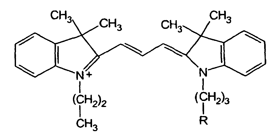
- the cyanine dyes have the following general structure, where X and Y can be O, S or (CH 3 ) 2 -C, m is an integer from 1 to 3 and at least one of R 1 , R 2 , R 3 , R 4 , R 5 , R 6 or R 7 is a reactive group which reacts with amino, hydroxy or sulfhydryl nucleophiles.
- the dotted lines represent the carbon atoms necessary for the formation of the cyanine dye, preferably for the carbon atoms forming three fused rings having 5 to 6 atoms in each ring.
- R 3 , R 4 , R 6 and R 7 are attached to the rings.
- the reactive moiety can be any known reactive group.
- Reactive groups that may be attached directly or indirectly to the chromophore to form R 1 , R 2 , R 3 , R 4 , R 5 , R 6 or R 7 groups may include reactive moieties such as groups containing isothiocyanate, isocyanate, monochlorotriazine, dichlorotriazine, mono- or di-halogen substituted pyridine, mono- or di-halogen substituted diazine, maleimide, aziridine, sulfonyl halide, acid halide, hydroxysuccinimide ester, hydroxysulfosuccinimide ester, imido ester, hydrazine, azidonitrophenyl, azide, 3-(2-pyridyl dithio)- proprionamide, glyoxal and aldehyde.
- reactive moieties such as groups containing isothiocyanate, isocyanate, monochlorotriazine, dichlorotriazine,
- the cyanine dyes described in the Waggoner et al. patent were the fluorophors of choice because of their intrinsic positive charge.
- the cyanines attach to the protein via the activated ester of hexanoic acid. While the coupling destroys the charge of the lysine side chain, the intrinsic charge in the dye compensates. It in effect moves the charge away from the protein molecule but maintains the same overall charge within the sample to be electrophoresed.
- two functionalized indole rings are connected via a polyene linker. The spectral characteristics of cyanine dyes can be easily modulated by simply changing the length of the linker between the indole rings of the dye.
- a longer or shorter linker length will result in fluorescence at different wavelengths and thus, different colors.
- changing the length of the linker changes the molecular mass of the dye. Since electrophoresis depends also on the mass of the proteins, the effect of the dye on a protein's mass can also be of concern. Because the proteins are labeled before electrophoresing, the mass of the dye attached to the protein must not significantly alter the relative differences in the molecular weights of the various proteins in the extracts. Molecular weight is not critical, however, because only a relatively small number of sites on the protein are labeled. As indicated above, preferably less than 1%, up to about 2% of the possible attachment sites on the proteins are labeled. If more are labeled, maintaining generally equal molecular weights for the dyes within the set of matched dyes becomes a greater concern.
- the difference in molecular weight caused by changing the linker length in the fluorescent cyanine dyes can be compensated for by modulating the size of an aliphatic chain R 1 or R 2 , attached to one of the dye's indole rings.
- R 1 or R 2 must be a reactive group.
- the cyanine dyes are one choice for the matched set of dyes used in the present invention.
- Other dye compounds may be used in place of the cyanines, such as dipyrromethene boron difluoride dyes, the derivatized 4,4-difluoro-4-bora-3a,4a,-diaza-S-indacene dyes, described in U.S. Patent No. 4,774,339 to Haugland et al., which are sold by Molecular Probes, Inc. under the trademark BODIPY®.
- the BODIPY® dyes which have no net charge, are covalently linked to lysine side chains using an activated n-hydroxysuccinimidyl ester which forms an amide bond.
- a positively charged linker group is used in the matched dyes used in the invention to replace the lost primary amine with the linker's tertiary amine.
- the procedures for making BODIPY® dyes are described in U.S. Patent No. 4,774,339. Addition of the positively charged linker is by techniques well known to those skilled in the art.
- a linker can be designed with three functional groups; (1) to react with the BODIPY®-NHS ester, (2) to carry the desired charge, and (3) to be activated so that the BODIPY®-linker construct will react with specific amino acid residues of the proteins in the extract.
- the major considerations for the matched set of dyes are the maintenance of charge and distinct and different spectral characteristics. Any neutral dyes with a positive linker or any positively charged dyes, preferably each having a +1 charge, that otherwise satisfy the requirements described herein can serve as the dyes in the matched set of dyes used in the present invention. Roughly equal molecular weight in the samples of labeled protein is desirable, but as explained above, not critical.
- the intrinsic positive charge of cyanine dyes is advantageously used in the preferred embodiment to replace the positive charge of lysine.
- the pK a of cyanines and lysine are rather different; however, conditions were selected for dye:protein ratio to be less than one.
- the attachment site on the protein may be a sulfhydryl or carboxylic group.
- the corresponding attachment site on the dye is an iodoalkyl group.
- the corresponding attachment site on the dye is a chloroketone or a carbodiimide.
- nucleic acids can be modified to have a free amino acid coming from the nucleic acid nucleus by techniques known to those skilled in the art. A lysine would be suitable in this instance also.
- the synthesis of the Cy-5 intermediate is the same as the synthesis of the Cy-3 intermediate in step 2 of the dye synthesis except that 2-methylene-1,3,3-trimethylindoline was used instead of propyl indole and the linker was malonaldehyde dianil. The gummy, bluish intermediate was washed twice with ethyl ether.
- Methyl Cy-5 and propyl Cy-3 were separated from contaminating side products by running flash chromatography with a silica gel solid phase and 40% MeOH in dichloromethane as the mobile phase.
- each dye was converted into an N-hydroxysuccinimidyl ester by dissolving a quantity of purified material in 5 ml of dry dimethylformamidine (DMF). 1.5 equivalents of N-N' disuccinimidyl carbonate (DSC) was added with 0.1 ml dry pyridine/100mg dye. The reaction was refluxed at 60°C for 90 minutes under nitrogen.
- DMF dry dimethylformamidine
- DSC N-N' disuccinimidyl carbonate
- Isolation of protein was as follows. The bacteria was isolated by centrifugation. Each bacterial pellet was washed with sonication buffer containing 5 mM Hepes KOH pH 8.4, 5 mM Mg(OAc)2. The pellet was resuspended in sonication buffer containing 50 ⁇ g/ml RNase to a final volume of 100 ⁇ l. This was then sonicated in ice until the solution was clear, usually several minutes. DNase was added to 50 ⁇ g/ml and the sample was incubated for 30 min at 0°C. Solid urea and CHAPS were added to a final concentration of 8 M and 5% respectively. The sample was taken off the ice and 1 volume of lysis buffer added. The sample was either labeled immediately or stored at -80°C.
- Propyl Cy-3-NHS was added to the first sample and Methyl Cy-5-NHS was added to the second sample of cell extract at a concentration of 2 nmole of dye/50 ⁇ g of protein.
- the dye stock solution was typically 2 mM in dimethyl formamide.
- the reaction was incubated at 0°C for 30 minutes. Incubation times may vary from about 15 to about 30 minutes, depending on the temperature and the type of cells being studied. Incubation can be for 15 minutes when the temperature is about 25°C. The temperature should not be above that which will cause the proteins to be degraded.
- the labeled sample was immediately subjected to isoelectric focusing or stored at -80°C.
- Bacteria were grown and isolated by sonication as in step 2. of the protein labeling procedure, except RNase or DNase was not added.
- the cell extract was directly labeled as in step 3 of the protein labeling procedure.
- SDS, glycerol, Tris HCl pH 6.8, and bromophenol blue were added to bring the final concentrations to 1%, 10%, 64 mM, and 5 ⁇ g/ml, respectively.
- the sample was then placed in a boiling water bath for 2 minutes and then subjected to electrophoresis.
- SDS polyacrylamide gel electrophoresis was carried out by known techniques.
- the gels were soaked in a solution of 25% methanol and 7% acetic acid.
- the fluorescently labeled proteins in the gel were imaged in the following manner. Gels were placed on a surface of anodized aluminum and irradiated at an incident angle of 60° with a 300 W halogen lamp housed in a slide projector. The light exiting the projector was passed through 1' diameter bandpass filters (Chroma Technologies, Brattleboro VT), 545 ⁇ 10 nm and 635 ⁇ 15 nm for Cy-3 and Cy-5, respectively.
- the images were collected on a cooled, CCD camera (Photometrics Inc., Arlington AZ) fitted with a 50 mm lens (Nikon) and a double bandpass emission filter (Chroma Technologies, Brattleboro VT), 587.5 ⁇ 17.5 nm and 695 ⁇ 30 nm for Cy-3 and Cy-5, respectively.
- the CCD camera was controlled by a Macintosh II si computer running Photometrics camera controller software. Image integration time ranged from tenths of seconds to several minutes.
- the excitation filters were housed in a filter wheel attached to the projector. Two successive images were recorded with irradiation from the two filters without moving the gel.
- the image files were transferred to a Personal Iris 4D/35 (Silicon Graphics Inc., Mountain View CA). The image files were then processed using the DeltaVisionTM software (Applied Precision, Mercer Island WA). The two schemes were used to determine the differences between the differently labeled samples on the gel:
- Each image can be considered as a grid-like array of pixel intensities. These arrays of values can be manipulated by a number of arithmetic operations. Here one image was subtracted from the other. Because the two samples loaded onto the gel were not perfectly balanced for overall fluorescence, one image was multiplied by a balancing constant. This factor was determined arbitrarily so that the number of differences between the samples were kept small.
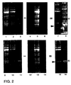
- Figure 2 shows images of Propyl Cy-3 and Methyl Cy-5 labeled proteins run on a single SDS polyacrylamide gel. Lanes 1-3 show Cy-3 labeled protein. The samples loaded in there lanes were:
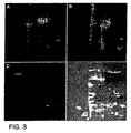
- Figure 3 shows images of a portion of a two-dimension gel loaded with Propyl Cy-3 labeled IPTG-induced bacterial extract plus Methyl Cy-5 labeled uninduced extract.
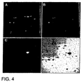
- Figure 4 shows images of a portion of a two-dimension gel loaded with Propyl Cy-3 labeled bacterial extract that had exogenously added carbonic anhydrase plus Methyl Cy-5 labeled extract without the added carbonic anhydrase.
- the method of the present invention provides a simple and inexpensive way to analyze the differences in protein content of different cells.
- the method eliminates problems which can occur using two separate gels which must be separately electrophoresed.
- the matched dyes used to label the different cell extracts allow simultaneous electrophoresis of two or more different samples of cell extract in a single gel. While the invention has been described with reference to two samples of cell extract and a matched pair of dyes, those skilled in the art will appreciate that more than two samples may be simultaneously tested using an equal number of matched dyes.
- the spectral characteristics of the dyes can be manipulated to provide fluorescence at a number of different wavelengths resulting in visually distinct images and the pH and ionic characteristics of the dyes can be generally equalized to compensate for changes made to the protein by virtue of covalent bonding to the dye, multiple dyes can be used.
Landscapes
- Chemical & Material Sciences (AREA)
- Health & Medical Sciences (AREA)
- Life Sciences & Earth Sciences (AREA)
- Physics & Mathematics (AREA)
- Biochemistry (AREA)
- Immunology (AREA)
- Molecular Biology (AREA)
- General Health & Medical Sciences (AREA)
- Analytical Chemistry (AREA)
- Organic Chemistry (AREA)
- Chemical Kinetics & Catalysis (AREA)
- General Physics & Mathematics (AREA)
- Pathology (AREA)
- Genetics & Genomics (AREA)
- Nuclear Medicine, Radiotherapy & Molecular Imaging (AREA)
- Medicinal Chemistry (AREA)
- Optics & Photonics (AREA)
- Proteomics, Peptides & Aminoacids (AREA)
- Biophysics (AREA)
- Spectroscopy & Molecular Physics (AREA)
- Electrochemistry (AREA)
- Investigating Or Analysing Biological Materials (AREA)
- Investigating, Analyzing Materials By Fluorescence Or Luminescence (AREA)
- Peptides Or Proteins (AREA)
- Cosmetics (AREA)
- Investigating Or Analysing Materials By The Use Of Chemical Reactions (AREA)
Abstract
Description



-
Lane 1. - Propyl Cy-3 labeled IPTG-induced bacterial extract.
-
Lane 2. - Propyl Cy-3 labeled IPTG-induced bacterial extract plus Methyl Cy-5 labeled uninduced extract.
-
Lane 3. - Propyl Cy-3 labeled purified GAL4VP16 protein.
-
Lane 4. - Propyl Cy-3 labeled IPTG-induced bacterial extract.
-
Lane 5. - Propyl Cy-3 labeled IPTG-induced bacterial extract plus Methyl Cy-5 labeled uninduced extract.
-
Lane 6 - Propyl Cy-3 labeled purified GAL4VP16 protein.
Lanes 9-11 show Cy-5 labeled protein. The samples loaded in these lanes were:
-
Lane 9. - Methyl Cy-5 labeled IPTG-induced bacterial extract.
-
Lane 10. - Methyl Cy-5 labeled IPTG-induced bacterial extract plus Propyl Cy-3 labeled uninduced extract.
-
Lane 11. - Methyl Cy-5 labeled purified GAL4VP16 protein.
-
Lane 12. - Methyl Cy-5 labeled IPTG-induced bacterial extract.
-
Lane 13. - Methyl Cy-5 labeled IPTG-induced bacterial extract plus Propyl Cy-3 labeled uninduced extract.
-
Lane 14. - Methyl Cy-5 labeled purified GAL4VP16 protein.
- Panel A.
- Images taken with Cy-3 excitation light showing the IPTG-induced proteins.
- Panel B.
- Images taken with Cy-5 excitation light showing the uninduced proteins.
- Panel C.
- Ratio of the Cy-3 image divided by the Cy-5 image.
- Panel D.
- Overlay of the image in Panel C, colored red, and placed on top of the image from Panel B, colored blue.
- Panel A.
- Image taken with Cy-3 excitation light showing the bacterial proteins plus carbonic anhydrase.
- Panel B.
- Images taken with Cy-5 excitation light showing the bacterial proteins alone.
- Panel C.
- Ratio of the Cy-3 image divided by the Cy-5 image.
- Panel D.
- Overlay of the image in Panel C, colored red, and placed on top of the image from Panel B, colored blue.
Claims (11)
- A method of detecting differences in the protein components of at least two different samples, the components having covalent attachment sites thereon, the method comprising:a) preparing an extract of proteins from each of said at least two samples;b) adding a different luminescent dye chosen from a set of matched luminescent dyes to each separate sample of protein extract under conditions to covalently bind the dye to the protein extract, each luminescent dye within said set of dyes being capable of covalently binding to protein and wherein each dye within said matched set:i) has a net charge which will maintain the overall net charge of the protein upon such covalent binding and has ionic and pH characteristics whereby relative electrophoretic migration of a protein labeled with any one of said dyes is the same as relative electrophoretic migration of said protein labeled with another dye in said matched set,ii) emits luminescent light at a wavelength that is sufficiently different from the emitted luminescent light of the remaining dyes in said matched set to provide a detectably different light signal;c) mixing the extracts together to form a mixture;d) electrophoresing the mixture on a single gel to separate the components of the extracts by differences in charge and mass; and(e) detecting the difference in luminescent intensity between the different dye-labeled proteins of interest by:capturing images of the dye-labeled proteins at different wavelengths of emitted luminescence; andprocessing the images to determine the difference in luminescent intensity between the different dye-labeled proteins of interest.
- The method according to claim 1, wherein said samples are cell samples.
- The method according to claim 1 or claim 2, wherein said capturing and processing steps are performed on at least a first and a second image.
- The method according to claim 1 or claim 2, wherein detecting the difference in luminescent intensity between the different dye-labeled proteins of interest comprises: capturing first and second images of the dye-labeled proteins; and performing arithmetic operations on values representative of pixel intensities in the first and second images.
- The method according to claim 1 or claim 2, wherein detecting the difference in luminescent intensity between the different dye-labeled proteins of interest comprises:a) capturing a first image of the dye labeled proteins using a first filter or filters that only allows the passage of light having the wavelength of the luminescent light emitted by a first dye used to covalently bind to protein components of interest;b) capturing a second image of the dye labeled proteins using a second filter or filters that only allows the passage of light having the wavelength of the luminescent light emitted by a second dye used to covalently bind to protein components of interest; andc) processing the first and second images to determine the difference in luminescent intensity.
- The method of claim 5, wherein processing the first and second images includes subtracting the first image from the second image.
- The method of claim 6, wherein processing the first and second images further includes multiplying one of the first and second image by a fluorescence balancing factor prior to subtracting the first image from the second image.
- The method of claim 5, wherein processing the first and second images includes dividing the first image by the second image.
- The method of claim 8, wherein processing the first and second images further includes normalizing the first and second images to a common intensity range prior to dividing the first image by the second image.
- The method of claim 9, wherein processing the first and second images further includes multiplying one of the first and second images by a fluorescence balancing factor.
- The method according to any of the previous claims, wherein processing the images comprises processing the images with a computer.
Applications Claiming Priority (3)
| Application Number | Priority Date | Filing Date | Title |
|---|---|---|---|
| US425480 | 1995-04-20 | ||
| US08/425,480 US6127134A (en) | 1995-04-20 | 1995-04-20 | Difference gel electrophoresis using matched multiple dyes |
| EP96912911A EP0821787B1 (en) | 1995-04-20 | 1996-04-19 | Difference gel electrophoresis using matched multiple dyes |
Related Parent Applications (1)
| Application Number | Title | Priority Date | Filing Date |
|---|---|---|---|
| EP96912911.3 Division | 1996-10-24 |
Publications (2)
| Publication Number | Publication Date |
|---|---|
| EP1494026A1 EP1494026A1 (en) | 2005-01-05 |
| EP1494026B1 true EP1494026B1 (en) | 2005-08-31 |
Family
ID=23686744
Family Applications (2)
| Application Number | Title | Priority Date | Filing Date |
|---|---|---|---|
| EP96912911A Expired - Lifetime EP0821787B1 (en) | 1995-04-20 | 1996-04-19 | Difference gel electrophoresis using matched multiple dyes |
| EP04023563A Expired - Lifetime EP1494026B1 (en) | 1995-04-20 | 1996-04-19 | Difference gel electrophoresis using matched multiple dyes |
Family Applications Before (1)
| Application Number | Title | Priority Date | Filing Date |
|---|---|---|---|
| EP96912911A Expired - Lifetime EP0821787B1 (en) | 1995-04-20 | 1996-04-19 | Difference gel electrophoresis using matched multiple dyes |
Country Status (9)
| Country | Link |
|---|---|
| US (2) | US6127134A (en) |
| EP (2) | EP0821787B1 (en) |
| JP (2) | JP3729859B2 (en) |
| AT (2) | ATE293252T1 (en) |
| AU (1) | AU709733B2 (en) |
| CA (1) | CA2218528C (en) |
| DE (2) | DE69634600T2 (en) |
| ES (2) | ES2248781T3 (en) |
| WO (1) | WO1996033406A1 (en) |
Families Citing this family (98)
| Publication number | Priority date | Publication date | Assignee | Title |
|---|---|---|---|---|
| US6593148B1 (en) | 1994-03-01 | 2003-07-15 | Li-Cor, Inc. | Cyanine dye compounds and labeling methods |
| US6426190B1 (en) * | 1995-04-20 | 2002-07-30 | Carnegie Mellon University | Difference detection methods using matched multiple dyes |
| GB9624927D0 (en) | 1996-11-29 | 1997-01-15 | Oxford Glycosciences Uk Ltd | Gels and their use |
| US5993627A (en) * | 1997-06-24 | 1999-11-30 | Large Scale Biology Corporation | Automated system for two-dimensional electrophoresis |
| JP4549528B2 (en) * | 1997-07-28 | 2010-09-22 | ジーイー・ヘルスケア・リミテッド | Cyanine dye |
| US6379971B1 (en) | 1998-02-24 | 2002-04-30 | Target Discovery, Inc. | Methods for sequencing proteins |
| GB9811656D0 (en) | 1998-05-29 | 1998-07-29 | Oxford Glycosciences Uk Ltd | Gels, methods and apparatus for identification and characterization of biomolecules |
| AU763102B2 (en) * | 1998-09-16 | 2003-07-10 | Applera Corporation | Spectral calibration of fluorescent polynucleotide separation apparatus |
| US6821402B1 (en) | 1998-09-16 | 2004-11-23 | Applera Corporation | Spectral calibration of fluorescent polynucleotide separation apparatus |
| US6716636B1 (en) | 1999-04-20 | 2004-04-06 | Target Discovery, Inc. | Methods for sequencing proteins |
| US6764817B1 (en) | 1999-04-20 | 2004-07-20 | Target Discovery, Inc. | Methods for conducting metabolic analyses |
| SE9903988D0 (en) * | 1999-11-03 | 1999-11-03 | Amersham Pharm Biotech Ab | Method of analysis |
| SE9903989D0 (en) * | 1999-11-03 | 1999-11-03 | Amersham Pharm Biotech Ab | Method of accurate analysis |
| SE9903987D0 (en) * | 1999-11-03 | 1999-11-03 | Amersham Pharm Biotech Ab | Method of comparison analysis |
| US20080096284A1 (en) * | 2000-02-08 | 2008-04-24 | Regents Of The University Of Michigan | Protein separation and analysis |
| US20030064527A1 (en) * | 2001-02-07 | 2003-04-03 | The Regents Of The University Of Michigan | Proteomic differential display |
| AU2001284853A1 (en) * | 2000-08-11 | 2002-02-25 | Invitrogen Corporation | Highly homogeneous molecular markers for electrophoresis |
| EP1770129B1 (en) | 2000-09-29 | 2012-07-25 | Life Technologies Corporation | Modified carbocyanine dyes and their conjugates |
| ATE352586T2 (en) | 2000-09-29 | 2007-02-15 | Molecular Probes Inc | MODIFIED CARBOCYANINE DYES AND CONJUGATES THEREOF |
| US20040209251A1 (en) | 2000-10-19 | 2004-10-21 | Schneider Luke V | Methods for determining protein and peptide terminal sequences |
| AU1702902A (en) * | 2000-11-16 | 2002-05-27 | Basf Ag | Method for separating and detecting proteins by means of electrophoresis |
| WO2002063271A2 (en) * | 2001-02-05 | 2002-08-15 | Activx Biosciences, Inc. | Activity based probe analysis |
| EP1370867B1 (en) * | 2001-03-02 | 2010-08-04 | Activx Biosciences, Inc. | Protein profiling platform |
| US7045296B2 (en) | 2001-05-08 | 2006-05-16 | Applera Corporation | Process for analyzing protein samples |
| US20050069962A1 (en) * | 2001-10-12 | 2005-03-31 | Archer Robert M | Antibody complexes and methods for immunolabeling |
| US8323903B2 (en) | 2001-10-12 | 2012-12-04 | Life Technologies Corporation | Antibody complexes and methods for immunolabeling |
| US6972326B2 (en) * | 2001-12-03 | 2005-12-06 | Molecular Probes, Inc. | Labeling of immobilized proteins using dipyrrometheneboron difluoride dyes |
| US20030134303A1 (en) * | 2001-12-11 | 2003-07-17 | Activx Biosciences, Inc, | Adenine nucleotide-binding protein-directed probes, and methods of their synthesis and use |
| AU2003207438A1 (en) * | 2002-01-02 | 2003-07-24 | Visen Medical, Inc. | Amine functionalized superparamagnetic nanoparticles for the synthesis of bioconjugates and uses therefor |
| JP2006505767A (en) * | 2002-05-06 | 2006-02-16 | パーキネルマー ラス インコーポレイテッド | Separation method and dye used therefor |
| CA2490961A1 (en) * | 2002-07-01 | 2004-01-08 | Guava Technologies, Inc. | Fluorescent dyes, energy transfer couples and methods |
| ES2341946T3 (en) * | 2002-07-08 | 2010-06-30 | Ge Healthcare Uk Limited | REAGENTS AND A METHOD FOR MARKING THROUGH PROTEIN SATURATION. |
| US7582260B2 (en) * | 2002-07-18 | 2009-09-01 | Montana State University | Zwitterionic dyes for labeling in proteomic and other biological analyses |
| AU2003282575A1 (en) * | 2002-10-09 | 2004-05-04 | Activx Biosciences, Inc. | Activity-based probes, and methods of their preparation and use |
| EP1441035B1 (en) * | 2003-01-23 | 2005-03-23 | BioVendor Laboratory Medicine, Inc. | Method of obtaining profiles of differential protein expression |
| CA2525685A1 (en) * | 2003-05-28 | 2004-12-09 | Amersham Biosciences Uk Limited | Differential analysis of cell surface proteins on closed membrane structures by labelling with dyes in the presence of an internal standard |
| EP1668146B1 (en) * | 2003-09-25 | 2014-11-12 | Life Technologies Corporation | Homogeneous populations of molecules |
| CN101296927A (en) | 2005-09-02 | 2008-10-29 | Visen医药公司 | Biocompatible Fluorescent Dye Labeling Containing N,N-Disubstituted Sulfonamides |
| WO2007028118A2 (en) | 2005-09-02 | 2007-03-08 | Visen Medical, Inc. | Nicotinic acid and picolinic acid derived near-infrared fluorophores |
| DK1937676T3 (en) * | 2005-09-02 | 2017-03-06 | Visen Medical Inc | Biocompatible fluorescent imaging compounds |
| WO2007034907A1 (en) * | 2005-09-22 | 2007-03-29 | Chemicals Evaluation And Research Institute | Method of estimating effect of test chemical on living organisms |
| US8093012B2 (en) * | 2005-10-13 | 2012-01-10 | Aureon Laboratories, Inc. | Multiplex in situ immunohistochemical analysis |
| WO2007111669A2 (en) | 2005-12-22 | 2007-10-04 | Visen Medical, Inc. | Combined x-ray and optical tomographic imaging system |
| EP1973575B1 (en) | 2005-12-22 | 2019-07-24 | Visen Medical, Inc. | Biocompatible fluorescent metal oxide nanoparticles |
| JP2009524832A (en) | 2006-01-24 | 2009-07-02 | ライフ テクノロジーズ コーポレーション | Device and method for quantifying analytes |
| EP1987361A4 (en) * | 2006-01-30 | 2009-03-04 | Invitrogen Corp | Compositions and methods for detecting and quantifying toxic substances in disease states |
| WO2008029281A2 (en) | 2006-02-10 | 2008-03-13 | Invitrogen Corporation | Labeling and detection of post translationally modified proteins |
| US8114636B2 (en) | 2006-02-10 | 2012-02-14 | Life Technologies Corporation | Labeling and detection of nucleic acids |
| US8562802B1 (en) | 2006-02-13 | 2013-10-22 | Life Technologies Corporation | Transilluminator base and scanner for imaging fluorescent gels, charging devices and portable electrophoresis systems |
| SG171598A1 (en) | 2006-03-23 | 2011-06-29 | Absorber Ab | Blood group antigens of different types for diagnostic and therapeutic applications |
| JP5337691B2 (en) | 2006-04-20 | 2013-11-06 | ベクトン・ディキンソン・アンド・カンパニー | Thermostable protein and method for producing and using the same |
| WO2008002236A1 (en) * | 2006-06-28 | 2008-01-03 | Ge Healthcare Bio-Sciences Ab | Method of detecting interactions between protein components |
| US20080220442A1 (en) * | 2006-12-06 | 2008-09-11 | Proteinics | Difference detection methods using isoelectric focusing chips |
| US8906354B2 (en) | 2007-02-28 | 2014-12-09 | Bruker Biospin Corporation | Loaded latex optical molecular imaging probes containing lipophilic large stokes shift dyes |
| US8017104B2 (en) * | 2007-02-28 | 2011-09-13 | Carestream Health, Inc. | Large stoke shift dye used for optical imaging |
| WO2008109832A2 (en) * | 2007-03-08 | 2008-09-12 | Visen Medical, Inc. | Viable near-infrared fluorochrome labeled cells and methods of making and using same |
| GB0714202D0 (en) * | 2007-07-20 | 2007-08-29 | Ge Healthcare Bio Sciences Ab | Isoelectric point markers |
| WO2009055095A1 (en) | 2007-10-19 | 2009-04-30 | Visen Medical, Inc. | Imaging systems featuring waveguiding compensation |
| US20090142775A1 (en) * | 2007-11-30 | 2009-06-04 | Agilent Technologies, Inc. | Methods For Separating Organelles |
| EP2732826B1 (en) | 2008-01-18 | 2017-11-08 | Visen Medical, Inc. | Fluorescent imaging agents |
| WO2009114776A2 (en) | 2008-03-14 | 2009-09-17 | Visen Medical, Inc. | Integrin targeting agents and methods of using same |
| CN104263816B (en) | 2008-05-16 | 2018-10-19 | 生命技术公司 | Double label method for measuring cell Proliferation |
| US20110186434A1 (en) * | 2008-07-23 | 2011-08-04 | Leibniz Institute of Plant Biochemistry | Qualitative and/or quantitative determination of a proteinaceous molecule in a plurality of samples |
| US8864821B2 (en) | 2008-11-26 | 2014-10-21 | Visen Medical, Inc. | Methods and compositions for identifying subjects at risk of developing stent thrombosis |
| US8729276B2 (en) | 2009-04-17 | 2014-05-20 | Korea Institute Of Science And Technology | Cyanine compound for labeling biomolecule and preparation method thereof |
| AU2010286592B2 (en) | 2009-08-28 | 2015-08-13 | Visen Medical, Inc. | Systems and methods for tomographic imaging in diffuse media using a hybrid inversion technique |
| JP5730311B2 (en) | 2009-08-31 | 2015-06-10 | プロメガ コーポレイションPromega Corporation | Reactive cyanine compounds |
| JP5945226B2 (en) * | 2009-09-22 | 2016-07-05 | ビセン メディカル, インコーポレイテッド | System and method for virtual refractive index matching of diffusing media |
| KR101695617B1 (en) * | 2009-09-25 | 2017-01-16 | (주)바이오액츠 | Benzindocyanine compound for labeling material, intermediate therefore, and process for producing the same |
| US8974651B2 (en) | 2010-04-17 | 2015-03-10 | C.C. Imex | Illuminator for visualization of fluorophores |
| US8623324B2 (en) | 2010-07-21 | 2014-01-07 | Aat Bioquest Inc. | Luminescent dyes with a water-soluble intramolecular bridge and their biological conjugates |
| WO2012022734A2 (en) | 2010-08-16 | 2012-02-23 | Medimmune Limited | Anti-icam-1 antibodies and methods of use |
| WO2012047212A1 (en) * | 2010-10-06 | 2012-04-12 | Hewlett-Packard Development Company, L.P. | Modified bodipy dye matrix |
| CN102095776B (en) * | 2011-01-06 | 2013-07-31 | 浙江大学 | Method for detecting surface difference membrane protein of mesenchymal stem cell of umbilical cord source |
| CN102095778B (en) * | 2011-01-06 | 2013-07-31 | 浙江大学 | Method for detecting surface difference membrane protein of mesenchymal stem cell of cord blood source |
| CA2835286C (en) | 2011-05-09 | 2021-03-23 | Visen Medical, Inc. | Carbonic anhydrase targeting agents and methods of using same |
| CN104245954A (en) | 2012-03-30 | 2014-12-24 | 文森医学公司 | Bacteria imaging agents and methods of use thereof |
| WO2014028057A1 (en) | 2012-08-15 | 2014-02-20 | Visen Medical, Inc. | Prostate specific antigen agents and methods of using same for prostate cancer imaging |
| CN104853669B (en) | 2012-10-15 | 2017-07-21 | 文森医学公司 | The system of the diffusing media imaging of cross-modality weighted feature with fluorescence and bioluminescence source, method and apparatus |
| US20150246146A1 (en) | 2012-10-25 | 2015-09-03 | Life Technologies Corporation | Methods and compositions for enzyme-mediated site-specific radiolabeling of glycoproteins |
| WO2014190230A1 (en) | 2013-05-23 | 2014-11-27 | Iphenotype Llc | Phenotypic integrated social search database and method |
| WO2015050959A1 (en) | 2013-10-01 | 2015-04-09 | Yale University | Anti-kit antibodies and methods of use thereof |
| PT3052192T (en) | 2013-10-02 | 2020-09-24 | Medimmune Llc | Neutralizing anti-influenza a antibodies and uses thereof |
| US10542918B2 (en) | 2013-10-23 | 2020-01-28 | Verily Life Sciences Llc | Modulation of a response signal to distinguish between analyte and background signals |
| US9504405B2 (en) | 2013-10-23 | 2016-11-29 | Verily Life Sciences Llc | Spatial modulation of magnetic particles in vasculature by external magnetic field |
| CN106455979A (en) | 2013-12-31 | 2017-02-22 | 纪念斯隆-凯特琳癌症中心 | Systems, methods and devices for real-time multi-channel imaging of fluorescent sources |
| US9835587B2 (en) | 2014-04-01 | 2017-12-05 | C.C. Imex | Electrophoresis running tank assembly |
| CA2954780A1 (en) | 2014-07-15 | 2016-01-21 | Medimmune, Llc | Neutralizing anti-influenza b antibodies and uses thereof |
| KR20170094268A (en) | 2014-12-15 | 2017-08-17 | 메모리얼 슬로안-케터링 캔서 센터 | Cyclic peptides with enhanced nerve-binding selectivity, nanoparticles bound with said cyclic peptides, and use of same for real-time in vivo nerve tissue imaging |
| US9861710B1 (en) | 2015-01-16 | 2018-01-09 | Verily Life Sciences Llc | Composite particles, methods, and in vivo diagnostic system |
| US10028659B2 (en) | 2015-03-26 | 2018-07-24 | Verily Life Sciences Llc | Aptamer-based sensors, implantable devices and detection system |
| BR112018011868A2 (en) | 2015-12-15 | 2018-12-04 | Univ Cornell | imaging systems and methods for tissue differentiation, eg for intraoperative visualization |
| EP3443126B1 (en) | 2016-04-15 | 2023-11-15 | Icahn School of Medicine at Mount Sinai | Tissue profiling using multiplexed immunohistochemical consecutive staining |
| BR112019010947A2 (en) | 2016-11-30 | 2019-10-01 | Memorial Sloan Kettering Cancer Center | ultra-small inhibitor functionalized nanoparticles and methods thereof |
| EP3601608A1 (en) | 2017-03-30 | 2020-02-05 | Life Technologies Corporation | Quantification of ngs dna by adapter sequence |
| CA3246952A1 (en) * | 2018-03-19 | 2025-10-31 | Regeneron Pharmaceuticals, Inc. | Microchip capillary electrophoresis assays and reagents |
| WO2022006368A2 (en) | 2020-07-02 | 2022-01-06 | Life Technologies Corporation | Trinucleotide cap analogs, preparation and uses thereof |
| EP4015004A1 (en) | 2020-12-18 | 2022-06-22 | Phi Pharma SA | Proteoglycan specific branched peptides |
Family Cites Families (19)
| Publication number | Priority date | Publication date | Assignee | Title |
|---|---|---|---|---|
| GB8513538D0 (en) * | 1985-05-29 | 1985-07-03 | Mackay C D | Electrophoresis |
| US4855225A (en) * | 1986-02-07 | 1989-08-08 | Applied Biosystems, Inc. | Method of detecting electrophoretically separated oligonucleotides |
| US5268486A (en) * | 1986-04-18 | 1993-12-07 | Carnegie-Mellon Unversity | Method for labeling and detecting materials employing arylsulfonate cyanine dyes |
| US5242796A (en) * | 1986-07-02 | 1993-09-07 | E. I. Du Pont De Nemours And Company | Method, system and reagents for DNA sequencing |
| JP2527340B2 (en) * | 1986-12-15 | 1996-08-21 | アプライド バイオシステムズ インコーポレイテッド | 5- and 6-succinimidyl carboxylate isomers of rhodamine dyes |
| US4774339A (en) * | 1987-08-10 | 1988-09-27 | Molecular Probes, Inc. | Chemically reactive dipyrrometheneboron difluoride dyes |
| US5262545A (en) * | 1989-03-09 | 1993-11-16 | Molecular Probes, Inc. | Fluorescent chloramphenicol derivatives for determination of chloramphenicol acetyltransferase activity |
| US5312921A (en) * | 1990-03-14 | 1994-05-17 | Regents Of The University Of California | Dyes designed for high sensitivity detection of double-stranded DNA |
| US5401847A (en) * | 1990-03-14 | 1995-03-28 | Regents Of The University Of California | DNA complexes with dyes designed for energy transfer as fluorescent markers |
| US5307148A (en) * | 1990-04-05 | 1994-04-26 | Hitachi, Ltd. | Fluorescence detection type electrophoresis apparatus |
| US5274113A (en) * | 1991-11-01 | 1993-12-28 | Molecular Probes, Inc. | Long wavelength chemically reactive dipyrrometheneboron difluoride dyes and conjugates |
| US5248782A (en) * | 1990-12-18 | 1993-09-28 | Molecular Probes, Inc. | Long wavelength heteroaryl-substituted dipyrrometheneboron difluoride dyes |
| US5338854A (en) * | 1991-02-13 | 1994-08-16 | Molecular Probes, Inc. | Fluorescent fatty acids derived from dipyrrometheneboron difluoride dyes |
| GB9103073D0 (en) * | 1991-02-13 | 1991-03-27 | Astromed Ltd | Improvements in or relating to electrophoresis |
| JP2873884B2 (en) * | 1991-03-22 | 1999-03-24 | 日立ソフトウェアエンジニアリング 株式会社 | Multicolor electrophoresis pattern reader |
| US5296599A (en) * | 1991-09-19 | 1994-03-22 | Millipore Corporation | Activated carbamates compounds |
| JPH05322771A (en) * | 1992-05-15 | 1993-12-07 | Toyobo Co Ltd | Multi-marker electrophoretic system |
| JPH05322770A (en) * | 1992-05-15 | 1993-12-07 | Toyobo Co Ltd | Multi-marker electrophoresis |
| US5654419A (en) * | 1994-02-01 | 1997-08-05 | The Regents Of The University Of California | Fluorescent labels and their use in separations |
-
1995
- 1995-04-20 US US08/425,480 patent/US6127134A/en not_active Expired - Lifetime
-
1996
- 1996-04-19 AU AU55573/96A patent/AU709733B2/en not_active Expired
- 1996-04-19 ES ES04023563T patent/ES2248781T3/en not_active Expired - Lifetime
- 1996-04-19 AT AT96912911T patent/ATE293252T1/en not_active IP Right Cessation
- 1996-04-19 EP EP96912911A patent/EP0821787B1/en not_active Expired - Lifetime
- 1996-04-19 JP JP53193396A patent/JP3729859B2/en not_active Expired - Lifetime
- 1996-04-19 DE DE69634600T patent/DE69634600T2/en not_active Expired - Lifetime
- 1996-04-19 WO PCT/US1996/005435 patent/WO1996033406A1/en not_active Ceased
- 1996-04-19 ES ES96912911T patent/ES2240993T3/en not_active Expired - Lifetime
- 1996-04-19 AT AT04023563T patent/ATE303593T1/en not_active IP Right Cessation
- 1996-04-19 CA CA002218528A patent/CA2218528C/en not_active Expired - Lifetime
- 1996-04-19 EP EP04023563A patent/EP1494026B1/en not_active Expired - Lifetime
- 1996-04-19 DE DE69635146T patent/DE69635146T2/en not_active Expired - Lifetime
-
1997
- 1997-10-10 US US08/949,115 patent/US6043025A/en not_active Expired - Lifetime
-
2005
- 2005-07-19 JP JP2005208506A patent/JP3890071B2/en not_active Expired - Lifetime
Also Published As
| Publication number | Publication date |
|---|---|
| JPH11505324A (en) | 1999-05-18 |
| ATE293252T1 (en) | 2005-04-15 |
| DE69635146D1 (en) | 2005-10-06 |
| EP0821787B1 (en) | 2005-04-13 |
| DE69634600T2 (en) | 2006-01-19 |
| JP3729859B2 (en) | 2005-12-21 |
| CA2218528A1 (en) | 1996-10-24 |
| ES2248781T3 (en) | 2006-03-16 |
| DE69634600D1 (en) | 2005-05-19 |
| CA2218528C (en) | 2003-06-24 |
| AU5557396A (en) | 1996-11-07 |
| EP1494026A1 (en) | 2005-01-05 |
| ATE303593T1 (en) | 2005-09-15 |
| JP2006023314A (en) | 2006-01-26 |
| JP3890071B2 (en) | 2007-03-07 |
| DE69635146T2 (en) | 2006-06-22 |
| US6043025A (en) | 2000-03-28 |
| US6127134A (en) | 2000-10-03 |
| WO1996033406A1 (en) | 1996-10-24 |
| AU709733B2 (en) | 1999-09-02 |
| ES2240993T3 (en) | 2005-10-16 |
| EP0821787A1 (en) | 1998-02-04 |
Similar Documents
| Publication | Publication Date | Title |
|---|---|---|
| EP0821787B1 (en) | Difference gel electrophoresis using matched multiple dyes | |
| US6426190B1 (en) | Difference detection methods using matched multiple dyes | |
| Ünlü et al. | Difference gel electrophoresis. A single gel method for detecting changes in protein extracts | |
| US7371745B2 (en) | Bis-transition-metal-chelate probes | |
| Gurrieri et al. | Direct visualization of individual DNA molecules by fluorescence microscopy: characterization of the factors affecting signal/background and optimization of imaging conditions using YOYO | |
| EP2223086B1 (en) | Fluorescent compounds - labeling dyes | |
| US6919333B2 (en) | Bis-transition-metal-chelate probes | |
| US20080220442A1 (en) | Difference detection methods using isoelectric focusing chips | |
| AU740831B2 (en) | Difference gel electrophoresis using matched multiple dyes | |
| JP4822409B2 (en) | Protein analysis method | |
| EP0938675A1 (en) | Analysis of carbohydrates | |
| US20110186434A1 (en) | Qualitative and/or quantitative determination of a proteinaceous molecule in a plurality of samples | |
| Chen et al. | Application of the commercial gel electrophoresis apparatus with intermittent fluorescence scanning to a nonfluorescing protein | |
| Minden | Jonathan zyxwvutsrqponmlkji | |
| Unlu | Identification of proteins involved in ventral furrow formation in Drosophila melanogaster embryos using difference gel electrophoresis, a modified two-dimensional gel electrophoresis technique |
Legal Events
| Date | Code | Title | Description |
|---|---|---|---|
| PUAI | Public reference made under article 153(3) epc to a published international application that has entered the european phase |
Free format text: ORIGINAL CODE: 0009012 |
|
| AC | Divisional application: reference to earlier application |
Ref document number: 0821787 Country of ref document: EP Kind code of ref document: P |
|
| AK | Designated contracting states |
Kind code of ref document: A1 Designated state(s): AT BE CH DE ES FR GB IT LI NL SE |
|
| GRAP | Despatch of communication of intention to grant a patent |
Free format text: ORIGINAL CODE: EPIDOSNIGR1 |
|
| 17P | Request for examination filed |
Effective date: 20050111 |
|
| RIN1 | Information on inventor provided before grant (corrected) |
Inventor name: WAGGONER, ALAN Inventor name: MINDEN, JONATHAN |
|
| GRAS | Grant fee paid |
Free format text: ORIGINAL CODE: EPIDOSNIGR3 |
|
| GRAA | (expected) grant |
Free format text: ORIGINAL CODE: 0009210 |
|
| AC | Divisional application: reference to earlier application |
Ref document number: 0821787 Country of ref document: EP Kind code of ref document: P |
|
| AK | Designated contracting states |
Kind code of ref document: B1 Designated state(s): AT BE CH DE ES FR GB IT LI NL SE |
|
| PG25 | Lapsed in a contracting state [announced via postgrant information from national office to epo] |
Ref country code: AT Free format text: LAPSE BECAUSE OF FAILURE TO SUBMIT A TRANSLATION OF THE DESCRIPTION OR TO PAY THE FEE WITHIN THE PRESCRIBED TIME-LIMIT Effective date: 20050831 Ref country code: BE Free format text: LAPSE BECAUSE OF FAILURE TO SUBMIT A TRANSLATION OF THE DESCRIPTION OR TO PAY THE FEE WITHIN THE PRESCRIBED TIME-LIMIT Effective date: 20050831 |
|
| REG | Reference to a national code |
Ref country code: CH Ref legal event code: EP Ref country code: GB Ref legal event code: FG4D |
|
| AKX | Designation fees paid |
Designated state(s): AT BE CH DE ES FR GB IT LI NL SE |
|
| REF | Corresponds to: |
Ref document number: 69635146 Country of ref document: DE Date of ref document: 20051006 Kind code of ref document: P |
|
| REG | Reference to a national code |
Ref country code: SE Ref legal event code: TRGR |
|
| REG | Reference to a national code |
Ref country code: CH Ref legal event code: NV Representative=s name: ISLER & PEDRAZZINI AG |
|
| REG | Reference to a national code |
Ref country code: ES Ref legal event code: FG2A Ref document number: 2248781 Country of ref document: ES Kind code of ref document: T3 |
|
| ET | Fr: translation filed | ||
| PLBE | No opposition filed within time limit |
Free format text: ORIGINAL CODE: 0009261 |
|
| STAA | Information on the status of an ep patent application or granted ep patent |
Free format text: STATUS: NO OPPOSITION FILED WITHIN TIME LIMIT |
|
| 26N | No opposition filed |
Effective date: 20060601 |
|
| REG | Reference to a national code |
Ref country code: CH Ref legal event code: PCAR Free format text: ISLER & PEDRAZZINI AG;POSTFACH 1772;8027 ZUERICH (CH) |
|
| REG | Reference to a national code |
Ref country code: DE Ref legal event code: R082 Ref document number: 69635146 Country of ref document: DE Representative=s name: J D REYNOLDS & CO., GB |
|
| REG | Reference to a national code |
Ref country code: FR Ref legal event code: PLFP Year of fee payment: 20 |
|
| PGFP | Annual fee paid to national office [announced via postgrant information from national office to epo] |
Ref country code: NL Payment date: 20150426 Year of fee payment: 20 |
|
| PGFP | Annual fee paid to national office [announced via postgrant information from national office to epo] |
Ref country code: DE Payment date: 20150429 Year of fee payment: 20 Ref country code: SE Payment date: 20150429 Year of fee payment: 20 Ref country code: ES Payment date: 20150427 Year of fee payment: 20 Ref country code: GB Payment date: 20150427 Year of fee payment: 20 Ref country code: CH Payment date: 20150427 Year of fee payment: 20 |
|
| PGFP | Annual fee paid to national office [announced via postgrant information from national office to epo] |
Ref country code: IT Payment date: 20150427 Year of fee payment: 20 Ref country code: FR Payment date: 20150417 Year of fee payment: 20 |
|
| REG | Reference to a national code |
Ref country code: DE Ref legal event code: R071 Ref document number: 69635146 Country of ref document: DE |
|
| REG | Reference to a national code |
Ref country code: NL Ref legal event code: MK Effective date: 20160418 |
|
| REG | Reference to a national code |
Ref country code: CH Ref legal event code: PL |
|
| REG | Reference to a national code |
Ref country code: GB Ref legal event code: PE20 Expiry date: 20160418 |
|
| REG | Reference to a national code |
Ref country code: SE Ref legal event code: EUG |
|
| REG | Reference to a national code |
Ref country code: ES Ref legal event code: FD2A Effective date: 20160727 |
|
| PG25 | Lapsed in a contracting state [announced via postgrant information from national office to epo] |
Ref country code: GB Free format text: LAPSE BECAUSE OF EXPIRATION OF PROTECTION Effective date: 20160418 |
|
| PG25 | Lapsed in a contracting state [announced via postgrant information from national office to epo] |
Ref country code: ES Free format text: LAPSE BECAUSE OF EXPIRATION OF PROTECTION Effective date: 20160420 |