EP1490682B1 - Detection of dna-binding proteins - Google Patents
Detection of dna-binding proteins Download PDFInfo
- Publication number
- EP1490682B1 EP1490682B1 EP03745653A EP03745653A EP1490682B1 EP 1490682 B1 EP1490682 B1 EP 1490682B1 EP 03745653 A EP03745653 A EP 03745653A EP 03745653 A EP03745653 A EP 03745653A EP 1490682 B1 EP1490682 B1 EP 1490682B1
- Authority
- EP
- European Patent Office
- Prior art keywords
- protein
- binding
- detection
- dna
- duplex
- Prior art date
- Legal status (The legal status is an assumption and is not a legal conclusion. Google has not performed a legal analysis and makes no representation as to the accuracy of the status listed.)
- Expired - Lifetime
Links
- 238000001514 detection method Methods 0.000 title claims abstract description 252
- 102000052510 DNA-Binding Proteins Human genes 0.000 title claims abstract description 56
- 108700020911 DNA-Binding Proteins Proteins 0.000 title abstract description 36
- 108090000623 proteins and genes Proteins 0.000 claims abstract description 229
- 102000004169 proteins and genes Human genes 0.000 claims abstract description 217
- 238000009739 binding Methods 0.000 claims abstract description 158
- 230000027455 binding Effects 0.000 claims abstract description 156
- 238000000034 method Methods 0.000 claims abstract description 72
- 239000000203 mixture Substances 0.000 claims abstract description 27
- 230000000694 effects Effects 0.000 claims abstract description 16
- 239000003153 chemical reaction reagent Substances 0.000 claims description 107
- 239000011324 bead Substances 0.000 claims description 78
- 238000003776 cleavage reaction Methods 0.000 claims description 72
- 230000007017 scission Effects 0.000 claims description 71
- 108091034117 Oligonucleotide Proteins 0.000 claims description 58
- YBJHBAHKTGYVGT-ZKWXMUAHSA-N (+)-Biotin Chemical compound N1C(=O)N[C@@H]2[C@H](CCCCC(=O)O)SC[C@@H]21 YBJHBAHKTGYVGT-ZKWXMUAHSA-N 0.000 claims description 29
- 102000004190 Enzymes Human genes 0.000 claims description 23
- 108090000790 Enzymes Proteins 0.000 claims description 23
- 108060002716 Exonuclease Proteins 0.000 claims description 23
- 102000013165 exonuclease Human genes 0.000 claims description 23
- 150000007523 nucleic acids Chemical group 0.000 claims description 21
- 101710096438 DNA-binding protein Proteins 0.000 claims description 20
- 102000039446 nucleic acids Human genes 0.000 claims description 17
- 108020004707 nucleic acids Proteins 0.000 claims description 17
- 108091008324 binding proteins Proteins 0.000 claims description 16
- 239000011616 biotin Substances 0.000 claims description 16
- 229960002685 biotin Drugs 0.000 claims description 16
- 235000020958 biotin Nutrition 0.000 claims description 15
- 230000000295 complement effect Effects 0.000 claims description 15
- 239000007787 solid Substances 0.000 claims description 14
- 239000000284 extract Substances 0.000 claims description 13
- 108090000765 processed proteins & peptides Proteins 0.000 claims description 13
- 102000023888 sequence-specific DNA binding proteins Human genes 0.000 claims description 10
- 108091008420 sequence-specific DNA binding proteins Proteins 0.000 claims description 10
- 108091028043 Nucleic acid sequence Proteins 0.000 claims description 9
- 230000005764 inhibitory process Effects 0.000 claims description 7
- 230000009870 specific binding Effects 0.000 claims description 7
- 239000002773 nucleotide Substances 0.000 claims description 6
- 125000003729 nucleotide group Chemical group 0.000 claims description 6
- 230000001413 cellular effect Effects 0.000 claims description 4
- 108010042407 Endonucleases Proteins 0.000 claims description 3
- 102000004533 Endonucleases Human genes 0.000 claims description 3
- 230000002285 radioactive effect Effects 0.000 claims description 3
- SHIBSTMRCDJXLN-UHFFFAOYSA-N Digoxigenin Natural products C1CC(C2C(C3(C)CCC(O)CC3CC2)CC2O)(O)C2(C)C1C1=CC(=O)OC1 SHIBSTMRCDJXLN-UHFFFAOYSA-N 0.000 claims description 2
- QONQRTHLHBTMGP-UHFFFAOYSA-N digitoxigenin Natural products CC12CCC(C3(CCC(O)CC3CC3)C)C3C11OC1CC2C1=CC(=O)OC1 QONQRTHLHBTMGP-UHFFFAOYSA-N 0.000 claims description 2
- SHIBSTMRCDJXLN-KCZCNTNESA-N digoxigenin Chemical compound C1([C@@H]2[C@@]3([C@@](CC2)(O)[C@H]2[C@@H]([C@@]4(C)CC[C@H](O)C[C@H]4CC2)C[C@H]3O)C)=CC(=O)OC1 SHIBSTMRCDJXLN-KCZCNTNESA-N 0.000 claims description 2
- 101710163270 Nuclease Proteins 0.000 claims 1
- 102000023732 binding proteins Human genes 0.000 claims 1
- 150000003384 small molecules Chemical class 0.000 claims 1
- 102000040945 Transcription factor Human genes 0.000 abstract description 52
- 108091023040 Transcription factor Proteins 0.000 abstract description 52
- -1 specifically Proteins 0.000 abstract description 11
- 230000004568 DNA-binding Effects 0.000 abstract description 7
- 235000018102 proteins Nutrition 0.000 description 214
- 239000000523 sample Substances 0.000 description 76
- 108020004414 DNA Proteins 0.000 description 73
- 108091008146 restriction endonucleases Proteins 0.000 description 34
- JLCPHMBAVCMARE-UHFFFAOYSA-N [3-[[3-[[3-[[3-[[3-[[3-[[3-[[3-[[3-[[3-[[3-[[5-(2-amino-6-oxo-1H-purin-9-yl)-3-[[3-[[3-[[3-[[3-[[3-[[5-(2-amino-6-oxo-1H-purin-9-yl)-3-[[5-(2-amino-6-oxo-1H-purin-9-yl)-3-hydroxyoxolan-2-yl]methoxy-hydroxyphosphoryl]oxyoxolan-2-yl]methoxy-hydroxyphosphoryl]oxy-5-(5-methyl-2,4-dioxopyrimidin-1-yl)oxolan-2-yl]methoxy-hydroxyphosphoryl]oxy-5-(6-aminopurin-9-yl)oxolan-2-yl]methoxy-hydroxyphosphoryl]oxy-5-(6-aminopurin-9-yl)oxolan-2-yl]methoxy-hydroxyphosphoryl]oxy-5-(6-aminopurin-9-yl)oxolan-2-yl]methoxy-hydroxyphosphoryl]oxy-5-(6-aminopurin-9-yl)oxolan-2-yl]methoxy-hydroxyphosphoryl]oxyoxolan-2-yl]methoxy-hydroxyphosphoryl]oxy-5-(5-methyl-2,4-dioxopyrimidin-1-yl)oxolan-2-yl]methoxy-hydroxyphosphoryl]oxy-5-(4-amino-2-oxopyrimidin-1-yl)oxolan-2-yl]methoxy-hydroxyphosphoryl]oxy-5-(5-methyl-2,4-dioxopyrimidin-1-yl)oxolan-2-yl]methoxy-hydroxyphosphoryl]oxy-5-(5-methyl-2,4-dioxopyrimidin-1-yl)oxolan-2-yl]methoxy-hydroxyphosphoryl]oxy-5-(6-aminopurin-9-yl)oxolan-2-yl]methoxy-hydroxyphosphoryl]oxy-5-(6-aminopurin-9-yl)oxolan-2-yl]methoxy-hydroxyphosphoryl]oxy-5-(4-amino-2-oxopyrimidin-1-yl)oxolan-2-yl]methoxy-hydroxyphosphoryl]oxy-5-(4-amino-2-oxopyrimidin-1-yl)oxolan-2-yl]methoxy-hydroxyphosphoryl]oxy-5-(4-amino-2-oxopyrimidin-1-yl)oxolan-2-yl]methoxy-hydroxyphosphoryl]oxy-5-(6-aminopurin-9-yl)oxolan-2-yl]methoxy-hydroxyphosphoryl]oxy-5-(4-amino-2-oxopyrimidin-1-yl)oxolan-2-yl]methyl [5-(6-aminopurin-9-yl)-2-(hydroxymethyl)oxolan-3-yl] hydrogen phosphate Polymers Cc1cn(C2CC(OP(O)(=O)OCC3OC(CC3OP(O)(=O)OCC3OC(CC3O)n3cnc4c3nc(N)[nH]c4=O)n3cnc4c3nc(N)[nH]c4=O)C(COP(O)(=O)OC3CC(OC3COP(O)(=O)OC3CC(OC3COP(O)(=O)OC3CC(OC3COP(O)(=O)OC3CC(OC3COP(O)(=O)OC3CC(OC3COP(O)(=O)OC3CC(OC3COP(O)(=O)OC3CC(OC3COP(O)(=O)OC3CC(OC3COP(O)(=O)OC3CC(OC3COP(O)(=O)OC3CC(OC3COP(O)(=O)OC3CC(OC3COP(O)(=O)OC3CC(OC3COP(O)(=O)OC3CC(OC3COP(O)(=O)OC3CC(OC3COP(O)(=O)OC3CC(OC3COP(O)(=O)OC3CC(OC3COP(O)(=O)OC3CC(OC3CO)n3cnc4c(N)ncnc34)n3ccc(N)nc3=O)n3cnc4c(N)ncnc34)n3ccc(N)nc3=O)n3ccc(N)nc3=O)n3ccc(N)nc3=O)n3cnc4c(N)ncnc34)n3cnc4c(N)ncnc34)n3cc(C)c(=O)[nH]c3=O)n3cc(C)c(=O)[nH]c3=O)n3ccc(N)nc3=O)n3cc(C)c(=O)[nH]c3=O)n3cnc4c3nc(N)[nH]c4=O)n3cnc4c(N)ncnc34)n3cnc4c(N)ncnc34)n3cnc4c(N)ncnc34)n3cnc4c(N)ncnc34)O2)c(=O)[nH]c1=O JLCPHMBAVCMARE-UHFFFAOYSA-N 0.000 description 26
- 238000003556 assay Methods 0.000 description 25
- 239000012148 binding buffer Substances 0.000 description 23
- TWRXJAOTZQYOKJ-UHFFFAOYSA-L Magnesium chloride Chemical compound [Mg+2].[Cl-].[Cl-] TWRXJAOTZQYOKJ-UHFFFAOYSA-L 0.000 description 22
- FAPWRFPIFSIZLT-UHFFFAOYSA-M Sodium chloride Chemical compound [Na+].[Cl-] FAPWRFPIFSIZLT-UHFFFAOYSA-M 0.000 description 22
- 102000003945 NF-kappa B Human genes 0.000 description 21
- 102000053602 DNA Human genes 0.000 description 20
- 108010057466 NF-kappa B Proteins 0.000 description 20
- 210000004027 cell Anatomy 0.000 description 17
- 238000006243 chemical reaction Methods 0.000 description 16
- 239000000126 substance Substances 0.000 description 16
- 102000014914 Carrier Proteins Human genes 0.000 description 15
- 238000003199 nucleic acid amplification method Methods 0.000 description 14
- 230000003321 amplification Effects 0.000 description 13
- 230000029087 digestion Effects 0.000 description 13
- 239000004005 microsphere Substances 0.000 description 13
- 238000011534 incubation Methods 0.000 description 12
- 239000007983 Tris buffer Substances 0.000 description 11
- 208000037265 diseases, disorders, signs and symptoms Diseases 0.000 description 11
- 239000000499 gel Substances 0.000 description 11
- 238000009396 hybridization Methods 0.000 description 11
- 229910001629 magnesium chloride Inorganic materials 0.000 description 11
- 239000011780 sodium chloride Substances 0.000 description 11
- 102100023050 Nuclear factor NF-kappa-B p105 subunit Human genes 0.000 description 10
- 201000010099 disease Diseases 0.000 description 10
- 102100033200 Rho guanine nucleotide exchange factor 7 Human genes 0.000 description 9
- 238000004458 analytical method Methods 0.000 description 9
- 230000014509 gene expression Effects 0.000 description 9
- 101710120037 Toxin CcdB Proteins 0.000 description 8
- 239000012634 fragment Substances 0.000 description 8
- 102000004196 processed proteins & peptides Human genes 0.000 description 8
- 238000003491 array Methods 0.000 description 7
- 230000008859 change Effects 0.000 description 6
- 239000007850 fluorescent dye Substances 0.000 description 6
- 238000002372 labelling Methods 0.000 description 6
- 239000000463 material Substances 0.000 description 6
- 238000002331 protein detection Methods 0.000 description 6
- 239000000243 solution Substances 0.000 description 6
- 108091003079 Bovine Serum Albumin Proteins 0.000 description 5
- 206010028980 Neoplasm Diseases 0.000 description 5
- 238000000137 annealing Methods 0.000 description 5
- 230000008512 biological response Effects 0.000 description 5
- 229940098773 bovine serum albumin Drugs 0.000 description 5
- 239000000872 buffer Substances 0.000 description 5
- 239000000975 dye Substances 0.000 description 5
- 238000002337 electrophoretic mobility shift assay Methods 0.000 description 5
- 238000005516 engineering process Methods 0.000 description 5
- 238000002474 experimental method Methods 0.000 description 5
- 230000003993 interaction Effects 0.000 description 5
- 125000002467 phosphate group Chemical group [H]OP(=O)(O[H])O[*] 0.000 description 5
- 108010001857 Cell Surface Receptors Proteins 0.000 description 4
- 238000000018 DNA microarray Methods 0.000 description 4
- NQTADLQHYWFPDB-UHFFFAOYSA-N N-Hydroxysuccinimide Chemical class ON1C(=O)CCC1=O NQTADLQHYWFPDB-UHFFFAOYSA-N 0.000 description 4
- 108020005187 Oligonucleotide Probes Proteins 0.000 description 4
- 229910019142 PO4 Inorganic materials 0.000 description 4
- 239000004793 Polystyrene Substances 0.000 description 4
- 102000014400 SH2 domains Human genes 0.000 description 4
- 108050003452 SH2 domains Proteins 0.000 description 4
- 108010090804 Streptavidin Proteins 0.000 description 4
- 230000004913 activation Effects 0.000 description 4
- 201000011510 cancer Diseases 0.000 description 4
- 239000011521 glass Substances 0.000 description 4
- 238000005259 measurement Methods 0.000 description 4
- 230000007246 mechanism Effects 0.000 description 4
- 102000006240 membrane receptors Human genes 0.000 description 4
- 102000044158 nucleic acid binding protein Human genes 0.000 description 4
- 108700020942 nucleic acid binding protein Proteins 0.000 description 4
- 239000002751 oligonucleotide probe Substances 0.000 description 4
- HXITXNWTGFUOAU-UHFFFAOYSA-N phenylboronic acid Chemical compound OB(O)C1=CC=CC=C1 HXITXNWTGFUOAU-UHFFFAOYSA-N 0.000 description 4
- DCWXELXMIBXGTH-UHFFFAOYSA-N phosphotyrosine Chemical compound OC(=O)C(N)CC1=CC=C(OP(O)(O)=O)C=C1 DCWXELXMIBXGTH-UHFFFAOYSA-N 0.000 description 4
- 229920002223 polystyrene Polymers 0.000 description 4
- 102000005962 receptors Human genes 0.000 description 4
- 108020003175 receptors Proteins 0.000 description 4
- 238000011160 research Methods 0.000 description 4
- 238000012216 screening Methods 0.000 description 4
- 230000019491 signal transduction Effects 0.000 description 4
- LENZDBCJOHFCAS-UHFFFAOYSA-N tris Chemical compound OCC(N)(CO)CO LENZDBCJOHFCAS-UHFFFAOYSA-N 0.000 description 4
- 238000005406 washing Methods 0.000 description 4
- 108091032973 (ribonucleotides)n+m Proteins 0.000 description 3
- AYXZIZMZXAORLO-UFLZEWODSA-N 5-[(3as,4s,6ar)-2-oxo-1,3,3a,4,6,6a-hexahydrothieno[3,4-d]imidazol-4-yl]pentanoic acid;1-hydroxypyrrolidine-2,5-dione Chemical compound ON1C(=O)CCC1=O.N1C(=O)N[C@@H]2[C@H](CCCCC(=O)O)SC[C@@H]21 AYXZIZMZXAORLO-UFLZEWODSA-N 0.000 description 3
- 108090001008 Avidin Proteins 0.000 description 3
- KCXVZYZYPLLWCC-UHFFFAOYSA-N EDTA Chemical compound OC(=O)CN(CC(O)=O)CCN(CC(O)=O)CC(O)=O KCXVZYZYPLLWCC-UHFFFAOYSA-N 0.000 description 3
- 238000002965 ELISA Methods 0.000 description 3
- 108020004711 Nucleic Acid Probes Proteins 0.000 description 3
- 230000010632 Transcription Factor Activity Effects 0.000 description 3
- 108010064978 Type II Site-Specific Deoxyribonucleases Proteins 0.000 description 3
- 150000001413 amino acids Chemical class 0.000 description 3
- 230000008878 coupling Effects 0.000 description 3
- 238000010168 coupling process Methods 0.000 description 3
- 238000005859 coupling reaction Methods 0.000 description 3
- 230000007423 decrease Effects 0.000 description 3
- 238000001962 electrophoresis Methods 0.000 description 3
- 230000006870 function Effects 0.000 description 3
- 239000012528 membrane Substances 0.000 description 3
- 238000002156 mixing Methods 0.000 description 3
- 230000004048 modification Effects 0.000 description 3
- 238000012986 modification Methods 0.000 description 3
- 239000002853 nucleic acid probe Substances 0.000 description 3
- NBIIXXVUZAFLBC-UHFFFAOYSA-K phosphate Chemical compound [O-]P([O-])([O-])=O NBIIXXVUZAFLBC-UHFFFAOYSA-K 0.000 description 3
- 239000010452 phosphate Substances 0.000 description 3
- 229920002401 polyacrylamide Polymers 0.000 description 3
- 238000002360 preparation method Methods 0.000 description 3
- 230000008569 process Effects 0.000 description 3
- 230000004044 response Effects 0.000 description 3
- 230000035945 sensitivity Effects 0.000 description 3
- 239000012536 storage buffer Substances 0.000 description 3
- 230000001225 therapeutic effect Effects 0.000 description 3
- 238000013518 transcription Methods 0.000 description 3
- 230000035897 transcription Effects 0.000 description 3
- WOVKYSAHUYNSMH-RRKCRQDMSA-N 5-bromodeoxyuridine Chemical compound C1[C@H](O)[C@@H](CO)O[C@H]1N1C(=O)NC(=O)C(Br)=C1 WOVKYSAHUYNSMH-RRKCRQDMSA-N 0.000 description 2
- 241000283707 Capra Species 0.000 description 2
- 238000012286 ELISA Assay Methods 0.000 description 2
- 102100036698 Golgi reassembly-stacking protein 1 Human genes 0.000 description 2
- 102000008394 Immunoglobulin Fragments Human genes 0.000 description 2
- 108010021625 Immunoglobulin Fragments Proteins 0.000 description 2
- 108010001127 Insulin Receptor Proteins 0.000 description 2
- 102000004856 Lectins Human genes 0.000 description 2
- 108090001090 Lectins Proteins 0.000 description 2
- 241000283973 Oryctolagus cuniculus Species 0.000 description 2
- 102000004160 Phosphoric Monoester Hydrolases Human genes 0.000 description 2
- 239000004698 Polyethylene Substances 0.000 description 2
- 229920001213 Polysorbate 20 Polymers 0.000 description 2
- 102000001253 Protein Kinase Human genes 0.000 description 2
- 150000001299 aldehydes Chemical group 0.000 description 2
- 230000008901 benefit Effects 0.000 description 2
- 150000001720 carbohydrates Chemical class 0.000 description 2
- 235000014633 carbohydrates Nutrition 0.000 description 2
- 239000007795 chemical reaction product Substances 0.000 description 2
- 239000003795 chemical substances by application Substances 0.000 description 2
- 239000003086 colorant Substances 0.000 description 2
- 239000000412 dendrimer Substances 0.000 description 2
- 229920000736 dendritic polymer Polymers 0.000 description 2
- 238000011161 development Methods 0.000 description 2
- 230000018109 developmental process Effects 0.000 description 2
- 229950006137 dexfosfoserine Drugs 0.000 description 2
- 238000003745 diagnosis Methods 0.000 description 2
- 238000007865 diluting Methods 0.000 description 2
- 231100000673 dose–response relationship Toxicity 0.000 description 2
- MHMNJMPURVTYEJ-UHFFFAOYSA-N fluorescein-5-isothiocyanate Chemical compound O1C(=O)C2=CC(N=C=S)=CC=C2C21C1=CC=C(O)C=C1OC1=CC(O)=CC=C21 MHMNJMPURVTYEJ-UHFFFAOYSA-N 0.000 description 2
- 238000002866 fluorescence resonance energy transfer Methods 0.000 description 2
- RWSXRVCMGQZWBV-WDSKDSINSA-N glutathione Chemical compound OC(=O)[C@@H](N)CCC(=O)N[C@@H](CS)C(=O)NCC(O)=O RWSXRVCMGQZWBV-WDSKDSINSA-N 0.000 description 2
- 230000000977 initiatory effect Effects 0.000 description 2
- 239000002523 lectin Substances 0.000 description 2
- 239000003446 ligand Substances 0.000 description 2
- 239000011159 matrix material Substances 0.000 description 2
- 239000011859 microparticle Substances 0.000 description 2
- 239000002245 particle Substances 0.000 description 2
- BZQFBWGGLXLEPQ-REOHCLBHSA-N phosphoserine Chemical compound OC(=O)[C@@H](N)COP(O)(O)=O BZQFBWGGLXLEPQ-REOHCLBHSA-N 0.000 description 2
- 229920000642 polymer Polymers 0.000 description 2
- 239000000256 polyoxyethylene sorbitan monolaurate Substances 0.000 description 2
- 235000010486 polyoxyethylene sorbitan monolaurate Nutrition 0.000 description 2
- 125000002924 primary amino group Chemical group [H]N([H])* 0.000 description 2
- 108091011138 protein binding proteins Proteins 0.000 description 2
- 108060006633 protein kinase Proteins 0.000 description 2
- 230000002829 reductive effect Effects 0.000 description 2
- 230000001105 regulatory effect Effects 0.000 description 2
- 230000000717 retained effect Effects 0.000 description 2
- 238000005096 rolling process Methods 0.000 description 2
- HBROZNQEVUILML-UHFFFAOYSA-N salicylhydroxamic acid Chemical compound ONC(=O)C1=CC=CC=C1O HBROZNQEVUILML-UHFFFAOYSA-N 0.000 description 2
- 230000011664 signaling Effects 0.000 description 2
- 230000003595 spectral effect Effects 0.000 description 2
- 239000000758 substrate Substances 0.000 description 2
- 239000000725 suspension Substances 0.000 description 2
- 238000012546 transfer Methods 0.000 description 2
- 0 *=C1C=CC=C1 Chemical compound *=C1C=CC=C1 0.000 description 1
- CZIHNRWJTSTCEX-UHFFFAOYSA-N 2 Acetylaminofluorene Chemical compound C1=CC=C2C3=CC=C(NC(=O)C)C=C3CC2=C1 CZIHNRWJTSTCEX-UHFFFAOYSA-N 0.000 description 1
- 229920001817 Agar Polymers 0.000 description 1
- WOVKYSAHUYNSMH-UHFFFAOYSA-N BROMODEOXYURIDINE Natural products C1C(O)C(CO)OC1N1C(=O)NC(=O)C(Br)=C1 WOVKYSAHUYNSMH-UHFFFAOYSA-N 0.000 description 1
- 208000024172 Cardiovascular disease Diseases 0.000 description 1
- 208000035473 Communicable disease Diseases 0.000 description 1
- 108020004394 Complementary RNA Proteins 0.000 description 1
- 102000005636 Cyclic AMP Response Element-Binding Protein Human genes 0.000 description 1
- 108010045171 Cyclic AMP Response Element-Binding Protein Proteins 0.000 description 1
- 239000003298 DNA probe Substances 0.000 description 1
- 102000004163 DNA-directed RNA polymerases Human genes 0.000 description 1
- 108090000626 DNA-directed RNA polymerases Proteins 0.000 description 1
- 108010054576 Deoxyribonuclease EcoRI Proteins 0.000 description 1
- 229920002307 Dextran Polymers 0.000 description 1
- 241000206602 Eukaryota Species 0.000 description 1
- 108010024636 Glutathione Proteins 0.000 description 1
- 108090000288 Glycoproteins Proteins 0.000 description 1
- 102000003886 Glycoproteins Human genes 0.000 description 1
- 102000003746 Insulin Receptor Human genes 0.000 description 1
- 102100036721 Insulin receptor Human genes 0.000 description 1
- AYFVYJQAPQTCCC-GBXIJSLDSA-N L-threonine Chemical compound C[C@@H](O)[C@H](N)C(O)=O AYFVYJQAPQTCCC-GBXIJSLDSA-N 0.000 description 1
- 239000007987 MES buffer Substances 0.000 description 1
- 241001465754 Metazoa Species 0.000 description 1
- 102100038895 Myc proto-oncogene protein Human genes 0.000 description 1
- 101710135898 Myc proto-oncogene protein Proteins 0.000 description 1
- 239000000020 Nitrocellulose Substances 0.000 description 1
- 108091005461 Nucleic proteins Proteins 0.000 description 1
- 239000004677 Nylon Substances 0.000 description 1
- BZQFBWGGLXLEPQ-UHFFFAOYSA-N O-phosphoryl-L-serine Natural products OC(=O)C(N)COP(O)(O)=O BZQFBWGGLXLEPQ-UHFFFAOYSA-N 0.000 description 1
- 208000008589 Obesity Diseases 0.000 description 1
- 108010016731 PPAR gamma Proteins 0.000 description 1
- 108091093037 Peptide nucleic acid Proteins 0.000 description 1
- 102100038825 Peroxisome proliferator-activated receptor gamma Human genes 0.000 description 1
- 108700019535 Phosphoprotein Phosphatases Proteins 0.000 description 1
- 108090000608 Phosphoric Monoester Hydrolases Proteins 0.000 description 1
- 108010004729 Phycoerythrin Proteins 0.000 description 1
- 239000004743 Polypropylene Substances 0.000 description 1
- 102000007056 Recombinant Fusion Proteins Human genes 0.000 description 1
- 108010008281 Recombinant Fusion Proteins Proteins 0.000 description 1
- 229920002684 Sepharose Polymers 0.000 description 1
- 108020004682 Single-Stranded DNA Proteins 0.000 description 1
- 229920002472 Starch Polymers 0.000 description 1
- 108010017842 Telomerase Proteins 0.000 description 1
- AYFVYJQAPQTCCC-UHFFFAOYSA-N Threonine Natural products CC(O)C(N)C(O)=O AYFVYJQAPQTCCC-UHFFFAOYSA-N 0.000 description 1
- 239000004473 Threonine Substances 0.000 description 1
- 101710150448 Transcriptional regulator Myc Proteins 0.000 description 1
- 101150111044 Ubx gene Proteins 0.000 description 1
- 241000700605 Viruses Species 0.000 description 1
- 239000008272 agar Substances 0.000 description 1
- 150000001412 amines Chemical class 0.000 description 1
- 230000001363 autoimmune Effects 0.000 description 1
- SQVRNKJHWKZAKO-UHFFFAOYSA-N beta-N-Acetyl-D-neuraminic acid Natural products CC(=O)NC1C(O)CC(O)(C(O)=O)OC1C(O)C(O)CO SQVRNKJHWKZAKO-UHFFFAOYSA-N 0.000 description 1
- 238000004166 bioassay Methods 0.000 description 1
- 239000012620 biological material Substances 0.000 description 1
- 230000031018 biological processes and functions Effects 0.000 description 1
- 230000033228 biological regulation Effects 0.000 description 1
- 239000012472 biological sample Substances 0.000 description 1
- 239000000090 biomarker Substances 0.000 description 1
- 230000015572 biosynthetic process Effects 0.000 description 1
- 210000004369 blood Anatomy 0.000 description 1
- 239000008280 blood Substances 0.000 description 1
- 210000001124 body fluid Anatomy 0.000 description 1
- 229950004398 broxuridine Drugs 0.000 description 1
- 230000005754 cellular signaling Effects 0.000 description 1
- 239000001913 cellulose Substances 0.000 description 1
- 229920002678 cellulose Polymers 0.000 description 1
- 230000000052 comparative effect Effects 0.000 description 1
- 239000003184 complementary RNA Substances 0.000 description 1
- 150000001875 compounds Chemical class 0.000 description 1
- 238000007796 conventional method Methods 0.000 description 1
- 238000001816 cooling Methods 0.000 description 1
- 210000004748 cultured cell Anatomy 0.000 description 1
- 230000008021 deposition Effects 0.000 description 1
- 238000011033 desalting Methods 0.000 description 1
- 230000001627 detrimental effect Effects 0.000 description 1
- 230000004069 differentiation Effects 0.000 description 1
- 238000010790 dilution Methods 0.000 description 1
- 239000012895 dilution Substances 0.000 description 1
- BFMYDTVEBKDAKJ-UHFFFAOYSA-L disodium;(2',7'-dibromo-3',6'-dioxido-3-oxospiro[2-benzofuran-1,9'-xanthene]-4'-yl)mercury;hydrate Chemical compound O.[Na+].[Na+].O1C(=O)C2=CC=CC=C2C21C1=CC(Br)=C([O-])C([Hg])=C1OC1=C2C=C(Br)C([O-])=C1 BFMYDTVEBKDAKJ-UHFFFAOYSA-L 0.000 description 1
- 208000035475 disorder Diseases 0.000 description 1
- 229940079593 drug Drugs 0.000 description 1
- 239000003814 drug Substances 0.000 description 1
- 238000007877 drug screening Methods 0.000 description 1
- 230000007613 environmental effect Effects 0.000 description 1
- 230000009088 enzymatic function Effects 0.000 description 1
- 238000001976 enzyme digestion Methods 0.000 description 1
- 150000002148 esters Chemical class 0.000 description 1
- 230000005281 excited state Effects 0.000 description 1
- 108010052305 exodeoxyribonuclease III Proteins 0.000 description 1
- 239000000835 fiber Substances 0.000 description 1
- 238000000684 flow cytometry Methods 0.000 description 1
- 239000012530 fluid Substances 0.000 description 1
- 125000002485 formyl group Chemical group [H]C(*)=O 0.000 description 1
- 108010055863 gene b exonuclease Proteins 0.000 description 1
- 238000011223 gene expression profiling Methods 0.000 description 1
- 102000034356 gene-regulatory proteins Human genes 0.000 description 1
- 108091006104 gene-regulatory proteins Proteins 0.000 description 1
- 230000007614 genetic variation Effects 0.000 description 1
- 230000014101 glucose homeostasis Effects 0.000 description 1
- 229960003180 glutathione Drugs 0.000 description 1
- 238000003505 heat denaturation Methods 0.000 description 1
- 238000010438 heat treatment Methods 0.000 description 1
- 229910001385 heavy metal Inorganic materials 0.000 description 1
- 229940042795 hydrazides for tuberculosis treatment Drugs 0.000 description 1
- 230000003100 immobilizing effect Effects 0.000 description 1
- 230000006872 improvement Effects 0.000 description 1
- 208000027866 inflammatory disease Diseases 0.000 description 1
- 230000002757 inflammatory effect Effects 0.000 description 1
- 230000002401 inhibitory effect Effects 0.000 description 1
- 230000002427 irreversible effect Effects 0.000 description 1
- 125000000468 ketone group Chemical group 0.000 description 1
- 229910052747 lanthanoid Inorganic materials 0.000 description 1
- 239000004816 latex Substances 0.000 description 1
- 229920000126 latex Polymers 0.000 description 1
- 238000007834 ligase chain reaction Methods 0.000 description 1
- 230000000670 limiting effect Effects 0.000 description 1
- 230000004130 lipolysis Effects 0.000 description 1
- 238000004020 luminiscence type Methods 0.000 description 1
- 229920002521 macromolecule Polymers 0.000 description 1
- 230000005291 magnetic effect Effects 0.000 description 1
- 239000000696 magnetic material Substances 0.000 description 1
- 238000012423 maintenance Methods 0.000 description 1
- 108020004999 messenger RNA Proteins 0.000 description 1
- 230000002503 metabolic effect Effects 0.000 description 1
- 229910052751 metal Inorganic materials 0.000 description 1
- 239000002184 metal Substances 0.000 description 1
- 238000002493 microarray Methods 0.000 description 1
- 244000005700 microbiome Species 0.000 description 1
- 238000012544 monitoring process Methods 0.000 description 1
- 238000007837 multiplex assay Methods 0.000 description 1
- 230000035772 mutation Effects 0.000 description 1
- 239000002159 nanocrystal Substances 0.000 description 1
- 230000000926 neurological effect Effects 0.000 description 1
- 239000002547 new drug Substances 0.000 description 1
- 229920001220 nitrocellulos Polymers 0.000 description 1
- 229920001778 nylon Polymers 0.000 description 1
- 235000020824 obesity Nutrition 0.000 description 1
- 239000013307 optical fiber Substances 0.000 description 1
- 210000000056 organ Anatomy 0.000 description 1
- 230000003647 oxidation Effects 0.000 description 1
- 238000007254 oxidation reaction Methods 0.000 description 1
- 230000005298 paramagnetic effect Effects 0.000 description 1
- 230000037361 pathway Effects 0.000 description 1
- 238000003909 pattern recognition Methods 0.000 description 1
- NMHMNPHRMNGLLB-UHFFFAOYSA-N phloretic acid Chemical compound OC(=O)CCC1=CC=C(O)C=C1 NMHMNPHRMNGLLB-UHFFFAOYSA-N 0.000 description 1
- 108091005981 phosphorylated proteins Proteins 0.000 description 1
- 230000026731 phosphorylation Effects 0.000 description 1
- 238000006366 phosphorylation reaction Methods 0.000 description 1
- 239000004033 plastic Substances 0.000 description 1
- 229920003023 plastic Polymers 0.000 description 1
- 229920000573 polyethylene Polymers 0.000 description 1
- 238000003752 polymerase chain reaction Methods 0.000 description 1
- 238000006116 polymerization reaction Methods 0.000 description 1
- 229920001184 polypeptide Polymers 0.000 description 1
- 229920001155 polypropylene Polymers 0.000 description 1
- 239000004800 polyvinyl chloride Substances 0.000 description 1
- 229920000915 polyvinyl chloride Polymers 0.000 description 1
- 239000011148 porous material Substances 0.000 description 1
- 229940124606 potential therapeutic agent Drugs 0.000 description 1
- 238000012545 processing Methods 0.000 description 1
- 239000000047 product Substances 0.000 description 1
- 102000021127 protein binding proteins Human genes 0.000 description 1
- 235000004252 protein component Nutrition 0.000 description 1
- 238000010791 quenching Methods 0.000 description 1
- 230000000171 quenching effect Effects 0.000 description 1
- 239000000700 radioactive tracer Substances 0.000 description 1
- 239000011541 reaction mixture Substances 0.000 description 1
- 239000000985 reactive dye Substances 0.000 description 1
- 230000009711 regulatory function Effects 0.000 description 1
- 230000033458 reproduction Effects 0.000 description 1
- 238000002165 resonance energy transfer Methods 0.000 description 1
- 230000002441 reversible effect Effects 0.000 description 1
- 238000012163 sequencing technique Methods 0.000 description 1
- 125000003607 serino group Chemical group [H]N([H])[C@]([H])(C(=O)[*])C(O[H])([H])[H] 0.000 description 1
- 210000002966 serum Anatomy 0.000 description 1
- SQVRNKJHWKZAKO-OQPLDHBCSA-N sialic acid Chemical compound CC(=O)N[C@@H]1[C@@H](O)C[C@@](O)(C(O)=O)OC1[C@H](O)[C@H](O)CO SQVRNKJHWKZAKO-OQPLDHBCSA-N 0.000 description 1
- 239000010703 silicon Substances 0.000 description 1
- 229910052710 silicon Inorganic materials 0.000 description 1
- 239000007790 solid phase Substances 0.000 description 1
- 238000011895 specific detection Methods 0.000 description 1
- 208000010110 spontaneous platelet aggregation Diseases 0.000 description 1
- 230000006641 stabilisation Effects 0.000 description 1
- 238000011105 stabilization Methods 0.000 description 1
- 235000019698 starch Nutrition 0.000 description 1
- 239000008107 starch Substances 0.000 description 1
- 238000002626 targeted therapy Methods 0.000 description 1
- 238000012360 testing method Methods 0.000 description 1
- 238000002560 therapeutic procedure Methods 0.000 description 1
- 150000003573 thiols Chemical class 0.000 description 1
- 230000030968 tissue homeostasis Effects 0.000 description 1
- 230000001960 triggered effect Effects 0.000 description 1
- XLYOFNOQVPJJNP-UHFFFAOYSA-N water Substances O XLYOFNOQVPJJNP-UHFFFAOYSA-N 0.000 description 1
Images


Classifications
-
- C—CHEMISTRY; METALLURGY
- C12—BIOCHEMISTRY; BEER; SPIRITS; WINE; VINEGAR; MICROBIOLOGY; ENZYMOLOGY; MUTATION OR GENETIC ENGINEERING
- C12Q—MEASURING OR TESTING PROCESSES INVOLVING ENZYMES, NUCLEIC ACIDS OR MICROORGANISMS; COMPOSITIONS OR TEST PAPERS THEREFOR; PROCESSES OF PREPARING SUCH COMPOSITIONS; CONDITION-RESPONSIVE CONTROL IN MICROBIOLOGICAL OR ENZYMOLOGICAL PROCESSES
- C12Q1/00—Measuring or testing processes involving enzymes, nucleic acids or microorganisms; Compositions therefor; Processes of preparing such compositions
- C12Q1/34—Measuring or testing processes involving enzymes, nucleic acids or microorganisms; Compositions therefor; Processes of preparing such compositions involving hydrolase
- C12Q1/44—Measuring or testing processes involving enzymes, nucleic acids or microorganisms; Compositions therefor; Processes of preparing such compositions involving hydrolase involving esterase
-
- G—PHYSICS
- G01—MEASURING; TESTING
- G01N—INVESTIGATING OR ANALYSING MATERIALS BY DETERMINING THEIR CHEMICAL OR PHYSICAL PROPERTIES
- G01N33/00—Investigating or analysing materials by specific methods not covered by groups G01N1/00 - G01N31/00
- G01N33/48—Biological material, e.g. blood, urine; Haemocytometers
- G01N33/50—Chemical analysis of biological material, e.g. blood, urine; Testing involving biospecific ligand binding methods; Immunological testing
- G01N33/53—Immunoassay; Biospecific binding assay; Materials therefor
- G01N33/5308—Immunoassay; Biospecific binding assay; Materials therefor for analytes not provided for elsewhere, e.g. nucleic acids, uric acid, worms, mites
-
- G—PHYSICS
- G01—MEASURING; TESTING
- G01N—INVESTIGATING OR ANALYSING MATERIALS BY DETERMINING THEIR CHEMICAL OR PHYSICAL PROPERTIES
- G01N33/00—Investigating or analysing materials by specific methods not covered by groups G01N1/00 - G01N31/00
- G01N33/48—Biological material, e.g. blood, urine; Haemocytometers
- G01N33/50—Chemical analysis of biological material, e.g. blood, urine; Testing involving biospecific ligand binding methods; Immunological testing
- G01N33/68—Chemical analysis of biological material, e.g. blood, urine; Testing involving biospecific ligand binding methods; Immunological testing involving proteins, peptides or amino acids
- G01N33/6875—Nucleoproteins
-
- G—PHYSICS
- G01—MEASURING; TESTING
- G01N—INVESTIGATING OR ANALYSING MATERIALS BY DETERMINING THEIR CHEMICAL OR PHYSICAL PROPERTIES
- G01N2333/00—Assays involving biological materials from specific organisms or of a specific nature
- G01N2333/435—Assays involving biological materials from specific organisms or of a specific nature from animals; from humans
- G01N2333/46—Assays involving biological materials from specific organisms or of a specific nature from animals; from humans from vertebrates
- G01N2333/47—Assays involving proteins of known structure or function as defined in the subgroups
- G01N2333/4701—Details
- G01N2333/4703—Regulators; Modulating activity
-
- G—PHYSICS
- G01—MEASURING; TESTING
- G01N—INVESTIGATING OR ANALYSING MATERIALS BY DETERMINING THEIR CHEMICAL OR PHYSICAL PROPERTIES
- G01N2333/00—Assays involving biological materials from specific organisms or of a specific nature
- G01N2333/90—Enzymes; Proenzymes
- G01N2333/914—Hydrolases (3)
- G01N2333/916—Hydrolases (3) acting on ester bonds (3.1), e.g. phosphatases (3.1.3), phospholipases C or phospholipases D (3.1.4)
- G01N2333/922—Ribonucleases (RNAses); Deoxyribonucleases (DNAses)
Definitions
- This invention provides compositions and methods for profiling transcription factor activity.
- the invention provides nucleic acid constructs containing protein binding sites and methods for detecting and measuring the binding of proteins and particularly transcription factors to the binding sites in these constructs.
- Eukaryotes are composed of specialized cell types that are organized into tissues and organs. Regardless of cell type and function, all cells within an individual eukaryotic organism contain the same set of genes referred to as the genome. Differences between cells arise through the differential expression of genes. Expression of individual genes is controlled through the binding of proteins to regulatory sequences of DNA in the genome such as promoters and repressors. Protein binding to such control sequences can cause an increase or decrease in the rate of transcription of a gene. These DNA-binding proteins, called transcription factors, regulate gene transcription and thereby control all of the essential characteristics of a cell including cellular reproduction, development and differentiation, response to environmental stimuli, and tissue homeostasis in normal and disease states. Transcription factors comprise hundreds of specialized proteins that regulate gene expression by either facilitating or inhibiting the enzyme RNA polymerase in the initiation and maintenance of transcription.
- the activation or inhibition of regulatory transcription factors occurs as a downstream event in signal transduction cascades that are initiated by perturbations such as a change in the oxidation state of a cell, or the binding of a ligand to its cell surface receptor.
- a ligand-binding event may trigger signaling cascades that fan out to regulate multiple genes that contribute to biological responses.
- Cross talk between signal transduction systems also is common, with disparate stimuli utilizing many of the same protein kinases, phosphatases and second messenger systems. Highly refined regulation of biological responses, therefore, occurs as webs of interacting signaling systems involving kinases, phosphatases and second messengers triggered by each stimulus that culminate in qualitatively and/or quantitatively different sets of transcription factor activation.
- Cell surface receptors have been a primary focus of pharmaceutical research and comprise the majority of therapeutic targets. These receptor-targeted strategies have been successful in treating disease and prolonging life, but most of these therapies suffer from a lack of specificity.
- cell surface receptors are multifunctional. For instance, a given receptor may reside on different cell types, and activate intricate webs of signaling cascades to regulate multiple biological responses.
- a well-characterized example of the multi-functional nature of receptors is the insulin receptor. Insulin receptors are broadly distributed in diverse tissues and activate multiple second messenger systems to directly affect metabolic responses ranging from glucose homeostasis to lipolysis, platelet aggregation, and more recently, the formation of memory (1-3).
- profiling transcription factor activity may provide an alternative means to diagnose and classify disease.
- results obtained from gene expression profiling of mRNA reports have demonstrated that significant qualitative and quantitative differences in transcription factor activation are associated with and may control the expression of disease-associated genes responsible for the onset and progression of infectious diseases, autoimmune, inflammatory, neurological, circulatory (14) and cardiovascular diseases (15,17), obesity (18) and cancer (15,16,19).
- Transcription factors have been demonstrated to have diagnostic (5) and prognostic (6) applications and have been identified as targets for therapeutic intervention into cancer and inflammatory diseases (4).
- progress in transcription factor targeted therapy and transcription-factor based diagnostic and prognostic application has been slow due to the bottleneck that exists for screening large numbers of samples for multiple transcription factors.
- no technology is available for the rapid comprehensive profiling of the activity of multiple transcription factors.
- EMSA electrophoretic mobility shift assay
- supershift EMSA 25
- ELISA-based techniques a simple and rapid method for detecting DNA-binding proteins such as transcription factors, and has been widely used.
- the assay is based on the observation that complexes of protein and DNA migrate through a non-denaturing polyacrylamide gel more slowly than free DNA fragments or double-stranded oligonucleotides.
- the EMSA is performed by incubating a purified protein, or a complex mixture of proteins (such as nuclear or whole cell extract preparations), with a labeled DNA fragment containing the putative protein binding site.
- the reaction products are then analyzed by electrophoresis on a nondenaturing polyacrylamide gel.
- the specificity of the DNA-binding protein for the putative binding site is established by performing competition experiments using DNA fragments or oligonucleotides containing a binding site for the protein of interest or other unrelated DNA sequences.
- Gel-shift assays typically use radioactively-labeled DNA probes, but non-radioactive labels such as biotin or fluorescent dyes can also be used. This method is not suited, however, for rapid screening of large numbers of samples or multiple transcription factors simultaneously.
- the supershift-EMSA is a complement to the gel shift assay that allows specific identification of the DNA-bound protein using specific antibodies.
- the supershift-EMSA is performed by incubating a purified protein, or a complex mixture of proteins (such as nuclear or whole cell extract preparations), with a labeled or unlabeled DNA fragment containing the putative protein binding site and an antibody to the putative protein.
- the reaction products are then analyzed by electrophoresis on a non-denaturing polyacrylamide gel and the DNA-protein-antibody complex can be detected by detecting the label on the DNA or by using an antibody to detect the antibody in the DNA-protein-antibody complex.
- DNA-binding protein for the putative binding site is established by competition experiments using DNA fragments or oligonucleotides containing a binding site for the protein of interest or other unrelated DNA sequences.
- the "super-complex" of DNA-protein-antibody has significantly reduced mobility than the DNA-protein complex when subjected to electrophoresis in non-denaturing gels.
- gel-shift and supershift assays have low sensitivity and very low throughput due to the large amount of handling that must be performed.
- the gel-shift and supershift assays are not quantitative and can only detect the presence or absence of a particular DNA-binding protein.
- DNA fragments containing a putative protein binding site are bound to a solid phase such as the bottoms of the wells of a 96-well polystyrene plate.
- the sample containing a purified protein, or a complex mixture of proteins is incubated in the well containing the immobilized DNA fragment containing the putative protein binding site.
- the well is then washed to remove all non-bound components of the sample, and an antibody specific for the putative bound protein is added. Binding of the antibody is accomplished using standard ELISA techniques with colorimetric, fluorescent, or chemiluminescent detection.
- ELISA assays are roughly 10-fold more sensitive than gel-shift assays and can be adapted to high-throughput analysis. However, they suffer a major disadvantage in that the target protein binding sequences must be known, and antibodies must be available to detect the bound protein. Thus, they are limited to studying systems that have already been well-characterized. Furthermore, these assays cannot be multiplexed and, accordingly, the sample volume required to obtain a panel of DNA-binding markers precludes the broad use of this technique for generating DNA-protein binding profiles.
- a multiplex transcription factor assay based on a combination of gel shift and DNA chip technology has also been recently described (23).
- a nuclear extract is incubated with a pool of biotin-labeled double-stranded oligonucleotides.
- the protein-bound oligonucleotides are electrophoresed, and the portion that have gel-shifted are excised from the gel and eluted.
- the sequences of the oligonucleotides are then determined by hybridization to a membrane array.
- compositions and methods that permit simultaneous detection of multiple DNA-binding proteins in a multiplex or array format, and that provide profiles of DNA binding activity by proteins, specifically, transcription factors, are greatly to be desired.
- assays that allow detection and measurement of multiple protein-DNA binding events in a single sample.
- the present invention therefore provides novel compositions and assay methods that permit specific detection of DNA-binding proteins.
- the present invention represents a substantial improvement over the prior art in that it provides a quantitative output without the need for specific antibodies or protein binding reagents.
- the present invention does not result in the release of a soluble signaling molecule so that detection of DNA-binding proteins can be performed in a solid- or liquid-array format, thereby facilitating the use of signal amplification techniques that cannot be used when a soluble signal is generated.
- a method for detecting sequence specific DNA binding proteins comprising (a) contacting a detection duplex with a sample suspected of containing at least one sequence specific DNA binding protein for a time sufficient to permit sequence-specific binding between said duplex and said binding protein; (b) contacting the mixture from step (a) with a cleavage reagent that is capable of cleaving the detection duplex, where cleavage of the detection duplex is inhibited by binding of said DNA binding protein to the duplex; and (c) detecting the inhibition of cleavage by the DNA binding protein.
- the cleavage reagent may be a sequence specific cleavage reagent, such as a restriction endonuclease, and the detection duplex may comprise a restriction endonuclease recognition site.
- the restriction endonuclease may be a Type II or Type IIs restriction endonuclease and the restriction endonuclease recognition site may be is a Type II or Type IIs restriction endonuclease recognition site respectively.
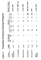
- the cleavage reagent also may be an exonuclease that lacks significant endonuclease activity.
- the detection duplex may comprise (i) a first oligonucleotide comprising a tag sequence and (ii) a second oligonucleotide that is complementary to the first oligonucleotide, where the second oligonucleotide comprises a detectable label.
- the detection duplex may further comprise a capture tag.
- the detection duplex may be immobilized on a solid support via the capture tag prior to contacting with the sample.
- a plurality of detection duplexes may be used, where each detection duplex carries a capture tag that permits capture of the duplex at a predetermined position on a solid surface.
- a method for detecting sequence specific DNA binding proteins comprising (a) contacting a sample suspected of containing at least one sequence specific DNA binding protein with a detection duplex for a time sufficient to permit sequence-specific binding between said duplex and the binding protein; and (b) detecting binding between the duplex and the binding protein.
- the duplex may be immobilized on a solid support before or after step (a) or (b). Immobilization may occur via a capture tag on the duplex.
- Detection may be achieved by labeling the protein sample with a detectable label prior to contacting with the detection duplex. Detection may be achieved via a detection reagent that specifically binds said binding protein, such as an antibody.
- the detection duplex may be labeled, and the immobilizing step can be achieved via capture of the binding protein onto a surface.
- a method for detecting sequence specific DNA binding proteins comprising: (a) contacting a capture surface with a sample suspected of containing at least one sequence specific DNA binding protein, for a time sufficient to permit capture of said sequence-specific binding protein on the capture surface; (b) contacting the capture surface with a detection duplex, and (c) detecting binding between the duplex and the binding protein.
- the invention described herein addresses the unmet need for a high-through-put multiplex assay for profiling transcription factor activity.
- the invention provides advantages over currently available technologies for profiling the activity of transcription factors.
- the invention provides methods and compositions for detecting and identifying sequence-specific nucleic acid binding proteins in a sample.
- the sample may be any sample, such as a cellular or tissue extract, that is suspected of containing such binding proteins.
- the methods and compositions may be used for detecting any protein that binds to nucleic acids in a sequence-specific manner. Examples of such proteins include, but are not limited to, eukaryotic transcription factors.
- the methods are suitable for the rapid and sensitive multiplex detection of nucleic acid binding proteins.
- the methods involve either a cleavage-based mechanism or a protein recognition element-based mechanism.
- a labeled detection duplex is contacted with a sample suspected of containing a sequence-specific nucleic acid binding protein that will bind to a sequence within the duplex.
- the detection duplex also may contain a moiety that permits capture of the duplex to a solid support or surface. Capture of the duplex can occur before or after mixing with the sample. Use of this capture moiety permits the generation of arrays for detecting multiple binding proteins in a sample, i.e. it permits "multiplexing" of the methods.
- the duplex is treated with a cleavage reagent. If protein is bound to the duplex, cleavage of the duplex by the cleavage agent is inhibited. Conversely, if no protein is bound, cleavage can proceed.
- the duplex is labeled in such a fashion that the presence of absence of cleavage can be identified by a change in signal from the label moiety initially present on the duplex. This change in signal then not only indicates the presence of absence of protein binding to the duplex, but the change in magnitude of the signal provides a quantitative measure of the amount of protein binding.
- the methods can be calibrated using known samples, and the results can also be compared to control reactions.
- the cleavage reagent can be non-specific, such as an exonuclease that can cleave one or both strands of the duplex from one terminus, or may be sequence specific, such as a restriction endonuclease that binds to the duplex at a specific site and cleaves at that site ( i.e. a Type II restriction endonuclease) or at a defined site some distance from the specific site (i.e. a Type IIs restriction endonuclease).
- a binding protein such as a transcription factor
- the proteins in a sample suspected of containing a protein binding protein may be labeled prior to contact with the detection duplex, in which case the binding protein itself functions as the recognition element, or the binding protein is specifically bound to a reagent, such as an antibody or other specific recognition element, before or after binding to the detection duplex.
- a reagent such as an antibody or other specific recognition element
- the binding protein is bound to a reagent prior to mixing with the detection duplex
- the duplex itself may be directly labeled to facilitate detection of binding.
- the detection of the label may be by direct observation or may be facilitated by secondary detection.
- secondary detection could be achieved by treatment with a cleavage agent that releases a label. Cleavage can only occur if the detection duplex is present, which can itself only occur if the duplex is bound to a specific binding protein.
- DNA-binding protein refers to any peptide, polypeptide, or peptide-containing substance or complex that can bind specifically to a defined nucleic acid sequence.
- the DNA-binding protein may be a complex of two or more individual molecules. Such complexes are commonly referred to as “homodimers”, “heterodimers” “homotypic complexes” and “heterotypic complexes.” Such complexes are composed of any number of individual entities that are held together by covalent bonds or non-covalent interactions.
- the DNA-binding protein may be natural or synthetic and is not required to be in any particular form.
- DNA-binding proteins examples include AP-1, Jum, Fos, CREB, ATF-1, Myc, Max, NF-kappa B, PPAR ⁇ , and Ubx.
- Nucleic acid-binding proteins of all kinds such as polymerases, proteins of the telomerases complex, gyrases, and splicing proteins, are also included in this definition.
- sample refers to any material that might contain a DNA-binding protein including but not limited to human and animal tissues, cultured cells, cultured or naturally occurring microorganisms, bodily fluids, blood, serum, and the like.
- the sample need not contain only the biological material.
- the sample may also consist of a DNA-binding protein-containing material on or in a physical matrix.
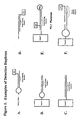
- detection duplex refers to a DNA molecule containing a double-stranded region that comprises a protein binding site.
- the detection duplex may be partially single-stranded and partially double-stranded and may contain gaps and nicks.
- the detection duplex may be constructed of one or more oligonucleotides and may comprise one or more self-hybridized regions that form hairpin loops.
- the detection duplex may contain any combination of natural and non-natural nucleotides and may contain non-natural linkages between nucleotides.
- the detection duplex may comprise one or more detectable labels.
- the detection duplex may comprise one or more binding moieties.
- the detection duplex may comprise one or more modifications that affect the stabilization of single-stranded or double-stranded DNA. Such modifications may include inverted 'T' residues, thiolated residues, peptide nucleic acid linkages, chimeras or RNA and DNA, and the like.
- the detection duplex may be immobilized on a support or may be capable of being immobilized on a support through a binding moiety or through hybridization of a single-stranded portion of the detection duplex to a complementary sequence on the support.
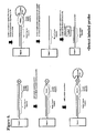
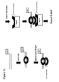
- the detection duplex may also contain intentional or unintentional mismatches. Examples of detection duplexes are shown in Figure 1. The skilled artisan will recognize that a wide range of suitable detection duplexes may be used in the present invention and the examples given in Figure 1 are not meant to be limiting of the present invention.
- capture tag refers to a sequence in the detection duplex that can be used to capture the duplex onto a support.
- a "binding moiety” is a chemical or biochemical moiety that may be used to attach a substance such as DNA or protein to a solid support.
- the binding moiety may form a non-covalent bond, a reversible covalent bond, or an irreversible covalent bond between the substance and the solid support.
- chemical binding moieties include the aldehyde moiety (CHO) and amino moiety (-NH2) which may be used to chemically bind the substance to the solid support using techniques well known in the art. The skilled artisan will recognize that other suitable binding moieties are known in the art and may be used in the present invention.
- biochemical moieties include biotin, IgG and DNA.
- a substance labeled with biotin will form a strong non-covalent bond with an avidin-coated solid support.
- IgG will bind to a solid support coated with Protein G
- DNA will bind to a solid support coated with a complementary RNA or DNA sequence.
- Other such binding interactions suitable for use in the present invention are known in the art.
- a "detection reagent” is a detectable entity that is capable of binding to a second entity to enable detection of the second entity directly or indirectly.
- Detection reagents may be nucleic acids, proteins, or peptides, or other biomolecules that may or may not comprise a label. Examples of detection reagents include peptides, oligonucleotides, mono and polyclonal antibodies, antibody fragments, lectins, stains, dyes, and the like and chimeric forms of these entities, thereof.
- Detection reagents that are not directly detectable may comprise a label or may be detected by a secondary detection reagent that comprises a label. For example, antibodies can be detected with labeled Protein G.
- a "label” is a detectable signal moiety or a reporter.
- labels or reporters may be used in the present invention, including, for example, radioactive isotopes, fluorescent labels, chemiluminescent labels, bioluminescent labels, and enzyme labels. Labels may be bound directly or indirectly.
- the labels also may be haptens that can be recognized by secondary reagents such as antibodies, peptides, direct chemical interactions, and other methods that are well known in the art.
- the label also may be an oligonucleotide or nucleic acid that can be detected by hybridization, polymerization, ligation and/or amplification by methods well known in the art.
- the label may be used to generate an increase or decrease in a signal readout.
- the label may also comprise two chromophores bound in close proximity to utilize a phenomenon called fluorescence resonance energy transfer (FRET).
- FRET fluorescence resonance energy transfer
- one chromophore absorbs a photon and then exists in the excited state.
- the energy from the excited chromophore is transferred to an acceptor molecule when the chromophore and the acceptor are in close spatial proximity to each other. This energy transfer prevents the excited chromophore from releasing the energy in the form of a photon of light thus quenching the fluorescence of the chromophore.
- the acceptor molecule is not sufficiently close in space, the energy transfer does not occur and the excited chromophore may then fluoresce.
- LRET luminescence resonance energy transfer
- micro- or nano-transponders of nanocrystals may be used as labels.
- other labels that can be used to accelerate detection include chemiluminescent labels, immuno-affinity tags such as c-myc, affinity tags such as cellulose binding domain, streptavidin, biotin, streptavidin or any whole or part macromolecule with a matching fit, reporter enzymes with chromogenic, luminescent, fluorescent, or other tracer capabilities.
- support may be any porous or non-porous material or matrix suitable for attaching proteins, peptides, nucleic acids and the like.
- the proteins, peptides, nucleic acids and the like may be bound covalently or non-covalently to the support by any technique or combination of techniques well known in the art.
- Supports of the invention may comprise nylon, nitrocellulose, diazonitrocellulose, glass, silicon, polystyrene, polyvinyl chloride, polypropylene, polyethylene, dextran, sepharose, agar, starch, or any other material that allows for the immobilization of biomolecules.
- the material can be formed in filters, membranes, flat surfaces, tubes, channels, wells, sheets, particles, beads, microspheres, columns, fibers (e.g. optical fibers) and the like.
- the support may also comprise a multiwell format (such as microtiter plates) such as 12-well, 24-well 48-well, 96-well, 384-well, and 1537-well plates. Particles or beads may be made of glass, latex, a magnetic material (magnetic, paramagnetic, or supermagnetic beads) or other suitable material.
- a support that may be used in the present invention is a set of color coded microspheres such as those manufactured and sold by Luminex Corporation (Austin, TX) .
- array refers to an orderly arrangement of distinct molecules or substances on a support including, but not limited to, biological molecules such as DNA, RNA, proteins, and the like or chemicals arrayed or immobilized to a support.
- biological molecules such as DNA, RNA, proteins, and the like or chemicals arrayed or immobilized to a support.
- Arrays of biological molecules such as oligonucleotides, probes, receptors, antibodies, or any entity reactive with targets have become an increasingly important tool in the biotechnology industry and related fields.
- Arrays comprising a plurality of biological molecules find use in a variety of applications including drug screening, nucleic acid sequencing, mutation analysis, genomic and proteomic applications and the like.
- arrays may be formed on microplates, glass slides, beads, microspheres, microfluidic devices or standard blotting membranes and may be referred to as "arrays", microarrays, or chips.
- Capture molecules may be bound to the support through covalent or non-covalent interactions. When bound to a planar surface, the capture molecules are bound in an orderly fashion such that the identity of any particular capture molecule can be identified by its position on the array.
- Such arrays may be constructed on planar objects such as glass or plastic microscope slides. Arrays may also be constructed on the inside surface of a tube or microplate well or may be constructed inside the channels of a microfluidic device. In general, there is no restriction on the format of the array provided the individual sites to which the capture molecules are bound can be identified. If the support is a set of beads or microspheres, then sets of beads or microspheres coupled to different capture molecules must be distinguishable in some way. In one embodiment, beads from Luminex Corporation (Austin, Texas) are color-coded by the addition of two different dyes at 10 different concentrations resulting in 100 different color beads. Capture molecules can be bound to specific bead colors and the color of each bead can be identified by flow cytometry.
- a bead array is prepared by binding specific capture molecules to sets of beads of a specific color, and then mixing different sets of colored beads to create an array.
- microparticles from Pharmaseq (Princeton, NJ) each containing a unique radio frequency tag, can be used to identify specific microparticles.
- the array may contain anywhere from 2 to 100,000 elements, preferably, between 3 and 5000 elements.
- the invention employs a bead array format such as commercially available Luminex LabMAP TM Technology but can be applied to virtually any type or array platform or format.
- the invention comprises an assay system with the capacity to quantitatively and qualitatively profile activities of up to 100,000 different regulatory proteins in a single reaction vessel, well or tube.
- a "capture reagent” refers to any molecule that will specifically capture a DNA-binding protein or a detection duplex from a solution containing one or more biological molecules.
- capture reagents are poly- and monoclonal antibodies and antibody fragments.
- Capture reagents may be molecules that bind to haptens or binding moieties. Proteins that have natural affinity for specific DNA-binding proteins and proteins that have been engineered to specifically bind to the DNA-binding proteins are also included in this definition.
- Capture molecules may also be molecules that bind to another molecule that binds the DNA-binding protein. For example, anti-rabbit IgG may be used to capture a rabbit antibody-protein complex.
- protein G may be used to capture a goat antibody-protein complex.
- capture reagents and corresponding binding moieties are given in Table I. Table 1.
- Capture Reagents and the haptens to which they bind Hapten / Binding Moiety Capture Reagent Biotin Avidin, Streptavidin Sialic Acid, carbohydrates, glycoproteins Lectins such as Concavalin A Digoxigenin Anti-digoxigenin Fc portion of IgG Protein A, Protein G, Protein A/G 5-BrdU (5-bromodeoxyuridine) Anti-BrdU Dinitrophenyl (DNP) Anti-DNP Fluorescein isothiocyanate (FITC) Anti-FITC N-2-Acetylaminofluoren (AAF) Anti-AAF N-2-Acetylamino-7-iodofluoren (AAIF) Anti-AAIF oligo or poly dA oligo or poly dT oligo or poly dC
- Capture reagents may also include chemicals or dyes that can bind to DNA, protein, or DNA-protein complexes. Capture reagents also include reagents that recognize specific conformations of biomolecules or may recognize particular modifications. For example, antibodies that react against phosphoserine can be used as capture reagents to capture proteins that contain an exposed phosphoserine residue. Additionally, SH2 domains may be used to capture proteins that contain a particular four amino acid motif that contains phosphotyrosine.
- a "profile” is a combination of the measurements of two or more properties of a biological, biochemical, or chemical system. The measurements may be made simultaneously or in sequence.
- a profile may comprise the concentration of two or more proteins in a sample.
- Another example of a profile is the phosphorylation state of two or more proteins in a sample.
- Profiles may comprise qualitative or quantitative measurements and may include subjective as well as objective data.
- sequence-specific cleavage reagent is a reagent that can cleave DNA at a specific location based upon the recognition of a specific DNA sequence.
- sequence-specific cleavage reagents include Type II restriction endonucleases such as EcoR1, Hind III, and BamHI.
- Sequence-specific cleavage reagents also include the class of Type IIs (or "homing") restriction endonucleases that bind to a specific DNA sequence, and cleave the DNA at a defined distance from the enzyme binding site.
- an "external cleavage reagent” refers to a reagent that initiates the digestion or cleavage of one or more strands of nucleic acid at or near one or more ends of the nucleic acid. The digestion or cleavage proceeds in a single direction relative to the initiation site.
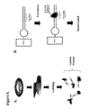
- External cleavage reagents include the enzymes commonly known as exonucleases. Examples of external cleavage reagents and their properties are shown in Figure 2.
- probe capture sequence refers to a sequence of DNA that can be used to capture a labeled or unlabeled oligonucleotide or nucleic acid probe.
- the sequence may be single-stranded or double-stranded.
- the probe capture sequence may be used to capture detection duplexes or DNA-protein complexes.
- signal amplification refers to any method used to increase the signal of a biological assay beyond the signal that can be achieved with a "one-label" detection strategy.
- Signal amplification may be based on an enzyme catalyzed reporter deposition such as tyramide signal amplification or may be based on enzyme amplification.
- strategies that increase the number of labels may be used. Such strategies include the binding of dendrimers, branched polymers, and long linear polymers that contain multiple binding sites for a secondary detectable reagent. Examples of these strategies include, without limitation, oligonucleotide dendrimers, branched DNA, and Hybrid Capture.
- nucleic acid amplification methods such as polymerase chain reaction and rolling circle amplification also may be used to amplify the signal obtained.
- a detection reagent such as an antibody, peptide, avidin, or streptavidin.
- any method of signal amplification may be used to increase the signal generated by the assay.
- the present invention therefore provides compositions and methods for detecting and measuring DNA-binding proteins.
- the invention provides compositions and methods for the simultaneous or near-simultaneous detection of multiple DNA-binding proteins in a multiplex or array format, and also provides compositions and methods for generating profiles of DNA binding activity by proteins, specifically, transcription factors. More specifically, the invention provides compositions and methods for detecting and measuring multiple protein-DNA binding events in a single sample in a high-throughput format.
- the invention provides a method for detecting protein binding to a detection duplex, in which binding of the protein to the detection duplex inhibits cleavage of the duplex by a site-specific cleavage reagent and thereby increases or diminishes a signal.
- the detection duplex comprises a DNA sequence that is recognized by a site-specific cleavage reagent such that the site-specific cleavage reagent will cleave the detection duplex when no protein has bound to the protein-binding site. However, if protein has bound to the protein-binding site of the detection duplex, the cleavage of the detection duplex will be inhibited and thereby increase or decrease a signal.
- Type II restriction enzymes may be used in this mode of the invention when the protein binding site in the detection duplex comprises a known site for a type II restriction enzyme, or is sufficiently close in space to the restriction enzyme site that binding of a specific binding protein to the protein binding site that restriction enzyme binding to the enzyme recognition site is inhibited or prevented.
- a detection duplex comprising a binding moiety, a label, and a protein binding site between the binding moiety and the label wherein the protein binding site comprises a restriction endonuclease cleavage site may be used.
- the detection duplex is contacted with the sample and DNA-binding proteins, if present, bind to the detection duplex.
- a site-specific cleavage reagent such as a type II restriction endonuclease is added. If protein has bound to the protein-binding site, then the detection duplex remains intact. If no protein has bound to the detection duplex, then the detection duplex is cleaved, thus separating the binding moiety from the label and preventing the label from being detected.
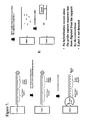
- An example of this embodiment of the invention is illustrated in Figure 3.
- site-specific cleavage reagent may be used in this mode of the invention.
- Type IIs restriction endonucleases recognize and bind to a specific DNA sequence but cleave the DNA at a defined region away from the enzyme binding site.
- Assays utilizing these enzymes are advantageous because the protein binding site need not comprise a recognition sequence for the site specific cleavage reagent. Rather, the binding site for the site-specific cleavage reagent can be designed into the detection duplex.
- a detection duplex comprising a binding moiety, a label, a protein binding site, and a binding site for a site-specific cleaveage reagent such that the site specific-cleavage reagent, when bound to the detection duplex, cleaves the duplex in or near the protein binding site.
- the detection duplex is contacted with the sample and DNA-binding proteins, if present, bind to the detection duplex.
- a site-specific cleavage reagent such as a type II restriction endonuclease is added. If protein has bound to the protein-binding site, then the detection duplex remains intact.
- the detection duplex is cleaved, thus separating the binding moiety from the label and preventing the label from being detected.
- An example of this embodiment of the invention is illustrated in Figure 4.
- the duplex may be captured onto a support to facilitate the detection of the label.
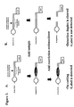
- the invention provides a method for detecting protein binding to a detection duplex, in which binding of the protein to the detection duplex inhibits the cleavage of the duplex by an external cleavage reagent and thereby increases or diminishes a signal.
- the external cleavage reagent may cleave one or both strands of the detection duplex beginning at one or more ends of the DNA strand in the duplex.
- the detection duplex is first immobilized on a support. The sample is contacted with the detection duplex on the support and protein is allowed to bind to the detection duplex. Subsequently, an external cleavage reagent is brought into contact with the detection duplex on the support.
- the external cleavage reagent will not be able to fully digest the detection duplex because it will be protected by the presence of the protein. If no protein is bound to the detection duplex, then the external cleavage reagent will digest one or both strands of the detection duplex and will release the label into medium where it can be washed away. If protein has bound to the detection duplex, then the label will remain bound to the support and will be detected.
- An example of this embodiment of the invention is illustrated in Figure 5.
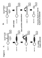
- the immobilized detection duplex comprises a probe capture sequence and a protein binding sequence and a phosphate moiety on the 5' end of the immobilized or immobilizable strand.
- the detection duplex may be immobilized on a support or may be captured onto a support at other steps as may be advantageous.
- the capture sequence and the protein binding sequence may be the same or may be different. In an array format it will be advantageous for the probe capture sequence and the protein binding sequence to be different so that the same detection reagent can be used with many different detection duplexes.
- the detection duplex is contacted with the sample that may contain DNA-binding proteins. If present, the proteins bind to the protein-binding site in the detection duplex.
- the detection duplex is then contacted with lambda exonuclease, an external cleavage reagent, that digests the 5' phosphorylated DNA strand in a 5' to 3' direction. If protein has bound to the detection duplex, then the enzyme will be prevented from cleaving the phosphorylated strand of the detection duplex. If no protein has bound, then the 5' phosphorylated strand of the detection duplex will be completely digested.
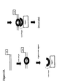
- the detection duplex is then heat denatured and washed to inactivate the external cleavage reagent, separate the strands of the detection duplex, and remove the cleavage products. If the detection duplex was protected by protein binding, then the strand containing the probe capture sequence remains bound to the support. If the duplex was not protected because no protein bound to the detection duplex and inhibited the digestion, then the strand containing the probe capture sequence will no longer be on the support. A labeled oligonucleotide or nucleic acid probe is then added and allowed to hybridize to the probe capture sequence, if present. The presence of signal indicates that the detection duplex was protected by bound protein ( Figure 6). The absence of signal indicates that the strand was digested by the external cleavage reagent and was not protected by protein binding ( Figure 7).
- a further embodiment of the invention provides a method for detecting protein binding to a detection duplex in which the proteins in a sample are labeled prior to or subsequent to binding to a detection duplex. After washing, proteins that have bound to the detection duplex are detected by the presence of the label bound to the detection duplex. Proteins in the sample are labeled by methods commonly used in the art including active esters such as N-hydroxysuccinimide esters of biotin, N-hydroxysuccinimide esters of fluorescent dyes such as Cy3 and Cy5, sulfhydryl-reactive labels and other methods commonly used in the art.
- a sample is labeled with an amine reactive dye such as the N-hydroxysuccinimide ester of the fluorescent dye Cy3 (Amersham Biosciences).
- the process will label essentially all, or nearly all of the proteins in the sample.
- the labeled sample will then be contacted with a detection duplex.
- the detection duplex may be immobilized on a support of may be in solution. An incubation period ensues to allow proteins to bind to the protein binding site in the detection duplex. If the detection duplex is in solution, it is now captured onto a support, and the unbound molecules are washed away. Proteins bound to the detection duplex are detected by the label.
- An example of this embodiment of the invention is illustrated in Figure 8.
- two or more samples are labeled separately with different labels and then mixed. This mixed sample is then contacted with the detection duplex. An incubation period ensues to allow time for the labeled proteins to bind to the detection duplex. If the detection duplex is in solution, it is now captured onto a support, and the unbound molecules are washed away. Proteins bound to the detection duplex are detected by detection of the labels. Since two or more samples were labeled with different labels, each label is detected and measured independently and the results can be expressed as a differential analysis of DNA-binding proteins in the two samples in a similar manner to the way RNA molecules are differentially labeled and measured on DNA microarrays (27).
- Yet another embodiment provides a method for detecting protein binding to a detection duplex in which the proteins in a sample are bound to a detection duplex, after which excess proteins are washed away and bound proteins are subsequently detected with a detection reagent.
- the detection duplex may be first captured onto a support or may be captured onto the support after other steps as may be advantageous.
- the order of contacting the duplex, sample and detection reagent may be carried out in any order.
- an immobilized detection duplex is contacted with a sample that may contain DNA-binding proteins.
- the immobilized detection duplex is incubated with the sample to allow the proteins to bind to the detection duplex. Unbound substances are washed away and the bound proteins are detected with a detection reagent.
- the proteins may be detected with specific antibodies that detect only a single protein or may be detected with antibodies that detect classes of proteins or particular protein motifs.
- DNA-binding proteins contain phosphorylated tyrosine, threonine, or serine residues and proteins may be detected with anti-phosphotyrosine, anti-phosphoserine, and anti-phosphothreonine antibodies.
- Proteins may also be detected with other proteins that binds to certain motifs.
- DNA-binding proteins may be detected with a class of proteins called SH2 domains. SH2 domains bind to a 4 amino acid motif that includes phosphotyrosine. Bound proteins may also be detected with a chemical or biochemical stain.
- a further embodiment of the invention provides a method for detecting protein binding to a detection duplex in which the protein is first bound to a labeled detection duplex, after which the protein-detection duplex complex is captured onto a support with a capture reagent and detected.
- the capture reagent, protein, and labeled detection duplex may be first contacted and subsequently captured onto a support.
- the addition of capture reagent, sample and labeled detection duplex and the capture onto a support may be carried out in any order.
- the capture reagent is immobilized onto a support.
- the labeled detection duplex is contacted with the sample that may contain DNA-binding proteins.
- the sample is incubated with the detection duplex to allow proteins, if any, to bind to the detection duplex.
- the mixture is contacted with an immobilized capture reagent that captures the protein-detection duplex complex onto the support and the label on the detection duplex is detected.
- an immobilized capture reagent that captures the protein-detection duplex complex onto the support and the label on the detection duplex is detected.
- the capture reagent could be an antibody to a specific DNA-binding protein that has been immobilized on the support.
- the labeled detection duplex is contacted with the sample that may contain DNA-binding proteins.
- the sample is incubated with the detection duplex to allow proteins, if any, to bind to the detection duplex.
- the mixture is contacted with a first capture reagent that binds to the protein-detection duplex complex.
- the first capture reagent-protein-detection duplex complex is then captured onto a support through a second capture reagent and the label on the detection duplex is detected.
- the first capture reagent could be an antibody specific for a DNA-binding protein and the second capture reagent on the support could be a molecule such as Protein A or Protein G that could bind the first antibody capture reagent.
- the invention provides for a method of detecting DNA-binding proteins comprising two or more detection duplexes immobilized on a support.
- the invention provides a method of detecting a plurality of DNA binding proteins using two or more detection duplexes.
- the duplexes are mixed with the sample in solution and subsequently are captured onto a support in an array format.
- detection duplexes containing unique single-stranded regions can be bound to a support that comprises an array of oligonucleotides that are complementary to the single-stranded regions of the detection duplexes.
- detection duplexes may be captured onto a support in an array format through a binding moiety either prior to or subsequent to contact with the sample.
- the array may also comprise an array of antibodies or other capture reagents that can be used to capture DNA-binding protein - detection duplex complexes.
- the invention described herein is well suited to application on a flow cytometric bead array with multiplex capability such as the LabMap system developed by Luminex Corporation (28).
- a flow cytometric bead array with multiplex capability such as the LabMap system developed by Luminex Corporation (28).
- polystyrene microspheres are internally dyed with precise ratios of two spectrally distinct fluorescent dyes.
- Each bead is dyed with one of 10 different concentrations of each fluorochrome resulting in a bead array consisting of 100 spectrally distinct microsphere sets.
- Each microsphere set can be distinguished by its spectral address (the ratio of two dye colors) and they can be combined in a single test allowing up to 100 different analytes to be measured simultaneously in a single reaction vessel.
- Substances such as proteins, peptides, nucleic acids, and the like can be coupled to the microspheres using standard chemisty that is will known in the art.
- a third fluorescent dye is coupled to a reporter molecule quantifies the biomolecular interaction that has occurred at the microsphere surface.
- Microspheres are interrogated individually in a rapidly flowing fluid stream as they pass by two separate lasers. High speed digital signal processing classifies the microsphere based on its spectral address and quantifies the reporter signal on the surface of the bead. Washing to remove unbound reporter is usually not necessarily, thus making the assay essentially homogeneous.
- microspheres are interrogated per second resulting in an analysis system capable of analyzing and reporting up to 100 different reactions in a single reaction vessel in a few seconds.
- the embodiments of the present invention can be easily adapted to a bead array format by binding detection duplexes, oligonucleotides, or capture reagents to beads.
- Detection duplexes may be immobilized on the beads or may be captured onto the beads after contact with the sample.
- Antibodies or other capture reagents may be immobilized onto the beads or complexes may be captured onto the beads after contact with the sample.
- the methods of the present invention may easily be adapted for use in conjunction with signal amplification methods that are used to increase the sensitivity of detection. Enzyme amplification, rolling circle amplification, ligase chain reaction, and other methods that can amplify a detectable signal are included in the scope of this invention.
- Methods of the present invention may be used in the detection, screening, and diagnosis of various diseases, disorders, or conditions in biological samples.
- the methods of the invention may also be used to for monitoring the progression of a disease or treatment.
- Methods of the present invention may also be used to screen compounds or biological molecules that may affect or alter the activity or the amount of DNA-binding proteins in a samples.
- the present invention may also be used to determine the effect of genetic variation on the binding ability of DNA-binding proteins.
- the invention could be used to simultaneously measure the affinity of a single or multiple DNA-binding proteins to a large number of different protein binding sequences.
- the present invention provides methods that can be used to elucidate and understand cellular networks and cellular signaling pathways.
- Profiles of DNA-binding proteins generated by the methods and compositions of this invention may be combined with software that analyzes the data and the profiles generated.
- Such software may include features such as pattern recognition and pattern discovery algorithms, unsupervised or supervised hierarchical clustering, and the like that will facilitate discovery of new bio-markers, diagnosis and staging of diseases, and can predict the effects of potential new drugs.
- the methods of the present invention may also be used in conjunction with software that correlates proteins and protein binding sequences with their functional and biological characteristics.
- Such characteristics may include structural, regulatory or enzymatic functions of the proteins detected, the biological 'objectives' to which the proteins contribute, the functional relationship of the detected protein to one or more other proteins or genes, the functional relationship of protein-binding sequences to other proteins or nucleic acid sequences, the relationship of detected proteins to major biological processes and biochemical functions.
- Oligonucleotides were coupled to beads using standard EDC carboxylate coupling chemistry. Briefly, beads are suspended in MES buffer pH 4.7 and oligonucleotides are added to a final concentration of 2uM. Fresh EDC is added to provide a final concentration of 1-2mg/ml and is reacted with the bead suspension for 30 minutes while rotating in the dark. A similar concentration of fresh EDC is again added to the bead suspension and reacted for and additional 2 hours while rotating in the dark.
- Coupled beads are washed once with 50mM Tris pH 7.5, 100mM NaCl, 0.05% tween-20, once with 0.2% SDS in PBS, twice with PBS pH 7.5 containing 0.02% Tween-20, once with PBS pH7.5 and resuspended in PBS pH 7.5 to a final concentration of 1000-2000 beads/microliter.
- Detection duplexes were assembled from the oligonucleotide PO4Kbfor (P04-AGTTGAGGGGATCCCCAGGAGCGGCTTATCGGTCTATTC-AACTCCCCTAGGGG) that carries a tag sequence, NF-kB binding site and 5 prime phosphate group, and the complement oligonucleotide KBrev (TCCTGGGGATCCCCTCAACT) devoid of tag sequence.
- PO4Kbfor and KBrev were annealed by first diluting 100uM oligonucleotide stocks into annealing buffer (40mM Tris pH 7.5, 100mM NaCl, and 1mM EDTA), to give a ratio of 20nM PO4Kbfor:1uM KBrev. 100u1 of the oligonucleotide mix was then heated to 95°C for 1 minute, and cooled linearly to room temperature over a period of 45 minutes.
- annealing buffer 40mM Tris pH 7.5, 100mM NaCl, and 1mM EDTA
- 1ul of the annealed mixture containing detection duplexes displaying a single stranded tag sequence was added to19 ul of transcription factor binding buffer (10mM tris pH 7.5, 50mM NaCl, 2mM KCl, 2 mM MgCl2) containing 250ng poly dI-dC, and 0.5% bovine serum albumin (fraction V) and incubated with varying amounts of NF-kB P50 protein for 15 minutes at room temperature.
- transcription factor binding buffer 10mM tris pH 7.5, 50mM NaCl, 2mM KCl, 2 mM MgCl2
- bovine serum albumin fraction V
- transcription factor binding buffer containing (0.1 ug) of NF-kB P50 specific primary antibody (Santa Cruz Biotechnolgy Cat #SC-114) was added to the detection duplex-sample-bead mix and incubated for 20 minutes at room temperature in the dark while shaking.
- NF-kB P50 specific primary antibody Santa Cruz Biotechnolgy Cat #SC-114
- Two micrograms of capture reagent (Santa Cruz Biotechnologies NFKb P65 antibody Cat#SC-372) is incubated with bead supports that are covalent coupled to recombinant Protein G (Upstate Biotechnology). This incubation is carried out while shaking for 2 hours at room temperature in the absence of light. Beads are then washed with 3 X 200ul volumes of transcription factor buffer to remove antibodies not bound the protein G bead surface.
- Beads are then washed with 2 X 200ul volumes of transcription factor binding buffer to remove hairpin oligonucleotides not captured on bead surfaces.
- Each assay is then resuspended in 100ul of streptavidin-phycoerythrin (SA-PE) conjugate (2ug/ml in transcription factor binding buffer), incubated for 10 minutes at room temperature and analyzed using the Luminex 100 instrumentation.
- SA-PE streptavidin-phycoerythrin
- TNF Stimulated extracts contain active NF-kB P65 protein that forms a complex with hairpin oligonucleotides used for detection.
- the NF-kB P65 - hairpin oligonucleotides are captured by the anti-P65 antibodies immobilized on bead surfaces and NF-kB P65-biotinylated-DNA complexes associated with bead surfaces are detected using SA-PE.
- Class II restriction endonucleases are used with detection cuplexes comprising hairpin oligonucleotides coupled to beads.
- Class II restriction enzymes cleave double stranded DNA at the same site where they recognize and bind to DNA.
- Hairpin oligonucleotides (KBREHP biotin- TCCAAGGGGATTCCCCAGTG-amino C-6-TACTGGGGAATCCCCTTGGA ) that present overlapping transcription factor (NF-kB) and restriction enzyme (EcoRI) binding sites are coupled to bead surfaces through amino-C6 using standard EDC chemistry as described previously. Approximately 1000 of the coupled beads are used per 25ul assay.
- KBREHP coupled beads are incubated for 10 minutes at room temperature with 2ug of nuclear extracts (obtained from control and TNF treated HeLa cells) in a 25 ul volume of transcription factor binding buffer (10mM tris pH 7.5, 50mM NaCl, 2mM KCl, 2 mM MgCl2) containing 100ng poly dI-dC, and 0.35% bovine serum albumin (fraction V). Following binding, reactions were brought to 50ul with transcription factor binding buffer and supplemented with 1M MgCl 2 to provide a final concentration of 10mM MgCl 2 . Samples are then incubated with 10u of restriction endonuclease EcoRI for 10 minutes at 37 degrees.
- NF-kB P50 binds to DNA and protects from digestion with EcoRI endonuclease. NF-kB is quantified as a measure of signal retained on bead surfaces.
- Example 5 Detection of Protein Binding to DNA with Inhibition of Site-Specific Cleavage by Class IIs Restriction Endonucleases.
- Detection duplexes were assembled by hybridizing oligonucleotides designated FOR (biotin-AAGGATGAGCGGGGGATCCCAATAGGCGGC-TGCTTATCGGTCTAT) that comprise an Nf-KB binding site, Fok1 binding site and tag sequence to oligonucleotides designated bREV (CTATTGGGATCCCCGCTCATCCTT) that is complementary to the FOR oligonucleotide but is devoid of complementary tag sequence.
- FOR biotin-AAGGATGAGCGGGGGATCCCAATAGGCGGC-TGCTTATCGGTCTAT
- bREV CATTGGGATCCCCGCTCATCCTT
- Hybridization of oligonucleotide pairs was accomplished as follows: bREV and FOR were diluted to 1uM and 40nM respectively in buffer containing 40mM Tris pH 7.5, 100mM NaCl, and 1mM EDTA. Oligonucleotides then were heated to 95°C for 1 minute and allowed to cool to room temperature over a period of 45 minutes.
- Binding of transcription factors was carried out by incubating 1ul of hybridized oligonucleotide with recombinant NF-kB P50 transcription factor (Promega Cat# ) in a total volume of 15ul binding buffer (10mM tris pH 7.5, 50mM NaCl, 2mM KCl, 2 mM MgCl2) containing 100ng poly dI-dC, and 0.35% bovine serum albumin (fraction V). Following a 10 minute incubation at room temperature, each assay was mixed with 15 ul of binding buffer supplemented to 20mM MgCl2 and containing 1ul of FokI (4 units) or 1ul of FokI storage buffer and incubated for and additional 10 minutes at 37 degrees.
- Lambda exonuclease is highly specific for double stranded DNA carrying a 5 prime phosphate and has greatly reduced activity on DNA that is not phosphorylated. Lambda exonuclease digestion of DNA occurs in the 5' to 3' direction.
- Oligonucleotide PO4Kbfor (P04-AGTTGAGGGGATCCCCAGGAGCGGCTTATCGGTCTA-TTCAACTCCCCTAGGGG) carrying tag sequence, NF-kB binding site and 5 prime phosphate group, was annealed to the biotinylated complement oligonucleotide biotinKBrev (TCCTGGGGATCCCCTCAACT-biotin) devoid of capture tag sequence.
- Annealing was carried out by first diluting 100uM oligonucleotide stocks into annealing buffer (40mM Tris pH 7.5, 100mM NaCl, and 1mM EDTA), to give a ratio of 400nM PO4Kbfor:10uM biotinKBrev.
- the oligonucleotide mix was then heated to 95° C for 1 minute, and cooled to room temperature over a period of 45 minutes of to form a double stranded pair displaying a single stranded tag sequence.
- Approximately 50,000 beads coupled to tag capture sequence were hybridized with 50 ul of annealed oligonucleotide for 1 hour at room temperature. Beads were then washed to remove unhybridized oligonucleotide and resuspended in transcription factor binding buffer.
- DNA binding to P50 transcription factor was carried out by incubating 0.5 gel shift units of recombinant P50 (Promega cat#E3770) (defined as the amount of P50 required to shift 190 finoles of double stranded NF-kb binding oligonucleotide using conventional gel shift assay-promega) with 1000 hybridized beads in a 20ul volume of transcription factor binding buffer (10mM tris pH 7.5, 50mM NaCl, 2mM KCl, 2 mM MgCl2) containing 100ng poly dI-dC, and 0.35% bovine serum albumin (fraction V).
- transcription factor binding buffer (10mM tris pH 7.5, 50mM NaCl, 2mM KCl, 2 mM MgCl2) containing 100ng poly dI-dC, and 0.35% bovine serum albumin (fraction V).
- Single stranded oligonucleotides PO4P50 (5'PO4AGTTGAGGGGATCCCCAAGGGCGAAGGAACTCGACGTGGAGCCGTTTTT-aminoC6) with a transcription factor binding sequence and a probe-capture sequence are coupled to beads through amino-C6 as described previously.
- Oligonucleotide PO4P50rev is hybridized with beads coupled to PO4P50 (1 pmole per 1000 beads) for 1 hour in transcription factor binding buffer(10mM tris pH 7.5, 50mM NaCl, 2mM KCl, 2 mM MgCl2). Beads are then washed and resuspended to a concentration of 1000 beads/ul in transcription factor binding buffer.
- Binding reactions are then carried out by incubating 1000 beads with samples for 10 minutes at room temperature in a 20ul volume of transcription factor binding buffer containing 0.35% BSA and 0.1-2ug Poly dI-dC. Following the binding reaction, 20ul of transcription factor binding buffer supplemented to 3mM MuCl 2 containing 3units of lambda exonuclease or an equivalent of lambda exonuclease storage buffer is added to appropriate reactions and incubated for 15 minutes at 37 degrees. 1pmole of biotin labeled single stranded probe DNA is then added to each reaction and hybridized with beads by heating to 95 C for 1 minute followed by cooling over a 30minute period to reach room temperature.
- assays are washed with 2 X 100 ul volumes of PBS pH 7.5 using a vacuum and filter plate. Beads are ressupended in 100ul of 2ug/ml SA-PE in PBS pH 7.5. Following a 10 minute incubation at room temperature, assays are analyzed using the Luminex 100 instrumentation.
- control cell extract and 10ug HeLa cell extracts spiked with 10 gel shift units of recombinant NF-kBP50 are suspended in 20ul of labeling buffer (PBS pH 8.0).
- labeling buffer PBS pH 8.0
- 0.5mg of the water soluble form of N-hydroxysuccinimide-biotin (NHS-biotin) is added to each labeling reaction. Vortex and incubate for 2 hours at room temperature.
- Add 30ul of transcription factor binding buffer (10mM tris pH 7.5, 50mM NaCl, 2mM KCl, 2 mM MgCl2) to each labeling reaction and remove unincorporated NHS-biotin by desalting over a G-25 spin column.
- Incubate 1 gel shift unit of the desalted-labeled sampled with beads coupled to a hairpin detection molecule carrying an NF-KB P50 binding site This incubation is carried out in a total volume or 20ul transcription factor binding buffer containing 100ng Poly dI-dC and 0.35% BSA fraction V. Following a 10 minute incubation at room temperature, add 80 ul of 2ug/ml streptavidin-phycoerythrin in transcription factor binding buffer, incubate 10 minutes and analyze using the Luminex 100 instrumentation. Beads incubated with labeled hela cell extracts spiked with recombinant NF-kB P50 have
Abstract
Description
- This invention provides compositions and methods for profiling transcription factor activity. In particular, the invention provides nucleic acid constructs containing protein binding sites and methods for detecting and measuring the binding of proteins and particularly transcription factors to the binding sites in these constructs.
- Eukaryotes are composed of specialized cell types that are organized into tissues and organs. Regardless of cell type and function, all cells within an individual eukaryotic organism contain the same set of genes referred to as the genome. Differences between cells arise through the differential expression of genes. Expression of individual genes is controlled through the binding of proteins to regulatory sequences of DNA in the genome such as promoters and repressors. Protein binding to such control sequences can cause an increase or decrease in the rate of transcription of a gene. These DNA-binding proteins, called transcription factors, regulate gene transcription and thereby control all of the essential characteristics of a cell including cellular reproduction, development and differentiation, response to environmental stimuli, and tissue homeostasis in normal and disease states. Transcription factors comprise hundreds of specialized proteins that regulate gene expression by either facilitating or inhibiting the enzyme RNA polymerase in the initiation and maintenance of transcription.
- The activation or inhibition of regulatory transcription factors occurs as a downstream event in signal transduction cascades that are initiated by perturbations such as a change in the oxidation state of a cell, or the binding of a ligand to its cell surface receptor. In the case of cell surface receptors, a ligand-binding event may trigger signaling cascades that fan out to regulate multiple genes that contribute to biological responses. Cross talk between signal transduction systems also is common, with disparate stimuli utilizing many of the same protein kinases, phosphatases and second messenger systems. Highly refined regulation of biological responses, therefore, occurs as webs of interacting signaling systems involving kinases, phosphatases and second messengers triggered by each stimulus that culminate in qualitatively and/or quantitatively different sets of transcription factor activation. It has been estimated that there are approximately 1000 transcription factors in the human genome that contribute the specificity to regulate the independent expression of approximately 40,000 genes. Consequently, biological responses that are characterized by changes in gene expression may be defined by distinct signatures of transcription factor activation. Transcription factors, therefore, are of significant interest as targets to affect specific individual or global changes in gene expression.
- Cell surface receptors have been a primary focus of pharmaceutical research and comprise the majority of therapeutic targets. These receptor-targeted strategies have been successful in treating disease and prolonging life, but most of these therapies suffer from a lack of specificity. In the majority of cases, cell surface receptors are multifunctional. For instance, a given receptor may reside on different cell types, and activate intricate webs of signaling cascades to regulate multiple biological responses. A well-characterized example of the multi-functional nature of receptors is the insulin receptor. Insulin receptors are broadly distributed in diverse tissues and activate multiple second messenger systems to directly affect metabolic responses ranging from glucose homeostasis to lipolysis, platelet aggregation, and more recently, the formation of memory (1-3).
- The consequence of a multifunctional role for individual receptors is that many drugs on the market today have detrimental side effects that exact an enormous cost on society and the pharmaceutical industry. The need to better define biological responses to potential therapeutic agents and to more fully understand the nature of potential therapeutic targets has spurred great interest in the development and application of DNA microarrays for comparative gene analysis.
- The result of this interest has been many advancements and successes using gene chip technology. By screening tens of thousands of genes on DNA microarrays, patterns or profiles of gene expression comprising up to several hundred genes have been used to diagnose and classify specific diseases (5). Nonetheless, obtaining profiles of gene expression that increase understanding of disease processes has proven difficult in many cases due to the complexity of the diseases.
- In these circumstances, profiling transcription factor activity may provide an alternative means to diagnose and classify disease. In contrast to results obtained from gene expression profiling of mRNA, reports have demonstrated that significant qualitative and quantitative differences in transcription factor activation are associated with and may control the expression of disease-associated genes responsible for the onset and progression of infectious diseases, autoimmune, inflammatory, neurological, circulatory (14) and cardiovascular diseases (15,17), obesity (18) and cancer (15,16,19). Transcription factors have been demonstrated to have diagnostic (5) and prognostic (6) applications and have been identified as targets for therapeutic intervention into cancer and inflammatory diseases (4). Unfortunately, progress in transcription factor targeted therapy and transcription-factor based diagnostic and prognostic application has been slow due to the bottleneck that exists for screening large numbers of samples for multiple transcription factors. Currently, no technology is available for the rapid comprehensive profiling of the activity of multiple transcription factors.
- Conventional methods of detecting and measuring DNA-binding proteins such as transcription factors include the electrophoretic mobility shift assay (EMSA) (24), supershift EMSA (25), and ELISA-based techniques. The EMSA or gel-shift assay provides a simple and rapid method for detecting DNA-binding proteins such as transcription factors, and has been widely used. The assay is based on the observation that complexes of protein and DNA migrate through a non-denaturing polyacrylamide gel more slowly than free DNA fragments or double-stranded oligonucleotides. The EMSA is performed by incubating a purified protein, or a complex mixture of proteins (such as nuclear or whole cell extract preparations), with a labeled DNA fragment containing the putative protein binding site. The reaction products are then analyzed by electrophoresis on a nondenaturing polyacrylamide gel. The specificity of the DNA-binding protein for the putative binding site is established by performing competition experiments using DNA fragments or oligonucleotides containing a binding site for the protein of interest or other unrelated DNA sequences. Gel-shift assays typically use radioactively-labeled DNA probes, but non-radioactive labels such as biotin or fluorescent dyes can also be used. This method is not suited, however, for rapid screening of large numbers of samples or multiple transcription factors simultaneously.
- The supershift-EMSA is a complement to the gel shift assay that allows specific identification of the DNA-bound protein using specific antibodies. The supershift-EMSA is performed by incubating a purified protein, or a complex mixture of proteins (such as nuclear or whole cell extract preparations), with a labeled or unlabeled DNA fragment containing the putative protein binding site and an antibody to the putative protein. The reaction products are then analyzed by electrophoresis on a non-denaturing polyacrylamide gel and the DNA-protein-antibody complex can be detected by detecting the label on the DNA or by using an antibody to detect the antibody in the DNA-protein-antibody complex. Again, the specificity of the DNA-binding protein for the putative binding site is established by competition experiments using DNA fragments or oligonucleotides containing a binding site for the protein of interest or other unrelated DNA sequences. The "super-complex" of DNA-protein-antibody has significantly reduced mobility than the DNA-protein complex when subjected to electrophoresis in non-denaturing gels. Although useful for basic research, gel-shift and supershift assays have low sensitivity and very low throughput due to the large amount of handling that must be performed. Furthermore, the gel-shift and supershift assays are not quantitative and can only detect the presence or absence of a particular DNA-binding protein.
- Recently, ELISA techniques have become available for detection of known DNA-binding proteins (22). In these ELISA assays, DNA fragments containing a putative protein binding site are bound to a solid phase such as the bottoms of the wells of a 96-well polystyrene plate. The sample containing a purified protein, or a complex mixture of proteins (such as nuclear or whole cell extract preparations) is incubated in the well containing the immobilized DNA fragment containing the putative protein binding site. The well is then washed to remove all non-bound components of the sample, and an antibody specific for the putative bound protein is added. Binding of the antibody is accomplished using standard ELISA techniques with colorimetric, fluorescent, or chemiluminescent detection.
- ELISA assays are roughly 10-fold more sensitive than gel-shift assays and can be adapted to high-throughput analysis. However, they suffer a major disadvantage in that the target protein binding sequences must be known, and antibodies must be available to detect the bound protein. Thus, they are limited to studying systems that have already been well-characterized. Furthermore, these assays cannot be multiplexed and, accordingly, the sample volume required to obtain a panel of DNA-binding markers precludes the broad use of this technique for generating DNA-protein binding profiles.
- A multiplex transcription factor assay based on a combination of gel shift and DNA chip technology has also been recently described (23). In this assay a nuclear extract is incubated with a pool of biotin-labeled double-stranded oligonucleotides. The protein-bound oligonucleotides are electrophoresed, and the portion that have gel-shifted are excised from the gel and eluted. The sequences of the oligonucleotides are then determined by hybridization to a membrane array. Although this technique is multiplexed and can provide a transcription factor profile, it involves multiple steps and requires many manipulations that must be performed by hand it and therefore is unsuitable for moderate or high-throughput analysis.
- It is apparent, therefore, that compositions and methods that permit simultaneous detection of multiple DNA-binding proteins in a multiplex or array format, and that provide profiles of DNA binding activity by proteins, specifically, transcription factors, are greatly to be desired. In particular, it is highly desirable to develop assays that allow detection and measurement of multiple protein-DNA binding events in a single sample.
- The present invention therefore provides novel compositions and assay methods that permit specific detection of DNA-binding proteins. In particular, the present invention represents a substantial improvement over the prior art in that it provides a quantitative output without the need for specific antibodies or protein binding reagents. Furthermore, the present invention does not result in the release of a soluble signaling molecule so that detection of DNA-binding proteins can be performed in a solid- or liquid-array format, thereby facilitating the use of signal amplification techniques that cannot be used when a soluble signal is generated.
- Accordingly, it is an object of the present invention to provide methods and compositions for detecting sequence specific DNA binding proteins.
- It is a further object of the invention to provide methods for the simultaneous detection of a plurality of sequence specific DNA binding proteins, such as transcription factors.
- In accordance with this object of the present invention, there is provided a method for detecting sequence specific DNA binding proteins, comprising (a) contacting a detection duplex with a sample suspected of containing at least one sequence specific DNA binding protein for a time sufficient to permit sequence-specific binding between said duplex and said binding protein; (b) contacting the mixture from step (a) with a cleavage reagent that is capable of cleaving the detection duplex, where cleavage of the detection duplex is inhibited by binding of said DNA binding protein to the duplex; and (c) detecting the inhibition of cleavage by the DNA binding protein. The cleavage reagent may be a sequence specific cleavage reagent, such as a restriction endonuclease, and the detection duplex may comprise a restriction endonuclease recognition site. The restriction endonuclease may be a Type II or Type IIs restriction endonuclease and the restriction endonuclease recognition site may be is a Type II or Type IIs restriction endonuclease recognition site respectively. The cleavage reagent also may be an exonuclease that lacks significant endonuclease activity.
- In these methods, the detection duplex may comprise (i) a first oligonucleotide comprising a tag sequence and (ii) a second oligonucleotide that is complementary to the first oligonucleotide, where the second oligonucleotide comprises a detectable label. The detection duplex may further comprise a capture tag. The detection duplex may be immobilized on a solid support via the capture tag prior to contacting with the sample. A plurality of detection duplexes may be used, where each detection duplex carries a capture tag that permits capture of the duplex at a predetermined position on a solid surface.
- In accordance with another object of the present invention, there is provided a method a method for detecting sequence specific DNA binding proteins, comprising (a) contacting a sample suspected of containing at least one sequence specific DNA binding protein with a detection duplex for a time sufficient to permit sequence-specific binding between said duplex and the binding protein; and (b) detecting binding between the duplex and the binding protein. The duplex may be immobilized on a solid support before or after step (a) or (b). Immobilization may occur via a capture tag on the duplex. Detection may be achieved by labeling the protein sample with a detectable label prior to contacting with the detection duplex. Detection may be achieved via a detection reagent that specifically binds said binding protein, such as an antibody. In this method, the detection duplex may be labeled, and the immobilizing step can be achieved via capture of the binding protein onto a surface.
- In accordance with another aspect of the invention there is provided a method for detecting sequence specific DNA binding proteins, comprising: (a) contacting a capture surface with a sample suspected of containing at least one sequence specific DNA binding protein, for a time sufficient to permit capture of said sequence-specific binding protein on the capture surface; (b) contacting the capture surface with a detection duplex, and (c) detecting binding between the duplex and the binding protein.
- The invention described herein addresses the unmet need for a high-through-put multiplex assay for profiling transcription factor activity. The invention provides advantages over currently available technologies for profiling the activity of transcription factors.
- Further objects, features, aspects, uses and advantages of the present invention will become apparent from the detailed description of preferred embodiments that follows.
-
- Figure 1 shows examples of Detection Duplexes. All detection duplexes contain a protein-binding sequence. The detection duplex may be completely double-stranded or may be partially single-stranded and partially double-stranded and may contain gaps and nicks. The detection duplex may be constructed of one or more oligonucleotides and may comprise one or more self-hybridized regions that form hairpin loops. The detection duplex may contain any combination of natural and non-natural nucleotides and may contain non-natural linkages between nucleotides. The detection duplex may comprise one or more detectable labels and one or more binding moieties. The detection duplex may be immobilized on a support or may be capable of being immobilized on a support through a binding moiety or through hybridization of a single-stranded portion of the detection duplex to a complementary sequence on the support. The detection duplex may also contain intentional or unintentional mismatches. The detection duplex may comprise other entities such as peptides, carbohydrates and the like.
- Figure 2. Properties of External Cleavage Reagents. This figure summarizes the properties of some of the external cleavage reagents of the present invention. Polarity describes the direction of digestion. Substrate describes the enzymes preference for single-stranded (SS) or double-stranded (DS) DNA. Structure denotes the particular nucleic acid structures that are compatible with each enzyme: "5' PO4" denotes whether or not a 5' phosphate is required for enzyme activity, "5' ext" denotes whether or not the enzyme will digest a DNA with a 5' extension, "3' ext" denotes whether or not the enzyme will digest DNA with a 3' extension, "blunt" denotes whether or not the enzyme has activity on blunt-ended DNA fragments and "nick" denotes whether the enzyme will digest beginning at a nick in double-stranded DNA.
- Figure 3 illustrates the detection of protein binding by protection from cleavage by site-specific cleavage reagents, specifically, type II restriction endonucleases. In this embodiment, the detection duplex contains a cleavage site for a restriction endonuclease in the protein binding sequence. Samples that may contain DNA-binding proteins are mixed with the duplexes, and DNA-binding proteins, if present, bind to the protein binding sequence. Subsequently, a restriction endonuclease is added. If protein has bound to the protein binding sequence, then the restriction endonuclease cannot cleave the double-stranded DNA and the label will be detected (Fig. 3A). If no protein has bound to the protein-binding sequence, then the restriction endonuclease will cleave the DNA and release the detectable label which will be washed away and will not be detected (Fig 3B).
- Figure 4. This figure illustrates the detection of protein binding by protection from cleavage by site-specific cleavage reagents, specifically type IIs restriction endonucleases that cleave DNA at a defined distance from the enzyme binding site. In this embodiment, the detection duplexes contain a binding site for homing restriction endonucleases outside of the protein binding sequence. Samples that may contain DNA-binding proteins are mixed with the duplexes, and DNA-binding proteins, if present, bind to the protein-binding sequence. Subsequently, a type IIs restriction endonuclease is added to the reaction. The type IIs restriction endonuclease binds to its specific binding site in the duplex and attempts to cleave the DNA strand in the protein binding sequence. If protein has bound to the protein binding sequence, then the homing restriction endonuclease will not be able to cleave the double-stranded DNA and the label will be detected (Fig. 4A). If no protein has bound to the protein-binding sequence, then the type IIs restriction endonuclease will cleave the DNA and will release the detectable label which can be washed away and will not be detected (Fig. 4B).
- Figure 5. This figure illustrates the detection of protein binding to DNA by protection of the detection duplex from digestion by external cleavage reagents. In this example, the detection duplex is bound to a support. The blunt end of the double-stranded DNA sequence is a substrate for blunt-end specific exonucleases such as T7 exonuclease and exonuclease III. A detectable label is attached to the detection duplex between the support and the protein binding sequence. Samples that may contain DNA-binding proteins are mixed with the duplexes and DNA-binding proteins, if present, bind to the protein-binding sequence. Subsequently, a blunt-end exonuclease is added. If no protein has bound to the protein-binding sequence, then the exonuclease is free to digest the detection duplex in the direction of the support. In this process, the detectable label is released and will not be detected (Fig 5B). If protein has bound to the protein binding sequence, then the exonuclease will not be able to digest the detection duplex past the binding site. Accordingly, the label will remain attached to the detection duplex and the support and will be detectabed (Fig 5A).
- Figures 6 and 7 illustrate the detection of protein binding to a detection duplex through the protection of a probe capture sequence and subsequent detection of the probe capture sequence through hybridization with a detectable probe. In this example, the detection duplex is bound to a support. The first strand of the DNA is covalently bound to the support on the 3' end and is labeled with phosphate on the 5' end. The second DNA strand is bound to the first DNA strand through hybridization. The detection duplex contains a protein binding sequence and a probe capture sequence wherein the probe capture sequence is between the support and the protein binding sequence. The probe capture sequence is designed to be complementary to a labeled oligonucleotide or nucleic acid probe. The DNA is mixed with a sample, and proteins, if present, bind to the protein binding sequence. Subsequently, lambda exonuclease is added to the reaction. Lambda exonuclease specifically digests only DNA strands from the 5' end that are phosphorylated. If protein has bound to the protein binding sequence, then the DNA strand will be protected from lambda exonuclease digestion (Figure 6). Next, the reaction mix is heat denatured and washed. The heat denaturation and washing will inactivate and remove the lambda exonuclease, separate the protein from the DNA binding sequence, and separate the strands of DNA all of which are washed away. The first DNA strand which contains the probe capture sequence remains bound to the support. A labeled oligonucleotide or nucleic acid is hybridized to this probe sequence to detect the presence of this strand. Alternatively, the protein binding sequence can also be used as the probe capture sequence. However, such a format would require a different labeled oligonucleotide for each protein capture sequence in a multiplex format whereas the arrangement with a separate probe capture sequence can be designed such that every probe capture sequence is the same while all of the protein binding sequences can be different.
If no protein has bound to the protein binding sequence (Figure 7), then lambda exonuclease will digest the first DNA strand down to the support and the second DNA strand that was hybridized to the first strand will be released since there no longer is a complementary strand for it to hybridize with. When the mixture is heat denatured and washed, the second DNA strand and the digested first strand are both washed away leaving nothing remaining for the labeled oligonucleotide or nucleic acid to bind to. Accordingly, no label will be detected. - Figure 8. This figure illustrates the detection of protein binding to DNA through the binding of labeled proteins to a detection duplex. In this example, the components of the sample are labeled with a reactive label moiety such as N-hydroxysuccinimide derivatives of biotin or fluorescent dyes such as Cy3 or Cy5 (Amersham Biosciences). After labeling, the labeled proteins are brought into contact with the detection duplex on a support. After washing away unbound material, proteins that bind to the protein binding sequence on the detection duplex are detected via the label that is now bound to the support. Alternatively, the labeled proteins can be contacted with the detection duplex in solution, and the detection duplex can be subsequently captured onto a support.
- Figure 9. This figure illustrates the detection of protein binding to detection duplexes through the use of detection reagents to detect the proteins bound to the detection duplex. In this example, the sample is reacted directly with an immobilized detection duplex and proteins are allowed to bind to the protein-binding sequence. The bound proteins are subsequently detected with a detection reagent that may consist of, for example, antibodies directed against specific proteins, antibodies directed against general classes of proteins, antibodies directed against specific biochemical motifs such as phosphotyrosine, or a mixture of antibodies to many proteins, classes or motifs. Alternatively, the bound proteins may be detected with other biochemicals such as proteins known as SH2 domains or other recombinant or synthetic protein amino acid that bind to the proteins bound to the DNA. Alternatively, chemical stains may be used to detect the proteins bound to the DNA. Such stains may change color when bound to certain types of proteins or binding of the stain to certain types of proteins may induce a change from a non-fluorescent state to a fluorescent state (for example, the PhosPhoQ protein stain from Pierce stains only phosphorylated proteins).
- Figure 10 illustrates the detection of protein binding to detection duplexes through a capture reagent. In this example, a labeled detection duplex is added to the sample and proteins are allowed to bind to the detection duplex. Protein-duplex complexes are then captured onto a support that has been modified to comprise a capture reagent on its surface. After the complexes have been capture, the label on the detection duplex is detected. In this format, for example, the capture reagent could be an antibody specific for the protein.
- Figure 11 figure illustrates the detection of protein binding to detection duplexes through a capture reagent. In this example, a labeled detection duplex is added to the sample and proteins are allowed to bind to the detection duplex. A first capture reagent is then added that binds to the protein component of the protein-duplex complexes. These first capture reagent-protein-duplex complexes are then captured onto a support that has been modified to comprise a second capture reagent on the surface. After the complexes have been captured, the label on the detection duplex is detected. In this format, for example, the first capture reagent could be a goat polyclonal antibody specific for the protein and the second capture reagent could be an antibody specific reagent such as Protein G.
- Figure 12 shows results of an experiment demonstrating the detection of protein binding to detection duplexes with Detection Reagents. A detection duplex containing a capture tag was created by annealing 2 oligonucleotides. The detection duplex thus formed contained an NF-kb p50 binding site and a capture tag sequence. The detection duplex was incubated with a range of concentrations of the NF-kB p50 protein and was then captured onto beads by hybridization of the capture tag to a complementary oligonucleotide bound to the bead. Bound protein was detected using a rabbit antibody to the NF-kB p50 protein followed by a phycorerythrin-labeled anti-rabbit antibody. Fluorescent signal on the beads was measured in a
Luminex 100 flow cytometer. - Figure 13 shows detection of NF-kB p50 DNA binding protein by protection of the detection duplex from digestion by the class IIs restriction enzyme Fok I. In this example a detection duplex was used that comprised a capture tag, a label, a protein binding site and a Fok I binding site. The detection duplex comprised a Fok I binding site and an NF-kB p50 binding site such that the Fok I would cleave the DNA within the p50 binding site. A biotin label was synthesized into the detection duplex such that the biotin label would be released if the duplex was cleaved by the Fok I. The detection duplex was incubated with and without NF-kB protein. Fok I enzyme was added and allowed to incubate. The detection duplexes were then captured onto beads by hybridization of the capture tag to complementary oligonucleotides bound to the beads. The biotin label was detected with a streptavidin-phycoerythrin conjugate. The sample that was incubated with p50 protein gave substantial signal while the sample with no p50 protein gave almost no signal, indicating that the binding of the p50 protein protected the duplex from digestion by the Fok I enzyme.
- Figure 14. Detection of NK-kB p50 protein by protection of the detection duplex from digestion by lambda exonuclease. In this example, the detection duplex comprised a protein binding sequence, a biotin label, and a 5' phosphate moiety on the 5' end of one of the DNA strands coupled to polystyrene beads. After coupling, detection duplexes coupled to beads were incubated with and without NF-kB p50 protein. Subsequently, lambda exonuclease was added and incubated to allow the enzyme time to digest the DNA strand labeled with the 5' phosphate group. Following digestion, streptavidin-phycoerythrin conjugate was added to detect the biotin label remaining on the detection duplex coupled to the beads. In the absence of p50 protein, the detection was completely digested away while in the presence of p50 protein, a signal of approximately 3600 fluorescent units was measured demonstrating that p50 could be detected by its ability to inhibit the activity of lambda exonuclease on a detection duplex.
- The invention provides methods and compositions for detecting and identifying sequence-specific nucleic acid binding proteins in a sample. The sample may be any sample, such as a cellular or tissue extract, that is suspected of containing such binding proteins. The methods and compositions may be used for detecting any protein that binds to nucleic acids in a sequence-specific manner. Examples of such proteins include, but are not limited to, eukaryotic transcription factors. The methods are suitable for the rapid and sensitive multiplex detection of nucleic acid binding proteins.
- The methods involve either a cleavage-based mechanism or a protein recognition element-based mechanism. In the cleavage-based mechanism, a labeled detection duplex is contacted with a sample suspected of containing a sequence-specific nucleic acid binding protein that will bind to a sequence within the duplex. In all cases, the detection duplex also may contain a moiety that permits capture of the duplex to a solid support or surface. Capture of the duplex can occur before or after mixing with the sample. Use of this capture moiety permits the generation of arrays for detecting multiple binding proteins in a sample, i.e. it permits "multiplexing" of the methods.
- After a period of time sufficient to permit binding between the duplex and any proteins in the sample, the duplex is treated with a cleavage reagent. If protein is bound to the duplex, cleavage of the duplex by the cleavage agent is inhibited. Conversely, if no protein is bound, cleavage can proceed. The duplex is labeled in such a fashion that the presence of absence of cleavage can be identified by a change in signal from the label moiety initially present on the duplex. This change in signal then not only indicates the presence of absence of protein binding to the duplex, but the change in magnitude of the signal provides a quantitative measure of the amount of protein binding. The methods can be calibrated using known samples, and the results can also be compared to control reactions.
- The cleavage reagent can be non-specific, such as an exonuclease that can cleave one or both strands of the duplex from one terminus, or may be sequence specific, such as a restriction endonuclease that binds to the duplex at a specific site and cleaves at that site (i.e. a Type II restriction endonuclease) or at a defined site some distance from the specific site (i.e. a Type IIs restriction endonuclease). In either case, the presence of a binding protein, such as a transcription factor, bound to the duplex sterically inhibits cleavage of the duplex. Absence of protein binding allows cleavage to occur, and this cleavage liberates the label from the duplex.
- In the methods that use a protein recognition element-based mechanism, the proteins in a sample suspected of containing a protein binding protein may be labeled prior to contact with the detection duplex, in which case the binding protein itself functions as the recognition element, or the binding protein is specifically bound to a reagent, such as an antibody or other specific recognition element, before or after binding to the detection duplex. If the binding protein is bound to a reagent prior to mixing with the detection duplex, the duplex itself may be directly labeled to facilitate detection of binding. The detection of the label may be by direct observation or may be facilitated by secondary detection. For example, secondary detection could be achieved by treatment with a cleavage agent that releases a label. Cleavage can only occur if the detection duplex is present, which can itself only occur if the duplex is bound to a specific binding protein.
- As used herein, "protein" and "DNA-binding protein" refers to any peptide, polypeptide, or peptide-containing substance or complex that can bind specifically to a defined nucleic acid sequence. The DNA-binding protein may be a complex of two or more individual molecules. Such complexes are commonly referred to as "homodimers", "heterodimers" "homotypic complexes" and "heterotypic complexes." Such complexes are composed of any number of individual entities that are held together by covalent bonds or non-covalent interactions. The DNA-binding protein may be natural or synthetic and is not required to be in any particular form. Examples of well-known DNA-binding proteins include AP-1, Jum, Fos, CREB, ATF-1, Myc, Max, NF-kappa B, PPARγ, and Ubx. Nucleic acid-binding proteins of all kinds such as polymerases, proteins of the telomerases complex, gyrases, and splicing proteins, are also included in this definition.
- As used herein, "sample" refers to any material that might contain a DNA-binding protein including but not limited to human and animal tissues, cultured cells, cultured or naturally occurring microorganisms, bodily fluids, blood, serum, and the like. The sample need not contain only the biological material. The sample may also consist of a DNA-binding protein-containing material on or in a physical matrix.
- As used herein, "detection duplex" refers to a DNA molecule containing a double-stranded region that comprises a protein binding site. The detection duplex may be partially single-stranded and partially double-stranded and may contain gaps and nicks. The detection duplex may be constructed of one or more oligonucleotides and may comprise one or more self-hybridized regions that form hairpin loops. The detection duplex may contain any combination of natural and non-natural nucleotides and may contain non-natural linkages between nucleotides. The detection duplex may comprise one or more detectable labels. The detection duplex may comprise one or more binding moieties. The detection duplex may comprise one or more modifications that affect the stabilization of single-stranded or double-stranded DNA. Such modifications may include inverted 'T' residues, thiolated residues, peptide nucleic acid linkages, chimeras or RNA and DNA, and the like. The detection duplex may be immobilized on a support or may be capable of being immobilized on a support through a binding moiety or through hybridization of a single-stranded portion of the detection duplex to a complementary sequence on the support. The detection duplex may also contain intentional or unintentional mismatches. Examples of detection duplexes are shown in Figure 1. The skilled artisan will recognize that a wide range of suitable detection duplexes may be used in the present invention and the examples given in Figure 1 are not meant to be limiting of the present invention.
- As used herein, "capture tag" refers to a sequence in the detection duplex that can be used to capture the duplex onto a support.
- As used herein, a "binding moiety" is a chemical or biochemical moiety that may be used to attach a substance such as DNA or protein to a solid support. The binding moiety may form a non-covalent bond, a reversible covalent bond, or an irreversible covalent bond between the substance and the solid support. Examples of chemical binding moieties include the aldehyde moiety (CHO) and amino moiety (-NH2) which may be used to chemically bind the substance to the solid support using techniques well known in the art. The skilled artisan will recognize that other suitable binding moieties are known in the art and may be used in the present invention. Examples of biochemical moieties include biotin, IgG and DNA. A substance labeled with biotin will form a strong non-covalent bond with an avidin-coated solid support. IgG will bind to a solid support coated with Protein G, and DNA will bind to a solid support coated with a complementary RNA or DNA sequence. Other such binding interactions suitable for use in the present invention are known in the art.
- As used herein, a "detection reagent" is a detectable entity that is capable of binding to a second entity to enable detection of the second entity directly or indirectly. Detection reagents may be nucleic acids, proteins, or peptides, or other biomolecules that may or may not comprise a label. Examples of detection reagents include peptides, oligonucleotides, mono and polyclonal antibodies, antibody fragments, lectins, stains, dyes, and the like and chimeric forms of these entities, thereof. Detection reagents that are not directly detectable may comprise a label or may be detected by a secondary detection reagent that comprises a label. For example, antibodies can be detected with labeled Protein G.
- As used herein, a "label" is a detectable signal moiety or a reporter. A wide variety of labels or reporters may be used in the present invention, including, for example, radioactive isotopes, fluorescent labels, chemiluminescent labels, bioluminescent labels, and enzyme labels. Labels may be bound directly or indirectly. The labels also may be haptens that can be recognized by secondary reagents such as antibodies, peptides, direct chemical interactions, and other methods that are well known in the art. The label also may be an oligonucleotide or nucleic acid that can be detected by hybridization, polymerization, ligation and/or amplification by methods well known in the art. The label may be used to generate an increase or decrease in a signal readout. The label may also comprise two chromophores bound in close proximity to utilize a phenomenon called fluorescence resonance energy transfer (FRET). When illuminated with light of the appropriate wavelength, one chromophore absorbs a photon and then exists in the excited state. The energy from the excited chromophore is transferred to an acceptor molecule when the chromophore and the acceptor are in close spatial proximity to each other. This energy transfer prevents the excited chromophore from releasing the energy in the form of a photon of light thus quenching the fluorescence of the chromophore. When the acceptor molecule is not sufficiently close in space, the energy transfer does not occur and the excited chromophore may then fluoresce. Pairs of suitable interacting signal moieties are well known in the art. A similar phenomenon known as luminescence resonance energy transfer (LRET) occurs between sensitized lanthanide metals and acceptor dyes and may be used in the present invention. Additionally, micro- or nano-transponders of nanocrystals may be used as labels. Additionally, other labels that can be used to accelerate detection include chemiluminescent labels, immuno-affinity tags such as c-myc, affinity tags such as cellulose binding domain, streptavidin, biotin, streptavidin or any whole or part macromolecule with a matching fit, reporter enzymes with chromogenic, luminescent, fluorescent, or other tracer capabilities.
- As used herein, "support" may be any porous or non-porous material or matrix suitable for attaching proteins, peptides, nucleic acids and the like. The proteins, peptides, nucleic acids and the like may be bound covalently or non-covalently to the support by any technique or combination of techniques well known in the art. Supports of the invention may comprise nylon, nitrocellulose, diazonitrocellulose, glass, silicon, polystyrene, polyvinyl chloride, polypropylene, polyethylene, dextran, sepharose, agar, starch, or any other material that allows for the immobilization of biomolecules. The material can be formed in filters, membranes, flat surfaces, tubes, channels, wells, sheets, particles, beads, microspheres, columns, fibers (e.g. optical fibers) and the like. The support may also comprise a multiwell format (such as microtiter plates) such as 12-well, 24-well 48-well, 96-well, 384-well, and 1537-well plates. Particles or beads may be made of glass, latex, a magnetic material (magnetic, paramagnetic, or supermagnetic beads) or other suitable material. One example of a support that may be used in the present invention is a set of color coded microspheres such as those manufactured and sold by Luminex Corporation (Austin, TX) .
- As used herein, the term "array" refers to an orderly arrangement of distinct molecules or substances on a support including, but not limited to, biological molecules such as DNA, RNA, proteins, and the like or chemicals arrayed or immobilized to a support. Arrays of biological molecules such as oligonucleotides, probes, receptors, antibodies, or any entity reactive with targets have become an increasingly important tool in the biotechnology industry and related fields. Arrays comprising a plurality of biological molecules find use in a variety of applications including drug screening, nucleic acid sequencing, mutation analysis, genomic and proteomic applications and the like. Such arrays may be formed on microplates, glass slides, beads, microspheres, microfluidic devices or standard blotting membranes and may be referred to as "arrays", microarrays, or chips. Capture molecules may be bound to the support through covalent or non-covalent interactions. When bound to a planar surface, the capture molecules are bound in an orderly fashion such that the identity of any particular capture molecule can be identified by its position on the array.
- Such arrays may be constructed on planar objects such as glass or plastic microscope slides. Arrays may also be constructed on the inside surface of a tube or microplate well or may be constructed inside the channels of a microfluidic device. In general, there is no restriction on the format of the array provided the individual sites to which the capture molecules are bound can be identified. If the support is a set of beads or microspheres, then sets of beads or microspheres coupled to different capture molecules must be distinguishable in some way. In one embodiment, beads from Luminex Corporation (Austin, Texas) are color-coded by the addition of two different dyes at 10 different concentrations resulting in 100 different color beads. Capture molecules can be bound to specific bead colors and the color of each bead can be identified by flow cytometry. A bead array is prepared by binding specific capture molecules to sets of beads of a specific color, and then mixing different sets of colored beads to create an array. In another embodiment, microparticles from Pharmaseq (Princeton, NJ) each containing a unique radio frequency tag, can be used to identify specific microparticles.
- Other methods can be used to tag individual beads for identification such as nucleic acid and peptide tags. The array may contain anywhere from 2 to 100,000 elements, preferably, between 3 and 5000 elements. In one embodiment, the invention employs a bead array format such as commercially available Luminex LabMAP™ Technology but can be applied to virtually any type or array platform or format. The invention comprises an assay system with the capacity to quantitatively and qualitatively profile activities of up to 100,000 different regulatory proteins in a single reaction vessel, well or tube.
- In the present invention, a "capture reagent" refers to any molecule that will specifically capture a DNA-binding protein or a detection duplex from a solution containing one or more biological molecules. Examples of capture reagents are poly- and monoclonal antibodies and antibody fragments. Capture reagents may be molecules that bind to haptens or binding moieties. Proteins that have natural affinity for specific DNA-binding proteins and proteins that have been engineered to specifically bind to the DNA-binding proteins are also included in this definition. Capture molecules may also be molecules that bind to another molecule that binds the DNA-binding protein. For example, anti-rabbit IgG may be used to capture a rabbit antibody-protein complex. Similarly, protein G may be used to capture a goat antibody-protein complex. Examples of capture reagents and corresponding binding moieties are given in Table I.
Table 1. Examples of Capture Reagents and the haptens to which they bind Hapten / Binding Moiety Capture Reagent Biotin Avidin, Streptavidin Sialic Acid, carbohydrates, glycoproteins Lectins such as Concavalin A Digoxigenin Anti-digoxigenin Fc portion of IgG Protein A, Protein G, Protein A/G 5-BrdU (5-bromodeoxyuridine) Anti-BrdU Dinitrophenyl (DNP) Anti-DNP Fluorescein isothiocyanate (FITC) Anti-FITC N-2-Acetylaminofluoren (AAF) Anti-AAF N-2-Acetylamino-7-iodofluoren (AAIF) Anti-AAIF oligo or poly dA oligo or poly dT oligo or poly dC oligo or poly dG phenylboronic acid (PBA) salicylhydroxamic acid (SHA) Aldehyde and ketone moieties Hydrazides Sulfhydryl moiety Maleimides Amino moiety N-hydroxysuccinimide esters Thiols (glutathione) Heavy metals ( Hg2+) - Capture reagents may also include chemicals or dyes that can bind to DNA, protein, or DNA-protein complexes. Capture reagents also include reagents that recognize specific conformations of biomolecules or may recognize particular modifications. For example, antibodies that react against phosphoserine can be used as capture reagents to capture proteins that contain an exposed phosphoserine residue. Additionally, SH2 domains may be used to capture proteins that contain a particular four amino acid motif that contains phosphotyrosine.
- As used herein, a "profile" is a combination of the measurements of two or more properties of a biological, biochemical, or chemical system. The measurements may be made simultaneously or in sequence. For example, a profile may comprise the concentration of two or more proteins in a sample. Another example of a profile is the phosphorylation state of two or more proteins in a sample. Profiles may comprise qualitative or quantitative measurements and may include subjective as well as objective data.
- As used herein, a "sequence-specific cleavage reagent" is a reagent that can cleave DNA at a specific location based upon the recognition of a specific DNA sequence. Examples of sequence-specific cleavage reagents include Type II restriction endonucleases such as EcoR1, Hind III, and BamHI. Sequence-specific cleavage reagents also include the class of Type IIs (or "homing") restriction endonucleases that bind to a specific DNA sequence, and cleave the DNA at a defined distance from the enzyme binding site.
- As used herein, an "external cleavage reagent" refers to a reagent that initiates the digestion or cleavage of one or more strands of nucleic acid at or near one or more ends of the nucleic acid. The digestion or cleavage proceeds in a single direction relative to the initiation site. External cleavage reagents include the enzymes commonly known as exonucleases. Examples of external cleavage reagents and their properties are shown in Figure 2.
- As used herein, "probe capture sequence" refers to a sequence of DNA that can be used to capture a labeled or unlabeled oligonucleotide or nucleic acid probe. The sequence may be single-stranded or double-stranded. The probe capture sequence may be used to capture detection duplexes or DNA-protein complexes.
- As used herein "signal amplification" refers to any method used to increase the signal of a biological assay beyond the signal that can be achieved with a "one-label" detection strategy. Signal amplification may be based on an enzyme catalyzed reporter deposition such as tyramide signal amplification or may be based on enzyme amplification. Alternatively, strategies that increase the number of labels may be used. Such strategies include the binding of dendrimers, branched polymers, and long linear polymers that contain multiple binding sites for a secondary detectable reagent. Examples of these strategies include, without limitation, oligonucleotide dendrimers, branched DNA, and Hybrid Capture. Other amplification strategies are known in the art and may be used in the context of the present invention. For example, nucleic acid amplification methods such as polymerase chain reaction and rolling circle amplification also may be used to amplify the signal obtained. Although many of these strategies initially were designed to increase the sensitivity of detecting nucleic acids, they can be readily adapted to detection of other molecules simply by attaching an appropriate nucleic acid molecule to a detection reagent such as an antibody, peptide, avidin, or streptavidin. In the present invention, any method of signal amplification may be used to increase the signal generated by the assay.
- The present invention therefore provides compositions and methods for detecting and measuring DNA-binding proteins. In addition, the invention provides compositions and methods for the simultaneous or near-simultaneous detection of multiple DNA-binding proteins in a multiplex or array format, and also provides compositions and methods for generating profiles of DNA binding activity by proteins, specifically, transcription factors. More specifically, the invention provides compositions and methods for detecting and measuring multiple protein-DNA binding events in a single sample in a high-throughput format.
- In one embodiment, the invention provides a method for detecting protein binding to a detection duplex, in which binding of the protein to the detection duplex inhibits cleavage of the duplex by a site-specific cleavage reagent and thereby increases or diminishes a signal.
- In this mode of the invention, the detection duplex comprises a DNA sequence that is recognized by a site-specific cleavage reagent such that the site-specific cleavage reagent will cleave the detection duplex when no protein has bound to the protein-binding site. However, if protein has bound to the protein-binding site of the detection duplex, the cleavage of the detection duplex will be inhibited and thereby increase or decrease a signal. Type II restriction enzymes may be used in this mode of the invention when the protein binding site in the detection duplex comprises a known site for a type II restriction enzyme, or is sufficiently close in space to the restriction enzyme site that binding of a specific binding protein to the protein binding site that restriction enzyme binding to the enzyme recognition site is inhibited or prevented. For example, a detection duplex comprising a binding moiety, a label, and a protein binding site between the binding moiety and the label wherein the protein binding site comprises a restriction endonuclease cleavage site may be used. The detection duplex is contacted with the sample and DNA-binding proteins, if present, bind to the detection duplex. A site-specific cleavage reagent such as a type II restriction endonuclease is added. If protein has bound to the protein-binding site, then the detection duplex remains intact. If no protein has bound to the detection duplex, then the detection duplex is cleaved, thus separating the binding moiety from the label and preventing the label from being detected. An example of this embodiment of the invention is illustrated in Figure 3.
- Another type of site-specific cleavage reagent may be used in this mode of the invention. Type IIs restriction endonucleases recognize and bind to a specific DNA sequence but cleave the DNA at a defined region away from the enzyme binding site. Assays utilizing these enzymes are advantageous because the protein binding site need not comprise a recognition sequence for the site specific cleavage reagent. Rather, the binding site for the site-specific cleavage reagent can be designed into the detection duplex. In this example, a detection duplex comprising a binding moiety, a label, a protein binding site, and a binding site for a site-specific cleaveage reagent such that the site specific-cleavage reagent, when bound to the detection duplex, cleaves the duplex in or near the protein binding site. The detection duplex is contacted with the sample and DNA-binding proteins, if present, bind to the detection duplex. A site-specific cleavage reagent such as a type II restriction endonuclease is added. If protein has bound to the protein-binding site, then the detection duplex remains intact. If no protein has bound to the detection duplex, then the detection duplex is cleaved, thus separating the binding moiety from the label and preventing the label from being detected. An example of this embodiment of the invention is illustrated in Figure 4. The duplex may be captured onto a support to facilitate the detection of the label.
- In another embodiment, the invention provides a method for detecting protein binding to a detection duplex, in which binding of the protein to the detection duplex inhibits the cleavage of the duplex by an external cleavage reagent and thereby increases or diminishes a signal. The external cleavage reagent may cleave one or both strands of the detection duplex beginning at one or more ends of the DNA strand in the duplex. In one form of this embodiment, the detection duplex is first immobilized on a support. The sample is contacted with the detection duplex on the support and protein is allowed to bind to the detection duplex. Subsequently, an external cleavage reagent is brought into contact with the detection duplex on the support. If protein has bound to the detection duplex, then the external cleavage reagent will not be able to fully digest the detection duplex because it will be protected by the presence of the protein. If no protein is bound to the detection duplex, then the external cleavage reagent will digest one or both strands of the detection duplex and will release the label into medium where it can be washed away. If protein has bound to the detection duplex, then the label will remain bound to the support and will be detected. An example of this embodiment of the invention is illustrated in Figure 5.
- Another example of this embodiment is illustrated in Figures 6 and 7. In this example, the immobilized detection duplex comprises a probe capture sequence and a protein binding sequence and a phosphate moiety on the 5' end of the immobilized or immobilizable strand. The detection duplex may be immobilized on a support or may be captured onto a support at other steps as may be advantageous. The capture sequence and the protein binding sequence may be the same or may be different. In an array format it will be advantageous for the probe capture sequence and the protein binding sequence to be different so that the same detection reagent can be used with many different detection duplexes.
- In this example, the detection duplex is contacted with the sample that may contain DNA-binding proteins. If present, the proteins bind to the protein-binding site in the detection duplex. The detection duplex is then contacted with lambda exonuclease, an external cleavage reagent, that digests the 5' phosphorylated DNA strand in a 5' to 3' direction. If protein has bound to the detection duplex, then the enzyme will be prevented from cleaving the phosphorylated strand of the detection duplex. If no protein has bound, then the 5' phosphorylated strand of the detection duplex will be completely digested. The detection duplex is then heat denatured and washed to inactivate the external cleavage reagent, separate the strands of the detection duplex, and remove the cleavage products. If the detection duplex was protected by protein binding, then the strand containing the probe capture sequence remains bound to the support. If the duplex was not protected because no protein bound to the detection duplex and inhibited the digestion, then the strand containing the probe capture sequence will no longer be on the support. A labeled oligonucleotide or nucleic acid probe is then added and allowed to hybridize to the probe capture sequence, if present. The presence of signal indicates that the detection duplex was protected by bound protein (Figure 6). The absence of signal indicates that the strand was digested by the external cleavage reagent and was not protected by protein binding (Figure 7).
- A further embodiment of the invention provides a method for detecting protein binding to a detection duplex in which the proteins in a sample are labeled prior to or subsequent to binding to a detection duplex. After washing, proteins that have bound to the detection duplex are detected by the presence of the label bound to the detection duplex. Proteins in the sample are labeled by methods commonly used in the art including active esters such as N-hydroxysuccinimide esters of biotin, N-hydroxysuccinimide esters of fluorescent dyes such as Cy3 and Cy5, sulfhydryl-reactive labels and other methods commonly used in the art. In one example of this embodiment, a sample is labeled with an amine reactive dye such as the N-hydroxysuccinimide ester of the fluorescent dye Cy3 (Amersham Biosciences). The process will label essentially all, or nearly all of the proteins in the sample. The labeled sample will then be contacted with a detection duplex. The detection duplex may be immobilized on a support of may be in solution. An incubation period ensues to allow proteins to bind to the protein binding site in the detection duplex. If the detection duplex is in solution, it is now captured onto a support, and the unbound molecules are washed away. Proteins bound to the detection duplex are detected by the label. An example of this embodiment of the invention is illustrated in Figure 8.
- In another variation of this embodiment, two or more samples are labeled separately with different labels and then mixed. This mixed sample is then contacted with the detection duplex. An incubation period ensues to allow time for the labeled proteins to bind to the detection duplex. If the detection duplex is in solution, it is now captured onto a support, and the unbound molecules are washed away. Proteins bound to the detection duplex are detected by detection of the labels. Since two or more samples were labeled with different labels, each label is detected and measured independently and the results can be expressed as a differential analysis of DNA-binding proteins in the two samples in a similar manner to the way RNA molecules are differentially labeled and measured on DNA microarrays (27).
- Yet another embodiment provides a method for detecting protein binding to a detection duplex in which the proteins in a sample are bound to a detection duplex, after which excess proteins are washed away and bound proteins are subsequently detected with a detection reagent. The detection duplex may be first captured onto a support or may be captured onto the support after other steps as may be advantageous. The order of contacting the duplex, sample and detection reagent may be carried out in any order. In one example of this embodiment, an immobilized detection duplex is contacted with a sample that may contain DNA-binding proteins. The immobilized detection duplex is incubated with the sample to allow the proteins to bind to the detection duplex. Unbound substances are washed away and the bound proteins are detected with a detection reagent. This example is illustrated in Figure 10. The proteins may be detected with specific antibodies that detect only a single protein or may be detected with antibodies that detect classes of proteins or particular protein motifs. For example, many DNA-binding proteins contain phosphorylated tyrosine, threonine, or serine residues and proteins may be detected with anti-phosphotyrosine, anti-phosphoserine, and anti-phosphothreonine antibodies. Proteins may also be detected with other proteins that binds to certain motifs. For example, DNA-binding proteins may be detected with a class of proteins called SH2 domains. SH2 domains bind to a 4 amino acid motif that includes phosphotyrosine. Bound proteins may also be detected with a chemical or biochemical stain.
- A further embodiment of the invention provides a method for detecting protein binding to a detection duplex in which the protein is first bound to a labeled detection duplex, after which the protein-detection duplex complex is captured onto a support with a capture reagent and detected. Alternatively, the capture reagent, protein, and labeled detection duplex may be first contacted and subsequently captured onto a support. In general, the addition of capture reagent, sample and labeled detection duplex and the capture onto a support may be carried out in any order. In one example of this embodiment, the capture reagent is immobilized onto a support. The labeled detection duplex is contacted with the sample that may contain DNA-binding proteins. The sample is incubated with the detection duplex to allow proteins, if any, to bind to the detection duplex. After incubation, the mixture is contacted with an immobilized capture reagent that captures the protein-detection duplex complex onto the support and the label on the detection duplex is detected. This example is illustrated in Figure 11. If no protein bound to the detection duplex, then the protein still may be captured by the capture reagent, but no label will be detected since the label is attached to the detection duplex. In this example, the capture reagent could be an antibody to a specific DNA-binding protein that has been immobilized on the support.
- In another example of this embodiment, illustrated in Figure 12, the labeled detection duplex is contacted with the sample that may contain DNA-binding proteins. The sample is incubated with the detection duplex to allow proteins, if any, to bind to the detection duplex. After incubation, the mixture is contacted with a first capture reagent that binds to the protein-detection duplex complex. The first capture reagent-protein-detection duplex complex is then captured onto a support through a second capture reagent and the label on the detection duplex is detected. In this example, the first capture reagent could be an antibody specific for a DNA-binding protein and the second capture reagent on the support could be a molecule such as Protein A or Protein G that could bind the first antibody capture reagent.
- The embodiments of the invention already described are readily adaptable for use in an array format. In particular, the invention provides for a method of detecting DNA-binding proteins comprising two or more detection duplexes immobilized on a support. In another embodiment, the invention provides a method of detecting a plurality of DNA binding proteins using two or more detection duplexes. The duplexes are mixed with the sample in solution and subsequently are captured onto a support in an array format. For example, detection duplexes containing unique single-stranded regions can be bound to a support that comprises an array of oligonucleotides that are complementary to the single-stranded regions of the detection duplexes. Additionally, detection duplexes may be captured onto a support in an array format through a binding moiety either prior to or subsequent to contact with the sample. The array may also comprise an array of antibodies or other capture reagents that can be used to capture DNA-binding protein - detection duplex complexes.
- The invention described herein is well suited to application on a flow cytometric bead array with multiplex capability such as the LabMap system developed by Luminex Corporation (28). To make a bead array, polystyrene microspheres are internally dyed with precise ratios of two spectrally distinct fluorescent dyes. Each bead is dyed with one of 10 different concentrations of each fluorochrome resulting in a bead array consisting of 100 spectrally distinct microsphere sets. Each microsphere set can be distinguished by its spectral address (the ratio of two dye colors) and they can be combined in a single test allowing up to 100 different analytes to be measured simultaneously in a single reaction vessel. Substances such as proteins, peptides, nucleic acids, and the like can be coupled to the microspheres using standard chemisty that is will known in the art. A third fluorescent dye is coupled to a reporter molecule quantifies the biomolecular interaction that has occurred at the microsphere surface. Microspheres are interrogated individually in a rapidly flowing fluid stream as they pass by two separate lasers. High speed digital signal processing classifies the microsphere based on its spectral address and quantifies the reporter signal on the surface of the bead. Washing to remove unbound reporter is usually not necessarily, thus making the assay essentially homogeneous. Thousands of microspheres are interrogated per second resulting in an analysis system capable of analyzing and reporting up to 100 different reactions in a single reaction vessel in a few seconds. The embodiments of the present invention can be easily adapted to a bead array format by binding detection duplexes, oligonucleotides, or capture reagents to beads. Detection duplexes may be immobilized on the beads or may be captured onto the beads after contact with the sample. Antibodies or other capture reagents may be immobilized onto the beads or complexes may be captured onto the beads after contact with the sample.
- The methods of the present invention may easily be adapted for use in conjunction with signal amplification methods that are used to increase the sensitivity of detection. Enzyme amplification, rolling circle amplification, ligase chain reaction, and other methods that can amplify a detectable signal are included in the scope of this invention.
- Methods of the present invention may be used in the detection, screening, and diagnosis of various diseases, disorders, or conditions in biological samples. The methods of the invention may also be used to for monitoring the progression of a disease or treatment. Methods of the present invention may also be used to screen compounds or biological molecules that may affect or alter the activity or the amount of DNA-binding proteins in a samples. The present invention may also be used to determine the effect of genetic variation on the binding ability of DNA-binding proteins. For example, the invention could be used to simultaneously measure the affinity of a single or multiple DNA-binding proteins to a large number of different protein binding sequences.
- The present invention provides methods that can be used to elucidate and understand cellular networks and cellular signaling pathways. Profiles of DNA-binding proteins generated by the methods and compositions of this invention may be combined with software that analyzes the data and the profiles generated. Such software may include features such as pattern recognition and pattern discovery algorithms, unsupervised or supervised hierarchical clustering, and the like that will facilitate discovery of new bio-markers, diagnosis and staging of diseases, and can predict the effects of potential new drugs. The methods of the present invention may also be used in conjunction with software that correlates proteins and protein binding sequences with their functional and biological characteristics. Such characteristics may include structural, regulatory or enzymatic functions of the proteins detected, the biological 'objectives' to which the proteins contribute, the functional relationship of the detected protein to one or more other proteins or genes, the functional relationship of protein-binding sequences to other proteins or nucleic acid sequences, the relationship of detected proteins to major biological processes and biochemical functions.
- Combinations of the methods described in the present invention with computer software and computer and laboratory hardware and are encompassed by this invention.
- Oligonucleotides were coupled to beads using standard EDC carboxylate coupling chemistry. Briefly, beads are suspended in MES buffer pH 4.7 and oligonucleotides are added to a final concentration of 2uM. Fresh EDC is added to provide a final concentration of 1-2mg/ml and is reacted with the bead suspension for 30 minutes while rotating in the dark. A similar concentration of fresh EDC is again added to the bead suspension and reacted for and additional 2 hours while rotating in the dark. Coupled beads are washed once with 50mM Tris pH 7.5, 100mM NaCl, 0.05% tween-20, once with 0.2% SDS in PBS, twice with PBS pH 7.5 containing 0.02% Tween-20, once with PBS pH7.5 and resuspended in PBS pH 7.5 to a final concentration of 1000-2000 beads/microliter.
- Detection duplexes were assembled from the oligonucleotide PO4Kbfor (P04-AGTTGAGGGGATCCCCAGGAGCGGCTTATCGGTCTATTC-AACTCCCCTAGGGG) that carries a tag sequence, NF-kB binding site and 5 prime phosphate group, and the complement oligonucleotide KBrev (TCCTGGGGATCCCCTCAACT) devoid of tag sequence.
- PO4Kbfor and KBrev were annealed by first diluting 100uM oligonucleotide stocks into annealing buffer (40mM Tris pH 7.5, 100mM NaCl, and 1mM EDTA), to give a ratio of 20nM PO4Kbfor:1uM KBrev. 100u1 of the oligonucleotide mix was then heated to 95°C for 1 minute, and cooled linearly to room temperature over a period of 45 minutes. 1ul of the annealed mixture containing detection duplexes displaying a single stranded tag sequence was added to19 ul of transcription factor binding buffer (10mM tris pH 7.5, 50mM NaCl, 2mM KCl, 2 mM MgCl2) containing 250ng poly dI-dC, and 0.5% bovine serum albumin (fraction V) and incubated with varying amounts of NF-kB P50 protein for 15 minutes at room temperature. 1800 beads suspended in 25ul of transcription factor binding buffer containing 0.35% BSA was added to appropriate reactions and incubated 30 minutes at room temperature. Following the hybridization of DNA binding protein-detection duplex complexes to bead surfaces, 25ul of transcription factor binding buffer containing (0.1 ug) of NF-kB P50 specific primary antibody (Santa Cruz Biotechnolgy Cat #SC-114) was added to the detection duplex-sample-bead mix and incubated for 20 minutes at room temperature in the dark while shaking. Using a filterplate (Millipore MABV1250) and vacuum manifold, beads were washed 1 time with 100ul of transcription factor binding buffer containing 0.35% BSA. 50 ul of a 1:500 dilution of Phycoerythrin-labeled anti-rabbit secondary anti body (Sigma Cat.#P9537) was added to each assay and following a 30 minute incubation while shaking in the dark at room temp. 25ul of each assay were read using the
Luminex 100 instrumentation. Data from this experiment is shown in Figure 12 and the signal obtained shows a dose dependent response to the amount of DNA-binding protein added to the detection duplex. - Two micrograms of capture reagent (Santa Cruz Biotechnologies NFKb P65 antibody Cat#SC-372) is incubated with bead supports that are covalent coupled to recombinant Protein G (Upstate Biotechnology). This incubation is carried out while shaking for 2 hours at room temperature in the absence of light. Beads are then washed with 3 X 200ul volumes of transcription factor buffer to remove antibodies not bound the protein G bead surface. 200 finoles of biotin-HPNFKB65 hairpin detection duplexis incubated with 2ug of nuclear extracts obtained from control and TNF alpha stimulated HeLa cells for 10 minutes at room temperature in 20 ul of transcription factor binding buffer (10mM tris pH 7.5, 50mM NaCl, 2mM KCl, 2 mM MgCl2) containing 1ug poly dI-dC, and 0.5% bovine serum albumin (fraction V). Following the incubation of samples with detection duplexes, 25 ul of transcription factor binding buffer containing 1000 protein-G beads bound to capture reagent are added to each assay and incubated for 30 minutes at room temperature. Beads are then washed with 2 X 200ul volumes of transcription factor binding buffer to remove hairpin oligonucleotides not captured on bead surfaces. Each assay is then resuspended in 100ul of streptavidin-phycoerythrin (SA-PE) conjugate (2ug/ml in transcription factor binding buffer), incubated for 10 minutes at room temperature and analyzed using the

Luminex 100 instrumentation. - TNF Stimulated extracts contain active NF-kB P65 protein that forms a complex with hairpin oligonucleotides used for detection. The NF-kB P65 - hairpin oligonucleotides are captured by the anti-P65 antibodies immobilized on bead surfaces and NF-kB P65-biotinylated-DNA complexes associated with bead surfaces are detected using SA-PE.
- In this example, Class II restriction endonucleases are used with detection cuplexes comprising hairpin oligonucleotides coupled to beads. Class II restriction enzymes cleave double stranded DNA at the same site where they recognize and bind to DNA.
- Hairpin oligonucleotides (KBREHP biotin-TCCAAGGGGATTCCCCAGTG-amino C-6-TACTGGGGAATCCCCTTGGA) that present overlapping transcription factor (NF-kB) and restriction enzyme (EcoRI) binding sites are coupled to bead surfaces through amino-C6 using standard EDC chemistry as described previously. Approximately 1000 of the coupled beads are used per 25ul assay. KBREHP coupled beads are incubated for 10 minutes at room temperature with 2ug of nuclear extracts (obtained from control and TNF treated HeLa cells) in a 25 ul volume of transcription factor binding buffer (10mM tris pH 7.5, 50mM NaCl, 2mM KCl, 2 mM MgCl2) containing 100ng poly dI-dC, and 0.35% bovine serum albumin (fraction V). Following binding, reactions were brought to 50ul with transcription factor binding buffer and supplemented with 1M MgCl2 to provide a final concentration of 10mM MgCl2. Samples are then incubated with 10u of restriction endonuclease EcoRI for 10 minutes at 37 degrees. 50ul of 2ug/ml Streptavidin-phycoeythrin (SA-PE) is added to each reaction and incubated for 10 minutes at room temperature prior to analysis using the
Luminex 100 instrumentation. Active NF-kB P50 binds to DNA and protects from digestion with EcoRI endonuclease. NF-kB is quantified as a measure of signal retained on bead surfaces. - Detection duplexes were assembled by hybridizing oligonucleotides designated FOR (biotin-AAGGATGAGCGGGGGATCCCAATAGGCGGC-TGCTTATCGGTCTAT) that comprise an Nf-KB binding site, Fok1 binding site and tag sequence to oligonucleotides designated bREV (CTATTGGGATCCCCGCTCATCCTT) that is complementary to the FOR oligonucleotide but is devoid of complementary tag sequence.
- Hybridization of oligonucleotide pairs was accomplished as follows: bREV and FOR were diluted to 1uM and 40nM respectively in buffer containing 40mM Tris pH 7.5, 100mM NaCl, and 1mM EDTA. Oligonucleotides then were heated to 95°C for 1 minute and allowed to cool to room temperature over a period of 45 minutes.
- Binding of transcription factors was carried out by incubating 1ul of hybridized oligonucleotide with recombinant NF-kB P50 transcription factor (Promega Cat# ) in a total volume of 15ul binding buffer (10mM tris pH 7.5, 50mM NaCl, 2mM KCl, 2 mM MgCl2) containing 100ng poly dI-dC, and 0.35% bovine serum albumin (fraction V). Following a 10 minute incubation at room temperature, each assay was mixed with 15 ul of binding buffer supplemented to 20mM MgCl2 and containing 1ul of FokI (4 units) or 1ul of FokI storage buffer and incubated for and additional 10 minutes at 37 degrees. 30ul of transcription factor binding buffer containing Luminex beads (1000 beads) coupled with single stranded capture oligonucleotide (ATAGACCGATAAGCAGCCGC) was added to each assay and incubated for 30 minutes to immobilize the digested and undigested detection duplexes on bead surfaces.. To visualize biotinylated oligonucleotides associated with bead surfaces, 60ul of 2ug/ml Streptavidin-phycoeythrin (SA-PE) was added to each reaction 10 minutes prior to analysis using the
Luminex 100 instrumentation. Active NF-kB P50 binds to DNA and protects from digestion by FokI type IIs restriction endonuclease. NF-kB is quantified as a measure of signal retained. - Lambda exonuclease is highly specific for double stranded DNA carrying a 5 prime phosphate and has greatly reduced activity on DNA that is not phosphorylated. Lambda exonuclease digestion of DNA occurs in the 5' to 3' direction.
- Oligonucleotide PO4Kbfor (P04-AGTTGAGGGGATCCCCAGGAGCGGCTTATCGGTCTA-TTCAACTCCCCTAGGGG) carrying tag sequence, NF-kB binding site and 5 prime phosphate group, was annealed to the biotinylated complement oligonucleotide biotinKBrev (TCCTGGGGATCCCCTCAACT-biotin) devoid of capture tag sequence.
- Annealing was carried out by first diluting 100uM oligonucleotide stocks into annealing buffer (40mM Tris pH 7.5, 100mM NaCl, and 1mM EDTA), to give a ratio of 400nM PO4Kbfor:10uM biotinKBrev. The oligonucleotide mix was then heated to 95° C for 1 minute, and cooled to room temperature over a period of 45 minutes of to form a double stranded pair displaying a single stranded tag sequence. Approximately 50,000 beads coupled to tag capture sequence were hybridized with 50 ul of annealed oligonucleotide for 1 hour at room temperature. Beads were then washed to remove unhybridized oligonucleotide and resuspended in transcription factor binding buffer.
- DNA binding to P50 transcription factor was carried out by incubating 0.5 gel shift units of recombinant P50 (Promega cat#E3770) (defined as the amount of P50 required to shift 190 finoles of double stranded NF-kb binding oligonucleotide using conventional gel shift assay-promega) with 1000 hybridized beads in a 20ul volume of transcription factor binding buffer (10mM tris pH 7.5, 50mM NaCl, 2mM KCl, 2 mM MgCl2) containing 100ng poly dI-dC, and 0.35% bovine serum albumin (fraction V). Following a 10 minute incubation at room temperature, 20ul of transcription factor binding buffer supplemented to 3mM MgCl2 containing 3units of lambda exonuclease or an equivalent of lambda exonuclease storage buffer was added to appropriate reactions and incubated for 15 minutes at 37 degrees. Following enzyme digestion, 40ul of 4ug/ml streptavidin-PE conjugate in PBS pH=7.5 added to samples 10 minutes prior to
analysis using Luminex 100 instrumentation. Results of this experiment are illustrated in Figure 13, which shows a dose response of signal obtained with the amount of DNA-binding protein added. - Single stranded oligonucleotides PO4P50 (5'PO4AGTTGAGGGGATCCCCAAGGGCGAAGGAACTCGACGTGGAGCCGTTTTT-aminoC6) with a transcription factor binding sequence and a probe-capture sequence are coupled to beads through amino-C6 as described previously. Oligonucleotide PO4P50rev is hybridized with beads coupled to PO4P50 (1 pmole per 1000 beads) for 1 hour in transcription factor binding buffer(10mM tris pH 7.5, 50mM NaCl, 2mM KCl, 2 mM MgCl2). Beads are then washed and resuspended to a concentration of 1000 beads/ul in transcription factor binding buffer. Binding reactions are then carried out by incubating 1000 beads with samples for 10 minutes at room temperature in a 20ul volume of transcription factor binding buffer containing 0.35% BSA and 0.1-2ug Poly dI-dC. Following the binding reaction, 20ul of transcription factor binding buffer supplemented to 3mM MuCl2 containing 3units of lambda exonuclease or an equivalent of lambda exonuclease storage buffer is added to appropriate reactions and incubated for 15 minutes at 37 degrees. 1pmole of biotin labeled single stranded probe DNA is then added to each reaction and hybridized with beads by heating to 95 C for 1 minute followed by cooling over a 30minute period to reach room temperature. Following the hybridization of the probe to bead coupled DNA, assays are washed with 2
X 100 ul volumes of PBS pH 7.5 using a vacuum and filter plate. Beads are ressupended in 100ul of 2ug/ml SA-PE in PBS pH 7.5. Following a 10 minute incubation at room temperature, assays are analyzed using theLuminex 100 instrumentation. - Separately, 10ug of control cell extract and 10ug HeLa cell extracts spiked with 10 gel shift units of recombinant NF-kBP50 are suspended in 20ul of labeling buffer (PBS pH 8.0). 0.5mg of the water soluble form of N-hydroxysuccinimide-biotin (NHS-biotin) is added to each labeling reaction. Vortex and incubate for 2 hours at room temperature. Add 30ul of transcription factor binding buffer (10mM tris pH 7.5, 50mM NaCl, 2mM KCl, 2 mM MgCl2) to each labeling reaction and remove unincorporated NHS-biotin by desalting over a G-25 spin column.
Incubate 1 gel shift unit of the desalted-labeled sampled with beads coupled to a hairpin detection molecule carrying an NF-KB P50 binding site. This incubation is carried out in a total volume or 20ul transcription factor binding buffer containing 100ng Poly dI-dC and 0.35% BSA fraction V. Following a 10 minute incubation at room temperature, add 80 ul of 2ug/ml streptavidin-phycoerythrin in transcription factor binding buffer, incubate 10 minutes and analyze using theLuminex 100 instrumentation. Beads incubated with labeled hela cell extracts spiked with recombinant NF-kB P50 have - Significant phycoerythin signal associated with their surfaces as compared to beads incubated with labeled HeLa cell nuclear extracts not spiked with recombinant NF-kb P50.
-
- 1. Kido et al.. Clin Endocrinol Metab 2001. Mar;86(3):972-9.
- 2. Ferrannini et al.. Eur J Clin Invest 1999. Oct;29(10):842-52.
- 3. Zhao et al.. Mol Cell Endocrinol 2001. May 25;177(1-2):125-34.
- 4. Braddock. Ann Med 2001 Jul;33(5):313-8.
- 5. Alizadeh et al.. .
- 6. Takanami et al. Tumour Biol 2001. Jul-Aug;22(4):205-10.
- 7. Szybalski et al. Gene 1991. Apr;100:13-26.
- 8. Kuo et al. Ann N Y Acad Sci 1994. Jul 29;726:223-34; discussion 234-5.
- 9. Elbrecht et al. DNA 1985 Jun;4(3):233-40.
- 10. Peck et al. Nucleic Acids Res 1994. Feb 11;22(3):443-9.
- 11. Brunet et al. Anal Biochem 1994. Oct;222(1):76-80.
- 12. Skulstad et al. Virus Res 1995 Aug;37(3):253-70.
- 13. Dignam et al., Methods Enzymol 1983.101:582-98.
- 14. Fujimaki et al., Opinions in Structural Biology 2001. Feb; 11(1):26-32.
- 15. Morishita et al.,. Pharmacol Ther 2001. Aug;91(2):105-14
- 16. Fruchart et al., Am J Cardiol 2001
- 17. Sporn et al., Trends Mol Med 2001. Sep;7(9):395-400.
- 18. Kersten et al.,. EMBO Rep 2001 Apr;2(4):282-6
- 19. Bamburger et al., .
- 20. Salti et al., British Journal of Cancer 1999. Sep; 81(1):133-40.
- 21. Aoyagi et al., Clin Cancer Res 1998. Sep;4(9):2153-60.
- 22. Shen et al., Biotechniques 2002 32(5): 1168-1177.
- 23. Lam et al., American Biotechnology Laboratory July 2002.
- 24. Ausubel, F.M. et al. (1989) In: Current Protocols in Molecular Biology, Vol. 2, John Wiley and Sons, New York.
- 25. Juan et al., Proc. Natl. Acad. Sci. USA. 1993 April 1; 90 (7): 2584-2588.
- 26. Shen et al., Biotechniques 2002 32(5): 1168-1177.
- 27. Shalon et al., Genome Research 1996 6(7):639-645.
- 28. Fulton et al., Clinical Chemistry 1997 43: 1749-1756.
- Those skilled in the art will recognize or be able to ascertain using no more than routine experimentation, many equivalents to the specific embodiments of the invention described herein. Such equivalents are intended to be encompassed by the following claims
Claims (9)
- A method for detecting a plurality of sequence specific DNA binding proteins, comprising:(a) contacting a plurality of detection duplexes, wherein each of said detection duplexes comprises (i) a first oligonucleotide comprising a distinguishable capture tag, wherein said capture tag is a nucleic acid sequence of natural nucleotides linked by natural linkages between nucleotides, and (ii) a second oligonucleotide that is complementary to said first oligonucleotide, wherein said second oligonucleotide comprises a detectable label, with a cellular or tissue extract suspected of containing a plurality of sequence specific DNA binding proteins for a time sufficient to permit sequence-specific binding between said duplex and said binding proteins;(b) contacting the mixture from step (a) with a cleavage reagent that is capable of cleaving said detection duplex, wherein cleavage of said detection duplex is inhibited by binding of said DNA binding protein to said duplex;(c) capturing the resulting mixture on a solid support by hybridizing said capture tag to a complementary oligonucleotide on said solid support and(c) detecting the inhibition of said cleavage by said DNA binding protein by detecting the presence or absence of the label on said solid support .
- The method according to claim 1, wherein said label comprises biotin, digoxigenin, a peptide, a protein, a small molecule, a radioactive isotope, a fluorescent moiety, a chemiluminescent label, a bioluminescent label, or an enzyme capable of generating a signal.
- The method according to claim 1 or 2, wherein each detection duplex is captured at a predetermined position on a solid surface.
- The method according to any of claims 1-3 wherein each detection duplex is captured on a distinguishable bead.
- The method according to any of claims 1-4, wherein said cleavage reagent is a non-sequence specific cleavage reagent.
- The method according to any of claims 1-4, wherein said cleavage reagent is an endonuclease.
- The method according to any of claims 1-6, wherein said cleavage reagent has a preference for double-stranded nucleic acids.
- The method according to any of claims 1-4, wherein said cleavage reagent is a sequence specific cleavage reagent.
- The method according to any of claims 1-4 wherein said nuclease is an exonuclease, that lacks significant endonuclease activity.
Applications Claiming Priority (3)
| Application Number | Priority Date | Filing Date | Title |
|---|---|---|---|
| US36770302P | 2002-03-28 | 2002-03-28 | |
| US367703P | 2002-03-28 | ||
| PCT/US2003/009604 WO2003083476A1 (en) | 2002-03-28 | 2003-03-28 | Detection of dna-binding proteins |
Publications (3)
| Publication Number | Publication Date |
|---|---|
| EP1490682A1 EP1490682A1 (en) | 2004-12-29 |
| EP1490682A4 EP1490682A4 (en) | 2006-05-24 |
| EP1490682B1 true EP1490682B1 (en) | 2007-11-07 |
Family
ID=28675388
Family Applications (1)
| Application Number | Title | Priority Date | Filing Date |
|---|---|---|---|
| EP03745653A Expired - Lifetime EP1490682B1 (en) | 2002-03-28 | 2003-03-28 | Detection of dna-binding proteins |
Country Status (9)
| Country | Link |
|---|---|
| US (2) | US7122317B2 (en) |
| EP (1) | EP1490682B1 (en) |
| JP (1) | JP4425640B2 (en) |
| AT (1) | ATE377753T1 (en) |
| AU (1) | AU2003226116B2 (en) |
| CA (1) | CA2480646C (en) |
| DE (1) | DE60317315T2 (en) |
| DK (1) | DK1490682T3 (en) |
| WO (1) | WO2003083476A1 (en) |
Families Citing this family (17)
| Publication number | Priority date | Publication date | Assignee | Title |
|---|---|---|---|---|
| US7122317B2 (en) * | 2002-03-28 | 2006-10-17 | Marligen Biosciences, Inc. | Multiplex method of detecting sequence-specific DNA binding proteins using detection duplexes comprising unmodified nucleic acid sequences as capture tags |
| US20040229284A1 (en) * | 2003-02-19 | 2004-11-18 | The Regents Of The University Of California | Multiplex analysis of proteins |
| US20060194215A1 (en) * | 2005-02-28 | 2006-08-31 | Kronick Mel N | Methods, reagents and kits for reusing arrays |
| US20070015166A1 (en) * | 2005-07-14 | 2007-01-18 | Nilsen Thor W | Lateral flow methods and devices for detection of nucleic acid binding proteins |
| JP2007222160A (en) * | 2006-01-26 | 2007-09-06 | National Institute Of Advanced Industrial & Technology | Method for analyzing protein in living body-originated specimen |
| US7833716B2 (en) | 2006-06-06 | 2010-11-16 | Gen-Probe Incorporated | Tagged oligonucleotides and their use in nucleic acid amplification methods |
| AU2008256851A1 (en) * | 2007-05-23 | 2008-12-04 | Oregon Health & Science University | Microarray systems and methods for identifying DNA-binding proteins |
| DE102011109063B3 (en) | 2011-07-28 | 2012-12-20 | NMI Naturwissenschaftliches und Medizinisches Institut an der Universität Tübingen | Method for the detection of biomolecules |
| GB201311057D0 (en) | 2013-06-21 | 2013-08-07 | Univ Greenwich | Antisense oligonucleotide compositions |
| US9989528B2 (en) | 2013-08-28 | 2018-06-05 | Oregon Health & Science University | Synthetic olgononucleotides for detection of nucleic acid binding proteins |
| CN103472236B (en) * | 2013-09-11 | 2015-06-03 | 深圳先进技术研究院 | Method for detecting DNA (deoxyribonucleic acid) binding protein |
| WO2016058171A1 (en) * | 2014-10-17 | 2016-04-21 | Shanghai Sidansai Biotechnology Co.,Ltd | Method for directional cloning |
| US10947599B2 (en) | 2017-06-13 | 2021-03-16 | Genetics Research, Llc | Tumor mutation burden |
| US20180355380A1 (en) * | 2017-06-13 | 2018-12-13 | Genetics Research, Llc, D/B/A Zs Genetics, Inc. | Methods and kits for quality control |
| US10081829B1 (en) | 2017-06-13 | 2018-09-25 | Genetics Research, Llc | Detection of targeted sequence regions |
| CA3069938A1 (en) | 2017-06-13 | 2018-12-20 | Genetics Research, Llc, D/B/A Zs Genetics, Inc. | Isolation of target nucleic acids |
| US10527608B2 (en) | 2017-06-13 | 2020-01-07 | Genetics Research, Llc | Methods for rare event detection |
Family Cites Families (7)
| Publication number | Priority date | Publication date | Assignee | Title |
|---|---|---|---|---|
| EP0467078B1 (en) * | 1990-07-18 | 1996-05-08 | Abbott Laboratories | An analyte-subtitute reagent for use in specific binding assay methods, devices and kits |
| US5518885A (en) * | 1994-04-19 | 1996-05-21 | The United States Of America As Represented By The Department Of Health & Human Services | ERBB2 promoter binding protein in neoplastic disease |
| US5834236A (en) * | 1996-06-27 | 1998-11-10 | The Salk Institute For Biological Studies | AATT repeat transcription enhancer element |
| US5939261A (en) | 1997-06-24 | 1999-08-17 | Sarnoff Corporation | Method for capturing a nucleic acid |
| US6312896B1 (en) | 1998-09-17 | 2001-11-06 | Igen Inaternational, Inc. | Assays for measuring nucleic acid binding proteins and enzyme activities |
| US6297013B1 (en) * | 1999-06-24 | 2001-10-02 | Dnab Diagnostics Inc. | Compositions and methods for determining the activity of DNA-binding proteins and of initiation of transcription |
| US7122317B2 (en) * | 2002-03-28 | 2006-10-17 | Marligen Biosciences, Inc. | Multiplex method of detecting sequence-specific DNA binding proteins using detection duplexes comprising unmodified nucleic acid sequences as capture tags |
-
2003
- 2003-03-28 US US10/400,670 patent/US7122317B2/en not_active Expired - Fee Related
- 2003-03-28 EP EP03745653A patent/EP1490682B1/en not_active Expired - Lifetime
- 2003-03-28 AU AU2003226116A patent/AU2003226116B2/en not_active Ceased
- 2003-03-28 DK DK03745653T patent/DK1490682T3/en active
- 2003-03-28 DE DE60317315T patent/DE60317315T2/en not_active Expired - Lifetime
- 2003-03-28 JP JP2003580858A patent/JP4425640B2/en not_active Expired - Fee Related
- 2003-03-28 WO PCT/US2003/009604 patent/WO2003083476A1/en active IP Right Grant
- 2003-03-28 CA CA2480646A patent/CA2480646C/en not_active Expired - Fee Related
- 2003-03-28 AT AT03745653T patent/ATE377753T1/en active
-
2006
- 2006-10-16 US US11/549,830 patent/US7771940B2/en not_active Expired - Fee Related
Also Published As
| Publication number | Publication date |
|---|---|
| US20030215854A1 (en) | 2003-11-20 |
| WO2003083476A1 (en) | 2003-10-09 |
| JP4425640B2 (en) | 2010-03-03 |
| AU2003226116A1 (en) | 2003-10-13 |
| US7122317B2 (en) | 2006-10-17 |
| DE60317315T2 (en) | 2008-08-21 |
| US7771940B2 (en) | 2010-08-10 |
| EP1490682A1 (en) | 2004-12-29 |
| JP2005521411A (en) | 2005-07-21 |
| DE60317315D1 (en) | 2007-12-20 |
| EP1490682A4 (en) | 2006-05-24 |
| DK1490682T3 (en) | 2008-03-25 |
| CA2480646A1 (en) | 2003-10-09 |
| US20090208927A1 (en) | 2009-08-20 |
| ATE377753T1 (en) | 2007-11-15 |
| CA2480646C (en) | 2010-08-03 |
| AU2003226116B2 (en) | 2010-04-01 |
Similar Documents
| Publication | Publication Date | Title |
|---|---|---|
| US7771940B2 (en) | Methods of detecting a plurality of sequence specific DNA binding proteins with oligonucleotide detection duplexes | |
| Haab | Applications of antibody array platforms | |
| EP2284284B1 (en) | Antigen detection kit and method | |
| AU2003295683B2 (en) | Caged sensors, regulators and compounds and uses thereof | |
| US7306904B2 (en) | Methods and kits for proximity probing | |
| US20060057608A1 (en) | Producing, cataloging and classifying sequence tags | |
| US20130023433A1 (en) | Methods of detecting nucleic acid sequences with high specificity | |
| KR20040083411A (en) | Bio-sensing platforms for detection and quantitation of biological molecules | |
| SE516272C2 (en) | Methods and kits for analyte detection using proximity probing | |
| JP2002508507A (en) | A method for simultaneous identification of novel biological targets and drug discovery lead structures | |
| JP2003528624A (en) | Screening, detection and / or quantification method and kit for transcription factor | |
| JP2006507004A (en) | Protease enzyme detection | |
| JP2004502130A (en) | Methods for identifying compounds that bind to a target species under isothermal denaturation | |
| US20030073091A1 (en) | Use of generic oligonucleotide microchips to detect protein-nucleic acid interactions | |
| US20150037797A1 (en) | Immunoassay for detection of specific nucleic acid sequences such as mirnas | |
| JP2002538836A (en) | Analysis of changes in gene expression | |
| EP1852186A1 (en) | Method and means for multiparametric assays being compartmentalized | |
| JP2007516973A (en) | Metal ion mediated fluorescence superquenching assays, kits and reagents | |
| Salas et al. | Advances in multiple analyte profiling | |
| US20180284114A1 (en) | Methods for processing biopolymeric arrays | |
| EP3929307A1 (en) | Methods for detecting the presence of sepsis | |
| US20100216249A1 (en) | DNA-Based Biosensors | |
| WO2001036585A1 (en) | Sequence tag microarray and method for detection of multiple proteins through dna methods | |
| He | Development of on-chip proximity ligation assay with in situ single molecule sequencing readout |
Legal Events
| Date | Code | Title | Description |
|---|---|---|---|
| PUAI | Public reference made under article 153(3) epc to a published international application that has entered the european phase |
Free format text: ORIGINAL CODE: 0009012 |
|
| 17P | Request for examination filed |
Effective date: 20041008 |
|
| AK | Designated contracting states |
Kind code of ref document: A1 Designated state(s): AT BE BG CH CY CZ DE DK EE ES FI FR GB GR HU IE IT LI LU MC NL PT RO SE SI SK TR |
|
| AX | Request for extension of the european patent |
Extension state: AL LT LV MK |
|
| A4 | Supplementary search report drawn up and despatched |
Effective date: 20060406 |
|
| 17Q | First examination report despatched |
Effective date: 20060928 |
|
| GRAP | Despatch of communication of intention to grant a patent |
Free format text: ORIGINAL CODE: EPIDOSNIGR1 |
|
| GRAS | Grant fee paid |
Free format text: ORIGINAL CODE: EPIDOSNIGR3 |
|
| GRAA | (expected) grant |
Free format text: ORIGINAL CODE: 0009210 |
|
| AK | Designated contracting states |
Kind code of ref document: B1 Designated state(s): AT BE BG CH CY CZ DE DK EE ES FI FR GB GR HU IE IT LI LU MC NL PT RO SE SI SK TR |
|
| REG | Reference to a national code |
Ref country code: GB Ref legal event code: FG4D |
|
| REG | Reference to a national code |
Ref country code: IE Ref legal event code: FG4D |
|
| REG | Reference to a national code |
Ref country code: CH Ref legal event code: EP |
|
| REF | Corresponds to: |
Ref document number: 60317315 Country of ref document: DE Date of ref document: 20071220 Kind code of ref document: P |
|
| REG | Reference to a national code |
Ref country code: DK Ref legal event code: T3 |
|
| PG25 | Lapsed in a contracting state [announced via postgrant information from national office to epo] |
Ref country code: SE Free format text: LAPSE BECAUSE OF FAILURE TO SUBMIT A TRANSLATION OF THE DESCRIPTION OR TO PAY THE FEE WITHIN THE PRESCRIBED TIME-LIMIT Effective date: 20080207 Ref country code: ES Free format text: LAPSE BECAUSE OF FAILURE TO SUBMIT A TRANSLATION OF THE DESCRIPTION OR TO PAY THE FEE WITHIN THE PRESCRIBED TIME-LIMIT Effective date: 20080218 |
|
| PG25 | Lapsed in a contracting state [announced via postgrant information from national office to epo] |
Ref country code: BG Free format text: LAPSE BECAUSE OF FAILURE TO SUBMIT A TRANSLATION OF THE DESCRIPTION OR TO PAY THE FEE WITHIN THE PRESCRIBED TIME-LIMIT Effective date: 20080207 Ref country code: SI Free format text: LAPSE BECAUSE OF FAILURE TO SUBMIT A TRANSLATION OF THE DESCRIPTION OR TO PAY THE FEE WITHIN THE PRESCRIBED TIME-LIMIT Effective date: 20071107 |
|
| PG25 | Lapsed in a contracting state [announced via postgrant information from national office to epo] |
Ref country code: CZ Free format text: LAPSE BECAUSE OF FAILURE TO SUBMIT A TRANSLATION OF THE DESCRIPTION OR TO PAY THE FEE WITHIN THE PRESCRIBED TIME-LIMIT Effective date: 20071107 |
|
| REG | Reference to a national code |
Ref country code: CH Ref legal event code: NV Representative=s name: MICHELI & CIE SA |
|
| ET | Fr: translation filed | ||
| PG25 | Lapsed in a contracting state [announced via postgrant information from national office to epo] |
Ref country code: SK Free format text: LAPSE BECAUSE OF FAILURE TO SUBMIT A TRANSLATION OF THE DESCRIPTION OR TO PAY THE FEE WITHIN THE PRESCRIBED TIME-LIMIT Effective date: 20071107 Ref country code: RO Free format text: LAPSE BECAUSE OF FAILURE TO SUBMIT A TRANSLATION OF THE DESCRIPTION OR TO PAY THE FEE WITHIN THE PRESCRIBED TIME-LIMIT Effective date: 20071107 |
|
| PLBE | No opposition filed within time limit |
Free format text: ORIGINAL CODE: 0009261 |
|
| STAA | Information on the status of an ep patent application or granted ep patent |
Free format text: STATUS: NO OPPOSITION FILED WITHIN TIME LIMIT |
|
| PG25 | Lapsed in a contracting state [announced via postgrant information from national office to epo] |
Ref country code: PT Free format text: LAPSE BECAUSE OF FAILURE TO SUBMIT A TRANSLATION OF THE DESCRIPTION OR TO PAY THE FEE WITHIN THE PRESCRIBED TIME-LIMIT Effective date: 20080407 |
|
| 26N | No opposition filed |
Effective date: 20080808 |
|
| PG25 | Lapsed in a contracting state [announced via postgrant information from national office to epo] |
Ref country code: MC Free format text: LAPSE BECAUSE OF NON-PAYMENT OF DUE FEES Effective date: 20080331 |
|
| PG25 | Lapsed in a contracting state [announced via postgrant information from national office to epo] |
Ref country code: IE Free format text: LAPSE BECAUSE OF NON-PAYMENT OF DUE FEES Effective date: 20080328 Ref country code: GR Free format text: LAPSE BECAUSE OF FAILURE TO SUBMIT A TRANSLATION OF THE DESCRIPTION OR TO PAY THE FEE WITHIN THE PRESCRIBED TIME-LIMIT Effective date: 20080208 Ref country code: EE Free format text: LAPSE BECAUSE OF FAILURE TO SUBMIT A TRANSLATION OF THE DESCRIPTION OR TO PAY THE FEE WITHIN THE PRESCRIBED TIME-LIMIT Effective date: 20071107 |
|
| PG25 | Lapsed in a contracting state [announced via postgrant information from national office to epo] |
Ref country code: FI Free format text: LAPSE BECAUSE OF FAILURE TO SUBMIT A TRANSLATION OF THE DESCRIPTION OR TO PAY THE FEE WITHIN THE PRESCRIBED TIME-LIMIT Effective date: 20071107 |
|
| PG25 | Lapsed in a contracting state [announced via postgrant information from national office to epo] |
Ref country code: CY Free format text: LAPSE BECAUSE OF FAILURE TO SUBMIT A TRANSLATION OF THE DESCRIPTION OR TO PAY THE FEE WITHIN THE PRESCRIBED TIME-LIMIT Effective date: 20071107 |
|
| PG25 | Lapsed in a contracting state [announced via postgrant information from national office to epo] |
Ref country code: HU Free format text: LAPSE BECAUSE OF FAILURE TO SUBMIT A TRANSLATION OF THE DESCRIPTION OR TO PAY THE FEE WITHIN THE PRESCRIBED TIME-LIMIT Effective date: 20080508 Ref country code: LU Free format text: LAPSE BECAUSE OF NON-PAYMENT OF DUE FEES Effective date: 20080328 |
|
| PG25 | Lapsed in a contracting state [announced via postgrant information from national office to epo] |
Ref country code: TR Free format text: LAPSE BECAUSE OF FAILURE TO SUBMIT A TRANSLATION OF THE DESCRIPTION OR TO PAY THE FEE WITHIN THE PRESCRIBED TIME-LIMIT Effective date: 20071107 |
|
| PG25 | Lapsed in a contracting state [announced via postgrant information from national office to epo] |
Ref country code: IT Free format text: LAPSE BECAUSE OF NON-PAYMENT OF DUE FEES Effective date: 20080331 |
|
| PGFP | Annual fee paid to national office [announced via postgrant information from national office to epo] |
Ref country code: DK Payment date: 20110310 Year of fee payment: 9 |
|
| PGFP | Annual fee paid to national office [announced via postgrant information from national office to epo] |
Ref country code: CH Payment date: 20110314 Year of fee payment: 9 Ref country code: AT Payment date: 20110228 Year of fee payment: 9 Ref country code: FR Payment date: 20110317 Year of fee payment: 9 Ref country code: NL Payment date: 20110317 Year of fee payment: 9 |
|
| PGFP | Annual fee paid to national office [announced via postgrant information from national office to epo] |
Ref country code: DE Payment date: 20110323 Year of fee payment: 9 Ref country code: GB Payment date: 20110323 Year of fee payment: 9 Ref country code: BE Payment date: 20110311 Year of fee payment: 9 |
|
| BERE | Be: lapsed |
Owner name: MARLIGEN BIOSCIENCES, INC. Effective date: 20120331 |
|
| REG | Reference to a national code |
Ref country code: NL Ref legal event code: V1 Effective date: 20121001 |
|
| REG | Reference to a national code |
Ref country code: CH Ref legal event code: PL |
|
| REG | Reference to a national code |
Ref country code: DK Ref legal event code: EBP |
|
| GBPC | Gb: european patent ceased through non-payment of renewal fee |
Effective date: 20120328 |
|
| REG | Reference to a national code |
Ref country code: AT Ref legal event code: MM01 Ref document number: 377753 Country of ref document: AT Kind code of ref document: T Effective date: 20120328 |
|
| REG | Reference to a national code |
Ref country code: FR Ref legal event code: ST Effective date: 20121130 |
|
| REG | Reference to a national code |
Ref country code: DE Ref legal event code: R119 Ref document number: 60317315 Country of ref document: DE Effective date: 20121002 |
|
| PG25 | Lapsed in a contracting state [announced via postgrant information from national office to epo] |
Ref country code: AT Free format text: LAPSE BECAUSE OF NON-PAYMENT OF DUE FEES Effective date: 20120328 Ref country code: BE Free format text: LAPSE BECAUSE OF NON-PAYMENT OF DUE FEES Effective date: 20120331 Ref country code: GB Free format text: LAPSE BECAUSE OF NON-PAYMENT OF DUE FEES Effective date: 20120328 Ref country code: CH Free format text: LAPSE BECAUSE OF NON-PAYMENT OF DUE FEES Effective date: 20120331 Ref country code: FR Free format text: LAPSE BECAUSE OF NON-PAYMENT OF DUE FEES Effective date: 20120402 Ref country code: LI Free format text: LAPSE BECAUSE OF NON-PAYMENT OF DUE FEES Effective date: 20120331 |
|
| PG25 | Lapsed in a contracting state [announced via postgrant information from national office to epo] |
Ref country code: NL Free format text: LAPSE BECAUSE OF NON-PAYMENT OF DUE FEES Effective date: 20121001 |
|
| PG25 | Lapsed in a contracting state [announced via postgrant information from national office to epo] |
Ref country code: DK Free format text: LAPSE BECAUSE OF NON-PAYMENT OF DUE FEES Effective date: 20120331 |
|
| PG25 | Lapsed in a contracting state [announced via postgrant information from national office to epo] |
Ref country code: DE Free format text: LAPSE BECAUSE OF NON-PAYMENT OF DUE FEES Effective date: 20121002 |