EP1489094A1 - Nuclear receptor rxr alpha isoforms - Google Patents
Nuclear receptor rxr alpha isoforms Download PDFInfo
- Publication number
- EP1489094A1 EP1489094A1 EP03712915A EP03712915A EP1489094A1 EP 1489094 A1 EP1489094 A1 EP 1489094A1 EP 03712915 A EP03712915 A EP 03712915A EP 03712915 A EP03712915 A EP 03712915A EP 1489094 A1 EP1489094 A1 EP 1489094A1
- Authority
- EP
- European Patent Office
- Prior art keywords
- polynucleotide
- protein
- seq
- nucleotide sequence
- nuclear receptor
- Prior art date
- Legal status (The legal status is an assumption and is not a legal conclusion. Google has not performed a legal analysis and makes no representation as to the accuracy of the status listed.)
- Withdrawn
Links
Images















Classifications
-
- C—CHEMISTRY; METALLURGY
- C07—ORGANIC CHEMISTRY
- C07K—PEPTIDES
- C07K14/00—Peptides having more than 20 amino acids; Gastrins; Somatostatins; Melanotropins; Derivatives thereof
- C07K14/435—Peptides having more than 20 amino acids; Gastrins; Somatostatins; Melanotropins; Derivatives thereof from animals; from humans
- C07K14/705—Receptors; Cell surface antigens; Cell surface determinants
- C07K14/70567—Nuclear receptors, e.g. retinoic acid receptor [RAR], RXR, nuclear orphan receptors
-
- A—HUMAN NECESSITIES
- A61—MEDICAL OR VETERINARY SCIENCE; HYGIENE
- A61P—SPECIFIC THERAPEUTIC ACTIVITY OF CHEMICAL COMPOUNDS OR MEDICINAL PREPARATIONS
- A61P43/00—Drugs for specific purposes, not provided for in groups A61P1/00-A61P41/00
Definitions
- the present invention relates to novel nuclear receptor isoforms, and their use.
- Nuclear receptors constitute a superfamily of a group of transcription factors whose activities are induced in a ligand-dependent manner. Nuclear receptors have specific structures, including a DNA-binding domain and a ligand-binding domain, which are highly homologous among family members.
- nuclear receptor genes By taking advantage of this high sequence homology, a number of novel nuclear receptor genes have been discovered. To date, forty or more nuclear receptor genes have been reported in humans. These include genes whose ligands have not yet been identified, and those that do not require a ligand to function.
- Retinoid X receptor (RXR) ⁇ was discovered using homology with the DNA binding domain of retinoic acid receptor (RAR) ⁇ (Mangelsdorf D.J., Ong E.S., Dyck J.A., and Evans R.M. "Nuclear receptor that identifies a novel retinoic acid response pathway.” Nature 345: 224-229 (1990)). It was later found that although RXR ⁇ comprises transcription-controlling activity alone, it can also function as a co-receptor in transcriptional activation by forming a heterodimer with other nuclear receptors, such as peroxisome proliferator activated receptor (PPAR), RAR, thyroid receptor (TR), and vitamin D3 receptor (VDR).
- PPAR peroxisome proliferator activated receptor
- TR thyroid receptor
- VDR vitamin D3 receptor
- RXR ⁇ is thought to play important roles in hormone-related physiological events (Yu V.C., Deisert C., Andersen B., Holloway J.M., Devary O.V., Naar A.M., Kim S.Y., Boutin J.M., Glass C.K., and Rosenfeld M.G. "RXR beta: a coregulator that enhances binding of retinoic acid, thyroid hormone, and vitamin D receptors to their cognate response elements.” Cell 67: 1251-1266 (1991)).
- 9-cis retinoic acid has been identified, in addition to retinoids, as an RXR-specific ligand (Heyman R.A., Mangelsdorf D.J., Dyck J.A., Stein R.B., Eichele G., Evans R.M, and Thaller C. "9-cis retinoic acid is a high affinity ligand for the retinoid X receptor.” Cell 68: 397-406 (1992)). Furthermore, RXR-specific agonists and antagonists are being developed.
- RXR is known to have three subtypes: ⁇ , ⁇ , and ⁇ (Pemrick S.M., Lucas D.A., and Grippo J.F. "The retinoid receptors.” Leukemia 8: 1797-1806 (1994)). Three isoforms of RXR ⁇ , and two isoforms of RXR ⁇ have been reported (Pemrick S.M., Lucas D.A., and Grippo J.F. "The retinoid receptors.” Leukemia 8: 1797-1806 (1994); Mahajna J., Shi B., and Bruskin A. "A four-amino-acid insertion in the ligand-binding domain inactivates hRXRbeta and renders dominant negative activity.” DNA Cell Biol. 16: 463-76 (1997)).
- RXR isoforms comprise a structure in which the N-terminal A/B domains vary in size, (Mahajna J., Shi B. , and Bruskin A. "A four-amino-acid insertion in the ligand-binding domain inactivates hRXRbeta and renders dominant negative activity.” DNA Cell Biol. 16: 463-76 (1997)). Since the A/B domains are responsible for ligand-independent transcriptional activation function, and also involved in interactions with other transcription factors, RXR isoforms are presumed to share in the transcription control of groups of genes whose expression is controlled under different physiological conditions.
- Nuclear receptors are presumed to dissociate from co-repressors when a ligand binds to a ligand-binding domain. They are then presumed to recruit co-activators to form complexes that interact with RNA polymerase II complexes, initiating transcription. Recently, a number of co-repressors and co-activators have been discovered, some of which are known for their tissue-specific or physiological stimuli-specific control of expression. Interactions between nuclear receptors and co-repressors/co-activators are thought to play important roles in the diverse transcription-controlling functions of nuclear receptors (Freedman L.P. "Increasing the complexity of coactivation in nuclear receptor signaling.” Cell 97: 5-8 (1999)).
- An objective of the present invention is to provide novel RXR ⁇ isoforms whose transcriptional activation function is enhanced by co-activators that differ from those that enhance the effect of known RXR ⁇ isoforms.
- another objective of the invention is to provide novel methods for evaluating the function of regulating RXR ⁇ transcription control activity, based on the functions of the novel isoforms.
- the present inventors attempted to discover novel nuclear receptors by searching for genes that encode proteins comprising a nuclear receptor specific DNA binding domain (DBD) and ligand-binding domain (LBD). They succeeded in isolating novel RXR ⁇ isoforms. Furthermore, the present inventors analyzed the functions of the genes narrowed down by this search. As a result, they found that some RXR ⁇ isoforms exhibited transcriptional activation function due to the action of co-activators different to those of known RXR ⁇ isoforms.
- DBD nuclear receptor specific DNA binding domain
- LBD ligand-binding domain
- the present inventors then established an assay system for evaluating the transcription-controlling activity of the RXR ⁇ isoforms in the presence of co-activators that activate nuclear receptors. Further, the present inventors used this system to confirm that the effects of test substances on the transcription-controlling activity of RXR ⁇ can be specifically evaluated for each isoform. Thus, they accomplished the present invention.
- the present invention relates to the polynucleotides below, and the proteins encoded by these polynucleotides. It also relates to methods for evaluating the effects of test substances on the transcription-controlling activity of each nuclear receptor RXR ⁇ isoform, in the presence of co-activators. Furthermore, the invention relates to methods, based on these evaluation methods, of screening for test substances that can regulate the transcription-controlling activity of RXR ⁇ isoforms.
- the present inventors targeted full-length cDNA libraries, searching for genes that encode proteins comprising a DNA binding domain (DBD) and a ligand-binding domain (LBD) that are specific for a nuclear receptor.
- DBD DNA binding domain
- LBD ligand-binding domain
- the present inventors focused on C-BNGH42008649, and C-BRAMY2003287 , which were confirmed to be highly homologous to RXR ⁇ .
- C-BNGH42008649 is a cDNA of 2393 base pairs, comprising a coding sequence (923-2230) that corresponds to a sequence of 435 amino acids. Compared to the known RXR ⁇ 1 isoform, C-BNGH42008649 lacks 27 N-terminal amino acids. Based on structural features, C-BNGH42008649 was judged a novel RXR ⁇ isoform, and named human RXR ⁇ 2.
- C-BRAMY2003287 is a cDNA of 2421 base pairs comprising a coding sequence (924-2021) that corresponds to a sequence of 365 amino acids. Comparing to known RXR ⁇ 1 isoforms, it lacks 97 N-terminal amino acids. Based on structural features, C-BNGH42008649 was judged a novel RXR ⁇ isoform, and named human RXR ⁇ 3.
- RXR ⁇ 3 and RXR ⁇ 2 were revealed to comprise 9 and 10 exons respectively. Similar homology searches were performed for RXR ⁇ 1, and the three were compared with each other.
- RXR ⁇ 1 uses common exons as for the downstream exons from the exon 2, but uses a different exon as for its 5'-most exon.
- RXR ⁇ 3 uses common exons as for the downstream exons from the exon 3, but uses a different exon as for its 5'-most exon (Fig. 26).
- the present invention relates to a polynucleotide encoding RXR ⁇ 2, comprising the coding region of the nucleotide sequence of SEQ ID NO: 1; and a polynucleotide encoding a protein comprising the amino acid sequence of SEQ ID NO: 2.
- the invention also relates to a polynucleotide encoding RXR ⁇ 3, comprising the coding region of the nucleotide sequence of SEQ ID NO: 3; and a polynucleotide encoding a protein comprising the amino acid sequence of SEQ ID NO: 4.
- the present invention also comprises a polynucleotide encoding a protein functionally equivalent to a protein comprising the amino acid sequence of SEQ ID NO: 2 or 4.
- a protein is defined as being functionally equivalent to the RXR ⁇ 2 or RXR ⁇ 3 proteins of the present invention if the protein of interest comprises the following (a) and (b):
- the present invention comprises a polynucleotide encoding a protein which comprises the aforementioned (a) and (b), and which is encoded by the following polynucleotide:
- test proteins to be examined for functional equivalence are described as test proteins.
- a reporter gene assay may be used to examine the aforementioned attribute (a).
- a test protein examined for attribute (a) is expressed in cells.
- the cells used as hosts are introduced with an expression vector for a reporter gene, under the regulation of a responsive element for the test protein.
- RXR responsive elements are already known.
- test protein comprises attribute (a)
- treating the cells with the ligand retinoic acid or its agonist will lead to an increase in reporter gene expression level. Therefore, based on a comparison of reporter gene expression levels before and after ligand treatment, a test protein can be determined to comprise attribute (a).
- Attribute (b), the use of SRC-1 as a co-activator, is another index for functional equivalence in the present invention.
- attribute (a) it can also be confirmed by reporter gene assay. As described above, host cells are introduced with a vector for expressing a reporter gene under the control of a test protein responsive element. If reporter gene expression level in the presence of SRC-1 is elevated in response to a ligand, the test protein may be confirmed to use SRC-1 as a co-activator.
- co-activators are molecules required for the expression of a nuclear receptor's transcriptional activation function.
- Ligand-responsive nuclear receptors exhibit transcriptional activation function in response to a ligand in the presence of a co-activator. While nuclear receptors exhibit transcriptional activation function in response to the amount of a ligand, co-activators have a qualitative effect. Thus, while the presence of a certain amount of co-activator is required for transcriptional activation function, changing the amount of co-activator has little effect.
- a nuclear receptor's transcription-controlling activity means a nuclear receptor's activity in initiating gene transcription via a transcriptional unit.
- nuclear receptors are considered to initiate transcription by recruiting multiple factors called transcriptional cofactors.
- Transcriptional activation brought by the transcription-controlling activity of a nuclear receptor is called transcriptional activation function.
- Proteins functionally equivalent to the nuclear receptors RXR ⁇ 2 or RXR ⁇ 3 of the present invention require SRC-1 as a co-activator.
- the proteins functionally equivalent to the nuclear receptors of the present invention comprise a transcriptional activation function that is also elevated in response to a ligand in the presence of CBP.
- the transcriptional activation function can be also achieved by co-activator CBP.
- Proteins functionally equivalent to the proteins identified in the examples of the present invention can be prepared by those skilled in the art, for example, by using a method of introducing mutations to the amino acid sequence of a protein.
- An example of such method for introducing mutations is the method of site-directed mutagenesis (Current Protocols in Molecular Biology, edit. Ausubel et al., Publish. John Wiley & Sons, Section 8.1-8.5 (1987)).
- this kind of protein may also be generated as a result of naturally occurring amino acid mutations.
- the present invention comprises proteins wherein one or more amino acids are replaced, deleted, inserted, and/or added to the amino acid sequence of SEQ ID NO: 2 or 4, as long as these proteins are functionally equivalent to the proteins identified in the examples of the present invention.
- the number of mutated amino acids in a protein, or the site of mutation is not limited, as long as the protein retains its function. Typically, the number of mutated amino acids is 10% or less of the total number of the amino acids in a protein sequence, preferably 5% or less, and more preferably 1% or less.
- amino acids are preferably replaced with amino acids comprising similar characteristics. For example, since Ala, Val, Leu, Ile, Pro, Met, Phe and Trp are all grouped into non-polar amino acids, they are considered to comprise similar characteristics.
- Uncharged amino acids include Gly, Ser, Thr, Cys, Tyr, Asn and Gln.
- Acidic amino acids include Asp and Glu, and basic amino acids include Lys, Arg and His.
- proteins functionally equivalent to the proteins of the present invention can be isolated using techniques known to those skilled in the art, such as hybridization or gene amplification. Those skilled in the art can conventionally carry out hybridization (Current Protocols in Molecular Biology, edit. Ausubel et al., Publish. John Wiley & Sons, Section 6.3-6.4 (1987)) using the nucleotide sequence of SEQ ID NO: 1 or 3, which encode the proteins of the present invention, or their partial sequences, to isolate polynucleotides comprising high homology to these nucleotides and obtain functionally equivalent proteins.
- the present invention comprises proteins encoded by polynucleotides that hybridize with polynucleotides encoding the proteins of the present invention, as long as these proteins are functionally equivalent.
- Stringent hybridization conditions for isolating polynucleotides that encode functionally equivalent proteins are normally washing in "1x SSC, 0.1% SDS at 37°C”. More stringent conditions are "0.5x SSC, 0.1% SDS at 42°C", and the most stringent conditions are "0.1x SSC, 0.1% SDS at 65°C”. By increasing the stringency of the conditions for hybridization, one can expect to isolate DNAs comprising greater homology to the probe sequence. While the above sets of SSC, SDS, and temperature conditions are given as examples, one skilled in the art can readily achieve stringencies similar to the above by appropriately combining these or other conditions that determine hybridization stringency. The other conditions influencing stringency include probe concentration, probe length, and incubation time for hybridization.
- amino acid sequences of the proteins isolated using the above hybridization, or the nucleotide sequences encoding these proteins normally comprise high homology with the proteins of the present invention of SEQ ID NO: 2 or 4.
- High homology means sequence identity of at least 60% or higher, preferably 70% or higher, and more preferably 80% or higher (for example, 90% or higher).
- Homology can be determined using BLAST2 homology search algorithm (Altschul, S.F. et al. "Gapped BLAST and PSI-BLAST: a new generation of protein database search programs.” Nucleic Acids Res. 25: 3389-3402 (1997)).
- polynucleotides of the present invention comprise polynucleotides with 70% or more homology to the nucleotide sequence from 103 to 1461 of SEQ ID NO: 5, and a nucleotide sequence such that the nucleotide sequence from 1 to 102 of SEQ ID NO: 5 is deleted, or replaced with another sequence.
- the nucleotide sequence described in SEQ ID NO: 5 is that of the known RXR ⁇ 1 isoform.
- the sequence from 1 to 102 corresponds to exon A of RXR ⁇ 1.
- any nucleotide sequence can replace the exon A nucleotide sequence.
- the replaceable nucleotide sequence may be a sequence for an untranslated region.
- the nucleotide sequence for exon A comprises an initiation codon at a position from 76 to 78. Thus, when this region is deleted or replaced, a novel initiation codon must be added to the region corresponding to the nucleotide sequence starting from 103.
- the initiation codons of the polynucleotides of the present invention can be, for example, 'atg' starting with 'a' at 157 in SEQ ID NO: 5. This initiation codon corresponds to that for the desired nucleotide sequence of SEQ ID NO: 1 of the present invention, as described later.
- the initiation codon in the nucleotide sequence of SEQ ID NO: 1 is the first 'atg' in the nucleotide sequence corresponding to the exon, adjacent to exon A.
- the first initiation codon utilized when exon A is deleted can be presented as an example of a desired initiation codon for the polynucleotides of the present invention.
- the initiation codon in the nucleotide sequence of SEQ ID NO: 3 can be also presented as such an example of an initiation codon for the polynucleotides of the present invention.
- the initiation codon at 924 of SEQ ID NO: 3 corresponds to the initiation codon at 367 of SEQ ID NO: 5, the first one appearing in the third exon counting from the 5' -terminus.
- the first initiation codon found in the adjacent exon can be used for translation initiation.
- Proteins encoded by polynucleotides that comprise the replaced nucleotide sequence can be confirmed to comprise the above attributes (a) and (b) by using the aforementioned methods.
- Methods for deleting a particular region of a polynucleotide that comprises a certain nucleotide sequence, or replacing a region with another nucleotide sequence are known to those skilled in the art.
- the nucleotide sequence of SEQ ID NO: 1 or 3 is a desired polynucleotide of the present invention.
- the nucleotide sequence (C-BNGH42008649) of SEQ ID NO: 1 comprises exon B in place of exon A in SEQ ID NO: 5 (RXR ⁇ 1).
- the nucleotide sequence (C-BRAMY2003287) of SEQ ID NO: 3 comprises exon D in place of exons A and C in SEQ ID NO: 5 (RXR ⁇ 1). Accordingly, compared to SEQ ID: 6 (RXR ⁇ 1), the amino acid sequences encoded by polynucleotides RXR ⁇ 2, and RXR ⁇ 3 are missing 27 and 97 N-terminal amino acids respectively.
- proteins functionally equivalent to the proteins of the present invention can be obtained by performing PCR (Current protocols in Molecular Biology, edit. Ausubel et al. , Publish. John Wiley & Sons, Section 6.1-6.4 (1987)) using primers designed based on the parts of the nucleotide sequences identified in the Examples of the present invention (SEQ ID NO: 1 or 3), and isolating DNA fragments comprising high homology to the nucleotide sequences or their parts.
- the proteins of the present invention, and functionally equivalent proteins can be used in the methods for evaluating a compound that modifies a protein's function, as described below.
- the polynucleotides encoding these proteins are useful for producing the proteins, or as primers or probes for examining expression level.
- the present invention also relates to polynucleotides that encode proteins comprising a dominant negative form against the proteins of the present invention.
- the polynucleotide encoding a protein capable of conferring a dominant negative effect exerts the function of eliminating or reducing the activity of endogenous wild type proteins originally contained in cells.
- an inactive nuclear receptor protein without transcriptional activation function, can be obtained by altering the part of an amino acid sequence of a protein of the present invention that corresponds to a region considered to influence its activity, such as a region required for interaction with another nuclear receptor, or a ligand binding domain.
- the expression of RXR ⁇ 2 or RXR ⁇ 3 of the present invention can be suppressed by dominant negative effect (competitive inhibition by an inactive form).
- the activity of a nuclear receptor can be controlled by using gene transfection technologies such as viral vectors to introduce cells with a gene that encodes the inactive form.
- the present invention also provides expression vectors comprising any of the above polynucleotides.
- the present invention relates to transformants carrying the above polynucleotides, or any of the above expression vectors, and methods of producing the proteins of the present invention or their partial peptides, which comprise culturing the transformant and isolating a protein of the present invention from the culture.
- the present invention provides proteins produced by the above methods, or their partial peptides.
- the desired polypeptides can be obtained with different types or degrees of glycosylation, depending on the type of host cell.
- the proteins comprise different kinds of terminal amino acid sequences (N- and/or C-termini), since the precursor polypeptides expressed in host cells undergo processing by signal peptidases and such. Therefore, those skilled in the art should easily understand that these polypeptides are comprised in the scope of the proteins of the present invention.
- the Examples below illustrate an example of the construction of an expression vector capable of functioning in mammalian cells.
- a DNA encoding a protein of the present invention herein, those skilled in the art can readily construct expression vectors capable of expressing and producing the protein when introduced into host cells, including fungi such as yeast, and prokaryotic cells. Therefore, the present invention comprises expression vectors that can be constructed using methods known in the art, based on the nucleotide sequences of the present invention.
- Microorganisms that can be used for expressing the polynucleotides that encode the proteins of the present invention include prokaryotic bacteria such as Escherichia coli and Bacillus subtilis, and eukaryotic yeast such as Saccharomyces cerevisiae .
- Mammalian cells include tissue-cultured human cells, and cultured animal cells. Furthermore, cultured plant cells may be used.
- microorganism host cells are bacteria belonging to Escherichia , and Saccharomyces cerevisiae . More specifically, the following strains are examples of microorganism hosts:
- mammalian host cells examples include human embryonic kidney-derived HEK293 cells, mouse L929 cells, and Chinese hamster ovary (CHO) cells.
- the expression vectors comprise at least a promoter, an initiation codon, a polynucleotide encoding an amino acid sequence of a protein of the present invention, a termination codon, and an autonomously replicable unit.
- the expression vector preferably comprises at least a promoter, an initiation codon, a polynucleotide encoding an amino acid sequence of a protein of the present invention, and a termination codon. It may further comprise insertion of an enhancer sequence, 5'- and 3'-untranslated regions of the protein of the present invention, a polyadenylation site, and an autonomously replicable unit.
- a promoter means a promoter/operator region, comprising a promoter, operator, and Shine-Dalgarno (SD) sequence (e.g. AAGG).
- SD Shine-Dalgarno
- Conventional promoter operator region are examples of such promoters.
- the lactose operon, PL-promoter, trp-promoter, or such can be used.
- An example of an expression vector for use in yeast host cells is the pho5 promoter.
- basic amino acids with affinity for metal ion chelates may be added to either end of the proteins of the present invention to facilitate purification.
- oligopeptides When adding basic amino acids, oligopeptides can be added to an arbitrary end of a gene of interest by performing PCR using a primer to which is attached at the 5'-end a sequence comprising a consecutive nucleotide sequence encoding the desired amino acids.
- Basic amino acids include histidine, lysine or arginine.
- Promoters used in expression vectors in mammalian cells include the HTLV-LTR promoter, SV40 early and late promoters, CMV promoter, and mouse metallothionein-I (MMT) promoter.
- Preferable initiation codons include the methionine codon (ATG).
- the termination codons include conventional termination codons (e.g. TAG, TGA and such).
- Autonomously replicable units are DNA sequences capable of autonomously replicating the entire DNA of the expression vector that comprises them within a host cell, and these include natural plasmids, artificially modified plasmids, and synthetic plasmids.
- Artificially modified plasmids include DNA fragments prepared from natural plasmids. When using E. coli as a host, preferred examples of such desired plasmids include plasmid pBR322 and its artificial modifications.
- the artificially modified plasmids include DNA fragments obtained by treating pBR322 with an appropriate restriction enzyme. When using yeast host cells, pJDB219, and pJDB207 may be used as examples.
- plasmids used in animal cells include plasmid pcDNA3.1 (Invitrogen) , pMM324, pSV2dhfr (ATCC 37145), pdBPV-MMTneo (ATCC 37224), and pSV2neo (ATCC 37149).
- An enhancer sequence may be the SV40 enhancer sequence, for example.
- the polyadenylation sites include the SV40 polyadenylation site.
- Polynucleotides encoding the amino acid sequences of the proteins of the present invention are obtainable by synthesizing the entire or partial sequence using a DNA synthesizer, for example. Alternatively, they can be obtained from human cDNA libraries by using probes or primers whose design is based on the nucleotide sequence of SEQ ID NO: 1 or 3. Furthermore, genomic DNAs that encode the proteins of the present invention may be prepared by processing genomic DNAs of standard methods. For example, genomic DNAs may be processed by methods that comprise the steps of digestion with restriction enzymes, dephosphorylation with bacterial alkaline phosphatase, phosphorylation with T4 polynucleotide kinase, and ligation with T4 DNA ligase.
- genomic DNAs obtained as above can be used to identify transcription initiation sites for the genes of the present invention that occur in the genome, and to specify the expression regulatory regions located further upstream.
- Regulatory regions such as promoters and enhancers that regulate the expression of genes encoding the proteins of the present invention, are useful as target regions for detecting abnormal protein expression.
- decoy nucleic acid medicines targeted at such regions may be used to achieve expression regulation.
- the host cells of the present invention also comprise cells used for the functional analysis of RXR ⁇ 2 or RXR ⁇ 3 of the present invention, or in the methods for evaluating function-inhibiting and function-enhancing agents which use these proteins.
- Vector transfection into host cells may be performed by calcium phosphate precipitation, electroporation (Current protocols in Molecular Biology, edit. Ausubel et al., Publish. John Wiley & Sons., Section 9.1-9.9 (1987)), by using LipofectAMINE (Gibco BRL) , microinj ection, or such.
- the proteins of the present invention may be prepared from transformants using methods of protein separation and purification commonly known to those skilled in the art. The present invention relates to substantially pure proteins that are purified as above.
- substantially pure proteins means free from contamination with other human-derived proteins.
- substantially pure proteins of the present invention are proteins comprising a purity of, for example, 80% or more, normally 90% or more, 95% or more, preferably 98% or more, and more preferably 99% or more.
- the present invention also provides polynucleotides comprising a nucleotide sequence of SEQ ID NO: 1 or 3, or a polynucleotide comprising at least 15 nucleotides that are complementary to the complementary strand of the polynucleotide.
- the "complementary strand” means the other strand to one strand of a double-stranded polynucleotide comprising A:T (A:U) and G:C base pairs.
- “complementary” refers not only to complete complementarity to a region of at least 15 consecutive nucleotides, but also to homology of at least 70% or more, preferably at least 80% or more, more preferably 90% or more, and further preferably 95% or more. Homology may be determined by the algorithms described in the present invention.
- Such polynucleotides may be used as probes for detecting or isolating DNAs or RNAs that encode the proteins of the present invention, or as primers for amplifying the polynucleotides of the present invention.
- the polynucleotide length is normally from 15 to 100 base pairs , and preferably from 15 to 35 base pairs.
- the polynucleotides comprise at least a part or an entire sequence of a polynucleotide of the present invention, and comprise at least 15 base pairs.
- the 3'-region must be complementary, but a recognition site for a restriction enzyme, tag or such can be added to the 5'-region.
- regions within the nucleotide sequence of SEQ ID NO: 1 or 3, and which comprise exons that are not shared with the known RXR ⁇ 1 isoform, are useful for detecting DNAs that comprise the nucleotide sequence of SEQ ID NO: 1 or 3.
- An oligonucleotide capable of hybridizing with the nucleotide sequence of an above region or its complementary sequence may be useful as a probe or primer for specifically detecting RXR ⁇ 2 or RXR ⁇ 3.
- a set of primers that anneals to a region where the exons are not shared is useful for specific amplification of the isoforms.
- the polynucleotides of the present invention may be used for testing and diagnosing abnormalities of the proteins of the present invention.
- Northern hybridization or RT-PCR that uses the polynucleotides of the present invention as probes or primers may be performed to test for abnormal expression.
- genomic DNA PCR and RT-PCR may be performed by carrying out polymerase chain reactions (PCR) using the polynucleotides of the present invention as primers.
- PCR polymerase chain reactions
- sequence abnormalities may be examined and diagnosed using RFLP analysis, SSCP, sequencing, and such.
- the RXR ⁇ isoforms of the present invention exhibit a transcriptional activation function that increases in response to stimulus with the ligand, retinoic acid.
- the RXR ⁇ isoforms of the present invention require SRC-1 as a co-activator. Therefore, polynucleotides encoding such factors can be used for gene therapy of diseases associated with the factors. Specifically, by expressing the RXR ⁇ 2 or RXR ⁇ 3 of the present invention through introducing a polynucleotide such that it is appropriately expressed at a host's disease site, diseases associated with that factor can be treated or prevented.
- "expression” includes transcription and/or translation. The expression of a gene at the transcription level can be examined and diagnosed by analyzing expression using a polynucleotide of the present invention. In addition, gene expression at the translation level can be examined and diagnosed using antibodies against the proteins of the present invention.
- RXR ⁇ was only known to have a single isoform, RXR ⁇ 1.
- the present invention revealed novel isoforms that differ in their co-activator requirements. Strictly speaking, diseases caused by RXR ⁇ dysfunction are caused by impaired function of different isoforms. Therefore, in patients for whom enhanced RXR ⁇ 1 activity does not improve symptoms, RXR ⁇ 2 or RXR ⁇ 3 dysfunction may be contributing to the disease.
- RXR ⁇ 1 requires a different co-activator to RXR ⁇ 2 or RXR ⁇ 3.
- agonists screened using RXR ⁇ 1 may not have any effect on RXR ⁇ 2 or RXR ⁇ 3.
- Agonists that can be obtained using the present invention may be effective therapeutic agents for diseases for which agonists against the known RXR ⁇ 1 isoform cannot achieve sufficient therapeutic effect.
- a "polynucleotide comprising the nucleotide sequence of SEQ ID NO: 1 or 3, or a polynucleotide comprising at least 15 nucleotides that are complementary to the complementary strand of the polynucleotide” comprises an antisense polynucleotide capable of suppressing the expression of a protein of the present invention.
- the antisense polynucleotide means a polynucleotide that suppresses the expression of a protein comprising the amino acid sequence of SEQ ID NO: 2 or 4.
- a "double-stranded RNA of 21 to 23 base pairs, comprising a sense RNA corresponding to a partial sequence of the nucleotide sequence of the polynucleotide of SEQ ID NO: 1 or 3, and its antisense RNA” comprises small interfering RNA (siRNA) for suppressing the expression of a protein of the present invention.
- the double-stranded RNAs of the present invention comprise not only double-stranded RNAs with two strands, but also single-stranded RNAs with hairpin loop structures comprising sense and antisense sequences.
- Antisense or siRNAs that suppress the expression of RXR ⁇ 2 or RXR ⁇ 3 may be used as agents to control their activity.
- the antisense polynucleotides may be antisense DNAs, or antisense oligonucleotides.
- Antisense DNAs are at least 15 base pairs or longer, preferably 100 base pairs or longer, more preferably 500 base pairs or longer, and normally 3000 base pairs or shorter, preferably 2000 base pairs or shorter.
- the antisense DNAs may be used alone, or by insertion in the antisense orientation into appropriate vectors, such as retrovirus vectors, or adenovirus vectors.
- the antisense oligonucleotides are at least ten base pairs or longer, and normally 100 base pairs or shorter; preferably 20 base pairs or longer and 50 base pairs or shorter.
- the antisense oligonucleotides may be prepared, for example, based on the nucleotide sequence information of SEQ ID NO: 1, or 3 by using the phosphorothioate method (Stein "Physicochemical properties of phosphorothioate oligodeoxynucleotides.” Nucleic Acids Res. 16: 3209-3221 (1988)).
- siRNAs may be introduced into an living organism by administering a vector that carry DNAs for both their sense and antisense strands.
- Vectors for siRNA expression are known.
- the antisense or siRNAs may be used for gene therapy of diseases caused by abnormalities of proteins comprising an amino acid sequence of SEQ ID NO: 2 or 4. Protein abnormalities include abnormal protein function, abnormal protein expression, and such.
- patients may be administered using ex vivo or in vivo methods, by using viral vectors such as retrovirus vectors, adenovirus vectors, and adeno-associated virus vectors, or non-viral vectors such as liposomes.
- the polynucleotides and antisense polynucleotides may be administered to patients using ex vivo or in vivo methods, by using viral vectors such as retrovirus vectors, adenovirus vectors, and adeno-associated virus vectors, or non-viral vectors such as liposomes.
- viral vectors such as retrovirus vectors, adenovirus vectors, and adeno-associated virus vectors, or non-viral vectors such as liposomes.
- the present invention also relates to methods of detecting the activity of a test substance in regulating the transcription-controlling activity of a nuclear receptor, wherein the nuclear receptor is a protein encoded by a polynucleotide of any one of the following (A) to (E):
- proteins functionally equivalent to proteins comprising the amino acid sequence of SEQ ID NO: 2, or 4 refer to proteins comprising the above attributes (a) and (b).
- Proteins comprising the amino acid sequence of SEQ ID NO: 2 or 4 include the novel isoforms of RXR ⁇ protein discovered by the present inventors.
- subtypes represent a group of molecules comprising similar structures.
- isoforms not only comprise common structures, and isoform can also refer to sharing exons, in particular. Human RXR ⁇ isoforms were unknown until the present inventors discovered two isoforms for the first time.
- the cells expressing a nuclear receptor and comprising an expression cassette in which a reporter gene is linked downstream of a nuclear receptor's responsive element, may be obtained as follows:
- DNAs encoding nuclear receptors for use in the present invention may be DNAs comprising a coding region of the nucleotide sequence of SEQ ID NO: 1 or 3.
- DNAs encoding proteins functionally equivalent to a protein comprising the amino acid sequence of SEQ ID NO: 2 or 4 may be used. As shown in the Examples, proteins comprising the amino acid sequence of SEQ ID NO: 2 or 4 bind to retinoic acid as a ligand, and exhibit transcriptional activation function in the presence of SRC-1 co-activator.
- DNAs are obtainable by screening cDNA libraries prepared from humans or other animals, such as nematodes, and yeast using PCR or hybridization based on respective nucleotide sequence information.
- DNAs comprising the essential nucleotide sequences may be synthesized based on nucleotide sequence information.
- the DNAs are cloned in expression vectors, and transfected into cells.
- the expression vectors include pcDNA3.1 (Invitrogen), pMM324, pSV2dhfr (ATCC 37145) , pdBPV-MMTneo (ATCC 37224), and pSV2neo (ATCC 37149).
- pcDNA3.1(+) is a vector enabling high level expression of a foreign gene in mammalian cells.
- cells transfected with a DNA encoding a nuclear receptor are those cells capable of translating the DNA into a protein, and comprising an expression cassette wherein a reporter gene is linked down stream of a responsive element for the introduced nuclear receptor.
- expression cassettes represent a responsive element and reporter gene set, in which reporter genes are located such that they are transcribed under the regulation of the responsive element. Expression cassettes may be introduced into cells as plasmids, or integrated into the cell genome.
- Responsive elements are nucleotide sequences capable of binding to the DBD of a nuclear receptor, and are necessary for the initiation of transcription to mRNA by the transcription unit.
- RXR ⁇ responsive elements are already known (Mangelsdorf D.J., Umesono K., Kliewer S.A., Borgmeyer U., Ong E.S., and Evans R.M.
- a direct repeat in the cellular retinol-binding protein type II gene confers differential regulation by RXR and RAR. Cell 66: 555-561 (1991)).
- the retinoic acid responsive element RRE used in the Examples comprises the nucleotide sequence of SEQ ID NO: 17.
- reporter genes under the regulation of the responsive elements may be achieved by replacing a gene under the regulation of a responsive element with a reporter gene.
- Plasmids comprising an expression cassette that comprises a reporter gene downstream of a responsive element are called reporter plasmids.
- Reporter plasmids may be readily constructed using reporter vectors. Reporter vectors comprise reporter genes, and upstream of that, cloning sites for inserting given responsive elements.
- the reporter vector PGVP2 which comprises the luciferase gene as a reporter, is commercially available.
- a reporter plasmid for use in the present invention may be obtained by inserting an above responsive element into the cloning site of a reporter vector. Multiple responsive elements may be inserted in tandem to enhance the response to a nuclear receptor. Thus, increased sensitivity in detecting transcriptional activation is expected.
- the responsive elements to be inserted may be chemically synthesized.
- the reporter genes can be any reporter genes for which signals can be easily measured.
- Reporter genes for analyzing the gene expression regulatory mechanisms are known. Specifically, genes encoding luciferase, catalase, ⁇ -galactosidase, green fluorescent protein (GFP), and such may be used as reporter genes.
- the cells transfected with the vectors may be, for example, animal cells in which that gene is deleted.
- the cells transfected with the different kinds of genes may be animal cells, yeast cells, insect cells, or such.
- One example of preferable cells in the present invention are the monkey-kidney-derived CV-1 cell line used in the Examples.
- the cells are cultured with co-activators in the presence of a test substance, under conditions enabling expression of the above nuclear receptor.
- the expression level of the reporter gene in the cells is then measured.
- conditions enabling the expression of an above nuclear receptor refer to conditions where a nuclear receptor gene comprised in an introduced vector is expressed in the host cells.
- the cells are cultured under conditions required to induce expression.
- the cells are preferably provided with the ligand.
- the nuclear receptor exhibits the transcriptional activation function ligand-independently, the above methods may be performed without providing a ligand.
- SRC-1 co-activators may be supplied to cells by transfecting the cells with an expression plasmid carrying the co-activator gene inserted into it, and expressing the co-activator within the cells.
- the amount of DNA used for transfection may be, for example, the same as the amount of RXR ⁇ expression plasmid.
- test substances are added to the culture to make contact with the above cells.
- the test substances are proteins, the genes encoding them may be transfected into the same cells, and contacted with the nuclear receptors.
- cells expressing a test substance may also be contacted by co-culturing with cells expressing an above nuclear receptor.
- the reporter gene is transcribed depending on the co-activator SRC-1, and the protein is expressed.
- the expression level of the reporter gene can be determined by measuring the intensity of the signal originating from this protein. For example, when using the luciferase gene as the reporter gene, luciferase activity can be measured as chemiluminescent intensity. Alternatively, if the reporter gene is GFP, the fluorescent intensity of GFP can be measured.
- a coexisting test substance is capable of regulating the transcription-controlling activity, the transcription level of the reporter gene will change. If the test substance inhibits transcription, reporter gene transcription is suppressed and the signal will be reduced, based on the extent of this change. In contrast, a test substance comprising the function of enhancing transcriptional activation function will increase the reporter gene signal. Therefore, the influence of test substances on transcription-controlling activity can be evaluated using reporter gene transcription level as an index.
- the methods of detection in the present invention may further comprise a step of detecting a test substance' s activity in regulating the transcription-controlling activity of other RXR ⁇ isoforms.
- Other isoforms of RXR ⁇ may include proteins encoded by the polynucleotide of any one of the following (A') to (E'):
- RXR ⁇ 1 is a known protein deposited in GenBank as accession number NM_002957.
- the differences between a substance's specificity for each isoform can be identified.
- each isoform shows a different level of activation depending on the type of co-activator.
- the novel isoforms discovered in the present invention, RXR ⁇ 2 and RXR ⁇ 3, require CBP or SRC-1 as a co-activator, whereas the known isoform RXR ⁇ 1 does not use SRC-1 as a co-activator.
- These differences may be the cause of functional differences between the isoforms in vivo. Therefore, the specificity of test substances for each isoform provides important information for enabling specific regulation of these nuclear receptors.
- isoform-selective agents selected by the evaluation methods of the present invention are expected to function specifically in tissues or organs where the isoform is expressed specifically, or under the physiological conditions to which the isoforms respond specifically. Such agents can be expected to have reduced side effects compared to conventional drugs that are not isoform-selective.
- CBP is a preferred co-activator for proteins encoded by the polynucleotide of the above (A') to (E').
- the activity of regulating the transcription-controlling activity ty of a nuclear receptor activated by SRC-1 can be evaluated by a detection method of the present invention.
- the method of detection may be used to evaluate substances capable of regulating the transcription-controlling activity.
- the present invention relates to methods for evaluating substances capable of regulating a transcription-controlling activity, wherein the methods comprise the following steps:
- the cells introduced with an expression vector for a nuclear receptor produce a signal generated from the reporter gene, in line with nuclear receptor expression.
- the obtained cells are seeded into a 96-well plate, and cultured under conditions that induce expression of the introduced genes.
- the function of suppressing or enhancing the transcriptional activation function of the nuclear receptor may be readily detected, using reporter gene expression level as an index.
- GFP GFP
- the methods may be applicable not only to animal cells, but to any host cells, regardless of whether they are eukaryotic or prokaryotic cells, as long as the transcriptional activation function of an introduced nuclear receptor can be reproduced.
- the cells are cultured under conditions that enable reporter gene expression.
- test substances for the evaluation methods of the present invention are not limited. Specifically, they include cell extracts, products expressed from DNA libraries, synthetic low molecular weight compounds, synthetic peptides, and natural compounds, for example. Alternatively, antibodies that recognize a protein encoded by a polynucleotide of any one of the above (A) to (E) may be useful as candidate substances for screening.
- substances already known to comprise a target function as controls, substances that induce a larger change in signal may be searched for selecting substances comprising relatively greater function.
- the evaluation methods of the present invention may be used, for example, for screening for compounds that comprise desired activities, or for characterizing compounds. For example, screening may be performed by applying the evaluation methods to a broad range of compounds, and repeatedly selecting compounds that excel in the activity of interest. Alternatively, these detection methods may be used to analyze specific compound attributes.
- the present invention relates to methods of detecting the activity of test substances in controlling the interaction between a nuclear receptor and a co-activator that activates the receptor, wherein the nuclear receptor is a protein encoded by the polynucleotide of any one of the following (A) to (E) :
- RXR ⁇ 2 or RXR ⁇ 3, or proteins functionally equivalent to these isoforms which were discovered by the present invention, are the nuclear receptors used in the present invention's methods of detecting a test substance's ability to bind a nuclear receptor.
- the activity of binding a nuclear receptor can be detected by monitoring competition between co-activators.
- co-activators for nuclear receptors may be SRC-1 or CBP, for example.
- SRC-1 enhances the transcriptional activation function of the isoforms RXR ⁇ 2 and RXR ⁇ 3 in a manner dependent on the amount of the ligand.
- CBP can function as a co-activator not only for RXR ⁇ 2 and RXR ⁇ 3, but also for known isoform RXR ⁇ 1.
- the nuclear receptors, their co-activators, and the test substances are exposed to each other in any of the following orders i) to iii) : First, i) by contacting the nuclear receptor with the test substance, and then with the co-activator, the function of inhibiting the co-activator's binding to the nuclear receptor can be evaluated. Next, ii) by contacting the nuclear receptor with the co-activator in the presence of the test substance, the function of the substance in competiting with the co-activator for nuclear receptor binding can be evaluated. Furthermore, iii) by contacting the nuclear receptor with the co-activator, and then with the test substance, the function of the substance in replacing co-activator binding to the nuclear receptor can be evaluated. Compounds comprising such functions, as detected by these methods, are useful as candidates for RXR ⁇ 2 or RXR ⁇ 3 agonists or antagonists.
- the detection methods of the present invention comprise measuring the amount of co-activators or test substances bound to nuclear receptors.
- the amount of co-activator or test substance bound to a nuclear receptor may be measured by labeling these substances.
- Co-activators or test substances may be labeled with radioisotopes, substances with chemiluminescent, fluorescent, or enzymatic active substances, or tags.
- the amount of co-activator or test substance bound to a nuclear receptor may be measured by isolating the nuclear receptor from the reaction mixture, and measuring the label in the isolated fraction.
- the nuclear receptor may first be immobilized onto a solid phase to facilitate its isolation from the reaction mixture. Such solid phases commonly include magnetic beads, or the inside wall of reaction containers.
- the amount of co-activator bound to a nuclear receptor is correlated to the activity of a test substance in binding to that nuclear receptor: when the amount of co-activator bound to a nuclear receptor decreases compared with the amount in the absence of the test substance, the test substance is judged capable of binding to the nuclear receptor.
- the test. substance is judged capable of inhibiting the interaction between the co-activator and the nuclear receptor if the binding of the substance decreases in the presence of the co-activator, compared with the amount in the absence of the co-activator.
- Methods for evaluating compounds capable of binding to nuclear receptors are provided based on the methods for detecting binding activity of the present invention. Specifically, compounds that comprise the ability to bind nuclear receptors can be screened by measuring the ability to bind nuclear receptors, the results of the above detection methods, and then selecting compounds with strong binding ability compared to a control. Substances obtained by the evaluation methods are very likely to function as agonists or antagonists of the nuclear receptors, since these substances are capable of binding to the receptors. Such substances are useful as agents for controlling the activity of RXR ⁇ 2 or RXR ⁇ 3, the nuclear receptors of the present invention.
- test substances similar to those used in the methods for evaluating the function of regulating transcription-controlling activity may be used.
- cases where test substances are not contacted may be used as controls.
- substances that comprise a binding ability greater that these compounds may be screened by using substances that clearly comprise the ability to bind a nuclear receptor of the present invention as a control.
- Compounds isolated by the evaluation methods of the present invention may be candidates for compounds that regulate the transcription-controlling activity of the nuclear receptors (i.e. agonists and antagonists). They may be also candidates for compounds that control in vivo interactions between nuclear receptors, between nuclear receptors and responsive elements, or between nuclear receptors and co-activators.
- regulating transcription-controlling activity or interaction includes inhibiting or elevating transcriptional activation function.
- the present invention provides methods of screening for the function of controlling activity in an RXR ⁇ isoform-specific manner.
- the present invention also relates to agents obtained by these methods that are capable of regulating transcription-controlling activity in RXR ⁇ isoform-specific manner.
- the invention relates to methods of controlling the transcription-controlling activity of RXR ⁇ in an isoform-specific manner, comprising the step of administrating a compound obtained by an above method.
- the invention relates to uses of the compounds obtained by the above methods for producing agents for regulating transcription-controlling activity in an RXR ⁇ isoform-specific manner.
- RXR ⁇ 2 or RXR ⁇ 3function constitutes the disease of patients whose symptoms do not improve on enhancement of RXR ⁇ 1 activity.
- RXR ⁇ 1 requires different co-activators to RXR ⁇ 2 or RXR ⁇ 3.
- agonists screened using RXR ⁇ 1 may not be useful for RXR ⁇ 2 or RXR ⁇ 3.
- the agonists obtained by the present invention may be effective therapeutic agents for diseases in which the agonists against the known isoform RXR ⁇ 1 fail to achieve therapeutic effects.
- kits comprise, for example, cells expressing a polynucleotide of (A) to (E), and expressing a reporter gene under the control of a responsive element of a nuclear receptor, and a co-activator required for maintaining the activity of the nuclear receptor.
- kits of the present invention may be further packaged with a medium for cell culture, and culture vessels.
- the culture vessels are preferably formed in a shape for application in high-throughput screening.
- the kits may further comprise an instruction manual illustrating the assay method, and a control that is a substance tested for a certain level of effect on the transcription-controlling activity of the nuclear receptor.
- the present invention further relates to the medicinal use of substances obtained by the evaluation methods.
- the invention relates to the use of compounds selected by the evaluation methods of the present invention in the regulation of the transcription-controlling activity of a nuclear receptor.
- the invention also relates to therapeutic agents for diseases characterized by nuclear receptor abnormalities, particularly comprising the above compounds as effective ingredients.
- the invention relates to methods of regulating the transcription-controlling activity of a nuclear receptor, comprising the step of administrating compounds selected by the evaluation methods.
- the invention also relates to the use of compounds selected by the evaluation methods for producing agents that regulate the transcription-controlling activity of nuclear receptors.
- diseases characterized by nuclear receptor abnormalities refer to diseases caused by abnormal levels of nuclear receptor expression or activity.
- diseases include a variety of cancers, hyperlipemia, arteriosclerosis, diabetes, a variety of inflammatory diseases such as inflammatory intestine, and psoriasis.
- the cancers include prostate cancer, acute promyelocytic leukemia, and hepatic cancer. All of these are diseases that accompany reduced RXR expression or function. Therefore, compounds promoting the transcriptional activation function of a nuclear receptor of the present invention are useful for treatment or prevention of these diseases.
- the present invention relates to agents that regulate the transcription-controlling activity of the nuclear receptors, comprising a polynucleotide of any one of the following (A) to (E) , or a protein encoded by that polynucleotide as the main ingredient.
- the present invention relates to methods of controlling the activity of a nuclear receptor, comprising the step of administrating a polynucleotide any one of the following (A) to (E), or a protein encoded by that polynucleotide.
- the invention also relates to the use of a polynucleotide any one of the following (A) to (E), or a protein encoded by that polynucleotide, in the production of agents that regulating the transcription-controlling activity of a nuclear receptor.
- the present invention also relates to agents that inhibit the activity of a nuclear receptor, comprising any one of the following (1) to (4) as an effective ingredient.
- the invention relates to methods of inhibiting the activity of a nuclear receptor, comprising the step of administrating any one of the following (1) to (4).
- the invention also relates to the use of any one of the following (1) to (4) in producing agents that inhibit the activity of a nuclear receptor.
- the proteins, polypeptides, antibodies and proteins with dominant negative form of the present invention, and the substances isolated by the above evaluation methods, are useful for regulating the transcription-controlling activity of nuclear receptors.
- these may be used as medicines by themselves, or formulated as drugs by using common pharmaceutical methods.
- they may be formulated by appropriately mixing with a pharmacologically acceptable carrier or vehicle; specifically, sterile water, saline, plant oils, emulsions, suspensions and such.
- Administration to patients may be performed by standard methods known to those skilled in the art, such as arterial injection, intravenous injection, and subcutaneous injection. Dosages may vary depending on patient body weight and age, or the method of administration; however, one skilled in the art can appropriately select a suitable dose. If the compound can be encoded by DNA, the DNA may be incorporated into a gene therapy vector to perform gene therapy.
- the dosages and methods of administration may vary depending on patient body weight, age, or symptoms. Those skilled in the art can appropriately select suitable dosages and methods of administration.
- the present invention relates to model animals in which the activity of a nuclear receptor is regulated, comprising transgenic non-human animals in which the expression of a protein encoded by a polynucleotide of any one of the following (A) to (E) is controlled:
- transgenic animals in which the expression or activity of a protein of any one of (A) to (E) is reduced may be useful as model animals in which diseases associated with reduced transcriptional activation function of the nuclear receptor can be induced.
- the animals may be used for evaluating therapeutic agents for treatment of diseases such as a variety of cancers, hyperlipemia, arteriosclerosis, diabetes, a variety of inflammatory diseases such as inflammatory intestine, or psoriasis.
- Cancers include prostate cancer, acute promyelocytic leukemia, and hepatic cancer.
- the transgenic animals of the present invention comprise animals in which the expression or activity of a protein of any one of (A) to (E) is regulated.
- a protein of any one of (A) to (E) is regulated.
- the expression and activity of the protein can be controlled.
- the initiation codon of the gene By changing the initiation codon of the gene into another codon, gene expression can be suppressed.
- the activity of a protein encoded by the gene may be diminished by introducing a mutation or deletion into the region essential for maintaining the protein activity. Such methods of genetic alteration are commonly known to those skilled in the art.
- the present invention relates to methods of diagnosing diseases caused by abnormal nuclear receptor activity.
- Such methods comprise the steps of measuring the expression level of a polynucleotide of any one of the following (A) to (E) in a biological sample collected from a test subject, and judging the subject to have a disease that results from impaired function of a protein encoded by the polynucleotide if the expression level is low compared with a control level. In contrast, if the expression level is high the subject is judged to have a disease that results from elevated function of the protein.
- the expression level of the polynucleotides may be measured by, for example, quantifying mRNA, or protein, which is the translated product, or using the protein activity as an index.
- Methods for quantifying mRNA or protein are commonly known. For example, cells can be collected from a target organ for a disease of a test subject by biopsy, an RNA fraction can be prepared using standard methods, and quantitative RT-PCR using a sequence specific to RXR ⁇ 2 or RXR ⁇ 3 as a primer can be performed, and the expression level of the isoforms in the disease organ can thus be measured.
- Proteins encoded by the above polynucleotides of any one of (A) to (E) are capable of exhibiting transcriptional activation function in a ligand-dependent manner in the presence of SRC-1 co-activator. Thus, by using this effect as an index, expression levels may be determined based on the activity of these proteins.
- the transcription-controlling activity of the nuclear receptor is suspected to be abnormal.
- diseases caused by abnormal RXR ⁇ 2 or RXR ⁇ 3 activity can be diagnosed using the mutation or polymorphism of the RXR ⁇ 2 or RXR ⁇ 3 genes.
- a variety of cancers, hyperlipemia, arteriosclerosis, diabetes, a variety of inflammatory diseases such as inflammatory intestine, or psoriasis are considered to be caused by reduced RXR function.
- Cancers include prostate cancer, acute promyelocytic leukemia, and hepatic cancer. These diseases, which are caused by impaired RXR function, can carry mutations that lead to reduced RXR ⁇ 2 or RXR ⁇ 3 activity.
- the mutation can be identified using methods for determining mutations within the RXR ⁇ 2 or RXR ⁇ 3 gene (for example, PCR-SSCP method) by using blood collected from a test subj ect. This information may be used to predict disease onset, or to clarify onset mechanisms.
- patients with a disease that accompanies impaired RXR function and who do not show improvement on enhancing RXR ⁇ 1 activity may have a disease caused by RXR ⁇ 2 or RXR ⁇ 3 dysfunction.
- the methods of diagnosis of the present invention may be useful for identifying the cause of disease in such patients. If the patients are predicted to have impaired RXR ⁇ 2 or RXR ⁇ 3 function, administration of RXR ⁇ 2 or RXR ⁇ 3 agonists can be selected as a treatment regimen.
- H4 cells Human cultured cells (ATCC #HTB-148) were cultured using the method described in the catalogue, and messenger RNA was extracted from the cells as total RNA using the methods in the literature (J. Sambrook, E. F. Fritsch, and T. Maniatis, Molecular Cloning Second edition, Cold Spring Harbor Laboratory Press (1989)).
- RNA extracted and purified as polyA(+) RNA from human brain amygdala (CLONTECH #6574-1), was purchased.
- cDNA libraries were constructed using RNA samples that were mixtures of polyA(-) RNAandpolyA(+) RNA from tissues.
- PolyA(-) RNA was prepared on oligo-dT cellulose by removing polyA(+) RNA from total RNA from whole brain (CLONTECH #64020-1).
- cDNA libraries were constructed from each RNA using a method (WO 01/04286) modified from oligo-capping (M. Maruyama and S. Sugano, Gene 138: 171-174 (1994)).
- a method (WO 01/04286) modified from oligo-capping (M. Maruyama and S. Sugano, Gene 138: 171-174 (1994)).
- an oligo-cap linker (SEQ ID NO: 7)
- an oligo-dT primer SEQ ID NO: 8
- BAP bacterial alkaline phosphatase
- TAP tobacco acid pyrophosphatase
- cDNA was converted to double-stranded cDNA by PCR (polymerase chain reaction) using 5'-(SEQ ID NO: 9) , and 3'- (SEQ ID NO: 10) PCR primers, and then digested with SfiI. Then, cDNA fragments were typically fractioned to obtain cDNAs of 2 kb or longer (3 kb or longer in some case), which were unidirectionally cloned into DraIII-digested pME18SFL3 vectors (Fig. 1) (GenBank AB009864, Expression vector) to construct cDNA libraries.
- Fig. 1 GenBank AB009864, Expression vector
- cDNA libraries containing a high percentage of full-length clones were constructed using a method modified from the oligo-capping method using the pME18SFL3 expression vector, which enables expression in eukaryotic cells.
- the pME18SFL3 vector contains a SR ⁇ promoter and an SV40 small t intron upstream of the cloning sites, and a SV40 polyA addition signal downstream.
- cloning sites contain asymmetric DraIII sites, and the cDNA fragments contain SfiI sites complementary to the DraIII sites at their ends, cloned cDNA fragments are unidirectionally inserted downstream of the SR ⁇ promoter. Therefore, clones containing full-length cDNA can be expressed transiently by introducing obtained plasmid into cells such as COS cells. Thus, it is very easy to experimentally analyze the gene products as a protein, or their biological activity.
- the nucleotide sequences of the 5'-end of the cDNA clones obtained from each cDNA library were analyzed on a DNA sequencer (ABI PRISM 3700, PE Biosystems). Sequencing reactions were performed according to the manuals and by using DNA sequencing reagents (Dye Terminator Cycle Sequencing FS Ready Reaction Kit, dRhodamine Terminator Cycle Sequencing FS Ready Reaction Kit, or BigDye Terminator Cycle Sequencing FS Ready Reaction Kit, PE Biosystems). The data obtained was stored in a database.
- the analyzed 5'-end sequences of the cDNA clones were searched for homology using BLAST, targeting recorded data of complete coding regions (CDS) in GenBank and UniGene. Sequences identical to the human mRNA sequences were removed. The sequences were then clustered into groups, whereby clones comprising homology of 90% or more and a consensus sequence of 50 bases or longer were deemed to be in the same group. Clones with a longer 5'-terminus were selected from the groups, and, as necessary, the 3'-end sequences of these selected clones were analyzed and obtained in the same way as for the 5'-end sequences. By analyzing these obtained terminal sequences, clones making contig at the 5'- and 3'- ends were removed. BLAST homology searches were performed again, as described above, to remove sequences identical to human mRNA sequences (including sequences patented or in patent applications). Clones whose full-length sequence was to be analyzed were obtained from the selected clones.
- the nucleotide sequence of each full-length cDNA was determined for those clones selected for full-length sequence analysis.
- the nucleotide sequences were determined mainly by primer walking with dideoxy terminator methods using custom-synthesized DNA primers. Specifically, sequence reactions were performed using custom-synthesized DNA primers, and DNA sequencing reagents, according to the manual (PE Biosystems). Nucleotide sequences were analyzed on a sequencer from the same company (PE Biosystems). Some clones were also analyzed using a DNA sequencer from Licor.
- Some clones were subjected to the shotgun method, which randomly cuts plasmids comprising cDNAs without using custom primers, to determine the nucleotide sequence with a DNA sequencer in the same way. Full-length sequences were finally obtained by overlapping the partial nucleotide sequences determined as above. These full-length nucleotide sequences were used to predict the regions translated into proteins, and to deduce amino acid sequences. BLAST homology searches were performed on databases containing the determined nucleotide sequences. The clones C-BNGH42008649 and C-BRAMY2003287, which showed high homology to nuclear receptor RXR ⁇ , were discovered.
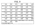
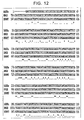
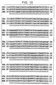
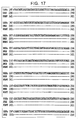
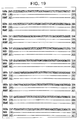
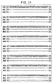
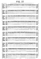
- the 2393 base pair cDNA for RXR ⁇ 2 (C-BNGH42008649) (Figs. 2-5; SEQ ID NO: 1), and the 2421 base pair cDNA for RXR ⁇ 3 (C-BRAMY2003287) (Figs. 6-9; SEQ ID NO: 3) , were subj ected to sequence homology searches and functional motif searches using bioSCOUT DNA sequence analysis software (Lion) on GenBank-, EMBL- and SWISS-PROT-derived databases. Furthermore, the sequences were compared with those of RXR ⁇ isoforms previously reported, based on information in the literature (Figs. 10-24).
- RXR ⁇ 2 contains a region of 1305 base pairs (923-2230) encoding a protein of 435 amino acids.
- RXR ⁇ 3 contains a region of 1095 base pairs (924-2021) encoding a protein of 365 amino acids.
- Both proteins contain a DNA binding domain (DBD) and a ligand-binding domain (LBD), common to the nuclear receptor family.
- DBD DNA binding domain
- LBD ligand-binding domain
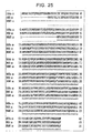
- RXR ⁇ 1 a known isoform of RXR ⁇ , showed highest homology to both of these cDNAs. Comparison of the amino acid sequence of each revealed that RXR ⁇ 2 and RXR ⁇ 3 have a shorter N-terminus than RXR ⁇ 1, with the deletion of 27 and 97 amino acids respectively (Fig. 25).
- the cDNA sequences of RXR ⁇ 2 (C-BNGH42008649), and RXR ⁇ 3 (C-BRAMY2003287) were searched against the GenBank and Sanger Center human genomic sequence databases using BLAST. With the exception of an unmatched part, the cDNA sequences matched the sequence of a clone on human chromosome 9q34.3, and the exon sequences were determined. Because the genomic sequence database is still a draft, literature reporting the result of experimental analysis of the genomic structure of RXR ⁇ 1 (Li G., Walch E., Yang X., Lipman S.M., and Clifford J.L. Cloning and characterization of the human retinoid X receptor á gene: conservation of structure with the mouse homolog.
- the tissue distribution of RXR ⁇ 2 and RXR ⁇ 3 expression was examined by PCR, using commercially available cDNA from human tissues as a template, and isoform-specific primer sets.
- 20 ⁇ L of PCR reaction mixture contains 1 ⁇ g of cDNA from human heart, kidney, liver, spleen, brain, or skeletal muscle, 2 ⁇ l of 10x Ex Taq Buffer, 1.6 ⁇ l of dNTP mixture, 1 ⁇ l of Ex Taq (TAKARA SHUZO), 1 ⁇ l of Perfect Match PCR Enhancer (STRATAGENE) , 1 ⁇ l of Forward primer, 1 ⁇ l of Reverse primer, and 11.4 ⁇ l of sterile water.
- PCR reaction mixture contains 1 ⁇ g of cDNA from human heart, kidney, liver, spleen, brain, or skeletal muscle, 2 ⁇ l of 10x Ex Taq Buffer, 1.6 ⁇ l of dNTP mixture, 1 ⁇ l of Ex Taq (TAKARA SHUZO), 1
- the nucleotide sequence of the primers is as follows:
- PCR amplification was performed for 35 cycles of 94°C for 1 minute, 55°C for 2 minutes, and 72°C for 3 minutes.
- Glyceraldehyde-3-phosphate dehydrogenase Glyceraldehyde-3-phosphate dehydrogenase (GAPDH) was also amplified by PCR from each tissue under the same conditions, and used as an expression level control.
- RXR ⁇ 1 expression was detected in kidney, spleen, liver, adipocytes, and prostate;
- RXR ⁇ 3 expression was detected in brain, spleen, and prostate (Fig. 27). Under these experimental conditions, the tissue expression of RXR ⁇ 2 was under detectable limits.
- a reporter plasmid into which RXRE (RXR response element) was inserted was prepared along with expression plasmids for RXR ⁇ 2, RXR ⁇ 3, and RXR ⁇ 1, as follows: A reporter gene assay was performed by transfecting the reporter plasmid and expression plasmids into cultured cells by lipofection, followed by incubating the cells for a certain time, treating the cells with cell lysis solution, and measuring reporter activity with a luminometer using a Dual luciferase assay.
- RXRE reporter plasmid was constructed by inserting multiple synthetic DNAs, corresponding to an RXRE sequence, into the BglII site of the luciferase reporter vector PGVP2 in tandem.
- Synthetic DNAs of 26 mer (5'-GATCTGAGGTCAGAGGTCAGAGAGCA-3'/SEQ ID NO: 17; 5'-GATCTGCTCTCTGACCTCTGACCTCA-3'/ SEQ ID NO: 18), corresponding to RXRE (RXR response element) and complementary to each other, were phosphorylated at the 5'-end with T4 polynucleotide kinase.
- the complementary strands were mixed at a 1:1 ratio, and annealed by heat denaturation at 65°C for 15 minutes, then slowly cooled to prepare the insert DNA fragment.
- the fragment was mixed with BglII-digested PGVP2 vector, ligated with T4 DNA ligase, and used to transform E. coli DH5 ⁇ . Transformation was performed using commercial E.coli DH5 ⁇ competent cells (TOYOBO) and standard methods, and transformants were selected for ampicillin resistance.
- the obtained transformants were examined by colony PCR for the presence or absence and size of the insert fragment in the recombinant plasmid.
- the nucleotide sequences of clones containing multiple inserts was then examined, and the number of inserted response elements and their orientation was confirmed. From these, a clone containing five response elements, in tandem and in the sense direction, was selected as a reporter plasmid.
- Expression plasmids for RXR ⁇ 1, RXR ⁇ 2, and RXR ⁇ 3 were constructed by inserting sequences comprising coding regions into the multi-cloning site of expression vector pcDNA3.1(+).
- the insert fragments were prepared by PCR amplification using pME18SFL3-C-BNGH42008649 as a template for RXR ⁇ 2 (C-BNGH42008649), and pME18SFL3-C-BRAMY2003287 as a template for RXR ⁇ 3 (C-BRAMY2003287).
- the control RXR ⁇ 1 was prepared by PCR using cDNA from human kidney as a template.
- the primers used for PCR were as follows:
- PCR amplification was performed on 35 cycles of 94°C for 1 minute, 55°C for 2 minutes, and 72°C for 3 minutes.
- the resulting insert fragments and expression vectors were digested with HindIII and XbaI, separated and purified by electrophoresis on an agarose gel, and then ligated with T4 DNA ligase.
- the reaction mixture was used to transform E.coli DH5 ⁇ cells, and transformants were selected for ampicillin resistance.
- the obtained transformants were checked by colony PCR for the presence of inserts, and the nucleotide sequence was determined to confirm that the sequence did not contain mutations.
- the DNAs of the reporter plasmids and expression plasmids were prepared by large scale culturing of strains carrying the plasmids, using a commercial DNA preparation kit (Qiagen). They were then subjected to reporter gene assays.
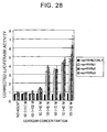
- RXR ⁇ 2 and RXR ⁇ 3 were evaluated by using a reporter gene assay. Using RXR ⁇ 1 as a control, the activities of RXR ⁇ 2 and RXR ⁇ 3 were compared. In addition, the response of each to an RXR ⁇ agonist, LG100268, was evaluated (Fig. 28).
- the reporter gene assay was performed using CV-1 cells (African green monkey kidney-derived cell line) as hosts. Host cells were transfected with luciferase reporter plasmids and expression plasmids for RXR ⁇ 1, RXR ⁇ 2, or RXR ⁇ 3. They were then exposed to LG100268 at different concentrations (0, 10 -10 to -10 -5 M) for 24 hours, whereupon luciferase activity within the cells was measured.
- Transfection was performed as follows: CV-1 cells were suspended at 2 x10 5 cells/2.5 ml in DMEM medium supplemented with 10% fetal bovine serum, 2000 U/1 of penicillin G, and 100 mg/1 streptomycin. These cells were seeded to a 6-well plate at 2.5 ml/well, and cultured for 18 to 24 hours in a CO 2 incubator at 37°C in the presence of 5% CO 2 . 1 ⁇ g of each of the transcription factor expression plasmid, the luciferase reporter plasmid, and another luciferase reporter plasmid for normalizing the measured value (pRL-TK) , were dissolved in Opti-MEM (Gibco BRL) to total of 150 ⁇ l.
- the LipofectAMINE-DNA mixture was diluted in 1.5 ml of Opti-MEM, and added to cells that had been twice washed with 1 ml of PBS(-). The cells were cultured for four hours. The transfection reagent was removed from the culture, and 200 ⁇ l of trypsin/EDTA was added to the cells, which were then incubated at 37°C for five minutes. 1 ⁇ l of DMEM was added to suspend the cells, which were transferred to a centrifuge tube, and centrifuged for three minutes at 1000 rpm.
- the cells were resuspended in DMEM to a concentration of 1.6 x10 4 cells/100 ⁇ l, and 100 ⁇ l samples were seeded to a 96-well plate.
- a two-fold concentrated solution was made by pre-diluting a 1000 times concentrated agent DMSO solution with culture medium. 50 ⁇ l of the solution was mixed with 50 ⁇ l of a cell suspension at 1.6x10 4 cells/50 ⁇ l, and the culture was incubated in a CO 2 incubator at 37°C and 5% CO 2 for 24 hours.
- Luciferase activity was measured using the Dual-LuciferaseTM Reporter Assay System (Promega). The culture supernatant was removed from each well of the 96-well plate and washed once with 100 ⁇ l of PBS(-). 20 ⁇ l of x 1 PLB (Passive Lysis Buffer) was added, and the mixture was incubated at room temperature for 15 minutes, and then stored at -20°C in the freezer until use. The luminometer used was a ARVOTM HTS 1420 multi-labeling counter (Wallac). The protocol for measurement was as follows: 50 ⁇ l of Luciferase Assay Reagent II was added to each well, stirred for 0.3 seconds and measured for one second.
- Stop&GloTM Reagent 50 ml of Stop&GloTM Reagent was then added, stirred for 0.3 seconds, and measured for one second.
- the measured values for firefly luciferase activity were corrected by dividing by the value for Renilla luciferase activity, the internal control. Average values and standard errors were calculated from triplicate samples.
- novel isoforms RXR ⁇ 2 and RXR ⁇ 3 were both demonstrated to confer transcriptional activation function. However, there was no significant difference among the three receptors RXR ⁇ 1, RXR ⁇ 2 and RXR ⁇ 3 in terms of their transcriptional activation function in response to LG100268.
- a reporter gene assay was used to compare the PPAR ⁇ -coupled transcription-controlling activities of RXR ⁇ 2 and RXR ⁇ 3 with that of RXR ⁇ 1 (Fig. 29).
- the particulars of the reporter gene assay are as for Example 8, except that (1) the PPARE reporter plasmid was used in place of the RXRE reporter plasmid, and (2) the host cells were co-transfected with PPAR ⁇ expression plasmid and an expression plasmid for each RXR ⁇ .
- a reporter gene assay was used to compare the PPAR ⁇ -coupled transcription-controlling activities of RXR ⁇ 2 and RXR ⁇ 3 in the presence of a co-activator with that of RXR ⁇ 1 (Fig. 30).
- the particulars of the reporter gene assay are as for Example 9, except that the host cells were co-transfected with PPAR ⁇ expression plasmid, each RXR ⁇ expression plasmid and each co-activator expression plasmid.
- RXR ⁇ isoforms did not show a significant difference in LG100268 response.
- RXR ⁇ 2 and RXR ⁇ 3 did not show any response to LG100268 in the presence of PCG-1, whereas RXR ⁇ 1 elevated the transcriptional activation activity in a manner dependent on LG100268 concentration, though this was not remarkable.
- RXR ⁇ 1 did not shown any response to LG100268.
- RXR ⁇ 2 and RXR ⁇ 3 elevated their transcriptional activation activity in a manner dependent on LG100268 concentration. Since co-activators are expressed dependent on tissue and physiological conditions, and their tissue distribution patterns are different, the results of the present invention strongly suggest that the novel RXR ⁇ isoforms play physiological roles that are different from those of RXR ⁇ 1.
- the present invention provides the novel nuclear receptor isoforms, RXR ⁇ 2 and RXR ⁇ 3.
- RXR ⁇ isoforms are nuclear receptors that bind to retinoic acid as a ligand and function with SRC-1 as a co-activator.
- SRC-1 as a co-activator.
- the RXR ⁇ 2 and RXR ⁇ 3 isoforms of the present invention were found to function with CBP as a co-activator.
- SRC-1 co-activator is thought to have multiple in vivo functions through interaction with a variety of nuclear receptors.
- Nishihara E. et al. constructed SRC-1 null (SRC-1 -/-) mice by gene targeting, and carried out functional analyses (Eijun Nishihara, Hiromi Yoshida-Komiya, Chi-Shing Chan, Lan Liao, Ronald L. Davis, Bert W. O'Malley, and Jianming Xu. "SRC-1 Null Mice Exhibit Moderate Motor dysfunction and Delayed Development of Cerebellar Purkinje Cells.” The Journal of Neuroscience 23: 213-222 (2003)). The knockout mice were found to exhibit defective development of the uterus, prostate, testis, and mammary gland.
- the RXR ⁇ isoforms discovered in the present invention are novel in their structure and co-activator specificity.
- By regulating the transcription-controlling activity of nuclear receptors cellular activities can be controlled by signal transduction.
- a number of nuclear receptors have been functionally analyzed to date. Attempts have been made to regulate cellular activities by regulating the functions thus revealed.
- many of those attempts have focused on the interaction between a particular nuclear receptor and its ligand.
- the transcription-controlling activity of nuclear receptors is known to be controlled by co-activators.
- co-activators not much attention has been paid to the fact that there are some differences in their co-activator specificity, even among closely related molecules such as isoforms.
- nuclear receptor activity is precisely controlled, not only by the ligand, but also by co-activator specificity.
- consideration is clearly required of not only functions with particular ligands, but also of interactions with co-activators that influence the nuclear receptor's transcription-controlling activity.
- the present invention provides methods for evaluating the activity of controlling the function of co-activators that regulate the transcription-controlling activity of RXR ⁇ isoforms. Furthermore, the invention provides methods for evaluating the effect of a test substance on the transcription activation enhancing function of co-activators. These evaluation methods enable evaluation of a test substance's activity to regulate a co-activator's function in enhancing each RXR ⁇ isoform's transcriptional activation function. Accordingly, based on the present invention, compounds comprising the function of controlling the above-mentioned function of enhancing transcriptional activation can be screened. Compounds identified by the screening methods of the present invention can regulate the function of enhancing transcriptional activation by RXR ⁇ isoforms. Since RXR ⁇ isoforms differ in their co-activator specificity, isoform-specific activity can be regulated by the compounds selected by these screening methods. Such compounds are useful as agents that act on novel targets in controlling cellular activities.
- RXR ⁇ 2 or RXR ⁇ 3 themselves, or compounds capable of controlling the activity or expression of these proteins, may be useful therapeutic agents for diseases caused by abnormalities in the transcription-controlling activity of nuclear receptors.
- the RXR ⁇ 2 or RXR ⁇ 3 proteins may be useful in the treatment of diseases characterized by reduced nuclear receptor expression or activity. More specifically, a variety of cancers, hyperlipemia, arteriosclerosis, diabetes, a variety of inflammatory disease such as enteric inflammation and psoriasis, are considered due to impaired RXR function. Cancers include prostate cancers, acute promyelocytic leukemia, and hepatic cancers.
- the present invention also provides antisenses for the polynucleotides encoding RXR ⁇ 2 or RXR ⁇ 3, as well as double-stranded RNAs, or proteins thereof. Antisenses or proteins comprising dominant negative function may be useful for treating diseases characterized by abnormalities in the transcription-controlling activity of nuclear receptors.
Landscapes
- Health & Medical Sciences (AREA)
- Chemical & Material Sciences (AREA)
- Life Sciences & Earth Sciences (AREA)
- Organic Chemistry (AREA)
- Medicinal Chemistry (AREA)
- General Health & Medical Sciences (AREA)
- Zoology (AREA)
- High Energy & Nuclear Physics (AREA)
- Gastroenterology & Hepatology (AREA)
- Toxicology (AREA)
- Biochemistry (AREA)
- Biophysics (AREA)
- Immunology (AREA)
- Genetics & Genomics (AREA)
- Cell Biology (AREA)
- Molecular Biology (AREA)
- Proteomics, Peptides & Aminoacids (AREA)
- Physics & Mathematics (AREA)
- Engineering & Computer Science (AREA)
- Bioinformatics & Cheminformatics (AREA)
- Chemical Kinetics & Catalysis (AREA)
- General Chemical & Material Sciences (AREA)
- Nuclear Medicine, Radiotherapy & Molecular Imaging (AREA)
- Pharmacology & Pharmacy (AREA)
- Animal Behavior & Ethology (AREA)
- Public Health (AREA)
- Veterinary Medicine (AREA)
- Peptides Or Proteins (AREA)
- Micro-Organisms Or Cultivation Processes Thereof (AREA)
Abstract
The present invention relates to the novel isoforms, RXR alpha 2 and RXR alpha 3, of nuclear receptor RXR alpha . Unlike known isoform RXR alpha 1, the transcriptional activation functions of RXR alpha 2 and RXR alpha 3 are augmented by SRC-1. The present invention provides methods for evaluating the function of regulating augmentation by co-activators of these RXR alpha isoforms, and screening methods based on these evaluation methods. By controlling the interaction between isoforms and co-activators, transcription-controlling activity can be regulated in an isoform-specific manner.
Description
The present invention relates to novel nuclear receptor
isoforms, and their use.
Nuclear receptors constitute a superfamily of a group of
transcription factors whose activities are induced in a
ligand-dependent manner. Nuclear receptors have specific structures,
including a DNA-binding domain and a ligand-binding domain, which
are highly homologous among family members.
By taking advantage of this high sequence homology, a number
of novel nuclear receptor genes have been discovered. To date, forty
or more nuclear receptor genes have been reported in humans. These
include genes whose ligands have not yet been identified, and those
that do not require a ligand to function.
Retinoid X receptor (RXR) α was discovered using homology with
the DNA binding domain of retinoic acid receptor (RAR) α (Mangelsdorf
D.J., Ong E.S., Dyck J.A., and Evans R.M. "Nuclear receptor that
identifies a novel retinoic acid response pathway." Nature 345:
224-229 (1990)). It was later found that although RXRα comprises
transcription-controlling activity alone, it can also function as
a co-receptor in transcriptional activation by forming a heterodimer
with other nuclear receptors, such as peroxisome proliferator
activated receptor (PPAR), RAR, thyroid receptor (TR), and vitamin
D3 receptor (VDR). RXRα is thought to play important roles in
hormone-related physiological events (Yu V.C., Deisert C., Andersen
B., Holloway J.M., Devary O.V., Naar A.M., Kim S.Y., Boutin J.M.,
Glass C.K., and Rosenfeld M.G. "RXR beta: a coregulator that enhances
binding of retinoic acid, thyroid hormone, and vitamin D receptors
to their cognate response elements." Cell 67: 1251-1266 (1991)).
With regards to RXRα ligands, 9-cis retinoic acid has been
identified, in addition to retinoids, as an RXR-specific ligand
(Heyman R.A., Mangelsdorf D.J., Dyck J.A., Stein R.B., Eichele G.,
Evans R.M, and Thaller C. "9-cis retinoic acid is a high affinity
ligand for the retinoid X receptor." Cell 68: 397-406 (1992)).
Furthermore, RXR-specific agonists and antagonists are being
developed. Clinical application of these antagonists as anti-tumor
drugs or therapeutic agents for diabetes is being considered
(Mukheerjee R., Davies P.J., Crombie D.L., Bischoff E.D., Cesario
R.M., Jow L., Hamann L.G., Boehm M.F., Mondon C.E., Nadzan A.M.,
Paterniti J.R.J. , and Heyman R.A. "Sensitization of diabetic and obese
mice to insulin by retinoid X receptor agonists." Nature 386: 407-410
(1997); Wu K., Lamph W.W., and Brown P.H. "RXR-selective retinoids
function independently of RAR and PPAR gamma to inhibit breast cell
growth." Breast Cancer Research and Treatment. 69: 371 (2001)).
RXR is known to have three subtypes: α, β, and γ (Pemrick S.M.,
Lucas D.A., and Grippo J.F. "The retinoid receptors." Leukemia 8:
1797-1806 (1994)). Three isoforms of RXRβ, and two isoforms of RXRγ
have been reported (Pemrick S.M., Lucas D.A., and Grippo J.F. "The
retinoid receptors." Leukemia 8: 1797-1806 (1994); Mahajna J., Shi
B., and Bruskin A. "A four-amino-acid insertion in the ligand-binding
domain inactivates hRXRbeta and renders dominant negative activity."
DNA Cell Biol. 16: 463-76 (1997)).
Three RXRα isoforms have been identified in mice (Brocaard J.,
Kastner P., and Chambon P. "Two novel RXR alpha isoforms from mouse
testis." BBRC 229: 211-218 (1996)). However, there has been no report
of RXRα isoforms in humans; cloning using mouse isoform sequence
information was attempted but failed (Li G. , Walch E. , Yang X. , Lipman
S.M., and Clifford J.L. "Cloning and characterization of the human
retinoid X receptor alpha gene: conservation of structure with the
mouse homolog." BBRC 269: 54-57 (2000)).
All except one of the known RXR isoforms comprise a structure
in which the N-terminal A/B domains vary in size, (Mahajna J., Shi
B. , and Bruskin A. "A four-amino-acid insertion in the ligand-binding
domain inactivates hRXRbeta and renders dominant negative activity."
DNA Cell Biol. 16: 463-76 (1997)). Since the A/B domains are
responsible for ligand-independent transcriptional activation
function, and also involved in interactions with other transcription
factors, RXR isoforms are presumed to share in the transcription
control of groups of genes whose expression is controlled under
different physiological conditions.
Nuclear receptors are presumed to dissociate from
co-repressors when a ligand binds to a ligand-binding domain. They
are then presumed to recruit co-activators to form complexes that
interact with RNA polymerase II complexes, initiating transcription.
Recently, a number of co-repressors and co-activators have been
discovered, some of which are known for their tissue-specific or
physiological stimuli-specific control of expression. Interactions
between nuclear receptors and co-repressors/co-activators are
thought to play important roles in the diverse
transcription-controlling functions of nuclear receptors (Freedman
L.P. "Increasing the complexity of coactivation in nuclear receptor
signaling." Cell 97: 5-8 (1999)).
An objective of the present invention is to provide novel RXRα
isoforms whose transcriptional activation function is enhanced by
co-activators that differ from those that enhance the effect of known
RXRα isoforms. In addition, another objective of the invention is
to provide novel methods for evaluating the function of regulating
RXRα transcription control activity, based on the functions of the
novel isoforms.
The present inventors attempted to discover novel nuclear
receptors by searching for genes that encode proteins comprising a
nuclear receptor specific DNA binding domain (DBD) and ligand-binding
domain (LBD). They succeeded in isolating novel RXRα isoforms.
Furthermore, the present inventors analyzed the functions of the genes
narrowed down by this search. As a result, they found that some RXRα
isoforms exhibited transcriptional activation function due to the
action of co-activators different to those of known RXRα isoforms.
The present inventors then established an assay system for
evaluating the transcription-controlling activity of the RXRα
isoforms in the presence of co-activators that activate nuclear
receptors. Further, the present inventors used this system to
confirm that the effects of test substances on the
transcription-controlling activity of RXRα can be specifically
evaluated for each isoform. Thus, they accomplished the present
invention.
Hence, the present invention relates to the polynucleotides
below, and the proteins encoded by these polynucleotides. It also
relates to methods for evaluating the effects of test substances on
the transcription-controlling activity of each nuclear receptor RXRα
isoform, in the presence of co-activators. Furthermore, the
invention relates to methods, based on these evaluation methods, of
screening for test substances that can regulate the
transcription-controlling activity of RXRα isoforms.
- (1) contacting a test substance with cells, which express a nuclear receptor and comprise an expression cassette in which a reporter gene is inserted downstream of a responsive element for the nuclear receptor, in the presence of a co-activator that activates the nuclear receptor;
- (2) culturing the cells under conditions enabling the nuclear receptor expression, and measuring the expression level of the reporter gene within the cells; and
- (3) detecting the activity of the test substance regulating the transcription-controlling activity of the nuclear receptor, using the measurement result of step (2) as an index;
- (1) contacting the nuclear receptor, SRC-1, and the test
substance in any of the following orders i) to iii):
- i) contacting the nuclear receptor and the test substance first, and then contacting with SRC-1;
- ii) contacting the nuclear receptor and SRC-1 in the presence of the test substance; or
- iii) contacting the nuclear receptor and SRC-1 first, and then contacting with the test substance;
To discover novel nuclear receptors, the present inventors
targeted full-length cDNA libraries, searching for genes that encode
proteins comprising a DNA binding domain (DBD) and a ligand-binding
domain (LBD) that are specific for a nuclear receptor. Of the genes
screened, the present inventors focused on C-BNGH42008649, and
C-BRAMY2003287 , which were confirmed to be highly homologous to RXRα.
C-BNGH42008649 is a cDNA of 2393 base pairs, comprising a coding
sequence (923-2230) that corresponds to a sequence of 435 amino acids.
Compared to the known RXRα1 isoform, C-BNGH42008649 lacks 27
N-terminal amino acids. Based on structural features,
C-BNGH42008649 was judged a novel RXRα isoform, and named human RXRα2.
C-BRAMY2003287 is a cDNA of 2421 base pairs comprising a coding
sequence (924-2021) that corresponds to a sequence of 365 amino acids.
Comparing to known RXRα1 isoforms, it lacks 97 N-terminal amino acids.
Based on structural features, C-BNGH42008649 was judged a novel
RXRα isoform, and named human RXRα3.
The nucleotide sequences of these isoforms were searched
against the human genomic sequence database using BLAST homology,
and were found to match the sequence of a clone of human chromosome
9q34.3. RXRα3 and RXRα2 were revealed to comprise 9 and 10 exons
respectively. Similar homology searches were performed for RXRα1,
and the three were compared with each other. In comparison with RXRα1,
RXRα2 uses common exons as for the downstream exons from the exon
2, but uses a different exon as for its 5'-most exon. In comparison
with RXRα1, RXRα3 uses common exons as for the downstream exons from
the exon 3, but uses a different exon as for its 5'-most exon (Fig.
26).
The present invention relates to a polynucleotide encoding
RXRα2, comprising the coding region of the nucleotide sequence of
SEQ ID NO: 1; and a polynucleotide encoding a protein comprising the
amino acid sequence of SEQ ID NO: 2. The invention also relates to
a polynucleotide encoding RXRα3, comprising the coding region of the
nucleotide sequence of SEQ ID NO: 3; and a polynucleotide encoding
a protein comprising the amino acid sequence of SEQ ID NO: 4.
The present invention also comprises a polynucleotide encoding
a protein functionally equivalent to a protein comprising the amino
acid sequence of SEQ ID NO: 2 or 4. Herein, a protein is defined as
being functionally equivalent to the RXRα2 or RXRα3 proteins of the
present invention if the protein of interest comprises the following
(a) and (b):
For example, the present invention comprises a polynucleotide
encoding a protein which comprises the aforementioned (a) and (b),
and which is encoded by the following polynucleotide:
Herein, increased transcriptional activation function of a
protein due to binding with the ligand retinoic acid, or its agonist,
can be examined as follows: In the following explanation, proteins
to be examined for functional equivalence are described as test
proteins. For example, a reporter gene assay may be used to examine
the aforementioned attribute (a). Specifically, a test protein
examined for attribute (a) is expressed in cells. The cells used as
hosts are introduced with an expression vector for a reporter gene,
under the regulation of a responsive element for the test protein.
RXR responsive elements are already known.
If the test protein comprises attribute (a) , treating the cells
with the ligand retinoic acid or its agonist will lead to an increase
in reporter gene expression level. Therefore, based on a comparison
of reporter gene expression levels before and after ligand treatment,
a test protein can be determined to comprise attribute (a).
Attribute (b), the use of SRC-1 as a co-activator, is another
index for functional equivalence in the present invention. As for
attribute (a), it can also be confirmed by reporter gene assay. As
described above, host cells are introduced with a vector for
expressing a reporter gene under the control of a test protein
responsive element. If reporter gene expression level in the
presence of SRC-1 is elevated in response to a ligand, the test protein
may be confirmed to use SRC-1 as a co-activator.
In general, co-activators are molecules required for the
expression of a nuclear receptor's transcriptional activation
function. Ligand-responsive nuclear receptors exhibit
transcriptional activation function in response to a ligand in the
presence of a co-activator. While nuclear receptors exhibit
transcriptional activation function in response to the amount of a
ligand, co-activators have a qualitative effect. Thus, while the
presence of a certain amount of co-activator is required for
transcriptional activation function, changing the amount of
co-activator has little effect.
Herein, a nuclear receptor's transcription-controlling
activity means a nuclear receptor's activity in initiating gene
transcription via a transcriptional unit. In general, nuclear
receptors are considered to initiate transcription by recruiting
multiple factors called transcriptional cofactors. Transcriptional
activation brought by the transcription-controlling activity of a
nuclear receptor is called transcriptional activation function.
Proteins functionally equivalent to the nuclear receptors RXRα2
or RXRα3 of the present invention require SRC-1 as a co-activator.
In a more preferable embodiment, and similar to known isoform RXRα1,
the proteins functionally equivalent to the nuclear receptors of the
present invention comprise a transcriptional activation function that
is also elevated in response to a ligand in the presence of CBP. Thus,
the transcriptional activation function can be also achieved by
co-activator CBP.
Proteins functionally equivalent to the proteins identified in
the examples of the present invention can be prepared by those skilled
in the art, for example, by using a method of introducing mutations
to the amino acid sequence of a protein. An example of such method
for introducing mutations is the method of site-directed mutagenesis
(Current Protocols in Molecular Biology, edit. Ausubel et al., Publish.
John Wiley & Sons, Section 8.1-8.5 (1987)). In addition, this kind
of protein may also be generated as a result of naturally occurring
amino acid mutations. The present invention comprises proteins
wherein one or more amino acids are replaced, deleted, inserted,
and/or added to the amino acid sequence of SEQ ID NO: 2 or 4, as long
as these proteins are functionally equivalent to the proteins
identified in the examples of the present invention.
The number of mutated amino acids in a protein, or the site of
mutation, is not limited, as long as the protein retains its function.
Typically, the number of mutated amino acids is 10% or less of the
total number of the amino acids in a protein sequence, preferably
5% or less, and more preferably 1% or less. To maintain protein
function, amino acids are preferably replaced with amino acids
comprising similar characteristics. For example, since Ala, Val, Leu,
Ile, Pro, Met, Phe and Trp are all grouped into non-polar amino acids,
they are considered to comprise similar characteristics. Uncharged
amino acids include Gly, Ser, Thr, Cys, Tyr, Asn and Gln. Acidic amino
acids include Asp and Glu, and basic amino acids include Lys, Arg
and His.
Alternatively, proteins functionally equivalent to the
proteins of the present invention can be isolated using techniques
known to those skilled in the art, such as hybridization or gene
amplification. Those skilled in the art can conventionally carry out
hybridization (Current Protocols in Molecular Biology, edit. Ausubel
et al., Publish. John Wiley & Sons, Section 6.3-6.4 (1987)) using
the nucleotide sequence of SEQ ID NO: 1 or 3, which encode the proteins
of the present invention, or their partial sequences, to isolate
polynucleotides comprising high homology to these nucleotides and
obtain functionally equivalent proteins. The present invention
comprises proteins encoded by polynucleotides that hybridize with
polynucleotides encoding the proteins of the present invention, as
long as these proteins are functionally equivalent.
Stringent hybridization conditions for isolating
polynucleotides that encode functionally equivalent proteins are
normally washing in "1x SSC, 0.1% SDS at 37°C". More stringent
conditions are "0.5x SSC, 0.1% SDS at 42°C", and the most stringent
conditions are "0.1x SSC, 0.1% SDS at 65°C". By increasing the
stringency of the conditions for hybridization, one can expect to
isolate DNAs comprising greater homology to the probe sequence.
While the above sets of SSC, SDS, and temperature conditions are given
as examples, one skilled in the art can readily achieve stringencies
similar to the above by appropriately combining these or other
conditions that determine hybridization stringency. The other
conditions influencing stringency include probe concentration, probe
length, and incubation time for hybridization.
The amino acid sequences of the proteins isolated using the
above hybridization, or the nucleotide sequences encoding these
proteins, normally comprise high homology with the proteins of the
present invention of SEQ ID NO: 2 or 4. High homology means sequence
identity of at least 60% or higher, preferably 70% or higher, and
more preferably 80% or higher (for example, 90% or higher). Homology
can be determined using BLAST2 homology search algorithm (Altschul,
S.F. et al. "Gapped BLAST and PSI-BLAST: a new generation of protein
database search programs." Nucleic Acids Res. 25: 3389-3402 (1997)).
The polynucleotides of the present invention comprise
polynucleotides with 70% or more homology to the nucleotide sequence
from 103 to 1461 of SEQ ID NO: 5, and a nucleotide sequence such that
the nucleotide sequence from 1 to 102 of SEQ ID NO: 5 is deleted,
or replaced with another sequence.
The nucleotide sequence described in SEQ ID NO: 5 is that of
the known RXRα1 isoform. The sequence from 1 to 102 corresponds to
exon A of RXRα1. Herein, any nucleotide sequence can replace the exon
A nucleotide sequence. For example, the replaceable nucleotide
sequence may be a sequence for an untranslated region. However, the
nucleotide sequence for exon A comprises an initiation codon at a
position from 76 to 78. Thus, when this region is deleted or replaced,
a novel initiation codon must be added to the region corresponding
to the nucleotide sequence starting from 103.
The initiation codons of the polynucleotides of the present
invention can be, for example, 'atg' starting with 'a' at 157 in SEQ
ID NO: 5. This initiation codon corresponds to that for the desired
nucleotide sequence of SEQ ID NO: 1 of the present invention, as
described later. The initiation codon in the nucleotide sequence of
SEQ ID NO: 1 is the first 'atg' in the nucleotide sequence
corresponding to the exon, adjacent to exon A. Thus, the first
initiation codon utilized when exon A is deleted can be presented
as an example of a desired initiation codon for the polynucleotides
of the present invention.
The initiation codon in the nucleotide sequence of SEQ ID NO:
3 can be also presented as such an example of an initiation codon
for the polynucleotides of the present invention. The initiation
codon at 924 of SEQ ID NO: 3 corresponds to the initiation codon at
367 of SEQ ID NO: 5, the first one appearing in the third exon counting
from the 5' -terminus. Thus , it is not necessary to actively introduce
an initiation codon after deleting or replacing exon A, the sequence
from 1 to 102 of SEQ ID NO: 5. The first initiation codon found in
the adjacent exon can be used for translation initiation.
Proteins encoded by polynucleotides that comprise the replaced
nucleotide sequence can be confirmed to comprise the above attributes
(a) and (b) by using the aforementioned methods. Methods for deleting
a particular region of a polynucleotide that comprises a certain
nucleotide sequence, or replacing a region with another nucleotide
sequence, are known to those skilled in the art. For example, the
nucleotide sequence of SEQ ID NO: 1 or 3 is a desired polynucleotide
of the present invention.
As shown in Fig. 26, the nucleotide sequence (C-BNGH42008649)
of SEQ ID NO: 1 comprises exon B in place of exon A in SEQ ID NO:
5 (RXRα1). The nucleotide sequence (C-BRAMY2003287) of SEQ ID NO:
3 comprises exon D in place of exons A and C in SEQ ID NO: 5 (RXRα1).
Accordingly, compared to SEQ ID: 6 (RXRα1), the amino acid sequences
encoded by polynucleotides RXRα2, and RXRα3 are missing 27 and 97
N-terminal amino acids respectively.
In addition, proteins functionally equivalent to the proteins
of the present invention can be obtained by performing PCR (Current
protocols in Molecular Biology, edit. Ausubel et al. , Publish. John
Wiley & Sons, Section 6.1-6.4 (1987)) using primers designed based
on the parts of the nucleotide sequences identified in the Examples
of the present invention (SEQ ID NO: 1 or 3), and isolating DNA
fragments comprising high homology to the nucleotide sequences or
their parts. The proteins of the present invention, and functionally
equivalent proteins, can be used in the methods for evaluating a
compound that modifies a protein's function, as described below. In
addition, the polynucleotides encoding these proteins are useful for
producing the proteins, or as primers or probes for examining
expression level.
The present invention also relates to polynucleotides that
encode proteins comprising a dominant negative form against the
proteins of the present invention. When expressed, the
polynucleotide encoding a protein capable of conferring a dominant
negative effect exerts the function of eliminating or reducing the
activity of endogenous wild type proteins originally contained in
cells. For example, an inactive nuclear receptor protein, without
transcriptional activation function, can be obtained by altering the
part of an amino acid sequence of a protein of the present invention
that corresponds to a region considered to influence its activity,
such as a region required for interaction with another nuclear
receptor, or a ligand binding domain. By expressing in cells a gene
that encodes such an amino acid sequence, the expression of RXRα2
or RXRα3 of the present invention can be suppressed by dominant
negative effect (competitive inhibition by an inactive form). Thus,
the activity of a nuclear receptor can be controlled by using gene
transfection technologies such as viral vectors to introduce cells
with a gene that encodes the inactive form.
The present invention also provides expression vectors
comprising any of the above polynucleotides. In addition, the
present invention relates to transformants carrying the above
polynucleotides, or any of the above expression vectors, and methods
of producing the proteins of the present invention or their partial
peptides, which comprise culturing the transformant and isolating
a protein of the present invention from the culture. Moreover, the
present invention provides proteins produced by the above methods,
or their partial peptides.
Those skilled in the art know that in using recombinant
technology to produce polypeptides, the desired polypeptides can be
obtained with different types or degrees of glycosylation, depending
on the type of host cell. Those skilled in the art also know that
when producing desired polypeptides as secreted proteins, the
proteins comprise different kinds of terminal amino acid sequences
(N- and/or C-termini), since the precursor polypeptides expressed
in host cells undergo processing by signal peptidases and such.
Therefore, those skilled in the art should easily understand that
these polypeptides are comprised in the scope of the proteins of the
present invention.
The Examples below illustrate an example of the construction
of an expression vector capable of functioning in mammalian cells.
On disclosure of a DNA encoding a protein of the present invention
herein, those skilled in the art can readily construct expression
vectors capable of expressing and producing the protein when
introduced into host cells, including fungi such as yeast, and
prokaryotic cells. Therefore, the present invention comprises
expression vectors that can be constructed using methods known in
the art, based on the nucleotide sequences of the present invention.
Microorganisms that can be used for expressing the
polynucleotides that encode the proteins of the present invention
include prokaryotic bacteria such as Escherichia coli and Bacillus
subtilis, and eukaryotic yeast such as Saccharomyces cerevisiae.
Mammalian cells include tissue-cultured human cells, and cultured
animal cells. Furthermore, cultured plant cells may be used.
Examples of microorganism host cells are bacteria belonging to
Escherichia, and Saccharomyces cerevisiae. More specifically, the
following strains are examples of microorganism hosts:
Bacteria belonging to Escherichia:
Yeast:
Examples of mammalian host cells include human embryonic
kidney-derived HEK293 cells, mouse L929 cells, and Chinese hamster
ovary (CHO) cells.
Normally, when the host cells are prokaryotic bacteria cells,
particularly E.coli, the expression vectors comprise at least a
promoter, an initiation codon, a polynucleotide encoding an amino
acid sequence of a protein of the present invention, a termination
codon, and an autonomously replicable unit. When used in eukaryotic
host cells such as yeast and mammalian cells, the expression vector
preferably comprises at least a promoter, an initiation codon, a
polynucleotide encoding an amino acid sequence of a protein of the
present invention, and a termination codon. It may further comprise
insertion of an enhancer sequence, 5'- and 3'-untranslated regions
of the protein of the present invention, a polyadenylation site, and
an autonomously replicable unit.
The above autonomously replicable units preferably comprise a
transformant selection marker (e.g. ampicillin resistance). In an
expression vector for a microorganism host cells, a promoter means
a promoter/operator region, comprising a promoter, operator, and
Shine-Dalgarno (SD) sequence (e.g. AAGG). Conventional promoter
operator region are examples of such promoters. Specifically, the
lactose operon, PL-promoter, trp-promoter, or such can be used. An
example of an expression vector for use in yeast host cells is the
pho5 promoter. In addition, basic amino acids with affinity for metal
ion chelates may be added to either end of the proteins of the present
invention to facilitate purification. When adding basic amino acids,
oligopeptides can be added to an arbitrary end of a gene of interest
by performing PCR using a primer to which is attached at the 5'-end
a sequence comprising a consecutive nucleotide sequence encoding the
desired amino acids. Basic amino acids include histidine, lysine or
arginine.
Promoters used in expression vectors in mammalian cells include
the HTLV-LTR promoter, SV40 early and late promoters, CMV promoter,
and mouse metallothionein-I (MMT) promoter. Preferable initiation
codons include the methionine codon (ATG).
The termination codons include conventional termination codons
(e.g. TAG, TGA and such). Autonomously replicable units are DNA
sequences capable of autonomously replicating the entire DNA of the
expression vector that comprises them within a host cell, and these
include natural plasmids, artificially modified plasmids, and
synthetic plasmids. Artificially modified plasmids include DNA
fragments prepared from natural plasmids. When using E. coli as a
host, preferred examples of such desired plasmids include plasmid
pBR322 and its artificial modifications. The artificially modified
plasmids include DNA fragments obtained by treating pBR322 with an
appropriate restriction enzyme. When using yeast host cells, pJDB219,
and pJDB207 may be used as examples. Further, plasmids used in animal
cells include plasmid pcDNA3.1 (Invitrogen) , pMM324, pSV2dhfr (ATCC
37145), pdBPV-MMTneo (ATCC 37224), and pSV2neo (ATCC 37149).
An enhancer sequence may be the SV40 enhancer sequence, for
example. The polyadenylation sites include the SV40 polyadenylation
site.
Polynucleotides encoding the amino acid sequences of the
proteins of the present invention are obtainable by synthesizing the
entire or partial sequence using a DNA synthesizer, for example.
Alternatively, they can be obtained from human cDNA libraries by using
probes or primers whose design is based on the nucleotide sequence
of SEQ ID NO: 1 or 3. Furthermore, genomic DNAs that encode the
proteins of the present invention may be prepared by processing
genomic DNAs of standard methods. For example, genomic DNAs may be
processed by methods that comprise the steps of digestion with
restriction enzymes, dephosphorylation with bacterial alkaline
phosphatase, phosphorylation with T4 polynucleotide kinase, and
ligation with T4 DNA ligase. Furthermore, genomic DNAs obtained as
above can be used to identify transcription initiation sites for the
genes of the present invention that occur in the genome, and to specify
the expression regulatory regions located further upstream.
Regulatory regions, such as promoters and enhancers that regulate
the expression of genes encoding the proteins of the present invention,
are useful as target regions for detecting abnormal protein expression.
Alternatively, for example, decoy nucleic acid medicines targeted
at such regions may be used to achieve expression regulation.
Methods known to those skilled in art or in the literature may
be used to perform those procedures required to implement the present
invention, such as DNA cloning, construction of each plasmid,
transfection into host cells, culture of transformants, and recovery
of proteins from the culture (Molecular Cloning, 2nd. edition,
Sambrook J. et al., Cold Spring Harbor Laboratory (1989) ; DNA Cloning,
Glover D.M., IRL PRESS (1985); Molecular Cloning, 3rd. edition,
Sambrook J. et al., Cold Spring Harbor Laboratory (2001)).
The host cells of the present invention also comprise cells used
for the functional analysis of RXRα2 or RXRα3 of the present invention,
or in the methods for evaluating function-inhibiting and
function-enhancing agents which use these proteins. Vector
transfection into host cells may be performed by calcium phosphate
precipitation, electroporation (Current protocols in Molecular
Biology, edit. Ausubel et al., Publish. John Wiley & Sons., Section
9.1-9.9 (1987)), by using LipofectAMINE (Gibco BRL) , microinj ection,
or such. The proteins of the present invention may be prepared from
transformants using methods of protein separation and purification
commonly known to those skilled in the art. The present invention
relates to substantially pure proteins that are purified as above.
Herein, "substantially pure" means free from contamination with other
human-derived proteins. Alternatively, substantially pure proteins
of the present invention are proteins comprising a purity of, for
example, 80% or more, normally 90% or more, 95% or more, preferably
98% or more, and more preferably 99% or more.
The present invention also provides polynucleotides comprising
a nucleotide sequence of SEQ ID NO: 1 or 3, or a polynucleotide
comprising at least 15 nucleotides that are complementary to the
complementary strand of the polynucleotide. Herein, the
"complementary strand" means the other strand to one strand of a
double-stranded polynucleotide comprising A:T (A:U) and G:C base
pairs. In addition, "complementary" refers not only to complete
complementarity to a region of at least 15 consecutive nucleotides,
but also to homology of at least 70% or more, preferably at least
80% or more, more preferably 90% or more, and further preferably 95%
or more. Homology may be determined by the algorithms described in
the present invention.
Such polynucleotides may be used as probes for detecting or
isolating DNAs or RNAs that encode the proteins of the present
invention, or as primers for amplifying the polynucleotides of the
present invention. When used a primer, the polynucleotide length is
normally from 15 to 100 base pairs , and preferably from 15 to 35 base
pairs. In addition, when used as a probe, the polynucleotides
comprise at least a part or an entire sequence of a polynucleotide
of the present invention, and comprise at least 15 base pairs. When
used as a primer, the 3'-region must be complementary, but a
recognition site for a restriction enzyme, tag or such can be added
to the 5'-region.
In particular, regions within the nucleotide sequence of SEQ
ID NO: 1 or 3, and which comprise exons that are not shared with the
known RXRα1 isoform, are useful for detecting DNAs that comprise the
nucleotide sequence of SEQ ID NO: 1 or 3. An oligonucleotide capable
of hybridizing with the nucleotide sequence of an above region or
its complementary sequence may be useful as a probe or primer for
specifically detecting RXRα2 or RXRα3.
For example, a set of primers that anneals to a region where
the exons are not shared is useful for specific amplification of the
isoforms.
The polynucleotides of the present invention may be used for
testing and diagnosing abnormalities of the proteins of the present
invention. For example, Northern hybridization or RT-PCR that uses
the polynucleotides of the present invention as probes or primers
may be performed to test for abnormal expression. In addition,
genomic DNA PCR and RT-PCR may be performed by carrying out
polymerase chain reactions (PCR) using the polynucleotides of the
present invention as primers. Furthermore, by amplifying genes that
encode the proteins of the present invention, or their expression
regulatory regions, sequence abnormalities may be examined and
diagnosed using RFLP analysis, SSCP, sequencing, and such.
The RXRα isoforms of the present invention exhibit a
transcriptional activation function that increases in response to
stimulus with the ligand, retinoic acid. In addition, the RXRα
isoforms of the present invention require SRC-1 as a co-activator.
Therefore, polynucleotides encoding such factors can be used for gene
therapy of diseases associated with the factors. Specifically, by
expressing the RXRα2 or RXRα3 of the present invention through
introducing a polynucleotide such that it is appropriately expressed
at a host's disease site, diseases associated with that factor can
be treated or prevented. Herein, "expression" includes
transcription and/or translation. The expression of a gene at the
transcription level can be examined and diagnosed by analyzing
expression using a polynucleotide of the present invention. In
addition, gene expression at the translation level can be examined
and diagnosed using antibodies against the proteins of the present
invention.
RXRα was only known to have a single isoform, RXRα1. The present
invention revealed novel isoforms that differ in their co-activator
requirements. Strictly speaking, diseases caused by RXRα
dysfunction are caused by impaired function of different isoforms.
Therefore, in patients for whom enhanced RXRα1 activity does not
improve symptoms, RXRα2 or RXRα3 dysfunction may be contributing to
the disease. RXRα1 requires a different co-activator to RXRα2 or
RXRα3. Thus, agonists screened using RXRα1 may not have any effect
on RXRα2 or RXRα3. Agonists that can be obtained using the present
invention may be effective therapeutic agents for diseases for which
agonists against the known RXRα1 isoform cannot achieve sufficient
therapeutic effect.
In addition, a "polynucleotide comprising the nucleotide
sequence of SEQ ID NO: 1 or 3, or a polynucleotide comprising at least
15 nucleotides that are complementary to the complementary strand
of the polynucleotide" comprises an antisense polynucleotide capable
of suppressing the expression of a protein of the present invention.
Herein, the antisense polynucleotide means a polynucleotide that
suppresses the expression of a protein comprising the amino acid
sequence of SEQ ID NO: 2 or 4.
A "double-stranded RNA of 21 to 23 base pairs, comprising a sense
RNA corresponding to a partial sequence of the nucleotide sequence
of the polynucleotide of SEQ ID NO: 1 or 3, and its antisense RNA"
comprises small interfering RNA (siRNA) for suppressing the
expression of a protein of the present invention. The
double-stranded RNAs of the present invention comprise not only
double-stranded RNAs with two strands, but also single-stranded RNAs
with hairpin loop structures comprising sense and antisense
sequences.
Antisense or siRNAs that suppress the expression of RXRα2 or
RXRα3 may be used as agents to control their activity.
The antisense polynucleotides may be antisense DNAs, or
antisense oligonucleotides. Antisense DNAs are at least 15 base
pairs or longer, preferably 100 base pairs or longer, more preferably
500 base pairs or longer, and normally 3000 base pairs or shorter,
preferably 2000 base pairs or shorter. The antisense DNAs may be used
alone, or by insertion in the antisense orientation into appropriate
vectors, such as retrovirus vectors, or adenovirus vectors. The
antisense oligonucleotides are at least ten base pairs or longer,
and normally 100 base pairs or shorter; preferably 20 base pairs or
longer and 50 base pairs or shorter. The antisense oligonucleotides
may be prepared, for example, based on the nucleotide sequence
information of SEQ ID NO: 1, or 3 by using the phosphorothioate method
(Stein "Physicochemical properties of phosphorothioate
oligodeoxynucleotides." Nucleic Acids Res. 16: 3209-3221 (1988)).
In addition, siRNAs may be introduced into an living organism
by administering a vector that carry DNAs for both their sense and
antisense strands. Vectors for siRNA expression are known.
The antisense or siRNAs may be used for gene therapy of diseases
caused by abnormalities of proteins comprising an amino acid sequence
of SEQ ID NO: 2 or 4. Protein abnormalities include abnormal protein
function, abnormal protein expression, and such. When used for gene
therapy, patients may be administered using ex vivo or in vivo methods,
by using viral vectors such as retrovirus vectors, adenovirus vectors,
and adeno-associated virus vectors, or non-viral vectors such as
liposomes.
When used for gene therapy, the polynucleotides and antisense
polynucleotides may be administered to patients using ex vivo or in
vivo methods, by using viral vectors such as retrovirus vectors,
adenovirus vectors, and adeno-associated virus vectors, or non-viral
vectors such as liposomes.
The present invention also relates to methods of detecting the
activity of a test substance in regulating the
transcription-controlling activity of a nuclear receptor, wherein
the nuclear receptor is a protein encoded by a polynucleotide of any
one of the following (A) to (E):
- (1) contacting test substances with cells expressing the nuclear receptor and comprising an expression cassette where a reporter gene is linked downstream of the nuclear receptor's responsive element;
- (2) culturing the cells under conditions that enable the expression of the nuclear receptor, and measuring the expression level of the reporter gene in the cells; and
- (3) detecting the activity of the test substances in regulating the transcription regulating activity of the nuclear receptor, using the measurement results of step (2) as an index.
In the above-mentioned methods of detection, proteins
functionally equivalent to proteins comprising the amino acid
sequence of SEQ ID NO: 2, or 4 refer to proteins comprising the above
attributes (a) and (b). Proteins comprising the amino acid sequence
of SEQ ID NO: 2 or 4 include the novel isoforms of RXRα protein
discovered by the present inventors. In general, subtypes represent
a group of molecules comprising similar structures. However,
isoforms not only comprise common structures, and isoform can also
refer to sharing exons, in particular. Human RXRα isoforms were
unknown until the present inventors discovered two isoforms for the
first time.
Methods for testing whether a given protein can use retinoic
acid or its agonists as a ligand are described above. In addition,
methods for confirming the transcriptional activation function of
a given protein by co-activator RC-1 are also described above. By
using these methods, proteins can be confirmed to be functionally
equivalent proteins of the present invention.
Herein, the cells expressing a nuclear receptor, and comprising
an expression cassette in which a reporter gene is linked downstream
of a nuclear receptor's responsive element, may be obtained as
follows:
First, a DNA encoding a nuclear receptor of interest is
introduced into the cells for expressing the nuclear receptor. DNAs
encoding nuclear receptors for use in the present invention may be
DNAs comprising a coding region of the nucleotide sequence of SEQ
ID NO: 1 or 3. In addition, DNAs encoding proteins functionally
equivalent to a protein comprising the amino acid sequence of SEQ
ID NO: 2 or 4 may be used. As shown in the Examples, proteins
comprising the amino acid sequence of SEQ ID NO: 2 or 4 bind to retinoic
acid as a ligand, and exhibit transcriptional activation function
in the presence of SRC-1 co-activator.
Such DNAs are obtainable by screening cDNA libraries prepared
from humans or other animals, such as nematodes, and yeast using PCR
or hybridization based on respective nucleotide sequence information.
Alternatively, DNAs comprising the essential nucleotide sequences
may be synthesized based on nucleotide sequence information.
The DNAs are cloned in expression vectors, and transfected into
cells. The expression vectors include pcDNA3.1 (Invitrogen), pMM324,
pSV2dhfr (ATCC 37145) , pdBPV-MMTneo (ATCC 37224), and pSV2neo (ATCC
37149). pcDNA3.1(+) is a vector enabling high level expression of
a foreign gene in mammalian cells.
Herein, cells transfected with a DNA encoding a nuclear receptor
are those cells capable of translating the DNA into a protein, and
comprising an expression cassette wherein a reporter gene is linked
down stream of a responsive element for the introduced nuclear
receptor. Herein, expression cassettes represent a responsive
element and reporter gene set, in which reporter genes are located
such that they are transcribed under the regulation of the responsive
element. Expression cassettes may be introduced into cells as
plasmids, or integrated into the cell genome.
Responsive elements are nucleotide sequences capable of binding
to the DBD of a nuclear receptor, and are necessary for the initiation
of transcription to mRNA by the transcription unit. RXRα responsive
elements are already known (Mangelsdorf D.J., Umesono K., Kliewer
S.A., Borgmeyer U., Ong E.S., and Evans R.M. A direct repeat in the
cellular retinol-binding protein type II gene confers differential
regulation by RXR and RAR. Cell 66: 555-561 (1991)). For example,
the retinoic acid responsive element RRE used in the Examples
comprises the nucleotide sequence of SEQ ID NO: 17.
Expressing reporter genes under the regulation of the
responsive elements may be achieved by replacing a gene under the
regulation of a responsive element with a reporter gene. Plasmids
comprising an expression cassette that comprises a reporter gene
downstream of a responsive element are called reporter plasmids.
Reporter plasmids may be readily constructed using reporter
vectors. Reporter vectors comprise reporter genes, and upstream of
that, cloning sites for inserting given responsive elements. For
example, the reporter vector PGVP2, which comprises the luciferase
gene as a reporter, is commercially available. A reporter plasmid
for use in the present invention may be obtained by inserting an above
responsive element into the cloning site of a reporter vector.
Multiple responsive elements may be inserted in tandem to enhance
the response to a nuclear receptor. Thus, increased sensitivity in
detecting transcriptional activation is expected. The responsive
elements to be inserted may be chemically synthesized.
Herein, the reporter genes can be any reporter genes for which
signals can be easily measured. Reporter genes for analyzing the gene
expression regulatory mechanisms are known. Specifically, genes
encoding luciferase, catalase, β-galactosidase, green fluorescent
protein (GFP), and such may be used as reporter genes. The cells
transfected with the vectors may be, for example, animal cells in
which that gene is deleted.
The cells transfected with the different kinds of genes may be
animal cells, yeast cells, insect cells, or such. One example of
preferable cells in the present invention are the
monkey-kidney-derived CV-1 cell line used in the Examples.
Herein, the cells are cultured with co-activators in the
presence of a test substance, under conditions enabling expression
of the above nuclear receptor. The expression level of the reporter
gene in the cells is then measured. Herein, conditions enabling the
expression of an above nuclear receptor refer to conditions where
a nuclear receptor gene comprised in an introduced vector is expressed
in the host cells. For example, if the vector utilizes an inducible
promoter, the cells are cultured under conditions required to induce
expression.
If the nuclear receptor functions in a ligand-dependent manner,
the cells are preferably provided with the ligand. On the other hand,
if the nuclear receptor exhibits the transcriptional activation
function ligand-independently, the above methods may be performed
without providing a ligand. SRC-1 co-activators may be supplied to
cells by transfecting the cells with an expression plasmid carrying
the co-activator gene inserted into it, and expressing the
co-activator within the cells. The amount of DNA used for
transfection may be, for example, the same as the amount of RXRα
expression plasmid.
In the methods of detection of the present invention, test
substances are added to the culture to make contact with the above
cells. Alternatively, if the test substances are proteins, the genes
encoding them may be transfected into the same cells, and contacted
with the nuclear receptors. Furthermore, cells expressing a test
substance may also be contacted by co-culturing with cells expressing
an above nuclear receptor.
Once a nuclear receptor is expressed, the reporter gene is
transcribed depending on the co-activator SRC-1, and the protein is
expressed. The expression level of the reporter gene can be
determined by measuring the intensity of the signal originating from
this protein. For example, when using the luciferase gene as the
reporter gene, luciferase activity can be measured as
chemiluminescent intensity. Alternatively, if the reporter gene is
GFP, the fluorescent intensity of GFP can be measured.
If a coexisting test substance is capable of regulating the
transcription-controlling activity, the transcription level of the
reporter gene will change. If the test substance inhibits
transcription, reporter gene transcription is suppressed and the
signal will be reduced, based on the extent of this change. In
contrast, a test substance comprising the function of enhancing
transcriptional activation function will increase the reporter gene
signal. Therefore, the influence of test substances on
transcription-controlling activity can be evaluated using reporter
gene transcription level as an index.
By using substances whose influence on
transcription-controlling activity is already known, the
relationship between the degree of influence on
transcription-controlling activity and the intensity of the reporter
gene signal can be clarified. Thus, the effect of substances whose
influence on transcription-controlling activity is unknown can be
quantitatively evaluated based on the intensity of this signal.
The methods of detection in the present invention may further
comprise a step of detecting a test substance' s activity in regulating
the transcription-controlling activity of other RXRα isoforms.
Other isoforms of RXRα may include proteins encoded by the
polynucleotide of any one of the following (A') to (E'):
RXRα1 is a known protein deposited in GenBank as accession
number NM_002957. By evaluating the effect of test substances on the
transcriptional activation function of not only the RXRα isoforms
of the present invention, but also of other isoforms using similar
techniques, the differences between a substance's specificity for
each isoform can be identified. For example, each isoform shows a
different level of activation depending on the type of co-activator.
For example, the novel isoforms discovered in the present invention,
RXRα2 and RXRα3, require CBP or SRC-1 as a co-activator, whereas the
known isoform RXRα1 does not use SRC-1 as a co-activator. These
differences may be the cause of functional differences between the
isoforms in vivo. Therefore, the specificity of test substances for
each isoform provides important information for enabling specific
regulation of these nuclear receptors.
For example, isoform-selective agents selected by the
evaluation methods of the present invention are expected to function
specifically in tissues or organs where the isoform is expressed
specifically, or under the physiological conditions to which the
isoforms respond specifically. Such agents can be expected to have
reduced side effects compared to conventional drugs that are not
isoform-selective.
Herein, to detect the function of regulating the
transcription-controlling activity of other isoforms, the above
methods may be performed in the presence of a co-activator capable
of activating each isoform. CBP, for example, is a preferred
co-activator for proteins encoded by the polynucleotide of the above
(A') to (E').
The activity of regulating the transcription-controlling
activity ty of a nuclear receptor activated by SRC-1 can be evaluated
by a detection method of the present invention. The method of
detection may be used to evaluate substances capable of regulating
the transcription-controlling activity. Thus, the present invention
relates to methods for evaluating substances capable of regulating
a transcription-controlling activity, wherein the methods comprise
the following steps:
The cells introduced with an expression vector for a nuclear
receptor produce a signal generated from the reporter gene, in line
with nuclear receptor expression. The obtained cells are seeded into
a 96-well plate, and cultured under conditions that induce expression
of the introduced genes. By adding a test substance to be evaluated
into each well, the function of suppressing or enhancing the
transcriptional activation function of the nuclear receptor may be
readily detected, using reporter gene expression level as an index.
For example, when GFP is used as the reporter gene, it is possible
to evaluate influence on the expression regulatory region by comparing
the fluorescence intensities of GFP in conditions with or without
the test substance. A significant difference may be judged from this
comparison when the intensity ratio is, for example, two fold or more,
or 1/2 or less, preferably five fold or more, or 1/5 or less, and
more preferably ten fold or more, or 1/10 or less. The methods may
be applicable not only to animal cells, but to any host cells,
regardless of whether they are eukaryotic or prokaryotic cells, as
long as the transcriptional activation function of an introduced
nuclear receptor can be reproduced. The cells are cultured under
conditions that enable reporter gene expression.
The test substances for the evaluation methods of the present
invention are not limited. Specifically, they include cell extracts,
products expressed from DNA libraries, synthetic low molecular weight
compounds, synthetic peptides, and natural compounds, for example.
Alternatively, antibodies that recognize a protein encoded by a
polynucleotide of any one of the above (A) to (E) may be useful as
candidate substances for screening.
Compounds known for their effect on transcription-controlling
activity can be used as controls for the evaluation methods of the
present invention. More specifically, compounds that clearly have
no effect on transcription-controlling activity, for example, may
be used as controls. Data measured without any test substance
influence can be used as a control for cases without any effect on
transcription-controlling activity.
Alternatively, by using substances already known to comprise
a target function as controls, substances that induce a larger change
in signal may be searched for selecting substances comprising
relatively greater function.
The evaluation methods of the present invention may be used,
for example, for screening for compounds that comprise desired
activities, or for characterizing compounds. For example, screening
may be performed by applying the evaluation methods to a broad range
of compounds, and repeatedly selecting compounds that excel in the
activity of interest. Alternatively, these detection methods may be
used to analyze specific compound attributes.
Furthermore, the present invention relates to methods of
detecting the activity of test substances in controlling the
interaction between a nuclear receptor and a co-activator that
activates the receptor, wherein the nuclear receptor is a protein
encoded by the polynucleotide of any one of the following (A) to (E) :
- (1) contacting the nuclear receptor, SRC-1, and a test
substance any of the following orders i) to iii):
- i) contacting the nuclear receptor and the test substance first, and then contacting with SRC-1;
- ii) contacting the nuclear receptor and SRC-1 in the presence of the test substance; or
- iii) contacting the nuclear receptor and SRC-1 first, and then contacting with the test substance;
In contrast to the aforementioned methods of detecting effect
on transcription-controlling activity using behavior within cells
as an index, these methods detect the activity of binding to a nuclear
receptor based on the interaction between substances. RXRα2 or RXRα3,
or proteins functionally equivalent to these isoforms, which were
discovered by the present invention, are the nuclear receptors used
in the present invention's methods of detecting a test substance's
ability to bind a nuclear receptor. The activity of binding a nuclear
receptor can be detected by monitoring competition between
co-activators.
Herein, co-activators for nuclear receptors may be SRC-1 or CBP,
for example. SRC-1 enhances the transcriptional activation function
of the isoforms RXRα2 and RXRα3 in a manner dependent on the amount
of the ligand. CBP can function as a co-activator not only for RXRα2
and RXRα3, but also for known isoform RXRα1.
Herein, the nuclear receptors, their co-activators, and the
test substances are exposed to each other in any of the following
orders i) to iii) : First, i) by contacting the nuclear receptor with
the test substance, and then with the co-activator, the function of
inhibiting the co-activator's binding to the nuclear receptor can
be evaluated. Next, ii) by contacting the nuclear receptor with the
co-activator in the presence of the test substance, the function of
the substance in competiting with the co-activator for nuclear
receptor binding can be evaluated. Furthermore, iii) by contacting
the nuclear receptor with the co-activator, and then with the test
substance, the function of the substance in replacing co-activator
binding to the nuclear receptor can be evaluated. Compounds
comprising such functions, as detected by these methods, are useful
as candidates for RXRα2 or RXRα3 agonists or antagonists.
Furthermore, the detection methods of the present invention
comprise measuring the amount of co-activators or test substances
bound to nuclear receptors. The amount of co-activator or test
substance bound to a nuclear receptor may be measured by labeling
these substances. Co-activators or test substances may be labeled
with radioisotopes, substances with chemiluminescent, fluorescent,
or enzymatic active substances, or tags. The amount of co-activator
or test substance bound to a nuclear receptor may be measured by
isolating the nuclear receptor from the reaction mixture, and
measuring the label in the isolated fraction. The nuclear receptor
may first be immobilized onto a solid phase to facilitate its isolation
from the reaction mixture. Such solid phases commonly include
magnetic beads, or the inside wall of reaction containers.
In the methods of detection of the present invention, the amount
of co-activator bound to a nuclear receptor is correlated to the
activity of a test substance in binding to that nuclear receptor:
when the amount of co-activator bound to a nuclear receptor decreases
compared with the amount in the absence of the test substance, the
test substance is judged capable of binding to the nuclear receptor.
Alternatively, when the amount of test substance bound to a nuclear
receptor is measured, the test. substance is judged capable of
inhibiting the interaction between the co-activator and the nuclear
receptor if the binding of the substance decreases in the presence
of the co-activator, compared with the amount in the absence of the
co-activator.
Methods for evaluating compounds capable of binding to nuclear
receptors are provided based on the methods for detecting binding
activity of the present invention. Specifically, compounds that
comprise the ability to bind nuclear receptors can be screened by
measuring the ability to bind nuclear receptors, the results of the
above detection methods, and then selecting compounds with strong
binding ability compared to a control. Substances obtained by the
evaluation methods are very likely to function as agonists or
antagonists of the nuclear receptors, since these substances are
capable of binding to the receptors. Such substances are useful as
agents for controlling the activity of RXRα2 or RXRα3, the nuclear
receptors of the present invention.
Herein, in the evaluation methods of the present invention, test
substances similar to those used in the methods for evaluating the
function of regulating transcription-controlling activity may be used.
In addition, cases where test substances are not contacted may be
used as controls. Alternatively, substances that comprise a binding
ability greater that these compounds may be screened by using
substances that clearly comprise the ability to bind a nuclear
receptor of the present invention as a control.
Compounds isolated by the evaluation methods of the present
invention may be candidates for compounds that regulate the
transcription-controlling activity of the nuclear receptors (i.e.
agonists and antagonists). They may be also candidates for compounds
that control in vivo interactions between nuclear receptors, between
nuclear receptors and responsive elements, or between nuclear
receptors and co-activators. Herein, regulating
transcription-controlling activity or interaction includes
inhibiting or elevating transcriptional activation function. These
compounds may be applied as medicines for preventing or treating
diseases associated with nuclear receptors.
Furthermore, by comparing the functions of test substances for
multiple isoforms, including the known RXRα isoform, the effect of
substances on the transcription-controlling activity of each isoform
can be compared. Thus, by selecting substances in which a different
effect on the transcription-controlling activities of the isoforms
was found, substances that selectively function on a particular
isoform can be selected. Hence, the present invention provides
methods of screening for the function of controlling activity in an
RXRα isoform-specific manner. The present invention also relates to
agents obtained by these methods that are capable of regulating
transcription-controlling activity in RXRα isoform-specific manner.
In addition, the invention relates to methods of controlling the
transcription-controlling activity of RXRα in an isoform-specific
manner, comprising the step of administrating a compound obtained
by an above method. Alternatively, the invention relates to uses
of the compounds obtained by the above methods for producing agents
for regulating transcription-controlling activity in an RXRα
isoform-specific manner.
In particular, it is likely that impaired RXRα2 or RXRα3function
constitutes the disease of patients whose symptoms do not improve
on enhancement of RXRα1 activity. RXRα1 requires different
co-activators to RXRα2 or RXRα3. Thus, agonists screened using RXRα1
may not be useful for RXRα2 or RXRα3. The agonists obtained by the
present invention may be effective therapeutic agents for diseases
in which the agonists against the known isoform RXRα1 fail to achieve
therapeutic effects.
The components required for the evaluation methods of the
present invention may be provided as kits. The kits of the present
invention comprise, for example, cells expressing a polynucleotide
of (A) to (E), and expressing a reporter gene under the control of
a responsive element of a nuclear receptor, and a co-activator
required for maintaining the activity of the nuclear receptor.
The kits of the present invention may be further packaged with
a medium for cell culture, and culture vessels. The culture vessels
are preferably formed in a shape for application in high-throughput
screening. The kits may further comprise an instruction manual
illustrating the assay method, and a control that is a substance tested
for a certain level of effect on the transcription-controlling
activity of the nuclear receptor.
The present invention further relates to the medicinal use of
substances obtained by the evaluation methods. Thus, the invention
relates to the use of compounds selected by the evaluation methods
of the present invention in the regulation of the
transcription-controlling activity of a nuclear receptor. The
invention also relates to therapeutic agents for diseases
characterized by nuclear receptor abnormalities, particularly
comprising the above compounds as effective ingredients. In addition,
the invention relates to methods of regulating the
transcription-controlling activity of a nuclear receptor, comprising
the step of administrating compounds selected by the evaluation
methods. The invention also relates to the use of compounds selected
by the evaluation methods for producing agents that regulate the
transcription-controlling activity of nuclear receptors.
Herein, diseases characterized by nuclear receptor
abnormalities refer to diseases caused by abnormal levels of nuclear
receptor expression or activity. Such diseases include a variety of
cancers, hyperlipemia, arteriosclerosis, diabetes, a variety of
inflammatory diseases such as inflammatory intestine, and psoriasis.
The cancers include prostate cancer, acute promyelocytic leukemia,
and hepatic cancer. All of these are diseases that accompany reduced
RXR expression or function. Therefore, compounds promoting the
transcriptional activation function of a nuclear receptor of the
present invention are useful for treatment or prevention of these
diseases.
Furthermore, the present invention relates to agents that
regulate the transcription-controlling activity of the nuclear
receptors, comprising a polynucleotide of any one of the following
(A) to (E) , or a protein encoded by that polynucleotide as the main
ingredient. In addition, the present invention relates to methods
of controlling the activity of a nuclear receptor, comprising the
step of administrating a polynucleotide any one of the following (A)
to (E), or a protein encoded by that polynucleotide. The invention
also relates to the use of a polynucleotide any one of the following
(A) to (E), or a protein encoded by that polynucleotide, in the
production of agents that regulating the transcription-controlling
activity of a nuclear receptor.
The present invention also relates to agents that inhibit the
activity of a nuclear receptor, comprising any one of the following
(1) to (4) as an effective ingredient. In addition, the invention
relates to methods of inhibiting the activity of a nuclear receptor,
comprising the step of administrating any one of the following (1)
to (4). The invention also relates to the use of any one of the
following (1) to (4) in producing agents that inhibit the activity
of a nuclear receptor.
The proteins, polypeptides, antibodies and proteins with
dominant negative form of the present invention, and the substances
isolated by the above evaluation methods, are useful for regulating
the transcription-controlling activity of nuclear receptors. Herein,
these may be used as medicines by themselves, or formulated as drugs
by using common pharmaceutical methods. For example, they may be
formulated by appropriately mixing with a pharmacologically
acceptable carrier or vehicle; specifically, sterile water, saline,
plant oils, emulsions, suspensions and such.
Administration to patients may be performed by standard methods
known to those skilled in the art, such as arterial injection,
intravenous injection, and subcutaneous injection. Dosages may vary
depending on patient body weight and age, or the method of
administration; however, one skilled in the art can appropriately
select a suitable dose. If the compound can be encoded by DNA, the
DNA may be incorporated into a gene therapy vector to perform gene
therapy.
The dosages and methods of administration may vary depending
on patient body weight, age, or symptoms. Those skilled in the art
can appropriately select suitable dosages and methods of
administration.
Furthermore, the present invention relates to model animals in
which the activity of a nuclear receptor is regulated, comprising
transgenic non-human animals in which the expression of a protein
encoded by a polynucleotide of any one of the following (A) to (E)
is controlled:
For example, transgenic animals in which the expression or
activity of a protein of any one of (A) to (E) is reduced may be useful
as model animals in which diseases associated with reduced
transcriptional activation function of the nuclear receptor can be
induced. More specifically, the animals may be used for evaluating
therapeutic agents for treatment of diseases such as a variety of
cancers, hyperlipemia, arteriosclerosis, diabetes, a variety of
inflammatory diseases such as inflammatory intestine, or psoriasis.
Cancers include prostate cancer, acute promyelocytic leukemia, and
hepatic cancer.
The transgenic animals of the present invention comprise
animals in which the expression or activity of a protein of any one
of (A) to (E) is regulated. For example, by obtaining in an animal
a gene that is counterpart to human RXRα2 or RXRα3, and controlling
the expression level or activity of this gene, the expression and
activity of the protein can be controlled. By changing the initiation
codon of the gene into another codon, gene expression can be suppressed.
Alternatively, the activity of a protein encoded by the gene may be
diminished by introducing a mutation or deletion into the region
essential for maintaining the protein activity. Such methods of
genetic alteration are commonly known to those skilled in the art.
Furthermore, the present invention relates to methods of
diagnosing diseases caused by abnormal nuclear receptor activity.
Such methods comprise the steps of measuring the expression level
of a polynucleotide of any one of the following (A) to (E) in a
biological sample collected from a test subject, and judging the
subject to have a disease that results from impaired function of a
protein encoded by the polynucleotide if the expression level is low
compared with a control level. In contrast, if the expression level
is high the subject is judged to have a disease that results from
elevated function of the protein.
The expression level of the polynucleotides may be measured by,
for example, quantifying mRNA, or protein, which is the translated
product, or using the protein activity as an index. Methods for
quantifying mRNA or protein are commonly known. For example, cells
can be collected from a target organ for a disease of a test subject
by biopsy, an RNA fraction can be prepared using standard methods,
and quantitative RT-PCR using a sequence specific to RXRα2 or RXRα3
as a primer can be performed, and the expression level of the isoforms
in the disease organ can thus be measured.
Proteins encoded by the above polynucleotides of any one of (A)
to (E) are capable of exhibiting transcriptional activation function
in a ligand-dependent manner in the presence of SRC-1 co-activator.
Thus, by using this effect as an index, expression levels may be
determined based on the activity of these proteins.
If a significant difference is found on comparing the obtained
data with that of normal subjects, the transcription-controlling
activity of the nuclear receptor is suspected to be abnormal.
Alternatively, diseases caused by abnormal RXRα2 or RXRα3
activity can be diagnosed using the mutation or polymorphism of the
RXRα2 or RXRα3 genes. For example, a variety of cancers, hyperlipemia,
arteriosclerosis, diabetes, a variety of inflammatory diseases such
as inflammatory intestine, or psoriasis are considered to be caused
by reduced RXR function. Cancers include prostate cancer, acute
promyelocytic leukemia, and hepatic cancer. These diseases, which
are caused by impaired RXR function, can carry mutations that lead
to reduced RXRα2 or RXRα3 activity. Once a correlation between a
genetic variation such as a SNP and susceptibility to an above disease
is proved with regard to RXRα2 or RXRα3, the mutation can be identified
using methods for determining mutations within the RXRα2 or RXRα3
gene (for example, PCR-SSCP method) by using blood collected from
a test subj ect. This information may be used to predict disease onset,
or to clarify onset mechanisms.
For example, patients with a disease that accompanies impaired
RXR function and who do not show improvement on enhancing RXRα1
activity may have a disease caused by RXRα2 or RXRα3 dysfunction.
The methods of diagnosis of the present invention may be useful for
identifying the cause of disease in such patients. If the patients
are predicted to have impaired RXRα2 or RXRα3 function, administration
of RXRα2 or RXRα3 agonists can be selected as a treatment regimen.
This invention will be explained in further detail below with
reference to Examples, but it is not to be construed as being limited
thereto. All the references to prior arts cited in the present
invention are comprised herein by reference.
Human cultured cells (H4 cells (ATCC #HTB-148)) were cultured
using the method described in the catalogue, and messenger RNA was
extracted from the cells as total RNA using the methods in the
literature (J. Sambrook, E. F. Fritsch, and T. Maniatis, Molecular
Cloning Second edition, Cold Spring Harbor Laboratory Press (1989)).
In addition, mRNA, extracted and purified as polyA(+) RNA from
human brain amygdala (CLONTECH #6574-1), was purchased. cDNA
libraries were constructed using RNA samples that were mixtures of
polyA(-) RNAandpolyA(+) RNA from tissues. PolyA(-) RNA was prepared
on oligo-dT cellulose by removing polyA(+) RNA from total RNA from
whole brain (CLONTECH #64020-1).
cDNA libraries were constructed from each RNA using a method
(WO 01/04286) modified from oligo-capping (M. Maruyama and S. Sugano,
Gene 138: 171-174 (1994)). Using an oligo-cap linker (SEQ ID NO: 7) ,
and an oligo-dT primer (SEQ ID NO: 8), BAP (bacterial alkaline
phosphatase) treatment, TAP (tobacco acid pyrophosphatase) treatment,
RNA ligation, first strand synthesis, and RNA removal were performed
as described in WO 01/04286. Then, the cDNA was converted to
double-stranded cDNA by PCR (polymerase chain reaction) using 5'-(SEQ
ID NO: 9) , and 3'- (SEQ ID NO: 10) PCR primers, and then digested
with SfiI. Then, cDNA fragments were typically fractioned to obtain
cDNAs of 2 kb or longer (3 kb or longer in some case), which were
unidirectionally cloned into DraIII-digested pME18SFL3 vectors (Fig.
1) (GenBank AB009864, Expression vector) to construct cDNA libraries.
cDNA libraries containing a high percentage of full-length
clones (the average percentage of full-length 5'-ends for each library
was 90%, calculated using the coding regions of known mRNAs as
indicators) were constructed using a method modified from the
oligo-capping method using the pME18SFL3 expression vector, which
enables expression in eukaryotic cells. The pME18SFL3 vector
contains a SRα promoter and an SV40 small t intron upstream of the
cloning sites, and a SV40 polyA addition signal downstream. As the
cloning sites contain asymmetric DraIII sites, and the cDNA fragments
contain SfiI sites complementary to the DraIII sites at their ends,
cloned cDNA fragments are unidirectionally inserted downstream of
the SRα promoter. Therefore, clones containing full-length cDNA can
be expressed transiently by introducing obtained plasmid into cells
such as COS cells. Thus, it is very easy to experimentally analyze
the gene products as a protein, or their biological activity.
The nucleotide sequences of the 5'-end of the cDNA clones
obtained from each cDNA library were analyzed on a DNA sequencer (ABI
PRISM 3700, PE Biosystems). Sequencing reactions were performed
according to the manuals and by using DNA sequencing reagents (Dye
Terminator Cycle Sequencing FS Ready Reaction Kit, dRhodamine
Terminator Cycle Sequencing FS Ready Reaction Kit, or BigDye
Terminator Cycle Sequencing FS Ready Reaction Kit, PE Biosystems).
The data obtained was stored in a database.
The analyzed 5'-end sequences of the cDNA clones were searched
for homology using BLAST, targeting recorded data of complete coding
regions (CDS) in GenBank and UniGene. Sequences identical to the
human mRNA sequences were removed. The sequences were then clustered
into groups, whereby clones comprising homology of 90% or more and
a consensus sequence of 50 bases or longer were deemed to be in the
same group. Clones with a longer 5'-terminus were selected from the
groups, and, as necessary, the 3'-end sequences of these selected
clones were analyzed and obtained in the same way as for the 5'-end
sequences. By analyzing these obtained terminal sequences, clones
making contig at the 5'- and 3'- ends were removed. BLAST homology
searches were performed again, as described above, to remove sequences
identical to human mRNA sequences (including sequences patented or
in patent applications). Clones whose full-length sequence was to
be analyzed were obtained from the selected clones.
The nucleotide sequence of each full-length cDNA was
determined for those clones selected for full-length sequence
analysis. The nucleotide sequences were determined mainly by primer
walking with dideoxy terminator methods using custom-synthesized DNA
primers. Specifically, sequence reactions were performed using
custom-synthesized DNA primers, and DNA sequencing reagents,
according to the manual (PE Biosystems). Nucleotide sequences were
analyzed on a sequencer from the same company (PE Biosystems). Some
clones were also analyzed using a DNA sequencer from Licor.
Some clones were subjected to the shotgun method, which randomly
cuts plasmids comprising cDNAs without using custom primers, to
determine the nucleotide sequence with a DNA sequencer in the same
way. Full-length sequences were finally obtained by overlapping the
partial nucleotide sequences determined as above. These full-length
nucleotide sequences were used to predict the regions translated into
proteins, and to deduce amino acid sequences. BLAST homology
searches were performed on databases containing the determined
nucleotide sequences. The clones C-BNGH42008649 and C-BRAMY2003287,
which showed high homology to nuclear receptor RXRα, were discovered.
The 2393 base pair cDNA for RXRα2 (C-BNGH42008649) (Figs. 2-5;
SEQ ID NO: 1), and the 2421 base pair cDNA for RXRα3 (C-BRAMY2003287)
(Figs. 6-9; SEQ ID NO: 3) , were subj ected to sequence homology searches
and functional motif searches using bioSCOUT DNA sequence analysis
software (Lion) on GenBank-, EMBL- and SWISS-PROT-derived databases.
Furthermore, the sequences were compared with those of RXRα isoforms
previously reported, based on information in the literature (Figs.
10-24).
RXRα2 contains a region of 1305 base pairs (923-2230) encoding
a protein of 435 amino acids. RXRα3 contains a region of 1095 base
pairs (924-2021) encoding a protein of 365 amino acids. Both proteins
contain a DNA binding domain (DBD) and a ligand-binding domain (LBD),
common to the nuclear receptor family. RXRα1, a known isoform of RXRα,
showed highest homology to both of these cDNAs. Comparison of the
amino acid sequence of each revealed that RXRα2 and RXRα3 have a
shorter N-terminus than RXRα1, with the deletion of 27 and 97 amino
acids respectively (Fig. 25).
The cDNA sequences of RXRα2 (C-BNGH42008649), and RXRα3
(C-BRAMY2003287) were searched against the GenBank and Sanger Center
human genomic sequence databases using BLAST. With the exception of
an unmatched part, the cDNA sequences matched the sequence of a clone
on human chromosome 9q34.3, and the exon sequences were determined.
Because the genomic sequence database is still a draft, literature
reporting the result of experimental analysis of the genomic structure
of RXRα1 (Li G., Walch E., Yang X., Lipman S.M., and Clifford J.L.
Cloning and characterization of the human retinoid X receptor á gene:
conservation of structure with the mouse homolog. BBRC 269:
54-57 (2000) ) was used as a reference to predict the genomic structure
of each isoform (Fig. 26). The results indicated that the RXRα2 and
RXRα3 genes are composed of at least 10 and 9 exons respectively.
The boundary sequences of the intron sequences, which were
unambiguously determined from these exons, fulfill the GT-AT rule,
thus demonstrating that differences between the RXRα2 and RXRα3 cDNAs
and the known isoform are not artifacts.
The tissue distribution of RXRα2 and RXRα3 expression was
examined by PCR, using commercially available cDNA from human tissues
as a template, and isoform-specific primer sets. 20 µL of PCR reaction
mixture contains 1 µg of cDNA from human heart, kidney, liver, spleen,
brain, or skeletal muscle, 2 µl of 10x Ex Taq Buffer, 1.6 µl of dNTP
mixture, 1 µl of Ex Taq (TAKARA SHUZO), 1 µl of Perfect Match PCR
Enhancer (STRATAGENE) , 1 µl of Forward primer, 1 µl of Reverse primer,
and 11.4 µl of sterile water.
The nucleotide sequence of the primers is as follows:



PCR amplification was performed for 35 cycles of 94°C for 1
minute, 55°C for 2 minutes, and 72°C for 3 minutes.
Glyceraldehyde-3-phosphate dehydrogenase (GAPDH) was also amplified
by PCR from each tissue under the same conditions, and used as an
expression level control. RXRα1 expression was detected in kidney,
spleen, liver, adipocytes, and prostate; RXRα3 expression was
detected in brain, spleen, and prostate (Fig. 27). Under these
experimental conditions, the tissue expression of RXRα2 was under
detectable limits.
In order to construct a reporter gene assay system for
evaluation of the transcription-controlling activity of RXRα2 and
RXRα3, a reporter plasmid into which RXRE (RXR response element) was
inserted was prepared along with expression plasmids for RXRα2, RXRα3,
and RXRα1, as follows: A reporter gene assay was performed by
transfecting the reporter plasmid and expression plasmids into
cultured cells by lipofection, followed by incubating the cells for
a certain time, treating the cells with cell lysis solution, and
measuring reporter activity with a luminometer using a Dual luciferase
assay.
RXRE reporter plasmid was constructed by inserting multiple
synthetic DNAs, corresponding to an RXRE sequence, into the BglII
site of the luciferase reporter vector PGVP2 in tandem. Synthetic
DNAs of 26 mer (5'-GATCTGAGGTCAGAGGTCAGAGAGCA-3'/SEQ ID NO: 17;
5'-GATCTGCTCTCTGACCTCTGACCTCA-3'/ SEQ ID NO: 18), corresponding to
RXRE (RXR response element) and complementary to each other, were
phosphorylated at the 5'-end with T4 polynucleotide kinase. The
complementary strands were mixed at a 1:1 ratio, and annealed by heat
denaturation at 65°C for 15 minutes, then slowly cooled to prepare
the insert DNA fragment. The fragment was mixed with BglII-digested
PGVP2 vector, ligated with T4 DNA ligase, and used to transform E. coli
DH5α. Transformation was performed using commercial E.coli DH5α
competent cells (TOYOBO) and standard methods, and transformants were
selected for ampicillin resistance. The obtained transformants were
examined by colony PCR for the presence or absence and size of the
insert fragment in the recombinant plasmid. The nucleotide sequences
of clones containing multiple inserts was then examined, and the
number of inserted response elements and their orientation was
confirmed. From these, a clone containing five response elements,
in tandem and in the sense direction, was selected as a reporter
plasmid.
Expression plasmids for RXRα1, RXRα2, and RXRα3 were
constructed by inserting sequences comprising coding regions into
the multi-cloning site of expression vector pcDNA3.1(+). The insert
fragments were prepared by PCR amplification using
pME18SFL3-C-BNGH42008649 as a template for RXRα2 (C-BNGH42008649),
and pME18SFL3-C-BRAMY2003287 as a template for RXRα3
(C-BRAMY2003287). The control RXRα1 was prepared by PCR using cDNA
from human kidney as a template. The primers used for PCR were as
follows:



PCR amplification was performed on 35 cycles of 94°C for 1 minute,
55°C for 2 minutes, and 72°C for 3 minutes. The resulting insert
fragments and expression vectors were digested with HindIII and XbaI,
separated and purified by electrophoresis on an agarose gel, and then
ligated with T4 DNA ligase. The reaction mixture was used to transform
E.coli DH5α cells, and transformants were selected for ampicillin
resistance. The obtained transformants were checked by colony PCR
for the presence of inserts, and the nucleotide sequence was
determined to confirm that the sequence did not contain mutations.
The DNAs of the reporter plasmids and expression plasmids were
prepared by large scale culturing of strains carrying the plasmids,
using a commercial DNA preparation kit (Qiagen). They were then
subjected to reporter gene assays.
The transcription-controlling activities of RXRα2 and RXRα3
were evaluated by using a reporter gene assay. Using RXRα1 as a
control, the activities of RXRα2 and RXRα3 were compared. In addition,
the response of each to an RXRα agonist, LG100268, was evaluated (Fig.
28).
The reporter gene assay was performed using CV-1 cells (African
green monkey kidney-derived cell line) as hosts. Host cells were
transfected with luciferase reporter plasmids and expression plasmids
for RXRα1, RXRα2, or RXRα3. They were then exposed to LG100268 at
different concentrations (0, 10-10 to -10-5 M) for 24 hours, whereupon
luciferase activity within the cells was measured.
Transfection was performed as follows: CV-1 cells were
suspended at 2 x10⁵ cells/2.5 ml in DMEM medium supplemented with 10%
fetal bovine serum, 2000 U/1 of penicillin G, and 100 mg/1 streptomycin.
These cells were seeded to a 6-well plate at 2.5 ml/well, and cultured
for 18 to 24 hours in a CO2 incubator at 37°C in the presence of 5%
CO2. 1 µg of each of the transcription factor expression plasmid,
the luciferase reporter plasmid, and another luciferase reporter
plasmid for normalizing the measured value (pRL-TK) , were dissolved
in Opti-MEM (Gibco BRL) to total of 150 µl. 7 µl of the cell
transfection reagent LipofectAMINE 2000 (Gibco BRL) was diluted in
143 µl of Opti-MEM, and this was added to the mixture drop by drop
while stirring. The mixture was incubated for 20 minutes.
The LipofectAMINE-DNA mixture was diluted in 1.5 ml of Opti-MEM,
and added to cells that had been twice washed with 1 ml of PBS(-).
The cells were cultured for four hours. The transfection reagent was
removed from the culture, and 200 µl of trypsin/EDTA was added to
the cells, which were then incubated at 37°C for five minutes. 1 µl
of DMEM was added to suspend the cells, which were transferred to
a centrifuge tube, and centrifuged for three minutes at 1000 rpm.
After centrifugation, the cells were resuspended in DMEM to a
concentration of 1.6 x10⁴ cells/100 µl, and 100 µl samples were seeded
to a 96-well plate. When evaluating agents, a two-fold concentrated
solution was made by pre-diluting a 1000 times concentrated agent
DMSO solution with culture medium. 50 µl of the solution was mixed
with 50 µl of a cell suspension at 1.6x10⁴ cells/50 µl, and the culture
was incubated in a CO2 incubator at 37°C and 5% CO2 for 24 hours.
Luciferase activity was measured using the Dual-Luciferase™
Reporter Assay System (Promega). The culture supernatant was removed
from each well of the 96-well plate and washed once with 100 µl of
PBS(-). 20 µl of x 1 PLB (Passive Lysis Buffer) was added, and the
mixture was incubated at room temperature for 15 minutes, and then
stored at -20°C in the freezer until use. The luminometer used was
a ARVO™ HTS 1420 multi-labeling counter (Wallac). The protocol for
measurement was as follows: 50 µl of Luciferase Assay Reagent II was
added to each well, stirred for 0.3 seconds and measured for one second.
50 ml of Stop&Glo™ Reagent was then added, stirred for 0.3 seconds,
and measured for one second. The measured values for firefly
luciferase activity were corrected by dividing by the value for
Renilla luciferase activity, the internal control. Average values
and standard errors were calculated from triplicate samples.
The novel isoforms RXRα2 and RXRα3 were both demonstrated to
confer transcriptional activation function. However, there was no
significant difference among the three receptors RXRα1, RXRα2 and
RXRα3 in terms of their transcriptional activation function in
response to LG100268.
A reporter gene assay was used to compare the PPARγ-coupled
transcription-controlling activities of RXRα2 and RXRα3 with that
of RXRα1 (Fig. 29). The particulars of the reporter gene assay are
as for Example 8, except that (1) the PPARE reporter plasmid was used
in place of the RXRE reporter plasmid, and (2) the host cells were
co-transfected with PPARγ expression plasmid and an expression
plasmid for each RXRα.
The PPARγ-coupled transcription-controlling activities of
RXRα1, RXRα2, and RXRα3 and their responses to LG100268 were not
significantly different.
A reporter gene assay was used to compare the PPARγ-coupled
transcription-controlling activities of RXRα2 and RXRα3 in the
presence of a co-activator with that of RXRα1 (Fig. 30). The
particulars of the reporter gene assay are as for Example 9, except
that the host cells were co-transfected with PPARγ expression plasmid,
each RXRα expression plasmid and each co-activator expression
plasmid.
In the presence of co-activator CBP-1, the RXRα isoforms did
not show a significant difference in LG100268 response. In contrast,
RXRα2 and RXRα3 did not show any response to LG100268 in the presence
of PCG-1, whereas RXRα1 elevated the transcriptional activation
activity in a manner dependent on LG100268 concentration, though this
was not remarkable. In the presence of SRC-1, RXRα1 did not shown
any response to LG100268. On the other hand, RXRα2 and RXRα3 elevated
their transcriptional activation activity in a manner dependent on
LG100268 concentration. Since co-activators are expressed dependent
on tissue and physiological conditions, and their tissue distribution
patterns are different, the results of the present invention strongly
suggest that the novel RXRα isoforms play physiological roles that
are different from those of RXRα1.
The present invention provides the novel nuclear receptor
isoforms, RXRα2 and RXRα3. These RXRα isoforms are nuclear receptors
that bind to retinoic acid as a ligand and function with SRC-1 as
a co-activator. Furthermore, as for known protein RXRα1, the RXRα2
and RXRα3 isoforms of the present invention were found to function
with CBP as a co-activator.
SRC-1 co-activator is thought to have multiple in vivo functions
through interaction with a variety of nuclear receptors. For example,
Nishihara E. et al. constructed SRC-1 null (SRC-1 -/-) mice by gene
targeting, and carried out functional analyses (Eijun Nishihara,
Hiromi Yoshida-Komiya, Chi-Shing Chan, Lan Liao, Ronald L. Davis,
Bert W. O'Malley, and Jianming Xu. "SRC-1 Null Mice Exhibit Moderate
Motor dysfunction and Delayed Development of Cerebellar Purkinje
Cells." The Journal of Neuroscience 23: 213-222 (2003)). The
knockout mice were found to exhibit defective development of the
uterus, prostate, testis, and mammary gland. Delays in the development
of Purkinje cells were also observed from an early stage, leading
to moderate motor disorder in adults. The fact that RXRα2 and RXRα3,
discovered in the present invention, use SRC-1 as a co-activator is
noteworthy in the context of this kind of defective organ development
and motor disorder.
The RXRα isoforms discovered in the present invention are novel
in their structure and co-activator specificity. By regulating the
transcription-controlling activity of nuclear receptors, cellular
activities can be controlled by signal transduction. Based on this
kind of idea, a number of nuclear receptors have been functionally
analyzed to date. Attempts have been made to regulate cellular
activities by regulating the functions thus revealed. However, many
of those attempts have focused on the interaction between a particular
nuclear receptor and its ligand. The transcription-controlling
activity of nuclear receptors is known to be controlled by
co-activators. However, not much attention has been paid to the fact
that there are some differences in their co-activator specificity,
even among closely related molecules such as isoforms. However, the
research of the present inventors has revealed that nuclear receptor
activity is precisely controlled, not only by the ligand, but also
by co-activator specificity. In other words, in order to regulate
signal transduction pathways by targeting nuclear receptors,
consideration is clearly required of not only functions with
particular ligands, but also of interactions with co-activators that
influence the nuclear receptor's transcription-controlling
activity.
The present invention provides methods for evaluating the
activity of controlling the function of co-activators that regulate
the transcription-controlling activity of RXRα isoforms.
Furthermore, the invention provides methods for evaluating the effect
of a test substance on the transcription activation enhancing function
of co-activators. These evaluation methods enable evaluation of a
test substance's activity to regulate a co-activator's function in
enhancing each RXRα isoform's transcriptional activation function.
Accordingly, based on the present invention, compounds comprising
the function of controlling the above-mentioned function of enhancing
transcriptional activation can be screened. Compounds identified by
the screening methods of the present invention can regulate the
function of enhancing transcriptional activation by RXRα isoforms.
Since RXRα isoforms differ in their co-activator specificity,
isoform-specific activity can be regulated by the compounds selected
by these screening methods. Such compounds are useful as agents that
act on novel targets in controlling cellular activities.
In addition, RXRα2 or RXRα3 themselves, or compounds capable
of controlling the activity or expression of these proteins, may be
useful therapeutic agents for diseases caused by abnormalities in
the transcription-controlling activity of nuclear receptors.
The RXRα2 or RXRα3 proteins may be useful in the treatment of
diseases characterized by reduced nuclear receptor expression or
activity. More specifically, a variety of cancers, hyperlipemia,
arteriosclerosis, diabetes, a variety of inflammatory disease such
as enteric inflammation and psoriasis, are considered due to impaired
RXR function. Cancers include prostate cancers, acute promyelocytic
leukemia, and hepatic cancers.
Furthermore, the present invention also provides antisenses for
the polynucleotides encoding RXRα2 or RXRα3, as well as
double-stranded RNAs, or proteins thereof. Antisenses or proteins
comprising dominant negative function may be useful for treating
diseases characterized by abnormalities in the
transcription-controlling activity of nuclear receptors.

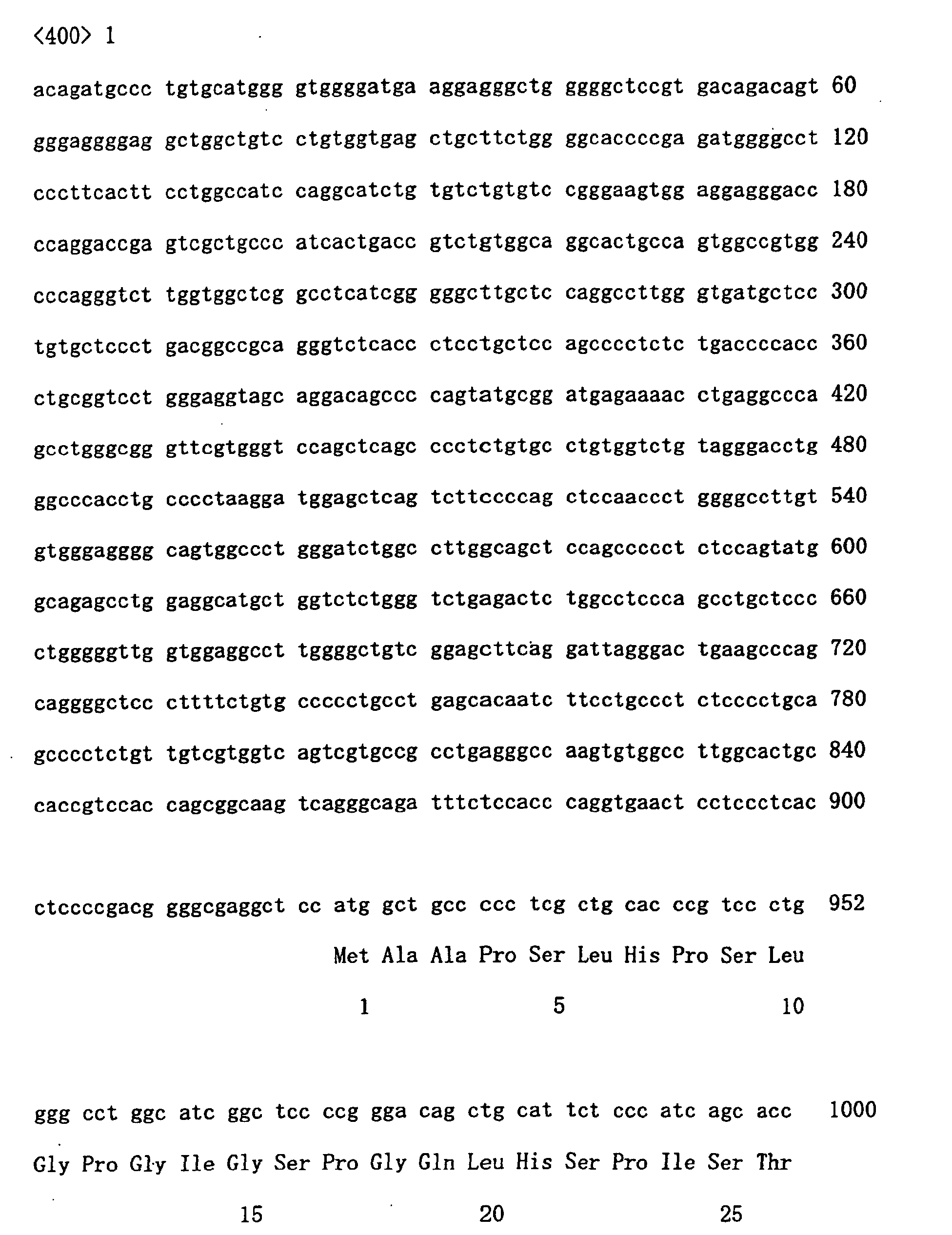
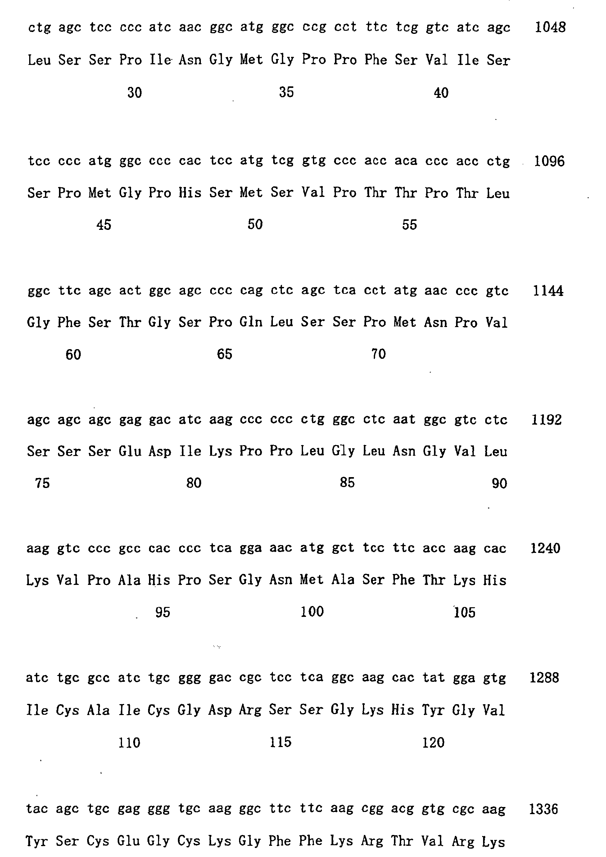
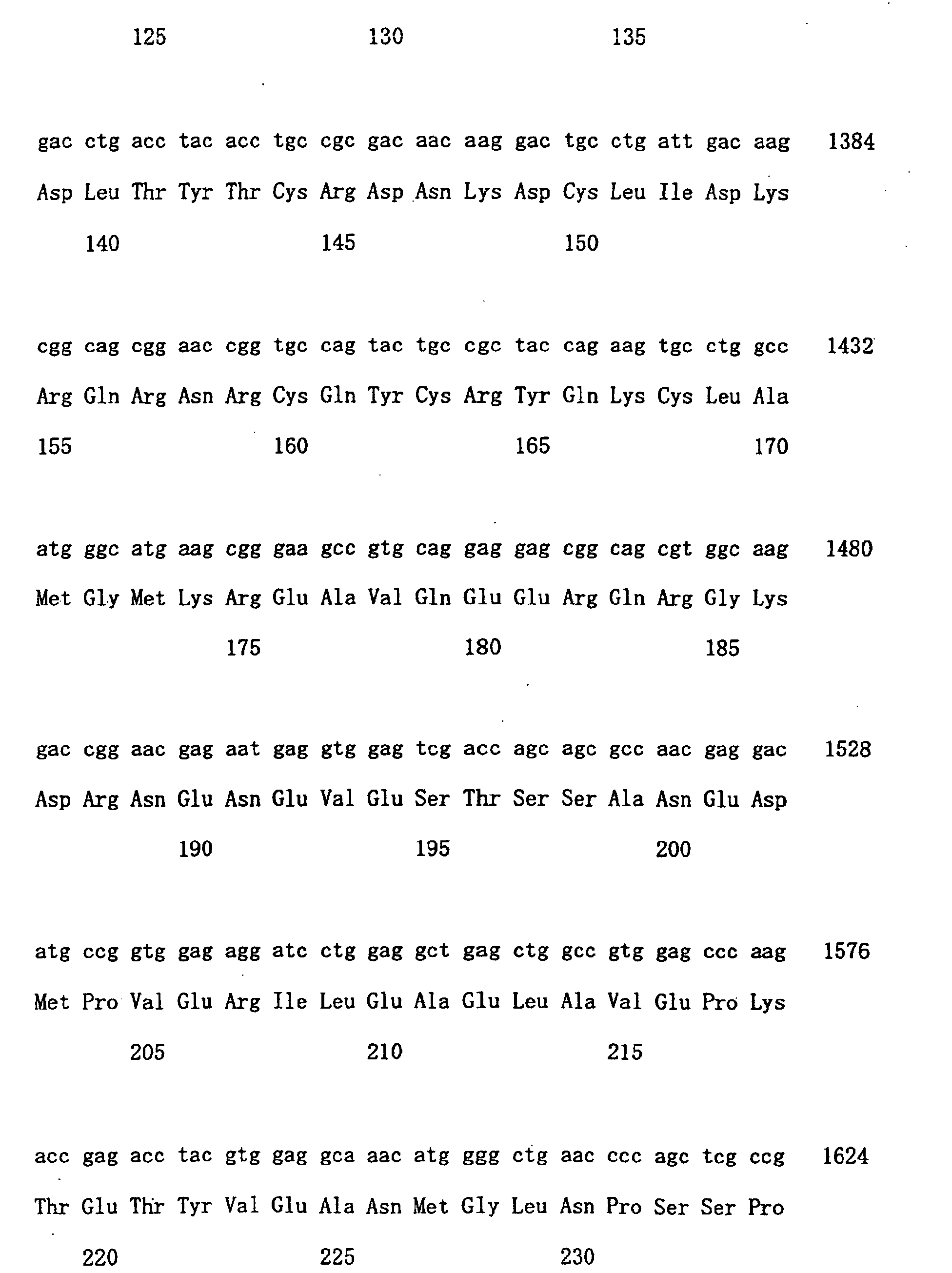

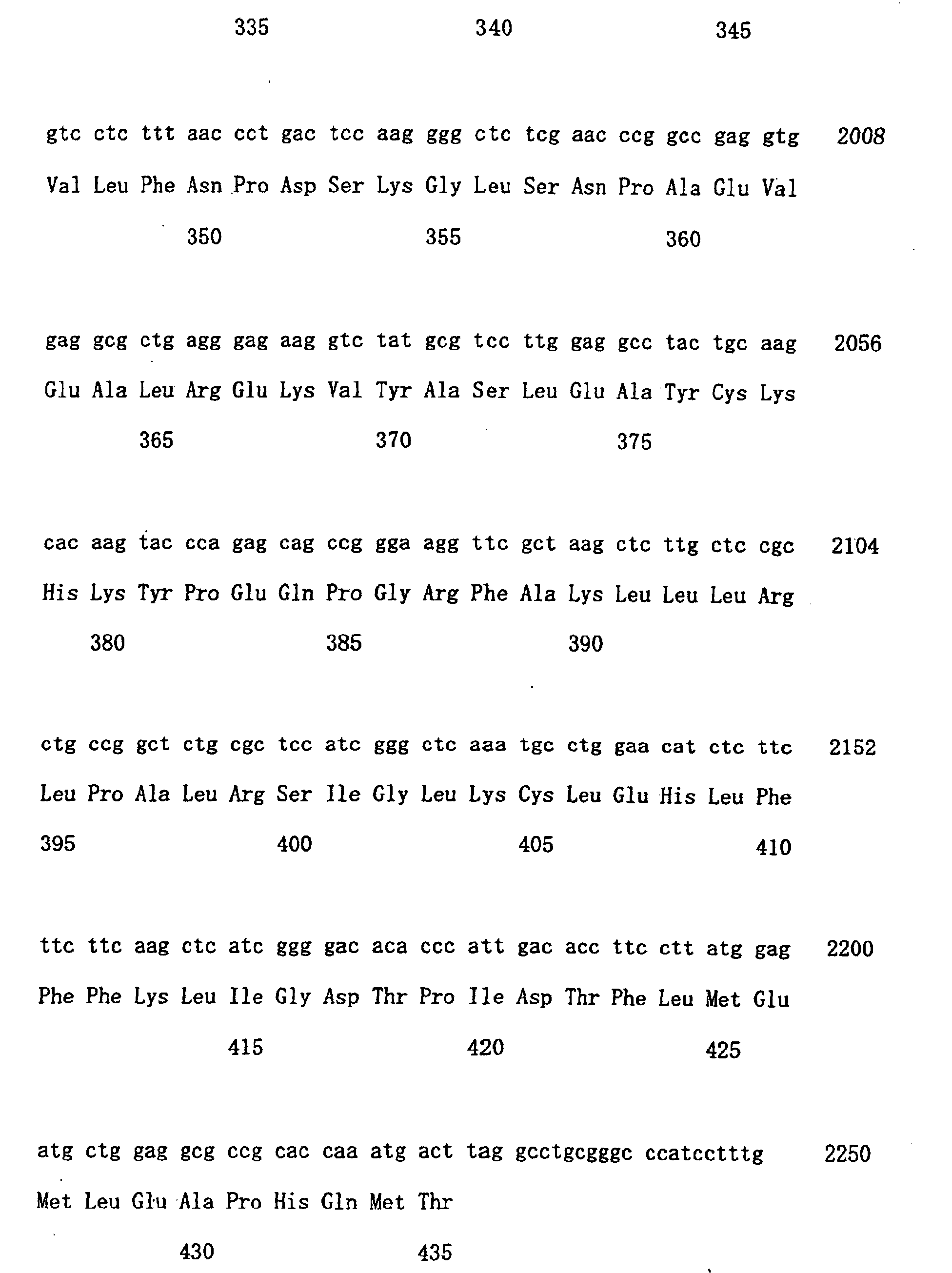
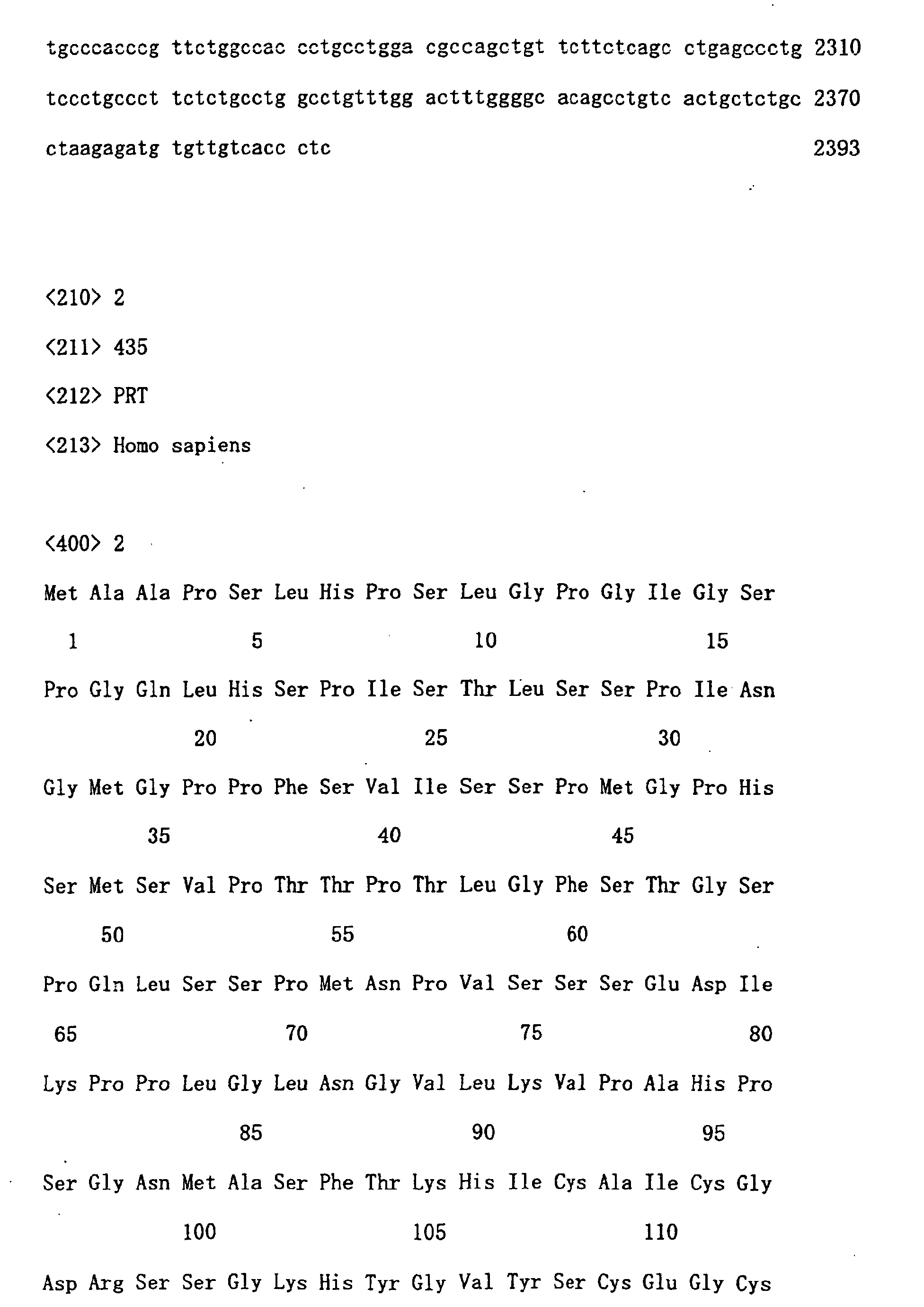
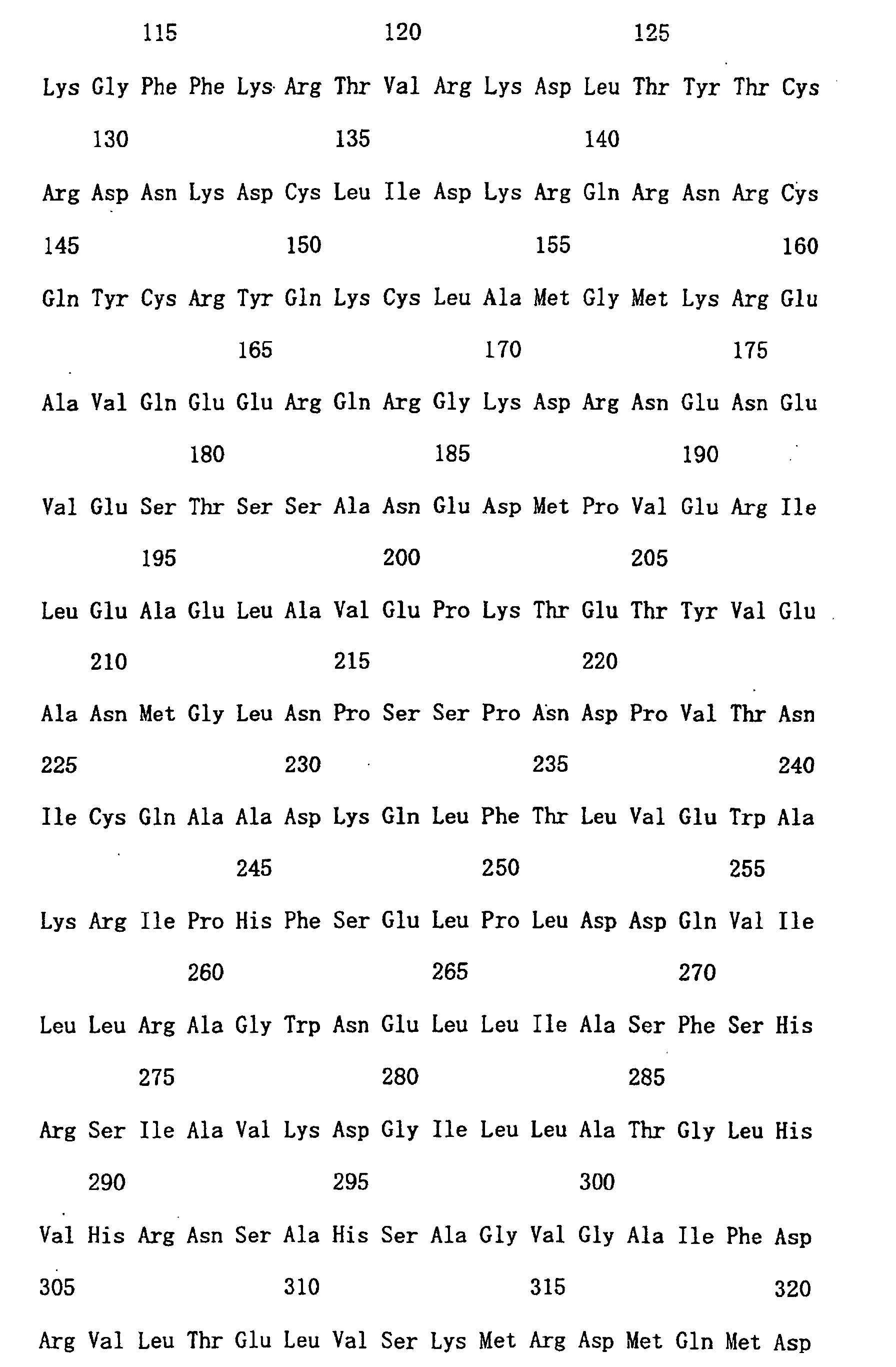


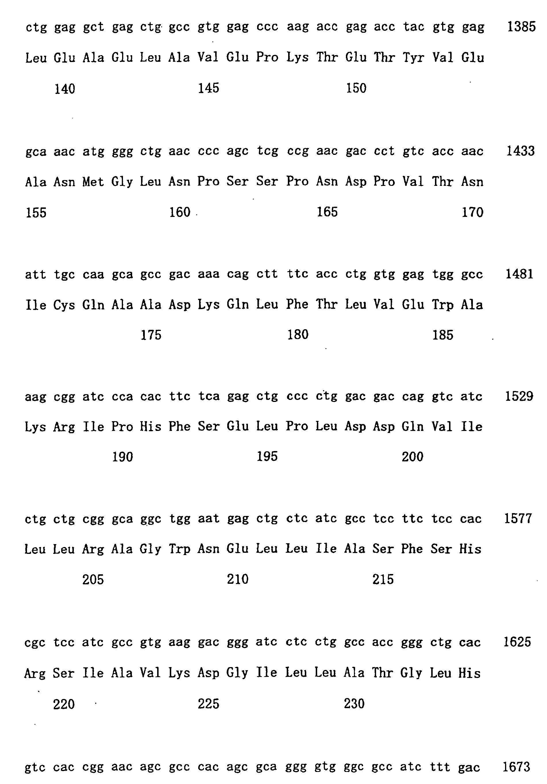
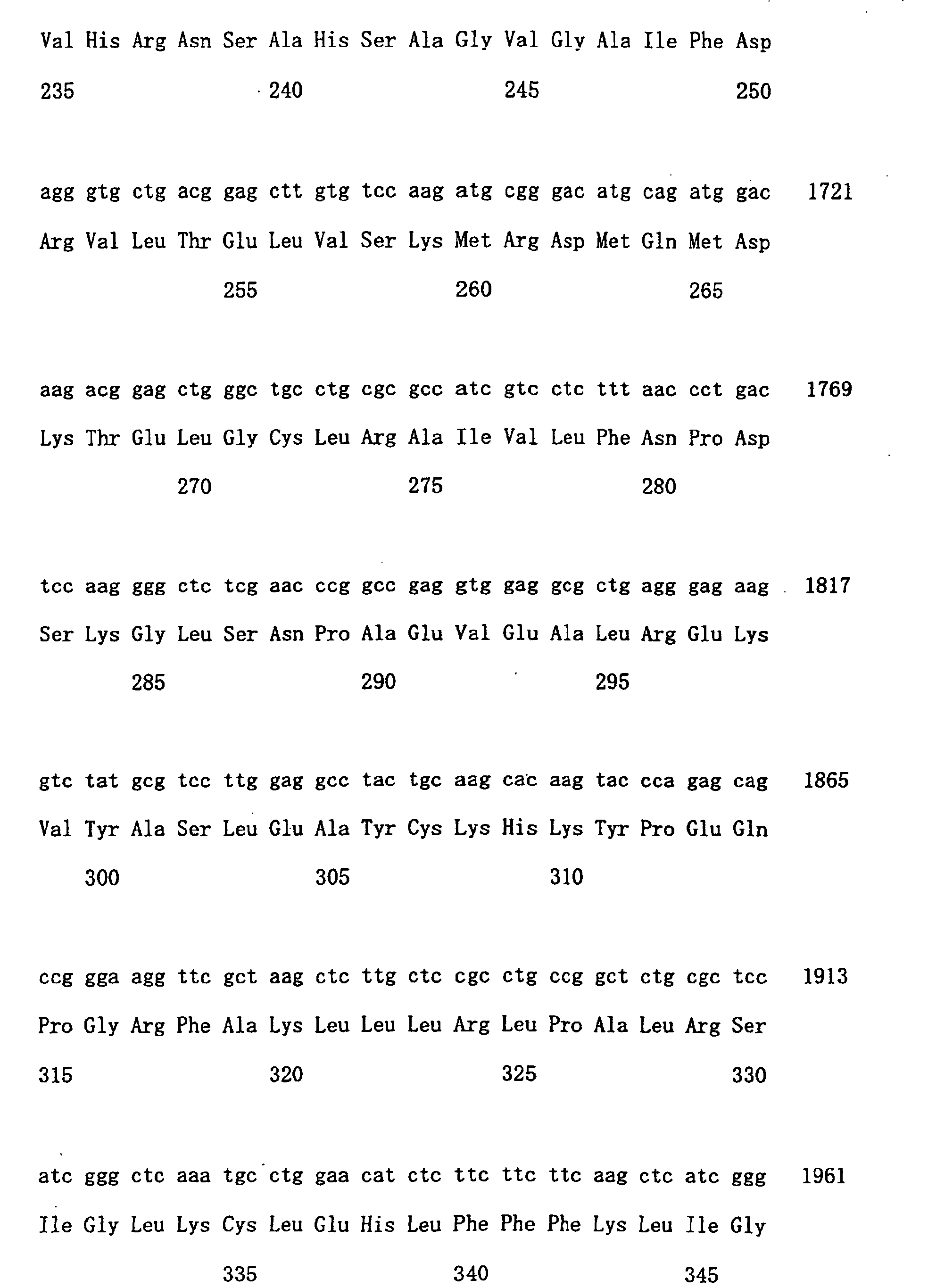


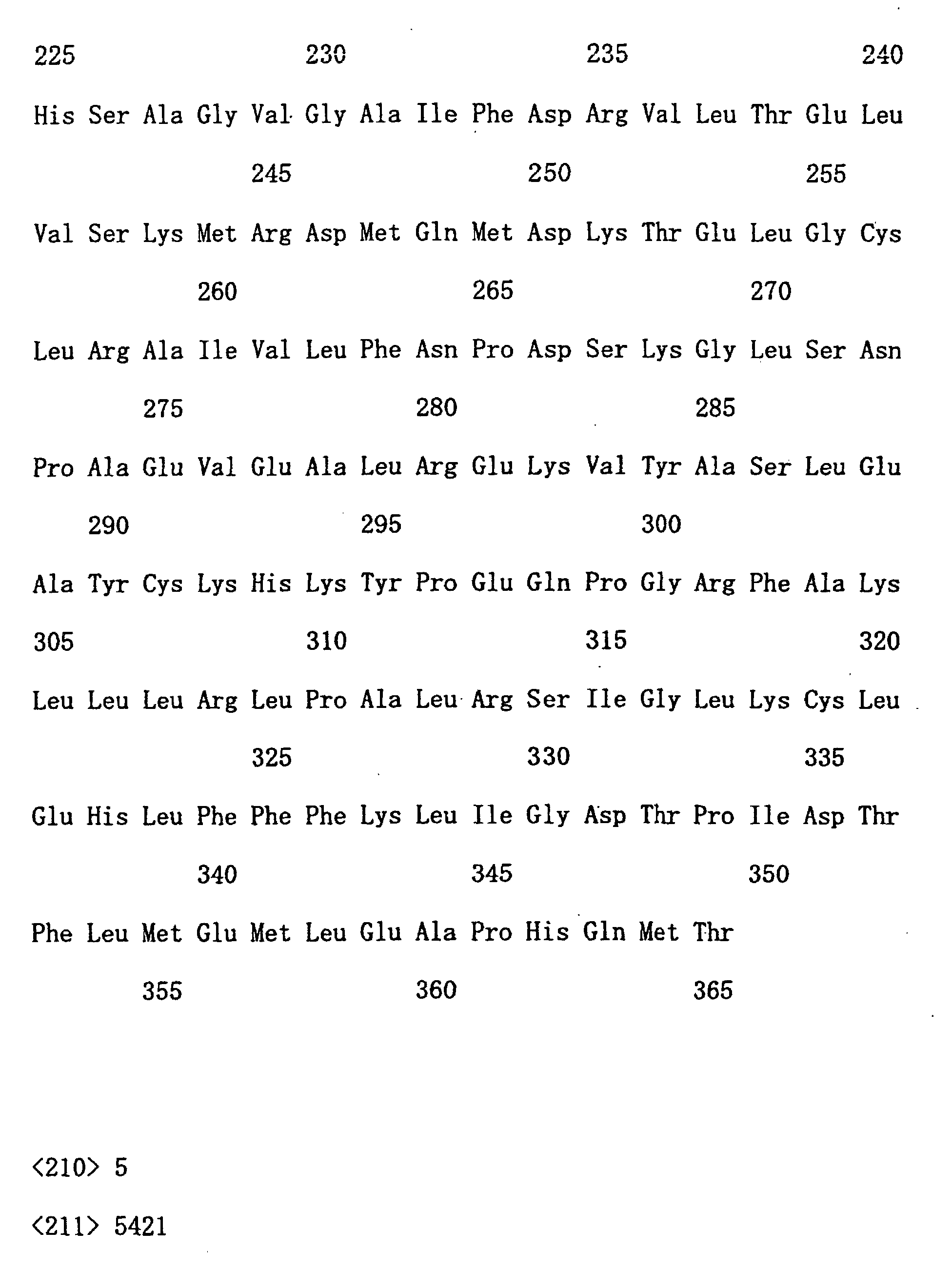

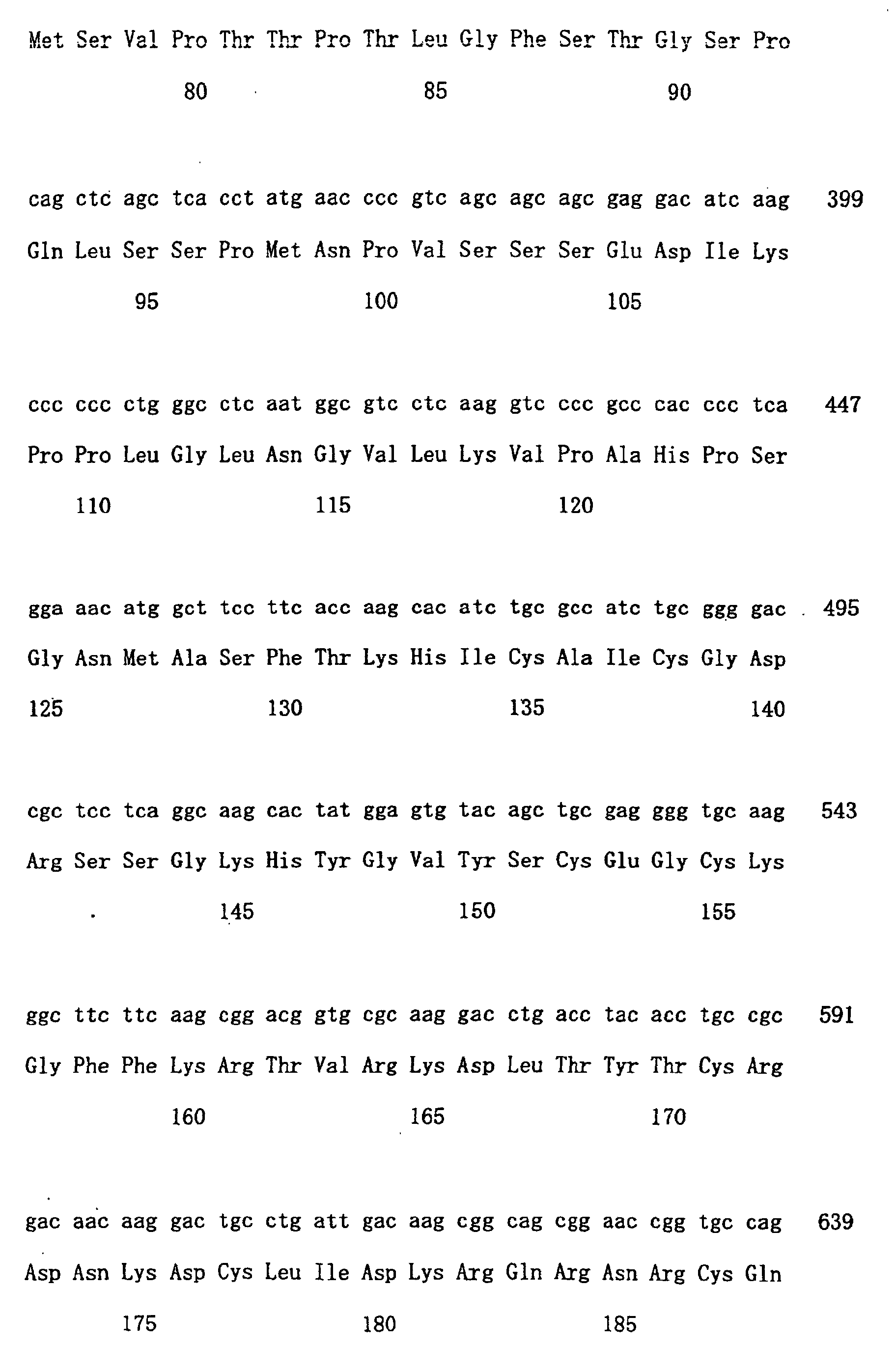

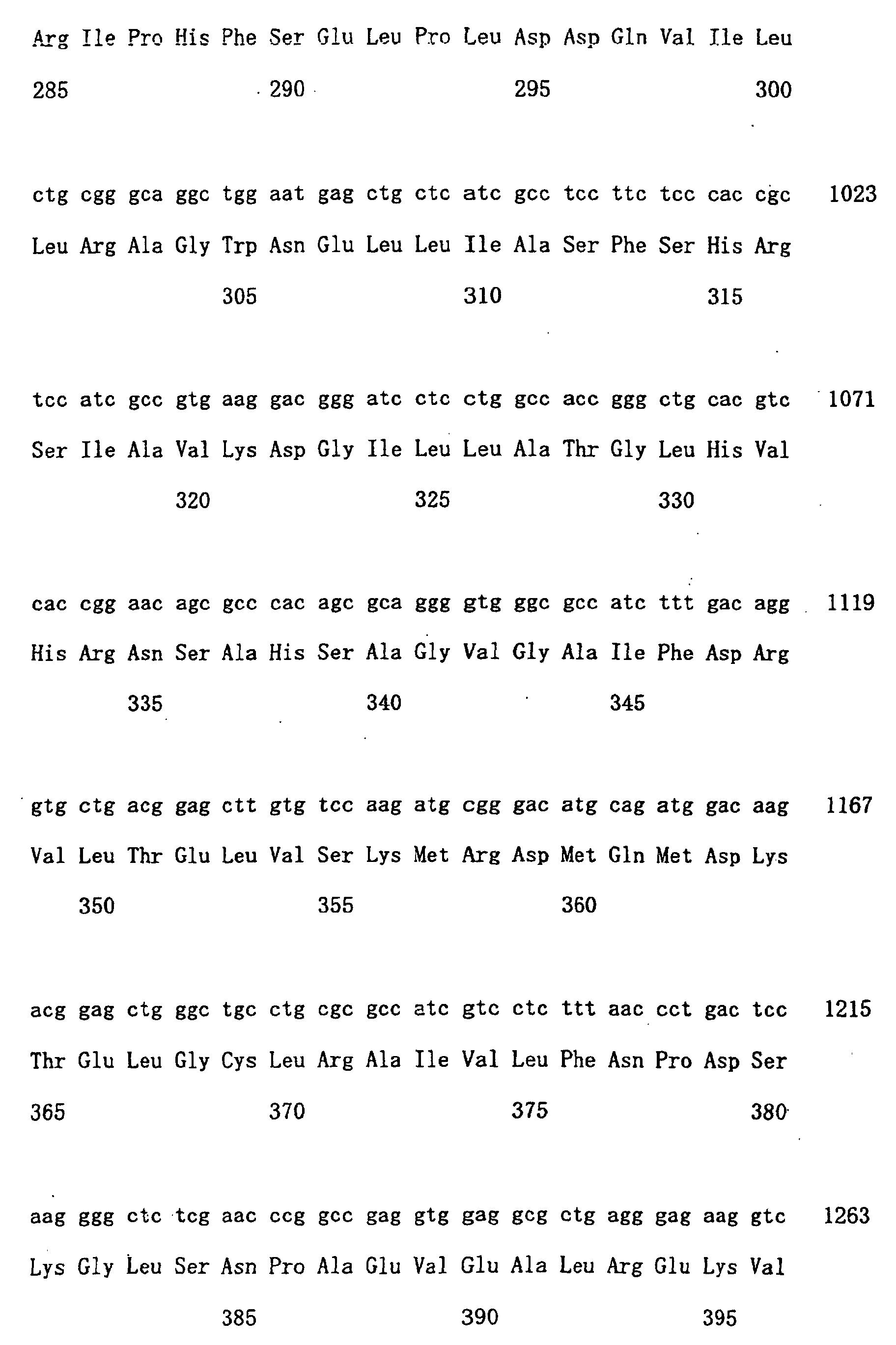


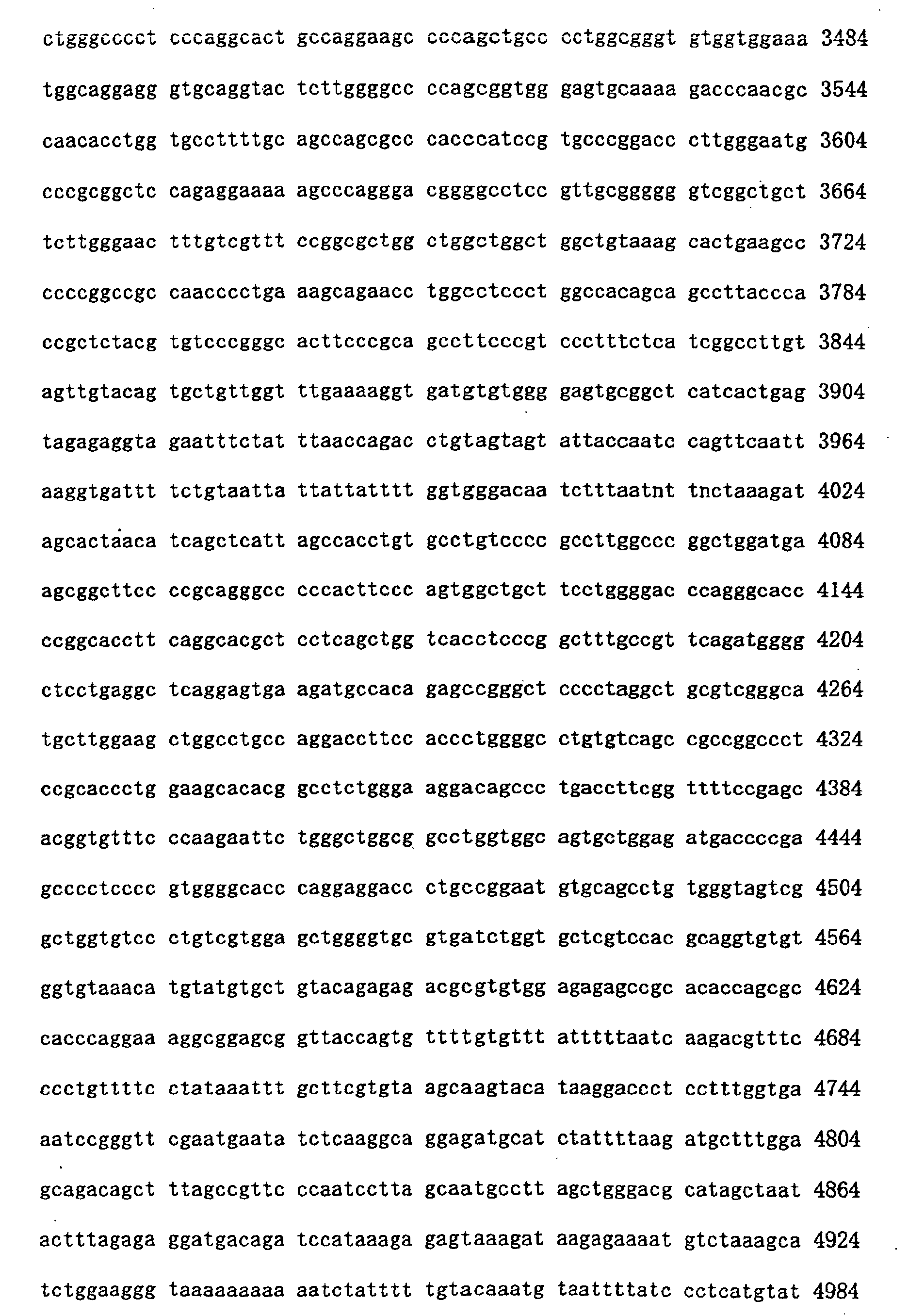

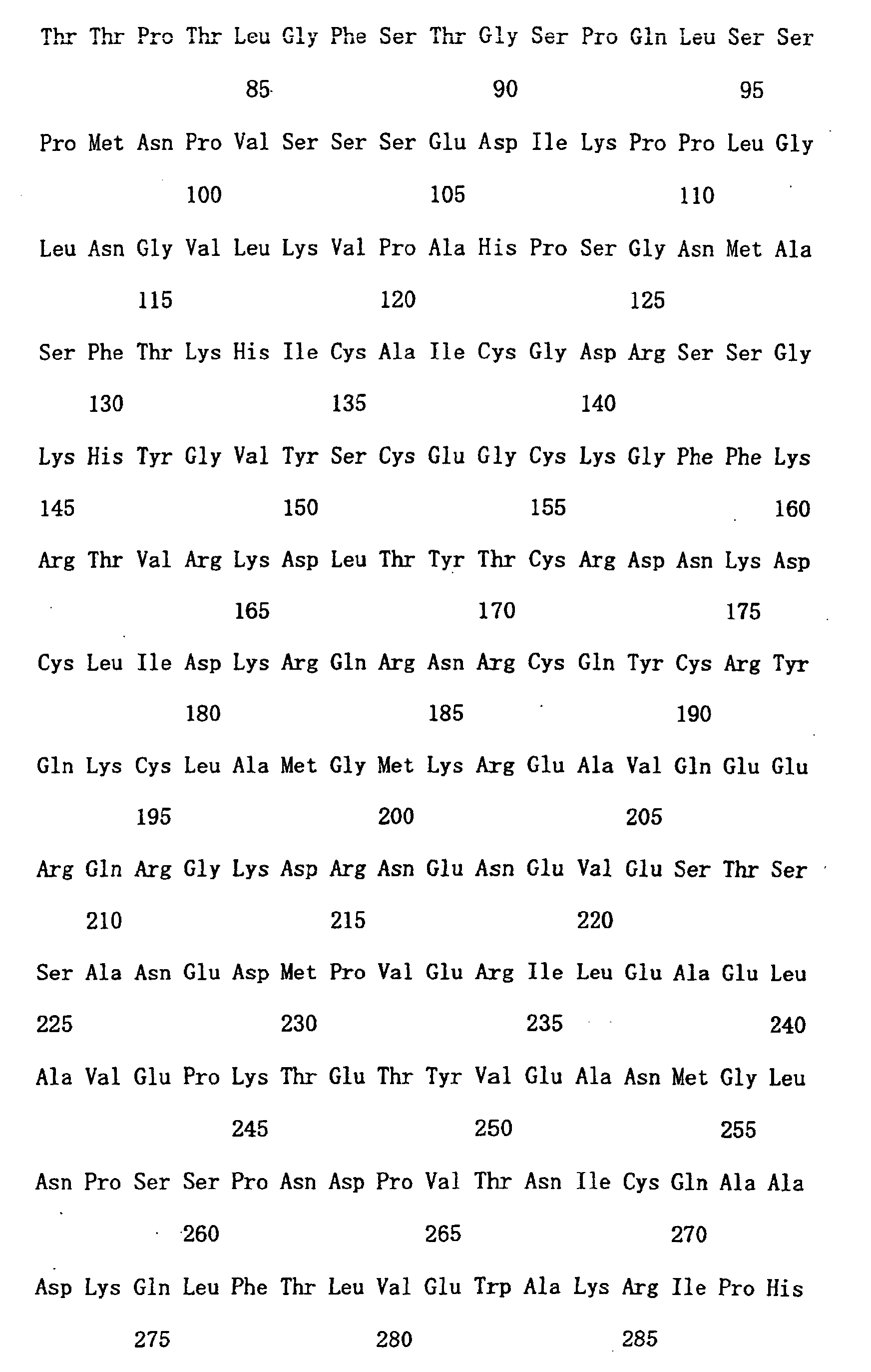
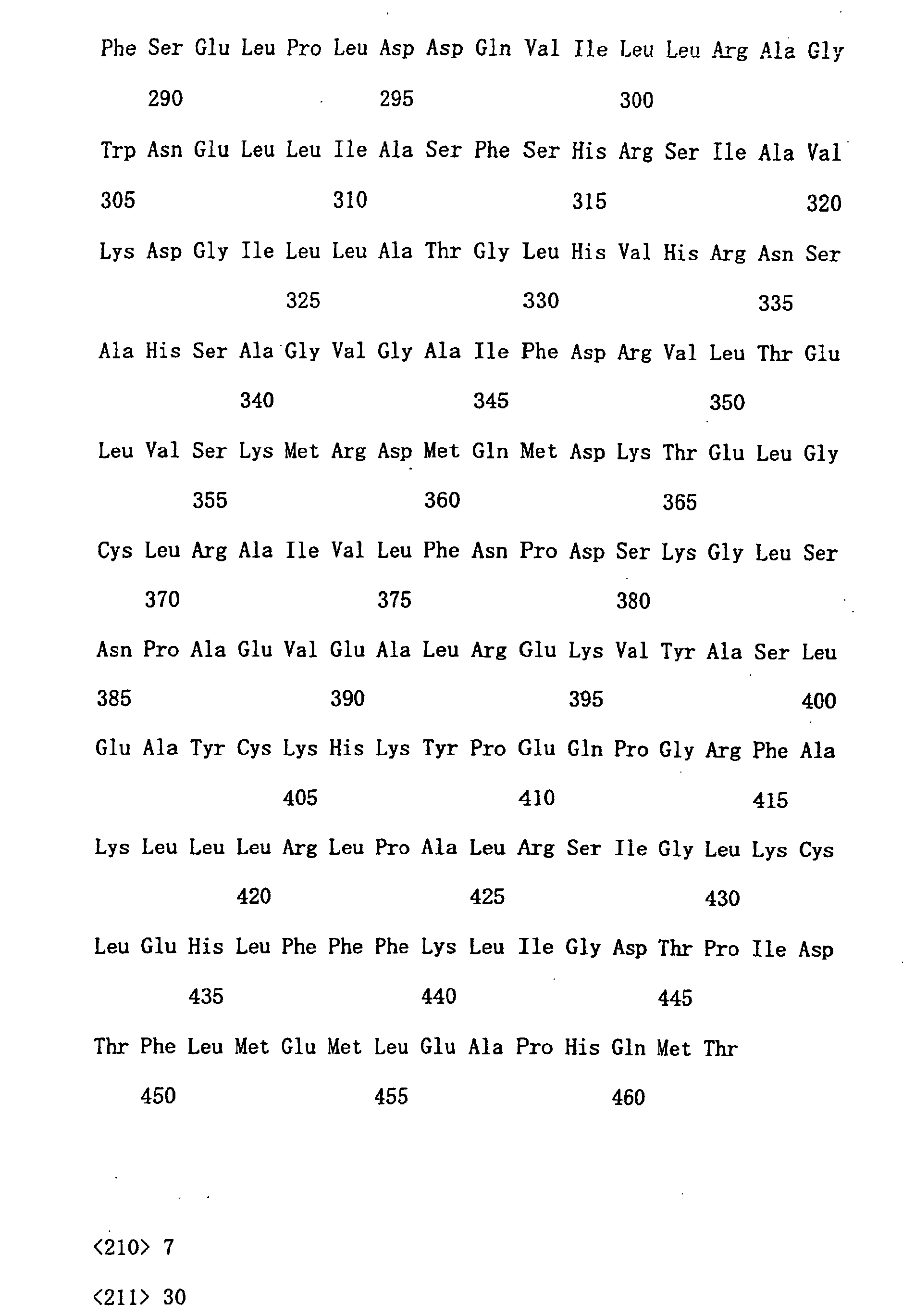


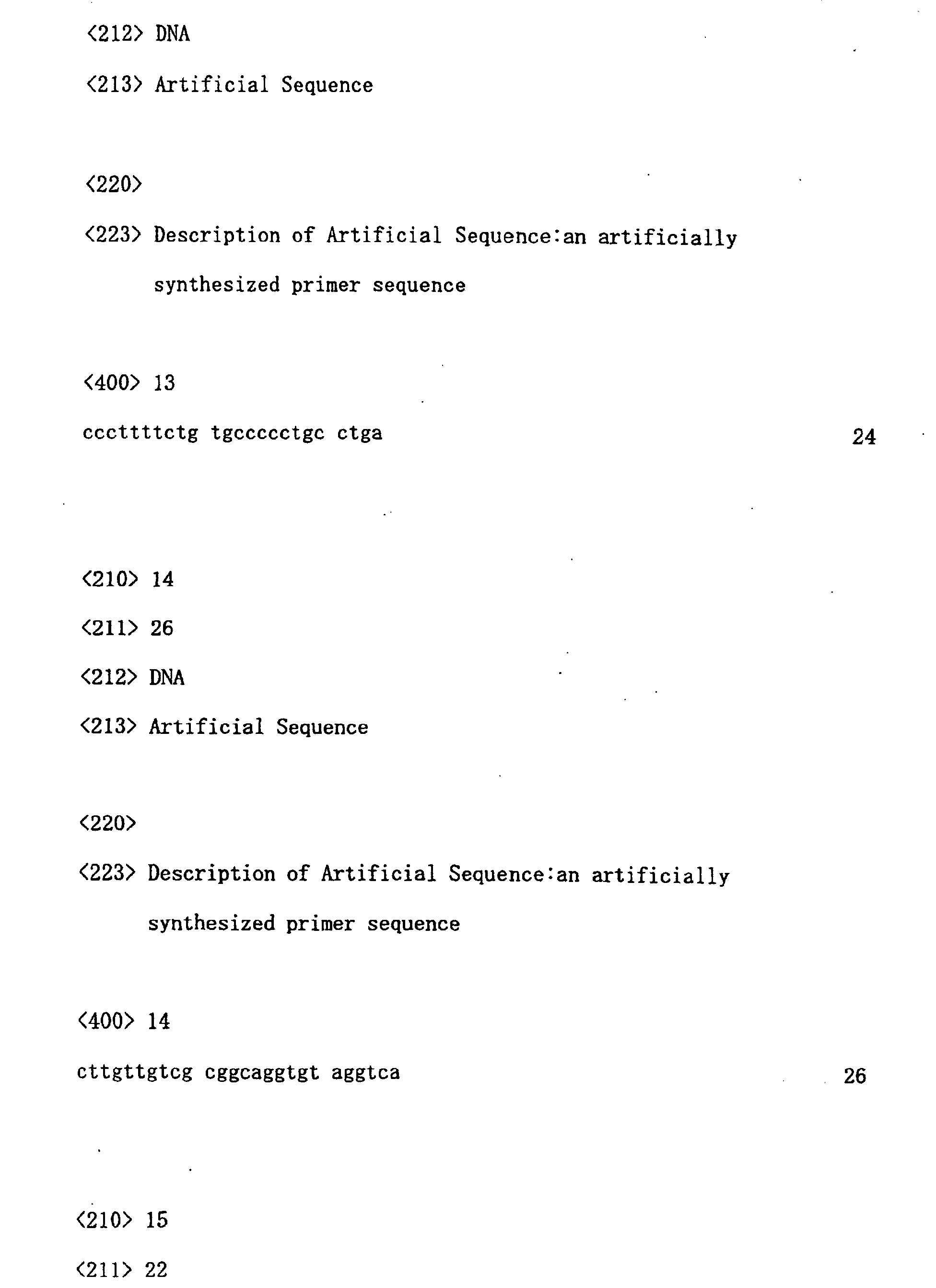


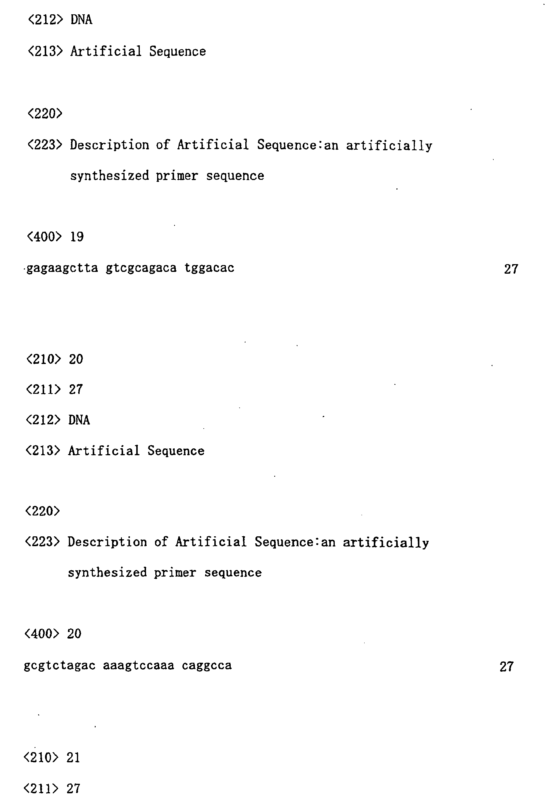


Claims (18)
- A polynucleotide of any one of the following (A) to (E):(A) a polynucleotide comprising the coding region of the nucleotide sequence of SEQ ID NO: 1 or 3;(B) a polynucleotide encoding a protein comprising the amino acid sequence of SEQ ID NO: 2 or 4;(C) a polynucleotide encoding a protein comprising the amino acid sequence wherein one or more amino acids are replaced, deleted, inserted, and/or added to the amino acid sequence of SEQ ID NO: 2 or 4, encoding a protein comprising the following (a) and (b):(a) The transcriptional activation function is enhanced in response to binding to the ligand retinoic acid, or its agonist; and(b) The protein requires SRC-1 as a co-activator;(D) a polynucleotide that hybridizes under stringent conditions with a polynucleotide comprising the nucleotide sequence of SEQ ID NO: 1 or 3, encoding a protein comprising the following (a) and (b):(a) The transcriptional activation function is enhanced in response to binding to the ligand retinoic acid, or its agonist; and(b) The protein requires SRC-1 as a co-activator;(E) a polynucleotide comprising a nucleotide sequence in which the nucleotide sequence from 1 to 102 of SEQ ID NO: 5 is deleted, or replaced with another nucleotide sequence, where the sequence has 70% or more homology to the nucleotide sequence from 103 to 1461 of SEQ ID NO: 5.
- A protein encoded by the polynucleotide of claim 1.
- A vector comprising the polynucleotide of claim 1.
- A transformant maintaining the polynucleotide of claim 1; or the vector of claim 3.
- A method for producing the protein of claim 2, comprising the steps of culturing the transformant of claim 4, and recovering the expressed product.
- A method for detecting the activity of a test substance that regulates the transcription-controlling activity of a nuclear receptor, wherein the nuclear receptor is a protein encoded by the polynucleotide of any one of the following (A) to (E):wherein the method comprises the following steps:(A) a polynucleotide comprising the coding region of the nucleotide sequence of SEQ ID NO: 1 or 3;(B) a polynucleotide encoding a protein comprising the amino acid sequence of SEQ ID NO: 2 or 4;(C) a polynucleotide encoding a protein comprising an amino acid sequence wherein one or more amino acids are replaced, deleted, inserted, and/or added to the amino acid sequence of SEQ ID NO: 2 or 4, where the protein comprises the following (a) and (b):(a) The transcriptional activation function is enhanced in response to binding to the ligand retinoic acid, or its agonist; and(b) The protein requires.SRC-1 as a co-activator;(D) a polynucleotide that hybridizes under stringent conditions with a polynucleotide comprising the nucleotide sequence of SEQ ID NO: 1 or 3, encoding a protein comprising the following (a) and (b):(a) The transcriptional activation function is enhanced in response to binding to the ligand retinoic acid, or its agonist; and(b) The protein requires SRC-1 as a co-activator;(E) a polynucleotide comprising a nucleotide sequence in which the nucleotide sequence from 1 to 102 of SEQ ID NO: 5 is deleted, or replaced with another nucleotide sequence, and which comprises 70% or more homology to the nucleotide sequence from 103 to 1461 of SEQ ID NO: 5;(1) contacting a test substance with cells, which express a nuclear receptor and comprise an expression cassette in which a reporter gene is inserted downstream of a responsive element for the nuclear receptor, in the presence of a co-activator that activates the nuclear receptor;(2) culturing the cells under conditions enabling the nuclear receptor expression, and measuring the expression level of the reporter gene within the cells; and(3) detecting the activity of the test substance regulating the transcription-controlling activity of the nuclear receptor, using the measurement result of step (2) as an index.
- The method of claim 6, wherein the method additionally comprises the step of detecting the activity of a test substance on a nuclear receptor that is a protein encoded by the polynucleotide of the following (A') to (E'):(A') a polynucleotide comprising the coding region of the nucleotide sequence of SEQ ID NO: 5;(B') a polynucleotide encoding a protein comprising the amino acid sequence of SEQ ID NO: 6;(C') a polynucleotide encoding a protein comprising an amino acid sequence wherein one or more amino acids are replaced, deleted, inserted, and/or added to the amino acid sequence of SEQ ID NO: 6, wherein the protein is functionally equivalent to a protein comprising the amino acid sequence of SEQ ID NO: 6;(D') a polynucleotide that hybridizes with a polynucleotide comprising the nucleotide sequence of SEQ ID NO: 5 under stringent conditions, encoding a protein functionally equivalent to a protein comprising the amino acid sequence of SEQ ID NO: 6; and(E') a polynucleotide comprising 60% or more homology to the nucleotide sequence of SEQ ID NO: 5, encoding a protein functionally equivalent to a protein comprising the amino acid sequence of SEQ ID NO: 6.
- A method for detecting the activity of a test substance controlling the binding between a nuclear receptor and a co-activator that activates the receptor, wherein the nuclear receptor is a protein encoded by the polynucleotide of any one of the following (A) to (E) :wherein the method comprises the following steps:(A) a polynucleotide comprising the coding region of the nucleotide sequence of SEQ ID NO: 1 or 3;(B) a polynucleotide encoding a protein comprising the amino acid sequence of SEQ ID NO: 2 or 4;(C) a polynucleotide encoding a protein comprising an amino acid sequence wherein one or more amino acids are replaced, deleted, inserted, and/or added to the amino acid sequence of SEQ ID NO: 2 or 4, encoding a protein comprising the following (a) and (b):(a) The transcriptional activation function is enhanced in response to binding to the ligand retinoic acid, or its agonist; and(b) The protein requires SRC-1 as a co-activator;(D) a polynucleotide that hybridizes with a polynucleotide comprising the nucleotide sequence of SEQ ID NO: 1 or 3 under stringent conditions, encoding a protein comprising the following (a) and (b):(a) The transcriptional activation function is enhanced in response to binding to the ligand retinoic acid, or its agonist; and(b) The protein requires SRC-1 as a co-activator;(E) a polynucleotide comprising a nucleotide sequence in which the nucleotide sequence from 1 to 102 of SEQ ID NO: 5 is deleted, or replaced with another nucleotide sequence, and which comprises 70% or more homology to the nucleotide sequence from 103 to 1461 of SEQ ID NO: 5;(1) contacting the nuclear receptor, SRC-1, and the test substance in any of the following orders i) to iii):i) contacting the nuclear receptor and the test substance first, and then contacting with SRC-1;ii) contacting the nuclear receptor and SRC-1 in the presence of the test substance; oriii) contacting the nuclear receptor and SRC-1 first, and then contacting with the test substance;(2) measuring the amount of SRC-1 or test substance bound to the nuclear receptor; and(3) detecting the activity of the test substance in binding to the nuclear receptor, using the measurement result of (2) as an index.
- A method for evaluating a substance capable of regulating the transcription-controlling activity of a nuclear receptor, comprising the following steps:(1) detecting the activity of a test substance in regulating the transcription-controlling activity of a nuclear receptor using the method of claim 6; and(2) selecting a test substance capable of inhibiting or enhancing the transcriptional activation function of the nuclear receptor by comparing with control.
- A method for evaluating a test substance capable of regulating the transcription-controlling activity of a specific nuclear receptor isoform, where the method comprises the following steps:(1) detecting the activity of a test substance in regulating the transcription-controlling activity of different kinds of nuclear receptors that comprise a protein encoded by the polynucleotide of any one of claim 6 (A) to (E), using the method of claim 6;(2) comparing the activity of a test substance in regulating the transcription-controlling activity of each of the different kinds of nuclear receptor; and(3) selecting a test substance whose effect on transcription-controlling activity differs between isoforms.
- A method for evaluating a test substance capable of regulating the transcription-controlling activity of a nuclear receptor, comprising the following steps:(1) detecting the activity of the test substance in controlling the binding between the nuclear receptor and a co-activator activating it, using the method of claim 8, and(2) selecting, by comparison with a control, a test substance capable of inhibiting or enhancing the binding of the co-activator to the nuclear receptor.
- An agent for controlling the activity of a nuclear receptor, comprising as an active ingredient a substance selected by the method of claim 9.
- An agent for controlling the activity of a nuclear receptor in an isoform-specific manner, comprising as an active ingredient a substance selected by the method of claim 10.
- An agent for controlling the activity of a nuclear receptor, comprising as an active ingredient a substance selected by the method of claim 11.
- An agent for controlling the activity of a nuclear receptor, comprising as the main ingredient a polynucleotide of any one of the following (A) to (E), or a protein encoded by the polynucleotide,(A) a polynucleotide comprising the coding region of the nucleotide sequence of SEQ ID NO: 1 or 3;(B) a polynucleotide encoding the amino acid sequence of SEQ ID NO: 2 or 4;(C) a polynucleotide encoding a protein comprising the amino acid sequence wherein one or more amino acids are replaced, deleted, inserted, and/or added to the amino acid sequence of SEQ ID NO: 2 or 4, encoding a protein comprising the following (a) and (b):(a) The transcriptional activation function is enhanced in response to binding to the ligand retinoic acid, or its agonist; and(b) The protein requires SRC-1 as a co-activator;(D) a polynucleotide that hybridizes under stringent conditions with a polynucleotide comprising the nucleotide sequence of SEQ ID NO: 1 or 3, encoding a protein comprising the following (a) and (b):(a) The transcriptional activation function is enhanced in response to binding to the ligand retinoic acid, or its agonist; and(b) The protein requires SRC-1 as a co-activator;(E) a polynucleotide comprising the nucleotide sequence in which the nucleotide sequence from 1 to 102 of SEQ ID NO: 5 is deleted, or replaced with another nucleotide sequence, and which comprises 70% or more homology to the nucleotide sequence from 103 to 1461 of SEQ ID NO: 5.
- An agent for suppressing the activity of a nuclear receptor, comprising as an active ingredient a component of any one of the following (1) to (4);(1) an antisense polynucleotide of the polynucleotide of the above (A) to (E);(2) an antibody that recognizes a protein encoded by the polynucleotide of the above (A) to (E);(3) a protein exerting a dominant-negative effect against a protein encoded by the polynucleotide of the above (A) to (E) ; and(4) a double-stranded RNA that comprises 21 to 23 base pairs, comprising a sense RNA corresponding to a partial sequence of the nucleotide sequence of the polynucleotide of the above (A) to (E), and its antisense RNA.
- A model animal wherein the activity of a nuclear receptor is controlled, wherein the animal is a transgenic non-human animal in which the expression of a protein encoded by the polynucleotide of any one of the following (A) to (E) is controlled,(A) a polynucleotide comprising the coding region of the nucleotide sequence of SEQ ID NO: 1 or 3;(B) a polynucleotide encoding the amino acid sequence of SEQ ID NO: 2 or 4;(C) a polynucleotide encoding a protein comprising an amino acid sequence wherein one or more amino acids are replaced, deleted, inserted, and/or added to the amino acid sequence of SEQ ID NO: 2 or 4, encoding a protein comprising the following (a) and (b):(a) The transcriptional activation function is enhanced in response to binding to the ligand retinoic acid, or its agonist; and(b) The protein requires SRC-1 as a co-activator;(D) a polynucleotide that hybridizes with a polynucleotide comprising the coding region of the nucleotide sequence of SEQ ID NO: 1 or 3 under stringent conditions, encoding a protein comprising the following (a) and (b):(a) The transcriptional activation function is enhanced in response to binding to the ligand retinoic acid, or its agonist; and(b) The protein requires SRC-1 as a co-activator;(E) a polynucleotide comprising a nucleotide sequence in which the nucleotide sequence from 1 to 102 of SEQ ID NO: 5 is deleted, or replaced with another nucleotide sequence, and which comprises 70% or more homology to the nucleotide sequence from 103 to 1461 of SEQ ID NO: 5.
- A method of diagnosing a disease caused by the abnormal activity of a nuclear receptor, comprising the steps of measuring the expression level of a polynucleotide in a biological sample collected from a subject, and judging that, if the expression level is low compared with a control, the subject has a disease resulting from impaired function of a protein encoded by the polynucleotide, and that, in contrast, if the expression level is high, the subject has a disease resulting from elevated function of the protein, wherein the polynucleotide is any one of the following (A) to (E):(A) a polynucleotide comprising the coding region of the nucleotide sequence of SEQ ID NO: 1 or 3;(B) a polynucleotide encoding the amino acid sequence of SEQ ID NO: 2 or 4;(C) a polynucleotide encoding a protein comprising an amino acid sequence wherein one or more amino acids are replaced, deleted, inserted, and/or added to the amino acid sequence of SEQ ID NO: 2 or 4, encoding a protein comprising the following (a) and (b):(a) The transcriptional activation function is enhanced in response to binding to the ligand retinoic acid, or its agonist; and(b) The protein requires SRC-1 as a co-activator;(D) a polynucleotide that hybridizes under stringent conditions with a polynucleotide comprising the coding region of the nucleotide sequence of SEQ ID NO: 1 or 3, encoding a protein comprising the following (a) and (b):(a) The transcriptional activation function is enhanced in response to binding to the ligand retinoic acid, or its agonist; and(b) The protein requires SRC-1 as a co-activator;(E) a polynucleotide comprising a nucleotide sequence in which the nucleotide sequence from 1 to 102 of SEQ ID NO: 5 is deleted, or replaced with another nucleotide sequence, and which comprises 70% or more homology to the nucleotide sequence from 103 to 1461 of SEQ ID NO: 5.
Applications Claiming Priority (3)
| Application Number | Priority Date | Filing Date | Title |
|---|---|---|---|
| JP2002084559 | 2002-03-25 | ||
| JP2002084559 | 2002-03-25 | ||
| PCT/JP2003/003612 WO2003080668A1 (en) | 2002-03-25 | 2003-03-25 | NUCLEAR RECEPTOR RXRα ISOFORMS |
Publications (2)
| Publication Number | Publication Date |
|---|---|
| EP1489094A1 true EP1489094A1 (en) | 2004-12-22 |
| EP1489094A4 EP1489094A4 (en) | 2005-11-02 |
Family
ID=28449215
Family Applications (1)
| Application Number | Title | Priority Date | Filing Date |
|---|---|---|---|
| EP03712915A Withdrawn EP1489094A4 (en) | 2002-03-25 | 2003-03-25 | ISOFORM RXR ALPHA NUCLEAR RECEPTOR |
Country Status (4)
| Country | Link |
|---|---|
| US (1) | US20050203283A1 (en) |
| EP (1) | EP1489094A4 (en) |
| JP (1) | JPWO2003080668A1 (en) |
| WO (1) | WO2003080668A1 (en) |
Cited By (2)
| Publication number | Priority date | Publication date | Assignee | Title |
|---|---|---|---|---|
| WO2005100396A1 (en) * | 2004-04-16 | 2005-10-27 | Bayer Healthcare Ag | Diagnostics and therapeutics for diseases associated with retinoid x receptor alpha (rxra) |
| WO2020000327A1 (en) * | 2018-06-28 | 2020-01-02 | 深圳市疾病预防控制中心 | CELL LINE FOR RXRα PROTEIN STABLE HIGH EXPRESSION AND PREPARATION METHOD THEREOF |
Family Cites Families (1)
| Publication number | Priority date | Publication date | Assignee | Title |
|---|---|---|---|---|
| ES2152920T3 (en) * | 1990-02-09 | 2001-02-16 | Salk Inst For Biological Studi | RECEIVING COMPOSITIONS OF RETINOIDS AND METHODS. |
-
2003
- 2003-03-25 JP JP2003578421A patent/JPWO2003080668A1/en not_active Withdrawn
- 2003-03-25 US US10/507,696 patent/US20050203283A1/en not_active Abandoned
- 2003-03-25 WO PCT/JP2003/003612 patent/WO2003080668A1/en not_active Ceased
- 2003-03-25 EP EP03712915A patent/EP1489094A4/en not_active Withdrawn
Non-Patent Citations (2)
| Title |
|---|
| LEO C ET AL: "The SRC family of nuclear receptor coactivators" GENE, ELSEVIER BIOMEDICAL PRESS. AMSTERDAM, NL, vol. 245, no. 1, March 2000 (2000-03), pages 1-11, XP004202955 ISSN: 0378-1119 * |
| See also references of WO03080668A1 * |
Cited By (2)
| Publication number | Priority date | Publication date | Assignee | Title |
|---|---|---|---|---|
| WO2005100396A1 (en) * | 2004-04-16 | 2005-10-27 | Bayer Healthcare Ag | Diagnostics and therapeutics for diseases associated with retinoid x receptor alpha (rxra) |
| WO2020000327A1 (en) * | 2018-06-28 | 2020-01-02 | 深圳市疾病预防控制中心 | CELL LINE FOR RXRα PROTEIN STABLE HIGH EXPRESSION AND PREPARATION METHOD THEREOF |
Also Published As
| Publication number | Publication date |
|---|---|
| WO2003080668A1 (en) | 2003-10-02 |
| JPWO2003080668A1 (en) | 2005-07-21 |
| US20050203283A1 (en) | 2005-09-15 |
| EP1489094A4 (en) | 2005-11-02 |
Similar Documents
| Publication | Publication Date | Title |
|---|---|---|
| US5955306A (en) | Genes encoding proteins that interact with the tub protein | |
| Tung et al. | FTO is necessary for the induction of leptin resistance by high-fat feeding | |
| WO2017044801A2 (en) | Methods of identifying drug-modulated polypeptide targets for degradation | |
| KR20010022741A (en) | ISOLATION OF A NOVEL SENESCENCE-FACTOR GENE, p23 | |
| Seifert et al. | PTPRQ is a novel phosphatidylinositol phosphatase that can be expressed as a cytoplasmic protein or as a subcellularly localized receptor-like protein | |
| US7029860B2 (en) | Amyloid-β protein aggregation-regulating factors | |
| WO2001019986A1 (en) | Peptide leukotriene receptor | |
| JPH1192401A (en) | Kinase controlled by human blood serum glucocorticoid and target against chronic renal failure | |
| US20050203283A1 (en) | ISOFORMS OF NUCLEAR RECEPTOR RXR a | |
| US6586581B1 (en) | Prolactin regulatory element binding protein and uses thereof | |
| JP2005530503A (en) | Use of PP2A phosphatase modulators in the treatment of mental disorders | |
| CA2406028A1 (en) | Method for testing for allergic disease | |
| EP1489172A1 (en) | Nuclear receptor err gamma 3 | |
| JP2004194534A (en) | Inflammatory bowel disease disease marker and use thereof | |
| US9624544B2 (en) | Allelic disorders caused by mutations in TRPV4 | |
| JP2000308488A (en) | Angiogenesis-related protein and gene encoding the protein | |
| US20040053282A1 (en) | Method of examining allergic disease | |
| EP2128267A2 (en) | Methods of evaluating phosphatase inhibitors | |
| JP2003516717A (en) | Full-length cDNA clone and protein encoding it | |
| US6960435B1 (en) | Amyloid β protein agglutination-controlling factor | |
| JP2009540851A (en) | Identification and use of GPRC variants in the treatment and diagnosis of Parkinson's disease | |
| JP2009060787A (en) | Method for screening and identification of substance that suppresses degranulation reaction of mast cell mediated by Rec168, and therapeutic agent for inflammatory disease involving mast cell, comprising Rec168 antagonist | |
| JPWO2001004299A1 (en) | Amyloid β protein aggregation regulator | |
| JPWO2003072778A1 (en) | Testing method for allergic diseases | |
| Kuppusamy | Prevalence of KCNJ5 mutations and functional impact of a novel KCNJ5-insT149 mutation in aldosterone producing adenoma causing resistant hypertension |
Legal Events
| Date | Code | Title | Description |
|---|---|---|---|
| PUAI | Public reference made under article 153(3) epc to a published international application that has entered the european phase |
Free format text: ORIGINAL CODE: 0009012 |
|
| 17P | Request for examination filed |
Effective date: 20040922 |
|
| AK | Designated contracting states |
Kind code of ref document: A1 Designated state(s): AT BE BG CH CY CZ DE DK EE ES FI FR GB GR HU IE IT LI LU MC NL PT RO SE SI SK TR |
|
| A4 | Supplementary search report drawn up and despatched |
Effective date: 20050916 |
|
| RAP1 | Party data changed (applicant data changed or rights of an application transferred) |
Owner name: ASTELLAS PHARMA INC. |
|
| 17Q | First examination report despatched |
Effective date: 20070319 |
|
| STAA | Information on the status of an ep patent application or granted ep patent |
Free format text: STATUS: THE APPLICATION IS DEEMED TO BE WITHDRAWN |
|
| 18D | Application deemed to be withdrawn |
Effective date: 20070731 |