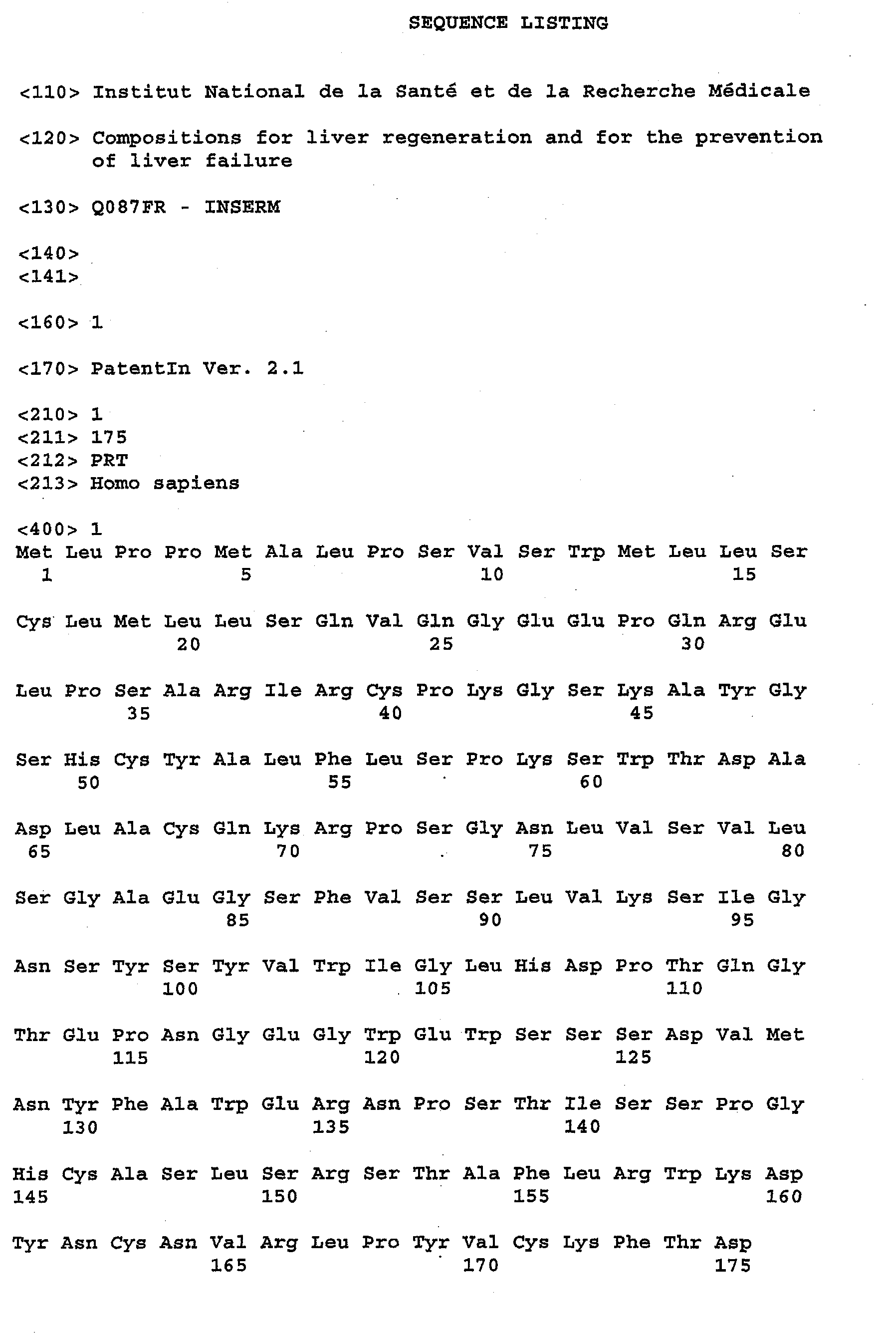
Field of the invention
-
The present invention concerns the use of the human
hepatocarcinoma-intestine-pancreas / pancreatic-associated protein
(HIP/PAP) for stimulating liver regeneration and also for the prevention of
liver failure.
Background art
-
Liver failure occurs in a number of acute and chronic clinical
conditions, including drug-induced hepatotoxicity, viral infections,
vascular injury, autoimmune disease, or blunts trauma. In addition,
patients subject to inborn errors of metabolism may be at risk for
developing liver failure. Symptoms of liver failure occurring as a result of
these clinical conditions include, for example, fulminant acute hepatitis,
chronic hepatitis, or cirrhosis, and in many instances, the restoration of
normal liver function is vital to the survival of patients. For example,
cirrhosis is the seventh leading cause of death and the fourth disease
related cause of death in people between the ages of 25 to 44. (Source:
American Liver Foundation).
-
In acute liver disease, the liver is able to regenerate after being
injured. If the disease progresses beyond the liver's capacity to
regenerate new cells, the body's entire metabolism is severely affected.
Loss of liver function may result in metabolic instability combined with
disruption of essential bodily functions (i.e., energy supply, acid-base
balance and thermoregulation.) If not rapidly reversed, complications
such as uncontrolled bleeding and sepsis occur, and dependent organs
such as the brain and kidneys cease to function because of toxic byproducts
or because the liver is no longer synthesizing important
nutrients. After large liver damage, liver tissue looses its regenerative
and metabolic functions, and liver transplantation is a therapeutic
strategy commonly used. However, the clinical application of liver
transplantation is limited by the availability of human hepatocytes, liver
tissue and the number of liver cells that can be transplanted safely at one
time. Moreover, latence before surgery and post-surgery complications
could be critical to counteract the acute phase of liver failure. Another
therapeutic strategy consists in a liver resection (removal of a portion of
the liver). The most typical indications for liver resection are a malignant
tumor, a hepatocellular carcinoma or a proliferative biliary diseases
including cholangiocarcinoma. Tumors can be primary (developed in the
liver) or metastatic (developed in another organ, then migrated to the
liver). The majority of liver metastases come from the colon. The single
tumor or more than one tumor confined to either left or right side of the
liver can be successfully resected with 5-year survival as high as 60%.
Liver resections performed on patients with extrahepatic disease may
relieve the symptoms caused by the tumor, but offer little improvement in
survival. Benign tumors of the liver (adenoma, and focal nodular
hyperplasia) can be successfully managed by liver resection as well.
Liver resections are also performed on people willing to donate part of
their liver.
-
Taking into account the importance of liver transplantation and
liver resection, Several strategies have been suggested to stimulate liver
regeneration and suppress or limiting liver failure in the case of liver
resection or transplantation.
-
Liver cell is believed to be controlled by various growth stimulatory
and growth inhibitory cytokines of autocrine or paracrine origin, however,
the exact role and action mechanism of these growth factors is far from
entirely understood. Cytokines are secreted peptides or proteins that
regulate the intermediary metabolism of amino acid, proteins,
carbohydrates, lipids and minerals. Cytokines include peptides or
proteins that act to mediate inflammation and are involved in intracellular
communication modulating cell proliferation, and adhesion of
inflammatory cells to the walls of the vessels, and to the extra cellular
matrix. Cytokines are essential for the communication between the liver
and extrahepatic sites and within the liver itself. Cytokines interact with
hormones such as glucocorticoids, resulting in a complex network of
mutual control. Many cytokines exert growth activity in addition to their
specific proinflammatory effects. The liver is an important site of cytokine
synthesis and the major clearance organ for several cytokines. In liver
disease, cytokines are involved in the onset of intrahepatic immune
responses, in liver regeneration, and in the fibrotic and cirrhotic
transformation of the liver.
-
Liver cell is also believed to be controlled by various growth
factors. Growth factors are required to regulate developmental events or
required to regulate expression of genes encoding other secreted
proteins that may participate in intracellular communication and
coordination of development and includes, insulin-like growth factor-I and
II (IGF I and II), epidermal growth factor (EGF), type a and type b
transforming growth factor (TGF-α and TGF-β), platelet-derived growth
factor (PDGF).
-
In vitro, DNA synthesis in isolated hepatocytes has been shown to
be stimulated by growth factors such as TGFα, or EGF. A further protein,
named hepatocyte growth factor (HGF) has been shown to be a mitogen
for primary hepatocytes.
-
Based on these observations, it has been proposed that these
factors may be important mediator of liver regeneration. Consequently,
growth factors as TGFα, EGF or HGF with growth factor-like activities
have been indicated in the treatment of liver regeneration. However,
these therapeutic strategies, suggested to stimulate liver regeneration
and suppress liver failure, have not proved their efficacity without toxicity,
and adverse effects. Namely, these growth factor favor tumor
progression (Gang-Hong, et al., 1992 ; Horiguchi, et al. 2002).
-
Consequently, there remains a need in the art for an
effective method which would stimulate liver regeneration, would protect
against liver failure, and would be deprived of adverse toxic and
tumorigenic effects. This need exists in any patient population in which
liver damage has been induced. This need exists not only for
transplanted patients but also for donors, and patients having undergone
a liver resection. Further, there is still a need in the art for novel
therapeutically useful compounds, which stimulate liver regeneration.
-
Additionally, taking into account the poor availability of donor
organs, living donor partial liver transplantation is recognized as a
measure for overcoming the lack of organs, and facilities for partial liver
transplantation. However, partial liver transplantation cannot be
considered as a safe operation for adults representing the majority of
transplantation patients because the resectable liver weight of donors is
often less than the necessary liver weight for recipients. Thus there is a
need for a mean for safe and rapid liver regeneration for small grafts.
-
Accordingly, it is an object of the present invention to provide a
mean for the stimulation of liver regeneration after partial resection. An
object of the present invention is also to provide a drug that can promote
liver regeneration or hepatocyte growth after liver transplantation such as
partial liver transplantation, and also after the occurrence of a discare
causing liver failure, such as cirrhosis, acute hepatitis and chronic
hepatitis.
-
These and further objects will be apparent to one ordinary skill in
the art.
Summary of the invention.
-
The present invention is based on the experimental finding that
HIP/PAP has mitogenic and antiapoptotic effects in vitro on hepatocytes
in primary culture. Moreover, HIP/PAP is a mitogenic molecule for
hepatocytes, during liver regeneration, in vivo. The present invention is
also based on the experimental finding that HIP/PAP has no adverse
effects in mammals.
-
A first object of the invention consists in a pharmaceutical
composition for stimulating liver regeneration in vivo comprising an
effective amount of a polypeptide comprising an amino acid sequence
having 90% amino acid identity with the polypeptide consisting of the
amino acid sequence starting at the amino acid residue 36 and ending at
the amino acid residue 175 of sequence SEQ ID N°1, in combination with
at least one physiologically acceptable excipient.
-
In another aspect, the present invention relates to a
pharmaceutical composition for stimulating liver regeneration
in vivo
comprising an effective amount of the human hepatocarcinoma-intestine-pancreas/
pancreatic-associated protein (HIP/PAP) of sequence SEQ ID
N°1, in combination with at least one physiologically acceptable
excipient.
the present invention also relates to a pharmaceutical composition
with limited adverse effects on liver necrosis comprising :
- (i) a therapeutically effective amount of a hepatotoxic compound,
- (ii) a liver damage effective amount of a polypeptide as defined
above.
-
Description of drawings:
-
- Figure 1 Schematic representation of the transgene.
The enhancer (2 kb) and promoter (0.3 kb) of the regulatory
regions of the mouse albumin gene are indicated by dotted lines. Exons
II, III, IV, V and VI and introns of the human HIP/PAP gene (1.6 kb) are
indicated by black boxes and a dotted line, respectively. The bovine
growth hormone poly A fragment (1021-1235) pcDNA 3.1 is indicated by
a dotted line. Plasmid DNA is indicated by the heavy line. Relevant
restriction sites are indicated by the arrows.
- Figure 2 Immunodetection of HIP/PAP protein.
- A. Immunohistochemistry: original magnification x20, 1 wild-type liver, 2
HIP/PAP transgenic liver, 3 wild-type hepatocytes, 4 HIP/PAP
hepatocytes.
- B. Western blot hybridised with HIP/PAP and actin antibodies showing
a band with the 16 kDA and the 45 kDa expected size, respectively.
Lane 1 purified HIP/PAP protein (10 ng), lanes 2 , 3 and 4, wild-type
liver; HIP/PAP transgenic liver 27 and 24 homozygous strains,
respectively, lanes 5, 6 and 7 wild-type, HIP/PAP 27 and 24
hepatocytes after isolation, respectively.
- Figure 3 Time course of in vivo hepatic regeneration after partial
hepatectomy.
- A. Immunodetection of BrU-positive nuclei, in wild-type (1,2,3,4) and
HIP/PAP transgenic livers (5,6,7,8); 1 and 5 24 hours, 2 and 6 36
hours, 3 and 7 46 hours, 4 and 8 55 hours after hepatectomy.
- B. Each box plot comprises five horizontal lines displaying the 10th, 25th,
50th, 75th, percentiles of a variable. All values for the variable above
the 90th percentile and below the 10th percentile are plotted
separately, so that the box plots are valuable in highlighting any
outliers. Wild-type mice (n = 9), and HIP/PAP transgenic mice (n=10)
(p=0.0014).
- C. Liver weights were measured in normal non hepatectomized mice.
The liver/body ratio of weight was calculated and expressed as the
average percentage ± SD. There was no difference in this ratio
between the two groups (0.0460 ± 0.0064, n = 12 and 0.0489 ±
0.0035 n = 16 for wild-type and HIP/PAP transgenic mice,
respectively). The average percentage recovery of normal liver weight
(± sd) in wild type (○) and HIP/PAP mice (▪) at various time points
after partial hepatectomy shows stimulated recovery in the HIP/PAP
transgenic mice (5 to 9 mice were hepatectomized at each time for
each group) The difference was statistically significant at 48 hours (p
< 0.001), 60 hours (p < 0.003) and 96 hours (p<0.002).
- Figure 4 DNA synthesis in wild-type and HIP/PAP transgenic
hepatocytes.
- A. Immunodetection of BrU-positive hepatocytes at 60 hours, wild-type
(a), HIP/PAP (b) (original magnification x 200). The values shown are
the mean ± SD of independent cultures from 12 mice of each
genotype.
- B. time-course of DNA synthesis in hepatocytes stimulated by EGF (30
ng.ml-1), wild-type (○), HIP/PAP (▪).
- C. DNA synthesis in cultured hepatocytes 60 hours after plating: Growth
Factors (EGF 30 ng ml-1, H(P/PAP 40 ng ml-1) were added after cell
attachment. Forskolin was added for the last 16 hours. The data from
4 to 20 of experiments were presented as mean ± SD (□) wild-type,
(▪) HIP/PAP.
- Figure 5 HIP/PAP inhibits TNF-α + ActD-induced apoptosis in
cultured primary hepatocytes .
- A. Dose-dependent TNF-α reduction in cell viability in wild-type (□) and
HIP/PAP (▪) transgenic hepatocytes. The data presented are the
mean ± s.e.m of independent cultures with four replicates from five
mice of each genotype.
- B. Hepatocytes were treated as indicated for 17 hours, wild-type (□),
HIP/PAP (▪). The histograms represent the mean values ± s.e.m. of
three separate experiments with four replicates.
- C. Pyknotic nuclei of hepatocytes still attached were stained with
Hoechst 33258 (magnification x 400). Arrows indicate features of
apoptotic bodies organized in "rosettes" characteristic of the
hepatocyte apoptosis induced by TNF-α, wild-type (1,3,5) HIP/PAP
(2,4,6) control cultures: no addition (1 and 2), TNF-α 2 ng ml-1 + ActD
(3 and 4), TNF-α 20 ng ml-1 + ActD (5 and 6).
- Figure 6 Stimulation of liver regeneration in SCID mice
transplanted with hepatocytes from wild-type or HIP/PAP transgenic
mice.
- A. Macroscopic evaluation of the livers of SCID mice, transplanted with
hepatocytes from wild-type or HIP/PAP transgenic mice, and killed 7
days after hepatectomy.
- B. Box plots of the liver weights of hepatocyte-transplanted SCID mice 7
days after hepatectomy. A significant difference was observed
(p=0.0008) between SCID transplanted with wild-type hepatocytes
versus SCID transplanted with HIP/PAP hepatocytes, by using the
Mann-Whitney test.
- Figure 7 Stimulation of liver regeneration in SCID mice by
HIP/PAP protein.
-
-
Box plots of the liver weights of SCID mice intra-splenic injected
36 hours after partial hepatectomy with HIP/PAP protein (600ng/mice) or
phosphate buffer saline (PBS) (100µL). Mice were killed 7 days after
hepatectomy. A significant difference was observed (p=0.0022) between
SCID injected with HIP/PAP protein versus SCID injected with PBS, by
using the Mann-Whitney test.
Detailed description of the invention :
-
The inventors have found according to the invention that HIP/PAP
has mitogenic and antiapoptotic effects in vitro on hepatocytes in primary
culture. Moreover, HIP/PAP is a mitogenic molecule for hepatocytes,
during liver regeneration, in vivo.
-
The human hepatocarcinoma-intestine-pancreas /pancreatic-associated
protein (HIP/PAP) gene was identified because of its
increased expression in 25% of human hepatocellular carcinoma by
using Northern blot analysis (Lasserre et al., 1992). It had been shown
that HIP/PAP protein is detected in normal subjects in the intestine
(Paneth and neuro-endoctine cells) and the pancreas (acinar pancreatic
cells and islets of Langerhans). The HIP/PAP protein is also detected in
some potential progenitor liver cells around portal area of normal liver
(Christa et al., 1999). HIP/PAP is rapidly overexpressed during the acute
phase of pancreatitis. It also acts as an adhesion molecule for rat
hepatocytes and interacts with extracellular matrix proteins such as
laminin-1 and fibronectin. This protein contains a putative signal peptide,
and thus belongs to group VII of the C-type lectin family, according to
Drickamer's classification and structural analysis (Abergel et al., 1999).
-
Now, the inventors have found that liver regeneration is
stimulated, in vivo, in mice expressing the human HIP/PAP gene, after
partial hepatectomy. Additionally, It has been found according to the
invention that HIP/PAP has a mitogenic effect also in vitro in primary
culture hepatocytes. In another aspect, it has also been found according
to the invention that HIP/PAP has an anti-apoptotic effect against
apoptosis induced by TNF-α combined with actinomycin D in primary
culture hepatocytes. It has also been shown according to the invention
that hepatocytes that recombinantly express HIP/PAP induce liver
regeneration, when injected locally in partially liver-resected mice. The
inventors have shown that the HIP/PAP protein, when injected to mice
having undergone partial hepatectomy, induces liver regeneration.
Taking these observations into account, the inventors have shown that
the HIP/PAP protein provides effective mitogenic and anti-apoptotic
effects, protects against liver failure, in vivo, has no adverse effects and
is particularly devoid of any carcinogenic effect, in contrast to the growth
factors known in the art such as HGF, TGFα or EGF, as described
above.
-
Taken together, these results demonstrate the therapeutic
importance of HIP/PAP in liver regeneration. Thus, these experimental
results have allowed the inventors to design pharmaceutical
compositions for stimulating liver regeneration in vivo comprising an
effective amount of the human hepatocarcinoma-intestine-pancreas/pancreatic-associated
protein (HIP/PAP) of sequence SEQ ID N°1, in
combination with at least one physiologically acceptable excipient.
-
A first object of the invention consists in a pharmaceutical
composition for stimulating liver regeneration in vivo comprising an
effective amount of a polypeptide comprising an amino acid sequence
having 90% amino acid identity with the polypeptide consisting of the
amino acid sequence starting at the amino acid residue 36 and ending at
the amino acid residue 175 of sequence SEQ ID N°1, in combination with
at least one physiologically acceptable excipient.
-
The invention also concerns a pharmaceutical composition
comprising a polypeptide fragment of HIP/PAP, which is effective for liver
regeneration. This polypeptide of sequence starting at the amino acid
residue 36 and ending at the amino acid residue 175 of sequence SEQ
ID N°1 consists of a biologically active portion of the HIP/PAP protein,
which had previously been described as a Carbohydrate Recognition
Domain (CRD) sequence (Christa et al. 1994).
-
In a first preferred embodiment, the pharmaceutical composition of
the invention comprises a biologically active portion of HIP/PAP as
described hereabove, which can be isolated from cell or tissue sources
by an appropriate purification scheme using standard protein purification
techniques.
-
In another preferred embodiment of said pharmaceutical
composition the biologically active portion of HIP/PAP is produced by
recombinant DNA techniques, such as described in the examples.
According to a third preferred embodiment, the biologically active portion
of HIP/PAP is synthetised chemically using standard peptide synthesis
techniques.
-
An isolated or purified biologically active portion of HIP/PAP is
substantially free of cellular material or other contamination proteins from
the cell or tissue source from which HIP/PAP is derived, or substantially
free from chemical precursors when chemically synthetised.
-
To determine the percent of identity of two amino acid sequences,
the sequence are aligned for optimal comparison purposes. For example,
gaps can be introduced in one or both of a first and a second amino acid
sequence for optimal alignment and non-homologous sequences can be
disregarded for comparison purposes.
-
For optimal comparison purposes, the percent of identity of two
amino acid sequences can be achieved with CLUSTAL W (version 1.82)
with the following parameters: (1) CPU MODE = ClustalW mp; (2)
ALIGNMENT = « full » ; (3) OUTPUT FORMAT = « aln w/numbers » ; (4)
OUTPUT ORDER = « aligned » ; (5) COLOR ALIGNMENT = « no » ; (6)
KTUP (word size) = « default » ; (7) WINDOW LENGTH = « default » ;
(8) SCORE TYPE = « percent » ; (9) TOPDIAG = « default » ; (10)
PAIRGAP = « default » ; (11) PHYLOGENETIC TREE/TREE TYPE =
« none » ; (12) MATRIX = « default » ; (13) GAP OPEN = « default » ;
(14) END GAPS = « default » ; (15) GAP EXTENSION = « default » ;
(16) GAP DISTANCES = « default » ; (17) TREE TYPE = « cladogram »
et (18) TREE GRAP DISTANCES = « hide ».
-
Biologically active portions of HIP/PAP include peptides
comprising amino acid sequences sufficiently homologous to the full
length amino acid sequence of HIP/PAP of SEQ ID N°1, which include
the same number of amino acids than the full length HIP/PAP, and
exhibit at least the same biological activity than HIP/PAP.
-
Biologically active portions of HIP/PAP include further peptides
comprising amino acid sequences sufficiently homologous to the full
length amino acid sequence of HIP/PAP of SEQ ID N°1, which include
more amino acids than the full length HIP/PAP, and exhibit at least the
same biological activity than HIP/PAP.
-
By the "same biological activity", as applied to biologically active
peptides homologous to HIP/PAP, it is herein intended peptides that
induce in vivo liver regeneration with the same order of magnitude than
the full length HIP/PAP; as it can be easily determined by the one skilled
in the art, for example by measuring BrdU incorporation by heparocytes,
and measuring liver mass restoration as it is shown in Example 2.
-
As used herein the biologically active portion of HIP/PAP
encompasses a polypeptide comprising an amino acid sequence having
90% of identity with the polypeptide of sequence starting at the amino
acid residue 36 and ending at the amino acid residue 175 of sequence
SEQ ID N°1. According to the invention a first amino acid sequence
having at least 90% of identity with a second amino acid sequence,
comprises at least 90 %, and preferably at least 91%, 92%, 93%, 94%,
95%, 96%, 97%, 98%, or 99% of identity in amino acids with said second
amino acid sequence.
-
Polypeptides according to the invention comprise also variants,
such as the CRD sequence from different mammals, and for example
from the bovine pancreatic thread protein (BPTP) or the pancreatic
associated protein 1 (PAP1) from the rat, described by Orelle, et al.
-
In addition to naturally occurring allelic variants of the biologically
active portion of HIP/PAP sequences that exist in mammals, the person
skilled in the art will further appreciate that changes can be introduced by
mutation into the nucleotide sequence of SEQ ID N°1, thereby leading to
changes in the amino acid sequence of HIP/PAP without altering the
biological activity of HIP/PAP.
-
In addition, substitutions of non-essential amino acid can be made
in the sequences corresponding to HIP/PAP. A "non essential" amino
acid residue is an amino acid residue that can be altered from the wild
type sequence of HIP/PAP without altering the biological activity,
whereas an "essential" amino acid residue is required for biological
activity.
-
A second object of the invention consists in a pharmaceutical
composition for stimulating liver regeneration in vivo comprising a
polypeptide of sequence starting at the amino acid residue 36 and ending
at the amino acid residue 175 of sequence SEQ ID N°1, in combination
with at least one physiologically acceptable excipient.
-
Another object of the invention is a pharmaceutical composition for
stimulating liver regeneration in vivo comprising an effective amount of a
polypeptide comprising an amino acid sequence having 90% amino acid
identity with the polypeptide consisting of the amino acid sequence
starting at the amino acid residue 27 and ending at the amino acid
residue 175 of sequence SEQ ID N°1, in combination with at least one
physiologically acceptable excipient.
-
A further object of the invention consists in a pharmaceutical
composition according to claim 1 comprising an effective amount of the
polypeptide consisting of the amino acid sequence starting at the amino
acid residue 27 and ending at the amino acid residue 175 of sequence
SEQ ID N°1, in combination with at least one physiologically acceptable
excipient.
-
Another object of the invention consists in a pharmaceutical
composition for stimulating liver regeneration in vivo comprising an
effective amount of the human hepatocarcinoma-intestine-pancreas/pancreatic-associated
protein (HIP/PAP), in combination with
at least one physiologically acceptable excipient.
-
Without wishing to be bound to any particular theory, the inventors
believe that the complete HIP/PAP protein of sequence SEQ ID N°1, i.e.
a polypeptide comprising the CRD sequence, a signal peptide, and a pro-peptide,
leads to a best folding of said protein, particularly when said
protein is produced through DNA recombinant techniques in eukaryotic
cells that have been transfected with an expression cassette encoding it.
By the way, a correct folding of the therapeutically active HIP/PAP may
lead to a best biological efficiency for the pharmaceutical composition
comprising said protein, for liver regeneration compared to a composition
comprising only a portion of the protein.
-
In a preferred embodiment, HIP/PAP has the amino acid
sequence shown in SEQ ID N°1. In other embodiments, HIP/PAP is
substantially identical to SEQ ID N°1 and retains the same biological
activity, for liver regeneration, when compared to the protein of sequence
SEQ ID N°1; but differs in amino acid sequence due to natural allelic
variations or mutagenesis. Accordingly, in another embodiment HIP/PAP
is a protein which comprises an amino acid sequence of at least about,
90 %, and preferably 91%, 92%, 93%, 94%, 95%, 96%, 97%, 98%,
99%, or more identity with the amino acid sequence of SEQ ID N°1.
-
The invention also encompasses HIP/PAP chimeric or fusion
proteins. As used herein, a chimeric protein or a fusion protein comprises
the polypeptides cited above which are fused to a non-HIP/PAP
polypeptide. Within the fusion protein, the HIP/PAP polypeptide and the
non-HIP/PAP polypeptide are fused to each other. The non-HIP/PAP
polypeptide can be fused to the N-terminus or to the C-terminus of the
HIP/PAP polypeptide.
-
For example, in one embodiment, the fusion protein is a GST-HIP/PAP
fusion protein in which the HIP/PAP sequence is fused to the C-terminus
of the GST sequence. Such fusion proteins can facilitate the
purification of recombinant HIP/PAP.
-
In all cases the fusion proteins of the invention possess the same
biological activity as HIP/PAP of SEQ ID N°1.
-
Two different pathways trigger liver regeneration, one causes the
replication of differentiated hepatocytes or biliary cells after partial
hepatectomy or bile duct ligation (Fausto et al., 1994, Fausto et al.,
2000). The second regenerative pathway is triggered after toxic injury, on
massive necrosis or carcinogenesis, when the proliferation of
hepatocytes or biliary cells is impaired or slowed by the injury (Factor VM
et al., Petersen B et al.,(1998), Akhurst B et al.). Under these conditions,
it has been proposed « stem-like » cells proliferate and differentiate into
hepatocytes and biliary epithelial cells, and then repopulate the liver. In
rodents, the so-called oval cells represent a heterogeneous cellular
compartment in which well-defined subpopulations have yet to be
isolated. In humans, the oval cell compartments may participate in
repopulating the liver after acute massive necrosis, and has also been
identified in chronic liver diseases (Roskams T, et al., Sell S et al.). As
used herein, The phrase « liver regeneration» concerns the process by
which « stem-like » cells proliferate and differentiate into hepatocytes and
biliary epithelial cells, and then repopulate the liver as well as hepatocyte
and biliary cells replication.
-
The term "Biologically active amounts" concerns the amount of the
composition according to the invention sufficient for treating the liver
diseases associated with a decreased number of hepatocytes, in which
liver regeneration conduced by HIP/PAP can restore hepatic function.
-
Liver regeneration conduced by HIP/PAP can be useful in several
situations such as surgery, transplantation, diseases, and after hepatoxic
compounds exposure conducing to liver necrosis or partial liver necrosis.
-
Firstly, the pharmaceutical compositions according to the invention
are suitable in the treatment of liver failure, consecutive to liver resection
and liver transplantation. The pharmaceutical composition according to
the invention can be administrated to the donor of a liver transplantation,
to the receipt of such transplantation, to patients after a liver resection, in
order to prevent the establishment or progress of liver failure by
stimulating liver regeneration.
-
By stimulating liver regeneration, the compositions of the
invention have other beneficial effects. Among them, we can cite the
opportunity to make liver transplantation and partial liver transplantation
with high effectiveness and also the opportunity to stimulate liver
regeneration ex vivo, for example stimulate the growth of a liver
transplant, liver epithelial cells, liver stem cells, or HIP/PAP genetically
modified cells before transplantation.
-
The phrase "liver failure " is used herein in the broadest sense, and
indicates any structural or functional injury resulting, directly or indirectly
from a decreased number of liver epithelial cells i.e. hepatocytes and
biliary cells.
-
The term "liver transplantation" has the common meaning in the art
and includes partial and whole liver transplantation in which a liver of a
donor is partially or wholly resected and partially or wholly transplanted
into a recipient. Partial liver transplantation is classified by operation
mode into orthotopic partial liver transplantation, heterotopic partial liver
transplantation, and the like, and the present invention can be applied to
any of them. In partial liver transplantation, a liver transplant or a partial
liver transplant from a donor corresponding to about 30-50% of the
normal liver volume of a recipient is typically transplanted as a graft into
the recipient whose liver has been whooly resected. But the present
invention has the effect of promoting liver regeneration or hepatocyte
growth even if the graft is about 30% or less.
-
As used herein "liver transplant" means a liver transplanted into a
recipient by the transplantation operation as described above, and also
includes the so-called "partial liver transplant" corresponding to a graft
consisting of the part of the liver of a donor. Liver transplantation means
also injection of hepatocytes (genetically modified or stimulated to
proliferate or differentiate) into portal vein.
-
As used in the case of liver transplantation, the phrase "liver
regeneration" means morphologic changes in which lost liver tissues are
replaced by hepatocyte growth of a liver transplant or partial liver
transplant, but also includes biochemical changes such as improvement,
recovery, or normalisation of hepatic functions. Specific subjects to be
treated by the composition of the invention includes, for example patients
who received partial liver transplant after the liver had been wholly
resected for treating hepatic failure caused by liver diseases such as
hepatitis, hepatic cirrhosis of alcoholic, viral, drug or unknown cause, or
hepatic cancer.
-
Compositions according to the invention are also suitable in the
treatment of liver failure, consecutive to other diseases such as :
- Hepatitis B, Hepatitis C, Urea Cycle defects, Familial
hypercholesterolemia, Alcohol induced cirrhosis, Glycogen Storage
Disease, Autoimmune Hepatitis, Primary Hyperoxaluria type I,
Cryptogenic cirrhosis, Crigler-Najjar syndrome type I, Congenital Hepatic
Fibrosis, Neimann- Pick Disease, Primary Biliary Cirrhosis, Familial
Amyloidosis, Biliary Atresia, Hepatocellular Carcinoma, Primary
Sclerosing Cholangitis, Hepatoblastoma, Alagille Syndrome,
Hemangioendothelioma, Familial Cholestasis, Non-Carciniod neuro-endocrine,
Drug induced liver failure, benign liver tumor such as focal
nodular hyperplasia Liver tumors such as Hepatocellular carcinoma and
Cholangiocarcinoma, Acute/fulminant liver failure, Budd-Chiari syndrome,
Alpha-1-antitrypsin deficiency, Wilson Disease, Hemochromatosis,
Tyrosinemia, Protoporphyria, and Cystic fibrosis.
-
-
Compositions according to the invention are also suitable in the
treatment of liver failure, consecutive to hepatotoxic compounds
exposure conducing to liver necrosis or partial liver necrosis. A number of
hepatotoxic compounds, including alcohol, virus, such as HBV, HCV or
HIV, mushrooms, such as phaloide amanite, parasites such as
Plasmodium Falciparum) or certain therapeutics, induce cytotoxicity and
liver necrosis. Among these therapeutics, we can disclose anaesthetics,
such as Enflurane, neuropsychotropics such as Hydrazides,
anticonvulsants such as valproic acid, analgesics, such as
Acetaminophen, antimicrobials such as Amphotericin B or Penicillin,
hormones such as Acetohexamides, cardiovascular drugs, such as
Papaverine, Immunosuppressives and antineoplastics, such as
asparaginase, anti-hypertension drugs, anti-inflammatory drugs and
miscellaneous drugs such as vitamin A, Oxyphenisatin, iodide Ion.
-
Although the exact mechanism of hepatotoxicity is uncertain, these
compounds have deleterious effects on hepatocyte metabolism and
contribute to the necrosis of hepatic tissue, and apparition of liver failure.
-
Thus, a further object of the invention consists in a kit with limited
adverse effect on liver necrosis comprising :
- (i) a therapeutically effective amount of a hepatotoxic compound,
- (ii) an effective amount of a polypeptide comprising an amino acid
sequence having 90% amino acid identity with the polypeptide
consisting of the amino acid sequence starting at the amino acid
residue 36 and ending at the amino acid residue 175 of sequence
SEQ ID N°1.
The invention also encompasses a kit comprising a polypeptide fragment
of HIP/PAP, biologically active portion of HIP/PAP or the entire HIP/PAP
protein as defined above. The hepatotoxic compound of the composition
can be one of those cited above.-
-
Although the following part of the specification relates especially to
the formulation of compositions comprising the HIP/PAP protein, it is also
suitable for therapeutic compositions comprising polypeptides fragments
or biologicaly active portions of HIP/PAP.
-
For the purpose of the present invention, HIP/PAP can be
formulated according to known methods to prepare pharmaceutically
useful compositions, whereby HIP/PAP is combined in admixture with a
pharmaceutically acceptable carrier. Suitable carriers and their
formulations are described in Remington's Pharmaceutical Science, 16th
ed;, 1980, Mack publishing Co, edited by Oslo et al.
-
By « physiologically acceptable excipient » is meant solid or liquid
filler, diluent or substance, which may be safely used in systemic or
topical administration. Depending on the particular route of
administration, a variety of pharmaceutically acceptable carriers well
known in the art include solid or liquid fillers, diluents, hydrotopes,
surface active agents, and encapsulating substances.
-
These compositions will typically contain an effective amount of
the HIP/PAP protein, for example, from on the order of about 6 µg/ml to
about 10 mg/ml, together with a suitable amount of carrier to prepare
pharmaceutically acceptable compositions suitable for effective
administration to the patient.
-
HIP/PAP may be administered parenterally or by other methods
that ensure its delivery to the bloodstream in an effective form. HIP/PAP
may preferably be administered using an intra-hepatic route. Dosages
and desired drug concentrations of such pharmaceutical compositions
may vary depending on the particular use envisioned. A typical effective
dose in mouse experiments is about 30 µg/kg. Interspecies scaling of
dosages can be performed in a manner known in the art., e.g. as
disclosed in Mordenti et al., Pharmaceut Res 8 p1351 (1991).
-
The pH of the formulation depends mainly on the particular type
and the concentration of HIP/PAP protein, but preferably ranges
anywhere from about 3 to about 8.
-
Compositions particularly well suited for the clinical administration
of HIP/PAP include sterile aqueous solutions or sterile hydratable
powders such as lyophilised protein. Typically, an appropriate amount of
a pharmaceutically acceptable salt is also used in the formulation to
render the formulation isotonic.
-
Sterility is readily accomplished by sterile filtration through (0,2
micron) membranes.
-
The HIP/PAP protein pharmaceutical composition will be
formulated, dosed, and administered in a fashion consistent with good
medical practice. Factors for consideration in this context include the
particular disorder being treated, the particular mammal being treated,
the clinical condition of the individual patient, the cause of the disorder,
the site of delivery of the agent, the method of administration, the
scheduling of administration, and other factors known to medical
practitioners.
-
The therapeutically « effective amount » of HIP/PAP protein to be
administered will be governed by such considerations, and is the
minimum amount necessary to induce, or alternatively enhance liver
regeneration and prevent liver failure. Such amount is preferably below
the amount that is toxic to the mammal or renders the mammal
significantly more susceptible to infections.
-
The term « administration » or « administered » as used herein in
reference to HIP/PAP protein refers to that administration of HIP/PAP
protein which occurs prior to, simultaneous with, or after a liver resection,
or a liver transplantation.
Cellular compositions according to the invention
-
An object of the invention is a composition comprising dividing
hepatocytes in combination with a polypeptide comprising an amino acid
sequence having 90% amino acid identity with the polypeptide consisting
of the amino acid sequence starting at the amino acid residue 36 and
ending at the amino acid residue 175 of sequence SEQ ID N°1.
-
Another object of the invention is a composition comprising
hepatocytes that have been transfected with an expression cassette that
drives the expression of a polypeptide comprising an amino acid
sequence having 90% amino acid identity with the polypeptide consisting
of the amino acid sequence starting at the amino acid residue 36 and
ending at the amino acid residue 175 of sequence SEQ ID N°1.
-
An expression cassette that drives the expression of a polypeptide
as described above can be obtained for example as described in the part
entitled " Transgenic mice expressing HIP/PAP in the liver".
-
The hepatocytes as used herein, are directly collected from a liver,
or obtained from stem cells and particularly from bone marrow stem cells
that have been differentiated into hepatocytes. The differentiation of
bone-marrow stem cells in hepatocytes has been reported by Petersen et
al., (1999) and Mitchell et al. The recourse to such bone-marrow stem
cells can avoid recourse to hepatectomy for obtaining in vitro
hepatocytes cultures.
-
A further object of the invention is a composition comprising an
effective amount of bone-marrow stem cells in combination with a
polypeptide comprising an amino acid sequence having 90% amino acid
identity with the polypeptide consisting of the amino acid sequence
starting at the amino acid residue 36 and ending at the amino acid
residue 175 of sequence SEQ ID N°1.
-
Without wishing to be bound to any particular theory, the inventors
believe that the administration of bone marrow stem cells treated with
HIP/PAP to a patient may accelerate the liver regeneration process.
-
The cellular compositions described above can be used for long-term
in vitro culture of hepatocytes, for example for the purpose of in vitro
cellular assays. The availability of the cellular compositions above avoid
a recurrent recourse to hepatectomy for obtaining in vitro hepatocytes
cultures. The invention also comprises pharmaceutical compositions for
stimulating liver regeneration in vivo comprising an effective amount of a
composition as defined here above.
-
In preferred embodiments of the present invention, the polypeptide
from the compositions cited above is replaced by a polypeptide fragment
of HIP/PAP, a biologically active portion of HIP/PAP or the entire
HIP/PAP protein as defined in the present specification.
Process according to the invention
-
Another object of the invention is a process for stimulating
hepatocyte growth
in vitro comprising :
- (a) collecting hepatocytes;
- (b) cultivating said hepatocytes in an appropriate culture medium ;
- (c) treating said hepatocytes with a mitogenic amount of a
polypeptide comprising an amino acid sequence having 90%
amino acid identity with the polypeptide consisting of the amino
acid sequence starting at the amino acid residue 36 and ending
at the amino acid residue 175 of sequence SEQ ID N°1.
By "mitogenic amount " it is meant that the hepatocytes will be treated
with a sufficient amount of the polypeptide as defined herein before to
induce hepatocytes growth when added into a culture of hepatocytes.
Generally, a "mitogenic amount" as specified above consists of an
amount of said polypeptide which induces proliferation of the cultured
hepatocytes, as it can be easily determined by the one skilled in the art,
for example through BrdU incorporation as disclosed in the examples.-
-
Another object of the invention is a process for stimulating
hepatocytes growth
in vitro comprising :
- (a) collecting hepatocytes;
- (b) cultivating said hepatocytes in an appropriate culture medium ;
- (c) transfecting said hepatocytes with an expression cassette that
drives the expression of the HIP/PAP protein in said hepatic
cells.
-
-
Steps (a) to (c) can be conduced according to the techniques
disclosed in example 5 and to the corresponding section in the part
"material and methods".
-
The phrase "collecting hepatocytes", as used herein, means that
hepatocytes are directly collected from a liver, or means that they are
obtained from stem cells and particularly from bone marrow stem cells
that have been differentiated into hepatocytes. The differentiation of
bone-marrow stem cells in hepatocytes has been reported by Petersen et
al., (1999) and Mitchell
et al. The recourse to such bone-marrow stem
cells can avoid recourse to hepatectomy for obtaining
in vitro
hepatocytes cultures. Thus, another object of the invention is a process
for stimulating hepatocyte growth
in vitro comprising :
- (a) collecting bone marrow stem cells;
- (b) cultivating said bone marrow stem cells in an appropriate culture
medium ;
- (c) treating said bone marrow stem cells with a mitogenic amount of
a polypeptide comprising an amino acid sequence having 90%
amino acid identity with the polypeptide consisting of the amino
acid sequence starting at the amino acid residue 36 and ending
at the amino acid residue 175 of sequence SEQ ID N°1.
-
-
Without wishing to be bound to any particular theory, the inventors
believe that the treatment described above enhances the bone marrow
stem cells ability to regenerate the liver. In preferred embodiments of the
present invention, the polypeptide from the process cited above is
replaced by a polypeptide fragment of HIP/PAP, a biologically active
portion of HIP/PAP or the entire HIP/PAP protein as defined in the
present specification.
Use according to the invention
-
Another object of the present invention consists of the use of a
polypeptide comprising an amino acid sequence having 90% amino acid
identity with the polypeptide consisting of the amino acid sequence
starting at the amino acid residue 36 and ending at the amino acid
residue 175 of sequence SEQ ID N°1 in the manufacture of a
pharmaceutical composition for stimulating liver regeneration in vivo.
-
A further object of the present invention consists of the use of a
polypeptide comprising an amino acid sequence having 90% amino acid
identity with the polypeptide consisting of the amino acid sequence
starting at the amino acid residue 36 and ending at the amino acid
residue 175 of sequence SEQ ID N°1 in the manufacture of a
pharmaceutical composition for the prevention of the establishment or
progress of liver failure in a patient at risk for developing or having been
diagnosed with liver failure.
-
The invention also encompasses the use of polypeptide fragments
from HIP/PAP and biologically active portions of HIP/PAP as defined
above.
-
In a preferred embodiment, the liver failure is a consequence of a
liver resection, a liver transplantation, or hepatitis.
-
In a further aspect of the invention, the use according to the
invention concerns a patient at risk for developing or having been
diagnosed with a liver failure caused by a disease comprised in the group
consisting of : Hepatitis B, Hepatitis C, Urea Cycle defects, Familial
hypercholesterolemia, Alcohol induced cirrhosis, Glycogen Storage
Disease, Autoimmune Hepatitis, Primary Hyperoxaluria type I,
Cryptogenic cirrhosis, Crigler-Najjar syndrome type I, Congenital Hepatic
Fibrosis, Neimann- Pick Disease, Primary Biliary Cirrhosis, Familial
Amyloidosis, Biliary Atresia, Hepatocellular Carcinoma, Primary
Sclerosing Cholangitis, Hepatoblastoma, Alagille Syndrome,
Hemangioendothelioma, Familial Cholestasis, Non-Carciniod neuro-endocrine,
Drug induced liver failure, benign liver tumor such as focal
nodular hyperplasia Liver tumors such as Hepatocellular carcinoma and
Cholangiocarcinoma, Acute/fulminant liver failure, Budd-Chiari syndrome,
Alpha-1-antitrypsin deficiency, Wilson Disease, Hemochromatosis,
Tyrosinemia, Protoporphyria, and Cystic fibrosis
Methods according to the invention
-
Another object of the invention is a method for stimulating liver
regeneration comprising administering an effective amount of a
polypeptide comprising an amino acid sequence having 90% amino acid
identity with the polypeptide consisting of the amino acid sequence
starting at the amino acid residue 36 and ending at the amino acid
residue 175 of sequence SEQ ID N°1 to a patient.
In a preferred embodiment, the method according to the invention
encompasses a method comprising administering an effective amount of
polypeptides fragments from HIP/PAP, biologically active portions of
HIP/PAP or the entire HIP/PAP protein as defined in the present
specification.
-
Another object of the invention is a method for the treatment of a
patient with a hepatotoxic therapeutic agent effective in the prevention or
treatment of a disorder or pathologic physiological conditions, comprising
- (a) administering to said patient, simultaneously or in optional order, a
biologically effective dose of said therapeutic agent and a
preventatively effective amount of a polypeptide comprising an amino
acid sequence having 90% amino acid identity with the polypeptide
consisting of the amino acid sequence starting at the amino acid
residue 36 and ending at the amino acid residue 175 of sequence
SEQ ID N°1.
-
-
Another object of the invention is a method for the prevention of the
establishment or progress of liver failure, consequence of a liver
resection, a liver transplantation, or a hepatitis comprising administering
to a patient an effective amount of a polypeptide comprising an amino
acid sequence having 90% amino acid identity with the polypeptide
consisting of the amino acid sequence starting at the amino acid residue
36 and ending at the amino acid residue 175 of sequence SEQ ID N°1.
-
According to the method above, the polypeptide is administrated
before, during or after a liver resection or a liver transplantation. The
polypeptide can also be administrated to the donor of a liver
transplantation, or to the receipt, in order to avoid for example post-surgery
complications. According to the method above the polypeptide
used is a fragment of HIP/PAP, a biologically active portion of HIP/PAP
or the entire HIP/PAP protein as defined in the present specification.
-
Another object of the invention is a method for stimulating liver
regeneration in a patient comprising:
- (a) collecting hepatocytes from said patient ;
- (b) cultivate said hepatocytes in an appropriate culture medium ;
- (c) treating said hepatocytes with a mitogenic amount of a
polypeptide comprising an amino acid sequence having 90%
amino acid identity with the polypeptide consisting of the amino
acid sequence starting at the amino acid residue 36 and ending
at the amino acid residue 175 of sequence SEQ ID N°1 ; and
- (d) injecting said cells into said patient.
By "mitogenic amount " it is meant that the hepatocytes will be
treated with a sufficient amount of the polypeptide as defined herein
before to induce a liver regeneration when injected in a patient.
Generally, a "mitogenic amount" as specified above consists of an
amount of said polypeptide which induces proliferation of the
cultured hepatocytes, as it can be easily determined by the one
skilled in the art, for example through BrdU incorporation as
disclosed in the examples.-
-
Steps (a) to (d) can be conduced according to the techniques
disclosed in example 5 and to the corresponding section in the part
"material and methods". In a preferred embodiment, the method
comprises additional steps:
- (e) monitoring said patient for indication of liver failure, and
- (f) Continuing injections according to step (d) until said liver
regeneration is sufficient.
-
-
Another object of the invention is a method for stimulating liver
regeneration in a patient comprising:
- (a) collecting hepatocytes from said patient ;
- (b) cultivate said hepatocytes in an appropriate culture medium ;
- (c) transfecting said hepatocytes with an expression cassette that
drives the expression of the HIP/PAP protein in said hepatic
cells, and
- (d) injecting said cells into said patient.
An expression cassette that drives the expression of a polypeptide as
described above can be obtained for example as described in the part
entitled " Transgenic mice expressing HIP/PAP in the liver".-
-
A further object of the invention is a method for stimulating liver
regeneration in a patient comprising:
- (a) collecting bone marrow stem cells from said patient;
- (b) cultivating said bone marrow stem cells in an appropriate culture
medium
- (c) treating said cells with a mitogenic amount of a polypeptide
comprising an amino acid sequence having 90% amino acid
identity with the polypeptide consisting of the amino acid
sequence starting at the amino acid residue 36 and ending at the
amino acid residue 175 of sequence SEQ ID N°1
- (d) injecting the cells obtained at step (c) into said patient.
-
-
The availability of the method above avoid a recurrent recourse to
hepatectomy for obtaining in vitro hepatocytes cultures. Without wishing
to be bound to any particular theory, the inventors believe that the
administration of bone marrow stem cells treated with HIP/PAP to a
patient may accelerate the liver regeneration process.
-
Steps (a) to (d) can be conduced according to the techniques
disclosed in example 5 and to the corresponding section in the part
"material and methods". In a preferred embodiment, the method
comprises additional steps:
- (e) Monitoring said patient for indication of liver failure, and
- (f) Continuing injections according to step (e) until said liver
regeneration is sufficient.
-
-
In another embodiment, the invention relates to a method for the
prevention of the establishment or progress of liver failure in a patient at
risk for developing or having been diagnosed with viral or autoimmune
hepatitis, or a cirrhosis comprising administering to said patient a liver
failure preventative amount of a polypeptide comprising an amino acid
sequence having 90% amino acid identity with the polypeptide consisting
of the amino acid sequence starting at the amino acid residue 36 and
ending at the amino acid residue 175 of sequence SEQ ID N°1.
-
In another embodiment of the method above, the polypeptide used,
is a fragment of HIP/PAP, a biologically active portion of HIP/PAP or the
entire HIP/PAP protein as defined in the present specification.
-
The invention also concerns HIP/PAP, as an active ingredient of a
composition for stimulating liver regeneration in vivo, comprising an
effective amount of a polypeptide comprising an amino acid sequence
having 90% amino acid identity with the polypeptide consisting of the
amino acid sequence starting at the amino acid residue 36 and ending at
the amino acid residue 175 of sequence SEQ ID N°1 in combination with
at least one physiologically acceptable excipient.
Further details of the invention are illustrated in the following non-limiting
examples.
MATERIALS AND METHODS
HIP/PAP production and purification :
-
HIP/PAP was produced in transgenic mouse milk carrying the
rabbit WAP gene able to drive expression of the HIP/PAP gene in the
mammary gland, as previously described by the inventors (Christa et al.,
2000).
Transgenic mice carrying the WAP/HIP construct were generated by
microinjection into one-cell mouse zygotes of C57BI/6xCBA hybrid
strains. They were identified by tail DNA analysis on Southern blots.
Mouse DNA was digested with Sacl, and the generated fragments were
separated on 1% agarose gels and transferred to Nytran 13N. The
presence of the transgene was detected using a 4.4-kb Xhol fragment
derived from the upstream region of the rabbit WAP gene.
-
All experiments, including animal welfare and conditions for animal
handling before slaughter, were conducted in accordance with French
Ministry of Agriculture guidelines (dated 19 April 1988).
- Milk samples
-
Milk was collected at day 13 postparturition from anaesthetised mice
previously injected with 0.05 U of oxytocin to stimulate milk letdown.
Mouse milk was diluted (1/10) in 10 mM Tris/HCI pH 7.5, 100 mM CaCl2,
and centrifuged for 30 min at 40 000 g . The pellet was discarded and the
supernatant was spun again under the same conditions. The supernatant
or lactoserum was used immediately for the purification of HIP/PAP or
kept frozen at 20 °C.
- Purification of HIP/PAP protein from transgenic mouse milk
-
The resulting lactoserum (see above) was acidified to pH 4.6 by the
addition of acetic acid (1 M) under stirring at 0 °C for 30 min. The
precipitated material was removed by centrifugation at 110 000 g for 1 h
in a Beckman 50.2 Ti rotor (Gagny, France). The supernatant was
dialysed overnight at 4 °C against 1 L of 20 mM sodium acetate buffer pH
4.8, clarified by high speed centrifugation as above and filtered on a
Millex 0.22 µm filter (Millipore, Guyancourt, France) before loading onto a
Mono S HR 5/5 cation-exchange column previously equilibrated with 70
mM sodium acetate buffer pH 4.8. The flowthrough was discarded, and a
20-mL gradient of 0-500 mM NaCl in the working buffer was started when
the absorbance returned to baseline. The column flow rate was 1 mL·min
1, and 1-mL fractions were collected. HIP/PAP-containing fractions were
pooled, diluted in 5 vol. of 140 mM sodium acetate buffer at pH 4.0 and
reapplied to the Mono S HR 5/5 column equilibrated with 140 mM sodium
acetate buffer pH 4.0. The flowthrough was discarded and the column
was developed with a 20-mL gradient ranging from 0 to 400 mM NaCl in
the working buffer. The column flow rate was 1 mL/min and 1-mL
fractions were collected. Fractions containing HIP/PAP were pooled,
diluted in 1 vol. of glycerol and stored at 20 °C.
Protein concentrations in the samples were determined using the
Peterson protein assay. Denaturing polyacrylamide gels in sodium
dodecyl sulfate (12.5% acrylamide, SDS/PAGE) were performed
according to Laemmli. Coomassie blue staining gels were scanned and
quantified using an imagemaster.
Animals
- Transgenic mice expressing HIP/PAP in the liver.
-
The regulatory region of the mice albumin gene 18 was cloned
upstream the HIP/PAP gene fragment to drive a human HIP/PAP gene
expression specifically in the liver as described in the figure 1. The entire
Notl/Kpnl- linearized construct was microinjected into single cell mouse
zygotes of hybrid strains in the Experimentation on the Transgenesis
department (Villejuif France). The 24 and 27 homozygous transgenic
lines were developed from independent founders on genetic background.
Animal welfare, conditions for animal handling before slaughter and all
experimental procedures were ensured in line with the French Ministry of
Agriculture guidelines (dated 19th, April 1988).
- Control mice
-
C57BL/6 mice were provided by IFFA CREDO (L'Arbresle, France)
and were used as controls of HIP/PAP transgenic mice.
- Recipients of isolated hepatocytes
-
Six-week-old female severe combined immunodeficient (SCID) mice
(IFFA-CREDO, L'Arbresle France) were used as the recipients of
hepatocytes isolated from male HIP/PAP transgenic mice or male
C57BL/6 mice (IFFA-CREDO, L'Arbresle France), to minimize any risk of
cell rejection.
Partial Hepatectomy and BrdU incorporation in vivo.
-
Liver resection represents 70% of the total liver mass, as
described by Higgins and Anderson (Higgins et al.) in two month old
mice. Animals received one intra-peritoneal injection of 60 mg per kg
body weight BrdU in 0.9% NaCl for 2 hours before dissection. They were
sacrified 24, 36, 46 and 55 hours post-hepatectomy. Animals and livers
were weighted and BrdU-labelled nuclei were scored after incubation
with anti-BrdU antibody (clone Bu 2OA) and revelation was performed
using the Universal LSAB2 horseradish peroxydase kit (Dako) with at
least 20 low magnification (x 10) microscope fields for each liver slide
(Olympus BX60). More than 1600 nuclei were screened per slide.
Hepatocytes in primary culture
-
Primary mouse hepatocytes were isolated from 2 to 3 months old mice,
as previously described (Klaunig et al, Renton et al) with Liberase
Blendzyme. Viable hepatocytes were purified using a low speed iso-density
Percoll centrifugation method, as described by (Kreamer et al).
Cells were resuspended in 199 medium containing penicillin,
streptomycin, fungizone, bovine serum albumin (0,1%) and fetal calf
serum (10%), at densities of 2 x 10⁵ and 4 x 10⁵ for proliferation and
apoptotic experiments respectively in Primaria plates. Cells were
maintained at 37°C in a humidified atmosphere and the medium was
changed after attachment to the plates for 2 and 3 hours. Following
attachment, the cells were rinsed once and cultured with the same
medium containing no serum and then exposed to ActD (0,05µg.ml-1)
plus TNF-α at ranges of concentration from 0,2 to 40 ng/ml for 17 to 18
hours, unless otherwise specified in the figure legends. For proliferation
experiments, the medium was supplemented with 3,5,3'-triiodothyronine
5 10-8 M, dexamethasone 10-7 M, Insulin 10µg/ml 2 10-6 M, transferrin
5,5 µg ml, selenium 7 ng/ml, pyruvate 20 mM and foetal calf serum 5%.
DNA synthesis in Primary culture hepatocytes
-
To measure DNA synthesis, BrdU (20 mM) was added for the last
16 hours prior to evaluation. The hepatocytes were washed with PBS,
fixed, and rendered permeable in 30:70 acetic acid/ethanol solution at
-20°C for 30 minutes. Incorporated BrdU was localised using the BrdU
Labelling and Detection kit II. Replicative DNA synthesis was measured
by scoring the percentage of BrdU labelled cells in at least 10 low
magnification microscope field for each sample (Olympus CK2). More
than 1000 hepatocytes were screened per well.
Cell viability and evaluation of apoptosis in primary culture
hepatocytes.
-
Seventeen hours after the addition of TNF-α, the monolayer was
fixed with 4% paraformaldehyde for 20 minutes at room temperature,
stained with Hoechst 33258 (0,5 µg/ml). Apoptotic cells were examined
at wavelengths, between 350 and 46O nm using an Olympus BX60
inverted fluorescence microscope (Olympus America Inc.). Loss of cell
viability was quantified using the MTT assay: 30,000 cells per well in a 96
well microtiter plate were treated with x µl (0,5 mg/ml) MTT solution,
freshly dissolved in medium for 1 hour at 37°C. The medium was then
aspirated and 100 µl DMSO were added to solubilize the dye.
Absorbance was measured at 570 nm using a Dynex MRX 96 well
microplate reader (Dynex Technologies, France). Each measurement
was performed in quadruplicate, for HIP/PAP and wild type hepatocytes
dispensed on the same plate. Percentage cell survival was calculated by
taking the optical density reading of cells receiving a particular treatment,
dividing that number by the OD reading for untreated, control cells and
then multiplying by 100. Comparison of the results with the number of
apoptotic cells visualised using Hoechst 33258 validated the accuracy of
the MTT assay.
Liver Cell isolation and Transplantation
-
Hepatocytes were isolated from two-month old male HIP/PAP
transgenic mice and male C57BL/6 mice, using the Liberase Blendzyme,
as previously described by Klaunig and Renton. Viable hepatocytes were
purified using a low-speed, iso-density Percoll centrifugation method, as
described by Kreamer. Female SCID mice were anesthetized with
xylazine (Bayer, Leverkusen, Germany) and ketamine (Biomérieux, Lyon
France) dissolved in NaCl 0.9%, spleens were exteriorized through a
small, left-flank incision, and a syringe with a 26-gauge needle was used
to inject 100µl of cell suspension (0.75 x 10⁶ viable hepatocytes) in
Willliams medium (Gibco/BRL). Recipient SCID was held for 30 days to
allow sufficient time for the proliferation and reorganization of donor
hepatocytes into the liver parenchyma, before partial hepatectomy was
performed.
Evaluation of Liver Regeneration.
-
Animals received one intra-peritoneal injection of 60 mg kg-1 body
weight BrdU in 0.9% NaCl 2 hours before dissection. They were
sacrificed 24, 36, 46 and 55 hours post-hepatectomy. Animals and livers
were weighted and BrdU-labelled nuclei were scored after incubation
with anti-BrdU antibody (clone Bu 20A) and revelation was performed
using the Universal LSAB2 horseradish peroxydase kit (Dako,) with at
least 20 low-magnification (x10) microscope fields for each liver slide
(Olympus BX60). More than 1600 nuclei were screened per slide.
HIP/PAP purified protein injection after Hepatectomy.
-
Recombinant HIP/PAP protein was produced and purified as
previously described (Christa et al., 2000), and was diluted in NaCl 0.9
%. at 6 µg/ml. 100 µl HIP/PAP protein or PBS (Phosphate Buffered
Saline) was injected into the spleens of Severe Cellular ImmunoDeficient
(SCID) mice 36 h after partial hepatectomy. The animals were killed 8
days after partial hepatectomy.
Detection of Transplanted Liver Cells by RT-PCR analysis.
-
RNA from frozen liver tissues was extracted according to TRIZOL
reagent (Life Technologies) supplied instructions. CDNA was
synthesised by 200 units Moloney murine leukaemia reverse
transcriptase (Promega) and primed with 400 ng random primers
(Invitrogen), from 1 µg total RNA, at 42°C for 45 min, in the presence of
10 U RNasin, 1 x buffer supplied by the enzyme, 40 mmol I
-1 of the four
deoxynucleotides. PCR was performed with 40 amplification cycles of 1
min each at the following temperatures : 94°C, 60°C, and 72°C, from 1/8
cDNA , by using pure Taq™ Ready -To-Go™ PCR Beads (Amersham
Biosciences). Human HIP/PAP transgene expression was detected with
primers 19/101. Endogenous HIP/PAP/Mo gene expression was
detected with 104/105 primers which from the mouse published
sequence of Itoh and Terakoa ().
- 19 sens : 5' cgc ccc ggg atg ctg cct ccc atg gcc ctg
- 101 antisens : 5' cgc gaa tcc gcc cat gat gag ttg cac acc aaa c 3'
- 104 sens : 5'cgc gga ttc atg ctg cct cca aca gcc tgc t 3'
- 105 antisens : 5' cgc aag ctt tta acc agt aaa ttt gca gac ata 3'
-
HIP/PAP assays : western blot analysis,
immunohistochemistry and ELISA Test.
-
HIP/PAP protein was produced and purified from the milk of
lactating transgenic mice as described above, and according to Christa et
al., 2000. Western blot analysis and immunohistochemistry were
performed with pre-HIP antibodies, as previously described (Christa et
al., 1999). Serum HIP/PAP levels were assayed using a sandwich ELISA
test, in accordance with the manufacturer's instructions (Dynabio, La
Gaude, France).
Statistical analyses.
-
Results for hepatocytes in primary culture were expressed as
mean +/- SD, and statistical signifiance (P<0.05) was determined using
an unpaired Student's test. In vivo liver regeneration was represented by
the percentages of nuclei incorporating BrdU using the box and whiskers
representation, and the statistical signifiance of differences between
HIP/PAP transgenic and wild-type mice was determined by the Mann-Whitney
U-test (P<0.05), because the data distribution was not normal
(Statview 5', Abacus Concepts, Berkeley, CA).
RESULTS
Example 1: Characterization of human HIP/PAP transgenic
mice.
-
The HIP/PAP transgene was specifically expressed in the liver,
and HIP/PAP-expressing mice did not develop livers tumours, after a two
year following. Immunohistolocalization analysis detected HIP/PAP
protein in the liver of transgenic mice as diffuse intra-hepatocyte
immunostaining, occupying most of the cytoplasm of the hepatocytes
(figures 2A, 1 and 2). Staining was heterogenous and positive regions
were located either in centrolobular or portal areas of the liver acinus.
This heterogeneous distribution likely reflects HIP/PAP secretion, thus
hepatocytes could be either positive or negative before or after HIP/PAP
secretion respectively. HIP/PAP protein was secreted into the serum
(250 ng/ml to 700 ng/ml) in homozygote transgenic lines 24 and 27, and
into the culture medium of primary hepatocytes (30 to 120 ng/ml per
2.105 cells). No difference in morphology and ploïdy was detected
between HIP/PAP-expressing and control hepatocytes by histological
examination (mouse hepatocytes were 80% binuclear after adhesion as
previously described by Leist et al.). HIP/PAP immunohistochemistry
views of hepatocytes in culture showed that more than 50% of the
hepatocytes were HIP/PAP labelled (Figure 2A, 3 and 4). Western-blot
analysis detected HIP/PAP as a 16 kDa protein in liver extracts and
primary culture hepatocytes from HIP/PAP transgenic mice (figure 2B).
HIP/PAP protein was not detected in wild type mice. Actin hybridization
allowed an accurate estimation of the 50 µg protein loaded for livers and
hepatocytes (50 µg corresponded approximately to 50,000 hepatocytes).
Example 2 Liver regeneration is stimulated in mice
expressing the human HIP/PAP gene.
-
To test in vivo the HIP/PAP effect on liver cell proliferation, liver
regeneration induced by partial hepatectomy was examined. Low
magnification (x20) views for times 24, 36, 46 and 55 hours post partial
hepatectomy are presented Figure 3A. At the indicated times,
percentages of positive BrdU cells were higher in HIP/PAP transgenic
than in wild-type livers, despite the low overall frequency of nuclei which
had incorporated BrdU in both groups. The percentages of nuclei
incorporating BrdU were significantly higher in HIP/PAP transgenic mice
(median 33%; range 20-42%) compared to wild-type (median 18%; range
11-27%) (P = 0.0014), 46 hours after partial hepatectomy (figure 3 B).
To reinforce the hypothesis that HIP/PAP protein may act as Growth
Factor during liver regeneration, the time-course of the hepatic mass
restoration in wild-type and transgenic mice was established, after
hepatectomy (figure 3C). Animal and liver weights were measured in
normal non hepatectomized mice. The liver/body ratio of weight was
calculated and expressed as the average percentage ± SD. There was
no difference in this ratio between the two groups: 0.0460 ± 0,0064, n=12
and 0.0489 ± 0.0035 n = 16 for wild-type and HIP/PAP transgenic mice,
respectively. Liver recovery was higher in the HIP/PAP transgenic than in
wild-type mice, and the difference was statistically significant at 48 hours
(p <0.001 ), 60 hours (p<0.003) and 96 hours (p<0.002). At 120 hours,
the liver weight recovered to the same percentage in both wild-type and
HIP/PAP transgenic mice.
Example 3 HIP/PAP mitogenic effect in primary culture
hepatocytes
-
In order to further investigate the enhanced liver regeneration
observed in vivo after hepatectomy in HIP/PAP transgenic mice, primary
cultures of hepatocytes were used to evaluate a HIP/PAP mitogenic
effect. Hepatocytes derived from HIP/PAP transgenic and wild-type mice
exhibited two peaks DNA synthesis, 60 and 84 hours after plating, when
stimulated by EGF (figures 4 A and B). At 60 hours, mean percentages of
BrdU-positive hepatocytes were 31 ±7% (n=19) and 16 ± 4% (n = 20) in
transgenic and wild-type mice, respectively (p<0.0001). When cells were
stimulated by HGF, DNA synthesis was also higher in HIP/PAP than in
wild-type hepatocytes (41 ± 14% n = 4, versus 31 ± 11%, n = 4,
respectively after 60 hours) although this difference did not attain
significance. When hepatocytes were not stimulated by Growth Factor,
BrdU incorporation were 11% ± 3 (n = 8) and 6% ± 3 (n = 7) in transgenic
and wild-type hepatocytes respectively and the difference was
statistically significant (p = 0.0146). HIP/PAP is a secreted protein, and it
was therefore tested whether it might act as a paracrine mitogenic factor.
When HIP/PAP protein (40 ng.ml-1) was added to wild-type hepatocytes,
EGF-induced DNA synthesis increased from 16 ± 4% to 24 ± 7% (p =
0.0168; n = 8; figure 4C). These results showed that HIP/PAP was a
mitogenic factor for hepatocytes in primary culture. The mitogenic effect
of HIP/PAP on hepatocyte proliferation was thus demonstrated both in
vivo and in vitro.
Example 4 HIP/PAP anti-apoptotic effect against apoptosis
induced by TNF-α + ActD in primary culture hepatocytes.
-
It was next examined whether the HIP/PAP mitogenic effect was
associated with a HIP/PAP antiapoptotic effect. Rat hepatocytes in
primary cultures were not sensitive to cell death caused by TNF-α
treatment alone. Instead, they die through apoptosis after exposure to
TNF-α combined with a low dose of ActD (22). Mouse hepatocyte cell
death was induced by TNF-α combined with an ActD dose as low as 0.05
µg/ml, despite ActD (0.05 µg ml-1) alone did not induce any loss of
viability (Figure 5B). It is shown (figure 5A) that hepatocytes expressing
HIP/PAP resisted TNF-α + ActD - induced apoptosis after a 16-17 hours
of treatment. Cell survival reached 75% versus 43% (p < 0.0001) for 2ng
ml-1 TNF-α, and 60% versus 27% for 20 ng ml-1TNF-α, (p<0.0001). The
LD50 for TNF-α was over 40 ng ml-1 and 1 ng ml-1 in HIP/PAP and wild-type
hepatocytes, respectively. Pre-treatment of cells with pananticaspase
z-VAD-fmk (50µM) completely prevented TNF-α-induced cell
death, thus indicating that this process occurs via hepatocyte apoptosis.
It was also examined whether dying cells exhibited the typical features of
apoptosis. When stained with Hoechst 33258, non-viable cells displayed
condensed chromatin, fragmented nuclei and apoptotic bodies, whereas
viable cells did not. The features of apoptotic bodies were organized in
"rosettes" characteristic of the hepatocyte apoptosis induced by TNF-α
(Figure 5C). When HIP/PAP protein (40 ng ml-1) was added to wild-type
hepatocytes, protection against 20 ng ml-1TNF-α + ActD rose from 27%
to 47% (p<0.0001). These data demonstrate that HIP/PAP partly
abrogated TNF-α-induced apoptosis in primary hepatocytes.
Example 5 Liver regeneration is stimulated in mice by
hepatocytes isolated from HIP/PAP transgenic mice.
-
An in vivo experimental model was set up to test for the effect on overall
liver regeneration of HIP/PAP expression in a minority of liver cells. Liver
cell transplantation of hepatocytes isolated from HIP/PAP transgenic and
C57BL/6 mice was thus performed, and then the extent of liver
regeneration after partial hepatectomy was tested in the SCID recipient
mice.
-
Two complementary approaches were used to assess liver cell
transplantation. First, advantage of the human HIP/PAP transgene
expression was took to monitor the fate of transplanted liver cells, by
using immunohistochemistry with HIP/PAP antibodies. A semi
quantitative estimation indicated that transplanted cells constituted less
than 1/1000 in the recipient livers. Moreover, HIP/PAP expression was
shown in a limited number of liver cells without preferential distribution in
the liver sections (portal or centrolobular area). Second, it was took
advantage of the presence of the human HIP/PAP sequence to perform
RT-PCR. Human HIP/PAP expression was indeed detected in recipient
SCID liver, before partial hepatectomy, thus confirming liver cell
transplantation Moreover; human HIP/PAP expression persists in the
liver section obtained at different times after hepatectomy. These results
demonstrated that transplanted cells persisted upon stimulation of
recipient liver regeneration and retained gene expression.
-
The effects of the intrahepatic implantation of liver cells concerning
the extent of liver regeneration after partial hepatectomy were then
assessed. Macroscopic evaluation of the liver 8 days after partial
hepatectomy showed a marked increase in liver mass recipient mice
transplanted with liver cells from transgenic HIP/PAP mice (Figure 6A) .
Moreover, these findings were confirmed by liver weight measurements,
which were significantly higher in recipient mice transplanted with liver
cells from transgenic HIP/PAP (Figure 6B). BrdU incorporation analysis
performed 48 h after partial hepatectomy did confirm a marked increase
in cellular DNA synthesis upon transplantation of human HIP/PAP-expressing
hepatocytes. Thus the transplantation of 750 000 viable
hepatocytes was sufficient to increase liver regeneration. Standard
histological examination of the liver did not reveal any obvious
morphological changes.
Example 6 Liver regeneration is stimulated in mice by
administration of HIP/PAP
-
It was tested whether the injection of purified HIP/PAP might have
the same effect as transplantation on liver regeneration. The weights of
remnant livers were compared 8 days after partial hepatectomy in SCID
mice that had been injected 36 h after partial hepatectomy with 100µl of
purified HIP/PAP (600ng per mice). The results presented on figure 7
indicated a 10% increasing of liver weight in mice injected with HIP/PAP
compared with that seen in mice injected with PBS. This observation
demonstrated a mitogenic paracrine effect of HIP/PAP protein in vivo.
References:
-
- Abergel C, Chenivesse S, Stinnakre MG, Guasco S, Brechot C,
Claverie JM, Devinoy E, Christa L. (1999) Crystallization and preliminary
crystallographic study of HIP/PAP, a human C-lectin overexpressed in
primary liver cancers. Acta Crystallogr D Biol Crystallogr 55, 1487-1489.
- Alison M, Sarraf C. (1998) Hepatic stem cells. J. Hepat 29, 676-682.
- Akhurst B., Croager EJ, Farley-Roche CA, Ong. JK., Dumble ML,
Knight B, Yeoh GC. (2001). A modified choline-deficient, ethionine-supplemented
diet protocol effectively induces oval cells in mouse liver.
Hepatology 34, 519-522.
- Christa, L., Felin, M., Morali, O., et al. (1994) The human HIP
gene, overexpressed in primary liver cancer encodes for a C-Type
carbohydrate binding protein with lactose binding activity. FEBS Letters
337, pp 114-118.
- Christa, L., Carnot, F., Simon, M.T., Levasseur, F., Stinnakre,
M.G., Lasserre, C., Thépot, D., Clément, B., Devinoy, E., Bréchot, C.
(1996) HIP/PAP, a human C-type lectin, is an adhesive molecule
expressed in hepatocellular carcinoma, as well as in normal paneth and
pancreatic cells. Am. J. Physiol. 271, G993 G1002.
- Christa L, Simon MT, Brezault-Bonnet C, Bonte E, Carnot F,
Zylberberg H, Franco D, Capron F, Roskams T, Brechot C. (1999)
Hepatocarcinoma-intestine-pancreas/pancreatic associated protein
(HIP/PAP) is expressed and secreted by proliferating ductules as well as
by hepatocarcinoma and cholangiocarcinoma cells. Am J Pathol 155,
1525-33.
- Christa L. Pauloin A, Simon MT, Stinnakre MG, Fontaine ML,
Delpal S. Ollivier-Bousquet M, Brechot C, Devinoy E. ((2000)) High
expression of the human hepatocarcinoma-intestine-pancreas/pancreatic
associated protein (HIP/PAP) gene in the mammary gland of lactating
transgenic mice. Secretion into the milk and purification of the HIP/PAP
lectin. Eur J. Biochem 267, 1665-1671.
- Drickamer K. (1993) Calcium-dependent carbohydrate-recognition
domains in animal proteins. Current Opinion in Structural Biology 3, 393-400.
- Factor VM, Radaeva SA, Thorgeirsson SS. (1994) Origin and fate
of oval cells in Dipin-induced hepatocarcinogenesis in the mouse. Am. J.
Pathol. 145, 409-422.
- Fausto NF. (1994) Liver stem cells. In: Arias IM, Boyer JL, Fausto
NF, Jakoby WB, Schachter DA, Shafritz DA, editors. The liver : Biology
and pathobiology. Third Ed. New York: Raven, 1501-1518.
- Fausto NF. (2000) Liver regeneration. Journal of Hepatol 32:19-31.
- Fu XX, Su CY, Lee Y, Hintz R, Biempica L, Snyder R, Rogler CE
(1988). Insulin like growth factor II expression and oval cell proliferation
associated with hepatocarcinogenesis in woodchuck virus carriers. J.
Virol. 62, 3422-3430.
- Gang-Hong L, Merlino, G, Fautso, N, (1992) Development of liver
tumors in transforming Growth Factor α transgenic mice. Cancer
Research. 52, pp 5162-5170.
- Higgins G & Anderson R. (1931) Experimental pathology of the
liver. Restoration of the liver of the white rat following partial surgical
removal, Arch Pathol 12, 186-202.
- Horigushi N, Takayama H, et al. (2002) Hepatocyte Growth
Factor promotes hepatocarcinogenesis through c-Met autocrine
activation and enhanced angiogenesis in transgenic mice treated with
diethylnitrosamine. Oncogene 21, pp 1791-1799.
- Klaunig JE, Goldblatt PJ, Hinton DE, Lipsky MM, Chacko J. Trump
BF (1981) Mouse liver cell culture. I. Hepatocyte isolation. In vitro 10,
913-925.
- Kreamer BL, Staecker JL, Sawada N, Sattler GL, Hsia MT, Pitot
HC (1986) Use of a low-speed, iso-density percoll centrifugation method
to increase the viability of isolated rat hepatocyte preparations. In vitro
Cell Dev Biol 4, 201-211.
- Laemmli, U.K. (1970) Cleavage of structural proteins during the
assembly of the head of bacteriophage T4. Nature (London) 227, 680
685.
- Lasserre, C., Christa, L., Simon, M.T., Vernier, P., Bréchot, C.
(1992) A novel gene (HIP) activated in human primary liver cancer.
Cancer Res. 52, 5089 5095.
- Leist M, Gantner F, Bohlinger I, Germann PG, Tiegs G, Wendel A.
(1994) Murine hepatocyte apoptosis induced in vitro and in vivo by TNF-alpha
requires transcriptional arrest. J. Immunol 15, 1778-1788.
- Libbrecht L, De Vos R, Cassiman D, Desmet V, Aerts R, Roskams
T. (2001) Hepatic progenitor cells in hepatocellular adenomas. Am J
Surg Pathol 11, 1388-96.
- Mitchell Claudia and Fausto Nelson (2002) Bone Marrow-derived
hepatocytes rare but promising Am. J. Pathol, vol 161, 349-350
- Orelle, B., Keim, V., Masciotra, L., Dagorn, J.C., and lovanna, J.L.
(1992) J. Clin. Invest 90, 2284-2291.
- Petersen BE, Zajac VF, Michalopoulos GK. (1998) Hepatic oval
cell activation in response to injury following chemically induced
periportal or pericentral damage in rats. Hepatology 27, 1030-1038.
- Petersen BE, Bowen W.C. et al. (1999) Bone marrow as a
potential source of hepatic oval cells. Science vol 284, 1168-1170
- Peterson, G.L. (1977) A simplification of the protein assay method
of Lowry et al. which is more generally applicable. Anal. Biochem. 83,
346 356.
- Pinkert CA, Ornitz DM, Brinster RL, Palmiter RD (1987) An
albumin enhancer located 10 kb upstream functions along with its
promoter to direct efficient, liver-specific expression in transgenic mice.
Genes Dev 3, 268-276.
- Renton KW, Deloria LB, Mannering GJ (1978) Effects of
polyribonoinosinic acid polyribocytidylic acid and a mouse interferon
preparation on cytochrome P-450-dependent monooxygenase systems
in cultures of primary mouse hepatocytes. Mol. Pharmacol 4, 672-681.
- Roskams T, De Vos R, Van Eyken P, Myazaki H, Van Damme B,
Desmet V. (1198). Hepatic OV-6 expression in human liver disease and
rat experiments: evidence for hepatic progenitor cells in man. J. Hepatol
29, 455-463.
- Sell S. (1998) Comparison of liver progenitor cells in human
atypical ductular reactions with those seen in experimental models of
liver injury. Hepatology 27, 317-331.
- Sirica AE, Gainey TW, Harrel MB, Caran N. (1997)
Cholangiocarcinogenesis and biliary adaptation responses in hepatic
injury. In: Biliary and pancreatic ductal epithelia; Pathobiology and
Pathophysiology. Edited by Sirica AE, Longnecker DS. New York, Marcel
Dekker, 229-290.
- Thépot, D., Devinoy, E., Fontaine, M.L., Stinnakre, M.G.,
Massoud, M., Kann, G., Houdebine, L.M. (1995) Rabbit whey acidic
protein gene upstream region controls high-level expression of bovine
growth hormone in the mammary gland of transgenic mice. Mol. Reprod.
Dev. 42, 261 267.
-