EP1483376B1 - High throughput system for producing recombinant viruses using site-specific recombination - Google Patents
High throughput system for producing recombinant viruses using site-specific recombination Download PDFInfo
- Publication number
- EP1483376B1 EP1483376B1 EP03744066A EP03744066A EP1483376B1 EP 1483376 B1 EP1483376 B1 EP 1483376B1 EP 03744066 A EP03744066 A EP 03744066A EP 03744066 A EP03744066 A EP 03744066A EP 1483376 B1 EP1483376 B1 EP 1483376B1
- Authority
- EP
- European Patent Office
- Prior art keywords
- site
- viral
- recombinant
- specific recombination
- recombinant viruses
- Prior art date
- Legal status (The legal status is an assumption and is not a legal conclusion. Google has not performed a legal analysis and makes no representation as to the accuracy of the status listed.)
- Expired - Lifetime
Links
- 241000700605 Viruses Species 0.000 title claims abstract description 117
- 230000006798 recombination Effects 0.000 title claims abstract description 68
- 238000005215 recombination Methods 0.000 title claims abstract description 68
- 108020004414 DNA Proteins 0.000 claims abstract description 77
- 238000000034 method Methods 0.000 claims abstract description 77
- 230000003612 virological effect Effects 0.000 claims abstract description 44
- 239000000463 material Substances 0.000 claims abstract description 38
- 238000000338 in vitro Methods 0.000 claims abstract description 30
- 108091008146 restriction endonucleases Proteins 0.000 claims abstract description 18
- 238000012216 screening Methods 0.000 claims abstract description 12
- 210000004027 cell Anatomy 0.000 claims description 89
- 108090000623 proteins and genes Proteins 0.000 claims description 80
- 108020005202 Viral DNA Proteins 0.000 claims description 43
- 241000701447 unidentified baculovirus Species 0.000 claims description 34
- 239000013603 viral vector Substances 0.000 claims description 31
- 239000012634 fragment Substances 0.000 claims description 30
- 230000014509 gene expression Effects 0.000 claims description 29
- 230000006801 homologous recombination Effects 0.000 claims description 24
- 238000002744 homologous recombination Methods 0.000 claims description 24
- 239000011541 reaction mixture Substances 0.000 claims description 21
- 230000008569 process Effects 0.000 claims description 8
- 208000015181 infectious disease Diseases 0.000 claims description 7
- 238000012546 transfer Methods 0.000 claims description 7
- 108020005091 Replication Origin Proteins 0.000 claims description 6
- 241000700721 Hepatitis B virus Species 0.000 claims description 5
- 108091028043 Nucleic acid sequence Proteins 0.000 claims description 5
- 230000001580 bacterial effect Effects 0.000 claims description 3
- 210000004102 animal cell Anatomy 0.000 claims description 2
- 230000002458 infectious effect Effects 0.000 claims description 2
- 239000003242 anti bacterial agent Substances 0.000 claims 1
- 229940088710 antibiotic agent Drugs 0.000 claims 1
- 239000000203 mixture Substances 0.000 abstract description 16
- 239000013598 vector Substances 0.000 description 34
- 238000002360 preparation method Methods 0.000 description 29
- 101710182846 Polyhedrin Proteins 0.000 description 24
- 241000238631 Hexapoda Species 0.000 description 18
- 230000000694 effects Effects 0.000 description 17
- 102000004169 proteins and genes Human genes 0.000 description 17
- 238000006243 chemical reaction Methods 0.000 description 12
- 238000003776 cleavage reaction Methods 0.000 description 11
- 239000013612 plasmid Substances 0.000 description 11
- 230000007017 scission Effects 0.000 description 11
- 108010048367 enhanced green fluorescent protein Proteins 0.000 description 10
- 238000004519 manufacturing process Methods 0.000 description 10
- 241000894006 Bacteria Species 0.000 description 9
- IAZDPXIOMUYVGZ-UHFFFAOYSA-N Dimethylsulphoxide Chemical compound CS(C)=O IAZDPXIOMUYVGZ-UHFFFAOYSA-N 0.000 description 9
- 102000004190 Enzymes Human genes 0.000 description 9
- 108090000790 Enzymes Proteins 0.000 description 9
- 102100034343 Integrase Human genes 0.000 description 9
- 108010061833 Integrases Proteins 0.000 description 9
- 239000002245 particle Substances 0.000 description 9
- HMNZFMSWFCAGGW-XPWSMXQVSA-N [3-[hydroxy(2-hydroxyethoxy)phosphoryl]oxy-2-[(e)-octadec-9-enoyl]oxypropyl] (e)-octadec-9-enoate Chemical compound CCCCCCCC\C=C\CCCCCCCC(=O)OCC(COP(O)(=O)OCCO)OC(=O)CCCCCCC\C=C\CCCCCCCC HMNZFMSWFCAGGW-XPWSMXQVSA-N 0.000 description 8
- 238000003556 assay Methods 0.000 description 7
- 238000010367 cloning Methods 0.000 description 7
- 239000001963 growth medium Substances 0.000 description 7
- 238000004806 packaging method and process Methods 0.000 description 7
- 241000201370 Autographa californica nucleopolyhedrovirus Species 0.000 description 6
- 101150066002 GFP gene Proteins 0.000 description 6
- 108050002653 Retinoblastoma protein Proteins 0.000 description 6
- 102000037865 fusion proteins Human genes 0.000 description 6
- 108020001507 fusion proteins Proteins 0.000 description 6
- 238000011534 incubation Methods 0.000 description 6
- 150000007523 nucleic acids Chemical group 0.000 description 6
- 108060006633 protein kinase Proteins 0.000 description 6
- 230000009822 protein phosphorylation Effects 0.000 description 6
- JXCKZXHCJOVIAV-UHFFFAOYSA-N 6-[(5-bromo-4-chloro-1h-indol-3-yl)oxy]-3,4,5-trihydroxyoxane-2-carboxylic acid;cyclohexanamine Chemical compound [NH3+]C1CCCCC1.O1C(C([O-])=O)C(O)C(O)C(O)C1OC1=CNC2=CC=C(Br)C(Cl)=C12 JXCKZXHCJOVIAV-UHFFFAOYSA-N 0.000 description 5
- 238000012408 PCR amplification Methods 0.000 description 5
- 102000001253 Protein Kinase Human genes 0.000 description 5
- 238000004458 analytical method Methods 0.000 description 5
- 210000004436 artificial bacterial chromosome Anatomy 0.000 description 5
- 101150102092 ccdB gene Proteins 0.000 description 5
- 239000000284 extract Substances 0.000 description 5
- 230000001965 increasing effect Effects 0.000 description 5
- 239000002609 medium Substances 0.000 description 5
- 241001430294 unidentified retrovirus Species 0.000 description 5
- KISWVXRQTGLFGD-UHFFFAOYSA-N 2-[[2-[[6-amino-2-[[2-[[2-[[5-amino-2-[[2-[[1-[2-[[6-amino-2-[(2,5-diamino-5-oxopentanoyl)amino]hexanoyl]amino]-5-(diaminomethylideneamino)pentanoyl]pyrrolidine-2-carbonyl]amino]-3-hydroxypropanoyl]amino]-5-oxopentanoyl]amino]-5-(diaminomethylideneamino)p Chemical compound C1CCN(C(=O)C(CCCN=C(N)N)NC(=O)C(CCCCN)NC(=O)C(N)CCC(N)=O)C1C(=O)NC(CO)C(=O)NC(CCC(N)=O)C(=O)NC(CCCN=C(N)N)C(=O)NC(CO)C(=O)NC(CCCCN)C(=O)NC(C(=O)NC(CC(C)C)C(O)=O)CC1=CC=C(O)C=C1 KISWVXRQTGLFGD-UHFFFAOYSA-N 0.000 description 4
- QKNYBSVHEMOAJP-UHFFFAOYSA-N 2-amino-2-(hydroxymethyl)propane-1,3-diol;hydron;chloride Chemical compound Cl.OCC(N)(CO)CO QKNYBSVHEMOAJP-UHFFFAOYSA-N 0.000 description 4
- HEDRZPFGACZZDS-UHFFFAOYSA-N Chloroform Chemical compound ClC(Cl)Cl HEDRZPFGACZZDS-UHFFFAOYSA-N 0.000 description 4
- 101100049582 Enterobacteria phage HK620 hkaC gene Proteins 0.000 description 4
- 241000588724 Escherichia coli Species 0.000 description 4
- LFQSCWFLJHTTHZ-UHFFFAOYSA-N Ethanol Chemical compound CCO LFQSCWFLJHTTHZ-UHFFFAOYSA-N 0.000 description 4
- 101710173779 Integration host factor subunit alpha Proteins 0.000 description 4
- 101710139927 Integration host factor subunit beta Proteins 0.000 description 4
- 102000047918 Myelin Basic Human genes 0.000 description 4
- 101710107068 Myelin basic protein Proteins 0.000 description 4
- FAPWRFPIFSIZLT-UHFFFAOYSA-M Sodium chloride Chemical compound [Na+].[Cl-] FAPWRFPIFSIZLT-UHFFFAOYSA-M 0.000 description 4
- 208000036142 Viral infection Diseases 0.000 description 4
- 239000000872 buffer Substances 0.000 description 4
- 238000001727 in vivo Methods 0.000 description 4
- 230000026731 phosphorylation Effects 0.000 description 4
- 238000006366 phosphorylation reaction Methods 0.000 description 4
- 239000000047 product Substances 0.000 description 4
- 238000000746 purification Methods 0.000 description 4
- 238000002415 sodium dodecyl sulfate polyacrylamide gel electrophoresis Methods 0.000 description 4
- 239000000243 solution Substances 0.000 description 4
- 239000000758 substrate Substances 0.000 description 4
- 241000701161 unidentified adenovirus Species 0.000 description 4
- 230000009385 viral infection Effects 0.000 description 4
- 238000001262 western blot Methods 0.000 description 4
- 101150031239 xis gene Proteins 0.000 description 4
- 229920000936 Agarose Polymers 0.000 description 3
- KCXVZYZYPLLWCC-UHFFFAOYSA-N EDTA Chemical compound OC(=O)CN(CC(O)=O)CCN(CC(O)=O)CC(O)=O KCXVZYZYPLLWCC-UHFFFAOYSA-N 0.000 description 3
- 102000003960 Ligases Human genes 0.000 description 3
- 108090000364 Ligases Proteins 0.000 description 3
- 240000004808 Saccharomyces cerevisiae Species 0.000 description 3
- 235000014680 Saccharomyces cerevisiae Nutrition 0.000 description 3
- 239000011543 agarose gel Substances 0.000 description 3
- 102000005936 beta-Galactosidase Human genes 0.000 description 3
- 108010005774 beta-Galactosidase Proteins 0.000 description 3
- 210000000234 capsid Anatomy 0.000 description 3
- 239000006143 cell culture medium Substances 0.000 description 3
- 230000001419 dependent effect Effects 0.000 description 3
- 230000004927 fusion Effects 0.000 description 3
- 238000001415 gene therapy Methods 0.000 description 3
- 238000003306 harvesting Methods 0.000 description 3
- 238000003780 insertion Methods 0.000 description 3
- 230000037431 insertion Effects 0.000 description 3
- 238000010899 nucleation Methods 0.000 description 3
- 108020004707 nucleic acids Proteins 0.000 description 3
- 102000039446 nucleic acids Human genes 0.000 description 3
- 239000002244 precipitate Substances 0.000 description 3
- 230000010076 replication Effects 0.000 description 3
- 238000003860 storage Methods 0.000 description 3
- 208000024891 symptom Diseases 0.000 description 3
- 238000001890 transfection Methods 0.000 description 3
- 230000001131 transforming effect Effects 0.000 description 3
- 230000029812 viral genome replication Effects 0.000 description 3
- 108091032973 (ribonucleotides)n+m Proteins 0.000 description 2
- OPIFSICVWOWJMJ-AEOCFKNESA-N 5-bromo-4-chloro-3-indolyl beta-D-galactoside Chemical compound O[C@@H]1[C@@H](O)[C@@H](O)[C@@H](CO)O[C@H]1OC1=CNC2=CC=C(Br)C(Cl)=C12 OPIFSICVWOWJMJ-AEOCFKNESA-N 0.000 description 2
- 241000702421 Dependoparvovirus Species 0.000 description 2
- 241000701959 Escherichia virus Lambda Species 0.000 description 2
- TWRXJAOTZQYOKJ-UHFFFAOYSA-L Magnesium chloride Chemical compound [Mg+2].[Cl-].[Cl-] TWRXJAOTZQYOKJ-UHFFFAOYSA-L 0.000 description 2
- ISWSIDIOOBJBQZ-UHFFFAOYSA-N Phenol Chemical compound OC1=CC=CC=C1 ISWSIDIOOBJBQZ-UHFFFAOYSA-N 0.000 description 2
- NBIIXXVUZAFLBC-UHFFFAOYSA-N Phosphoric acid Chemical compound OP(O)(O)=O NBIIXXVUZAFLBC-UHFFFAOYSA-N 0.000 description 2
- CZMRCDWAGMRECN-UGDNZRGBSA-N Sucrose Chemical compound O[C@H]1[C@H](O)[C@@H](CO)O[C@@]1(CO)O[C@@H]1[C@H](O)[C@@H](O)[C@H](O)[C@@H](CO)O1 CZMRCDWAGMRECN-UGDNZRGBSA-N 0.000 description 2
- 229930006000 Sucrose Natural products 0.000 description 2
- 230000003321 amplification Effects 0.000 description 2
- 238000004113 cell culture Methods 0.000 description 2
- 238000005119 centrifugation Methods 0.000 description 2
- 239000002299 complementary DNA Substances 0.000 description 2
- 238000012217 deletion Methods 0.000 description 2
- 230000037430 deletion Effects 0.000 description 2
- 238000010195 expression analysis Methods 0.000 description 2
- 230000001939 inductive effect Effects 0.000 description 2
- 239000002502 liposome Substances 0.000 description 2
- 239000006166 lysate Substances 0.000 description 2
- -1 media Substances 0.000 description 2
- 238000002844 melting Methods 0.000 description 2
- 239000012528 membrane Substances 0.000 description 2
- 210000004779 membrane envelope Anatomy 0.000 description 2
- 238000002156 mixing Methods 0.000 description 2
- 238000003199 nucleic acid amplification method Methods 0.000 description 2
- 238000011160 research Methods 0.000 description 2
- 230000001177 retroviral effect Effects 0.000 description 2
- 239000013605 shuttle vector Substances 0.000 description 2
- 239000011780 sodium chloride Substances 0.000 description 2
- 239000005720 sucrose Substances 0.000 description 2
- 230000009466 transformation Effects 0.000 description 2
- 241001515965 unidentified phage Species 0.000 description 2
- 101150110633 112 gene Proteins 0.000 description 1
- 241000701242 Adenoviridae Species 0.000 description 1
- 241000478345 Afer Species 0.000 description 1
- 229920001817 Agar Polymers 0.000 description 1
- 108020005544 Antisense RNA Proteins 0.000 description 1
- 241000701412 Baculoviridae Species 0.000 description 1
- 101100008044 Caenorhabditis elegans cut-1 gene Proteins 0.000 description 1
- 108091026890 Coding region Proteins 0.000 description 1
- 241000557626 Corvus corax Species 0.000 description 1
- 241000238424 Crustacea Species 0.000 description 1
- 230000004544 DNA amplification Effects 0.000 description 1
- 238000007399 DNA isolation Methods 0.000 description 1
- 102100031780 Endonuclease Human genes 0.000 description 1
- 108010042407 Endonucleases Proteins 0.000 description 1
- 108010067770 Endopeptidase K Proteins 0.000 description 1
- 241000702191 Escherichia virus P1 Species 0.000 description 1
- 102000006947 Histones Human genes 0.000 description 1
- 108010033040 Histones Proteins 0.000 description 1
- 102100021244 Integral membrane protein GPR180 Human genes 0.000 description 1
- 108090000467 Interferon-beta Proteins 0.000 description 1
- 102000014150 Interferons Human genes 0.000 description 1
- 108010050904 Interferons Proteins 0.000 description 1
- 206010028980 Neoplasm Diseases 0.000 description 1
- 108010036939 Occlusion Body Matrix Proteins Proteins 0.000 description 1
- 239000008118 PEG 6000 Substances 0.000 description 1
- 239000002033 PVDF binder Substances 0.000 description 1
- 229920002584 Polyethylene Glycol 6000 Polymers 0.000 description 1
- 241001112090 Pseudovirus Species 0.000 description 1
- 101710185720 Putative ethidium bromide resistance protein Proteins 0.000 description 1
- 108020004511 Recombinant DNA Proteins 0.000 description 1
- 102000018120 Recombinases Human genes 0.000 description 1
- 108010091086 Recombinases Proteins 0.000 description 1
- 201000000582 Retinoblastoma Diseases 0.000 description 1
- 108010052160 Site-specific recombinase Proteins 0.000 description 1
- 108020004459 Small interfering RNA Proteins 0.000 description 1
- 108010003533 Viral Envelope Proteins Proteins 0.000 description 1
- 235000010724 Wisteria floribunda Nutrition 0.000 description 1
- 108700010877 adenoviridae proteins Proteins 0.000 description 1
- 239000008272 agar Substances 0.000 description 1
- 229910000147 aluminium phosphate Inorganic materials 0.000 description 1
- 230000035578 autophosphorylation Effects 0.000 description 1
- 239000007853 buffer solution Substances 0.000 description 1
- 201000011510 cancer Diseases 0.000 description 1
- 229940041514 candida albicans extract Drugs 0.000 description 1
- 239000013592 cell lysate Substances 0.000 description 1
- 239000003153 chemical reaction reagent Substances 0.000 description 1
- 239000007376 cm-medium Substances 0.000 description 1
- 239000003184 complementary RNA Substances 0.000 description 1
- 238000007796 conventional method Methods 0.000 description 1
- 230000007423 decrease Effects 0.000 description 1
- 230000002950 deficient Effects 0.000 description 1
- 238000004925 denaturation Methods 0.000 description 1
- 230000036425 denaturation Effects 0.000 description 1
- 238000001514 detection method Methods 0.000 description 1
- 239000003599 detergent Substances 0.000 description 1
- 238000011161 development Methods 0.000 description 1
- MTHSVFCYNBDYFN-UHFFFAOYSA-N diethylene glycol Chemical compound OCCOCCO MTHSVFCYNBDYFN-UHFFFAOYSA-N 0.000 description 1
- 229940079593 drug Drugs 0.000 description 1
- 239000003814 drug Substances 0.000 description 1
- 238000007876 drug discovery Methods 0.000 description 1
- 238000001962 electrophoresis Methods 0.000 description 1
- 238000010828 elution Methods 0.000 description 1
- 238000005516 engineering process Methods 0.000 description 1
- 239000003623 enhancer Substances 0.000 description 1
- 108700004025 env Genes Proteins 0.000 description 1
- DEFVIWRASFVYLL-UHFFFAOYSA-N ethylene glycol bis(2-aminoethyl)tetraacetic acid Chemical compound OC(=O)CN(CC(O)=O)CCOCCOCCN(CC(O)=O)CC(O)=O DEFVIWRASFVYLL-UHFFFAOYSA-N 0.000 description 1
- 108010052305 exodeoxyribonuclease III Proteins 0.000 description 1
- 239000013604 expression vector Substances 0.000 description 1
- 108700004026 gag Genes Proteins 0.000 description 1
- 238000001502 gel electrophoresis Methods 0.000 description 1
- 101150054900 gus gene Proteins 0.000 description 1
- 230000010354 integration Effects 0.000 description 1
- 229930027917 kanamycin Natural products 0.000 description 1
- SBUJHOSQTJFQJX-NOAMYHISSA-N kanamycin Chemical compound O[C@@H]1[C@@H](O)[C@H](O)[C@@H](CN)O[C@@H]1O[C@H]1[C@H](O)[C@@H](O[C@@H]2[C@@H]([C@@H](N)[C@H](O)[C@@H](CO)O2)O)[C@H](N)C[C@@H]1N SBUJHOSQTJFQJX-NOAMYHISSA-N 0.000 description 1
- 229960000318 kanamycin Drugs 0.000 description 1
- 229930182823 kanamycin A Natural products 0.000 description 1
- 101150066555 lacZ gene Proteins 0.000 description 1
- 229910001629 magnesium chloride Inorganic materials 0.000 description 1
- 238000005259 measurement Methods 0.000 description 1
- 239000008363 phosphate buffer Substances 0.000 description 1
- 108700004029 pol Genes Proteins 0.000 description 1
- 229920002981 polyvinylidene fluoride Polymers 0.000 description 1
- 230000004481 post-translational protein modification Effects 0.000 description 1
- 239000000843 powder Substances 0.000 description 1
- 108090000765 processed proteins & peptides Proteins 0.000 description 1
- 102000004196 processed proteins & peptides Human genes 0.000 description 1
- 230000003252 repetitive effect Effects 0.000 description 1
- 238000010187 selection method Methods 0.000 description 1
- 238000012163 sequencing technique Methods 0.000 description 1
- 239000012137 tryptone Substances 0.000 description 1
- 241001529453 unidentified herpesvirus Species 0.000 description 1
- 229960005486 vaccine Drugs 0.000 description 1
- 108700026220 vif Genes Proteins 0.000 description 1
- XLYOFNOQVPJJNP-UHFFFAOYSA-N water Substances O XLYOFNOQVPJJNP-UHFFFAOYSA-N 0.000 description 1
- 239000012138 yeast extract Substances 0.000 description 1
- DGVVWUTYPXICAM-UHFFFAOYSA-N β‐Mercaptoethanol Chemical compound OCCS DGVVWUTYPXICAM-UHFFFAOYSA-N 0.000 description 1
Images








Classifications
-
- C—CHEMISTRY; METALLURGY
- C12—BIOCHEMISTRY; BEER; SPIRITS; WINE; VINEGAR; MICROBIOLOGY; ENZYMOLOGY; MUTATION OR GENETIC ENGINEERING
- C12N—MICROORGANISMS OR ENZYMES; COMPOSITIONS THEREOF; PROPAGATING, PRESERVING, OR MAINTAINING MICROORGANISMS; MUTATION OR GENETIC ENGINEERING; CULTURE MEDIA
- C12N15/00—Mutation or genetic engineering; DNA or RNA concerning genetic engineering, vectors, e.g. plasmids, or their isolation, preparation or purification; Use of hosts therefor
- C12N15/09—Recombinant DNA-technology
- C12N15/63—Introduction of foreign genetic material using vectors; Vectors; Use of hosts therefor; Regulation of expression
- C12N15/79—Vectors or expression systems specially adapted for eukaryotic hosts
- C12N15/85—Vectors or expression systems specially adapted for eukaryotic hosts for animal cells
- C12N15/86—Viral vectors
-
- C—CHEMISTRY; METALLURGY
- C12—BIOCHEMISTRY; BEER; SPIRITS; WINE; VINEGAR; MICROBIOLOGY; ENZYMOLOGY; MUTATION OR GENETIC ENGINEERING
- C12N—MICROORGANISMS OR ENZYMES; COMPOSITIONS THEREOF; PROPAGATING, PRESERVING, OR MAINTAINING MICROORGANISMS; MUTATION OR GENETIC ENGINEERING; CULTURE MEDIA
- C12N15/00—Mutation or genetic engineering; DNA or RNA concerning genetic engineering, vectors, e.g. plasmids, or their isolation, preparation or purification; Use of hosts therefor
- C12N15/09—Recombinant DNA-technology
- C12N15/63—Introduction of foreign genetic material using vectors; Vectors; Use of hosts therefor; Regulation of expression
-
- C—CHEMISTRY; METALLURGY
- C12—BIOCHEMISTRY; BEER; SPIRITS; WINE; VINEGAR; MICROBIOLOGY; ENZYMOLOGY; MUTATION OR GENETIC ENGINEERING
- C12N—MICROORGANISMS OR ENZYMES; COMPOSITIONS THEREOF; PROPAGATING, PRESERVING, OR MAINTAINING MICROORGANISMS; MUTATION OR GENETIC ENGINEERING; CULTURE MEDIA
- C12N15/00—Mutation or genetic engineering; DNA or RNA concerning genetic engineering, vectors, e.g. plasmids, or their isolation, preparation or purification; Use of hosts therefor
- C12N15/09—Recombinant DNA-technology
- C12N15/10—Processes for the isolation, preparation or purification of DNA or RNA
- C12N15/102—Mutagenizing nucleic acids
- C12N15/1027—Mutagenizing nucleic acids by DNA shuffling, e.g. RSR, STEP, RPR
-
- C—CHEMISTRY; METALLURGY
- C12—BIOCHEMISTRY; BEER; SPIRITS; WINE; VINEGAR; MICROBIOLOGY; ENZYMOLOGY; MUTATION OR GENETIC ENGINEERING
- C12N—MICROORGANISMS OR ENZYMES; COMPOSITIONS THEREOF; PROPAGATING, PRESERVING, OR MAINTAINING MICROORGANISMS; MUTATION OR GENETIC ENGINEERING; CULTURE MEDIA
- C12N7/00—Viruses; Bacteriophages; Compositions thereof; Preparation or purification thereof
-
- C—CHEMISTRY; METALLURGY
- C12—BIOCHEMISTRY; BEER; SPIRITS; WINE; VINEGAR; MICROBIOLOGY; ENZYMOLOGY; MUTATION OR GENETIC ENGINEERING
- C12N—MICROORGANISMS OR ENZYMES; COMPOSITIONS THEREOF; PROPAGATING, PRESERVING, OR MAINTAINING MICROORGANISMS; MUTATION OR GENETIC ENGINEERING; CULTURE MEDIA
- C12N2710/00—MICROORGANISMS OR ENZYMES; COMPOSITIONS THEREOF; PROPAGATING, PRESERVING, OR MAINTAINING MICROORGANISMS; MUTATION OR GENETIC ENGINEERING; CULTURE MEDIA dsDNA viruses
- C12N2710/00011—Details
- C12N2710/14011—Baculoviridae
- C12N2710/14111—Nucleopolyhedrovirus, e.g. autographa californica nucleopolyhedrovirus
- C12N2710/14141—Use of virus, viral particle or viral elements as a vector
- C12N2710/14143—Use of virus, viral particle or viral elements as a vector viral genome or elements thereof as genetic vector
Definitions
- This invention relates to a method for producing recombinant viruses and vectors for the same. Furthermore, this invention relates to DNA constructs for the method and the recombinant viruses produced by the method.
- a recombinant viral vector means a genetically engineered vector, which is used in, for example: the preparation of a vaccine; analysis of function of a gene, a group of genes, or a genomic domain; production of proteins; and gene therapy.
- Genomic materials which could be integrated into a viral vector, include any genomic materials such as a gene, cDNA, genomic DNA, DNA sequences encoding peptides or proteins, RNA, anti-sense RNA, siRNA, DNA for si RNA, promoter, enhancer etc.
- a virus which is used in the preparation of a vector, includes baculovirus, adenovirus, adeno-associated virus (AAV), retrovirus, Herpes virus, hepatitis B virus (HBV), polioma virus, Sindbid virus, and vacxnia virus etc.
- Retrovirus is known as one of the most widely used viruses in gene therapy [ Clive Patience et al. J Virol, 72:2671-2676, 1998 , Marshall, Science, 269: 1050-1055,1995 ]. Retroviral genome has two LTRs, capsid sequences and 3 (three) coding regions (gag, pol and env), and the method of its preparation and its use in vitro and in vivo are disclosed (see WO9908692A1 and EP 453242 ). The clinical application of retroviral vector, however, has the following disadvantages: it forms a replication-competent retrovirus (RCR) ( Sharon K. Powell et al.
- RCR replication-competent retrovirus
- Adenovirus has a linear genome (sized about 36 kb) and includes replication origin and capsid signals near to the left inverse terminal region (ITR) (103 bp) [ Shenk, Adenoviridae: The Viruses and Their Replication. In: Fields BN, Knipe DM Howley PM, ed. Virology. Philadelphia: Raven publishers, 1996: 2111-2148 ].
- ITR left inverse terminal region
- adenoviral vectors have been made by deleting E1 site of the viral genome, which is essential for the viral replication.
- the viruses including foreign genomic materials substituting E1 site are transferred to the packaging cells that provide E1 proteins. Thereby, the viral replication occurs only in the packaging cells.
- Representative packaging cells include, for example, HEK293 cell, 911 cell, and PER. C6 cell, etc. [ Hitt MM et al., Advances in Pharmacology, vol. 40, 137-206 (1997) , Wang Y. and Huang S., Drug].
- Adenoviral vector has advantages such as safty, affinity to various cells, possibility of infecting dividing cells and higher titer (10 11 pfu/ml).
- adenovirus has been used in the preparation of a vector expressing heterologous genes.
- Adeno-associated virus (sized about 4700 bp) has ITRs (sized about 145 bp) as replication origins at each terminus. It can be integrated into the genomic DNAs of various types of host cells safely and specifically.
- a recombinant virus In order to use a recombinant virus as a vector, it should be modified not to replicate themselves in the infected cells. Therefore, defective viruses, which have deletions of some essential regions of genome for viral replication, are usually employed in the preparation of the recombinant viral vectors.
- defective viruses As for a retrovirus, for example, gag, pol and/or env genes are deleted, and the regions are replaced with desired genomic materials [ Bender et al., J. Virol. 61 (1987) 1639 ].
- E1, E2 and/or E4 sites are deleted for the preparation of a viral vector [ Levrero et al., Gene 101 (1991) 195 ; Gosh-Choudhury et al., Gene 50 (1986) 161 , Van der Vliet et al., (1975)].
- a pseudo-virus vector which consists of only the essential regions (such as, ITR and capsid sequences) of the replication, has been used also for obtaining recombinant viruses (see WO95/02697 ).
- baculoviruses which are known as having circular genomic DNAs, have been used for the preparation of viral vectors.
- Baculoviridae are infectious on invertebrates, such as insects and crustacea.
- AcNPV is one of the most widely used baculoviruses, and it contains double strand circular genomic DNAs (sized about 134 kb) (GenBank: NC_001623).
- Smith et al. developed recombinant baculoviruses by inserting ⁇ -interferon genes, and successfully obtained interferon proteins from insect cells using the recombinant baculoviruses ( Smith GE., Mol. Cell. Biol., V3, p2156-2165, 1983 ).
- carrier vectors containing desired genes and nucleic acid sequences necessary for the homologous recombination are prepared. Then, insect cells are transfected with the carrier vectors and viral genomic DNAs to induce recombination. In that method, however, since the production efficiency of recombinant viruses is relatively low (0.1-2%), repetitive screening of baculoviral plaques should be performed in order to obtain the desired recombinant viruses. Thus, it is also considered as being an inefficient method.
- Kitts PA et al. disclosed an improved method that increased production efficiency of recombinant viruses ( Nucleic Acids Res., Oct 1990; 18: 5667 - 5672. Kitts PA et al). They made baculoviral genomic DNAs to be in linear form using restriction enzyme.
- Kitts' method a linker, which includes Bsu36I recognition base sequences, CCTNAGG, or lacZ genes, is inserted to wild-type Autographa californica nuclear polyhedrosis virus (AcNPV) genome at polyherin locus in order to introduce Bsu36I site recognition nucleic acid sequences.
- AcNPV Autographa californica nuclear polyhedrosis virus
- the viral DNAs are digested with Bsu36I restriction enzyme, and then transferred to insect cells together with carrier vectors containing desired genomic materials so as to induce homologous recombination.
- the production efficiency of the desired recombinant viruses is from 26% to 44%. According to that method, linear digested DNA segments, which are not subjected to recombination, are excluded electively from the production of viral vectors. Thus, the desired recombinant viruses expressing desired protein could be generated with relatively high efficiency.
- the method has disadvantages as follows: i) it is not possible to digest Bsu36I recognition sites up to 100% and ii) since the digested DNA segments are fused with each other in insect cells by ligase, selection procedures for the desired viral vectors need be employed.
- Peakman TC et al. provided another method using site-specific recombination of cre/loxP system in vitro ( Peakman TC et al., Nucleic Acids Research, Vol 20, Issue 3 495-500 ). Briefly, they prepared baculoviruses containing loxP sites and applied them to site-specific recombination in vitro with desired genomic materials by reacting the viruses with transfer vectors having the genomic materials flanked with loxP sites, in the presence of cre recombination enzyme. In that method, time-period for producing recombinant viruses is shortened. However, the production efficiency of desired recombinant viruses is still low (0.2% to 49%), thereby it is not suitable to be used as a high-throughput system for the preparation of recombinant viruses.
- Luckow VA et al. reported a method of inserting desired genomic cassette to baculoviral genome by site-specific recombination in a cell ( Luckow VA et al., Virol., Aug 1993; 67: 4566 - 4579 ).
- a shuttle vector (sized about 130 kb) is prepared by inserting plasmid replication origin, which replicates in bacteria, kanamycin resistance gene and attTn7 recombination sequences into baculoviral genome.
- the shuttle was named "Bacmid”.
- DH10Bac was generated by transforming E.
- Bacmid and helper plasmid which in turn was transformed using a plasmid containing desired genes and polyhedrin promoter to prepare recombinant Bacmid in E coli.
- the resulting recombinant Bacmid is isolated and transferred to insect cells to obtain recombinant viruses. According to that method, baculoviruses could be obtained with relatively high efficiency by using site-specific recombination of transposon.
- That method however, required complex procedures, for example, cloning desired genomic materials to Tn7 Bacmid carrier vectors; transforming DH10Bac for the recombination of Tn7 baculoviruses; selecting and isolating bacteria having Bacmid that contains desired genomic materials; purifying Bacmid DNAs (sized about 130 kb) from the isolated bacteria; and transferring the DNAs again to insect cells to produce recombinant virues.
- US 6143557 discloses recombinational cloning for the use of nucleic acids, vectors and methods, in vitro and in vivo, for moving or exchanging segments of DNA molecules using engineered recombination sites and recombination proteins to provide chimeric DNA molecules that have the desired characteristic(s) and/or DNA segment(s).
- buffers, media, reagents, cells, culture conditions and the like or to some subclass of same, is not intended to be limiting, but should be read to include all such related materials that one of ordinary skill in the art would recognize as being of interest or value in the particular context in which that discussion is presented. For example, it is often possible to substitute one buffer system or culture medium for another, such that a different but known way is used to achieve the same goals as those to which the use of suggested method, material or composition is directed.
- this invention employs recombinant viral vectors that are prepared in vitro by site-specific recombination.
- a method for preparing expressive circular recombinant viral vectors characterized in that the method comprises the steps of:
- a method of screening recombinant viruses containing desired genomic materials characterized in that the method comprises the steps of:
- the method method of preparing recombinant viruses having circular genomic DNAs characterized in that the method comprises the steps of:
- viral genomic DNAs are made in linear form by restriction enzymes before being applied to site-specific recombination in vitro . Since the linear viral genomic DNAs, which are not subjected to the site-specific recombination, cannot replicate by themselves, almost all of the expressed viruses are believed to be those from the circular viral genomic DNAs. Thus, according to the present invention, it is possible to obtain recombinant viruses at high efficiency without the procedures for selecting and isolating the desired recombinant viruses. Accordingly, the method of the present invention can be applied to a virus having circular genomic DNAs, for example, polioma virus, papiloma virus and hepatitis B virus, etc.
- site-specific recombination it is an event occurring naturally in various living organisms. Enzymes for the site-specific recombination contain not only endonuclease activity but also ligase activity, so they recognize a certain part of DNA sequences and replace it with any other corresponding DNA sequences [ Yang W. and Mizuuchi K., Structure, 1997, Vol. 5, 1401-1406(9 )]. Int/att system from bacterio ⁇ phage, Cre/LoxP system from P1 bacterio phage and FLP-FRT system from yeast are well developed site-specific recombination systems.
- Site-specific recombination system has been used in i) expression control of desired genomic materials cloned into recombinant viruses, ii) excising helper-virus when preparing helper-viral dependent viruses, iii) increasing the efficiency of homologous recombination in packaging cells, or iv) inducing site-specific recombination between carrier vectors and viral genome in packaging cells to prepare recombinant viruses
- Philip NG et al., Biotechniques, vol. 29. 524-528 (2000 ) Philip NG et al., Human Gene therapy, vol. 10., 2667-2672 (1999 ), Hardy S et al., Journal of Viology, vol.
- the present invention is characterized by employing site-specific recombination in preparing recombinant viral vectors in vitro .
- the present invention has unexpected advantages compared to the prior arts as follows: i) it is possible to obtain recombinant viral vectors more easily and more rapidly by employing site-specific recombinant in vitro ; ii) it does not require any further procedure of selecting desired recombinant viruses from intermediate host cells, since the viral genomic DNAs are made in linear form and thereby the linear viral DNAs, which do not form in circular form by integration of the desired genomic materials, cannot be expressed; and iii) it is possible to generate the same kind of genomic materials from various types of viruses or to generate various genomic materials from the same kind of viral type muti-well (e.g. 96-well or 384-well) by employing common transfer vector containing expression cassette that does not have DNAs from viral genome.
- homologous recombination in the packaging cells is not required any more.
- particular cell lines for inducing homologous recombination are required.
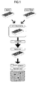
- Table 1 and Table 2 show the results of the comparisons between the method of the present invention (BacHTS system) and the methods of the prior arts (BacPAK6 system and BacToBac system) with regard to the processes. With the results, it is clear that the desired recombinant viruses can be obtained even more rapidly by using the method of this invention in comparison to the prior arts. Table 1.
- promoter for the expression of the desired genomic materials, and fusion system and polyadenyl sequences for the preparation of fusion protein are provided from parent viral genome, the gene cassette cloned to common carrier vector could be applied to produce various types of viruses.
- the present invention provides significantly useful tools for studying functions of genes of which demands have been increasing greatly since the completion of genome project [ Wang Y. and Huang S., Drug Discovery Today, vol. 5, 10-16 (2000) ].
- the present invention employs bacuovirus having circular genomic DNAs, and Int/att system from bacteriophage ⁇ (Genebank: NC 001416) as a site-specific recombination system.
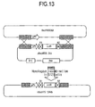
- Int/att system attR site-specific recombination sites within viral genomic DNA fragment react with corresponding attL site-specific recombination sites including desired genomic materials from carrier vector, in the presence of ⁇ integrase recombination enzyme, IHF as a co-factor and Xis, and result in vector DNA fragment containing attB and attP sites [see Hartley JL. Genome Research, 2000, Vol. 10, 1788-1795 ].
- Other recombination systems from various organisms for example, the Cre/loxP system from bacteriophage P1, and the FLP/FRT system from the Saccharomyces cerevisiae 2 ⁇ circle plasmid, can be employed.
- vBacHTS viral DNAs are constructed to include site-specific recombination sequences and Bsu36I recognition base sequences, which in turn are made to linear form viral DNAs flanked by 2 (two) site-specific sites with treatment of Bsu36I restriction enzyme.
- the linear vBacHTS DNAs cannot form viral particles since the vBacHTS viral DNAs are digested at 2 (two) Bsu36I recognition sites having nucleic acid sequences different from each other, while the circular form DNAs generated by site-specific recombination formed viral particles efficiently.
- the reaction mixture containing viral DNAs having desired genomic materials, which are integrated by site-specific recombination is transferred to Sf21 insect cells, and then viral packaging is detected.
- recombinant viruses can be obtained without employing intermediate host cells, such as E. coli or yeast, for selecting and amplifying suitable genomic DNAs. Therefore, the recombinant viruses are suitable for the purpose of expressing desired genes from host cells.
- the activity measurement of GFP and GUS proteins indicates that the method of the present invention is more efficient than the method of the prior arts.
- pBacHTS viral vectors were prepared.
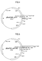
- pBacPAK8_R1 (see FIG.4 ) was provided by cloning amplified attR1 sites and Bsu36I recognition base sequences at BamHI/EcoRI sites of a shuttle vector of pBacPAK8 (Genebank: U2446, from Clontech) and transforming DH5 ⁇ (from LTI, Life Technologies Inc.) with the cloned vector, and then by incubating and screening in agar medium including ampicilin.
- pBacPAK8_R1R2 was prepared by cloning PCR amplified attR2 sites and Bsu36I recognition site sequences at EcoRI/PacI site sequences of pBacPAK8_R1 (see FIG.5 ).
- DNA fragment (427 bp) including ccdB gene from EcoR1 digested fragments of pEntr4 plasmid was inserted to the pBacPAK8_R1R2, and then the resulting plasmid was transferred to DB3.1 cell (from LTI).
- pBacHTS contained two different Bsu36I recognition sequences between attR1 site and attR2 site ( FIG.2 ).
- the attR1 sites and the attR2 sites included site-specific recombinase target sequences that reacted with attL1 and attL2 sites, respectively, in the presence of integrase from ⁇ bacteriophage, Xis, IHF- ⁇ and IHF- ⁇ .
- EXAMPLE 2 Preparation of pBacHTS 6His, pBacHTS_GST, pBacHTS_HisGst, pBacHTS_GFP and pBacHTS_Flag
- Fusion tag was inserted to the sites in back of polyhedrin promoter in order to prepare recombinant baculovirus expressing fusion proteins.
- pBacHTS_Gst was designed for the preparation of expression vector for GST fusion protein.
- pGEX-2T (Genbank:U13850) was PCR amplified using a primer including Bg1II and a primer including BamH1 linker, which in turn was treated with BglII and BamHI and then was inserted to pBacHTS/BamHI restriction sites.
- pBacHTS_His was designed for the preparation of a vector expressing His tag fusion protein by inserting 6 His linker to BamHI site of pBacHTS.
- pBacHTS_HisGST was designed by inserting BglII and BamHI treated GST genes, which were PCR amplified, to BamHI site of pBacHTS_His.
- GFP gene was PCR amplified using a primer containing BamHI linker and a primer containing KpnI linker, which in turn was digested with BamHI/KpnI.
- pBacHTS_Flag was prepared by inserting a linker expressing Flag tag at BamHI/KpnI sites of pBacHTS (see FIG.7 ).
- EXAMPLE 3 Preparation of DNAs of vBacHTS, vBacHTS_His, vBacHTS_HisGst, vBacHTS_GST and vBacHTS_GFP viruses
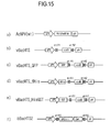
- Baculoviruses of which polyhedrin loci were replaced by pBacHTS plasmids were prepared using homologous recombination. Briefly, Sf21 insect cells were transfected with a mixture of pBacHTS plasmid, Bsu36I digested BacPAK6 viral DNAs (from Clontech#6144-1) and Lipofectin (from LTI) to induce homologous recombination in the cells. The recombinant viruses were isolated through twice repeated plaque assays, which in turn were subjected to PCR. The purified viral clone was named vBacHTS virus (SEQ. ID No.1: the sequences of its polyhedrin locus) (see FIG.15 ).
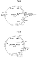
- pBacHTS_His see FIG.8
- pBacHTS_HisGst see FIG.9
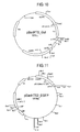
- pBacHTS_GST see FIG.10
- pBacHTS_GFP see FIG.6
- BacPAK6 virus to generate vBacHTS_His (SEQ. ID No.2: the sequences of its polyhedrin locus)
- vBacHTS_HisGst SEQ. ID No.3: the sequences of its polyhedrin locus
- vBacHTS_GST SEQ.
- sequences of the obtained viruses are the same as those of the wild type AcNPV (GenBank: NC_001623) except for the above-designated sequences of the polyhedrin loci.
- Viral plaques were selected from agarose medium.
- EXAMPLE 4 Purification of viral DNAs of vBacHTS, vBacHTS_His, vBacHTS_HisGst, vBacHTS_GFP, and vBacHTS
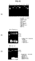
- the viral lysate was extracted twice with the same amount of phenol/chloroform solution, and ethanol was added to the extract, thereby pure viral DNAs were obtained as precipitates. Then, 50 units of restriction enzyme Bsu36I (from New England Bio labs Inc. NEB#524) were added to the purified DNAs (10 ug). The mixture was left at 37°C for 2 hrs before used in the following studies (see FIG.16 ).
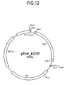
- a pEntr_GFP gene cassette was prepared by inserting PCR products of GFP between attL1 site and attL2 site.
- pEntr_Gus gene cassette from LTI was employed.
- EXAMPLE 6 Preparation of the desired recombinant viruses having GFP gene or GUS gene
- Recombinant viruses expressing GFP and Gus were generated in this study.
- the pEntr_GFP gene cassette (100 ng) or the pEntr_Gus gene cassette (100 ng) was transferred to a test-tube, and reacted with 100 ng of Bsu36I treated vBacHTS viral DNAs at 25°C for 2 hrs, in the presence of integrase mixture comprising 4 ul of integrase, Xis, IHF- ⁇ and IHF- ⁇ .
- Sf21 cells were infected with the reaction mixture together with lipofectin (from LTI), thereby recombinant viruses were generated.
- green fluorescence was observed from the insect cells including GFP gene cassette.
- after 4 days almost every cell showed green fluorescence.
- fluorescence and symptoms of viral infection were observed in the infected cells infected.
- a mixture containing pEntr_Gus gene cassette was transferred to Sf21 cells. After 4 days, X-Gluc was added to the cells. After 2 hrs of the reaction, the Sf21 cells became blue, which supported the expression of GUS (see FIG.18 ).
- 4-day viral culture was dilated and transferred to insect cells. Next, plaques were detected 4 days after adding culture medium containing 1% low-melting point agarose to the infected cells. As the result, most plaques represented blue fluorescence (see FIG.19 ).
- Recombinant viruses expressing GFP were prepared in this study.
- 100 ng of pEntr_GFP gene cassette containing GFP genes and 200 ng of viral DNAs of Bsu36I treated vBacHTS, vBacHTS_His or vBacHTS_HisGST were reacted in the presence of 4ul of integrase mixture comprising integrase, Xis (exisionase), IHF- ⁇ and IHF- ⁇ at 25 °C for 6 hrs.
- the resulting mixture was transferred to Sf21 cells together with lipofectin (from LTI) in order to generate recombinant baculoviruses.
- GFP baculoviruses Virus DNA Bsu36I gene cassette GFP positive cell# BacPAK6 Yes pBacPAK8 EGFP 154 VbacHTS pEntr EGFP 1752 VbacHTS yes pEntr EGFP 572 VbacHTS His pEntr EGFP 2236 VbacHTS His yes pEntr EGFP 654 VbacHTS HisGST pEntr EGFP 1433 VbacHTS HisGST yes pEntr EGFP 475
- EXAMPLE 8 Detection of the efficiency of recombinant viral expression
- Recombinant viruses expressing Gus were prepared, and the number of plaques expressing Gus genes was counted.
- 100 ng of pEntr_Gus gene cassette containing Gus genes and 200 ng of each of viral DNAs of Bsu36I treated vBacHTS, vBacHTS_His or vBacHTS_HisGST were reacted in the presence of 4 ul of integrase mixture comprising integrase, exisionase, IHF- ⁇ and IHF- ⁇ at 25°C for 6 hrs.
- the resulting mixture was transferred to Sf21 cells together with lipofectin (from LTI) to generate recombinant baculobiruses.
- Plaque assays were carried out for the viral culture medium.
- 4-day viral culture medium was dilated and transferred to insect cells. Subsequently, the cells were incubated for 4 days after adding medium containing 1% low-melting point agarose thereto.
- the numbers of plaques expressing Gus genes were counted after 4 days from the addition of 100 ul of 0.33% nutral red water solution and 25 ul of X-Gluc (20 mg/ml in DMSO) and afer subsequent dye treatment (see Table 4). Table 4.
- Desired recombinant viruses were prepared simultaneously from multi-well plate.
- the vBacHTS viral DNAs were treated with Bsu36I restriction enzyme.
- GFP gene cassette was employed to confirm the generation of recombinant baculoviruses and gene expression.
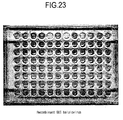
- the GFP gene cassettes were incubated in 96-deep well plate simultaneously and purified using automatic device. All the processes were carried out in multi-well plate while recombination reaction, insect cell incubation, and viral infection were performed using 8-channel pipette.
- Baculo-Forward primer 5-actgttttcgtaacagttttg-3 (SEQ. ID No.: 6)
- Baculo-Reverse primer 5-acaacgcacagaatctagc-3 (SEQ. ID No.: 7)
- liposome was prepared by mixing 5 ul of the recombinant reaction mixture with 5 ul of 10% lipofectin dilates, which in turn was transferred to 50, 000 of Sf21 cells in 96-well plate (SPL) followed by the incubation of the cells for 4 days. Protein expression was assayed after 3-day incubation of 50,000 of the Sf21 cells that were infected again with 10 ul of viruses. As for GFP viruses, the expression of green fluorescence was detected under the fluorescence microscope. And, the exhibition of symptoms of GFP viral infection was detected also.
- the cells were isolated from each of the wells with SDS-PAGE, and the expression of GFP was assayed using GFP-antibody with western blot. With regard to GFP proteins, the measured value showed similar pattern as detected under the fluorescence microscope (see FIG.22 ).
- EXAMPLE 10 High-throughput preparation and enzyme activity screening of LacZ gene mutants in 96-well plate using GFP recombinant baculoviruses
- vBacHTS_GFP viral DNAs were treated with Bsu36I restriction enzyme.
- Gene cassettes of pEntr_LacZdel were employed in this study. The gene cassettes were prepared in 96-well plate by PCR cloning of the gene of ⁇ -galactosidase from E. coli (sized about 3.2 kb), wherein the gene was deleted of 3'-terminus. More specifically, pEntr_LacZ was prepared by PCR cloning of gene of LacZ.
- the pEntr_LacZ was treated with ExoIII/S1 deletion kit (#K0421) (from Fermentas) to generate of pEntr_LacZdel. All the processes were carried out in multi-well plate, while the recombination reaction, insect cell incubation and viral infection were performed with 8-channel pipette. 50 ng of gene cassettes and 200 ng of vBacHTS baculoviral DNAs were reacted in the presence of 2 ul of recombines and 4 ul of buffer at 25 °C for 12 hrs. A total of 20 ul of reaction mixture was used in this reaction.
- PCR amplification was performed using a pair of primers (actgttttcgtaacagttttg and acaacgcacagaatctagc) for the amplification of polyhedrin locus.
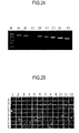
- recombinant viruses were generated at very high efficiency (see FIG.25 ).
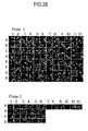
- liposome was prepared by mixing 5 ul of the recombinant reaction mixture and 5 ul of 10% lipofectin dilates, which in turn was transferred to 50,000 of Sf21 cells in 96-well plate (SPL) followed by incubation of the cells for 4 days (see FIG.26 ).
- SPL 96-well plate
- FIG.26 In order to assay the expression of GFP fusion proteins, SDS-PAGE analysis and western blotting using GFP antibodies were carried out (see FIG.26 ).
- EXAMPLE 11 High-throughput heterologous gene expression using recombinant baculoviruses in 96-well plate and the screening of enzyme activity using the same
- S. cerevisie which includes 124 protein kinase genes, was employed in this study. PCR amplification and cloning were carried out using gene-specific primers to obtain 112 gene cassettes from the S . cerevisie genome.
- Recombinant baculoviruses were prepared by the recombination of the gene cassettes and vBacHTS_GFP vector DNA. The respective recombinant viruses represented particular fluorescence according to the proteins fused to GFP (see FIG.28 ). Also, the recombinant baculoviruses having 32 human cDNA were prepared in the same manner.
- Viruses which exhibited protein kinase activities to substrates of Histon H1, MBP (myelin basic protein) and Rb were screened.
- EXAMPLE 12 The preparation of pBacHTS2 and pBacHTS2_GFP
- Baculoviral transfer vector of pBacHTS2 was prepared in order to generate vBacHTS2 viruses (SEQ. ID No.8: the sequences of its polyhedrin locus) that can replicate and can be applied to recombination reaction in vitro .
- the sequences of the obtained vBacHTS2 virus are the same as those of the wild type AcNPV (GenBank: NC_001623) except for the above-designated sequences of the polyhedrin locus.
- the pBacHTS2 had BAC vector originated replication origin and CmR (chloramphenichol resistance gene) at Bsu36I restriction enzyme recognition site located between attR1 site and attR2 site thereof. Thus, it was a kind of BAC vectors that can replicate in bacteria. Replacing the BAC vector originated replication origin and CmR with desired gene cassette generated vBacHTS viruses in vitro.
- pBacHTS2 In order to prepare pBacHTS2, firstly, 6.5 kb of DNA fragment containing BAC (Bacterial artificial chromosome) vector replication origin and CmR was generated using pBACe3.6 (Genbank:U80929) as a template. Then, PCR amplification was carried out using 10 pmole of a pair of sequences having Bsu36I recognition sequences, 200 uM of dNTPs and 2.5 U of Pfu turbo polymerase (Stratagene) with PCR cycler (Applied Biosystem, Gene Amp PCR System 2700). A total of 50 ul of reaction mixture was used.
- PCR was carried out for 20 cycles (DNA denaturation at 95 °C, 30 sec, DNA extension at 60 °C, 30 sec, DNA amplification at 72 °C, 7 min).
- PCR products of 6.5 kb of DNA fragment and vBacHTS vector were digested by Bsu36I, and were reacted in the presence of T4 ligase at 16 °C overnight.
- E. coli (DH5a) was transformed using the resulting mixture, which in turn was incubated with Cm and Amp medium (for selection) to produce pBacHTS2.
- Bsu36I treated PCR products of 6.5 kb of DNA fragment were inserted to Bsu36I site of the pBacHTS_GFP vector so as to obtain pBacHTS2_GFP vector.
- Baculoviruses were prepared using homologous recombination, which replicated in bacteria. Briefly, the homologous recombination was induced in Sf21 cells by transferring a mixture of pBacHTS2 plasmid, Bsu36I digested BacPAK66 DNA and Lipofectin (from LTI). The infected cells were incubated at 27 °C for 4 days, and were transferred to 5x10 6 of Sf21 cells (in 100 mm of dish). Thereby, high titer of viruses was obtained. Then, precipitates of viral particles were produced by centrifugation after adding 2ml of 50% PEG6000 to the cell culture medium.
- vBacHTS2 viral DNAs were obtained.
- DH10B cell lines from LTI
- MicroPulser from Bio-Rad
- the bacteria were incubated in 1 liter of 2 ⁇ YT medium (10 g of Yeast extract powder, 16 g of tryptone, 5 g of NaCl within per 1 (one) Liter), then DNAs were purified and named as bBacHTS2 ( FIG.16 ).
- bBacHTS2 FIG.16
- Purified 10 ug of DNAs was digested with 50 unit of Bsu36I at 37 °C for 5 hrs, which in turn was heat treated at 80 °C for 20 min to inactivate Bsu36I enzyme.
- vBacHTS2_GFP (SEQ. ID No.9: the sequences of its polyhedrin locus) was prepared from pBacHTS2_GFP vector by homologous recombination, and then was transferred to DH10B by electrophoration. The DNAs were purified and named as bBacHTS2_GFP (see FIG.16 ). The sequences of the vBacHTS2_GFP virus are the same as those of the wild type AcNPV (GenBank: NC_001623) except for the above-designated sequences of the polyhedrin locus.
- EXAMPLE 14 Preparation of the desired recombinant viruses using vBacHTS2 and vBacHTS2_GFP DNAs generated from bacteria
- PCR was performed as disclosed in the Examples mentioned above. PCR amplification was carried out using the reaction mixture, as templates, and a pair of primers amplifying viral polyhedrin locus to confirm whether recombination reaction occurred.
- a total of 20 cycles of PCR were carried out using each of 10 pmole of the primers and 1 tube of Bioneer premix kit, in which one cycle of PCR was performed at 95 °C for 30 sec, 50 °C for 30 sec and 72 °C for 3 min.
- electrophoresis was carried out for the PCR products on 1% agarose gel to confirm efficient performance of recombination.
- the gene cassette reaction mixture was transferred to Sf21 cells together with 5 ul of Lipofectin (from LTI) so as to obtain baculoviruses. Green fluorescence was observed in insect cells, after 2 days from transferring GFP gene cassette reaction mixture thereto. In addition, most of the cells represented green fluorescence after 4 days from transfer ( FIG.25 ).
- the present invention it is possible to prepare recombinant viruses more rapidly and more easily. Also, it is possible to obtain high titer viruses at high efficiency.
- the desired genes can be inserted into any target site of viral genome accurately, and also numerous viruses can be generated simultaneously. Accordingly, the present invention can be applied to high-throughput system to produce hundreds or thousands of viruses to study functions of genes at the same time, as well as to obtain several recombinant viruses.
- recombinant baculoviral library can be produced easily according to the present invention, and the library can be used in the screening of the desired genomic materials.
Landscapes
- Health & Medical Sciences (AREA)
- Genetics & Genomics (AREA)
- Life Sciences & Earth Sciences (AREA)
- Engineering & Computer Science (AREA)
- Chemical & Material Sciences (AREA)
- Organic Chemistry (AREA)
- Zoology (AREA)
- Wood Science & Technology (AREA)
- Bioinformatics & Cheminformatics (AREA)
- Biomedical Technology (AREA)
- Biotechnology (AREA)
- General Engineering & Computer Science (AREA)
- Microbiology (AREA)
- Biochemistry (AREA)
- General Health & Medical Sciences (AREA)
- Plant Pathology (AREA)
- Molecular Biology (AREA)
- Biophysics (AREA)
- Physics & Mathematics (AREA)
- Virology (AREA)
- Crystallography & Structural Chemistry (AREA)
- Immunology (AREA)
- Medicinal Chemistry (AREA)
- Micro-Organisms Or Cultivation Processes Thereof (AREA)
- Measuring Or Testing Involving Enzymes Or Micro-Organisms (AREA)
Abstract
Description
- This invention relates to a method for producing recombinant viruses and vectors for the same. Furthermore, this invention relates to DNA constructs for the method and the recombinant viruses produced by the method.
- A recombinant viral vector means a genetically engineered vector, which is used in, for example: the preparation of a vaccine; analysis of function of a gene, a group of genes, or a genomic domain; production of proteins; and gene therapy. Genomic materials, which could be integrated into a viral vector, include any genomic materials such as a gene, cDNA, genomic DNA, DNA sequences encoding peptides or proteins, RNA, anti-sense RNA, siRNA, DNA for si RNA, promoter, enhancer etc. A virus, which is used in the preparation of a vector, includes baculovirus, adenovirus, adeno-associated virus (AAV), retrovirus, Herpes virus, hepatitis B virus (HBV), polioma virus, sindbid virus, and vacxnia virus etc.
- Retrovirus is known as one of the most widely used viruses in gene therapy [Clive Patience et al. J Virol, 72:2671-2676, 1998, Marshall, Science, 269: 1050-1055,1995]. Retroviral genome has two LTRs, capsid sequences and 3 (three) coding regions (gag, pol and env), and the method of its preparation and its use in vitro and in vivo are disclosed (see
WO9908692A1 EP 453242 - Adenovirus has a linear genome (sized about 36 kb) and includes replication origin and capsid signals near to the left inverse terminal region (ITR) (103 bp) [Shenk, Adenoviridae: The Viruses and Their Replication. In: Fields BN, Knipe DM Howley PM, ed. Virology. Philadelphia: Raven publishers, 1996: 2111-2148]. Usually, adenoviral vectors have been made by deleting E1 site of the viral genome, which is essential for the viral replication. The viruses including foreign genomic materials substituting E1 site, are transferred to the packaging cells that provide E1 proteins. Thereby, the viral replication occurs only in the packaging cells. Representative packaging cells include, for example, HEK293 cell, 911 cell, and PER. C6 cell, etc. [Hitt MM et al., Advances in Pharmacology, vol. 40, 137-206 (1997), Wang Y. and Huang S., Drug]. Adenoviral vector has advantages such as safty, affinity to various cells, possibility of infecting dividing cells and higher titer (1011 pfu/ml). Thus, adenovirus has been used in the preparation of a vector expressing heterologous genes. Adeno-associated virus (sized about 4700 bp) has ITRs (sized about 145 bp) as replication origins at each terminus. It can be integrated into the genomic DNAs of various types of host cells safely and specifically.
- In order to use a recombinant virus as a vector, it should be modified not to replicate themselves in the infected cells. Therefore, defective viruses, which have deletions of some essential regions of genome for viral replication, are usually employed in the preparation of the recombinant viral vectors. As for a retrovirus, for example, gag, pol and/or env genes are deleted, and the regions are replaced with desired genomic materials [Bender et al., J. Virol. 61 (1987) 1639]. Regarding an adenovirus, E1, E2 and/or E4 sites are deleted for the preparation of a viral vector [Levrero et al., Gene 101 (1991) 195; Gosh-Choudhury et al., Gene 50 (1986) 161, Van der Vliet et al., (1975)]. On the other hand, a pseudo-virus vector, which consists of only the essential regions (such as, ITR and capsid sequences) of the replication, has been used also for obtaining recombinant viruses (see
WO95/02697 - In addition, baculoviruses, which are known as having circular genomic DNAs, have been used for the preparation of viral vectors. Baculoviridae are infectious on invertebrates, such as insects and crustacea. AcNPV is one of the most widely used baculoviruses, and it contains double strand circular genomic DNAs (sized about 134 kb) (GenBank: NC_001623). Smith et al. developed recombinant baculoviruses by inserting β-interferon genes, and successfully obtained interferon proteins from insect cells using the recombinant baculoviruses (Smith GE., Mol. Cell. Biol., V3, p2156-2165, 1983). Since then, numerous genes were expressed and produced using recombinant baculoviral system. The features of the methods are that: i) it is possible to produce desired recombinant baculoviruses at high efficiency by employing polyhedrin promoter; and ii) it can be used for the expression of genes that do not exhibit their activities when incubated in bacteria. That is because post-translational modification is carried out in insect cells. However, the size of genome of baculovirus made it difficult to engineer the genome through conventional restriction and ligation method. Therefore, homologous recombination has been used in insect cells generally. In order for the homologous recombination, carrier vectors containing desired genes and nucleic acid sequences necessary for the homologous recombination are prepared. Then, insect cells are transfected with the carrier vectors and viral genomic DNAs to induce recombination. In that method, however, since the production efficiency of recombinant viruses is relatively low (0.1-2%), repetitive screening of baculoviral plaques should be performed in order to obtain the desired recombinant viruses. Thus, it is also considered as being an inefficient method.
- Kitts PA et al. disclosed an improved method that increased production efficiency of recombinant viruses (Nucleic Acids Res., Oct 1990; 18: 5667 - 5672. Kitts PA et al). They made baculoviral genomic DNAs to be in linear form using restriction enzyme. In Kitts' method, a linker, which includes Bsu36I recognition base sequences, CCTNAGG, or lacZ genes, is inserted to wild-type Autographa californica nuclear polyhedrosis virus (AcNPV) genome at polyherin locus in order to introduce Bsu36I site recognition nucleic acid sequences. Next, the viral DNAs are digested with Bsu36I restriction enzyme, and then transferred to insect cells together with carrier vectors containing desired genomic materials so as to induce homologous recombination. The production efficiency of the desired recombinant viruses is from 26% to 44%. According to that method, linear digested DNA segments, which are not subjected to recombination, are excluded electively from the production of viral vectors. Thus, the desired recombinant viruses expressing desired protein could be generated with relatively high efficiency. However, the method has disadvantages as follows: i) it is not possible to digest Bsu36I recognition sites up to 100% and ii) since the digested DNA segments are fused with each other in insect cells by ligase, selection procedures for the desired viral vectors need be employed.
- Peakman TC et al. provided another method using site-specific recombination of cre/loxP system in vitro (Peakman TC et al., Nucleic Acids Research, Vol 20, ). Briefly, they prepared baculoviruses containing loxP sites and applied them to site-specific recombination in vitro with desired genomic materials by reacting the viruses with transfer vectors having the genomic materials flanked with loxP sites, in the presence of cre recombination enzyme. In that method, time-period for producing recombinant viruses is shortened. However, the production efficiency of desired recombinant viruses is still low (0.2% to 49%), thereby it is not suitable to be used as a high-throughput system for the preparation of recombinant viruses.
- In addition, Luckow VA et al. reported a method of inserting desired genomic cassette to baculoviral genome by site-specific recombination in a cell (Luckow VA et al., Virol., Aug 1993; 67: 4566 - 4579). According to Luckow's method, firstly, a shuttle vector (sized about 130 kb) is prepared by inserting plasmid replication origin, which replicates in bacteria, kanamycin resistance gene and attTn7 recombination sequences into baculoviral genome. The shuttle was named "Bacmid". DH10Bac was generated by transforming E. coli with Bacmid and helper plasmid, which in turn was transformed using a plasmid containing desired genes and polyhedrin promoter to prepare recombinant Bacmid in E coli. The resulting recombinant Bacmid is isolated and transferred to insect cells to obtain recombinant viruses. According to that method, baculoviruses could be obtained with relatively high efficiency by using site-specific recombination of transposon. That method, however, required complex procedures, for example, cloning desired genomic materials to Tn7 Bacmid carrier vectors; transforming DH10Bac for the recombination of Tn7 baculoviruses; selecting and isolating bacteria having Bacmid that contains desired genomic materials; purifying Bacmid DNAs (sized about 130 kb) from the isolated bacteria; and transferring the DNAs again to insect cells to produce recombinant virues.
-
US 6143557 discloses recombinational cloning for the use of nucleic acids, vectors and methods, in vitro and in vivo, for moving or exchanging segments of DNA molecules using engineered recombination sites and recombination proteins to provide chimeric DNA molecules that have the desired characteristic(s) and/or DNA segment(s). - Thus, non of the above-mentioned methods of the prior arts have provided suitable ways to overcome the problems of relatively long time-period in preparing recombinant viruses (about 3 to 6 weeks) and low production efficiency. Therefore, there have been demands for the development of an improved method.
- In this regard, we have carried out studies to produce recombinant viruses having desired genomic materials more rapidly and efficiently, thereby we have completed this invention by preparing recombinant viral vectors in vitro using site-specific recombination and employing the vectors to produce recombinant viruses. Since the viral vectors obtained in vitro according to this invention can be applied to animal cells directly without further procedures like selection, the present invention not only simplifies the total procedures significantly but also provides desired recombinant viruses with up to 100% production efficiency. Thus, higher virus titer can be obtained according to the present invention.
- Any publications referenced herein are hereby incorporated by reference in this application in order to more fully describe the state of the art to which the present invention pertains.
- It is important to understand the present invention to note that all technical and scientific terms used herein, unless otherwise defined, are intended to have the same meaning as commonly understood by one of ordinary skill in the art. The techniques used herein are also those that are known to one of ordinary skill in the art, unless stated otherwise.
- Reference to particular buffers, media, reagents, cells, culture conditions and the like, or to some subclass of same, is not intended to be limiting, but should be read to include all such related materials that one of ordinary skill in the art would recognize as being of interest or value in the particular context in which that discussion is presented. For example, it is often possible to substitute one buffer system or culture medium for another, such that a different but known way is used to achieve the same goals as those to which the use of suggested method, material or composition is directed.
- It is an object of this invention to provide more rapid and efficient method of producing recombinant viruses.
- In order to accomplish this object, this invention employs recombinant viral vectors that are prepared in vitro by site-specific recombination.
- In accordance with the present invention, there is provided a method for preparing expressive circular recombinant viral vectors, characterized in that the method comprises the steps of:
- i) providing a linearized viral DNA fragment containing at least two site-specific recombination sites and at least one restriction enzyme recognition site;
- ii) providing a gene cassette containing desired genomic material flanked with at least two site-specific recombination sites for homologous recombination with the linearized viral DNA fragment at the two site specific recombination sites; and
- iii) reacting the linearized viral DNA fragment and the gene cassette together under conditions effective to allow in vitro homologous recombination to take place so as to form a recombinant viral vector; and
- iv) preventing the expression of a viral vector that does not contain the desired genomic material.
- In accordance with a further aspect of the present invention, there is provided a method of screening recombinant viruses containing desired genomic materials, characterized in that the method comprises the steps of:
- i) providing a linearised viral DNA fragment containing at least two site-specific recombination sites and at least one restriction enzyme recognition site;
- ii) providing a gene cassette containing desired genomic material flanked with at least two site-specific recombination sites for homologous recombination with the linearized viral DNA fragment at the two site specific recombination sites;
- iii) reacting the linearized viral DNA fragment and the gene cassette together under conditions effective to allow the in vitro homologous recombination to take place so as to form a recombinant virusin vitro to produce reaction mixture including expressive circular recombinant viral vectors formed in vitro;
- iv) preventing the expression of a virus that does not contain the desired genomic material; and
- v) transferring the reaction mixture of the iii) to host cells in multi-well plate.
- In accordance with yet a further aspect of the present invention, there is provided the method method of preparing recombinant viruses having circular genomic DNAs, characterized in that the method comprises the steps of:
- i) providing a linearised viral DNA fragment containing at least two site-specific recombination sites and at least one restriction enzyme recognition site;
- ii) providing a gene cassette containing desired genomic material flanked with at least two site-specific recombination sites for homologous recombination with the linearized DNA fragment at the two site specific recombination sites;
- iii) reacting the linearized viral DNA fragment and the gene cassette to create a homologous recombination in vitro to produce reaction mixture including expressive circular recombinant viral vectors;
- iv) preventing the expression of a viral vector that does not contain the desired genomic material; and
- v) transferring the reaction mixture of the iii) to host cells in multi-well plate.
- In the present invention, viral genomic DNAs are made in linear form by restriction enzymes before being applied to site-specific recombination in vitro. Since the linear viral genomic DNAs, which are not subjected to the site-specific recombination, cannot replicate by themselves, almost all of the expressed viruses are believed to be those from the circular viral genomic DNAs. Thus, according to the present invention, it is possible to obtain recombinant viruses at high efficiency without the procedures for selecting and isolating the desired recombinant viruses. Accordingly, the method of the present invention can be applied to a virus having circular genomic DNAs, for example, polioma virus, papiloma virus and hepatitis B virus, etc.
- Referring to site-specific recombination, it is an event occurring naturally in various living organisms. Enzymes for the site-specific recombination contain not only endonuclease activity but also ligase activity, so they recognize a certain part of DNA sequences and replace it with any other corresponding DNA sequences [Yang W. and Mizuuchi K., Structure, 1997, Vol. 5, 1401-1406(9)]. Int/att system from bacterio λ phage, Cre/LoxP system from P1 bacterio phage and FLP-FRT system from yeast are well developed site-specific recombination systems.
- Site-specific recombination system has been used in i) expression control of desired genomic materials cloned into recombinant viruses, ii) excising helper-virus when preparing helper-viral dependent viruses, iii) increasing the efficiency of homologous recombination in packaging cells, or iv) inducing site-specific recombination between carrier vectors and viral genome in packaging cells to prepare recombinant viruses [Philip NG et al., Biotechniques, vol. 29. 524-528 (2000), Philip NG et al., Human Gene therapy, vol. 10., 2667-2672 (1999), Hardy S et al., Journal of Viology, vol. 71., 1842-1849 (1997), Parks RJ et al., Proc. Natl. Acad. Sci USA,, vol 93., 13565-13570 (1996)]. However, the said applications of the site-specific recombination disclosed in the prior arts were merely indirect and supplementary uses in vivo or in a cell line, not in vitro.
- In addition, applications of site-specific recombination in the preparation of recombinant viruses having circular genomic DNAs were reported: i) in the preparation of recombinant baculoviruses in bacteria using Tn7 recombinantion, and ii) in the preparation of recombinant baculoviruses using cre/lox-P system in vitro.
- However, there have been no applications of site-specific recombination in vitro for specific insertion of desired genomic materials into viral genomic DNAs in order to obtain recombinant viral vectors. In particular, there have been no applications to prevent the expression of viral genomic DNAs that do not contain desired genomic materials. Thus, the present invention is characterized by employing site-specific recombination in preparing recombinant viral vectors in vitro. The present invention has unexpected advantages compared to the prior arts as follows: i) it is possible to obtain recombinant viral vectors more easily and more rapidly by employing site-specific recombinant in vitro; ii) it does not require any further procedure of selecting desired recombinant viruses from intermediate host cells, since the viral genomic DNAs are made in linear form and thereby the linear viral DNAs, which do not form in circular form by integration of the desired genomic materials, cannot be expressed; and iii) it is possible to generate the same kind of genomic materials from various types of viruses or to generate various genomic materials from the same kind of viral type muti-well (e.g. 96-well or 384-well) by employing common transfer vector containing expression cassette that does not have DNAs from viral genome.
- Furthermore, according to the present invention, homologous recombination in the packaging cells is not required any more. Nor, particular cell lines for inducing homologous recombination are required. Table 1 and Table 2 show the results of the comparisons between the method of the present invention (BacHTS system) and the methods of the prior arts (BacPAK6 system and BacToBac system) with regard to the processes. With the results, it is clear that the desired recombinant viruses can be obtained even more rapidly by using the method of this invention in comparison to the prior arts.
Table 1. The comparisons of the number of procedures between the method of the present invention and the methods of prior arts the number of procedures days time- period BacPAK6 system 11 6 10 BacToBac system 13 8 12 BacHTS system 5 3 7 Table 2. The comparisons of the processes required for preparation of baculoviruses BacPAK system BacToBac system BacHTS system 1. RE cut 1. LR reaction 1. Recombination 2. Agarose gel elution 2. Transformation 2. Transfection 3. Colony seeding 3. Infection 3. Ligaion 4. Plasmid purification 4. Harvest and virus storage 5. Colony seeding 5. RE analysis 6. Plasmid purification 6. DH10Bac transformation 7. RE analysis 7. Streaking 8. Transfection 8. Seeding 9. Infection 9. Bacmid purification 10. Harvest and virus storage 10. Transfection 11. Protein expression analysis 11. Infection 12. Harvest and virus storage 13. Protein expression analysis - Furthermore, according to the present invention, since promoter for the expression of the desired genomic materials, and fusion system and polyadenyl sequences for the preparation of fusion protein are provided from parent viral genome, the gene cassette cloned to common carrier vector could be applied to produce various types of viruses.
- Thus, the present invention provides significantly useful tools for studying functions of genes of which demands have been increasing greatly since the completion of genome project [Wang Y. and Huang S., Drug Discovery Today, vol. 5, 10-16 (2000)].
- In one embodiment, the present invention employs bacuovirus having circular genomic DNAs, and Int/att system from bacteriophage λ (Genebank: NC 001416) as a site-specific recombination system. In the Int/att system, attR site-specific recombination sites within viral genomic DNA fragment react with corresponding attL site-specific recombination sites including desired genomic materials from carrier vector, in the presence of λ integrase recombination enzyme, IHF as a co-factor and Xis, and result in vector DNA fragment containing attB and attP sites [see Hartley JL. Genome Research, 2000, Vol. 10, 1788-1795]. Other recombination systems from various organisms, for example, the Cre/loxP system from bacteriophage P1, and the FLP/FRT system from the
Saccharomyces cerevisiae 2 µ circle plasmid, can be employed. - When the mixture is transferred directly to suitable host cells, which are expected to express desired recombinant viruses, only the circular viral genomic DNAs containing attB1-[desired genomic materials]-attB2 can replicate and form viral particles. The linear viral DNA fragment and the vector DNAs themselves, which do not react with each other, and the circular viral DNAs having attP sites cannot replicate and cannot form viral particles. In
FIG.1 , site-specific recombination between gene cassette and vBacHTS baculoviral DNAs in vitro is presented. - In another embodiment, vBacHTS viral DNAs are constructed to include site-specific recombination sequences and Bsu36I recognition base sequences, which in turn are made to linear form viral DNAs flanked by 2 (two) site-specific sites with treatment of Bsu36I restriction enzyme. The linear vBacHTS DNAs cannot form viral particles since the vBacHTS viral DNAs are digested at 2 (two) Bsu36I recognition sites having nucleic acid sequences different from each other, while the circular form DNAs generated by site-specific recombination formed viral particles efficiently.
- In another embodiment, the reaction mixture containing viral DNAs having desired genomic materials, which are integrated by site-specific recombination, is transferred to Sf21 insect cells, and then viral packaging is detected. Thus, according to the present invention, recombinant viruses can be obtained without employing intermediate host cells, such as E. coli or yeast, for selecting and amplifying suitable genomic DNAs. Therefore, the recombinant viruses are suitable for the purpose of expressing desired genes from host cells. The activity measurement of GFP and GUS proteins indicates that the method of the present invention is more efficient than the method of the prior arts.
-
-
FIG.1 illustrates gene-screening processes using BacHTS system. -
FIG.2 depicts the cleavage map of pBacHTS. -
FIG.3 depicts the cleavage map of pBacHTS2. -
FIG.4 depicts the cleavage map of pBacPAK8_attR1. -
FIG.5 depicts the cleavage map of pBacPAK_attR1R2. -
FIG.6 depicts the cleavage map of pBacHTS_GFP. -
FIG.7 depicts the cleavage map of pBacHTS_Flag. -
FIG.8 depicts the cleavage map of pBacHTS_His. -
FIG.9 depicts the cleavage map of pBacHTS_HisGst. -
FIG.10 depicts the cleavage map of pBacHTS_Gst. -
FIG.11 depicts the cleavage map of pBacHTS2_EGFP. -
FIG.12 depicts the cleavage map of pEntr_EGFP. -
FIG.13 illustrates the process of preparation of vBacHTS virus. -
FIG.14 illustrates the process of preparation of bBacHTS2 clone. -
FIG.15 shows the map of baculoviral polyhedrin locus. -
FIG.16 illustrates agarose gel electrophoretic analysis of vBacHTS viral DNAs and bBacHTS2 viral DNAs. -
FIG.17 illustrates procedures for the preparation of desired recombinant viruses using vBacHTS and gene cassette. -
FIG.18 shows the results of activity assays of GFP baculoviruses and GUS recombinant viruses in vBacHTS system. -
FIG.19 shows plaque assays for GFP baculoviruses and GUS recombinant viruses in vBacHTS system. -
FIG.20 shows the results of activity assays of GFP baculoviruses in bBacHTS2 system. -
FIG. 21 shows the results of activity assays of GUS recombinant baculoviruses in bBacHTS2 system. -
FIG.22 illustrates GFP image in multi-well plate and the results of western blot. -
FIG.23 shows enzyme activity using X-Gluc of GUS recombinant viruses in multi-well plate. -
FIG.24 shows the results of site-specific recombination in vitro. -
FIG.25 depicts expression of GFP-LacZ fusion proten in Sf21 cells after 4 days from the transfer of recombinant DNAs to the cells. -
FIG.26 shows the results of western blot of the expression of GFP-LacZ fusion protein using SDS-PAGE and GFP antibodies. -
FIG.27 depicts LacZ enzyme activity in recombinant virus (GFP-LacZ) using X-Gal substrates. -
FIG.28 shows the image of Sf21 cells infected with GFP-YPK baculoviruses from multi-well plate. -
FIG.29 shows the results of screening of protein kinase (Rb, Histone H1, MBP). -
FIG.30 depicts phosphorylation of Rb (retinoblastoma) protein. -
FIG.31 illustrates phosphorylation of Rb protein by #2-A9 protein kinase. - Preferred embodiments of this invention are described in the following examples. Other embodiments within the scope of the claims herein will be apparent to those skilled in the art from consideration of the specification or practice of the invention as disclosed herein. It is intended that the specification, together with the examples, be considered exemplary only, with the scope and the spirit of the invention being indicated by the claims that follow the example. The examples herein are meant to exemplify the various aspects of carrying out the invention and not intended to limit the scope of the invention in any way. The examples do not include detailed descriptions of conventional methods employed, such as in the performance of genomic DNA isolation, PCR, and sequencing procedures. Such methods are well known to those skilled in the art and are described in numerous publications. In addition, all the publications referred herein are integrated hereto as references.
- pBacHTS viral vectors were prepared. For the preparation, firstly, pBacPAK8_R1 (see
FIG.4 ) was provided by cloning amplified attR1 sites and Bsu36I recognition base sequences at BamHI/EcoRI sites of a shuttle vector of pBacPAK8 (Genebank: U2446, from Clontech) and transforming DH5α (from LTI, Life Technologies Inc.) with the cloned vector, and then by incubating and screening in agar medium including ampicilin. Likewise, pBacPAK8_R1R2 was prepared by cloning PCR amplified attR2 sites and Bsu36I recognition site sequences at EcoRI/PacI site sequences of pBacPAK8_R1 (seeFIG.5 ). In order to obtain pBacHTS, DNA fragment (427 bp) including ccdB gene from EcoR1 digested fragments of pEntr4 plasmid (from Invitrogen) was inserted to the pBacPAK8_R1R2, and then the resulting plasmid was transferred to DB3.1 cell (from LTI). pBacHTS contained two different Bsu36I recognition sequences between attR1 site and attR2 site (FIG.2 ). The attR1 sites and the attR2 sites included site-specific recombinase target sequences that reacted with attL1 and attL2 sites, respectively, in the presence of integrase from λ bacteriophage, Xis, IHF-α and IHF-β. - Fusion tag was inserted to the sites in back of polyhedrin promoter in order to prepare recombinant baculovirus expressing fusion proteins. pBacHTS_Gst was designed for the preparation of expression vector for GST fusion protein. In order for this, pGEX-2T (Genbank:U13850) was PCR amplified using a primer including Bg1II and a primer including BamH1 linker, which in turn was treated with BglII and BamHI and then was inserted to pBacHTS/BamHI restriction sites. In addition, pBacHTS_His was designed for the preparation of a vector expressing His tag fusion protein by inserting 6 His linker to BamHI site of pBacHTS. Furthermore, pBacHTS_HisGST was designed by inserting BglII and BamHI treated GST genes, which were PCR amplified, to BamHI site of pBacHTS_His. On the other hand, GFP gene was PCR amplified using a primer containing BamHI linker and a primer containing KpnI linker, which in turn was digested with BamHI/KpnI. After that, the digested gene was integrated into BamHI/KpnI restriction sites of pBacHTS to generate pBacHTS_GFP. Furthermore, pBacHTS_Flag was prepared by inserting a linker expressing Flag tag at BamHI/KpnI sites of pBacHTS (see
FIG.7 ). - Baculoviruses of which polyhedrin loci were replaced by pBacHTS plasmids were prepared using homologous recombination. Briefly, Sf21 insect cells were transfected with a mixture of pBacHTS plasmid, Bsu36I digested BacPAK6 viral DNAs (from Clontech#6144-1) and Lipofectin (from LTI) to induce homologous recombination in the cells. The recombinant viruses were isolated through twice repeated plaque assays, which in turn were subjected to PCR. The purified viral clone was named vBacHTS virus (SEQ. ID No.1: the sequences of its polyhedrin locus) (see
FIG.15 ). As disclosed inFIG.13 , pBacHTS_His (seeFIG.8 ), pBacHTS_HisGst (seeFIG.9 ) pBacHTS_GST (seeFIG.10 ), pBacHTS_GFP (seeFIG.6 ) were respectively transferred together with BacPAK6 virus to generate vBacHTS_His (SEQ. ID No.2: the sequences of its polyhedrin locus), vBacHTS_HisGst (SEQ. ID No.3: the sequences of its polyhedrin locus), vBacHTS_GST (SEQ. ID No.4: the sequences of its polyhedrin locus) and vBacHTS_GFP (SEQ. ID No.5: the sequences of its polyhedrin locus) (seeFIG.15 ). The sequences of the obtained viruses are the same as those of the wild type AcNPV (GenBank: NC_001623) except for the above-designated sequences of the polyhedrin loci. Viral plaques were selected from agarose medium. - Viral culture medium having 1.2x10⁸ pfu/ml of viral titer was produced after 3 days from infecting Sf21 cells on cell culture dish (100 mm diameter) in order to isolate vBacHTS viral DNAs. Then, 1.25x10⁷ of Sf21 cells were plated again on cell culture dish (150 mm diameter), which in turn were infected with 20 times larger numbers of viruses (MOI=20). After incubating for 48 hrs, about 25 ml of cell culture medium per 1 dish was obtained. A total of 150 ml of cell culture medium from 6 dishes (150 mm diameter) was centrifugated at 20,000 rpm for 90 min, and the resulting precipitated viral particles were recovered (with Hanil, Supra22k). The recovered viral particles were suspended in 2ml of TE (10mM Tris-HCl pH=8.0, 1mM EDTA), and then were treated with sucrose concentration gradient ultra-centrifuge (Beckman SW41 rotor) at 30,000 rpm. Viral particles were purified from the layer between 50% and 40% sucrose solution. The purified viruses were subjected to centrifugation again at 18,000 rpm for 90 min. After that, the precipitates of viral particles were suspended again with 2 ml of TE (10mM Tris-HCl pH=8.0, 1mM EDTA), and were left at 42°C for 2 hrs after adding 0.5% SDS, 1% beta-mercaptoethanol and 0.2 mg of ProteinaseK thereto, so as to isolate viral envelopes. The viral lysate was extracted twice with the same amount of phenol/chloroform solution, and ethanol was added to the extract, thereby pure viral DNAs were obtained as precipitates. Then, 50 units of restriction enzyme Bsu36I (from New England Bio labs Inc. NEB#524) were added to the purified DNAs (10 ug). The mixture was left at 37°C for 2 hrs before used in the following studies (see
FIG.16 ). - A pEntr_GFP gene cassette was prepared by inserting PCR products of GFP between attL1 site and attL2 site. pEntr_Gus gene cassette from LTI was employed.
- Recombinant viruses expressing GFP and Gus were generated in this study. The pEntr_GFP gene cassette (100 ng) or the pEntr_Gus gene cassette (100 ng) was transferred to a test-tube, and reacted with 100 ng of Bsu36I treated vBacHTS viral DNAs at 25°C for 2 hrs, in the presence of integrase mixture comprising 4 ul of integrase, Xis, IHF-α and IHF-β. Then, Sf21 cells were infected with the reaction mixture together with lipofectin (from LTI), thereby recombinant viruses were generated. After 2 days, green fluorescence was observed from the insect cells including GFP gene cassette. Furthermore, after 4 days, almost every cell showed green fluorescence. Furthermore, fluorescence and symptoms of viral infection were observed in the infected cells infected.
- On the other hand, a mixture containing pEntr_Gus gene cassette was transferred to Sf21 cells. After 4 days, X-Gluc was added to the cells. After 2 hrs of the reaction, the Sf21 cells became blue, which supported the expression of GUS (see
FIG.18 ). In order to determine the productivity of the recombinant viruses, 4-day viral culture was dilated and transferred to insect cells. Next, plaques were detected 4 days after adding culture medium containing 1% low-melting point agarose to the infected cells. As the result, most plaques represented blue fluorescence (seeFIG.19 ). - Recombinant viruses expressing GFP were prepared in this study. In order for this, 100 ng of pEntr_GFP gene cassette containing GFP genes and 200 ng of viral DNAs of Bsu36I treated vBacHTS, vBacHTS_His or vBacHTS_HisGST were reacted in the presence of 4ul of integrase mixture comprising integrase, Xis (exisionase), IHF-α and IHF-β at 25 °C for 6 hrs. The resulting mixture was transferred to Sf21 cells together with lipofectin (from LTI) in order to generate recombinant baculoviruses. After incubating at 27 °C for 48 hrs, the expression of GFP was detected under fluorescence microscope. Since the cells, in which baculoviruses replicated, represented fluorescence, the efficiency of recombinant viral expression was determined by counting the cells showing green fluorescence. In the meantime, BacPAK6 viral DNAs (from Clontech) treated with Bsu36I and 500 ng of pBacPAK_GFP were transferred to Sf21 cells, then the expression of GFP in the Sf21 cells was monitored.
Table 3. The expression efficiency of GFP baculoviruses Virus DNA Bsu36I gene cassette GFP positive cell# BacPAK6 Yes pBacPAK8 EGFP 154 VbacHTS pEntr EGFP 1752 VbacHTS yes pEntr EGFP 572 VbacHTS His pEntr EGFP 2236 VbacHTS His yes pEntr EGFP 654 VbacHTS HisGST pEntr EGFP 1433 VbacHTS HisGST yes pEntr EGFP 475 - As the result, the efficiency of the viral expression is much higher in the method of the present invention compared to the prior arts (see Table 3).
- Recombinant viruses expressing Gus were prepared, and the number of plaques expressing Gus genes was counted. In order for this, firstly, 100 ng of pEntr_Gus gene cassette containing Gus genes and 200 ng of each of viral DNAs of Bsu36I treated vBacHTS, vBacHTS_His or vBacHTS_HisGST were reacted in the presence of 4 ul of integrase mixture comprising integrase, exisionase, IHF-α and IHF-β at 25°C for 6 hrs. The resulting mixture was transferred to Sf21 cells together with lipofectin (from LTI) to generate recombinant baculobiruses. Plaque assays were carried out for the viral culture medium. On the other hand, in order to detect productivity of the recombinant viruses, 4-day viral culture medium was dilated and transferred to insect cells. Subsequently, the cells were incubated for 4 days after adding medium containing 1% low-melting point agarose thereto. The numbers of plaques expressing Gus genes were counted after 4 days from the addition of 100 ul of 0.33% nutral red water solution and 25 ul of X-Gluc (20 mg/ml in DMSO) and afer subsequent dye treatment (see Table 4).
Table 4. The efficiency of GUS recombinant viral expression Virus DNA Bsu36I gene cassette Total Recombinant rate % BacPAK6 yes pBacPAK8 Gus 46 41 89% VbacHTS pEntr Gus 28 25 89% VbacHTS yes pEntr Gus 57 57 100% VbacHTS His pEntr Gus 44 36 82% VbacHTS His yes pEntr Gus 46 45 98% VbacHTS HisGST pEntr Gus 82 57 70% VbacHTS HisGST yes pEntr Gus 32 32 100% VbacHTS GFP pEntr Gus 27 22 81% VbacHTS GFP yes pEntr Gus 21 20 95% - As the result, most of the plaques represented blue color when they were treated with X-Gluc, which supported that the efficiency of GUS viral generation was very high in comparison to the prior arts.
- Desired recombinant viruses were prepared simultaneously from multi-well plate. For this purpose, the vBacHTS viral DNAs were treated with Bsu36I restriction enzyme. In this study, GFP gene cassette was employed to confirm the generation of recombinant baculoviruses and gene expression. The GFP gene cassettes were incubated in 96-deep well plate simultaneously and purified using automatic device. All the processes were carried out in multi-well plate while recombination reaction, insect cell incubation, and viral infection were performed using 8-channel pipette. About 50 ng of gene cassette and 200 ng of vBacHTS baculoviral DNAs were reacted in the presence of 2 ul of recombinase and 4 ul of buffer at 25 °C for 12 hrs. A total of 20 ul of reaction mixture was used in this reaction. In order to detect the insertion of gene cassette, PCR amplification was performed using primers for polyhedrin locus amplification.
- Baculo-Forward primer: 5-actgttttcgtaacagttttg-3 (SEQ. ID No.: 6)
- Baculo-Reverse primer: 5-acaacgcacagaatctagc-3 (SEQ. ID No.: 7)
- As the result, recombinant viral DNAs were generated with very high efficiency. Meanwhile, liposome was prepared by mixing 5 ul of the recombinant reaction mixture with 5 ul of 10% lipofectin dilates, which in turn was transferred to 50, 000 of Sf21 cells in 96-well plate (SPL) followed by the incubation of the cells for 4 days. Protein expression was assayed after 3-day incubation of 50,000 of the Sf21 cells that were infected again with 10 ul of viruses. As for GFP viruses, the expression of green fluorescence was detected under the fluorescence microscope. And, the exhibition of symptoms of GFP viral infection was detected also. The cells were isolated from each of the wells with SDS-PAGE, and the expression of GFP was assayed using GFP-antibody with western blot. With regard to GFP proteins, the measured value showed similar pattern as detected under the fluorescence microscope (see
FIG.22 ). - In order to prepare desired recombinant baculoviruses (sized about 1-3.2 kb) from multi-well plate simultaneously, the vBacHTS_GFP viral DNAs were treated with Bsu36I restriction enzyme. Gene cassettes of pEntr_LacZdel were employed in this study. The gene cassettes were prepared in 96-well plate by PCR cloning of the gene of β-galactosidase from E. coli (sized about 3.2 kb), wherein the gene was deleted of 3'-terminus. More specifically, pEntr_LacZ was prepared by PCR cloning of gene of LacZ. Afterward, the pEntr_LacZ was treated with ExoIII/S1 deletion kit (#K0421) (from Fermentas) to generate of pEntr_LacZdel. All the processes were carried out in multi-well plate, while the recombination reaction, insect cell incubation and viral infection were performed with 8-channel pipette. 50 ng of gene cassettes and 200 ng of vBacHTS baculoviral DNAs were reacted in the presence of 2 ul of recombines and 4 ul of buffer at 25 °C for 12 hrs. A total of 20 ul of reaction mixture was used in this reaction. In order to detect the insertion of gene cassette, PCR amplification was performed using a pair of primers (actgttttcgtaacagttttg and acaacgcacagaatctagc) for the amplification of polyhedrin locus. As the result, recombinant viruses were generated at very high efficiency (see
FIG.25 ). Meanwhile, liposome was prepared by mixing 5 ul of the recombinant reaction mixture and 5 ul of 10% lipofectin dilates, which in turn was transferred to 50,000 of Sf21 cells in 96-well plate (SPL) followed by incubation of the cells for 4 days (seeFIG.26 ). In order to assay the expression of GFP fusion proteins, SDS-PAGE analysis and western blotting using GFP antibodies were carried out (seeFIG.26 ). - In order to screen the gene activities, 1.5 ul of X-Gal (25 mg/ml in DMSO) of developer of β-galactosidase was added to each well that contained viruses incubated for 3 days after the primary infection. After 12 hrs of reaction, viruses containing β-galactosidase activity were detected in 2 (two) wells (see
FIG.27 ). - S. cerevisie, which includes 124 protein kinase genes, was employed in this study. PCR amplification and cloning were carried out using gene-specific primers to obtain 112 gene cassettes from the S. cerevisie genome. Recombinant baculoviruses were prepared by the recombination of the gene cassettes and vBacHTS_GFP vector DNA. The respective recombinant viruses represented particular fluorescence according to the proteins fused to GFP (see
FIG.28 ). Also, the recombinant baculoviruses having 32 human cDNA were prepared in the same manner. - Firstly, cells were infected with these viruses and incubated for 3 days. The cell lysate was generated using a buffer to lyse cells. Viruses, which exhibited protein kinase activities to substrates of Histon H1, MBP (myelin basic protein) and Rb were screened. 2 ug of protein substrates (histon H1, MBP and Rb) were reacted with 10 ul of cell extracts at 30 °C for 10 min, in the presence of 10uM ATP, 0.2 uCi of P32-δ-labeled ATP (gamma, P32 ATP), and 2 ul of phosphate buffer [200 mM Tris-HCl (pH=8.0), 100 mM of MgCl2, 10 mM of EGTA and 10 mM of DTT]. After stopping the reaction by adding 120 ul of 1% phosphoric acid solution, the reaction mixture was transferred to PVDF membrane (from Milipore, #MAIP-N45). Afterward, the membrane was washed with detergent [10 mM Tris-HCl (pH=8.0), 1 mM EDTA and 150 mM NaCl] four times and dried, which in turn was exposed to phosphoscreen at room temperature, and then signals were detected with Phosphoimager (from MD or Fuji). As the result, it was observed that protein kinase activities were increased in some viruses (see
FIG.29 ). Subsequently, SDS-PAGE electophoretic analysis was carried out to confirm the specificity of protein phosphorylation. At that time, the level of Rb protein phosphorylation with regard to the concentration of protein kinase included in cell extracts was detected, while varying the concentrations of cell extracts. As the result, only the cell extracts of the baculoviruses expressing plate#2-D3 protein and plate#2-A9 protein increased Rb protein phosphorylation in a concentration dependent manner (seeFIG.30 ). Meanwhile, proteins of plate#2-D1 and plate#2-D2 exhibited very weak phosphorylation activities. And, it was acknowledged that the positive signals from the proteins of plate#2-D1 and plate#2-D2 were resulted from auto-phosphorylation activities rather than from Rb protein phosphorylation. Rb protein phosphorylations by plate#2-D3 and plate#2-A9 proteins exhibited a typical protein phosphorylation pattern in that phosphorylation of Rb protein was dependent on the concentrations of the substrate and the enzyme (FIG.31 ). Thus, it is possible to find out new useful proteins from baculoviral libraries obtained from the high-throughput system of the present invention according to the present invention. - Baculoviral transfer vector of pBacHTS2 was prepared in order to generate vBacHTS2 viruses (SEQ. ID No.8: the sequences of its polyhedrin locus) that can replicate and can be applied to recombination reaction in vitro. The sequences of the obtained vBacHTS2 virus are the same as those of the wild type AcNPV (GenBank: NC_001623) except for the above-designated sequences of the polyhedrin locus. The pBacHTS2 had BAC vector originated replication origin and CmR (chloramphenichol resistance gene) at Bsu36I restriction enzyme recognition site located between attR1 site and attR2 site thereof. Thus, it was a kind of BAC vectors that can replicate in bacteria. Replacing the BAC vector originated replication origin and CmR with desired gene cassette generated vBacHTS viruses in vitro.
- In order to prepare pBacHTS2, firstly, 6.5 kb of DNA fragment containing BAC (Bacterial artificial chromosome) vector replication origin and CmR was generated using pBACe3.6 (Genbank:U80929) as a template. Then, PCR amplification was carried out using 10 pmole of a pair of sequences having Bsu36I recognition sequences, 200 uM of dNTPs and 2.5 U of Pfu turbo polymerase (Stratagene) with PCR cycler (Applied Biosystem, Gene Amp PCR System 2700). A total of 50 ul of reaction mixture was used. PCR was carried out for 20 cycles (DNA denaturation at 95 °C, 30 sec, DNA extension at 60 °C, 30 sec, DNA amplification at 72 °C, 7 min). PCR products of 6.5 kb of DNA fragment and vBacHTS vector were digested by Bsu36I, and were reacted in the presence of T4 ligase at 16 °C overnight. E. coli (DH5a) was transformed using the resulting mixture, which in turn was incubated with Cm and Amp medium (for selection) to produce pBacHTS2. Likewise, Bsu36I treated PCR products of 6.5 kb of DNA fragment were inserted to Bsu36I site of the pBacHTS_GFP vector so as to obtain pBacHTS2_GFP vector.
- Baculoviruses were prepared using homologous recombination, which replicated in bacteria. Briefly, the homologous recombination was induced in Sf21 cells by transferring a mixture of pBacHTS2 plasmid, Bsu36I digested BacPAK66 DNA and Lipofectin (from LTI). The infected cells were incubated at 27 °C for 4 days, and were transferred to 5x10⁶ of Sf21 cells (in 100 mm of dish). Thereby, high titer of viruses was obtained. Then, precipitates of viral particles were produced by centrifugation after adding 2ml of 50% PEG6000 to the cell culture medium. Next, proteinase K was added to the mixture at 42 °C and left the mixture alone for 2 hrs to dismantle viral envelope. The viral lysate was extracted twice using the same amount of phenol/chloroform, and ethanol was added thereto. Thereby, precipitated and purified vBacHTS2 viral DNAs were obtained. Then, DH10B cell lines (from LTI) were transformed with the purified DNAs using MicroPulser (from Bio-Rad), which are in turn incubated in Cm medium to form bacterial colony. The bacteria were incubated in 1 liter of 2× YT medium (10 g of Yeast extract powder, 16 g of tryptone, 5 g of NaCl within per 1 (one) Liter), then DNAs were purified and named as bBacHTS2 (
FIG.16 ). Thus, according to this method, it is possible to reduce the required time-period and cost significantly, since the viral DNAs can be obtained from bacterial culture without the need of incubation in insect cells. Purified 10 ug of DNAs was digested with 50 unit of Bsu36I at 37 °C for 5 hrs, which in turn was heat treated at 80 °C for 20 min to inactivate Bsu36I enzyme. Likewise, vBacHTS2_GFP (SEQ. ID No.9: the sequences of its polyhedrin locus) was prepared from pBacHTS2_GFP vector by homologous recombination, and then was transferred to DH10B by electrophoration. The DNAs were purified and named as bBacHTS2_GFP (seeFIG.16 ). The sequences of the vBacHTS2_GFP virus are the same as those of the wild type AcNPV (GenBank: NC_001623) except for the above-designated sequences of the polyhedrin locus. - 50 ng of the pEntr_GFP and the pEntr_Gus gene cassettes were reacted with 200 ng of Bsu36I treated vBacHTS2 viral DNAs in the presence of 1 ul of integrase mixture in vitro at 25 °C for 12 hrs. The same reaction was carried out except for the use of the pEntr_Gus gene cassette instead of pEntr_GFP gene cassette. PCR was performed as disclosed in the Examples mentioned above. PCR amplification was carried out using the reaction mixture, as templates, and a pair of primers amplifying viral polyhedrin locus to confirm whether recombination reaction occurred. A total of 20 cycles of PCR were carried out using each of 10 pmole of the primers and 1 tube of Bioneer premix kit, in which one cycle of PCR was performed at 95 °C for 30 sec, 50 °C for 30 sec and 72 °C for 3 min. After completing PCR reaction, electrophoresis was carried out for the PCR products on 1% agarose gel to confirm efficient performance of recombination. The gene cassette reaction mixture was transferred to Sf21 cells together with 5 ul of Lipofectin (from LTI) so as to obtain baculoviruses. Green fluorescence was observed in insect cells, after 2 days from transferring GFP gene cassette reaction mixture thereto. In addition, most of the cells represented green fluorescence after 4 days from transfer (
FIG.25 ). Sf21 cells were infected again with the viral culture medium to confirm green fluorescence and the symptoms of infection. Likewise, pEntr_Gus gene cassette was reacted with Bsu36I treated vBacHTS2 DNAs at 25 °C for 12 hrs. Then, the obtained reaction mixture was transferred to Sf21 cells and was incubated at 27 °C for 4 days. When 6 ul of X-Gluc (20 mg/ml in DMSO) was added to incubated cells, all the culture medium represented strong blue, which supported not only the expressions of Gus genes but also efficient generation of recombinant viruses containing Gus genes. - As mentioned above, according to the present invention, it is possible to prepare recombinant viruses more rapidly and more easily. Also, it is possible to obtain high titer viruses at high efficiency. In the present invention, since site-specific recombination is employed in vitro, the desired genes can be inserted into any target site of viral genome accurately, and also numerous viruses can be generated simultaneously. Accordingly, the present invention can be applied to high-throughput system to produce hundreds or thousands of viruses to study functions of genes at the same time, as well as to obtain several recombinant viruses. Furthermore, recombinant baculoviral library can be produced easily according to the present invention, and the library can be used in the screening of the desired genomic materials.
- <110> Neurogenex Co., Ltd.
- <120> HIGH THROUGHPUT SYSTEM FOR PRODUCING RECOMBINANT VIRUSES USING SITE-SPECIFIC RECOMBINATION
- <130> PN22782.00
- <150>
KR10-2002-0012634
<151> 2002-03-09 - <160> 9
- <170> Kopatentln 1.71
- <210> 1
<211> 10000
<212> DNA
<213> Artificial Sequence - <220>
<223> Polyhedrin locus of vBacHTS virus - <220>
<221> - promoter
<222> (4428)..(4520)
<223> F pPolyhedrin - <220>
<221> misc_feature
<222> (4084)..(5109)
<223> F ccdB - <220>
<221> misc_feature
<222> (5126)..(5250)
<223> R attR2 - <220>
<221> misc_feature
<222> (4537)..(4661)
<223> F attR1 - <400> 1


- <210> 2
<211> 10000
<212> DNA
<213> Artificial Sequence - <220>
<223> Polyhedrin locus of vBacHTS_His - <220>
<221> source
<222> (4523)..(4553)
<223> F 6His - <220>
<221> misc_feature
<222> (4831)..(5136)
<223> F ccdB - <220>
<221> promoter
<222> (4428)..(4520)
<223> F pPolyhedrin - <220>
<221> misc_feature
<222> (5153)..(5277)
<223> R attR2 - <220>
<221> misc_feature
<222> (4564)..(4688)
<223> F attR1 - <400> 2


- <210> 3
<211> 10000
<212> DNA
<213> Artificial Sequence - <220>
<223> Polyhedrin locus of vBacHTS_HisGst - <220>
<221> source
<222> (4523)..(4553)
<223> F 6His - <220>
<221> source
<222> (4523)..(5231)
<223> F HGst - <220>
<221> promoter
<222> (4428)..(4520)
<223> F pPolyhedrin - <220>
<221> misc_feature
<222> (5831)..(5955)
<223> R attR2 - <220>
<221> misc_feature
<222> (5242)..(5366)
<223> F attR1 - <400> 3


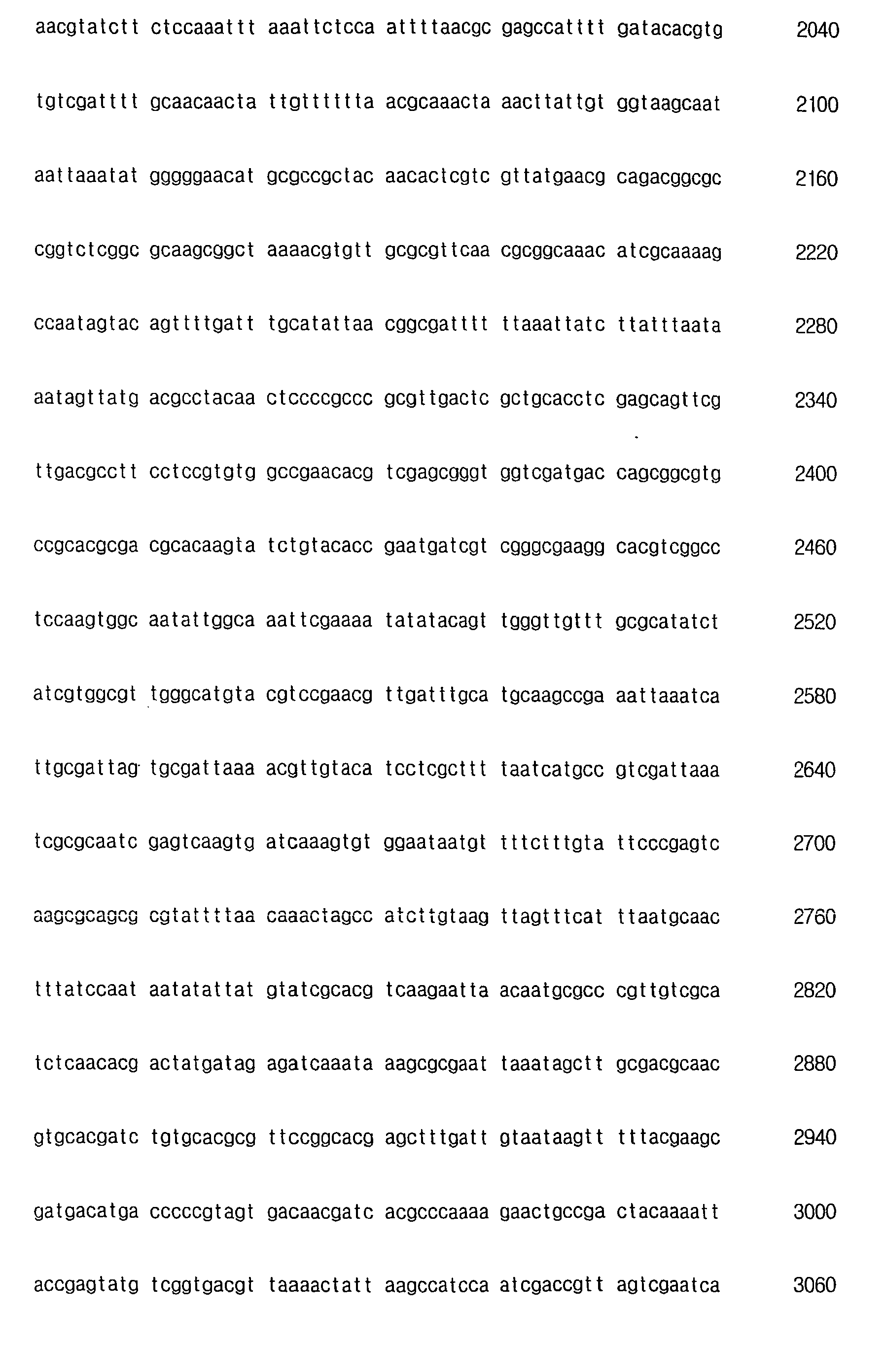


- <210> 4
<211> 10000
<212> DNA
<213> Artificial Sequence - <220>
<223> Polyhedrin locus of vBacHTS_GST - <220>
<221> CDS
<222> (4528)..(5181)
<223> F GST - <220>
<221> misc_feature
<222> (5482)..(5787)
<223> F ccdB - <220>
<221> promoter
<222> (4428)..(4520)
<223> F pPolyhedrin - <220>
<221> misc_feature
<222> (5215)..(5339)
<223> F attR1 - <220>
<221> misc_feature
<222> (5804)..(5928)
<223> R attR2 - <400> 4


- <210> 5
<211> 10000
<212> DNA
<213> Artificial Sequence - <220>
<223> Polyhedrin locus of vBacHTS_GFP virus - <220>
<221> CDS
<222> (4531)..(5247)
<223> F EGFP - <220>
<221> disc-features
<222> (5521)..(5826)
<223> F ccdB - <220>
<221> promoter
<222> (4428)..(4520)
<223> F pPolyhedrin - <220>
<221> misc_feature
<222> (5843)..(5967)
<223> R attR2 - <220>
<221> misc_feature
<222> (5254)..(5378)
<223> F attR1 - <400> 5

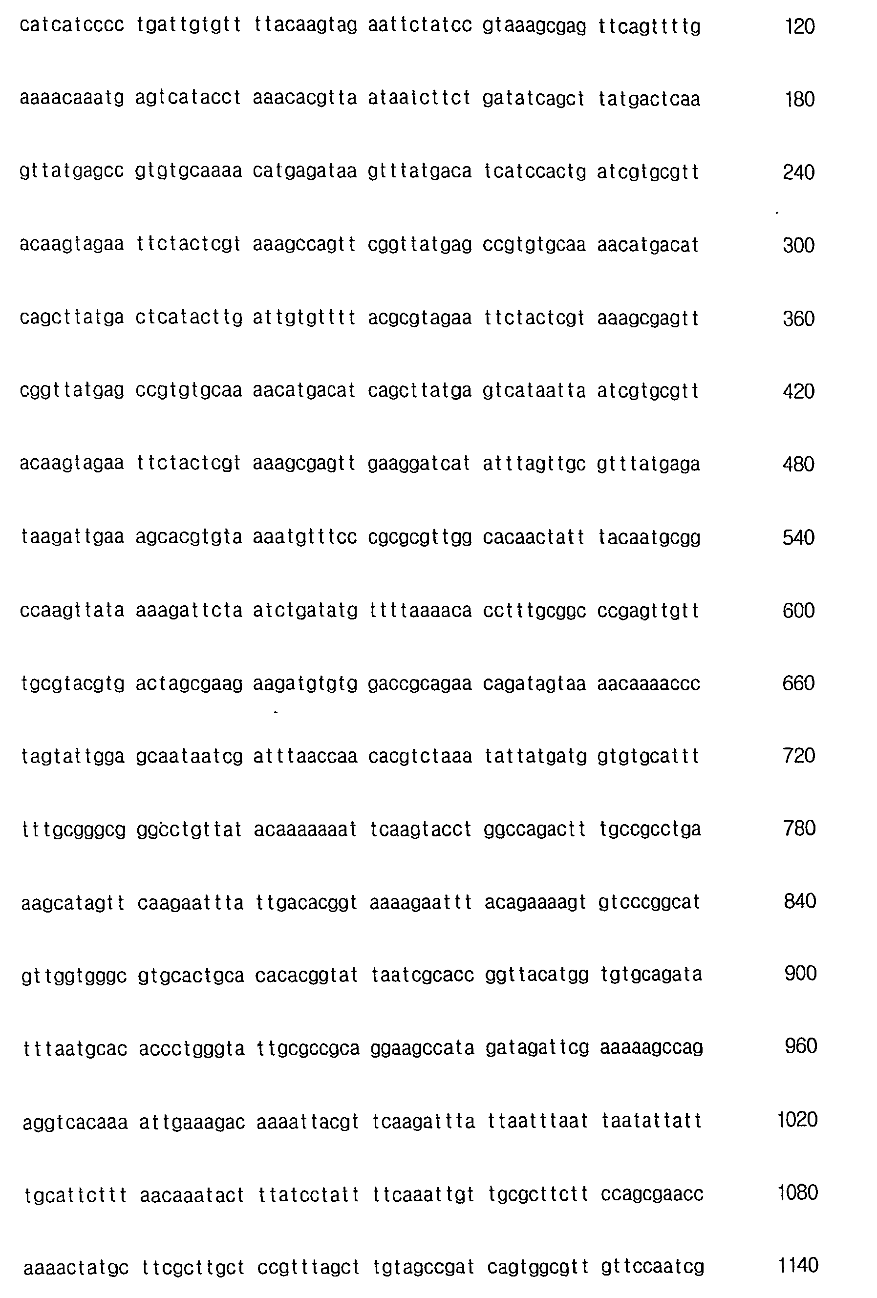
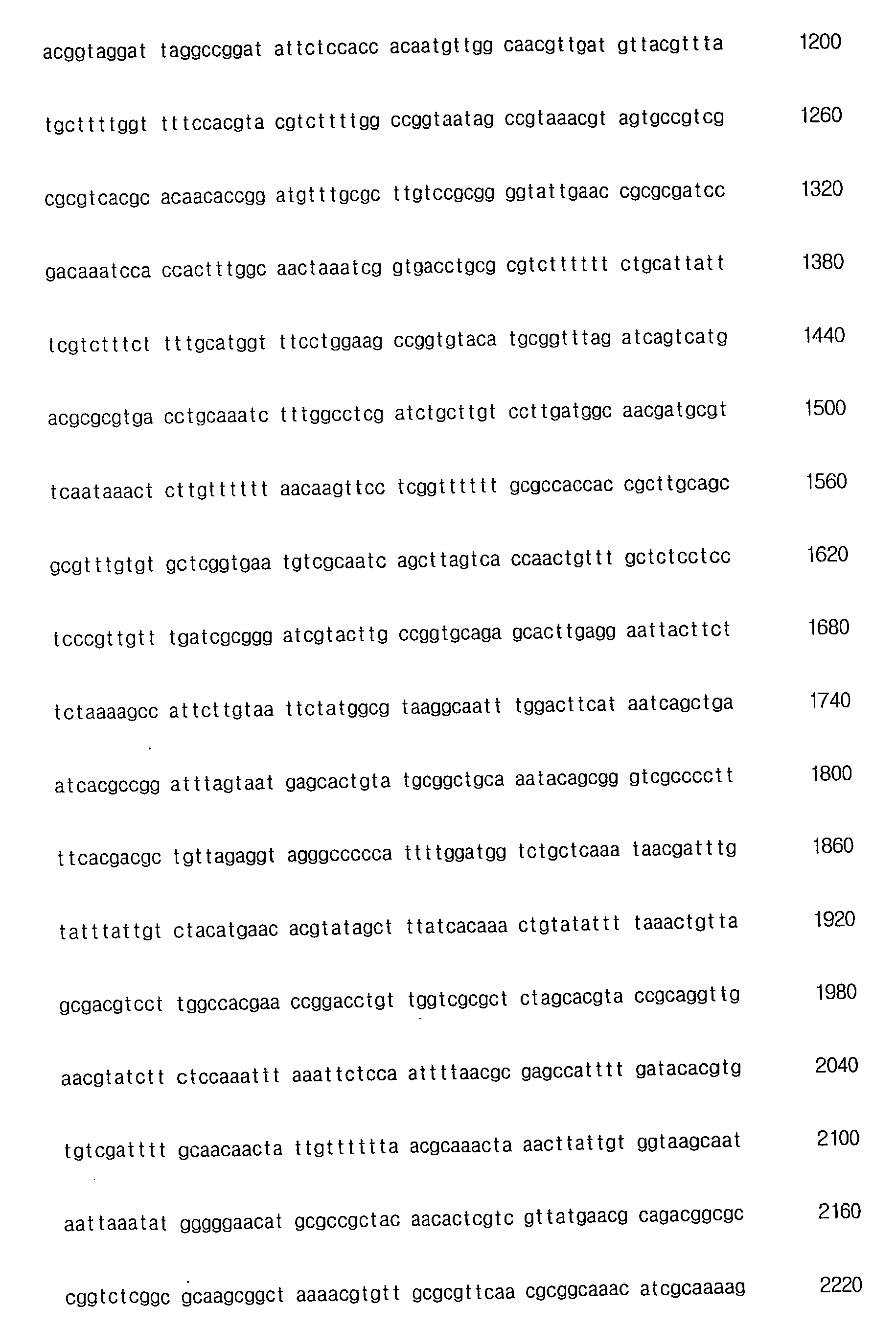

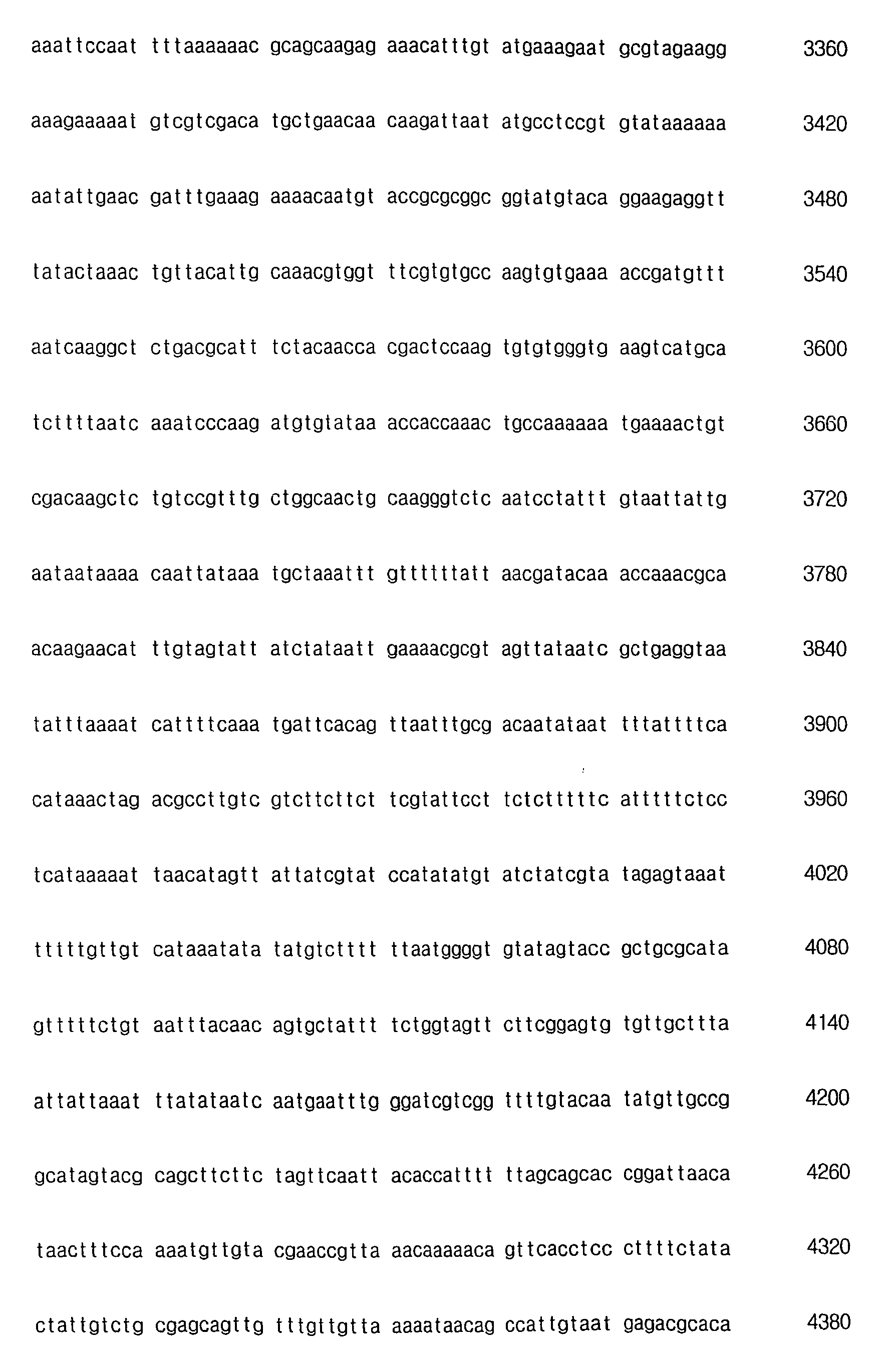
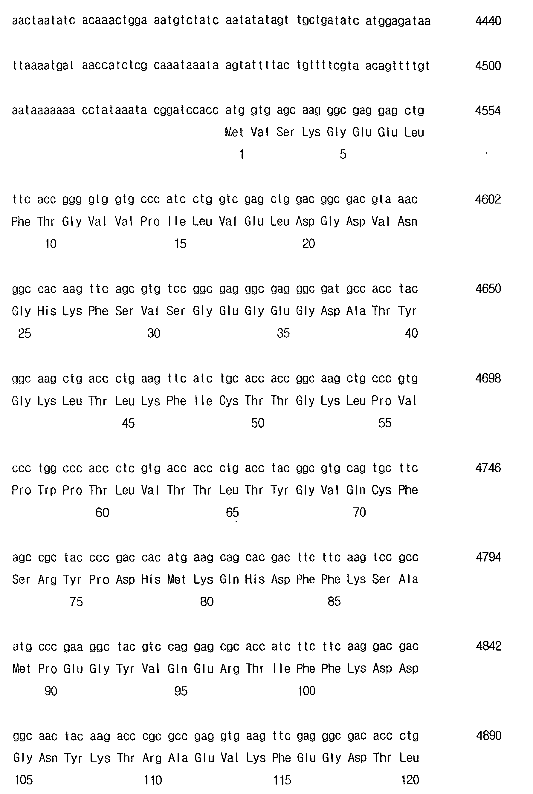

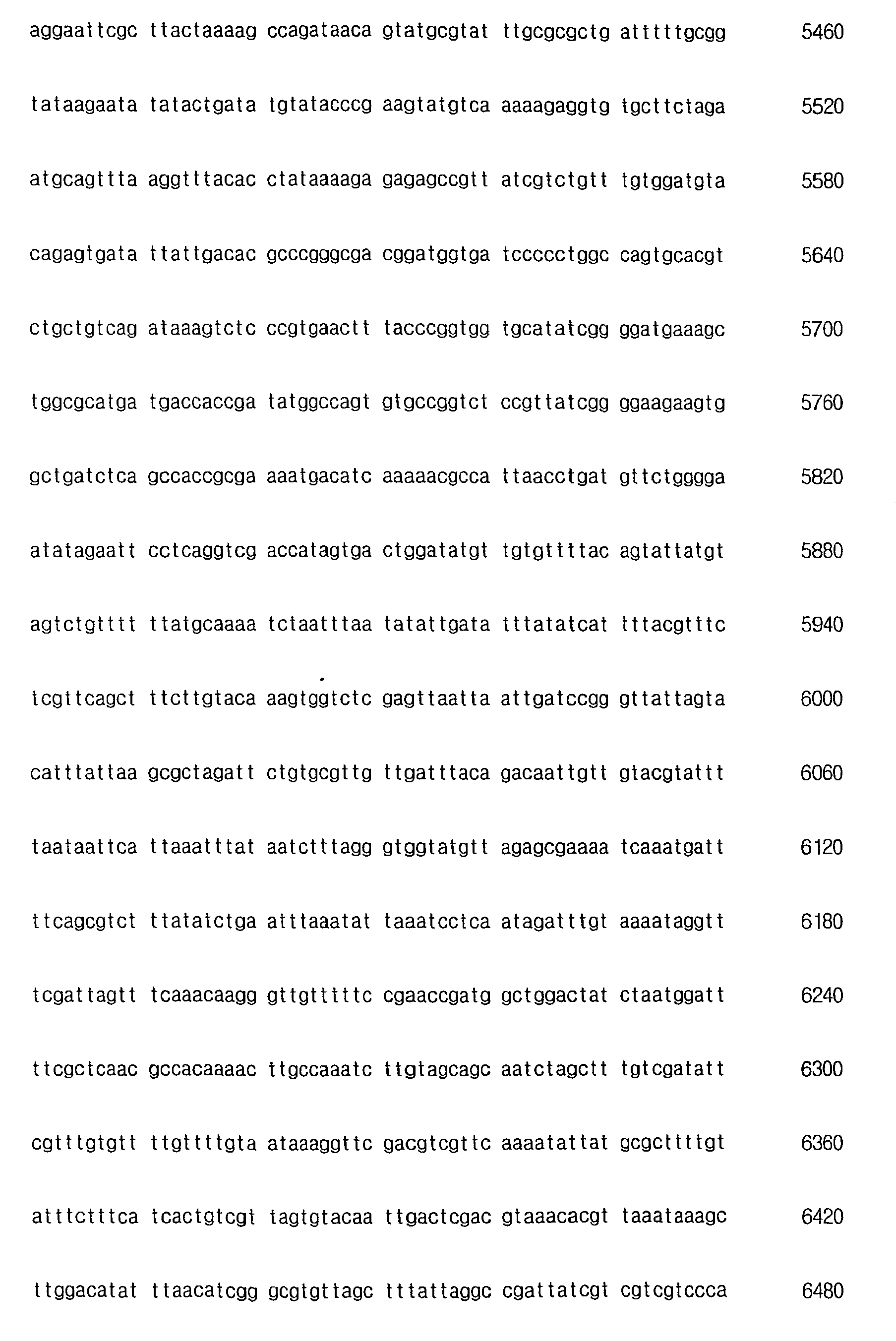


- <210> 6
<211> 21
<212> DNA
<213> Artificial Sequence - <220>
<223> Baculo-Forward primer - <400> 6
actgttttcg taacagtttt g 21 - <210> 7
<211> 19
<212> DNA
<213> Artificial Sequence - <220>
<223> Baculo-Reverse primer - <400> 7
acaacgcaca gaatctagc 19 - <210> 8
<211> 14000
<212> DNA
<213> Artificial Sequence - <220>
<223> Polyhedrin locus of vBacHTS2 - <220>
<221> source
<222> (4687)..(11207)
<223> F pBACe3.6 - <220>
<221> misc_feature
<222> (10307)..(10966)
<223> F CmR - <220>
<221> promoter
<222> (4428)..(4520)
<223> F pPolyhedrin - <220>
<221> misc_feature
<222> (11220)..(11344)
<223> R attR2 - <220>
<221> misc_feature
<222> (4537)..(4661)
<223> F attR1 - <400> 8

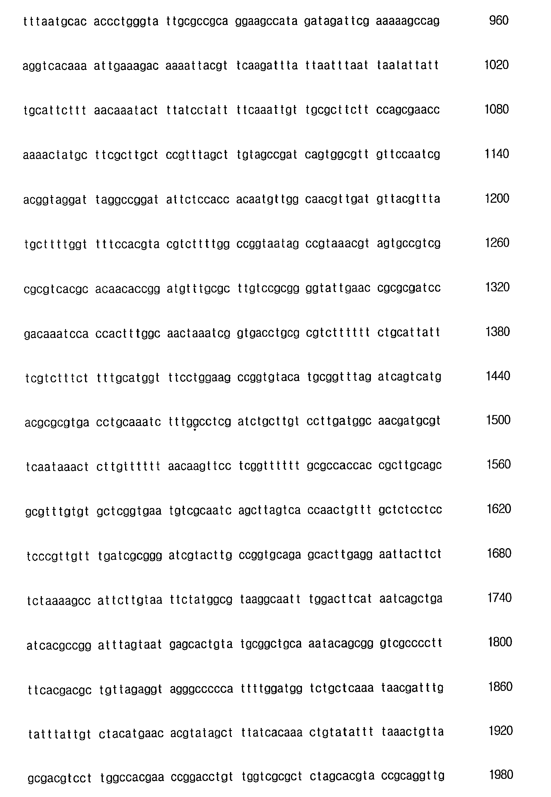


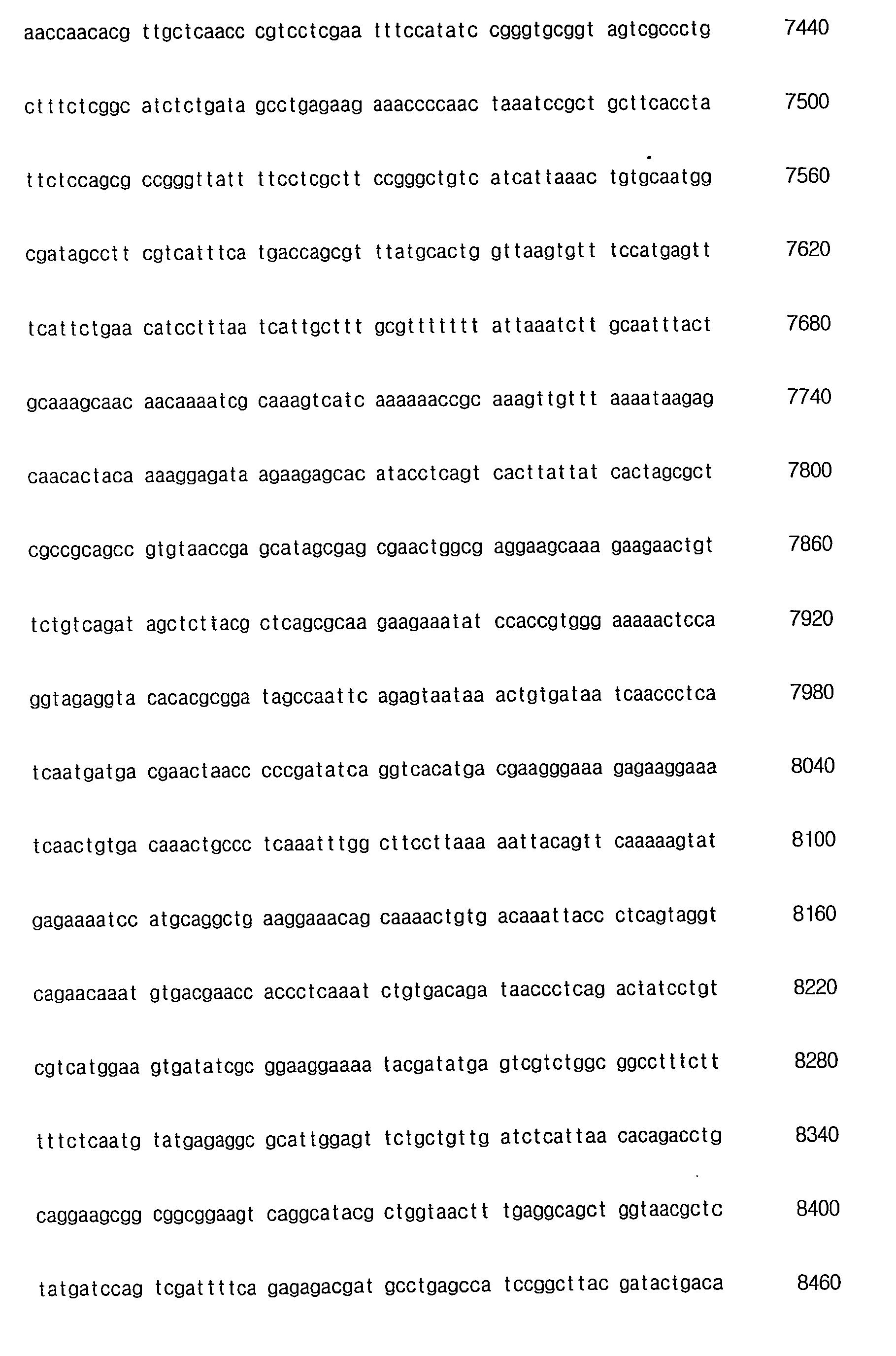


- <210> 9
<211> 13100
<212> DNA
<213> Artificial Sequence - <220>
<223> Polyhedrin locus of vBacHTS2_GFP - <220>
<221> source
<222> (4523)..(5252)
<223> F EGFP - <220>
<221> source
<222> (5404)..(11924)
<223> F pBACe3.6 - <220>
<221> CDS
<222> (4531)..(5247)
<223> F EGFP - <220>
<221> misc_feature
<222> (11024)..(11683)
<223> F CmR - <220>
<221> promoter
<222> (4428)..(4520)
<223> F pPolyhedrin - <220>
<221> misc_feature
<222> (11937)..(12061)
<223> R attR2 - <220>
<221> misc_feature
<222> (5254)..(5378)
<223> F attR1 - <400> 9


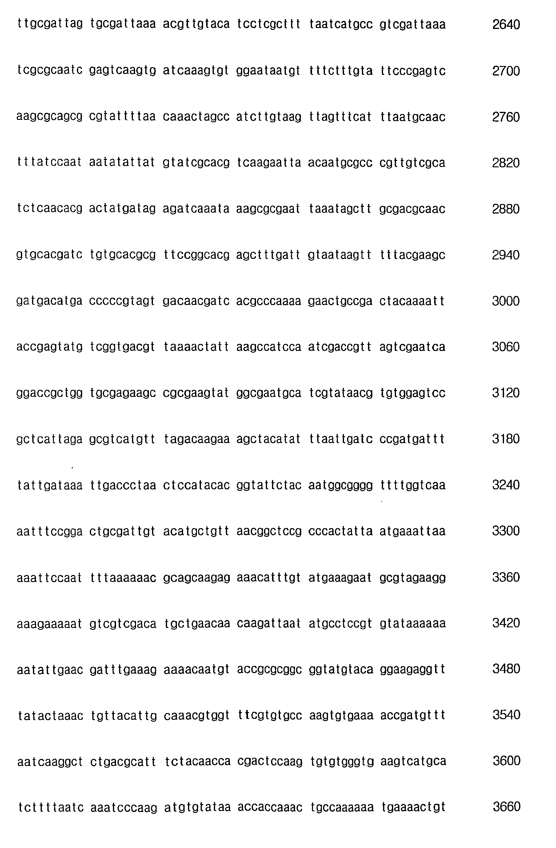
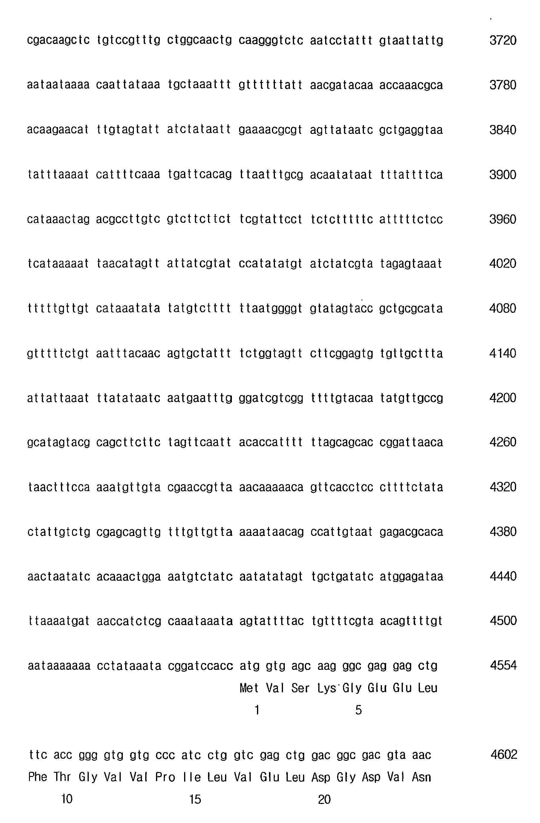


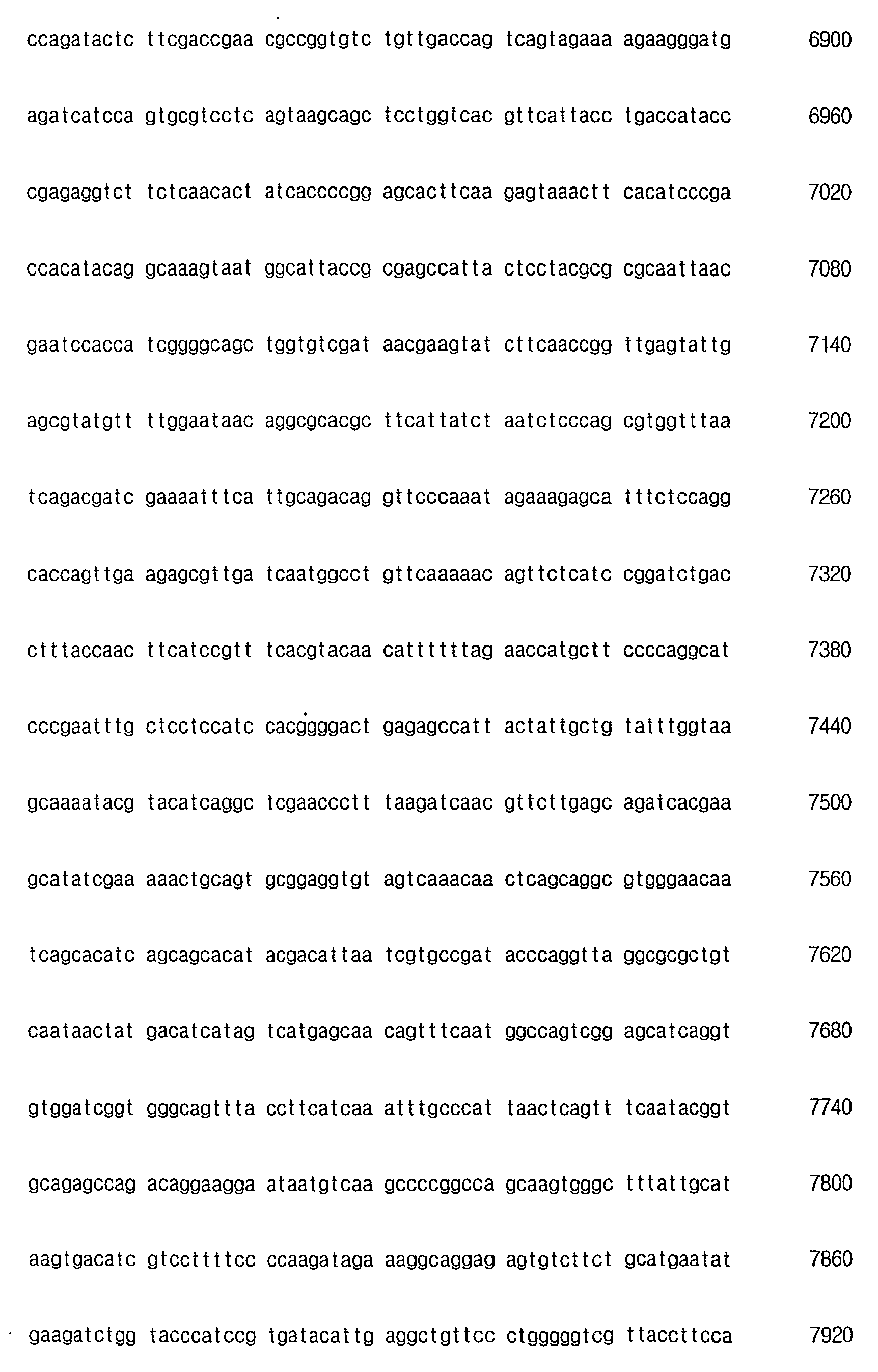


Claims (13)
- A method for preparing expressive circular recombinant viral vectors, characterized in that the method comprises the steps of:i) providing a linearized viral DNA fragment containing at least two site-specific recombination sites and at least one restriction enzyme recognition site;ii) providing a gene cassette containing desired genomic material flanked with at least two site-specific recombination sites for homologous recombination with the linearized viral DNA fragment at the two site specific recombination sites; andiii) reacting the linearized viral DNA fragment, and the gene cassette together under conditions effective to allow in vitro homologous recombination to take place so as to form a recombinant viral vector; andiv) preventing the expression of a viral vector that does not contain the desired genomic material.
- The method for preparing expressive circular recombinant viral vectors as claimed in claim 1, wherein one or more viral DNA fragments are digested with restriction enzymes at one or more sites to generate the linearized DNA fragments.
- The method for preparing expressive circular recombinant viral vectors as claimed in claim 2, wherein said one or more restriction enzyme sites comprise(s) the nucleotide sequences of CCTNAGG.
- The method for preparing expressive circular recombinant viral vectors as claimed in claim 1, wherein the recombinant viral vector is obtained from a group consisting of baculovirus, poliomavirus, papilomavirus and hepatitis B virus (HBV).
- A method of screening recombinant viruses containing desired genomic materials, characterized in that the method comprises the steps of:i) providing a linearised viral DNA fragment containing at least two site-specific recombination sites and at least one restriction enzyme recognition site;ii) providing a gene cassette containing desired genomic material flanked with at least two site-specific recombination sites for homologous recombination with the linearized viral DNA fragment at the two site specific recombination sites;iii) reacting the linearized viral DNA fragment and the gene cassette together under conditions effective to allow the in vitro homologous recombination to take place so as to form a recombinant virus in vitro to produce reaction mixture including expressive circular recombinant viral vectors formed in vitro;iv) preventing the expression of a virus that does not contain the desired genomic material; andv) transferring the reaction mixture of the iii) to host cells in multi-well plate.
- A method of screening recombinant viruses as claimed in claim 4, wherein the method further comprises the step of:vi) identifying the features expressed in host cells.
- The method of screening recombinant viruses as claimed in claim 5 or 6, wherein the virus is baculovis, poliomavirus, papilomavirus, and hepatitis B virus (HBV).
- A method of preparing recombinant viruses having circular genomic DNAs, characterized in that the method comprises the steps of:i) providing a linearised viral DNA fragment containing at least two site-specific recombination sites and at least one restriction enzyme recognition site;ii) providing a gene cassette containing desired genomic material flanked with at least two site-specific recombination sites for homologous recombination with the linearized DNA fragment at the two site specific recombination sites;iii) reacting the linearized viral DNA fragment and the gene cassette to create a homologous recombination in vitro to produce reaction mixture including expressive circular recombinant viral vectors;iv) preventing the expression of a viral vector that does not contain the desired genomic material; andv) transferring the reaction mixture of the iii) to host cells in multi-well plate.
- The method of preparing recombinant viruses having circular genomic DNAs as claimed in claim 8,
wherein the linearized viral DNA fragment is non-infectious to animal cells or cannot replicate, while includes bacterial replication origin and antibiotics resistance sequences. - The method of preparing recombinant viruses having circular genomic DNAs as claimed in claim 8,
wherein the transfer of the reaction mixture to host cells is carried out without the need for a selection process to isolate the circular genomic DNA. - The method of preparing recombinant viruses having circular genomic DNAs as claimed in claim 7, wherein viral genomic DNAs of i) are digested with restriction enzyme at one or more sites to generate the linearized viral DNA fragments.
- The method of preparing Recombinant viruses having circular genomic DNAs as claimed in claim 10, wherein said one or more sites comprise(s) the nucleotide sequences of CCTNAGG.
- The method of preparing recombinant viruses having circular genomic DNAs as claimed in claim 8, wherein the recombinant viral vectors is obtained from a group consisting of baculovirus, poliomavirus, papilomavirus, and hepatitis B virus (HBV).
Applications Claiming Priority (3)
| Application Number | Priority Date | Filing Date | Title |
|---|---|---|---|
| KR20020012634 | 2002-03-09 | ||
| KR2002012634 | 2002-03-09 | ||
| PCT/KR2003/000454 WO2003076606A1 (en) | 2002-03-09 | 2003-03-08 | High throughput system for producing recombinant viruses using site-specific recombination |
Publications (3)
| Publication Number | Publication Date |
|---|---|
| EP1483376A1 EP1483376A1 (en) | 2004-12-08 |
| EP1483376A4 EP1483376A4 (en) | 2005-08-10 |
| EP1483376B1 true EP1483376B1 (en) | 2008-10-15 |
Family
ID=27800660
Family Applications (1)
| Application Number | Title | Priority Date | Filing Date |
|---|---|---|---|
| EP03744066A Expired - Lifetime EP1483376B1 (en) | 2002-03-09 | 2003-03-08 | High throughput system for producing recombinant viruses using site-specific recombination |
Country Status (9)
| Country | Link |
|---|---|
| US (1) | US7319001B2 (en) |
| EP (1) | EP1483376B1 (en) |
| JP (2) | JP4587671B2 (en) |
| KR (1) | KR100631429B1 (en) |
| CN (1) | CN100564520C (en) |
| AT (1) | ATE411381T1 (en) |
| AU (1) | AU2003210034A1 (en) |
| DE (1) | DE60324108D1 (en) |
| WO (1) | WO2003076606A1 (en) |
Families Citing this family (4)
| Publication number | Priority date | Publication date | Assignee | Title |
|---|---|---|---|---|
| EP2801614B1 (en) * | 2002-08-29 | 2019-05-15 | The Board of Trustees of the Leland Stanford Junior University | Circular nucleic acid vectors and methods for making and using the same |
| GB0424356D0 (en) * | 2004-11-03 | 2004-12-08 | Ark Therapeutics Ltd | Baculovirus-based gene libraries |
| GB0710535D0 (en) * | 2007-06-01 | 2007-07-11 | Ark Therapeutics Ltd | Baculovirus generation |
| CN103146753B (en) * | 2013-03-18 | 2013-12-18 | 山东维真生物科技有限公司 | Adenovirus vector and production method for same |
Family Cites Families (8)
| Publication number | Priority date | Publication date | Assignee | Title |
|---|---|---|---|---|
| CA2039921A1 (en) | 1990-04-16 | 1991-10-17 | Xandra O. Breakefield | Transfer and expression of gene sequences into central nervous system cells using herpes simplex virus mutants with deletions in genes for viral replication |
| US5348886A (en) * | 1992-09-04 | 1994-09-20 | Monsanto Company | Method of producing recombinant eukaryotic viruses in bacteria |
| CA2144040A1 (en) | 1993-07-13 | 1995-01-26 | Michel Perricaudet | Defective adenovirus vectors and use thereof in gene therapy |
| US6143557A (en) * | 1995-06-07 | 2000-11-07 | Life Technologies, Inc. | Recombination cloning using engineered recombination sites |
| US5851808A (en) * | 1997-02-28 | 1998-12-22 | Baylor College Of Medicine | Rapid subcloning using site-specific recombination |
| US6110461A (en) | 1997-08-13 | 2000-08-29 | Oncolytics Biotech Inc. | Reovirus for the treatment of neoplasia |
| ATE391789T1 (en) * | 1999-12-10 | 2008-04-15 | Invitrogen Corp | USE OF A VARIETY OF RECOMBINATION SITE WITH UNIQUE SPECIFICITY IN RECOMBINATORY CLONING |
| US6406863B1 (en) * | 2000-06-23 | 2002-06-18 | Genetastix Corporation | High throughput generation and screening of fully human antibody repertoire in yeast |
-
2003
- 2003-03-07 US US10/383,838 patent/US7319001B2/en not_active Expired - Lifetime
- 2003-03-08 AU AU2003210034A patent/AU2003210034A1/en not_active Abandoned
- 2003-03-08 EP EP03744066A patent/EP1483376B1/en not_active Expired - Lifetime
- 2003-03-08 WO PCT/KR2003/000454 patent/WO2003076606A1/en not_active Ceased
- 2003-03-08 JP JP2003574813A patent/JP4587671B2/en not_active Expired - Fee Related
- 2003-03-08 CN CNB038056437A patent/CN100564520C/en not_active Expired - Fee Related
- 2003-03-08 KR KR1020047014160A patent/KR100631429B1/en not_active Expired - Fee Related
- 2003-03-08 AT AT03744066T patent/ATE411381T1/en not_active IP Right Cessation
- 2003-03-08 DE DE60324108T patent/DE60324108D1/en not_active Expired - Lifetime
-
2010
- 2010-04-15 JP JP2010094517A patent/JP2010207231A/en not_active Abandoned
Also Published As
| Publication number | Publication date |
|---|---|
| ATE411381T1 (en) | 2008-10-15 |
| US20040185565A1 (en) | 2004-09-23 |
| WO2003076606A1 (en) | 2003-09-18 |
| JP2005519604A (en) | 2005-07-07 |
| DE60324108D1 (en) | 2008-11-27 |
| EP1483376A1 (en) | 2004-12-08 |
| KR100631429B1 (en) | 2006-10-11 |
| JP4587671B2 (en) | 2010-11-24 |
| CN100564520C (en) | 2009-12-02 |
| US7319001B2 (en) | 2008-01-15 |
| JP2010207231A (en) | 2010-09-24 |
| KR20040089723A (en) | 2004-10-21 |
| AU2003210034A1 (en) | 2003-09-22 |
| EP1483376A4 (en) | 2005-08-10 |
| CN1639326A (en) | 2005-07-13 |
Similar Documents
| Publication | Publication Date | Title |
|---|---|---|
| US6225060B1 (en) | Baculovirus expression system and method for high throughput expression of genetic material | |
| Lo et al. | Rapid titer determination of baculovirus by quantitative real‐time polymerase chain reaction | |
| US6291214B1 (en) | System for generating recombinant viruses | |
| US6953689B2 (en) | Cloning system for construction of recombinant expression vectors | |
| JP4597188B2 (en) | New expression tools for multiprotein applications | |
| Ho Je et al. | A defective viral genome maintained in Escherichia coli for the generation of baculovirus expression vectors | |
| Ciccarone et al. | Generation of recombinant baculovirus DNA in E. coli using a baculovirus shuttle vector | |
| EP1483375B1 (en) | A method for producing recombinant adenovirus viruses using site-specific recombination | |
| US20090176660A1 (en) | Engineered Baculoviruses and Their Use | |
| JP2010207231A (en) | Highly productive mechanism for producing recombinant virus by using site-specific recombination | |
| AU2003212521B2 (en) | Engineered baculoviruses and their use | |
| CN102286534A (en) | Insect bioreactor capable of expressing multiple exogenous genes, and construction method and application thereof | |
| de Jong et al. | Scarless baculovirus genome editing using lambda-red recombineering in E. coli | |
| US6806064B2 (en) | Baculovirus enhancer-like sequence | |
| Yao et al. | Construction of recombinant vaccinia viruses using leporipoxvirus-catalyzed recombination and reactivation of orthopoxvirus DNA | |
| Possee et al. | Baculovirus transfer vectors | |
| Laudermilch et al. | MAVERICC: Efficient marker-free rescue of vaccinia virus recombinants by in vitro CRISPR-Cas9 engineering | |
| Chen et al. | Analysis of BmNPV orf101 disruption: orf101 is essential for mediating budded virus production | |
| WO2026013698A2 (en) | Baculovirus expression systems and methods thereof | |
| Putman | Evaluating Modified Polyhedrin Promoters for Improved Co-Expression of KRAS4b | |
| Graves et al. | Unit Title | |
| Hayes | Baculovirus Production Methods For Reliable Isolation of Recombinant Baculovirus Engineered For Modified Actin Expression | |
| Powichrowski et al. | Probing Baculovirus Vector Gene Essentiality for Foreign Gene Expression Using a CRISPR-Cas9 System | |
| CN1242428A (en) | Gene engineering carrier for recombination salvation linearization silk worm nuclear polyhedral virus |
Legal Events
| Date | Code | Title | Description |
|---|---|---|---|
| PUAI | Public reference made under article 153(3) epc to a published international application that has entered the european phase |
Free format text: ORIGINAL CODE: 0009012 |
|
| 17P | Request for examination filed |
Effective date: 20040830 |
|
| AK | Designated contracting states |
Kind code of ref document: A1 Designated state(s): AT BE BG CH CY CZ DE DK EE ES FI FR GB GR HU IE IT LI LU MC NL PT RO SE SI SK TR |
|
| AX | Request for extension of the european patent |
Extension state: AL LT LV MK |
|
| RIN1 | Information on inventor provided before grant (corrected) |
Inventor name: YI, YONG-WEON Inventor name: KIM, MIN-SUNG Inventor name: CHOI, EUN-WOOK Inventor name: SEEN, DONG-SEUNG Inventor name: JUNG, NEON C. Inventor name: KIM, DO-HUI Inventor name: KIM, KYUNG-JIN Inventor name: SON, HO-SUN |
|
| A4 | Supplementary search report drawn up and despatched |
Effective date: 20050629 |
|
| RIC1 | Information provided on ipc code assigned before grant |
Ipc: 7C 12N 15/86 B Ipc: 7C 12N 7/01 A |
|
| 17Q | First examination report despatched |
Effective date: 20060215 |
|
| GRAP | Despatch of communication of intention to grant a patent |
Free format text: ORIGINAL CODE: EPIDOSNIGR1 |
|
| GRAS | Grant fee paid |
Free format text: ORIGINAL CODE: EPIDOSNIGR3 |
|
| GRAA | (expected) grant |
Free format text: ORIGINAL CODE: 0009210 |
|
| RAP1 | Party data changed (applicant data changed or rights of an application transferred) |
Owner name: NEWGEX INC. |
|
| AK | Designated contracting states |
Kind code of ref document: B1 Designated state(s): AT BE BG CH CY CZ DE DK EE ES FI FR GB GR HU IE IT LI LU MC NL PT RO SE SI SK TR |
|
| REG | Reference to a national code |
Ref country code: GB Ref legal event code: FG4D Ref country code: CH Ref legal event code: EP |
|
| REG | Reference to a national code |
Ref country code: IE Ref legal event code: FG4D |
|
| REF | Corresponds to: |
Ref document number: 60324108 Country of ref document: DE Date of ref document: 20081127 Kind code of ref document: P |
|
| REG | Reference to a national code |
Ref country code: CH Ref legal event code: NV Representative=s name: ISLER & PEDRAZZINI AG |
|
| NLV1 | Nl: lapsed or annulled due to failure to fulfill the requirements of art. 29p and 29m of the patents act | ||
| PG25 | Lapsed in a contracting state [announced via postgrant information from national office to epo] |
Ref country code: BG Free format text: LAPSE BECAUSE OF FAILURE TO SUBMIT A TRANSLATION OF THE DESCRIPTION OR TO PAY THE FEE WITHIN THE PRESCRIBED TIME-LIMIT Effective date: 20090115 Ref country code: ES Free format text: LAPSE BECAUSE OF FAILURE TO SUBMIT A TRANSLATION OF THE DESCRIPTION OR TO PAY THE FEE WITHIN THE PRESCRIBED TIME-LIMIT Effective date: 20090126 Ref country code: AT Free format text: LAPSE BECAUSE OF FAILURE TO SUBMIT A TRANSLATION OF THE DESCRIPTION OR TO PAY THE FEE WITHIN THE PRESCRIBED TIME-LIMIT Effective date: 20081015 |
|
| PG25 | Lapsed in a contracting state [announced via postgrant information from national office to epo] |
Ref country code: NL Free format text: LAPSE BECAUSE OF FAILURE TO SUBMIT A TRANSLATION OF THE DESCRIPTION OR TO PAY THE FEE WITHIN THE PRESCRIBED TIME-LIMIT Effective date: 20081015 Ref country code: FI Free format text: LAPSE BECAUSE OF FAILURE TO SUBMIT A TRANSLATION OF THE DESCRIPTION OR TO PAY THE FEE WITHIN THE PRESCRIBED TIME-LIMIT Effective date: 20081015 Ref country code: SI Free format text: LAPSE BECAUSE OF FAILURE TO SUBMIT A TRANSLATION OF THE DESCRIPTION OR TO PAY THE FEE WITHIN THE PRESCRIBED TIME-LIMIT Effective date: 20081015 Ref country code: PT Free format text: LAPSE BECAUSE OF FAILURE TO SUBMIT A TRANSLATION OF THE DESCRIPTION OR TO PAY THE FEE WITHIN THE PRESCRIBED TIME-LIMIT Effective date: 20090316 |
|
| PG25 | Lapsed in a contracting state [announced via postgrant information from national office to epo] |
Ref country code: DK Free format text: LAPSE BECAUSE OF FAILURE TO SUBMIT A TRANSLATION OF THE DESCRIPTION OR TO PAY THE FEE WITHIN THE PRESCRIBED TIME-LIMIT Effective date: 20081015 Ref country code: EE Free format text: LAPSE BECAUSE OF FAILURE TO SUBMIT A TRANSLATION OF THE DESCRIPTION OR TO PAY THE FEE WITHIN THE PRESCRIBED TIME-LIMIT Effective date: 20081015 Ref country code: BE Free format text: LAPSE BECAUSE OF FAILURE TO SUBMIT A TRANSLATION OF THE DESCRIPTION OR TO PAY THE FEE WITHIN THE PRESCRIBED TIME-LIMIT Effective date: 20081015 Ref country code: RO Free format text: LAPSE BECAUSE OF FAILURE TO SUBMIT A TRANSLATION OF THE DESCRIPTION OR TO PAY THE FEE WITHIN THE PRESCRIBED TIME-LIMIT Effective date: 20081015 |
|
| PLBE | No opposition filed within time limit |
Free format text: ORIGINAL CODE: 0009261 |
|
| STAA | Information on the status of an ep patent application or granted ep patent |
Free format text: STATUS: NO OPPOSITION FILED WITHIN TIME LIMIT |
|
| PG25 | Lapsed in a contracting state [announced via postgrant information from national office to epo] |
Ref country code: CZ Free format text: LAPSE BECAUSE OF FAILURE TO SUBMIT A TRANSLATION OF THE DESCRIPTION OR TO PAY THE FEE WITHIN THE PRESCRIBED TIME-LIMIT Effective date: 20081015 Ref country code: IT Free format text: LAPSE BECAUSE OF FAILURE TO SUBMIT A TRANSLATION OF THE DESCRIPTION OR TO PAY THE FEE WITHIN THE PRESCRIBED TIME-LIMIT Effective date: 20081015 Ref country code: SE Free format text: LAPSE BECAUSE OF FAILURE TO SUBMIT A TRANSLATION OF THE DESCRIPTION OR TO PAY THE FEE WITHIN THE PRESCRIBED TIME-LIMIT Effective date: 20090115 |
|
| 26N | No opposition filed |
Effective date: 20090716 |
|
| PG25 | Lapsed in a contracting state [announced via postgrant information from national office to epo] |
Ref country code: SK Free format text: LAPSE BECAUSE OF FAILURE TO SUBMIT A TRANSLATION OF THE DESCRIPTION OR TO PAY THE FEE WITHIN THE PRESCRIBED TIME-LIMIT Effective date: 20081015 |
|
| PG25 | Lapsed in a contracting state [announced via postgrant information from national office to epo] |
Ref country code: MC Free format text: LAPSE BECAUSE OF NON-PAYMENT OF DUE FEES Effective date: 20090331 |
|
| PG25 | Lapsed in a contracting state [announced via postgrant information from national office to epo] |
Ref country code: IE Free format text: LAPSE BECAUSE OF NON-PAYMENT OF DUE FEES Effective date: 20090308 |
|
| PG25 | Lapsed in a contracting state [announced via postgrant information from national office to epo] |
Ref country code: GR Free format text: LAPSE BECAUSE OF FAILURE TO SUBMIT A TRANSLATION OF THE DESCRIPTION OR TO PAY THE FEE WITHIN THE PRESCRIBED TIME-LIMIT Effective date: 20090116 |
|
| PG25 | Lapsed in a contracting state [announced via postgrant information from national office to epo] |
Ref country code: LU Free format text: LAPSE BECAUSE OF NON-PAYMENT OF DUE FEES Effective date: 20090308 |
|
| PG25 | Lapsed in a contracting state [announced via postgrant information from national office to epo] |
Ref country code: HU Free format text: LAPSE BECAUSE OF FAILURE TO SUBMIT A TRANSLATION OF THE DESCRIPTION OR TO PAY THE FEE WITHIN THE PRESCRIBED TIME-LIMIT Effective date: 20090416 |
|
| PG25 | Lapsed in a contracting state [announced via postgrant information from national office to epo] |
Ref country code: TR Free format text: LAPSE BECAUSE OF FAILURE TO SUBMIT A TRANSLATION OF THE DESCRIPTION OR TO PAY THE FEE WITHIN THE PRESCRIBED TIME-LIMIT Effective date: 20081015 |
|
| PG25 | Lapsed in a contracting state [announced via postgrant information from national office to epo] |
Ref country code: CY Free format text: LAPSE BECAUSE OF FAILURE TO SUBMIT A TRANSLATION OF THE DESCRIPTION OR TO PAY THE FEE WITHIN THE PRESCRIBED TIME-LIMIT Effective date: 20081015 |
|
| REG | Reference to a national code |
Ref country code: FR Ref legal event code: PLFP Year of fee payment: 13 |
|
| REG | Reference to a national code |
Ref country code: FR Ref legal event code: PLFP Year of fee payment: 14 |
|
| REG | Reference to a national code |
Ref country code: FR Ref legal event code: PLFP Year of fee payment: 15 |
|
| PGFP | Annual fee paid to national office [announced via postgrant information from national office to epo] |
Ref country code: DE Payment date: 20170322 Year of fee payment: 15 Ref country code: CH Payment date: 20170322 Year of fee payment: 15 Ref country code: FR Payment date: 20170322 Year of fee payment: 15 |
|
| PGFP | Annual fee paid to national office [announced via postgrant information from national office to epo] |
Ref country code: GB Payment date: 20170322 Year of fee payment: 15 |
|
| REG | Reference to a national code |
Ref country code: DE Ref legal event code: R119 Ref document number: 60324108 Country of ref document: DE |
|
| REG | Reference to a national code |
Ref country code: CH Ref legal event code: PL |
|
| GBPC | Gb: european patent ceased through non-payment of renewal fee |
Effective date: 20180308 |
|
| PG25 | Lapsed in a contracting state [announced via postgrant information from national office to epo] |
Ref country code: DE Free format text: LAPSE BECAUSE OF NON-PAYMENT OF DUE FEES Effective date: 20181002 |
|
| PG25 | Lapsed in a contracting state [announced via postgrant information from national office to epo] |
Ref country code: CH Free format text: LAPSE BECAUSE OF NON-PAYMENT OF DUE FEES Effective date: 20180331 Ref country code: LI Free format text: LAPSE BECAUSE OF NON-PAYMENT OF DUE FEES Effective date: 20180331 Ref country code: GB Free format text: LAPSE BECAUSE OF NON-PAYMENT OF DUE FEES Effective date: 20180308 |
|
| PG25 | Lapsed in a contracting state [announced via postgrant information from national office to epo] |
Ref country code: FR Free format text: LAPSE BECAUSE OF NON-PAYMENT OF DUE FEES Effective date: 20180331 |