EP1470821A2 - Cell therapy method for the treatment of tumours - Google Patents
Cell therapy method for the treatment of tumours Download PDFInfo
- Publication number
- EP1470821A2 EP1470821A2 EP03257064A EP03257064A EP1470821A2 EP 1470821 A2 EP1470821 A2 EP 1470821A2 EP 03257064 A EP03257064 A EP 03257064A EP 03257064 A EP03257064 A EP 03257064A EP 1470821 A2 EP1470821 A2 EP 1470821A2
- Authority
- EP
- European Patent Office
- Prior art keywords
- cells
- melanoma
- antigen
- peripheral blood
- subject
- Prior art date
- Legal status (The legal status is an assumption and is not a legal conclusion. Google has not performed a legal analysis and makes no representation as to the accuracy of the status listed.)
- Withdrawn
Links
Images















Classifications
-
- C—CHEMISTRY; METALLURGY
- C12—BIOCHEMISTRY; BEER; SPIRITS; WINE; VINEGAR; MICROBIOLOGY; ENZYMOLOGY; MUTATION OR GENETIC ENGINEERING
- C12N—MICROORGANISMS OR ENZYMES; COMPOSITIONS THEREOF; PROPAGATING, PRESERVING, OR MAINTAINING MICROORGANISMS; MUTATION OR GENETIC ENGINEERING; CULTURE MEDIA
- C12N5/00—Undifferentiated human, animal or plant cells, e.g. cell lines; Tissues; Cultivation or maintenance thereof; Culture media therefor
- C12N5/06—Animal cells or tissues; Human cells or tissues
- C12N5/0602—Vertebrate cells
- C12N5/0634—Cells from the blood or the immune system
- C12N5/0636—T lymphocytes
-
- A—HUMAN NECESSITIES
- A61—MEDICAL OR VETERINARY SCIENCE; HYGIENE
- A61K—PREPARATIONS FOR MEDICAL, DENTAL OR TOILETRY PURPOSES
- A61K38/00—Medicinal preparations containing peptides
- A61K38/16—Peptides having more than 20 amino acids; Gastrins; Somatostatins; Melanotropins; Derivatives thereof
- A61K38/17—Peptides having more than 20 amino acids; Gastrins; Somatostatins; Melanotropins; Derivatives thereof from animals; from humans
- A61K38/19—Cytokines; Lymphokines; Interferons
- A61K38/20—Interleukins [IL]
- A61K38/2013—IL-2
-
- A—HUMAN NECESSITIES
- A61—MEDICAL OR VETERINARY SCIENCE; HYGIENE
- A61K—PREPARATIONS FOR MEDICAL, DENTAL OR TOILETRY PURPOSES
- A61K38/00—Medicinal preparations containing peptides
- A61K38/16—Peptides having more than 20 amino acids; Gastrins; Somatostatins; Melanotropins; Derivatives thereof
- A61K38/17—Peptides having more than 20 amino acids; Gastrins; Somatostatins; Melanotropins; Derivatives thereof from animals; from humans
- A61K38/19—Cytokines; Lymphokines; Interferons
- A61K38/21—Interferons [IFN]
- A61K38/212—IFN-alpha
-
- A—HUMAN NECESSITIES
- A61—MEDICAL OR VETERINARY SCIENCE; HYGIENE
- A61K—PREPARATIONS FOR MEDICAL, DENTAL OR TOILETRY PURPOSES
- A61K40/00—Cellular immunotherapy
- A61K40/10—Cellular immunotherapy characterised by the cell type used
- A61K40/11—T-cells, e.g. tumour infiltrating lymphocytes [TIL] or regulatory T [Treg] cells; Lymphokine-activated killer [LAK] cells
-
- A—HUMAN NECESSITIES
- A61—MEDICAL OR VETERINARY SCIENCE; HYGIENE
- A61K—PREPARATIONS FOR MEDICAL, DENTAL OR TOILETRY PURPOSES
- A61K40/00—Cellular immunotherapy
- A61K40/30—Cellular immunotherapy characterised by the recombinant expression of specific molecules in the cells of the immune system
- A61K40/35—Cytokines
-
- A—HUMAN NECESSITIES
- A61—MEDICAL OR VETERINARY SCIENCE; HYGIENE
- A61K—PREPARATIONS FOR MEDICAL, DENTAL OR TOILETRY PURPOSES
- A61K40/00—Cellular immunotherapy
- A61K40/40—Cellular immunotherapy characterised by antigens that are targeted or presented by cells of the immune system
- A61K40/41—Vertebrate antigens
- A61K40/42—Cancer antigens
- A61K40/4202—Receptors, cell surface antigens or cell surface determinants
- A61K40/4203—Receptors for growth factors
- A61K40/4204—Epidermal growth factor receptors [EGFR]
-
- A—HUMAN NECESSITIES
- A61—MEDICAL OR VETERINARY SCIENCE; HYGIENE
- A61K—PREPARATIONS FOR MEDICAL, DENTAL OR TOILETRY PURPOSES
- A61K40/00—Cellular immunotherapy
- A61K40/40—Cellular immunotherapy characterised by antigens that are targeted or presented by cells of the immune system
- A61K40/41—Vertebrate antigens
- A61K40/42—Cancer antigens
- A61K40/4202—Receptors, cell surface antigens or cell surface determinants
- A61K40/4203—Receptors for growth factors
- A61K40/4205—Her-2/neu/ErbB2, Her-3/ErbB3 or Her 4/ ErbB4
-
- A—HUMAN NECESSITIES
- A61—MEDICAL OR VETERINARY SCIENCE; HYGIENE
- A61K—PREPARATIONS FOR MEDICAL, DENTAL OR TOILETRY PURPOSES
- A61K40/00—Cellular immunotherapy
- A61K40/40—Cellular immunotherapy characterised by antigens that are targeted or presented by cells of the immune system
- A61K40/41—Vertebrate antigens
- A61K40/42—Cancer antigens
- A61K40/4202—Receptors, cell surface antigens or cell surface determinants
- A61K40/4214—Receptors for cytokines
-
- A—HUMAN NECESSITIES
- A61—MEDICAL OR VETERINARY SCIENCE; HYGIENE
- A61K—PREPARATIONS FOR MEDICAL, DENTAL OR TOILETRY PURPOSES
- A61K40/00—Cellular immunotherapy
- A61K40/40—Cellular immunotherapy characterised by antigens that are targeted or presented by cells of the immune system
- A61K40/41—Vertebrate antigens
- A61K40/42—Cancer antigens
- A61K40/4202—Receptors, cell surface antigens or cell surface determinants
- A61K40/4224—Molecules with a "CD" designation not provided for elsewhere
-
- A—HUMAN NECESSITIES
- A61—MEDICAL OR VETERINARY SCIENCE; HYGIENE
- A61K—PREPARATIONS FOR MEDICAL, DENTAL OR TOILETRY PURPOSES
- A61K40/00—Cellular immunotherapy
- A61K40/40—Cellular immunotherapy characterised by antigens that are targeted or presented by cells of the immune system
- A61K40/41—Vertebrate antigens
- A61K40/42—Cancer antigens
- A61K40/4231—Cytokines
- A61K40/4235—Interferons [IFN]
-
- A—HUMAN NECESSITIES
- A61—MEDICAL OR VETERINARY SCIENCE; HYGIENE
- A61K—PREPARATIONS FOR MEDICAL, DENTAL OR TOILETRY PURPOSES
- A61K40/00—Cellular immunotherapy
- A61K40/40—Cellular immunotherapy characterised by antigens that are targeted or presented by cells of the immune system
- A61K40/41—Vertebrate antigens
- A61K40/42—Cancer antigens
- A61K40/4244—Enzymes
-
- A—HUMAN NECESSITIES
- A61—MEDICAL OR VETERINARY SCIENCE; HYGIENE
- A61K—PREPARATIONS FOR MEDICAL, DENTAL OR TOILETRY PURPOSES
- A61K40/00—Cellular immunotherapy
- A61K40/40—Cellular immunotherapy characterised by antigens that are targeted or presented by cells of the immune system
- A61K40/41—Vertebrate antigens
- A61K40/42—Cancer antigens
- A61K40/4256—Tumor associated carbohydrates
- A61K40/4257—Mucins, e.g. MUC-1
-
- A—HUMAN NECESSITIES
- A61—MEDICAL OR VETERINARY SCIENCE; HYGIENE
- A61K—PREPARATIONS FOR MEDICAL, DENTAL OR TOILETRY PURPOSES
- A61K40/00—Cellular immunotherapy
- A61K40/40—Cellular immunotherapy characterised by antigens that are targeted or presented by cells of the immune system
- A61K40/41—Vertebrate antigens
- A61K40/42—Cancer antigens
- A61K40/4264—Cancer antigens from embryonic or fetal origin
- A61K40/4266—Carcinoembryonic antigen [CEA]
-
- A—HUMAN NECESSITIES
- A61—MEDICAL OR VETERINARY SCIENCE; HYGIENE
- A61K—PREPARATIONS FOR MEDICAL, DENTAL OR TOILETRY PURPOSES
- A61K40/00—Cellular immunotherapy
- A61K40/40—Cellular immunotherapy characterised by antigens that are targeted or presented by cells of the immune system
- A61K40/41—Vertebrate antigens
- A61K40/42—Cancer antigens
- A61K40/4267—Cancer testis antigens, e.g. SSX, BAGE, GAGE or SAGE
- A61K40/4268—MAGE
-
- A—HUMAN NECESSITIES
- A61—MEDICAL OR VETERINARY SCIENCE; HYGIENE
- A61K—PREPARATIONS FOR MEDICAL, DENTAL OR TOILETRY PURPOSES
- A61K40/00—Cellular immunotherapy
- A61K40/40—Cellular immunotherapy characterised by antigens that are targeted or presented by cells of the immune system
- A61K40/41—Vertebrate antigens
- A61K40/42—Cancer antigens
- A61K40/4267—Cancer testis antigens, e.g. SSX, BAGE, GAGE or SAGE
- A61K40/4269—NY-ESO
-
- A—HUMAN NECESSITIES
- A61—MEDICAL OR VETERINARY SCIENCE; HYGIENE
- A61K—PREPARATIONS FOR MEDICAL, DENTAL OR TOILETRY PURPOSES
- A61K40/00—Cellular immunotherapy
- A61K40/40—Cellular immunotherapy characterised by antigens that are targeted or presented by cells of the immune system
- A61K40/41—Vertebrate antigens
- A61K40/42—Cancer antigens
- A61K40/4271—Melanoma antigens
-
- A—HUMAN NECESSITIES
- A61—MEDICAL OR VETERINARY SCIENCE; HYGIENE
- A61K—PREPARATIONS FOR MEDICAL, DENTAL OR TOILETRY PURPOSES
- A61K40/00—Cellular immunotherapy
- A61K40/40—Cellular immunotherapy characterised by antigens that are targeted or presented by cells of the immune system
- A61K40/41—Vertebrate antigens
- A61K40/42—Cancer antigens
- A61K40/4271—Melanoma antigens
- A61K40/4272—Melan-A/MART
-
- A—HUMAN NECESSITIES
- A61—MEDICAL OR VETERINARY SCIENCE; HYGIENE
- A61K—PREPARATIONS FOR MEDICAL, DENTAL OR TOILETRY PURPOSES
- A61K40/00—Cellular immunotherapy
- A61K40/40—Cellular immunotherapy characterised by antigens that are targeted or presented by cells of the immune system
- A61K40/41—Vertebrate antigens
- A61K40/42—Cancer antigens
- A61K40/4271—Melanoma antigens
- A61K40/4273—Glycoprotein 100 [Gp100]
-
- A—HUMAN NECESSITIES
- A61—MEDICAL OR VETERINARY SCIENCE; HYGIENE
- A61K—PREPARATIONS FOR MEDICAL, DENTAL OR TOILETRY PURPOSES
- A61K41/00—Medicinal preparations obtained by treating materials with wave energy or particle radiation ; Therapies using these preparations
-
- A—HUMAN NECESSITIES
- A61—MEDICAL OR VETERINARY SCIENCE; HYGIENE
- A61K—PREPARATIONS FOR MEDICAL, DENTAL OR TOILETRY PURPOSES
- A61K48/00—Medicinal preparations containing genetic material which is inserted into cells of the living body to treat genetic diseases; Gene therapy
-
- A—HUMAN NECESSITIES
- A61—MEDICAL OR VETERINARY SCIENCE; HYGIENE
- A61P—SPECIFIC THERAPEUTIC ACTIVITY OF CHEMICAL COMPOUNDS OR MEDICINAL PREPARATIONS
- A61P35/00—Antineoplastic agents
-
- A—HUMAN NECESSITIES
- A61—MEDICAL OR VETERINARY SCIENCE; HYGIENE
- A61P—SPECIFIC THERAPEUTIC ACTIVITY OF CHEMICAL COMPOUNDS OR MEDICINAL PREPARATIONS
- A61P35/00—Antineoplastic agents
- A61P35/04—Antineoplastic agents specific for metastasis
-
- A—HUMAN NECESSITIES
- A61—MEDICAL OR VETERINARY SCIENCE; HYGIENE
- A61P—SPECIFIC THERAPEUTIC ACTIVITY OF CHEMICAL COMPOUNDS OR MEDICINAL PREPARATIONS
- A61P37/00—Drugs for immunological or allergic disorders
- A61P37/02—Immunomodulators
- A61P37/04—Immunostimulants
-
- C—CHEMISTRY; METALLURGY
- C12—BIOCHEMISTRY; BEER; SPIRITS; WINE; VINEGAR; MICROBIOLOGY; ENZYMOLOGY; MUTATION OR GENETIC ENGINEERING
- C12N—MICROORGANISMS OR ENZYMES; COMPOSITIONS THEREOF; PROPAGATING, PRESERVING, OR MAINTAINING MICROORGANISMS; MUTATION OR GENETIC ENGINEERING; CULTURE MEDIA
- C12N5/00—Undifferentiated human, animal or plant cells, e.g. cell lines; Tissues; Cultivation or maintenance thereof; Culture media therefor
- C12N5/06—Animal cells or tissues; Human cells or tissues
- C12N5/0601—Invertebrate cells or tissues, e.g. insect cells; Culture media therefor
-
- A—HUMAN NECESSITIES
- A61—MEDICAL OR VETERINARY SCIENCE; HYGIENE
- A61K—PREPARATIONS FOR MEDICAL, DENTAL OR TOILETRY PURPOSES
- A61K35/00—Medicinal preparations containing materials or reaction products thereof with undetermined constitution
- A61K35/12—Materials from mammals; Compositions comprising non-specified tissues or cells; Compositions comprising non-embryonic stem cells; Genetically modified cells
- A61K2035/124—Materials from mammals; Compositions comprising non-specified tissues or cells; Compositions comprising non-embryonic stem cells; Genetically modified cells the cells being hematopoietic, bone marrow derived or blood cells
-
- A—HUMAN NECESSITIES
- A61—MEDICAL OR VETERINARY SCIENCE; HYGIENE
- A61K—PREPARATIONS FOR MEDICAL, DENTAL OR TOILETRY PURPOSES
- A61K39/00—Medicinal preparations containing antigens or antibodies
- A61K2039/555—Medicinal preparations containing antigens or antibodies characterised by a specific combination antigen/adjuvant
- A61K2039/55511—Organic adjuvants
- A61K2039/55522—Cytokines; Lymphokines; Interferons
- A61K2039/55527—Interleukins
-
- A—HUMAN NECESSITIES
- A61—MEDICAL OR VETERINARY SCIENCE; HYGIENE
- A61K—PREPARATIONS FOR MEDICAL, DENTAL OR TOILETRY PURPOSES
- A61K39/00—Medicinal preparations containing antigens or antibodies
- A61K2039/555—Medicinal preparations containing antigens or antibodies characterised by a specific combination antigen/adjuvant
- A61K2039/55511—Organic adjuvants
- A61K2039/55522—Cytokines; Lymphokines; Interferons
- A61K2039/55527—Interleukins
- A61K2039/55533—IL-2
-
- A—HUMAN NECESSITIES
- A61—MEDICAL OR VETERINARY SCIENCE; HYGIENE
- A61K—PREPARATIONS FOR MEDICAL, DENTAL OR TOILETRY PURPOSES
- A61K39/00—Medicinal preparations containing antigens or antibodies
- A61K2039/555—Medicinal preparations containing antigens or antibodies characterised by a specific combination antigen/adjuvant
- A61K2039/55511—Organic adjuvants
- A61K2039/55522—Cytokines; Lymphokines; Interferons
- A61K2039/55527—Interleukins
- A61K2039/55538—IL-12
-
- A—HUMAN NECESSITIES
- A61—MEDICAL OR VETERINARY SCIENCE; HYGIENE
- A61K—PREPARATIONS FOR MEDICAL, DENTAL OR TOILETRY PURPOSES
- A61K2239/00—Indexing codes associated with cellular immunotherapy of group A61K40/00
- A61K2239/46—Indexing codes associated with cellular immunotherapy of group A61K40/00 characterised by the cancer treated
- A61K2239/49—Breast
-
- C—CHEMISTRY; METALLURGY
- C12—BIOCHEMISTRY; BEER; SPIRITS; WINE; VINEGAR; MICROBIOLOGY; ENZYMOLOGY; MUTATION OR GENETIC ENGINEERING
- C12N—MICROORGANISMS OR ENZYMES; COMPOSITIONS THEREOF; PROPAGATING, PRESERVING, OR MAINTAINING MICROORGANISMS; MUTATION OR GENETIC ENGINEERING; CULTURE MEDIA
- C12N2501/00—Active agents used in cell culture processes, e.g. differentation
- C12N2501/20—Cytokines; Chemokines
- C12N2501/23—Interleukins [IL]
-
- C—CHEMISTRY; METALLURGY
- C12—BIOCHEMISTRY; BEER; SPIRITS; WINE; VINEGAR; MICROBIOLOGY; ENZYMOLOGY; MUTATION OR GENETIC ENGINEERING
- C12N—MICROORGANISMS OR ENZYMES; COMPOSITIONS THEREOF; PROPAGATING, PRESERVING, OR MAINTAINING MICROORGANISMS; MUTATION OR GENETIC ENGINEERING; CULTURE MEDIA
- C12N2501/00—Active agents used in cell culture processes, e.g. differentation
- C12N2501/50—Cell markers; Cell surface determinants
- C12N2501/515—CD3, T-cell receptor complex
-
- C—CHEMISTRY; METALLURGY
- C12—BIOCHEMISTRY; BEER; SPIRITS; WINE; VINEGAR; MICROBIOLOGY; ENZYMOLOGY; MUTATION OR GENETIC ENGINEERING
- C12N—MICROORGANISMS OR ENZYMES; COMPOSITIONS THEREOF; PROPAGATING, PRESERVING, OR MAINTAINING MICROORGANISMS; MUTATION OR GENETIC ENGINEERING; CULTURE MEDIA
- C12N2502/00—Coculture with; Conditioned medium produced by
- C12N2502/99—Coculture with; Conditioned medium produced by genetically modified cells
-
- C—CHEMISTRY; METALLURGY
- C12—BIOCHEMISTRY; BEER; SPIRITS; WINE; VINEGAR; MICROBIOLOGY; ENZYMOLOGY; MUTATION OR GENETIC ENGINEERING
- C12N—MICROORGANISMS OR ENZYMES; COMPOSITIONS THEREOF; PROPAGATING, PRESERVING, OR MAINTAINING MICROORGANISMS; MUTATION OR GENETIC ENGINEERING; CULTURE MEDIA
- C12N2510/00—Genetically modified cells
Definitions
- the present invention relates to a cell therapy method for the treatment of tumors.
- the present invention relates to a treatment regimen for melanoma using ex vivo-generated autologous T lymphocytes with specificity for melanoma-associated target antigen.
- Cancer continues to be a major health problem, despite significant progress made in the area of treatment.
- the cancer patient having undergone the treatment often relapses back to the disease condition after some period of time, further exacerbating the problem.
- cancers have been found to be caused not by a single biological agent or factor, but rather by a combination of agents and factors. Unlike most medical treatments where a single causative agent or event is the focus of the treatment, cancer therapy requires addressing a plurality of biological factors.
- adoptive immunotherapy calls for using the patient's own cells to generate cytotoxic T lymphocytes (CTLs) to treat a tumor or cancerous cells.
- CTLs cytotoxic T lymphocytes
- melanoma is known for it's potential to elicit immune responses and is resistant to currently used regimens of systemic treatment, such as chemotherapy and hormone-therapy, most preclinical and clinical studies of immunotherapy regimes target this malignancy.
- Melanoma is a significant health problem. Over the past four decades the incidence of melanoma has been increasing at a higher rate than any other type of cancer. Although most melanomas are managed by routine surgical excision, for patients with malignant melanoma not amenable to surgical extirpation, treatment options are limited. dacarbazine remains the drug of choice in disseminated melanoma, but remissions are usually short lived.
- Interleukin and biochemotherapy have yielded good results but the percentage of patients benefiting this therapy is small. Although high dose interferon increases survival rates in some patients, interferon remains a controversial drug that is not easily tolerated. Sequential chemotherapy has promise, but, current treatment options for individuals suffering from metastatic melanoma are unsatisfactory.
- the present invention fulfills unmet needs, by providing a cell therapy method for the treatment of tumors.
- the present invention relates to a treatment regimen for melanoma using ex vivo -generated autologous T lymphocytes with specificity for melanoma-associated target antigen.
- Concomitant administration of either IFN- ⁇ or IL-2, or both cytokines at specific times and doses can benefit the priming of tumor cells for lysis by the antigen-specific T cells and the in vivo persistence of the CTLs.
- the present invention provides a non-naturally occurring antigen-presenting cell (nnAPC) capable of presenting up to ten or more different peptides simultaneously, methods of manufacturing nnACP, methods of using said nnACP for the treatment of cancer, particularly malignant melanoma.
- nnAPC non-naturally occurring antigen-presenting cell
- the present invention relates to a method for treating a viral infection in a subject comprising:
- the nnAPC is capable of presenting up to about ten peptide molecules and preferably the peptide molecules are about eight to ten amino acids in length. Preferably the molecules are in a concentration range of about 10nM to 100 ⁇ M. Also preferably the cytokine component is IL-2 or IL-2 and IL-7 in combination. In another embodiment of this method the dose of ⁇ -radiation is about 3,000 to 7,000 rads and preferably about 5,000 rads.
- the invention also relates to a method for treating a subject with melanoma, comprising the steps of:
- the method further comprises the step of administering to the subject an effective amount of interleukin-2 that is capable of enhancing the in vivo maintenance of the autologous cytotoxic T lymphocytes with specificity for melanoma-associated target antigen.
- interleukin-2 that is capable of enhancing the in vivo maintenance of the autologous cytotoxic T lymphocytes with specificity for melanoma-associated target antigen.
- the interferon-alfa is selected from interferon-alfa-2a or interferon-alfa-2b and the effective amount of interferon-alfa is about 10 MU/m 2 /day and is subcutaneously administered to the subject consecutively from day 5 to day 1 prior to inoculating said subject with the effective amount of autologous cytotoxic T lymphocytes with specificity for melanoma-associated target antigen.
- the effective amount of autologous cytotoxic T lymphocytes with specificity for melanoma-associated target antigen is about 1-10 x 10 9 cells/infusion.
- the autologous cytotoxic T lymphacytes with specificity for melanoma-assaciated target antigen are obtained by a method comprising steps of: preparing a non-naturally occurring antigen-presenting cell line (nnAPC), wherein the nnAPC is capable of presenting up to about fifteen different epitopes associated with the melanoma simultaneously where each epitope is a peptide of eight to ten amino acids in length; loading the nnAPC with up to about fifteen different epitopes associated with the melanoma; harvesting CD8 + cells from the subject; stimulating the CD8 + cells with the epitope-loaded nnAPC cell line to obtain CD8 + cells specific for the melanoma; growing the CD8 + cells specific for the melanoma in media containing IL-2 and IL
- the restimulating step can be repeated at least one more time.
- the non-naturally occurring antigen-presenting cell line is loaded with epitopes that are peptides derived from tyrosinase, gp100, and MART-1 and preferably the non-naturally occurring antigen-presenting cell line is loaded with epitopes comprising amino acid sequences of SEQ ID NO: 1, SEQ ID NO: 2, SEQ ID NO: 4, SEQ ID NO: 5, and SEQ ID NO: 6.
- the effective amount of interleukin-2 is about 3 MIU/day and is subcutaneously administered to the subject consecutively from day 0 to day 28 after inoculating said subject with the effective amount of autologous cytotoxic T lymphocytes with specificity for melanoma-associated target antigen.
- the method is repeated at an interval of about 2 months. In a preferred embodiment the method is repeated for at least two cycles and further comprises the step of evaluating a response in said subject after each cycle.
- the method comprises the steps of: subcutaneously administering to the subject 10 MU/m 2 /day of interferon-alpha-2b consecutively from day 5 to day 1 prior to inoculating said subject with autologous cytotoxic T lymphocytes with specificity for melanoma-associated target antigen; infusing about 1-10 x 10 9 cells/infusion of the autologous cytotoxic T lymphocytes with specificity for melanoma-associated target antigen; and subcutaneously administering to the subject about 3 MIU/day of interleukin-2 consecutively from day 0 to day 28 after inoculating said subject with the autologous cytotoxic T lymphocytes with specificity for melanoma-associated target antigen.
- the present invention provides a method for treating a subject with cancer comprising:
- Another embodiment of the present invention provides a method for treating a subject with cancer comprising:
- Another embodiment of the present invention provides a method for treating a subject with melanoma comprising:
- Another embodiment of the present invention is a method of treating melanoma wherein the nnAPC presents the following peptides, Tyrosinase 369-377 , Tyrosinase 207-216 , gp100 209-217 , gp100 154-162 , MART-1 27-35 , HER-2/neu 789-797 , HER-2/neu 369-377 , C-lectin 8-16 , Pec60 20-29 , and Pec60 25-33 .
- Another embodiment of the present invention is a method of treating a disease or disease condition that results in an insufficient or inadequate immune response that is normally associated with Class I HLA molecules, wherein the treatment eliminates infected or transformed cells.
- Another embodiment of the present invention is a method of treating a disease or disease condition that results in an insufficient or inadequate immune response that is normally associated with Class I HLA molecules, wherein infected or transformed cells that have been shown to be susceptible to elimination by CTL are treated by the method comprising:
- the present invention provides a non-naturally occurring antigen-presenting cell (nnAPC) derived from Drosaphila melanogaster cells transfected with DNA encoding human class I HLA, binding, and co-stimulatory molecules for expression, wherein the nnAPC is capable of presenting up to fifteen different peptide molecules, preferably ten peptide molecules.
- nnAPC non-naturally occurring antigen-presenting cell
- Another embodiment of the present invention provides an nnAPC that presents peptides that are associated with various desired functions that enhance the treatment of the subject.
- the nnAPC can express proteins associated with accessory molecules such as, lymphocyte function antigens (LFA-1, LFA-2 and LFA-3), intercellular adhesion molecules 1 and 2 (ICAM-1, ICAM-2), T-cell co-stimulatory factors (CD40, CD70,, B7) enhance cell-cell adhesion or transduce additional cell activation signals.
- LFA-1, LFA-2 and LFA-3 lymphocyte function antigens
- ICAM-1, ICAM-2 intercellular adhesion molecules 1 and 2
- T-cell co-stimulatory factors CD40, CD70,, B7 enhance cell-cell adhesion or transduce additional cell activation signals.
- nnAPC that presents peptides. that are associated with several types of cancers.
- the peptides associated or derived from a breast cancer related polypeptide such as, HER-2/neu
- Another embodiment of the present invention provides a method for manufacturing non-naturally occurring antigen-presenting cell (nnAPC) capable of presenting up to ten different peptide molecules simultaneously, said method comprising of the step:
- insects growth media are commercially available from a number of vendors, such as, SchneiderTM's Drosophila Medium, Grace's Insect Media, and TC-100 Insect Media.
- insect growth media can be prepared by one of ordinary skill in the art.
- the media will include components necessary to promote and sustain the growth of insects cells, such as, inorganic salts (for example, calcium chloride, magnesium sulfate, potassium chloride, potassium phosphate, sodium bicarbonate, sodium chloride, and sodium phosphate), amino acids various carbohydrate and chemical species (Imogene Schneider, Exp. Zool . (1964) 156(1): pg. 91).
- the media can also include vitamins, minerals, and other components that aid in the growth of insect cells.
- the present invention further provides a method for treating a subject with cancer, comprising the steps of:
- the method of treatment further comprises a step of administering to said subject an effective amount of interleukin-2 that is capable of enhancing the in vivo maintenance of the autologous cytotoxic T lymphocytes with specificity for melanoma-associated target antigen.
- tyrosinase 369-377 refers to the amino acid sequence YMNGTMSQV (SEQ ID NO: 1). Also included within this definition is the peptide of the sequence YMDGTMSQV (SEQ ID NO: 2), which results from a post -transitional event that modifies the amino acid residue "N” of sequence YMNGTMSQV (SEQ ID NO: 1) to "D” resulting in the amino acid sequence of YMDGTMSQV (SEQ ID NO: 2) ( Skipper et al., J. Exp. Med . (1996) 183 :527-534).
- tyrasinase 207-216 refers to the amino acid sequence FLPWHRLFLL (SEQ ID NO: 3).
- gp100 209-217 or “gp100 209-217” refers to the amino acid sequence ITDQVPFSV (SEQ ID NO: 4).
- gp100 154-162 refers to the amino acid sequence KTWGQYWQV (SEQ ID NO: 5).
- MART-1 27-35 refers to the amino acid sequence AAGIGILTV (SEQ ID NO: 6).
- HER-2/neu 789-797 refers to the amino acid sequence CLTSTVQLV (SEQ ID NO: 7).
- HER-2/neu 369-377 refers to the amino acid sequence KIFGSLAFL (SEQ ID NO: 8).
- C-lectin 8-16 refers to the amino acid sequence KMASRSMRL (SEQ ID NO: 9).
- Pec60 20-29 refers to the amino acid sequence ALALAALLVV (SEQ ID NO: 10).
- Pec60 25-33 refers to the amino acid sequence ALLVVDREV (SEQ ID NO: 11).
- CD8 peptide 59-70 refers to the amino acid sequence of AAEGLDTQRFSG (SEQ ID NO: 12).
- adoptive immunotherapy refers the administration of donor or autologous T lymphocytes for the treatment of a disease or disease condition, wherein the disease or disease condition results in an insufficient or inadequate immune response that is normally associated with Class I HLA molecules.
- adoptive immunotherapy is an appropriate treatment for any disease or disease condition where the elimination of infected or transformed cells has been demonstrated to be achieved by CTLs.
- disease or disease conditions include but are not limited to cancer and/or tumors, such as, melanoma, prostate, breast, colorectal, stomach, throat and neck, pancreatic, cervical, ovarian, bone, leukemia and lung cancer; viral infections, such as, hepatitis B, hepatitis C, human immunodeficiency virus; bacterial infections, such as tuberculosis, leprosy and listeriosis, and parasitic infections such as malaria.
- cancer and/or tumors such as, melanoma, prostate, breast, colorectal, stomach, throat and neck, pancreatic, cervical, ovarian, bone, leukemia and lung cancer
- viral infections such as, hepatitis B, hepatitis C, human immunodeficiency virus
- bacterial infections such as tuberculosis, leprosy and listeriosis
- parasitic infections such as malaria.
- B7.1, B7.2 refers to co-stimulatory molecules associated with antigen-presenting cells.
- BCNU refers to carmustine, also known as, 1,3-bis (2chloroethyl)-1-nitrosourea.
- BSE bovine spongiform encephalitis
- CD refers to clusters of differentiation, T lymphocytes (originally), B lymphocytes, monocytes, macrophages, and granulocytes grouped by antigen epitopes and function.
- DTIC refers to dacarbazine, 5-(3,3-dimethyl-1-triazeno)-imidazole-4-carboxamide.
- ex vivo refers to a therapy where biological materials, typically cells, are obtained from a patient or a suitable alternate source, such as, a suitable donor, and are modified, such that the modified cells can be used to treat a pathological condition which will be improved by the long-term or constant delivery of the therapeutic benefit produced by the modified cells.
- Treatment includes the re-introduction of the modified biological materials, obtained from either the patient or from the alternate source, into the patient
- a benefit of ex vivo therapy is the ability to provide the patient the benefit of the treatment, without exposing the patient to undesired collateral effects from the treatment.
- cytokines are often administered to patients with cancer or viral infections to stimulate expansion of the patient's CTLs.
- cytokines often cause the onset of flu like symptoms in the patients.
- cytokines are used to stimulate expansion of the CTLs outside of the patient's body, and the patient is spared the exposure and the consequent side effects of the cytokines.
- the subject can be treated concurrently with low level dosages of ⁇ interferon, ⁇ interferon and/or IL-2.
- the expected effect of the interferons is to possibly sensitize the tumor cells to lysis by antigen specific CTL, and the effect of the IL-2 is to possibly enhance antigen specific CTL persistence.
- HEPES N-2-hydroxyethylpiperazine-N'2-ethanesulfonic acid buffer
- HGA-A2.1 refers to a HLA Class I molecule found in approximately 45% of Caucasians.
- MART-1 or "(melanoma antigen recognized by T-Cells-1” refers to a melanoma-associated antigen.
- the amino acid and nucleic acid sequences, as well as various characteristics of this antigen are disclosed in U.S. Patent No. 5,994,523, issued November 30, 1999 entitled “ Melanoma Antigens and Their Use in Diagnostic and Therapeutic Methods "; U.S. Patent No. 5,874,560, issued February 23,1999 entitled “ Melanoma Antigens and Their Use in Diagnostic and Therapeutic Methods”; and U.S. Patent No. 5,844,075, issued December 1, 1998 entitled “ Melanoma Antigens and Their Use in Diagnostic and Therapeutic Methods.”
- U.S. Patent No. 5,994,523 discloses full length nucleic acid and amino acid sequences of MART-1 in Figure 1 as SEQ ID NO: 1, and SEQ ID NO: 2, respectively.
- the aforementioned Figure 1 is herein incorporated by reference.
- MAGE refers to a melanoma-associated antigen.
- the amino acid and nucleic acid sequences, as well as various characteristics of this antigen are disclosed in U.S. Patent No. 6,140,050, issued October 31, 2000 entitled “ Methods for Determining Breast Cancer and Melanoma by Assaying for a Plurality of Antigens Associated Therewith "; U.S. Patent No. 5,759,783, issued June 2, 1998 entitled “ Method of Screening for Cancer by Detecting Messenger RNA for a MAGE-XP Gene "; and U.S. Patent No. 5,662,907, issued September 2, 1997 entitled “ Induction of Anti-Tumor Cytotoxic T Lymphocytes in Humans Using Synthetic Peptide Epitopes .”
- MPC-10 refers to a magnetic particle concentrator
- NK cells refers to natural killer cells.
- OKT3 refers to ORTHOCLONE OKT3, muromonab-CD3, anti-CD3 monoclonal antibody.
- TEP-1,2 refers to Transporter Associated with Antigen Processing-1,2.
- Th cells refers to Helper T cells, CD4+.
- tyrosinase refers to a protein associated with melanoma ( Brichard et al., J . Exp. Med . (1993) 178 :489-495; Robbins et al., Cancer Res . (1994) 54: 3124-3126).
- U.S. Patent No. 5,843,648, issued December 1, 1998 entitled “P15 and Tyrosinase Melanoma Antigens and Their Use in Diagnostic and Therapeutic Methods discloses antigenic peptides and associated polynucleic acids related to tyrosinase in Figure 7, Panels A to D, the aforementioned figure incorporated herein by reference.
- gp100 refers to a melanoma antigen recognized by tumor infiltrating lymphocytes (TIL).
- TIL tumor infiltrating lymphocytes
- the TIL which recognize gp100 is associated with in vivo tumor rejection ( Bakker et al., J. Exp. Med . (1994) 179 :1005-1009; Kawakami et al., J. Immunol . (1995) 154: 3961-3968).
- Antigenic peptides related to gp100 are disclosed in U.S. Patent No. 5,994,523, issued November 30, 1999 entitled “ Melanoma Antigens and Their Use in Diagnostic and Therapeutic Methods "; U.S. Patent No.
- U.S. Patent No. 5,994,523 discloses nucleic acid and amino acid sequences related to GP100 in Figures 4 and 5, respectively. Also disclosed are antigenic peptides derived from the amino acid sequences, including those identified as SEQ ID NOs: 27, 33, 34, 35, 36, 37, 38, 39, 40, and 41. All of the aforementioned Figures 4 and 5, and the peptides identified by SEQ ID NOs are herein incorporated by referenced.
- melanoma refers to, but is not limited to, melanomas, metastatic melanomas, melanomas derived from either melanocytes or melanocytes related nevus cells, melanosarcomas, melanocarcinomas, melanoepitheliomas, melanoma in situ superficial spreading melanoma, nodular melanoma, lentigo maligna melanoma, acral lentiginous melanoma, invasive melanoma or familial atypical mole and melanoma (FAM-M) syndrome.
- Such melanomas in mammals may be caused by, chromosomal abnormalities, degenerative growth and developmental disorders, mitogenic agents, ultraviolet radiation (UV), viral infections, inappropriate tissue expression of a gene, alterations in expression of a gene, and presentation on a cell, or carcinogenic agents.
- the aforementioned melanomas can be diagnosed, assessed or treated by methods described in the present application.
- the term "C-lectin” refers to a peptide of the sequence that has been found to be associated with ovarian cancer.
- MHC major histocompatibility complex
- HLA human leucocyte antigens
- epitope refers to a peptide derived from an antigen capable of causing a cellular immune response in a mammal. Such peptides may also be reactive with antibodies from an animal immunized with the peptides. Such peptides may be about five to twenty amino acid in length preferably about eight to fifteen amino acids in length, and most preferably about nine to ten amino acids in length.
- Pec60 refers to a peptide of the sequence that has been found to be associated with ovarian and breast cancer.
- analog includes any polypeptide having an amino acid residue sequence substantially identical to the sequences of the present invention, specifically shown herein in which one or more residues have been conservatively substituted with a functionally similar residue and which displays the functional aspects of the present invention as described herein.
- conservative substitutions include the substitution of one non-polar (hydrophobic) residue such as isoleucine, valine, leucine or methionine for another, the substitution of one polar (hydrophilic) residue for another such as between arginine and lysine, between glutamine and asparagine, between glycine and serine, the substitution of one basic residue such as lysine, arginine or histidine for another, or the substitution of one acidic residue, such as aspartic acid or glutamic acid or another.
- conservative substitution also includes the use of a chemically derivatized residue in place of a non-derivatized residue.
- the term "chemical derivative” refers to a subject polypeptide having one or more residues chemically derivatized by reaction of a functional side group.
- derivatized molecules include for example, those molecules in which free amino groups have been derivatized to form amine hydrochlorides, p-toluene sulfonyl groups, carbobenzoxy groups, t-butyloxycarbcmyl groups, chloroacetyl groups or formyl groups.
- Free carboxyl groups may be derivatized to form salts, methyl and ethyl esters or other types of esters or hydrazides.
- Free hydroxyl groups may be derivatized to form O-acyl or O-alkyl derivatives.
- the imidazole nitrogen of histidine may be derivatized to form N-imbenzylhistidine.
- chemical derivatives are those proteins or peptides, which contain one or more naturally-occurring amino acid derivatives of the twenty standard amino acids. For examples: 4-hydroxyproline may be substituted for proline; 5-hydroxylysine may be substituted for lysine; 3-methylhistidine may be substituted for histidine; homoserine may be substituted for serine; and ornithine may be substituted for lysine.
- Proteins or polypeptides of the present invention also include any polypeptide having one or more additions and/or deletions or residues relative to the sequence of a polypeptide whose sequence is encoded is the corresponding nucleic sequence of the present invention, so long as the requisite activity is maintained.
- HER-2/neu refers to an oncogene, which express or over-express, one or more membrane-associated, receptor-like oncogene proteins.
- cancers which have been found to be associated with expression or over-expression of HER-2/neu are certain breast, stomach, ovarian colon and salivary gland cancers.
- the HER-2/neu oncogene is a member of the tyrosine protein kinase family of oncogenes and shares a high degree of homology with the epidermal growth factor receptor (EGFR).
- EGFR epidermal growth factor receptor
- HER-2/neu appears to induce malignancies through quantitative mechanisms that result from increased or deregulated expression of an essentially normal gene product.
- U.S. Patent No. 6,075,122, issued June 13, 2000 entitled “ Immune Reactivity to HER -2/ neu Protein for Diagnosis and Treatment of Malignancies in Which the HER-2 / neu Oncogene is Associated” discloses peptides that elicit CD8 + T cell responses at column 12, line 31 to column 13, line 7, identified according to SEQ ID numbers are herein incorporated by reference.
- HER-2/neu is the protein product of the HER-2/neu oncogene.
- the HER-2/neu gene is amplified and the HER-2/neu protein is over-expressed in a variety of cancers including breast, ovarian, colon, lung and prostate cancer.
- HER-2/neu is related to malignant transformation. It is found in 50% to 60% of ductal in situ carcinoma and 20% to 40% or all breast cancers, as well as a substantial fraction of adenocarcinomas arising in the ovaries, prostate, colon and lung.
- HER-2/neu is intimately associated not only with the malignant phenotype, but also with the aggressiveness of the malignancy, being found in one-fourth of all invasive breast cancers.
- HER-2/neu over-expression is correlated with a poor prognosis in both breast and ovarian cancer.
- HER-2/neu is a transmembrane protein with a relative molecular mass of 185 kd that is approximately 1255 amino acids (aa) in length. It has an extracellular binding domain (ECD) of approximately 645 aa, with 40% homology to epidermal growth factor receptor (EGFR), a highly hydrophobic transmembrane anchor domain (TMD), and a carboxyterminal cytoplasmic domain (CD) of approximately 580 amino acids with 80% homology to EGFR.
- ECD extracellular binding domain
- interferon-alpha refers to a family of highly homologous species-specific proteins that inhibit viral replication and cellular proliferation and modulate immune response.
- suitable interferon-alphas for the method of the present invention include, but are not limited to, recombinant interferon alpha-2b such as Intren-A® interferon available from Schering Corporation, Kenilworth, N.J., recombinant interferon alpha-2a such as Roferon interferon available from Hoffmann-La Roche, Nutley, N.J., recombinant interferon alpha-2c such as Berofor alpha 2 interferon available from Boehringer Ingelheim Pharmaceutical, Inc., Ridgefield, Conn., interferon alpha-n1, a purified blend of natural alpha interferons such as Sumiferon available from Sumitomo, Japan, or as Wellferon interferon alpha-n1 (INS) available from the Glaxo-Wellcome
- recombinant interferon alpha-2b
- interferon alpha-2a or alpha-2b is preferred.
- interferon alpha-2b Since inberferon alpha-2b, among all interferons, has the broadest approval throughout the world for treating chronic hepatitis C infection, it is most preferred, The manufacture of interferon alpha-2b is described in U.S. Pat. No. 4,530,901.
- Interleukin 2 refers to a cytokine which stimulates the immune system and which exerts its biological effects following binding to specific receptors on the surface of target cells.
- IL-2 has many biological effects, for example, it is known to induce the stimulation of activated B and T cells (including cytotoxic T cells), natural killer (NK) cells, and lymphokine activated killer (LAK) cells.
- IL-2 may be obtainted as a prescription drug, for example, PROLEUKIN®, manufactured by Chiron Corporation (Emeryville, CA).
- IL-2 may be prepared from various sources and by different methods, as disclosed in numerous U.S. patents.
- patents include, but not limited to, the preparation of IL-2 from T cells, such as from hybrid murine T cell lines or malignant human T cell lines, as disclosed in U.S. Pat. No. 4,407,945, 4,473,642, and 4,401,756, respectively, and the preparation of recombinant human IL-2 as disclosed in U.S. Pat. No. 4,992,367, 4, 407,945, and 4,473,642.
- Ongoing research involving oncogenes has identified at least forty oncogenes operative in malignant cells and responsible for, or associated with, transformation. Oncogenes have been classified into different groups based on the putative function or location of their gene products (such as the protein expressed by the oncogene). Oncogenes are believed to be essential for certain aspects of normal cellular physiology.
- Cancer continues to be a major health problem, despite significant progress made in the area of treatment.
- the standard treatment regimes of chemotherapy, radiation therapy, surgical intervention and combinations of the three often fail to produce a long lasting cure.
- the cancer patient having undergone the treatment often relapses back to the disease condition after some period of time. Further exacerbating the problem is the severity of these treatment regimes to the patient.
- a cure for metastatic melanoma has not been achieved using conventional chemotherapy.
- Response rates of 35% to 50% have been reported with the Dartmouth regimen of combination chemotherapy (DTIC, cis-platin, BCNU and tamoxifen), but the duration of survival has remained at six to ten months.
- High rates of remission have been reported for aggressive "high dose intensity" chemotherapy and repletion of hematopoeisis with autologous bone marrow transplants. The median duration of survival in these treated patients was short, approximately four months.
- LAK lymphokine-activated killer cells
- TIL tumor-infiltrating lymphocytes
- cytokines like IL-2, ⁇ -interferon and ⁇ -inberferon.
- cytokines like IL-2, ⁇ -interferon and ⁇ -inberferon.
- side effects such as, nausea, fever, and flu-like syndrome.
- Cytolytic T cells are the main line of defense against viral infections. CD8 + lymphocytes specifically recognize and kill host cells that are infected by a virus. Theoretically, it should be possible to harness the immune system to combat other types of diseases including cancer. However, few in vitro / ex vivo procedures have been available for specifically activating CTLs. The identification of key melanoma antigens noted above and a method for specific in vitro activation CTLs described below now allow testing of the concept of adoptive immunotherapy of metastatic melanoma.
- TTLs CD8 + lymphocytes
- the first signal which imparts specificity, consists of presentation to the CD8 + cell of an immunogenic peptide fragment (epitope) of the antigen bound to the Class I MHC (HLA) complex present on the surface of antigen-presenting cells (APCs).
- HLA Class I MHC
- APCs antigen-presenting cells
- Binding to the T cell receptor is necessary but not sufficient to induce T cell activation, and usually will not lead to cell proliferation or cytokine secretion. Complete activation requires a second co-stimulatory signal(s), these signals serve to further enhance the activation cascade.
- B7 and cell adhesion molecules (integrins) such as ICAM-1 assist in this process by binding to CD28 and LFA-1, respectively, on the T cell.
- Lymphocyte-mediated cell killing involves a sequence of biological events beginning with the binding of the CD8 + CTL to an antigen-bearing target (tumor) cell by means of the recognition process described above for T cell activation.
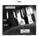
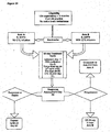
- the interaction between CD8+ cells and antigen-presenting cells or target (tumor) cells as described above is depicted in Figure 1.
- the interaction begins with the binding of antigen in association with an MHC Class I molecule on the APC or target cell to the T cell antigen receptor (TCR).
- Accessory molecules such as lymphocyte function antigens (LFA-1, LFA-2 and LFA-3), intercellular adhesion molecule 1 (ICAM-1, ICAM-2) and T cell co-stimulatory factors (CD40, CD70, B7) enhance cell-cell adhesion or transduce additional cell activation signals.
- LFA-1, LFA-2 and LFA-3 lymphocyte function antigens
- ICAM-1, ICAM-2 intercellular adhesion molecule 1
- CD40, CD70, B7 T cell co-stimulatory factors
- the CTL kills the target cell through the action of soluble cytolytic mediators (perforin and granzymes stored in cytoplasmic granules in the T cell) and a CTL surface molecule (Fas ligand).
- soluble cytolytic mediators perforin and granzymes stored in cytoplasmic granules in the T cell
- Fas ligand a CTL surface molecule
- target cells die by necrosis (membrane perforation and organelle destruction) or apotosis (chromatin condensation, DNA fragmentation and membrane blobbing).
- lymphocyte-mediated cytolysis is graphically depicted in Figure 2.
- Panel A of Figure 2 after binding to the target cell, cytoplasmic granules in the CTL are rapidly reoriented toward the target cell for release of granules containing perform and granzymes into the intercellular space. These proteolytic enzymes form pores in the plasma membrane of the target cell eventually leading to cell necrosis.
- Panel B after binding to the target cell, the level of Fas ligand expression on the CTL increases. The interaction of Fas ligand and the Fas receptor on the target cell leads to apoptosis.
- Proteases such as CPP32 and others related to IL-1b-converting enzyme (ICE) have been implicated in the induction of apoptosis.
- a more direct and efficient approach to this problem is to specifically activate CD8 + cells only with those epitopes relevant to combating a specific disease, (such as, cancer) or tumor specific antigens (such as, melanoma-specific antigens).
- a specific disease such as, cancer
- tumor specific antigens such as, melanoma-specific antigens.
- an artificial antigen presenting cell is created by expressing MHC Class I molecules in Drosophila melanogaster (fruit fly) cells. Since Drosophila does not have an immune system, the TAP-1,2 peptide transporters involved in loading peptide epitopes onto class I molecules are absent. As a result, the class I molecules appear on the Drosophila cell surface as empty vessels.
- this feature eliminates the need for in vivo stimulation of the immune system with high doses of various cytokines, thereby resulting in a treatment that foregoes the side effects caused by cytokines.
- the subject can be treated concurrently with low level dosages of a interferon, ⁇ -interferon, and/or IL-2.
- Eliminating the need for in vivo stimulation with cytokines provides an improvement to the quality of patient care.
- Treatment regimes that include the administration of cytokines to patients often result in the patient developing flu-like symptoms, such as nausea, vomiting, and fever. These side reactions are generally not life threatening, although a particularly severe reaction occurring in a patient who is already in a weaken condition could result in a life endangering situation. Another consideration is the adverse impact such side reactions have on patient acceptance and compliance of an otherwise beneficial treatment regime.
- Removing the need for in vivo stimulation with cytokines results in a treatment regime that improves the comfort of the patient, and provides the clinician with an effective method of treatment that his or her patient is more likely to comply with.
- transfected Drosophila cells as APCs and CD8 + cells from the 2C line of T cell receptor (TCR) transgenic mice.
- TCR T cell receptor
- purified CD8 + 2C cells are highly responsive to in vitro peptides presented by MHC Class I (L d )-transfected Drosophila cells also bearing the co-stimulatory molecules B7-1 and ICAM-1.
- Transfected Drosophila cells as a probe for defining the minimal requirements for stimulating unprimed CD8+ T cells ( Cai et al., P . N . A . S . USA (1996) 93 :14736-14741).
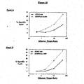
- Panel A represents the results of the combo mix protocol on peptide-loaded target cells.
- Panel B represents the results of combo load protocol on peptide-loaded target cells.
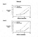
- Panel C represents the killing on melanoma targets generated from both protocols.
- APC antigen presenting cell
- APC systems have been utilized to generate antigen-specific CTL's to single epitopes: 1) human dendritic cells (DC) pulsed with defined peptides; 2) peripheral blood mononuclear cells (PBMCs) which have been driven to lymphoblasts and pulsed with peptides; 3) lymphoblastoid cell lines (LCL) where the natural peptides are acid-stripped and loaded with the peptides of interest; 4) Drosophila cells engineered to express empty class I molecules; and Mouse 3T3 cells transfected with human class I and co-stimulatory molecules (J. B. Latouche and M. Sadelain, Nature Biotech (2000) 18: 405-409).
- DC dendritic cells
- PBMCs peripheral blood mononuclear cells
- LCL lymphoblastoid cell lines
- DCs Dendritic cells
- HLA molecules HLA molecules
- Drosophila cell stimulation usually resulted in CTLs directed against up to ten different types of peptides. This provides CTLs that are active to each of the ten peptides.
- Drosophila cells and DC were evaluated, initially to a single peptide epitope, following the standard stimulation protocols for each.
- immature DCs were generated by culturing for one week autologous monocytes in the presence of IL-4 and GM-CSF.
- Mature DCs were obtained from immature DCs by addition of TNF ⁇ to the culture medium twenty-four hours prior to harvesting.
- DCs (immature and mature) were harvested, pulsed with peptides and mixed with purified CD8 cells following the procedure used for the stimulation of CD8 cells and peptide-pulsed Drosophila cells.
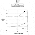
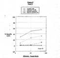
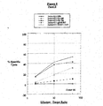
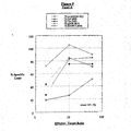
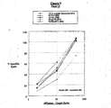
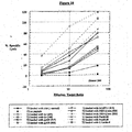
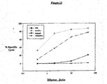
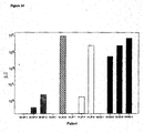
- Drosophila cells were found to be generally better stimulators than DC when evaluated for tyrosinase peptide epitope 689, as shown in Figure 7. Further, DCs displaying either the immature or mature phenotype (Figure 8) were not as efficient as Drosophila cells in eliciting specific CTL responses when defined peptides were used to pulse the APCs. This is particularly surprising, because of the dominant role played by DCs in the immune system. A comparison study with one donor was performed, as shown in Figure 9. Specific killing was generated against four different peptides when using fly cells as stimulators whereas immature DCs resulted in marginal specific killing and mature DCs resulted in specific killing against only one of the four peptides used for stimulation.
- CD8 + cells isolated from leukapheresis samples by positive selection with anti-CD8 antibody are stimulated against four different melanoma associated peptides presented by Drosophila cells expressing Human Class I molecules (HLA-A2.1), B7.1, ICAM-1, LFA-3 and B7.2
- CD8 + cells are re-stimulated for two rounds with autologous monocytes loaded with the peptide epitope in the presence of IL-2 and IL-7.
- CTLs are non-specifically expanded with OKT3 and IL-2.
- CTL activity is measured against Malme 3M cells and purity of CD8 + T cells is assessed by flow cytometry.
- the manufacturing processes and protocols are done according to Good Laboratory Practices and Good Manufacturing Practices. "Good Laboratory Practices” and “Good Manufacturing Practices” are standards of laboratory and manufacturing practices which are set by United States Food and Drug Administration, and are readily known to those of skill in the art.
- the CTLs are monitored for identity, viability, CTL activity, sterility, and endotoxin content.
- peptide epitopes suitable for use in the methods of the present invention to treat breast and ovarian cancers are shown in the following Table 1. It is readily apparent to those of ordinary skill in the art that a wide variety of peptide epitopes in addition to those listed in the following Table 1 will also be suitable for use in the methods of the present invention to treat breast and ovarian cancers, provided that such peptides are T cell epitopes.
- Interferon-alfa has a broad spectrum of immunomodulatory and antiproliferative effects in a variety of malignancies.
- IFN ⁇ Interferon-alfa
- rIFN- ⁇ -2a and rIFN- ⁇ -2b have produced a mean response rate of 15% (range, 6% - 27%) with response duration ranging from 1 to 60+ months.
- Tables 2 and 3 A summary of several single-agent IFN- ⁇ trials in metastatic melanoma is listed in Tables 2 and 3.
- IFN- ⁇ immunologically important molecules on the surface of the tumor.
- immunologically important molecules include, but are not limited to, the histocompatibility (mouse H-2 or human HLA) antigens, accessory molecules such as the intracellular adhesion molecule-1 (ICAM-1/CD54), as well as, tumor-associated antigens (Robincon et al., Immunobiology (1986) 172 :275-282; Borden et al., in Mitchell, MS, (ed): Biological Approaches to Cancer Treatment; Biomodulation . New York, NY, McGraw-Hill, (1992) pp 440-476).
- the immunomodulatory effects of IFN- ⁇ can potentially improve the ability of the immune system, including both antibodies and lymphocytes, to recognize and attack the tumor in vivo .
- the Eastern Cooperative Oncology Group completed a prospective randomized controlled study of interferon alfa-2b (IFN- ⁇ -2b) versus observation as surgical adjuvant therapy in 287 patients with melanoma (Kirkwood et aL, J Clin Oncol (1996) 14 :7-17).
- the dose used in the study was the maximum tolerated dose: 20MU/m 2 /d intravenously (IV) five days per week for four weeks followed by 10MU/m 2 three times per week subcutaneously (SC) for 48 weeks.
- IV intravenously
- SC subcutaneously
- the World Health Organization conducted a trial of low-dose interferon alfa-2a: 3 MU/m 2 SC three time per week for 3 years versus observation as adjuvant therapy in 427 patients with high-risk melanoma (Cascinelli, Proc Am Soc Clin Oncol , (1995) pp 410 (abstr)). There was no difference in the disease-free survival or survival in the patients receiving interferon.
- the present invention provides a method for treating a subject with melanoma, comprising the steps of:
- INF- ⁇ -2a or INF- ⁇ -2b can be used in the present method of treatment.
- INF- ⁇ can be administered to the patient via SC, IM, or IV.
- the effective amount of interferon-alfa for the present method is the amount of INF- ⁇ that is sufficient to up-regulate both HLA (class I) expression and melanoma-associated antigen expression, two key requirements for recognition and lysis by antigen-specific T cells. This effective amount can be determined by those skilled in the art.
- Example 5 (infra) describes some considerations in determining the effective amount of INF- ⁇ to be used in the present method of treatment.
- the effective amount of INF- ⁇ -2a is about 5 - 20 MU/m2/day, and the effective amount of INF- ⁇ -2b is about 5 - 15 MU/m2/day.
- the effective amount of INF- ⁇ is administered to the patient only for a relatively short time period, for example, 3 to 7 days, immediately prior to inoculating the subject with an effective amount of autologous cytotoxic T lymphocytes with specificity for melanoma-associated target antigen.
- the effective amount of interferon-alfa is about 10 MU/m 2 /day that is subcutaneously administered to the subject consecutively from day 5 to day 1 prior to inoculating said subject with the effective amount of autologous cytotoxic T lymphocytes with specificity for melanoma-associated target antigen.
- the effective amount of autologous cytotoxic T lymphocytes with specificity for melanoma-associated target antigen is the amount of the ex vivo generated CTLs with specificity for melanoma-associated target antigen that is sufficient to stop the growth or cause a decrease in size of a melanoma lesion when the CTLs are inoculated to the subject.
- the effective amount of autologous cytotoxic T lymphocytes with specificity for melanoma-associated target antigen is about 1-10 x 10 9 cells/infusion.
- the autologous cytotoxic T lymphocytes with specificity for melanoma-associated target antigen are obtained by a method comprising steps of:
- the non-naturally occurring antigen-presenting cell line is loaded with epitopes that are peptides derived from tyrosinase, gp100, and MART-1, such as those illustrated in SEQ ID NO: 1, SEQ ID NO: 2, SEQ ID NO: 4, SEQ ID NO: 5, and SEQ ID NO: 6.
- Human recombinant interleukin-2 is a lymphokine produced by recombinant DNA technology that has been shown to exhibit a variety of biological activities. In vitro, it has been shown to enhance lymphokine mitogenesis, enhance lymphocyte cyboxicity, induce the killer activity of both lymphokine-activated and natural killer cells, and it also induces interferon-gamma production.
- LAK lymphokine activated killer
- TILs tumor infiltrating lymphocytes
- biologics such as interferon. It has been administered at both high and low doses and by various infusion schedules (bolus and continuous). Response rates ranged from 15-20%.
- the Cytokine Working Group performed a retrospective analysis of 266 patients treated with high dose IL-2 bolus therapy that produced CR in 16 patients (6%), 69% of those were alive and progression-free at 5 years (Atkins et al., Proc Am Soc Clin Oncol (1997) 16 : 494a).
- IL-2 can be used immediately after the CTL infusion to enhance lymphocyte mitogenesis, lymphocyte cytotoxicity and gamma interferon production in an effort to maintain the antigen-specific T cells in vivo .
- the present invention provides a method for treating a subject with melanoma, comprising the steps of:
- Interleultin-2 can be administered to the patient via SC, IM, or IV.
- the effective amount of interleukin-2 is the amount of IL-2 that is sufficient to maintain the ex vivo generated antigen-specific CTLs in vivo .
- This effective amount can be determined by those skilled in the art.
- Example 5 (infra) describes some considerations in determining the effective amount of IL-2 to be used in the present method of treatment.
- the effective amount of IL-2 is about 2 -10 MIU/day.
- the effective amount of IL-2 is administered to the patient immediately after inoculating the subject with an effective amount of autologous cytotoxic T lymphocytes with specificity for melanoma-associated target antigen.
- the effective amount of interleukin-2 is about 3 MIU/ day that is subcutaneously administered to the subject consecutively from day 0 to day 27 after inoculating said subject with the effective amount of autologous cytotoxic T lymphocytes with specificity for melanoma-associated target antigen.
- the present invention provides a method for treating a subject with melanoma, comprising the steps of:
- the treatment cycle can be repeated at an interval of about 2 months.
- the Schneider S2 cell line was prepared from Drosophila melanogaster (Oregon-R) eggs according to published procedures and has been deposited with the American Type Culture Collection (CRL 10974). S2 cells are grown in commercial Schneider's Drosophila medium supplemented with 10% fetal bovine serum.
- the pRmHa-3 plasmid vector for expressing MHC Class I and co-stimulatory proteins in S2 cells was derived from the pRmHa-1 expression vector constructed as described in the literature. It contains a metallothionein promoter, metal response consensus sequences and an alcohol dehydrogenase gene bearing a polyadenylation signal isolated from Drosophila melanogaster .
- Complementary DNAs were individually inserted into the pRmHa-3 vector.
- S2 cells were transfected with a mixture of HLA-A2.1, B7.1 and ICAM-1 plasmid DNAs and the phshneo plasmid using the calcium phosphate precipitation method.
- Stably transfected cells were selected by culturing in Schneider's medium containing geneticin. Twenty-four hours before use, expression of the transfected genes was induced by addition of CuSO 4 . The level of expression was assessed by flow cytometry using anti-HLA-A2.1, anti-B7.1 and anti-ICAM-1 antibodies. HLA expression by greater than 30% of the cells is necessary for efficient in vitro activation of CD8 + lymphocytes.
- CD8 + cells are isolated from leukapheresis samples by positive selection using the DynabeadsTM isolation procedure (Dynal).
- An anti-human CD8 mouse monoclonal antibody (50 ⁇ g/ml in human gamma globulin [Gammagard®]) is added to washed cells in Dulbecco's PBS supplemented with 1% human serum albumin (Baxter-Hyland) and 0.2% Na citrate. After incubation at 4°C for forty-five minutes with gentle mixing, the cells are washed and re-suspended in the same buffer containing Dynal magnetic beads (DynabeadsTM) coated with sheep anti-mouse IgG at a bead to cell ratio of 1:1.
- Dynal magnetic beads Dynal magnetic beads
- the cells and beads are placed into a sterile tube and gently mixed at 4°C for forty-five minutes. At the end of this time, the antibody-bound cells are removed magnetically using the MPC-1® separator according to the manufacturer's instructions (Dynal). Dissociation of the CD8 cell-bead complex is achieved by incubation at 37°C for forty-five minutes in the presence of CD8 peptide 59-70 (AAEGLDTQRFSG; SEQ ID NO: 12). Free beads are removed magnetically and the CD8 cells are counted and analyzed by flow cytometry to evaluate purity. Recovery of CD8 + cells is typically greater than 80%.
- Table 4 summarizes the cell composition of fourteen separate CD8 + preparations from normal human PBMC preparations by positive selection with anti-CD8 antibody.
- Transfected Drosophila S2 cells are incubated in Schneider's medium (10 6 cells/ml) supplemented with 10% fetal calf serum and CuSO 4 at 27°C for twenty-four hours. Cells are harvested, washed and re-suspended in Insect X-press medium (BioWhittaker) containing 100 ⁇ g/ml human tyrosinase 369-377 . Following incubation at 27°C for three hours, the S2 cells are mixed with CD8 + cells at a ratio of 1:10 in RPMI medium (Gibco) supplemented with 10% autologous serum. The cell mixture is incubated for four days at 37°C during which the Drosophila cells die off. On Day five, IL-2 (20 U/ml) and IL-7 (30 U/ml) are added to selectively expand the tyrosinase-specific CTL population.
- Schneider's medium (10 6 cells/ml) supplemented with 10% fetal calf serum and
- Adherent monocytes are loaded with the tyrosinase epitope by incubation for 90 minutes in Hepes-buffered RPMI medium containing 10% autologous serum and 10 ⁇ g/ml tyrosinase 369-377 .
- the supernatant is removed and the Drosophila -activated CD8 + cell suspension (3 x 10 6 cells/ml in RPMI medium with 10% autologous serum) is added at a ratio of 10 CD8 + cells to 1 adherent monocyte.
- IL-2 (20 U/ml) and IL-7 (30 U/ml) are added with a medium change to selectively expand the tyrosinase-specific CTL population.
- Effector cells are non-specifically expanded by culturing them in RPMI medium supplemented with autologous serum, anti-CD3 monoclonal antibody (OKT®3), IL-2 and ⁇ irradiated autologous PBMCs.
- RPMI medium supplemented with autologous serum, anti-CD3 monoclonal antibody (OKT®3), IL-2 and ⁇ irradiated autologous PBMCs.
- Malme 3M cells are used as target cells in a 51 Cr release assay. 5 x 10 6 Malme 3M cells in RPMI medium containing 4% fetal calf serum, 1% HEPES buffer and 0.25% gentamycin are labeled at 37°C for one hour with 0.1 mCi 51 Cr. Cells are washed four times and diluted to 10 5 cells/ml in RPMI with 10% fetal bovine serum (HyClone). In a 96-well microtiter plate, 100 ⁇ l effector CTLs and 100 ⁇ l peptide-loaded, 51 Cr-labeled Malme 3M target cells are combined at ratios of 100:1, 20:1 and 4:1 (effector: target).
- K562 cells are added at a ratio of 20:1 (K562:Malme 3M) to reduce natural killer cell background lysis.
- Non-specific lysis is assessed using the non-tumor HLA-A2.1 fibroblast cell line, Malme 3. Controls to measure spontaneous release and maximum release of 51 Cr are included in duplicate. After incubation at 37°C for six hours, the plates are centrifuged and the supernatants counted to measure 51 Cr release.
- Percent specific lysis is calculated using the following equation: cpm sample - cpm spontaneous release cpm maximum release - cpm spontaneous release x 100
- CD8 + cells before and after in vitro activation, were analyzed for a number of cell surface markers using fluorescent monoclonal antibodies and FACS analysis. Results from a typical activation protocol using cells from a healthy donor is shown in Table 5.
- Flow Cytometry Analysis of In Vitro Activated CD8 + Cells MARKER/CELL TYPE PRE-ACTIVATION POST-ACTIVATION Mean % Mean % CD8 T cell 98 99 TCR ⁇ T cell receptor 98 92 CD 44 lymph node homing receptor 91 99 CD45RO memory T cell 58 88 CD45RA 41 31 CD62L HEV homing receptor 24 38 CD56 NK cell 1 11 CD25 activated T cell 0.1 29
- CTL preparations will be assayed for sterility and endotoxin content.
- This example teaches the effectiveness of cytotoxic T Cell infusions in the treatment of melanoma as assessed according to the following factors:
- Eligibility for treatment required patients to have histologically-documented, unresectable malignant melanoma that was measurable or otherwise capable of evaluation, and the HLA-A2 haplotype.
- Pretreatment evaluation included radiologic evaluation of the brain by MRI or CT scan, CT scanning of the chest and abdomen, and physical examination, especially of the skin and lymph nodes.
- the total number of patients treated was fifteen (nine male and six female). The ages ranged from 33 to 75 years with an average of 58 years. The average duration of metastatic disease was 1.5 years.
- a pretreatment skin test to determine whether a state of anergy existed was performed on 14/15 patients with 5/14 testing negative for all seven of the common antigens evaluated.
- HLA-A2 haplotype Patients were screened for the HLA-A2 haplotype by FACS analysis with an HLA-A2 specific monoclonal antibody (BB7.2). Subtyping was performed by PCR analysis. All, but one of the patients, were HLA-A*0201, the exception (patient 08) was HLA-A*0205.
- T cells infused ranged from a minimum of 4x10 7 (patient 08) to a maximum of 3.2x10 9 (patient 13).
- Patients were entered into a second, third or fourth cycle of treatment based on their clinical status at the end of each cycle.
- the number of PBMCs obtained from the aphaeresis samples tended to be lower in patients undergoing additional cycles, especially if the start of the subsequent cycle was close to the end of the previous one. This is attributed to persistent lymphopenia due to the IFN ⁇ -2b administered during the previous cycle.
- the total number of na ⁇ ve CD8 + T cells isolated was dependent on its percentage in each of the PBMC preparations.
- the percent of CD8 + T cells varied between 8% to 31% among the patients.
- the obtained expansion factor also contributed to the final cell numbers and ranged from 0.1-6.0 fold.
- the procedure for generating CTLs ex vivo is taught in the Specification and Example 1, above.
- biopsy B Following five days of subcutaneous injections of 10MU/m 2 , a dramatic increase in these two markers was noted (biopsy B). For tyrosinase and gp100, immunohistochemical staining was negative to weakly positive, respectively in the pretreatment samples (biopsy A). After the initial five-day IFN ⁇ dose, and thirteen additional treatments, expression of these later antigens was increased in the stained tissue samples (biopsy C).
- CTLs generated from all patients were evaluated on the day of release against peptide-loaded T2 targets, an HLA-A2 melanoma cell line (Malme3M) and an autologous melanoma line, if biopsy material was available to establish a line.
- Each prepared dose of cells was evaluated for its cytolytic activity.
- Peptide-loaded T2 cells presenting, either each peptide alone, or all four peptides simultaneously, were used to determine the specificity of the CTL response generated for each patient.
- the ability to lyse endogenously-expressed, melanoma-associated, antigen-bearing cells was assessed with an HLA-A2 matched line or an autologous tumor line.
- antigen-specificity was evaluated with an established method for detecting intracellular gamma interferon production, made in response to a specific peptide stimulus.
- the CTLs generated at the end of the ex vivo protocol were evaluated by this method.
- the percent of cells specific for each of the peptides was recorded individually.
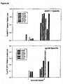
- the total number of specific cells in each bulk CD8 culture from patient 13 was calculated by adding each of the peptide specificities detected in that population of T cells. An increase in the total number of specific cells could be detected with each successive treatment cycle.
- Biopsy samples from all patients prior to, during and after treatment would have been ideal. However, the experimental conditions allowed for biopsy samples from only a limited number of patients. Tumor tissue was obtained from five of the fifteen patients enrolled in the study. In two patients (patients 08 and 13) biopsy samples were available at five and six weeks post T cell therapy, respectively. Examination of the tissue samples revealed the presence of both infiltrating CDS and CD4 cells. One of the tumor samples was taken from a skin lesion in the occipital region of the scalp, which increased in size by the time of the follow up examination, four weeks after a second infusion of T cells. The biopsy revealed necrosis of the tissue that was heavily infiltrated with lymphocytes.
- the other biopsy was from the head of a femur bone, removed during hip replacement surgery.
- the skin lesion from patient 08 was strongly positive (4+) for both a general class I, and a specific HLA-A2 marker.
- Tyrosinase and gp100 were weakly positive (1+ and 2+, respectively), while MART-1 was negative in this same sample.
- Regions of the biopsy from patient 13 were also necrotic, with more heterogeneous staining; distinct populations of tumor cells lacking expression of the HLA-A2.1 molecule, and one or more of the MAAs.
- intact tissue regions revealed strong class I (4+), and all of the melanoma-associated antigens.
- lymphocytic infiltrations in this later sample appeared to surround the tumor nodules rather than to deeply infiltrate them.
- CDS cells the highest percentage of cells directly associated with the tumor were CDS cells.
- CT scans were part of the pretreatment screening criteria and the post treatment follow-up examination.
- Patient 10 received a single infusion of 8x10 8 CTLs (7/27/99) five weeks after the pretreatment scan (6/23/99).
- a CT scan of the chest was repeated one month after the infusion (8/27/99)
- a dramatic decrease in the size of a lung lesion was noted.
- patient 14 underwent a chest CT scan as part of the enrollment process (9/10/99), three and one-half weeks before a first infusion with 6.6x10 8 cells (10/5/99).
- a follow-up CT scan (1/7/99) one month after a second infusion with 11.5x10 8 cells, revealed dramatic shrinkage in three separate lesions.
- Patient 13 also had an objective response as measured in pre and past CT scans.
- Paratracheal adenopathy went from 7.8cm 2 (pre-study) to 4.4cm 2 after cycle I, and disappeared following cycle II.
- HER-2/neu is a proto-oncogene with homology to EGFR that is amplified and over-expressed in many human cancers, largely adenocarcinomas of the breast, ovary and colon. It is often associated with aggressive disease and can be an indicator of a poor prognosis. It has been studied in several clinical trials as a possible target for these types of cancers.
- HHR-2/neu HLA-A2.1 restricted peptide epitopes were identified either by computer-assisted peptide binding algorithms or by mapping CTLs isolated from ascites of ovarian cancer patients (Table 6).
- transfected Drosophila cells have the unique ability to present up to ten different peptide epitopes (Figure 10), we selected the four HER-2 peptides 826, 835, 861 and 863 due to the frequency of generating CTLs to these peptides ex vivo .
- These four different HER-2 peptides represent weak to moderate binders to the HLA-A2.1 molecule presented on the surface of the transfected Drosophila cells.
- HER-2/neu is a member of the EGF-R family and functions as a growth factor receptor.
- HER-2 protein is expressed during fetal development in humans. In adults, the protein is weakly detectable in epithelial cells of many normal tissues. In normal cells the HER-2 gene is present as a single copy. Amplification of the gene and/or over-expression of the associated protein has been identified in many human cancers including breast, ovarian, uterine, stomach and adenocarcinoma of the lung. Sequence differences between HER-2 and EGF-R receptor are noted in Table 8. Three of the four HER-2 peptides we have evaluated have three or more amino acids changes between the two proteins. A single amino acid change is sufficient to discriminate between the two proteins.
- the CTLs generated were evaluated for antigen-specificity.
- Drosophila cells were loaded with a combination of the four HER-2 peptides.
- the bulk CD8 culture was evaluated for antigen-specificity.
- T2 cells loaded with each of the immunizing peptides were used as target cells.
- a typical response is depicted.
- the bulk culture contains specificity for each of the four HER-2 peptides.
- the anti-tumor response was assessed on an ovarian tumor cell line (ATCC; HTB-77).
- HER-2 specific effectors representing the individual peptides were evaluated to confirm the presentation of each of the peptide epitopes on this tumor cell line.
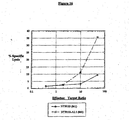
- a breast adenocarcinoma cell line (ATCC; HTB-131), transfected with HLA-A2.1 was also evaluated for the ability to demonstrate tumor lysis with the HER-2 specific peptide effectors. CTLs specific for peptide 861 could lysis this tumor cell line when transfected with HLA-A2.1 ( Figure 16).
- the HTB-77/A2.1 cell line requires a pretreatment with IFN ⁇ to demonstrate peptide-specific lysis.
- the cells were treated with 500U/ml of IFN ⁇ (specific activity of 25ng/ml) for twenty-four hours prior to the initiation of the 51 Cr-release assay.
- the addition of the IFN ⁇ resulted in enhanced lysis of the HLA-A2.1 transfected cell line.
- a FACS analysis was performed to determine the levels of these molecules after both twenty-four and forty-eight hours of induction.
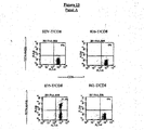
- Panels A and B depict the FACS analysis results.
- Synthetic peptides were made by standard Fmoc chemistry using a peptide synthesizer (Gilson Company, Inc.) All peptides were purified to >95% purity by reverse-phase HPLC on a C-8 column. Purity and identity were established using a mass spectrometer with electrospray ionization.
- peptide 819 was MART-1 specific (AAGIGILTV SEQ ID NO:6), 817 and 853 were both gp100 peptides (ITDQVPFSV SEQ ID NO:4 and KTWGQYWQV SEQ ID NO:5, respectively), tyrosinase-specific peptides were 689 and 792, with 792 representing the post translational modified version (YMDGTMSQV SEQ ID NO:2) of the native sequence (YMNGTMSQV SEQ ID NO:1) represented by peptide 689.
- Peptides 826 (CLTSTVQLV SEQ ID NO:7) and 835 (KIFGSLAFL SEQ ID NO:8) represented HER-2/neu sequences from the intracellular and extracellular domains, respectively of the p185 protein.
- Pec60 20 (ALALAALLVV SEQ ID NO:10) Fec60 25 (ALLVVDREV SEQ ID NO:11) were overlapping sequences representing a mucinous protein detected in ovarian tumor lines.
- C-lectin also was a protein detected in ovarian tumor cell lines and a peptide from its sequence (C-lectin 8 ) is represented by KMASRSMRL SEQ ID NO:9.
- Standard 51 Cr-release assays were performed to determine CTL effector cell recognition of melanoma-associated peptide epitopes loaded onto T2 cells.
- Harvest 3x10⁶ T2 cells were grown in RPMI +10% FBS (media). 0.1mCi of 51 Cr was added and incubated at 37°C in a water bath. Labeled cells were added to 10ml of 4% wash (RPMI + 4% FBS) and pellet, washed two additional times, and re-suspended in media to a final concentration of 0.2x10⁶/mL to record radioactivity of spontaneous versus detergent lysed cells. The cells were pulsed with the appropriate peptide(s) at 20 ⁇ g/mL for thirty minutes. 50 ⁇ L was added to each 96-well plate each containing CD8 effector cells at 10, 2, 0.4, and 0.08 x 10 6 /mL, which was incubated at 37°C for six hours, spun and harvested for supernatant.
- the cells were labeled with FITC- or PE conjugated monoclonal antibodies by incubation at 4°C for 30 minutes in FACS buffer (1% BSA, 0.02% NaN 3 in PBS), followed by a wash in the same buffer. Cells were fixed in 0.5% formaldehyde prior to data acquisition and analysis on a FACScan flow cytometer (Becton Dickinson) with its CellQuest software. Nonspecific staining was measured with the same secondary antibody used to label purified primary antibodies, or an isotype-matched control when the primary antibodies were directly labeled.
- Tetrameric staining was performed with HLA-A2.1 specific HIV gag tetrameric molecules (Beckman Coulter) harboring the sequence SLYVTVATL SEQ ID NO:43 as a negative control.
- HER-2 specific tetramers were made with the sequences CLTSTVQLV (826 SEQ ID NO:7), KIFGSLAFL (835 SEQ ID NO:8), or VMAGVGFSPYV (861 SEQ ID NO:16) peptides.
- PE-labeled tetrameric HLA-A2.1-peptide complexes were used in conjunction with fluorescein isothiocyanate (FITC)-labeled anti-human CD8a (BD PharMagin) monoclonal antibodies to stain epitope-specific CD8+ T cells as described in package insert. Samples were analyzed by two-color flow cytometry on a Becton Dickinson FACScan, and gated CD8+ T cells were examined for staining with tetrameric HLA-A2.1-peptide complexes.
- FITC fluorescein isothiocyanate
- BD PharMagin anti-human CD8a
- MUC-1 Mucin is a transmembrane glycoprotein expressed on both normal and malignant epithelium.
- the underglycosylated form of MUC-1 over-expressed on the cell surface of many human adenocarcinomas such as breast and ovarian cancer, as well as hematological malignancies including multiple myeloma and B-cell lymphoula.
- G250 A renal cell carcinoma associated antigen expressed in 85% of RCC's but not normal kidney tissue.
- FBP Folate binding protein is a receptor involved in folate transport. It is over-expressed in over 90% of ovarian tumors and 20-50% of breast cancers.
- HER-2/neu is over-expressed as much as 200 fold over normal tissues in breast and ovarian tumors.
- MAGE-3 A cancer-testis antigen expressed on 70 - 80% of metastatic melanoma lesions and cell lines. It is a member of the family of melanoma associated or MAGE proteins. In addition, MAGE-3 has been found in 20-60% of epithelial tumors (colon, breast, lung, gastric carcinomas).
- AES The amino enhancer of split protein is part of a set of transcriptional repressors encoded by the Enhancer of split genes. This tumor antigen was identified in tumor-associated lymphocytes of ovarian and breast tumors.
- HTR Telomerase(hTR) is a specialized type of reverse transcriptase (hTRT or hTERT) that catalyzes the synthesis and extension of telomeric DNA.
- hTRT reverse transcriptase
- hTERT reverse transcriptase
- This example teaches the effectiveness of cytotoxic T cell infusions in the treatment of melanoma as assessed according to the following factors:
- Eligibility for treatment required patients to have histologically-documented, unresectable malignant melanoma that was measurable and readily subject to evaluation. In addition, patients were required to have an HLA-A2 haplotype. Pretreatment evaluation included radiologic evaluation of the brain by MRI or CT scan, CT scanning of the chest and abdomen, and physical examination, especially of the skin and lymph nodes. The total number of patients to be enrolled is 42. Twenty-one (21) patients are on the control cytokine only arm and twenty-one (21) patients are on the cytokine + T cell therapy arm. Thirty-one (31) patients have been enrolled to date (17 entered on the control arm and 14 have entered on the T cell treatment arm).
- HLA-A2 haplotype by FACS analysis with an HLA-A2 specific monoclonal antibody (ATCC: BB7.2). Subtyping was performed by PCR analysis. All patients, with the exception of two, were HLA-A*0201-positive. The other HLA-A2 subtypes were HLA-A2*0202 and HLA-A2*0205.
- Interferon- ⁇ -2b (Intron-A®; recombinant interferon alfa-2b, Schering Corporation, Kenilworth, NJ) was administered subcutaneously to the patient at 10 MU/m 2 for 5 consecutive days prior to the CTL infusion.
- Ex vivo Drosophila cell-activated autologous lymphocytes (1-10 x 10 9 ) were infused to the patient on the day after INF- ⁇ administration.
- Interleukin-2 PROLEUKIN®; Aldesleukin, recombinant IL-2, Chiron Corporation, Emeryville, CA
- Table 11 shows a comparison of the cytokine dose used in study CTL-03 with those used in FDA approved protocols for the single administration of IFN- ⁇ in the adjuvant setting (post surgical remove of all detectable tumor) or high dose IL-2.
- Transfected Drosophila S2 cells are incubated in Schneider's medium (10 6 cells/mL) supplemented with 10% fetal calf serum and copper sulfate at 27-28°C for 24-72 hours. S2 cells are harvested, washed and re-suspended in Insect X-press medium (BioWhittaker) containing 0.1 ⁇ g/mL of each peptide; human tyrosinase 369-377 (SEQ ID NO:1 YMNGTMSQV and SEQ ID NO: 2 YMDGTMSQV), gp100 209-217 (SEQ ID NO: 4 ITDQVPFSV), gp100 154-162 (SEQ ID NO: 5 KTWGQYWQV) and MART-1 27-35 (SEQ ID NO: 6 AAGIGILTV) and 5 ⁇ g/mL human ⁇ 2 microglobulin.
- Schneider's medium 10 6 cells/mL
- the S2 cells are mixed with CD8+ cells at a ratio of 1:10 in Roswell Park Memorial Institute (RPMI) medium (a defined medium, Gibco) supplemented with 5-10% autologous serum.
- RPMI Roswell Park Memorial Institute
- the cell mixture is incubated for four days at 37°C during which time the Drosophila cells die off.
- IL-2 (20 U/mL) and IL-7 (30 U/mL) are added with a media change to selectively expand the antigen-specific CTL population consisting of cells with specificity for the melanoma-associated antigens (MART-1, gp100 and tyrosinase).
- Follwing ⁇ -irradiation (5,000 rads), the cells are incubated at 37°C for two hours. Non-adherent cells are removed by washing with Dulbecco's PBS.
- Adherent monocytes are loaded with the five peptide epitopes described supra by incubation for 90 minutes in Leibowitz medium containing 5 ⁇ g/mL human ⁇ 2 microglobulin in 1% HSA (in place of autologous serum, to avoid the possibility of introducing potential proteases, which might be present in the serum) and 5-10 ⁇ g/mL of each of the five peptide epitopes.
- the supernatant is removed and the Drosophila-activated CD8 cell suspension (2.5 x 10 6 cells/mL in RPMI medium with 10% autologous serum) is added at a ratio of 10 CD8 cells to 1 adherent monocyte.
- IL-2 (20 U/mL
- IL-7 (30 U/mL) are added with a medium change to selectively expand the melanoma-specific CTL population.
- a total of two of such adherent cell, peptide-specific restimulation steps take place, one approximately one week after the primary stimulation, and the second approximately one week later.
- a non-specific expansion step occurs at the start of week 4 of the ex vivo protocol.
- the CD8+ effector cells that have undergone two rounds of restimulation are expanded in cell culture bags along with feeder cells (irradiated non-CD8+ selected cells) after being stimulated with OKT3 antibody.
- Frozen non-CD8+ selected cells are thawed, washed and then gamma irradiated (3500 rads).
- a ratio of 4:1 (feeder:effector) is placed in T-225 flasks that have been coated with OKT3 antibody.
- OKT3 stimulation is performed ion complete RPMI medium containing 10% autologous serum supplemented with 20 U/mL of IL-2. Two days later the stimulated T cells are diluted with fresh media and transferred to cell culture bags for expansion. Fresh media and IL-2 are supplemented approximately every two days to feed the rapidly expanding T cells.
- Phenotypic analysis as measured by flow cytometry, was performed on purified CD8+ cells on the day of isolation from the leukapheresis sample and on the day the cells were released for infusion back into the patient. A statistical analysis was performed with samples from patients and from normal donors, which had undergone the same ex vivo stimulation protocol. Statistical significant differences were found between the na ⁇ ve CD8+ samples and the CD8+ effector cells obtained at the end of the ex vivo stimulation protocol, regardless if the samples were derived from patients or normal donors.
- Activation markers e.g., CD69, which is not expressed an resting lymphocytes but is rapidly induced upon activation; HLA-DR a class II molecule expressed on activated T cells), memory markers (e.g., CD45RO expressed on activated cells and most memory cells) integrin markers (e.g., CD49d/CD29, which is involved in the migration of lymphocytes from blood to tissues at sites of inflammation), accessory molecules (e.g., CD11a/CD18; which aids in cytotoxic lymphocyte killing and mediates adhesion to many cells including endothelium) and cytokine receptor markers (e.g., the functional high affinity IL-2R receptor is composed of a non-covalenty associated CD25/CD122/CD132 heterodimer) were all found to be upregulated on the CD8 effector cells following the completion of the ex vivo stimulation protocol.
- integrin markers e.g., CD49d/CD29, which is involved in the migration of lymphocytes from blood to tissues at sites
- Table 12 summarizes the results of the analysis. Phenotypic Analysis of CD8+ Cells Post Ex Vivo Protocol Marker Pairs Fold change Na ⁇ ve/FAA 95%Confindence 99% Confidence From To From To CD69-F 51 7.35 5.78 9.33 5.33 10.00 CD25-F 51 5.64 4.97 6.40 4.76 6.68 CD49d-PE 51 4.79 4.29 5.33 4.13 5.53 CD8-F 50 4.59 3.98 5.31 4.13 5.12 HLA DR-PE 51 3.78 2.96 4.82 2.73 5.22 CD11a-F 51 3.56 3.24 3.92 3.13 4.05 CD18-PE 51 3.32 2.99 3.68 2.89 3.81 CD45RO-F 51 2.89 2.47 3.39 2.34 3.57 CD29-F 51 2.82 2.49 3.19 2.38 3.33 CD132-PE 22 2.75 2.17 3.49 1.99 3.81 HLA ABC 47 2.26 2.04 2.51 1.96 2.60 CD3-F 44 2.14 1.91 2.39 1.84 2.49 TCR ⁇ 50 2.05 1.78 2.37 1.69 2.49 CD122-
- CD8+ effector cells are activated by the ex vivo protocol described herein.
- These activated CD8+ effector cells are capable of migrating from blood to the tumor sites in vivo and are capable of killing the tumor target cells with extraordinar sensitivity and potentially can proliferate in the presence of a low dose of IL-2 as the high affinity IL-2R is present on these cells.
- CD122 is constitutively expressed on a subpopulation of resting T cells.
- the white areas are statistically significant values, which are upregulated after completion of the CD8+ ex vivo stimulation protocol.
- the light gray areas do not show significant upregulation or downregulation.
- the dark gray areas show a statistically significant downregulation of the reported molecules. Regulation of these markers is consistent with the induction of activated T cells fully capable of tracking and lysing tumor cells in vivo, as measured by phenotypic analysis.
- results from immunohistochemistry analysis on expression of HLA (HLA-A2.1) and melanoma-associated antigen (MART-1) from the four biopsy samples are summarized in Table 13, wherein higher number indicates a higher level of expression of the respective protein.
- the results showed that there was no additional increase in expression of HLA (HLA-A2.1) and melanoma-associated antigen (MART-1) after the first five consecutive days IFN- ⁇ treatment, indicating that interferon- ⁇ 2b treatment beyond 5 consecutive days was not necessary to reach optimal levels of HLA-A2.1 and MART-1 expression.
- Biopsy samples were shipped overnight at 4°C in RPMI media.
- the tissue was placed in OCT® (Tissue-Tek) embedding medium for frozen tissue specimens.
- the tissue OCT matrix was placed on filter paper prior to a quick-freezing step in liquid NO 2 .
- Frozen samples were stored at -80°C, prior to sectioning.
- Cryostat sections (5 ⁇ m) were placed on SuperfrostTM (Fisher) charged slides. Slides were fixed in cold acetone (-20°C) and stored at -80°C until stained. Endogenous peroxidase activity was blocked by incubating in 0.3% H 2 O 2 in methanol for 10 minutes. Primary antibody was added for 1 hour in a humidified chamber.
- Biotinylated secondary antibody was added and incubated for 30 minutes at room temperature. Streptavidin-HRP was added under the same conditions. DAB substrate was added for up to 5 minutes and rinsed in water. Slides were hydrated through 4 cycles of alcohol (70-95-95-100%), followed by 3 cycles of xylene.
- Antibodies used in the immunohistochemical analysis were from ATCC (pan HLA class I, W6/32 and HLA-A2-specific, BB7.2), or NeoMarkers (MART-1, M2-7C10).
- Figure 21A represents a typical growth curve for the ex vivo generated CD8+ cells in the CTL-02 study.
- a typical drop at day 6 reflected the dying off of non-specific cells.
- the CD8 cells from each cycle of retreatment for patient 15-RT had similar growth curves, suggesting that there were no memory cells remaining in the peripheral blood after each cycle of treatment. This was not the case in study CTL-03.
- the typical drop in cell numbers is noted with 01-KN-1, reflecting the drop seen in the cells obtaining during the first cycle.
- tetrameric molecules containing either MART-1 or gp100 melanoma-associated peptides were commercially available for analysis of antigen-specific T cells (Beckman Coulter; Immunomics). These tetrameric molecules were not available for study CTL-02.
- the antigen-specific cells for MART-1 were 0.29% and 0.46% for gp100. This would suggest that the antigen specific cells were being maintained in vivo, even after the cytokine and cell therapy were suspended.
- Tetrameric Staining Approximately 1x10 7 of purified CD8+ cells were obtained from the donor by leukapheresis. Each sample was placed in sterile tubes at 1x10 6 per 100 ⁇ l of wash buffer (PBS+1%BSA+0.02% NaN3). In each tube with 100 ⁇ l of cell suspension 5 ⁇ l of the HLA-A2.1-tetramer-streptavidin-phycoerythrin (SA-PE) labeled molecules (loaded with the peptide of interest (MART-1, gp100 or Tyrosinsase-specific) was added and was incubated at room temperature for 30 minutes.
- SA-PE HLA-A2.1-tetramer-streptavidin-phycoerythrin
- Anti-CD8+ monoclonal antibody (FITC-labeled) was added and the incubation was continued for an additional 30 minubes. 1-2mL of wash buffer was used to wash the samples at the end of the incubation period. Cells were pelleted for 10 minutes at low speed (400 x g) and the supernatant was discarded. Each cell pellet was resuspended in 0.5 ml of 0.5% formaldehyde/PBS fixative and read on a FACScan (Becton Dickinson) flow cytometer at the appropriate settings.
- FACScan Becton Dickinson
- T2 cells were grown in RPMI +10% FBS (media) and harvested. 100mL of 51 Cr per target was added and incubated at 37°C in a water bath. Labeled cells were added to a volume of 10 mL of 4% wash (RPMI with 4% FBS) and pelleted. Washes were performed 2 additional times.
- the cells were resuspend in media to a final concentration of 0.2x10 6 /mL and radioactivity of spontaneous versus maximum (detergent-lysed) cells was recorded.
- the cells were pulsed with the appropriate peptide(s) at 14-20 ⁇ g/mL for 30 minutes. 50 ⁇ L/96 well plate each containing CD8+ effector cells at different effector:target ratios. The mixture was incubated at 37°C for 6 hours. Plates were spun and harvest supernatant. Radioactive supernatants were counted in a Cobra gamma counter. Specific 51 Cr release was calculated using the formula ( 51 Cr release-Spontaneous release) / (Maximum release-Spontaneous release) X 100.
- Intracellular gamma interferon assay Effector cells are stimulated with peptide-pulsed T2 cells (ratio of CD8:T2 was 2:1) for 5 hours in the presence of GlogiStop (PharMingen). Stimulated cells were surfaced stained with anti-CD8-PE, fixed and permeabilized with Cytofix/Cytoperm (PharMingen Cat no 2076KK), followed by staining with anti-IFN ⁇ -FITC. The dual-labeled cells were analyzed by flow cytometry (Becton Dickson FACScan) and expressed as a percent of maximum expression. Maximum expression of IFN ⁇ was measured as the amount expressed in response to OKT3 (Orthoclone) activation of the same effector cells.
- the aim of clinical research with biological response modifiers is to determine the optimum conditions that maintain the benefit of treatment, while reducing toxicity to a minimum level.
- IFN- ⁇ -2b at currently approved doses and schedules of administration can significantly prolong survival in melanoma patients.
- Administration can also be associated with severe toxicity.
- High doses of IL-2 appears to be more effective than low dose continuous infusions, yet high doses of IL-2 are also more toxic.
- the most common side effects are flu-like symptoms. The most severe side effects are hypotension, capillary leak syndrome, and reduced organ perfusion.
- IL-2 has been used clinically for both renal cell carcinoma and malignant melanoma.
- IFN- ⁇ -2b and IL-2 were administered in a 3-drug combination immunochemotherapy regimen in metastatic renal cell cancer (RCC) patients.
- the phase II study was carried out to define the activity of low dose subcutaneous administration of IL-2 [4.5mU x 2/24h thrice weekly for 2 wk] and IFN- ⁇ -2b [3MU/24h thrice weekly (on alternating days with IL-2) for 2 wk] and vinblastine (VLB) [4mg/m2 every 3 weeks].
- Patients had a one-week rest from cytokines, at which time they received the VLB. Treatment was repeated every 3 weeks, with a maximum duration of 1 year.
- Toxicity was mild to moderate (Grade I and II), consisting of fever, anorexia, malaise and nausea-vomiting in >80% of the patients. None of the patients experienced major VLB-related toxicity. It was proposed that the 3-drug combination might be a promising regimen (overall response rate of 38.7%) with modest toxicity in advanced RCC.
- IFN- ⁇ -2b [3MU/m2 daily, intramuscularly continuously] and IL-2 (0.5MU/m2 x2/24h subcutaneously on days 1-5 for the first week and 1MU/m2 x2/24h for 5 days in the following 3 weeks were prescribed.
- Side effects were said to be low (Grade I and II) in most of the patients, with abnormalities in liver and renal function tests noted.
- White the regimen was said to be of moderate toxicity, low cost and required no hospitalization, the flu-like syndrome observed in most patients was sufficient enough to cause one-third of the patients to refuse subsequent treatment.
- IL-2 granulocyte macrophage colony-stimulating factor
- IFN- ⁇ -2b IFN- ⁇ -2b in addition to IL-2
- GM-CSF granulocyte macrophage colony-stimulating factor
- IFN- ⁇ -2b IFN- ⁇ -2b in addition to IL-2
- MLT pineal hormone melatonin
- IL-2 low-dose IL-2
- the advanced solid tumors included lung, kidney, stomach, liver and melanoma.
- the IL-2 was administered at a daily dose of 3MIU, subcutaneously for 6 days/week for 4 weeks; a dosing that is almost identical to the one used in CTL-03.
- the MLT was given orally at a daily dose of 40mg/day, starting 7 days prior to IL-2. Objective tumor regression was noted in 3/14 patients (21%).
- MLT enhanced the antitumor immune effect of IL-2 and/or increased the susceptibility of the cancer cells to cytolysis mediated by IL-2 induced cytotoxic lymphocytes.
- the combination of the cytokine therapy in conjunction with the added T cell therapy represents a novel way to treat advanced, metastatic melanoma.
Landscapes
- Health & Medical Sciences (AREA)
- Life Sciences & Earth Sciences (AREA)
- General Health & Medical Sciences (AREA)
- Veterinary Medicine (AREA)
- Public Health (AREA)
- Animal Behavior & Ethology (AREA)
- Epidemiology (AREA)
- Engineering & Computer Science (AREA)
- Chemical & Material Sciences (AREA)
- Biomedical Technology (AREA)
- Bioinformatics & Cheminformatics (AREA)
- Zoology (AREA)
- Immunology (AREA)
- Organic Chemistry (AREA)
- Pharmacology & Pharmacy (AREA)
- Medicinal Chemistry (AREA)
- Biotechnology (AREA)
- Genetics & Genomics (AREA)
- Wood Science & Technology (AREA)
- Microbiology (AREA)
- Gastroenterology & Hepatology (AREA)
- Cell Biology (AREA)
- Biochemistry (AREA)
- General Engineering & Computer Science (AREA)
- Proteomics, Peptides & Aminoacids (AREA)
- Nuclear Medicine, Radiotherapy & Molecular Imaging (AREA)
- General Chemical & Material Sciences (AREA)
- Chemical Kinetics & Catalysis (AREA)
- Hematology (AREA)
- Oncology (AREA)
- Molecular Biology (AREA)
- Medicines Containing Material From Animals Or Micro-Organisms (AREA)
- Medicines That Contain Protein Lipid Enzymes And Other Medicines (AREA)
- Micro-Organisms Or Cultivation Processes Thereof (AREA)
- Mycology (AREA)
Abstract
Description
- Figure 1:
- This figure is a graphic depiction of the interaction between CD8+ cells, also known as cytotoxic T lymphocytes, with antigen-presenting cells or target cells, in this case tumor cells.
- Figure 2, Panels A and B:
- This figure is a two panel graphical depiction of mechanisms of lymphocyte-mediated cytosis.
- Figure 3:
- This figure shows the result of an experiment where several different peptides were tested in a competition assay to identify peptide binders that could be used to load multiple peptides onto Drosophila cells expressing human empty class I molecules.
- Figure 4, Panels A, B and C:
- This figure shows the result of an experiment where three melanoma peptides were tested for the ability to raise CTLs when added as single epitopes on Drosophila cells. In this donor (#60), CTL activity was elicited to each of the peptides when added alone to three different Drosophila preparations. The specificity of the response was compared with control HBc peptide, a high affinity binder.
- Figure 5, Panels A, B and C:
- This figure shows the results of a series of experiments where up to four different peptides were added to single Drosophila cells. CTL activity against each of the represented peptides was seen after a three-week stimulation protocol and is graphically depicted in this figure. Results from three different donors (# 93, 94, 95) are represented.
- Figure 6, Panels A, B and C:
- This figure shows CTL activity after two different primary in vitro stimulation protocols.
- Figure 7, Panels A and B:
- This figure compares the ability of Drosophila cells versus dendritic cells to elicit CTL responses to a single peptide epitope following standard stimulation protocols.
- Figure 8:
- This figure shows that the dendritic cells displaying either a mature or immature phenotype were not as efficient as Drosophila cells in eliciting specific CTL responses when defined peptides were used to pulse the cells.
- Figure 9, Panels A, B and C:
- This figure shows CTL activity generated by a single donor to three different in vitro stimulation protocols presenting four peptides.
- Figure 10:
- This figure show CTL activity generated to ten (10) peptides loaded, in combination, to Drosophila cells.
- Figure 11:
- This figure shows the peptide binding capacity of the HER-2 peptides (826, 835, 861 and 863) on the Drosophila cells transfected with the human HLA-A2.1 class I molecule.
- Figure 12:
- This figure demonstrates the anti-peptide and anti-tumor response for MART-1 specific effector cells. T2 cells were loaded with MART-1 peptide or a negative control (HBc). Malme3M is a melanoma line, Malme3 is a non-tumor cell line.
- Figure 13, Panels A and B:
- This figure shows the tetrameric staining of the HER-2 specific CD8 effector cells from two different donors.
- Figure 14:
- This figure reveals the anti-peptide response for the HER-2 effector cells evaluated on peptide-loaded T2 cells.
- Figure 15, Panels, A, B, C, D:
- This figure demonstrates the enhanced killing of an ovarian, tumor cell line (HTB-77) when transfected with HLA-A2.1.
- Figure 16:
- This figure shows the enhanced killing of a breast cancer cell line (HTB-133) when transfected with HLA-A2.1
- Figure 17:
- This figure shows that IFNγ pre-treatment is required to demonstrate lysis of the tumor cell line HTB-77/A2.1.
- Figure 18, Panels A and B:
- This graph demonstrates that the surface expression of HLA-A2 and HER-2 is unaffected by IFNγ induction in the two cell lines (HTB-77 and HTB-77/A2.1).
- Figure 19:
- This graph shows which protein mRNA levels are elevated in the HTB-77/A2.1 cells after an induction with IFNγ.
- Figure 20:
- This graph shows the experimental outline of clinical study CTL-03.
- Figure 21:
- A) shows in vitro growth curve of CTL cells isolated from a patient (15-RT)
under clinical study CTL-02. The cells were isolated from the patient on
day 0 of each of the three treatment cycles starting on: 9/28/99 (15-RT-1), 11/22/99 (15-RT-2), and 2/15/99 (15-RT-3), and were cultured in vitro.
B) shows in vitro growth curve of CTL cells isolated from a patient (01-KN) under clinical study CTL-03. The cells were isolated from the patient onday 0 of each of the three treatment cycles starting on: 10/30/00 (01-KN-1), 1/30/01 (01-KN-2), and 4/9/01 (01-KN-3), and were cultured in vitro. - Figure 22:
- This figure shows that the number of melanoma antigen-specific T cells increased in the patient after one cycle of treatment in study CTL-03. HLA-A2.1 tetrameric molecules prepared with either gp100 or MART-1 peptides were used to monitor the presence of antigen-specific T cells in purified CD8+ preparations obtained from the leukapheresis samples. The percent (%) of tetramer-positive cells was recorded at the time of the first (I) and second (II) leukapheresis sample, which could be as much as two (2) months apart.
- Figure 23
- This figure shows the correlation among three different in vitro assays used to measure cytolytic activity, antigen-specificity and cell proliferation of CTLs. Three different assays were performed on all of the CD8+ CTL preparations at the end of the ex vivo stimulation protocol (CTL-03). All three assays are generally in agreement for determining the makeup of the final bulk culture. In this particular preparation the majority of the T cells in the product were directed to gp100 peptide (817). CTL activity defines the cytolytic and effector function of the cells. Tetramer staining determines the frequency of the antigen-specific cells and intracellular IFN-gamma staining reflects the ability of the T cells to respond to a particular peptide.
- Figure 24
- This figure shows an increase in the amount of interferon gamma at the same time point of the cycle with each repeat cycle of treatment in study CTL-03, suggesting the presence of memory T cells at the same time point of the cycle. Interferon gamma was measured in the supernatant of the ex vivo cultures six (6) days following the primary stimulation with the Drosophila cells.
- Figure 25
- This figure illustrates the number of cycles and doses of cells administered to each patient in the clinical study described in Example 2. The potency of each dose was calculated by multiplying the number of CD8+ cells by the lytic units recorded for the lysis of a combination of the peptides loaded on the target cells.
- APC
- Antigen-presenting cells
- CD8+
- CD8+ T cells
- CTL
- Cytotoxic T lymphocyte
- E
- Effector
- Fas
- Also known as CD95, epitope on T cells
- ICAM
- Intercellular adhesion molecule
- IL
- Interleukin
- LAK
- Lymphokine-activated killer cells
- LFA
- Lymphocyte function antigens
- MHC
- Major histocompatibility complex
- nnAPC
- non-naturally occurring antigen-presenting cell
- NP
- Nuclear protein
- PBMC
- Peripheral blood mononuclear cell
- PBS
- Phosphate-buffered saline
- PCR
- Polymerase chain reaction
- RPMI
- Roswell Park Memorial Institute
- RWJPRI
- The R.W. Johnson Pharmaceutical Research Institute
- T
- Target
- TCR
- T cell antigen receptor
- TIL
- Tumor-infiltrating lymphocytes
| Amino Acid | Abbreviations | |
| 3-Letter | 1-Letter | |
| alanine | ala | A |
| valine | val | V |
| leucine | leu | L |
| isoleucine | ile | I |
| proline | pro | P |
| phenylalanine | phe | F |
| trytophan | tyr | W |
| methionine | met | M |
| glycine | gly | G |
| serine | ser | S |
| threonine | thr | T |
| cysteine | cys | C |
| tyrosine | tyr | Y |
| asparagine | asn | N |
| glutamine | gln | Q |
| aspartic acid | asp | D |
| glutamic acid | glu | E |
| lysine | lys | K |
| arginine | arg | R |
| histidine | his | H |
As used herein, the term "C-lectin" refers to a peptide of the sequence that has been found to be associated with ovarian cancer.
As used herein, the term "conservative substitution" also includes the use of a chemically derivatized residue in place of a non-derivatized residue.
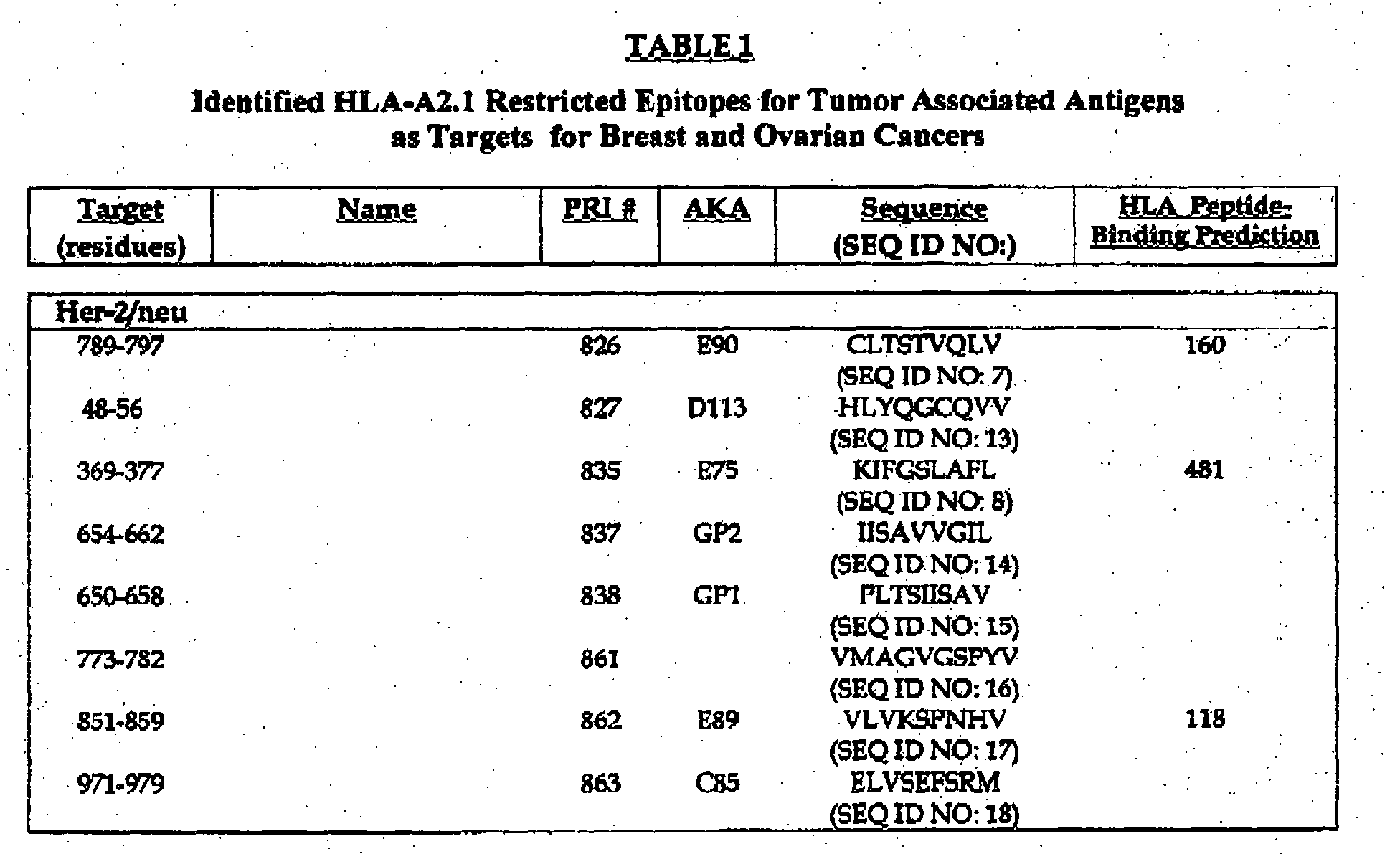


| Single-Agent IFN-α Trials in Metastatic Melanoma (IFN-α-2a) | |||||||
| Study | Dose (MU) | Route/schedule | entered* | CR | PR | % | Duration (mo) |
| Creagan et al, 1984 | 12/m2 | IM, TIW for 12 Wks | 30/30 | 1 | 5 | 20 | 2 to 13+ |
| Creagan et al,1984 | 50/m2 | IM, TIW for 12 Wks | 31/31 | 3 | 4 | 23 | 3 to 11 |
| Coates et al,1986 | 20/m2 | IV, daily for 6 of 14 d | 16/15 | 0 | 0 | 0 | |
| Hersey et al, 1985 | 15 to 50/m2 | IM, TIW | 20/18 | 2 | 0 | 11 | 6 to 12 |
| Legha et al, 1987 | 18 to 36/m2 | IM, daily for 10 Wks | 35/31 | 0 | 3 | 10 | 6 to 7 |
| Steiner et al, 1987 | 9 to 36/m2 | IM, daily | 12/12 | 1 | 0 | 8 | 4+ |
| Elsasser-Beile et al, 1987 | 18/m2 | IM, daily for 10 Wks | 21/21 | 3 | 0 | 14 | 12+ to 16+ |
| IFN-α-2a | Subtotal | 196/189 | 10 | 14 | 12 |
| Single-Agent IFN-α Trials in Metastatic Melanoma (IFN-α-2b) | |||||||
| Study | Dose (MU) | Route/schedule | entered* | CR | PR | % | Duration (mo) |
| Kirkwood et al, 1985 | 10 to 100/m2 | IM or IV, daily x 28 | 23/23 | 2 | 2 | 22 | 3 to 60+ |
| Dorval et al, 1986 | 10/m2 | SC, TIW | 24/22 | 2 | 4 | 27 | 2 to 5 |
| Sertoli et al, 1989 | 10/m2 | IM, TIW | 21/21 | 0 | 3 | 14 | 7 to 13 |
| Robinson et al, 1986 | 10/m2 | SC, TIW for x 12 wk | 51/40 | 4 | 6 | 25 | 1 to 54+ |
| IFN-α-2b | Subtotal | 119/106 | 9 | 15 | 20 |
- HLA-A2.1 and β-2 microglobulin:
- Reverse transcription-PCR from K562 cells using primers derived from the published sequence
- B7.1:
- Reverse transcription-PCR from K562 cells using primers derived from the published sequence
- ICAM-1:
- Reverse transcription-PCR from K562 cells using primers derived from the published sequence
- B7.2:
- Reverse transcription-PCR from HL-60 cells (ATCC CCL-240) using primers derived from the published sequence
- LFA-3:
- Reverse transcription-PCR from HL-60 cells (ATCC CCL-240) using primers derived from the published sequence
| Purification of CD8+ Cells by Positive Selection Analyzed by Flow Cytometry | ||||
| CELL TYPE | PBMC | POST SELECTION | ||
| Mean % | (Range) | Mean % | (Range) | |
| | 15% | (7-24) | 82% | (56-95) |
| CD4 T cells | 36% | (14-52) | 2% | (0.1-10) |
| | 15% | (7-26) | 0.8% | (0.2-2) |
| | 12% | (8-21) | 0.6% | (0.1-3) |
| | 2% | (0.4-7) | 3% | (0.5-9) |
| | 6% | (2-17) | 6% | (0.1-20) |
| Flow Cytometry Analysis of In Vitro Activated CD8+ Cells | ||
| MARKER/CELL TYPE | PRE-ACTIVATION | POST-ACTIVATION |
| Mean % | Mean % | |
| CD8 T cell | 98 | 99 |
| TCRαβ T cell receptor | 98 | 92 |
| CD 44 lymph node homing receptor | 91 | 99 |
| CD45RO memory T cell | 58 | 88 |
| CD45RA | 41 | 31 |
| CD62L | 24 | 38 |
| CD56 | 1 | 11 |
| CD25 activated T cell | 0.1 | 29 |
| In addition to activity and purity, CTL preparations will be assayed for sterility and endotoxin content. |
| REAGENTS | |||
| REAGENT | SUPPLIER | GRADE | NOTES |
| rh IL-2 | Chiron | USP | sterile solution |
| rh IL-7 | Genzyme | Research | lyophilized, sterile solution |
| Human tyrosinase369-377 | Research | ||
| Dynabeads® M-450 | Dynal | GMP | sheep anti-mouse IgG magnetic beads |
| Human serum albumin | Baxter | USP | sterile, non-pyrogenic hepatitis virus-free, 25% solution |
| fetal bovine serum | Gemini | Research | sterile, BSE-, endotoxin-, mycoplasma-free |
| Gammagard® | Baxter | USP | sterile, human immune globulin solution for injection |
| anti-CD8 antibody | Research | mouse anti-human CD8 monoclonal antibody | |
| CD8 peptide 59-70 | Research | release of CD8+ cells from magnetic beads | |
| W6/32 | ATCC | Research | mouse anti-human HLA-A,B,C monoclonal antibody |
| CELL LINES | ||
| CELL LINE | SUPPLIER | NOTES |
| Drosophila S2 | ATCC | CRL 10974 |
| M3 | UCSD | Non-HLA-A2.1 |
| Malme | ||
| 3 | ATCC | Normal skin fibroblast from a melanoma patient |
| Malme 3M | ATCC | Metastatic melanoma from lung (same patient as Malme 3) |
| M14 | UCSD | HLA-A2.1 human melanoma |
| K562 | ATCC | human erythroleukemic cell line; target for NK cells |
| JY cells | ATCC | EBV-transformed, human B cell line expressing HLA-A2.1 and B7 |
| P815 and P1024 | ATCC | DBA/2 mouse mastocytoma cell lines |
| Jurkat A2.1 | ATCC | acute T cell leukemia transifected with human HLA-A2.1 |
| ATCC: American Type Culture Collection |
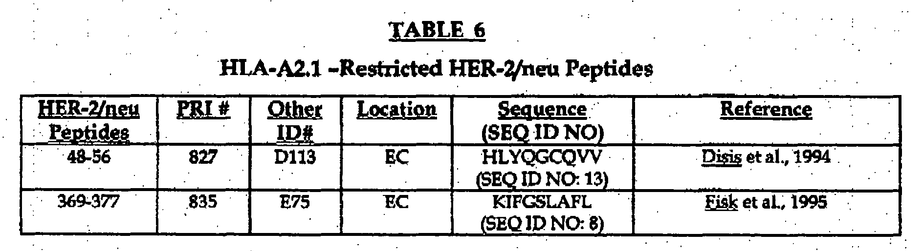

| Frequency of HER-2/neu CTL Generation in | ||||||||
| Donor | ||||||||
| 826 | 827 | 835 | 837 | 838 | 861 | 862 | 863 | |
| 193 | + | + | ||||||
| 194 | + | - | + | - | - | + | - | + |
| 195 | + | + | + | + | ||||
| 196 | + | - | + | - | - | + | ||
| 197 | + | - | + | - | + | + | - | + |
| 198 | - | - | + | - | + | + | - | + |
| 207 | + | + | + | + | ||||
| 212 | + | + | + | + | ||||
| 218 | + | + | + | + | ||||
| 232 | - | + | + | - | ||||
| 233 | + | + | + | + | ||||
| 241 | + | + | ||||||
| 243 | + | + |
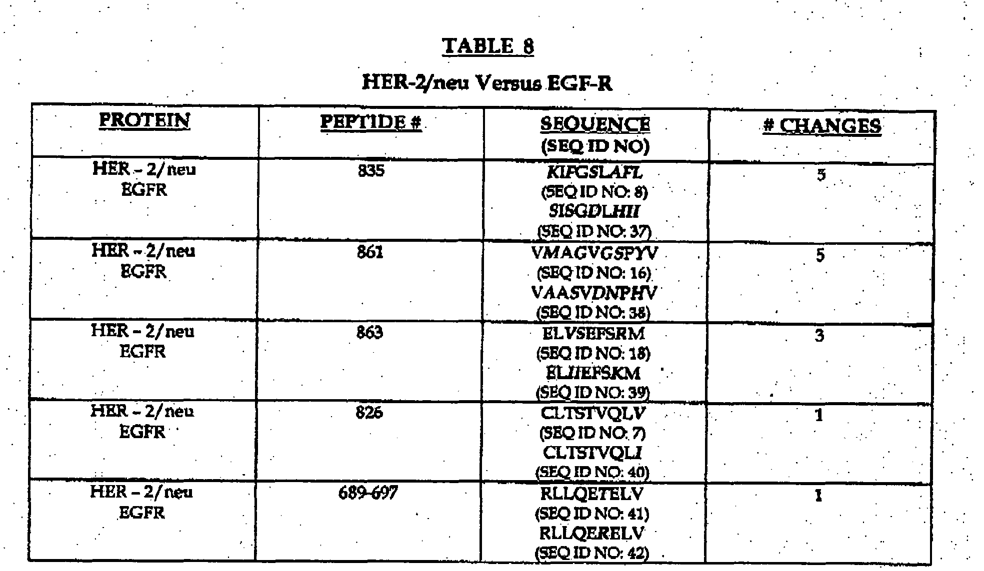
| Frequency of Response to Breast and Ovarian Peptide Epitopes in Normal Donors | |||||||||||||||||
| Donor | 879 | 893 | 894 | 899 | 900 | 901 | 902 | 903 | 906 | 907 | 908 | 909 | 910 | 911 | 912 | 913 | 914 |
| 248 | + | + | + | ||||||||||||||
| 249 | + | + | + | ||||||||||||||
| 250 | + | - | + | ||||||||||||||
| 251 | - | - | + | ||||||||||||||
| 252 | + | - | + | ||||||||||||||
| 253 | - | - | + | + | |||||||||||||
| 254 | + | - | + | + | |||||||||||||
| 255 | - | + | + | - | + | + | + | + | |||||||||
| 256 | + | - | + | + | + | + | - | + | |||||||||
| 257 | + | + | + | ||||||||||||||
| 259 | + | - | - | ||||||||||||||
| 260 | - | - | - | - | + | ||||||||||||
| 261 | + | + | + | + | |||||||||||||
| 262 | + | + | - | + | - | ||||||||||||
| 265 | + | + | + | + | - | - | - | - | + | + | + | - |
| Tumor Antigen Descriptions | |
| Antigen | Description |
| CA-125 | Cancer Antigen 125 is an epithelial cell marker expressed by ovarian tumors and some ovarian cell lines. About 85% of ovarian cancer patients have an increased serum CA125 and is therefore commonly used as a serum tumor marker. (Cancer Letters (1999, Oct.) 145(1-2) pg. 133-141 ) |
| MUC-1 | Mucin is a transmembrane glycoprotein expressed on both normal and malignant epithelium. The underglycosylated form of MUC-1 over-expressed on the cell surface of many human adenocarcinomas such as breast and ovarian cancer, as well as hematological malignancies including multiple myeloma and B-cell lymphoula. (Blood (1999, June) 93(12) pg. 4309-4317) |
| G250 | A renal cell carcinoma associated antigen expressed in 85% of RCC's but not normal kidney tissue. It is identical to the tumor-associated antigen MN/CAIX which is expressed in about 50% of invasive breast cancers. (Cancer Research (1999, Nov.);59(21) pg. 5554-5559) |
| FBP | Folate binding protein is a receptor involved in folate transport. It is over-expressed in over 90% of ovarian tumors and 20-50% of breast cancers. (Anticancer Research (1999 Jul-Aug) 19(4B) pg. 2907-2916) |
| HER-2/neu | A proto-oncogene (HER-2) encoding a transmembrane protein similar in sequence and structure to EGF-R. HER-2/neu is over-expressed as much as 200 fold over normal tissues in breast and ovarian tumors. It has also been identified in renal cell and lung carcinomas. (J. Exp .Med.(1995, June) Vol. 181, pg. 2109-2117) |
| NY-ESO-1 | A cancer-testes antigen found in 30% of breast, prostate and ovarian cancers , lung cancer, bladder cancer, head and neck cancer and melanoma. Patients who have cancers with tumors expressing this antigen usually have circulating antibodies against it as well. (J. Immunology (2000) vol. 165 pg. 948-955) |
| CEA | Carcinoembryonic antigen is a tumor-associated antigen frequently expressed in epithelial tumors (colon, breast, lung). CEA levels in the serum can correlate with disease stage and is used to monitor treatment and reoccurrence of disease. (Human Immunology (1998) vol. 59 pg. 1-14) |
| MAGE-3 | A cancer-testis antigen expressed on 70 - 80% of metastatic melanoma lesions and cell lines. It is a member of the family of melanoma associated or MAGE proteins. In addition, MAGE-3 has been found in 20-60% of epithelial tumors (colon, breast, lung, gastric carcinomas). (Human Immunology (1998) vol. 59 pg. 1-14) |
| AES | The amino enhancer of split protein is part of a set of transcriptional repressors encoded by the Enhancer of split genes. This tumor antigen was identified in tumor-associated lymphocytes of ovarian and breast tumors. (Molecular Immunology (1998) 35(17)pg. 1121-1133) |
| HTR | Telomerase(hTR) is a specialized type of reverse transcriptase (hTRT or hTERT) that catalyzes the synthesis and extension of telomeric DNA. The activity of this enzyme is elevated in about 90% of all human tumors including cancers of the breast, thyroid, bladder, cervix, prostate, colon, pancreas and stomach. (Cancer Research (2001, Dec) 61(23)pg. 8366-8370) |
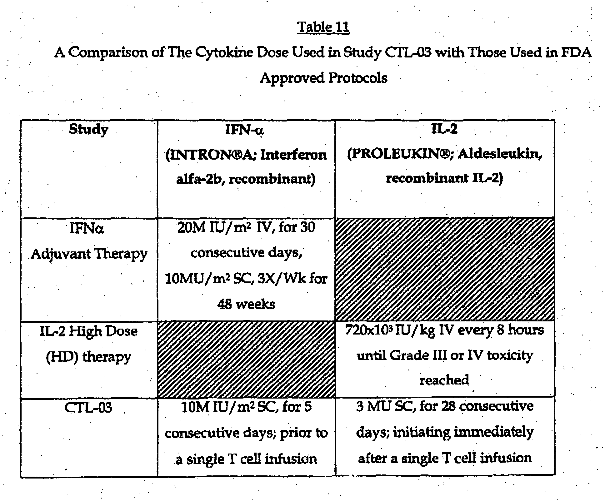
| Phenotypic Analysis of CD8+ Cells Post Ex Vivo Protocol | ||||||
| Marker | Pairs | Fold change Naïve/ | 95%Confindence | 99% Confidence | ||
| From | To | From | To | |||
| CD69-F | 51 | 7.35 | 5.78 | 9.33 | 5.33 | 10.00 |
| CD25-F | 51 | 5.64 | 4.97 | 6.40 | 4.76 | 6.68 |
| CD49d-PE | 51 | 4.79 | 4.29 | 5.33 | 4.13 | 5.53 |
| CD8- | 50 | 4.59 | 3.98 | 5.31 | 4.13 | 5.12 |
| HLA DR-PE | 51 | 3.78 | 2.96 | 4.82 | 2.73 | 5.22 |
| CD11a-F | 51 | 3.56 | 3.24 | 3.92 | 3.13 | 4.05 |
| CD18-PE | 51 | 3.32 | 2.99 | 3.68 | 2.89 | 3.81 |
| CD45RO-F | 51 | 2.89 | 2.47 | 3.39 | 2.34 | 3.57 |
| CD29-F | 51 | 2.82 | 2.49 | 3.19 | 2.38 | 3.33 |
| CD132-PE | 22 | 2.75 | 2.17 | 3.49 | 1.99 | 3.81 |
| HLA ABC | 47 | 2.26 | 2.04 | 2.51 | 1.96 | 2.60 |
| CD3-F | 44 | 2.14 | 1.91 | 2.39 | 1.84 | 2.49 |
| TCRαβ | 50 | 2.05 | 1.78 | 2.37 | 1.69 | 2.49 |
| CD122-PE | 21 | 1.54 | 1.15 | 2.07 | 1.03 | 2.31 |
| CD44-F | 51 | 1.00 | 0.86 | 1.16 | 0.82 | 1.21 |
| CD28-F | 51 | 0.92 | 0.77 | 1.10 | 0.72 | 1.16 |
| CD62L-F | 51 | 0.77 | 0.63 | 0.94 | 0.59 | 1.00 |
| CD16-PE | 28 | 0.45 | 0.32 | 0.62 | 0.29 | 0.69 |
| CLA | 45 | 0.42 | 0.33 | 0.53 | 0.31 | 0.57 |
| CD45RA-F | 51 | 0.31 | 0.26 | 0.38 | 0.24 | 0.41 |
| Five Consecutive Days Interferon-α-2b Treatment Was Sufficient to Obtain Optimal Levels of HLA-A2.1 And MART-1 Expression | |||||
| Patient | Biopsy Sample | Time Point | HLA-A2.1 | MART-1 | |
| 04-MJ | A | Pretreatment (No IFNα) | 1-2+ | 1 + | |
| | Post | 5 days IFNα; Pre T cell therapy | 4+ | 4+ | |
| C | Post 11 days IFNα; Post a single T cell infusion | 4+ | 4+ | ||
| D | Post 17 days IFNα; | 4+ | 4+ |

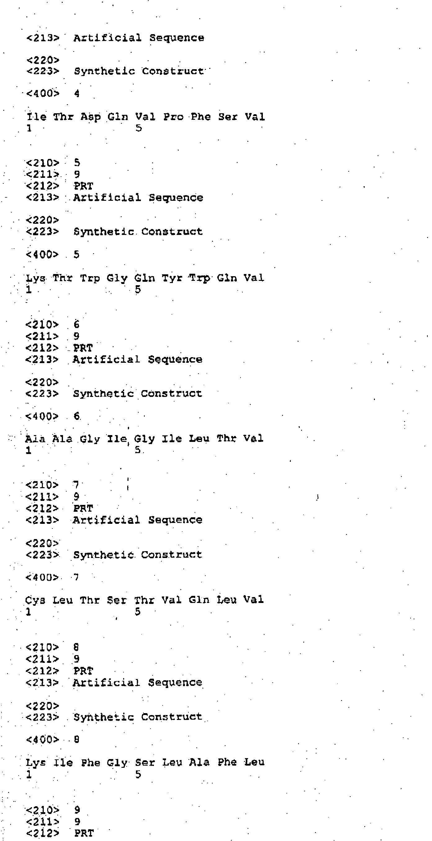
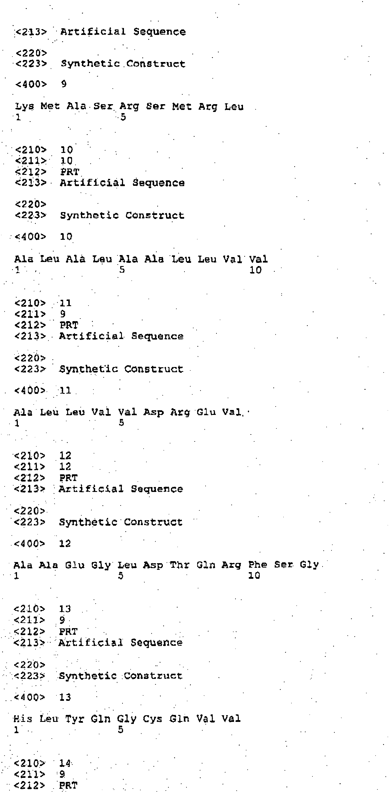
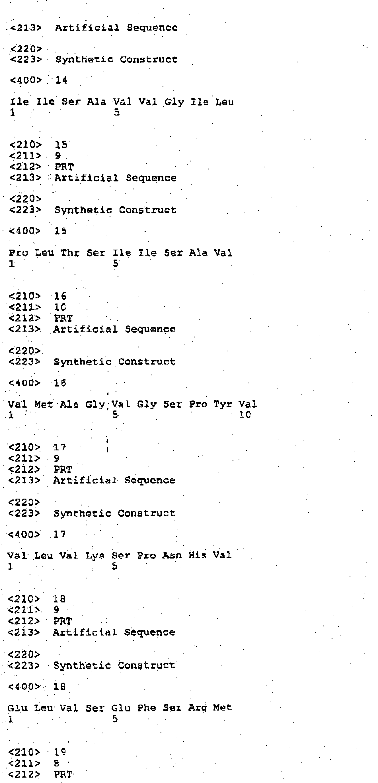

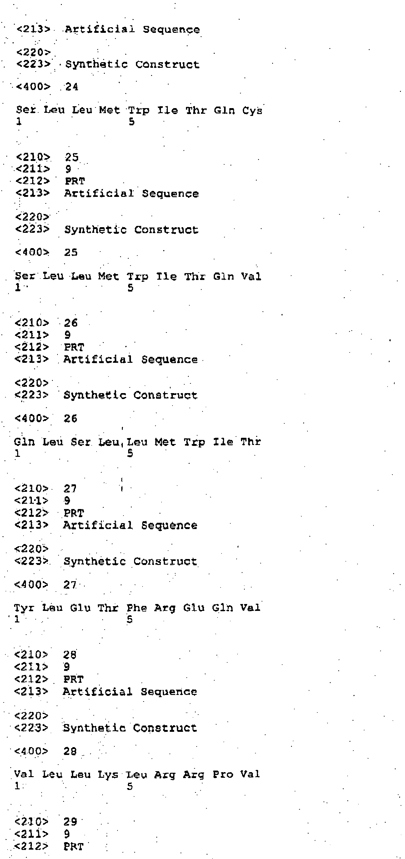

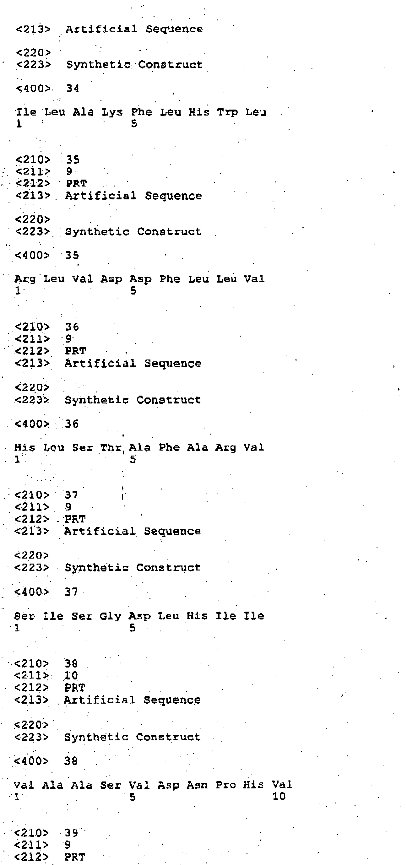
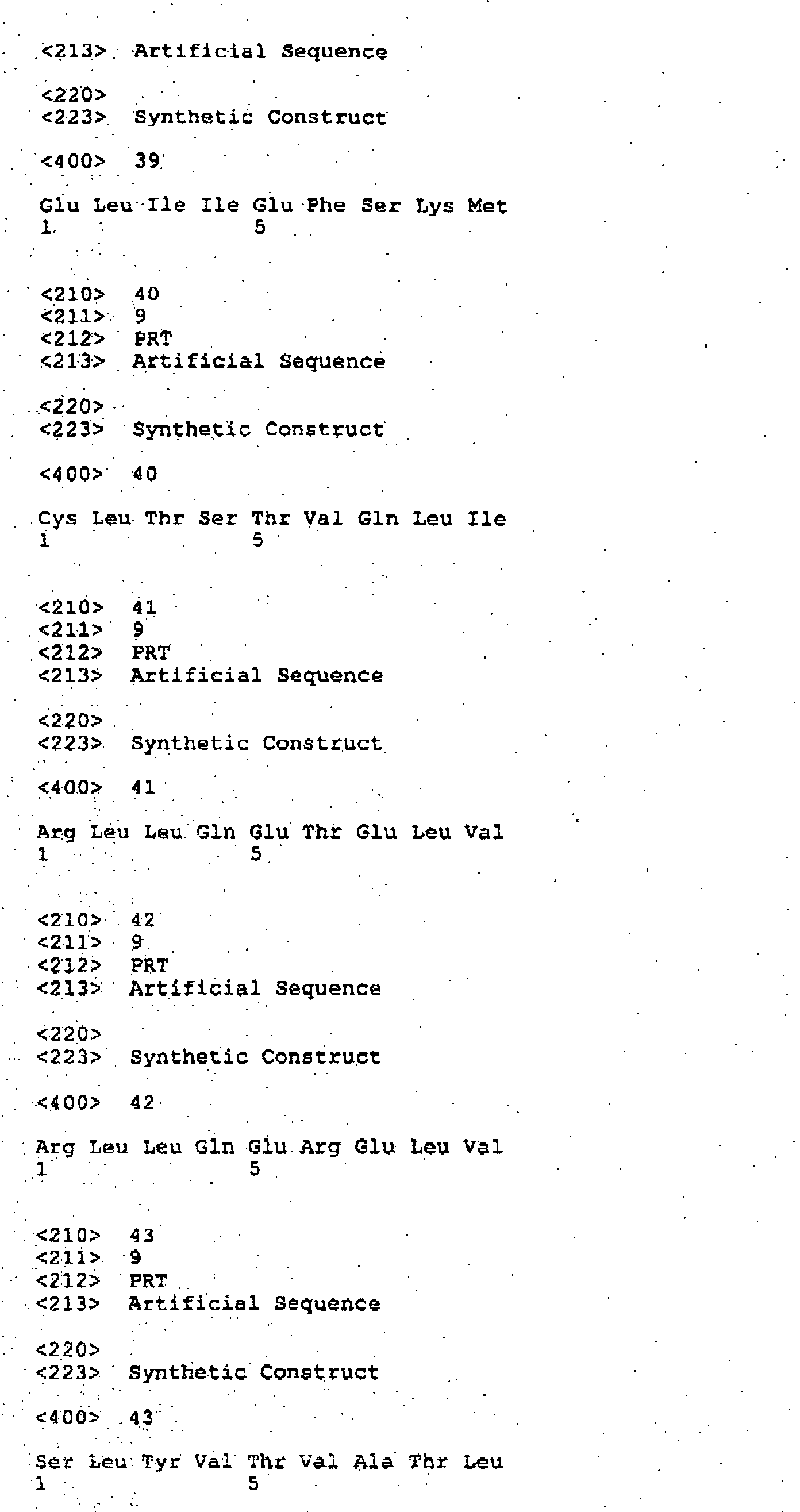
Claims (18)
- A method for preparing a CD8+ suspension for use in treating a viral infection in a subject comprising:a. preparing a non-naturally occurring antigen-presenting cell line (nnAPC), wherein said nnAPC is capable of presenting up to about fifteen different peptide molecules associated with said viral infection simultaneously wherein said peptide molecules are each about six to twelve amino acids in length;b. stimulating CD8+ cells harvested from said subject or a suitable donor with said nnAPC cell line;c. adding said CD8+ cells to media that contains a cytokine selected from the group consisting of IL-2, IL-7 or conditioned growth medium (CGM), wherein said cytokines can be used individually or in combination;d. mixing unsuspended peripheral blood monocytes, or CD8 depleted peripheral blood monocytes collected from said subject or a suitable donor with about 1 to 50µg/ml of one of said peptides that said nnAPC can simultaneously present;e. irradiating said peripheral blood monocyte suspension with a sufficient dose of γ-radiation necessary to sterilize all components in the suspension, except the desired peripheral blood monocytes;f. isolating adherent peripheral blood monoctyes;g. loading said adherent peripheral blood monocytes with about 1 ug/ml to 50µg/ml of said each peptide; andh. combining said CD8+ cells with said adherent peripheral blood monocytes at a ratio of about ten CD8+ cells to one peripheral blood monoctye.
- The method of claim 1, wherein said nnAPC is capable of presenting up to about ten peptide molecules.
- The method of claim 1, wherein said peptide molecules are about eight to ten amino acids in length.
- The method of claim 1, wherein said peptide molecules are in a concentration range of about 10nM to 100µM.
- The method of claim 1, wherein said cytokine component is IL-2.
- The method of claim 1, wherein said cytokine component is IL-2 and IL-7 in combination.
- The method of claim 1, wherein the dose of γ-radiation is about 3,000 to 7,000 rads.
- The method of claim 1, wherein the dose of γ-radiation is about 5,000 rads.
- Use, in the manufacture of a medicament for treating a subject with melanoma, of:a. interferon-alfa; andb. autologous cytotoxic T lymphocytes with specificity for melanoma-associated target antigen.
- The use of claim 9, further comprising use of interleukin-2 in the manufacture of the medicament.
- The use of claim 9 or 10, wherein the interferon-alfa is selected from interferon-alfa-2a or interferon-alfa-2b.
- The use of any one of claims 9 to 11, wherein the interferon-alfa is for subcutaneous administration to the subject at about 10 MU/m2/day consecutively from day 5 to day 1 prior to inoculating said subject with autologous cytotoxic T lymphocytes with specificity for melanoma-associated target antigen.
- The use of any one of claims 9 to 12, wherein the autologous cytotoxic T lymphocytes with specificity for melanoma-associated target antigen is for administration at about 1-10 x 109 cells/infusion.
- The use of any one of claims 9 to 13, wherein the autologous cytotoxic T lymphocytes with specificity for melanoma-associated target antigen are obtained by a method comprising steps of:a) preparing a non-naturally occurring antigen-presenting cell line (nnAPC), wherein said nnAPC is capable of presenting up to about fifteen different epitopes associated with said melanoma simultaneously and where each epitope is a peptide of eight to ten amino acids in length;b) loading the nnAPC with up to about fifteen different epitopes associated with said melanoma;c) stimulating CD8+ cells harvested from said subject with the epitope-loaded nnAPC cell line to obtain CD8+ cells specific for the melanoma;d) growing the CD8+ cells specific for the melanoma in media containing IL-2 and IL-7;e) mixing CD8-depleted peripheral blood monoctyes collected from said subject with each epitope used for nnAPC loading;f) irradiating said CD8-depleted peripheral blood monocytes with γ-radiation;g) isolating adherent CD8-depleted peripheral blood monocytes;h) loading said adherent peripheral blood monocytes with each epitope used for nnAPC loading;i) restimulating said CD8+ cells specific for the melanoma with the epitope-loaded adherent peripheral blood monocytes;j) growing the restimulated CD8+ cells specific for the melanoma in media containing IL-2 and IL-7; andk) expanding the restimulated CD8+ cells specific for the melanoma by OKT3 antibody stimulation.
- The use of claim 14, wherein step (i) can be repeated at least one more time.
- The use of claim 14 or 15, wherein said non-naturally occurring antigen-presenting cell line is loaded with epitopes that are peptides derived from tyrosinase, gp100, and MART-1.
- The use of claim 16, wherein said non-naturally occurring antigen-presenting cell line is loaded with epitopes comprising amino acid sequences of SEQ ID NO: 1, SEQ ID NO: 2, SEQ ID NO: 4, SEQ ID NO: 5, and SEQ ID NO: 6.
- A kit comprising:-a. interferon-alpha-2b for subcutaneous administration to a subject at 10 MU/m2/day consecutively from day 5 to day 1 prior to inoculating said subject with autologous cytotoxic T lymphocytes with specificity for melanoma-associated target antigen;b. autologous cytotoxic T lymphocytes with specificity for melanoma-associated target antigen for introduction to the subject at about 1-10 x 109 cells/infusion;c. interleukin-2 for subcutaneous administration to the subject at about 3 MIU/day consecutively from day 0 to day 28 following the introduction step.
Applications Claiming Priority (2)
| Application Number | Priority Date | Filing Date | Title |
|---|---|---|---|
| US10/289,566 US20040071671A1 (en) | 2001-02-20 | 2002-11-07 | Cell therapy method for the treatment of tumors |
| US289566 | 2002-11-07 |
Publications (2)
| Publication Number | Publication Date |
|---|---|
| EP1470821A2 true EP1470821A2 (en) | 2004-10-27 |
| EP1470821A3 EP1470821A3 (en) | 2006-07-19 |
Family
ID=32467737
Family Applications (1)
| Application Number | Title | Priority Date | Filing Date |
|---|---|---|---|
| EP03257064A Withdrawn EP1470821A3 (en) | 2002-11-07 | 2003-11-07 | Cell therapy method for the treatment of tumours |
Country Status (6)
| Country | Link |
|---|---|
| US (2) | US20040071671A1 (en) |
| EP (1) | EP1470821A3 (en) |
| AU (1) | AU2003261546B2 (en) |
| CA (1) | CA2448481A1 (en) |
| MX (1) | MXPA03010254A (en) |
| NZ (1) | NZ529404A (en) |
Cited By (1)
| Publication number | Priority date | Publication date | Assignee | Title |
|---|---|---|---|---|
| WO2018115146A1 (en) * | 2016-12-21 | 2018-06-28 | Danmarks Tekniske Universitet | Antigen presenting scaffolds for immune-cell manipulation |
Families Citing this family (27)
| Publication number | Priority date | Publication date | Assignee | Title |
|---|---|---|---|---|
| CA2643337C (en) * | 2006-03-01 | 2016-01-12 | Janssen Pharmaceutica N.V. | Cancer treatment combining lymphodepleting agent with ctls and cytokines |
| JP5543207B2 (en) * | 2006-10-04 | 2014-07-09 | ジヤンセン・フアーマシユーチカ・ナームローゼ・フエンノートシヤツプ | Preparation of inactivated artificial antigen-presenting cells and their use in cell therapy |
| US8075895B2 (en) | 2009-09-22 | 2011-12-13 | Janssen Pharmaceutica N.V. | Identification of antigenic peptides from multiple myeloma cells |
| EP2569633B1 (en) * | 2010-05-14 | 2016-02-10 | The General Hospital Corporation | Compositions and methods of identifying tumor specific neoantigens |
| WO2012087943A2 (en) * | 2010-12-20 | 2012-06-28 | The Regents Of The University Of Michigan | Inhibitors of the epidermal growth factor receptor-heat shock protein 90 binding interaction |
| WO2013163171A1 (en) | 2012-04-24 | 2013-10-31 | Kaufman Dan S | Method for developing natural killer cells from stem cells |
| WO2013167136A1 (en) | 2012-05-08 | 2013-11-14 | Herlev Hospital | Improving adoptive cell therapy with interferon gamma |
| EP3783098A1 (en) | 2013-05-14 | 2021-02-24 | Board Of Regents, The University Of Texas System | Human application of engineered chimeric antigen receptor (car) t-cells |
| KR102375998B1 (en) | 2014-02-14 | 2022-03-21 | 더 보드 오브 리젠츠 오브 더 유니버시티 오브 텍사스 시스템 | Chimeric antigen receptors and methods of making |
| CA2945388A1 (en) | 2014-04-23 | 2015-10-29 | Board Of Regents, The University Of Texas System | Chimeric antigen receptors (car) for use in therapy and methods for making the same |
| KR20200138445A (en) | 2014-04-24 | 2020-12-09 | 보드 오브 리전츠, 더 유니버시티 오브 텍사스 시스템 | Application of induced pluripotent stem cells to generate adoptive cell therapy products |
| US20170333480A1 (en) | 2014-11-05 | 2017-11-23 | Board Of Regents, The University Of Texas System | Gene modified immune effector cells and engineered cells for expansion of immune effector cells |
| CA2964783C (en) | 2014-11-05 | 2024-01-23 | Board Of Regents, The University Of Texas System | Chimeric antigen receptors (car) to selectively target protein complexes |
| EP3368075B1 (en) | 2015-10-27 | 2020-01-29 | Board of Regents, The University of Texas System | Chimeric antigen receptor molecules and uses thereof |
| CA3025667A1 (en) | 2016-06-08 | 2017-12-14 | Intrexon Corporation | Cd33 specific chimeric antigen receptors |
| JP7062640B2 (en) | 2016-07-29 | 2022-05-06 | ジュノー セラピューティクス インコーポレイテッド | Anti-idiotype antibody against anti-CD19 antibody |
| EP3568474A1 (en) | 2017-01-10 | 2019-11-20 | Intrexon Corporation | Modulating expression of polypeptides via new gene switch expression systems |
| CA3065930A1 (en) | 2017-06-07 | 2018-12-13 | Intrexon Corporation | Expression of novel cell tags |
| JP2021500878A (en) | 2017-10-12 | 2021-01-14 | ボード・オブ・リージエンツ,ザ・ユニバーシテイ・オブ・テキサス・システム | T cell receptor for immunotherapy |
| WO2019139972A1 (en) | 2018-01-09 | 2019-07-18 | Board Of Regents, The University Of Texas System | T cell receptors for immunotherapy |
| US12473336B2 (en) | 2018-02-21 | 2025-11-18 | Board Of Regents, The University Of Texas System | Methods for activation and expansion of natural killer cells and uses thereof |
| MX2021006208A (en) | 2018-11-28 | 2021-10-01 | Univ Texas | EDITION BY MULTIPLEXATION OF THE GENOME OF IMMUNE CELLS TO IMPROVE THE FUNCTIONALITY AND RESISTANCE TO THE SUPPRESSIVE ENVIRONMENT. |
| MX2021006393A (en) | 2018-11-29 | 2021-10-13 | Univ Texas | METHODS FOR EX VIVO EXPANSION OF NATURAL KILLER CELLS AND THEIR USE. |
| WO2020200481A1 (en) * | 2019-04-05 | 2020-10-08 | Biontech Rna Pharmaceuticals Gmbh | Treatment involving interleukin-2 (il2) and interferon (ifn) |
| US20220387493A1 (en) | 2019-11-06 | 2022-12-08 | Baylor College Of Medicine | Method for producing cytotoxic effector memory t-cells for car t-cell treatment of cancer |
| US20230079539A1 (en) | 2020-02-10 | 2023-03-16 | Board Of Regents, The University Of Texas System | Methods for rapid cloning and expression of hla class i cells |
| CN115715295A (en) | 2020-05-27 | 2023-02-24 | 安逊生物科学股份有限公司 | Adapter molecules to redirect CAR T cells to antigens of interest |
Family Cites Families (40)
| Publication number | Priority date | Publication date | Assignee | Title |
|---|---|---|---|---|
| US4530901A (en) * | 1980-01-08 | 1985-07-23 | Biogen N.V. | Recombinant DNA molecules and their use in producing human interferon-like polypeptides |
| US4401756A (en) * | 1981-04-14 | 1983-08-30 | Immunex Corporation | Process for preparing human interleukin 2 |
| US4473642A (en) * | 1981-04-29 | 1984-09-25 | Immunex Corporation | Constitutive production of interleukin 2 by a T cell hybridoma |
| US4407945A (en) * | 1981-04-29 | 1983-10-04 | Immunex Corporation | Constitutive production of interleukin 2 by a T cell hybridoma |
| US6936694B1 (en) * | 1982-05-06 | 2005-08-30 | Intermune, Inc. | Manufacture and expression of large structural genes |
| US4992367A (en) * | 1986-05-12 | 1991-02-12 | Hoffmann-La Roche Inc. | Enhanced expression of human interleukin-2 in mammalian cells |
| US5229115A (en) | 1990-07-26 | 1993-07-20 | Immunex Corporation | Adoptive immunotherapy with interleukin-7 |
| US5637483A (en) * | 1991-10-04 | 1997-06-10 | Whitehead Institute For Biomedical Research | Irradiated tumor cell vaccine engineered to express GM-CSF |
| US5583031A (en) * | 1992-02-06 | 1996-12-10 | President And Fellows Of Harvard College | Empty major histocompatibility class II heterodimers |
| US5314813A (en) * | 1992-02-19 | 1994-05-24 | Scripps Research Institute | Drosophila cell lines expressing genes encoding MHC class I antigens and B2-microglobulin and capable of assembling empty complexes and methods of making said cell lines |
| CA2069541C (en) | 1992-05-26 | 2005-02-01 | Cornelis J. M. Melief | Induction of an antigen-specific t-lymphocyte response |
| US5397703A (en) * | 1992-07-09 | 1995-03-14 | Cetus Oncology Corporation | Method for generation of antibodies to cell surface molecules |
| US5662907A (en) * | 1992-08-07 | 1997-09-02 | Cytel Corporation | Induction of anti-tumor cytotoxic T lymphocytes in humans using synthetic peptide epitopes |
| US5487974A (en) * | 1992-12-22 | 1996-01-30 | Ludwig Institute For Cancer-Research | Method for detecting complexes containing human leukocyte antigen A2 (HLA-A2) molecules and a tyrosinase drived peptide on abnormal cells |
| US5801005A (en) * | 1993-03-17 | 1998-09-01 | University Of Washington | Immune reactivity to HER-2/neu protein for diagnosis of malignancies in which the HER-2/neu oncogene is associated |
| US5820866A (en) * | 1994-03-04 | 1998-10-13 | National Jewish Center For Immunology And Respiratory Medicine | Product and process for T cell regulation |
| US5585461A (en) * | 1994-03-24 | 1996-12-17 | Ludwig Institute For Cancer Research | Isolated, MAGE-3 derived peptides which complex with HLA-A2 molecules and uses thereof |
| US5874560A (en) * | 1994-04-22 | 1999-02-23 | The United States Of America As Represented By The Department Of Health And Human Services | Melanoma antigens and their use in diagnostic and therapeutic methods |
| US6017719A (en) | 1994-06-14 | 2000-01-25 | Nexell Therapeutics, Inc. | Positive and positive/negative cell selection mediated by peptide release |
| US5595881A (en) | 1994-08-09 | 1997-01-21 | Anergen, Inc. | Method for the detection of antigen presenting cells |
| US5827642A (en) * | 1994-08-31 | 1998-10-27 | Fred Hutchinson Cancer Research Center | Rapid expansion method ("REM") for in vitro propagation of T lymphocytes |
| US5843648A (en) * | 1995-01-10 | 1998-12-01 | The United States Of America As Represented By The Secretary, Department Of Health And Human Services | P15 and tyrosinase melanoma antigens and their use in diagnostic and therapeutic methods |
| US5645837A (en) * | 1995-01-17 | 1997-07-08 | Thomas Jefferson University | Peptides that inhibit T cell activation and methods of using the same |
| CA2214649C (en) | 1995-03-08 | 2007-06-12 | Zeling Cai | Antigen presenting system and methods for activation of t-cells |
| US5759783A (en) * | 1995-03-14 | 1998-06-02 | Ludwig Institute For Cancer Research | Method of screening for cancer by detecting messenger RNA for a MAGE-XP gene |
| US5587289A (en) * | 1995-03-14 | 1996-12-24 | Ludwig Institute For Cancer Research | Isolated nucleic acid molecules which are members of the MAGE-Xp family and uses thereof |
| US5695760A (en) | 1995-04-24 | 1997-12-09 | Boehringer Inglehiem Pharmaceuticals, Inc. | Modified anti-ICAM-1 antibodies and their use in the treatment of inflammation |
| US5985270A (en) | 1995-09-13 | 1999-11-16 | Fordham University | Adoptive immunotherapy using macrophages sensitized with heat shock protein-epitope complexes |
| CA2247131A1 (en) | 1996-03-04 | 1997-09-12 | Targeted Genetics Corporation | Modified rapid expansion methods ("modified-rem") for in vitro propagation of t lymphocytes |
| ES2278390T3 (en) * | 1996-05-23 | 2007-08-01 | The Scripps Research Institute | CMH CLASS II ANTIGENS PRESENTATION SYSTEM AND T CD4 + LYMPHOCYT ACTIVATION PROCEDURES. |
| JP2002500872A (en) | 1998-01-26 | 2002-01-15 | ジェンザイム・コーポレーション | Immune effector cell hybrid |
| WO1999054345A1 (en) | 1998-04-21 | 1999-10-28 | Thomas Jefferson University | Cd8 antagonists |
| US6140050A (en) * | 1998-06-26 | 2000-10-31 | Ludwig Institute For Cancer Research | Methods for determining breast cancer and melanoma by assaying for a plurality of antigens associated therewith |
| US6913749B2 (en) | 1998-11-02 | 2005-07-05 | Resistentia Pharmaceuticals Ab | Immunogenic polypeptides for inducing anti-self IgE responses |
| HK1041312B (en) * | 1999-04-16 | 2006-09-29 | Ortho-Mcneil Pharmaceutical, Inc. | Method for the detection of antigen specific t-cells |
| JP2001089389A (en) | 1999-07-22 | 2001-04-03 | Sumitomo Pharmaceut Co Ltd | Inducers for antigen-specific T cells |
| JP2003533181A (en) | 2000-02-01 | 2003-11-11 | ジ・オースティン・リサーチ・インスティテュート | Mucin-1 derived antigen and its use in immunotherapy |
| AU3823601A (en) | 2000-02-11 | 2001-08-20 | Dana Farber Cancer Inst Inc | Cytotoxic t lymphocytes activated by dendritic cell hybrids |
| WO2002004603A2 (en) | 2000-07-10 | 2002-01-17 | Eppendorf Ag | Method for the modification of biological cells |
| ES2643582T3 (en) * | 2001-02-20 | 2017-11-23 | Janssen Pharmaceuticals, Inc. | Artificial Drosophila antigen presenting cell to prepare suspension of CD8 cells for use in cancer treatment |
-
2002
- 2002-11-07 US US10/289,566 patent/US20040071671A1/en not_active Abandoned
-
2003
- 2003-11-06 AU AU2003261546A patent/AU2003261546B2/en not_active Ceased
- 2003-11-07 MX MXPA03010254A patent/MXPA03010254A/en unknown
- 2003-11-07 NZ NZ529404A patent/NZ529404A/en not_active IP Right Cessation
- 2003-11-07 CA CA002448481A patent/CA2448481A1/en not_active Abandoned
- 2003-11-07 EP EP03257064A patent/EP1470821A3/en not_active Withdrawn
-
2007
- 2007-07-24 US US11/782,264 patent/US9222070B2/en not_active Expired - Fee Related
Cited By (3)
| Publication number | Priority date | Publication date | Assignee | Title |
|---|---|---|---|---|
| WO2018115146A1 (en) * | 2016-12-21 | 2018-06-28 | Danmarks Tekniske Universitet | Antigen presenting scaffolds for immune-cell manipulation |
| US11590214B2 (en) | 2016-12-21 | 2023-02-28 | Danmarks Tekniskte Universitet | Antigen presenting scaffolds for immune-cell manipulation |
| US12036272B2 (en) | 2016-12-21 | 2024-07-16 | Danmarks Tekniske Universitet | Antigen presenting scaffolds for immune-cell manipulation |
Also Published As
| Publication number | Publication date |
|---|---|
| AU2003261546A1 (en) | 2004-05-27 |
| US20040071671A1 (en) | 2004-04-15 |
| AU2003261546B2 (en) | 2010-04-22 |
| US20090004142A1 (en) | 2009-01-01 |
| EP1470821A3 (en) | 2006-07-19 |
| NZ529404A (en) | 2005-10-28 |
| US9222070B2 (en) | 2015-12-29 |
| MXPA03010254A (en) | 2005-06-06 |
| CA2448481A1 (en) | 2004-05-07 |
Similar Documents
| Publication | Publication Date | Title |
|---|---|---|
| US9222070B2 (en) | Cell therapy method for the treatment of tumors | |
| US9222071B2 (en) | Cell therapy method for the treatment of tumors | |
| JP2005139118A (en) | Cell therapy method for treating tumor | |
| JP2010235611A (en) | Method of cell therapy for treatment of tumor | |
| HK1068805A (en) | Cell therapy method for the treatment of tumours | |
| AU2008200524B2 (en) | A cell therapy method for the treatment of tumors | |
| HK1208376B (en) | Artificial drosophila antigen presenting cell for preparing cd8 cell suspension for use in the treatment of cancer | |
| HK1058485B (en) | A cell therapy method for the treatment of tumors | |
| AU2002306587A1 (en) | A cell therapy method for the treatment of tumors |
Legal Events
| Date | Code | Title | Description |
|---|---|---|---|
| PUAI | Public reference made under article 153(3) epc to a published international application that has entered the european phase |
Free format text: ORIGINAL CODE: 0009012 |
|
| AK | Designated contracting states |
Kind code of ref document: A2 Designated state(s): AT BE BG CH CY CZ DE DK EE ES FI FR GB GR HU IE IT LI LU MC NL PT RO SE SI SK TR |
|
| AX | Request for extension of the european patent |
Extension state: AL LT LV MK |
|
| REG | Reference to a national code |
Ref country code: HK Ref legal event code: DE Ref document number: 1068805 Country of ref document: HK |
|
| PUAL | Search report despatched |
Free format text: ORIGINAL CODE: 0009013 |
|
| AK | Designated contracting states |
Kind code of ref document: A3 Designated state(s): AT BE BG CH CY CZ DE DK EE ES FI FR GB GR HU IE IT LI LU MC NL PT RO SE SI SK TR |
|
| AX | Request for extension of the european patent |
Extension state: AL LT LV MK |
|
| 17P | Request for examination filed |
Effective date: 20070110 |
|
| AKX | Designation fees paid |
Designated state(s): AT BE BG CH CY CZ DE DK EE ES FI FR GB GR HU IE IT LI LU MC NL PT RO SE SI SK TR |
|
| 17Q | First examination report despatched |
Effective date: 20080331 |
|
| STAA | Information on the status of an ep patent application or granted ep patent |
Free format text: STATUS: THE APPLICATION IS DEEMED TO BE WITHDRAWN |
|
| 18D | Application deemed to be withdrawn |
Effective date: 20081011 |
|
| REG | Reference to a national code |
Ref country code: HK Ref legal event code: WD Ref document number: 1068805 Country of ref document: HK |