-
The present invention relates to the regulation of the cell cycle in eukaryotic
cells. In particular, the invention relates to the key regulator of the cell cycle,
the Anaphase Promoting Complex (APC) or cyclosome, as a target for
chemotherapeutic drugs.
-
The aim of most chemotherapeutic approaches against cancer is to kill rapidly
proliferating cells while leaving non-proliferating, differentiated cells unaffected.
Since the state of the components regulating the cell cycle is different between
proliferating and quiescent cells, these components are potential targets for
anti-cancer drugs.
-
The Anaphase-Promoting Complex or Cyclosome (APC) is a multisubunit
ubiquitin-protein ligase (E3) that controls important transitions in the eukaryotic
cell cycle. The main APC targets in mitosis are the anaphase inhibitor securin,
whose destruction promotes sister chromatid separation, and mitotic B-type
cyclins, which have to be degraded in order to exit from mitosis (reviewed
in 1,2) . The APC achieves this by transferring activated ubiquitin residues from
ubiquitin-conjugating enzymes (E2 or UBC) onto substrates, resulting in the
formation of polyubiquitin chains. These chains act as recognition signal that
target the substrate proteins for proteolysis by the 26S proteasome (reviewed
in 3).
-
APC is an unusually complex ubiquitin ligase, sedimenting with an S value
of 22 and being comprised of at least 11 core subunits. Sequence motifs found
in APC subunits include tetratrico peptide repeats (TPR) in APC3/CDC27,
APC6/CDC16, APC7, and APC8/CDC23 4,5, the DOC domain in
APC10/DOC1 6,7 , a cullin homology domain in APC2 8,9, and a RING finger in
APC11 10,11. Like several other RING finger proteins, APC11 alone is sufficient
to catalyze polyubiquitination reactions, although with reduced substrate
specificity and regulation 10,11. Furthermore, APC11 tightly interacts with APC2's
cullin domain12,13. Similar pairs of cullin and RING proteins are also found at the
core of SCF and VHL complex-type ubiquitin ligases, where they form the
minimal catalytically active module. These observations imply that APC2 and
APC11 constitute the active ubiquitin-conjugating component within holo-APC
(the naturally occuring APC), whereas other subunits might confer substrate
specificity or play roles in regulation. Alternatively, their main function might be
a structural one, as suggested by a 3D model of human APC reconstituted
from cryo-electron microscopy. This model shows a cage-like structure that
might shield the active subunits on the inside of a reaction chamber14.
-
Recently, some evidence for how substrate specificity is achieved has been
found. APC activity strictly depends on transient association with one of the
activator proteins CDH1 and CDC20, which might be directly involved in
substrate recognition (reviewed in 15). Most recently, APC10 has been
implicated in aiding in substrate binding 16 and processivity 17 of
polyubiquitination. CDH1 and CDC20 possess WD40 repeats, a motif found in
other adaptor proteins, and APC10 consists largely of the DOC domain, a
conserved motif that is often found in conjunction with other ubiquitination
domains and folds into a structure likely to interact with ligands 18,19. Within
CDH1, two conserved motifs have been found to be important for interaction
with the APC, the N-terminal C-box (also called DRY-box) and the C-terminal
amino acid motif isoleucine-arginine (IR) 16,20 . Apart from its possible role in
substrate recruitment, it is not known how CDH1 activates the APC, or where it
binds to the complex.
-
Reconstitution from recombinant subunits has been crucial for functional and
structural studies of several macromolecular complexes, including the SCF
ubiquitin ligase, whose crystal structure has recently been solved 21. Partially
due to its greater complexity, reconstitution of the APC has proven difficult.
-
The APC has been suggested as a target for chemotherapeutic intervention for
the following reasons:
- 1. The activity of the APC is essential for sister chromatid separation, for the
function of the mitotic spindle and for exit from mitosis during cell poliferation.
Interfering with this function would prevent tumor cells from completing mitosis.
- 2. Most tumor cells have highly abnormal karyotypes. They undergo anaphase
in the presence of chromosomal damage that would prevent activation of the
APC in normal cells. Tumor cells might therefore be especially sensitive to
drugs that interfere with APC function.
-
-
In WO 96/33286, it is suggested to identify APC inhibitors by incubating the
mitotic destruction complex, APC, in a reaction containing a ubiquitin
activating enzyme (E1 ), a suitable ubiquitin conjugating enzyme (E2),
ubiquitin, ATP and a substrate (e. g. cyclin B) which can be detected either
by immunoblotting or because it is labeled radioactively, and determining
whether a test compound inhibits the ubiquitination reaction.
-
WO 98/21326 describes a method for identifying the APC subunits and
using these subunits to provide APC in recombinant form to be employed in
an assay to identify APC inhibitors.
-
The screening methods for identifying compounds that inhibit the
ubiquitination reaction mediated by the APC, as described in WO 02/00923,
are based on the ability of the APC subunit APC11 to autoubiquitinate and
to be sufficient to ubiquitinate substrate proteins of native APC.
-
It was an object of the invention to investigate the mechanism of APC
activation in order to provide screening assays for identifying chemical
compounds that interfere with this mechanism and thus specifically inhibit the
APC.
-
To solve the problem underlying the invention, in a first step, partial complexes
from dissociation of native, purified APC were derived. It was found that APC2
and APC11 can be reversibly dissociated from the complex, leaving an intact
residual complex that still interacts with CDH1 and whose activity can be
restored by addition of recombinant APC2/11. Furthermore, the inventors
isolated a subcomplex consisting of APC1, 2, 4, 5 and 11 that is unable to
ubiquitinate substrates or bind CDH1. Thus the role of TPR subunits in CDH1
binding was investigated and it was found that, remarkably, the C-terminal IR
interaction motif in CDH1 and CDC20 is shared with APC10 and mediates
interaction with the TPR proteins APC3 and APC7. Binding and activation of
the APC through recombinant CDH1 was shown to depend on both the C-box
and the IR tail in vitro. Since these motifs are also found in CDC20, it may be
presumed that APC activation by CDC20 occurs through the same
mechanism.
-
Importantly, it was found that peptides bearing the IR (isoleucine-arginine)
motif can competitively inhibit APC activation and might prove useful for the
development of APC inhibitors and for screening assays to identify APC
inhibitors. Based on these findings, new assays for identifying specific
APC inhibitors have been developed.
-
Figure 8 shows a cartoon summarizing the findings on APC assembly and
mechanism. APC2/11, which recruit ubiquitin-loaded E2, can be dissociated
from the remaining complex. They can also form a stable complex together
with APC1/4/5 in the absence of other subunit, especially the TPR proteins.
CDH1 interacts with the TPR subunits APC3 and APC7 and might therefore be
juxtaposed to APC2/11, consistent with its apparent role as substrate-presenting
factor.
-
The present invention relates to a method for determining whether a
compound has the ability to specifically inhibit the APC, wherein
- A) in a primary screen a compound is tested for its ability to interfere with
binding of CDH1 or CDC20 to the APC by incubating
- a) the APC or one or more APC fragments that are able to bind to
CDH1 or CDC20 and
- b) a peptide with the ability to interfere with binding of CDH1 or
CDC20 to the APC
in the presence and in the absence of the test compound, for a period of
time sufficient for binding of the peptide b) to the APC (fragment) a), and
determining whether the compound competes for the binding, and
wherein - B) in a subsequent secondary screen the compound is tested for its ability
to interfere with the activation of the APC by CDH1 or CDC20.
-
-
An "APC fragment" in the meaning of the present invention may be the
full-length APC subunits APC3 or APC7, or variants thereof that have the
binding properties of the naturally occuring proteins, or the C-terminal portion
of APC3 or APC7 that is responsible for binding to the peptide. In the preferred
embodiment, the APC fragment corresponds to the TPR region of the protein,
which encompasses amino acids 498 to 824 of human APC3 (accession
number NP_001247) or amino acids 297 to 565 of human APC7 (accession
number NP_057322), respectively. The APC fragments are preferably
employed as recombinant proteins, which can be obtained according to
conventional methods by transforming and culturing a suitable host with a
plasmid carrying the sequence encoding the protein. The cDNA sequences of
the protein components are available from the literature and from databases
(see above). The APC subunits or their fragments may contain deviations from
the amino acid sequence of the naturally occuring protein as long as these
deviations do not impair the protein's functional activity. Preferably, the APC
subunits (fragments) are fused to an affinity tag, a protein which is suitable for
affinity purification, such as gluthathione S-transferase (GST, Amersham
Pharmacia), maltose binding protein (MBP, New England Biolabs) or short
tags like His(6) (Qiagen). The preferred host cells are E. coli cells, because
they are devoid of endogenous APC. The proteins are purified according to
standard protocols, preferably by affinity chromatography.
-
In the following, the peptide defined in b) is designated a "APC-binding
peptide".
-
In a first aspect, the APC-binding peptide is an "IR peptide". As described
above, this peptide has isoleucine-arginine as the C-terminal amino acid motif.
In the experiments of the invention, it has been shown that an IR peptide can
inhibit binding of the APC to CDH1 or CDC20 and thus activation of the APC.
The principle of the method of the invention is to find compounds that compete
with the IR peptide and thus function as specific inhibitors for APC activation.
-
In a preferred emboment, the IR peptide is the C-terminal peptide derived from
CDH1 or CDC20.
-
In an embodiment of the invention, the IR peptide has the sequence
CFSKTRSTKESVSVLNLFTRIR (SEQ ID NO:1; authentic human CDH1
C-terminus), or CLRREREKASTSKSSLIHQGIR (SEQ ID NO:2; the authentic
human CDC20 C-terminus).
-
These peptides may be modified with regard to their length and sequence,
e.g. they may be shortened at the N-terminus for easier handling, or mutated
in view of increasing the binding. These alterations may be carried out
routinely, by peptide synthesis, and the candidate IR peptides tested in serial
experiments for their binding efficiency.
-
In another embodiment, the APC-binding peptide is derived from the DRY box,
e.g. a peptide with the sequence GDRFIPSR (SEQ ID NO:3), which is derived
from human CDH1, or the sequence GDRYIPHR (SEQ ID NO:4) derived from
human CDC20. It has been shown that a mutation of this sequence strongly
prevents activation of the APC, which is obivously a result of reduced binding
of the mutated CDH1 to the APC.
-
In the method of the invention, the ability of the test compound to interfere with
binding of CDH1 or CDC20 to the APC can be tested according to various
assay principles.
-
In a first embodiment, the primary screening assay (step A) is a fluorescence
polarization assay (FPA).
-
The fluorescence polarization assays that can be employed in the method of
the invention are well known in the art. The basic principle of fluorescence
polarization (FP) involves a change in the rotational Brownian motion of a
small molecule upon binding to a larger molecule. Thus, following a binding
event, a small molecule that previously tumbled rapidly in solution, will assume
the slower motion of the large molecule to which it has bound. The change in
the mobility of a small fluorescent ligand can be detected with high sensitivity
by measuring the depolarization of the light emitted following excitation with
polarized light. FP permits direct measurement of complex formation in
solution without its separation from reactants.
-
Thus, in a first embodiment, the primary screen (step A) comprises the steps
of
- a) incubating, in the presence and absence of a test compound, a mixture
comprising the APC or an APC fragment and a fluorescence labeled
APC-binding peptide for a period of time sufficient for binding,
- b) determining binding of the fluorescence-labeled APC-binding peptide to
the APC (fragment) by measuring fluorescence polarization in the label;
and
- c) comparing the fluorescence polarization values obtained in the
presence and absence of the test compound, wherein a reduction in the
value is indicative of the compound's ability to interfere with binding of
the APC-binding peptide to the APC.
-
-
Suitable fluorescent labels are commercially available, examples are
fluorescein, rhodamine, oregon green, Bodipi FL, etc.; methods for conjugation
are well known in the art. The fluorescent value must be detectable at low
concentrations.
-
In a preferred embodiment, holo APC, preferably from human cells, is used in
this type of FP assay.
-
In alternative embodiment of step A, the extent of binding is determined by
measuring the physical proximity of the binding partners upon incubation, e.g.
by means of a FRET assay (fluorescence resonance energy transfer, as
described by 27 or by 28). FRET can only be achieved if certain conditions are
fullfilled, i.e. fluorophor pairs with overlapping emission and excitation
wavelenghts, like europium/allophycocyanin, europium/Cy5, europium/PE (all
commercially available from Wallac, Turku, Finnland) and an minimal proximity
of these fluorophors below 5-10nM.ln a FRET assay, the APC-binding peptide
and an APC fragment carry fluorophors that form a suitable FRET pair. These
fluorophors may be present on the molecule by being bound to antibodies
directed against the affinity label present on the APC subunit (or a fragment
thereof) as obtained after purification, e.g. GST or the myc epitope, or they are
directly coupled to the APC subunit (or fragment). The APC-binding peptide
usually carries the fluorophor either directly (direct coupling is usually achieved
by commercial custom service, e.g. Wallac) or the biotinylated peptide is used
in combination with fluorophor-labeled streptavidin. When coupled to
antibodies, the fluorophors are added to the reaction after its completion. No
further washing steps are necessary and signals (excitation at 340 nm and
emission measurement at 665 nm in the case of the FRET pair
allophycocyanin and Europium) are measured, e.g. after incubation at 4ºC for
30 min, allowing the binding of the antibodies and the subsequent energy
transfer between the fluorophors.
-
If a compound is able to interfere with binding, it reduces the FRET signal.
-
Thus, in this embodiment, the primary screen of step A comprises the steps of
- a) incubating, in the presence and absence of a test compound, a mixture
comprising an APC-binding peptide and an APC fragment,
- i) wherein the binding partners carrying fluorophors to form a
fluorescence resonance energy transfer pair are incubated for a
period of time sufficient for binding, or
- ii) wherein the binding partners are incubated for a period of time
sufficient for binding and subsequently provided with reagents
carrying fluorophors tho form a fluorescence resonance energy
transfer pair,
- b) measuring the FRET signal and comparing the FRET signal obtained in
the presence and absence of the test compound, wherein a reduction in
the signal is indicative of the compound's ability to interfere with binding
of the APC-binding peptide to the APC fragment.
-
-
As a specificity control, unlabeled peptide that competes for binding with the
labeled peptid, is present in excess.
-
In an alternative embodiment, the primary screen (step A) in the method of the
invention is based on an ELISA-type assay employing reagents that recognize
the binding partners.
-
In an embodiment, such reagents are antibodies; the assay is an ELISA-type
assay that may be carried out according to standard methodology that is
commercially available. By way of example, such an assay may be conducted
as follows: One of the binding partners (APC subunit or a fragment thereof) is
immobilized on a microtiter plate by means of an antibody (against the APC
subunit or a fragment thereof or against a tag, e.g. GST, His, Myc) that is
bound to the plate. The other binding partner, the APC-binding peptide, is
added in biotinylated form, in the presence and absence of the test compound,
and binding is determined by addition of streptavidin (or avidin) that carries a
detectable label, e.g. horseradish peroxidase, alkaline phosphatase or a
fluorophor.
-
This assay may also be conducted in a reverse manner: in this case the
biotinylated APC-binding peptide is immobilized on a streptavidin plate and the
APC subunit (fragment) is added in solution. Binding is determined, in the
presence or absence of the test compound, by addition of a labeled antibody
against the APC subunit (or against a tag) or by addition of a labeled
secondary antibody that binds to an antibody against the APC subunit or
against a tag.
-
In both variants, a test compound with the ability to inhibit binding of the
APC-binding peptide to the APC subunit is identified by a reduced signal. For
specificity control, non-biotinylated APC-binding peptide is present in excess.
-
Alternatively, the primary screening assay of step A may be a radioactive
assay, e.g. a scintillation proximity assay (SPA).
-
The APC subunit (fragment) is immobilized on a solid support, e.g. an SPA or
LEAD Seeker bead (Amersham), using an epitope tag such as GST or His.
The APC-binding peptide carrying a radioactive label, e.g. 33P, 3H, 125I, is
added in the absence or presence of test compounds, incubated for a period of
time sufficient for binding and the amount of radioactivity remaining associated
with the beads is measured.
-
In an alternative embodiment, the method of step A is carried out as a
Amplified Luminescent Proximity Homogeneous Assay (ALPHA).
-
This so-called AlphaScreen, which is commercially available (Life Sciences
Perkin Elmer) is a bead-based non-radioactive assay. When a biological
interaction brings the beads together, a cascade of chemical reactions acts to
produce a greatly amplified signal. On laser excitation, a photosensitizer in the
"donor" bead converts ambient oxygen to a more excited singlet state. The
singlet state oxygen molecules diffuse across to react with a thioxene
derivative in the "acceptor" bead generating chemiluminescence at 370 nm
that further activates fluorophors contained in the same bead. The fluorophors
subsequently emit light at 520-620 nm.
-
In the absence of a specific biological interaction, the singlet state oxygen
molecules produced by the donor bead go un-detected without the close
proximity of the acceptor bead. As a result, only a very low background signal
is produced.
-
When biological interactions bring the donor and acceptor beads into close
proximity, a highly amplified signal with output in the 520-620 nm range is
generated.
-
The application of this assay in the method of the present invention may be as
follows: the biotinylated APC-binding peptide is bound to the streptavidin-coated
donor beads and the APC subunit (fragment) is bound to the acceptor
beads via a tag on the protein and a corresponding binding partner on the
beads, e.g. a GST tag and an anti-GST antibody, a His6 tag and NiNTA
(Ni-nitrilotriacetic acid), a Myc tag and an anti-Myc antibody, a HA tag
(hemagglutinin) an anti-HA antibody or a Flag tag and anti Flag antibodies.
The donor and acceptor beads are incubated for a period of time sufficient for
binding of the APC-binding peptide and the APC fragment, in the presence or
absence of the test compound, and the chemiluminescence signals are
compared. A reduction of the signal is indicative of the compound's ability to
interfere with binding.
-
To confirm that a compound that has been identified in the above-described
primary screens of step A) according to its ability to interfere with binding of an
APC-binding peptide to the APC (fragment) will also inhibit holo APC and is
therefore a drug candidate which is capable of inhibiting the cell's entry into the
subsequent cell cycle, the compound is, in the next step B), subjected to a
secondary screen employing holo APC.
-
Together with the ubiquitin-activating enzyme E1 and ubiquitin-conjugating
enzymes (E2s) the APC assembles multiubiquitin chains on a variety of
regulatory proteins and thus targets them for proteolysis by the 26S
proteasome. To initiate sister chromatid separation, the APC has to
ubiquitinate the anaphase inhibitor securin, whereas exit from mitosis requires
the APC-mediated ubiquitination of B-type cyclins. These reactions depend on
activation of the APC by CDC20 and CDH1, which transiently associate with
the APC at the end of mitosis and in G1, respectively. APC substrates contain
either a destruction (D) or a KEN box sequence whose presence is required
for recognition by the APC (3).
-
In the secondary screen of step B), an APC with the ability of the native APC
to ubiquitinate APC substrate protein in a strictly regulated manner (the
regulation depending inter alia, on binding to regulatory proteins like CDH1 or
CDC20 and on the phosphorylation status) is employed, e.g.
immunoprecipitated APC, preferably from human cells, in particular from HeLa
cells. Since a candidate compound useful for therapy should interfere with
mitotic APC in order to inhibit cell cycle progression, the APC employed in the
this step is preferably mitotic APC. Such mitotic APC can be obtained
according to standard methods, e.g. from Nocodazole-arrested HeLa cells, as
described in 26.
-
Alternatively, recombinant APC may be used, which can be produced
according to the method described in WO 98/21326. This APC may be
immobilized on a solid support, preferably beads, covalently associated with
antibodies against an APC subunit, e.g. APC3/CDC27; in the case of
recombinant APC, it may be present in solution.
-
In addition to the mitotic APC, either immunoprecipitated on beads or in
solution, the following components are employed in the reaction, which occurs
in the presence or absence of the candidate compound: E1, E2, in particular
UBCH5b or UBCH10, or a combination of both, ubiquitin, either radioactively
labeled for direct detection of polyubiquitin chains or unlabeled for detection of
substrate conjugates, an ATP regenerating system and substrate proteins
such as CyclinB or Securin, preferably from human source. Both substrate
proteins may be tagged at the carboxy-terminus with a myc-epitope for
antibody detection and a his(6) tag for affinity purification, or alternatively with
a GST tag. The substrate proteins are either labeled with 125I or detectable
through a carboxy-terminal tag, e.g. the myc tag recognized by the 9E10
antibody. The secondary screening assay system also requires the presence
of an APC activator, i.e. CDH1 or CDC20 in recombinant form, preferably
expressed in insect cells. Reactions can either be analysed by SDS-PAGE
following phosphorimaging (when using iodinated proteins) or Western blotting
using appropriate antibodies (9E10, anti ubiquitin).
-
Alternatively, this assay can also be performed on microtiter plates covered
with antibodies, e.g. against GST. Substrate proteins like CyclinB or Securin,
when fused to GST, can be captured on the plates after the reaction is
completed. The amount of associated ubiquitin can be measured either
directly, when using 125I-ubiquitin or quantified using antibodies against
ubiquitin, or against a tag associated with ubiquitin. These antibodies are
either directly labeled, i.e. with a fluorophor or with 125l, or will be detected with
a secondary, labeled antibody.
-
A suitable ubiquitin activating enzyme is the wheat UBA1 E1 (gene bank
accession number M55604), however, UBA1 E1 from other species may also
be used, which can be purified on a ubiquitin affinity matrix according to
published procedures (e.g. 28,29).
-
CDH1 or CDC20, respectively, are employed as recombinant proteins,
preferably expressed in insect cells, as described in 23.
-
An example for an ubiquitin conjugating enzyme (E2) is, in a preferred
embodiment, the human variant UBCH5b (gene bank accession number
U39317), although, also in this case, UBCH5b homologs from other species,
e.g. Xenopus laevis, may be employed. Alternatively, UBCH5a or UBCH5c can
be used. Preferably the ubiquitin conjugating enzyme E2 is fused to an affinity
tag.
-
To provide ATP for the ubiquitination reaction, a so-called "ATP regenerating
system" is employed (31).
-
Untagged ubiquitin is commercially available (Sigma). Recombinantly
produced ubiquitin is fused to different tags for purification, i.e. His(6), GST or
for detection, i.e. myc-epitope, HA-epitope. In both cases, ubiquitin comprises
the N-terminal 76 amino acids required for its function.
-
To make sure that the secondary assay of step B) is indeed suitable to confirm
that the compounds inhibit APC activation, a control reaction is peformed prior
to the screen. In such control reaction the APC activators are omitted and the
background activity of the APC determined.
-
Compounds identified in the screening method of the invention, which function
as specific APC inhibitors, are expected to arrest cells in metaphase of mitosis;
this arrest is expected to subsequently induce apoptotic cell death.
-
Due to this ability, such compounds are drug candidates for the therapy of
cancer.
-
To test the ability of a compound to inhibit tumor cell proliferation, primary
human tumor cells are incubated with the compound identified in the screen
and the inhibition of tumor cell proliferation is tested by conventional methods,
e.g. bromo-desoxy-uridine or 3H incorporation. Compounds that exhibit an antiproliferative
effect in these assays may be further tested in tumor animal
models and used for the therapy of tumors.
-
Toxicity and therapeutic efficacy of the compounds identified as drug
candidates can be determined by standard pharmaceutical procedures, which
include conducting cell culture and animal experiments to determine the IC50,
LD50, ED50. The data obtained are used for determining the human dose
range, which will also depend on the dosage form (tablets, capsules, aerosol
sprays, ampules, etc.) and the administration route (oral, buccal, nasal,
paterental or rectal). A pharmaceutical composition containing the compound
as the active ingredient can be formulated in conventional manner using one
or more physologically active carriers and excipients. Methods for making such
formulations can be found in manuals, e.g. "Remington Pharmaceutical
Sciences".
-
In another aspect, the present invention relates to peptidomimetics that are
derived from the APC binding peptides, i.e. CFSKTRSTKESVSVLNLFTRIR
(SEQ ID NO:1), CLRREREKASTSKSSLIHQGIR (SEQ ID NO:2), GDRFIPSR
(SEQ ID NO:3) or GDRYIPHR (SEQ ID NO:4). Based on the amino acid
sequence, peptidomimetics can be obtained by known methods, e.g. as
described by 32, or by 33. These peptidomimetics may be tested for their ability
to inhibit APC activation, as described above for the secondary screening
assay, and to inhibit tumor proliferation and used as cancer therapeutics like
small molecule compounds.
Brief description of the figures:
Figure 1
-
APC2 and APC11 can be dissociated from the APC
- A) Silver-stained gel (left) and western blot (right) of APC
re-immunoprecipitated after treatment with different pH conditions.
- B) CDH1 binding assay. Holo-APC and Δ2/11-APC were incubated with
buffer or recombinant CHD1 and probed with CDH1 antibodies for the
presence of endogenous (lower band) and recombinant (upper band,
shifted due to HisHA-tag) CDH1.
- C) Western blot of an in vitro ubiquitination assay with holo-APC and
Δ2/11-APC, using recombinant securin as a substrate.
-
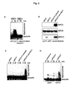
Figure 2
-
Reconstitution of active, CDH1-responsive holo-APC from Δ2/11-APC and
recombinant APC2 and APC11
- A) In vitro ubiquitination assay with purified GST-APC11/APC2
subcomplexes and securin in the presence of either UBC4 or UBCx.
- B) Western blot of Δ2/11-APC bound to αAPC4 beads after incubation with
APC2/11 lysates.
- C) In vitro ubiquitination assay with reconstituted APC and UBCx. The
substrate securin is detected by western blot.
- D) In vitro ubiquitination assay with reconstituted APC in the absence and
presence of CDH1.
-
Figure 3
-
A subcomplex consisting of APC1, 2, 4, 5, 11 elutes separately from holo-APC
from an anion exchange resin.
- A) HeLa cell lysates were subjected to Resource Q anione exchange
chromatography and eluted with a KCI gradient. αAPC4-IPs from
different fractions were separated by SDS-PAGE and visualized by
silver staining (left) and western blotting (right).
- B) In vitro ubiquitination assay with holo-APC or Q350-APC and
recombinant securin as substrate. Substrate and ubiquitin chains were
detected by western blot.
- C) CDH1 binding assay. IPs of holo-APC or Q350-APC where incubated
with recombinant HisHA-tagged CDH1 and immunoblotted for the
presence of endogenous and recombinant CDH1.
-
Figure 4
-
A possible APC-coactivator interaction module
-
3D model of the TPR domain of APC3, based on predicted structural
homologies to PEX5. Individual TPRs are depicted in different colors. The
PTS1 peptide crystallized with PEX5 has been replaced by the C-terminal
pentapeptide of APC10 (YRSIR), which is known to interact with APC3.
Figure 5
-
The TPR-containing subunits APC3 and APC7 mediate APC interactions with
C-terminal IR motifs
- A) Matrix-coupled C-terminal peptides were incubated with insect cell
lysates containing recombinant APC subunits. 10% of input (In),
supernatant (S), and 100% of bound material (B) were analyzed by
western blot.
- B) Late-eluting Q column fractions containing mainly TPR subunits were
used as input in peptide binding assays as in A), with CDC20/CDH1
peptides or two different control peptides.
- C) Fractions containing holo-APC were used as input in the described
peptide binding assay.
- D) Peptide binding assay with recombinantly expressed fragments of
APC7.
-
Figure 6
-
C-terminal CDH1/CDC20 peptides containing the IR motif inhibit CDH1 binding
and APC activation
- A) CDH1 binding assay. APC-IPs were incubated with recombinant CDH1
in the presence or absence of different peptides. CDH1 binding was
analyzed by quantitative western blotting with [125l]-labelled secondary
antibodies.
- B) In vitro ubiquitination assay. APC-IPs treated as in A were washed and
used in ubiquitination assays with [125l]-labelled cyclin B as substrate.
-
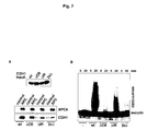
Figure 7
-
Binding and activation of the APC depends on CDH1's C-terminal IR motif and
the C-box
- A) CDH1 binding assay. αAPC3-lPs from APC-containing fractions or
APC-free control fractions were incubated with insect cell lysates
containing recombinant wild-type CDH1 or CDH1 mutated in the C-box
(ΔCB), the IR motif (ΔIR), or in both motifs (2xΔ).
- B) In vitro ubiquitination assay. APC was incubated with wild-type or
mutant CDH1 as in A and used in a ubiquitination assay with
recombinant securin as substrate.
-
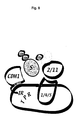
Figure 8
-
Cartoon summarizing our current views of APC assembly and mechanism
APC2/11, who recruit ubiquitin-loaded E2, can be dissociated from the
remaining complex. They can also form a stable complex together with
APC1/4/5 in the absence of other subunit, especially the TPR proteins.
CDH1 interacts with the TPR subunits APC3 and APC7 and might therefore
be juxtaposed to APC2/11, consistent with its apparent role as substrate-presenting
factor.
Figure 9
Δ2/11-APC sediments as an intact complex
-
Sucrose density gradient centrifugation of holo-APC (A) and Δ2/11-APC (B).
Fractions were concentrated adnd anlayzed by western blot with APC
antibodies. As input sample and antibody control, holo-APC was used in
A and B.
Figure 10
-
Anion exchange chromatography separates distinct populations of APC
subunits
-
Hela extracts were separated on a Source15Q column and eluted with a
KCI gradient. Protein-containing fractions were collected and analyzed by
western blotting for APC subunits, four of which are shown as examples.
-
In the Examples, if not otherwise stated, the following materials and methods
were used:
a) Antibodies, peptides, mutagenesis and recombinant protein
expression
-
Antibodies raised against APC2, APC3/CDC27, APC8/CDC23, CDH1, APC10
and CDC26 have been described 7,10,14,25,26 . Antibodies against APC4, APC5,
APC6/CDC16, APC7 and APC11 were raised by immunizing rabbits with
synthetic peptides coupled to Keyhole Limpet Hemocyanin (APC4:
CIVIKVEKLDPELDS, SEQ ID NO:5. APC5: CELTSRDEGERKMEKEEL;
SEQ ID NO:6. APC6: CLETSRKTPDSRPSL; SEQ ID NO:7. APC7:
CQKMEKEESPTDATQEED; SEQ ID NO:8. APC11: CRQEWKFKE,
SEQ ID NO:9). UBCx antibodies were raised in mice against recombinantly
expresses UBCx.
-
Synthetic peptides used in binding and inhibition assays were
CLRREREKASTSKSSLIHQGIR (CDC20 C-terminus; SEQ ID NO:2),
CFSKTRSTKESVSVLNLFTRIR (CDH1 C-terminus;SEQ ID NO:1),
CESDLHSLLQLDAPIPNAPPARW (CDC20 N-terminus; SEQ ID NO:10), and
CAVWSLSSCKPGFGVD (control;SEQ ID NO:11).
-
Expression and purfication of recombinant E1, E2 enzymes as well as
His-myc-tagged securin was performed as described 10.
-
Recombinant HisHA-tagged CDH1 was expressed and purified as
described 23. The QuickChange Site-directed mutagenesis kit (Stratagene,
La Jolla, CA) was used to generated the DRY box deletion (GDRFIPSR to
AAAAAAAA; SEQ ID NO:12). For truncation of the C-terminal IR, a premature
stop codon was introduced by PCR.
-
Baculoviruses encoding the CDH1 mutants, His-APC2, HisHA-tagged APC3,
5, 6, 7, 8 and HA-APC11 were generated using the BAC-TO-BAC system
(Invitrogen), and proteins were expressed in Sf9 or Hi5 cells.
b) Isolation and Reconstitution of Δ2/11-APC
-
Extracts from logarithmically growing HeLa cells were prepared in buffer A
(20 mM Tris-HCl, pH 7.5, 100 mM NaCl, 0.2% NP-40, 20 mM -
glycerophosphate, 10% glycerol, 1 mM NaF, 0.5 mM DTT), and APC was
immunopurfied using an APC3 peptide antibody 14 . Bead-bound APC was
washed for 90 min with buffer B (50 mM NaP04, 500 mM NaCl, 1 % Triton
X-100, 1 mM DTT, pH 6.5) for removal of APC2 and APC11 followed by a brief
5 min wash with buffer C (20 mM Tris/HCl, 150 mM NaCl, 0.02% Tween-20,
0.5 mM DTT, pH 7.6). Complexes were eluted with buffer C plus 1 mg/ml
CDC27-peptide for 2 h, and holo-APC removed from the eluate by
immunoprecipitation with APC2-30 monoclonal antibodies 25. The remaining
supernatant containing the purified Δ2/11-APC was finally re-immunoprecipitated
using APC4 antibodies or analyzed directly by western
blotting or sucrose density gradient centrifugation.
-
For reconstitution, Δ2/11-APC on APC4 antibody beads was incubated for
30 min at 4°C in extracts prepared from Sf9 insect cells (in buffer A). That had
been infected with baculoviruses encoding His-tagged APC2, HA-tagged
APC11 or both proteins. Extracts were centrifuged at 100.000xg for 60 min
before incubation with Δ2/11 APC. Beads were subsequently washed 5x with
buffer C and analysed for APC2/11 binding and ubiquitination activity.
c) CDH1 binding and in vitro ubiquitination assays
-
Immunopurified holo-APC or subcomplexes were washed and incubated with
purified recombinant CDH1 (0.5-1.0 µg per 10 µl bead volume) in TBS/T, or
with insect cell lysates in TBS/T containing recombinant CDH1 proteins as
indicated. Peptides for competition were used at 1 mg/ml, with the addition of
0.5 mM DTT to the buffer. Subsequently, bead-bound complexes were washed
3x with TBS/T and immunoblotted for binding of CDH1 or used in ubiquitination
assays. In vitro ubiquitination reactions were performed as described26.
Securin-myc-his substrate was detected by immunoblotting with monoclonal
anti-Myc 9E10 antibodies. Alternatively, an iodinated N-terminal recombinant
fragment of sea urchin cyclin B (amino acids 13-110) was used and visualized
by autoradiography14.
d) Anion exchange chromatography
-
Pellets from 5x10⁹ logarithmically grown HeLa cells (4C, Belgium) were lysed
in 20 mM TrisCl pH7.7, 150 mM KCI, 0.5 mM DTT, 0.1% Tween-20, Protease
Inhibitor mix (Sigma) using a french press and cleared by 100,000xg
centrifugation for 1 h. After filtration through a 0.45 µm filter, extracts were
loaded onto a 60ml Source15Q anion exchange column (Amersham) at
4 ml/min. The column was washed with 20 mM Tris-Cl pH7.7, 150 mM KCI,
0.5 mM DTT until a drop in A280 indicated the end of the flowthrough. Elution
was carried out with a gradient from 150 mM to 600 mM KCl. Elution fractions
were collected, desalted using PD10 gravity flow colums (Pharmacia) and
concentrated in Centriprep cartridges.
e) Peptide binding assays
-
Peptide interaction assays were carried out as described in Wendt et al,
200118. Synthetic peptides (see above) were immobilized via N-terminal
cysteine residues onto maleinimide-activated POROS matrix (Applied
Biosciences) in 0.1 M HEPES, pH 7.9 for 3 hours. After washing with slightly
acidic buffers, 100 µg of the resulting matrix were incubated with insect cell
lysates containing recombinant APC subunits, or with fractions from anion
chromatography, in total volumes of 100 µl of TBS/T pH 7.5. After stringent
washing in TBS/T 0.5 M NaCl, samples were analyzed by SDS-PAGE and
immunoblotted with 12CA5 anti-HA antibodies or antibodies against APC
subunits.
Example 1
-
The catalytic subunits APC2 and APC11 can be dissociated from holo-APC
-
Total or partial reconstitution from recombinant subunits has been essential for
understanding the assembly and function of several macromolecular
complexes. However, to date all attempts to reconstitute the APC have failed,
and little information about the function of each of its at least 11 subunits is
available. So far, the only recombinant subcomplex that has yielded any
functional information is APC2/11, consisting of the cullin domain protein APC2
and the small RING-H2 finger protein APC11 12,13. This subcomplex is able to
form polyubiquitin chains and transfer some of them onto APC substrates, but
in a non-regulated manner. To gain further insight into the functions and
molecular interactions of APC subunits, a reverse strategy was pursued and
conditions that dissociate individual subunits from holo-APC searched for.
-
Based on previously developed methods to immunopurify human APC14, APC
bound to APC3 antibody beads was treated with various buffer conditions.
Surprisingly, it was found that slightly lowered pH reduced the amount of APC2
and APC11 bound to holo-APC. After extensive washing of immunopurified
APC with a high salt sodium phosphate buffer of pH 6.5 (see material and
methods for details) and subsequent peptide elution, complexes still containing
APC2 were depleted by immunoprecipitation with APC2 antibodies.
Re-immunoprecipitation with APC4 antibodies showed that the remaining
complex (henceforth called Δ2/11-APC) has indeed quantitatively lost APC2
and APC11 (Figure 1A). This pH-dependent dissociation can first be observed
at pH 7.0 and increases with further lowering of the pH. Decrease below
pH 6.3 caused considerable loss of APC from the antibody beads, and it was
not possible to assay loss of additional subunits under these conditions. The
only other subunit slightly affected by lowered pH appears to be APC10,
whereas the relative amounts of all other subunits were unchanged
(Figure 1 A).
-
As further confirmation that Δ2/11-APC is still an intact macromolecular
complex, it was fractionated by sucrose density gradient centrifugation. All
remaining subunits of Δ2/11-APC sedimented as a single peak of about 22S
(Figure 9), this S value being almost identical to the one observed with
holo-APC 14 . This indicates that the loss of APC2/11 does not grossly change
mass, shape and molecular composition of the APC.
-
Having confirmed the integrity of Δ2/11-APC, the inventors wanted to know
which functional properties of native APC it still retains. Interaction with
activators of the WD40 repeat family is essential for substrate-specific APC
activity. Δ2/11-APC was tested for binding to the activator CDH1. Both
holo-APC and Δ2/11-APC contained substoichiometric amounts of bound
endogenous CDH1, and incubation with purified recombinant CDH1 resulted in
additional loading of more activator (Figure 1B). APC2 and APC11 therefore
appear to be dispensable for CDH1 binding, consistent with the observations
that CDH1 can neither bind to nor activate recombinant APC2/11 complexes
(data not shown). Subsequently CDH1-loaded holo-APC and Δ2/11-APC were
analyzed for ubiquitination activity towards the substrate securin. Whereas
holo-APC converts securin into ubiquitin conjugates of higher molecular mass
in the presence of ATP, E1 and E2 enzymes, Δ2/11-APC is completely inactive
in this in vitro ubiquitination assay (Figure 1 C). APC2/11 are therefore not only
sufficient, but also strictly required for ubiquitination activity of the APC.
-
Despite the apparent integrity of Δ2/11-APC (Figure 1 A and Figure 9), the
possibility could not be fully excluded that the low pH treatment during
preparation might have caused irreversible damage to the subcomplex, and
that this damage has lead to loss of enzymatic activity. It was hence tried to
reconstitute active holo-APC by loading Δ2/11 -APC with recombinant APC2/11
complexes generated in baculovirus-infected insect cells. As shown in
Figure 2B, APC2/11 did indeed bind back to the depleted complex. To
distinguish a functional interaction from unspecific binding, an in vitro
ubiquitination reaction with UBCx as E2 enzyme was carried out. According to
these experiments, APC2/11 is only supported by the E2 enzyme UBC4,
whereas holo-APC is active in the presence of either UBCx or UBC4
(Figure 2A). Any substrate ubiquitination observed with UBCx must therefore
be due to holo-APC activity. Δ2/11-APC reconstituted with co-expressed
APC2/11 has regained UBCx-dependent activity towards securin (Figure 2C),
whereas incubation with APC11 or APC2 alone is not sufficient (Figure 2C).
Interestingly, re-binding was only observed when coexpressed APC2 and
APC11 was used - neither protein did bind alone, and posttranslationally
mixed APC2 and APC11 also failed to bind and reconstitute activity
(Figure 2B). This is consistent with the observation of Tang and coworkers 12.
That APC2 and APC11 only interact upon co-expression, and implies that this
interaction either creates a common interface for APC association or is
essential for proper folding and structural integrity of these two subunits.
-
It was tested if reconstituted APC would be responsive to CDH1 activation.
Indeed, ubiquitination activity of reconstituted APC in the absence of CDH1
was only slightly increased compared to Δ2/11-APC incubated with control
extracts, but significant activation was achieved in the presence of CDH1
(Figure 2D). This strict CDH1-dependence is a hallmark of holo-APC and
further proof that the observed activity is not due to APC2/11 subcomplexes,
since they are not CDH1-responsive (data not shown and 12).
Example 2
-
APC1, 2, 4, 5 and 11 form a stable subcomplex
-
In previous sucrose density gradient or anion exchange fractionation
experiments, it had been noted that APC2 is not only found in one peak
fraction with holo-APC, but sometimes also in a distinct second peak whose
sedimentation behavior implied association with additional proteins (data not
shown and Figure 9). To investigate this further, extracts from logarithmically
growing HeLa cells were subjected to anion exchange chromatography on a
Source15Q column. Bound material was eluted with a potassium chloride
gradient. Holo-APC (identified by the presence of all 11 known subunits) eluted
in a sharp peak at around 450 mM KCI (Figure 10). While some subunits,
especially the TPR-containing proteins APC3, APC6 and APC7, were
additionally found trailing through several more fractions, other subunits
formed a distinct second peak eluting already at 350 mM KCI (Figure 10).
These fractions did indeed contain APC2 as well as its close interaction
partner, APC11. To test if these subunits are actually associated in an
independent subcomplex, APC3-containing complexes were depleted from the
350 mM fractions using APC3 antibodies. Subsequent immunoprecipitation
with antibodies against APC2, APC4 or APC5 reproducibly yielded the same
set of APC1, 2, 4, 5, and 11 in apparently stoichiometric amounts (Figure 3A),
without significant contamination by any other subunit. This subcomplex,
hereafter called "Q350-APC", was stable even after peptide elution from the
APC4 antibody and subsequent re-precipitation with APC5 antibodies (data
not shown).
-
It could not be judged if Q350-APC was generated during extract preparation or
fractionation, or if it represents a physiological subcomplex, either as an
assembly intermediate or even a functional entity. Nevertheless, its apparent
stability opened the opportunity to investigate its properties further. In a
ubiquitination assay, Q350 -APC did not show significant activity towards securin
(Figure 3B), despite the presence of APC2 and APC11. This is unexpected
given the ability of recombinant APC2/11 complexes to ubiquitinate APC
substrates, but might support a model in which additional subunits would limit
unregulated access of substrates to the catalytically active subunits.
Consistent with this, Q350-APC was still able to generate polyubiquitin chains
(data not shown). This implies that the deficiency in securin ubiquitination
might be a direct consequence of the absence of substrate specificity factors.
Therefore Q350-APC and APC from the 450mM KCI fractions were incubated
with recombinant CDH1 and assayed binding. As shown in Figure 3C,
holo-APC was associated with endogenous and additional recombinant CDH1.
Neither form of CDH1, on the other hand, was bound to Q350-APC (Figure 3C).
The lack of substrate ubiquitination in the absence of CDH1 is therefore
consistent with a role for CDH1 in substrate recognition, as implied by studies
from several labs.
Example 3
-
TPR domains and a C-terminal peptide motif as APC-CDH1 interaction
modules
-
To understand APC activation, it is important to map the molecular interactions
between CDH1 and APC subunits. Several determinants in CDH1 have been
found to influence APC binding, namely the so-called C-box 20, the C-terminal
isoleucine-arginine (IR) motif 16,20, and phosphorylation status of CDH122,23. On
the other hand, which subunits and sequences within the APC are responsible
for interaction with CDH1 is unclear. The analysis of Δ2/11-APC and Q350-APC
has revealed that APC2/11 are dispensable for CDH1 binding and that APC1,
2, 4, 5 and 11 are not sufficient for CDH1 binding. This leaves APC3, APC6,
APC7, APC8, CDC26 and APC10 as possible CDH1 receptors. Interestingly,
APC3, 6, 7 and 8 all contain multiple TPRs, a motif present in several adaptor
proteins like the co-chaperone Hop or the peroxisomal importing receptor
PEX5, where it is involved in interactions with C-terminal peptides. Structure
prediction tools suggest that the TPR domains in APC3, APC6 and APC7 fold
in a very similar manner to PEX5 (Figure 4), and the inventors have previously
shown that APC3 binds APC10 through APC10's extended C-terminal
peptide 18. Figure 4 shows APC3's TPR domain modeled on the PEX5
structure 24, with PEX5's cognate PTS1 peptide replaced by APC10's
C-terminal pentapeptide, YRSIR. Strikingly, it was noted that APC10
orthologues from different species all share this conserved IR (LR in yeast)
dipeptide with the APC activators CDH1 and CDC20. This remarkable
conservation in several proteins in the APC context suggested an important,
possibly common functional role for this motif.
-
To test if APC3 would also interact with the C-termini of CDH1 and its related
activator CDC20, insect cell lysates containing recombinantly expressed APC3
were incubated with synthetic peptides coupled to a matrix through their
N-terminus. After washing, peptide-bound material was analyzed by western
blot. In this assay, about 10% of input APC3 bound to the C-terminal peptide of
CDC20, and even stronger binding was observed to a CDH1 C-terminal
peptide (Figure 5A). The interaction was specific, since APC3 did not bind to a
control peptide, and recombinant APC5 interacted with neither the CDC20 nor
the CDH1 peptide (Figure 5A). When the other three TPR subunits were
tested, it was found that APC8 did not bind either peptide, and APC6 bound
only in low amounts that were also observed to bind to control peptides
(Figure 5A and data not shown). APC7, on the other hand, interacted strongly
with the CDH1 C-terminus and moderately with CDC20's C-terminus
(Figure 5A), in a manner very similar to APC3. Interestingly, APC3 and APC7
are highly related in their primary sequences. In budding yeast, which lacks
APC7, they share Cdc27p as their closest homologue. This suggests that
APC3 and APC7 might have originated from gene duplication and might still
have overlapping functions.
-
In order to exclude any non-specific effects caused by incorrect folding of
recombinantly expressed proteins, an alternative source of APC subunits was
used in the peptide binding assays. As described above, late-eluting fractions
from the ReSource Q column contained considerable amounts of the TPR
subunits APC3, APC6, and APC7, while other subunits were found in lower
abundance (Figure 10). Although these TPR subunits can still be partially
co-precipitated, they do not seem to be in stoichiometric complexes anymore
(data not shown), and the inventors thus considered them a satisfactory
source of "native" APC subunits. As with recombinant proteins, APC3 and
APC7 were significantly retained by the CDH1 peptide matrix, whereas APC6
was not (Figure 5B). CDC26, which is also present in these fractions, showed
no binding at all (Figure 5B).
-
It was tested if CDH1's C-terminus would be sufficient to interact with
holo-APC. For this, APC-containing fractions were incubated with the peptide
matrix and analyzed for binding of APC by detection of all subunits. Every
known subunit was strongly enriched on the CDH1 peptide matrix (Figure 5C)
but not on control peptides (data not shown). Since APC6 and CDC26 from
late-eluting fractions did not bind the CDH1 peptide by themselves (Figure 5B),
their interaction, as well as that of other subunits like APC2 and APC11, must
be mediated through the holo-complex. Surprisingly, endogenous CDH1 was
also enriched on the peptide matrix (Figure 5C). Since purified APC is loaded
with substoichiometric amounts of endogenous CDH1 (Figures 1 B and 3C),
this co-binding could be mediated by dimerization of APC. An alternative and
more intuitive explanation is the presence of more than one receptor for IR
peptides per complex (see discussion).
-
Lastly, the region in APC7 responsible for binding to IR peptides was mapped.
An APC7 fragment containing the N-terminal 297 amino acids did not exhibit
binding, whereas the C-terminal part (amino acids 298 to 565) comprising the
block of TPRs was stronlgy enriched on the peptide matrix (Figure 5D). This
shows that the TPR domain is sufficient for interaction with IR peptides.
Example 4
-
C-terminal peptides of CDH1 and CDC20 inhibit APC activation
-
What are the mechanistic consequences of the observed interactions between
IR tail peptides and APC subunits? To address this, it was tested if the
peptides would influence APC activity in in vitro ubiquitination assays.
Immunopurified APC was loaded with recombinant CDH1 in the presence or
absence of IR-tail or control peptides, washed, and used to ubiquitinate a
radioactively labeled fragment of cyclin B (Figure 6B). The C-terminal peptides
of CDH1 and CDC20 both blocked APC activation efficiently, lowering
ubiquitination activity to almost the level of APC that has not been activated by
exogenous CDH1. In contrast, an N-terminal CDC20 peptide had no effect on
APC activation. Quantitative immunoblotting using radiolabeled antibodies
against APC2 and CDH1 showed that the inhibition corresponded with
diminished binding of CDH1 (Figure 6A). As in the peptide binding assays
(Figure 5), CDH1's C-terminal peptide had a stronger effect than the one from
CDC20. This competitive inhibition of CDH1 loading implies that the IR tails do
not function in catalysis, but are rather responsible for actively targeting CDH1
to the APC.
Example 5
-
CDH1 binding and APC activation depends on two motifs in CDH1
-
Having established that C-terminal IR tails are sufficient to interact with APC
and interfere with its activation, the inventors wanted to know how their loss
would influence CDH1 activity. To this end, a baculovirus encoding CDH1 with
a deletion of the C-terminal dipeptide (ΔIR) was generated. Another conserved
motif located in CDH1's N-terminal region, the C-box, has been shown to be
required for APC binding in yeast 20. Therefore CDH1 versions with mutations
in the C-box (ΔDRY), and in both motifs (2xΔ) were also generated. After
expression in insect cells, the lysates were incubated with antibody-bound
APC or control antibody beads, washed, and tested for activity and binding of
recombinant CDH1. Figure 7A shows that both mutations in the DRY box and
in the IR tail diminished binding significantly but not completely. Combining
both mutations resulted in a further decrease in affinity, indicating that DRY
box and IR tails cooperate to achieve optimal APC binding (Figure 7A).
-
Similar effects were seen in ubiquitination assays: the IR tail deletion mutant
retained significant levels of activity, whereas the DRY box mutant was almost
completely inactive (Figure 7B). Taken together, these data strongly argue for
additive roles of CDH1's conserved APC interaction motifs, and they also show
that binding and activation go hand in hand.
References
-
- 1. Peters, J. M. The anaphase-promoting complex: proteolysis in mitosis
and beyond. Mol Cell 9, 931-43. (2002).
- 2. Harper, J. W., Burton, J. L. & Solomon, M. J. The anaphase-promoting
complex: it's not just for mitosis any more. Genes Dev 16, 2179-206.
(2002).
- 3. Pickart, C. M. Mechanisms underlying ubiquitination. Annu Rev
Biochem 70, 503-33. (2001).
- 4. Lamb, J. R., Michaud, W. A., Sikorski, R. S. & Hieter, P. A. Cdc16p,
Cdc23p and Cdc27p form a complex essential for mitosis. Embo J 13,
4321-8. (1994).
- 5. Blatch, G. L. & Lassle, M. The tetratricopeptide repeat: a structural motif
mediating protein-protein interactions. Bioessays 21, 932-9. (1999).
- 6. Kominami, K., Seth-Smith, H. & Toda, T. Apc10 and Ste9/Srw1, two
regulators of the APC-cyclosome, as well as the CDK inhibitor Rum1
are required for G1 cell-cycle arrest in fission yeast. Embo J 17,
5388-99. (1998).
- 7. Grossberger, R. et al. Characterization of the DOC1/APC10 subunit of
the yeast and the human anaphase-promoting complex. J Biol Chem
274, 14500-7. (1999).
- 8. Zachariae, W. et al. Mass spectrometric analysis of the anaphase-promoting
complex from yeast: identification of a subunit related to
cullins. Science 279, 1216-9. (1998).
- 9. Yu, H. et al. Identification of a cullin homology region in a subunit of the
anaphase-promoting complex. Science 279, 1219-22. (1998).
- 10. Gmachl, M., Gieffers, C., Podtelejnikov, A. V., Mann, M. & Peters, J. M.
The RING-H2 finger protein APC11 and the E2 enzyme UBC4 are
sufficient to ubiquitinate substrates of the anaphase-promoting complex.
Proc Natl Acad Sci U S A 97, 8973-8. (2000).
- 11. Leverson, J. D. et al. The APC11 RING-H2 finger mediates
E2-dependent ubiquitination. Mol Biol Cell 11, 2315-25. (2000).
- 12. Tang, Z. et al. APC2 Cullin protein and APC11 RING protein comprise
the minimal ubiquitin ligase module of the anaphase-promoting
complex. Mol Biol Cell 12, 3839-51. (2001).
- 13. Ohta, T., Michel, J. J., Schottelius, A. J. & Xiong, Y. ROC1, a homolog
of APC11, represents a family of cullin partners with an associated
ubiquitin ligase activity. Mol Cell 3 , 535-41. (1999).
- 14. Gieffers, C., Dube, P., Harris, J. R., Stark, H. & Peters, J. M.
Three-dimensional structure of the anaphase-promoting complex.
Mol Cell 7, 907-13. (2001).
- 15. Vodermaier, H. C. Cell cycle: Waiters serving the Destruction
machinery. Curr Biol 11, R834-7. (2001).
- 16. Passmore, L. A. et al. Doc1 mediates the activity of the anaphase-promoting
complex by contributing to substrate recognition. Embo J 22,
786-96. (2003).
- 17. Carroll, C. W. & Morgan, D. O. The Doc1 subunit is a processivity factor
for the anaphase-promoting complex. Nat Cell Biol 4, 880-7. (2002).
- 18. Wendt, K. S. et al. Crystal structure of the APC10/DOC1 subunit of the
human anaphase-promoting complex. Nat Struct Biol 8, 784-8. (2001).
- 19. Au, S. W., Leng, X., Harper, J. W. & Barford, D. Implications for the
ubiquitination reaction of the anaphase-promoting complex from the
crystal structure of the Doc1/Apc10 subunit. J Mol Biol 316, 955-68.
(2002).
- 20. Schwab, M., Neutzner, M., Mocker, D. & Seufert, W. Yeast Hct1
recognizes the mitotic cyclin Clb2 and other substrates of the ubiquitin
ligase APC. Embo J 20, 5165-75. (2001).
- 21. Zheng, N. et al. Structure of the Cul1-Rbx1-Skp1-F boxSkp2 SCF
ubiquitin ligase complex. Nature 416, 703-9. (2002).
- 22. Zachariae, W., Schwab, M., Nasmyth, K. & Seufert, W. Control of cyclin
ubiquitination by CDK-regulated binding of Hct1 to the anaphase
promoting complex. Science 282, 1721-4. (1998).
- 23. Kramer, E. R., Scheuringer, N., Podtelejnikov, A. V., Mann, M. & Peters,
J. M. Mitotic regulation of the APC activator proteins CDC20 and CDH1.
Mol Biol Cell 11, 1555-69. (2000).
- 24. Gatto, G. J., Jr., Geisbrecht, B. V., Gould, S. J. & Berg, J. M.
Peroxisomal targeting signal-1 recognition by the TPR domains of
human PEX5. Nat Struct Biol 7, 1091-5. (2000).
- 25. Gieffers, C., Peters, B. H., Kramer, E. R., Dotti, C. G. & Peters, J. M.
Expression of the CDH1-associated form of the anaphase-promoting
complex in postmitotic neurons. Proc Natl Acad Sci U S A 96,
11317-22. (1999).
- 26. Kramer, E. R., Gieffers, C., Holzl, G., Hengstschlager, M. & Peters, J.
M. Activation of the human anaphase-promoting complex by proteins of
the CDC20/Fizzy family. Curr Biol 8 , 1207-10. (1998).
- 27. Gershkovich, A.A. and Kholodovych, V.V. (1996), J Biochem Biophys
Meth 33, 135
- 28. Matayoshi, E.D. (1990), Science 247, 954
- 29. Hatfield P.M., Callis J. & Vierstra R.D. (1990) J Biol Chem 265, 15813-7
- 30. Hatfield P.M. & Vierstra R.D. (1992)Biol Chem 267, 14799-803
- 31. Murray, A. (1991) Methods Cell Biol. 36, 581-605
- 32. Kieber-Emmons T, Murali R, Greene MI. Therapeutic peptides and
peptidomimetics. Curr Opin Biotechnol 1997 Aug;8(4):435-41
- 33. Ripka A.S., and Rich D.H., Peptidomimetic design. Curr Opin Chem Biol
1998 Aug; 2(4):441-52
-