-
The present invention relates to gene expression in normal
cells and cells of tumors and particularly to a novel marker
protein associated with epithelial tumors, e.g., ovarian,
mammary, stomach, kidney, thyroid, and lung carcinomas, Drop1,
a dystrophin-related protein as well as a nucleic acid
molecule encoding Drop1. Furthermore, the present invention
relates to various diagnostic and therapeutic uses based on
the finding that the expression of Drop1 is downregulated in
epithelial tumors.
-
Tumorigenesis represents a complex multistage process in which
genetic changes and environmental factors are thought to
deregulate the cellular processes that control cell
proliferation and differentiation. Among the gynaecological
tumors, ovarian carcinoma has the highest mortality. It
typically metastasizes to the peritoneal cavity. A
comprehensive biological characterisation is crucial to
improve the prognosis for cancer, e.g., ovarian cancer
patients. Though morphological characterisation of the tumor
in many cases gives a good hint towards prognosis and
prediction of the response to specific therapies, its accuracy
is limited and fails in many cases. Especially some tumors of
low malignant potential turn out to be much more dangerous
than initially thought. Therefore, markers are important which
define the malignancy by its biological characteristics on the
level of gene expression. These markers can also predict the
possible success of a chemotherapy. Im mammary carcinoma, for
instance, tumors overexpressing HER2/neu respond better to
chemotherapy involving a taxane and anthracyclin than to one
using alkylating agents. Nuclear proteins interacting with
chemotherapeutic agents might also be involved in the
cytotoxic mechanisms of these drugs, or their modified
expression might be responsible for the failure of the
treatment. In summary, the use of reliable diagnostic
molecular markers would be helpful for an understanding of the
molecular basis of tumors, e.g. ovarian and mammary
carcinomas, for distinguishing benign from malign tissue and
for grading and staging carcinoma. It can be expected that
such markers are also useful for the development of novel
therapeutic avenues for cancer treatment.
-
Thus, the technical problem underlying the present invention
is to provide means for diagnosis and therapy of tumors, in
particular epithelial carcinomas, which overcome the
disadvantages of the prior art diagnostic and therapeutic
methods.
-
The solution to said technical problem is achieved by
providing the embodiments characterized in the claims.
-
During the experiments leading to the present invention a gene
encoding a novel protein, Drop1, was found, which is
downregulated in most cases of several types of carcinoma. Its
expression is downregulated 14- to 210-fold in 85% of the
ovarian and mammary carcinomas investigated. Similar
downregulation was observed in a cancer profiling array for
uterine, stomach, kidney, lung, and thyroid carcinomas. The
Drop1 mRNA is present in only two out of eight ovarian
carcinoma cell lines. The Drop1 gene comprises 56 exons in a
genomic sequence of 180 kb. Starting from a differential
display fragment of 680 bp, the 10.5 kb mRNA sequence was
completed using RACE and an ovarian cDNA library as a
template. The results were confirmed by overlapping PCR
products using total cDNA from ovarian epithelial cells.
Northern blots also show a diffuse band at 10 - 11 kb, mainly
expressed in testes and in ovary. This was confirmed by in
situ hybridisation. The gene is located in the chromosome
region 6q25, where a loss of heterozygosity has been described
for several types of carcinomas. Therefore, deletions may also
cause down-regulation of this gene. The ORF predicts a protein
of 349 kDa with a pI of 5.7, the sequence of which is only
distantly related to muscle dystrophin, yet the overall
structure is similar. It features two calponin homology
domains and 12 spectrin-like repeats. Such domains are also
found in muscle dystrophin. Their loss or truncation results
in progressive muscular dystrophy with frequently fatal
consequences. Drop1, in addition, displays an AAA domain of
some 220 amino acid residues, which characterizes a large
family of ATPases acting as ATP-dependent protein clamps.
Dystrophin is present at the cytoplasmic membrane, where it
integrates proteins into sterically fixed complexes. Drop1, in
contrast, is located at or in the nucleus, where it may have a
similar integrating function for nuclear protein complexes.
Drop1 malfunction may impede cell cycle regulation, centrosome
orientation and chromosome distribution. Dysfunction of
mechanisms regulating these processes occur during
pathogenesis and are key problems in malignant disease.
-
During the experiments resulting in the present invention it
has been shown that Drop1 has high homology in an overlap to
recently published proteins with unknown functions called
enaptin (EMBL/Genbank/DDBJ databases AF535142), Nesprin-1
(Zhang et al., Genomics 80(5),pp. 473-481 (2002)), KIAA1756
(EMBL/Genbank/DDBJ databases AB051543) and Syne-1
(EMBL/Genbank/DDBJ databases AY135172). Furthermore, some
sequences having homologies to Drop1 are shown in WO-A-02/40715.
However, this international publication comprises a
huge number of sequences without any specific relation to a
disease. It is not plausible that all these sequences play the
same or any role in such a large number of mentioned diseases.
-
In summary, Drop1 is a novel gene downregulated in epithelial
tumors with a broad range of characteristics relevant for
tumor biology. Downregulation, e.g. by promotor methylation,
deletion, or truncation is expected to contribute to the
development of carcinomas. Thus, Drop1 is very promising as a
prognostic as well as a predictive marker.
Brief description of the drawings
Figure 1: Differential display with cDNA from normal and tumor
tissue of patient 31, using anchor primer G and arbitrary
primer DOC, generated a cDNA fragment 31N/GDOC/1 dd (arrow)
-
To avoid artefacts, RNA from both sources was twice reversely
transcribed, and two differential display experiments were
performed with each of the resulting four cDNAs, yielding
eight samples run in parallel on the same gel for comparison.
The figure shows the autoradiogram of the bands. The
differentially expressed band was excised from the gel,
eluted, amplified and cloned into pCRII-TOPO.
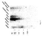
Figure 2: Mutitissue Northern Blot hybridized with a randomly
promed probe using the insert of clone 3R9 (see Fig. 6) as a
template
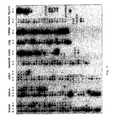
Figure 3: Autoradiogram of a cancer profiling array (BD
Biosciences Clontech, Cat.# 7841-1 Lot #2040887) including
amplified and normalized cDNA from 241 tumor (C) and
corresponding normal tissues (N) from individual patients,
spotted side by side
-
Boxed areas contain three cDNA spots from the same patient:
top left normal, top right metastatic tissue, bottom right
primary tumor. The array had previously been tested for equal
loading of cDNA in the spots by hybridisation with a
digoxigenin-labeled cDNA probe. Clone 3R9 was used to generate
the radioactive Drop1 probe for hybridisation.
Figure 4: Comparative in situ hybridisation of tissue sections
with an antisense probe generated from clone 3R9
-
Right: both, tube epithelium (top) and ovarian surface
epithelium (bottom) of patient 91 express Drop1 at a
comparable level. Left: Tube epithelium (top) of patient 87
expresses Drop1 at a considerably higher level that the
ovarian carcinoma (bottom) of the same patient.
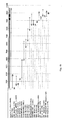
Figure 5: Chromosome region 6q25.1, containing the Drop1 gene
-
Several groups have reported loss of heterozygosity for this
region in ovarian, breast, uterine, and non small cell lung
cancer, as indicated. Top line: chromosomal markers. Estrogen
receptor 1 (ESR1, alpha) and early mitotic inhibitor 1(EMI1)
are expressed from the plus strand (>), Drop1 from the minus
strand (<).
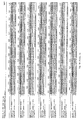
Figure 6: Sequence details for Drop1
-
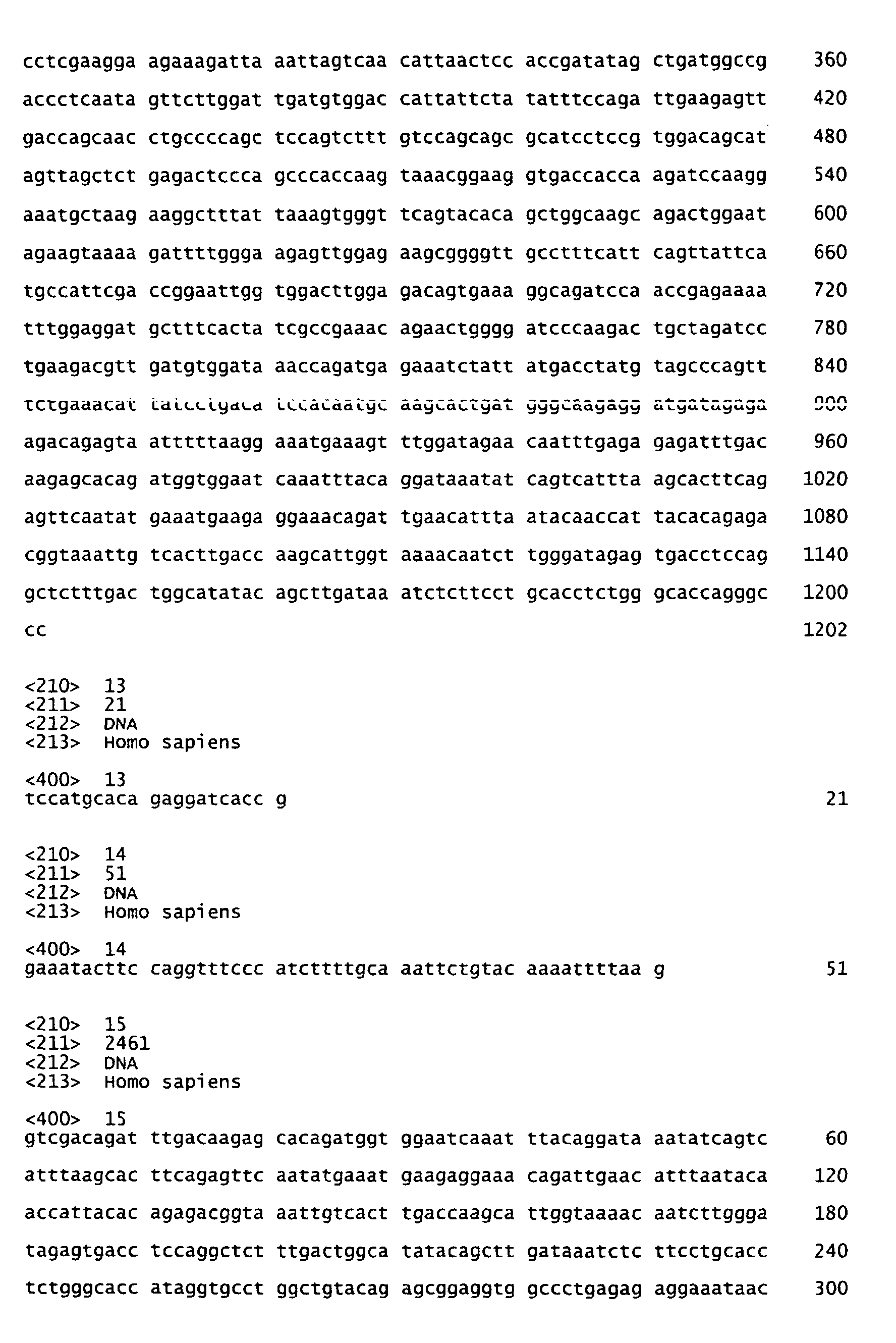
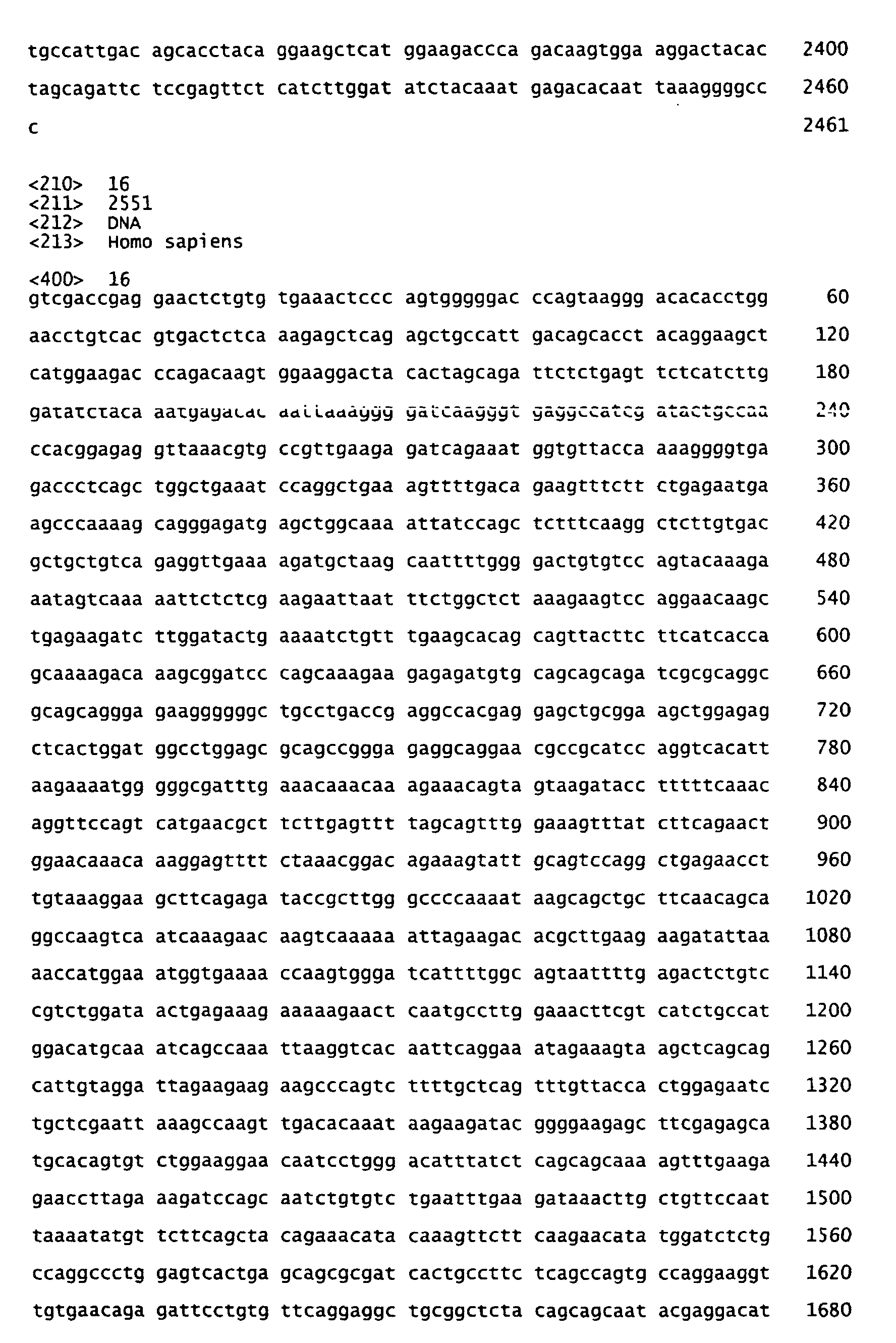
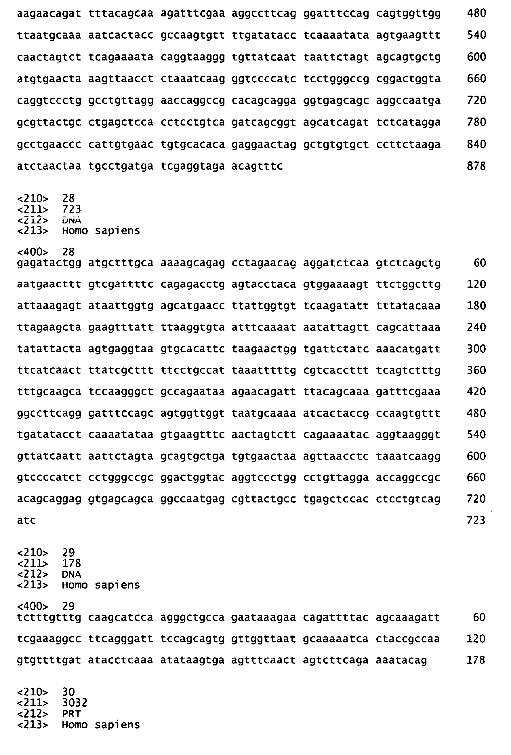
The figure shows the location in the contig of sequences
mentioned in the text (A). The sequence was established
starting with the 681 bp of the differential display
fragment 31N/GDOC/1 dd. Primers L467 and U285 (their
direction inverted in the final ORF) were derived from it
and used throughout for quantitative PCR. "RACE walking"
experiments were performed, based on a commercial RACE
ovarian library. Of these, 5R9 marks the 5' region of drop1,
3R9 was used to generate the probes for in situ
hybridisation, for multitissue Northern blots, and for the
cancer profiling array. For validation of these sequences by
overlapping PCR, total cDNA from SKOV-3 ovarian carcinoma
cells was used as a template (Drop 1A,C,B,D). The same
template yielded the clones used in bacterial and mammalian
vectors for expression of domains (Drop1 alpha, beta, gamma,
epsilon, and C-terminal domain). In total, more than 70
clones were aligned. Only upon completion of the Drop1
structure the enaptin sequence became available, of which
here the 5'10 000 bp are shown (B). For ease of comparison
the exons are designated accordingly. Drop1 is encoded by
the 5'region (exons 2 to 58) of the enaptin gene (147
exons). The Drop1 mRNA and protein, however, are entities
different from enaptin. Enaptin exons 07 and 12 are not used
in Drop1, and enaptin-exon 58 is read differently: for Drop1
it is read beyond the enaptin-exon58-intron transition. It
carries the stop codon for the ORF and a long 3' UTR that
includes the unused sequence of enaptin-exon 59.
Figure 7: Drop1 protein domains and overlapping expression
clones
-
CH1, 2: calponin homology domains, AAA: ATPase domain, NLS:
putative nuclear localization signal, S1 to S12: spectrin-like
domains. alpha to epsilon: overlapping clones for expression
from mammalian vectors.
Figure 8: Nuclear fluorescence of COS-7 cells transformed
with GFP-Drop1a including the two calponin homology domains at
the aminoterminus of Drop1
Figure 9: Monoclonal antibody mAB2.9, raised to the
recombinant carboxyterminal spectrin-like domain of Drop1,
recognizes this recombinant domain on immunoblots (left)
-
On blots of SKOV-3 ovarian carcinoma cell lysates (center) it
stains a high molecular band and various fragments in the
pellet (P), and a high molecular weight band and various
fragments in the pellet (P), and a large protein in the
supernatant (S). In cytospins of the same cells it stains
nuclei (right).
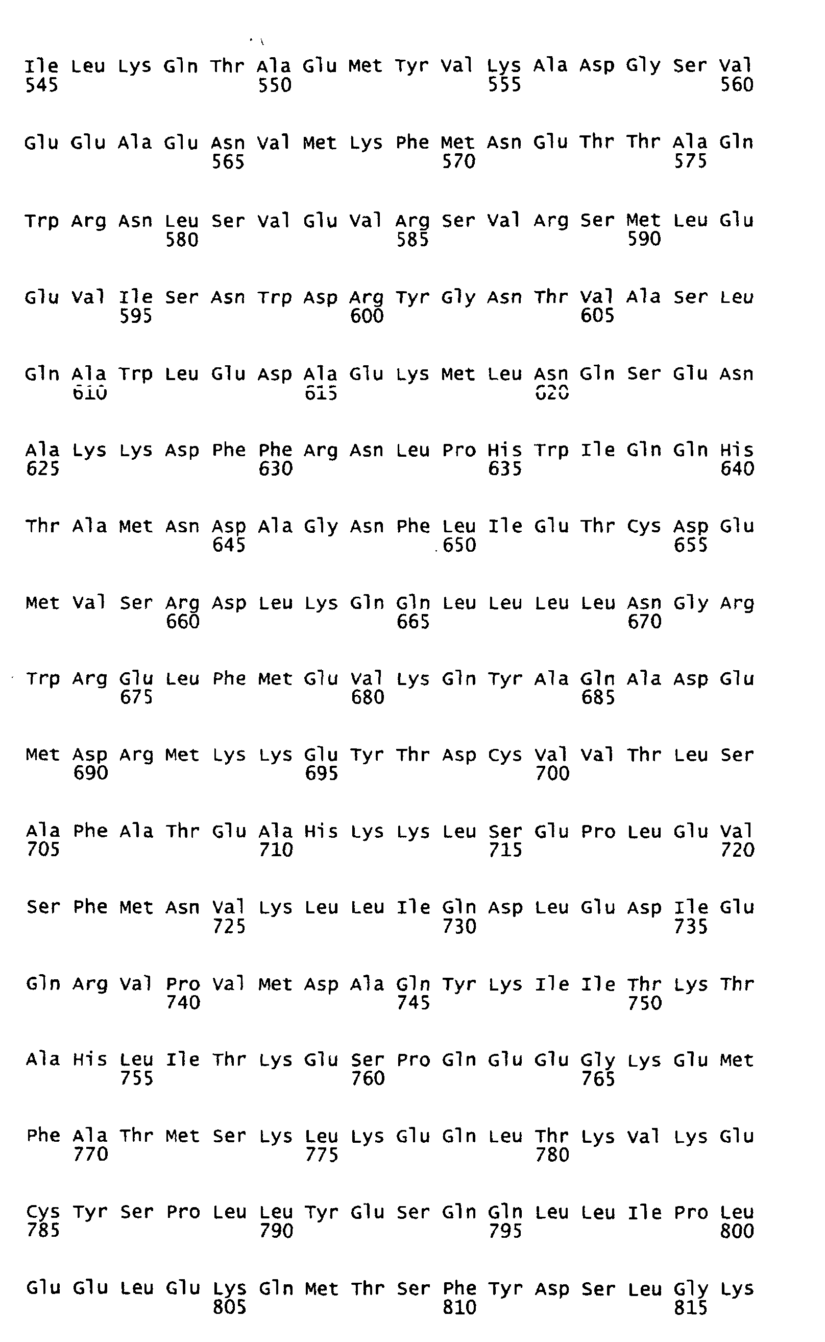
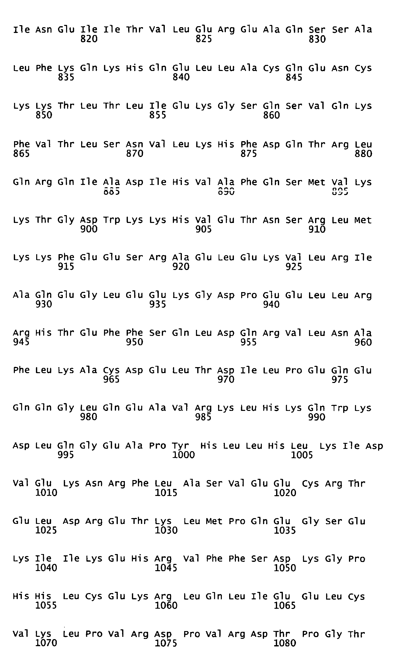
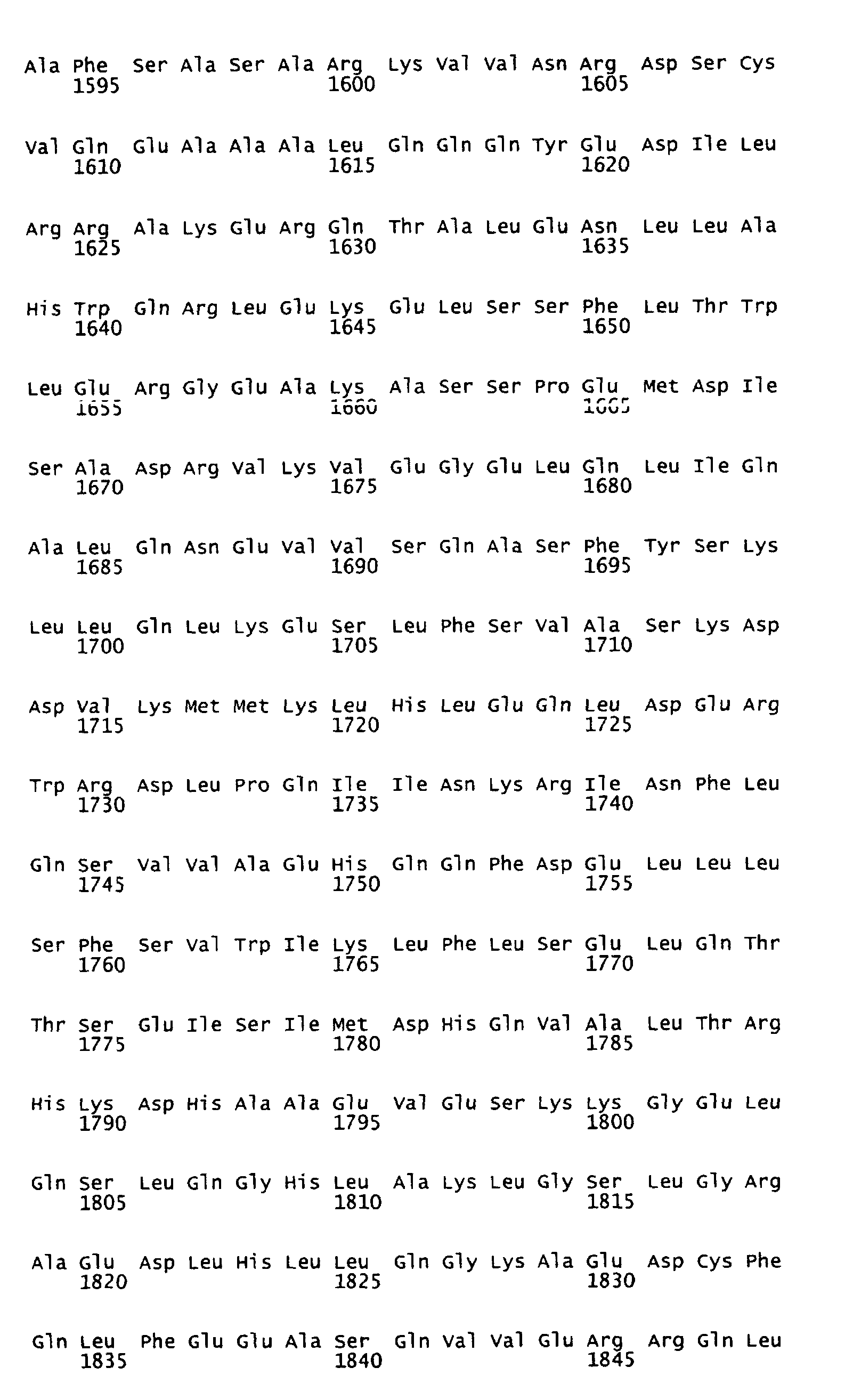
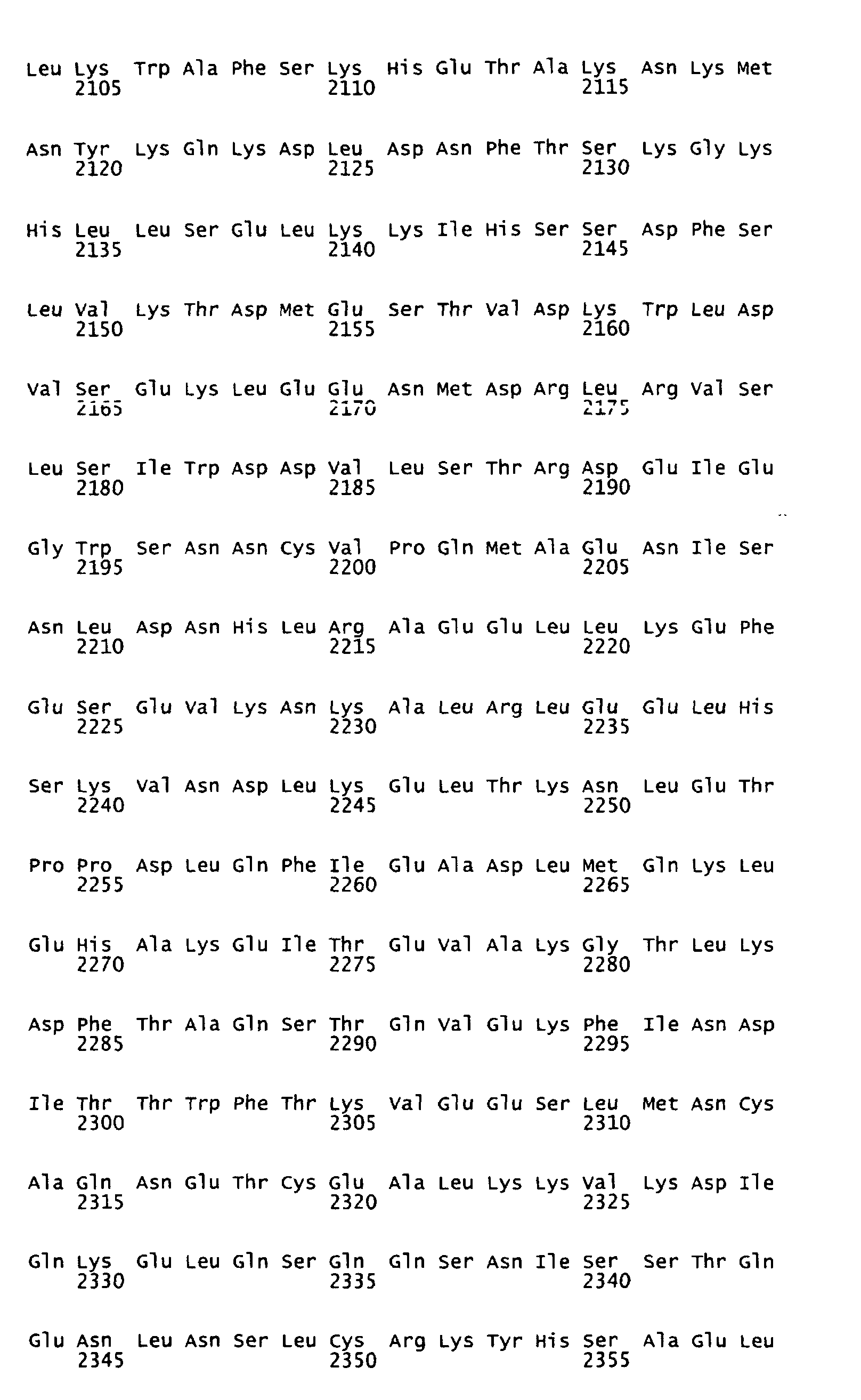
Figure 10: Predicted amino acid sequence of Drop1 and
predicted protein domains
-
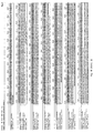
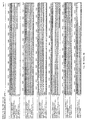
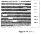
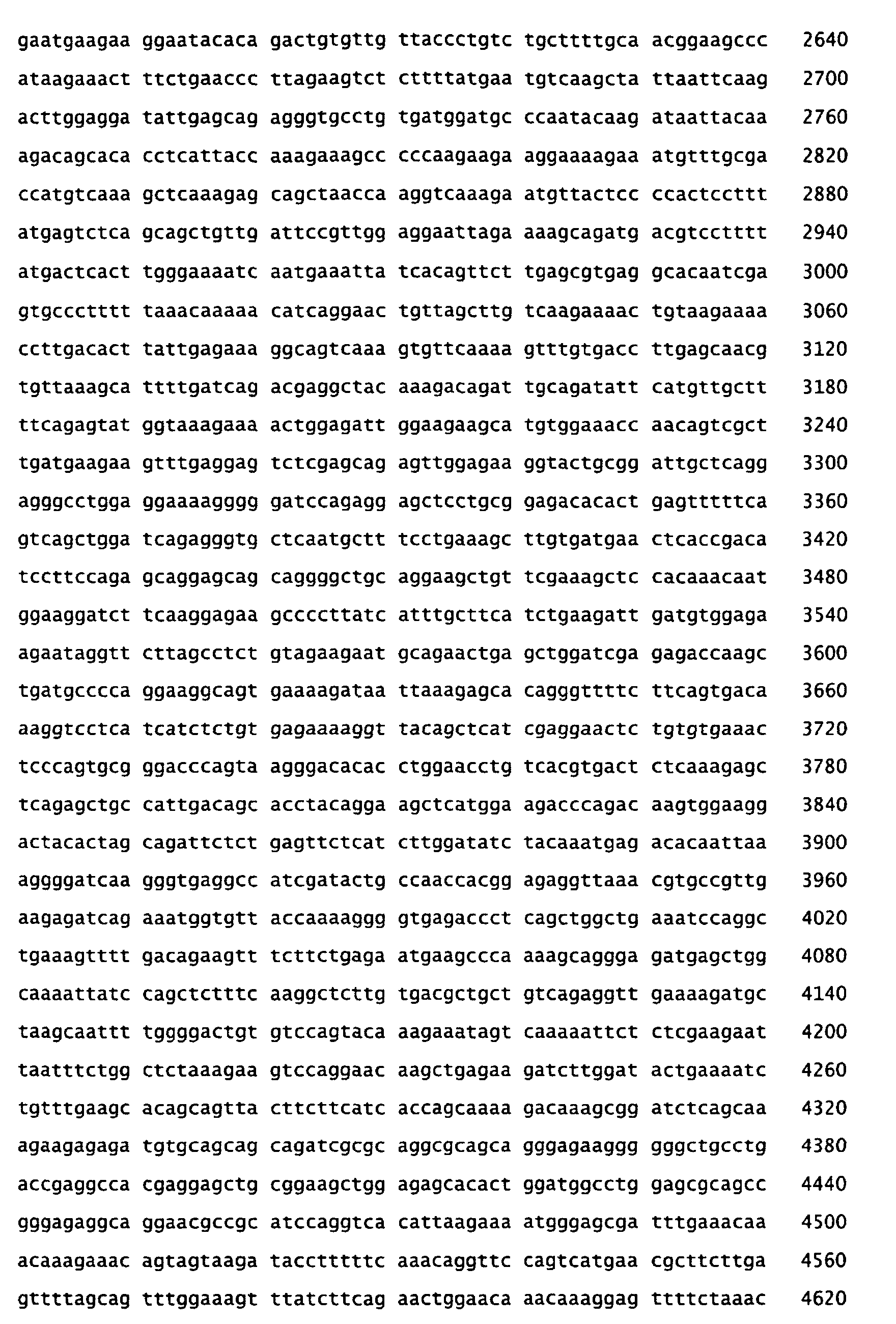
The ORF encodes a protein of 3032 amino acid residues.
Analysis using SMART (Simple Modular Architecture Research
Tool) software indicates two calponin homology (CH) domains,
an AAA, four coiled coil (CC) motifs and twelve spectrin-like
(SPEC) regions, as highlighted in gray in the sequence and
indicated in the margin.
-
The present invention relates to a nucleic acid molecule
encoding the human dystrophin-related polypeptide Drop1 or a
polypeptide exhibiting a biological property of Drop1, which
is:
- (a) a nucleic acid molecule encoding a Drop1 polypeptide
that consists of the amino acid sequence as depicted in
Figure 10;
- (b) a nucleic acid molecule consisting of the nucleotide
sequence as depicted in Figure 6B;
- (c) a nucleic acid molecule the sequence of which differs
from the sequence of a nucleic acid molecule of (a) or
(b) due to the degeneracy of the genetic code;
- (d) a nucleic acid molecule, which represents a fragment of
a nucleic acid molecule specified in (a) to (c); or
- (e) a nucleic acid molecule complementary to the nucleic
acid molecule specified in (a) to (d).
-
-
As used herein, a polypeptide exhibiting a biological property
of Drop1 is understood to be a polypeptide having at least one
of the activities of Drop1 as illustrated in the Examples,
below, i.e. at least one biological activity of an ATPase.
-
In a first embodiment, the invention provides a nucleic acid
molecule encoding a Drop1 polypeptide that consists of the
amino acid sequence as depicted in Figure 10.
-
The nucleic acid molecules of the invention can be both DNA
and RNA molecules. Suitable DNA molecules are, for example,
genomic or cDNA molecules. It is understood that all nucleic
acid molecules encoding all or a portion of Drop1 are also
included, as long as they encode a polypeptide with biological
activity. The nucleic acid molecules of the invention can be
isolated from natural sources or can be synthesised according
to known methods.
-
The nucleic acid molecules of the present invention also
include molecules which differ from the nucleic acid molecules
with sequences shown in Figure 6B due to the degeneracy of the
genetic code.
-
In a further embodiment, the present invention provides
nucleic acid molecules which comprise fragments of the nucleic
acid molecules described above encoding a polypeptide of the
invention. "Fragments" are understood to be parts of the
nucleic acid molecules that are long enough to encode one of
the described polypeptids. These fragments also comprise
nucleic acid molecules specifically hybridizing to transcripts
of the nucleic acid molecules of the invention. These nucleic
acid molecules can be used, for example, as probes or primers
in the diagnostic assay and/or kit described below and,
preferably, are oligonucleotides having a length of at least
10, in particular of at least 15 and particularly preferred of
at least 25 contiguous nucleotides of the nucleic acid
molecule of the invention. The nucleic acid molecules and
oligonucleotides (fragments) of the invention can also be
used, for example, as primers for a PCR reaction.
-
In a still further embodiment, the present invention provides
nucleic acid molecules which are complementary to the nucleic
acid molecules of the invention.
-
Generally, by means of conventional molecular biological
processes it is possible (see, e.g., Sambrook et al., supra)
to introduce different mutations into the nucleic acid
molecules of the invention. As a result Drop1 polypeptides or
Drop1 related polypeptids with possibly modified biological
properties are synthesised. One possibility is the production
of deletion mutants in which nucleic acid molecules are
produced by continuous deletions from the 5'- or 3'-terminal
of the coding DNA sequence and that lead to the synthesis of
polypeptids that are shortened accordingly. Another
possibility is the introduction of single-point mutation at
positions where a modification of the amino aid sequence
influences, e.g., the proteolytic properties. By this method
muteins can be produced, for example, that possess a modified
Km-value or that are no longer subject to the regulation
mechanisms that normally exist in the cell, e.g. with regard
to allosteric regulation or covalent modification.
-
For the manipulation in prokaryotic cells by means of genetic
engineering the nucleic acid molecules of the invention or
parts of these molecules can be introduced into plasmids
allowing a mutagenesis or a modification of a sequence by
recombination of DNA sequences. By means of conventional
methods (cf. Sambrook et al., supra) bases can be exchanged
and natural or synthetic sequences can be added. In order to
link the DNA fragments with each other adapters or linkers can
be added to the fragments. Furthermore, manipulations can be
performed that provide suitable cleavage sites or that remove
superfluous DNA or cleavage sites. If insertions, deletions or
substitutions are possible, in vitro mutagenesis, primer
repair, restriction or ligation can be performed. As analysis
method usually sequence analysis, restriction analysis and
other biochemical or molecular biological methods are used.
-
The polypeptids encoded by the various variants of the nucleic
acid molecules of the invention show certain common
characteristics, such as ATPase activity, molecular weight,
immunological reactivity or conformation or physical
properties like the electrophoretical mobilty, chromatographic
behavior, sedimentation coefficients, solubility,
spectroscopic properties, stability, pH optimum, temperature
optimum.
-
The invention furthermore relates to recombinant vectors
containing the nucleic acid molecules of the invention.
Preferably, they are plasmids, cosmids, viruses,
bacteriophages and other vectors usually used in the field of
genetic engineering. Vectors suitable for use in the present
invention include, but are not limited to the T7-based
expression vector for expression in mammalian cells and
baculovirus-derived vectors for expression in insect cells.
Preferably, the nucleic acid molecule of the invention is
operatively linked to the regulatory elements in the
recombinant vector of the invention that guarantee the
transcription and synthesis of an mRNA in prokryotic and/or
eukaryotic cells that can be translated. The nucleotide
sequence to be transcribed can be operably linked to a
promoter like a T7, metallothionein I or polyhedrin promoter.
-
In a further embodiment, the present invention relates to
recombinant host cells transiently or stably containing the
nucleic acid molecules or vectors of the invention. A host
cell is understood to be an organism that is capable to take
up in vitro recombinant DNA and, if the case may be, to
synthesize the polypeptids encoded by the nucleic acid
molecules of the invention. Preferably, these cells are
prokaryotic or eukaryotic cells, for example mammalian cells,
bacterial cells, insect cells or yeast cells. The host cells
of the invention are preferably characterized by the fact that
the introduced nucleic acid molecule of the invention either
is heterologous with regard to the transformed cell, i.e. that
it does not naturally occur in these cells, or is localized at
a place in the genome different from that of the corresponding
naturally occurring sequence.
-
A further embodiment of the invention relates to a polypeptide
exhibiting a biological property of the human dystrophin-related
polypeptide Drop1 and being encoded by the nucleic
acid molecules of the invention, as well as to methods for
their production, whereby, e.g., a host cell of the invention
is cultivated under conditions allowing the synthesis of the
polypeptide and the polypeptide is subsequently isolated from
the cultivated cells and/or the culture medium. Isolation and
purification of the recombinantly produced polypeptide may be
carried out by conventional means including preparative
chromatography and affinity and immunological separations
using, e.g., an anti-Drop1 antibody, or, e.g., can be
substantially purified by the one-step method described in
Smith and Johnson, Gene 67; 31-40 (1988). These polypeptides,
however, not only comprise recombinantly produced polypeptides
but include isolated naturally occurring polypeptides,
synthetically produced polypeptides, or polypeptides produced
by a combination of these methods. Means for preparing such
polypeptides or related polypeptides are well understood in
the art. These polypeptides are preferably in a substantially
purified form.
-
The present invention also relates to an antibody that binds
specifically to a Drop1 polypeptide or a related polypeptide
as defined above. Cells producing said antibody have been
deposited on March 26, 2003 according to the Budapest Treaty
with the Deutsche Sammlung von Mikrorganismen und
Zellkulturen, Mascheroder Weg, Braunschweig, Germany. The term
"antibody", preferably, relates to antibodies which consist
essentially of pooled monoclonal antibodies with different
epitopic specifities, as well as distinct monoclonal antibody
preparations. Monoclonal antibodies are made from an antigen
containing fragments of the polypeptides of the invention by
methods well known to those skilled in the art (see, e.g.,
Köhler et al., Nature 256 (1975), 495). As used herein, the
term "antibody" (Ab) or "monoclonal antibody" (Mab) is meant
to include intact molecules as well as antibody fragments
(such as, for example, Fab and F(ab') 2 fragments) which are
capable of specifically binding to protein. Fab and f(ab')2
fragments lack the Fc fragment of intact antibody, clear more
rapidly from the circulation, and may have less non-specific
tissue binding than an intact antibody. (Wahl et al., J. Nucl.
Med. 24: 316-325 (1983)). Thus, these fragments are preferred,
as well as the products of a FAB or other immunoglobulin
expression library. Moreover, antibodies of the present
invention include chimerical, single chain, and humanized
antibodies.
-
For certain purposes, e.g. diagnostic methods, the antibody of
the present invention can be detectably labeled, for example,
with a radioisotope, a bioluminescent compound, a
chemiluminescent compound, a fluorescent compound, a metal
chelate, or an enzyme.
-
The invention also relates to a transgenic non-human animal
such as transgenic mouse, rats, hamsters, dogs, monkeys,
rabbits, pigs, C. elegans and fish such as torpedo fish
comprising a nucleic acid molecule or vector of the invention,
preferably wherein said nucleic acid molecule or vector is
stably integrated into the genome of said non-human animal,
preferably such that the presence of said nucleic acid
molecule or vector leads to the expression of a Drop1
polypeptide (or related polypeptide) of the invention. Said
animal may have one or several copies of the same or different
nucleic acid molecules encoding one or several forms of a
Drop1 polypeptide or mutant forms thereof. This animal has
numerous utilities, including as a research model for
development/progression of carcinomas and therefore, presents
a novel and valuable animal in the development of therapies,
treatment, etc. for carcinomas associated with downregulation
of Drop1, e.g., epithelial tumors. Accordingly, in this
instance, the non-human mammal is preferably a laboratory
animal such as a mouse or rat.
-
Preferably, the transgenic non-human animal of the invention
further comprises at least one inactivated wild type allele of
the corresponding Drop1 encoding gene. This embodiment allows
for example the study of the interaction of various mutant
forms of Drop1 polypeptides on the onset of the clinical
symtoms of a carcinoma. All the applications that have been
herein before discussed with regard to a transgenic animal
also apply to animals carrying two, three or more transgenes.
It might be also desirable to inactivate Drop1 protein
expression or function at a certain stage of development
and/or life of the transgenic animal. This can be achieved by
using, for example, tissue specific, developmental and/or cell
regulated and/or inducible promoters which drive the
expression of, e.g., an antisense or ribozyme directed against
the RNA transcript encoding the Drop1 encoding mRNA; see also
supra. A suitable inducible system is for example
tetracycline-regulated gene expression as described, e.g., by
Gossen and Bujard (Proc. Natl. Acad. Sci. 89 USA (1992), 5547-5551)
and Gossen et al. (Trends Biotech. 12 (1994), 58-62).
Similar, the expression of a mutant Drop1 protein may be
controlled by such regulatory elements.
-
Furthermore, the invention also relates to a transgenic
mammalian cell which contains (preferably stably integrated into
its genome) a nucleic acid molecule according to the invention
or part thereof, wherein the transcription and/or expression of
the nucleic acid molecule or part thereof leads to reduction of
the synthesis of a Drop1 protein. In a preferred embodiment, the
reduction is achieved by an anti-sense, sense, ribozyme, co-suppression
and/or dominant mutant effect. "Antisense" and
"antisense nucleotides" means DNA or RNA constructs which block
the expression of the naturally occurring gene product.
-
The provision of the nucleic acid molecule according to the
invention opens up the possibility to produce transgenic non-human
animals with a reduced level of a Drop1 protein as
described above. Techniques how to achieve this are well known
to the person skilled in the art. These include, for example,
the expression of antisense-RNA, ribozymes, of molecules which
combine antisense and ribozyme functions and/or of molecules
which provide for a co-suppression effect. When using the
antisense approach for reduction of the amount of Drop1
proteins in cells, the nucleic acid molecule encoding the
antisense-RNA is preferably of homologous origin with respect
to the animal species used for transformation. However, it is
also possible to use nucleic acid molecules which display a
high degree of homology to endogenously occurring nucleic acid
molecules encoding a Drop1 protein. In this case the homology
is preferably higher than 80%, particularly higher than 90%
and still more preferably higher than 95%. The reduction of
the synthesis of a polypeptide according to the invention in
the transgenic mammalian cells can lead to various
physiological, developmental and/or morphological changes.
-
Thus, the present invention also relates to transgenic non-human
animals comprising the above-described transgenic cells. These
may show, for example, a deficiency in Drop1 due to the stable
or transient presence of a foreign DNA resulting in at least
one of the following features:
- (a) disruption of (an) endogenous gene(s) encoding Drop1;
- (b) expression of at least one antisense RNA and/or ribozyme
against a transcript comprising a nucleic acid molecule of
the invention;
- (c) expression of a sense and/or non-translatable mRNA of the
nucleic acid molecule of the invention;
- (d) expression of an antibody of the invention;
- (e) incorporation of a functional or non-functional copy of
the regulatory sequence of the invention; or
- (f) incorporation of a recombinant DNA molecule or vector of
the invention.
-
-
Methods for the production of a transgenic non-human animal of
the present invention, preferably transgenic mouse, are well
known to the person skilled in the art. Such methods, e.g.,
comprise the introduction of a nucleic acid molecule or vector
of the invention into a germ cell, an embryonic cell, stem
cell or an egg or a cell derived therefrom. The non-human
animal can be used in accordance with a screening method of
the invention described herein and may be a non-transgenic
healthy animal, or may have a disorder, preferably a disorder
caused by at least one mutation in the Drop1 protein. Such
transgenic animals are well suited for, e.g., pharmacological
studies of drugs in connection with mutant forms of the above
described Drop1 polypeptide. Production of transgenic embryos
and screening of those can be performed, e.g., as described by
A. L. Joyner Ed., Gene Targeting, A Practical Approach (1993),
Oxford University Press. The DNA of the embryonal membranes of
embryos can be analyzed using, e.g., Southern blots with an
appropriate probe; see supra.
-
In a further aspect, the invention relates to a method of
identifying activators/agonists of a Drop1 polypeptide
comprising the steps of: (a) incubating a candidate compound
with a polypeptide of the invention, b) assaying a biological
activity, and (c) determining if a biological activity of the
polypeptide of the invention has been altered.
-
Preferably, the screening for these compounds involves
producing appropriate cells which express Drop1, either as a
secreted protein or on the cell membrane. Preferred cells
include cells from mammals, yeast, Drosophila, or E. coli.
Cells expressing Drop1 (or cell membrane containing the
expressed polypeptide) are then preferably contacted with a
test compound potentially containing the molecule to observe
binding, stimulation, or inhibition of activity of Drop1.
-
The assay may simply test binding of a candidate compound to
Drop1, wherein binding is detected by a label, or in an assay
involving competition with a labeled competitor. Further, the
assay may test whether the candidate compound results in a
signal generated by binding to Drop1.
-
Alternatively, the assay can be carried out using cell-free
preparations, polypeptide/molecule affixed to a solid support,
chemical libraries, or natural product mixtures. The assay may
also simply comprise the steps of mixing a candidate compound
with a solution containing Drop1, measuring Drop1/molecule
activity or binding, and comparing the Drop1/molecule activity
or binding to a standard.
-
Preferably, an ELISA assay can measure Drop1 level or activity
in a sample (e.g., biological sample) using a monoclonal or
polyclonal antibody. The antibody can measure Drop1 level or
activity by either binding, directly or indirectly, to Drop1
or by competing with Drop1 for a substrate. All of these above
assays can be used as diagnostic or prognostic markers. The
molecules discovered using these assays can be used to treat
disease or to bring about a particular result in a patient
(e.g., elimination of an epithelial tumor or stop of
progression of tumor growth) by activating the Drop1 molecule.
Moreover, the assays can discover agents which may enhance the
production of Drop1 from suitably manipulated cells or
tissues.
-
The candidate compound may be a single compound or a plurality
of compounds. The term "plurality of compounds" in a method of
the invention is to be understood as a plurality of substances
which may or may not be identical.
-
Said compound or plurality of compounds may be chemically
synthesized or microbiologically produced and/or comprised in,
for example, samples, e.g., cell extracts from, e.g., plants,
animals or microorganisms. Furthermore, said compound(s) may
be known in the art but hitherto not known to be capable of
activating Drop1 polypeptides. The reaction mixture may be a
cell free extract or may comprise a cell or tissue culture.
Suitable set ups for the method of the invention are known to
the person skilled in the art and are, for example, generally
described in Alberts et al., Molecular Biology of the Cell,
third edition (1994) and in the appended examples. The
plurality of compounds may be, e.g., added to the reaction
mixture, culture medium, injected into a cell or otherwise
applied to the transgenic animal. The cell or tissue that may
be employed in the method of the invention preferably is a
host cell, mammalian cell or non-human transgenic animal of
the invention described in the embodiments hereinbefore.
-
If a sample containing a compound or a plurality of compounds
is identified in the method of the invention, then it is
either possible to isolate the compound from the original
sample identified as containing the compound capable of
activating Drop1, or one can further subdivide the original
sample, for example, if it consists of a plurality of
different compounds, so as to reduce the number of different
substances per sample and repeat the method with the
subdivisions of the original sample. Depending on the
complexity of the samples, the steps described above can be
performed several times, preferably until the sample
identified according to the method of the invention only
comprises a limited number of or only one substance(s).
Preferably said sample comprises substances of similar
chemical and/or physical properties, and most preferably said
substances are identical.
-
Several methods are known to the person skilled in the art for
producing and screening large libraries to identify compounds
having specific affinity for a target. These methods include
the phage-display method in which randomized peptides are
displayed from phage and screened by affinity chromatography
to an immobilized receptor; see, e.g., WO 91/17271, WO
92/01047, US-A-5,223,409. In another approach, combinatorial
libraries of polymers immobilized on a chip are synthesized
using photolithography; see, e.g., US-A-5,143,854, WO 90/15070
and WO 92/10092. The immobilized polymers are contacted with a
labeled receptor and scanned for label to identify polymers
binding to the receptor. The synthesis and screening of
peptide libraries on continuous cellulose membrane supports
that can be used for identifying binding ligands of the
polypeptide of the invention and thus possible activators is
described, for example, in Kramer, Methods Mol. Biol. 87
(1998), 25-39.
-
More recently, WO 98/25146 described further methods for
screening libraries of complexes for compounds having a
desired property, especially, the capacity to agonize, bind
to, or antagonize a polypeptide or its cellular receptor. The
complexes in such libraries comprise a compound under test, a
tag recording at least one step in synthesis of the compound,
and a tether susceptible to modification by a reporter
molecule. Modification of the tether is used to signify that a
complex contains a compound having a desired property. The tag
can be decoded to reveal at least one step in the synthesis of
such a compound. Other methods for identifying compounds which
interact with the polypeptides according to the invention or
nucleic acid molecules encoding such molecules are, for example,
the in vitro screening with the phage display system as well as
filter binding assays or "real time" measuring of interaction
using, for example, the BIAcore apparatus (Pharmacia).
-
All these methods can be used in accordance with the present
invention to identify activators/agonists of the Drop1
polypeptide or related polypeptide of the invention.
-
The nucleic acid molecule of the invention can also serve as a
target for activators. Activators may comprise, for example,
proteins that bind to the mRNA of a gene encoding a Drop1
polypeptide of the invention, thereby stabilizing the native
conformation of the mRNA and facilitating transcription and/or
translation, e.g., in like manner as Tat protein acts on HIV-RNA.
Furthermore, methods are described in the literature for
identifying nucleic acid molecules such as an RNA fragment
that mimics the structure of a defined or undefined target RNA
molecule to which a compound binds inside of a cell resulting
in retardation of cell growth or cell death; see, e.g., WO
98/18947 and references cited therein. These nucleic acid
molecules can be used for identifying unknown compounds of
pharmaceutical interest, and for identifying unknown RNA
targets for use in treating a disease. These methods and
compositions can be used in screening for identifying
compounds useful to alter expression levels of proteins
encoded by a nucleic acid molecule. Alternatively, for
example, the conformational structure of the RNA fragment
which mimics the binding site can be employed in rational drug
design to modify known antibiotics to make them bind more
avidly to the target. One such methodology is nuclear magnetic
resonance (NMR), which is useful to identify drug and RNA
conformational structures. Still other methods are, for
example, the drug design methods as described in WO 95/35367
or US-A-5,322,933, where the crystal structure of the RNA
fragment can be deduced and computer programs are utilized to
design novel binding compounds which can act as antibiotics.
-
The identification of nucleic acid molecules which encode
polypeptides which interact with the Drop1 polypeptides
described above can also be achieved, for example, as described
in Scofield (Science 274 (1996), 2063-2065) by use of the so-called
yeast "two-hybrid system". In this system the polypeptide
encoded by a nucleic acid molecule according to the invention or
a smaller part thereof is linked to the DNA-binding domain of
the GAL4 transcription factor. A yeast strain expressing this
fusion polypeptide and comprising a lacZ reporter gene driven by
an appropriate promoter, which is recognized by the GAL4
transcription factor, is transformed with a library of cDNAs
which will express plant proteins or peptides thereof fused to
an activation domain. Thus, if a peptide encoded by one of the
cDNAs is able to interact with the fusion peptide comprising a
peptide of a Drop1 polypeptide of the invention, the complex is
able to direct expression of the reporter gene. In this way the
nucleic acid molecules according to the invention and the
encoded peptide can be used to identify peptides and proteins
interacting with Drop1 protein.
-
Once the transacting factor is identified, modulation of its
binding to or regulation of expression of the Drop1
polypeptide of the invention can be pursued, beginning with,
for example, screening for inhibitors against the binding of
the transacting factor to the protein of the present
invention. Activation of Drop1 proteins could then be achieved
in animals by applying the transacting factor or the gene
encoding it, e.g. in an expression vector. Furthermore, upon
identification of the transacting factor, further components
in the pathway leading to activation (e.g. signal
transduction) or repression of a gene involved in the control
of Drop1 then can be identified. Modulation of the activities
of these components can then be pursued, in order to develop
additional drugs and methods for enhancing the activity/amount
of Drop1. Thus, the present invention also relates to the use
of the two-hybrid system as defined above for the
identification of activators of Drop1.
-
The compounds isolated by the above methods also serve as lead
compounds for the development of analog compounds. The analogs
should have a stabilized electronic configuration and
molecular conformation that allows key functional groups to be
presented to the Drop1 or its possible receptor in
substantially the same way as the lead compound. In
particular, the analog compounds have spatial electronic
properties which are comparable to the binding region, but can
be smaller molecules than the lead compound, frequently having
a molecular weight below about 2 kD and preferably below about
1 kD. Identification of analog compounds can be performed
through use of techniques such as self-consistent field (SCF)
analysis, configuration interaction (CI) analysis, and normal
mode dynamics analysis. Computer programs for implementing
these techniques are available; e.g., Rein, Computer-Assisted
Modeling of Receptor-Ligand Interactions (Alan Liss, New York,
1989). Methods for the preparation of chemical derivatives and
analogues are well known to those skilled in the art and are
described in, for example, Beilstein, Handbook of Organic
Chemistry, Springer edition New York Inc., 175 Fifth Avenue,
New York, N.Y. 10010 U.S.A. and Organic Synthesis, Wiley, New
York, USA. Furthermore, said derivatives and analogues can be
tested for their effects according to methods known in the
art; see also supra. Furthermore, peptidomimetics and/or
computer aided design of appropriate derivatives and analogues
can be used, for example, according to the methods described
above.
-
Once the described compound has been identified and obtained,
it is preferably provided in a therapeutically acceptable
form.
-
Accordingly, the present invention also relates to a
pharmaceutical composition comprising a nucleic acid molecule,
polypeptide, recombinant vector, antibody, or
activator/agonist according to the present invention and
optionally a pharmaceutically acceptable excipient, diluent or
carrier.
-
The nucleic acid molecule suitable for pharmaceutical or
diagnostic purposes is defined to be
- (a) a nucleic acid molecule encoding a Drop1 polypeptide
that comprises the amino acid sequence as depicted in
Figure 10;
- (b) a nucleic acid molecule comprising the nucleotide
sequence as depicted in Figure 6B;
- (c) a nucleic acid molecule encoding a polypeptide the
sequence of which shows at least 65% idendity to the
amino acid sequence of the polypeptide encoded by a
nucleic acid molecule specified in (a) or (b);
- (d) a nucleic acid molecule the sequence of which differs
from the sequence of a nucleic acid molecule of (a) to
(c) due to the degeneracy of the genetic code;
- (e) a nucleic acid molecule, which represents a fragment of
a nucleic acid molecule specified in (a) to (d); or
- (f) a nucleic acid molecule complementary to the nucleic
acid molecule specified in (a) to (e).
-
-
The present invention also provides the pharmaceutical or
diagnostic use of nucleic acid molecules encoding a
polypeptide the amino acid sequence of which shows an identity
of at least 80%, in particular an identity of at least 90%,
preferably of at least 95%, 96%, 97%, 98% or 99% to the amino
acid sequence of the polypeptide of Figure 10. The amino acid
sequences of such polypeptides are characterized by deletion,
substitution and/or insertion of amino acid residue(s)
compared to the amino acid sequence shwon in Fig. 10 or the
result of recombination. They can be naturally occurring
variations, for example sequences from other organisms, or
mutations that cam either occur naturally or that have been
introduced by genetic mutagenesis. In order to identify and
isolate such nucleic acid molecules, the molecules of the
invention or parts thereof can be used, for example by means
of hybridization. As a hybridization probe nucleic acid
molecules can be used, for example, that have exactly or
basically the nucleic acid sequence as depicted in Fig. 6B, or
parts of these sequences. The fragments that have been used as
hybridization probes can be synthetic fragments that were
produced by means of conventional synthetic methods and the
sequence of which basically corresponds to the sequence of the
Fig. 6B sequence.
-
As used herein, the term "hybridisation" has the meaning of
hybridisation under conventional hybridisation conditions,
preferably under stringent conditions as described, for
example, in Sambrook et al., Molecular Cloning, A Laboratory
Manual 2nd edition (1989) Cold Spring Harbor Laboratory Press,
Cold Spring Harbor, NY. However, in certain cases, a
hybridizing nucleic acid molecule can also be detected at
lower stringency hybridisation conditions. Changes in the
stringency of hybridisation and signal detection are primarily
accomplished through the manipulation of formamide
concentration (lower percentages of formamide result in
lowered stringency), salt conditions, or temperature. For
example, lower stringency conditions include an overnight
incubation at 37°Cin a solution comprising 6X SSPE (20X SSPE =
3M NaCl; 9.2M NaH2PO4 0.02M EDTA, pH7.4), 0.5% SDS, 30%
formamide, 100 µg/ml salmon sperm blocking DNA, following by
washes at 50°C with 1 X SSPE, 0.1% SDS. In addition, to
achieve even lower stringency, washes performed following
stringent hybridisation can be done at higher salt
concentrations (e.g. 5X SSC). Variations in the above
conditions may be accomplished through the inclusion and/or
substitution of alternate blocking reagents used to suppress
background in hybridisation experiments. The inclusion of
specific blocking reagents may require modification of the
hybridisation conditions described above, due to problems with
compatibility.
-
Examples of suitable pharmaceutical carriers etc. are well
known in the art and include phosphate buffered saline
solutions, water, emulsions, such as oil/water emulsions,
various types of wetting agents, sterile solutions etc. Such
carriers can be formulated by conventional methods and can be
administered to the subject at a suitable dose. Administration
of the suitable compositions may be effected by different
ways, e.g. by intravenous, intraperetoneal, subcutaneous,
intramuscular, topical or intradermal administration. The
route of administration, of course, depends on the nature of
the tumor, its localisation and the kind of compound contained
in the pharmaceutical composition. The dosage regimen will be
determined by the attending physician and other clinical
factors. As is well known in the medical arts, dosages for any
one patient depends on many factors, including the patient's
size, body surface area, age, sex, the particular compound to
be administered, time and route of administration, the kind
and stage of an epithelial tumor, general health and other
drugs being administered concurrently.
-
The delivery of the nucleic acid molecules of the invention
can be achieved by direct application or, preferably, by using
a recombinant expression vector such as a chimeric virus
containing these compounds or a colloidal dispersion system.
Direct application to the target site can be performed, e.g.,
by ballistic delivery, as a colloidal dispersion system or by
catheter to a site in artery. The colloidal dispersion systems
which can be used for delivery of the above nucleic acid
molecules include macromolecule complexes, nanocapsules,
microspheres, beads and lipid-based systems including oil-in-water
emulsions (mixed), micelles, liposomes and lipoplexes,
The preferred colloidal system is a liposome. The composition
of the liposome is usually a combination of phospholipids and
steroids, especially cholesterol. The skilled person is in a
position to select such liposomes which are suitable for the
delivery of the desired nucleic acid molecule. Organ-specific
or cell-specific liposomes can be used in order to achieve
delivery only to the desired tumor. The targeting of liposomes
can be carried out by the person skilled in the art by
applying commonly known methods. This targeting includes
passive targeting (utilizing the natural tendency of the
liposomes to distribute to cells of the RES in organs which
contain sinusoidal capillaries) or active targeting (for
example by coupling the liposome to a specific ligand, e.g.,
an antibody, a receptor, sugar, glycolipid, protein etc., by
well known methods). In the present invention monoclonal
antibodies are preferably used to target liposomes to specific
epithelial tumors via specific cell-surface ligands.
-
Preferred recombinant vectors useful for gene therapy are
viral vectors, e.g. adenovirus, herpes virus, vaccinia, or,
more preferably, an RNA virus such as a retrovirus. Even more
preferably, the retroviral vector is a derivative of a murine
or avian retrovirus. Examples of such retroviral vectors which
can be used in the present invention are: Moloney murine
leukemia virus (MoMuLV), Harvey murine sarcoma virus (HaMuSV),
murine mammary tumor virus (MuMTV) and Rous sarcoma virus
(RSV). Most preferably, a non-human primate retroviral vector
is employed, such as the gibbon ape leukemia virus (GaLV),
providing a broader host range compared to murine vectors.
Since recombinant retroviruses are defective, assistance is
required in order to produce infectious particles. Such
assistance can be provided, e.g., by using helper cell lines
that contain plasmids encoding all of the structural genes of
the retrovirus under the control of regulatory sequences
within the LTR. Suitable helper cell lines are well known to
those skilled in the art. Said vectors can additionally
contain a gene encoding a selectable marker so that the
transduced cells can be identified. Moreover, the retroviral
vectors can be modified in such a way that they become target
specific. This can be achieved, e.g., by inserting a
polynucleotide encoding a sugar, a glycolipid, or a protein,
preferably an antibody. Those skilled in the art know
additional methods for generating target specific vectors.
Further suitable vectors and methods for in vitro- or in vivo-gene
therapy are described in the literature and are known to
the persons skilled in the art; see, e.g., WO 97/00957.
-
In order to achieve expression only in the target organ, e.g.,
an epithelial tumor to be treated, the nucleic acid molecules
of the present invention can be linked to a tissue specific
promoter and used for gene therapy. Such promoters are well
known to those skilled in the art (see e.g. Zimmermann et al.,
(1994) Neuron 12, 11-24; Vidal et al.; (1990) EMBO J. 9, 833-840).
-
The present invention also relates to the use of the above
compounds of the invention for the preparation of a
pharmaceutical composition for treatment of an epithelial
tumor, preferably for the treatment of ovarian, mammary,
stomach, kidney, thyroid, pancreas, cervical, testis or lung
carcinomas.
-
Recombinant re-expression of a protein which got lost or lost
its activity by mutations during tumor progression is a
strategy which has been clinically evaluated for ovarian
carcinoma. Wild-type P53 was re-introduced into mutant cells
in a phase I clinical trial using a viral vector. This gave a
proof of principle that gene therapy is a promising strategy
in ovarian cancer. The predominantly intraperitoneal pattern
of metastasising of ovarian carcinoma makes it prone for
intraperitoneal application of the therapeutic agent which
increases the safety of the therapy and should lead to only
moderate toxicity of such therapies. Considering the size of
Drop1, even transfection of defective cells with cDNA
fragments may have a therapeutic effect. This strategy has
already been used successfully with muscle dystrophies in
animal models (Chamberlain, Hum. Mol. Genet. 11 (2002), 2355-2362;
Scott et al., Neuromuscular Disord. 12 (2002), S23-S29).
In addition, it can be expected that expression of Drop1 can
be upregulated in tumors by therapeutic use of demethylating
drugs.
-
Finally, the present invention also relates to a method of
diagnosing an epithelial tumor or a susceptibility to an
epithelial tumor in a subject comprising:
- (a)determining the presence or amount of expression of the
Drop1 polypeptide in a biological sample; and
- (b) diagnosing an epithelial tumor or a susceptibility to
an epithelial tumor based on the presence or amount of
expression of the Drop1 polypeptide.
-
-
The present invention also relates to a diagnostic kit useful
for the detection and/or characterization of an epithelial
tumor or a predisposition to such a tumor, e.g., an ovarian or
mammary carcinoma, containing a nucleic acid molecule,
polypeptide and/or antibody of the invention. Such kits are
useful for the detection of a target cellular component, which
is Drop1 or, alternatively, Drop1 encoding mRNA, wherein an
decreased concentration of Drop1 (compared to the
concentration in normal tissue) or, alternatively, Drop1
encoding mRNA is indicative for a epithelial tumor or a
disposition to such a tumor.
-
The Drop1 polypeptide or the corresponding mRNA, e.g. in
biological fluids or tissues, may be detected directly in
situ, e.g. by in situ hybridisation (e.g., according to the
examples, below) or it may be isolated from other cell
components by common methods known to those skilled in the art
before contacting with a probe. Detection methods include
Northern Blot analysis, RNase protection, in situ methods,
e.g. in situ hybridisation, in vitro amplification methods
(PCR, LCR, QRNA replicase or RNA-transcription/amplification
(TAS, 3SR), reverse dot blot disclosed in EP-B1 O 237 362)),
immunoassays, Western Blot and other detection assays that are
known to those skilled in the art. Prferably, these detection
methods are in an array formate.
-
The probe (e.g. a specific antibody or specific
oligonucleotide) of the diagnostic kit can be detectably
labeled. In a preferred embodiment, said kit contains an anti-Drop1
antibody and allows said diagnosis, e.g., by ELISA and
contains the antibody bound to a solid support, for example, a
polystyrene microtiter dish or nitrocellulose paper, using
techniques known in the art. Alternatively, said kits are
based on a RIA and contain said antibody marked with a
radioactive isotope. In a preferred embodiment of the kit of
the invention the antibody is labeled. Suitable antibody assay
labels are known in the art and include enzyme labels, such
as, glucose oxidase, and radioisotopes, such as iodine (125I,
121I), carbon (14C), sulfur (35S) , tritium (3H), indium (112In),
and technetium rhodamine, and biotin. In addition to assaying
Drop1 levels in a biological sample, the polypeptide can also
be detected in vivo by imaging. Antibody labels or markers for
in vivo imaging of protein include those detectable by X-radiography,
NMR or ESR. For X-radiography, suitable labels
include radioisotopes such as barium or caesium, which emit
detectable radiation but are not overtly harmful to the
subject. Suitable markers for NMR and ESR include those with a
detectable characteristic spin, such as deuterium, which may
be incorporated into the antibody by labeling of nutrients for
the relevant hybridoma. A protein-specific antibody or
antibody fragment which has been labeled with an appropriate
detectable imaging moiety, such as a radioisotope (for
example, 131I, 112In, 99mTc), a radio-opaque substance, or a
material detectable by nuclear magnetic resonance, is
introduced (for example, parenterally, subcutaneously, or
intraperitoneally) into the mammal. It will be understood in
the art that the size of the subject and the imaging system
used will determine the quantity of imaging moiety needed to
produce diagnostic images. In the case of a radioisotope
moiety, for a human subject, the quantity of radioactivity
injected will normally range from about 5 to 20 millicuries of
99mTc. The labeled antibody or antibody fragment will then
preferentially accumulate at the location of cells which
contain the specific protein. In vivo tumor imaging is
described in S.W. Burchiel et al., "Immunopharmacokinetics of
Radiolabeled Antibodies and Their Fragments". (Chapter 13 in
Tumor Imaging: The Radiochemical Detection of Cancer, S.W.
Burchiel and B.A. Rhodes, eds., Masson Publishing Inc.
(1982)).
-
For diagnostic purposes, preferably the probes (e.g.,
antibodies or oligonucleotides) of the invention do not bind
to enaptin and do not hybridize with enaptin encoding mRNA,
respectively.
-
The marker Drop1 is also useful for prognosis, for monitoring
the progression of an epithelial tumor, for monitoring the
therapeutic success during and/or after a tumor therapy, for
predictive purposes and the diagnostic evaluation of the
degree of malignancy of the tumor (grading and staging), e.g.
by using in situ hybridisation, e.g. according to the examples
below.
-
In addition, the inventors have discovered that if the Drop 1
promotor is hypermethylated, Drop 1 will not be expressed or
it will be expressed on a lower level (i.e. it is
downregulated). Thus, a therapeutic approach is the use of
methyltransferase inhibitors. Furthermore, the promotor may be
used for epigenomic diagnostic, preferably on an array
formate.
-
The following examples illustrate the invention.
Example 1
General methods
(A) Reaction kits
-
ABI PRISM-BigDye-Terminator-Cycle-Sequencing-Ready-Reaction-Kit,
Version 2 (Applied Biosystems, Weiterstadt, Germany); DIG
RNA Labeling Kit (Roche Diagnostics, Mannheim, Germany); DNA
Lab Chip Kit (Agilent Technologies, Waldbronn, Germany); PCR
Product Purification Kit (Roche Diagnostics); QIAprep Spin
Miniprep Kit (Qiagen, Hilden, Germany); QIAquick Gel
Extraction Kit (Qiagen); QIA shredder columns (Qiagen); Rneasy
Mini Kit (Qiagen); TOPO TA Cloning Kit (Invitrogen, Karlsruhe,
Germany); Qiagen PCR Cloning plus kit (Qiagen).
(B) Enzymes, buffers, reagents and markers
-
Advantage-cDNA-Polymerase-Mix (Clontech, Heidelberg, Germany);
anti-DIG-FAB-Fragments (Boehringer, Mannheim, Germany); BamHI-restriction
enzyme and -reactionbuffer (Gibco BRL, Eggenstein,
Germany); cDNA PCR-Reaktionsbuffer (Clontech);
Desoxyribonuklease I (Gibco BRL); DNTP Mix (Amersham,
Cleveland); DTT (Sigma-Aldrich, Deisenhofen, Germany);
Dulbeccoís Modified Eagle Medium (DMEM) (Invitrogen,
Karlsruhe, Germany); EcoRV-restriction enzyme (Gibco BRL);
Fetal calf serum (FCS) (Bio Whittaker, Verviers); First strand
buffer (Gibco BRL); DNA size standard: Gene Ruler 100 bp (MBI
Fermentas, St. Leon Roth, Germany); Gene Ruler 1 kb (MBI
Fermentas); Goldstar DNA-Polymerase (Eurogentec, Karlsruhe,
Germany); Goldstar DNA-Polymerase-Buffer (Eurogentec);
Phosphate Buffered Saline (PBS) (Invitrogen); Proteinase K
(Sigma-Aldrich); RLT Lysis Buffer (Qiagen); RNAse A (Sigma-Aldrich);
RNA size standard (Gibco BRL); Rnase inhibitor
(Boehringer, Mannheim); RNAlater (ams, Wiesbaden, Germany);
SuperSCRIPT II RNase H-Reverse Transcriptase (Gibco BRL); t-RNA
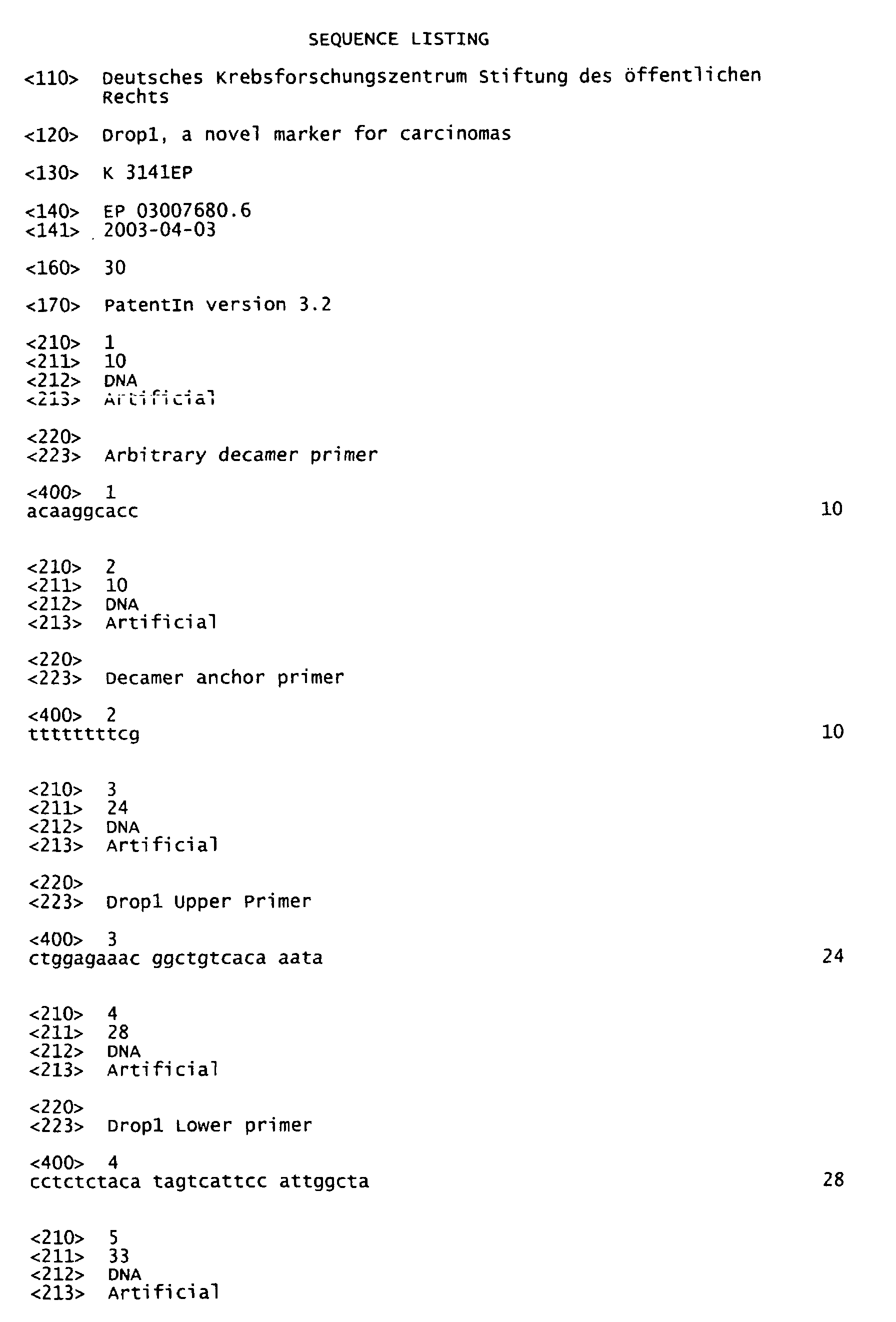
(Sigma-Aldrich); Trypan blue (Biochrom, Berlin, Germany);
Trypsin (Panbiotech, Aidenbach, Germany).
(C) Carcinoma cell lines
-
Cell lines CaOv-3, ES-2, MDAH 2774, NIH:OVCAR-3, SK-OV-3 were
obtained from the
American
Type
Culture
Collection (Rockville,
USA), GG HeST and MT were provided by Dr. M. Lindner, Oncology
Laboratory of the Women's University Hospital in Heidelberg
and Dr. B. Gückel, Women's University Hospital Tübingen
| Cell line | Characteristics |
| CaOv-3 | Derived from a human adenocarcinoma of the ovary, grown as monolayer, epithelial -like morphology |
| ES-2 | Derived from a poorly differentiated human ovarian clear cell carcinoma, hyperdiploid karyotype |
| MDAH 2774 | human adenocarcinoma of the ovary, grown as monolayer with epithelial morphology |
| NIH:OVCAR-3 | Derived from the malignant ascites of a patient with progressive adenocarcinoma of the ovary, grown as monolayer with epithelial morphology, exhibits androgen- and estrogen receptors, near triploid karyotype, large number of structurally abnormal and unassignable chromosomes. |
| SK-OV-3 | Derived from from malignant ascites of a patient with adenocarcinoma of the ovary, grown as monolayer; epithelial -like morphology |
| GG | Derived from the malignant ascites of a patient with poorly differentiated serous-papillary cystadenocarcinoma of the ovary, stage FIGO III, GIII, with metastases in the ovary, both fallopian tubes, isthmus of the cervix uteri, perimetrium of the corpus utery, bladder peritoneum and omentum majus. Grown as monolayer. |
| HeSt | Derived from the malignant ascites of a patient with serous-papillary cystadenocarcinoma of the ovary, stage FIGO III, grown as monolayer. |
| MT | Derived from the malignant ascites of a patient with serous-papillary cystadenocarcinoma of the ovary, stage FIGO III, grown as monolayer. |
(D) Procurement of tissues
-
Upon agreement of the patient in writing, procurement of tumor
and normal tissue was performed intraoperatively under sterile
conditions at the Women's University Hospitals in Heidelberg
and Tubingen. Epithelia were removed by 10 to 15 smears using
a plastic loop. In cases were no healthy ovarian epithelium
could be obtained, epithelium from the infundibulum tubae
uterinae (the uppermost part of the fallopian tube) was used
as a reference tissue. In terms of expression patterns this is
the tissue most closely related to the ovarian surface
epithelium. Each smear was immediately transferred to a 1.5 ml
Eppendorf tube containing 350 µl of RNA-Later. These samples
were stored at -80°C.
-
In addition, tissue blocks of solid carcinoma of tumor free
ovarian tissue and of fallopian tube were procured by
transferring them into Falcon tubes containing 4%
paraformaldehyde buffer and were fixed for in-situ
hybridisation. After fixation for 15 h at 4°C, the blocks were
washed twice with PBS for 15 min each and stored in 70%
ethanol at 4°C. Additional small blocks of tissue were
procured for laser captured microdissection in cryotubes
containing 350 µl of RNAlater, shock frozen in liquid nitrogen
at -196 °C and stored at -80°C. Dehydrated frozen sections of
7 µm were microdissected using an Arcturus PixCell II
microdissector.
(E) RNA isolation, quality control, and reverse transcription
-
Samples in RNAlater were thawed. Three volumes of RLT lysis
buffer containing 10% 2-mercaptoethanol were added, the cells
were lysed by incubation for 30 min at 37°C under gentle
shaking and finally vortexed. Cell fragments were removed by
passing the solution through a QIA shredder column for 2 min
at 8,000 rpm in a microcentrifuge. The eluant was mixed with
an equal volume of 70% ethanol and centrifuged through RNeasy
mini spin columns for 15 sec at 8,000 rpm. The RNA bound to
the columns was washed with 700 µl of RW1 buffer and twice
with 500 µl RPE buffer containing ethanol. The RNeasy filters
were dried by centrifugation for 2 min at maximum speed. RNA
was eluted with 2x30 µl RNase free DEPC water at 8,000 rpm.
RNA concentration was determined spectrophotometrically and
adjusted to equal concentration for each individual pair of
tissues. Its quality was assessed by agarose gel
electrophoresis or by using an Agilent Bioanalyzer 2100. The
ratio of 28S:18S rRNA should be close to 2:1. Residual DNA was
digested with 2U of DNAse I for 15 min at 25°C. The reaction
was terminated by addition of 1/10 vol. 25 mM EDTA pH 8.0 and
denaturation for 10 min at 65°C. In addition, RNA was isolated
from frozen tissue sections, microdissected by laser capture
microdissection (Arcturus PixCell II). Integrity of the RNA
was checked using the Agilent system. From this source, pA-RNA
was reversely transcribed and used without amplification
for Differential Display and quantitative PCR.
(F) Differential display
-
Differential display was performed using a modification of
Liang's and Pardee's method with a self-designed set of
arbitrary primers. A strong difference in expression was found
in patient 31 with the arbitrary decamer primer 5' ACAAGGCACC
3'( designated DOC), and a decamer anchor primer 5' TTTTTTTTVG
3', resulting in a 680 bp PCR fragment in normal epithelium,
which was absent from the pattern derived from the tumor of
the same patient. This fragment was eluted from the gel,
amplified, cloned into TOPORII, and sequenced.
(G) Validation of Differential Expression by Quantitative and
Semi-Quantitative RT-PCR
-
The differential expression was validated by quantitative PCR
and analyzed in the Agilent 2100 Bioanalyzer (Agilent
Technologies, Waldbronn, Germany, PC Hewlett packard, Vectra
VL, Hewlett packard, Boeblingen; Software Agilent 2100 Bio
Sizing 1.3, Agilent Technologies, Waldbronn) or by semiquantitative
analysis of the resulting cDNA on a 2% agarose
gel.
-
- 1 µl cDNA of the 20 µl RT-mixture (corresponding to 0.5 ng of
cDNA);
- 1 µl Drop1 Upper primer (5' CTGGAGAAACGGCTGTCACAAATA 3'
corresponding to positions 8255-8278 in exon 53), 20 µM;
- 1 µl Drop1 Lower primer (Lower primer 5'
CCTCTCTACATAGTCATTCCATTGGCTA 3', complementary to positions
8500-8473 in exon 55), 20 µM;
- 5 µl cDNA-PCR reaction buffer, 10 x;
- 1 µl dNTP-Mix, 10 mM;
- 1 µl advantage-cDNA-polymerase mix, 50 x;
- 40 µl aqua dest.
| advantage-cDNA polymerase mix | cDNA-PCR reaction buffer |
| Glycerol |
| | 1,0 % | Tricine-KOH, pH 9,2, | 40 mM |
| Tris-HCL, pH 7,5, | 0,8 mM | KOAc | 15 mM |
| KCL |
| | 1,0 mM | Mg(OAc)2 | 3,5 mM |
| (NH4)2SO4 | 0,5 mM | BSA | | 3,75 µg/ml |
| EDTA |
| | 2,0 µM |
| β-Mercaptoethanol | 0,1 mM |
| Thesit | 0,005 % |
-
-
The PCR reactions were performed in a PTC-200 Peltier Thermal
Cycler (Biozym, Hess. Oldendorf, Germany) under the following
conditions:
| 94°C | 4 min | Denaturation of DNA |
| 1. 94°C | 1 min | Denaturation of DNA |
| 2. 58°C | 40sec | Annealing | |
| 3. 72°C | 1 min | Elongation | |
| 4°C | hold |
-
Steps 1 to 3 were repeated 34 to 46 times. After individual
cycles in that range, aliquots were removed and run on a gel.
-
The amount of PCR-product from the first cycle producing a
visible band of 246 bp was then quantitated using the Agilent
system and an intercalating dye to detect double-stranded DNA.
Under these conditions, quantitative PCR detected Drop1 plus
enaptin, yet not cpg2, and the various nexins.
(H) Northern blot analysis and cancer profiling array
-
Clone 3R9 had been generated in a 3' RACE experiment using an
ovarian RACE library. Two human multiple-tissue Northern blots
(Clontech) were pre-hybridized in 0,5 M sodium phosphate, pH
7,2, 7% SDS, 1 mM EDTA and 0,1 mg/ml salmon sperm DNA at 65°C
for 1 h, and subsequently hybridized for 14 h at 65°C in the
same buffer containing the labelled probe at a concentration
of 1,5 x 10⁶ cpm/ml. The filters were washed twice at 65°C in
a buffer containing 0,2 X SSC and 0,2 % SDS and exposed for 20
h. To confirm an equal loading of mRNA in each lane, the blots
were hybridized with labelled human β-actin cDNA.
-
The cancer profiling array from BD Biosciences Clontech (Palo
Alto) catalog # 7841-1 included normalized cDNA from 241 tumor
and corresponding normal tissues from individual patients,
amplified by SMART technology. Human ubiquitin cDNA control
probe had been hybridized to the array to ensure equal
loading.
(I) In situ hybridisation
-
In situ hybridisation was carried out as described by DeJong
et al., 1985; Straus, 1982; Scopsi und Larson, 1986).
(J) Digoxigenin-labeling of RNA
-
A digoxigenin-labeled RNA-probe was generated by in vitro-transcription
of the 3R9 clone from pCRII-Topo (see above),
into which the cDNA fragment had been cloned to produce cDNA
for sequencing this product. The vector is suited for in
vitro-transcription, as it has promotors for the phage RNA-polymerases
SP6 und T7 on either side of the insert. Thus,
after linearization at the restriction sites BamHI and EcoRV,
two single-stranded RNA-probes of opposite orientation
("sense" and "antisense") could be transcribed.
-
Linearization was assayed by gel electrophoresis of a small
aliquot, and labeling was performed using the DIG-RNA-labeling-kit.
Digoxigenin-labeling of RNA using
SP6-polymerase after EcoRV cleavage | T7-polymerase after BamHI-cleavage |
| linearized vector-DNA 7,4 µl | (1 µg) | linearized vector-DNA 5µl | (1 µg) |
| transcription buffer , 10x, | 2 µl | transcription buffer, 10x | 2 µl |
| Labeling-Mix, 10x | 2 µl | Labeling-Mix, 10x | 2 µl |
| RNase-Inhibitor, 20 U | 1 µl | RNase-Inhibitor, 20 U | 1 µl |
| SP6-polymerase, 40 U | 2 µl | T7-polymerase, 40 U | 2 µl |
| DEPC-H2O | 5,6 µl | DEPC-H2O | 8 µl |
-
The components were mixed on ice and incubated for 2 h at
37°C. To assess the quality and the amount of the resulting
single-stranded RNA, 1 µl of the product was run on a 2% TAE-agarose
gel. Then, 2 µl (= 2U) RNase-free DNase I was added
and incubated at 37°C for 15 min, to destroy the vector and
avoid disturbance of the hybridisation by unlabeled DNA. The
reaction was stopped by adding 2 µl of 0,5 mM EDTA pH 8, and
the probes were stored at -20°C.
(K) Preparation of tissue sections
-
Procured tissues that had been stored in 70% ethanol/PBS were
washed in 10% Ethanol/PBS at 25°C and dehydrated by subsequent
washes in 85%, 95% ethanol/PBS, 3x 100% ethanol at 25°C, 2 x
in xylol, and incubated in xylol for 15 h at 25°C.
Subsequently the tissues were incubated for 60 min at 56°C in
a paraffin/xylol-mixture, and covered with pure paraffin for
3x for 60min in a Tissue Tek TEC (Vogel, Gießen). Storage was
at 4°C.
-
Sections of 7 µm were taken on a microtome (Leica, Nussloch,
Germany), placed on silanized glass slides and dried for 15 h
at 40°C. To remove the paraffin, the tissues were transferred
into xylol and rehydrated in seven successive steps for 2 min
each in 100%, 95%, 90%, 80%, 70%, 50% and 30% ethanol, 5 min
in 0,9% NaCl, 5 min in PBS-buffer. After fixation for 20 min
in 4% paraformaldehyde/ PBS the sections were washed 2 x 5min
in PBS, digested with with RNase-free proteinase K (20 µg/ml)
at 37°C for 20 min, washed again in PBS for 5 min. Excess
paraformaldehyde was blocked by acetylation with 0,625 ml
acetic anhydride in 3,125 ml triethanolamine and 250 ml H2O
for 10 min, and the slides were finally washed for 5 min in
PBS. To reduce background, the sections were prehybridized for
1-2h in hybridisation solution in the absence of the probe.
(L) Hybridisation solution
-
10x stock solution of salts: 0,2% polyvinylpolypyrrolidone;
0,2% Ficoll 400; 1,38% NaH2PO4, pH 6,8; 50 mM EDTA; 3 M NaCl;
0,1 M Tris, pH 8,0.
-
100 µl of this stock solution were mixed with 500 µl of
formamide, 2 µl 10 mg/ml t-RNA, 200 µl of 50% dextran sulfate,
100 µl 1 M 1,4-dithiothreitol, and 100 µl H2O.
(M) Preparation of probes
-
To remove contaminating free labeled nucleotides from the
digitonin-labeled RNA probes, 20 µl aliquots of probe were
precipitated by addition of 2,4 µl 4M LiCl and 75 µl 100%
ethanol and incubation at -70°C for 15 h. The precipitate was
spun down in a centrifuge, the supernatant removed, and the
probe washed in 50 µl of 70% ethanol. The sediment was
centrifuged, air-dried and taken up in the hybridisation
solution to a final concentration of 5 µg/ ml. The probe was
denatured for 7 min at 70°C and applied to the tissue
sections. In parallel, a known labeled probe was applied to
control sections.
(N) Hybridisation
-
Prehybridisation buffer was cautiously removed from the
slides. 100 µl of hybridisation solution containing the
digitonin-labeled RNA probe was applied to each section.
Slides were incubated in a moist chamber for 15 h at 45°C.
(O) Posthybridisation
-
- SSC 88,2 g Na-citrate x 2H2O 175,325 g NaCl in
1000 ml H2O, adjusted to pH 7.0 with HCl;
- 2 x SSC; 2 x SSC/50% formaledehyde; 5 x SSC and 5x SSC/ 50%
formaldeyhde were prepared accordingly:
- NTE-buffer 100 ml 5 M NaCl, 10 ml 1 M Tris pH 8,0, 100
ml 0,5 M EDTA, 790 ml H2O;
- B1 DIG 100 ml 1 M Tris pH 7,5, 30 ml 5 M NaCl, 870
ml H2O;
- Blocking solution 50 ml B1 DIG, 0,5 g blocking reagent, 2 mM
Levamisol;
- Anti-DIG-FAB-fragment 1: 500 dilution in 4 ml B1 DIG;
- NTMT 10ml 5 M NaCl, 50 ml 1 M Tris- HCl pH 9,5, 25
ml 1 M MgCl2, 415 ml H2O, 500 µl Tween 20;
- Staining solution amount corresponding to anti-DIG-FAB-fragment:
NTMT, 2 mM Levamisol, BCIP (5-bromo-4-chloro-3-indolylphosphate),
NBT (Nitroblue Tetrazolium)
-
-
The hybridisation solution was removed from the slides, the
sections were washed 2 x 5 min in 5xSSC at 25°C, 30 min with
5x SSC/50% formamide while the temperature increased from 37°C
to 60°C, and 60 min in 2x SSC/50% formamide. To remove unbound
single-stranded RNA-probe, the sections were then incubated
for 15 min at 37°C in NTE-buffer, unbound probe was digested
for 30 min at 37°C with RNAse A (20 µg/ml) in NTE-buffer,
followed by a wash for 15 min at 25°C in NTE-buffer, and 2xSSC
for 30 min at 25°C.
-
The tissue sections were now treated for 15 min with B1 DIG
at 25°C. The reaction was terminated by blocking solution for
60 min at 25°C. The blocking solution was removed, anti-DIG-FAB-fragments
in blocking solution were added and incubated
for 15 h at 4°C.
-
The sections were washed for 10 min with B1 DIG and NTMT with
2 mM Levamisol at 25°C. Staining solution was added and the
slides were incubated in the dark. A red-brown staining
results. When the desired intensity is reached, the reaction
is stopped by washing with PBS. For conservation, the slides
were treated 2 x 5min with 70% and 90% ethanol, followed by 2
x 10min 100% ethanol and xylol. Finally the stained sections
were covered with Entellan. Micrographs were viewed and
documented using the foto microscope of the Arcturus
microdissector.
(P) Treatment of cells with methyltransferase inhibitors
-
Treatment of cells with methyltransferase inhibitors was
carried out as described by Widschwendter et al., J.Nat.Cancer
Inst. 92 (2000), 826-832). All-trans-retinoic acid (ATRA) and
5-aza-deoxycytidine (Aza-CdR) were from Sigma Chemical Co.
(St. Louis, MO). 1 mM solutions were prepared in dimethyl
sulfoxide and diluted in culture medium. Cells were incubated
either for 5 days, with 2.5 mM Aza-CdR, for 24 h with 1 mM
ATRA, or for 5 days with 2.5 mM Aza-CdR followed by 24 h with
1 mM ATRA, or left untreated. They were detached with
trypsin (0.05%)-EDTA (0.02%) in Dulbecco's phosphate-buffered
saline (PBS), washed, and pelleted.
(Q) Cloning of the GFP- Drop1 aminoterminal domain fusion
construct for expression in mammalian cells
-
The 5' region of the ORF, encoding aar 1 - 396 (Drop1) the two
calponin homology domains, was amplified using the primers
5'-GTCGACGATGGCAACCTCCAGAGGGGCCTCCCG-3' with a Sal I
restriction site as upper primer (U278) and
5'-GGGCCCTGGTGCCCAGAGGTGCAGGAAGAGA-3' with an Apa I
restriction site as lower primer (L1492) and a previously
sequenced overlap clone (H) on the basis of cDNA from the
SKOV-3 cell line as template (annealing at 68 øC, 25 cycles).
The PCR product was cloned into pDrive for amplification and
sequencing, cut out with Sal I and Apa I, and cloned into
pEGFP-C3 (Clontech, GenBank Accession # U57607), behind a
GFP-domain. Correct insertion was confirmed by sequencing. 5-10
x 104COS-7 cells/well were grown in DMEM, containing 10%
fetal calf serum, 1% L- glutamine and 1% nonesssential amino
acids, for 24 h in Lab-Tek chambered cover glasses,
transfected with 0,75 - 1.5 _g of DNA/ well mixed with 3 to 6
_1 FuGENE 6 (Roche). After 24 h cells were observed under a
Zeiss Axiovert S100 TV microscope (excitation at 485 nm,
filter 485/20, - emission at 515 nm, filter 530/30, dichroic
mirror 505) using a 20x objective and a Plan-Neofluar oil
objective with 63x magnification. Fluorescence images were
recorded for 100 to 250 msec with 30% gain.
(R) Cloning of the carboxyterminal spectrin-like domain
-
The carboxyterminal spectrin-like domain of Drop1 was selected
as an antigen. cDNA encoding that region was obtained using
cDNA from CAOV-3 tumor cells as template, 5'-GATATCATGTATTTAGATGCGGTCCACGAGTTC-3',
containing an EcoR V
restriction site as an upper primer, and
5'-GTCGACTGGGGACCCTTGATTTAGAGGTTAACTT-3', containing a Sal I
restriction site as lower primer. This primer binds to a
sequence in the 3' UTR of Drop1, which in the enaptin gene is
located in a putative intron. The PCR product (annealing at
64 øC, 45 cycles) was cloned into TopoTA for amplification and
sequencing, cut out with EcoR V and Sal I, and cloned into
pET32a for expression in E. coli BL21(DE3).
(S) Expression of the recombinant protein in E. coli and its
purification from cell lysates
-
Bacteria were induced at 25° C for 3 h with 1 mM IPTG,
harvested by low-speed centrifugation, washed in PBS
containig protease inhibitor cocktail (Roche), swollen on ice
in hypotonic 50 mM sodium phosphate pH 8.0 containing 1 mg/ ml
lysozyme, supplemented by EDTA-free protease inhibitor
cocktail, and lysed by ultrasonication. DNA was digested with
1 U/ml of benzonase (Merck) for 30 min at 25 øC. Upon addition
of 300 mM NaCl the lysate was centrifuged in an
ultracentrifuge at 226,000 x g for 60 min. The His-tag-containing
proteins in the supernatant were bound to Ni-NTA
Superflow in a column of 1,6 cm _ x 10 cm (Qiagen). Unbound
material was removed by 50 mM Na phosphate, 300 mM NaCl, 20 mM
imidazole, and the bound protein was eluted with the same
buffer containing 150 mM imidazole. Fractions of 1 ml were
taken and aliquots were checked for protein bands by gel
electrophoresis.
-
Fractions of interest were pooled, diluted ten-fold with 25 mM
Tris.HCl pH7.5, loaded onto a MonoQ HR 5/5 column 0.5 cm _ x 5
cm (Amersham Bioscience) and a gradient was run from the
loading buffer with 30 mM NaCl to the same buffer containing
1M NaCl. The relevant fractions were pooled, concentrated, and
the buffer was changed to 50 mM Tris pH 8.0, as required for
cleavage with enterokinase (Invitrogen). With10 U of this
protease/ mg of protein the thioredoxin fused to the C-terminal
Drop1-fragment was cleaved for 16 h at 8 C. The
enterokinase was removed by EK-Away resin (Invitrogen). The
solution was diluted with 50 mM sodium phosphate containing
500 mM NaCl pH 8.0 as the starting buffer for a Ni-NTA column
(1 cm _ x 5.5 cm), to which the thioredoxin containing the
His-tag was bound, while the C-terminal Drop1 domain remained
in the flowthrough. It was concentrated in a Savant Speedvac
Plus vacuum centrifuge and for the immunization of mice and
rabbits the buffer was changed by passage over a NAP25 column
(Pharmacia). Purity and quantity of the protein (0.6 mg/ml)
were checked by gel electrophoresis on a 15% Kornberg gel.
(T) Generation of monoclonal antibody DROP1-2.9
-
Female BALB/c mice were immunized with the purified C-terminal
spectrin-like domain.
-
For the first immunization the protein emulsified in complete
Freund_s adjuvant was injected subcutaneously at 4 sites at
the flank. Additional five immunizations were given
intraperitoneally in intervals of two weeks consisting of the
purified protein diluted in sterile PBS. Three days after the
last challenge, immune spleen cells were fused to Ag8.653
mouse myeloma cells according to standard procedures.
-
Supernatants of growing hybridomas were screened for binding
to the protein by enzyme-linked immunosorbent assay (ELISA).
Positive samples were further analyzed by immunocytology using
cytocentrifuge preparations of SK-OV-3 ovarian carcinoma
cells. Thus, hybridoma DROP1-2 was identified and subsequently
cloned by limiting dilution. Clone DROP1-2.9 was selected
according to the same assays mentioned above. It secretes an
IgG1 kappa antibody as demonstrated by a mouse isotype-specific
ELISA.
(U) Staining of cytospins of SKOV-3 cells
-
SKOV-3 cells were spun down on a coverslip and . permeabilized
and fixed for 10 min at -20 øC in acetone. After drying they
were incubated with peroxidase-coupled anti-mouse IgG-Fc for
30 min at 25 øC: A stock solution of 8 mg/ ml 3-amino-9-ethylcarbazole
(Sigma) in dimethyl formamide was prepared.
-
For use it was diluted 20-fold with 0.1 m Na acetate pH 5.2
and 2 _1/ml of H2O2 were added This solution was used for
generating a red stain with the peroxidase within 5 - 10 min.
The reaction was stopped by two washes with PBS.
Example 2
Differential display PCR
-
In an analysis of differential gene expression of ovarian
carcinomas a 691 bp cDNA fragment was found which was
downregulated in an ovarian carcinoma (Figure 1). The band
labeled with an arrow shows a PCR product in normal tissue
absent from the tumor. A specifically designed arbitrary
primer and a G-anchor primer had been used for this PCR. The
product was eluted from the gel, re-amplified, cloned into the
TOPO TA vector and sequenced.
Example 3
Validation of differential display data by quantitative PCR
using cDNA from tissues of patients
-
Using gene specific primers, normal and ovarian carcinoma
tissue of eight individual patients was analyzed by
quantitative PCR for differences in expression (Table 1). In
all patients Drop1 was expressed at a higher level in the
healthy tissue, the ratio of expression between normal tissue
and carcinoma varying from 14 : 1 to 210 : 1. In addition, it
was shown that healthy fallopian tube epithelium is, in this
case, a suitable reference tissue for analyzing expression
differences in ovarian carcinomas, since microdissected
epithelium cells from healthy fallopian tube and ovarian
surface epithelium express Drop1 at a comparable level. The
ratio of fallopian tube to ovarian surface epithelium was
0.95.
Drop1 expression level in tissue pairs from individual
patients and in ovarian carcinoma cell lines |
| patient | normal | | Ratio N:C | OVCA cell lines |
| | microdissected |
| 82-84 | tube epithelium
20 nM | ovarian surface epithelium
21 nM | 0.95 | NIH:OVCA R-3 | 43 pM |
| | cDNA from swabs normal | carcinoma | | MDAH 2774 | 34 pM |
| 30 | 336 nM | 5.8 nM | 58 | CAOV-3 | 5.0 nM |
| 31 | 1020 nM | 6,9 nM | 148 | SKOV-3 | 11 nM |
| 44 | 94 nM | 0.65 nM | 140 | HEST | 0.26 nM |
| 48 | 320 nM | 23 nM | 14 | ES-2 | 51 pM |
| 50 | 300 nM | 2.3 nM | 130 | GG | 19 pM |
| 53 | 928 nM | 4.4 nM | 210 | MT | 66 pM |
| 93 | 3.4 nM | Neg | | 130 |
| 100 | 19 nM | Neg | 48 |
| | microdissected |
| BL | ductal epithelium
8.5 nM | ductal invasive carcinoma 8.5 nM | 1.0 | MCF-7 | 20 pM |
| | | | | HeLa |
| |
| | 5,3 nM |
| Calculated to the level after 38 PCR cycles; concentration expressed as nM. |
Example 4
Expression of Drop1 in tumor cell lines and in frozen sections
from tumors
-
Ovarian carcinoma cell lines were also analyzed for Drop1
expression (Table 1). Quantitative PCR with pairs of tissues
from individual patients was performed with normalized cDNA
derived without amplification from healthy tube epithelium and
ovarian carcinoma. As expected, expression was low in most
cell lines. However, two cell lines, SKOV-3 and CAOV-3 had a
normal expression level of approximately 5 nM after 38 PCR
cycles. In addition, cDNA was prepared from frozen sections of
ovarian and mammary carcinomas from individual patients. In
these specimens, Drop1 was downregulated in 6 out of 12
ovarian carcinomas and in eight out of 16 mammary carcinomas
(Table 2).
Drop1 expression levels in frozen sections of tumors from
individual patients |
| Patient | Drop level | patient | Drop1 level |
| Ovarian CA | | Mammary CA |
| 829 | 2.7 nM | 1170 | 0.020 nM |
| 895 | 3.1 nM | 1177 | neg. |
| 897 | 0.011 nM. | 1179 | neg. |
| 1111 | 3,0 nM | 1181 | 150 nM |
| 1117 | neg | 1182 | neg |
| 1130 | 7.2 nM | 1184 | neg |
| 1131 | 0.005 nM | 1188 | 31 nM |
| 1160 | 3.8 nM | 1189 | 24 nM |
| 1161 | 0.012 nM | 1191 | 11 nM |
| 1172 | 0.019 nM | 1193 | neg. |
| 1174 | 0.014 nM. | 1194 | neg. |
| 1178 | 3.8 nM | 1195 | neg. |
| | | 1198 | 3.0 nM |
| | | 1202 | 5.3 nM |
| | | 1203 | 4.0 nM |
| | | 1205 | 4.1 nM |
| |
| Results of quantitative PCR using normalized cDNA without amplification from ovarian carcinoma cell lines, from frozen ovarian carcinomas and from mammary carcinomas; drop level calculated for 38 cycles. |
Example 5
Cancer profiling array
-
Using a cancer profiling array, the differential expression of
Drop1 was analysed in pairs of tissues in an additional 14
patients (Figure 3). On this array, cDNA from normal ovarian
tissue (N) and carcinoma tissue (C) has been spotted in an
amount normalized to equal ubiquitin cDNA concentration. The
tumors were 12 adenocarcinomas, one mucinous borderline tumor
and one clear cell carcinoma. Upon hybridisation with the
radioactive Northern probe, in 12 patients the expression of
Drop1 was distinctly lower in the tumor. For two patients,
cDNA prepared from metastatic tissue was also spotted and
featured a similar level of expression as the corresponding
primary tumor.
-
Tissue pairs from patients with other types of carcinomas
were likewise analyzed for Drop1 expression. It was
significantly lower than in the corresponding normal tissue in
85% of the mammary carcinomas, stomach, lung, kidney and
thyroidea carcinomas. Colon and rectum carcinomas have a low
expression level with a less distinct pattern, and for cervix,
pancreas, and small intestine the number of cases is not
representative.
Example 6
Regulation of Drop1 by methylation of the promoter
-
The possibility that promoter methylation might downregulate
Drop1 was investigated. Treatment of cell lines exhibiting a
low level of Drop1 with the demethylating agents AZA (5-aza-cytidine
) and ATRA (all-trans-retinoic acid) resulted in an
increase of Drop1 mRNA by an order of magnitude (Table 3). No
increase of expression was observed in SKOV-3, a cell line
with a physiological level of Drop1. This indicates that the
lower level of Drop1 expression in cell lines GG and M130 was
caused by methylation of the Drop1 promoter.
| Expression of downregulated Drop1 in cells treated with
inhibitors of gene methylation |
| Cells | in medium | in Aza | in ATRA | in Aza+ATRA |
| GG |
| | 5,2 x 10⁻⁵ | 4,7 x 10⁻⁵ | 1,3 x 10⁻⁴ 2.5fold | 7,7 x 10⁻⁴ 15fold |
| M130 |
| | 1,0 x 10⁻³ | 1,0 x 10⁻³ | 1,8 x 10⁻² 18fold | 5,6 x 10⁻⁴ |
| SKOV-3 | 2,2 x 10⁻⁴ | 2,4 x 10⁻⁴ | 2,2 x 10⁻⁴ | 1,9 x 10⁻⁴ |
Aza: 5'-aza-2'-deoxycytidine, ATRA: all-trans-retinoic acid
Ratios are [31N/GDOC/1], measured in the LightCycler [G3PDH] |
Example 7
In situ hybridisation
-
Localization of the mRNA of Drop1 in healthy ovarian tissue,
fallopian tube tissue and tumor tissue was performed by in-situ
hybridisation (Figure 4). Drop1 was specifically
expressed in epithelium of the healthy ovary and at a
comparable level in the epithelium of a healthy fallopian
tube. Other cell types of these organs do not express Drop1.
This figure again shows that tube epithelium in this case is a
suitable reference tissue and that the difference in
expression of Drop1 is not based on the characteristics of the
tube epithelium.
Example 8
Multi-tissue-Northern blot
-
It could be demonstrated (Figure 2), that Drop1 mRNA is
present in testes (Figure 2; slot 4) and in ovary (Figure 2;
slot 5) in a multi tissue Northern Blot. The remaining tissues
yielded only a weak signal or nor signal at all. The smear may
be caused by differentially used exons resulting in mRNAs of
different lengths.
Example 9
Completion and characteristics of the Drop1 sequence
-
The sequence of the differential display fragment had
featured an open reading frame. By "RACE-walking" using a
commercial ovarian RACE library and by validating the
resulting RACE clones using overlap PCR on the basis of one
ovarian carcinoma cell line that did express the Drop1
fragment, an open reading frame of 9,099 bp (Fig. 6B; sequence
details) could be completed. The encoding gene comprises 57
exons in chromosome region 6q25. Upon completion of this
sequence a cDNA with a much larger ORF for enaptin was
submitted to the data bases (Accession no. AF535142). Exon
usage for Drop1, however, was different from that of enaptin
in that (a) exon 5 is 36 bp longer than predicted, (b) exon 7
(21 bp) is not used and (c) exon 12 is used in a commercial
ovarian cDNA library, yet not in the ovarian carcinoma cell
line CAOV-3. It contains the start codon for a putative human
homologue of rat cpg2. Exon 58 is longer in Drop1 than
predicted, In all clones derived from OVCA-R3 it contains a
stop codon immediately following the predicted transition to
exon 59. 2,5 kb of the genomic sequence following this stop
codon are found in the Drop1 mRNA as 3'UTR. In contrast, an
ovarian RACE library yielded clones for both variants: an
extended exon 58 with the stop codon for Drop1 as well as the
transition to exon 59.
-
Drop1 encodes a protein of 349 kDa (Figure 10) that is
structurally similar to dystrophin. At the N-terminus it
features 2 calponin homology domains followed by an ATPase-domain
and 12 spectrin-like domains (Figure 7). The homologues
of these structures are known to bind other intracellular or
transmembrane proteins.
Example 10
Cloning and expression of a Drop1-GFP fusion construct
-
The 5' fragment, Drop1α, containing the two calponin homology
domains of the open reading frame, was cloned into the
eukaryotic expression vector pEGFP-C3. Transfected cells show
a distinct fluorescence of nuclei (Figure 8), suggesting a
nuclear association of the N-terminal region.
Example 11
Raising of a monoclonal antibody and detection of the antigen
in cells and on immunoblots
-
A C-terminal recombinant protein fragment, amino acid residues
2827 - 3032, was produced in bacteria, isolated and purified
to serve as an antigen for generation of a monoclonal
antibody. A clone that recognized the recombinant protein on
an immunoblot also identified a high molecular weight band in
cell lysates of SKOV-3 and stained nuclei of the same cells
(Figure 9). Additional smaller protein bands in the pellet of
the cells presumably are Drop1 degradation products.