EP1457211A1 - Recombinant vaccinia virus vaccine - Google Patents
Recombinant vaccinia virus vaccine Download PDFInfo
- Publication number
- EP1457211A1 EP1457211A1 EP01274763A EP01274763A EP1457211A1 EP 1457211 A1 EP1457211 A1 EP 1457211A1 EP 01274763 A EP01274763 A EP 01274763A EP 01274763 A EP01274763 A EP 01274763A EP 1457211 A1 EP1457211 A1 EP 1457211A1
- Authority
- EP
- European Patent Office
- Prior art keywords
- vaccinia virus
- dis
- cells
- virus strain
- recombinant vaccinia
- Prior art date
- Legal status (The legal status is an assumption and is not a legal conclusion. Google has not performed a legal analysis and makes no representation as to the accuracy of the status listed.)
- Withdrawn
Links
Images


Classifications
-
- C—CHEMISTRY; METALLURGY
- C12—BIOCHEMISTRY; BEER; SPIRITS; WINE; VINEGAR; MICROBIOLOGY; ENZYMOLOGY; MUTATION OR GENETIC ENGINEERING
- C12N—MICROORGANISMS OR ENZYMES; COMPOSITIONS THEREOF; PROPAGATING, PRESERVING, OR MAINTAINING MICROORGANISMS; MUTATION OR GENETIC ENGINEERING; CULTURE MEDIA
- C12N7/00—Viruses; Bacteriophages; Compositions thereof; Preparation or purification thereof
-
- C—CHEMISTRY; METALLURGY
- C07—ORGANIC CHEMISTRY
- C07K—PEPTIDES
- C07K14/00—Peptides having more than 20 amino acids; Gastrins; Somatostatins; Melanotropins; Derivatives thereof
- C07K14/005—Peptides having more than 20 amino acids; Gastrins; Somatostatins; Melanotropins; Derivatives thereof from viruses
-
- C—CHEMISTRY; METALLURGY
- C12—BIOCHEMISTRY; BEER; SPIRITS; WINE; VINEGAR; MICROBIOLOGY; ENZYMOLOGY; MUTATION OR GENETIC ENGINEERING
- C12N—MICROORGANISMS OR ENZYMES; COMPOSITIONS THEREOF; PROPAGATING, PRESERVING, OR MAINTAINING MICROORGANISMS; MUTATION OR GENETIC ENGINEERING; CULTURE MEDIA
- C12N15/00—Mutation or genetic engineering; DNA or RNA concerning genetic engineering, vectors, e.g. plasmids, or their isolation, preparation or purification; Use of hosts therefor
- C12N15/09—Recombinant DNA-technology
- C12N15/63—Introduction of foreign genetic material using vectors; Vectors; Use of hosts therefor; Regulation of expression
- C12N15/79—Vectors or expression systems specially adapted for eukaryotic hosts
- C12N15/85—Vectors or expression systems specially adapted for eukaryotic hosts for animal cells
- C12N15/86—Viral vectors
-
- A—HUMAN NECESSITIES
- A61—MEDICAL OR VETERINARY SCIENCE; HYGIENE
- A61K—PREPARATIONS FOR MEDICAL, DENTAL OR TOILETRY PURPOSES
- A61K39/00—Medicinal preparations containing antigens or antibodies
- A61K2039/51—Medicinal preparations containing antigens or antibodies comprising whole cells, viruses or DNA/RNA
- A61K2039/525—Virus
- A61K2039/5256—Virus expressing foreign proteins
-
- A—HUMAN NECESSITIES
- A61—MEDICAL OR VETERINARY SCIENCE; HYGIENE
- A61K—PREPARATIONS FOR MEDICAL, DENTAL OR TOILETRY PURPOSES
- A61K39/00—Medicinal preparations containing antigens or antibodies
- A61K2039/51—Medicinal preparations containing antigens or antibodies comprising whole cells, viruses or DNA/RNA
- A61K2039/53—DNA (RNA) vaccination
-
- A—HUMAN NECESSITIES
- A61—MEDICAL OR VETERINARY SCIENCE; HYGIENE
- A61K—PREPARATIONS FOR MEDICAL, DENTAL OR TOILETRY PURPOSES
- A61K39/00—Medicinal preparations containing antigens or antibodies
-
- C—CHEMISTRY; METALLURGY
- C12—BIOCHEMISTRY; BEER; SPIRITS; WINE; VINEGAR; MICROBIOLOGY; ENZYMOLOGY; MUTATION OR GENETIC ENGINEERING
- C12N—MICROORGANISMS OR ENZYMES; COMPOSITIONS THEREOF; PROPAGATING, PRESERVING, OR MAINTAINING MICROORGANISMS; MUTATION OR GENETIC ENGINEERING; CULTURE MEDIA
- C12N2710/00—MICROORGANISMS OR ENZYMES; COMPOSITIONS THEREOF; PROPAGATING, PRESERVING, OR MAINTAINING MICROORGANISMS; MUTATION OR GENETIC ENGINEERING; CULTURE MEDIA dsDNA viruses
- C12N2710/00011—Details
- C12N2710/24011—Poxviridae
- C12N2710/24041—Use of virus, viral particle or viral elements as a vector
- C12N2710/24043—Use of virus, viral particle or viral elements as a vector viral genome or elements thereof as genetic vector
-
- C—CHEMISTRY; METALLURGY
- C12—BIOCHEMISTRY; BEER; SPIRITS; WINE; VINEGAR; MICROBIOLOGY; ENZYMOLOGY; MUTATION OR GENETIC ENGINEERING
- C12N—MICROORGANISMS OR ENZYMES; COMPOSITIONS THEREOF; PROPAGATING, PRESERVING, OR MAINTAINING MICROORGANISMS; MUTATION OR GENETIC ENGINEERING; CULTURE MEDIA
- C12N2740/00—Reverse transcribing RNA viruses
- C12N2740/00011—Details
- C12N2740/10011—Retroviridae
- C12N2740/16011—Human Immunodeficiency Virus, HIV
- C12N2740/16111—Human Immunodeficiency Virus, HIV concerning HIV env
- C12N2740/16122—New viral proteins or individual genes, new structural or functional aspects of known viral proteins or genes
Definitions
- the invention of this application relates to a recombinant vaccinia virus vaccine. More precisely, the invention of this application relates to a virus vaccine for various infectious diseases of humans and animals, and to a recombinant vaccinia virus strain DIs that serves as the effective ingredient of the vaccine.
- Vaccinia virus is utilized as smallpox vaccines, and has much contributed toward eradicating smallpox.
- This virus is a large-size DNA virus, belonging to Poxviridae, and, as its genome, it has a linear double-stranded DNA of about 190 kbp, coding for about 200 types of different proteins.
- Panicali and Paoletti Proc. Natl. Acad. Sci. USA 79:4927, 1982
- Mackett et al. Proc. Natl. Acad. Sci.
- a vaccinia virus strain DIs is highly attenuated, and it was isolated through subcultivation of a vaccinia virus strain Talien (DEI) in hen's egg embryos for developing safe smallpox vaccines (Nature 192:381, 1961).
- the This strain DIs is specifically characterized in that it grows and propagates in fetal avian primary culture cells (DEF) but little in any other mammal cells. In those days, this was tried for clinical application along with a strain LC16 that had been developed together with it, and 200 Japanese children were actually vaccinated with it.
- the strain DIs was unfavorable for smallpox vaccines because of its poor immune inducibility, and the other strain LC16 better than the strain DIs in point of immune induction was employed for smallpox vaccines. This is the history of the strain DIs.
- MVA vaccinia virus strain Ankara
- NYVAC vaccinia virus strain New York
- the inventors of this application had succeeded in inserting a foreign gene into the genome of this strain DIs, and had verified that the recombinant strain DIs is useful as a viral vector for animal cells. However, since the immune inducibility of the strain. DIs is poor as so mentioned hereinabove, it has been believed that, even if the strain DIs is so recombined with a foreign antigenic protein as to express the foreign antigenic protein, the resulting recombinant strain DIs will be ineffective for vaccines.
- the cytotoxicity of the vaccinia virus strain DIs is low, it is easy to believe that the strain will be better than any other strains in point of its safety in human applications.
- the immune inducibility of the strain DIs is poor, no one has heretofore considered the use of the strain DIs for recombinant virus vaccines.
- the inventors of the present application have succeeded in introducing a foreign gene into the vaccinia virus strain DIs and in expressing the foreign gene in the recombinant strain DIs, and, in animal experiments, the inventors have found the possibility of inducing the humoral and cellular immune reaction in the foreign gene product from the recombinant vaccinia virus strain DIs.
- the vaccinia virus strain DIs since the vaccinia virus strain DIs does not propagate in animal cells, it has heretofore been believed that the strain will be unsuitable for an expression vector for useful proteins. However, the inventors of this application have found that, owing to its low cytotoxicity, the vaccinia virus strain DIs enables its host cells to live for a long period of time and is therefore useful as a vector for mass-expression of proteins.
- the invention of this application has been made on the basis of the novel findings of the inventors as above, and its object is to provide a recombinant vaccinia virus vaccine of high safety.
- Another object of the invention is to provide the recombinant vaccinia virus strain DIs that serves as the active ingredient of the vaccine.
- Still another object of the invention is to provide the recombinant vaccinia virus strain DIs that expresses a reporter molecule and is therefore useful for construction of other recombinant virus strains.
- Still another object of the invention is to provide a method of using the vaccinia virus strain DIs for a vector.
- the vaccine of the inventions (1) to (5) is a highly-attenuated live vaccine that contains, as the active ingredient, the recombinant vaccinia virus strain DIs of the inventions (6) to (10).
- the recombinant vaccinia virus strain DIs of the invention (6) is a recombinant virus obtained through homologous recombination of the genome DNA of DIs with a transfer vector having a vaccinia virus-derived gene promoter sequence and having a polynucleotide that encodes an intended, foreign (that is, excluding vaccinia viruses) antigenic protein, linked downstream the promoter sequence.
- the foreign antigenic protein will be referred to as "foreign polypeptide"; and the polynucleotide encoding it will be referred to as "foreign polynucleotide”.
- the promoter sequence to be inserted into the transfer vector may be any and every one capable of being recognized by the RNA polymerase which is encoded by the gene of a vaccinia virus itself.
- a 7.5 kD polypeptide gene promoter p7.5 that exists inside the inverted terminal repeats of the genome of a vaccinia virus (Stunnenberg, H. G., et al., Nucleic Acids Res., 16, 2431, 1988) may be used.
- the foreign polynucleotide may be any and every one that encodes an antigenic protein except vaccinia viruses; and the foreign polypeptide may be any and every one capable of inducing antigen-antibody reaction in an animal body.
- a gag precursor protein p55 an env protein gp120 or gp160, a pol precursor protein, a nef protein and a tat protein of a human immunodeficiency virus (HIV) that causes human acquired immunodeficiency syndrome (AIDS); and a gag precursor protein of a simian immunodeficiency virus (SIV).
- HIV human immunodeficiency virus
- SIV simian immunodeficiency virus
- other polynucleotides encoding other pathogens (other pathogenic viruses and bacteria) or for antigenic proteins of cancer cells.
- Such foreign polynucleotides may be obtained, for example, by cleaving a plasmid clone of a genomic gene encoding an intended foreign polypeptide or its cDNA, or by amplifying through polymerase chain reaction (PCR) using primers having a suitable sequence and the plasmid clone as a template. If not cloned, the bacterial or animal genomic DNA having the gene is amplified through PCR for which is used a virus-infected animal cell-derived DNA or RNA as the template in case of viruses.
- PCR polymerase chain reaction
- the thus-obtained foreign polynucleotide is ligated with the above-mentioned promoter in a correct reading frame, and the fused polynucleotide (this will be hereinafter referred to as "expression unit") is then inserted into a desired plasmid vector to construct a transfer vector.
- a vaccinia virus genome DNA is first extracted from virus particles which are purified through sucrose density gradient centrifugation (Joklik, W. K., Virology, 18, 9, 1962), using L-laurylsarucosine (Mackett, M. and Archard, L. C. J., Gen. Virol., 45, 683, 1979).
- the non-essential gene region that has no influence on the vaccinia virus replication must be specifically defined for the site for homologous recombination.
- the first candidate for the non-essential gene region is a thymidine kinase (TK) gene region.
- inserting the TK gene region into a plasmid vector such as pUC18, and further inserting an expression unit at the unique restriction site in that region may construct a transfer vector.
- the foreign polynucleotide is inserted into the vaccinia virus genome through homologous recombination with the TK gene in the virus genome. After the homologous recombination, the recombinant virus is produced in the TK-lacking cell line, and incubated in the presence of bromodeoxyuridine to thereby select the recombinant virus not expressing TK.
- the second candidate for the non-essential gene region is genomic gene-lacking region of DIs. Comparing the restriction enzyme cleavage patterns of the genomic DNA of DIs with that of the parent strain DIE may identify the defective region in the genome of DIs. This region is basically unnecessary for DIs replication.
- the DNA sequences before and after the defective region are amplified through PCR, and then inserted into a plasmid vector. An expression unit is further inserted into the plasmid to construct a transfer vector. Using the transfer vector, the DIs genomic DNA is subjected to homologous recombination, whereby the expression unit is inserted into the defective region of the DIs genomic DNA. In this method, however, the recombinant virus is difficult to select.
- a polynucleotide encoding a reporter molecule is first inserted into the defective region to construct a recombinant virus.
- the reporter molecule for example, any of bacteria-derived coloring enzyme such as ⁇ -galactosidase, or a green fluorescence protein (GFP) or its derivatives (e.g., EGFP) may be used.
- GFP green fluorescence protein
- X-gal 5-bromo-4-chloro-3-indolyl- ⁇ -D-galactopyranoside
- the cells expressing ⁇ -galactosidase are selected based on their plaque color (blue).
- the fluorescence-emitting plaques are selected through fluorescent microscopy.
- the intended foreign polynucleotide is inserted into the region of the reporter molecule through homologous recombination.
- the virus not expressing the reporter molecule is selected to thereby obtain the recombinant virus characterized by having the foreign polynucleotide inserted into the gene defective region through homologous recombination.
- the thus-obtained recombinant virus is cloned and purified into a monoclone, and then confirmed for the presence or absence of the inserted gene therein through PCR.
- Western blotting or enzyme-linked immunosorbent assay (ELISA) the monoclonal or polyclonal antibody against the foreign polypeptide inserted thereinto is tried in the ultrasonic extract of virus-infected CEF cells for confirming the expression of the foreign polypeptide in the infected cells.
- mice and others such as rabbits and monkeys may be tried.
- mice are intravenously, intramuscularly or intraperitoneally inoculated with the recombinant virus, and the antibody value to the foreign polypeptide in the serum may be measured at any intervals through Western blotting or ELISA to thereby determine the humoral immune response of the recombinant virus in mice.
- mice are inoculated with the recombinant virus, then the lymphocytes in the blood collected from each mouse at any intervals are stimulated in vitro with the foreign polypeptide antigen, and the amount of the tritium thymidine intake into the lymphocytes is measured time-dependently.
- foreign polypeptide antigen-specific cytotoxic T cells (CTL) in mice are identified as follows: After inoculated with the recombinant virus, the mice are grown for a predetermined period of time. Then, the spleen is taken out of each mouse, and mashed to obtain the spleen cells.
- CTL cytotoxic T cells
- the cells are stimulated in vitro with a synthetic peptide that corresponds to the CTL epitope in the foreign polypeptide, for a predetermined period of time.
- the cells serve as effector cells.
- the mouse cells of the same series are infected with the same recombinant vaccinia virus, or the mouse cells having been incubated along with a peptide antigen identified as the CTL epitope, and these cells are labeled with 51Cr to be target cells.
- the effector cells are reacted with the target cells in different ratios, and the radioactivity of 51Cr released in the culture supernatant is counted to thereby confirm the presence or absence of the CTL activity in the immunized mouse spleen cells.
- the methods of the inventions (17) to (19) are for mass-expression of the foreign polynucleotide including useful proteins in the vaccinia virus strain DIs.
- the vaccinia virus strain DIs accepts any type of polypeptide in a broad range for the foreign protein to be inserted thereinto, irrespective of the length and the structure of the foreign polynucleotide.
- the vaccinia virus strain DIs can be handled safely with no problem.
- the recombinant vaccinia virus strain DIs is used as the expression vector in the methods, and it can be constructed in any desired manner using any desired foreign polynucleotide, like the recombinant virus strain DIs for vaccines mentioned above.
- the virus vector, strain DIs is transfected into any of CEF cells, mouse-derived p813 cells and fibroblasts in any ordinary method, and the resulting transfectant cells are incubated to thereby express a large amount of the protein that is encoded by the foreign polynucleotide inserted into the vector, for a long period of time.
- the cells infected with the recombinant vaccinia virus strain DIs (concentration: 10 pfu) for 1 hour realized significant expression of the recombinant protein after incubation for 6 to 12 hours.
- the transfectant cells kept living for a long period of time, continuously expressing the protein.
- the fragments deleted in the strain DIs were amplified through PCR and cloned in a PCR2.1 TA cloning vector (by Invitrogen).
- the PCR primers used are Vac H-C (oligonucleotide of SEQ ID NO. 1) and Vac H-F (oligonucleotide of SEQ ID NO. 2).
- the EcoRI fragment including the amplified region was cut out, and subcloned in the EcoRI site of pUC19 to obtain a vector pUC-DIs.
- the DIs fragment of the vector has one HindII site.
- an E. coli DNA fragment including the open reading frame of ⁇ -galactosidase gene (LacZ) was subcloned downstream the vaccinia virus latter-stage promoter p11 to give a transfer vector pUC-DIs-LacZ.
- a virus suspension containing about 10 5 pfu (plaque-forming unit) of vaccinia virus DIs cells were inoculated into CEF cells (in a 6 cm-diameter laboratory dish), and the dish was lightly shaken four times at intervals of 15 minutes. After one hour, 2.5 ml of 1 % FBS/MEM medium was added thereto, and the cells were incubated for 6 hours under the condition of 37°C, 5 % CO 2 . The medium was removed, and the cells were washed twice with PBS (phosphate buffered saline), then released in a trypsin-EDTA solution (by GIBCO), collected, and finally re-suspended in 400 ⁇ m of PBS.
- PBS phosphate buffered saline
- agar medium (4-fold concentrated MEM medium 25 ml, distilled water 20.7 ml, 7.5 % NaHCO 3 3 ml, 2.92 % L-glutamine 1.0 ml, 2 % X-gal 0.2 ml, 30 mg/ml kanamycin sulfate 0.1 ml, 2.4 % Agar Noble 50.0 ml) was added to the cells. After 4 hours, the blue-emitting CPE part of the agar was picked up, using a Pasteur pipette, and collected in 500 ⁇ l of the same medium as above.
- a subtype B HIV molecule clone pNL432 (Adachi, A., et al., J. Virol., 59, 284, 1986) was completely digested with restriction endonucleases BssHI and HincII, and the resulting fragment (about 1.8 kbp) was blunted with a Klenow fragment.
- This fragment was subcloned into the BamHI-SmaI site of a plasmid pUC-VVp7.5H that had been constructed by inserting the p7.5 promoter fragment cut out of a plasmid pAK2, into the PstI-XbaI site of pUC18, to thereby obtain pVV7.5-gagB.
- This plasmid was partially digested with HindIII, and its fragment (about 2.18 kbp) was separated and purified through 1 % agarose gel electrophoresis, and subcloned in the HindIII site of the transfer vector pUC-DIs to thereby obtain the intended vector pUC-DIs-gagB.
- CEF cells were infected with 500 ⁇ l of a virus suspension containing about 10 5 pfu of cells of the ⁇ -galactosidase-expressing vaccinia virus strain DIs (rVV-DIs-LacZ) that had been prepared in the above, and the cells were released in a trypsin-EDTA solution, collected, and finally re-suspended in 400 ⁇ m of PBS.
- the cells were released, collected along with the culture medium, then diluted 10-, 100- and 1000-fold, and thereafter inoculated into CEF cells for subcultivation in the same manner as above.
- the medium was removed, and an X-gal added agar medium was added to the cells.
- the white CPE part of the agar not emitting in blue was collected in 500 ⁇ l of the same medium as above, using a Pasteur pipette.
- the thus-picked up agar was ultrasonically mashed, and then inoculated into CEF cells for subcultivation in the same manner as above.
- the same cycle as above was repeated to further clone the cells until the CPE in one laboratory dish with the cells therein did not emit in blue at all.
- the HIV gag gene insertion into the thus-obtained recombinant virus was confirmed through PCR.
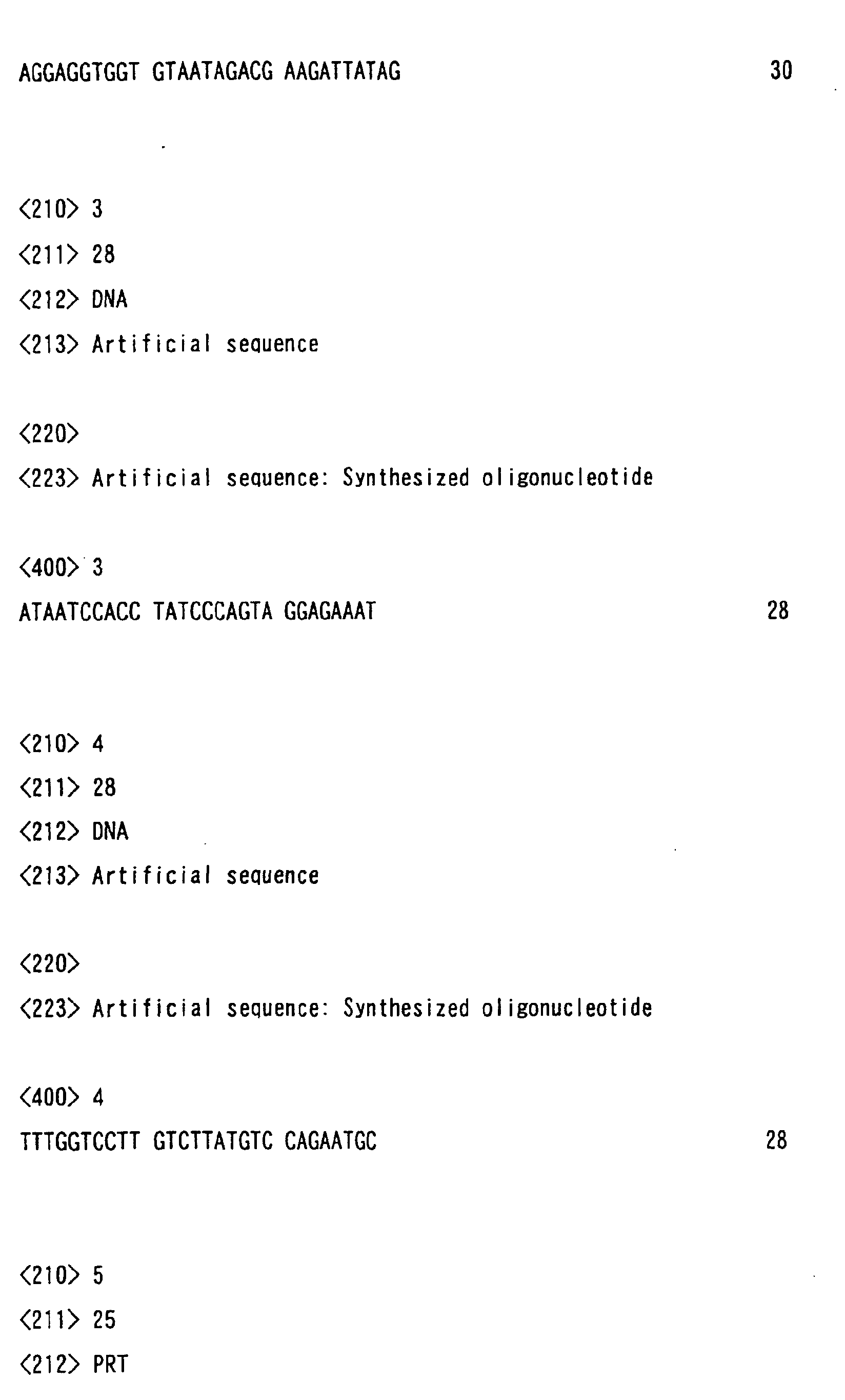
- primers for subtype B and E used were ordinary SK38 (oligonucleotide of SEQ ID NO. 3: from 1551 to 1578) and SK 89 (oligonucleotide of SEQ ID NO. 4: from 1683 to 1665).
- the white plaques formed indicate that 5 recombinant virus strains obtained herein all amplified the 114 bp DNA and had the HIV gag gene inserted therein.
- Fig. 2 shows the data of Western blotting of the infected cells.
- the ultrasonic extract of the infected cells was separated through SDS-polyacrylamide gel electrophoresis, and assayed against an anti-gag monoclonal antibody (Matsuo, K., et al., J. Gen. Virol., 73, 2445, 1992).
- One of the 5 strains gave a specific band that might indicate the gag precursor p55 protein.
- the insert protein lysates existed.
- the assay data indicate that the recombinant virus expressed the gag antigen. This strain was named rVV-DIs-gagB.
- CEF cells were infected with rVV-DIs-gagB (in five laboratory dishes of 10 cm in diameter), and incubated in 10 ml of a 1 % PBS/MEM medium at 37°C in the presence of 5 % CO 2 . After 2 to 4 days, the medium was removed when CPE was seen in the dishes. Then, the cells were disrupted in 15 ml of 10 mM Tris-HCl (pH 8.0), and then physically released and collected, and the virus having adhered to the cell debris was ultrasonically isolated. This was then centrifuged for 20 minutes at 3000 rpm, and the supernatant was further centrifuged for 90 minutes at 13000 rpm.
- the resulting mass of viruses was lysed in 8 ml of 10 mM Tris-HCl buffer capable of precipitating the viruses, to which was gently added 3 ml of 36 % sucrose. This was again centrifuged for 90 minutes at 13000 rpm. The resulting pellets were dissolved in 1 ml of 10 mM Tris-HCl buffer to prepare a partially-purified virus suspension. The virus suspension was stepwise diluted, and the quantity of viruses in the CEF cells was measured. It was 10 10 pfu/ml.
- Hela is a human-derived cell strain
- CV-1 is an African green monkey-derived cell strain
- RK is a rabbit kidney-derived cell strain
- CHO is a Chinese hamster ovary cell strain.
- the parent strain (DEI) most actively propagated in CEF cells, and propagated in some degree in the other mammal cells except CHO. Contrary to this, however, the recombinant virus strain DIs did not propagate at all in the mammal cells except CEF cells.
- a recombinant strain DIs capable of expressing a bacteriophage T7 polymerase was constructed in the same manner as in Example 1. Based on the method described in references, Virology 231;192-200, 1997, Virology 250:140-150, 1998, the T7 polymerase activity (degree of expression) of the recombinant strain DIs was measured, using reporter genes pT7Luc and pT7EMCLuc. Concretely, different types of mammal-derived cells were incubated in a 48-well plate in a mode of monolayer culture, and infected with 2 moi of any of the strain DIs or the recombinant vaccinia virus rDIs-T7pol.
- the cells were infected with 20 moi of a T7 polymerase-expressing adenovirus rAdexCAT7. After 24 hours, the cells of each type were transfected with 1 ⁇ g of a reporter plasmid pAcTU7Luc or pAcT7EMCLuc. The luciferase activity of the cells was measured in an assay kit, Pica Gene (by Tokyo Ink). 24 hours after thus transfected, the cells were collected, washed twice with PBS, and lysed in 100 ⁇ l of a cytolytic buffer LUC/PGC-50 (by Tokyo Ink).
- each transparent cell lysate was incubated along with 100 ⁇ l of a reactive solution (containing 137 mM NaCl, 2.7 mM KCl, 4.3 mM Na 2 HPO 4 , 1.4 mM KH 2 PO 4 , 20 mM Tricine, 1.07 mM (MgCO 3 ) 4 Mg(OH) 2 ⁇ 5H 2 O, 2.67 mM MgSO 4 , 0.1mM EDTA, 33.3 mM DTT, 270 ⁇ M Coenzyme A, 470 ⁇ M luciferin, and 530 ⁇ M ATP).
- the relative luminosity unit (RLU) of each reaction liquid was measured with a luminometer (by Berthold).
- Fig. 4 BHK is a hamster baby cell strain; and HepG2 is a human-derived cell strain.
- the recombinant vaccinia virus rDIs-T7pol has no propagation potency in the mammal-derived cells, but expressed the exogenous gene in the cells to the same degree as that of adenovirus.
- the results confirm that the recombinant vaccinia virus strain DIs of this invention, when used as a vaccine ingredient, does not so much propagate to an unnecessary degree, but expresses the intended antigenic protein necessary for antibody formation.
- the excellent characteristic of the recombinant vaccinia virus strain DIs of this invention supports the favorable applicability of the strain to vaccinia virus vaccines especially for patients suffering from immunodeficiency such as AIDS.
- mice were intravenously inoculated with 10 7 pfu/mouse of rVV-DIs-gagB. At regular intervals of 2, 6, 8 and 12 weeks, the spleen was taken out of each mouse. Each spleen was mashed along with PBS in a cell strainer having a pore size of 70 ⁇ m (by Becton Dickinson), the resulting spleen cells were transferred into a 15-ml centrifugal tube and centrifuged.
- the suspension was diluted with the same medium to have a cell concentration of 1 ⁇ 10 7 cells/ml, to which was added an aqueous solution of two gag epitope peptides (concentration: 1 mg/ml) to have a final concentration of 10 ⁇ g/ml. Then, this was incubated at 37°C for 5 days.
- the amino acid sequences of the peptides (gag B253-277 and gag B287-309) are Sequence Numbers 5 and 6, respectively.
- the number of the live cells in the suspension was again counted, and the cell concentration of the suspension was controlled to be 1 ⁇ 10 7 cells/ml with 10 % FBS/RPMI1640 added thereto.
- the cells are effector cells.
- M 12.4.5 mouse cells (H2d haplo-type) were incubated in 10 % FBS/RPMI1640, and after reached 80 % confluence, these were washed once with PBS, then released in a trypsin-EDTA solution, and collected. These were suspended in 10 ml of 10 % FBS/RPMI1640, and the number of the live cells in the suspension was counted. Through centrifugation, 1 ⁇ 10 7 cells were collected, and suspended in 100 ⁇ m of the same medium. A solution of the two peptides as above was added to the suspension to have a final concentration of 50 ⁇ g/ml. For control, the peptide solution was not added to the cell suspension.
- the cells were incubated at 37°C for 3 hours.
- 100 ⁇ l (100 ⁇ Ci) of an aqueous solution of 51Cr-labeled sodium chromate (by New England Biolab) was added to each cell suspension, and the cells were further incubated at 37°C in 5 % CO 2 for 1 hour and 30 minutes.
- the cells were washed three times with 10 % FBS/RPMI1640, and then diluted with the same medium to have a cell concentration of 1 ⁇ 10 5 cells/ml.
- the cells are target cells.
- the effector cells and the target cells were mixed in different ratios, 100/1, 50/1, 25/1 and 12.5/1 in a 96-well plate (total volume: 200 ⁇ l/well), and incubated at 37°C in 5 % CO 2 for 4 hours.
- 100 ⁇ l of 10 % FBS/RPMI1640 or 100 ⁇ l of aqueous 1 % Triton ⁇ 100 was added to 100 ⁇ l of the target cell suspension, and the cells were incubated under the same condition at 37°C in 5 % CO 2 for 4 hours.
- CTL activity (%) (data of target cells - data of spontaneous emission/data of maximum emission - data of spontaneous emission) ⁇ 100.
- Example 5 the rVV-DIs-gagB-immunized mouse blood was collected on 2, 6, 8 and 12 weeks after the immunization to prepare serum samples. Of those, 20 ⁇ l of the 12-weeks immunized serum was tested for its reactivity, using an HIV antigen-blotted nitrocellulose membrane filter (Genelabs Diagnostics' Western blotting kit).
- a plasmid pEGFP-1 (BY Clontach) was digested with EcoRI, and blunted through treatment with a Klenow fragment. The blunt DNA was further digested with BamHI, and the resulting fragment of about 700 bp was subcloned in the BamHI-SmaI site of pUC-VVp7.5H, like in Example 1. Next, this was cleaved with HindIII to obtain a fragment containing the region of p7.5 promoter and EGFP open reading frame. This was introduced into the HindIII site of pUC-DIs to construct a transfer vector.
- this plasmid was electroporated into CEF cells that had been infected with a ⁇ -galactosidase-expressing recombinant vaccinia virus strain DIs. With that, the cells were incubated on an X-gal agar medium, on which the cells giving white plaques were selected. These are of a recombinant virus strain. The virus-infected CEF cells were found to give green fluorescence when observed with a fluorescent microscope. This confirmed that the cells expressed the protein EGFP.
- the genetic clone pKS460 of a simian immunodeficiency virus SIV was digested with restriction endonucleases NdeI and XhoI, and blunted through treatment with a Klenow fragment.
- the resulting fragment of about 1500 bp was subcloned in the BamHI-Smal site of pUC-VVp7.5H, like in Example 1.
- this was cleaved with HindIII to obtain a fragment containing the region of p7.5 promoter and EGFP open reading frame. This was introduced into the HindIII site of pUC-DIs to construct a transfer vector.
- this plasmid was electroporated into CEF cells that had been infected with a ⁇ -galactosidase-expressing recombinant vaccinia virus strain DIs. With that, the cells were incubated on an X-gal agar medium, on which the cells giving white plaques were selected. These are of a recombinant virus strain rVV-DIs-SIV gag.
- the recombinant virus rVV-DIs-SIV gag that had been constructed in Example 8 was intravenously administered to crab-eating monkeys twice at an interval of 45 days (10 6 pfu/monkey), and the immune induction in the thus-immunized monkeys was evaluated through a test for antibody production in the monkeys, a test for lymphocyte propagation therein, an ELISPOT assay and an intracellular cytokine production assay. In those tests and assays, all the rVV-DIs-SIV gag-immunized, crab-eating monkeys showed the induction of anti-gag antibody, gag-specific helper, and CTL activity. On the other hand, no abnormal observations were found in all the immunized monkeys.
- the monkeys were infected with a pathogenic chimera virus SHIV C21 10TCID, and the number of the CD4-positive lymphocytes in the peripheral blood collected from each monkey was counted.
- the results are shown in Fig. 7.
- the number of the CD4-positive lymphocytes significantly decreased in the monkeys of the control group within 2 weeks after the pathogenic virus challenge, while the reduction in the CD4-positive lymphocytes in the rVV-DIS-SIV gag-immunized monkeys was retarded by 70 to 95 %.
- the number of viral RNA copies in plasma at set points was decreased to a degree of 1/50 to 1/100.
- the invention of this application provides an attenuated, recombinant vaccinia virus vaccine having high antigenicity against pathogens except vaccinia viruses, and provides a recombinant vaccinia virus strain DIs that serves as the active ingredient of the vaccine.
- the recombinant vaccinia virus strain DIs which the invention provides herein has the ability to induce both humoral and cellular immunity against the foreign polypeptide inserted thereinto.
- the recombinant virus does not propagate in ordinary animal cells but expresses the immunogenic antigen (having the potency of inducing cellular immunity and antibody production), and the virus cells are easy to incubate and can be produced at low costs.
- the recombinant strain of this invention can be used as an inexpensive substitute for antigens for boosts in the prime and boost method that is much used in the art for developing AIDS vaccines. Accordingly, the recombinant strain of this invention is extremely useful for antigens substitutable for pure recombinant proteins that require high production costs.
- the recombinant strain of this invention is highly safe to humans, it is useful for a recombinant vaccinia virus strain to be infected into target cells in CTL activity determination from the viewpoint of safe protection of test subjects.
Landscapes
- Health & Medical Sciences (AREA)
- Life Sciences & Earth Sciences (AREA)
- Chemical & Material Sciences (AREA)
- Genetics & Genomics (AREA)
- Organic Chemistry (AREA)
- Engineering & Computer Science (AREA)
- Bioinformatics & Cheminformatics (AREA)
- Zoology (AREA)
- Wood Science & Technology (AREA)
- Biomedical Technology (AREA)
- General Health & Medical Sciences (AREA)
- Virology (AREA)
- General Engineering & Computer Science (AREA)
- Biochemistry (AREA)
- Biotechnology (AREA)
- Biophysics (AREA)
- Microbiology (AREA)
- Molecular Biology (AREA)
- Medicinal Chemistry (AREA)
- Physics & Mathematics (AREA)
- Plant Pathology (AREA)
- Gastroenterology & Hepatology (AREA)
- Proteomics, Peptides & Aminoacids (AREA)
- Immunology (AREA)
- Micro-Organisms Or Cultivation Processes Thereof (AREA)
- Medicines Containing Antibodies Or Antigens For Use As Internal Diagnostic Agents (AREA)
Abstract
The invention provides a recombinant vaccinia virus strain DIs that
possesses a polynucleotide encoding a foreign antigenic protein in the
non-essential gene region of the chromosome DNA and expresses the antigenic
protein; and provides a highly-safe, vaccinia virus vaccine containing the
recombinant virus strain DIs as the active ingredient. The invention also provides
a method of using the vaccinia virus strain DIs as a vector for protein expression.
Description
The invention of this application relates to a recombinant vaccinia virus
vaccine. More precisely, the invention of this application relates to a virus vaccine
for various infectious diseases of humans and animals, and to a recombinant
vaccinia virus strain DIs that serves as the effective ingredient of the vaccine.
With the development and improvement of the recombination technology
in these ten years and more, various studies of denaturing microorganisms such
as viruses and bacteria through genetic recombination are now made for
application of the resulting recombinants to vaccine vectors for prevention and
treatment of various infectious diseases and cancers. For the studies, various
microorganisms have been tried, including, for example, polio virus, influenza
viruses, rhino virus, chickenpox virus, Salmonella bacteria, bovine tubercle
bacillus-attenuated strain BCG, and Listeria monocytogenes, for which, however,
vaccinia virus has the oldest history of all.
Vaccinia virus is utilized as smallpox vaccines, and has much contributed
toward eradicating smallpox. This virus is a large-size DNA virus, belonging to
Poxviridae, and, as its genome, it has a linear double-stranded DNA of about 190
kbp, coding for about 200 types of different proteins. In 1982, Panicali and
Paoletti (Proc. Natl. Acad. Sci. USA 79:4927, 1982), and Mackett et al. (Proc. Natl.
Acad. Sci. USA 79:7415, 1982) first reported the expression of a recombinant
vaccinia virus with a thymidine kinase (TK) gene of herpes simplex virus inserted
thereinto. After that, various types of different recombinant vaccinia viruses have
been constructed and tried for application to immunological analysis and to
vaccine production. However, it is said that, depending on the toxicity of the
parent strains used for their recombination, some of such recombinant vaccinia
viruses will often induce encephalitis in humans and their toxicity will be often
strengthened through atavism. From the viewpoint of safety, therefore, it is
difficult to use the conventional recombinant vaccinia viruses for clinical vaccines
for human use, especially for those for human immunodeficiency disorders such
as AIDS, etc.
On the other hand, a vaccinia virus strain DIs is highly attenuated, and it
was isolated through subcultivation of a vaccinia virus strain Talien (DEI) in hen's
egg embryos for developing safe smallpox vaccines (Nature 192:381, 1961). The
This strain DIs is specifically characterized in that it grows and propagates in fetal
avian primary culture cells (DEF) but little in any other mammal cells. In those
days, this was tried for clinical application along with a strain LC16 that had been
developed together with it, and 200 Japanese children were actually vaccinated
with it. However, as lacking the ability to propagate in human cells, the strain
DIs was unfavorable for smallpox vaccines because of its poor immune inducibility,
and the other strain LC16 better than the strain DIs in point of immune induction
was employed for smallpox vaccines. This is the history of the strain DIs.
Still other attenuated vaccinia viruses, MVA (vaccinia virus strain Ankara)
(Zur Beurteilung der MVA-Stufenimpfung bei Pockenerstrimpfungen, Med. Diss.
Munchen, 1976) and NYVAC (vaccinia virus strain New York) (Virology
188:217-232) were reported. In particular, it was suggested that MVA could
serve as a smallpox vaccine in humans. At present, MVA is propagated in
mammal-derived cells such as CV-1 or BHK-16, and the attenuated virus MVA is
now used as a vector for constructing various recombinant vaccines. However,
the above-mentioned, attenuated strain DIs differs from the other attenuated
strains MVA and NYVAC in that the former DIs does not propagate at all in
mammal cells and is therefore useless for human vaccination.
The inventors of this application had succeeded in inserting a foreign gene
into the genome of this strain DIs, and had verified that the recombinant strain
DIs is useful as a viral vector for animal cells. However, since the immune
inducibility of the strain. DIs is poor as so mentioned hereinabove, it has been
believed that, even if the strain DIs is so recombined with a foreign antigenic
protein as to express the foreign antigenic protein, the resulting recombinant
strain DIs will be ineffective for vaccines.
As so mentioned hereinabove, the cytotoxicity of the vaccinia virus strain
DIs is low, it is easy to believe that the strain will be better than any other strains
in point of its safety in human applications. Despite of the situation, however,
because the immune inducibility of the strain DIs is poor, no one has heretofore
considered the use of the strain DIs for recombinant virus vaccines. Contrary to
this, the inventors of the present application have succeeded in introducing a
foreign gene into the vaccinia virus strain DIs and in expressing the foreign gene
in the recombinant strain DIs, and, in animal experiments, the inventors have
found the possibility of inducing the humoral and cellular immune reaction in the
foreign gene product from the recombinant vaccinia virus strain DIs.
In addition, since the vaccinia virus strain DIs does not propagate in
animal cells, it has heretofore been believed that the strain will be unsuitable for
an expression vector for useful proteins. However, the inventors of this
application have found that, owing to its low cytotoxicity, the vaccinia virus strain
DIs enables its host cells to live for a long period of time and is therefore useful as
a vector for mass-expression of proteins.
The invention of this application has been made on the basis of the novel
findings of the inventors as above, and its object is to provide a recombinant
vaccinia virus vaccine of high safety.
Another object of the invention is to provide the recombinant vaccinia
virus strain DIs that serves as the active ingredient of the vaccine.
Still another object of the invention is to provide the recombinant vaccinia
virus strain DIs that expresses a reporter molecule and is therefore useful for
construction of other recombinant virus strains.
Still another object of the invention is to provide a method of using the
vaccinia virus strain DIs for a vector.
To achieve the objects noted above, the invention of this application
provides the following inventions (1) to (19):
The vaccine of the inventions (1) to (5) is a highly-attenuated live vaccine
that contains, as the active ingredient, the recombinant vaccinia virus strain DIs
of the inventions (6) to (10). The recombinant vaccinia virus strain DIs of the
invention (6) is a recombinant virus obtained through homologous recombination
of the genome DNA of DIs with a transfer vector having a vaccinia virus-derived
gene promoter sequence and having a polynucleotide that encodes an intended,
foreign (that is, excluding vaccinia viruses) antigenic protein, linked downstream
the promoter sequence. In the following description, the foreign antigenic protein
will be referred to as "foreign polypeptide"; and the polynucleotide encoding it will
be referred to as "foreign polynucleotide".
The promoter sequence to be inserted into the transfer vector may be any
and every one capable of being recognized by the RNA polymerase which is
encoded by the gene of a vaccinia virus itself. For it, for example, a 7.5 kD
polypeptide gene promoter p7.5 that exists inside the inverted terminal repeats of
the genome of a vaccinia virus (Stunnenberg, H. G., et al., Nucleic Acids Res., 16,
2431, 1988) may be used.
The foreign polynucleotide (e.g., cDNA fragment) may be any and every
one that encodes an antigenic protein except vaccinia viruses; and the foreign
polypeptide may be any and every one capable of inducing antigen-antibody
reaction in an animal body. Concretely, herein directed thereto are a gag
precursor protein p55, an env protein gp120 or gp160, a pol precursor protein, a
nef protein and a tat protein of a human immunodeficiency virus (HIV) that causes
human acquired immunodeficiency syndrome (AIDS); and a gag precursor protein
of a simian immunodeficiency virus (SIV). In addition, also employable herein are
other polynucleotides encoding other pathogens (other pathogenic viruses and
bacteria) or for antigenic proteins of cancer cells.
Such foreign polynucleotides may be obtained, for example, by cleaving a
plasmid clone of a genomic gene encoding an intended foreign polypeptide or its
cDNA, or by amplifying through polymerase chain reaction (PCR) using primers
having a suitable sequence and the plasmid clone as a template. If not cloned,
the bacterial or animal genomic DNA having the gene is amplified through PCR for
which is used a virus-infected animal cell-derived DNA or RNA as the template in
case of viruses.
The thus-obtained foreign polynucleotide is ligated with the
above-mentioned promoter in a correct reading frame, and the fused
polynucleotide (this will be hereinafter referred to as "expression unit") is then
inserted into a desired plasmid vector to construct a transfer vector.
Next, a method of homologous recombination for introducing the foreign
polynucleotide into a vaccinia virus genome will be described. A vaccinia virus
genome DNA is first extracted from virus particles which are purified through
sucrose density gradient centrifugation (Joklik, W. K., Virology, 18, 9, 1962), using
L-laurylsarucosine (Mackett, M. and Archard, L. C. J., Gen. Virol., 45, 683, 1979).
In the thus-extracted genome DNA, the non-essential gene region that has no
influence on the vaccinia virus replication must be specifically defined for the site
for homologous recombination.
The first candidate for the non-essential gene region is a thymidine kinase
(TK) gene region. In this case, inserting the TK gene region into a plasmid vector
such as pUC18, and further inserting an expression unit at the unique restriction
site in that region may construct a transfer vector. Using this vector, the foreign
polynucleotide is inserted into the vaccinia virus genome through homologous
recombination with the TK gene in the virus genome. After the homologous
recombination, the recombinant virus is produced in the TK-lacking cell line, and
incubated in the presence of bromodeoxyuridine to thereby select the recombinant
virus not expressing TK.
The second candidate for the non-essential gene region is genomic
gene-lacking region of DIs. Comparing the restriction enzyme cleavage patterns
of the genomic DNA of DIs with that of the parent strain DIE may identify the
defective region in the genome of DIs. This region is basically unnecessary for DIs
replication. The DNA sequences before and after the defective region are
amplified through PCR, and then inserted into a plasmid vector. An expression
unit is further inserted into the plasmid to construct a transfer vector. Using the
transfer vector, the DIs genomic DNA is subjected to homologous recombination,
whereby the expression unit is inserted into the defective region of the DIs
genomic DNA. In this method, however, the recombinant virus is difficult to
select. To solve the problem, therefore, a polynucleotide encoding a reporter
molecule is first inserted into the defective region to construct a recombinant virus.
For the reporter molecule, for example, any of bacteria-derived coloring enzyme
such as β-galactosidase, or a green fluorescence protein (GFP) or its derivatives
(e.g., EGFP) may be used. For example, for the recombinant virus that expresses
β-galactosidase, X-gal (5-bromo-4-chloro-3-indolyl-β-D-galactopyranoside) is
added to the medium, by which the cells expressing β-galactosidase are selected
based on their plaque color (blue). For GFP or its derivatives, the
fluorescence-emitting plaques are selected through fluorescent microscopy. Into
the reporter molecule-expressing virus, the intended foreign polynucleotide is
inserted into the region of the reporter molecule through homologous
recombination. In the process, the virus not expressing the reporter molecule is
selected to thereby obtain the recombinant virus characterized by having the
foreign polynucleotide inserted into the gene defective region through homologous
recombination.
The thus-obtained recombinant virus is cloned and purified into a
monoclone, and then confirmed for the presence or absence of the inserted gene
therein through PCR. In Western blotting or enzyme-linked immunosorbent
assay (ELISA), the monoclonal or polyclonal antibody against the foreign
polypeptide inserted thereinto is tried in the ultrasonic extract of virus-infected
CEF cells for confirming the expression of the foreign polypeptide in the infected
cells.
The recombinant virus is grown in CEF cells, and then partially purified
through sucrose density gradient centrifugation, and used in animal experiments.
In animal experiments, mice and others such as rabbits and monkeys may be
tried. For example, mice are intravenously, intramuscularly or intraperitoneally
inoculated with the recombinant virus, and the antibody value to the foreign
polypeptide in the serum may be measured at any intervals through Western
blotting or ELISA to thereby determine the humoral immune response of the
recombinant virus in mice. On the other hand, the cellular immune response of
the recombinant virus in mice is determined as follows: First, mice are
inoculated with the recombinant virus, then the lymphocytes in the blood
collected from each mouse at any intervals are stimulated in vitro with the foreign
polypeptide antigen, and the amount of the tritium thymidine intake into the
lymphocytes is measured time-dependently. Secondly, foreign polypeptide
antigen-specific cytotoxic T cells (CTL) in mice are identified as follows: After
inoculated with the recombinant virus, the mice are grown for a predetermined
period of time. Then, the spleen is taken out of each mouse, and mashed to
obtain the spleen cells. The cells are stimulated in vitro with a synthetic peptide
that corresponds to the CTL epitope in the foreign polypeptide, for a
predetermined period of time. Thus stimulated, the cells serve as effector cells.
On the other hand, the mouse cells of the same series are infected with the same
recombinant vaccinia virus, or the mouse cells having been incubated along with a
peptide antigen identified as the CTL epitope, and these cells are labeled with 51Cr
to be target cells. The effector cells are reacted with the target cells in different
ratios, and the radioactivity of 51Cr released in the culture supernatant is counted
to thereby confirm the presence or absence of the CTL activity in the immunized
mouse spleen cells.
The methods of the inventions (17) to (19) are for mass-expression of the
foreign polynucleotide including useful proteins in the vaccinia virus strain DIs.
Specifically, the vaccinia virus strain DIs accepts any type of polypeptide in a
broad range for the foreign protein to be inserted thereinto, irrespective of the
length and the structure of the foreign polynucleotide. In addition, the vaccinia
virus strain DIs can be handled safely with no problem.
The recombinant vaccinia virus strain DIs is used as the expression vector
in the methods, and it can be constructed in any desired manner using any
desired foreign polynucleotide, like the recombinant virus strain DIs for vaccines
mentioned above. The virus vector, strain DIs is transfected into any of CEF cells,
mouse-derived p813 cells and fibroblasts in any ordinary method, and the
resulting transfectant cells are incubated to thereby express a large amount of the
protein that is encoded by the foreign polynucleotide inserted into the vector, for a
long period of time. For example, in the experiments made by the inventors of
this invention, the cells infected with the recombinant vaccinia virus strain DIs
(concentration: 10 pfu) for 1 hour realized significant expression of the
recombinant protein after incubation for 6 to 12 hours. In addition, the
transfectant cells kept living for a long period of time, continuously expressing the
protein.
The invention of this application is described in detail and more concretely
with reference to the following Examples, which, however, are not intended to
restrict the scope of the invention.
In preparing a recombinant vaccinia virus from DIs, first investigated was
the deletion site in the DIs genome. The parent strain DE1 and the DIs genome
DNA were digested with HindIII, and the resulting fragments were compared
through agarose gel electrophoresis. It was found that, of the 16 fragments, A to
P (in the order of their size) of the parent strain genome DNA, the fragment N (1.5
kbp) and the fragment M (2.2 kbp) were deleted, the fragment C (25.1 kbp) and the
fragment K (4.6 kbp) were shortened, and the 15.4 kbp region including almost all
the 3'-side fragment C to the fragment K was lost in the DIs genome DNA.
Accordingly, of the fragments deleted in the strain DIs, the fragment C and the
fragment K, and the region (about 1.9 kbp) including the 5'-side of the fragment F
(13.5 kbp) that is in contact with the adjacent fragment at the 3'-side of the
fragment K were amplified through PCR and cloned in a PCR2.1 TA cloning vector
(by Invitrogen). The PCR primers used are Vac H-C (oligonucleotide of SEQ ID NO.
1) and Vac H-F (oligonucleotide of SEQ ID NO. 2).
From the plasmid, the EcoRI fragment including the amplified region was
cut out, and subcloned in the EcoRI site of pUC19 to obtain a vector pUC-DIs.
The DIs fragment of the vector has one HindII site. In this site, an E. coli DNA
fragment including the open reading frame of β-galactosidase gene (LacZ) was
subcloned downstream the vaccinia virus latter-stage promoter p11 to give a
transfer vector pUC-DIs-LacZ.
500 µl of a virus suspension containing about 105 pfu (plaque-forming
unit) of vaccinia virus DIs cells were inoculated into CEF cells (in a 6 cm-diameter
laboratory dish), and the dish was lightly shaken four times at intervals of 15
minutes. After one hour, 2.5 ml of 1 % FBS/MEM medium was added thereto,
and the cells were incubated for 6 hours under the condition of 37°C, 5 % CO2.
The medium was removed, and the cells were washed twice with PBS (phosphate
buffered saline), then released in a trypsin-EDTA solution (by GIBCO), collected,
and finally re-suspended in 400 µm of PBS. To the cell suspension, added was
20 µg of the transfer vector pUC-DIs-LacZ. In a 0.4 cm-cuvette, this was
electroporated once at 250 V and 500 µFD (for this, used was BioRad's Gene
Puisar II). The cells were then left at room temperature for 15 minutes or longer,
and then spread in a laboratory dish and incubated therein for 4 days. The cells
were released, collected along with the culture medium, then diluted 10-, 100- and
1000-fold, and thereafter inoculated into CEF cells for subcultivation in the
same manner as above. After 4 days, when CPE was seen in the cells, the
medium was removed, and an X-gal added agar medium (4-fold concentrated
MEM medium 25 ml, distilled water 20.7 ml, 7.5 % NaHCO 3 3 ml, 2.92 %
L-glutamine 1.0 ml, 2 % X-gal 0.2 ml, 30 mg/ml kanamycin sulfate 0.1 ml, 2.4 %
Agar Noble 50.0 ml) was added to the cells. After 4 hours, the blue-emitting CPE
part of the agar was picked up, using a Pasteur pipette, and collected in 500 µl of
the same medium as above. The thus-picked up agar was ultrasonically mashed,
and then inoculated into CEF cells for subcultivation in the same manner as
above. The same cycle as above was repeated to further clone the cells until the
CPE in one laboratory dish with the cells therein was entirely in blue.
Next, in the HindIII site of the same transfer vector as above, inserted was
an expression unit that had been constructed by linking the gag precursor protein
gene of an HIV subtype B to the downstream of the promoter of a vaccinia virus
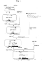
p7.5. The outline of the transfer vector construction is shown in Fig. 1.
Concretely, a subtype B HIV molecule clone pNL432 (Adachi, A., et al., J. Virol., 59,
284, 1986) was completely digested with restriction endonucleases BssHI and
HincII, and the resulting fragment (about 1.8 kbp) was blunted with a Klenow
fragment. This fragment was subcloned into the BamHI-SmaI site of a plasmid
pUC-VVp7.5H that had been constructed by inserting the p7.5 promoter fragment
cut out of a plasmid pAK2, into the PstI-XbaI site of pUC18, to thereby obtain
pVV7.5-gagB. This plasmid was partially digested with HindIII, and its fragment
(about 2.18 kbp) was separated and purified through 1 % agarose gel
electrophoresis, and subcloned in the HindIII site of the transfer vector pUC-DIs to
thereby obtain the intended vector pUC-DIs-gagB.
Similarly, CEF cells were infected with 500 µl of a virus suspension
containing about 105 pfu of cells of the β-galactosidase-expressing vaccinia virus
strain DIs (rVV-DIs-LacZ) that had been prepared in the above, and the cells were
released in a trypsin-EDTA solution, collected, and finally re-suspended in 400 µm
of PBS. To the cell suspension, added was 20 µg of the transfer vector
pUC-DIs-gagB. Then, this was electroporated under the same condition as above.
The cells were then left at room temperature for 15 minutes or longer, and then
spread in a laboratory dish and incubated therein for 4 days. The cells were
released, collected along with the culture medium, then diluted 10-, 100- and
1000-fold, and thereafter inoculated into CEF cells for subcultivation in the same
manner as above. After 4 days, when CPE was seen in the cells, the medium was
removed, and an X-gal added agar medium was added to the cells. After 4 hours,
the white CPE part of the agar not emitting in blue was collected in 500 µl of the
same medium as above, using a Pasteur pipette. The thus-picked up agar was
ultrasonically mashed, and then inoculated into CEF cells for subcultivation in the
same manner as above. The same cycle as above was repeated to further clone
the cells until the CPE in one laboratory dish with the cells therein did not emit in
blue at all.
The HIV gag gene insertion into the thus-obtained recombinant virus was
confirmed through PCR. For the primers for subtype B and E, used were
ordinary SK38 (oligonucleotide of SEQ ID NO. 3: from 1551 to 1578) and SK 89
(oligonucleotide of SEQ ID NO. 4: from 1683 to 1665).
The white plaques formed indicate that 5 recombinant virus strains
obtained herein all amplified the 114 bp DNA and had the HIV gag gene inserted
therein.
Next, these recombinant viruses were tested for HIV gag antigen
expression in virus-infected CEF cells.
Fig. 2 shows the data of Western blotting of the infected cells. Concretely,
the ultrasonic extract of the infected cells was separated through
SDS-polyacrylamide gel electrophoresis, and assayed against an anti-gag
monoclonal antibody (Matsuo, K., et al., J. Gen. Virol., 73, 2445, 1992). One of
the 5 strains gave a specific band that might indicate the gag precursor p55
protein. In all the strains assayed, the insert protein lysates existed. The assay
data indicate that the recombinant virus expressed the gag antigen. This strain
was named rVV-DIs-gagB.
CEF cells were infected with rVV-DIs-gagB (in five laboratory dishes of 10
cm in diameter), and incubated in 10 ml of a 1 % PBS/MEM medium at 37°C in
the presence of 5 % CO2. After 2 to 4 days, the medium was removed when CPE
was seen in the dishes. Then, the cells were disrupted in 15 ml of 10 mM
Tris-HCl (pH 8.0), and then physically released and collected, and the virus having
adhered to the cell debris was ultrasonically isolated. This was then centrifuged
for 20 minutes at 3000 rpm, and the supernatant was further centrifuged for 90
minutes at 13000 rpm. The resulting mass of viruses was lysed in 8 ml of 10 mM
Tris-HCl buffer capable of precipitating the viruses, to which was gently added 3
ml of 36 % sucrose. This was again centrifuged for 90 minutes at 13000 rpm.
The resulting pellets were dissolved in 1 ml of 10 mM Tris-HCl buffer to prepare a
partially-purified virus suspension. The virus suspension was stepwise diluted,
and the quantity of viruses in the CEF cells was measured. It was 1010 pfu/ml.
Different types of mammal-derived cells were incubated in a 48-well plate
in a mode of monolayer culture, and infected with 105 pfu of the recombinant
virus strain rVV-DIs-LacZ that had been constructed in Example 1. After kept at
37°C for 1 hour, the cells of each type were separately washed with PBS, and
further incubated in a fresh medium at 37°C. 0 hour and 48 hours after the
absorption, the cells were collected, frozen and thawed. These were then
ultrasonically processed, and the quantity of the viruses at 48 hours was divided
by that at 0 hour to thereby determine the degree of virus propagation. The data
are given in Table 1 below. In Table 1, Hela is a human-derived cell strain; CV-1
is an African green monkey-derived cell strain; RK is a rabbit kidney-derived cell
strain; and CHO is a Chinese hamster ovary cell strain. As is obvious from Table
1, the parent strain (DEI) most actively propagated in CEF cells, and propagated in
some degree in the other mammal cells except CHO. Contrary to this, however,
the recombinant virus strain DIs did not propagate at all in the mammal cells
except CEF cells.
| HeLa | CV-1 | RK13 | CHO | CEF | |
| Parent Strain | 7890 | 1578 | 842 | <1 | 9474 |
| rDIs | <1 | <1 | <1 | N.D. | 1600 |
Next, 2.0 moi of the recombinant vaccinia virus rVV-DIs-gagB constructed
in Example 1 was incubated at 37°C for 1 hour along with CV-1 cells, and then
cultured for 2 days. On the other hand, 0.1 moi of the parent strain (DEI) was
also infected with CV-1 cells in the same manner, and then cultured for 4 days.
After thus cultured, the cells of each type were separately recovered in 0.125 %
trypsin-EDTA to prepare cell lysates. Each cell lysate was applied to 10-day-old
eggs, in which the infectivity potency of each virus was determined.
The data are shown in Table 3. As in this, the parent vaccinia virus
strain DEI infected the CV-1 cells, but the recombinant virus strain DIs of this
invention did not at all.
A recombinant strain DIs capable of expressing a bacteriophage T7
polymerase was constructed in the same manner as in Example 1. Based on the
method described in references, Virology 231;192-200, 1997, Virology
250:140-150, 1998, the T7 polymerase activity (degree of expression) of the
recombinant strain DIs was measured, using reporter genes pT7Luc and
pT7EMCLuc. Concretely, different types of mammal-derived cells were incubated
in a 48-well plate in a mode of monolayer culture, and infected with 2 moi of any
of the strain DIs or the recombinant vaccinia virus rDIs-T7pol. For control, the
cells were infected with 20 moi of a T7 polymerase-expressing adenovirus
rAdexCAT7. After 24 hours, the cells of each type were transfected with 1 µg of a
reporter plasmid pAcTU7Luc or pAcT7EMCLuc. The luciferase activity of the cells
was measured in an assay kit, Pica Gene (by Tokyo Ink). 24 hours after thus
transfected, the cells were collected, washed twice with PBS, and lysed in 100 µl of
a cytolytic buffer LUC/PGC-50 (by Tokyo Ink). Next, 20 µl of each transparent
cell lysate was incubated along with 100 µl of a reactive solution (containing 137
mM NaCl, 2.7 mM KCl, 4.3 mM Na2HPO4, 1.4 mM KH2PO4, 20 mM Tricine, 1.07
mM (MgCO3)4Mg(OH)2·5H2O, 2.67 mM MgSO4, 0.1mM EDTA, 33.3 mM DTT, 270
µM Coenzyme A, 470 µM luciferin, and 530 µM ATP). The relative luminosity unit
(RLU) of each reaction liquid was measured with a luminometer (by Berthold).
The data are shown in Fig. 4. In Fig. 4, BHK is a hamster baby cell
strain; and HepG2 is a human-derived cell strain. As in Example 3, the
recombinant vaccinia virus rDIs-T7pol has no propagation potency in the
mammal-derived cells, but expressed the exogenous gene in the cells to the same
degree as that of adenovirus. The results confirm that the recombinant vaccinia
virus strain DIs of this invention, when used as a vaccine ingredient, does not so
much propagate to an unnecessary degree, but expresses the intended antigenic
protein necessary for antibody formation. The excellent characteristic of the
recombinant vaccinia virus strain DIs of this invention supports the favorable
applicability of the strain to vaccinia virus vaccines especially for patients suffering
from immunodeficiency such as AIDS.
BALB/c mice were intravenously inoculated with 107 pfu/mouse of
rVV-DIs-gagB. At regular intervals of 2, 6, 8 and 12 weeks, the spleen was taken
out of each mouse. Each spleen was mashed along with PBS in a cell strainer
having a pore size of 70 µm (by Becton Dickinson), the resulting spleen cells were
transferred into a 15-ml centrifugal tube and centrifuged. 5 ml of an ACK buffer
(aqueous solution of 1.0 mM potassium bicarbonate, 0.15 M ammonium chloride
and 0.1 mM of EDTA·2Na) was added to the resulting cell pellets, and mildly
stirred to hemolyse the cells. 10 ml of PBS was added thereto and centrifuged,
and the resulting pellets were washed twice with 10 ml of PBS. These were
suspended in 5 ml of a 10 % FBS/RPMI1640 medium (by Nikken Biomedical
Laboratory) containing 5 × 10⁻⁵ M 2-mercaptomethanol, and the number of the live
cells therein was counted. The suspension was diluted with the same medium to
have a cell concentration of 1 × 10⁷ cells/ml, to which was added an aqueous
solution of two gag epitope peptides (concentration: 1 mg/ml) to have a final
concentration of 10 µg/ml. Then, this was incubated at 37°C for 5 days. The
amino acid sequences of the peptides (gag B253-277 and gag B287-309) are
Sequence Numbers 5 and 6, respectively.
After 5 days, the number of the live cells in the suspension was again
counted, and the cell concentration of the suspension was controlled to be 1 × 10⁷
cells/ml with 10 % FBS/RPMI1640 added thereto. Thus processed, the cells are
effector cells.
On the other hand, M 12.4.5 mouse cells (H2d haplo-type) were incubated
in 10 % FBS/RPMI1640, and after reached 80 % confluence, these were washed
once with PBS, then released in a trypsin-EDTA solution, and collected. These
were suspended in 10 ml of 10 % FBS/RPMI1640, and the number of the live cells
in the suspension was counted. Through centrifugation, 1 × 10⁷ cells were
collected, and suspended in 100 µm of the same medium. A solution of the two
peptides as above was added to the suspension to have a final concentration of 50
µg/ml. For control, the peptide solution was not added to the cell suspension.
Then, the cells were incubated at 37°C for 3 hours. Next, 100 µl (100 µCi) of an
aqueous solution of 51Cr-labeled sodium chromate (by New England Biolab) was
added to each cell suspension, and the cells were further incubated at 37°C in 5 %
CO2 for 1 hour and 30 minutes. Then, the cells were washed three times with
10 % FBS/RPMI1640, and then diluted with the same medium to have a cell
concentration of 1 × 10⁵ cells/ml. The cells are target cells.
The effector cells and the target cells were mixed in different ratios, 100/1,
50/1, 25/1 and 12.5/1 in a 96-well plate (total volume: 200 µl/well), and
incubated at 37°C in 5 % CO2 for 4 hours. To measure the spontaneous Cr
emission and the maximum Cr emission, 100 µl of 10 % FBS/RPMI1640 or 100 µl
of aqueous 1 % Triton×100 was added to 100 µl of the target cell suspension, and
the cells were incubated under the same condition at 37°C in 5 % CO2 for 4 hours.
After 4 hours, the cells were deposited through centrifugation of the 96-well plate,
and 20 µl of the supernatant was transferred into a luma plate (by Packard), left
overnight as such to dry it in air. Using a gamma-ray counter (by Packard), the
radioactivity of the dried supernatant was measured, and the CTL activity (%) of
each sample was calculated according to the following formula:
CTL activity (%)
= (data of target cells - data of spontaneous emission/data of maximum
emission - data of spontaneous emission) × 100.
The results are shown in Fig. 5. On two weeks after the immunization,
the CTL activity was not clear; but on 6 weeks, the anti-gag CTL activity was
detected in one of two, and on 8 weeks, the activity was detected in two of two.
Even on 12 weeks, the CTL activity in these was kept as such. These results
confirm that the rVV-DIs-gagB keeps the CTL induction potency in mice.
As in Example 5, the rVV-DIs-gagB-immunized mouse blood was collected
on 2, 6, 8 and 12 weeks after the immunization to prepare serum samples. Of
those, 20 µl of the 12-weeks immunized serum was tested for its reactivity, using
an HIV antigen-blotted nitrocellulose membrane filter (Genelabs Diagnostics'
Western blotting kit).
The results are shown in Fig. 6. The samples of the immunized group
gave p55, p24 and p1 bands, but those of the non-immunized group did not.
This supports the induction of anti-gag antibody production in the immunized
mice.
A plasmid pEGFP-1 (BY Clontach) was digested with EcoRI, and blunted
through treatment with a Klenow fragment. The blunt DNA was further digested
with BamHI, and the resulting fragment of about 700 bp was subcloned in the
BamHI-SmaI site of pUC-VVp7.5H, like in Example 1. Next, this was cleaved
with HindIII to obtain a fragment containing the region of p7.5 promoter and
EGFP open reading frame. This was introduced into the HindIII site of pUC-DIs
to construct a transfer vector. In the same manner as in Example 1, this plasmid
was electroporated into CEF cells that had been infected with a
β-galactosidase-expressing recombinant vaccinia virus strain DIs. With that, the
cells were incubated on an X-gal agar medium, on which the cells giving white
plaques were selected. These are of a recombinant virus strain. The
virus-infected CEF cells were found to give green fluorescence when observed with
a fluorescent microscope. This confirmed that the cells expressed the protein
EGFP.
The genetic clone pKS460 of a simian immunodeficiency virus SIV was
digested with restriction endonucleases NdeI and XhoI, and blunted through
treatment with a Klenow fragment. The resulting fragment of about 1500 bp was
subcloned in the BamHI-Smal site of pUC-VVp7.5H, like in Example 1. Next, this
was cleaved with HindIII to obtain a fragment containing the region of p7.5
promoter and EGFP open reading frame. This was introduced into the HindIII
site of pUC-DIs to construct a transfer vector. In the same manner as in Example
1, this plasmid was electroporated into CEF cells that had been infected with a
β-galactosidase-expressing recombinant vaccinia virus strain DIs. With that, the
cells were incubated on an X-gal agar medium, on which the cells giving white
plaques were selected. These are of a recombinant virus strain rVV-DIs-SIV gag.
The recombinant virus rVV-DIs-SIV gag that had been constructed in
Example 8 was intravenously administered to crab-eating monkeys twice at an
interval of 45 days (106 pfu/monkey), and the immune induction in the
thus-immunized monkeys was evaluated through a test for antibody production in
the monkeys, a test for lymphocyte propagation therein, an ELISPOT assay and an
intracellular cytokine production assay. In those tests and assays, all the
rVV-DIs-SIV gag-immunized, crab-eating monkeys showed the induction of
anti-gag antibody, gag-specific helper, and CTL activity. On the other hand, no
abnormal observations were found in all the immunized monkeys.
30 days after the immunization with rVV-DIS-SIV gag, the monkeys were
infected with a pathogenic chimera virus SHIV C21 10TCID, and the number of
the CD4-positive lymphocytes in the peripheral blood collected from each monkey
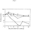
was counted. The results are shown in Fig. 7. As in this, the number of the
CD4-positive lymphocytes significantly decreased in the monkeys of the control
group within 2 weeks after the pathogenic virus challenge, while the reduction in
the CD4-positive lymphocytes in the rVV-DIS-SIV gag-immunized monkeys was
retarded by 70 to 95 %. In addition, it was further confirmed that, in the
immunized monkeys, the number of viral RNA copies in plasma at set points was
decreased to a degree of 1/50 to 1/100.
The results as above confirm that the recombinant virus vaccine of this
invention has the potency of antigen-specific immune induction and immune
protection in the primates.
As described in detail hereinabove, the invention of this application
provides an attenuated, recombinant vaccinia virus vaccine having high
antigenicity against pathogens except vaccinia viruses, and provides a
recombinant vaccinia virus strain DIs that serves as the active ingredient of the
vaccine. The recombinant vaccinia virus strain DIs which the invention provides
herein has the ability to induce both humoral and cellular immunity against the
foreign polypeptide inserted thereinto. In addition, it is easy to change the foreign
polypeptide in the recombinant strain with any other polypeptide through genetic
recombination technology, the recombinant strain is useful for vectors for
recombinant virus vaccines for various infectious diseases. Further, the
recombinant virus does not propagate in ordinary animal cells but expresses the
immunogenic antigen (having the potency of inducing cellular immunity and
antibody production), and the virus cells are easy to incubate and can be
produced at low costs. For these reasons, the recombinant strain of this
invention can be used as an inexpensive substitute for antigens for boosts in the
prime and boost method that is much used in the art for developing AIDS vaccines.
Accordingly, the recombinant strain of this invention is extremely useful for
antigens substitutable for pure recombinant proteins that require high production
costs. Moreover, since the recombinant strain of this invention is highly safe to
humans, it is useful for a recombinant vaccinia virus strain to be infected into
target cells in CTL activity determination from the viewpoint of safe protection of
test subjects.


Claims (19)
- A vaccinia virus vaccine containing, as the active ingredient, a recombinant vaccinia virus strain DIs that possesses a polynucleotide encoding a foreign antigenic protein in the non-essential gene region of the chromosome DNA and expresses the antigenic protein.
- The vaccine of claim 1, wherein the antigenic protein is one derived from a human immunodeficiency virus.
- The vaccine of claim 2, wherein the antigenic protein of the human immunodeficiency virus is a gag gene product.
- The vaccine of claim 1, wherein the antigenic protein is one derived from a simian immunodeficiency virus.
- The vaccine of claim 4, wherein the antigenic protein of the simian immunodeficiency virus is a gag gene product.
- A recombinant vaccinia virus strain DIs that possesses a polynucleotide encoding a foreign antigenic protein in the non-essential gene region of the chromosome DNA and expresses the antigenic protein.
- The recombinant vaccinia virus strain DIs of claim 6, wherein the antigenic protein is one derived from a human immunodeficiency virus.
- The recombinant vaccinia virus strain DIs of claim 7, wherein the antigenic protein of the human immunodeficiency virus is a gag gene product.
- The recombinant vaccinia virus strain DIs of claim 6, wherein the antigenic protein is one derived from a simian immunodeficiency virus.
- The recombinant vaccinia virus strain DIs of claim 9, wherein the antigenic protein of the simian immunodeficiency virus is a gag gene product.
- The recombinant vaccinia virus strain DIs of any of claims 6 to 10, wherein the non-essential gene region of the chromosome DNA is a thymidine kinase gene region.
- The recombinant vaccinia virus strain DIs of any of claims 6 to 10, wherein the non-essential gene region of the chromosome DNA is a region deleted in the vaccinia virus strain DIs when compared with the parent strain.
- A recombinant vaccinia virus strain DIs that possesses a polynucleotide encoding a reporter molecule in the non-essential region of the chromosome DNA and expresses the reporter molecule.
- The recombinant vaccinia virus strain DIs of claim 13, wherein the reporter molecule is a green fluorescence protein or an E. coli β-galactosidase.
- The recombinant vaccinia virus strain DIs of claim 13 or 14, wherein the non-essential gene region of the chromosome DNA is a thymidine kinase gene region.
- The recombinant vaccinia virus strain DIs of claim 13 or 14, wherein the non-essential gene region of the chromosome DNA is a region deleted in the vaccinia virus strain DIs when compared with the parent strain.
- A method of protein expression, which comprises inserting a polynucleotide encoding a foreign protein, into the non-essential gene region of the chromosome DNA of a vaccinia virus strain DIs, followed by mass-expressing the exogenous protein in host cells.
- The method of protein expression as claimed in claim 17, wherein the non-essential gene region of the chromosome DNA is a thymidine kinase gene region.
- The method of protein expression as claimed in claim 17, wherein the non-essential gene region of the chromosome DNA is a region deleted in the vaccinia virus strain DIs when compared with the parent strain.
Applications Claiming Priority (1)
| Application Number | Priority Date | Filing Date | Title |
|---|---|---|---|
| PCT/JP2001/010141 WO2003043654A1 (en) | 2001-11-20 | 2001-11-20 | Recombinant vaccinia virus vaccine |
Publications (2)
| Publication Number | Publication Date |
|---|---|
| EP1457211A1 true EP1457211A1 (en) | 2004-09-15 |
| EP1457211A4 EP1457211A4 (en) | 2006-06-14 |
Family
ID=11737953
Family Applications (1)
| Application Number | Title | Priority Date | Filing Date |
|---|---|---|---|
| EP01274763A Withdrawn EP1457211A4 (en) | 2001-11-20 | 2001-11-20 | VACCINE AGAINST RECOMBINANT VACCINE VIRUS |
Country Status (3)
| Country | Link |
|---|---|
| US (1) | US20050053620A1 (en) |
| EP (1) | EP1457211A4 (en) |
| WO (1) | WO2003043654A1 (en) |
Families Citing this family (1)
| Publication number | Priority date | Publication date | Assignee | Title |
|---|---|---|---|---|
| EP2274442B1 (en) * | 2008-04-04 | 2016-06-29 | The Trustees Of The University Of Pennsylvania | Consensus sequences of chikungunya viral proteins, nucleic acid molecules encoding the same, and compositions and methods for using the same |
Family Cites Families (2)
| Publication number | Priority date | Publication date | Assignee | Title |
|---|---|---|---|---|
| EP0575491B1 (en) * | 1991-03-07 | 2003-08-13 | Virogenetics Corporation | Genetically engineered vaccine strain |
| JP4344805B2 (en) * | 2000-07-07 | 2009-10-14 | 独立行政法人科学技術振興機構 | Genetically modified vaccinia virus vaccine |
-
2001
- 2001-11-20 WO PCT/JP2001/010141 patent/WO2003043654A1/en not_active Ceased
- 2001-11-20 EP EP01274763A patent/EP1457211A4/en not_active Withdrawn
- 2001-11-20 US US10/495,974 patent/US20050053620A1/en not_active Abandoned
Also Published As
| Publication number | Publication date |
|---|---|
| US20050053620A1 (en) | 2005-03-10 |
| EP1457211A4 (en) | 2006-06-14 |
| WO2003043654A1 (en) | 2003-05-30 |
Similar Documents
| Publication | Publication Date | Title |
|---|---|---|
| Hirsch et al. | Patterns of viral replication correlate with outcome in simian immunodeficiency virus (SIV)-infected macaques: effect of prior immunization with a trivalent SIV vaccine in modified vaccinia virus Ankara | |
| JP5123343B2 (en) | HIVCON: HIV immunogen and use thereof | |
| KR101971808B1 (en) | Methods and Compositions for Inducing Protective Immunity Against Human Immunodeficiency Virus Infection | |
| US20130302367A1 (en) | Virus vector for prime/boost vaccines, which comprises vaccinia virus vector and sendai virus vector | |
| US20150359879A1 (en) | Recombinant particle based vaccines against human cytomegalovirus infection | |
| Earl et al. | Design and evaluation of multi-gene, multi-clade HIV-1 MVA vaccines | |
| Perdiguero et al. | Deletion of the viral anti-apoptotic gene F1L in the HIV/AIDS vaccine candidate MVA-C enhances immune responses against HIV-1 antigens | |
| Willems et al. | The YXXL signalling motifs of the bovine leukemia virus transmembrane protein are required for in vivo infection and maintenance of high viral loads | |
| Wyatt et al. | Multiprotein HIV type 1 clade B DNA and MVA vaccines: construction, expression, and immunogenicity in rodents of the MVA component | |
| Di Bonito et al. | Anti-tumor CD8+ T cell immunity elicited by HIV-1-based virus-like particles incorporating HPV-16 E7 protein | |
| US20090227658A1 (en) | Methods and compositions for immunization against hiv | |
| CN103946385B (en) | As the chimeric circles lentiviral gene group of the vaccine to anti-HIV-1 | |
| Fang et al. | Expression of vaccinia E3L and K3L genes by a novel recombinant canarypox HIV vaccine vector enhances HIV-1 pseudovirion production and inhibits apoptosis in human cells | |
| JP4344805B2 (en) | Genetically modified vaccinia virus vaccine | |
| KR100347220B1 (en) | Recombinant adenovirus HIV vaccines | |
| US20050053620A1 (en) | Recombinant vaccinia virus vaccine | |
| JPH01500962A (en) | A viral vector, a DNA recombinant encoding the F protein of the AIDS-causing virus, a cell culture infected with the vector, a method for producing the above protein, the obtained protein, a vaccine, and the obtained antibody | |
| WO2010009465A2 (en) | Htlv-ii vector and methods of use | |
| Sanchez et al. | Visna/maedi virus Env protein expressed by a vaccinia virus recombinant induces cell-to-cell fusion in cells of different origins in the apparent absence of Env cleavage: role of glycosylation and of proteoglycans | |
| JPH01148183A (en) | Recombinant vaccinia virus | |
| KR101560793B1 (en) | Avirulent infectious bursal disease variant virus and use as a vaccine | |
| CN115418368A (en) | Method for detecting SARS-CoV-2 antibody neutralization activity | |
| Young | Construction, characterization and immunogenicity of human immunodeficiency virus-like particles | |
| WO2013011179A2 (en) | Recombinant vectors based on the modified vaccinia ankara (mva) virus, with deletion in the c6l gene, as vaccines against hiv/aids and other diseases | |
| CN114058644A (en) | Recombinant vaccinia virus vector capable of escaping existing anti-vaccinia virus neutralizing antibody existing in vivo |
Legal Events
| Date | Code | Title | Description |
|---|---|---|---|
| PUAI | Public reference made under article 153(3) epc to a published international application that has entered the european phase |
Free format text: ORIGINAL CODE: 0009012 |
|
| 17P | Request for examination filed |
Effective date: 20040617 |
|
| AK | Designated contracting states |
Kind code of ref document: A1 Designated state(s): AT BE CH CY DE DK ES FI FR GB GR IE IT LI LU MC NL PT SE TR |
|
| A4 | Supplementary search report drawn up and despatched |
Effective date: 20060517 |
|
| 17Q | First examination report despatched |
Effective date: 20081127 |
|
| STAA | Information on the status of an ep patent application or granted ep patent |
Free format text: STATUS: THE APPLICATION IS DEEMED TO BE WITHDRAWN |
|
| 18D | Application deemed to be withdrawn |
Effective date: 20090408 |