EP1453978B9 - Analysis and detection of multiple target sequences using circular probes - Google Patents
Analysis and detection of multiple target sequences using circular probes Download PDFInfo
- Publication number
- EP1453978B9 EP1453978B9 EP02795451A EP02795451A EP1453978B9 EP 1453978 B9 EP1453978 B9 EP 1453978B9 EP 02795451 A EP02795451 A EP 02795451A EP 02795451 A EP02795451 A EP 02795451A EP 1453978 B9 EP1453978 B9 EP 1453978B9
- Authority
- EP
- European Patent Office
- Prior art keywords
- primer
- probes
- dna
- probe
- sample
- Prior art date
- Legal status (The legal status is an assumption and is not a legal conclusion. Google has not performed a legal analysis and makes no representation as to the accuracy of the status listed.)
- Expired - Lifetime
Links
- 238000001514 detection method Methods 0.000 title claims abstract description 107
- 239000000523 sample Substances 0.000 title claims description 484
- 238000004458 analytical method Methods 0.000 title claims description 25
- 238000003199 nucleic acid amplification method Methods 0.000 claims abstract description 136
- 230000003321 amplification Effects 0.000 claims abstract description 133
- 238000000034 method Methods 0.000 claims abstract description 128
- 239000002773 nucleotide Substances 0.000 claims description 152
- 125000003729 nucleotide group Chemical group 0.000 claims description 149
- 108091093088 Amplicon Proteins 0.000 claims description 98
- 230000000295 complement effect Effects 0.000 claims description 91
- 239000012634 fragment Substances 0.000 claims description 80
- 108700028369 Alleles Proteins 0.000 claims description 57
- 108020005187 Oligonucleotide Probes Proteins 0.000 claims description 57
- 239000002751 oligonucleotide probe Substances 0.000 claims description 57
- 108091034117 Oligonucleotide Proteins 0.000 claims description 38
- 150000007523 nucleic acids Chemical class 0.000 claims description 35
- 230000000903 blocking effect Effects 0.000 claims description 31
- 108020004707 nucleic acids Proteins 0.000 claims description 27
- 102000039446 nucleic acids Human genes 0.000 claims description 27
- 230000002441 reversible effect Effects 0.000 claims description 27
- 239000000203 mixture Substances 0.000 claims description 21
- 230000000694 effects Effects 0.000 claims description 17
- 238000000295 emission spectrum Methods 0.000 claims description 17
- 108091028043 Nucleic acid sequence Proteins 0.000 claims description 11
- 102000054765 polymorphisms of proteins Human genes 0.000 claims description 11
- 238000006073 displacement reaction Methods 0.000 claims description 9
- 108090000623 proteins and genes Proteins 0.000 claims description 9
- 102000004190 Enzymes Human genes 0.000 claims description 8
- 108090000790 Enzymes Proteins 0.000 claims description 8
- 108020004414 DNA Proteins 0.000 claims description 7
- 230000002068 genetic effect Effects 0.000 claims description 7
- 239000003550 marker Substances 0.000 claims description 7
- 239000002299 complementary DNA Substances 0.000 claims description 6
- 102000053602 DNA Human genes 0.000 claims description 5
- 208000005652 acute fatty liver of pregnancy Diseases 0.000 claims description 5
- 238000013507 mapping Methods 0.000 claims description 5
- 108020004711 Nucleic Acid Probes Proteins 0.000 claims description 3
- 239000002853 nucleic acid probe Substances 0.000 claims description 3
- 208000035240 Disease Resistance Diseases 0.000 claims description 2
- 238000003908 quality control method Methods 0.000 claims description 2
- 239000002253 acid Substances 0.000 claims 1
- 238000002347 injection Methods 0.000 abstract description 32
- 239000007924 injection Substances 0.000 abstract description 32
- 238000003556 assay Methods 0.000 abstract description 21
- 238000000926 separation method Methods 0.000 abstract description 18
- 230000001419 dependent effect Effects 0.000 abstract description 5
- 239000000047 product Substances 0.000 description 48
- 235000007688 Lycopersicon esculentum Nutrition 0.000 description 41
- 240000003768 Solanum lycopersicum Species 0.000 description 41
- 102100025230 2-amino-3-ketobutyrate coenzyme A ligase, mitochondrial Human genes 0.000 description 37
- 108010087522 Aeromonas hydrophilia lipase-acyltransferase Proteins 0.000 description 37
- 238000003752 polymerase chain reaction Methods 0.000 description 33
- 238000009396 hybridization Methods 0.000 description 29
- 102100036263 Glutamyl-tRNA(Gln) amidotransferase subunit C, mitochondrial Human genes 0.000 description 26
- 101001001786 Homo sapiens Glutamyl-tRNA(Gln) amidotransferase subunit C, mitochondrial Proteins 0.000 description 26
- 239000000975 dye Substances 0.000 description 25
- 239000000872 buffer Substances 0.000 description 22
- 238000006243 chemical reaction Methods 0.000 description 21
- 238000000137 annealing Methods 0.000 description 19
- FCKYPQBAHLOOJQ-UHFFFAOYSA-N Cyclohexane-1,2-diaminetetraacetic acid Chemical compound OC(=O)CN(CC(O)=O)C1CCCCC1N(CC(O)=O)CC(O)=O FCKYPQBAHLOOJQ-UHFFFAOYSA-N 0.000 description 18
- 102000012410 DNA Ligases Human genes 0.000 description 17
- 108010061982 DNA Ligases Proteins 0.000 description 17
- JLCPHMBAVCMARE-UHFFFAOYSA-N [3-[[3-[[3-[[3-[[3-[[3-[[3-[[3-[[3-[[3-[[3-[[5-(2-amino-6-oxo-1H-purin-9-yl)-3-[[3-[[3-[[3-[[3-[[3-[[5-(2-amino-6-oxo-1H-purin-9-yl)-3-[[5-(2-amino-6-oxo-1H-purin-9-yl)-3-hydroxyoxolan-2-yl]methoxy-hydroxyphosphoryl]oxyoxolan-2-yl]methoxy-hydroxyphosphoryl]oxy-5-(5-methyl-2,4-dioxopyrimidin-1-yl)oxolan-2-yl]methoxy-hydroxyphosphoryl]oxy-5-(6-aminopurin-9-yl)oxolan-2-yl]methoxy-hydroxyphosphoryl]oxy-5-(6-aminopurin-9-yl)oxolan-2-yl]methoxy-hydroxyphosphoryl]oxy-5-(6-aminopurin-9-yl)oxolan-2-yl]methoxy-hydroxyphosphoryl]oxy-5-(6-aminopurin-9-yl)oxolan-2-yl]methoxy-hydroxyphosphoryl]oxyoxolan-2-yl]methoxy-hydroxyphosphoryl]oxy-5-(5-methyl-2,4-dioxopyrimidin-1-yl)oxolan-2-yl]methoxy-hydroxyphosphoryl]oxy-5-(4-amino-2-oxopyrimidin-1-yl)oxolan-2-yl]methoxy-hydroxyphosphoryl]oxy-5-(5-methyl-2,4-dioxopyrimidin-1-yl)oxolan-2-yl]methoxy-hydroxyphosphoryl]oxy-5-(5-methyl-2,4-dioxopyrimidin-1-yl)oxolan-2-yl]methoxy-hydroxyphosphoryl]oxy-5-(6-aminopurin-9-yl)oxolan-2-yl]methoxy-hydroxyphosphoryl]oxy-5-(6-aminopurin-9-yl)oxolan-2-yl]methoxy-hydroxyphosphoryl]oxy-5-(4-amino-2-oxopyrimidin-1-yl)oxolan-2-yl]methoxy-hydroxyphosphoryl]oxy-5-(4-amino-2-oxopyrimidin-1-yl)oxolan-2-yl]methoxy-hydroxyphosphoryl]oxy-5-(4-amino-2-oxopyrimidin-1-yl)oxolan-2-yl]methoxy-hydroxyphosphoryl]oxy-5-(6-aminopurin-9-yl)oxolan-2-yl]methoxy-hydroxyphosphoryl]oxy-5-(4-amino-2-oxopyrimidin-1-yl)oxolan-2-yl]methyl [5-(6-aminopurin-9-yl)-2-(hydroxymethyl)oxolan-3-yl] hydrogen phosphate Polymers Cc1cn(C2CC(OP(O)(=O)OCC3OC(CC3OP(O)(=O)OCC3OC(CC3O)n3cnc4c3nc(N)[nH]c4=O)n3cnc4c3nc(N)[nH]c4=O)C(COP(O)(=O)OC3CC(OC3COP(O)(=O)OC3CC(OC3COP(O)(=O)OC3CC(OC3COP(O)(=O)OC3CC(OC3COP(O)(=O)OC3CC(OC3COP(O)(=O)OC3CC(OC3COP(O)(=O)OC3CC(OC3COP(O)(=O)OC3CC(OC3COP(O)(=O)OC3CC(OC3COP(O)(=O)OC3CC(OC3COP(O)(=O)OC3CC(OC3COP(O)(=O)OC3CC(OC3COP(O)(=O)OC3CC(OC3COP(O)(=O)OC3CC(OC3COP(O)(=O)OC3CC(OC3COP(O)(=O)OC3CC(OC3COP(O)(=O)OC3CC(OC3CO)n3cnc4c(N)ncnc34)n3ccc(N)nc3=O)n3cnc4c(N)ncnc34)n3ccc(N)nc3=O)n3ccc(N)nc3=O)n3ccc(N)nc3=O)n3cnc4c(N)ncnc34)n3cnc4c(N)ncnc34)n3cc(C)c(=O)[nH]c3=O)n3cc(C)c(=O)[nH]c3=O)n3ccc(N)nc3=O)n3cc(C)c(=O)[nH]c3=O)n3cnc4c3nc(N)[nH]c4=O)n3cnc4c(N)ncnc34)n3cnc4c(N)ncnc34)n3cnc4c(N)ncnc34)n3cnc4c(N)ncnc34)O2)c(=O)[nH]c1=O JLCPHMBAVCMARE-UHFFFAOYSA-N 0.000 description 17
- 102100034330 Chromaffin granule amine transporter Human genes 0.000 description 16
- 101000641221 Homo sapiens Chromaffin granule amine transporter Proteins 0.000 description 16
- 239000011159 matrix material Substances 0.000 description 16
- 102000003960 Ligases Human genes 0.000 description 14
- 108090000364 Ligases Proteins 0.000 description 14
- 238000005251 capillar electrophoresis Methods 0.000 description 11
- 238000012937 correction Methods 0.000 description 11
- 238000001962 electrophoresis Methods 0.000 description 11
- 239000012507 Sephadex™ Substances 0.000 description 10
- 230000008901 benefit Effects 0.000 description 10
- 108060002716 Exonuclease Proteins 0.000 description 9
- 229920005654 Sephadex Polymers 0.000 description 9
- 230000015572 biosynthetic process Effects 0.000 description 9
- 238000004925 denaturation Methods 0.000 description 9
- 230000036425 denaturation Effects 0.000 description 9
- 238000013461 design Methods 0.000 description 9
- 238000005516 engineering process Methods 0.000 description 9
- 102000013165 exonuclease Human genes 0.000 description 9
- 239000007850 fluorescent dye Substances 0.000 description 9
- 230000009467 reduction Effects 0.000 description 9
- 230000002829 reductive effect Effects 0.000 description 9
- JEPVUMTVFPQKQE-AAKCMJRZSA-N 2-[(1s,2s,3r,4s)-1,2,3,4,5-pentahydroxypentyl]-1,3-thiazolidine-4-carboxylic acid Chemical compound OC[C@H](O)[C@@H](O)[C@H](O)[C@H](O)C1NC(C(O)=O)CS1 JEPVUMTVFPQKQE-AAKCMJRZSA-N 0.000 description 8
- 239000000499 gel Substances 0.000 description 8
- 239000000243 solution Substances 0.000 description 8
- 238000001228 spectrum Methods 0.000 description 8
- 238000012408 PCR amplification Methods 0.000 description 7
- 238000001502 gel electrophoresis Methods 0.000 description 7
- 238000011160 research Methods 0.000 description 7
- 238000006467 substitution reaction Methods 0.000 description 7
- 241000219194 Arabidopsis Species 0.000 description 6
- 238000002844 melting Methods 0.000 description 6
- 230000008018 melting Effects 0.000 description 6
- 239000000126 substance Substances 0.000 description 6
- QKNYBSVHEMOAJP-UHFFFAOYSA-N 2-amino-2-(hydroxymethyl)propane-1,3-diol;hydron;chloride Chemical compound Cl.OCC(N)(CO)CO QKNYBSVHEMOAJP-UHFFFAOYSA-N 0.000 description 5
- 108010014303 DNA-directed DNA polymerase Proteins 0.000 description 5
- 102000016928 DNA-directed DNA polymerase Human genes 0.000 description 5
- BAWFJGJZGIEFAR-NNYOXOHSSA-O NAD(+) Chemical compound NC(=O)C1=CC=C[N+]([C@H]2[C@@H]([C@H](O)[C@@H](COP(O)(=O)OP(O)(=O)OC[C@@H]3[C@H]([C@@H](O)[C@@H](O3)N3C4=NC=NC(N)=C4N=C3)O)O2)O)=C1 BAWFJGJZGIEFAR-NNYOXOHSSA-O 0.000 description 5
- 238000011161 development Methods 0.000 description 5
- 230000018109 developmental process Effects 0.000 description 5
- 238000009826 distribution Methods 0.000 description 5
- 238000002372 labelling Methods 0.000 description 5
- 238000011068 loading method Methods 0.000 description 5
- 238000000329 molecular dynamics simulation Methods 0.000 description 5
- 230000036961 partial effect Effects 0.000 description 5
- 238000000746 purification Methods 0.000 description 5
- 150000003839 salts Chemical class 0.000 description 5
- 230000003595 spectral effect Effects 0.000 description 5
- YBJHBAHKTGYVGT-ZKWXMUAHSA-N (+)-Biotin Chemical compound N1C(=O)N[C@@H]2[C@H](CCCCC(=O)O)SC[C@@H]21 YBJHBAHKTGYVGT-ZKWXMUAHSA-N 0.000 description 4
- 102100025570 Cancer/testis antigen 1 Human genes 0.000 description 4
- -1 Cy2 Chemical compound 0.000 description 4
- 241000196324 Embryophyta Species 0.000 description 4
- 241000282414 Homo sapiens Species 0.000 description 4
- 101000856237 Homo sapiens Cancer/testis antigen 1 Proteins 0.000 description 4
- TWRXJAOTZQYOKJ-UHFFFAOYSA-L Magnesium chloride Chemical compound [Mg+2].[Cl-].[Cl-] TWRXJAOTZQYOKJ-UHFFFAOYSA-L 0.000 description 4
- 230000001351 cycling effect Effects 0.000 description 4
- 230000002255 enzymatic effect Effects 0.000 description 4
- 238000011534 incubation Methods 0.000 description 4
- 238000003780 insertion Methods 0.000 description 4
- 230000037431 insertion Effects 0.000 description 4
- 239000000463 material Substances 0.000 description 4
- 230000008569 process Effects 0.000 description 4
- 230000002035 prolonged effect Effects 0.000 description 4
- 238000003786 synthesis reaction Methods 0.000 description 4
- PEDCQBHIVMGVHV-UHFFFAOYSA-N Glycerine Chemical compound OCC(O)CO PEDCQBHIVMGVHV-UHFFFAOYSA-N 0.000 description 3
- 241000282412 Homo Species 0.000 description 3
- 239000012901 Milli-Q water Substances 0.000 description 3
- 235000010724 Wisteria floribunda Nutrition 0.000 description 3
- 238000010521 absorption reaction Methods 0.000 description 3
- 238000005119 centrifugation Methods 0.000 description 3
- 150000001875 compounds Chemical class 0.000 description 3
- 238000012217 deletion Methods 0.000 description 3
- 230000037430 deletion Effects 0.000 description 3
- 230000029087 digestion Effects 0.000 description 3
- 238000010790 dilution Methods 0.000 description 3
- 239000012895 dilution Substances 0.000 description 3
- IIRDTKBZINWQAW-UHFFFAOYSA-N hexaethylene glycol Chemical compound OCCOCCOCCOCCOCCOCCO IIRDTKBZINWQAW-UHFFFAOYSA-N 0.000 description 3
- 238000004519 manufacturing process Methods 0.000 description 3
- 230000035772 mutation Effects 0.000 description 3
- 108091033319 polynucleotide Proteins 0.000 description 3
- 102000040430 polynucleotide Human genes 0.000 description 3
- 239000002157 polynucleotide Substances 0.000 description 3
- 238000002360 preparation method Methods 0.000 description 3
- 230000002285 radioactive effect Effects 0.000 description 3
- 238000005096 rolling process Methods 0.000 description 3
- 230000004304 visual acuity Effects 0.000 description 3
- 239000012103 Alexa Fluor 488 Substances 0.000 description 2
- 239000012099 Alexa Fluor family Substances 0.000 description 2
- 241000193738 Bacillus anthracis Species 0.000 description 2
- 206010071602 Genetic polymorphism Diseases 0.000 description 2
- 241001465754 Metazoa Species 0.000 description 2
- 108091092878 Microsatellite Proteins 0.000 description 2
- 108091093037 Peptide nucleic acid Proteins 0.000 description 2
- 108020004682 Single-Stranded DNA Proteins 0.000 description 2
- FAPWRFPIFSIZLT-UHFFFAOYSA-M Sodium chloride Chemical compound [Na+].[Cl-] FAPWRFPIFSIZLT-UHFFFAOYSA-M 0.000 description 2
- 229920004890 Triton X-100 Polymers 0.000 description 2
- 230000004913 activation Effects 0.000 description 2
- 238000007792 addition Methods 0.000 description 2
- 229960002685 biotin Drugs 0.000 description 2
- 235000020958 biotin Nutrition 0.000 description 2
- 239000011616 biotin Substances 0.000 description 2
- 210000004027 cell Anatomy 0.000 description 2
- 239000007795 chemical reaction product Substances 0.000 description 2
- 230000004069 differentiation Effects 0.000 description 2
- 239000003814 drug Substances 0.000 description 2
- 238000002474 experimental method Methods 0.000 description 2
- MHMNJMPURVTYEJ-UHFFFAOYSA-N fluorescein-5-isothiocyanate Chemical compound O1C(=O)C2=CC(N=C=S)=CC=C2C21C1=CC=C(O)C=C1OC1=CC(O)=CC=C21 MHMNJMPURVTYEJ-UHFFFAOYSA-N 0.000 description 2
- 125000000524 functional group Chemical group 0.000 description 2
- 238000003205 genotyping method Methods 0.000 description 2
- 230000006872 improvement Effects 0.000 description 2
- 238000011901 isothermal amplification Methods 0.000 description 2
- 238000002032 lab-on-a-chip Methods 0.000 description 2
- 230000000670 limiting effect Effects 0.000 description 2
- 229910001629 magnesium chloride Inorganic materials 0.000 description 2
- 238000011176 pooling Methods 0.000 description 2
- SCVFZCLFOSHCOH-UHFFFAOYSA-M potassium acetate Chemical compound [K+].CC([O-])=O SCVFZCLFOSHCOH-UHFFFAOYSA-M 0.000 description 2
- 230000037452 priming Effects 0.000 description 2
- 238000012545 processing Methods 0.000 description 2
- 239000012264 purified product Substances 0.000 description 2
- 239000012521 purified sample Substances 0.000 description 2
- 108091008146 restriction endonucleases Proteins 0.000 description 2
- MYFATKRONKHHQL-UHFFFAOYSA-N rhodamine 123 Chemical compound [Cl-].COC(=O)C1=CC=CC=C1C1=C2C=CC(=[NH2+])C=C2OC2=CC(N)=CC=C21 MYFATKRONKHHQL-UHFFFAOYSA-N 0.000 description 2
- 238000004513 sizing Methods 0.000 description 2
- 239000007858 starting material Substances 0.000 description 2
- ABZLKHKQJHEPAX-UHFFFAOYSA-N tetramethylrhodamine Chemical compound C=12C=CC(N(C)C)=CC2=[O+]C2=CC(N(C)C)=CC=C2C=1C1=CC=CC=C1C([O-])=O ABZLKHKQJHEPAX-UHFFFAOYSA-N 0.000 description 2
- MPLHNVLQVRSVEE-UHFFFAOYSA-N texas red Chemical compound [O-]S(=O)(=O)C1=CC(S(Cl)(=O)=O)=CC=C1C(C1=CC=2CCCN3CCCC(C=23)=C1O1)=C2C1=C(CCC1)C3=[N+]1CCCC3=C2 MPLHNVLQVRSVEE-UHFFFAOYSA-N 0.000 description 2
- 238000005382 thermal cycling Methods 0.000 description 2
- 238000005406 washing Methods 0.000 description 2
- 241000219195 Arabidopsis thaliana Species 0.000 description 1
- 241000894006 Bacteria Species 0.000 description 1
- 108020004998 Chloroplast DNA Proteins 0.000 description 1
- 108020004635 Complementary DNA Proteins 0.000 description 1
- 238000007399 DNA isolation Methods 0.000 description 1
- 102100029995 DNA ligase 1 Human genes 0.000 description 1
- 238000000018 DNA microarray Methods 0.000 description 1
- 238000001712 DNA sequencing Methods 0.000 description 1
- 230000006820 DNA synthesis Effects 0.000 description 1
- KCXVZYZYPLLWCC-UHFFFAOYSA-N EDTA Chemical compound OC(=O)CN(CC(O)=O)CCN(CC(O)=O)CC(O)=O KCXVZYZYPLLWCC-UHFFFAOYSA-N 0.000 description 1
- 241000588724 Escherichia coli Species 0.000 description 1
- 241001524679 Escherichia virus M13 Species 0.000 description 1
- 208000028782 Hereditary disease Diseases 0.000 description 1
- 229930010555 Inosine Natural products 0.000 description 1
- UGQMRVRMYYASKQ-KQYNXXCUSA-N Inosine Chemical compound O[C@@H]1[C@H](O)[C@@H](CO)O[C@H]1N1C2=NC=NC(O)=C2N=C1 UGQMRVRMYYASKQ-KQYNXXCUSA-N 0.000 description 1
- 108020005196 Mitochondrial DNA Proteins 0.000 description 1
- 206010028980 Neoplasm Diseases 0.000 description 1
- 108091092740 Organellar DNA Proteins 0.000 description 1
- 241000205160 Pyrococcus Species 0.000 description 1
- 108010066717 Q beta Replicase Proteins 0.000 description 1
- 239000013614 RNA sample Substances 0.000 description 1
- 108010006785 Taq Polymerase Proteins 0.000 description 1
- 241000589500 Thermus aquaticus Species 0.000 description 1
- 101900061264 Thermus thermophilus DNA ligase Proteins 0.000 description 1
- RYYWUUFWQRZTIU-UHFFFAOYSA-N Thiophosphoric acid Chemical group OP(O)(S)=O RYYWUUFWQRZTIU-UHFFFAOYSA-N 0.000 description 1
- 108700019146 Transgenes Proteins 0.000 description 1
- 239000013504 Triton X-100 Substances 0.000 description 1
- 238000000862 absorption spectrum Methods 0.000 description 1
- 238000009825 accumulation Methods 0.000 description 1
- 230000009471 action Effects 0.000 description 1
- 230000006978 adaptation Effects 0.000 description 1
- 230000002411 adverse Effects 0.000 description 1
- 239000000427 antigen Substances 0.000 description 1
- 102000036639 antigens Human genes 0.000 description 1
- 108091007433 antigens Proteins 0.000 description 1
- 238000013459 approach Methods 0.000 description 1
- 238000003491 array Methods 0.000 description 1
- 238000000376 autoradiography Methods 0.000 description 1
- 229940065181 bacillus anthracis Drugs 0.000 description 1
- 239000011324 bead Substances 0.000 description 1
- 239000012620 biological material Substances 0.000 description 1
- 239000003153 chemical reaction reagent Substances 0.000 description 1
- 239000003086 colorant Substances 0.000 description 1
- 230000002860 competitive effect Effects 0.000 description 1
- 108091036078 conserved sequence Proteins 0.000 description 1
- 238000010276 construction Methods 0.000 description 1
- 238000007796 conventional method Methods 0.000 description 1
- 238000004163 cytometry Methods 0.000 description 1
- SUYVUBYJARFZHO-RRKCRQDMSA-N dATP Chemical compound C1=NC=2C(N)=NC=NC=2N1[C@H]1C[C@H](O)[C@@H](COP(O)(=O)OP(O)(=O)OP(O)(O)=O)O1 SUYVUBYJARFZHO-RRKCRQDMSA-N 0.000 description 1
- SUYVUBYJARFZHO-UHFFFAOYSA-N dATP Natural products C1=NC=2C(N)=NC=NC=2N1C1CC(O)C(COP(O)(=O)OP(O)(=O)OP(O)(O)=O)O1 SUYVUBYJARFZHO-UHFFFAOYSA-N 0.000 description 1
- RGWHQCVHVJXOKC-SHYZEUOFSA-J dCTP(4-) Chemical compound O=C1N=C(N)C=CN1[C@@H]1O[C@H](COP([O-])(=O)OP([O-])(=O)OP([O-])([O-])=O)[C@@H](O)C1 RGWHQCVHVJXOKC-SHYZEUOFSA-J 0.000 description 1
- HAAZLUGHYHWQIW-KVQBGUIXSA-N dGTP Chemical compound C1=NC=2C(=O)NC(N)=NC=2N1[C@H]1C[C@H](O)[C@@H](COP(O)(=O)OP(O)(=O)OP(O)(O)=O)O1 HAAZLUGHYHWQIW-KVQBGUIXSA-N 0.000 description 1
- NHVNXKFIZYSCEB-XLPZGREQSA-N dTTP Chemical compound O=C1NC(=O)C(C)=CN1[C@@H]1O[C@H](COP(O)(=O)OP(O)(=O)OP(O)(O)=O)[C@@H](O)C1 NHVNXKFIZYSCEB-XLPZGREQSA-N 0.000 description 1
- 238000013480 data collection Methods 0.000 description 1
- 238000011033 desalting Methods 0.000 description 1
- 201000010099 disease Diseases 0.000 description 1
- 208000037265 diseases, disorders, signs and symptoms Diseases 0.000 description 1
- 229940079593 drug Drugs 0.000 description 1
- 230000008030 elimination Effects 0.000 description 1
- 238000003379 elimination reaction Methods 0.000 description 1
- 238000010828 elution Methods 0.000 description 1
- 238000001704 evaporation Methods 0.000 description 1
- 230000008020 evaporation Effects 0.000 description 1
- 230000005284 excitation Effects 0.000 description 1
- 238000002189 fluorescence spectrum Methods 0.000 description 1
- PCHJSUWPFVWCPO-UHFFFAOYSA-N gold Chemical compound [Au] PCHJSUWPFVWCPO-UHFFFAOYSA-N 0.000 description 1
- 239000010931 gold Substances 0.000 description 1
- 229910052737 gold Inorganic materials 0.000 description 1
- 238000009499 grossing Methods 0.000 description 1
- 238000010438 heat treatment Methods 0.000 description 1
- 229910001385 heavy metal Inorganic materials 0.000 description 1
- 238000012203 high throughput assay Methods 0.000 description 1
- 238000003384 imaging method Methods 0.000 description 1
- 238000000338 in vitro Methods 0.000 description 1
- 238000001727 in vivo Methods 0.000 description 1
- 238000010348 incorporation Methods 0.000 description 1
- 229960003786 inosine Drugs 0.000 description 1
- 230000003993 interaction Effects 0.000 description 1
- 101150044508 key gene Proteins 0.000 description 1
- 239000003446 ligand Substances 0.000 description 1
- 244000144972 livestock Species 0.000 description 1
- UEGPKNKPLBYCNK-UHFFFAOYSA-L magnesium acetate Chemical compound [Mg+2].CC([O-])=O.CC([O-])=O UEGPKNKPLBYCNK-UHFFFAOYSA-L 0.000 description 1
- 239000011654 magnesium acetate Substances 0.000 description 1
- 235000011285 magnesium acetate Nutrition 0.000 description 1
- 229940069446 magnesium acetate Drugs 0.000 description 1
- 230000005291 magnetic effect Effects 0.000 description 1
- 238000012423 maintenance Methods 0.000 description 1
- 238000005259 measurement Methods 0.000 description 1
- 230000001404 mediated effect Effects 0.000 description 1
- 238000013508 migration Methods 0.000 description 1
- 230000005012 migration Effects 0.000 description 1
- 230000002438 mitochondrial effect Effects 0.000 description 1
- 238000002156 mixing Methods 0.000 description 1
- 238000010369 molecular cloning Methods 0.000 description 1
- 238000007837 multiplex assay Methods 0.000 description 1
- 239000002105 nanoparticle Substances 0.000 description 1
- 238000001668 nucleic acid synthesis Methods 0.000 description 1
- 238000002515 oligonucleotide synthesis Methods 0.000 description 1
- 230000005298 paramagnetic effect Effects 0.000 description 1
- 244000052769 pathogen Species 0.000 description 1
- 229920002401 polyacrylamide Polymers 0.000 description 1
- 229920000642 polymer Polymers 0.000 description 1
- 235000011056 potassium acetate Nutrition 0.000 description 1
- 102000004169 proteins and genes Human genes 0.000 description 1
- 238000011002 quantification Methods 0.000 description 1
- 239000011541 reaction mixture Substances 0.000 description 1
- 238000009877 rendering Methods 0.000 description 1
- 230000010076 replication Effects 0.000 description 1
- 238000007894 restriction fragment length polymorphism technique Methods 0.000 description 1
- 238000010079 rubber tapping Methods 0.000 description 1
- 230000035945 sensitivity Effects 0.000 description 1
- 238000012163 sequencing technique Methods 0.000 description 1
- 239000011780 sodium chloride Substances 0.000 description 1
- 239000007787 solid Substances 0.000 description 1
- 241000894007 species Species 0.000 description 1
- 238000010186 staining Methods 0.000 description 1
- 238000010561 standard procedure Methods 0.000 description 1
- 239000000758 substrate Substances 0.000 description 1
- 238000007862 touchdown PCR Methods 0.000 description 1
- 239000001226 triphosphate Substances 0.000 description 1
- 235000011178 triphosphate Nutrition 0.000 description 1
- 125000002264 triphosphate group Chemical class [H]OP(=O)(O[H])OP(=O)(O[H])OP(=O)(O[H])O* 0.000 description 1
- 238000010200 validation analysis Methods 0.000 description 1
- 108700026220 vif Genes Proteins 0.000 description 1
- XLYOFNOQVPJJNP-UHFFFAOYSA-N water Substances O XLYOFNOQVPJJNP-UHFFFAOYSA-N 0.000 description 1
Images


Classifications
-
- C—CHEMISTRY; METALLURGY
- C12—BIOCHEMISTRY; BEER; SPIRITS; WINE; VINEGAR; MICROBIOLOGY; ENZYMOLOGY; MUTATION OR GENETIC ENGINEERING
- C12Q—MEASURING OR TESTING PROCESSES INVOLVING ENZYMES, NUCLEIC ACIDS OR MICROORGANISMS; COMPOSITIONS OR TEST PAPERS THEREFOR; PROCESSES OF PREPARING SUCH COMPOSITIONS; CONDITION-RESPONSIVE CONTROL IN MICROBIOLOGICAL OR ENZYMOLOGICAL PROCESSES
- C12Q1/00—Measuring or testing processes involving enzymes, nucleic acids or microorganisms; Compositions therefor; Processes of preparing such compositions
- C12Q1/68—Measuring or testing processes involving enzymes, nucleic acids or microorganisms; Compositions therefor; Processes of preparing such compositions involving nucleic acids
- C12Q1/6813—Hybridisation assays
- C12Q1/6827—Hybridisation assays for detection of mutation or polymorphism
Definitions
- the present invention relates to the field of biotechnology.
- the present invention provides a method for the high throughput separation and detection of nucleotide sequences, and the use of the method in the discrimination and identification of target sequence such as single nucleotide polymorphisms.
- the invention further provides for probes that are capable of hybridising to the target sequence of interest, primers for the amplification of ligated probes, use of these probes and primers in the identification and/or detection of nucleotide sequences that are related to a wide variety of genetic traits and genes and kits of primers and/or probes suitable for use in the method according to the invention.
- single nucleotide substitutions are one of the main causes of a significant number of monogenically and multigenically inherited diseases, for instance in humans, or are otherwise involved in the development of complex phenotypes such as performance traits in plants and livestock species.
- single nucleotide substitutions are in many cases also related to or at least indicative of important traits in humans, plants and animal species.
- amplification technique such as the PCR technique it is possible to amplify a limited number of target sequences by combining the corresponding primer pairs in a single amplification reaction but the number of target sequences that can be amplified simultaneously is small and extensive optimisation may be required to achieved similar amplification efficiencies of the individual target sequences.
- One of the solutions to multiplex amplification is to use a single primer pair for the amplification of all target sequences, which requires that all targets must contain the corresponding primer-binding sites. This principle is incorporated in the AFLP technique ( EP-A 0 534 858 ). Using AFLP, the primer-binding sites result from a digestion of the target nucleic acid (i.e.
- AFLP essentially targets a random selection of sequences contained in the target nucleic acid. It has been shown that, using AFLP, a practically unlimited number of target sequences can be amplified in a single reaction, depending on the number of target sequences that contain primer-binding region(s) that are perfectly complementary to the amplification primers. Exploiting the use of single primer-pair for amplification in combination with a non-random method for SNP target selection and efficient use of a high throughput detection platform may therefore substantially increase the efficiency of SNP genotyping, however such technology has not been provided in the art yet.
- One of the principal methods used for the analysis of the nucleic acids of a known sequence is based on annealing two probes to a target sequence and, when the probes are hybridised adjacently to the target sequence, ligating the probes.
- the OLA-principle (Oligonucleotide Ligation Assay) has been described, amongst others, in US 4,988,617 (Landegren et al .) and in WO01/06012 .
- This publication discloses a method for determining the nucleic acid sequence in a region of a known nucleic acid sequence having a known possible mutation. To detect the mutation, oligonucleotides are selected to anneal to immediately adj acent segments of the sequence to be determined.
- One of the selected oligonucleotide probes has an end region wherein one of the end region nucleotides is complementary to either the normal or to the mutated nucleotide at the corresponding position in the known nucleic acid sequence.
- a ligase is provided which covalently connects the two probes when they are correctly base paired and are located immediately adjacent to each other. The presence or absence of the linked probes is an indication of the presence of the known sequence and/or mutation.
- Abbot et al . in WO 96/15271 developed a method for a multiplex ligation amplification procedure comprising the hybridisation and ligation of adjacent probes. These probes are provided with an additional length segment, the sequence of which, according to Abbot et al., is unimportant.
- the deliberate introduction of length differences intends to facilitate the discrimination on the basis of fragment length in gel-based techniques.
- WO 97/45559 (Barany et al. ) describes a method for the detection of nucleic acid sequence differences by using combinations of ligase detection reactions (LDR) and polymerase chain reactions (PCR).
- LDR ligase detection reactions
- PCR polymerase chain reactions
- Amplification of the ligated products with fluorescently labelled primers results in a fluorescently labelled amplified product. Detection of the products is based on separation by size or electrophoretic mobility or on an addressable array.
- Detection of the amplified probes is performed on a universally addressable array containing capturing oligonucleotides. These capturing oligonucleotides contain a region that is capable of annealing to a pre-determined region in the amplified probe, a so-called zip-region or zip code. Each amplified probe contains a different zip code and each zip code will hybridise to its corresponding capturing oligonucleotide on the array. Detection of the label in combination with the position on the array provides information on the presence of the target sequence in the sample. This method allows for the detection of a number of nucleic acid sequences in a sample.
- arrays for the detection of amplified probes involves many steps (ligation, amplification, optionally purification of the amplified material, array production, hybridisation, washing, scanning and data quantification), of which some (particularly hybridisation and washing) are difficult to automate.
- Array-based detection is therefore laborious and costly to analyse a large number of samples for a large number of SNPs.
- the LDR oligonucleotide probes in a given set may generate a unique length product and thus may be distinguished from other products based on size.
- a primer set is provided wherein one of the primers contains a label. Different primers can be provided with different labels to allow for the distinction of products.
- the method and the various embodiments described by Barany et al. are found to have certain disadvantages.
- One of the major disadvantages is that the method in principle does not provide for a true high throughput process for the determination of large numbers of target sequences in short periods of time using reliable and robust methods without compromising the quality of the data produced and the efficiency of the process.
- one of the disadvantages of the means and methods as disclosed by Barany et al. resides in the limited multiplex capacity when discrimination is based inter alia, on the length of the allele specific probe sets. Discrimination between sequences that are distinguishable by only a relatively small length difference is, in general, not straightforward and carefully optimised conditions may be required in order to come to the desired resolving power. Discrimination between sequences that have a larger length differentiation is in general easier to accomplish. This may provide for an increase in the number of sequences that can be analysed in the same sample. However, providing for the necessary longer nucleotide probes is a further hurdle to be taken.
- nucleotide sequences are produced by conventional chemical step-by-step oligonucleotide synthesis with a yield of about 98.5% per added nucleotide.
- yield generally drops and the reliability and purity of the synthetically produced sequence can become a problem.
- WO 01/61033 discloses the preparation of longer probes for use in ligation-amplification assays. They provided probes that are considerably longer than those that can be obtained by conventional chemical synthesis methods to avoid the problem associated with the length-based discrimination of amplified products using slab-gels or capillary electrophoresis, namely that only a small part of the detection window / resolving capacity of up to 1 kilo base length is used when OLA probes are synthesised by chemical means.
- Rolling circle amplification is an amplification method wherein a first primer is hybridised to a ligated or connected circular probe. Subsequent primer elongation, using a polymerase with strand displacement activity results in the formation of a long polynucleotide strand which contains multiple representations of the connected circular probe. Such a long strand of concatamers of the connected probe is subsequently detected by the use of hybridisation probes. These probes can be labelled. Exponential amplification of the ligated probe can be achieved by the hybridisation of a second primer that hybridises to the concatameric strand and is subsequently elongated. (Exponential) Rolling Circle Amplification ((E)RCA) is described inter alia in US5854033 , US6143495 WO97/19193 , Lizardi et al, Nature genetics 19(3):225-232 (1998) .
- US 6,221,603 disclosed a circular probe that contains a restriction site.
- the probe is amplified using (E)RCA and the resulting concatamers are restricted at the restriction site.
- the restriction fragments are then separated by length and detected. Separation and detection is performed on a capillary electrophoretic platform, such as the MegaBACE equipment available from Molecular Dynamics Amersham-Pharmacia.
- a capillary electrophoretic platform such as the MegaBACE equipment available from Molecular Dynamics Amersham-Pharmacia.
- For detection labelled dNTP's may be incorporated into the fragments during amplification, or the fragments may be detected by staining or by labelled detection probes. Partial digestion by the restriction enzyme may however affect the reliability of the method.
- the methods for labelling of the fragments as disclosed in US 6,221,603 do not allow to fully utilise the MegaBACE's capacity of simultaneous detection of multiple colours.
- the present invention relates to methods for high throughput separation and detection of multiple sequences.
- the present method resolves many of the problems previously encountered in the art. More in particular the present invention provides for a multiple ligation and amplification assay that allows for the rapid and high throughput analysis of a multiplicity of samples, preferably containing a multiplicity of sequences.
- the present invention also provides for a method for the high throughput discrimination and detection of a multitude of nucleotide sequences based on a combination of length differences and labels.
- the present invention combines the advantages of certain methods while at the same time avoids disadvantages associated with the various technique, thereby providing for an improved method for the detection of targets sequences in a reliable and reproducible manner and suitable for a high throughput detection method.
- the invention relates to a method for high throughput separation and detection of a multiplicity of target sequences, optionally in a multiplicity of samples comprising subjecting each sample to a ligation-dependent amplification assay.
- the method preferably is a method for determining the presence or absence of at least one target sequence (2) in a sample, wherein the method comprises the steps of:
- the circular oligonucleotide probe used in the present invention is a single linear oligonucleotide probe that is provided in step (a) for each target sequence in a sample.
- This single linear oligonucleotide probe combines the two target specific section into a single molecule that is circularised in step (c) when the annealed complementarity sections are connected.
- the sections of target complementarity are each present at the extreme ends of the single linear probe.
- the complementarity sections at the extreme ends are intervened by the sequences that may serve as primer-binding sequences and may further be intervened by stuffer sequences of variable length.
- circular probe An example of such an arrangement of functional groups in the circular probe is: (target-complementarity section 1 - stuffer sequence 1, primer-binding sequence 1 - primer-binding sequence 2 - stuffer sequence 2 - target-complementarity section 2).
- the circular probes are synthesised and applied in a linear form and that they will only be circular when the two complementary sections at the extreme ends of the probe are connected (ligated) annealing to the appropriate target sequence.
- the term "circular probe” as used herein actually refers to a linear molecule that is circularised by target sequence dependent connection (ligation). Only the term “connected circular probe” as used herein refers to a molecule in true circular form.
- the complementary sections of the oligonucleotide probes are designed such that for each target sequence in a sample a probe is provided, preferably a specific probe, whereby the probes each contain a section at both their extreme ends that is complementary to a part of the target sequence and the corresponding complementary parts of the target sequence are located essentially adjacent to each other.
- the oligonucleotide probe has a section at its 5'-end that is complementary to a first part of a target sequence and a section at its 3'-end that is complementary to a second part of the target sequence.
- the 5'-end of the oligonucleotide probe is essentially adjacent to the 3'-end of the oligonucleotide probe such that the respective ends of the probe may be ligated to form a phosphodiester bond and hence become a circular probe.
- Circular probes are advantageous in the ligation step (c) because both target-complementarity sections are contained in the same molecule. Compared with conventional linear probes such as disclosed inter alia by WO97/45559 , this means that there are equimolar amounts of the two target specific sections present and in each others vicinity. Such probes are more likely to hybridise to their respective target sequences because hybridisation of the first target-complementarity section to the target facilitates hybridisation of the second one and vice versa.
- the use of circular probes reduces the chances of the formation of incorrect ligation products that result from ligation between probes of different target sequences, due to the lower number of possible combinations of ligation products that can be formed when the first and second probes are part of the same circular molecule.
- padlock probes Circularizing Oligonucleotides for Localized DNA Detection," Science 265: 2085-88 (1994) ; Pickering et al. in Nucleic Acids Research, 2002, vol. 30 , e60- US5854033 ; US5912124 ; WO 02/068683 , WO 01/06012 , WO 0077260 , WO 01/57256 the contents of which are hereby incorporated by reference.
- a specific oligonucleotide probe is designed with sections complementary to the adjacent complementary parts of each target sequence.
- a corresponding (specific) amplicon may be obtained in the amplified sample.
- a multiplicity of oligonucleotide probes complementary to a multiplicity of target sequences in a sample is provided.
- An oligonucleotide probe for a given target sequence in a sample will at least differ in nucleotide sequence from probes for other target sequences, and will preferably also differ in length from probes for other targets, more preferably a probe for a given target will produce a connected probe and/or amplicon that differs in length from connected probes corresponding to other targets in the sample as described below.
- amplicons corresponding to different targets may have an identical length if they can be otherwise distinguished e.g. by different labels as described below.
- the oligonucleotide probe further contains a tag that is essentially non-complementary to the target sequence.
- the tag does not or not significantly hybridise, preferably at least not under the above annealing conditions, to any of the target sequences in a sample, preferably not to any of the sequences in a sample.
- the tag preferably comprises at least one, preferably two primer-binding sites and may optionally comprises one or more stuffer sequences of variable length and/or a blocking section (see below).
- the tag of the oligonucleotide probes may comprise one or more stuffer sequence of a variable length.
- the length of the stuffer varies from 0 to 500, preferably from 0 to 100, more preferably from 1 to 50.
- the length of the tag varies from 15 to 540, preferably from 18 to 140, more preferably from 20 to 75.
- the stuffer may be a unique sequence as is known as a Zip-code sequence as described by Iannone et al. (2000), Cytometry 39: pp. 131-140 .
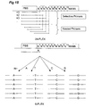
- the circular probe can contain a blocking section (27).
- the blocking section blocks primer elongation.
- the blocking section is preferably located between the two primer binding sites.
- Preferably the blocking section is located essentially adjacent to the 3'-end of the forward primer and essentially adjacent to the 5'-end of the reverse primer binding site, see also Figure 14.
- An example of such an arrangement of functional groups in the circular probe is: (target-complementarity section 1 - stuffer sequence 1, primer-binding sequence 1- blocking section - primer-binding sequence 2 - stuffer sequence 2 - target-complementarity section 2). This blocking section will effectively limit the primer elongation during amplification, thereby providing linear representations of the connected circular probes.
- the blocking section itself is located such between the two primer binding sites that the section is excluded from the amplification.
- the blocking section can comprise non-nucleotide polymers such as HEG (Hexaethylene glycol). If a blocking section is present, such as a HEG group, the DNA polymerase used may have a strand displacement activity as the blocking section will prevent the formation of long concatamers.
- the ligated or connected circular probe comprising a blocking section can also be amplified using only one primer, preferably the forward primer. This amplification will result in the linear accumulation of amplicons with each amplification round.
- the circular probe in this case may contain one or more primer binding sites as long as only one primer is provided.
- step (a) a multiplicity of different target sequences, i.e. at least two different target sequences, is brought into contact with a multiplicity of specific oligonucleotide probes under hybridising conditions.
- the oligonucleotide probes are subsequently allowed to anneal to the adjacent complementary parts of the multiple target sequences in the sample.
- Methods and conditions for specific annealing of oligonucleotide probes to complementary target sequences are well known in the art (see e.g. in Sambrook and Russel (2001) "Molecular Cloning: A Laboratory Manual (3rd edition), Cold Spring Harbor Laboratory, Cold Spring Harbor Laboratory Press ).
- the nucleic acids are denatured by incubation (generally at between 94 °C and 96 °C) for a short period of time (e.g. 30 seconds to 5 minutes) in a low salt buffer (e.g. a buffer containing no salts or less salts than the ionic strength equivalent of 10 mM NaCl).
- a low salt buffer e.g. a buffer containing no salts or less salts than the ionic strength equivalent of 10 mM NaCl.
- the sample containing the denatured probes and target sequences is anthen allowed to cool to an optimal hybridisation temperature for specific annealing of the probes and target sequences, which usually is about 5°C below the melting temperature of the hybrid between the complementary section of the probe and its complementary sequence (in the target sequence).
- the sections of the probes that are complementary to the target sequences are of a similar, preferably identical melting temperatures between the different target sequences present in the sample.
- the complementary sections of the probes preferably differ less than 20, 15, 10, 5, or 2 °C in melting temperature. This is facilitated by using complementary sections of the probes with a similar length and similar G/C content.
- the complementary sections preferably differ less than 20, 15, 10, 5, or 2 nucleotides in length and their G/C contents differ by less than 30, 20, 15, 10, or 5 %.
- a first nucleotide sequence is capable of specifically hybridising to second nucleotide sequence under normal stringency conditions.
- a nucleotide sequence that is considered complementary to another nucleotide sequence may contain a minor amount, i.e. preferably less than 20, 15, 10, 5 or 2%, of mismatches.
- it may be necessary to compensate for mismatches e.g. by incorporation of so-called universal nucleotides, such as for instance described in EP-A 974 672 , incorporated herein by reference or by the use of suitable locked nucleic acids (LNAs) and peptide nucleic acids (PNAs).
- LNAs locked nucleic acids
- PNAs peptide nucleic acids
- annealing of probes to target sequences is concentration dependent, annealing is preferably performed in a small volume, i.e. less than 10 ⁇ l. Under these hybridisation conditions, annealing of probes to target sequences usually is fast and does not to proceed for more than 5,10 or 15 minutes, although longer annealing time may be used as long as the hybridisation temperature is maintained to avoid aspecific annealing.
- the walls and lids of the reaction chambers may also be heated to the same temperature as the reaction mixture.
- the length of the complementary section is preferably at least 15, 18 or 20 nucleotides and preferably not more than 30, 40, or 50 nucleotides and the probes preferably have a melting temperature of at least 50°C, 55°C or 60°C.
- the probes that are not complementary to a part of the target sequence or that contain too many mismatches will not or only to a reduced extent hybridise to the target sequence when the sample is submitted to hybridisation conditions. Accordingly ligation is less likely to occur.
- the number of spurious ligation products from these probes in general will therefore not be sufficient and much smaller than the bona fide ligation products such that they are outcompeted during subsequent multiplex amplification. Consequently, they will not be detected or only to a minor extent.
- the respective 5'- and 3'-ends of the oligonucleotide probe that are annealed essentially adjacent to the complementary parts of a target sequence are connected in step (c) to form a covalent bond by any suitable means known in the art.
- the ends of the probes may be enzymatically connected in a phosphodiester bond by a ligase, preferably a DNA ligase.
- DNA ligases are enzymes capable of catalysing the formation of a phosphodiester bond between (the ends of) two polynucleotide strands bound at adjacent sites on a complementary strand.
- DNA ligases usually require ATP (EC 6.5.1.1) or NAD (EC 6.5.1.2) as a cofactor to seal nicks in double stranded DNA.
- Suitable DNA ligase for use in the present invention are T4 DNA ligase, E . coli DNA ligase or preferably a thermostable ligase like e.g. Thermus aquaticus (Taq) ligase, Thermus thermophilus DNA ligase, or Pyrococcus DNA ligase.
- modified polynucleotide ends may be used to ligate two oligonucleotide probes annealed at adjacent sites on the complementary parts of a target sequence ( Xu and Kool, 1999, Nucleic Acid Res. 27: 875-881 ).
- a DNA ligase is employed that remains active at 50 - 65°C for prolonged times, but which is easily inactivated at higher temperatures, e.g. used in the denaturation step during a PCR, usually 90 - 100°C.
- One such DNA ligase is a NAD requiring DNA ligase from a Gram-positive bacterium (strain MRCH 065) as known from WO O1/61033 . This ligase is referred to as "Ligase 65" and is commercially available from MRC Holland, Amsterdam.
- the respective ends may be annealed such that a gap is left.
- This gap can be filled with a suitable oligonucleotide and ligated.
- oligonucleotide and ligated Such methods are known in the art as 'gap ligation' and are disclosed inter alia in WO 00/77260 .
- Another possibility to fill this gap is by extension of one end of the probe using a polymerase and a ligase in combination with single nucleotides, optionally preselected from A,T, C, or G, or di-, tri- or other small oligonucleotides.
- the connected probes are amplified using a pair of primers corresponding to the primer-binding sites.
- at least one of the primers or the same set of primers is used for the amplification of two or more different connected probes in a sample, preferably for the amplification of all connected probes in a sample.
- a primer is sometimes referred to as a universal primer as these primers are capable of priming the amplification of all probes containing the corresponding universal primer binding site and consequently of all ligated probes containing the universal primer binding site.
- the different primers that are used in the amplification in step (d) are preferably essentially equal in annealing and priming efficiency.
- the primers in a sample preferably differ less than 20, 15, 10, 5, or 2 °C in melting temperature. This can be achieved as outlined above for the complementary section of the oligonucleotide probes. Unlike the sequence of the complementary sections, the sequence of the primers is not dictated by the target sequence. Primer sequences may therefore conveniently be designed by assembling the sequence from tetramers of nucleotides wherein each tetramer contains one A,T,C and G or by other ways that ensure that the G/C content and melting temperature of the primers are identical or very similar.
- the length of the primers (and corresponding primer-binding sites in the tags of the probes) is preferably at least 12, 15 or 17 nucleotides and preferably not more than 25, 30, 40 nucleotides.
- At least two of the oligonucleotide probes that are complementary to at least two different target sequences in a sample comprise a tag sequence that comprises a primer-binding site that is complementary to a single primer sequence.
- at least one of the first and second primer in a primer pair is used for the amplification of connected probes corresponding to at least two different target sequences in a sample, more preferably for the amplification of connected probes corresponding to all target sequences in a sample.
- Preferably only a single first primer is used and in some embodiments only a single first and a single second primer is used for amplification of all connected probes.
- Using common primers for amplification of multiple different fragments usually is advantageous for the efficiency of the amplification step.
- the connected probes obtained from the ligation of the adjacently annealed probe sections are amplified in step (d), using a primer set, preferably consisting of a pair of primers for each of the connected probes in the sample.
- the primer pair comprises primers that are complementary to primer-binding sequences that are present in the connected probes.
- a primer pair usually comprises a first and at least a second primer, but may consist of only a single primer that primes in both directions. Excellent results have been obtained using primers that are known in the art as AFLP primers such as described inter alia in EP534858 and in Vos et al., Nucleic Acid Research, 1995, vol. 23,4407-44014 .
- one or more of the primers used in the amplification step of the present invention is a selective primer.
- a selective primer is defined herein as a primer that, in addition to its universal sequence which is complementary to a primer binding site in the probe, contains a region that comprises so-called "selective nucleotides". The region containing the selective nucleotides is located at the 3'-end of the universal primer.
- the principle of selective nucleotides is disclosed inter alia in EP534858 and in Vos et al., Nucleic Acid Research, 1995, vol. 23,4407-44014 .
- the selective nucleotides are complementary to the nucleotides in the (ligated) probes that are located adjacent to the primer sequence.
- the selective nucleotides generally do not form part of the region in the (ligated) probes that is depicted as the primer sequence.
- Primers containing selective nucleotide are denoted as +N primers, in which N stands for the number of selective nucleotides present at the 3'-end of the primer. N is preferably selected from amongst A, C, T or G.
- N may also be selected from amongst various nucleotide alternatives, i.e. compounds that are capable of mimicking the behavior of ACTG-nucleotides but in addition thereto have other characteristics such as the capability of improved hybridisation compared to the ACTG-nucleotides or the capability to modify the stability of the duplex resulting from the hybridisation. Examples thereof are PNA's, LNA's, inosine etc.
- PNA's i.e. compounds that are capable of mimicking the behavior of ACTG-nucleotides but in addition thereto have other characteristics such as the capability of improved hybridisation compared to the ACTG-nucleotides or the capability to modify the stability of the duplex resulting from the hybridisation. Examples thereof are PNA's, LNA's, inosine etc.
- the amplification is performed with more than one primer, such as with PCR using two primers, one or both primers can be equipped with selective nucleotides.
- a +1 primer thus contains one selective nucleotide
- a +2 primer contains 2 selective nucleotides etc.
- a primer with no selective nucleotides i.e. a conventional primer
- a subset of (ligated) probes is obtained, provided that the complementary base is incorporated at the appropriate position in the desired of the probes that are supposed to be selectively amplified using the selective primer.
- the multiplex factor of the amplified mixture is reduced by a factor 4 compared to the mixture of ligated probes prior to amplification. Higher reductions can be achieved by using primers with multiple selective nucleotides, i.e. 16 fold reduction of the original multiplex ration is obtained with 2 selective nucleotides etc.
- the probe When an assay is developed which, after ligation, is to be selectively amplified, it is preferred that the probe contains the complementary nucleotide adjacent to the primer binding sequence. This allows for pre-selection of the ligated probe to be selectively amplified.
- One of the examples in which this is useful and advantageous is in case of analysis of samples that contain only minute amounts of DNA and/or for the identification of different (strains of) pathogens.
- an assay directed to the detection of various strains of anthrax Bacillus anthracis
- a set of representative probes is designed for each of the strains.
- the detection of the presence or absence of this set (or a characterizing portion thereof) of ligated probes after the hybridisation and ligation steps of the method of the invention may serve as an identification of the strain concerned.
- the selective amplification with specifically designed primers (each selective primer is linked to a specific strain) can selectively amplify the various strains, allowing their identification.
- amplification with an +A primer selectively amplifies the ligated probes directed to strain X where a +G primer selectively amplifies the ligated probes directed to strain Y.
- an optional first amplification with a +0 primer will increase the amount of ligated probes, thereby facilitating the selective amplification.
- a universal primer of 20 nucleotides becomes a selective primer by the addition of one selective nucleotide at its 3' end, the total length of the primer now is 21 nucleotides. See also Figure 15.
- the universal primer can be shortened at its 5' end by the number of selective nucleotides added. For instance, adding two selective nucleotides at the 3' end of the primer sequence can be combined with the absence (or removal) of two nucleotides from the 5'end of the universal primer, compared to the original universal primer.
- a universal primer of 20 nucleotides is replaced by a selective primer of 20 nucleotides.
- selective primers based on universal primers has the advantage that amplification parameters such as stringency and temperatures may remain essentially the same for amplification with different selective primers or vary only to a minor extent.
- selective amplification is carried out under conditions of increased stringency compared to non-selective amplification.
- increased stringency is meant that the conditions for annealing the primer to the ligated probe are such that only perfectly matching selective primers will be extended by the polymerase used in the amplification step.
- the specific amplification of only perfectly matching primers can be achieved in practice by the use of a so-called touchdown PCR profile wherein the temperature during the primer annealing step is stepwise lowered by for instance 0.5 °C to allow for perfectly annealed primers.
- Suitable stringency conditions are for instance as described for AFLP amplification in EP 534858 and in Vos et al., Nucleic Acid Research, 1995, vol. 23, 4407-44014 .
- the skilled man will, based on the guidance find ways tot adapt the stringency conditions to suit his specific need without departing from the gist of the invention.
- One of the further advantages of the selective amplification of ligated probes is that an assay with a high multiplex ratio can be adapted easily for detection with methods or on platforms that prefer a lower multiplex ratio.
- One of many examples thereof is the detection based on length differences such as electrophoresis and preferably capillary electrophoresis such as is performed on a MegaBACE or using nano-technology such as Lab-on-a-Chip.
- the connected probes are amplified to produce (detectable) amplified connected probes (amplicons) that are linear representations of the connected circular probes by any suitable nucleic acid amplification method known in the art.
- Nucleic acid amplification methods usually employ two primers, dNTP's, and a (DNA) polymerase.
- a preferred method for amplification is PCR.
- PCR or “Polymerase Chain Reaction” is a rapid procedure for in vitro enzymatic amplification of a specific DNA segment.
- the DNA to be amplified is denatured by heating the sample.
- oligonucleotides that hybridise specifically to the target sequence prime new DNA synthesis.
- the polymerase is a DNA polymerase that does not express strand displacement activity or at least not significantly. Examples thereof are Amplitaq and Amplitaq Gold (supplier Perkin Elmer) and Accuprime (Invitrogen).
- Amplitaq and Amplitaq Gold supplier Perkin Elmer
- Accuprime Invitrogen
- the second cycle of denaturation, annealing and synthesis produces two single-stranded products that together compose a discrete doublestranded product, exactly the length between the primer ends.
- This discrete product accumulates exponentially with each successive round of amplification. Over the course of about 20 to 30 cycles, many million-fold amplification of the discrete fragment can be achieved.
- PCR protocols are well known in the art, and are described in standard laboratory textbooks, e.g. Ausubel et al., Current Protocols in Molecular Biology, John Wiley & Sons, Inc. (1995 ). Suitable conditions for the application of PCR in the method of the invention are described in EP-A 0 534 858 and Vos et al.
- RNA polymerase-binding site a suitable (RNA) polymerase-binding site as long as they lead to linear amplification products as defined herein before, i.e. of discrete lengths and corresponding to the length of the circular probes.
- linear representations of the connected circular probes can be obtained by exponential amplification of the circular probe with two primers, one forward and one reverse, using a polymerase that does not or not significantly have a strand displacement activity.
- the first primer elongation in the amplification with the forward primer generates an oligonucleotide product until the 5'end of the forward primer is reached. There the primer elongation is terminated, due to the substantial absence of strand displacement activity of the polymerase used, leaving a elongated primer with substantially the same length as the connected circular probe.
- the second cycle of denaturation, primer hybridisation and primer elongation will, for the forward primer, produce the identical strand as during the first primer elongation, while the reverse primer will hybridise to the oligonucleotide product from the elongation of the first primer elongation and thereby produce the complementary strand, resulting in the exponential amplification of the circular probe to thereby produce amplicons of discrete length which are representations of the connected circular oligonucleotide probes.
- the term 'amplicon' as used herein refers to the product of the amplification step of the connected or ligated probe.
- the term 'amplicon' as used herein thus refers to an amplified connected probe.
- the connected or ligated probe is combined with one or more primers and a polymerase and amplified.
- the ligated probe, the primers, the polymerase and/or other parameters and variables are such that the amplification results in linear representations of the circular probe.
- the amplicon is a linear oligonucleotide having a length that does not substantially exceed the length of the circular probe.
- the minimum length of the amplicon is at least the sum of the length of the two target complementary sections. It is preferred that the length of the amplicon corresponds to the length of the circular probe. It is more preferred that the length of the amplicon is indicative of the ligation of the corresponding circular probe.
- an amplicon does not contain repetitions of sections of the circular probe, i.e. is not a concatamer or a multimer of the circular probe or a multimeric representation thereof.
- an amplicon is a linear and monomeric representation of the connected circular probe.
- Detection of the labelled separated samples is performed by a detector to result in detection data.
- the detector is of course dependent on the general system on which the separation is carried out (capillary electrophoresis, slab-gel electrophoresis, fixed detector-continuous gel-electrophoresis) but is also depending on the label that is present on the primer, such as a fluorescent or a radioactive label.
- the amplicons in a sample are preferably analysed on an electrophoretic device.
- the electrophoretic device preferably separates the different amplicons in an amplified sample on the basis of length, after which the separated amplicons may be detected as described below.
- a suitable electrophoretic device may be a gel-electrophoresis device, e.g. for conventional (polyacrylamide) slab gel-electrophoresis, or a capillary electrophoresis device such as exemplified by the MegaBACE equipment available from Molecular Dynamics Amersham-Biosciences.
- An alternative is the nano-sized capillary electrophoretic devices known as Lab-on-a-Chip.
- the electrophoretic device preferably is a multichannel device in which multiple samples are electrophoresed in multiple channels in parallel.
- the electrophoretic device has an application location (per channel) for application (loading) of the amplified sample to be electrophoresed, a separation area over which the fragments in the sample migrate by electrophoresis, and preferably also a detection device located at a detection location distal from the application location.
- the detection device will usually comprises a photomultiplier for the detection of fluorescence, phosphorescence or chemiluminescence.
- the separated fragments may be detected in the gel e.g. by autoradiography or fluorography.
- a difference in length of the respective corresponding amplicons is used.
- the discrimination between amplicons derived from different target sequences in a sample is based on a length difference between the respective amplicons corresponding to different target sequences in a sample or amplified sample.
- the length difference is provided by the length of the stuffer sequence(s) in the oligonucleotide probes.
- a stuffer of a pre-determined length the length of each amplicon in an amplified sample can be controlled such that an adequate discrimination based on length differences of the amplicon obtained in step (d) is enabled.
- the stuffer is located between the probe's section complementary to the target sequence and a primer-binding sequence. As there are two target specific sections at both ends of the probe and two primer binding sites, two stuffer can be incorporated in the probe therein between.
- the total length of the stuffer is provided by the combination of the length of the first stuffer and second stuffer in the probe.
- the oligonucleotide probe comprises two stuffers, preferably in the non target complementary tags. A graphic representation thereof can be found in Figure 14.
- the length differentiation between amplicons obtained from target sequences in the sample is preferably chosen such that the amplicons can be distinguished based on their length. This is accomplished by using stuffer sequences or combinations of stuffer sequences which (together) result in clear length differences that may be distinguished on electrophoretic devices. Thus, from the perspective of resolving power, the length differences between the different amplicons, as may be caused by their stuffers, are as large as possible.
- the length differences between the different amplicon is preferably as small as possible: (1) the upper limit that exists in practice with respect to the length of chemically synthesised probes of about 100-150 bases at most; (2) the less efficient amplification of larger fragments, (3) the increased chances for differential amplification efficiencies of fragments with a large length variation; and (4) the use of multiple injections of detection samples on the detection device which works best with fragments in a narrow length range.
- the length differences between the sequences to be determined and provided by the stuffers is at least sufficient to allow discrimination between essentially all amplicons.
- the minimal useable size difference between different amplicon in an amplified sample is one base, and this size difference fits within the resolving power of most electrophoresis devices, especially in the lower size ranges.
- the length difference between different amplicons in an amplified sample is at least two nucleotides.
- the amplicon corresponding to different target sequences in a sample have a length difference of two nucleotides.
- the primers complementary to the primer-binding sites of the first and second oligonucleotide probes in the sample comprises a label
- the second primer comprises a label.
- the label can be selected from a large group, amongst others comprising fluorescent and/or phosphorescent moieties such as dyes, chromophores, or enzymes, antigens, heavy metals, magnetic probes, phosphorescent moieties, radioactive labels, chemiluminescent moieties or electrochemical detecting moieties.
- the label is a fluorescent or phosphorescent dye, more preferably selected from the group of FAM, HEX, TET, JOE, NED, and (ET-)ROX.
- Dyes such as FITC, Cy2, Texas Red, TAMRA, Alexa fluor 488 TM , Bodipy TM FL, Rhodamine 123, R6G, Bodipy 530, Alexafluor TM 532 and IRDyes TM by Licor as used on the NEN Glober IR 2 platform are also suitable for use in the present invention.
- the label may be chosen from amongst the fluorescent or phosphorescent dyes in the group consisting of FAM, TET, JOE, NED, HEX, (ET-)ROX, FITC, Cy2, Texas Red, TAMRA, Alexa fluor 488 TM , Bodipy TM FL, Rhodamine 123, R6G, Bodipy 530, Alexafluor TM 532 and IRDyes TM .
- fluorescent or phosphorescent dyes in the group consisting of FAM, TET, JOE, NED, HEX, (ET-)ROX, FITC, Cy2, Texas Red, TAMRA, Alexa fluor 488 TM , Bodipy TM FL, Rhodamine 123, R6G, Bodipy 530, Alexafluor TM 532 and IRDyes TM .
- the number of connected probes that can be discriminated in a sample and hence the number of target sequences in a sample can be doubled for each additional label.
- the number of target sequences that can be analysed in a sample is doubled.
- the maximum number of labels that can be used in one sample in a high throughput method is governed mostly by the limitations in the detection capabilities of the available detection platforms. At present, one of the most frequently used platforms (MegaBACE, by Molecular Dynamics -Amersham-Biosciences Ltd. allows the simultaneous detection of up to four fluorescent dyes, being FAM, JOE or HEX, NED and (ET-)ROX.
- capillary electrophoresis instruments are also suitable, which includes ABI310, ABI3100, ABI3700 (Perkin-Elmer Corp.), CEQ2000 XL (Beckman Coulter) and others.
- slab-gel based electrophoresis devices include ABI377 (Perkin Elmer Corp.) and the global IR 2 automated DNA sequencing system, available from LI-COR, Lincoln, California, USA.
- Cross talk or residual cross talk refers to the overlap between the emission spectra of different (fluorescent) labels. For instance when fluorescent dyes are used, each dye has a different emission (and absorption) spectrum. In case of two dyes in one sample, these spectra can overlap and may cause a disturbance of the signal, which contravenes the quality of the data obtained.
- Chehab et al. (Proc. Natl. Acad. Sci. USA, 86:9178-9182 (1989) have attempted to discriminate between alleles by attaching different fluorescent dyes to competing alleles in a single reaction tube by selecting combinations of labels such that the emission maximum of one dye essentially coincides with the emission minimum of the other dye.
- the emission maximum of one dye essentially coincides with the emission minimum of the other dye.
- the present invention provides for a solution to this problem such that two (or more) labels with overlapping spectra can be used in the same sample without significantly affecting data quality.
- a predetermined combination of length differences and labels an increase in the number of target nucleotide sequences that can be detected in sample is obtained while the quality of the data remains at least constant.
- spectral overlap between two differently labelled sequences is reduced by the introduction of a length difference between the two sequences.
- This label-related length difference can be provided for by the length of the stuffer sequence as described herein.
- the number of different labels that can be used in the same sample in the present method is at least two, preferably at least three, more preferably at least four.
- the maximum number of labels is functionally limited by the minimum of spectral overlap that remains acceptable, which for most applications typically amounts to less than 15 percent of the true signal, preferably less than 10 percent, more preferably lees than 5 percent and most preferably less than 1 percent of the true signal.
- the stuffer sequences such that amplicons differ by at least two base pairs within a multiplex set and differ by a single base pair between multiplex sets labelled with the different dyes that have overlapping spectra.
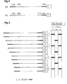
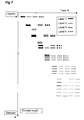
- the length of the fragments labelled with the respective dyes can be chosen such that the potential influence of residual cross-talk on the quality of the data is circumvented because unique combinations of fragments size and labelling dye are defined (Figure 3).
- a particular preferred embodiment of the invention is directed to a method in which a sample comprising amplicons is derived from a multiplicity of target sequences. These amplicons are differently labelled, thereby defining groups of amplicons carrying the same label.
- the stuffer provided for a length difference of at least two, preferably two nucleotides. Between two groups with labels having spectral overlap, the stuffer provides a length difference of one nucleotide, effectively resulting in one group having an even number of nucleotides and one group having an odd number of nucleotides as described above.
- the present invention pertains to a method for the improved discrimination and detection of target sequences in a sample, comprising providing at least a two or more groups of oligonucleotide probes, wherein the amplicons obtained with different groups of oligonucleotide probes have different labels, wherein substantially each amplified connected probe target sequence within a group has the same label, wherein within a group of identically labelled amplicons a length difference is provided between each identically labelled probe within that group, wherein between the first and second group an additional length difference is provided such that each amplified connected probe in the amplified sample is characterised by a combination of length of the sequence and the label.
- each group of oligonucleotide probes has tag sequences with at least one group specific primer-binding site.
- the connected probes of each group are amplified from a primer pair wherein at least one of the first and second primers is complementary to the group specific primer-binding site, and whereby at least one of the first and second primers of a group comprises a group specific label.
- an amplicon corresponding to a target sequence in the sample differs in length from an amplicon corresponding to a different target sequence in the sample.
- the group specific labels are preferably such that the detection device can distinguish between the different group specific labels.
- the length difference is preferably provided by the length of the stuffer sequence.
- a first part of the groups has amplicons having an even number of nucleotides and a second part of the groups has amplicons having an odd number of nucleotides.
- the groups of amplicons having an even number of nucleotides and the groups amplicons having an odd number of nucleotides are labelled with (fluorescent) labels, which have the least overlap in their emission spectra.
- two groups of amplicons, each group having an odd number of nucleotides are labelled with labels, which have the least overlap in their emission spectra.
- Two groups of amplicons each group having an even number of nucleotides.
- Two groups of amplicons, one group having an odd number of nucleotides and the other group having an even number of nucleotides are labelled with labels that have a larger overlap in their emission spectra.
- the relative notions as used herein of 'the least overlap in their emission spectra' and have a larger overlap in their emission spectra' refer to a group of labels from which a selection of the labels can be made for use in the present invention. This group of labels may depend on the detection platform used to other factors such as those disclosed herein before.
- a first and second groups of amplicons having an even number of nucleotides are produced and a third and fourth group of connected amplified probes having an odd number of nucleotides are produced and whereby the first and second group are labelled with FAM and NED, respectively, and the third and fourth group are labelled with (ET-)ROX and either JOE or HEX, respectively; or vice versa, whereby the first and second group are labelled with (ET-)ROX and either JOE or HEX, respectively, and the third and fourth group are labelled with FAM and NED, respectively.
- the fluorescent labels are chosen such that the groups of amplicons that co-migrate, because they both contain fragments with either even or odd numbers of nucleotides, have labels which have the least overlap in their emission spectra, thereby avoiding as much as possible cross-talk in the detection of amplicons in different groups (see also below).
- the connected probes derived from each target sequence differ from any other connected probe in the sample in length, and/or in the label or, preferably in the combination of the length and the label.
- the length difference between two different connected probes is at least two nucleotides, preferably two.
- the difference in length between two or more (SNP) alleles of the polymorphism is not more than two, thereby ensuring that the efficiency of the amplification is similar between different alleles or forms of the same polymorphism. This implies that preferably both alleles are amplified with the same pair of primers and hence will be labelled with the same dye.
- the distribution between odd/even lengths within a group can be designed in the following way.
- Two loci L1, L2 are each represented by two alleles A11, A12 for L1 and A21, A22 for L2.
- G1 and G2 carrying labels that may have an overlap in their spectra there can be a length difference of 1 nucleotide.
- This distribution has some significant advantages compared to the more densely packed distribution disclosed herein. It is known that due to conformational differences that different sequences of identical length generally differ in their electrophoretic mobility. When there is only a difference in length of one nucleotide, this may cause overlap between the peaks if the sequences are of a very different mobility. For instance the difference in mobility between two alleles of one locus (A11, A12), will be less than the difference in mobility between two alleles from different loci (A12, A21). When there is a significant difference in mobility between A12 and A21, this may lead to unreliable detection. By creating length distributions as herein disclosed this can be avoided. The lower throughput is then weighed against the reliability of the detection.
- a length difference of alternating two and three nucleotides i.e. 0, 2, 5, 7, 10, 12 etc.
- the other group then has a length difference of 1, 3, 6, 8, 11, 13 etc.
- the target sequence may be any nucleotide sequence of interest.
- the target sequence preferably is a nucleotide sequence that contains, represents or is associated with a polymorphism.
- polymorphism herein refers to the occurrence of two or more genetically determined alternative sequences or alleles in a population.
- a polymorphic marker or site is the locus at which divergence occurs. Preferred markers have at least two alleles, each occurring at frequency of greater than 1%, and more preferably greater than 10% or 20% of a selected population.
- a polymorphic locus may be as small as one base pair.
- Polymorphic markers include restriction fragment length polymorphisms, variable number of tandem repeats (VNTR's), hypervariable regions, minisatellites, dinucleotide repeats, trinucleotide repeats, tetranucleotide repeats, simple sequence repeats, and insertion elements such as Alu.
- the first identified allelic form is arbitrarily designated as the reference form and other allelic forms are designated as alternative or variant alleles.
- the allelic form occurring most frequently in a selected population is sometimes referred to as the wild type form. Diploid organisms may be homozygous or heterozygous for allelic forms.
- a diallelic polymorphism has two forms.
- a triallelic polymorphism has three forms.
- a single nucleotide polymorphism occurs at a polymorphic site occupied by a single nucleotide, which is the site of variation between allelic sequences.
- the site is usually preceded by and followed by highly conserved sequences of the allele (e.g., sequences that vary in less than 1/100 or 1/1000 members of the populations).
- a single nucleotide polymorphism usually arises due to substitution of one nucleotide for another at the polymorphic site.
- Single nucleotide polymorphisms can also arise from a deletion of a nucleotide or an insertion of a nucleotide relative to a reference allele.
- a preferred target sequence is a target sequence that is associated with an AFLP ® marker, i.e. a polymorphism that is detectable with AFLP ® .
- the nucleic acids comprising the target may be any nucleic acid of interest.
- the nucleotide sequence information contained in the sample may be from any source of nucleic acids, including e.g. RNA, polyA + RNA, cDNA, genomic DNA, organellar DNA such as mitochondrial or chloroplast DNA, synthetic nucleic acids, DNA libraries, clone banks or any selection or combinations thereof.
- the DNA in the nucleic acid sample may be double stranded, single stranded and double stranded DNA denatured into single stranded DNA.
- Preferred nucleic acid samples comprise target sequences on cDNA, genomic DNA, restriction fragments, adapter-ligated restriction fragments, amplified adapter-ligated restriction fragments. AFLP fragments or fragments obtained in an AFLP-template preamplification.
- a sample contains two or more different target sequences, i.e. two or more refers to the identity rather than the quantity of the target sequences in the sample.
- the sample comprises at least two different target sequences, in particular at least 10, preferably at least 25, more preferably at least 50, more in particular at least 100, preferably at least 250, more preferably at least 500 and most preferably at least 1000 additional target sequences.
- the number of target sequences is limited, among others, by the number of connected circular probes. E.g., too many different oligonucleotide probes in a sample may corrupt the reliability of the multiplex amplification step.
- a further limitation is formed e.g. by the number of fragments in a sample that can be resolved by the electrophoretic device in one injection.
- the number can also be limited by the genome size of the organism or the transcriptome complexity of a particular cell type from which the DNA or cDNA sample, respectively, is derived.
- a number of samples are treated similar to thereby generate a multiplicity of amplified detection samples which can then be analysed on a multichannel device which is at least capable of detecting the labels and/or length differences. Suitable devices are described herein.
- Steps (a) to (e) of the method of the invention may be performed on two or more nucleic acid samples, each containing two or more different target nucleic acids, to produce two or more amplified samples in which is presence or absence of amplicons is analysed.
- the multiplex analysis of the amplified samples following the method of the invention comprises applying at least part of an amplified sample to an electrophoretic device for subsequent separation and detection.
- an amplified sample contains, or is at least suspected to contain, amplified connected probes, which is an indication that a target sequence has hybridised with the provided oligonucleotide probes and that those probes were annealed adjacently on the complementary target sequence so that they where connected, i.e. ligated.
- an amplified sample is subjected to a separating step for a selected time period before a next amplified sample is submitted.
- the time period (23) between two (or more) consecutively applied amplified samples is such that the slowest migrating amplified connected probe (19) in an amplified sample is detected at the detection location (24), before the fastest migrating amplified connected probe of a subsequently applied amplified sample is detected at the detection location (24).
- the time intervals between subsequent multiple injections in one channel of the device are chosen such that consecutively applied samples after separation do not overlap at a point of detection.
- the method according to the invention allows for the high throughput analysis of a multiplicity of samples each comprising a multiplicity of different target sequences by the consecutive injection of amplified samples, comprising amplicons corresponding to the target sequences in the samples, in a channel of a multichannel electrophoretic device such as a capillary electrophoresis device.
- the method according to the invention allows for the analysis of a multiplicity of target sequences in a multiplicity of samples on a multiplicity of channels, thereby significantly increasing the throughput of the number of samples that can be analysed in a given time frame compared to conventional methods for the analysis of nucleotide sequences.
- This method profits from samples containing amplicons to be detected that are of a discrete size range as thereby the time period (23) between the successive injections can be significantly reduced compared to methods wherein the (remains of) concatamers are present.
- the selected time period prevents that consecutively applied samples after separation have an overlap of amplicons at the detection point.
- the selected time period is influenced by i). the length of the amplicons; ii). the length variation in amplicons; and iii). the detection device and its operating conditions.
- Applying samples and separating consecutively applied samples in the same channel can be repeatedly performed in one or more channels, preferably simultaneously to allow for consecutive electrophoretic separation of multiple samples in one channel and/or simultaneous analysis of multiple samples over multiple channels and/or simultaneous analysis of multiple samples over multiple channels carried out consecutively.
- a graphic representation thereof is given in Figure 8.
- the period of time between two consecutively loaded amplified samples can be determined experimentally prior to executing the method. This period of time is selected such that, given the characteristics of an amplified sample, especially the difference in length between the shortest and the longest amplicons in an amplified sample, as well as other experimental factors such as gel (matrix) and/or buffer concentrations, ionic strength etc., the fragments in an amplified samples are separated to such extent at the detection location which is located at the opposite end (distal) from the application location where the sample was applied, that the different amplicons in a sample may be individually detected. After applying the last amplified sample, the separation can be continued for an additional period of time to allow the amplicons of the last sample to be separated and detected.
- the combination of the selected period of time between applying two consecutive samples and the optional additional time period is chosen such that at the detection location the different amplicons in consecutively applied samples are separated such that they may be individually detected, despite the limited length variation that exists between the different amplicons within a single sample.
- overlapping migration patterns are prevented when samples containing fragments of varying length are consecutively applied (injected) on the electrophoretic device.
- the device is able to perform such operation automatically, e.g. controlled by a programmable computer.
- the multichannel device is suitable for such operation or is at least equipped for a prolonged operation without maintenance such as replacement of buffers, parts etcetera. However, in practice this will generally not be the case.
- the stuffers present in the tags of the oligonucleotide probes is are used to provide the length differences (i.e.
- the range of lengths of amplicons in an amplified sample has a lower limit of 40, 60, 80, or 100 and an upper limit of 120, 140, 160, or 180 nucleotides, bases or base pairs, for conventional (capillary) electrophoresis platforms. It is particularly preferred that the range of lengths of the amplicons varies from 100 to 140 nucleotides. However, these numbers are strongly related to the current limits of the presently known techniques. Based on the knowledge provided by this invention, the skilled artisan is capable of adapting these parameters when other circumstances apply.
- the reliability of the multiplex amplification is further improved by limiting the variation in the length of the amplified connected probes. Limitations in the length variation of amplicons is preferred to use multiple injection more efficiently and further results in reduction of the preferential amplification of smaller amplicon in a competitive amplification reaction with larger connected probes. This improves the reliability of the high throughput method of the present invention. Together with the multiple injection protocol as herein disclosed, these measures, alone or in combination provide for a significant increase in throughput in comparison with the art. A further improvement of the high throughput capacity is obtained by limiting the number of different amplicons in a sample. It is regarded as more efficient and economical to limit the multiplex capacity of the ligation/amplification step in combination with the introduction of a multiple injection protocol.
- One of the most advantageous aspects of the present invention lies in the combination of multiplex ligation, multiplex amplification, preferably with a single primer pair or with multiple primer pairs which each amplify multiple connected probes, repeated injection and multiplex detection of different labels.
- One of the further advantageous aspects of the present invention resides in the combined application of length differences with different (overlapping) labels such that each connected probe and hence each target sequence within one sample can be characterised by a unique combination of length and label. This allows for a significant improvement of the efficiency of the analysis of target sequences as well as a significant reduction in the costs for each target analysed.
- the multiple injection protocol can be performed in a variety of ways.
- One of these is the multiple loading of two or more samples in the same matrix. This is considered as advantageously as the matrix is re-used by performing consecutive short runs, thereby increasing efficiency and throughput.
- Another one is the multiple loading of two or more samples in the same matrix in the same run. It is preferred to re-use the matrix by performing short consecutive runs.
- a first sample is injected and separated. As soon as the last fragment is detected, the next sample is loaded. Preferably, between these two consecutive short runs the matrix is not replaced so that the runs are performed in the same matrix. This provides for additional efficiency and improved economics as less changes o the matrix need to occur, reducing the amount of consumables of this type of analysis ( i.e. buffers etc.), reducing the cost per datapoint. Furthermore time-consuming replacements of the matrix can be avoided to a large extent, further increasing the efficiency of the method.
- primer extension product is of a constant length.
- primers of a different length can be used, varying typically from 15 to 25 nucleotides.
- the present invention contemplates analysing multiplex amplification products themselves directly with a length variation typically between 50 and 150 nucleotides.
- a preferred method of the invention further comprises a step for the removal of oligonucleotide probes that are not annealed to target sequences and/or that are non-connected/ligated. Removal of such probes preferably is carried out prior to amplification, and preferably by digestion with exonucleases.
- exonuclease to digest non-connected/ligated oligonucleotide probes.issensitive. sensitive.
- Blocking groups include use of a thiophosphate group and/or use of 2-O-methyl ribose sugar groups in the backbone.
- Exonucleases include Exol (3'-5' activity), Exo III (3'-5' activity), and Exo IV (both 5'-3' and 3'-5' activity).
- the circular probes of the present invention are, once ligated, insensitive to the exonuclease, as opposed to the unligated circular probes This is a further advantage of the use of padlock probes in the present invention.
- exonucleases for example a combination of Exo I (single strand specific) and Exo III (double strand specific), is the ability to destroy both the target sequence and the unligated oligonucleotide probes, while leaving the ligation product sequences substantially undigested.
- the oligonucleotide probes in each set are substantially reduced, and thus hybridisation of the remaining unligated oligonucleotide probes to the original target DNA (which is also substantially reduced by exonuclease treatment) and formation of a ligation product sequence which is a suitable substrate for PCR amplification by the oligonucleotide primer set is substantially reduced, thereby improving the signal to noise ratio.
- the sample can be supplied with a nucleotide fragment size standard comprising one or more nucleotide fragments of known length.
- a nucleotide fragment size standard comprising one or more nucleotide fragments of known length.
- Methods of preparing and using nucleotide size standards are well known in the art (see e.g. Sambrook and Russel, 2001, supra).
- Such a size standard forms the basis for appropriate sizing of the amplicons in the sample, and hence, for the proper identification of the detected fragment.
- the size standard is preferably supplied with every sample and/or with every injection.
- a size standard preferably contains a variety of lengths that preferably spans the entire region of lengths to be analysed. In a particular embodiment of the invention, it is considered advantageously to add flanking size standards from which the sizes of the amplicons can be derived by interpolation.
- a flanking size standard is a size standard that comprises at least two labelled oligonucleotide sequences of which preferably one has a length that is at least one base shorter than the shortest amplified connected probe and preferably one that is a least one base longer than the longest amplified connected probe to allow interpolation and minimise the introduction of further length variation in the sample.
- a preferred flanking size standard contains one nucleotide that is one nucleotide shorter the shortest amplified connected probe and one that is a least one base longer than the longest amplified connected probe and is labelled with at least one dye that is identical to the label used for labelling the amplicons contained in the sample.
- a convenient way to assemble a suitable size standard is by (custom) chemical synthesis of oligonucleotides of the appropriate lengths, which are end-labelled with a suitable label.
- the size standard is applied with every consecutively applied sample to serve as local size references to size the loaded sample fragments.
- the size standard may be applied in the same channel or lane of the electrophoretic device as the sample to be analysed, i.e. together with the sample, or may be applied in a parallel channel or lane of a multichannel/lane device.
- the flanking size standard can be labelled with any of the labels used in the method. If the size standard is applied in the same channel of the device, the fragments of the standard are preferably labelled with a label that can be distinguished from the labels used for the detection of the amplicons in a sample.
- the starting (DNA) material of multiple individuals are pooled such that less detection samples containing this material are loaded on the detection device,
- This can be advantageous in the case of Linkage Disequilibrium (LD mapping) when the objective is to identify amplified connected probes (such as those representing SNP alleles) that are specific for a particular pool of starting samples, for example pools of starting material derived from individuals which have different phenotypes for a particular trait.
- LD mapping Linkage Disequilibrium
- One aspect of the invention pertains to the use of the method in a variety of applications.
- Application of the method according to the invention is found in, but not limited to, techniques such as genotyping, transcript profiling, genetic mapping, gene discovery, marker assisted selection, seed quality control, hybrid selection, QTL mapping, bulked segregant analysis, DNA fingerprinting and microsatellite analysis.
- Another aspect pertains to the simultaneous high throughput detection of the quantitative abundance of target nucleic acids sequences. This approach is commonly known as Bulk Segregant Analysis (BSA).
- BSA Bulk Segregant Analysis
- a first target complementary part of the circular oligonucleotide probes is preferably located adjacent to the polymorphic site, i.e. the single polymorphic nucleotide.
- a second target complementary part is designed such that its terminal base is located at the polymorphic site, i.e. is complementary to the single polymorphic nucleotide. If the terminal base is complementary to the nucleotide present at the polymorphic site in a target sequence, it will anneal to the target sequence and will result in the ligation of the two target complementary parts.
- the end-nucleotide i.e. the allele-specific nucleotide does not match, no ligation or only a low level of ligation will occur and the polymorphism will remain undetected.
- one of the target sequences in a sample is derived from or contains a single nucleotide polymorphism (SNP), in addition to the probes specific for that allele, further probes can be provided that not only allow for the identification of that allele, but also for the identification of each of the possible alleles of the SNP (co-dominant scoring).
- SNP single nucleotide polymorphism
- a combination of target complementary parts can be provided: one complementary part is the same for all alleles concerned and one or more of the other complementary parts which is specific for each of the possible alleles.
- These one or more other type of complementary parts contain the basically the same complementary sequence but differ in that each contains a nucleotide, preferably at the end, that corresponds to the specific allele.
- the allele specific part can be provided in a number corresponding to the number of different alleles expected.
- the result is that one SNP can be characterised by the combination of one complementary part with four other (allele-specific) complementary parts, identifying all four theoretically possible alleles (one for A, T, C, and G), by incorporating stuffer sequences of different lengths (preferred) or different labels into the allele specific probes.
- the first complementary part of the oligonucleotide probe is directed to a part of the target sequence that does not contain the polymorphic site and the second complementary part of the oligonucleotide probe contains, preferably at the end distal from first complementary part, one or more nucleotide(s) complementary to the polymorphic site of interest.
- the connected probe is specific for one of the alleles of a single nucleotide polymorphism.
- a set of oligonucleotide probes can be provided wherein one first complementary part is provided and one or more second complementary parts.
- Each second complementary part then contains a specific nucleotide at the end of the complementary sequence, preferably the 3'-end, in combination with a known length of the stuffer.
- the second complementary part can contain a specific nucleotide T in combination with a stuffer length of 2 nucleotides and another second complementary part for this polymorphism combines G with a stuffer length of 0.
- the primers and the complementary parts of the probes are preferably the same length, this creates a length difference of the resulting amplicons of 2 nucleotides.
- the stuffer-specific nucleotide combination can be adapted accordingly.
- the combined stuffer lengths are chosen such that all connected probes are of a unique length.
- FIG 4 an illustration of this principle is provided of two loci and for each locus two alleles. In a preferred embodiment this principle can be extended to at least ten loci with at least two alleles per locus.
- the target sequence contains a known nucleotide sequence derived from a genome. Such a sequence does not necessarily contain a polymorphism, but is for instance specific for a gene, a promoter, an introgression segment or a transgene or contains information regarding a production trait, disease resistance, yield, hybrid vigour, is indicative of tumours or other diseases and/or gene function in humans, animals and plants.
- the first and second complementary parts of the circular probe are designed to correspond to a, preferably unique, target sequence in genome, associated with the desired information.
- the complementary parts in the target sequence are located adjacent to each other. In case the desired target sequence is present in the sample, the two probes will anneal adjacently and after ligation and amplification can be detected.
- AFLP AFLP
- its application and technology is described in Vos et al., Nucleic Acids Research, vol. 23, (1995), 4407-4414 as well as in EP-A 0 534 858 and US 6045994 , all incorporated herein by reference.
- AFLP its advantages, its embodiments, its techniques, enzymes, adapters, primers and further compounds, tools and definitions used, explicit reference is made to the relevant passages of the publications mentioned hereinbefore relating to AFLP.
- AFLP and its related technology is a powerful DNA fingerprinting technique for the identification of for instance specific genetic markers (so-called AFLP-markers), which can be indicative of the presence of certain genes or genetic traits or can in general be used for comparing DNA, cDNA or RNA samples of known origin or restriction pattern.
- AFLP-markers are in general associated with the presence of polymorphic sites in a nucleotide sequence to be analysed. Such a polymorphism can be present in the restriction site, in the selective nucleotides, for instance in the form of indels or substitutions or in the rest of the restriction fragment, for instance in the form of indels or substitutions.
- the present invention pertains to a circular nucleic acid probe comprising a first and a second part that is capable of hybridising to corresponding parts of a target sequence and further comprising at least one, preferably two primer-binding sequence and a stuffer. Further embodiments of the probe according to the present invention are as described herein above.
- the invention also pertains to a set of probes comprising two or more probes wherein each probe comprises a first part and a second part that is complementary to part of a target sequence and wherein the complementary first an second parts are located essentially adjacent when hybridised to the target sequence and wherein each probe further comprises a stuffer, which stuffer is located essentially next to the complementary part and at least one, preferably two primer-binding sequence located essentially adjacent to the stuffer.
- the invention in a further aspect, pertains to the use of a circular probe or set of probes in the analysis of at least one nucleotide sequence and preferably in the detection of a single nucleotide polymorphism, wherein the set further comprises at least one additional probe that contains a nucleotide that is complementary to the known SNP allele.
- the set comprises a probe for each allele of a specific single nucleotide polymorphism.
- the use of a set of probes is further preferred in a method for the high throughput detection of single nucleotide polymorphisms wherein the length of the stuffer in the probe is specific for a locus and/or allele of a single nucleotide polymorphism
- Another aspect of the invention relates to the primers and more in particular to the set of primers comprising a first primer and one or more second primers, wherein each second primer contains a label and which second primer comprises a nucleotide sequence that is specific for said label.
- kits according to the invention are for instance kits comprising probes suitable for use in the method as well as a kit comprising primers, further a combination kit, comprising primers and probes, preferably all suitably equipped with enzymes buffers etcetera, is provided by the present invention.
- the efficiency of the present invention can be illustrated as follows.
- co-dominant SNP-detection data regarding 46080 SNPs can be detected in a single run.
- the sequences of the stuffer sequences are different and do not form hairpins.
- stuffer sequences are presented which can be used for the development of probes for each fluorescent dye, and have been verified for the absence of hairpins using Primer Designer version 2.0 (copyright 1990,1991, Scientific and Educational software)
- the stuffer sequences are assembled from randomly chosen tetramer blocks containing one G, C, T and A, and have therefore by definition a 50% GC content.
- the stuffer sequence in the forward OLA probe for the two SNP alleles are kept identical to avoid preferential SNP allele amplification.
- Table 1 Lengths of stuffer sequences ET-ROX and JOE probes.
- Example 1 Description of biological materials and DNA isolation.
- RI Recombinant Inbred lines generated from a cross between the Arabidopsis ecotypes Colombia and Landsberg erecta ( Lister and Dean, Plant Journal, 4, pp 745-750, (1993) were used. Seeds from the parental and RI lines were obtained from the Nottingham Arabidopsis Stock Centre.
- IX TE 10 mM Tris-HCl pH 8.0 containing 1 mM EDTA
- oligonucleotide probes (5'-3' orientation) were selected to discriminate the SNP alleles for each of the twelve SNP loci described in Example 2. PCR binding regions are underlined, stuffer sequences are double underlined. Reverse primers are phosphorylated at the 5' end:. p indicates phosphorylated. The sequences are summarised in Table 7.
- SNPfwd017 (A-allele).
- SNPfwd018 (T-allele) CGCCAGGGTTTTCCCAGTCACGACGA AGTTTCAAAACCCATGACGCTTCTT
- SNPrev009 (common reverse SNP119) SGCSNP164
- SNPfwd019 (T-allele)
- SNPfwd020 (C-allele) CGCCAGGGTTTTCCCAGTCACGACGAATACTCCAATTGCTCAGGCACAGTC
- SNPrev010 (common reverse SNP164) SGCSNP209
- SNPfwd021 (C-allele) CGCCAGGGTTTTCCCAGTCACGAC GTAGAGGCTCTAAACAGCTGCTTCC
- SNPfwd022
- oligonucleotides were purchased from MWG, Ebersberg, Germany. The concentration of the oligonucleotides was adjusted to 1 ⁇ M
- the sequences of the primer used for PCR amplification were complementary to the PCR primer binding regions incorporated in the ligation probes described in Example 3.
- the sequences represent the so called M1 3 forward and M13 reverse primers.
- the forward primer is labelled with FAM or ⁇ 33 P-dATP depending on the detection platform.
- the sequence of the primers in 5'-3' orientation are:
- the concentration of these oligo's was adjusted to 50 ng / ⁇ l.
- the composition of the buffers was: Hybridisation buffer (1X), 20 mM Tris-HCl pH 8.5, 5 mM MgCl 2 ,100 mM KCl, 10 mM DTT, 1 mM NAD + Ligation buffer (1X) 20 mM Tris-HCl pH 7.6, 25 mM Kac, 10 mM MgAc 2 ,10 mM DTT, 1 mM NAD + , 0.1 % Triton-X100.
- Ligation reactions were carried out as follows: 100 ng genomic DNA (1 ⁇ l of 100 ng / ⁇ l) in 5 ⁇ l total volume was heat denatured by incubation for 5 minutes at 94 °C and cooled on ice. Next 4 fmol of each OLA forward and reverse probes described in Example 3 (36 oligonucleotides in total) were added, and the mixture was incubated for 16 hours at 60 °C. Next, 1 unit of Taq Ligase (NEB) was added and the mixture was incubated for 15 minutes at 60 °C.
- NEB Taq Ligase
- the ligase was heat-inactivated by incubation for 5x minutes at 94 °C and stored at -20°C until further use.
- PCR reactions mixture contained 10 ⁇ l ligation mixture, 1 ⁇ l of 50 ng/ ⁇ l (FAM or 33 P) labelled M13 forward and reverse primer (as described in Example 4), 200 ⁇ M of each dNTP, 2.5 Units HotStarTaq Polymerase Qiagen, 5 ⁇ l 10X PCR buffer in a total volume of 50 ⁇ l.
- Figure 10 shows an electrophoretic gel from a multiplex oligonucleotide ligation assay of the 12 Arabidopsis SNPs listed in Example 2. Following the procedures described here-in before, using DNA of the Colombia ecotype (C), Landsberg erecta ecotype (L) or a mixture of equal amount of both ecotype (C+L) as the starting material.
- Figure 10 shows that the appropriate alleles of SNP's SNP SGCSNP164, SGCSNP119, SGCSNP69, SGCSNP29, SGCSNP27 and SGCSNP1 are clearly observed in the Colombia sample, , and the appropriate SNP alleles of SNP loci SGCSNP164, SGCSNP119, SGCSNP69, SGCSNP29, SGCSNP27 and SGCSNP1 are clearly observed in the Landsberg sample and that all these SNP alleles together are observed in the mixture of both samples.
- This Example illustrates that at least six SNP's can be simultaneously ligated and amplified using the multiplex ligation / amplification procedure. This example further illustrates that at least 12 SNPs can be detected in one sample.
- the results are represented in Table 8 Table 8 SNP Name Length Allele Result Lan SGCSNP164 136 C Yes Col SGCSNP164 134 T Yes Lan SGCSNP119 132 T Yes Col SGCSNP119 130 A Yes Lan SGCSNP69 128 A Yes Col SGCSNP69 126 G Yes Lan SGCSNP55 124 A No Col SGCSNP55 122 C yes Lan SGCSNP44 120 T No Col SGCSNP44 118 A No Lan SGCSNP39 116 C No Col SGCSNP39 114 T yes Lan SGCSNP37 112 G Yes Col SGCSNP37 110 C No Lan SGCSNP27 108 G Yes Col SGCSNP27 106 T Yes Lan SGCSNP20 104 C Ns* Col SGCSNP20 102 A
- Dry Sephadex TM G-50 superfine (Amersham Pharmacia Biotech, Uppsala, Sweden) was loaded into the wells of a 96-well plate (MultiScreen ® -HV, Millipore Corporation, Bedford, MA, USA), using the 45 microliter column loader (Millipore Corporation) as follows:
- ET-900 Rox size standard (Amersham Pharmacia Biotech) was made in water. 8 ⁇ l diluted ET-900 Rox was added to 2 ⁇ l purified sample. Prior to running, the sample containing the sizing standard was heat denatured by incubation for 1 min at 94 °C and subsequently put on ice.
- MegaBACE capillaries were filled with 1X LPA matrix (Amersham Pharmacia Biotech, Piscataway, NJ, USA) according to the manufacturer's instructions. Parameters for electrokinetic injection of the samples were as follows: 45 sec at 3 kV. The run parameters were 110 min at 10 kV. Post-running, the cross-talk correction, smoothing of the peaks and cross-talk correction was carried out using Genetic Profiler software, version 1.0 build 20001017 (Molecular Dynamics, Sunnyvale, CA, USA), and electropherograms generated.
- the minimum time interval for adequate separation between two consecutively injected samples was determined by injecting the sizer sample as described in Example 8. The resulting time interval was used, with a small additional margin, when injecting the purified amplified connected probes from the oligonucleotide assay. The results are presented in Fig 11.
- SNPs are identified and summarized in Table 9. Table 9.
- Length SNP position SEQUENCE 85 9861-e05 11 544 342 86 3348.2 12 596 126 87 9572-f05 13 660 177 88 9682-a05 14 370 201 89 9651-b05 15 879 387 90 9572-g11 16 400 361 91 9703-f12 17 310 145 92 9782-c03 18 610 161 93 9782-b11 19 340 245 94 9652-f0 20 443 370 Set 4 Fragment code Locus nr.
- the circular oligonucleotide probes (5'-3' orientation) were selected to discriminate the SNP alleles for each of the SNP loci described in Example 14. PCR binding regions are underlined, stuffer sequences are double underlined. Reverse primers are phosphorylated at the 5' end:. p indicates phosphorylated.
- Fragment code Locus nr. Length (bp) 5'-PH -3' 135 02W581 9861-e05 11 124 136 02W582 9861-e05 11 122 137 02W583 3348.2 12 119 138 02W584 3348.2 12 117 139 02W585 9572f05 13 114 140 02W586 9572f05 13 112 141 02W587 9682a05 14 109 142 02W588 9682a05 14 107 143 02W589 9651-b05 15 104 144 02W590 9651-b05 15 102 145 02W591 9572g11 16 99 146 02W592 9572g11 16 97 147 02W593 9703f12 17 94 148 02W594 9703f12 17 92 149 02W595 9782c03 18 89 150 02W596 9782c03 18 89 150 02W596 9782c03
- Fragment code Locus nr. Length (bp) 5'-PH -3' 155 02W601 43F 31 124 156 02W602 43F 31 122 157 02W603 61F 32 119 158 02W604 61F 32 117 159 02W605 64F 33 114 160 02W606 64F 33 112 161 02W607 75F 34 109 162 02W608 75F 34 107 163 02W609 92R 35 104 164 02W610 92R 35 102 165 02W611 28R 36 99 166 02W612 28R 36 97 167 02W613 14446E10 40 94 168 02W614 14446E10 40 92 169 02W615 14447CO6 38 89 170 02W616 14447CO6 38 87 171 02W617 14446E01 39 84 172 02W618 14446E01 39 82 173 02W619 14157A04 37
- Fragment code Locus nr. Length (bp) 5'-PH -3' 175 02W621 14446E01 41 124 176 02W622 14446E01 41 122 177 02W623 15091F12 42 119 178 02W624 15091F12 42 117 179 02W625 15089D06 43 114 180 02W626 15089D06 43 112 181 02W627 14447D01 44 109 182 02W628 14447DOl 44 107 183 02W629 257F 45 104 184 02W630 257F 45 102 185 02W631 15090H06 46 99 186 02W632 15090H06 46 97 187 02W633 15091H02 47 94 188 02W634 15091H02 47 92 189 02W635 15091A10 48 89 190 02W636 15091A10 48 87 191 02W637 14157A04 49 84 192 02W638 14157A
- the sequence of one of the primers used for PCR amplification was complementary to the PCR primer binding regions incorporated in the ligation probes described in Example 15.
- the sequence of the second PCR primer matched the PCR primer binding region of the probe.
- the forward primer is labelled.
- the concentration of the oligonucleotides was adjusted to 50 ng / ⁇ l.
- the sequence of the primers in 5'-3' orientation are depicted in Table 11. Table 11.
- samples 9 samples (samples1-9) of homozygous tomato lines (Example 14) were subjected to a multiplex oligonucleotide ligation reaction using a mixture of 20 padlock probes (set 4). Conditions used were 1x Taq DNA ligase buffer (NEB), 0.2 U/ ⁇ l Taq DNA ligase, and 0.05 fmol/ ⁇ l of each probe in a volume of 10 ⁇ l. Ligation was performed in a thermocycler (Perkin Elmer) with the following cycling conditions: 2 minutes at 94 °C + 10*(15 seconds at 94 °C + 60 minutes at 60°C) + 4 °C continuously.
- NEB 1x Taq DNA ligase buffer
- 0.05 fmol/ ⁇ l of each probe in a volume of 10 ⁇ l. Ligation was performed in a thermocycler (Perkin Elmer) with the following cycling conditions: 2 minutes at 94 °C + 10*(15 seconds at 94 °C + 60 minutes at 60°
- the 10 ⁇ l ligation product was diluted with 30 ⁇ l 1x Taq DNA ligase buffer.
- the 40 ⁇ l of each reaction was used to perform 4 amplification reactions using 4 different labelled E00k primers each combined with M00k.
- the E00k primer labelled with ET-ROX and JOE were designed with an extra 1 bp in comparison with E00k labelled with FAM and NED length, to prevent possible crosstalk between fluorescent labels when analysing these products on the MegaBACE.
- PCR was performed in a thermocycler with the following cycling conditions: 2 minutes at 94 °C + 35 *(15 seconds at 94 °C + 30 seconds at 56 °C + 60 seconds at 68 °C) +4 °C continuously.
- PCR product was purified using Sephadex 50 and diluted 80 times with MQ. Diluted PCR product was analysed on the MegaBACE. The different fluorescent-labelled products were run separately and in different combinations (2, 3 and 4 fluorescent dyes). The results are presented in Fig 16.
- Example 18 Use of length/dye combinations and the principle of repeated injection in combination with reuse of the LPA matrix.
- the amplification products of set 4 were analysed using consecutive runs without replacement of the LPA matrix between runs. Samples of the amplification products were injected after a run period of 40 minutes without changing the matrix. Results are presented in Fig 17. Consecutive runs can be performed without changing the matrix and without significant loss of data quality.
- Example 19 Selective amplification of a multiplex ligation sample
- This experiment demonstrates the possibility of a higher multiplex of oligonucleotide ligation, in combination with the selective amplification of a subset of the formed ligation products using (AFLP) amplification primers with selective nucleotides.
- primers are based on set 1,2,4 and 5 but with additional selective nucleotides located immediately 3' of the primer binding sites in the probes.
- Conditions used were 1x Taq DNA ligase buffer (NEB), 0.2 U/ ⁇ l Taq DNA ligase, and 0.05 fmol/ ⁇ l of each probe in a volume of 10 ⁇ l. Ligation was performed in a thermocycler (Perkin Elmer) with the following cycling conditions: 2 minutes at 94 °C+10*(15 seconds at 94 °C; 60 minutes at 60 °C)+ 4 °C continuously. Following ligation, the 10 ⁇ l ligation product was diluted with 30 ⁇ l 1x Taq DNA ligase buffer.
- PCR was performed in a thermocycler with the following cycling conditions: 2 minutes at 94 °C+35*(15 seconds at 94 °C + 30 seconds at 56 °C + 6 minutes at 68 °C)+ 4 °C continuously.
- PCR product was purified using Sephadex 50 and diluted 80 times with MQ. Diluted PCR product was analysed on the Megabace. The different fluorescent-labelled products were run in separate capillaries. The results are presented in Fig 18.
Abstract
Description
- The present invention relates to the field of biotechnology. In particular the present invention provides a method for the high throughput separation and detection of nucleotide sequences, and the use of the method in the discrimination and identification of target sequence such as single nucleotide polymorphisms. The invention further provides for probes that are capable of hybridising to the target sequence of interest, primers for the amplification of ligated probes, use of these probes and primers in the identification and/or detection of nucleotide sequences that are related to a wide variety of genetic traits and genes and kits of primers and/or probes suitable for use in the method according to the invention.
- There is a rapidly growing interest in the detection of specific nucleic acid sequences. This interest has not only arisen from the recently disclosed draft nucleotide sequence of the human genome and the presence therein, as well as in the genomes of many other organisms, of an abundant amount of single nucleotide polymorphisms (SNP), but also from marker technologies such as AFLP. The recognition that the presence of single nucleotide substitutions (and other types of genetic polymorphisms such as small insertion/deletions; indels) in genes provide a wide variety of information has also attributed to this increased interest. It is now generally recognised that these single nucleotide substitutions are one of the main causes of a significant number of monogenically and multigenically inherited diseases, for instance in humans, or are otherwise involved in the development of complex phenotypes such as performance traits in plants and livestock species. Thus, single nucleotide substitutions are in many cases also related to or at least indicative of important traits in humans, plants and animal species.
- Analysis of these single nucleotide substitutions and indels will result in a wealth of valuable information, which will have widespread implications on medicine and agriculture in the widest possible terms. It is for instance generally envisaged that these developments will result in patient-specific medication. To analyse these genetic polymorphisms, there is a growing need for adequate, reliable and fast methods that enable the handling of large numbers of samples and large numbers of (predominantly) SNPs in a high throughput fashion, without significantly compromising the quality of the data obtained.
- Even though a wide diversity of high-throughput detection platforms for SNPs exist at present (such as fluorometers, DNA microarrays, mass-spectrometers and capillary electrophoresis instruments), the major limitation to achieve cost-effective high throughput detection is that a robust and efficient multiplex amplification technique for non-random selection of SNPs is currently lacking to utilise these platforms efficiently, which results in suboptimal use of these powerful detection platforms and/or high costs per datapoint. "Throughput" as used herein, defines a relative parameter indicating the number of samples and target sequences that can be analysed per unit of time.
- Specifically, using common amplification techniques such as the PCR technique it is possible to amplify a limited number of target sequences by combining the corresponding primer pairs in a single amplification reaction but the number of target sequences that can be amplified simultaneously is small and extensive optimisation may be required to achieved similar amplification efficiencies of the individual target sequences. One of the solutions to multiplex amplification is to use a single primer pair for the amplification of all target sequences, which requires that all targets must contain the corresponding primer-binding sites. This principle is incorporated in the AFLP technique (
EP- ). Using AFLP, the primer-binding sites result from a digestion of the target nucleic acid (i.e. total genomic DNA or cDNA) with one or more restriction enzymes, followed by adapter ligation. AFLP essentially targets a random selection of sequences contained in the target nucleic acid. It has been shown that, using AFLP, a practically unlimited number of target sequences can be amplified in a single reaction, depending on the number of target sequences that contain primer-binding region(s) that are perfectly complementary to the amplification primers. Exploiting the use of single primer-pair for amplification in combination with a non-random method for SNP target selection and efficient use of a high throughput detection platform may therefore substantially increase the efficiency of SNP genotyping, however such technology has not been provided in the art yet.A 0 534 858 - One of the principal methods used for the analysis of the nucleic acids of a known sequence is based on annealing two probes to a target sequence and, when the probes are hybridised adjacently to the target sequence, ligating the probes. The OLA-principle (Oligonucleotide Ligation Assay) has been described, amongst others, in
US 4,988,617 (Landegren et al .) and inWO01/06012 - Abbot et al. in
WO 96/15271 -
WO 97/45559 (Barany et al. - Detection of the amplified probes is performed on a universally addressable array containing capturing oligonucleotides. These capturing oligonucleotides contain a region that is capable of annealing to a pre-determined region in the amplified probe, a so-called zip-region or zip code. Each amplified probe contains a different zip code and each zip code will hybridise to its corresponding capturing oligonucleotide on the array. Detection of the label in combination with the position on the array provides information on the presence of the target sequence in the sample. This method allows for the detection of a number of nucleic acid sequences in a sample. However, the design, validation and routine use of arrays for the detection of amplified probes involves many steps (ligation, amplification, optionally purification of the amplified material, array production, hybridisation, washing, scanning and data quantification), of which some (particularly hybridisation and washing) are difficult to automate. Array-based detection is therefore laborious and costly to analyse a large number of samples for a large number of SNPs.
- The LDR oligonucleotide probes in a given set may generate a unique length product and thus may be distinguished from other products based on size. For the amplification a primer set is provided wherein one of the primers contains a label. Different primers can be provided with different labels to allow for the distinction of products.
- The method and the various embodiments described by Barany et al. are found to have certain disadvantages. One of the major disadvantages is that the method in principle does not provide for a true high throughput process for the determination of large numbers of target sequences in short periods of time using reliable and robust methods without compromising the quality of the data produced and the efficiency of the process.
- More in particular, one of the disadvantages of the means and methods as disclosed by Barany et al. resides in the limited multiplex capacity when discrimination is based inter alia, on the length of the allele specific probe sets. Discrimination between sequences that are distinguishable by only a relatively small length difference is, in general, not straightforward and carefully optimised conditions may be required in order to come to the desired resolving power. Discrimination between sequences that have a larger length differentiation is in general easier to accomplish. This may provide for an increase in the number of sequences that can be analysed in the same sample. However, providing for the necessary longer nucleotide probes is a further hurdle to be taken. In the art, synthetic nucleotide sequences are produced by conventional chemical step-by-step oligonucleotide synthesis with a yield of about 98.5% per added nucleotide. When longer probes are synthesised (longer than ca. 60 nucleotides) the yield generally drops and the reliability and purity of the synthetically produced sequence can become a problem.
- These and other disadvantages of the methods disclosed in
WO 97/45559 - The specific problem of providing for longer probes has been solved by
Schouten et al. (WO 01/61033 WO 01/61033 M 13 phage. - However, the production and purification of such biological probes requires a collection of suitable host strains containing M13 phage conferring the desired length variations and the use of multiple short chemically synthesised oligonucleotides in the process, thus their use is very laborious and time-consuming, hence costly and not suitable for high-throughput assay development. Furthermore, the use of relatively long probes and relatively large length differences between the amplifiable target sequences may result in differential amplification efficiencies in favour of the shorter target sequences. This adversely affects the overall data quality, hampering the development of a true high throughput method. Thus the need for a reliable and cost-efficient solution to multiplex amplification and subsequent length-based detection for high throughput application remains.
- Other solutions that have been suggested in the art such as the use of circular (padlock) probes (
WO 95/22623 - Rolling circle amplification is an amplification method wherein a first primer is hybridised to a ligated or connected circular probe. Subsequent primer elongation, using a polymerase with strand displacement activity results in the formation of a long polynucleotide strand which contains multiple representations of the connected circular probe. Such a long strand of concatamers of the connected probe is subsequently detected by the use of hybridisation probes. These probes can be labelled. Exponential amplification of the ligated probe can be achieved by the hybridisation of a second primer that hybridises to the concatameric strand and is subsequently elongated. (Exponential) Rolling Circle Amplification ((E)RCA) is described inter alia in
US5854033 ,US6143495 WO97/19193 -
US 5,876,924 ,WO98/04745 WO98/04746 by Zhang - However, these methods have proven to be less desirable in high throughput fashion. One of the reasons is that, for a high throughput method based on length discrimination, the use of (E)RCA results in the formation of long concatamers. These concatamers are problematic, as they are not suitable for high throughput detection.
-
US 6,221,603 disclosed a circular probe that contains a restriction site. The probe is amplified using (E)RCA and the resulting concatamers are restricted at the restriction site. The restriction fragments are then separated by length and detected. Separation and detection is performed on a capillary electrophoretic platform, such as the MegaBACE equipment available from Molecular Dynamics Amersham-Pharmacia. For detection labelled dNTP's may be incorporated into the fragments during amplification, or the fragments may be detected by staining or by labelled detection probes. Partial digestion by the restriction enzyme may however affect the reliability of the method. Furthermore, the methods for labelling of the fragments as disclosed inUS 6,221,603 , do not allow to fully utilise the MegaBACE's capacity of simultaneous detection of multiple colours. - The present inventors have set out to eliminate or at least diminish the existing problems in the art while at the same time attempting to maintain the advantageous aspects thereof, and to further improve the technology. Other problems in the art and solutions provided thereto by the present invention will become clear throughout the description, the figures and the various embodiments and examples.
- The present invention relates to methods for high throughput separation and detection of multiple sequences. The present method resolves many of the problems previously encountered in the art. More in particular the present invention provides for a multiple ligation and amplification assay that allows for the rapid and high throughput analysis of a multiplicity of samples, preferably containing a multiplicity of sequences. The present invention also provides for a method for the high throughput discrimination and detection of a multitude of nucleotide sequences based on a combination of length differences and labels. The present invention combines the advantages of certain methods while at the same time avoids disadvantages associated with the various technique, thereby providing for an improved method for the detection of targets sequences in a reliable and reproducible manner and suitable for a high throughput detection method.
- In a first aspect the invention relates to a method for high throughput separation and detection of a multiplicity of target sequences, optionally in a multiplicity of samples comprising subjecting each sample to a ligation-dependent amplification assay.
- The method preferably is a method for determining the presence or absence of at least one target sequence (2) in a sample, wherein the method comprises the steps of:
- (a) providing to a nucleic acid sample at least one circular probe (26) for each target sequence to be detected in the sample, whereby the probe has a first target specific section at its 5'-end (4) that is complementary to a first part of a target sequence (5) and a second target specific section at its 3'-end (6) that is complementary to a second part of the target sequence (7), whereby the first and second part of the target sequence are located adjacent to each other, and whereby the probe further comprises a tag section (8, 9) that is essentially non-complementary to the target sequence, whereby the tag section may comprise a stuffer sequence (10,11) and whereby the tag section comprises at least one primer-binding sequence (12, 13);
- (b) allowing the first and second target specific sections of the circular probe to anneal to the first and second parts of target sequences whereby the first and second target specific sections of the probe are annealed adjacent on the target sequence;
- (c) providing means for ligating the first and second target specific sections annealed adjacently to the target sequence and allowing the first and second target specific sections to be connected, to produce a ligated circular probe (28), corresponding to a target sequence in the sample;
- (d) providing at least a first primer that is complementary to the primer-binding sequence (12), and a polymerase enzyme;
- (e) amplifying the resulting mixture to produce an amplified sample (19) comprising amplicons (20) that are linear representations of the ligated circular probes;
- (f) determining the presence or absence of a target sequence in a sample by detecting the presence or absence of the corresponding amplicon;
- The circular oligonucleotide probe used in the present invention is a single linear oligonucleotide probe that is provided in step (a) for each target sequence in a sample. This single linear oligonucleotide probe combines the two target specific section into a single molecule that is circularised in step (c) when the annealed complementarity sections are connected. Thus, in the single linear probe the sections of target complementarity are each present at the extreme ends of the single linear probe. The complementarity sections at the extreme ends are intervened by the sequences that may serve as primer-binding sequences and may further be intervened by stuffer sequences of variable length. An example of such an arrangement of functional groups in the circular probe is: (target-complementarity section 1 -
stuffer sequence 1, primer-binding sequence 1 - primer-binding sequence 2 - stuffer sequence 2 - target-complementarity section 2). The skilled person will appreciate that the circular probes are synthesised and applied in a linear form and that they will only be circular when the two complementary sections at the extreme ends of the probe are connected (ligated) annealing to the appropriate target sequence. Thus, the term "circular probe" as used herein actually refers to a linear molecule that is circularised by target sequence dependent connection (ligation). Only the term "connected circular probe" as used herein refers to a molecule in true circular form. - The complementary sections of the oligonucleotide probes are designed such that for each target sequence in a sample a probe is provided, preferably a specific probe, whereby the probes each contain a section at both their extreme ends that is complementary to a part of the target sequence and the corresponding complementary parts of the target sequence are located essentially adjacent to each other. Within a circular oligonucleotide probe, the oligonucleotide probe has a section at its 5'-end that is complementary to a first part of a target sequence and a section at its 3'-end that is complementary to a second part of the target sequence. Thus, when the circular probe is annealed to complementary parts of a target sequence the 5'-end of the oligonucleotide probe is essentially adjacent to the 3'-end of the oligonucleotide probe such that the respective ends of the probe may be ligated to form a phosphodiester bond and hence become a circular probe.
- Circular probes are advantageous in the ligation step (c) because both target-complementarity sections are contained in the same molecule. Compared with conventional linear probes such as disclosed inter alia by
WO97/45559 - For more details regarding the characteristics, design and construction, use and advantages of padlock probes reference is made, inter alia, to the following documents: M. Nilsson et. al., "Padlock Probes: Circularizing Oligonucleotides for Localized DNA Detection," Science 265: 2085-88 (1994); Pickering et al. in Nucleic Acids Research, 2002, vol. 30, e60-
US5854033 ;US5912124 ;WO 02/068683 WO 01/06012 WO 0077260 WO 01/57256 - For each target sequence for which the presence or absence in a sample is to be determined, a specific oligonucleotide probe is designed with sections complementary to the adjacent complementary parts of each target sequence. Thus, in the method of the invention, for each target sequence that is present in a sample, a corresponding (specific) amplicon may be obtained in the amplified sample. Preferably, a multiplicity of oligonucleotide probes complementary to a multiplicity of target sequences in a sample is provided. An oligonucleotide probe for a given target sequence in a sample will at least differ in nucleotide sequence from probes for other target sequences, and will preferably also differ in length from probes for other targets, more preferably a probe for a given target will produce a connected probe and/or amplicon that differs in length from connected probes corresponding to other targets in the sample as described below. Alternatively, amplicons corresponding to different targets may have an identical length if they can be otherwise distinguished e.g. by different labels as described below.
- The oligonucleotide probe further contains a tag that is essentially non-complementary to the target sequence. The tag does not or not significantly hybridise, preferably at least not under the above annealing conditions, to any of the target sequences in a sample, preferably not to any of the sequences in a sample. The tag preferably comprises at least one, preferably two primer-binding sites and may optionally comprises one or more stuffer sequences of variable length and/or a blocking section (see below).
- The tag of the oligonucleotide probes may comprise one or more stuffer sequence of a variable length. The length of the stuffer varies from 0 to 500, preferably from 0 to 100, more preferably from 1 to 50. The length of the tag varies from 15 to 540, preferably from 18 to 140, more preferably from 20 to 75. The stuffer may be a unique sequence as is known as a Zip-code sequence as described by Iannone et al. (2000), Cytometry 39: pp. 131-140.
- In an alternative embodiment, the circular probe can contain a blocking section (27). The blocking section blocks primer elongation. The blocking section is preferably located between the two primer binding sites. Preferably the blocking section is located essentially adjacent to the 3'-end of the forward primer and essentially adjacent to the 5'-end of the reverse primer binding site, see also Figure 14. An example of such an arrangement of functional groups in the circular probe is: (target-complementarity section 1 -
stuffer sequence 1, primer-binding sequence 1- blocking section - primer-binding sequence 2 - stuffer sequence 2 - target-complementarity section 2). This blocking section will effectively limit the primer elongation during amplification, thereby providing linear representations of the connected circular probes. Preferably the blocking section itself is located such between the two primer binding sites that the section is excluded from the amplification. The blocking section can comprise non-nucleotide polymers such as HEG (Hexaethylene glycol). If a blocking section is present, such as a HEG group, the DNA polymerase used may have a strand displacement activity as the blocking section will prevent the formation of long concatamers. - In an alternative embodiment, the ligated or connected circular probe comprising a blocking section can also be amplified using only one primer, preferably the forward primer. This amplification will result in the linear accumulation of amplicons with each amplification round. The circular probe in this case may contain one or more primer binding sites as long as only one primer is provided.
- Generating linear representations of the connected circular probes, the amplicons, the problem of long concatamers can be overcome, rendering the method suitable for true high throughput electrophoretic technologies. By amplification of only short strands of oligonucleotides, using the blocking section as described hereinabove or by using at least one primer in combination with a polymerase lacking in strand displacement activity, a set of amplicons representing a sample can be obtained wherein the amplicons are of a discrete length within a predetermined range, based on the design of the probes. Subsequent loading on a electrophoretic device will result in the swift separation of the amplicons.
- In step (a) a multiplicity of different target sequences, i.e. at least two different target sequences, is brought into contact with a multiplicity of specific oligonucleotide probes under hybridising conditions. The oligonucleotide probes are subsequently allowed to anneal to the adjacent complementary parts of the multiple target sequences in the sample. Methods and conditions for specific annealing of oligonucleotide probes to complementary target sequences are well known in the art (see e.g. in Sambrook and Russel (2001) "Molecular Cloning: A Laboratory Manual (3rd edition), Cold Spring Harbor Laboratory, Cold Spring Harbor Laboratory Press). Usually, after mixing of the oligonucleotide probes and target sequences the nucleic acids are denatured by incubation (generally at between 94 °C and 96 °C) for a short period of time (e.g. 30 seconds to 5 minutes) in a low salt buffer (e.g. a buffer containing no salts or less salts than the ionic strength equivalent of 10 mM NaCl). The sample containing the denatured probes and target sequences is anthen allowed to cool to an optimal hybridisation temperature for specific annealing of the probes and target sequences, which usually is about 5°C below the melting temperature of the hybrid between the complementary section of the probe and its complementary sequence (in the target sequence). In order to prevent aspecific or inefficient hybridisation of one of the two probe sections, or in a sample with multiple target sequences, it is preferred that, within one sample, the sections of the probes that are complementary to the target sequences are of a similar, preferably identical melting temperatures between the different target sequences present in the sample. Thus, the complementary sections of the probes preferably differ less than 20, 15, 10, 5, or 2 °C in melting temperature. This is facilitated by using complementary sections of the probes with a similar length and similar G/C content. Thus, the complementary sections preferably differ less than 20, 15, 10, 5, or 2 nucleotides in length and their G/C contents differ by less than 30, 20, 15, 10, or 5 %. Complementary as used herein means that a first nucleotide sequence is capable of specifically hybridising to second nucleotide sequence under normal stringency conditions. A nucleotide sequence that is considered complementary to another nucleotide sequence may contain a minor amount, i.e. preferably less than 20, 15, 10, 5 or 2%, of mismatches. Alternatively, it may be necessary to compensate for mismatches e.g. by incorporation of so-called universal nucleotides, such as for instance described in
EP-A 974 672 - In a preferred embodiment of the invention, excellent results have been obtained by prolonged hybridisation times such as overnight hybridisation or for more than one hour. Prolonged hybridisation times can be advantageous in these assays as the difference in signal due to different hybridisation efficiencies is reduced and it is considered desirable to achieve complete hybridisation and ligation of all probes for which a target sequence is present. Excellent results have been obtained by a combined hybridisation-ligation step using a thermostable ligase described herein. In this embodiment the hybridisation-ligation was performed by allowing the probes to hybridise during 1 hour in the presence of a thermostable ligase, followed by a denaturation step. Repeating these steps for at least 2 times provided good results. Repeating these
steps 10 times provided excellent results. - To avoid evaporation during denaturation and annealing, the walls and lids of the reaction chambers (i.e. tubes or microtitre wells) may also be heated to the same temperature as the reaction mixture. In preferred oligonucleotide probes the length of the complementary section is preferably at least 15, 18 or 20 nucleotides and preferably not more than 30, 40, or 50 nucleotides and the probes preferably have a melting temperature of at least 50°C, 55°C or 60°C.
- The probes that are not complementary to a part of the target sequence or that contain too many mismatches will not or only to a reduced extent hybridise to the target sequence when the sample is submitted to hybridisation conditions. Accordingly ligation is less likely to occur. The number of spurious ligation products from these probes in general will therefore not be sufficient and much smaller than the bona fide ligation products such that they are outcompeted during subsequent multiplex amplification. Consequently, they will not be detected or only to a minor extent.
- The respective 5'- and 3'-ends of the oligonucleotide probe that are annealed essentially adjacent to the complementary parts of a target sequence are connected in step (c) to form a covalent bond by any suitable means known in the art. The ends of the probes may be enzymatically connected in a phosphodiester bond by a ligase, preferably a DNA ligase. DNA ligases are enzymes capable of catalysing the formation of a phosphodiester bond between (the ends of) two polynucleotide strands bound at adjacent sites on a complementary strand. DNA ligases usually require ATP (EC 6.5.1.1) or NAD (EC 6.5.1.2) as a cofactor to seal nicks in double stranded DNA. Suitable DNA ligase for use in the present invention are T4 DNA ligase, E. coli DNA ligase or preferably a thermostable ligase like e.g. Thermus aquaticus (Taq) ligase, Thermus thermophilus DNA ligase, or Pyrococcus DNA ligase. Alternatively, chemical autoligation of modified polynucleotide ends may be used to ligate two oligonucleotide probes annealed at adjacent sites on the complementary parts of a target sequence (Xu and Kool, 1999, Nucleic Acid Res. 27: 875-881).
- Both chemical and enzymatic ligation occur much more efficient on perfectly matched probe-target sequence complexes compared to complexes in which one or both of the ends of the probe form a mismatch with the target sequence at, or close to the ligation site (Wu and Wallace, 1989, Gene 76: 245-254; Xu and Kool, supra). In order to increase the ligation specificity, i.e. the relative ligation efficiencies of perfectly matched oligonucleotides compared to mismatched oligonucleotides, the ligation is preferably performed at elevated temperatures. Thus, in a preferred embodiment of the invention, a DNA ligase is employed that remains active at 50 - 65°C for prolonged times, but which is easily inactivated at higher temperatures, e.g. used in the denaturation step during a PCR, usually 90 - 100°C. One such DNA ligase is a NAD requiring DNA ligase from a Gram-positive bacterium (strain MRCH 065) as known from
WO O1/61033 - In an alternative embodiment, for instance directed to the identification of indels, the respective ends may be annealed such that a gap is left. This gap can be filled with a suitable oligonucleotide and ligated. Such methods are known in the art as 'gap ligation' and are disclosed inter alia in
WO 00/77260 - The connected probes are amplified using a pair of primers corresponding to the primer-binding sites. In a preferred embodiment at least one of the primers or the same set of primers is used for the amplification of two or more different connected probes in a sample, preferably for the amplification of all connected probes in a sample. Such a primer is sometimes referred to as a universal primer as these primers are capable of priming the amplification of all probes containing the corresponding universal primer binding site and consequently of all ligated probes containing the universal primer binding site. The different primers that are used in the amplification in step (d) are preferably essentially equal in annealing and priming efficiency. Thus, the primers in a sample preferably differ less than 20, 15, 10, 5, or 2 °C in melting temperature. This can be achieved as outlined above for the complementary section of the oligonucleotide probes. Unlike the sequence of the complementary sections, the sequence of the primers is not dictated by the target sequence. Primer sequences may therefore conveniently be designed by assembling the sequence from tetramers of nucleotides wherein each tetramer contains one A,T,C and G or by other ways that ensure that the G/C content and melting temperature of the primers are identical or very similar. The length of the primers (and corresponding primer-binding sites in the tags of the probes) is preferably at least 12, 15 or 17 nucleotides and preferably not more than 25, 30, 40 nucleotides.
- In a preferred embodiment, at least two of the oligonucleotide probes that are complementary to at least two different target sequences in a sample comprise a tag sequence that comprises a primer-binding site that is complementary to a single primer sequence. Thus, preferably at least one of the first and second primer in a primer pair is used for the amplification of connected probes corresponding to at least two different target sequences in a sample, more preferably for the amplification of connected probes corresponding to all target sequences in a sample. Preferably only a single first primer is used and in some embodiments only a single first and a single second primer is used for amplification of all connected probes. Using common primers for amplification of multiple different fragments usually is advantageous for the efficiency of the amplification step.
- The connected probes obtained from the ligation of the adjacently annealed probe sections are amplified in step (d), using a primer set, preferably consisting of a pair of primers for each of the connected probes in the sample. The primer pair comprises primers that are complementary to primer-binding sequences that are present in the connected probes. A primer pair usually comprises a first and at least a second primer, but may consist of only a single primer that primes in both directions. Excellent results have been obtained using primers that are known in the art as AFLP primers such as described inter alia in
EP534858 - In a particular preferred embodiment, one or more of the primers used in the amplification step of the present invention is a selective primer. A selective primer is defined herein as a primer that, in addition to its universal sequence which is complementary to a primer binding site in the probe, contains a region that comprises so-called "selective nucleotides". The region containing the selective nucleotides is located at the 3'-end of the universal primer.
- The principle of selective nucleotides is disclosed inter alia in
EP534858 - N may also be selected from amongst various nucleotide alternatives, i.e. compounds that are capable of mimicking the behavior of ACTG-nucleotides but in addition thereto have other characteristics such as the capability of improved hybridisation compared to the ACTG-nucleotides or the capability to modify the stability of the duplex resulting from the hybridisation. Examples thereof are PNA's, LNA's, inosine etc. When the amplification is performed with more than one primer, such as with PCR using two primers, one or both primers can be equipped with selective nucleotides. The number of selective nucleotides may vary, depending on the species or on other particulars determinable by the skilled man. In general the number of selective nucleotides is not more than 10, but at least 5, preferably 4, more preferably 3, most preferred 2 and especially preferred is 1 selective nucleotide.
- A +1 primer thus contains one selective nucleotide, a +2 primer contains 2 selective nucleotides etc. A primer with no selective nucleotides (i.e. a conventional primer) can be depicted as a +0 primer (no selective nucleotides added). When a specific selective nucleotide is added, this is depicted by the notion +A or +C etc.
- By amplifying a set of (ligated) probes with a selective primer, a subset of (ligated) probes is obtained, provided that the complementary base is incorporated at the appropriate position in the desired of the probes that are supposed to be selectively amplified using the selective primer. Using a +1 primer, for example, the multiplex factor of the amplified mixture is reduced by a
factor 4 compared to the mixture of ligated probes prior to amplification. Higher reductions can be achieved by using primers with multiple selective nucleotides, i.e. 16 fold reduction of the original multiplex ration is obtained with 2 selective nucleotides etc. - When an assay is developed which, after ligation, is to be selectively amplified, it is preferred that the probe contains the complementary nucleotide adjacent to the primer binding sequence. This allows for pre-selection of the ligated probe to be selectively amplified.
- The use of selective primers in the present invention has proven to be advantageously when developing ligation based assays with high multiplex ratios of which subsequently only a specific part needs to be analyzed resulting in further cost reduction of the ligation reaction per datapoint. By designing primers together with adjacent selective nucleotides, the specific parts of the sample that are to be amplified separately can be selected beforehand.
- One of the examples in which this is useful and advantageous is in case of analysis of samples that contain only minute amounts of DNA and/or for the identification of different (strains of) pathogens. For example, in an assay directed to the detection of various strains of anthrax (Bacillus anthracis), for each of the strains a set of representative probes is designed. The detection of the presence or absence of this set (or a characterizing portion thereof) of ligated probes after the hybridisation and ligation steps of the method of the invention may serve as an identification of the strain concerned. The selective amplification with specifically designed primers (each selective primer is linked to a specific strain) can selectively amplify the various strains, allowing their identification. For instance, amplification with an +A primer selectively amplifies the ligated probes directed to strain X where a +G primer selectively amplifies the ligated probes directed to strain Y. If desired, for instance in the case of small amounts of sample DNA, an optional first amplification with a +0 primer will increase the amount of ligated probes, thereby facilitating the selective amplification.
- For example, a universal primer of 20 nucleotides becomes a selective primer by the addition of one selective nucleotide at its 3' end, the total length of the primer now is 21 nucleotides. See also Figure 15. Alternatively, the universal primer can be shortened at its 5' end by the number of selective nucleotides added. For instance, adding two selective nucleotides at the 3' end of the primer sequence can be combined with the absence (or removal) of two nucleotides from the 5'end of the universal primer, compared to the original universal primer. Thus a universal primer of 20 nucleotides is replaced by a selective primer of 20 nucleotides. These primers are depicted as 'nested primers' throughout this application. The use of selective primers based on universal primers has the advantage that amplification parameters such as stringency and temperatures may remain essentially the same for amplification with different selective primers or vary only to a minor extent. Preferably, selective amplification is carried out under conditions of increased stringency compared to non-selective amplification. With increased stringency is meant that the conditions for annealing the primer to the ligated probe are such that only perfectly matching selective primers will be extended by the polymerase used in the amplification step. The specific amplification of only perfectly matching primers can be achieved in practice by the use of a so-called touchdown PCR profile wherein the temperature during the primer annealing step is stepwise lowered by for instance 0.5 °C to allow for perfectly annealed primers. Suitable stringency conditions are for instance as described for AFLP amplification in
EP 534858 - One of the further advantages of the selective amplification of ligated probes is that an assay with a high multiplex ratio can be adapted easily for detection with methods or on platforms that prefer a lower multiplex ratio.
- One of many examples thereof is the detection based on length differences such as electrophoresis and preferably capillary electrophoresis such as is performed on a MegaBACE or using nano-technology such as Lab-on-a-Chip.
- In step (d) of the method of the invention, the connected probes are amplified to produce (detectable) amplified connected probes (amplicons) that are linear representations of the connected circular probes by any suitable nucleic acid amplification method known in the art. Nucleic acid amplification methods usually employ two primers, dNTP's, and a (DNA) polymerase. A preferred method for amplification is PCR. "PCR" or "Polymerase Chain Reaction" is a rapid procedure for in vitro enzymatic amplification of a specific DNA segment. The DNA to be amplified is denatured by heating the sample. In the presence of DNA polymerase and excess deoxynucleotide triphosphates, oligonucleotides that hybridise specifically to the target sequence prime new DNA synthesis. It is preferred that the polymerase is a DNA polymerase that does not express strand displacement activity or at least not significantly. Examples thereof are Amplitaq and Amplitaq Gold (supplier Perkin Elmer) and Accuprime (Invitrogen). One round of synthesis results in new strands of determinate length, which, like the parental strands, can hybridise to the primers upon denaturation and annealing. The second cycle of denaturation, annealing and synthesis produces two single-stranded products that together compose a discrete doublestranded product, exactly the length between the primer ends. This discrete product accumulates exponentially with each successive round of amplification. Over the course of about 20 to 30 cycles, many million-fold amplification of the discrete fragment can be achieved. PCR protocols are well known in the art, and are described in standard laboratory textbooks, e.g. Ausubel et al., Current Protocols in Molecular Biology, John Wiley & Sons, Inc. (1995). Suitable conditions for the application of PCR in the method of the invention are described in
EP- and Vos et al. (1995; Nucleic Acids Res.23: 4407- 23:4407- 4407-4407-4414), where multiple DNA fragments between 70 and 700 nucleotides and containing identical primer-binding sequences are amplified with near equal efficiency using one primer pair. Other multiplex and/or isothermal amplification methods that may be applied include e.g. LCR, self-sustained sequence replication (3SR), Q-β-replicase mediated RNA amplification, or strand displacement amplification (SDA). In some instances this may require replacing the primer-binding sites in the tags of the probes by a suitable (RNA) polymerase-binding site as long as they lead to linear amplification products as defined herein before, i.e. of discrete lengths and corresponding to the length of the circular probes.A 0 534 858 - As described herein, linear representations of the connected circular probes can be obtained by exponential amplification of the circular probe with two primers, one forward and one reverse, using a polymerase that does not or not significantly have a strand displacement activity. The first primer elongation in the amplification with the forward primer generates an oligonucleotide product until the 5'end of the forward primer is reached. There the primer elongation is terminated, due to the substantial absence of strand displacement activity of the polymerase used, leaving a elongated primer with substantially the same length as the connected circular probe. The second cycle of denaturation, primer hybridisation and primer elongation will, for the forward primer, produce the identical strand as during the first primer elongation, while the reverse primer will hybridise to the oligonucleotide product from the elongation of the first primer elongation and thereby produce the complementary strand, resulting in the exponential amplification of the circular probe to thereby produce amplicons of discrete length which are representations of the connected circular oligonucleotide probes.
- The term 'amplicon' as used herein refers to the product of the amplification step of the connected or ligated probe. The term 'amplicon' as used herein thus refers to an amplified connected probe. After the ligation step wherein the two target specific section are connected by mean of a ligase, the connected or ligated probe is combined with one or more primers and a polymerase and amplified. The ligated probe, the primers, the polymerase and/or other parameters and variables are such that the amplification results in linear representations of the circular probe. In the present invention the amplicon is a linear oligonucleotide having a length that does not substantially exceed the length of the circular probe. The minimum length of the amplicon is at least the sum of the length of the two target complementary sections. It is preferred that the length of the amplicon corresponds to the length of the circular probe. It is more preferred that the length of the amplicon is indicative of the ligation of the corresponding circular probe. Preferably an amplicon does not contain repetitions of sections of the circular probe, i.e. is not a concatamer or a multimer of the circular probe or a multimeric representation thereof. Preferably an amplicon is a linear and monomeric representation of the connected circular probe.
- The advantage obtained by the conversion from circular probes to linear amplicons is that the advantageous characteristics of the circular probe are used (improved kinetics, increased hybridisation to the target strand due to the formation of the 'padlock' conformation), while the resulting amplicons are of a discrete length an can be detected subsequently without the need for additional steps such as restriction and labelling. Figure 14 displays a schematic representation of circular probes and amplicons. The various embodiments of the present invention will provide further detail in this respect.
- Detection of the labelled separated samples is performed by a detector to result in detection data. The detector is of course dependent on the general system on which the separation is carried out (capillary electrophoresis, slab-gel electrophoresis, fixed detector-continuous gel-electrophoresis) but is also depending on the label that is present on the primer, such as a fluorescent or a radioactive label.
- The amplicons in a sample are preferably analysed on an electrophoretic device. The electrophoretic device preferably separates the different amplicons in an amplified sample on the basis of length, after which the separated amplicons may be detected as described below. A suitable electrophoretic device may be a gel-electrophoresis device, e.g. for conventional (polyacrylamide) slab gel-electrophoresis, or a capillary electrophoresis device such as exemplified by the MegaBACE equipment available from Molecular Dynamics Amersham-Biosciences. An alternative is the nano-sized capillary electrophoretic devices known as Lab-on-a-Chip. The electrophoretic device preferably is a multichannel device in which multiple samples are electrophoresed in multiple channels in parallel. The electrophoretic device has an application location (per channel) for application (loading) of the amplified sample to be electrophoresed, a separation area over which the fragments in the sample migrate by electrophoresis, and preferably also a detection device located at a detection location distal from the application location. The detection device will usually comprises a photomultiplier for the detection of fluorescence, phosphorescence or chemiluminescence. Alternatively, in the case of gel-electrophoresis, the separated fragments may be detected in the gel e.g. by autoradiography or fluorography.
- To discriminate between different target sequences in the sample preferably a difference in length of the respective corresponding amplicons is used. By separating the amplicons based on length, the presence of the corresponding target nucleotides sequences in the sample can be determined. Accordingly, in a preferred embodiment of the present invention, the discrimination between amplicons derived from different target sequences in a sample is based on a length difference between the respective amplicons corresponding to different target sequences in a sample or amplified sample.
- Preferably, the length difference is provided by the length of the stuffer sequence(s) in the oligonucleotide probes. By including in each oligonucleotide probe a stuffer of a pre-determined length, the length of each amplicon in an amplified sample can be controlled such that an adequate discrimination based on length differences of the amplicon obtained in step (d) is enabled. In a preferred embodiment of a probe according to the invention, the stuffer is located between the probe's section complementary to the target sequence and a primer-binding sequence. As there are two target specific sections at both ends of the probe and two primer binding sites, two stuffer can be incorporated in the probe therein between. As such, the total length of the stuffer is provided by the combination of the length of the first stuffer and second stuffer in the probe. Accordingly, in a preferred embodiment, the oligonucleotide probe comprises two stuffers, preferably in the non target complementary tags. A graphic representation thereof can be found in Figure 14.
- The length differentiation between amplicons obtained from target sequences in the sample is preferably chosen such that the amplicons can be distinguished based on their length. This is accomplished by using stuffer sequences or combinations of stuffer sequences which (together) result in clear length differences that may be distinguished on electrophoretic devices. Thus, from the perspective of resolving power, the length differences between the different amplicons, as may be caused by their stuffers, are as large as possible. However, for several other important considerations, as noted before, the length differences between the different amplicon is preferably as small as possible: (1) the upper limit that exists in practice with respect to the length of chemically synthesised probes of about 100-150 bases at most; (2) the less efficient amplification of larger fragments, (3) the increased chances for differential amplification efficiencies of fragments with a large length variation; and (4) the use of multiple injections of detection samples on the detection device which works best with fragments in a narrow length range. Preferably the length differences between the sequences to be determined and provided by the stuffers is at least sufficient to allow discrimination between essentially all amplicons. By definition, based on chemical, enzymatic and biological nucleic acid synthesis procedures, the minimal useable size difference between different amplicon in an amplified sample is one base, and this size difference fits within the resolving power of most electrophoresis devices, especially in the lower size ranges. Thus based on the above it is preferred to use multiplex assays with amplification products with differ in length by a single base(pair). In a preferred embodiment, the length difference between different amplicons in an amplified sample is at least two nucleotides. In a particularly preferred embodiment of the invention the amplicon corresponding to different target sequences in a sample have a length difference of two nucleotides.
- In a preferred embodiment, at least one of the primers complementary to the primer-binding sites of the first and second oligonucleotide probes in the sample comprises a label, preferably the second primer comprises a label. The label can be selected from a large group, amongst others comprising fluorescent and/or phosphorescent moieties such as dyes, chromophores, or enzymes, antigens, heavy metals, magnetic probes, phosphorescent moieties, radioactive labels, chemiluminescent moieties or electrochemical detecting moieties. Preferably the label is a fluorescent or phosphorescent dye, more preferably selected from the group of FAM, HEX, TET, JOE, NED, and (ET-)ROX. Dyes such as FITC, Cy2, Texas Red, TAMRA, Alexa fluor 488™, Bodipy™ FL, Rhodamine 123, R6G, Bodipy 530, Alexafluor™532 and IRDyes™ by Licor as used on the NEN Glober IR2 platform are also suitable for use in the present invention. Preferably the label may be chosen from amongst the fluorescent or phosphorescent dyes in the group consisting of FAM, TET, JOE, NED, HEX, (ET-)ROX, FITC, Cy2, Texas Red, TAMRA, Alexa fluor 488™, Bodipy™ FL, Rhodamine 123, R6G, Bodipy 530, Alexafluor™532 and IRDyes ™.
- By using a primer set comprising differently labelled primers, the number of connected probes that can be discriminated in a sample and hence the number of target sequences in a sample can be doubled for each additional label. Thus, for each additional label that is used in a sample, the number of target sequences that can be analysed in a sample is doubled. The maximum number of labels that can be used in one sample in a high throughput method is governed mostly by the limitations in the detection capabilities of the available detection platforms. At present, one of the most frequently used platforms (MegaBACE, by Molecular Dynamics -Amersham-Biosciences Ltd. allows the simultaneous detection of up to four fluorescent dyes, being FAM, JOE or HEX, NED and (ET-)ROX. However, alternative capillary electrophoresis instruments are also suitable, which includes ABI310, ABI3100, ABI3700 (Perkin-Elmer Corp.), CEQ2000 XL (Beckman Coulter) and others. Nonlimiting examples of slab-gel based electrophoresis devices include ABI377 (Perkin Elmer Corp.) and the global IR2 automated DNA sequencing system, available from LI-COR, Lincoln, Nebraska, USA.
- Throughput can be increased by the use of multiple labelled primers. One of the problems associated with the use of different labels in one sample is cross talk or residual cross talk. Cross talk or residual cross talk, as used herein, refers to the overlap between the emission spectra of different (fluorescent) labels. For instance when fluorescent dyes are used, each dye has a different emission (and absorption) spectrum. In case of two dyes in one sample, these spectra can overlap and may cause a disturbance of the signal, which contravenes the quality of the data obtained. Particularly when two nucleotide fragments to be detected in a sample are labelled with a different label and one of the fragments is present in an abundant amount whereas the other is present only in minute amounts, residual cross talk can cause that the measured signal of the fragment that is present in only minute amounts is mostly derived from the emission of another label with an overlapping emission spectrum that is abundantly contained in a fragment with identical size of another sample. The reciprocal effect of the other dye may also occur but in this example its effect is probably less because of the abundance differences between the amplicons labelled with the respective dyes.
- Chehab et al. (Proc. Natl. Acad. Sci. USA, 86:9178-9182 (1989) have attempted to discriminate between alleles by attaching different fluorescent dyes to competing alleles in a single reaction tube by selecting combinations of labels such that the emission maximum of one dye essentially coincides with the emission minimum of the other dye. However, at a certain wavelength at which one dye expresses an absorption maximum, there is always also some remaining absorption from another dye present in the sample, especially when the sample contains multiple dyes.
- This route to multiplex analysis was found to be limited in scale by the relatively few dyes that can be spectrally resolved. One of the major problems with the use of multiple dyes is that the emission spectra of different fluorescent labels often overlap. The resulting raw data signals have to be corrected for the contribution of similar size fragments that are detected simultaneously and are labelled with another fluorescent dye by a process called cross-talk correction. Cross-talk correction is commonly carried out by mathematical means, based on the known theoretical absorption spectra for both dyes, after "raw" data collection from the detection device. Mathematical correction is based on theoretical spectra and ignores that emission spectra of labels are sensitive and often affected by the composition of the detection sample. These sensitivities can affect the brightness and/or the wavelength of the emission. This means that parameters such as pH, temperature, excitation light intensity, non-covalent interactions, salt concentration and ionic strength strongly influence the resulting emission spectrum. In particular, it is known that the presence of residual salts in a sample affects the fluorescence signal emitted by the dye and is a critical factor in case of detection by capillary electrophoresis using electrokinetic injection because it then also affects the injection efficiency. Thus, spectral overlap is a potential source of error that negatively impacts on data quality in case of multiplex detection using different fluorescent dyes.
- The present invention provides for a solution to this problem such that two (or more) labels with overlapping spectra can be used in the same sample without significantly affecting data quality. By a predetermined combination of length differences and labels, an increase in the number of target nucleotide sequences that can be detected in sample is obtained while the quality of the data remains at least constant. In a preferred embodiment of the invention, spectral overlap between two differently labelled sequences is reduced by the introduction of a length difference between the two sequences. This label-related length difference can be provided for by the length of the stuffer sequence as described herein. The number of different labels that can be used in the same sample in the present method is at least two, preferably at least three, more preferably at least four. The maximum number of labels is functionally limited by the minimum of spectral overlap that remains acceptable, which for most applications typically amounts to less than 15 percent of the true signal, preferably less than 10 percent, more preferably lees than 5 percent and most preferably less than 1 percent of the true signal.
- In order to avoid the potential influence of residual cross-talk on the data quality in case different samples are labelled with multiple fluorescent dyes with overlapping emission spectra and fragments with identical length are detected simultaneously in the same run, in a particular preferred embodiment it is preferred to choose the stuffer sequences such that amplicons differ by at least two base pairs within a multiplex set and differ by a single base pair between multiplex sets labelled with the different dyes that have overlapping spectra. By doing so, the length of the fragments labelled with the respective dyes can be chosen such that the potential influence of residual cross-talk on the quality of the data is circumvented because unique combinations of fragments size and labelling dye are defined (Figure 3).
- A particular preferred embodiment of the invention is directed to a method in which a sample comprising amplicons is derived from a multiplicity of target sequences. These amplicons are differently labelled, thereby defining groups of amplicons carrying the same label. Within each group, the stuffer provided for a length difference of at least two, preferably two nucleotides. Between two groups with labels having spectral overlap, the stuffer provides a length difference of one nucleotide, effectively resulting in one group having an even number of nucleotides and one group having an odd number of nucleotides as described above.
- In one aspect the present invention pertains to a method for the improved discrimination and detection of target sequences in a sample, comprising providing at least a two or more groups of oligonucleotide probes, wherein the amplicons obtained with different groups of oligonucleotide probes have different labels, wherein substantially each amplified connected probe target sequence within a group has the same label, wherein within a group of identically labelled amplicons a length difference is provided between each identically labelled probe within that group, wherein between the first and second group an additional length difference is provided such that each amplified connected probe in the amplified sample is characterised by a combination of length of the sequence and the label.
- In a preferred embodiment of the method of the invention, at least two groups of oligonucleotide probes are provided to a sample, whereby each group of oligonucleotide probes has tag sequences with at least one group specific primer-binding site. The connected probes of each group are amplified from a primer pair wherein at least one of the first and second primers is complementary to the group specific primer-binding site, and whereby at least one of the first and second primers of a group comprises a group specific label. In each group, an amplicon corresponding to a target sequence in the sample differs in length from an amplicon corresponding to a different target sequence in the sample. The group specific labels are preferably such that the detection device can distinguish between the different group specific labels. The length difference is preferably provided by the length of the stuffer sequence. Preferably in this embodiment of the method of the invention, a first part of the groups has amplicons having an even number of nucleotides and a second part of the groups has amplicons having an odd number of nucleotides. Preferably, the groups of amplicons having an even number of nucleotides and the groups amplicons having an odd number of nucleotides are labelled with (fluorescent) labels, which have the least overlap in their emission spectra. Thus, two groups of amplicons, each group having an odd number of nucleotides are labelled with labels, which have the least overlap in their emission spectra. The same holds for two groups of amplicons, each group having an even number of nucleotides. Two groups of amplicons, one group having an odd number of nucleotides and the other group having an even number of nucleotides are labelled with labels that have a larger overlap in their emission spectra. The relative notions as used herein of 'the least overlap in their emission spectra' and have a larger overlap in their emission spectra' refer to a group of labels from which a selection of the labels can be made for use in the present invention. This group of labels may depend on the detection platform used to other factors such as those disclosed herein before. In a particularly preferred embodiment of this method, a first and second groups of amplicons having an even number of nucleotides are produced and a third and fourth group of connected amplified probes having an odd number of nucleotides are produced and whereby the first and second group are labelled with FAM and NED, respectively, and the third and fourth group are labelled with (ET-)ROX and either JOE or HEX, respectively; or vice versa, whereby the first and second group are labelled with (ET-)ROX and either JOE or HEX, respectively, and the third and fourth group are labelled with FAM and NED, respectively. Thus, in these embodiments, the fluorescent labels are chosen such that the groups of amplicons that co-migrate, because they both contain fragments with either even or odd numbers of nucleotides, have labels which have the least overlap in their emission spectra, thereby avoiding as much as possible cross-talk in the detection of amplicons in different groups (see also below).
- In a preferred embodiment to avoid cross-talk it is therefore desirable to combine a difference in length with a different label when analysing a set of amplicons in such a way that the influence of spectral overlap on the data quality is avoided by length differences between the amplicons labelled with the dyes that have overlapping emission spectra.
- It is preferred that in each sample the connected probes derived from each target sequence differ from any other connected probe in the sample in length, and/or in the label or, preferably in the combination of the length and the label. To provide for an adequate separation of the amplicons of different length it is preferred that the length difference between two different connected probes is at least two nucleotides, preferably two. When detecting polymorphisms it is preferred that the difference in length between two or more (SNP) alleles of the polymorphism is not more than two, thereby ensuring that the efficiency of the amplification is similar between different alleles or forms of the same polymorphism. This implies that preferably both alleles are amplified with the same pair of primers and hence will be labelled with the same dye.
- In a preferred embodiment, for example directed to the detection of different alleles of a multiplicity of loci, the distribution between odd/even lengths within a group can be designed in the following way. Two loci L1, L2 are each represented by two alleles A11, A12 for L1 and A21, A22 for L2. The lengths of the various alleles (or ligated and amplified probes representing those alleles) is such that
A1 1>A12>A21>A22; A12-A1 1=2; A22-A21=2; A12-A21=3. Between groups G1 and G2 carrying labels that may have an overlap in their spectra there can be a length difference of 1 nucleotide. Thus G1(A11)-G2(A11)=1, hence the group starts with either an even or an uneven length. - This distribution has some significant advantages compared to the more densely packed distribution disclosed herein. It is known that due to conformational differences that different sequences of identical length generally differ in their electrophoretic mobility. When there is only a difference in length of one nucleotide, this may cause overlap between the peaks if the sequences are of a very different mobility. For instance the difference in mobility between two alleles of one locus (A11, A12), will be less than the difference in mobility between two alleles from different loci (A12, A21). When there is a significant difference in mobility between A12 and A21, this may lead to unreliable detection. By creating length distributions as herein disclosed this can be avoided. The lower throughput is then weighed against the reliability of the detection.
- The problem of the overlap between the spectra of the different labels is then adequately avoided. This is schematically depicted in Table A.
Table A Alternative distribution scheme of labels and lengths of probes. Length Group 1- Label 1Group 2- Label 2Group 3- Label 3Group 4-Label 4 N G1A11 G3A11 N+1 G2A11 G4A11 N+2 G1A12 G3A12 N+3 G2A12 G4A12 N+4 N+5 G1A21 G3A21 N+6 G2A21 G4A21 N+7 G1A22 G3A22 N+8 G2A22 G4A22 N+9 N+10 G1A31 G3A31 N+11 G2A31 G4A31 N+12 G1A32 G3A32 N+13 G2A32 G4A32 N+14 N+15 G1A41 G3A41 N+16 G2A41 G4A41 N+17 G1A42 G3A42 N+18 G2A42 G4A42 - In an embodiment of the present invention there is provided between the amplicons within one group, a length difference of alternating two and three nucleotides, i.e. 0, 2, 5, 7, 10, 12 etc. The other group then has a length difference of 1, 3, 6, 8, 11, 13 etc.
- In its widest definition, the target sequence may be any nucleotide sequence of interest. The target sequence preferably is a nucleotide sequence that contains, represents or is associated with a polymorphism. The term polymorphism herein refers to the occurrence of two or more genetically determined alternative sequences or alleles in a population. A polymorphic marker or site is the locus at which divergence occurs. Preferred markers have at least two alleles, each occurring at frequency of greater than 1%, and more preferably greater than 10% or 20% of a selected population. A polymorphic locus may be as small as one base pair. Polymorphic markers include restriction fragment length polymorphisms, variable number of tandem repeats (VNTR's), hypervariable regions, minisatellites, dinucleotide repeats, trinucleotide repeats, tetranucleotide repeats, simple sequence repeats, and insertion elements such as Alu. The first identified allelic form is arbitrarily designated as the reference form and other allelic forms are designated as alternative or variant alleles. The allelic form occurring most frequently in a selected population is sometimes referred to as the wild type form. Diploid organisms may be homozygous or heterozygous for allelic forms. A diallelic polymorphism has two forms. A triallelic polymorphism has three forms. A single nucleotide polymorphism occurs at a polymorphic site occupied by a single nucleotide, which is the site of variation between allelic sequences. The site is usually preceded by and followed by highly conserved sequences of the allele (e.g., sequences that vary in less than 1/100 or 1/1000 members of the populations). A single nucleotide polymorphism usually arises due to substitution of one nucleotide for another at the polymorphic site. Single nucleotide polymorphisms can also arise from a deletion of a nucleotide or an insertion of a nucleotide relative to a reference allele. Other polymorphisms include small deletions or insertions of several nucleotides, referred to as indels. A preferred target sequence is a target sequence that is associated with an AFLP® marker, i.e. a polymorphism that is detectable with AFLP®.
- In the nucleic acid sample, the nucleic acids comprising the target may be any nucleic acid of interest. Even though the nucleic acids in the sample will usually be in the form of DNA, the nucleotide sequence information contained in the sample may be from any source of nucleic acids, including e.g. RNA, polyA+ RNA, cDNA, genomic DNA, organellar DNA such as mitochondrial or chloroplast DNA, synthetic nucleic acids, DNA libraries, clone banks or any selection or combinations thereof. The DNA in the nucleic acid sample may be double stranded, single stranded and double stranded DNA denatured into single stranded DNA. Denaturation of double stranded sequences yields two single stranded fragments one or both of which can be analysed by probes specific for the respective strands. Preferred nucleic acid samples comprise target sequences on cDNA, genomic DNA, restriction fragments, adapter-ligated restriction fragments, amplified adapter-ligated restriction fragments. AFLP fragments or fragments obtained in an AFLP-template preamplification.
- It is preferred that a sample contains two or more different target sequences, i.e. two or more refers to the identity rather than the quantity of the target sequences in the sample. In particular, the sample comprises at least two different target sequences, in particular at least 10, preferably at least 25, more preferably at least 50, more in particular at least 100, preferably at least 250, more preferably at least 500 and most preferably at least 1000 additional target sequences. In practice, the number of target sequences is limited, among others, by the number of connected circular probes. E.g., too many different oligonucleotide probes in a sample may corrupt the reliability of the multiplex amplification step.
- A further limitation is formed e.g. by the number of fragments in a sample that can be resolved by the electrophoretic device in one injection. The number can also be limited by the genome size of the organism or the transcriptome complexity of a particular cell type from which the DNA or cDNA sample, respectively, is derived.
- In a preferred embodiment of the invention, in order to come to a high throughput method of a multiplicity of samples, a number of samples are treated similar to thereby generate a multiplicity of amplified detection samples which can then be analysed on a multichannel device which is at least capable of detecting the labels and/or length differences. Suitable devices are described herein.
- To increase throughput on electrophoretic platforms methods have been developed that are described in this application and are commonly depicted as multiple injection. By injecting multiple samples containing fragments of discrete, pre-determined lengths, in the same electrophoretic matrix and/or in short consecutive runs, throughput can be increased. All detectable fragments preferably have a length within a specific span and only a limited number of fragments can be detected in one sample, hence the advantage of selective amplification for the reduction of the multiplex ratio by the selection of a subset of the connected probes in the amplification step resulting in a subset of amplicons.
- Steps (a) to (e) of the method of the invention may be performed on two or more nucleic acid samples, each containing two or more different target nucleic acids, to produce two or more amplified samples in which is presence or absence of amplicons is analysed.
- The multiplex analysis of the amplified samples following the method of the invention comprises applying at least part of an amplified sample to an electrophoretic device for subsequent separation and detection. Preferably such an amplified sample contains, or is at least suspected to contain, amplified connected probes, which is an indication that a target sequence has hybridised with the provided oligonucleotide probes and that those probes were annealed adjacently on the complementary target sequence so that they where connected, i.e. ligated. Subsequently, an amplified sample is subjected to a separating step for a selected time period before a next amplified sample is submitted.
- In the method of the invention, (parts of) two or more different amplified samples are applied consecutively to the same channel of the electrophoretic device (Fig 8). Depending on the electrophoresis conditions, the time period (23) between two (or more) consecutively applied amplified samples is such that the slowest migrating amplified connected probe (19) in an amplified sample is detected at the detection location (24), before the fastest migrating amplified connected probe of a subsequently applied amplified sample is detected at the detection location (24). Thus, the time intervals between subsequent multiple injections in one channel of the device are chosen such that consecutively applied samples after separation do not overlap at a point of detection.
- In a preferred embodiment the method of the invention further comprises the following steps:
- (e1) repeating steps (a) to (e) to generate at least two amplified samples;
- (e2) consecutively applying at least part of the amplified samples obtained in steps (e) and (e1), to an application location of a channel of an electrophoretic device, electrophoretically separating the amplicons in the amplified samples and detecting the separated amplicons at a detection location located distal from the application location of the channel; whereby the time period between the consecutively applied amplified samples is such that the slowest migrating amplified connected probe in an amplified sample is detected at the detection location before the fastest migrating amplified connected probe of a subsequently applied amplified sample is detected at the detection location.
- The method according to the invention allows for the high throughput analysis of a multiplicity of samples each comprising a multiplicity of different target sequences by the consecutive injection of amplified samples, comprising amplicons corresponding to the target sequences in the samples, in a channel of a multichannel electrophoretic device such as a capillary electrophoresis device. The method according to the invention allows for the analysis of a multiplicity of target sequences in a multiplicity of samples on a multiplicity of channels, thereby significantly increasing the throughput of the number of samples that can be analysed in a given time frame compared to conventional methods for the analysis of nucleotide sequences. This method profits from samples containing amplicons to be detected that are of a discrete size range as thereby the time period (23) between the successive injections can be significantly reduced compared to methods wherein the (remains of) concatamers are present.
- The selected time period prevents that consecutively applied samples after separation have an overlap of amplicons at the detection point. The selected time period is influenced by i). the length of the amplicons; ii). the length variation in amplicons; and iii). the detection device and its operating conditions. Applying samples and separating consecutively applied samples in the same channel can be repeatedly performed in one or more channels, preferably simultaneously to allow for consecutive electrophoretic separation of multiple samples in one channel and/or simultaneous analysis of multiple samples over multiple channels and/or simultaneous analysis of multiple samples over multiple channels carried out consecutively. A graphic representation thereof is given in Figure 8.
- The period of time between two consecutively loaded amplified samples can be determined experimentally prior to executing the method. This period of time is selected such that, given the characteristics of an amplified sample, especially the difference in length between the shortest and the longest amplicons in an amplified sample, as well as other experimental factors such as gel (matrix) and/or buffer concentrations, ionic strength etc., the fragments in an amplified samples are separated to such extent at the detection location which is located at the opposite end (distal) from the application location where the sample was applied, that the different amplicons in a sample may be individually detected. After applying the last amplified sample, the separation can be continued for an additional period of time to allow the amplicons of the last sample to be separated and detected. The combination of the selected period of time between applying two consecutive samples and the optional additional time period is chosen such that at the detection location the different amplicons in consecutively applied samples are separated such that they may be individually detected, despite the limited length variation that exists between the different amplicons within a single sample. Thus overlapping migration patterns are prevented when samples containing fragments of varying length are consecutively applied (injected) on the electrophoretic device.
- Using the method according to the invention, it is in principle possible and preferred to continuously apply, load or inject samples. Preferably the device is able to perform such operation automatically, e.g. controlled by a programmable computer. Preferably the multichannel device is suitable for such operation or is at least equipped for a prolonged operation without maintenance such as replacement of buffers, parts etcetera. However, in practice this will generally not be the case. When a final sample is submitted it is generally needed to continue the separation for an additional time period until the last fragment of the final sample has been detected. In a preferred embodiment of the invention, the stuffers present in the tags of the oligonucleotide probes is are used to provide the length differences (i.e. 0 to 500 nucleotides, bases or base pairs) between the amplified connected probes. The total length of the amplicon and the variation in the length is governed mostly by the techniques by which these fragments are analysed. In the high throughput multiple injection method of the present invention, it is preferred that the range of lengths of amplicons in an amplified sample has a lower limit of 40, 60, 80, or 100 and an upper limit of 120, 140, 160, or 180 nucleotides, bases or base pairs, for conventional (capillary) electrophoresis platforms. It is particularly preferred that the range of lengths of the amplicons varies from 100 to 140 nucleotides. However, these numbers are strongly related to the current limits of the presently known techniques. Based on the knowledge provided by this invention, the skilled artisan is capable of adapting these parameters when other circumstances apply.
- The reliability of the multiplex amplification is further improved by limiting the variation in the length of the amplified connected probes. Limitations in the length variation of amplicons is preferred to use multiple injection more efficiently and further results in reduction of the preferential amplification of smaller amplicon in a competitive amplification reaction with larger connected probes. This improves the reliability of the high throughput method of the present invention. Together with the multiple injection protocol as herein disclosed, these measures, alone or in combination provide for a significant increase in throughput in comparison with the art. A further improvement of the high throughput capacity is obtained by limiting the number of different amplicons in a sample. It is regarded as more efficient and economical to limit the multiplex capacity of the ligation/amplification step in combination with the introduction of a multiple injection protocol. One of the most advantageous aspects of the present invention lies in the combination of multiplex ligation, multiplex amplification, preferably with a single primer pair or with multiple primer pairs which each amplify multiple connected probes, repeated injection and multiplex detection of different labels. One of the further advantageous aspects of the present invention resides in the combined application of length differences with different (overlapping) labels such that each connected probe and hence each target sequence within one sample can be characterised by a unique combination of length and label. This allows for a significant improvement of the efficiency of the analysis of target sequences as well as a significant reduction in the costs for each target analysed.
- The multiple injection protocol can be performed in a variety of ways. One of these is the multiple loading of two or more samples in the same matrix. This is considered as advantageously as the matrix is re-used by performing consecutive short runs, thereby increasing efficiency and throughput. Another one is the multiple loading of two or more samples in the same matrix in the same run. It is preferred to re-use the matrix by performing short consecutive runs. In this embodiment, a first sample is injected and separated. As soon as the last fragment is detected, the next sample is loaded. Preferably, between these two consecutive short runs the matrix is not replaced so that the runs are performed in the same matrix. This provides for additional efficiency and improved economics as less changes o the matrix need to occur, reducing the amount of consumables of this type of analysis ( i.e. buffers etc.), reducing the cost per datapoint. Furthermore time-consuming replacements of the matrix can be avoided to a large extent, further increasing the efficiency of the method.
- In itself, certain aspects of multiple loading or multiple injection have been described inter alia in
US6156178 andWO O1/04618 - A preferred method of the invention further comprises a step for the removal of oligonucleotide probes that are not annealed to target sequences and/or that are non-connected/ligated. Removal of such probes preferably is carried out prior to amplification, and preferably by digestion with exonucleases. By removal/elimination of the oligonucleotide probes that are not connected/ligated a significant reduction of ligation independent (incorrect) target amplification can be achieved, resulting in an increased signal-to-noise ratio. One solution to eliminate one or more of the non-connected/ligated components without removing the information content of the connected probes is to use exonuclease to digest non-connected/ligated oligonucleotide probes.issensitive. sensitive. Blocking groups include use of a thiophosphate group and/or use of 2-O-methyl ribose sugar groups in the backbone. Exonucleases include Exol (3'-5' activity), Exo III (3'-5' activity), and Exo IV (both 5'-3' and 3'-5' activity). The circular probes of the present invention are, once ligated, insensitive to the exonuclease, as opposed to the unligated circular probes This is a further advantage of the use of padlock probes in the present invention.
- An advantage of using exonucleases, for example a combination of Exo I (single strand specific) and Exo III (double strand specific), is the ability to destroy both the target sequence and the unligated oligonucleotide probes, while leaving the ligation product sequences substantially undigested. By using an exonuclease treatment prior to amplification, the oligonucleotide probes in each set are substantially reduced, and thus hybridisation of the remaining unligated oligonucleotide probes to the original target DNA (which is also substantially reduced by exonuclease treatment) and formation of a ligation product sequence which is a suitable substrate for PCR amplification by the oligonucleotide primer set is substantially reduced, thereby improving the signal to noise ratio.
- The sample can be supplied with a nucleotide fragment size standard comprising one or more nucleotide fragments of known length. Methods of preparing and using nucleotide size standards are well known in the art (see e.g. Sambrook and Russel, 2001, supra). Such a size standard forms the basis for appropriate sizing of the amplicons in the sample, and hence, for the proper identification of the detected fragment. The size standard is preferably supplied with every sample and/or with every injection. A size standard preferably contains a variety of lengths that preferably spans the entire region of lengths to be analysed. In a particular embodiment of the invention, it is considered advantageously to add flanking size standards from which the sizes of the amplicons can be derived by interpolation. A flanking size standard is a size standard that comprises at least two labelled oligonucleotide sequences of which preferably one has a length that is at least one base shorter than the shortest amplified connected probe and preferably one that is a least one base longer than the longest amplified connected probe to allow interpolation and minimise the introduction of further length variation in the sample. A preferred flanking size standard contains one nucleotide that is one nucleotide shorter the shortest amplified connected probe and one that is a least one base longer than the longest amplified connected probe and is labelled with at least one dye that is identical to the label used for labelling the amplicons contained in the sample.
- A convenient way to assemble a suitable size standard is by (custom) chemical synthesis of oligonucleotides of the appropriate lengths, which are end-labelled with a suitable label. The size standard is applied with every consecutively applied sample to serve as local size references to size the loaded sample fragments. The size standard may be applied in the same channel or lane of the electrophoretic device as the sample to be analysed, i.e. together with the sample, or may be applied in a parallel channel or lane of a multichannel/lane device. The flanking size standard can be labelled with any of the labels used in the method. If the size standard is applied in the same channel of the device, the fragments of the standard are preferably labelled with a label that can be distinguished from the labels used for the detection of the amplicons in a sample.
- In a variant of the technology, the starting (DNA) material of multiple individuals are pooled such that less detection samples containing this material are loaded on the detection device, This can be advantageous in the case of Linkage Disequilibrium (LD mapping) when the objective is to identify amplified connected probes (such as those representing SNP alleles) that are specific for a particular pool of starting samples, for example pools of starting material derived from individuals which have different phenotypes for a particular trait.
- One aspect of the invention pertains to the use of the method in a variety of applications. Application of the method according to the invention is found in, but not limited to, techniques such as genotyping, transcript profiling, genetic mapping, gene discovery, marker assisted selection, seed quality control, hybrid selection, QTL mapping, bulked segregant analysis, DNA fingerprinting and microsatellite analysis. Another aspect pertains to the simultaneous high throughput detection of the quantitative abundance of target nucleic acids sequences. This approach is commonly known as Bulk Segregant Analysis (BSA).
- One particular preferred application of the high throughput method according to the invention is found in the detection of single nucleotide polymorphisms (SNPs). A first target complementary part of the circular oligonucleotide probes is preferably located adjacent to the polymorphic site, i.e. the single polymorphic nucleotide. A second target complementary part is designed such that its terminal base is located at the polymorphic site, i.e. is complementary to the single polymorphic nucleotide. If the terminal base is complementary to the nucleotide present at the polymorphic site in a target sequence, it will anneal to the target sequence and will result in the ligation of the two target complementary parts. When the end-nucleotide, i.e. the allele-specific nucleotide does not match, no ligation or only a low level of ligation will occur and the polymorphism will remain undetected.
- When one of the target sequences in a sample is derived from or contains a single nucleotide polymorphism (SNP), in addition to the probes specific for that allele, further probes can be provided that not only allow for the identification of that allele, but also for the identification of each of the possible alleles of the SNP (co-dominant scoring). To this end a combination of target complementary parts can be provided: one complementary part is the same for all alleles concerned and one or more of the other complementary parts which is specific for each of the possible alleles. These one or more other type of complementary parts contain the basically the same complementary sequence but differ in that each contains a nucleotide, preferably at the end, that corresponds to the specific allele. The allele specific part can be provided in a number corresponding to the number of different alleles expected. The result is that one SNP can be characterised by the combination of one complementary part with four other (allele-specific) complementary parts, identifying all four theoretically possible alleles (one for A, T, C, and G), by incorporating stuffer sequences of different lengths (preferred) or different labels into the allele specific probes.
- In a particular embodiment, preferably directed to the identification of single nucleotide polymorphisms, the first complementary part of the oligonucleotide probe is directed to a part of the target sequence that does not contain the polymorphic site and the second complementary part of the oligonucleotide probe contains, preferably at the end distal from first complementary part, one or more nucleotide(s) complementary to the polymorphic site of interest. After ligation of the adjacent parts, the connected probe is specific for one of the alleles of a single nucleotide polymorphism.
- To identify the allele of polymorphic site in the target sequence, a set of oligonucleotide probes can be provided wherein one first complementary part is provided and one or more second complementary parts. Each second complementary part then contains a specific nucleotide at the end of the complementary sequence, preferably the 3'-end, in combination with a known length of the stuffer. For instance, in case of an A/C polymorphism, the second complementary part can contain a specific nucleotide T in combination with a stuffer length of 2 nucleotides and another second complementary part for this polymorphism combines G with a stuffer length of 0. As the primers and the complementary parts of the probes are preferably the same length, this creates a length difference of the resulting amplicons of 2 nucleotides. In case the presence and/or the absence of all four theoretically possible nucleotides of the polymorphic site is desired, the stuffer-specific nucleotide combination can be adapted accordingly. In a sample containing multiple target sequences, amplified with the same pair of amplification-primers (and hence label) or with multiple pairs of amplifications primers with labels that have overlapping emission spectra, the combined stuffer lengths are chosen such that all connected probes are of a unique length. In Figure 4 an illustration of this principle is provided of two loci and for each locus two alleles. In a preferred embodiment this principle can be extended to at least ten loci with at least two alleles per locus.
- The target sequence contains a known nucleotide sequence derived from a genome. Such a sequence does not necessarily contain a polymorphism, but is for instance specific for a gene, a promoter, an introgression segment or a transgene or contains information regarding a production trait, disease resistance, yield, hybrid vigour, is indicative of tumours or other diseases and/or gene function in humans, animals and plants. To this end, the first and second complementary parts of the circular probe are designed to correspond to a, preferably unique, target sequence in genome, associated with the desired information. The complementary parts in the target sequence are located adjacent to each other. In case the desired target sequence is present in the sample, the two probes will anneal adjacently and after ligation and amplification can be detected.
- AFLP, its application and technology is described in Vos et al., Nucleic Acids Research, vol. 23, (1995), 4407-4414 as well as in
EP- andA 0 534 858US 6045994 , all incorporated herein by reference. For a further description of AFLP, its advantages, its embodiments, its techniques, enzymes, adapters, primers and further compounds, tools and definitions used, explicit reference is made to the relevant passages of the publications mentioned hereinbefore relating to AFLP. AFLP and its related technology is a powerful DNA fingerprinting technique for the identification of for instance specific genetic markers (so-called AFLP-markers), which can be indicative of the presence of certain genes or genetic traits or can in general be used for comparing DNA, cDNA or RNA samples of known origin or restriction pattern. AFLP-markers are in general associated with the presence of polymorphic sites in a nucleotide sequence to be analysed. Such a polymorphism can be present in the restriction site, in the selective nucleotides, for instance in the form of indels or substitutions or in the rest of the restriction fragment, for instance in the form of indels or substitutions. Once an AFLP marker is identified as such, the polymorphism associated with the AFLP-marker can be identified and probes can be developed for use in the ligation assay of the present invention. - In another aspect the present invention pertains to a circular nucleic acid probe comprising a first and a second part that is capable of hybridising to corresponding parts of a target sequence and further comprising at least one, preferably two primer-binding sequence and a stuffer. Further embodiments of the probe according to the present invention are as described herein above. The invention also pertains to a set of probes comprising two or more probes wherein each probe comprises a first part and a second part that is complementary to part of a target sequence and wherein the complementary first an second parts are located essentially adjacent when hybridised to the target sequence and wherein each probe further comprises a stuffer, which stuffer is located essentially next to the complementary part and at least one, preferably two primer-binding sequence located essentially adjacent to the stuffer.
- The invention in a further aspect, pertains to the use of a circular probe or set of probes in the analysis of at least one nucleotide sequence and preferably in the detection of a single nucleotide polymorphism, wherein the set further comprises at least one additional probe that contains a nucleotide that is complementary to the known SNP allele. Preferably the set comprises a probe for each allele of a specific single nucleotide polymorphism. The use of a set of probes is further preferred in a method for the high throughput detection of single nucleotide polymorphisms wherein the length of the stuffer in the probe is specific for a locus and/or allele of a single nucleotide polymorphism
- Another aspect of the invention relates to the primers and more in particular to the set of primers comprising a first primer and one or more second primers, wherein each second primer contains a label and which second primer comprises a nucleotide sequence that is specific for said label.
- The present invention also finds embodiments in the form of kits. Kits according to the invention are for instance kits comprising probes suitable for use in the method as well as a kit comprising primers, further a combination kit, comprising primers and probes, preferably all suitably equipped with enzymes buffers etcetera, is provided by the present invention.
- The efficiency of the present invention can be illustrated as follows. When a capillary electrophoretic device with 96 channels and capable of detecting four labels simultaneously is used, allowing for 12 subsequent injections per run per channel with a empirically optimised minimum selected time period between the injections, a sample containing 20 target sequences of interest allows for the high throughput detection of 96 (channels) * 12 (injections) * 20 (targets) * 4 (labels) = 92160 target sequences, using the method of the present invention. In the case of co-dominant SNP-detection, data regarding 46080 SNPs can be detected in a single run.
- This invention is illustrated by the accompanying figures. In the figures, many of the features of the invention are demonstrated using two linear probes that hybridise adjacently. The skilled man will appreciate that most of these features also apply to other embodiments disclosed herein such as the circular probes and how to include those features in the other embodiments such as the circular probes based on the information provided in this application.
- Figure 1: Schematic representation of the oligonucleotide ligation- amplification assay, resulting in amplified connected probes.
A target sequence (2) comprising a first (5) and a second (7) part to which parts first and second probes can be hybridised with sections (4) and (6) that are complementary, respectively. The probes contain a tag sequence (8,9) that is not complementary to the target sequence. The tag sequence may comprise a stuffer sequence (10,11) and a primer-binding site (12,13). After probe hybridisation and ligation the connected probe (15) can be amplified using primers (16, 17) capable of hybridising to the corresponding primer-binding sites. At least one of the primers contains a label (L). Amplification results in an amplified sample, comprising amplicons (20) - Figure 2: Schematic representation of two connected probes, wherein
- (a) only one probe contains a stuffer (10) and primer-binding sequences (12,13); and
- (b) both probes contain a stuffer (10, 11) and primer-binding sequences (12,13).
- Figure 3: Schematic representation of the unique combination of different lengths and labels with a schematic elution profile in one channel of a multichannel device.
- Figure 4: Schematic representation of the oligonucleotide ligation-ligation assay of the present invention. The principle is represented for two
loci - Figure 5: Schematic representation of the oligonucleotide ligation-ligation assay of the present invention. The principle is represented for two
loci - Figure 6: Schematic representation of the results of a sample containing 80 amplified connected probes with:
- a length difference between 135 base pairs (bp) to 97 bp for the amplified connected probes with an odd length and labelled with
Label 1 andLabel 3; and - a length difference between 134 bp to 96 bp for the amplified connected probes with an even length and labelled with
Label 2 andLabel 4; and - a flanking size ladder with oligonucleotides of 94/95 and 136/137 (bp) carrying
label
- a length difference between 135 base pairs (bp) to 97 bp for the amplified connected probes with an odd length and labelled with
- Figure 7: Schematic representation of the separation profile in one channel, submitting one sample comprising multiple amplified connected probes labelled with
Label - Figure 8: Schematic representation of the multiple injection of samples in one channel, with a graphic illustration of the selected time period (23) between the injection of subsequent samples and the additional time period (25) after submitting the last sample.
- Figure 9: Schematic representation of the ligation of up to 40 loci, and the subsequent amplification and detection phase of the method. Depending on the complexity and the number of loci to be analysed, the points in the procedure at which pooling can be contemplated is indicated as an optional (dotted) feature). Amplification is here carried out by using one forward primer (Forward) and for each label one (differently labelled) reverse primer (
Reverse Reverse primers Reverse 1, followed by Forward andReverse 2 or vice versa. Detection can also be performed in a similar way, detecting both labels simultaneously orfirst label 1, followed bylabel 2, optionally by double injection. - Figure 10: A gel of a multiplex oligonucleotide ligation assay of 12 SNPs from the Colombia ecotype, the Landsberg erecta ecotype and a 50/50 mixture of the Colombia and the Landsberg erecta ecotypes.
- Figure 11: A. Partial electropherogram of FAM labelled detection of the Colombia sample on a capillary electrophoretic device (MEGABace). The same multiplex mixture was injected. Amplified connected probes in a size range 97-134 bp and flanking sizer fragments (designated S) are 94, 95 and 137 bp. Probes and sizers are all labelled with FAM.
- B. Partial electropherogram of FAM labelled detection of the Landsberg erecta sample on a capillary electrophoretic device (MEGABace). The same multiplex mixture was injected. Amplified connected probes in a size range 97-134 bp and flanking sizer fragments (designated S) are 94, 95 and 137 bp. Probes and sizers are all labelled with FAM.
- Figure 12: A: Raw trace file of a sample containing a 120 bp ET-ROX labelled fragment and a 124 bp NED -labelled fragments. Note the FAM and JOE labels from other labelled fragments in the sample with the same length. FAM and JOE have overlapping fluorescence spectra (ET-ROX and FAM, JOE and NED), resulting in overlapping signals (cross-talk) with sequences of equal length.
- B: Mathematical cross-talk correction resulting in a processed, cross-talk corrected trace file. Cross talk is reduced, but remains of the overlapping spectra (FAM, JOE) are present, resulting in false positive (or negative) signals.
- C, D, E, F: single label plots illustrate the presence of remnants (D, E) of the mathematical correction, compared to the positive signals (C, F)
- Figure 13 A: Representation of the effect of incomplete removal of cross-talk of a 120 bp ET-ROC fragment and a 124 bp NED fragment, resulting in incorrect scored data, compared to theoretically expected data.
- B: Representation of the effect of the use of cross-talk correction by length-label combinations. Scored data and expected data are correctly interpreted and false-positive or negative data are eliminated.
- Figure 14: Representation of a circular probe with primer binding sites, primers and an optional blocking section and their relative positioning in the circular probe. After amplification amplicons are formed that are representations of the circular probe.
- Figure 15: Representation of the design of the selective or nested primers used in the selective amplification of a sample of connected circular probes. The connected circular probe is schematically drawn with one primer binding site and adjacent nucleotides denoted as N. For a 24-plex ligation assay, the selective amplification with one selective nucleotide is used to visualise the reduction to 6-plex amplification and detection assays.
- Figure 16: Amplification with primer Eook+T5'-JOE of a 10 plex ligation product of
set 4 onsample 2. Signal of Joe channel is shown.- A. Cross-talk in the NED channel caused by the amplification of the 10 plex ligation of
set 4 on sample 2with primer Eook+T5'-JOE (see A). NED signal has been omitted. - B. Signal in the NED channel caused by the amplification with primer Eook+T5'-JOE and a NED labelled E00k amplification of a 10 plex ligation of
set 4 on sample 2 (see A). Because 5'+T E00k-Joe signal in NED differs 1bp, this two peaks can be distinguished. X means cross-talk of the Joe fluorescent dye in Ned channel (corresponds to signal in B). - C. Amplification of a 10-plex ligation of
set 4 onsample 2 was carried out using a NED labelled E00k amplification primer and a 5' +T E00k JOE labelled primer and the reaction products were combined for detection on the MegaBACE. Unprocessed signal in the NED channel is shown. Because the JOE labelled products differ by one bp in length, the peaks from NED and JOE can be distinguished in the NED channel. - D. The same reaction products shown in C but after processing of the raw data, i.e. after cross talk removal. The 1 bp size difference of the 5'T E00k JOE products prevent miss-scoring caused by cross-talk of JOE signals into the NED channel as show in Figures 16 A, B and C.
Genetic Profiler version 1 software from Molecular Dynamics. Signal shown in D is corrected for cross talk and hence shows processed signals. The signals in A, B, and C are raw data and are not corrected for cross talk. - A. Cross-talk in the NED channel caused by the amplification of the 10 plex ligation of
- Figure 17:
- A. Analysis of 5'+T Joe and FAM labelled E00k amplification of ligation products of
set 4 for sample5 (capillary G05) and 6 (capillary G06). Run time was 40 minutes. - B. Second analysis of 5'+T Joe and FAM labelled E00k amplification of ligation products of
set 4 for sample5 (capillary G05) and 6 (capillary G06). This run was performed directly after the one shown in A, on the same matrix. Run time was 40 minutes.
- A. Analysis of 5'+T Joe and FAM labelled E00k amplification of ligation products of
- Figure 18:
Selective amplification of 3 sets out of one 40-plex ligation forsets sample 3.- A. Selective amplification of
set 1 with E01k-Ned and M01k. - B. Selective amplification of
set 2 with E03k-5'+T-JOE and M04k. - C. Selective amplification of
set 5 with E04k-Fam and M03k.
- A. Selective amplification of
- All channels are visible. It is clear that it is possible to amplify a specific set out of a multiplex ligation product for more sets.
- In order to prevent cross-hybridisation between the amplification products, it is preferred that the sequences of the stuffer sequences are different and do not form hairpins. In the tables 1-5, stuffer sequences are presented which can be used for the development of probes for each fluorescent dye, and have been verified for the absence of hairpins using Primer Designer version 2.0 (copyright 1990,1991, Scientific and Educational software) The stuffer sequences are assembled from randomly chosen tetramer blocks containing one G, C, T and A, and have therefore by definition a 50% GC content. The stuffer sequence in the forward OLA probe for the two SNP alleles are kept identical to avoid preferential SNP allele amplification.
Table 1: Lengths of stuffer sequences ET-ROX and JOE probes. FAM and NED probes. Total stuffer length Stuffer length 1st type probe Stuffer length 2nd type probe Total stuffer length Stuffer length 1st type probe Stuffer length 2nd type probe 0 0 0 1 1 0 2 0 2 3 1 2 4 4 0 5 5 0 6 4 2 7 5 2 8 8 0 9 9 0 10 8 2 11 9 2 12 12 0 13 13 0 14 12 2 15 13 2 16 16 0 17 17 0 18 16 2 19 17 2 20 20 0 21 21 0 22 20 2 23 21 2 24 24 0 25 25 0 26 24 2 27 25 2 28 28 0 29 29 0 30 28 2 31 29 2 32 32 0 33 33 0 34 32 2 35 33 2 36 36 0 37 37 0 38 36 2 39 37 2 Table 2: Stuffer sequences for ET-ROX probes (5'-3'). Stuffer length 1st type probe 2nd type probe 0 0 0 2 CA 4 TGCA 0 4TGCA 2CA 8 ACGT TACG 0 8 ACGT TACG 2CA 12 TAGC GTCA GCAT 0 12 TAGC GTCA GCAT 2 CA 16 CATG GCAT ACGT TACG 0 16 CATG GCAT ACGT TACG 2 CA 20 GATC GCTA ACGT TACG GCAT 0 20 GATC GCTA ACGT TACG GCAT 2 CA 24 TCGA GATC ACGT CATG CTGA GCAT 0 24 TCGA GATC ACGT CATG CTGA GCAT 2 CA 28 CAGT TCAG GCAT TCGA CTAG CGTA TACG 0 28 CAGT TCAG GCAT TCGA CTAG CGTA TACG 2 CA 32 GTCA ATCG GACT CTGA GACT CATG CGAT GACT 0 32 GTCA ATCG GACT CTGA GACT CATG CGAT GACT 2 CA 36 GATC CGAT CGAT ATCG ACGT AGCT GCAT CGTA ATCG 0 36 GATC CGAT CGAT ATCG ACGT AGCT GCAT CGTA ATCG 2 CA Table 3: Stuffer sequences for JOE probes (5'-3'). Stuffer length First type probe 2nd type probe 0 0 0 2 TG 4 ACTG 0 4 ACTG 2 TG 8 GCAT CAGT 0 8 GCAT CAGT 2 TG 12 ATCG GCAT TACG 0 12 ATCG GCAT TACG 2 TG 16 TACG GCAT AGTC ACGT 0 16 TACG GCAT AGTC ACGT 2 TG 20 GATC GCTA ACGT TACG GCAT 0 20 GATC GCTA ACGT TACG GCAT 2 TG 24 CTAG ATGC TCAG GCTA TCGA CATG 0 24 CTAG ATGC TCAG GCTA TCGA CATG 2 TG 28 GTAC CGAT ACGT TAGC GACT TAGC CGTA 0 28 GTAC CGAT ACGT TAGC GACT TAGC CGTA 2 TG 32 CGTA ATCG GATC CGTA ACGT GCAT ATGC CAGT 0 32 CGTA ATCG GATC CGTA ACGT GCAT ATGC CAGT 2 TG 
0 
2 TG Table 4: Stuffer sequences for FAM probes (5'-3'). Stuffer length First type probe 2nd type probe 1C 0 1 C 2 GA 5 C GACT 0 5 C GACT 2 GA 9 C CGAT TAGC 0 9 C CGAT TAGC 2 GA 13 C ATCG GATC AGCT 0 13 C ATCG GATC AGCT 2 GA 17 C ATGC TAGC ACGT ACTG 0 17 C ATGC TAGC ACGT ACTG 2 GA 21 C GTAC CAGT CATG GATC CGAT 0 21 C GTAC CAGT CATG GATC CGAT 2 GA 25 C GATC ATCG ACTG GTAC TACG GACT 0 25 C GATC ATCG ACTG GTAC TACG GACT 2 GA 29 C GTAC GCAT GCTA ACGT TACG GACT ATCG 0 29 C GTAC GCAT GCTA ACGT TACG GACT ATCG 2 GA 33 C CGTA GCAT CGAT ATCG GTCA ACTG GATC AGCT 0 33 C CGTA GCAT CGAT ATCG GTCA ACTG GATC AGCT 2 GA 37 C GTAC CATG TCGA CGTA GATC CGTA TAGC ACTG AGTC 0 37 C GTAC CATG TCGA CGTA GATC CGTA TAGC ACTG AGTC 2 GA Table 5: Stuffer sequences for NED probes (5'-3'). Stuffer length First type probe 2nd type probe 1C 0 1 C 2 TC 5 C GTAC 0 5 C GTAC 2 TC 9 C GCAT TCGA 0 9 C GCAT TCGA 2 TC 13 C ATCG GCAT GACT 0 13 C ATCG GCAT GACT 2 TC 17 C GTCA ATGC ACGT TACG 0 17 C GTCA ATGC ACGT TACG 2 TC 21 C GCAT CGAT AGCT CTGA ACGT 0 21 C GCAT CGAT AGCT CTGA ACGT 2 TC 25 C GCAT ATCG GATC GATC GCAT ACGT 0 25 C GCAT ATCG GATC GATC GCTA ACGT 2 TC 29 C ATCG GATC CATG CGTA GCAT ATCG ACGT 0 29 C ATCG GATC CATG CGTA GCAT ATCG ACGT 2 TC 33 C TGCA AGTC CGAT TACG ATCG ACGT GCTA TGCA 0 33 C TGCA AGTC CGAT TACG ATCG ACGT GCTA TGCA 2 TC 37 C AGCT CAGT ATCG AGTC GACT ACGT TGCA TACG GATC 0 37 C AGCT CAGT ATCG AGTC GACT ACGT TGCA TACG GATC 2 TC - Recombinant Inbred (RI) lines generated from a cross between the Arabidopsis ecotypes Colombia and Landsberg erecta (Lister and Dean, Plant Journal, 4, pp 745-750, (1993) were used. Seeds from the parental and RI lines were obtained from the Nottingham Arabidopsis Stock Centre.
- DNA was isolated from leaf material of individual seedlings using methods known per se, for instance essentially as described in
EP-0534858 , and stored in IX TE (10 mM Tris-HCl pH 8.0 containing 1 mM EDTA) solution. Concentrations were determined by UV measurements in a spectrophotometer (MERK) using standard procedures, and adjusted to 100 ng / µl using 1X TE. - The Arabidopsis SNP's that were selected from The Arabidopsis Information Resource (TAIR) website: http://www.arabidopsis.org/SNPs.html:, are summarised in Table 6 in
Table 6. Selected SNPs from Arabidopsis thaliana. SNP SNP alleles* RI Map position 1 SGCSNP1 G/A chr. 2; 72,81 2 SGCSNP20 A/C chr. 4; 15,69 3 SGCSNP27 T/G chr. 3; 74,81 4 SGCSNP37 C/ G chr 2; 72,45 5 SGCSNP39 T/C chr.5;39,64 6 SGCSNP44 A/T not mapped 7 SGCSNP55 C/A chr. 5; 27,68 8 SGCSNP69 G/A chr. 1; 81,84 9 SGCSNP119 A/T chr. 4; 62,06 10 SGCSNP164 T/C chr. 5; 83,73 11 SGCSNP209 C/G chr. 1; 70,31 12 SGCSNP312 G/T chr. 4; 55,95 * For all SNP's the allele preceding the backslash is the Colombia allele. - The oligonucleotide probes (5'-3' orientation) were selected to discriminate the SNP alleles for each of the twelve SNP loci described in Example 2. PCR binding regions are underlined, stuffer sequences are double underlined. Reverse primers are phosphorylated at the 5' end:. p indicates phosphorylated. The sequences are summarised in Table 7.
TABLE 7 Oligonucleotide probes for detection of Colombia and Landsberg SNPs SEQ.ID Code Nucleotide sequence SCGSNP1 1 SNPfwd001 (G allele) CGCCAGGGTTTTCCCAGTCACG ACTTCAGGACTAGTCTATACCTTGAG 2 SNPfwd002 (A allele) CGCCAGGGTTTTCCCAGTCACG ACGACTTCAGGACTAGTCTATACCTTGAA 3 SNPrev001 (Common reverse SNP001) pCTATGTGAACCAAATTAAAGTTTACTCCTGTGTGAAATTGTTATCCGCT SGCSNP20 4 SNPfwd003 (A-allele) CGCCAGGGTTTTCCCAGTCAC GACCTGCTCTTTCCTCGCTAGCTTCAGA 5 SNPfwd004 (C-allele) CGCCAGGGTTTTCCCAGTCACGAC GACTGCTCTTTCCTCGCTAGCTTCAGC 6 SNPrev002 (common reverse SNP20): pAGATTCGGACCTTCTCTCATAATCCGACTTCCTGTGTGAAATTGTTATCCGCT SGCSNP27 7 SNPfwd005 (T-allele) CGCCAGGGTTTTCCCAGTC ACGACGAAGAGGAGAGTGGCTACGAACTCT 8 SNPfwd006 (G-allele) CGCCAGGGTTTTCCCAGTCACGA CGAGAAGAGGAGAGTGGCTACGAACTCG 9 SNPrev003 (common reverse SNP27) 
SGCSNP37 10 SNPfwd007 (C-allele) CGCCAGGGTTTTCCCAGTCACGACAATCGGCCTAAGCAAGCTTGTTTTC 11 SNPfwd008 (G-allele) CGCCAGGGTTTTCCCAGTCACGACGAAATCGGCCTAAGCAAGCTTGTTTTG 12 SNPrev004 (common reverse SNP37) 
SGCSNP39 13 SNPfwd009 (T-allele CGCCAGGGTTTTCCCAGTCACGACGACCGGAAAGATATCGGAGCTCCTT 14 SNPfwd010 (C-allele) . CGCCAGGGTTTTCCCAGTCACGA CGAGATCGGAAAGATATCGGAGCTCCTG 15 SNPrev005 (common reverse SNP39) 
SGCSNP44 16 SNPfwd011 (A-allele) CGCCAGGGTTTTCCCAGTCAC GACGAACTGGCATCAATCAGGCCTCCAA 17 SNPfwd012 (T-allele) CGCCAGGGTTTTCCCAGTCACGACGAGAACTGGCATCAATCAGGCCTCCAT 18 SNPrev006 (common reverse SNP44) 
SGCSNP55: 19 SNPfwd013 (C-allele) CGCCAGGGTTTTCCCAGTCACGACGGACTCCAAGGTATTGTTAGGCGCC 20 SNPfwd014 (A-allele) CGCCAGGGTTTTCCCAGTCACGACGAGGACTCCAAGGTATTGTTAGGCGCA 21 SNPrev007 (common reverse SNP55) 
SGCSNP69 22 SNPfwd015 (G-allele) CGCCAGGGTTTTCCCAGTCACGACCATCTCTTGCGCCTTCTCAGTGTTG 23 SNPfwd016 (A-allele) CGCCAGGGTTTTCCCAGTCACGACGACATCTCTTGCGCCTTCTCAGTGTTA 24 SNPrev008 (common reverse SNP69) 
SGCSNP 119 25. SNPfwd017 (A-allele). CGCCAGGGTTTTCCCAGTCACGTAC AGTTTCAAAACCCATGACGCTTCTA 26 SNPfwd018 (T-allele) CGCCAGGGTTTTCCCAGTCACGACGAAGTTTCAAAACCCATGACGCTTCTT 27 SNPrev009 (common reverse SNP119) 
SGCSNP164 28 SNPfwd019 (T-allele) CGCCAGGGTTTTCCCAGTCACGACATACTCCAATTGCTCAGGCACAGTT 29 SNPfwd020 (C-allele) CGCCAGGGTTTTCCCAGTCACGACGAATACTCCAATTGCTCAGGCACAGTC 30 SNPrev010 (common reverse SNP164) 
SGCSNP209 31 SNPfwd021 (C-allele) CGCCAGGGTTTTCCCAGTCACGACGTAGAGGCTCTAAACAGCTGCTTCC 32 SNPfwd022 (G-allele). CGCCAGGGTTTTCCCAGTCACGACGAGTAGAGGCTCTAAACAGCTGCTTCG 33 SNPrev011 (common reverse SNP209) pCTTGTTTATGCTAAGGGCCGGCTCCTCCTGTGTGAAATTGTTATCCGCT SGCSNP312 34 SNPfwd023 (G-allele) CGCCAGGGTTTTCCCAGTCACGACTAAGTCAGCTCCTAAGCTTCCATCG 35 SNPfwd024 (T-allele) CGCCAGGGTTTTCCCAGTCACGACGATAAGTCAGCTCCTAAGCTTCCATCT 36 SNPrev012 (common reverse SNP312) pAAGCCACTTCCTCCTGCTCAAGCGCGACTTCCTGTGTGAAATTGTTATCCGCT All oligonucleotides were purchased from MWG, Ebersberg, Germany. The concentration of the oligonucleotides was adjusted to 1µM - The sequences of the primer used for PCR amplification were complementary to the PCR primer binding regions incorporated in the ligation probes described in Example 3. The sequences represent the so called
M1 3 forward and M13 reverse primers. Usually the forward primer is labelled with FAM or □33P-dATP depending on the detection platform. The sequence of the primers in 5'-3' orientation are: - M13 forward: CGCCAGGGTTTTCCCAGTCACGAC [SEQ ID No. 37]
- M13 reverse: AGCGGATAACAATTTCACACAGGA [SEQ ID No.38]
- The concentration of these oligo's was adjusted to 50 ng / µl.
- The composition of the buffers was: Hybridisation buffer (1X), 20 mM Tris-HCl pH 8.5, 5 mM MgCl2,100 mM KCl, 10 mM DTT, 1 mM NAD+ Ligation buffer (1X) 20 mM Tris-HCl pH 7.6, 25 mM Kac, 10 mM MgAc2,10 mM DTT, 1 mM NAD+, 0.1 % Triton-X100.PCR buffer (10X):10x PCR buffer (contains 15 mM MgCl2). (Qiagen, Valencia, United States of America).No additions were used in the PCR
- Ligation reactions were carried out as follows: 100 ng genomic DNA (1 µl of 100 ng / µl) in 5 µl total volume was heat denatured by incubation for 5 minutes at 94 °C and cooled on ice. Next 4 fmol of each OLA forward and reverse probes described in Example 3 (36 oligonucleotides in total) were added, and the mixture was incubated for 16 hours at 60 °C. Next, 1 unit of Taq Ligase (NEB) was added and the mixture was incubated for 15 minutes at 60 °C.
- Next, the ligase was heat-inactivated by incubation for 5x minutes at 94 °C and stored at -20°C until further use.
- PCR reactions mixture contained 10 µl ligation mixture, 1 µl of 50 ng/µl (FAM or 33P) labelled M13 forward and reverse primer (as described in Example 4), 200 µM of each dNTP, 2.5 Units HotStarTaq Polymerase Qiagen, 5 µl 10X PCR buffer in a total volume of 50 µl.
- Amplifications were carried out by thermal cycling in a Perkin Elmer 9700 thermo cycler (Perkin Elmer Cetus, Foster City, United States of America), according to the following thermal cycling profile:
- Profile 1: Initial denaturation/
enzyme activation 15 min at 94 °C, followed by 35 cycles of: 30 sec at 94 °C, 30 sec at 55 °C, 1 min at 72 °C, and a final extension of 2 min at 72 °C, 4 °C, forever. - Profile 2: Initial denaturation/
enzyme activation 15 min at 94 °C, followed by 35 cycles of: 5 se - In case a 33P end-labelled M13 forward PCR primers was used, the labelling was carried out by kination as described in Vos et al., 1995 (Nucleic Acids Research, vol. 23: no. 21, pp. 4407-4414, 1995 and patent
EP0534858 ). - Figure 10 shows an electrophoretic gel from a multiplex oligonucleotide ligation assay of the 12 Arabidopsis SNPs listed in Example 2. Following the procedures described here-in before, using DNA of the Colombia ecotype (C), Landsberg erecta ecotype (L) or a mixture of equal amount of both ecotype (C+L) as the starting material.
- Figure 10 shows that the appropriate alleles of SNP's SNP SGCSNP164, SGCSNP119, SGCSNP69, SGCSNP29, SGCSNP27 and SGCSNP1 are clearly observed in the Colombia sample, , and the appropriate SNP alleles of SNP loci SGCSNP164, SGCSNP119, SGCSNP69, SGCSNP29, SGCSNP27 and SGCSNP1 are clearly observed in the Landsberg sample and that all these SNP alleles together are observed in the mixture of both samples.
- This Example illustrates that at least six SNP's can be simultaneously ligated and amplified using the multiplex ligation / amplification procedure. This example further illustrates that at least 12 SNPs can be detected in one sample. The results are represented in Table 8
Table 8 SNP Name Length Allele Result Lan SGCSNP164 136 C Yes Col SGCSNP164 134 T Yes Lan SGCSNP119 132 T Yes Col SGCSNP119 130 A Yes Lan SGCSNP69 128 A Yes Col SGCSNP69 126 G Yes Lan SGCSNP55 124 A No Col SGCSNP55 122 C yes Lan SGCSNP44 120 T No Col SGCSNP44 118 A No Lan SGCSNP39 116 C No Col SGCSNP39 114 T yes Lan SGCSNP37 112 G Yes Col SGCSNP37 110 C No Lan SGCSNP27 108 G Yes Col SGCSNP27 106 T Yes Lan SGCSNP20 104 C Ns* Col SGCSNP20 102 A Ns Lan SGCSNP312 104 T Ns Col SGCSNP312 102 G Ns Lan SGCSNP209 100 G Yes Col SGCSNP209 98 C Yes Lan SGCSNP1 100 A Yes Col SGCSNP1 97 G Yes *; not scored; Col: Colombia allele, Lan: Landsberg allele - Gel electrophoresis was performed as described in Vos et al., Nucleic Acids research 23(21),(1995), 4407-4414. After exposure of the dried gel to phospho-imaging screens (Fuji Photo Film Co., LTD, Type BAS III) for 16 hours, an image was obtained by scanning using the Fuji scanner (Fuji Photo Film Co., LTD, Fujix BAS 2000) and stored in digital form.
-

-

-

- In case of detection using the MegaBACE 1000 capillary sequencing instrument, desalting and purification of the PCR reactions mixtures was carried in 96-well format, using the following procedure:
- Dry Sephadex™ G-50 superfine (Amersham Pharmacia Biotech, Uppsala, Sweden) was loaded into the wells of a 96-well plate (MultiScreen®-HV, Millipore Corporation, Bedford, MA, USA), using the 45 microliter column loader (Millipore Corporation) as follows:
- 1. Sephadex G-50 superfine was added to the column loader.
- 2. Excess Sephadex was removed from the top of the column loader with a scraper.
- 3. The Multiscreen-HV plate was placed upside-down on top of the Column Loader.
- 4. The Multiscreen-HV plate and the Column Loader were both inverted.
- 5. The Sephadex G-50 was released by tapping on top or at the side of the Column Loader.
- Next, the Sephadex G-50 was swollen en rinsed as follows:
- 6. 200 µl Milli-Q water was added per well using a multi-channel pipettor.
- 7. A centrifuge alignment frame was placed on top of a standard 96-well microplate, the Multiscreen-HV plate was place on top and the minicolumns were packed by centrifugation for 5 min at 900 g.
- 8. The 96-well plate was emptied and placed back.
- 9. Steps 5-7 were repeated once.
- 10. 200 µl Milli-Q water was added to each well to swell the Sephadex G-50 and incubated for 2-3 hours. Occasionaly, at this stage the Multiscreen-HV plates with swollen mini-columns of Sephadex G-50 superfine were tightly sealed with parafilm and stored a refrigerator at 4 °C until further use.
- 11. A centrifuge alignment frame was placed on top of a standard 96-well microplate, the Multiscreen-HV plate was placed on top of the assembly and the minicolumns were packed by centrifugation for 5 min at 900 g.
- 12. The 96-well microplate was removed.
- 13. The mixtures containing the amplified connected probes were carefully added to the centre of each well.
- 14. Using the centrifuge alignment frame, the Multiscreen-HV plate was placed on top of a new standard U-bottom microtitre plate and centrifugation was carried out for 5 min at 900 g.
- 15. The eluate in the standard 96-well plate (approximately 25 µl per well) contains the purified product.
- Purified samples were diluted 25-75 fold in Milli-Q water before injection.
- A 800-fold dilution of ET-900 Rox size standard (Amersham Pharmacia Biotech) was made in water. 8 µl diluted ET-900 Rox was added to 2 µl purified sample. Prior to running, the sample containing the sizing standard was heat denatured by incubation for 1 min at 94 °C and subsequently put on ice.
- MegaBACE capillaries were filled with 1X LPA matrix (Amersham Pharmacia Biotech, Piscataway, NJ, USA) according to the manufacturer's instructions. Parameters for electrokinetic injection of the samples were as follows: 45 sec at 3 kV. The run parameters were 110 min at 10 kV. Post-running, the cross-talk correction, smoothing of the peaks and cross-talk correction was carried out using Genetic Profiler software, version 1.0 build 20001017 (Molecular Dynamics, Sunnyvale, CA, USA), and electropherograms generated.
- The minimum time interval for adequate separation between two consecutively injected samples was determined by injecting the sizer sample as described in Example 8. The resulting time interval was used, with a small additional margin, when injecting the purified amplified connected probes from the oligonucleotide assay. The results are presented in Fig 11.
- A. Partial electropherogram of FAM labelled detection of the Colombia sample on a capillary electrophoretic device (MegaBACE). The same multiplex mixture was injected twice. Amplified connected probes (size range 97-134 bp) and flanking sizer fragments (94, 95 and 137 bp) are all labelled with FAM
- B. Partial electropherogram of FAM labelled detection of the Landsberg erecta sample on a capillary electrophoretic device (MegaBACE). The same multiplex mixture was injected twice. Amplified connected probes (size range 97-134 bp) and flanking sizer fragments (94, 95 and 137 bp) are all labelled with FAM.
- In this experiment the use of different length-label combinations to avoid the negative influence of incomplete cross-talk removal on the quality of a dominantly scored (presence /absence) dataset of SNP markers is demonstrated. Stuffer lengths were chosen such that ET-ROX and JOE-labelled fragments have identical sizes, and that FAM and NED fragments have identical sizes, but differing by 1 basepair from those of ET-ROX and JOE-labelled fragments. The result is that even in case of incomplete cross-talk removal between dyes with overlapping emission spectra, the observed signal will not result in incorrect scoring because the expected sizes of the amplification products are known for every label. Hence length-label combinations define the expectance patterns for genuine signals are signals originating from incomplete cross-talk correction. The results are presented in Fig 12 and 13.
- The example shows in Figure 13:
- A). The effect of incomplete cross talk removal on the data quality in case of a sample that contains a ET-ROX labelled fragment of 120 basepair and a NED labelled fragment of 124 basepairs in a situation where fragments of a particular size can be observed in combination with all labels. In this case, incomplete cross-talk of ET-ROX signal into the FAM Channel at 120 bp removal leads to the incorrect scoring of a FAM fragment of 120 basepairs (in reality an ET-ROX labelled fragment of 120 basepairs).Similarly, incomplete cross-talk correction removal of NED signal into JOE at 124 bp leads to incorrect scoring of a JOE fragment of 124 basepairs (in reality a NED labelled fragment of 124 basepairs), in addition to the correct fragments.
- B). The effect of the use of cross-talk-optimised length-label combinations such that ET-ROX- and FAM-labelled fragments of the same length are not avoided by choosing different stuffer lengths, because their emission spectra overlap. Similarly, same-size amplified connected probe fragments labelled with JOE and NED are avoided. In case of a hypothetical sample containing a 120 bp ET-ROX -labelled fragment and a 124 bp NED labelled fragment (identical to the that described above in A), the small but detectable signals (peaks) of FAM at 120 bp and of JOE at 124 bp that remain after incomplete (mathematical) cross-talk correction will not be scored because they are known to originate from cross talk of ET-ROX and NED signals, respectively. Hence, they have no impact on the data quality and both fragments are scored correctly.
- The selected SNPs are identified and summarized in Table 9.
Table 9. Selected SNP sequences and position of the SNP. SEQ ID # W= A or T; M= A or C; R= A or G; Y= C or T; K= G or T; S= G or C; H= A, C or T; B= C, G or T; V= A, C or G; D= A, G or T; N= A, C, G or T Set 1 Fragment code Locus nr Length SNP position. SEQUENCE 75 9651- f06 1 735 174 
76 9372- d11 2 561 475 

77 9371- d06 66 827 164 
78 9651- b06 53 363 187 
79 9651- d02 5 247 125 
80 9861- c03 4 390 90 
81 9861-c03 7 390 285 
82 9703- a03 10 491 267 
83 9651- f04 9 842 182 

84 9371- f08 8 257 179 
Set 2Fragment code Locus nr. Length SNP position SEQUENCE 85 9861- e05 11 544 342 

86 3348.2 12 596 126 
87 9572- f05 13 660 177 
88 9682-a05 14 370 201 
89 9651- b05 15 879 387 
90 9572- g11 16 400 361 

91 9703- f12 17 310 145 
92 9782-c03 18 610 161 
93 9782- b11 19 340 245 
94 9652- f0 20 443 370 
Set 4Fragment code Locus nr. Length SNP position SEQUENCE 95 43F 31 472 246 
96 61F 32 222 175 
97 64F 33 133 121 
98 75F 34 250 47 
99 92R 35 284 84 
100 28R 36 320 31 
101 14446E1 040 193 159 
102 14447C0 638 291 89 
103 14446E0 139 337 122 

104 14157A0 437 373 63 
Set 5Fragment code Locus nr. Length SNP position 105 14446E0 141 337 252 
106 15091F1 242 264 215 
107 15089D0 643 451 393 
108 14447D0 144 124 98 
109 257F 45 149 50 
110 15090H0 646 267 90 
111 5091H0 247 211 168 
112 5091A1 048 165 113 
113 4157A0 449 373 63 
114 5091F0 950 312 47 
- The circular oligonucleotide probes (5'-3' orientation) were selected to discriminate the SNP alleles for each of the SNP loci described in Example 14. PCR binding regions are underlined, stuffer sequences are double underlined. Reverse primers are phosphorylated at the 5' end:. p indicates phosphorylated. The sequences are summarised in Table 10.
Table 10. Oligonucleotide probes for detection of SNPs from Table 9. SEQ ID # Set 1 AFLP + 1 Padlock nr. Fragment code Locus nr. Length (bp) 5'-PH -3' 115 02W561 9651- fU6 1 124 
116 02W562 9651- f06 122 
117 02W563 9372- d11 2 119 
118 02W564 9372- d11 2 117 
119 02W565 9371- d06 6 114 
120 02W566 9371- d06 6 112 
121 02W567 9651- b06 3 109 
122 02W568 9651- b06 3 107 
123 02W569 9651- d02 5 104 
124 02W570 9651- d02 5 102 
125 02W571 9861- c03 4 99 
126 02W572 9861- c03 4 97 
127 02W573 9861-c03 7 94 
128 02W574 9861-c03 7 92 

129 02W575 9703- a03 10 89 
130 02W576 9703- a03 10 87 
131 02W577 9651- f04 8 84 
132 02W578 9651- f04 9 82 
133 02W579 9371- f08 8 79 
134 02W580 9371- f08 8 77 
Set 2 AFLP+1Padlock nr. Fragment code Locus nr. Length (bp) 5'-PH -3' 135 02W581 9861- e05 11 124 
136 02W582 9861- e05 11 122 
137 02W583 3348.2 12 119 
138 02W584 3348.2 12 117 
139 02W585 9572f05 13 114 
140 02W586 9572f05 13 112 
141 02W587 9682a05 14 109 
142 02W588 9682a05 14 107 
143 02W589 9651- b05 15 104 

144 02W590 9651- b05 15 102 
145 02W591 9572g11 16 99 
146 02W592 9572g11 16 97 
147 02W593 9703f12 17 94 
148 02W594 9703f12 17 92 
149 02W595 9782c03 18 89 
150 02W596 9782c03 18 87 
151 02W597 9782b11 19 84 
152 02W598 9782b11 19 82 
153 02W599 9652- f04 20 79 
154 02W600 9652- f04 20 77 
Set 4 AFLP+1Padlock nr. Fragment code Locus nr. Length (bp) 5'-PH -3' 155 02W601 43F 31 124 
156 02W602 43F 31 122 
157 02W603 61F 32 119 
158 02W604 61F 32 117 

159 02W605 64F 33 114 
160 02W606 64F 33 112 
161 02W607 75F 34 109 
162 02W608 75F 34 107 
163 02W609 92R 35 104 
164 02W610 92R 35 102 
165 02W611 28R 36 99 
166 02W612 28R 36 97 
167 02W613 14446E10 40 94 
168 02W614 14446E10 40 92 
169 02W615 14447CO6 38 89 
170 02W616 14447CO6 38 87 
171 02W617 14446E01 39 84 
172 02W618 14446E01 39 82 
173 02W619 14157A04 37 79 
174 02W620 14157A04 37 77 

Set 5 AFLP+1Padlock nr. Fragment code Locus nr. Length (bp) 5'-PH -3' 175 02W621 14446E01 41 124 
176 02W622 14446E01 41 122 
177 02W623 15091F12 42 119 
178 02W624 15091F12 42 117 
179 02W625 15089D06 43 114 
180 02W626 15089D06 43 112 
181 02W627 14447D01 44 109 
182 02W628 14447DOl 44 107 
183 02W629 257F 45 104 
184 02W630 257F 45 102 
185 02W631 15090H06 46 99 
186 02W632 15090H06 46 97 
187 02W633 15091H02 47 94 
188 02W634 15091H02 47 92 
189 02W635 15091A10 48 89 

190 02W636 15091A10 48 87 
191 02W637 14157A04 49 84 
192 02W638 14157A04 49 82 
193 02W639 15091F09 50 79 
194 02W640 15091F09 50 77 
Set 4 AFLP+0Padlock nr. Fragment code Locus nr. Length (bp) 5'-PH -3' 195 02R123 43F 31 120 
196 02R124 43F 31 118 
197 02R125 61F 32 116 
198 02R126 61F 32 114 
199 02R127 64F 33 112 
200 02R128 64F 33 110 
201 02R129 75F 34 108 
202 02R130 75F 34 106 
203 02R131 92R 35 104 
204 02R132 92R 35 102 

205 02R133 28R 36 100 
206 02R134 28R 36 98 
207 02R135 14157A04 37 96 
208 02R136 14157A04 37 94 
209 02R137 14447C06 38 92 
210 02R138 14447C06 38 90 
211 02R139 14446E01 39 88 
212 02R140 14446E01 39 86 
213 02R141 14446E10 40 84 
214 02R142 14446E10 40 82 
- The sequence of one of the primers used for PCR amplification was complementary to the PCR primer binding regions incorporated in the ligation probes described in Example 15. The sequence of the second PCR primer matched the PCR primer binding region of the probe. Usually the forward primer is labelled. The concentration of the oligonucleotides was adjusted to 50 ng / µl. The sequence of the primers in 5'-3' orientation are depicted in Table 11.
Table 11. PCR amplification primers SEQ ID # Primer nr 5'-3' 215 MseI+0: 93E40 GATGAGTCCTGAGTAA* M00k 216 EcoRI+0 93L01 GACTGCGTACCAATTC* E00k 217 EcoRI+1 93LO2 GACTGCGTACCAATTCA E01K NED 218 EcoRI+1 93LO4 GACTGCGTACCAATTCG E03K 5'+T Joe 219 EcoRI+1 93LO5 GACTGCGTACCAATTCT E04K FAM *Multiple labels possible - 9 samples (samples1-9) of homozygous tomato lines (Example 14) were subjected to a multiplex oligonucleotide ligation reaction using a mixture of 20 padlock probes (set 4). Conditions used were 1x Taq DNA ligase buffer (NEB), 0.2 U/µl Taq DNA ligase, and 0.05 fmol/µl of each probe in a volume of 10 µl. Ligation was performed in a thermocycler (Perkin Elmer) with the following cycling conditions: 2 minutes at 94 °C + 10*(15 seconds at 94 °C + 60 minutes at 60°C) + 4 °C continuously. Following ligation, the 10 µl ligation product was diluted with 30 µl 1x Taq DNA ligase buffer. The 40 µl of each reaction was used to perform 4 amplification reactions using 4 different labelled E00k primers each combined with M00k. The E00k primer labelled with ET-ROX and JOE were designed with an extra 1 bp in comparison with E00k labelled with FAM and NED length, to prevent possible crosstalk between fluorescent labels when analysing these products on the MegaBACE. Conditions used were 30 ng labelled E00k primer and 30 ng M00k primer, 1x Accuprime buffer I, 0.4 ul Accuprime polymerase (Invitrogen) on 10 µl diluted ligation product in a 20 µl PCR reaction. PCR was performed in a thermocycler with the following cycling conditions: 2 minutes at 94 °C + 35 *(15 seconds at 94 °C + 30 seconds at 56 °C + 60 seconds at 68 °C) +4 °C continuously. PCR product was purified using Sephadex 50 and diluted 80 times with MQ. Diluted PCR product was analysed on the MegaBACE. The different fluorescent-labelled products were run separately and in different combinations (2, 3 and 4 fluorescent dyes). The results are presented in Fig 16.
- The amplification products of
set 4 were analysed using consecutive runs without replacement of the LPA matrix between runs. Samples of the amplification products were injected after a run period of 40 minutes without changing the matrix. Results are presented in Fig 17. Consecutive runs can be performed without changing the matrix and without significant loss of data quality. - This experiment demonstrates the possibility of a higher multiplex of oligonucleotide ligation, in combination with the selective amplification of a subset of the formed ligation products using (AFLP) amplification primers with selective nucleotides.
- Using the 4 designed probe sets, primers are based on
set - Each set was ligated separately, and in combination with other sets, up to a multiplex of 40 based on the 4 sets together. AFLP+1/+1 amplifications using different labelled E00k primers were performed using the scheme depicted below.
Ligation set Amplification Label Primers Selective bases Set 1 NED E01k/M01k +A/ +A 1 2 JOE E03k/M04k +G/+ T 2 5 FAM E04k/M03k +T/+ G 5 1+4 NED E01k/M01k +A/ +A 1 2+4 JOE E03k/M04k +G/+ T 2 4+5 FAM E04k/M03k +T/+ G 5 1+2+4+5 JOE E03k/M04k +G/+ T 2 1+2+4+5 NED E01k/M01k +A/ +A 1 1+2+4+5 FAM E04k/M03k +T/+ G 5 - Conditions used were 1x Taq DNA ligase buffer (NEB), 0.2 U/µl Taq DNA ligase, and 0.05 fmol/µl of each probe in a volume of 10 µl. Ligation was performed in a thermocycler (Perkin Elmer) with the following cycling conditions: 2 minutes at 94 °C+10*(15 seconds at 94 °C; 60 minutes at 60 °C)+ 4 °C continuously. Following ligation, the 10 µl ligation product was diluted with 30 µl 1x Taq DNA ligase buffer. Conditions used were 30 ng labelled E0k primer and 30 ng M00k primer, 1x Accuprime buffer (Invitrogen) I, 0.4 ul Accuprime polymerase (Invitrogen) on 10 µl diluted ligation product in a 20 µl PCR reaction. PCR was performed in a thermocycler with the following cycling conditions: 2 minutes at 94 °C+35*(15 seconds at 94 °C + 30 seconds at 56 °C + 6 minutes at 68 °C)+ 4 °C continuously. PCR product was purified using Sephadex 50 and diluted 80 times with MQ. Diluted PCR product was analysed on the Megabace. The different fluorescent-labelled products were run in separate capillaries. The results are presented in Fig 18.
-
- 20 mM Tris-HCl
- 25 mM potassium acetate
- 10 mM Magnesium acetate
- 10 mM DTT
- 1 mM NAD
- 0.1% Triton X-100
- (pH 7.6@ 25°C)
-
- 20 mM Tris-HCl (pH8.4)
- 50 mM KCl
- 1.5 mM MgCl2
- 0.2 mM dGTP, dATP, dTTP and dCTP
- thermostable AccuPrime™ protein
- 10% glycerol
-
- <110> Keygene N.V.
- <120> Analysis and detection of multiple target sequences using circular probes
- <130> P205833PCT
- <140> P205833PCT
<141> 2002-12-16 - <150>
EP 01204912.8
<151> 2001-12-14 - <160> 219
- <170> PatentIn version 3.1
- <210> 1
<211> 48
<212> DNA
<213> artificial - <400> 1

- <210> 2
<211> 51
<212> DNA
<213> artificial - <400> 2

- <210> 3
<211> 49
<212> DNA
<213> artificial - <400> 3

- <210> 4
<211> 49
<212> DNA
<213> artificial - <400> 4

- <210> 5
<211> 51
<212> DNA
<213> artificial - <400> 5

- <210> 6
<211> 53
<212> DNA
<213> artificial - <400> 6

- <210> 7
<211> 49
<212> DNA
<213> artificial - <400> 7

- <210> 8
<211> 51
<212> DNA
<213> artificial - <400> 8

- <210> 9
<211> 57
<212> DNA
<213> artificial - <400> 9

- <210> 10
<211> 49
<212> DNA
<213> artificial - <400> 10

- <210> 11
<211> 51
<212> DNA
<213> artificial - <400> 11

- <210> 12
<211> 61
<212> DNA
<213> artificial - <400> 12

- <210> 13
<211> 49
<212> DNA
<213> artificial - <400> 13

- <210> 14
<211> 51
<212> DNA
<213> artificial - <400> 14

- <210> 15
<211> 65
<212> DNA
<213> artificial - <400> 15

- <210> 16
<211> 49
<212> DNA
<213> artificial - <400> 16

- <210> 17
<211> 51
<212> DNA
<213> artificial - <400> 17

- <210> 18
<211> 69
<212> DNA
<213> artificial - <400> 18

- <210> 19
<211> 49
<212> DNA
<213> artificial - <400> 19

- <210> 20
<211> 51
<212> DNA
<213> artificial - <400> 20

- <210> 21
<211> 73
<212> DNA
<213> artificial - <400> 21

- <210> 22
<211> 49
<212> DNA
<213> artificial - <400> 22

- <210> 23
<211> 51
<212> DNA
<213> artificial - <400> 23

- <210> 24
<211> 77
<212> DNA
<213> artificial - <400> 24

- <210> 25
<211> 49
<212> DNA
<213> artificial - <400> 25

- <210> 26
<211> 51
<212> DNA
<213> artificial - <400> 26

- <210> 27
<211> 81
<212> DNA
<213> artificial - <400> 27

- <210> 28
<211> 49
<212> DNA
<213> artificial - <400> 28

- <210> 29
<211> 51
<212> DNA
<213> artificial - <400> 29

- <210> 30
<211> 85
<212> DNA
<213> artificial - <400> 30

- <210> 31
<211> 49
<212> DNA
<213> artificial - <400> 31

- <210> 32
<211> 51
<212> DNA
<213> artificial - <400> 32

- <210> 33
<211> 49
<212> DNA
<213> artificial - <400> 33

- <210> 34
<211> 49
<212> DNA
<213> artificial - <400> 34

- <210> 35
<211> 51
<212> DNA
<213> artificial - <400> 35

- <210> 36
<211> 53
<212> DNA
<213> artificial - <400> 36

- <210> 37
<211> 24
<212> DNA
<213> artificial - <400> 37

- <210> 38
<211> 24
<212> DNA
<213> artificial - <400> 38

- <210> 39
<211> 4
<212> DNA
<213> artificial - <400> 39

- <210> 40
<211> 8
<212> DNA
<213> artificial - <400> 40

- <210> 41
<211> 12
<212> DNA
<213> artificial - <400> 41

- <210> 42
<211> 16
<212> DNA
<213> artificial - <400> 42

- <210> 43
<211> 20
<212> DNA
<213> artificial - <400> 43

- <210> 44
<211> 24
<212> DNA
<213> artificial - <400> 44

- <210> 45
<211> 28
<212> DNA
<213> artificial - <400> 45

- <210> 46
<211> 32
<212> DNA
<213> artificial - <400> 46

- <210> 47
<211> 36
<212> DNA
<213> artificial - <400> 47

- <210> 48
<211> 4
<212> DNA
<213> artificial - <400> 48

- <210> 49
<211> 8
<212> DNA
<213> artificial - <400> 49

- <210> 50
<211> 12
<212> DNA
<213> artificial - <400> 50

- <210> 51
<211> 16
<212> DNA
<213> artificial - <400> 51

- <210> 52
<211> 20
<212> DNA
<213> artificial - <400> 52

- <210> 53
<211> 24
<212> DNA
<213> artificial - <400> 53

- <210> 54
<211> 28
<212> DNA
<213> artificial - <400> 54

- <210> 55
<211> 32
<212> DNA
<213> artificial - <400> 55

- <210> 56
<211> 36
<212> DNA
<213> artificial - <400> 56

- <210> 57
<211> 5
<212> DNA
<213> artificial - <400> 57

- <210> 58
<211> 9
<212> DNA
<213> artificial - <400> 58

- <210> 59
<211> 13
<212> DNA
<213> artificial - <400> 59

- <210> 60
<211> 17
<212> DNA
<213> artificial - <400> 60

- <210> 61
<211> 21
<212> DNA
<213> artificial - <400> 61

- <210> 62
<211> 25
<212> DNA
<213> artificial - <400> 62

- <210> 63
<211> 29
<212> DNA
<213> artificial - <400> 63

- <210> 64
<211> 33
<212> DNA
<213> artificial - <400> 64

- <210> 65
<211> 37
<212> DNA
<213> artificial - <400> 65

- <210> 66
<211> 5
<212> DNA
<213> artificial - <400> 66

- <210> 67
<211> 9
<212> DNA
<213> artificial - <400> 67

- <210> 68
<211> 13
<212> DNA
<213> artificial - <400> 68

- <210> 69
<211> 17
<212> DNA
<213> artificial - <400> 69

- <210> 70
<211> 21
<212> DNA
<213> artificial - <400> 70

- <210> 71
<211> 25
<212> DNA
<213> artificial - <400> 71

- <210> 72
<211> 29
<212> DNA
<213> artificial - <400> 72

- <210> 73
<211> 33
<212> DNA
<213> artificial - <400> 73

- <210> 74
<211> 37
<212> DNA
<213> artificial - <400> 74

- <210> 75
<211> 669
<212> DNA
<213> Lycopersicon esculentum - <400> 75


- <210> 76
<211> 556
<212> DNA
<213> Lycopersicon esculentum - <400> 76

- <210> 77
<211> 727
<212> DNA
<213> Lycopersicon esculentum - <220>
<221> misc_feature
<222> (1)..(727)
<223> - <220>
<221> misc_feature
<222> (1)..(727)
<223> W= A or T; M= A or C; R= A or G; Y= C or T; K= G or T; S= G or C;
H= A, C or T; B= C, G or T; V= A, C or G; D= A, G or T; N= A, C, G or T - <400> 77


- <210> 78
<211> 350
<212> DNA
<213> Lycopersicon esculentum - <400> 78

- <210> 79
<211> 240
<212> DNA
<213> Lycopersicon esculentum - <400> 79

- <210> 80
<211> 389
<212> DNA
<213> Lycopersicon esculentum - <400> 80

- <210> 81
<211> 389
<212> DNA
<213> Lycopersicon esculentum - <400> 81


- <210> 82
<211> 489
<212> DNA
<213> Lycopersicon esculentum - <400> 82

- <210> 83
<211> 754
<212> DNA
<213> Lycopersicon esculentum - <220>
<221> misc_feature
<222> (749)..(799)
<223> W= A or T; M= A or C; R= A or G; Y= C or T; K= G or T; S= G or C; H= A, C or T; B= C, G or T; V= A, C or G; D= G or T; N= A, C, G or T - <400> 83

- <210> 84
<211> 251
<212> DNA
<213> Lycopersicon esculentum - <400> 84

- <210> 85
<211> 539
<212> DNA
<213> Lycopersicon esculentum - <400> 85

- <210> 86
<211> 521
<212> DNA
<213> Lycopersicon esculentum - <400> 86

- <210> 87
<211> 654
<212> DNA
<213> Lycopersicon esculentum - <400> 87


- <210> 88
<211> 356
<212> DNA
<213> Lycopersicon esculentum - <400> 88

- <210> 89
<211> 824
<212> DNA
<213> Lycopersicon esculentum - <400> 89


- <210> 90
<211> 395
<212> DNA
<213> Lycopersicon esculentum - <400> 90


- <210> 91
<211> 318
<212> DNA
<213> Lycopersicon esculentum - <220>
<221> misc_feature
<222> (315)..(315)
<223> W= A or T; M= A or C; R= A or G; Y= C or T; K= G or T; S= G or C;
H= A, C or T; B= C, G or T; V= A, C or G; D= A, G or T; N= A, C, G or T - <220>
<221> misc_feature
<222> (316)..(316)
<223> W= A or T; M= A or C; R= A or G; Y= C or T; K= G or T; S= G or C;
H= A, C or T; B= C, G or T; V= A, C or G; D= A, G or T; N= A, C, G or T - <220>
<221> misc_feature
<222> (318)..(318)
<223> W= A or T; M= A or C; R= A or G; Y= C or T; K= G or T; S= G or C;
H= A, C or T; B= C, G or T; V= A, C or G; D= A, G or T; N= A, C, G or T - <400> 91

- <210> 92
<211> 595
<212> DNA
<213> Lycopersicon esculentum - <220>
<221> misc_feature
<222> (418)..(418)
<223> W= A or T; M= A or C; R= A or G; Y= C or T; K= G or T; S= G or C;
H= A, C or T; B= C, G or T; V= A, C or G; D= A, G or T; N= A, C, G or T - <220>
<221> misc_feature
<222> (433)..(433)
<223> W= A or T; M= A or C; R= A or G; Y= C or T; K= G or T; S= G or C;
H= A, C or T; B= C, G or T; V= A, C or G; D= A, G or T; N= A, C, G or T - <220>
<221> misc_feature
<222> (484)..(484)
<223> W= A or T; M= A or C; R= A or G; Y= C or T; K= G or T; S= G or C;
H= A, C or T; B= C, G or T; V= A, C or G; D= A, G or T; N= A, C, G or T - <220>
<221> misc_feature
<222> (549)..(549)
<223> W= A or T; M= A or C; R= A or G; Y= C or T; K= G or T; S= G or C;
H= A, C or T; B= C, G or T; V= A, C or G; D= A, G or T; N= A, C, G or T - <400> 92

- <210> 93
<211> 342
<212> DNA
<213> Lycopersicon esculentum - <400> 93

- <210> 94
<211> 434
<212> DNA
<213> Lycopersicon esculentum - <400> 94


- <210> 95
<211> 472
<212> DNA
<213> Lycopersicon esculentum - <400> 95

- <210> 96
<211> 222
<212> DNA
<213> Lycopersicon esculentum - <400> 96


- <210> 97
<211> 133
<212> DNA
<213> Lycopersicon esculentum - <400> 97

- <210> 98
<211> 249
<212> DNA
<213> Lycopersicon esculentum - <400> 98

- <210> 99
<211> 284
<212> DNA
<213> Lycopersicon esculentum - <400> 99

- <210> 100
<211> 320
<212> DNA
<213> Lycopersicon esculentum - <400> 100

- <210> 101
<211> 191
<212> DNA
<213> Lycopersicon esculentum - <400> 101

- <210> 102
<211> 279
<212> DNA
<213> Lycopersicon esculentum - <400> 102

- <210> 103
<211> 336
<212> DNA
<213> Lycopersicon esculentum - <400> 103


- <210> 104
<211> 373
<212> DNA
<213> Lycopersicon esculentum - <400> 104

- <210> 105
<211> 336
<212> DNA
<213> Lycopersicon esculentum - <400> 105


- <210> 106
<211> 261
<212> DNA
<213> Lycopersicon esculentum - <400> 106

- <210> 107
<211> 450
<212> DNA
<213> Lycopersicon esculentum - <400> 107


- <210> 108
<211> 124
<212> DNA
<213> Lycopersicon esculentum - <400> 108

- <210> 109
<211> 149
<212> DNA
<213> Lycopersicon esculentum - <400> 109

- <210> 110
<211> 267
<212> DNA
<213> Lycopersicon esculentum - <400> 110

- <210> 111
<211> 210
<212> DNA
<213> Lycopersicon esculentum - <400> 111

- <210> 112
<211> 165
<212> DNA
<213> Lycopersicon esculentum - <400> 112

- <210> 113
<211> 373
<212> DNA
<213> Lycopersicon esculentum - <400> 113

- <210> 114
<211> 312
<212> DNA
<213> Lycopersicon esculentum - <400> 114


- <210> 115
<211> 124
<212> DNA
<213> artificial - <400> 115

- <210> 116
<211> 122
<212> DNA
<213> artificial - <400> 116

- <210> 117
<211> 119
<212> DNA
<213> artificial - <400> 117

- <210> 118
<211> 117
<212> DNA
<213> artificial - <400> 118

- <210> 119
<211> 114
<212> DNA
<213> artificial - <400> 119

- <210> 120
14 <211> 112
<212> DNA
<213> artificial - <400> 120


- <210> 121
<211> 109
<212> DNA
<213> artificial - <400> 121

- <210> 122
<211> 107
<212> DNA
<213> artificial - <400> 122

- <210> 123
<211> 104
<212> DNA
<213> artificial - <400> 123

- <210> 124
<211> 102
<212> DNA
<213> artificial - <400> 124

- <210> 125
<211> 99
<212> DNA
<213> artificial - <400> 125

- <210> 126
<211> 97
<212> DNA
<213> artificial - <400> 126

- <210> 127
<211> 94
<212> DNA
<213> artificial - <400> 127

- <210> 128
<211> 92
<212> DNA
<213> artificial - <400> 128

- <210> 129
<211> 89
<212> DNA
<213> artificial - <400> 129

- <210> 130
<211> 87
<212> DNA
<213> artificial - <400> 130


- <210> 131
<211> 84
<212> DNA
<213> artificial - <400> 131

- <210> 132
<211> 82
<212> DNA
<213> artificial - <400> 132

- <210> 133
<211> 79
<212> DNA
<213> artificial - <400> 133

- <210> 134
<211> 77
<212> DNA
<213> artificial - <400> 134

- <210> 135
<211> 124
<212> DNA
<213> artificial - <400> 135

- <210> 136
<211> 124
<212> DNA
<213> artificial - <400> 136

- <210> 137
<211> 119
<212> DNA
<213> artificial - <400> 137

- <210> 138
<211> 117
<212> DNA
<213> artificial - <400> 138

- <210> 139
<211> 114
<212> DNA
<213> artificial - <400> 139

- <210> 140
<211> 112
<212> DNA
<213> artificial - <400> 140

- <210> 141
<211> 109
<212> DNA
<213> artificial - <400> 141

- <210> 142
<211> 107
<212> DNA
<213> artificial - <400> 142

- <210> 143
<211> 104
<212> DNA
<213> artificial - <400> 143


- <210> 144
<211> 102
<212> DNA
<213> artificial - <400> 144

- <210> 145
<211> 99
<212> DNA
<213> artificial - <400> 145

- <210> 146
<211> 97
<212> DNA
<213> artificial - <400> 146

- <210> 147
<211> 94
<212> DNA
<213> artificial - <400> 147

- <210> 148
<211> 92
<212> DNA
<213> artificial - <400> 148

- <210> 149
<211> 89
<212> DNA
<213> artificial - <400> 149

- <210> 150
<211> 87
<212> DNA
<213> artificial - <400> 150

- <210> 151
<211> 84
<212> DNA
<213> artificial - <400> 151

- <210> 152
<211> 82
<212> DNA
<213> artificial - <400> 152

- <210> 153
<211> 79
<212> DNA
<213> artificial - <400> 153


- <210> 154
<211> 77
<212> DNA
<213> artificial - <400> 154

- <210> 155
<211> 124
<212> DNA
<213> artificial - <400> 155

- <210> 156
<211> 122
<212> DNA
<213> artificial - <400> 156


- <210> 157
<211> 119
<212> DNA
<213> artificial - <400> 157

- <210> 158
<211> 117
<212> DNA
<213> artificial - <400> 158

- <210> 159
<211> 114
<212> DNA
<213> artificial - <400> 159

- <210> 160
<211> 112
<212> DNA
<213> artificial - <400> 160

- <210> 161
<211> 109
<212> DNA
<213> artificial - <400> 161

- <210> 162
<211> 107
<212> DNA
<213> artificial - <400> 162

- <210> 163
<211> 104
<212> DNA
<213> artificial - <400> 163

- <210> 164
<211> 102
<212> DNA
<213> artificial - <400> 164

- <210> 165
<211> 99
<212> DNA
<213> artificial - <400> 165

- <210> 166
<211> 97
<212> DNA
<213> artificial - <400> 166


- <210> 167
<211> 94
<212> DNA
<213> artificial - <400> 167

- <210> 168
<211> 92
<212> DNA
<213> artificial - <400> 168

- <210> 169
<211> 89
<212> DNA
<213> artificial - <400> 169

- <210> 170
<211> 87
<212> DNA
<213> artificial - <400> 170

- <210> 171
<211> 84
<212> DNA
<213> artificial - <400> 171

- <210> 172
<211> 82
<212> DNA
<213> artificial - <400> 172

- <210> 173
<211> 79
<212> DNA
<213> artificial - <400> 173

- <210> 174
<211> 77
<212> DNA
<213> artificial - <400> 174

- <210> 175
<211> 124
<212> DNA
<213> artificial - <400> 175

- <210> 176
<211> 122
<212> DNA
<213> artificial - <400> 176

- <210> 177
<211> 119
<212> DNA
<213> artificial - <400> 177

- <210> 178
<211> 117
<212> DNA
<213> artificial - <400> 178

- <210> 179
<211> 114
<212> DNA
<213> artificial - <400> 179


- <210> 180
<211> 112
<212> DNA
<213> artificial - <400> 180

- <210> 181
<211> 109
<212> DNA
<213> artificial - <400> 181

- <210> 182
<211> 107
<212> DNA
<213> artificial - <400> 182

- <210> 183
<211> 104
<212> DNA
<213> artificial - <400> 183

- <210> 184
<211> 102
<212> DNA
<213> artificial - <400> 184

- <210> 185
<211> 99
<212> DNA
<213> artificial - <400> 185

- <210> 186
<211> 97
<212> DNA
<213> artificial - <400> 186

- <210> 187
<211> 94
<212> DNA
<213> artificial - <400> 187

- <210> 188
<211> 94
<212> DNA
<213> artificial - <400> 188

- <210> 189
<211> 89
<212> DNA
<213> artificial - <400> 189


- <210> 190
<211> 87
<212> DNA
<213> artificial - <400> 190

- <210> 191
<211> 84
<212> DNA
<213> artificial - <400> 191

- <210> 192
<211> 82
<212> DNA
<213> artificial - <400> 192

- <210> 193
<211> 79
<212> DNA
<213> artificial - <400> 193

- <210> 194
<211> 77
<212> DNA
<213> artificial - <400> 194

- <210> 195
<211> 120
<212> DNA
<213> artificial - <400> 195

- <210> 196
<211> 118
<212> DNA
<213> artificial - <400> 196

- <210> 197
<211> 116
<212> DNA
<213> artificial - <400> 197

- <210> 198
<211> 114
<212> DNA
<213> artificial - <400> 198

- <210> 199
<211> 112
<212> DNA
<213> artificial - <400> 199


- <210> 200
<211> 110
<212> DNA
<213> artificial - <400> 200

- <210> 201
<211> 108
<212> DNA
<213> artificial - <400> 201

- <210> 202
<211> 106
<212> DNA
<213> artificial - <400> 202

- <210> 203
<211> 104
<212> DNA
<213> artificial - <400> 203

- <210> 204
<211> 102
<212> DNA
<213> artificial - <400> 204

- <210> 205
<211> 100
<212> DNA
<213> artificial - <400> 205

- <210> 206
<211> 98
<212> DNA
<213> artificial - <400> 206

- <210> 207
<211> 96
<212> DNA
<213> artificial - <400> 207

- <210> 208
<211> 94
<212> DNA
<213> artificial - <400> 208

- <210> 209
<211> 92
<212> DNA
<213> artificial - <400> 209


- <210> 210
<211> 90
<212> DNA
<213> artificial - <400> 210

- <210> 211
<211> 88
<212> DNA
<213> artificial - <400> 211

- <210> 212
<211> 86
<212> DNA
<213> artificial - <400> 212

- <210> 213
<211> 84
<212> DNA
<213> artificial - <400> 213

- <210> 214
<211> 82
<212> DNA
<213> artificial - <400> 214

- <210> 215
<211> 16
<212> DNA
<213> artificial - <400> 215

- <210> 216
<211> 16
<212> DNA
<213> artificial - <400> 216

- <210> 217
<211> 17
<212> DNA
<213> artificial - <400> 217

- <210> 218
<211> 17
<212> DNA
<213> artificial - <400> 218

- <210> 219
<211> 17
<212> DNA
<213> artificial - <400> 219

Claims (29)
- A method for determining the presence or absence of a target sequence in a nucleic acid sample, wherein the method comprises the steps of :(a) providing to a nucleic acid sample at least one circularisable probe for each target sequence to be detected in the sample, whereby the probe has a first target specific section at its 5 '-end that is complementary to a first part of a target sequence and a second target specific section at its 3' -end that is complementary to a second part of the target sequence, whereby the first and second part of the target sequence are located adjacent to each other, and whereby the probe further comprises a tag section that is essentially non-complementary to the target sequence, whereby the tag section may comprise a stuffer sequence and whereby the tag section comprises at least one primer-binding sequence;(b) allowing the first and second target specific sections of the circular probe to anneal to the first and second parts of target sequences whereby the first and second target specific sections of the probe are annealed adjacent on the target sequence;(c) providing means for ligating the first and second target specific sections annealed adjacently to the target sequence and allowing the first and second target specific sections to be connected, to produce a ligated circular probe, corresponding to a target sequence in the sample;(d) providing at least a first primer that is complementary to the primer-binding sequence, and a polymerase enzyme;(e) amplifying the resulting mixture to produce an amplified sample comprising amplicons that are linear representations of the ligated circular probes;(f) determining the presence or absence of a target sequence in a sample by detecting the presence or absence of the correspondihg amplicon;wherein the at least one circularisable probe contains a blocking section that comprises a blocking group that is located adjacent to the 3' end of the primer binding site, such that the blocking section stops elongation or amplification of the primer hybridised to the circularised probe, thereby generating a linear representation of the circular probe and such that the blocking group is excluded from the primer elongation or amplification.
- A method according to claim 1, wherein an amplicon in an amplified sample corresponding to a target sequence in a sample differs in length from an amplicon in the amplified sample corresponding to a different target sequence in the sample, wherein the difference in length is provided by stuffer sequences of different length incorporated in the probe.
- A method according to claims 1-3, wherein the amplicon has a length that corresponds to the length of the connected circular probe.
- A method according to claims 1-3, wherein the length difference is provided by the length of the stuffer sequence.
- A method according to any one of claims 1-4, wherein two different circular probes contain the same primer-binding site capable of hybridising to a single primer sequence.
- A method according to any one of claims 1-5, wherein the primer comprises a label.
- A method according to claim 1-6, wherein the primer contains a universal primer having a section that is complementary to the primer binding sequence and further contains at least one selective nucleotide at its 3' -end.
- A method according to claim 1, wherein:(a) at least two groups of circularisable oligonucleotide probes are provided to a sample, whereby each group of circularisable oligonucleotide probes has tag sequences with at least one group-specific primer-binding site;(b) the ligated circular probes of each group are amplified from a first primer wherein at least one primer is complementary to the group-specific primer-binding site, and whereby at least one of the primers of a group comprises a group-specific label; and,(c) in each group, an amplified ligated probe corresponding to a target sequence in the sample, differs in length from an amplified ligated probe corresponding to a different target sequence in the sample;wherein the difference in length is provided by the use of stuffer sequences incorporated in the probe of different length.
- A method according to claim 8, whereby in a first part of the groups of amplified ligated probes are produced having an even number of nucleotides and in a second part of the groups of amplified ligated probes are produced having an odd number of nucleotides.
- A method according to claim 9, wherein the groups of ligated amplified probes having an even number of nucleotides and the groups of ligated amplified probes having an odd number of nucleotides are labelled with fluorescent labels which have the least overlap in their emission spectra.
- A method according to claim 10, wherein a first and second groups of ligated amplified probes having an even number of nucleotides are produced and a third and fourth group of ligated amplified probes having an odd number of nucleotides are produced and whereby the first and second group are labelled with FAM and NED, respectively, and the third and fourth group are labelled with (ET-) ROX and either JOE or HEX, respectively; or whereby the first and second group are labelled with (ET-)ROX and either JOE or HEX, respectively, and the third and fourth group are labelled with FAM and NED, respectively.
- A method according to any one of the preceding claims, wherein the circularisable probe further comprises a second primer-binding site.
- A method according to claim 12, wherein the blocking section stopping the primer elongation comprises a blocking group, and wherein the blocking group is located between the two primer binding sites, adjacent to the 3'end of the forward primer binding site and adjacent to the 5'-end of the reverse primer binding site such that the blocking group is excluded from the primer elongation or amplification.
- A method according to any of the preceding claims, wherein the polymerase does not express significant strand displacement activity.
- Method according to claim 1, further comprising the following steps:(a) (e1) repeating steps (a) to (e) to generate at least two amplified samples;(b) (e2) consecutively applying at least part of the amplified samples obtained in steps (e) and (e1), to an application location of a channel of an electrophoretic device, electrophoretically separating the amplicons in the amplified samples and detecting the separated amplicons at a detection location located distal from the application location of the channel; whereby the time period between the consecutively applied amplified samples is such that the slowest migrating amplified connected probe in an amplified sample is detected at the detection location before the fastest migrating amplified connected probe of a subsequently applied amplified sample is detected at the detection location.
- A method according to any one of the preceding claims, wherein the target nucleotide sequence contains a polymorphism, preferably a single nucleotide polymorphism.
- A method according to any one of the preceding claims, wherein the target nucleotide sequence is a DNA molecule selected from the group consisting of: cDNA, genomic DNA, restriction fragments, adapter-ligated restriction fragments, amplified adapter-ligated restriction fragments and AFLP fragments.
- Use of a method as defined in any of the preceding claims, for high throughput detection of a multiplicity of target nucleotide sequences.
- Use of a method according to claim 18, for the detection of polymorphisms, preferably single nucleotide polymorphisms.
- Use of a method according to claim 19, for transcript profiling.
- Use according to claim 18, for the detection of the quantitative abundance of target nucleic acid sequences.
- Use according to claim 18, for genetic mapping, gene discovery, marker assisted selection, seed quality control, hybrid selection, QTL mapping, bulked segregant analysis, DNA fingerprinting and for disclosing information relating to traits, disease resistance, yield, hybrid vigour, and/or gene function.
- Circularisable nucleic acid probe comprising a first target specific section at its 5' -end that is complementary to a first part of a target sequence and a second target specific section at its 3'-end that is complementary to a second part of the target sequence, whereby the probe further comprises a tag section that is essentially non-complementary to the target sequence, whereby the tag section may comprise a stuffer sequence and whereby the tag section comprises at least one primer-binding sequence, wherein the taq-section further contains a blocking section that comprises a blocking group that is located adjacent to the 3' end of the primer binding site such that the blocking section is able to stop elongation or amplification of any primer hybridised to said circularizable nucleic acid probe.
- A circularisable oligonucleotide acid probe according to claim 23 for use in a method as defined in claims 1-16.
- A set of two or more circularisable oligonucleotide probes according to claim 23, for use in a method as defined in claims 1-16.
- Use of a set of two or more oligonucleotide probes as defined in claim 23, wherein the set comprises a probe for each allele of a single nucleotide polymorphism.
- A set of primers, together with a probe or set of probes as defined in claims 23-26 for use in a method according to any one of claims 1-16, the set of primers comprising a first primer and one or more second primers, wherein each second primer contains a label.
- A kit comprising oligonucleotide probes as defined in claims 23, 24 or 25, suitable for use in a method as defined in claims 1-16.
- A kit according to claim 28, further comprising primers for use in a method as defined in claims 1-16.
Priority Applications (1)
| Application Number | Priority Date | Filing Date | Title |
|---|---|---|---|
| EP02795451A EP1453978B9 (en) | 2001-12-14 | 2002-12-16 | Analysis and detection of multiple target sequences using circular probes |
Applications Claiming Priority (4)
| Application Number | Priority Date | Filing Date | Title |
|---|---|---|---|
| EP01204912 | 2001-12-14 | ||
| EP01204912A EP1319718A1 (en) | 2001-12-14 | 2001-12-14 | High throughput analysis and detection of multiple target sequences |
| PCT/NL2002/000834 WO2003052142A2 (en) | 2001-12-14 | 2002-12-16 | Analysis and detection of multiple target sequences using circular probes |
| EP02795451A EP1453978B9 (en) | 2001-12-14 | 2002-12-16 | Analysis and detection of multiple target sequences using circular probes |
Publications (3)
| Publication Number | Publication Date |
|---|---|
| EP1453978A2 EP1453978A2 (en) | 2004-09-08 |
| EP1453978B1 EP1453978B1 (en) | 2007-03-28 |
| EP1453978B9 true EP1453978B9 (en) | 2007-09-12 |
Family
ID=8181448
Family Applications (2)
| Application Number | Title | Priority Date | Filing Date |
|---|---|---|---|
| EP01204912A Withdrawn EP1319718A1 (en) | 2001-12-14 | 2001-12-14 | High throughput analysis and detection of multiple target sequences |
| EP02795451A Expired - Lifetime EP1453978B9 (en) | 2001-12-14 | 2002-12-16 | Analysis and detection of multiple target sequences using circular probes |
Family Applications Before (1)
| Application Number | Title | Priority Date | Filing Date |
|---|---|---|---|
| EP01204912A Withdrawn EP1319718A1 (en) | 2001-12-14 | 2001-12-14 | High throughput analysis and detection of multiple target sequences |
Country Status (9)
| Country | Link |
|---|---|
| US (1) | US7498131B2 (en) |
| EP (2) | EP1319718A1 (en) |
| JP (1) | JP4226476B2 (en) |
| AT (1) | ATE358186T1 (en) |
| AU (3) | AU2002359090A1 (en) |
| CA (1) | CA2470356A1 (en) |
| DE (1) | DE60219199T2 (en) |
| ES (1) | ES2284967T3 (en) |
| WO (3) | WO2003052142A2 (en) |
Families Citing this family (30)
| Publication number | Priority date | Publication date | Assignee | Title |
|---|---|---|---|---|
| AU2003220254B2 (en) * | 2002-03-13 | 2008-09-18 | Syngenta Participations, Ag. | Nucleic acid detection method |
| WO2004111266A1 (en) | 2003-06-17 | 2004-12-23 | Human Genetic Signatures Pty Ltd | Methods for genome amplification |
| WO2005021794A2 (en) * | 2003-09-02 | 2005-03-10 | Keygene N.V. | Ola-based methods for the detection of target nucleic acid sequences |
| CA2537810C (en) * | 2003-09-04 | 2012-12-18 | Human Genetic Signatures Pty Ltd | Nucleic acid detection assay |
| ATE399884T1 (en) | 2004-03-24 | 2008-07-15 | Applera Corp | CODING AND DECODING REACTIONS FOR DETERMINING TARGET POLYNUCLEOTIDES |
| US8168777B2 (en) | 2004-04-29 | 2012-05-01 | Human Genetic Signatures Pty. Ltd. | Bisulphite reagent treatment of nucleic acid |
| JP2008507284A (en) * | 2004-07-23 | 2008-03-13 | (オーエスアイ)アイテツク・インコーポレーテツド | Detection of oligonucleotides by dual hybridization |
| ATE476439T1 (en) | 2004-09-10 | 2010-08-15 | Human Genetic Signatures Pty | AMPLIFICATION BLOCKERS COMPRISING INTERCALATING NUCLEIC ACIDS (INA) CONTAINING INTERCALATING PSEUDONUCLEOTIDES (IPN) |
| EP1828411B1 (en) | 2004-12-03 | 2012-11-07 | Human Genetic Signatures PTY Ltd | Methods for simplifying microbial nucleic acids by chemical modification of cytosines |
| JP2008541705A (en) | 2005-05-26 | 2008-11-27 | ヒューマン ジェネティック シグネチャーズ ピーティーワイ リミテッド | Isothermal strand displacement amplification using primers containing non-standard bases |
| US7759064B2 (en) * | 2005-08-01 | 2010-07-20 | Janet Elizabeth Hill | Development of PCR primers and primer mixtures for amplification of cnp60 target sequences |
| ATE531820T1 (en) | 2005-09-14 | 2011-11-15 | Human Genetic Signatures Pty | HEALTH CONDITION TEST |
| WO2007061284A1 (en) | 2005-11-22 | 2007-05-31 | Plant Research International B.V. | Multiplex nucleic acid detection |
| CN101395280A (en) * | 2006-03-01 | 2009-03-25 | 凯津公司 | High throughput sequence-based detection of snps using ligation assays |
| WO2008118998A2 (en) * | 2007-03-27 | 2008-10-02 | Primera Biosystems Inc. | Method for multiplex detection and quantitation of nucleic acids |
| EP2215250B1 (en) | 2007-11-27 | 2013-02-27 | Human Genetic Signatures Pty Ltd | Enzymes for amplification and copying bisulphite modified nucleic acids |
| EP2113574A1 (en) * | 2008-04-28 | 2009-11-04 | Biotype AG | Substances and methods for a DNA based profiling assay |
| CN101532014B (en) * | 2008-12-12 | 2010-12-22 | 深圳华大基因研究院 | Public connectors for connecting amplification edges of target genome area edges and connecting method |
| KR101306988B1 (en) * | 2011-03-16 | 2013-09-10 | 연세대학교 산학협력단 | Assembly Methods of Multiple Target Loci to a Single Nucleotide Sequence |
| WO2013009175A1 (en) | 2011-07-08 | 2013-01-17 | Keygene N.V. | Sequence based genotyping based on oligonucleotide ligation assays |
| NZ622418A (en) | 2011-09-07 | 2014-12-24 | Human Genetic Signatures Pty | Molecular detection assay |
| US10648030B2 (en) | 2012-01-13 | 2020-05-12 | Affymetrix, Inc. | Methods of determining the presence or absence of a plurality of target polynucleotides in a sample |
| US10059983B2 (en) | 2012-09-10 | 2018-08-28 | Genesky Diagnostics (Suzhou) Inc. | Multiplex nucleic acid analysis |
| CA2961209A1 (en) * | 2014-09-17 | 2016-03-24 | Theranos, Inc. | Diagnostic methods and compositions |
| CN107849600A (en) | 2015-06-09 | 2018-03-27 | 生命技术公司 | For the method for molecular labeling, system, composition, kit, device and computer-readable media |
| US11118216B2 (en) | 2015-09-08 | 2021-09-14 | Affymetrix, Inc. | Nucleic acid analysis by joining barcoded polynucleotide probes |
| US10822648B1 (en) | 2016-07-29 | 2020-11-03 | Labrador Diagnostics Llc | Hybrid multi-step nucleic acid amplification |
| EP3607092B1 (en) * | 2018-01-12 | 2022-12-21 | Stem Arts Projects, LLC | Reaction condition composition for circularizing oligonucleotide probes |
| CA3102892A1 (en) | 2018-06-12 | 2019-12-19 | Keygene N.V. | Nucleic acid amplification method |
| AU2020225760A1 (en) | 2019-02-21 | 2021-08-19 | Keygene N.V. | Genotyping of polyploids |
Family Cites Families (8)
| Publication number | Priority date | Publication date | Assignee | Title |
|---|---|---|---|---|
| SE9400522D0 (en) * | 1994-02-16 | 1994-02-16 | Ulf Landegren | Method and reagent for detecting specific nucleotide sequences |
| WO1996015271A1 (en) * | 1994-11-16 | 1996-05-23 | Abbott Laboratories | Multiplex ligations-dependent amplification |
| EP0912761A4 (en) * | 1996-05-29 | 2004-06-09 | Cornell Res Foundation Inc | Detection of nucleic acid sequence differences using coupled ligase detection and polymerase chain reactions |
| US6156178A (en) * | 1999-07-13 | 2000-12-05 | Molecular Dynamics, Inc. | Increased throughput analysis of small compounds using multiple temporally spaced injections |
| WO2001006012A1 (en) * | 1999-07-14 | 2001-01-25 | Packard Bioscience Company | Derivative nucleic acids and uses thereof |
| US6221603B1 (en) * | 2000-02-04 | 2001-04-24 | Molecular Dynamics, Inc. | Rolling circle amplification assay for nucleic acid analysis |
| EP1130113A1 (en) * | 2000-02-15 | 2001-09-05 | Johannes Petrus Schouten | Multiplex ligation dependent amplification assay |
| US7846657B2 (en) * | 2003-06-17 | 2010-12-07 | Keygene N.V. | Detection of target nucleotide sequences using ligation assays with improved oligonucleotide probe pairs |
-
2001
- 2001-12-14 EP EP01204912A patent/EP1319718A1/en not_active Withdrawn
-
2002
- 2002-12-16 AU AU2002359090A patent/AU2002359090A1/en not_active Abandoned
- 2002-12-16 ES ES02795451T patent/ES2284967T3/en not_active Expired - Lifetime
- 2002-12-16 WO PCT/NL2002/000834 patent/WO2003052142A2/en active IP Right Grant
- 2002-12-16 WO PCT/NL2002/000832 patent/WO2003052140A2/en not_active Application Discontinuation
- 2002-12-16 EP EP02795451A patent/EP1453978B9/en not_active Expired - Lifetime
- 2002-12-16 CA CA002470356A patent/CA2470356A1/en not_active Abandoned
- 2002-12-16 WO PCT/NL2002/000833 patent/WO2003052141A2/en not_active Application Discontinuation
- 2002-12-16 AU AU2002360223A patent/AU2002360223B2/en not_active Ceased
- 2002-12-16 AT AT02795451T patent/ATE358186T1/en not_active IP Right Cessation
- 2002-12-16 JP JP2003553009A patent/JP4226476B2/en not_active Expired - Fee Related
- 2002-12-16 US US10/498,524 patent/US7498131B2/en not_active Expired - Fee Related
- 2002-12-16 AU AU2002360222A patent/AU2002360222A1/en not_active Abandoned
- 2002-12-16 DE DE60219199T patent/DE60219199T2/en not_active Expired - Lifetime
Also Published As
| Publication number | Publication date |
|---|---|
| JP2005512549A (en) | 2005-05-12 |
| ATE358186T1 (en) | 2007-04-15 |
| DE60219199D1 (en) | 2007-05-10 |
| AU2002360223B2 (en) | 2006-04-27 |
| WO2003052140A3 (en) | 2003-11-13 |
| AU2002359090A1 (en) | 2003-06-30 |
| DE60219199T2 (en) | 2008-01-03 |
| WO2003052141A3 (en) | 2004-02-26 |
| WO2003052142A3 (en) | 2003-10-30 |
| EP1453978B1 (en) | 2007-03-28 |
| WO2003052140A2 (en) | 2003-06-26 |
| JP4226476B2 (en) | 2009-02-18 |
| AU2002360223A1 (en) | 2003-06-30 |
| EP1453978A2 (en) | 2004-09-08 |
| EP1319718A1 (en) | 2003-06-18 |
| US7498131B2 (en) | 2009-03-03 |
| WO2003052141A9 (en) | 2003-12-18 |
| CA2470356A1 (en) | 2003-06-26 |
| US20060121458A1 (en) | 2006-06-08 |
| ES2284967T3 (en) | 2007-11-16 |
| WO2003052142A2 (en) | 2003-06-26 |
| AU2002360222A1 (en) | 2003-06-30 |
| WO2003052141A2 (en) | 2003-06-26 |
Similar Documents
| Publication | Publication Date | Title |
|---|---|---|
| EP1453978B9 (en) | Analysis and detection of multiple target sequences using circular probes | |
| US8808991B2 (en) | Ola-based methods for the detection of target nucleic avid sequences | |
| EP1633888B1 (en) | Means and method for the detection of target nucleotide sequences using ligation assays with improved oligonucleotide probe pairs | |
| US7262030B2 (en) | Multiple sequencible and ligatible structures for genomic analysis | |
| US6221603B1 (en) | Rolling circle amplification assay for nucleic acid analysis | |
| US8771952B2 (en) | Substances and methods for a DNA based profiling assay | |
| US20060088826A1 (en) | Discrimination and detection of target nucleotide sequences using mass spectrometry | |
| WO2005118847A1 (en) | Detection of target nucleotide sequences using an asymmetric oligonucleotide ligation assay | |
| WO2003020983A1 (en) | Allele specific pcr for genotyping |
Legal Events
| Date | Code | Title | Description |
|---|---|---|---|
| PUAI | Public reference made under article 153(3) epc to a published international application that has entered the european phase |
Free format text: ORIGINAL CODE: 0009012 |
|
| 17P | Request for examination filed |
Effective date: 20040610 |
|
| AK | Designated contracting states |
Kind code of ref document: A2 Designated state(s): AT BE BG CH CY CZ DE DK EE ES FI FR GB GR IE IT LI LU MC NL PT SE SI SK TR |
|
| AX | Request for extension of the european patent |
Extension state: AL LT LV MK RO |
|
| 17Q | First examination report despatched |
Effective date: 20050118 |
|
| GRAP | Despatch of communication of intention to grant a patent |
Free format text: ORIGINAL CODE: EPIDOSNIGR1 |
|
| GRAS | Grant fee paid |
Free format text: ORIGINAL CODE: EPIDOSNIGR3 |
|
| GRAA | (expected) grant |
Free format text: ORIGINAL CODE: 0009210 |
|
| STAA | Information on the status of an ep patent application or granted ep patent |
Free format text: STATUS: THE PATENT HAS BEEN GRANTED |
|
| AK | Designated contracting states |
Kind code of ref document: B1 Designated state(s): AT BE BG CH CY CZ DE DK EE ES FI FR GB GR IE IT LI LU MC NL PT SE SI SK TR |
|
| PG25 | Lapsed in a contracting state [announced via postgrant information from national office to epo] |
Ref country code: FI Free format text: LAPSE BECAUSE OF FAILURE TO SUBMIT A TRANSLATION OF THE DESCRIPTION OR TO PAY THE FEE WITHIN THE PRESCRIBED TIME-LIMIT Effective date: 20070328 Ref country code: AT Free format text: LAPSE BECAUSE OF FAILURE TO SUBMIT A TRANSLATION OF THE DESCRIPTION OR TO PAY THE FEE WITHIN THE PRESCRIBED TIME-LIMIT Effective date: 20070328 Ref country code: SI Free format text: LAPSE BECAUSE OF FAILURE TO SUBMIT A TRANSLATION OF THE DESCRIPTION OR TO PAY THE FEE WITHIN THE PRESCRIBED TIME-LIMIT Effective date: 20070328 |
|
| REG | Reference to a national code |
Ref country code: GB Ref legal event code: FG4D |
|
| REG | Reference to a national code |
Ref country code: CH Ref legal event code: EP |
|
| REF | Corresponds to: |
Ref document number: 60219199 Country of ref document: DE Date of ref document: 20070510 Kind code of ref document: P |
|
| REG | Reference to a national code |
Ref country code: IE Ref legal event code: FG4D |
|
| REG | Reference to a national code |
Ref country code: CH Ref legal event code: NV Representative=s name: MICHELI & CIE SA |
|
| PG25 | Lapsed in a contracting state [announced via postgrant information from national office to epo] |
Ref country code: SE Free format text: LAPSE BECAUSE OF FAILURE TO SUBMIT A TRANSLATION OF THE DESCRIPTION OR TO PAY THE FEE WITHIN THE PRESCRIBED TIME-LIMIT Effective date: 20070628 |
|
| PG25 | Lapsed in a contracting state [announced via postgrant information from national office to epo] |
Ref country code: PT Free format text: LAPSE BECAUSE OF FAILURE TO SUBMIT A TRANSLATION OF THE DESCRIPTION OR TO PAY THE FEE WITHIN THE PRESCRIBED TIME-LIMIT Effective date: 20070828 |
|
| ET | Fr: translation filed | ||
| REG | Reference to a national code |
Ref country code: ES Ref legal event code: FG2A Ref document number: 2284967 Country of ref document: ES Kind code of ref document: T3 |
|
| PG25 | Lapsed in a contracting state [announced via postgrant information from national office to epo] |
Ref country code: SK Free format text: LAPSE BECAUSE OF FAILURE TO SUBMIT A TRANSLATION OF THE DESCRIPTION OR TO PAY THE FEE WITHIN THE PRESCRIBED TIME-LIMIT Effective date: 20070328 |
|
| PG25 | Lapsed in a contracting state [announced via postgrant information from national office to epo] |
Ref country code: CZ Free format text: LAPSE BECAUSE OF FAILURE TO SUBMIT A TRANSLATION OF THE DESCRIPTION OR TO PAY THE FEE WITHIN THE PRESCRIBED TIME-LIMIT Effective date: 20070328 |
|
| PG25 | Lapsed in a contracting state [announced via postgrant information from national office to epo] |
Ref country code: DK Free format text: LAPSE BECAUSE OF FAILURE TO SUBMIT A TRANSLATION OF THE DESCRIPTION OR TO PAY THE FEE WITHIN THE PRESCRIBED TIME-LIMIT Effective date: 20070328 |
|
| PLBE | No opposition filed within time limit |
Free format text: ORIGINAL CODE: 0009261 |
|
| STAA | Information on the status of an ep patent application or granted ep patent |
Free format text: STATUS: NO OPPOSITION FILED WITHIN TIME LIMIT |
|
| 26N | No opposition filed |
Effective date: 20080102 |
|
| PG25 | Lapsed in a contracting state [announced via postgrant information from national office to epo] |
Ref country code: GR Free format text: LAPSE BECAUSE OF FAILURE TO SUBMIT A TRANSLATION OF THE DESCRIPTION OR TO PAY THE FEE WITHIN THE PRESCRIBED TIME-LIMIT Effective date: 20070629 Ref country code: IT Free format text: LAPSE BECAUSE OF FAILURE TO SUBMIT A TRANSLATION OF THE DESCRIPTION OR TO PAY THE FEE WITHIN THE PRESCRIBED TIME-LIMIT Effective date: 20070328 |
|
| PG25 | Lapsed in a contracting state [announced via postgrant information from national office to epo] |
Ref country code: MC Free format text: LAPSE BECAUSE OF NON-PAYMENT OF DUE FEES Effective date: 20071231 |
|
| PG25 | Lapsed in a contracting state [announced via postgrant information from national office to epo] |
Ref country code: IE Free format text: LAPSE BECAUSE OF NON-PAYMENT OF DUE FEES Effective date: 20071217 |
|
| PG25 | Lapsed in a contracting state [announced via postgrant information from national office to epo] |
Ref country code: EE Free format text: LAPSE BECAUSE OF FAILURE TO SUBMIT A TRANSLATION OF THE DESCRIPTION OR TO PAY THE FEE WITHIN THE PRESCRIBED TIME-LIMIT Effective date: 20070328 |
|
| PGFP | Annual fee paid to national office [announced via postgrant information from national office to epo] |
Ref country code: CH Payment date: 20081229 Year of fee payment: 7 |
|
| PGFP | Annual fee paid to national office [announced via postgrant information from national office to epo] |
Ref country code: BE Payment date: 20081229 Year of fee payment: 7 |
|
| PG25 | Lapsed in a contracting state [announced via postgrant information from national office to epo] |
Ref country code: CY Free format text: LAPSE BECAUSE OF FAILURE TO SUBMIT A TRANSLATION OF THE DESCRIPTION OR TO PAY THE FEE WITHIN THE PRESCRIBED TIME-LIMIT Effective date: 20070328 |
|
| PG25 | Lapsed in a contracting state [announced via postgrant information from national office to epo] |
Ref country code: LU Free format text: LAPSE BECAUSE OF NON-PAYMENT OF DUE FEES Effective date: 20071216 Ref country code: BG Free format text: LAPSE BECAUSE OF FAILURE TO SUBMIT A TRANSLATION OF THE DESCRIPTION OR TO PAY THE FEE WITHIN THE PRESCRIBED TIME-LIMIT Effective date: 20070628 |
|
| PG25 | Lapsed in a contracting state [announced via postgrant information from national office to epo] |
Ref country code: TR Free format text: LAPSE BECAUSE OF FAILURE TO SUBMIT A TRANSLATION OF THE DESCRIPTION OR TO PAY THE FEE WITHIN THE PRESCRIBED TIME-LIMIT Effective date: 20070328 |
|
| BERE | Be: lapsed |
Owner name: KEYGENE N.V. Effective date: 20091231 |
|
| REG | Reference to a national code |
Ref country code: CH Ref legal event code: PL |
|
| PG25 | Lapsed in a contracting state [announced via postgrant information from national office to epo] |
Ref country code: LI Free format text: LAPSE BECAUSE OF NON-PAYMENT OF DUE FEES Effective date: 20091231 Ref country code: CH Free format text: LAPSE BECAUSE OF NON-PAYMENT OF DUE FEES Effective date: 20091231 Ref country code: BE Free format text: LAPSE BECAUSE OF NON-PAYMENT OF DUE FEES Effective date: 20091231 |
|
| PGFP | Annual fee paid to national office [announced via postgrant information from national office to epo] |
Ref country code: GB Payment date: 20101129 Year of fee payment: 9 |
|
| PGFP | Annual fee paid to national office [announced via postgrant information from national office to epo] |
Ref country code: FR Payment date: 20130121 Year of fee payment: 11 Ref country code: DE Payment date: 20121219 Year of fee payment: 11 |
|
| PGFP | Annual fee paid to national office [announced via postgrant information from national office to epo] |
Ref country code: NL Payment date: 20121219 Year of fee payment: 11 |
|
| PGFP | Annual fee paid to national office [announced via postgrant information from national office to epo] |
Ref country code: ES Payment date: 20111227 Year of fee payment: 10 |
|
| GBPC | Gb: european patent ceased through non-payment of renewal fee |
Effective date: 20121216 |
|
| PG25 | Lapsed in a contracting state [announced via postgrant information from national office to epo] |
Ref country code: GB Free format text: LAPSE BECAUSE OF NON-PAYMENT OF DUE FEES Effective date: 20121216 |
|
| REG | Reference to a national code |
Ref country code: ES Ref legal event code: FD2A Effective date: 20140307 |
|
| PG25 | Lapsed in a contracting state [announced via postgrant information from national office to epo] |
Ref country code: ES Free format text: LAPSE BECAUSE OF NON-PAYMENT OF DUE FEES Effective date: 20121217 |
|
| REG | Reference to a national code |
Ref country code: DE Ref legal event code: R119 Ref document number: 60219199 Country of ref document: DE |
|
| REG | Reference to a national code |
Ref country code: NL Ref legal event code: V1 Effective date: 20140701 |
|
| REG | Reference to a national code |
Ref country code: FR Ref legal event code: ST Effective date: 20140829 |
|
| REG | Reference to a national code |
Ref country code: DE Ref legal event code: R119 Ref document number: 60219199 Country of ref document: DE Effective date: 20140701 |
|
| PG25 | Lapsed in a contracting state [announced via postgrant information from national office to epo] |
Ref country code: DE Free format text: LAPSE BECAUSE OF NON-PAYMENT OF DUE FEES Effective date: 20140701 Ref country code: NL Free format text: LAPSE BECAUSE OF NON-PAYMENT OF DUE FEES Effective date: 20140701 |
|
| PG25 | Lapsed in a contracting state [announced via postgrant information from national office to epo] |
Ref country code: FR Free format text: LAPSE BECAUSE OF NON-PAYMENT OF DUE FEES Effective date: 20131231 |