EP1444523B1 - Screening for hepatitis c virus entry inhibitors - Google Patents
Screening for hepatitis c virus entry inhibitors Download PDFInfo
- Publication number
- EP1444523B1 EP1444523B1 EP02802643A EP02802643A EP1444523B1 EP 1444523 B1 EP1444523 B1 EP 1444523B1 EP 02802643 A EP02802643 A EP 02802643A EP 02802643 A EP02802643 A EP 02802643A EP 1444523 B1 EP1444523 B1 EP 1444523B1
- Authority
- EP
- European Patent Office
- Prior art keywords
- hcv
- binding
- cells
- protein
- polypeptide
- Prior art date
- Legal status (The legal status is an assumption and is not a legal conclusion. Google has not performed a legal analysis and makes no representation as to the accuracy of the status listed.)
- Expired - Lifetime
Links
- 238000012216 screening Methods 0.000 title claims abstract description 8
- 241000711549 Hepacivirus C Species 0.000 title claims description 107
- 239000003112 inhibitor Substances 0.000 title description 5
- 238000009739 binding Methods 0.000 claims abstract description 91
- 230000027455 binding Effects 0.000 claims abstract description 90
- 238000000034 method Methods 0.000 claims abstract description 40
- 150000001875 compounds Chemical class 0.000 claims abstract description 28
- 108091005484 scavenger receptor class B Proteins 0.000 claims abstract description 5
- 108090000765 processed proteins & peptides Proteins 0.000 claims description 40
- 102000004196 processed proteins & peptides Human genes 0.000 claims description 40
- 229920001184 polypeptide Polymers 0.000 claims description 39
- 241000282414 Homo sapiens Species 0.000 claims description 13
- 108090000623 proteins and genes Proteins 0.000 claims description 13
- 102000004169 proteins and genes Human genes 0.000 claims description 12
- 238000012360 testing method Methods 0.000 claims description 12
- 108010045374 CD36 Antigens Proteins 0.000 claims description 9
- 102000053028 CD36 Antigens Human genes 0.000 claims description 9
- 239000012528 membrane Substances 0.000 claims description 6
- 238000002360 preparation method Methods 0.000 claims description 3
- 208000015181 infectious disease Diseases 0.000 abstract description 8
- 230000002401 inhibitory effect Effects 0.000 abstract description 5
- 210000004027 cell Anatomy 0.000 description 91
- 102000005962 receptors Human genes 0.000 description 28
- 108020003175 receptors Proteins 0.000 description 28
- 235000001014 amino acid Nutrition 0.000 description 25
- 229940024606 amino acid Drugs 0.000 description 25
- 108020004705 Codon Proteins 0.000 description 24
- 150000001413 amino acids Chemical class 0.000 description 24
- 150000007523 nucleic acids Chemical class 0.000 description 17
- 230000000694 effects Effects 0.000 description 15
- 108020004707 nucleic acids Proteins 0.000 description 15
- 102000039446 nucleic acids Human genes 0.000 description 15
- 102100027221 CD81 antigen Human genes 0.000 description 13
- 101000914479 Homo sapiens CD81 antigen Proteins 0.000 description 13
- 101000740659 Homo sapiens Scavenger receptor class B member 1 Proteins 0.000 description 13
- 108090000288 Glycoproteins Proteins 0.000 description 12
- 102000003886 Glycoproteins Human genes 0.000 description 12
- 238000001890 transfection Methods 0.000 description 12
- 108091026890 Coding region Proteins 0.000 description 11
- 102000051417 human SCARB1 Human genes 0.000 description 11
- 238000011534 incubation Methods 0.000 description 11
- 239000002953 phosphate buffered saline Substances 0.000 description 11
- 235000018102 proteins Nutrition 0.000 description 11
- 238000012413 Fluorescence activated cell sorting analysis Methods 0.000 description 10
- 238000001514 detection method Methods 0.000 description 10
- 108010062580 Concanavalin A Proteins 0.000 description 9
- 102000007056 Recombinant Fusion Proteins Human genes 0.000 description 9
- 108010008281 Recombinant Fusion Proteins Proteins 0.000 description 9
- 239000011324 bead Substances 0.000 description 9
- 238000004132 cross linking Methods 0.000 description 9
- 108091003079 Bovine Serum Albumin Proteins 0.000 description 8
- 101710125507 Integrase/recombinase Proteins 0.000 description 8
- 125000003275 alpha amino acid group Chemical group 0.000 description 8
- 239000013604 expression vector Substances 0.000 description 8
- JQWHASGSAFIOCM-UHFFFAOYSA-M sodium periodate Chemical compound [Na+].[O-]I(=O)(=O)=O JQWHASGSAFIOCM-UHFFFAOYSA-M 0.000 description 8
- 108020004414 DNA Proteins 0.000 description 7
- DCXYFEDJOCDNAF-REOHCLBHSA-N L-asparagine Chemical compound OC(=O)[C@@H](N)CC(N)=O DCXYFEDJOCDNAF-REOHCLBHSA-N 0.000 description 7
- 238000004458 analytical method Methods 0.000 description 7
- 238000003556 assay Methods 0.000 description 7
- 229940098773 bovine serum albumin Drugs 0.000 description 7
- 210000004978 chinese hamster ovary cell Anatomy 0.000 description 7
- 238000002821 scintillation proximity assay Methods 0.000 description 7
- 238000002415 sodium dodecyl sulfate polyacrylamide gel electrophoresis Methods 0.000 description 7
- 230000000692 anti-sense effect Effects 0.000 description 6
- HVYWMOMLDIMFJA-DPAQBDIFSA-N cholesterol Chemical compound C1C=C2C[C@@H](O)CC[C@]2(C)[C@@H]2[C@@H]1[C@@H]1CC[C@H]([C@H](C)CCCC(C)C)[C@@]1(C)CC2 HVYWMOMLDIMFJA-DPAQBDIFSA-N 0.000 description 6
- 238000010367 cloning Methods 0.000 description 6
- 239000002773 nucleotide Substances 0.000 description 6
- 125000003729 nucleotide group Chemical group 0.000 description 6
- 239000013598 vector Substances 0.000 description 6
- DHMQDGOQFOQNFH-UHFFFAOYSA-N Glycine Chemical compound NCC(O)=O DHMQDGOQFOQNFH-UHFFFAOYSA-N 0.000 description 5
- ROHFNLRQFUQHCH-YFKPBYRVSA-N L-leucine Chemical compound CC(C)C[C@H](N)C(O)=O ROHFNLRQFUQHCH-YFKPBYRVSA-N 0.000 description 5
- FAPWRFPIFSIZLT-UHFFFAOYSA-M Sodium chloride Chemical compound [Na+].[Cl-] FAPWRFPIFSIZLT-UHFFFAOYSA-M 0.000 description 5
- DBMJMQXJHONAFJ-UHFFFAOYSA-M Sodium laurylsulphate Chemical compound [Na+].CCCCCCCCCCCCOS([O-])(=O)=O DBMJMQXJHONAFJ-UHFFFAOYSA-M 0.000 description 5
- -1 antibodies Chemical class 0.000 description 5
- 230000001413 cellular effect Effects 0.000 description 5
- 230000022811 deglycosylation Effects 0.000 description 5
- 238000002474 experimental method Methods 0.000 description 5
- 238000001943 fluorescence-activated cell sorting Methods 0.000 description 5
- 230000003993 interaction Effects 0.000 description 5
- 239000013612 plasmid Substances 0.000 description 5
- 238000000746 purification Methods 0.000 description 5
- 230000001105 regulatory effect Effects 0.000 description 5
- 230000010076 replication Effects 0.000 description 5
- 239000000523 sample Substances 0.000 description 5
- 241000894007 species Species 0.000 description 5
- 239000006228 supernatant Substances 0.000 description 5
- GPRLSGONYQIRFK-MNYXATJNSA-N triton Chemical compound [3H+] GPRLSGONYQIRFK-MNYXATJNSA-N 0.000 description 5
- 238000001262 western blot Methods 0.000 description 5
- 108091032973 (ribonucleotides)n+m Proteins 0.000 description 4
- 239000004475 Arginine Substances 0.000 description 4
- 102000004190 Enzymes Human genes 0.000 description 4
- 108090000790 Enzymes Proteins 0.000 description 4
- WHUUTDBJXJRKMK-VKHMYHEASA-N L-glutamic acid Chemical compound OC(=O)[C@@H](N)CCC(O)=O WHUUTDBJXJRKMK-VKHMYHEASA-N 0.000 description 4
- ZDXPYRJPNDTMRX-VKHMYHEASA-N L-glutamine Chemical compound OC(=O)[C@@H](N)CCC(N)=O ZDXPYRJPNDTMRX-VKHMYHEASA-N 0.000 description 4
- KDXKERNSBIXSRK-YFKPBYRVSA-N L-lysine Chemical compound NCCCC[C@H](N)C(O)=O KDXKERNSBIXSRK-YFKPBYRVSA-N 0.000 description 4
- KDXKERNSBIXSRK-UHFFFAOYSA-N Lysine Natural products NCCCCC(N)C(O)=O KDXKERNSBIXSRK-UHFFFAOYSA-N 0.000 description 4
- 101000740649 Mus musculus Scavenger receptor class B member 1 Proteins 0.000 description 4
- 102000000447 Peptide-N4-(N-acetyl-beta-glucosaminyl) Asparagine Amidase Human genes 0.000 description 4
- 108010055817 Peptide-N4-(N-acetyl-beta-glucosaminyl) Asparagine Amidase Proteins 0.000 description 4
- 108010076039 Polyproteins Proteins 0.000 description 4
- 229920002684 Sepharose Polymers 0.000 description 4
- PXIPVTKHYLBLMZ-UHFFFAOYSA-N Sodium azide Chemical compound [Na+].[N-]=[N+]=[N-] PXIPVTKHYLBLMZ-UHFFFAOYSA-N 0.000 description 4
- KZSNJWFQEVHDMF-UHFFFAOYSA-N Valine Natural products CC(C)C(N)C(O)=O KZSNJWFQEVHDMF-UHFFFAOYSA-N 0.000 description 4
- 241000700605 Viruses Species 0.000 description 4
- 230000004075 alteration Effects 0.000 description 4
- ODKSFYDXXFIFQN-UHFFFAOYSA-N arginine Natural products OC(=O)C(N)CCCNC(N)=N ODKSFYDXXFIFQN-UHFFFAOYSA-N 0.000 description 4
- 230000006287 biotinylation Effects 0.000 description 4
- 238000007413 biotinylation Methods 0.000 description 4
- 239000003153 chemical reaction reagent Substances 0.000 description 4
- 230000002255 enzymatic effect Effects 0.000 description 4
- 230000004927 fusion Effects 0.000 description 4
- 238000010369 molecular cloning Methods 0.000 description 4
- 230000009257 reactivity Effects 0.000 description 4
- MTCFGRXMJLQNBG-REOHCLBHSA-N (2S)-2-Amino-3-hydroxypropansäure Chemical compound OC[C@H](N)C(O)=O MTCFGRXMJLQNBG-REOHCLBHSA-N 0.000 description 3
- QKNYBSVHEMOAJP-UHFFFAOYSA-N 2-amino-2-(hydroxymethyl)propane-1,3-diol;hydron;chloride Chemical compound Cl.OCC(N)(CO)CO QKNYBSVHEMOAJP-UHFFFAOYSA-N 0.000 description 3
- IJJWOSAXNHWBPR-HUBLWGQQSA-N 5-[(3as,4s,6ar)-2-oxo-1,3,3a,4,6,6a-hexahydrothieno[3,4-d]imidazol-4-yl]-n-(6-hydrazinyl-6-oxohexyl)pentanamide Chemical compound N1C(=O)N[C@@H]2[C@H](CCCCC(=O)NCCCCCC(=O)NN)SC[C@@H]21 IJJWOSAXNHWBPR-HUBLWGQQSA-N 0.000 description 3
- DCXYFEDJOCDNAF-UHFFFAOYSA-N Asparagine Natural products OC(=O)C(N)CC(N)=O DCXYFEDJOCDNAF-UHFFFAOYSA-N 0.000 description 3
- 241000699800 Cricetinae Species 0.000 description 3
- 239000004971 Cross linker Substances 0.000 description 3
- IAZDPXIOMUYVGZ-UHFFFAOYSA-N Dimethylsulphoxide Chemical compound CS(C)=O IAZDPXIOMUYVGZ-UHFFFAOYSA-N 0.000 description 3
- WHUUTDBJXJRKMK-UHFFFAOYSA-N Glutamic acid Natural products OC(=O)C(N)CCC(O)=O WHUUTDBJXJRKMK-UHFFFAOYSA-N 0.000 description 3
- PEDCQBHIVMGVHV-UHFFFAOYSA-N Glycerine Chemical compound OCC(O)CO PEDCQBHIVMGVHV-UHFFFAOYSA-N 0.000 description 3
- 102100034349 Integrase Human genes 0.000 description 3
- XUJNEKJLAYXESH-REOHCLBHSA-N L-Cysteine Chemical compound SC[C@H](N)C(O)=O XUJNEKJLAYXESH-REOHCLBHSA-N 0.000 description 3
- CKLJMWTZIZZHCS-REOHCLBHSA-N L-aspartic acid Chemical compound OC(=O)[C@@H](N)CC(O)=O CKLJMWTZIZZHCS-REOHCLBHSA-N 0.000 description 3
- AGPKZVBTJJNPAG-WHFBIAKZSA-N L-isoleucine Chemical compound CC[C@H](C)[C@H](N)C(O)=O AGPKZVBTJJNPAG-WHFBIAKZSA-N 0.000 description 3
- COLNVLDHVKWLRT-QMMMGPOBSA-N L-phenylalanine Chemical compound OC(=O)[C@@H](N)CC1=CC=CC=C1 COLNVLDHVKWLRT-QMMMGPOBSA-N 0.000 description 3
- AYFVYJQAPQTCCC-GBXIJSLDSA-N L-threonine Chemical compound C[C@@H](O)[C@H](N)C(O)=O AYFVYJQAPQTCCC-GBXIJSLDSA-N 0.000 description 3
- ROHFNLRQFUQHCH-UHFFFAOYSA-N Leucine Natural products CC(C)CC(N)C(O)=O ROHFNLRQFUQHCH-UHFFFAOYSA-N 0.000 description 3
- 239000004472 Lysine Substances 0.000 description 3
- 108010090804 Streptavidin Proteins 0.000 description 3
- 235000009582 asparagine Nutrition 0.000 description 3
- 229960001230 asparagine Drugs 0.000 description 3
- 238000002820 assay format Methods 0.000 description 3
- 239000000872 buffer Substances 0.000 description 3
- 238000006243 chemical reaction Methods 0.000 description 3
- 235000012000 cholesterol Nutrition 0.000 description 3
- 238000003776 cleavage reaction Methods 0.000 description 3
- VHJLVAABSRFDPM-QWWZWVQMSA-N dithiothreitol Chemical compound SC[C@@H](O)[C@H](O)CS VHJLVAABSRFDPM-QWWZWVQMSA-N 0.000 description 3
- 238000010828 elution Methods 0.000 description 3
- 239000012634 fragment Substances 0.000 description 3
- 230000002068 genetic effect Effects 0.000 description 3
- ZDXPYRJPNDTMRX-UHFFFAOYSA-N glutamine Natural products OC(=O)C(N)CCC(N)=O ZDXPYRJPNDTMRX-UHFFFAOYSA-N 0.000 description 3
- 235000004554 glutamine Nutrition 0.000 description 3
- SHFKGANKURXVMY-LCWPZEQJSA-N hcv e2 protein Chemical compound CC(C)[C@@H](C(O)=O)NC(=O)[C@H](CC(C)C)NC(=O)[C@H](CCC(N)=O)NC(=O)[C@H]([C@@H](C)CC)NC(=O)[C@H](CCCNC(N)=N)NC(=O)[C@H](CCC(N)=O)NC(=O)[C@H](CCC(N)=O)NC(=O)[C@@H]1CCCN1C(=O)[C@H](CCC(N)=O)NC(=O)[C@H](CCCNC(N)=N)NC(=O)[C@H]([C@@H](C)O)NC(=O)[C@@H](NC(=O)[C@H](CC(C)C)NC(=O)[C@H](CO)NC(=O)[C@H](C)NC(=O)[C@H](CC=1C=CC=CC=1)NC(=O)[C@H](CCCNC(N)=N)NC(=O)[C@H](CCC(N)=O)NC(=O)[C@@H](NC(=O)[C@@H](NC(=O)[C@H](CC=1NC=NC=1)NC(=O)[C@H](C)NC(=O)[C@H](CCC(N)=O)NC(=O)[C@H](C)NC(=O)CNC(=O)CNC(=O)[C@@H](NC(=O)[C@@H](NC(=O)[C@@H](NC(=O)[C@@H](NC(=O)[C@H](CC=1C=CC(O)=CC=1)NC(=O)CNC(=O)[C@H](CC(O)=O)NC(=O)[C@@H](NC(=O)CNC(=O)[C@H](C)NC(=O)[C@H](CC=1C=CC=CC=1)NC(=O)[C@H](CC(C)C)NC(=O)[C@H](CC(C)C)NC(=O)[C@@H](N)CCSC)C(C)C)[C@@H](C)O)[C@@H](C)O)C(C)C)[C@@H](C)O)[C@@H](C)O)[C@@H](C)O)CC1=CC=CC=C1 SHFKGANKURXVMY-LCWPZEQJSA-N 0.000 description 3
- 238000009396 hybridization Methods 0.000 description 3
- 238000001114 immunoprecipitation Methods 0.000 description 3
- 239000011159 matrix material Substances 0.000 description 3
- 229940126619 mouse monoclonal antibody Drugs 0.000 description 3
- 230000007017 scission Effects 0.000 description 3
- 239000011780 sodium chloride Substances 0.000 description 3
- 239000007787 solid Substances 0.000 description 3
- 150000003573 thiols Chemical class 0.000 description 3
- 239000004474 valine Substances 0.000 description 3
- YBJHBAHKTGYVGT-ZKWXMUAHSA-N (+)-Biotin Chemical compound N1C(=O)N[C@@H]2[C@H](CCCCC(=O)O)SC[C@@H]21 YBJHBAHKTGYVGT-ZKWXMUAHSA-N 0.000 description 2
- 208000002109 Argyria Diseases 0.000 description 2
- 229920000858 Cyclodextrin Polymers 0.000 description 2
- 108010014303 DNA-directed DNA polymerase Proteins 0.000 description 2
- 102000016928 DNA-directed DNA polymerase Human genes 0.000 description 2
- 101710091045 Envelope protein Proteins 0.000 description 2
- 239000004471 Glycine Substances 0.000 description 2
- QNAYBMKLOCPYGJ-REOHCLBHSA-N L-alanine Chemical compound C[C@H](N)C(O)=O QNAYBMKLOCPYGJ-REOHCLBHSA-N 0.000 description 2
- ODKSFYDXXFIFQN-BYPYZUCNSA-P L-argininium(2+) Chemical compound NC(=[NH2+])NCCC[C@H]([NH3+])C(O)=O ODKSFYDXXFIFQN-BYPYZUCNSA-P 0.000 description 2
- HNDVDQJCIGZPNO-YFKPBYRVSA-N L-histidine Chemical compound OC(=O)[C@@H](N)CC1=CN=CN1 HNDVDQJCIGZPNO-YFKPBYRVSA-N 0.000 description 2
- FFEARJCKVFRZRR-BYPYZUCNSA-N L-methionine Chemical compound CSCC[C@H](N)C(O)=O FFEARJCKVFRZRR-BYPYZUCNSA-N 0.000 description 2
- OUYCCCASQSFEME-QMMMGPOBSA-N L-tyrosine Chemical group OC(=O)[C@@H](N)CC1=CC=C(O)C=C1 OUYCCCASQSFEME-QMMMGPOBSA-N 0.000 description 2
- KZSNJWFQEVHDMF-BYPYZUCNSA-N L-valine Chemical compound CC(C)[C@H](N)C(O)=O KZSNJWFQEVHDMF-BYPYZUCNSA-N 0.000 description 2
- 108010007622 LDL Lipoproteins Proteins 0.000 description 2
- 102000007330 LDL Lipoproteins Human genes 0.000 description 2
- 102000000853 LDL receptors Human genes 0.000 description 2
- 108010001831 LDL receptors Proteins 0.000 description 2
- 108090001090 Lectins Proteins 0.000 description 2
- 102000004856 Lectins Human genes 0.000 description 2
- 108091007491 NSP3 Papain-like protease domains Proteins 0.000 description 2
- 241000283973 Oryctolagus cuniculus Species 0.000 description 2
- 229910019142 PO4 Inorganic materials 0.000 description 2
- ONIBWKKTOPOVIA-UHFFFAOYSA-N Proline Natural products OC(=O)C1CCCN1 ONIBWKKTOPOVIA-UHFFFAOYSA-N 0.000 description 2
- 229940124158 Protease/peptidase inhibitor Drugs 0.000 description 2
- 101710188315 Protein X Proteins 0.000 description 2
- 108091005487 SCARB1 Proteins 0.000 description 2
- MTCFGRXMJLQNBG-UHFFFAOYSA-N Serine Natural products OCC(N)C(O)=O MTCFGRXMJLQNBG-UHFFFAOYSA-N 0.000 description 2
- BQCADISMDOOEFD-UHFFFAOYSA-N Silver Chemical compound [Ag] BQCADISMDOOEFD-UHFFFAOYSA-N 0.000 description 2
- AYFVYJQAPQTCCC-UHFFFAOYSA-N Threonine Natural products CC(O)C(N)C(O)=O AYFVYJQAPQTCCC-UHFFFAOYSA-N 0.000 description 2
- 239000004473 Threonine Substances 0.000 description 2
- GSEJCLTVZPLZKY-UHFFFAOYSA-N Triethanolamine Chemical compound OCCN(CCO)CCO GSEJCLTVZPLZKY-UHFFFAOYSA-N 0.000 description 2
- 108010067390 Viral Proteins Proteins 0.000 description 2
- 235000004279 alanine Nutrition 0.000 description 2
- 230000003321 amplification Effects 0.000 description 2
- 239000005557 antagonist Substances 0.000 description 2
- 235000003704 aspartic acid Nutrition 0.000 description 2
- OQFSQFPPLPISGP-UHFFFAOYSA-N beta-carboxyaspartic acid Natural products OC(=O)C(N)C(C(O)=O)C(O)=O OQFSQFPPLPISGP-UHFFFAOYSA-N 0.000 description 2
- WHGYBXFWUBPSRW-FOUAGVGXSA-N beta-cyclodextrin Chemical class OC[C@H]([C@H]([C@@H]([C@H]1O)O)O[C@H]2O[C@@H]([C@@H](O[C@H]3O[C@H](CO)[C@H]([C@@H]([C@H]3O)O)O[C@H]3O[C@H](CO)[C@H]([C@@H]([C@H]3O)O)O[C@H]3O[C@H](CO)[C@H]([C@@H]([C@H]3O)O)O[C@H]3O[C@H](CO)[C@H]([C@@H]([C@H]3O)O)O3)[C@H](O)[C@H]2O)CO)O[C@@H]1O[C@H]1[C@H](O)[C@@H](O)[C@@H]3O[C@@H]1CO WHGYBXFWUBPSRW-FOUAGVGXSA-N 0.000 description 2
- 235000011175 beta-cyclodextrine Nutrition 0.000 description 2
- 229960000074 biopharmaceutical Drugs 0.000 description 2
- 108091006004 biotinylated proteins Proteins 0.000 description 2
- 238000009835 boiling Methods 0.000 description 2
- 210000000170 cell membrane Anatomy 0.000 description 2
- 238000001311 chemical methods and process Methods 0.000 description 2
- 238000012411 cloning technique Methods 0.000 description 2
- 239000011248 coating agent Substances 0.000 description 2
- 238000000576 coating method Methods 0.000 description 2
- 239000002299 complementary DNA Substances 0.000 description 2
- XUJNEKJLAYXESH-UHFFFAOYSA-N cysteine Natural products SCC(N)C(O)=O XUJNEKJLAYXESH-UHFFFAOYSA-N 0.000 description 2
- 235000018417 cysteine Nutrition 0.000 description 2
- 230000003412 degenerative effect Effects 0.000 description 2
- 239000003599 detergent Substances 0.000 description 2
- 238000000684 flow cytometry Methods 0.000 description 2
- 239000012530 fluid Substances 0.000 description 2
- 235000013922 glutamic acid Nutrition 0.000 description 2
- 239000004220 glutamic acid Substances 0.000 description 2
- 206010073071 hepatocellular carcinoma Diseases 0.000 description 2
- HNDVDQJCIGZPNO-UHFFFAOYSA-N histidine Natural products OC(=O)C(N)CC1=CN=CN1 HNDVDQJCIGZPNO-UHFFFAOYSA-N 0.000 description 2
- 238000003119 immunoblot Methods 0.000 description 2
- AGPKZVBTJJNPAG-UHFFFAOYSA-N isoleucine Natural products CCC(C)C(N)C(O)=O AGPKZVBTJJNPAG-UHFFFAOYSA-N 0.000 description 2
- 229960000310 isoleucine Drugs 0.000 description 2
- 210000003734 kidney Anatomy 0.000 description 2
- 238000002372 labelling Methods 0.000 description 2
- 239000002523 lectin Substances 0.000 description 2
- 150000002632 lipids Chemical class 0.000 description 2
- 210000004185 liver Anatomy 0.000 description 2
- 239000000463 material Substances 0.000 description 2
- 229930182817 methionine Natural products 0.000 description 2
- 238000002156 mixing Methods 0.000 description 2
- 239000000178 monomer Substances 0.000 description 2
- 230000007935 neutral effect Effects 0.000 description 2
- 238000003199 nucleic acid amplification method Methods 0.000 description 2
- 230000003647 oxidation Effects 0.000 description 2
- 238000007254 oxidation reaction Methods 0.000 description 2
- 239000002245 particle Substances 0.000 description 2
- 244000052769 pathogen Species 0.000 description 2
- 239000000137 peptide hydrolase inhibitor Substances 0.000 description 2
- 102000013415 peroxidase activity proteins Human genes 0.000 description 2
- 108040007629 peroxidase activity proteins Proteins 0.000 description 2
- COLNVLDHVKWLRT-UHFFFAOYSA-N phenylalanine Natural products OC(=O)C(N)CC1=CC=CC=C1 COLNVLDHVKWLRT-UHFFFAOYSA-N 0.000 description 2
- NBIIXXVUZAFLBC-UHFFFAOYSA-K phosphate Chemical compound [O-]P([O-])([O-])=O NBIIXXVUZAFLBC-UHFFFAOYSA-K 0.000 description 2
- 239000010452 phosphate Substances 0.000 description 2
- BASFCYQUMIYNBI-UHFFFAOYSA-N platinum Chemical compound [Pt] BASFCYQUMIYNBI-UHFFFAOYSA-N 0.000 description 2
- 230000001124 posttranscriptional effect Effects 0.000 description 2
- 239000002243 precursor Substances 0.000 description 2
- 238000012545 processing Methods 0.000 description 2
- 230000009467 reduction Effects 0.000 description 2
- 108091008146 restriction endonucleases Proteins 0.000 description 2
- 239000012723 sample buffer Substances 0.000 description 2
- 238000009738 saturating Methods 0.000 description 2
- 102000014452 scavenger receptors Human genes 0.000 description 2
- 108010078070 scavenger receptors Proteins 0.000 description 2
- 229910052709 silver Inorganic materials 0.000 description 2
- 239000004332 silver Substances 0.000 description 2
- 238000010186 staining Methods 0.000 description 2
- 239000000758 substrate Substances 0.000 description 2
- 230000008685 targeting Effects 0.000 description 2
- 238000013518 transcription Methods 0.000 description 2
- 230000035897 transcription Effects 0.000 description 2
- 230000002103 transcriptional effect Effects 0.000 description 2
- 239000011534 wash buffer Substances 0.000 description 2
- VOTJUWBJENROFB-UHFFFAOYSA-N 1-[3-[[3-(2,5-dioxo-3-sulfopyrrolidin-1-yl)oxy-3-oxopropyl]disulfanyl]propanoyloxy]-2,5-dioxopyrrolidine-3-sulfonic acid Chemical compound O=C1C(S(=O)(=O)O)CC(=O)N1OC(=O)CCSSCCC(=O)ON1C(=O)C(S(O)(=O)=O)CC1=O VOTJUWBJENROFB-UHFFFAOYSA-N 0.000 description 1
- HKAVADYDPYUPRD-UHFFFAOYSA-N 1h-pyrazine-2-thione Chemical compound SC1=CN=CC=N1 HKAVADYDPYUPRD-UHFFFAOYSA-N 0.000 description 1
- 208000030090 Acute Disease Diseases 0.000 description 1
- 102000053642 Catalytic RNA Human genes 0.000 description 1
- 108090000994 Catalytic RNA Proteins 0.000 description 1
- 241000699802 Cricetulus griseus Species 0.000 description 1
- 102000004163 DNA-directed RNA polymerases Human genes 0.000 description 1
- 108090000626 DNA-directed RNA polymerases Proteins 0.000 description 1
- KCXVZYZYPLLWCC-UHFFFAOYSA-N EDTA Chemical compound OC(=O)CN(CC(O)=O)CCN(CC(O)=O)CC(O)=O KCXVZYZYPLLWCC-UHFFFAOYSA-N 0.000 description 1
- 241001466953 Echovirus Species 0.000 description 1
- 241000234283 Galanthus nivalis Species 0.000 description 1
- 206010019663 Hepatic failure Diseases 0.000 description 1
- 108700008783 Hepatitis C virus E1 Proteins 0.000 description 1
- 108010001336 Horseradish Peroxidase Proteins 0.000 description 1
- 241000714260 Human T-lymphotropic virus 1 Species 0.000 description 1
- 241000701024 Human betaherpesvirus 5 Species 0.000 description 1
- ONIBWKKTOPOVIA-BYPYZUCNSA-N L-Proline Chemical compound OC(=O)[C@@H]1CCCN1 ONIBWKKTOPOVIA-BYPYZUCNSA-N 0.000 description 1
- QIVBCDIJIAJPQS-VIFPVBQESA-N L-tryptophane Chemical compound C1=CC=C2C(C[C@H](N)C(O)=O)=CNC2=C1 QIVBCDIJIAJPQS-VIFPVBQESA-N 0.000 description 1
- 239000000232 Lipid Bilayer Substances 0.000 description 1
- 239000012097 Lipofectamine 2000 Substances 0.000 description 1
- 241000829100 Macaca mulatta polyomavirus 1 Species 0.000 description 1
- 102000018697 Membrane Proteins Human genes 0.000 description 1
- 108010052285 Membrane Proteins Proteins 0.000 description 1
- 241000699666 Mus <mouse, genus> Species 0.000 description 1
- 208000037581 Persistent Infection Diseases 0.000 description 1
- 229920001213 Polysorbate 20 Polymers 0.000 description 1
- 108010029485 Protein Isoforms Proteins 0.000 description 1
- 102000001708 Protein Isoforms Human genes 0.000 description 1
- 108010092799 RNA-directed DNA polymerase Proteins 0.000 description 1
- 241000700159 Rattus Species 0.000 description 1
- 102100037118 Scavenger receptor class B member 1 Human genes 0.000 description 1
- 241000580858 Simian-Human immunodeficiency virus Species 0.000 description 1
- 210000001744 T-lymphocyte Anatomy 0.000 description 1
- 239000007983 Tris buffer Substances 0.000 description 1
- QIVBCDIJIAJPQS-UHFFFAOYSA-N Tryptophan Natural products C1=CC=C2C(CC(N)C(O)=O)=CNC2=C1 QIVBCDIJIAJPQS-UHFFFAOYSA-N 0.000 description 1
- 108010022164 acetyl-LDL Proteins 0.000 description 1
- 239000002253 acid Substances 0.000 description 1
- 230000002378 acidificating effect Effects 0.000 description 1
- 125000003172 aldehyde group Chemical group 0.000 description 1
- 125000001931 aliphatic group Chemical group 0.000 description 1
- 125000000539 amino acid group Chemical group 0.000 description 1
- 238000002306 biochemical method Methods 0.000 description 1
- 230000015572 biosynthetic process Effects 0.000 description 1
- 229960002685 biotin Drugs 0.000 description 1
- 235000020958 biotin Nutrition 0.000 description 1
- 239000011616 biotin Substances 0.000 description 1
- 210000004899 c-terminal region Anatomy 0.000 description 1
- 239000001506 calcium phosphate Substances 0.000 description 1
- 229910000389 calcium phosphate Inorganic materials 0.000 description 1
- 235000011010 calcium phosphates Nutrition 0.000 description 1
- 125000000837 carbohydrate group Chemical group 0.000 description 1
- 125000003178 carboxy group Chemical group [H]OC(*)=O 0.000 description 1
- 238000004113 cell culture Methods 0.000 description 1
- 230000007910 cell fusion Effects 0.000 description 1
- 230000007541 cellular toxicity Effects 0.000 description 1
- 230000004700 cellular uptake Effects 0.000 description 1
- 230000008859 change Effects 0.000 description 1
- 238000012512 characterization method Methods 0.000 description 1
- 150000005829 chemical entities Chemical class 0.000 description 1
- 230000007882 cirrhosis Effects 0.000 description 1
- 208000019425 cirrhosis of liver Diseases 0.000 description 1
- 239000013599 cloning vector Substances 0.000 description 1
- 238000004590 computer program Methods 0.000 description 1
- 239000013068 control sample Substances 0.000 description 1
- 239000012228 culture supernatant Substances 0.000 description 1
- 230000003413 degradative effect Effects 0.000 description 1
- 238000012217 deletion Methods 0.000 description 1
- 230000037430 deletion Effects 0.000 description 1
- LOKCTEFSRHRXRJ-UHFFFAOYSA-I dipotassium trisodium dihydrogen phosphate hydrogen phosphate dichloride Chemical compound P(=O)(O)(O)[O-].[K+].P(=O)(O)([O-])[O-].[Na+].[Na+].[Cl-].[K+].[Cl-].[Na+] LOKCTEFSRHRXRJ-UHFFFAOYSA-I 0.000 description 1
- 238000009826 distribution Methods 0.000 description 1
- 231100000673 dose–response relationship Toxicity 0.000 description 1
- 238000001378 electrochemiluminescence detection Methods 0.000 description 1
- 150000002148 esters Chemical class 0.000 description 1
- 210000003527 eukaryotic cell Anatomy 0.000 description 1
- 239000000284 extract Substances 0.000 description 1
- 238000000605 extraction Methods 0.000 description 1
- 108091005632 fatty acylated proteins Proteins 0.000 description 1
- 239000012894 fetal calf serum Substances 0.000 description 1
- 102000013361 fetuin Human genes 0.000 description 1
- 108060002885 fetuin Proteins 0.000 description 1
- 229930195712 glutamate Natural products 0.000 description 1
- 230000013595 glycosylation Effects 0.000 description 1
- 238000006206 glycosylation reaction Methods 0.000 description 1
- 239000001046 green dye Substances 0.000 description 1
- 208000006454 hepatitis Diseases 0.000 description 1
- 231100000844 hepatocellular carcinoma Toxicity 0.000 description 1
- 210000003494 hepatocyte Anatomy 0.000 description 1
- 229940042795 hydrazides for tuberculosis treatment Drugs 0.000 description 1
- 230000002209 hydrophobic effect Effects 0.000 description 1
- 125000002887 hydroxy group Chemical group [H]O* 0.000 description 1
- 238000002649 immunization Methods 0.000 description 1
- 230000003053 immunization Effects 0.000 description 1
- 238000000338 in vitro Methods 0.000 description 1
- 238000001727 in vivo Methods 0.000 description 1
- 230000005764 inhibitory process Effects 0.000 description 1
- 238000003780 insertion Methods 0.000 description 1
- 230000037431 insertion Effects 0.000 description 1
- 238000005304 joining Methods 0.000 description 1
- 208000032839 leukemia Diseases 0.000 description 1
- 239000007788 liquid Substances 0.000 description 1
- 231100000835 liver failure Toxicity 0.000 description 1
- 208000007903 liver failure Diseases 0.000 description 1
- 208000018191 liver inflammation Diseases 0.000 description 1
- 235000020121 low-fat milk Nutrition 0.000 description 1
- 239000006166 lysate Substances 0.000 description 1
- 238000004519 manufacturing process Methods 0.000 description 1
- 239000003550 marker Substances 0.000 description 1
- 230000001404 mediated effect Effects 0.000 description 1
- 230000005012 migration Effects 0.000 description 1
- 238000013508 migration Methods 0.000 description 1
- 239000000203 mixture Substances 0.000 description 1
- 239000003068 molecular probe Substances 0.000 description 1
- 229920001542 oligosaccharide Polymers 0.000 description 1
- 150000002482 oligosaccharides Chemical class 0.000 description 1
- 210000001672 ovary Anatomy 0.000 description 1
- 108010071584 oxidized low density lipoprotein Proteins 0.000 description 1
- 230000037361 pathway Effects 0.000 description 1
- 230000000144 pharmacologic effect Effects 0.000 description 1
- NMHMNPHRMNGLLB-UHFFFAOYSA-N phloretic acid Chemical compound OC(=O)CCC1=CC=C(O)C=C1 NMHMNPHRMNGLLB-UHFFFAOYSA-N 0.000 description 1
- 150000003904 phospholipids Chemical class 0.000 description 1
- 229910052697 platinum Inorganic materials 0.000 description 1
- 230000008488 polyadenylation Effects 0.000 description 1
- 235000010486 polyoxyethylene sorbitan monolaurate Nutrition 0.000 description 1
- 239000000256 polyoxyethylene sorbitan monolaurate Substances 0.000 description 1
- 238000011533 pre-incubation Methods 0.000 description 1
- 150000003141 primary amines Chemical class 0.000 description 1
- 239000000047 product Substances 0.000 description 1
- 239000012521 purified sample Substances 0.000 description 1
- 238000000163 radioactive labelling Methods 0.000 description 1
- 108091092562 ribozyme Proteins 0.000 description 1
- 238000002864 sequence alignment Methods 0.000 description 1
- 238000012163 sequencing technique Methods 0.000 description 1
- 108091069025 single-strand RNA Proteins 0.000 description 1
- 150000003384 small molecules Chemical class 0.000 description 1
- 239000011734 sodium Substances 0.000 description 1
- 238000005063 solubilization Methods 0.000 description 1
- 230000007928 solubilization Effects 0.000 description 1
- 239000000243 solution Substances 0.000 description 1
- 230000009870 specific binding Effects 0.000 description 1
- 230000000365 steroidogenetic effect Effects 0.000 description 1
- 239000000126 substance Substances 0.000 description 1
- 238000006467 substitution reaction Methods 0.000 description 1
- 239000013589 supplement Substances 0.000 description 1
- 238000003786 synthesis reaction Methods 0.000 description 1
- 231100000331 toxic Toxicity 0.000 description 1
- 230000002588 toxic effect Effects 0.000 description 1
- 239000012096 transfection reagent Substances 0.000 description 1
- 238000003146 transient transfection Methods 0.000 description 1
- QORWJWZARLRLPR-UHFFFAOYSA-H tricalcium bis(phosphate) Chemical compound [Ca+2].[Ca+2].[Ca+2].[O-]P([O-])([O-])=O.[O-]P([O-])([O-])=O QORWJWZARLRLPR-UHFFFAOYSA-H 0.000 description 1
- LENZDBCJOHFCAS-UHFFFAOYSA-N tris Chemical compound OCC(N)(CO)CO LENZDBCJOHFCAS-UHFFFAOYSA-N 0.000 description 1
- OUYCCCASQSFEME-UHFFFAOYSA-N tyrosine Natural products OC(=O)C(N)CC1=CC=C(O)C=C1 OUYCCCASQSFEME-UHFFFAOYSA-N 0.000 description 1
- 231100000402 unacceptable toxicity Toxicity 0.000 description 1
- 125000002987 valine group Chemical group [H]N([H])C([H])(C(*)=O)C([H])(C([H])([H])[H])C([H])([H])[H] 0.000 description 1
- 230000003612 virological effect Effects 0.000 description 1
- XLYOFNOQVPJJNP-UHFFFAOYSA-N water Substances O XLYOFNOQVPJJNP-UHFFFAOYSA-N 0.000 description 1
- DGVVWUTYPXICAM-UHFFFAOYSA-N β‐Mercaptoethanol Chemical compound OCCS DGVVWUTYPXICAM-UHFFFAOYSA-N 0.000 description 1
Images


Classifications
-
- G—PHYSICS
- G01—MEASURING; TESTING
- G01N—INVESTIGATING OR ANALYSING MATERIALS BY DETERMINING THEIR CHEMICAL OR PHYSICAL PROPERTIES
- G01N33/00—Investigating or analysing materials by specific methods not covered by groups G01N1/00 - G01N31/00
- G01N33/48—Biological material, e.g. blood, urine; Haemocytometers
- G01N33/50—Chemical analysis of biological material, e.g. blood, urine; Testing involving biospecific ligand binding methods; Immunological testing
- G01N33/53—Immunoassay; Biospecific binding assay; Materials therefor
- G01N33/576—Immunoassay; Biospecific binding assay; Materials therefor for hepatitis
- G01N33/5767—Immunoassay; Biospecific binding assay; Materials therefor for hepatitis non-A, non-B hepatitis
-
- A—HUMAN NECESSITIES
- A61—MEDICAL OR VETERINARY SCIENCE; HYGIENE
- A61P—SPECIFIC THERAPEUTIC ACTIVITY OF CHEMICAL COMPOUNDS OR MEDICINAL PREPARATIONS
- A61P1/00—Drugs for disorders of the alimentary tract or the digestive system
- A61P1/16—Drugs for disorders of the alimentary tract or the digestive system for liver or gallbladder disorders, e.g. hepatoprotective agents, cholagogues, litholytics
-
- A—HUMAN NECESSITIES
- A61—MEDICAL OR VETERINARY SCIENCE; HYGIENE
- A61P—SPECIFIC THERAPEUTIC ACTIVITY OF CHEMICAL COMPOUNDS OR MEDICINAL PREPARATIONS
- A61P31/00—Antiinfectives, i.e. antibiotics, antiseptics, chemotherapeutics
- A61P31/12—Antivirals
- A61P31/14—Antivirals for RNA viruses
-
- G—PHYSICS
- G01—MEASURING; TESTING
- G01N—INVESTIGATING OR ANALYSING MATERIALS BY DETERMINING THEIR CHEMICAL OR PHYSICAL PROPERTIES
- G01N2333/00—Assays involving biological materials from specific organisms or of a specific nature
- G01N2333/005—Assays involving biological materials from specific organisms or of a specific nature from viruses
- G01N2333/08—RNA viruses
- G01N2333/18—Togaviridae; Flaviviridae
-
- G—PHYSICS
- G01—MEASURING; TESTING
- G01N—INVESTIGATING OR ANALYSING MATERIALS BY DETERMINING THEIR CHEMICAL OR PHYSICAL PROPERTIES
- G01N2500/00—Screening for compounds of potential therapeutic value
- G01N2500/02—Screening involving studying the effect of compounds C on the interaction between interacting molecules A and B (e.g. A = enzyme and B = substrate for A, or A = receptor and B = ligand for the receptor)
Definitions
- HCV hepatitis C virus
- HCV can be classified into a number of distinct genotypes (1 to 6), and subtypes (a to c). The distribution of the genotypes and subtypes varies both geographically and between risk groups. ( Robertson et al., Arch Virol, 143:2493-2503, 1998 .)
- the HCV genome consists of a single strand RNA about 9.5 kb encoding a precursor polyprotein of about 3000 amino acids.
- the HCV polyprotein contains the viral proteins in the order: C-E1-E2-p7-NS2-NS3-NS4A-NS4B-NS5A-NS5B. Cleavage of the precursor polyprotein results in mature structural and non-structural viral proteins.
- HCV enters into a cell.
- the LDL receptor and CD81 molecule have been identified as putative HCV receptors.
- the LDL receptor has been suggested to mediate virus internalization via binding to LDL particles that are virus-associated.
- the CD81 molecule has been suggested to bind HCV E2 based on recombinant envelope protein E2 from HCV genotype 1a. ( Pileri et al., Science 282:938-941,1998 .)
- TAKIKAWA SHINGO ET AL Journal of Virology, 74, 5066 - 5074 disclose a cell fusion assay using chimeric HCV envelope proteins.
- Cells expressing one or both of HCV E1 and E2 were incubated with cells known to be susceptible to HCV infection. The requirements for fusion were probed and it was determined that both E1 and E2 were needed as well as low pH environment.
- Mouse cells normally not infected by HCV
- HepG2 cells showed the best fusion activity in the assay yet expressed low levels of CD81. Taken together, this caused the authors to postulate that additional cofactors were needed for fusion besides CD81 or that fusion could happen in a CD81 independent manner (see page 5072, paragraph spanning the columns).
- the present invention features methods of screening for compounds that inhibit HCV binding to a cell.
- the different methods are based on the identification of the scavenger receptor class B type I (SR-BI) as a target site for HCV E2 binding to a cell.
- SR-BI scavenger receptor class B type I
- Targeting the SR-BI to inhibit HCV entry into a cell can be achieved by inhibiting one or more of the following: (a) activities relating to HCV binding to SR-BI, (b) activities related to HCV internalization mediated by SR-BI, including activities downstream from SR-BI binding to HCV, and (c) activities related to functional surface expression of SR-BI.
- the present invention features a method of screening for a compound that inhibits the ability of hepatitis C virus E2 (HCV E2) polypeptide to bind to a scavenger receptor class B type I (SR-BI) comprising: a) contacting a SR-BI protein that is human or has at least 95% sequence identity to SEQ ID NO:1 with: (i) a polypeptide that binds to a site on SR-BI to which HCV E2 binds, wherein the polypeptide comprises a naturally occurring SR-BI binding region from HCV E2; and (ii) a test compound; and b) measuring binding of the polypeptide to the SR-BI protein.
- HCV E2 hepatitis C virus E2
- SR-BI scavenger receptor class B type I
- the SR-BI protein is present as a soluble protein. In another embodiment it is present in a membrane preparation. In a further embodiment it is expressed on a cell.
- a “compound” or “test compound” refers to a discrete chemical entity.
- the term compounds includes molecules of different sizes and compositions. Examples of compounds include small molecules, peptides, polypeptides, antibodies, and nucleic acid.
- the "SR-BI HCV E2 binding site” is the site to which at least the HCV E2 polypeptide from HCV 1a binds SR-BI.
- An example of such an HCV E2 polypeptide from HCV 1a is provided in the Examples infra .
- Reference to the ability of HCV 1a to bind SR-BI does not exclude binding of HCV E2 from other HCV strains to SR-BI.
- HCV E2 from other HCV strains such as HCV 1b can bind to the naturally occurring human SR-BI HCV E2 binding site.
- SR-BI and functional derivatives of SR-BI contain a SR-BI amino acid sequence region of at least 20 contiguous amino acids as that present in SEQ. ID. NO. 1 and can bind at least HCV E2 from HCV 1a.
- SEQ. ID. NO. 1 provides a human SR-BI sequence.
- the presence of at least 20 contiguous amino acids as provided in SEQ. ID. NO. 1 provides a structural tag distinguishing SR-BI or a functional derivative thereof from other proteins.
- references to “inhibit” or “Inhibiting” indicates a detectable reduction in activity. Preferably, there is at least about a 50%, at least about 75%, or at least about 95% percent reduction in activity.
- test compound is preincubated with SR-BI prior to adding the polypeptide that binds to the SR-BI HCV E2 binding site.
- Pre-incubation with a test compound is a preferred method for assaying SR-BI functional surface expression inhibitors.
- Expression inhibitors include compounds such as antisense nucleic acid and ribozymes able to decrease activity of nucleic acid encoding for SR-BI and compounds that can modulate functional surface expression of SR-BI at the transcriptional or post-transcriptional levels.
- SR-BI activity that can be altered by such compounds include HCV binding and internalization.
- a "SR-BI E2 binding antagonist” can at least inhibit binding of a naturally occuring HCV E2 two the SR-BI of SHQ. ID. NO. 1.
- the SR-BI E2 binding antagonist inhibits at least binding of HCV E2 from HCV 1a.
- SR-BI is identified herein as a HCV receptor. SR-BI binding to HCV is independent from CD81. Without being limited to any particular theory, SR-BI may provide a privileged HCV entry site by mediating E2 viral glycoprotein interaction with raft domains.
- SR-BI Naturally occurring SR-BI is highly expressed in the liver hepatocytes and steroidogenic tissues, and mediates the selective cellular uptake of cholesterol and phospholipids.
- SR-BI and other scavenger receptors recognize modified lipid particles both acetylated LDL and oxidized LDL.
- SR-BI also binds with high affinity to native HDL and LDL.
- SR-BI has been located into raft domains. Rafts domains are thought to represent a specific physical state of lipid bilayer, the liquid order phase.
- Brown et al. Annu. Rev. Cell. Dev. Biol. 14:11-136, 1998 , van der Goot et al., Semin. Immunol. 13:89-97, 2001 .
- Proteins localized into raft domains are resistant to cold non-ionic detergent extraction (detergent resistant membranes). ( London et al., Biochim. Biophys Acta. 1508:182-195, 2000 .)
- SR-BI is a fatty acylated protein.
- Rafts domains may represent a preferential entry site for pathogens providing them a way to escape from the classical degradative pathway.
- pathogens that may enter a cell by targeting raft domain components include SV40, echoviruses, HIV, and HTLV-1.
- SR-BI as a site for HCV E2 binding provides a target that can be modulated to study the HCV infection cycle and to inhibit HCV replication or infection.
- the ability of a test compound to modulate the interaction between SR-BI and HCV E2 can be performed for example, using assays employing a naturally occurring SR-BI or derivative thereof that binds HCV E2, a compound that binds to the SR-BI HCV E2 binding site, and the test compound.
- Test compounds found to inhibit HCV E2 interaction with SR-BI can be used, for example, to study the effect of modulating SR-BI HCV E2 interaction on HCV replication or infection. Those test compounds having appropriate pharmacological properties such as efficacy and lack of unacceptable toxicity may be used to treat or inhibit HCV infection in a patient.
- SR-BI and sequences having at least 95% identity to SEQ ID NO:1 used to screen for modulators of SR-BI interaction with HCV E2 can bind at least HCV E2 from HCV 1a.
- SR-BI and sequences having at least 95% identity to SR-BI contain a SR-BI amino acid sequence region of at least 20 contiguous amino acids as that present in SEQ. ID. NO. 1. The presence of at least 20 contiguous amino acids as provided in SEQ. ID. NO. 1 provides a tag distinguishing SR-BI from other proteins.
- SR-BI can be obtained from mammalian sources such as human, hamster, mouse and rat. The ability of SR-BI obtained from a particular source to bind HCV E2 can be confirmed using techniques such as those described in the
- Examples infra Examples of naturally occurring SR-BI amino acid and nucleic acid sequences are provided for by SEQ. ID. NO. 1, SEQ. ID. NO. 2, and in references such as Acton U.S. Patent No. 5,998,141 , Calvo et al., J. Biol. Chem. 268:18929-18935, 1993 , Acton et al., J. Biol, Chem. 269:21003-21009, 1994 , Cao et al., J. Biol. Chem, 272:33068-33076, 1997 , and Webb et al., J. Biol. Chem. 24:15241-15248, 1998 .
- SR-BI Human SR-BI is also referred to in the literature as CLA-1. Splice variants or isoforms, and different polymorphic forms of SR-BI that bind to HCV E2 are included within the definition of SR-BI by reference to the presence of an at least 20 amino acid tag.
- SR-BI sequences known in the art additional naturally occurring SR-BI encoding nucleic acid, preferably of human origin, can be obtained. Cloning techniques well known in the art, such as those employing probes, primers, and degenerative probes and primers, can be used to clone SR-BI.
- Hybridization conditions can be selected to control probe or primer specificity to allow for hybridization to nucleic acids having similar sequences.
- SR-BI that binds HCV E2 can be used to produce functional variants.
- Variants include naturally occuring SR-BI with one or more amino acid alterations. Amino acid alterations are substitutions, editions and deletions.
- SR-BI activity such as binding to HCV, SR-BI functional expression, and HCV internalization can be measured based on the guidance described herein.
- R groups may be taken into account in designing variants.
- An R group affects different properties of an amino acid such as physical size, charge, and hydrophobicity.
- Amine acids can be divided into different groups as follows: neutral and hydrophobic (alanine, valine, leucine, isoleucine, proline, tyrptophan, phenylalanine, and methionine); neutral and polar (glycine, serine, threonine, tryosine, cysteine, asparagine, and glutamine); basic (lysine, arginine, and histidine); and acidic (aspartic acid and glutamic acid).
- neutral and hydrophobic alanine, valine, leucine, isoleucine, proline, tyrptophan, phenylalanine, and methionine
- neutral and polar glycine, serine, threonine, tryosine, cysteine, asparagine, and glutamine
- basic
- Changes outside of different amino acid groups can also be made. Preferably, such changes are made taking into account the position of the amino acid to be substituted in the polypeptide.
- arginine can substitute more freely for nonpolar amino acids in the interior of a polypeptide than glutamate because of its long aliphatic side chain.
- SEQ. ID. NO. 1 provides a reference sequence for SR-BI including sequences that have at least 95% sequence identity.
- Amino acid sequence identity can be determined by methods well known in the art that compare the amino acid sequence of one polypeptide to the amino acid sequence of a second polypeptide and generate a sequence alignment. Amino acid identity can be calculated from the alignment by counting the number of aligned residue pairs that have identical amino acids.
- Methods for determining sequence identity include those described by Schuler, G.D. in Bioinformatics: A Practical Guide to the Analysis of Genes and Proteins, Baxevanis, A.D. and Ouelette, B.F.F., eds., John Wiley & Sons, Inc, 2001 ; Yona et al., in Bioinformatics: Sequence, Structure and Databanks, Higgins, D. and Taylor, W. eds., Oxford University Press, 2000 ; and Bioinformatics: Sequence and Genome Analysis, Mount, D.W., ed., Cold Spring Harbor Laboratory Press, 2001 ).
- GAP Genetics Computer Group
- GAP uses the alignment method of Needleman and Wunsch. ( Needleman et al., J. Mol. Biol. 48:443-453, 1970 .) GAP considers all possible alignments and gap positions between two sequences and creates a global alignment that maximizes the number of matched residues and minimizes the number and size of gaps. A scoring matrix is used to assign values for symbol matches. In addition, a gap creation penalty and a gap extension penalty are required to limit the insertion of gaps into the alignment.
- Polypeptides capable of binding to the SR-BI HCV E2 binding site contain a region able bind to the same site as HCV E2.
- the polypeptide region that binds to SR-BI includes naturally occurring E2 regions or binding fragments thereof, derivatives of such E2 regions or binding fragments thereof, and E2 mimotopes.
- Polypeptides capable of binding to the SR-BI HCV E2 binding site can also contain regions not involved in SR-BI binding. Non-binding regions, if present, do not prevent the polypeptide from binding to at least the human SR-BI of SEQ. ID. NO. 1. Preferred non-binding regions are additional HCV regions and regions that facilitate detection of binding.
- Regions facilitating detection of binding include detectable labels and regions that can be detected.
- detectable labels include moieties such as radiolabels, luminescent molecules, haptens and enzyme substrates.
- regions that can be detected include regions that provide an epitope for antibody binding or that provide a specific binding region for other types of compounds.
- HCV E2 binding mimotopes have a primary structure unrelated to HCV E2, but share binding characteristics with HCV E2.
- References describing techniques for producing mimotopes in general and describing different HCV E2 mimotopes include Felici et al., U.S. Patent No. 5,994,083 and Nicosia et al., International Application Number WO 99/60132 .
- the ability of a polypeptide to bind to the SR-BI E2 binding site can be determined, for example, by competition experiments with a HCV E2 polypeptide already shown to bind to SR-BI. Such experiments can be performed employing a polypeptide that may bind to the SR-BI HCV E2 binding site as a test compound.
- SR-BI Screening for compounds inhibiting HCV binding to SR-BI is facilitated using recombinant nucleic acid expressing SR-BI or a sequence having at least 95% identity to SEQ ID NO:1.
- Recombinantly expressed receptors offers several advantages in screening for compounds active at a polypeptide, such as the ability to express the polypeptide in a cell having little or no endogenous expression of the polypeptide and using the same cell without recombinantly expressed polypeptide as a control.
- SR-BI can be expressed in BHK-21 or CHO cells using an expression vector, wherein the same cell line without the expression vector can act as a control. Additional cell lines lacking SR-BI expression can be identified using techniques such as those employing SR-BI antibodies or those measuring SR-BI RNA.
- a recombinant "nucleic acid” refers to an artificial combination of two or more nucleotide sequence regions. The artificial combination is not found in nature.
- Recombinant nucleic acid includes nucleic acid having a first coding region and a regulatory element or a second coding region not naturally associated with the first coding region.
- the recombinant nucleotide sequence can be present in a cellular genome or can be part of an expression vector.
- expression is achieved in a host cell using an expression vector.
- An expression vector contains recombinant nucleic acid encoding a polypeptide along with regulatory elements for proper transcription and processing.
- the regulatory elements that may be present include those naturally associated with the nucleotide sequence encoding the polypeptide and exogenous regulatory elements not naturally associated with the nucleotide sequence.
- an expression vector includes a transcriptional promoter, a ribosome binding site, a terminator, and an optionally present operator. Another preferred element is a polyadenylation signal providing for processing in eukaryotic cells.
- an expression vector also contains an origin of replication for autonomous replication in a host cell, a selectable marker, a limited number of useful restriction enzyme sites, and a potential for high copy number. Examples of expression vectors are cloning vectors, modified cloning vectors, specifically designed plasmids and viruses.
- An alternative means to produce recombinant nucleic acid is by altering the cellular genome.
- One type of alteration that can increase cellular expression is the use of a strong promoter such as the immediate early human cytomegalovirus promoter. Alterations to the cellular genome can be performed, for example, using techniques described by Ausubel, Current Protocols in Molecular Biology, John Wiley, 1987-1998 .
- Nucleic acid having a desired sequence can be synthesized using chemical and biochemical techniques. Examples of chemical techniques are described in Ausubel, Current Protocols in Molecular Biology, John Wiley, 1987-1998 , and Sambrook et al., Molecular Cloning, A Laboratory Manual, 2nd Edition, Cold Spring Harbor Laboratory Press, 1989 .
- Biochemical synthesis techniques involve the use of a nucleic acid template and appropriate enzymes such as DNA and/or RNA polymerases.
- examples of such techniques include in vitro amplification techniques such as PCR and transcription based amplification, and in vivo nucleic acid replication. Examples of suitable techniques are provided by Ausubel, Current Protocols in Molecular Biology, John Wiley, 1987-1998 , Sambrook et al., Molecular Cloning, A Laboratory Manual, 2nd Edition, Cold Spring Harbor Laboratory Press, 1989 , and Kacian et al., U.S. Patent No. 5,480,784 .
- assay formats may be employed to measure activities relating to HCV binding to SR-BI or sequences having at least 95% identity to SEQ ID NO:1. Depending on the activity being assayed, assays can be performed using whole cells, membrane preparations, and purified SR-BI.
- Techniques for detecting binding to SR-BI include those employing a HCV E2 polypeptide containing a detectable label or region, and those involving the use of secondary antibodies to a region distinct from the SR-BI HCV E2 binding region.
- Assay formats that may be employed include FACS analysis, a scintillation proximity assay ("SPA"), and sandwich type assays where a detectable antibody is targeted to a region distinct from the SR-BI HCV E2 binding region.
- FACS analysis FACS analysis
- SPA scintillation proximity assay
- sandwich type assays where a detectable antibody is targeted to a region distinct from the SR-BI HCV E2 binding region.
- An example of techniques that can be employed for a FACS analysis are provided in Example 1 infra.
- SPA can be performed using a bead or a plate coated with a scintillant fluid and a radiolabeled molecule. Proximity of the radiolabeled molecule to the scintillant fluid stimulates light emission.
- SPA can be used to measure binding to SR-BI by, for example, Joining SR-BI to a SPA bead or growing cells expressing SR-BI on a SPA plate (cytostar), radiolabeling a HCV E2 polypeptide, and measuring the ability of a test compound to inhibit light production from the radiolabeled polypeptide.
- Antibodies binding to a region distinct from the SR-BI HCV E2 binding site can be employed to detect binding using capture assays formats. For example, cell membranes expressing SR-BI or purified SR-BI protein are attached to a solid support and a HCV E2 polypeptide is added to the solid support in presence of the test compound, the solid support is washed, and the presence of E2 on the support is determined using an antibody with a detectable label that binds to E2.
- This example describes different materials and method that were employed to study HCV E2 binding to SR-BI.
- Molt-4 human T-cell leukaemia cells were obtained from the MRC ADP Repository.
- HepG2 human hepatoma
- HEK-293 human embryonic kidney
- BHK-21 baby hamster kidney
- CHO Choinese hamster ovary
- HCV E2 protein representative of genotype 1a (strain H) and genotype 1b (strain BK) were cloned into VIJnsTPA as described by Meola et al., J. Virol 74:5933-5938, 2000 .
- the cloned E2 fragment contains the coding sequence for E2 from amino acid 384 to amino acid 661 of the HCV polyprotein and a tag of 6 His at the C-terminal. 293 cells were transfected by calcium phosphate method.
- Recombinant proteins produced by transfected HEK 293 cells were collected from culture supernatant harvested 48 hours after transfection, concentrated 40 times using filter devices (Millipore Centricon ® Plus-80) and supplemented with protease inhibitor cocktail tablets (Boehringer Mannheim) and 10% glycerol.
- the amount of E2 in the extracts was quantified by using a GNA (lectin from Galanthus nivalis ) capture assay as described by Flint et. al. J. Virol. 73:6782-6790, 1999 .
- HepG2 cells were incubated with a saturating concentration of 1a-derived E2 recombinant protein for 2 hours at room temperature.
- the E2 bound protein was revealed as described above and analyzed in a FACS Vantage ® (Becton Dickinson) flow cytometer. The cells that showed the highest fluorescence intensity were sorted and expanded. The procedure was repeated four times.
- Dynabeads ® M-450 rat anti-mouse IgG1, (Dynal, Oxoid) were washed once with PBS 0.1% BSA before the addition of 15 ⁇ g of the anti-His Mab in 4 ml volume of PBS 0.1% BSA. After 1 hour of coating, the beads were washed twice with PBS 0.1% BSA and once with 0.2 M triethanolamine pH 9. To cross-link the bound antibody dimethylpimlimidate dihydrochlorite (DMP) was added at a 20 mM concentration to the Dynabeads ® in 10 ml volume of 0.2 M triethanolamine pH 9. The reaction was stopped by adding 0.2 M Tris HCl at pH 8. Beads were washed first with PBS 0.5% Triton and then with PBS 0.1% BSA. Ones prepared the Dynabeads ® were stored at 4°C in presence of 0.1% sodium azide.
- DMP dimethylpimlimidate dihydrochlorite
- HepG2s4 cells (1x10 8 ) were harvested by trypsinization and washed once with cold PUBS in 250 ml volume. Cells were incubated in the dark for 15 minutes with a freshly prepared solution of 2 mM sodium periodate at the concentration of 1x10 7 cell/ml. After this mild oxidation, cells were washed twice and incubated with 5 mM biotin-LC hydrazide for 10 minutes at room temperature at the concentration of 5x10 7 cell/ml. Biotin-LC hydrazide was freshly prepared by solubilization in DMSO at a 50 mM concentration.
- DTSSP dithiobis-sulfosuccinimidylpropionate, Pierce, was used as a cross linker.
- DTSSP is a water soluble cross-linker homobifunctional N-hydroxysuccinimide ester and is thiol cleavable.
- HepG2s4 cells (5x10 7 cell/ml) were washed once after binding to E2 and incubated with the DTSSP at 2 mM concentration in PBS for 20 minutes at room temperature. The reaction was stopped by incubation with Tris HCl 50 mM at pH 7.5. Cells were lysed in PBS 1% Triton in the presence of protease inhibitor cocktail (Boehringer Mannheim) for 20 minutes at 37°C. E2-receptor complexes were immunoprecipitated with the anti-His Dynabeads ® by incubation overnight at 4°C.
- Triton ® elution was performed either boiling directly the beads in sodium dodecyl sulfate (SDS) sample buffer or incubating beads 30 minutes at 37°C in 50 mM dithiothreitol (DTT), 50 mM NaCl, 50 mM Tris HCl pH 8.
- SDS sodium dodecyl sulfate
- Eluates obtained from the immunoprecipitation on with anti-His Dynabeads ® were diluted 1:2 with 1 M NaCl, 0.2% Triton and incubated with Con-A beads for two hours at room temperature. After three washes in incubation buffer, elution was performed under denaturing conditions in 1% SDS. 1% ⁇ -mercaptoethanol and 100 mM phosphate pH 7.5 at 95°C for 10 minutes. The eluates were diluted 1:10 in the PNGase F incubation buffer containing 0.1% SDS, 0.5% NP40, 10 mM EDTA, and 100 mM Na phosphate pH7.5. Eluates were divided into aliquots one of which was treated with the enzyme PNGase F (Bio-rad) for 3 hours at 37°C.
- the restriction site for XbaI is in italics.
- PCR An aliquot of the cDNA was amplified by PCR using Platinum Pfx DNA polymerase (Gibco ® BRL) following manufacturer's instructions.
- the primers used were the antisense SR-BI primer and the sense SR-BI 5'-AGGC AAGCTT GCCGCCATGGGTGCTCCGCCAAAGCGCGCTGGG-3', (SEQ. ID. NO. 4) where the restriction site for Hind III is in italics, PCR was performed in a Perkin-Elmer 2400 thermocycler, denaturing samples for 4 minutes at 94°C and then running 35 cycles of incubation at 94°C for 30 seconds, at 50°C for 30 seconds, and 68° for 2 minutes.
- the PCR product was digested with the restriction enzymes Hind III and Xba I for directional cloning in the vector pcDNA3.
- Clones were sequenced with the Big Dye Terminator Cycle Sequencing Kit using AmpliTaq ® (Applied Biosystems) and an Applied Biosystem Model 373A Sequencer. Sequences were analyzed by the Vector NTI program.
- the BHK-21 cell line was used as recipient for transient transfection. Transfection was performed by mixing plasmid DNA with the FuGENE ® 6 transfection reagent (Roche). Cells were harvested 24 hours after transfection and analyzed by FACS for E2 binding.
- SR-BI was identified as an E2 receptor by examining the ability of E2 to bind to HepG2 cells, enriching for HepG2 cells having increased ability to bind E2, and determining that SR-BI binds E2. HepG2 cells were used to search for the E2 receptor because they were found to lack CD81 and retain the ability to bind HCV E2.
- the ability of HepG2 cells to bind E2 independent of CD81 was determined using a FACS analysis.
- the absence of the CD81 molecule from HepG2 Cells was determined using an anti-CD81 mouse monoclonal antibody, and a secondary antibody anti-mouse IgG1-phycoerythrin conjugate, Binding of recombinant E2 proteins derived from genotype 1a and 1b to cells was detected by antibodies reactive against a 6-His tag present in the recombinant proteins followed by incubation with an anti-mouse IgG1-phycoerythrin conjugate.
- FACS analysis for CD81 expression was performed using HepG2 cells and Moli-4 cells as described in Example 1. HepG2 cells were negative for CD81 expression. In contrast, Molt-4 cells showed high levels of CD81 on the cell surface.
- E2 binding was performed using HepG2 cells and Molt-4 cells as described in Example 1. Both cell lines tested showed binding to recombinant E2 derived from genotype 1a. However, the E2 recombinant glycoprotein derived from 1b genotype showed a much reduced binding to Molt-4, cells, while the binding to HepG2 cells was comparable to that obtained with 1a derived E2 recombinant protein.
- HepG2s4 Enrichment of HepG2 cells expressing high levels of the E2 receptor was achieved with four subsequent rounds of sorting. A subpopulation of HepG2 cells "HepG2s4" showed upon binding to E2, a mean fluorescence intensity value 3,5 times higher than the original cell population. The observed phenotype was stable after several weeks of cell culture.
- HepG2s4 Surface labeling of HepG2s4 was performed using a biotin-LC-hydrazide reagent.
- the hydrazides are reactive with the aldehyde groups obtained by mild oxidation with sodium periodate of hydroxyl groups of the glycoprotein carbohydrate moieties.
- This biotinylation method is compatible with the subsequent step of cross-linking involving a primary amine as a target for a NHS (N-hydroxisuccinimide) ester cross-linker.
- Biotinylated HepG2s4 cells were incubated with the E2 glycoprotein 1a.
- the reactivity of E2 to the putative receptor was unaffected by the biotinylation procedure as detected by flow cytometric analysis after staining of bound E2.
- Cross-linking was performed after the E2 binding by addition of the DTSSP cross-Linker, that is cleavable by thiol.
- Cells were finally lysed in PBS 1% Triton ® and the E2-receptor complexes were immunoprecipitated under non-reducing conditions by Dynabeads ® conjugated with an antibody reactive against the His tag of recombinant E2.
- the experiment was run in parallel with a control sample where the biotinylated HepG2s4 cells were incubated with the concentrated supernatant from the mock transfected 293 cells before the cross-linking.
- Immunoprecipitated samples were eluted directly in sample buffer both under reducing and non-reducing conditions and loaded on SDS-page. Immunoblot was performed by using an anti-E2 rat monoclonal antibody, 6/1a, specific for genotype 1a protein ( Flint et al, J. Virol, 73:6782-6790, 1990 ), followed by an anti-rat secondary antibody conjugated with peroxidase for enhanced chemiluminescence detection.
- E2 protein was detected as a diffuse hand of the expected molecular weight for the monomer species under reducing condition. Under non-reducing conditions most of E2 protein was detected at higher molecular weight probably representing the E2 receptor-complexes and additional aggregated forms of E2 ( Fig. 1A ).
- E2/receptor complexes were purified from the lysate with anti-His Dynabeads ® as described in Example 1, and eluting the receptor molecule from the complex by incubation in 50 mM DTT at 37°C for 30 minutes, The analysis of the eluate on SDS page by silver staining indicated that several molecular species were elated together with the putative receptor.
- Con-A is a commonly used lectin for purification of glycoproteins that binds Asn-linked glycans. Samples eluted from the anti-His Dynabeads ® were incubated with Con-A sepharose for a second immunoprecipitation.
- PNGas F is an enzyme that releases all Asn-linked oligosaccharides from the glycoproteins.
- the eluted sample was enzymatically deglycosylated by using the PNGase F enzyme.
- Analysis of samples on silver stained gel visualized the glycosylated receptor band with an apparent molecular weight of 82 kDa and the deglycosylated form migrating at 54 kDa compatible with the presence of 10 potential Asn glycosylation sites ( Figure 2 ).
- SR-BI was suspected of being an HCV receptor.
- Preliminary data on inhibition of the E2 binding to HepG2 cells with ⁇ -cyclodextrins suggested that cholesterol may play a role in the observed binding.
- ⁇ -Cyclodextrins can selectively remove cholesterol from cell membranes. ( Yancey et al., J. Biol. Chem. 271:16026-16034, 1996 .) Additionally, the migration pattern on SDS page of the glycosylated and deglycosylated receptor were very similar to that for SR-BI.
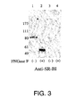
- SR-BI was confirmed to be the receptor binding HCV E2 using anti-SR-BI antibodies.
- the reactivity of the purified proteins in Western blot with antibodies against SR-BI is shown in Figure 3 .
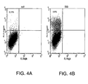
- the coding sequence for the human SR-BI was amplified from RNA of HepG2s4 cells and cloned in a vector suitable for transfection. Transfection was performed in BHK-21 recipient cells since they were negative for E2 binding. FACS analysis of cells 24 hours after transfection indicated that SR-BI transfected cells acquired binding for E2 ( Figure 4 ).
- the human SR-BI coding sequence in Example 3 was amplified with the sense primer (SEQ. ID. NO. 4) and the antisense primer (SEQ. ID. NO. 3).
- the stop codon in this construct was provided by the cloning vector 36 nucleotides after the SR-BI coding sequence, and therefore 12 amino acids were added to the carboxyl terminal of the SR-BI natural sequence.
- the coding sequence for human SR-BI was PCR amplified by using the sense primer (SEQ. ID. NO. 4) and a novel antisense primer (SEQ. ID. NO. 5).
- the new primer contains a stop codon in the primer sequence.
- the mouse SR-BI sequence was amplified from IMAGE clone BC004656 by using the sense primer (SEQ. ID. NO. 6), and the antisense primer (SEQ. ID. NO. 7).
- the human and the mouse sequences were cloned in pcDNA3 vector and clones obtained were sequenced.
- the hamster CHO cell line negative for binding to HCV E2 protein was transfected with the plasmids using lipofectamine 2000 reagent (Invitrogen). The combination of CHO cells and lipofectamine reagent gave improved transfection efficiency. Transfected cells were harvested 24 hours after transfection and analyzed by FACS, for receptor expression and E2 binding capability.
- Results demonstrate that the human and the mouse receptors, were expressed at comparable levels ( Figures 5A-C ), but only cells transfected with the human SR-BI acquired the ability to bind HCV E2 ( Figures 5D-F ). Moreover, the mouse SR-BI, showing 80% of homology at amino acid level to the human receptor, doesn't bind to HCV E2, mirroring the species specificity of HCV infection ( Figures 5D-F ).
- Example 5 A monoclonal antibody against the hypervariable region 1 (HVR1) of HCV E2 glycoprotein inhibits the E2 binding to SR-BI.
- HVR1 hypervariable region 1
- a biological relevant question concerns the ability of antibodies against the hypervariable region 1 to neutralize HCV virus.
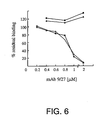
- a monoclonal antibody (9/27; Flint, et al., 2000, J. Virol., 74, 702-709 ), obtained upon immunization with E2 and reactive against the HVR1 of E2 derived from H isolate was used to inhibit E2 binding to SR-B1.
- the antibody showed a dose dependent inhibitory activity for the binding of the E2 protein from genotype 1a to HepG2 cells and to CHO cells stably transfected with SR-BI with an apparent IC 50 of about 500nM ( Figure 6 ).
- the antibody was not effective on the binding of the E2 protein derived from genotype 1b, BK strain, consistent with its lack of reactivity with this variant ( Figure 6 ).
Abstract
Description
- The references cited in the present application are not admitted to be priot art to the claimed invention.
- It is estimated that about 3% of the world's population is infected with the hepatitis C virus (HCV). (Wasley et al., Semin. Liver Dis. 20:1-16, 2000.) HCV exposure results in an overt acute disease in a small percentage of cases, while in most instances the virus establishes a chronic infection causing liver inflammation and slowly progresses into liver failure and cirrhosis. (Strader et al., ILAR J. 42:107-116, 2001.) Epidemiological surveys indicate an important role for HCV in the onset of hepatocellular carcinoma. (Strader et al., ILAR J. 42:107-116,2001.)
- HCV can be classified into a number of distinct genotypes (1 to 6), and subtypes (a to c). The distribution of the genotypes and subtypes varies both geographically and between risk groups. (Robertson et al., Arch Virol, 143:2493-2503, 1998.)
- The HCV genome consists of a single strand RNA about 9.5 kb encoding a precursor polyprotein of about 3000 amino acids. (Choo et al., Science 244:362-364, 1989, Choo et al., Science 244:359-362, 1989.) The HCV polyprotein contains the viral proteins in the order: C-E1-E2-p7-NS2-NS3-NS4A-NS4B-NS5A-NS5B. Cleavage of the precursor polyprotein results in mature structural and non-structural viral proteins. (Neddermann et al., Biol. Chem. 378:469-476, 1997.)
- As part of its infection cycle, HCV enters into a cell. The LDL receptor and CD81 molecule have been identified as putative HCV receptors. The LDL receptor has been suggested to mediate virus internalization via binding to LDL particles that are virus-associated. (Agnello et al., Proc. Natl. Acad. Sci. U.S.A. 96:12766-12771, 1999.) The CD81 molecule has been suggested to bind HCV E2 based on recombinant envelope protein E2 from HCV genotype 1a. (Pileri et al., Science 282:938-941,1998.)
- TAKIKAWA SHINGO ET AL (Journal of Virology, 74, 5066 - 5074) disclose a cell fusion assay using chimeric HCV envelope proteins. Cells expressing one or both of HCV E1 and E2 were incubated with cells known to be susceptible to HCV infection. The requirements for fusion were probed and it was determined that both E1 and E2 were needed as well as low pH environment. Mouse cells (normally not infected by HCV) were transfected with CD81 but failed to fuse with cells expressing E1 and E2 Furthermore, HepG2 cells showed the best fusion activity in the assay yet expressed low levels of CD81. Taken together, this caused the authors to postulate that additional cofactors were needed for fusion besides CD81 or that fusion could happen in a CD81 independent manner (see page 5072, paragraph spanning the columns).
- The present invention features methods of screening for compounds that inhibit HCV binding to a cell. The different methods are based on the identification of the scavenger receptor class B type I (SR-BI) as a target site for HCV E2 binding to a cell.
- Targeting the SR-BI to inhibit HCV entry into a cell can be achieved by inhibiting one or more of the following: (a) activities relating to HCV binding to SR-BI, (b) activities related to HCV internalization mediated by SR-BI, including activities downstream from SR-BI binding to HCV, and (c) activities related to functional surface expression of SR-BI.
- Thus, the present invention features a method of screening for a compound that inhibits the ability of hepatitis C virus E2 (HCV E2) polypeptide to bind to a scavenger receptor class B type I (SR-BI) comprising: a) contacting a SR-BI protein that is human or has at least 95% sequence identity to SEQ ID NO:1 with: (i) a polypeptide that binds to a site on SR-BI to which HCV E2 binds, wherein the polypeptide comprises a naturally occurring SR-BI binding region from HCV E2; and (ii) a test compound; and b) measuring binding of the polypeptide to the SR-BI protein.
- In one embodiment the SR-BI protein is present as a soluble protein. In another embodiment it is present in a membrane preparation. In a further embodiment it is expressed on a cell.
- A "compound" or "test compound" refers to a discrete chemical entity. The term compounds includes molecules of different sizes and compositions. Examples of compounds include small molecules, peptides, polypeptides, antibodies, and nucleic acid.
- The "SR-BI HCV E2 binding site" is the site to which at least the HCV E2 polypeptide from HCV 1a binds SR-BI. An example of such an HCV E2 polypeptide from HCV 1a is provided in the Examples infra. Reference to the ability of HCV 1a to bind SR-BI does not exclude binding of HCV E2 from other HCV strains to SR-BI. For example, HCV E2 from other HCV strains such as HCV 1b can bind to the naturally occurring human SR-BI HCV E2 binding site.
- SR-BI and functional derivatives of SR-BI contain a SR-BI amino acid sequence region of at least 20 contiguous amino acids as that present in SEQ. ID. NO. 1 and can bind at least HCV E2 from HCV 1a.
- SEQ. ID. NO. 1 provides a human SR-BI sequence. The presence of at least 20 contiguous amino acids as provided in SEQ. ID. NO. 1 provides a structural tag distinguishing SR-BI or a functional derivative thereof from other proteins.
- Reference to "inhibit" or "Inhibiting" indicates a detectable reduction in activity. Preferably, there is at least about a 50%, at least about 75%, or at least about 95% percent reduction in activity.
- The test compound is preincubated with SR-BI prior to adding the polypeptide that binds to the SR-BI HCV E2 binding site. Pre-incubation with a test compound is a preferred method for assaying SR-BI functional surface expression inhibitors.
- Reference to "capable of expressing" a polypeptide indicates that in the absence of an expression inhibitor, the polypeptide will be expressed in detectable amounts and has detectable activity related to HCV binding or HCV internationalization. Expression inhibitors include compounds such as antisense nucleic acid and ribozymes able to decrease activity of nucleic acid encoding for SR-BI and compounds that can modulate functional surface expression of SR-BI at the transcriptional or post-transcriptional levels.
- Compounds modulating functional surface expression at the post-transcriptional level include compounds acting on lipid rafts membrane compartments (referred to as "raft domains") to alter SR-BI activity. SR-BI activity that can be altered by such compounds include HCV binding and internalization.
- A "SR-BI E2 binding antagonist" can at least inhibit binding of a naturally occuring HCV E2 two the SR-BI of SHQ. ID. NO. 1. Preferably, the SR-BI E2 binding antagonist inhibits at least binding of HCV E2 from HCV 1a.
- Other features and advantages of the present invention are apparent from the additional descriptions provided herein including the different examples. The provided examples illustrate different components and methodology useful in practicing the present invention. The examples do not limit the claimed invention. Based on the present disclosure the skilled artisan can identify and employ other components and methodology useful for practicing the present invention.
-
-
Figures 1A and 1B illustrate immunoblot detection of HCV E2 obtained from genotype 1a (Figure 1A ) and biotinylated cell surface proteins interacting with HCV E2 (Figure 1B ). Biotinylated HepG2 cells were incubated in presence (lanes 1 and 3) or absence of HCV E2 recombinant protein (lanes 2 and 4). The bound species were cross-linked with DTSSP and the complexes were immunoprecipitated with an antibody against the His tag of the HCV E2 recombinant protein. Samples were eluted both under non-reducing condition, (lanes 1 and 2) and reducing conditions (lanes 3 and 4), that allowed the cleavage between the cross-linked molecular species, and loaded on 10% SDS-PAGE. InFigure 1A , HCV E2 protein is detected with anti-E2 rat mAb followed by anti-rat HRP conjugated as a monomer under reducing conditions (lane 3) and at higher molecular weight under non-reducing conditions (lane1). InFigure 1B , the reactivity with streptavidin HRP conjugated reveals under reducing conditions (lane 3) a biotinylated protein band at 82 kDa. -
Figure 2 illustrates silver staining of a 7.5% SDS-PAGE loaded with samples obtained after a purification step with Con-A sepharose and the deglycosylation step with the PNG-ase F enzyme. The arrows show the purified receptor migrating at 82 kDa (lane 1) before PNG-ase treatment (-) and migrating at 54 kDa (lane 2) after the deglycosylation (+). In the control samples cross-linking was performed in absence of HCV E2 (lanes 3 and 4). Fetuin was loaded on the SDS-PAGE before deglycosylation and after (lane 5 and lane 6) as a control for the PNG-ase F enzymatic activity. -
Figure 3 illustrates a Western blot analysis of the HCV E2 receptor purified on Con-A sepharose and deglycosylated with PNG-ase F (+). Rabbit anti-SR-BI polyclonal antibodies incubation was followed by anti-rabbit HRP conjugated for detection of both the glycosylated (lane 1) and deglycosylated (lane 2) receptor protein.Lanes -
Figures 4A and 4B illustrates a FACS analysis of the binding of HCV E2 recombinant protein from strain H to mock transfected (Figure 4A ) or SR-BI transfected BHK-21 cells (Figure 4B ). Dot plot analysis showed that 10% of the cells transfected with pcDNA3-SR-BI show binding for HCV E2. -
Figures 5A-F illustrate CHO cells transfected with the plasmid pcDNA3 and pcDNA3 encoding the human SR-BI or the mouse SR-BI.Figures 5A-C illustrate FACS analysis of the anti-SR-BI (Novus Biologicals NB 400-104) binding to CHO transfected cells.Figures 5D-F illustrate the analysis of the E2 protein binding to transfected cells. -
Figure 6 illustrates competition of E2 binding to HepG2 cells and CHO cells transfected with human SR-BI (CHO-SR-BI) by the anti-HVR1 mAb 9/27. Binding was detected by FACS analysis and expressed as percentage of the median fluorescence intensity values obtained in absence of competitor. Binding to HepG2 (open triangle) and CHO-SR-BI (open square) of E2 from H isolate. Binding of E2 from BK isolate to HepG2 (filled triangle) and to CHO-SR-BI (filled square). E2 recombinant proteins were used at half saturating concentration. - SR-BI is identified herein as a HCV receptor. SR-BI binding to HCV is independent from CD81. Without being limited to any particular theory, SR-BI may provide a privileged HCV entry site by mediating E2 viral glycoprotein interaction with raft domains.
- Naturally occurring SR-BI is highly expressed in the liver hepatocytes and steroidogenic tissues, and mediates the selective cellular uptake of cholesterol and phospholipids. (Acton et al., Science 271:518-520, 1996, Urban et al., J. Biol. Chem. 275:33409-33415, 2000.) SR-BI and other scavenger receptors recognize modified lipid particles both acetylated LDL and oxidized LDL. In contrast to other scavenger receptors, SR-BI also binds with high affinity to native HDL and LDL. (Acton et al., Science 271:518-520, 1996.)
- SR-BI has been located into raft domains. Rafts domains are thought to represent a specific physical state of lipid bilayer, the liquid order phase. (Brown et al., Annu. Rev. Cell. Dev. Biol. 14:11-136, 1998, van der Goot et al., Semin. Immunol. 13:89-97, 2001.) Proteins localized into raft domains are resistant to cold non-ionic detergent extraction (detergent resistant membranes). (London et al., Biochim. Biophys Acta. 1508:182-195, 2000.)
- Proteins localized in raft domains are either GPI membrane anchored or fatty acylated. (Brown et al., Annu. Rev. Cell. Dev Biol. 14:11-136, 1998.) SR-BI is a fatty acylated protein. (Babitt et al., J. Biol. Chem. 272:13242-13249, 1997.)
- Rafts domains may represent a preferential entry site for pathogens providing them a way to escape from the classical degradative pathway. (van der Goot et al., Semin. Immunol. 13:89-97, 2001). Examples of pathogens that may enter a cell by targeting raft domain components include SV40, echoviruses, HIV, and HTLV-1. (Bergelson et al., Proc. Natl. Acad. Sci. U.S.A. 91:6245-6249, 1994, Manes et al., EMBO Rep. 1:190-196, 2000, Niyogi et al., J. Virol. 75:7351-7361, 2001, Parton et al., Immunol. Rev. 168:23-31, 1999, van der Goot et al., Semin. Immunol. 13:89-97, 2001.)
- The identification of SR-BI as a site for HCV E2 binding provides a target that can be modulated to study the HCV infection cycle and to inhibit HCV replication or infection. The ability of a test compound to modulate the interaction between SR-BI and HCV E2 can be performed for example, using assays employing a naturally occurring SR-BI or derivative thereof that binds HCV E2, a compound that binds to the SR-BI HCV E2 binding site, and the test compound.
- Test compounds found to inhibit HCV E2 interaction with SR-BI can be used, for example, to study the effect of modulating SR-BI HCV E2 interaction on HCV replication or infection. Those test compounds having appropriate pharmacological properties such as efficacy and lack of unacceptable toxicity may be used to treat or inhibit HCV infection in a patient.
- SR-BI and sequences having at least 95% identity to SEQ ID NO:1 used to screen for modulators of SR-BI interaction with HCV E2 can bind at least HCV E2 from HCV 1a. SR-BI and sequences having at least 95% identity to SR-BI contain a SR-BI amino acid sequence region of at least 20 contiguous amino acids as that present in SEQ. ID. NO. 1. The presence of at least 20 contiguous amino acids as provided in SEQ. ID. NO. 1 provides a tag distinguishing SR-BI from other proteins.
- SR-BI can be obtained from mammalian sources such as human, hamster, mouse and rat. The ability of SR-BI obtained from a particular source to bind HCV E2 can be confirmed using techniques such as those described in the
- Examples infra. Examples of naturally occurring SR-BI amino acid and nucleic acid sequences are provided for by SEQ. ID. NO. 1, SEQ. ID. NO. 2, and in references such as
Acton U.S. Patent No. 5,998,141 , Calvo et al., J. Biol. Chem. 268:18929-18935, 1993, Acton et al., J. Biol, Chem. 269:21003-21009, 1994, Cao et al., J. Biol. Chem, 272:33068-33076, 1997, and Webb et al., J. Biol. Chem. 24:15241-15248, 1998. - Human SR-BI is also referred to in the literature as CLA-1. Splice variants or isoforms, and different polymorphic forms of SR-BI that bind to HCV E2 are included within the definition of SR-BI by reference to the presence of an at least 20 amino acid tag.
- Based on SR-BI sequences known in the art, additional naturally occurring SR-BI encoding nucleic acid, preferably of human origin, can be obtained. Cloning techniques well known in the art, such as those employing probes, primers, and degenerative probes and primers, can be used to clone SR-BI.
- Sets of degenerative probes and primers can be produced taking into account the degeneracy of the genetic code. Hybridization conditions can be selected to control probe or primer specificity to allow for hybridization to nucleic acids having similar sequences.
- Techniques employed for hybridization detection and PCR cloning are well known in the art. Nucleic acid detection techniques are described, for example, in Sambrook, et al., Molecular Cloning, A Laboratory Manual, 2nd Edition, Cold Spring Harbor Laboratory Press, 1989. PCR cloning techniques are described, for example, in White, Methods in Molecular Cloning, volume 67, Humana Press, 1997.
- A naturally occuring SR-BI that binds HCV E2 can be used to produce functional variants. Variants include naturally occuring SR-BI with one or more amino acid alterations. Amino acid alterations are substitutions, editions and deletions. SR-BI activity, such as binding to HCV, SR-BI functional expression, and HCV internalization can be measured based on the guidance described herein.
- Differences in naturally occurring amino acids R groups may be taken into account in designing variants. An R group affects different properties of an amino acid such as physical size, charge, and hydrophobicity. Amine acids can be divided into different groups as follows: neutral and hydrophobic (alanine, valine, leucine, isoleucine, proline, tyrptophan, phenylalanine, and methionine); neutral and polar (glycine, serine, threonine, tryosine, cysteine, asparagine, and glutamine); basic (lysine, arginine, and histidine); and acidic (aspartic acid and glutamic acid).
- Generally, in substituting different amino acids it is preferable to exchange amino acids having similar properties. Substituting different amino acids within a particular group, such as substituting valine for leucine, arginine for lysine, and asparagine for glutamine are good candidates for not causing a change in polypeptide functioning.
- Changes outside of different amino acid groups can also be made. Preferably, such changes are made taking into account the position of the amino acid to be substituted in the polypeptide. For example, arginine can substitute more freely for nonpolar amino acids in the interior of a polypeptide than glutamate because of its long aliphatic side chain. (See, Ausubel, Current Protocols in Molecular Biology, John Wiley, 1987-1998, Supplement 33 Appendix 1C.)
- SEQ. ID. NO. 1 provides a reference sequence for SR-BI including sequences that have at least 95% sequence identity.
- Amino acid sequence identity can be determined by methods well known in the art that compare the amino acid sequence of one polypeptide to the amino acid sequence of a second polypeptide and generate a sequence alignment. Amino acid identity can be calculated from the alignment by counting the number of aligned residue pairs that have identical amino acids.
- Methods for determining sequence identity include those described by Schuler, G.D. in Bioinformatics: A Practical Guide to the Analysis of Genes and Proteins, Baxevanis, A.D. and Ouelette, B.F.F., eds., John Wiley & Sons, Inc, 2001; Yona et al., in Bioinformatics: Sequence, Structure and Databanks, Higgins, D. and Taylor, W. eds., Oxford University Press, 2000; and Bioinformatics: Sequence and Genome Analysis, Mount, D.W., ed., Cold Spring Harbor Laboratory Press, 2001). Methods to determine amino acid sequence identity are codified in publicly available computer programs such as GAP (Wisconsin Package Version 10.2, Genetics Computer Group (GCG), Madison, Wisc.), BLAST (Altschul et al., J. Mol. Biol. 215(3):403-10, 1990), and FASTA (Pearson, Methods in Enzymology 183:63-98, 1990, R.F. Doolittle, ed.).
- Further disclosed is a sequence identity between two polypeptides is determined using the GAP program (Wisconsin Package Version 10.2, Genetics Computer Group (GCG), Madison, Wisc.). GAP uses the alignment method of Needleman and Wunsch. (Needleman et al., J. Mol. Biol. 48:443-453, 1970.) GAP considers all possible alignments and gap positions between two sequences and creates a global alignment that maximizes the number of matched residues and minimizes the number and size of gaps. A scoring matrix is used to assign values for symbol matches. In addition, a gap creation penalty and a gap extension penalty are required to limit the insertion of gaps into the alignment. Default program parameters for polypeptide comparisons using GAP are the BLOSUM62 (Henikoff et al., Proc. Natl. Acad. Sci. USA, 89:10915-10919, 1992) amino acid scoring matrix (MATrix=blosum62.cmp), a gap creation parameter (GAPweight=S), and a gap extension pararameter (LENgthweight=2).
- Polypeptides capable of binding to the SR-BI HCV E2 binding site contain a region able bind to the same site as HCV E2. The polypeptide region that binds to SR-BI includes naturally occurring E2 regions or binding fragments thereof, derivatives of such E2 regions or binding fragments thereof, and E2 mimotopes.
- Polypeptides capable of binding to the SR-BI HCV E2 binding site can also contain regions not involved in SR-BI binding. Non-binding regions, if present, do not prevent the polypeptide from binding to at least the human SR-BI of SEQ. ID. NO. 1. Preferred non-binding regions are additional HCV regions and regions that facilitate detection of binding.
- Regions facilitating detection of binding include detectable labels and regions that can be detected. Examples of detectable labels include moieties such as radiolabels, luminescent molecules, haptens and enzyme substrates. Examples of regions that can be detected include regions that provide an epitope for antibody binding or that provide a specific binding region for other types of compounds.
- HCV E2 binding mimotopes have a primary structure unrelated to HCV E2, but share binding characteristics with HCV E2. References describing techniques for producing mimotopes in general and describing different HCV E2 mimotopes include
Felici et al., U.S. Patent No. 5,994,083 andNicosia et al., International Application Number WO 99/60132 - The ability of a polypeptide to bind to the SR-BI E2 binding site can be determined, for example, by competition experiments with a HCV E2 polypeptide already shown to bind to SR-BI. Such experiments can be performed employing a polypeptide that may bind to the SR-BI HCV E2 binding site as a test compound.
- Screening for compounds inhibiting HCV binding to SR-BI is facilitated using recombinant nucleic acid expressing SR-BI or a sequence having at least 95% identity to SEQ ID NO:1. Recombinantly expressed receptors offers several advantages in screening for compounds active at a polypeptide, such as the ability to express the polypeptide in a cell having little or no endogenous expression of the polypeptide and using the same cell without recombinantly expressed polypeptide as a control. For example, SR-BI can be expressed in BHK-21 or CHO cells using an expression vector, wherein the same cell line without the expression vector can act as a control. Additional cell lines lacking SR-BI expression can be identified using techniques such as those employing SR-BI antibodies or those measuring SR-BI RNA.
- A recombinant "nucleic acid" refers to an artificial combination of two or more nucleotide sequence regions. The artificial combination is not found in nature. Recombinant nucleic acid includes nucleic acid having a first coding region and a regulatory element or a second coding region not naturally associated with the first coding region. The recombinant nucleotide sequence can be present in a cellular genome or can be part of an expression vector.
- Preferably, expression is achieved in a host cell using an expression vector. An expression vector contains recombinant nucleic acid encoding a polypeptide along with regulatory elements for proper transcription and processing. The regulatory elements that may be present include those naturally associated with the nucleotide sequence encoding the polypeptide and exogenous regulatory elements not naturally associated with the nucleotide sequence.
- Generally, the regulatory elements that are present in an expression vector include a transcriptional promoter, a ribosome binding site, a terminator, and an optionally present operator. Another preferred element is a polyadenylation signal providing for processing in eukaryotic cells. Preferably, an expression vector also contains an origin of replication for autonomous replication in a host cell, a selectable marker, a limited number of useful restriction enzyme sites, and a potential for high copy number. Examples of expression vectors are cloning vectors, modified cloning vectors, specifically designed plasmids and viruses.
- An alternative means to produce recombinant nucleic acid is by altering the cellular genome. One type of alteration that can increase cellular expression is the use of a strong promoter such as the immediate early human cytomegalovirus promoter. Alterations to the cellular genome can be performed, for example, using techniques described by Ausubel, Current Protocols in Molecular Biology, John Wiley, 1987-1998.
- Starting with a particular amino acid sequence and the known degeneracy of the genetic code, a large number of different encoding nucleic acid sequences can be obtained. The degeneracy of the genetic code arises because almost all amino acids are encoded by different combinations of nucleotide triplets or "codons". Amino acids are encoded by codons as follows:
- A=Ala=Alanine: codons GCA, GCC, GCG, GCU
- C=Cys=Cysteine: codons UGC, UGU
- D=Asp=Aspartic acid: codons GAC, GAU
- E=Glu=Glutamic acid: codons GAA, GAG
- F=Phe=Phenylalanine: codons UUC, UUU
- G=Gly=Glycine: codons GGA, GGC, GGG, GGU
- H=His=Histidine: codons CAC, CAU
- I=Ile=Isoleucine: codons AUA, AUC, AUU
- K=Lys=Lysine: codons AAA, AAG
- L=Leu=Leucine: codons UUA, UUG, CUA, CUC, CUG, CUU
- M=Met=Methionine: codon AUG
- N=Asn=Asparagine: codons AAC, AAU
- P=Pro=Proline: codons CCA, CCC, CCG, CCU
- Q=Gln=Glutamine: codons CAA, CAG
- R=Arg=Arginine: codons AGA, AGG, CGA, CGC, CGG, CGU
- S=Ser=Serine: codons AGC, AGU, UCA, UCC, UCG, UCU
- T=Thr=Threonine: codons ACA, ACC, ACG, ACU
- V=Val=Valine: codons GUA, GUC, GUG, GUU
- W=Trp=Tryptophan: codon UGG
- Y=Tyr=Tyrosine: codons UAC, UAU
- Nucleic acid having a desired sequence can be synthesized using chemical and biochemical techniques. Examples of chemical techniques are described in Ausubel, Current Protocols in Molecular Biology, John Wiley, 1987-1998, and Sambrook et al., Molecular Cloning, A Laboratory Manual, 2nd Edition, Cold Spring Harbor Laboratory Press, 1989.
- Biochemical synthesis techniques involve the use of a nucleic acid template and appropriate enzymes such as DNA and/or RNA polymerases. Examples of such techniques include in vitro amplification techniques such as PCR and transcription based amplification, and in vivo nucleic acid replication. Examples of suitable techniques are provided by Ausubel, Current Protocols in Molecular Biology, John Wiley, 1987-1998, Sambrook et al., Molecular Cloning, A Laboratory Manual, 2nd Edition, Cold Spring Harbor Laboratory Press, 1989, and
Kacian et al., U.S. Patent No. 5,480,784 . - A variety of different assay formats may be employed to measure activities relating to HCV binding to SR-BI or sequences having at least 95% identity to SEQ ID NO:1. Depending on the activity being assayed, assays can be performed using whole cells, membrane preparations, and purified SR-BI.
- Techniques for detecting binding to SR-BI include those employing a HCV E2 polypeptide containing a detectable label or region, and those involving the use of secondary antibodies to a region distinct from the SR-BI HCV E2 binding region. Assay formats that may be employed include FACS analysis, a scintillation proximity assay ("SPA"), and sandwich type assays where a detectable antibody is targeted to a region distinct from the SR-BI HCV E2 binding region. An example of techniques that can be employed for a FACS analysis are provided in Example 1 infra.
- Techniques for performing a SPA are well known in the art. SPA can be performed using a bead or a plate coated with a scintillant fluid and a radiolabeled molecule. Proximity of the radiolabeled molecule to the scintillant fluid stimulates light emission. SPA can be used to measure binding to SR-BI by, for example, Joining SR-BI to a SPA bead or growing cells expressing SR-BI on a SPA plate (cytostar), radiolabeling a HCV E2 polypeptide, and measuring the ability of a test compound to inhibit light production from the radiolabeled polypeptide.
- Antibodies binding to a region distinct from the SR-BI HCV E2 binding site can be employed to detect binding using capture assays formats. For example, cell membranes expressing SR-BI or purified SR-BI protein are attached to a solid support and a HCV E2 polypeptide is added to the solid support in presence of the test compound, the solid support is washed, and the presence of E2 on the support is determined using an antibody with a detectable label that binds to E2.
- Examples are provided below to further illustrate different features of the present invention. The examples also illustrate useful methodology for practicing the invention. These examples do not limit the claimed invention.
- This example describes different materials and method that were employed to study HCV E2 binding to SR-BI.
- Molt-4 (human T-cell leukaemia) cells were obtained from the MRC ADP Repository. HepG2 (human hepatoma), HEK-293 (human embryonic kidney) BHK-21 (baby hamster kidney) and CHO (Chinese hamster ovary) cell lines were obtained from the ATCC Repository. Cells were grown under standard conditions in presence of 10% fetal calf serum.
- HCV E2 protein representative of genotype 1a (strain H) and genotype 1b (strain BK) were cloned into VIJnsTPA as described by Meola et al., J. Virol 74:5933-5938, 2000. The cloned E2 fragment contains the coding sequence for E2 from amino acid 384 to amino acid 661 of the HCV polyprotein and a tag of 6 His at the C-terminal. 293 cells were transfected by calcium phosphate method. Recombinant proteins produced by transfected HEK 293 cells were collected from culture supernatant harvested 48 hours after transfection, concentrated 40 times using filter devices (Millipore Centricon® Plus-80) and supplemented with protease inhibitor cocktail tablets (Boehringer Mannheim) and 10% glycerol. The amount of E2 in the extracts was quantified by using a GNA (lectin from Galanthus nivalis) capture assay as described by Flint et. al. J. Virol. 73:6782-6790, 1999.
- Cells were washed twice in phosphate-buffered
saline 1% FCS, 0.05% sodium azide (washing buffer). Then, 2x10⁵ cells were allowed to bind at room temperature for 1 hour with E2 concentrated supernatants or, as control, with the concentrated supernatant of HEK 293 cells transfected with VIJnsTPA plasmid (mock). After one wash in washing buffer the anti-His mouse monoclonal antibody (mAb, Quiagen) was added at the concentration of 2 µg/ml 1 hour at room temperature. Binding to anti-CD81 was also performed at room temperature for 1 hour by using the mouse monoclonal antibody (1.3.3.22 Santa Cruz Biotechnology) diluted 1:200. - Cell bound mAbs were visualized with anti-mouse IgG1-phycoerythrin (PE) conjugate (Serotec). Flow cytometry data acquisition was perform using a Becton-Dickinson FACS Vantage® flow cytometer. Dead cells were detected as Sytox® Green dye (Molecular Probes®) stained positive and were excluded from analyses.
- HepG2 cells were incubated with a saturating concentration of 1a-derived E2 recombinant protein for 2 hours at room temperature. The E2 bound protein was revealed as described above and analyzed in a FACS Vantage® (Becton Dickinson) flow cytometer. The cells that showed the highest fluorescence intensity were sorted and expanded. The procedure was repeated four times.
- Dynabeads® M-450 rat anti-mouse IgG1, (Dynal, Oxoid) were washed once with PBS 0.1% BSA before the addition of 15 µg of the anti-His Mab in 4 ml volume of PBS 0.1% BSA. After 1 hour of coating, the beads were washed twice with PBS 0.1% BSA and once with 0.2 M triethanolamine pH 9. To cross-link the bound antibody dimethylpimlimidate dihydrochlorite (DMP) was added at a 20 mM concentration to the Dynabeads® in 10 ml volume of 0.2 M triethanolamine pH 9. The reaction was stopped by adding 0.2 M Tris HCl at pH 8. Beads were washed first with PBS 0.5% Triton and then with PBS 0.1% BSA. Ones prepared the Dynabeads® were stored at 4°C in presence of 0.1% sodium azide.
- HepG2s4 cells (1x10⁸) were harvested by trypsinization and washed once with cold PUBS in 250 ml volume. Cells were incubated in the dark for 15 minutes with a freshly prepared solution of 2 mM sodium periodate at the concentration of 1x10⁷ cell/ml. After this mild oxidation, cells were washed twice and incubated with 5 mM biotin-LC hydrazide for 10 minutes at room temperature at the concentration of 5x10⁷ cell/ml. Biotin-LC hydrazide was freshly prepared by solubilization in DMSO at a 50 mM concentration.
- After biotinylation two washes in 250 ml volume of cold PBS were performed, and cells were incubated with the concentrated supernatant containing E2 recombinant protein or as a control with the supernatant obtained from the mock transfection. Incubation with E2 lasted 1 hour at room temperature. Staining of bound E2 was performed as described above and analyzed by FACS.
- DTSSP (dithiobis-sulfosuccinimidylpropionate, Pierce,) was used as a cross linker. DTSSP is a water soluble cross-linker homobifunctional N-hydroxysuccinimide ester and is thiol cleavable.
- HepG2s4 cells (5x10⁷ cell/ml) were washed once after binding to E2 and incubated with the DTSSP at 2 mM concentration in PBS for 20 minutes at room temperature. The reaction was stopped by incubation with Tris HCl 50 mM at pH 7.5. Cells were lysed in
PBS 1% Triton in the presence of protease inhibitor cocktail (Boehringer Mannheim) for 20 minutes at 37°C. E2-receptor complexes were immunoprecipitated with the anti-His Dynabeads® by incubation overnight at 4°C. After 5 washes inPBS 1% Triton® elution was performed either boiling directly the beads in sodium dodecyl sulfate (SDS) sample buffer or incubating beads 30 minutes at 37°C in 50 mM dithiothreitol (DTT), 50 mM NaCl, 50 mM Tris HCl pH 8. - Samples obtained from boiling the beads were loaded on SDS-Page and analyzed by Western blot for detection of biotinylated proteins using streptavidin horseradish peroxidase conjugated (HRP, Pierce) diluted 1:25,000 in
Tris Buffer Saline 0,05% Tween 20 (TBST), 2% bovine serum albumin (BSA). For detecting recombinant E2 protein of genotype la the rat monoclonal antibody 6-1/a (Flint et al., J. Virol, 73:6782-6790,1990) was used diluted 1:50 inTBST 5% low fat milk, followed by incubation with anti-rat HARP (Dako) conjugated 1:2000. The chemiluminescent Super Signal West Pico (Pierce) substrate was used and immunoreactive proteins were detected by exposure on X ray film (Kodak Biomax ML). - Eluates obtained from the immunoprecipitation on with anti-His Dynabeads® were diluted 1:2 with 1 M NaCl, 0.2% Triton and incubated with Con-A beads for two hours at room temperature. After three washes in incubation buffer, elution was performed under denaturing conditions in 1% SDS. 1% β-mercaptoethanol and 100 mM phosphate pH 7.5 at 95°C for 10 minutes. The eluates were diluted 1:10 in the PNGase F incubation buffer containing 0.1% SDS, 0.5% NP40, 10 mM EDTA, and 100 mM Na phosphate pH7.5. Eluates were divided into aliquots one of which was treated with the enzyme PNGase F (Bio-rad) for 3 hours at 37°C.
- Samples were loaded on 7.5% SDS pre-cast gel (Bio-rad) and silver stained. Immunoreactivity of the purified samples was probed in Western blot by using rabbit anti-SR-BI purified polyclonal antibodies (Novus Biologicals NB 400-104) diluted 1:1500 in
TBST 2% BSA followed by anti-rabbit HRP (Pierce) diluted 1: 20.000. The chemiluminescent subsrate Super Signal West Pico (Pierce) was used for detection. - RNA was extracted from HepG2s4 cells with TRIzol® reagent (Gibco® BRL) following manufacturer's instructions. First-strand cDNA was produced mixing total RNA (2 µg) with 10 pmol of the antisense primer SR-BI 5'-CCAGTCTAGACAGTTTTGCTTCCTGCAGCACACAGAGCCC-3' (SEQ. ID. NO. 3). The restriction site for XbaI is in italics. The reaction was performed by using Superscript® II reverse transcriptase (Gibco® BRL) as described by Meola et al., J. Virol. 74;5933-5938, 2000.
- An aliquot of the cDNA was amplified by PCR using Platinum Pfx DNA polymerase (Gibco® BRL) following manufacturer's instructions. The primers used were the antisense SR-BI primer and the sense SR-BI 5'-AGGCAAGCTTGCCGCCATGGGTGCTCCGCCAAAGCGCGCTGGG-3', (SEQ. ID. NO. 4) where the restriction site for Hind III is in italics, PCR was performed in a Perkin-Elmer 2400 thermocycler, denaturing samples for 4 minutes at 94°C and then running 35 cycles of incubation at 94°C for 30 seconds, at 50°C for 30 seconds, and 68° for 2 minutes.
- The PCR product was digested with the restriction enzymes Hind III and Xba I for directional cloning in the vector pcDNA3. Clones were sequenced with the Big Dye Terminator Cycle Sequencing Kit using AmpliTaq® (Applied Biosystems) and an Applied Biosystem Model 373A Sequencer. Sequences were analyzed by the Vector NTI program.
- The BHK-21 cell line was used as recipient for transient transfection. Transfection was performed by mixing plasmid DNA with the
FuGENE ® 6 transfection reagent (Roche). Cells were harvested 24 hours after transfection and analyzed by FACS for E2 binding. - SR-BI was identified as an E2 receptor by examining the ability of E2 to bind to HepG2 cells, enriching for HepG2 cells having increased ability to bind E2, and determining that SR-BI binds E2. HepG2 cells were used to search for the E2 receptor because they were found to lack CD81 and retain the ability to bind HCV E2.
- The ability of HepG2 cells to bind E2 independent of CD81 was determined using a FACS analysis. The absence of the CD81 molecule from HepG2 Cells was determined using an anti-CD81 mouse monoclonal antibody, and a secondary antibody anti-mouse IgG1-phycoerythrin conjugate, Binding of recombinant E2 proteins derived from genotype 1a and 1b to cells was detected by antibodies reactive against a 6-His tag present in the recombinant proteins followed by incubation with an anti-mouse IgG1-phycoerythrin conjugate.
- FACS analysis for CD81 expression was performed using HepG2 cells and Moli-4 cells as described in Example 1. HepG2 cells were negative for CD81 expression. In contrast, Molt-4 cells showed high levels of CD81 on the cell surface.
- FACS analysis for E2 binding was performed using HepG2 cells and Molt-4 cells as described in Example 1. Both cell lines tested showed binding to recombinant E2 derived from genotype 1a. However, the E2 recombinant glycoprotein derived from 1b genotype showed a much reduced binding to Molt-4, cells, while the binding to HepG2 cells was comparable to that obtained with 1a derived E2 recombinant protein.
- Enrichment of HepG2 cells expressing high levels of the E2 receptor was achieved with four subsequent rounds of sorting. A subpopulation of HepG2 cells "HepG2s4" showed upon binding to E2, a mean
fluorescence intensity value - Surface labeling of HepG2s4 was performed using a biotin-LC-hydrazide reagent. (Kahne et al., J. Immunol. Methods. 168; 209-21-8, 1994.) The hydrazides are reactive with the aldehyde groups obtained by mild oxidation with sodium periodate of hydroxyl groups of the glycoprotein carbohydrate moieties. This biotinylation method is compatible with the subsequent step of cross-linking involving a primary amine as a target for a NHS (N-hydroxisuccinimide) ester cross-linker.
- Biotinylated HepG2s4 cells were incubated with the E2 glycoprotein 1a. The reactivity of E2 to the putative receptor was unaffected by the biotinylation procedure as detected by flow cytometric analysis after staining of bound E2.
- Cross-linking was performed after the E2 binding by addition of the DTSSP cross-Linker, that is cleavable by thiol. Cells were finally lysed in
PBS 1% Triton® and the E2-receptor complexes were immunoprecipitated under non-reducing conditions by Dynabeads® conjugated with an antibody reactive against the His tag of recombinant E2. The experiment was run in parallel with a control sample where the biotinylated HepG2s4 cells were incubated with the concentrated supernatant from the mock transfected 293 cells before the cross-linking. - Immunoprecipitated samples were eluted directly in sample buffer both under reducing and non-reducing conditions and loaded on SDS-page. Immunoblot was performed by using an anti-E2 rat monoclonal antibody, 6/1a, specific for genotype 1a protein (Flint et al, J. Virol, 73:6782-6790, 1990), followed by an anti-rat secondary antibody conjugated with peroxidase for enhanced chemiluminescence detection.
- E2 protein was detected as a diffuse hand of the expected molecular weight for the monomer species under reducing condition. Under non-reducing conditions most of E2 protein was detected at higher molecular weight probably representing the E2 receptor-complexes and additional aggregated forms of E2 (
Fig. 1A ). - For detection of biotinylated species Western blot were developed with streptavidin peroxidase conjugate. By thiol cleavage of the E2/receptor complexes, it was possible to detect only under reducing condition a predominant biotinylated band with an apparent molecular weight of 82 kDa, probably corresponding to the biotinylated receptor (
Fig. 1B ). - The cross-liking experiment indicated that the receptor involved in E2 binding was an 82 kd glycoprotein, since only glycoproteins could have been biotinylated by the biotin hydrazide reagent. The protein involved in E2 binding was identified by performing cross-linking in the absence of the surface labeling step with biotin due to cellular toxicity associated with sodium periodate. Treatment with sodium periodate was quite toxic for the cells increasing significantly the number of cells lost before binding,
- Cells were harvested (6x10⁸ cells) to perform binding to E2 followed by cross-linking. The E2/receptor complexes were purified from the lysate with anti-His Dynabeads® as described in Example 1, and eluting the receptor molecule from the complex by incubation in 50 mM DTT at 37°C for 30 minutes, The analysis of the eluate on SDS page by silver staining indicated that several molecular species were elated together with the putative receptor.
- A second purification step was performed using Concanavalin A lectin (Con-A). Con-A is a commonly used lectin for purification of glycoproteins that binds Asn-linked glycans. Samples eluted from the anti-His Dynabeads® were incubated with Con-A sepharose for a second immunoprecipitation.
- After several washes in the incubation buffer, elution from the Con-A beads was performed under denaturing condition compatible with the enzymatic activity of PNGase F. PNGas F is an enzyme that releases all Asn-linked oligosaccharides from the glycoproteins.
- The eluted sample was enzymatically deglycosylated by using the PNGase F enzyme. Analysis of samples on silver stained gel visualized the glycosylated receptor band with an apparent molecular weight of 82 kDa and the deglycosylated form migrating at 54 kDa compatible with the presence of 10 potential Asn glycosylation sites (
Figure 2 ). - Based on preliminary data, SR-BI was suspected of being an HCV receptor. Preliminary data on inhibition of the E2 binding to HepG2 cells with β-cyclodextrins suggested that cholesterol may play a role in the observed binding. β-Cyclodextrins can selectively remove cholesterol from cell membranes. (Yancey et al., J. Biol. Chem. 271:16026-16034, 1996.) Additionally, the migration pattern on SDS page of the glycosylated and deglycosylated receptor were very similar to that for SR-BI.
- SR-BI was confirmed to be the receptor binding HCV E2 using anti-SR-BI antibodies. The reactivity of the purified proteins in Western blot with antibodies against SR-BI is shown in
Figure 3 . - The coding sequence for the human SR-BI was amplified from RNA of HepG2s4 cells and cloned in a vector suitable for transfection. Transfection was performed in BHK-21 recipient cells since they were negative for E2 binding. FACS analysis of cells 24 hours after transfection indicated that SR-BI transfected cells acquired binding for E2 (
Figure 4 ). - The human SR-BI coding sequence in Example 3 was amplified with the sense primer (SEQ. ID. NO. 4) and the antisense primer (SEQ. ID. NO. 3). The stop codon in this construct was provided by the cloning vector 36 nucleotides after the SR-BI coding sequence, and therefore 12 amino acids were added to the carboxyl terminal of the SR-BI natural sequence. To obtain the human SR-BI protein of its natural size ending with the leu 509 (SEQ. ID. NO. 1), the coding sequence for human SR-BI was PCR amplified by using the sense primer (SEQ. ID. NO. 4) and a novel antisense primer (SEQ. ID. NO. 5). The new primer contains a stop codon in the primer sequence.
- The mouse SR-BI sequence was amplified from IMAGE clone BC004656 by using the sense primer (SEQ. ID. NO. 6), and the antisense primer (SEQ. ID. NO. 7). The human and the mouse sequences were cloned in pcDNA3 vector and clones obtained were sequenced.
- The hamster CHO cell line negative for binding to HCV E2 protein was transfected with the plasmids using lipofectamine 2000 reagent (Invitrogen). The combination of CHO cells and lipofectamine reagent gave improved transfection efficiency. Transfected cells were harvested 24 hours after transfection and analyzed by FACS, for receptor expression and E2 binding capability.
- Results demonstrate that the human and the mouse receptors, were expressed at comparable levels (
Figures 5A-C ), but only cells transfected with the human SR-BI acquired the ability to bind HCV E2 (Figures 5D-F ). Moreover, the mouse SR-BI, showing 80% of homology at amino acid level to the human receptor, doesn't bind to HCV E2, mirroring the species specificity of HCV infection (Figures 5D-F ). - A biological relevant question concerns the ability of antibodies against the
hypervariable region 1 to neutralize HCV virus. A monoclonal antibody (9/27; Flint, et al., 2000, J. Virol., 74, 702-709), obtained upon immunization with E2 and reactive against the HVR1 of E2 derived from H isolate was used to inhibit E2 binding to SR-B1. The antibody showed a dose dependent inhibitory activity for the binding of the E2 protein from genotype 1a to HepG2 cells and to CHO cells stably transfected with SR-BI with an apparent IC50 of about 500nM (Figure 6 ). The antibody was not effective on the binding of the E2 protein derived from genotype 1b, BK strain, consistent with its lack of reactivity with this variant (Figure 6 ). -
- <110> Instituto Di Ricerche Di Biologia Molecolare P. Angeletti
- <120> SCREENING FOR HEPATITIS C VIRUS ENTRY INHIBITORS
- <130> ITR0038Y PCT
- <150>
60/344,504
<151> 2001-11-09 - <160> 7
- <170> FastSEQ for Windows Version 4.0
- <210> 1
<211> 509
<212> PRT
<213> Human - <400> 1


- <210> 2
<211> 1527
<212> DNA
<213> Human - <400> 2


- <210> 3
<211> 38
<212> DNA
<213> Artificial Sequence - <220>
<223> Primer - <400> 3
ccagtctaga cagttttgct tcctgcagca cagagccc 38 - <210> 4
<211> 44
<212> DNA
<213> Artificial Sequence - <220>
<223> Primer - <400> 4
aggcaagctt gccgccatgg gctgctccgc caaagcgcgc tggg 44 - <210> 5
<211> 41
<212> DNA
<213> Artificial Sequence - <220>
<223> Primer - <400> 5
ccagtctaga ctacagtttt gcttcctgca gcacagagcc c 41 - <210> 6
<211> 44
<212> DNA
<213> Artificial Sequence - <220>
<223> Primer - <400> 6
aggcaagctt gccgccatgg gcggcagctc cagggcgcgc tggg 44 - <210> 7
<211> 41
<212> DNA
<213> Artificial Sequence - <220>
<223> Primer - <400> 7
ccagtctaga ctatagcttg gcttcttgca gcaccgtgcc c 41
Claims (4)
- A method of screening for a compound that inhibits the ability of hepatitis C virus E2 (HCV E2) polypeptide to bind to a scavenger receptor class B type I (SR-BI) comprising:a) contacting a SR-BI protein that is human or has at least 95% sequence identity to SEQ ID NO: 1 with:(i) a polypeptide that binds to a site on SR-BI to which HCV E2 binds, wherein the polypeptide comprises a naturally occurring SR-BI binding region from HCV E2; and(ii) a test compound; andb) measuring binding of the polypeptide to the SR-BI protein.
- The method of claim 1 wherein the SR-BI protein is present as a soluble protein.
- The method of claim 1 wherein the SR-BI protein is present in a membrane preparation.
- The method of claim 1 wherein the SR-BI protein is expressed on a cell.
Applications Claiming Priority (3)
| Application Number | Priority Date | Filing Date | Title |
|---|---|---|---|
| US34450401P | 2001-11-09 | 2001-11-09 | |
| US344504P | 2001-11-09 | ||
| PCT/EP2002/012272 WO2003040726A2 (en) | 2001-11-09 | 2002-11-01 | Screening for hepatitis c virus entry inhibitors |
Publications (2)
| Publication Number | Publication Date |
|---|---|
| EP1444523A2 EP1444523A2 (en) | 2004-08-11 |
| EP1444523B1 true EP1444523B1 (en) | 2010-12-15 |
Family
ID=23350803
Family Applications (1)
| Application Number | Title | Priority Date | Filing Date |
|---|---|---|---|
| EP02802643A Expired - Lifetime EP1444523B1 (en) | 2001-11-09 | 2002-11-01 | Screening for hepatitis c virus entry inhibitors |
Country Status (7)
| Country | Link |
|---|---|
| US (1) | US20050019751A1 (en) |
| EP (1) | EP1444523B1 (en) |
| JP (1) | JP2005509160A (en) |
| AT (1) | ATE491952T1 (en) |
| CA (1) | CA2465731A1 (en) |
| DE (1) | DE60238633D1 (en) |
| WO (1) | WO2003040726A2 (en) |
Families Citing this family (10)
| Publication number | Priority date | Publication date | Assignee | Title |
|---|---|---|---|---|
| GB0315248D0 (en) * | 2003-06-30 | 2003-08-06 | Hoffmann La Roche | HCV regulated protein expression |
| WO2006005465A1 (en) * | 2004-07-08 | 2006-01-19 | Istituto Di Ricerche Di Biologia Molecolare P Angeletti Spa | Antigen binding proteins directed against scavenger receptor b1 that inhibit hcv replication |
| WO2006103493A1 (en) * | 2005-03-29 | 2006-10-05 | Epixis | Methods for enhancing the potency of hcv neutralizing antibodies |
| EP1991215A1 (en) * | 2006-03-09 | 2008-11-19 | Cenix Bioscience GmbH | Use of inhibitors of scavenger receptor class proteins for the treatment of infectious diseases |
| EP1832283A1 (en) * | 2006-03-09 | 2007-09-12 | Cenix Bioscience GmbH | Use of inhibitors of scavenger receptor class proteins for the treatment of infectious diseases |
| WO2008013918A2 (en) * | 2006-07-26 | 2008-01-31 | Myelin Repair Foundation, Inc. | Cell cycle regulation and differentiation |
| WO2010039649A2 (en) * | 2008-10-03 | 2010-04-08 | The United States Of America, As Represented By The Secretary, Department Of Health & Human Services | Flavivirus-based system for production of hepatitis c virus (hcv) |
| EP3374338A1 (en) * | 2015-11-12 | 2018-09-19 | SABIC Global Technologies B.V. | Methods for producing aromatics and olefins |
| CN112121168B (en) * | 2020-04-09 | 2022-03-04 | 中国人民解放军军事科学院军事医学研究院 | Application of inhibitor in preparing medicine for treating SARS-CoV-2 pneumonia and its complication |
| WO2024052379A1 (en) * | 2022-09-07 | 2024-03-14 | United Kingdom Research And Innovation | Therapeutic agents for metabolism-associated diseases |
Family Cites Families (3)
| Publication number | Priority date | Publication date | Assignee | Title |
|---|---|---|---|---|
| IT1270939B (en) * | 1993-05-11 | 1997-05-26 | Angeletti P Ist Richerche Bio | PROCEDURE FOR THE PREPARATION OF IMMUNOGEN AND DIAGNOSTIC REAGENTS, AND IMMUNOGEN AND DIAGNOSTIC REAGENTS SO OBTAINABLE. |
| US5925333A (en) * | 1995-11-15 | 1999-07-20 | Massachusetts Institute Of Technology | Methods for modulation of lipid uptake |
| US5998141A (en) * | 1997-07-10 | 1999-12-07 | Millennium Pharmaceuticals, Inc. | Intronic and polymorphic SR-BI nucleic acids and uses therefor |
-
2002
- 2002-11-01 DE DE60238633T patent/DE60238633D1/en not_active Expired - Lifetime
- 2002-11-01 EP EP02802643A patent/EP1444523B1/en not_active Expired - Lifetime
- 2002-11-01 JP JP2003542928A patent/JP2005509160A/en not_active Withdrawn
- 2002-11-01 CA CA002465731A patent/CA2465731A1/en not_active Abandoned
- 2002-11-01 AT AT02802643T patent/ATE491952T1/en not_active IP Right Cessation
- 2002-11-01 US US10/494,555 patent/US20050019751A1/en not_active Abandoned
- 2002-11-01 WO PCT/EP2002/012272 patent/WO2003040726A2/en active Application Filing
Non-Patent Citations (1)
| Title |
|---|
| BARTOSCH BIRKE ET AL: "Cell entry of hepatitis C virus requires a set of co-receptors that include the CD81 tetraspanin and the SR-B1 scavenger receptor.", THE JOURNAL OF BIOLOGICAL CHEMISTRY, vol. 278, no. 43, 24 October 2003 (2003-10-24), UNITED STATES, pages 41624 - 41630 * |
Also Published As
| Publication number | Publication date |
|---|---|
| CA2465731A1 (en) | 2003-05-15 |
| WO2003040726A3 (en) | 2003-12-18 |
| DE60238633D1 (en) | 2011-01-27 |
| JP2005509160A (en) | 2005-04-07 |
| WO2003040726A2 (en) | 2003-05-15 |
| US20050019751A1 (en) | 2005-01-27 |
| ATE491952T1 (en) | 2011-01-15 |
| EP1444523A2 (en) | 2004-08-11 |
Similar Documents
| Publication | Publication Date | Title |
|---|---|---|
| Scarselli et al. | The human scavenger receptor class B type I is a novel candidate receptor for the hepatitis C virus | |
| Hamamoto et al. | Human VAP-B is involved in hepatitis C virus replication through interaction with NS5A and NS5B | |
| Wang et al. | Identification of FBL2 as a geranylgeranylated cellular protein required for hepatitis C virus RNA replication | |
| Petracca et al. | Structure-function analysis of hepatitis C virus envelope-CD81 binding | |
| Catanese et al. | Role of scavenger receptor class B type I in hepatitis C virus entry: kinetics and molecular determinants | |
| Ying et al. | Proteomic analysis on structural proteins of Severe Acute Respiratory Syndrome coronavirus | |
| EP1444523B1 (en) | Screening for hepatitis c virus entry inhibitors | |
| Hiebert et al. | Cell surface expression and orientation in membranes of the 44-amino-acid SH protein of simian virus 5 | |
| Allander et al. | Hepatitis C virus envelope protein E2 binds to CD81 of tamarins | |
| Dong et al. | Possible endogenous agonist mechanism for the activation of secretin family G protein-coupled receptors | |
| Wu et al. | Palmitoylation of hepatitis C virus NS2 regulates its subcellular localization and NS2-NS3 autocleavage | |
| Hara et al. | Involvement of creatine kinase B in hepatitis C virus genome replication through interaction with the viral NS4A protein | |
| Fukuda et al. | Analysis of the role of Rab27 effector Slp4‐a/granuphilin‐a in dense‐core vesicle exocytosis | |
| CA2603615A1 (en) | Methods of reducing viral infection and kits therefore | |
| Edwards et al. | Identification of the epitope of an anti-peptide antibody which binds to CYP1A2 in many species including man | |
| CN103687869A (en) | HCV genotype 3 replicons | |
| Wu et al. | Isoform-specific interaction of pyruvate kinase with hepatitis C virus NS5B | |
| Liefhebber et al. | Hepatitis C virus NS4B carboxy terminal domain is a membrane binding domain | |
| Chou et al. | A novel cell-based binding assay system reconstituting interaction between SARS-CoV S protein and its cellular receptor | |
| CN103687868A (en) | Hcv genotype 4 replicons | |
| Yurkova et al. | Characterisation of bacterially expressed structural protein E2 of hepatitis C virus | |
| Schreiter et al. | Detection of circulating autoantibodies directed against the asialoglycoprotein receptor using recombinant receptor subunit H1 | |
| CA2227301C (en) | Hcv binding protein | |
| US20060252119A1 (en) | FBL2-specific agents as modulators of Flaviviridae RNA replication | |
| EP1444522B1 (en) | Screening for antiviral agents affecting signal peptidase |
Legal Events
| Date | Code | Title | Description |
|---|---|---|---|
| PUAI | Public reference made under article 153(3) epc to a published international application that has entered the european phase |
Free format text: ORIGINAL CODE: 0009012 |
|
| AK | Designated contracting states |
Kind code of ref document: A2 Designated state(s): AT BE BG CH CY CZ DE DK EE ES FI FR GB GR IE IT LI LU MC NL PT SE SK TR |
|
| 17P | Request for examination filed |
Effective date: 20040618 |
|
| RAP3 | Party data changed (applicant data changed or rights of an application transferred) |
Owner name: ISTITUTO DI RICERCHE DI BIOLOGIA MOLECOLARE P.ANGE |
|
| 17Q | First examination report despatched |
Effective date: 20060920 |
|
| GRAP | Despatch of communication of intention to grant a patent |
Free format text: ORIGINAL CODE: EPIDOSNIGR1 |
|
| GRAS | Grant fee paid |
Free format text: ORIGINAL CODE: EPIDOSNIGR3 |
|
| GRAA | (expected) grant |
Free format text: ORIGINAL CODE: 0009210 |
|
| AK | Designated contracting states |
Kind code of ref document: B1 Designated state(s): AT BE BG CH CY CZ DE DK EE ES FI FR GB GR IE IT LI LU MC NL PT SE SK TR |
|
| REG | Reference to a national code |
Ref country code: CH Ref legal event code: EP Ref country code: GB Ref legal event code: FG4D |
|
| REG | Reference to a national code |
Ref country code: CH Ref legal event code: NV Representative=s name: RITSCHER & PARTNER AG |
|
| REG | Reference to a national code |
Ref country code: IE Ref legal event code: FG4D |
|
| REF | Corresponds to: |
Ref document number: 60238633 Country of ref document: DE Date of ref document: 20110127 Kind code of ref document: P |
|
| REG | Reference to a national code |
Ref country code: NL Ref legal event code: VDEP Effective date: 20101215 |
|
| PG25 | Lapsed in a contracting state [announced via postgrant information from national office to epo] |
Ref country code: BG Free format text: LAPSE BECAUSE OF FAILURE TO SUBMIT A TRANSLATION OF THE DESCRIPTION OR TO PAY THE FEE WITHIN THE PRESCRIBED TIME-LIMIT Effective date: 20110315 Ref country code: NL Free format text: LAPSE BECAUSE OF FAILURE TO SUBMIT A TRANSLATION OF THE DESCRIPTION OR TO PAY THE FEE WITHIN THE PRESCRIBED TIME-LIMIT Effective date: 20101215 Ref country code: CY Free format text: LAPSE BECAUSE OF FAILURE TO SUBMIT A TRANSLATION OF THE DESCRIPTION OR TO PAY THE FEE WITHIN THE PRESCRIBED TIME-LIMIT Effective date: 20101215 Ref country code: AT Free format text: LAPSE BECAUSE OF FAILURE TO SUBMIT A TRANSLATION OF THE DESCRIPTION OR TO PAY THE FEE WITHIN THE PRESCRIBED TIME-LIMIT Effective date: 20101215 Ref country code: FI Free format text: LAPSE BECAUSE OF FAILURE TO SUBMIT A TRANSLATION OF THE DESCRIPTION OR TO PAY THE FEE WITHIN THE PRESCRIBED TIME-LIMIT Effective date: 20101215 Ref country code: SE Free format text: LAPSE BECAUSE OF FAILURE TO SUBMIT A TRANSLATION OF THE DESCRIPTION OR TO PAY THE FEE WITHIN THE PRESCRIBED TIME-LIMIT Effective date: 20101215 |
|
| PG25 | Lapsed in a contracting state [announced via postgrant information from national office to epo] |
Ref country code: CZ Free format text: LAPSE BECAUSE OF FAILURE TO SUBMIT A TRANSLATION OF THE DESCRIPTION OR TO PAY THE FEE WITHIN THE PRESCRIBED TIME-LIMIT Effective date: 20101215 Ref country code: ES Free format text: LAPSE BECAUSE OF FAILURE TO SUBMIT A TRANSLATION OF THE DESCRIPTION OR TO PAY THE FEE WITHIN THE PRESCRIBED TIME-LIMIT Effective date: 20110326 Ref country code: GR Free format text: LAPSE BECAUSE OF FAILURE TO SUBMIT A TRANSLATION OF THE DESCRIPTION OR TO PAY THE FEE WITHIN THE PRESCRIBED TIME-LIMIT Effective date: 20110316 Ref country code: PT Free format text: LAPSE BECAUSE OF FAILURE TO SUBMIT A TRANSLATION OF THE DESCRIPTION OR TO PAY THE FEE WITHIN THE PRESCRIBED TIME-LIMIT Effective date: 20110415 Ref country code: EE Free format text: LAPSE BECAUSE OF FAILURE TO SUBMIT A TRANSLATION OF THE DESCRIPTION OR TO PAY THE FEE WITHIN THE PRESCRIBED TIME-LIMIT Effective date: 20101215 Ref country code: BE Free format text: LAPSE BECAUSE OF FAILURE TO SUBMIT A TRANSLATION OF THE DESCRIPTION OR TO PAY THE FEE WITHIN THE PRESCRIBED TIME-LIMIT Effective date: 20101215 |
|
| PG25 | Lapsed in a contracting state [announced via postgrant information from national office to epo] |
Ref country code: SK Free format text: LAPSE BECAUSE OF FAILURE TO SUBMIT A TRANSLATION OF THE DESCRIPTION OR TO PAY THE FEE WITHIN THE PRESCRIBED TIME-LIMIT Effective date: 20101215 |
|
| PLBE | No opposition filed within time limit |
Free format text: ORIGINAL CODE: 0009261 |
|
| STAA | Information on the status of an ep patent application or granted ep patent |
Free format text: STATUS: NO OPPOSITION FILED WITHIN TIME LIMIT |
|
| PG25 | Lapsed in a contracting state [announced via postgrant information from national office to epo] |
Ref country code: DK Free format text: LAPSE BECAUSE OF FAILURE TO SUBMIT A TRANSLATION OF THE DESCRIPTION OR TO PAY THE FEE WITHIN THE PRESCRIBED TIME-LIMIT Effective date: 20101215 |
|
| 26N | No opposition filed |
Effective date: 20110916 |
|
| PG25 | Lapsed in a contracting state [announced via postgrant information from national office to epo] |
Ref country code: IT Free format text: LAPSE BECAUSE OF FAILURE TO SUBMIT A TRANSLATION OF THE DESCRIPTION OR TO PAY THE FEE WITHIN THE PRESCRIBED TIME-LIMIT Effective date: 20101215 |
|
| REG | Reference to a national code |
Ref country code: DE Ref legal event code: R097 Ref document number: 60238633 Country of ref document: DE Effective date: 20110916 |
|
| REG | Reference to a national code |
Ref country code: DE Ref legal event code: R082 Ref document number: 60238633 Country of ref document: DE Representative=s name: DR. VOLKER VOSSIUS PATENT- UND RECHTSANWALTSKA, DE Ref country code: DE Ref legal event code: R082 Ref document number: 60238633 Country of ref document: DE Representative=s name: ZSP PATENTANWAELTE PARTNERSCHAFTSGESELLSCHAFT, DE Ref country code: DE Ref legal event code: R082 Ref document number: 60238633 Country of ref document: DE Representative=s name: ZSP PATENTANWAELTE PARTG MBB, DE |
|
| PG25 | Lapsed in a contracting state [announced via postgrant information from national office to epo] |
Ref country code: MC Free format text: LAPSE BECAUSE OF NON-PAYMENT OF DUE FEES Effective date: 20111130 |
|
| REG | Reference to a national code |
Ref country code: IE Ref legal event code: MM4A |
|
| PG25 | Lapsed in a contracting state [announced via postgrant information from national office to epo] |
Ref country code: IE Free format text: LAPSE BECAUSE OF NON-PAYMENT OF DUE FEES Effective date: 20111101 |
|
| PG25 | Lapsed in a contracting state [announced via postgrant information from national office to epo] |
Ref country code: LU Free format text: LAPSE BECAUSE OF NON-PAYMENT OF DUE FEES Effective date: 20111101 |
|
| PG25 | Lapsed in a contracting state [announced via postgrant information from national office to epo] |
Ref country code: TR Free format text: LAPSE BECAUSE OF FAILURE TO SUBMIT A TRANSLATION OF THE DESCRIPTION OR TO PAY THE FEE WITHIN THE PRESCRIBED TIME-LIMIT Effective date: 20101215 |
|
| REG | Reference to a national code |
Ref country code: CH Ref legal event code: PFA Owner name: ISTITUTO DI RICERCHE DI BIOLOGIA MOLECOLARE P., IT Free format text: FORMER OWNER: ISTITUTO DI RICERCHE DI BIOLOGIA MOLECOLARE P. ANGELETTI S.P.A., IT |
|
| PGFP | Annual fee paid to national office [announced via postgrant information from national office to epo] |
Ref country code: GB Payment date: 20141128 Year of fee payment: 13 Ref country code: CH Payment date: 20141126 Year of fee payment: 13 Ref country code: DE Payment date: 20141128 Year of fee payment: 13 |
|
| PGFP | Annual fee paid to national office [announced via postgrant information from national office to epo] |
Ref country code: FR Payment date: 20141126 Year of fee payment: 13 |
|
| REG | Reference to a national code |
Ref country code: DE Ref legal event code: R119 Ref document number: 60238633 Country of ref document: DE |
|
| REG | Reference to a national code |
Ref country code: CH Ref legal event code: PL |
|
| GBPC | Gb: european patent ceased through non-payment of renewal fee |
Effective date: 20151101 |
|
| PG25 | Lapsed in a contracting state [announced via postgrant information from national office to epo] |
Ref country code: CH Free format text: LAPSE BECAUSE OF NON-PAYMENT OF DUE FEES Effective date: 20151130 Ref country code: LI Free format text: LAPSE BECAUSE OF NON-PAYMENT OF DUE FEES Effective date: 20151130 |
|
| REG | Reference to a national code |
Ref country code: FR Ref legal event code: ST Effective date: 20160729 |
|
| PG25 | Lapsed in a contracting state [announced via postgrant information from national office to epo] |
Ref country code: DE Free format text: LAPSE BECAUSE OF NON-PAYMENT OF DUE FEES Effective date: 20160601 Ref country code: GB Free format text: LAPSE BECAUSE OF NON-PAYMENT OF DUE FEES Effective date: 20151101 |
|
| PG25 | Lapsed in a contracting state [announced via postgrant information from national office to epo] |
Ref country code: FR Free format text: LAPSE BECAUSE OF NON-PAYMENT OF DUE FEES Effective date: 20151130 |