EP1441028A2 - Cell cultivation method for the preparation of chondrocytes/chondroblasts - Google Patents
Cell cultivation method for the preparation of chondrocytes/chondroblasts Download PDFInfo
- Publication number
- EP1441028A2 EP1441028A2 EP04002205A EP04002205A EP1441028A2 EP 1441028 A2 EP1441028 A2 EP 1441028A2 EP 04002205 A EP04002205 A EP 04002205A EP 04002205 A EP04002205 A EP 04002205A EP 1441028 A2 EP1441028 A2 EP 1441028A2
- Authority
- EP
- European Patent Office
- Prior art keywords
- cartilage
- membrane
- cells
- chondrocytes
- chondroblasts
- Prior art date
- Legal status (The legal status is an assumption and is not a legal conclusion. Google has not performed a legal analysis and makes no representation as to the accuracy of the status listed.)
- Withdrawn
Links
Images
Classifications
-
- A—HUMAN NECESSITIES
- A61—MEDICAL OR VETERINARY SCIENCE; HYGIENE
- A61L—METHODS OR APPARATUS FOR STERILISING MATERIALS OR OBJECTS IN GENERAL; DISINFECTION, STERILISATION OR DEODORISATION OF AIR; CHEMICAL ASPECTS OF BANDAGES, DRESSINGS, ABSORBENT PADS OR SURGICAL ARTICLES; MATERIALS FOR BANDAGES, DRESSINGS, ABSORBENT PADS OR SURGICAL ARTICLES
- A61L27/00—Materials for grafts or prostheses or for coating grafts or prostheses
- A61L27/36—Materials for grafts or prostheses or for coating grafts or prostheses containing ingredients of undetermined constitution or reaction products thereof, e.g. transplant tissue, natural bone, extracellular matrix
- A61L27/38—Materials for grafts or prostheses or for coating grafts or prostheses containing ingredients of undetermined constitution or reaction products thereof, e.g. transplant tissue, natural bone, extracellular matrix containing added animal cells
- A61L27/3886—Materials for grafts or prostheses or for coating grafts or prostheses containing ingredients of undetermined constitution or reaction products thereof, e.g. transplant tissue, natural bone, extracellular matrix containing added animal cells comprising two or more cell types
- A61L27/3891—Materials for grafts or prostheses or for coating grafts or prostheses containing ingredients of undetermined constitution or reaction products thereof, e.g. transplant tissue, natural bone, extracellular matrix containing added animal cells comprising two or more cell types as distinct cell layers
-
- A—HUMAN NECESSITIES
- A61—MEDICAL OR VETERINARY SCIENCE; HYGIENE
- A61F—FILTERS IMPLANTABLE INTO BLOOD VESSELS; PROSTHESES; DEVICES PROVIDING PATENCY TO, OR PREVENTING COLLAPSING OF, TUBULAR STRUCTURES OF THE BODY, e.g. STENTS; ORTHOPAEDIC, NURSING OR CONTRACEPTIVE DEVICES; FOMENTATION; TREATMENT OR PROTECTION OF EYES OR EARS; BANDAGES, DRESSINGS OR ABSORBENT PADS; FIRST-AID KITS
- A61F2/00—Filters implantable into blood vessels; Prostheses, i.e. artificial substitutes or replacements for parts of the body; Appliances for connecting them with the body; Devices providing patency to, or preventing collapsing of, tubular structures of the body, e.g. stents
- A61F2/02—Prostheses implantable into the body
- A61F2/28—Bones
- A61F2/2846—Support means for bone substitute or for bone graft implants, e.g. membranes or plates for covering bone defects
-
- A—HUMAN NECESSITIES
- A61—MEDICAL OR VETERINARY SCIENCE; HYGIENE
- A61F—FILTERS IMPLANTABLE INTO BLOOD VESSELS; PROSTHESES; DEVICES PROVIDING PATENCY TO, OR PREVENTING COLLAPSING OF, TUBULAR STRUCTURES OF THE BODY, e.g. STENTS; ORTHOPAEDIC, NURSING OR CONTRACEPTIVE DEVICES; FOMENTATION; TREATMENT OR PROTECTION OF EYES OR EARS; BANDAGES, DRESSINGS OR ABSORBENT PADS; FIRST-AID KITS
- A61F2/00—Filters implantable into blood vessels; Prostheses, i.e. artificial substitutes or replacements for parts of the body; Appliances for connecting them with the body; Devices providing patency to, or preventing collapsing of, tubular structures of the body, e.g. stents
- A61F2/02—Prostheses implantable into the body
- A61F2/30—Joints
- A61F2/30756—Cartilage endoprostheses
-
- A—HUMAN NECESSITIES
- A61—MEDICAL OR VETERINARY SCIENCE; HYGIENE
- A61L—METHODS OR APPARATUS FOR STERILISING MATERIALS OR OBJECTS IN GENERAL; DISINFECTION, STERILISATION OR DEODORISATION OF AIR; CHEMICAL ASPECTS OF BANDAGES, DRESSINGS, ABSORBENT PADS OR SURGICAL ARTICLES; MATERIALS FOR BANDAGES, DRESSINGS, ABSORBENT PADS OR SURGICAL ARTICLES
- A61L27/00—Materials for grafts or prostheses or for coating grafts or prostheses
- A61L27/14—Macromolecular materials
- A61L27/22—Polypeptides or derivatives thereof, e.g. degradation products
- A61L27/227—Other specific proteins or polypeptides not covered by A61L27/222, A61L27/225 or A61L27/24
-
- A—HUMAN NECESSITIES
- A61—MEDICAL OR VETERINARY SCIENCE; HYGIENE
- A61L—METHODS OR APPARATUS FOR STERILISING MATERIALS OR OBJECTS IN GENERAL; DISINFECTION, STERILISATION OR DEODORISATION OF AIR; CHEMICAL ASPECTS OF BANDAGES, DRESSINGS, ABSORBENT PADS OR SURGICAL ARTICLES; MATERIALS FOR BANDAGES, DRESSINGS, ABSORBENT PADS OR SURGICAL ARTICLES
- A61L27/00—Materials for grafts or prostheses or for coating grafts or prostheses
- A61L27/14—Macromolecular materials
- A61L27/22—Polypeptides or derivatives thereof, e.g. degradation products
- A61L27/24—Collagen
-
- A—HUMAN NECESSITIES
- A61—MEDICAL OR VETERINARY SCIENCE; HYGIENE
- A61L—METHODS OR APPARATUS FOR STERILISING MATERIALS OR OBJECTS IN GENERAL; DISINFECTION, STERILISATION OR DEODORISATION OF AIR; CHEMICAL ASPECTS OF BANDAGES, DRESSINGS, ABSORBENT PADS OR SURGICAL ARTICLES; MATERIALS FOR BANDAGES, DRESSINGS, ABSORBENT PADS OR SURGICAL ARTICLES
- A61L27/00—Materials for grafts or prostheses or for coating grafts or prostheses
- A61L27/36—Materials for grafts or prostheses or for coating grafts or prostheses containing ingredients of undetermined constitution or reaction products thereof, e.g. transplant tissue, natural bone, extracellular matrix
- A61L27/38—Materials for grafts or prostheses or for coating grafts or prostheses containing ingredients of undetermined constitution or reaction products thereof, e.g. transplant tissue, natural bone, extracellular matrix containing added animal cells
- A61L27/3804—Materials for grafts or prostheses or for coating grafts or prostheses containing ingredients of undetermined constitution or reaction products thereof, e.g. transplant tissue, natural bone, extracellular matrix containing added animal cells characterised by specific cells or progenitors thereof, e.g. fibroblasts, connective tissue cells, kidney cells
- A61L27/3817—Cartilage-forming cells, e.g. pre-chondrocytes
-
- A—HUMAN NECESSITIES
- A61—MEDICAL OR VETERINARY SCIENCE; HYGIENE
- A61L—METHODS OR APPARATUS FOR STERILISING MATERIALS OR OBJECTS IN GENERAL; DISINFECTION, STERILISATION OR DEODORISATION OF AIR; CHEMICAL ASPECTS OF BANDAGES, DRESSINGS, ABSORBENT PADS OR SURGICAL ARTICLES; MATERIALS FOR BANDAGES, DRESSINGS, ABSORBENT PADS OR SURGICAL ARTICLES
- A61L27/00—Materials for grafts or prostheses or for coating grafts or prostheses
- A61L27/36—Materials for grafts or prostheses or for coating grafts or prostheses containing ingredients of undetermined constitution or reaction products thereof, e.g. transplant tissue, natural bone, extracellular matrix
- A61L27/38—Materials for grafts or prostheses or for coating grafts or prostheses containing ingredients of undetermined constitution or reaction products thereof, e.g. transplant tissue, natural bone, extracellular matrix containing added animal cells
- A61L27/3804—Materials for grafts or prostheses or for coating grafts or prostheses containing ingredients of undetermined constitution or reaction products thereof, e.g. transplant tissue, natural bone, extracellular matrix containing added animal cells characterised by specific cells or progenitors thereof, e.g. fibroblasts, connective tissue cells, kidney cells
- A61L27/3821—Bone-forming cells, e.g. osteoblasts, osteocytes, osteoprogenitor cells
-
- A—HUMAN NECESSITIES
- A61—MEDICAL OR VETERINARY SCIENCE; HYGIENE
- A61L—METHODS OR APPARATUS FOR STERILISING MATERIALS OR OBJECTS IN GENERAL; DISINFECTION, STERILISATION OR DEODORISATION OF AIR; CHEMICAL ASPECTS OF BANDAGES, DRESSINGS, ABSORBENT PADS OR SURGICAL ARTICLES; MATERIALS FOR BANDAGES, DRESSINGS, ABSORBENT PADS OR SURGICAL ARTICLES
- A61L27/00—Materials for grafts or prostheses or for coating grafts or prostheses
- A61L27/36—Materials for grafts or prostheses or for coating grafts or prostheses containing ingredients of undetermined constitution or reaction products thereof, e.g. transplant tissue, natural bone, extracellular matrix
- A61L27/38—Materials for grafts or prostheses or for coating grafts or prostheses containing ingredients of undetermined constitution or reaction products thereof, e.g. transplant tissue, natural bone, extracellular matrix containing added animal cells
- A61L27/3804—Materials for grafts or prostheses or for coating grafts or prostheses containing ingredients of undetermined constitution or reaction products thereof, e.g. transplant tissue, natural bone, extracellular matrix containing added animal cells characterised by specific cells or progenitors thereof, e.g. fibroblasts, connective tissue cells, kidney cells
- A61L27/3826—Muscle cells, e.g. smooth muscle cells
-
- A—HUMAN NECESSITIES
- A61—MEDICAL OR VETERINARY SCIENCE; HYGIENE
- A61L—METHODS OR APPARATUS FOR STERILISING MATERIALS OR OBJECTS IN GENERAL; DISINFECTION, STERILISATION OR DEODORISATION OF AIR; CHEMICAL ASPECTS OF BANDAGES, DRESSINGS, ABSORBENT PADS OR SURGICAL ARTICLES; MATERIALS FOR BANDAGES, DRESSINGS, ABSORBENT PADS OR SURGICAL ARTICLES
- A61L27/00—Materials for grafts or prostheses or for coating grafts or prostheses
- A61L27/36—Materials for grafts or prostheses or for coating grafts or prostheses containing ingredients of undetermined constitution or reaction products thereof, e.g. transplant tissue, natural bone, extracellular matrix
- A61L27/38—Materials for grafts or prostheses or for coating grafts or prostheses containing ingredients of undetermined constitution or reaction products thereof, e.g. transplant tissue, natural bone, extracellular matrix containing added animal cells
- A61L27/3804—Materials for grafts or prostheses or for coating grafts or prostheses containing ingredients of undetermined constitution or reaction products thereof, e.g. transplant tissue, natural bone, extracellular matrix containing added animal cells characterised by specific cells or progenitors thereof, e.g. fibroblasts, connective tissue cells, kidney cells
- A61L27/383—Nerve cells, e.g. dendritic cells, Schwann cells
-
- A—HUMAN NECESSITIES
- A61—MEDICAL OR VETERINARY SCIENCE; HYGIENE
- A61L—METHODS OR APPARATUS FOR STERILISING MATERIALS OR OBJECTS IN GENERAL; DISINFECTION, STERILISATION OR DEODORISATION OF AIR; CHEMICAL ASPECTS OF BANDAGES, DRESSINGS, ABSORBENT PADS OR SURGICAL ARTICLES; MATERIALS FOR BANDAGES, DRESSINGS, ABSORBENT PADS OR SURGICAL ARTICLES
- A61L27/00—Materials for grafts or prostheses or for coating grafts or prostheses
- A61L27/36—Materials for grafts or prostheses or for coating grafts or prostheses containing ingredients of undetermined constitution or reaction products thereof, e.g. transplant tissue, natural bone, extracellular matrix
- A61L27/38—Materials for grafts or prostheses or for coating grafts or prostheses containing ingredients of undetermined constitution or reaction products thereof, e.g. transplant tissue, natural bone, extracellular matrix containing added animal cells
- A61L27/3839—Materials for grafts or prostheses or for coating grafts or prostheses containing ingredients of undetermined constitution or reaction products thereof, e.g. transplant tissue, natural bone, extracellular matrix containing added animal cells characterised by the site of application in the body
- A61L27/3843—Connective tissue
- A61L27/3847—Bones
-
- A—HUMAN NECESSITIES
- A61—MEDICAL OR VETERINARY SCIENCE; HYGIENE
- A61L—METHODS OR APPARATUS FOR STERILISING MATERIALS OR OBJECTS IN GENERAL; DISINFECTION, STERILISATION OR DEODORISATION OF AIR; CHEMICAL ASPECTS OF BANDAGES, DRESSINGS, ABSORBENT PADS OR SURGICAL ARTICLES; MATERIALS FOR BANDAGES, DRESSINGS, ABSORBENT PADS OR SURGICAL ARTICLES
- A61L27/00—Materials for grafts or prostheses or for coating grafts or prostheses
- A61L27/36—Materials for grafts or prostheses or for coating grafts or prostheses containing ingredients of undetermined constitution or reaction products thereof, e.g. transplant tissue, natural bone, extracellular matrix
- A61L27/38—Materials for grafts or prostheses or for coating grafts or prostheses containing ingredients of undetermined constitution or reaction products thereof, e.g. transplant tissue, natural bone, extracellular matrix containing added animal cells
- A61L27/3839—Materials for grafts or prostheses or for coating grafts or prostheses containing ingredients of undetermined constitution or reaction products thereof, e.g. transplant tissue, natural bone, extracellular matrix containing added animal cells characterised by the site of application in the body
- A61L27/3843—Connective tissue
- A61L27/3852—Cartilage, e.g. meniscus
-
- A—HUMAN NECESSITIES
- A61—MEDICAL OR VETERINARY SCIENCE; HYGIENE
- A61L—METHODS OR APPARATUS FOR STERILISING MATERIALS OR OBJECTS IN GENERAL; DISINFECTION, STERILISATION OR DEODORISATION OF AIR; CHEMICAL ASPECTS OF BANDAGES, DRESSINGS, ABSORBENT PADS OR SURGICAL ARTICLES; MATERIALS FOR BANDAGES, DRESSINGS, ABSORBENT PADS OR SURGICAL ARTICLES
- A61L27/00—Materials for grafts or prostheses or for coating grafts or prostheses
- A61L27/36—Materials for grafts or prostheses or for coating grafts or prostheses containing ingredients of undetermined constitution or reaction products thereof, e.g. transplant tissue, natural bone, extracellular matrix
- A61L27/38—Materials for grafts or prostheses or for coating grafts or prostheses containing ingredients of undetermined constitution or reaction products thereof, e.g. transplant tissue, natural bone, extracellular matrix containing added animal cells
- A61L27/3895—Materials for grafts or prostheses or for coating grafts or prostheses containing ingredients of undetermined constitution or reaction products thereof, e.g. transplant tissue, natural bone, extracellular matrix containing added animal cells using specific culture conditions, e.g. stimulating differentiation of stem cells, pulsatile flow conditions
-
- A—HUMAN NECESSITIES
- A61—MEDICAL OR VETERINARY SCIENCE; HYGIENE
- A61P—SPECIFIC THERAPEUTIC ACTIVITY OF CHEMICAL COMPOUNDS OR MEDICINAL PREPARATIONS
- A61P19/00—Drugs for skeletal disorders
-
- C—CHEMISTRY; METALLURGY
- C12—BIOCHEMISTRY; BEER; SPIRITS; WINE; VINEGAR; MICROBIOLOGY; ENZYMOLOGY; MUTATION OR GENETIC ENGINEERING
- C12N—MICROORGANISMS OR ENZYMES; COMPOSITIONS THEREOF; PROPAGATING, PRESERVING, OR MAINTAINING MICROORGANISMS; MUTATION OR GENETIC ENGINEERING; CULTURE MEDIA
- C12N5/00—Undifferentiated human, animal or plant cells, e.g. cell lines; Tissues; Cultivation or maintenance thereof; Culture media therefor
- C12N5/0068—General culture methods using substrates
-
- C—CHEMISTRY; METALLURGY
- C12—BIOCHEMISTRY; BEER; SPIRITS; WINE; VINEGAR; MICROBIOLOGY; ENZYMOLOGY; MUTATION OR GENETIC ENGINEERING
- C12N—MICROORGANISMS OR ENZYMES; COMPOSITIONS THEREOF; PROPAGATING, PRESERVING, OR MAINTAINING MICROORGANISMS; MUTATION OR GENETIC ENGINEERING; CULTURE MEDIA
- C12N5/00—Undifferentiated human, animal or plant cells, e.g. cell lines; Tissues; Cultivation or maintenance thereof; Culture media therefor
- C12N5/06—Animal cells or tissues; Human cells or tissues
- C12N5/0602—Vertebrate cells
- C12N5/0652—Cells of skeletal and connective tissues; Mesenchyme
- C12N5/0654—Osteocytes, Osteoblasts, Odontocytes; Bones, Teeth
-
- C—CHEMISTRY; METALLURGY
- C12—BIOCHEMISTRY; BEER; SPIRITS; WINE; VINEGAR; MICROBIOLOGY; ENZYMOLOGY; MUTATION OR GENETIC ENGINEERING
- C12N—MICROORGANISMS OR ENZYMES; COMPOSITIONS THEREOF; PROPAGATING, PRESERVING, OR MAINTAINING MICROORGANISMS; MUTATION OR GENETIC ENGINEERING; CULTURE MEDIA
- C12N5/00—Undifferentiated human, animal or plant cells, e.g. cell lines; Tissues; Cultivation or maintenance thereof; Culture media therefor
- C12N5/06—Animal cells or tissues; Human cells or tissues
- C12N5/0602—Vertebrate cells
- C12N5/0652—Cells of skeletal and connective tissues; Mesenchyme
- C12N5/0655—Chondrocytes; Cartilage
-
- A—HUMAN NECESSITIES
- A61—MEDICAL OR VETERINARY SCIENCE; HYGIENE
- A61B—DIAGNOSIS; SURGERY; IDENTIFICATION
- A61B17/00—Surgical instruments, devices or methods
- A61B17/04—Surgical instruments, devices or methods for suturing wounds; Holders or packages for needles or suture materials
- A61B17/06—Needles ; Sutures; Needle-suture combinations; Holders or packages for needles or suture materials
- A61B17/06166—Sutures
-
- A—HUMAN NECESSITIES
- A61—MEDICAL OR VETERINARY SCIENCE; HYGIENE
- A61B—DIAGNOSIS; SURGERY; IDENTIFICATION
- A61B17/00—Surgical instruments, devices or methods
- A61B17/00491—Surgical glue applicators
- A61B2017/00495—Surgical glue applicators for two-component glue
-
- A—HUMAN NECESSITIES
- A61—MEDICAL OR VETERINARY SCIENCE; HYGIENE
- A61F—FILTERS IMPLANTABLE INTO BLOOD VESSELS; PROSTHESES; DEVICES PROVIDING PATENCY TO, OR PREVENTING COLLAPSING OF, TUBULAR STRUCTURES OF THE BODY, e.g. STENTS; ORTHOPAEDIC, NURSING OR CONTRACEPTIVE DEVICES; FOMENTATION; TREATMENT OR PROTECTION OF EYES OR EARS; BANDAGES, DRESSINGS OR ABSORBENT PADS; FIRST-AID KITS
- A61F2/00—Filters implantable into blood vessels; Prostheses, i.e. artificial substitutes or replacements for parts of the body; Appliances for connecting them with the body; Devices providing patency to, or preventing collapsing of, tubular structures of the body, e.g. stents
- A61F2/02—Prostheses implantable into the body
- A61F2/28—Bones
-
- A—HUMAN NECESSITIES
- A61—MEDICAL OR VETERINARY SCIENCE; HYGIENE
- A61F—FILTERS IMPLANTABLE INTO BLOOD VESSELS; PROSTHESES; DEVICES PROVIDING PATENCY TO, OR PREVENTING COLLAPSING OF, TUBULAR STRUCTURES OF THE BODY, e.g. STENTS; ORTHOPAEDIC, NURSING OR CONTRACEPTIVE DEVICES; FOMENTATION; TREATMENT OR PROTECTION OF EYES OR EARS; BANDAGES, DRESSINGS OR ABSORBENT PADS; FIRST-AID KITS
- A61F2/00—Filters implantable into blood vessels; Prostheses, i.e. artificial substitutes or replacements for parts of the body; Appliances for connecting them with the body; Devices providing patency to, or preventing collapsing of, tubular structures of the body, e.g. stents
- A61F2/0077—Special surfaces of prostheses, e.g. for improving ingrowth
- A61F2002/0086—Special surfaces of prostheses, e.g. for improving ingrowth for preferentially controlling or promoting the growth of specific types of cells or tissues
-
- A—HUMAN NECESSITIES
- A61—MEDICAL OR VETERINARY SCIENCE; HYGIENE
- A61F—FILTERS IMPLANTABLE INTO BLOOD VESSELS; PROSTHESES; DEVICES PROVIDING PATENCY TO, OR PREVENTING COLLAPSING OF, TUBULAR STRUCTURES OF THE BODY, e.g. STENTS; ORTHOPAEDIC, NURSING OR CONTRACEPTIVE DEVICES; FOMENTATION; TREATMENT OR PROTECTION OF EYES OR EARS; BANDAGES, DRESSINGS OR ABSORBENT PADS; FIRST-AID KITS
- A61F2/00—Filters implantable into blood vessels; Prostheses, i.e. artificial substitutes or replacements for parts of the body; Appliances for connecting them with the body; Devices providing patency to, or preventing collapsing of, tubular structures of the body, e.g. stents
- A61F2/02—Prostheses implantable into the body
- A61F2/28—Bones
- A61F2002/2817—Bone stimulation by chemical reactions or by osteogenic or biological products for enhancing ossification, e.g. by bone morphogenetic or morphogenic proteins [BMP] or by transforming growth factors [TGF]
-
- A—HUMAN NECESSITIES
- A61—MEDICAL OR VETERINARY SCIENCE; HYGIENE
- A61F—FILTERS IMPLANTABLE INTO BLOOD VESSELS; PROSTHESES; DEVICES PROVIDING PATENCY TO, OR PREVENTING COLLAPSING OF, TUBULAR STRUCTURES OF THE BODY, e.g. STENTS; ORTHOPAEDIC, NURSING OR CONTRACEPTIVE DEVICES; FOMENTATION; TREATMENT OR PROTECTION OF EYES OR EARS; BANDAGES, DRESSINGS OR ABSORBENT PADS; FIRST-AID KITS
- A61F2/00—Filters implantable into blood vessels; Prostheses, i.e. artificial substitutes or replacements for parts of the body; Appliances for connecting them with the body; Devices providing patency to, or preventing collapsing of, tubular structures of the body, e.g. stents
- A61F2/02—Prostheses implantable into the body
- A61F2/28—Bones
- A61F2002/2835—Bone graft implants for filling a bony defect or an endoprosthesis cavity, e.g. by synthetic material or biological material
-
- A—HUMAN NECESSITIES
- A61—MEDICAL OR VETERINARY SCIENCE; HYGIENE
- A61F—FILTERS IMPLANTABLE INTO BLOOD VESSELS; PROSTHESES; DEVICES PROVIDING PATENCY TO, OR PREVENTING COLLAPSING OF, TUBULAR STRUCTURES OF THE BODY, e.g. STENTS; ORTHOPAEDIC, NURSING OR CONTRACEPTIVE DEVICES; FOMENTATION; TREATMENT OR PROTECTION OF EYES OR EARS; BANDAGES, DRESSINGS OR ABSORBENT PADS; FIRST-AID KITS
- A61F2/00—Filters implantable into blood vessels; Prostheses, i.e. artificial substitutes or replacements for parts of the body; Appliances for connecting them with the body; Devices providing patency to, or preventing collapsing of, tubular structures of the body, e.g. stents
- A61F2/02—Prostheses implantable into the body
- A61F2/30—Joints
- A61F2002/30001—Additional features of subject-matter classified in A61F2/28, A61F2/30 and subgroups thereof
- A61F2002/30003—Material related properties of the prosthesis or of a coating on the prosthesis
- A61F2002/3006—Properties of materials and coating materials
- A61F2002/30062—(bio)absorbable, biodegradable, bioerodable, (bio)resorbable, resorptive
-
- A—HUMAN NECESSITIES
- A61—MEDICAL OR VETERINARY SCIENCE; HYGIENE
- A61F—FILTERS IMPLANTABLE INTO BLOOD VESSELS; PROSTHESES; DEVICES PROVIDING PATENCY TO, OR PREVENTING COLLAPSING OF, TUBULAR STRUCTURES OF THE BODY, e.g. STENTS; ORTHOPAEDIC, NURSING OR CONTRACEPTIVE DEVICES; FOMENTATION; TREATMENT OR PROTECTION OF EYES OR EARS; BANDAGES, DRESSINGS OR ABSORBENT PADS; FIRST-AID KITS
- A61F2/00—Filters implantable into blood vessels; Prostheses, i.e. artificial substitutes or replacements for parts of the body; Appliances for connecting them with the body; Devices providing patency to, or preventing collapsing of, tubular structures of the body, e.g. stents
- A61F2/02—Prostheses implantable into the body
- A61F2/30—Joints
- A61F2002/30001—Additional features of subject-matter classified in A61F2/28, A61F2/30 and subgroups thereof
- A61F2002/30003—Material related properties of the prosthesis or of a coating on the prosthesis
- A61F2002/3006—Properties of materials and coating materials
- A61F2002/30062—(bio)absorbable, biodegradable, bioerodable, (bio)resorbable, resorptive
- A61F2002/30064—Coating or prosthesis-covering structure made of biodegradable material
-
- A—HUMAN NECESSITIES
- A61—MEDICAL OR VETERINARY SCIENCE; HYGIENE
- A61F—FILTERS IMPLANTABLE INTO BLOOD VESSELS; PROSTHESES; DEVICES PROVIDING PATENCY TO, OR PREVENTING COLLAPSING OF, TUBULAR STRUCTURES OF THE BODY, e.g. STENTS; ORTHOPAEDIC, NURSING OR CONTRACEPTIVE DEVICES; FOMENTATION; TREATMENT OR PROTECTION OF EYES OR EARS; BANDAGES, DRESSINGS OR ABSORBENT PADS; FIRST-AID KITS
- A61F2/00—Filters implantable into blood vessels; Prostheses, i.e. artificial substitutes or replacements for parts of the body; Appliances for connecting them with the body; Devices providing patency to, or preventing collapsing of, tubular structures of the body, e.g. stents
- A61F2/02—Prostheses implantable into the body
- A61F2/30—Joints
- A61F2002/30001—Additional features of subject-matter classified in A61F2/28, A61F2/30 and subgroups thereof
- A61F2002/30316—The prosthesis having different structural features at different locations within the same prosthesis; Connections between prosthetic parts; Special structural features of bone or joint prostheses not otherwise provided for
- A61F2002/30317—The prosthesis having different structural features at different locations within the same prosthesis
- A61F2002/30322—The prosthesis having different structural features at different locations within the same prosthesis differing in surface structures
-
- A—HUMAN NECESSITIES
- A61—MEDICAL OR VETERINARY SCIENCE; HYGIENE
- A61F—FILTERS IMPLANTABLE INTO BLOOD VESSELS; PROSTHESES; DEVICES PROVIDING PATENCY TO, OR PREVENTING COLLAPSING OF, TUBULAR STRUCTURES OF THE BODY, e.g. STENTS; ORTHOPAEDIC, NURSING OR CONTRACEPTIVE DEVICES; FOMENTATION; TREATMENT OR PROTECTION OF EYES OR EARS; BANDAGES, DRESSINGS OR ABSORBENT PADS; FIRST-AID KITS
- A61F2/00—Filters implantable into blood vessels; Prostheses, i.e. artificial substitutes or replacements for parts of the body; Appliances for connecting them with the body; Devices providing patency to, or preventing collapsing of, tubular structures of the body, e.g. stents
- A61F2/02—Prostheses implantable into the body
- A61F2/30—Joints
- A61F2002/30001—Additional features of subject-matter classified in A61F2/28, A61F2/30 and subgroups thereof
- A61F2002/30316—The prosthesis having different structural features at different locations within the same prosthesis; Connections between prosthetic parts; Special structural features of bone or joint prostheses not otherwise provided for
- A61F2002/30329—Connections or couplings between prosthetic parts, e.g. between modular parts; Connecting elements
- A61F2002/30448—Connections or couplings between prosthetic parts, e.g. between modular parts; Connecting elements using adhesives
-
- A—HUMAN NECESSITIES
- A61—MEDICAL OR VETERINARY SCIENCE; HYGIENE
- A61F—FILTERS IMPLANTABLE INTO BLOOD VESSELS; PROSTHESES; DEVICES PROVIDING PATENCY TO, OR PREVENTING COLLAPSING OF, TUBULAR STRUCTURES OF THE BODY, e.g. STENTS; ORTHOPAEDIC, NURSING OR CONTRACEPTIVE DEVICES; FOMENTATION; TREATMENT OR PROTECTION OF EYES OR EARS; BANDAGES, DRESSINGS OR ABSORBENT PADS; FIRST-AID KITS
- A61F2/00—Filters implantable into blood vessels; Prostheses, i.e. artificial substitutes or replacements for parts of the body; Appliances for connecting them with the body; Devices providing patency to, or preventing collapsing of, tubular structures of the body, e.g. stents
- A61F2/02—Prostheses implantable into the body
- A61F2/30—Joints
- A61F2002/30001—Additional features of subject-matter classified in A61F2/28, A61F2/30 and subgroups thereof
- A61F2002/30316—The prosthesis having different structural features at different locations within the same prosthesis; Connections between prosthetic parts; Special structural features of bone or joint prostheses not otherwise provided for
- A61F2002/30535—Special structural features of bone or joint prostheses not otherwise provided for
- A61F2002/30589—Sealing means
-
- A—HUMAN NECESSITIES
- A61—MEDICAL OR VETERINARY SCIENCE; HYGIENE
- A61F—FILTERS IMPLANTABLE INTO BLOOD VESSELS; PROSTHESES; DEVICES PROVIDING PATENCY TO, OR PREVENTING COLLAPSING OF, TUBULAR STRUCTURES OF THE BODY, e.g. STENTS; ORTHOPAEDIC, NURSING OR CONTRACEPTIVE DEVICES; FOMENTATION; TREATMENT OR PROTECTION OF EYES OR EARS; BANDAGES, DRESSINGS OR ABSORBENT PADS; FIRST-AID KITS
- A61F2/00—Filters implantable into blood vessels; Prostheses, i.e. artificial substitutes or replacements for parts of the body; Appliances for connecting them with the body; Devices providing patency to, or preventing collapsing of, tubular structures of the body, e.g. stents
- A61F2/02—Prostheses implantable into the body
- A61F2/30—Joints
- A61F2/30756—Cartilage endoprostheses
- A61F2002/30761—Support means for artificial cartilage, e.g. cartilage defect covering membranes
-
- A—HUMAN NECESSITIES
- A61—MEDICAL OR VETERINARY SCIENCE; HYGIENE
- A61F—FILTERS IMPLANTABLE INTO BLOOD VESSELS; PROSTHESES; DEVICES PROVIDING PATENCY TO, OR PREVENTING COLLAPSING OF, TUBULAR STRUCTURES OF THE BODY, e.g. STENTS; ORTHOPAEDIC, NURSING OR CONTRACEPTIVE DEVICES; FOMENTATION; TREATMENT OR PROTECTION OF EYES OR EARS; BANDAGES, DRESSINGS OR ABSORBENT PADS; FIRST-AID KITS
- A61F2/00—Filters implantable into blood vessels; Prostheses, i.e. artificial substitutes or replacements for parts of the body; Appliances for connecting them with the body; Devices providing patency to, or preventing collapsing of, tubular structures of the body, e.g. stents
- A61F2/02—Prostheses implantable into the body
- A61F2/30—Joints
- A61F2/30756—Cartilage endoprostheses
- A61F2002/30762—Means for culturing cartilage
-
- A—HUMAN NECESSITIES
- A61—MEDICAL OR VETERINARY SCIENCE; HYGIENE
- A61F—FILTERS IMPLANTABLE INTO BLOOD VESSELS; PROSTHESES; DEVICES PROVIDING PATENCY TO, OR PREVENTING COLLAPSING OF, TUBULAR STRUCTURES OF THE BODY, e.g. STENTS; ORTHOPAEDIC, NURSING OR CONTRACEPTIVE DEVICES; FOMENTATION; TREATMENT OR PROTECTION OF EYES OR EARS; BANDAGES, DRESSINGS OR ABSORBENT PADS; FIRST-AID KITS
- A61F2/00—Filters implantable into blood vessels; Prostheses, i.e. artificial substitutes or replacements for parts of the body; Appliances for connecting them with the body; Devices providing patency to, or preventing collapsing of, tubular structures of the body, e.g. stents
- A61F2/02—Prostheses implantable into the body
- A61F2/30—Joints
- A61F2/30767—Special external or bone-contacting surface, e.g. coating for improving bone ingrowth
- A61F2002/3093—Special external or bone-contacting surface, e.g. coating for improving bone ingrowth for promoting ingrowth of bone tissue
-
- A—HUMAN NECESSITIES
- A61—MEDICAL OR VETERINARY SCIENCE; HYGIENE
- A61F—FILTERS IMPLANTABLE INTO BLOOD VESSELS; PROSTHESES; DEVICES PROVIDING PATENCY TO, OR PREVENTING COLLAPSING OF, TUBULAR STRUCTURES OF THE BODY, e.g. STENTS; ORTHOPAEDIC, NURSING OR CONTRACEPTIVE DEVICES; FOMENTATION; TREATMENT OR PROTECTION OF EYES OR EARS; BANDAGES, DRESSINGS OR ABSORBENT PADS; FIRST-AID KITS
- A61F2/00—Filters implantable into blood vessels; Prostheses, i.e. artificial substitutes or replacements for parts of the body; Appliances for connecting them with the body; Devices providing patency to, or preventing collapsing of, tubular structures of the body, e.g. stents
- A61F2/02—Prostheses implantable into the body
- A61F2/30—Joints
- A61F2/46—Special tools for implanting artificial joints
- A61F2002/4635—Special tools for implanting artificial joints using minimally invasive surgery
-
- A—HUMAN NECESSITIES
- A61—MEDICAL OR VETERINARY SCIENCE; HYGIENE
- A61F—FILTERS IMPLANTABLE INTO BLOOD VESSELS; PROSTHESES; DEVICES PROVIDING PATENCY TO, OR PREVENTING COLLAPSING OF, TUBULAR STRUCTURES OF THE BODY, e.g. STENTS; ORTHOPAEDIC, NURSING OR CONTRACEPTIVE DEVICES; FOMENTATION; TREATMENT OR PROTECTION OF EYES OR EARS; BANDAGES, DRESSINGS OR ABSORBENT PADS; FIRST-AID KITS
- A61F2210/00—Particular material properties of prostheses classified in groups A61F2/00 - A61F2/26 or A61F2/82 or A61F9/00 or A61F11/00 or subgroups thereof
- A61F2210/0004—Particular material properties of prostheses classified in groups A61F2/00 - A61F2/26 or A61F2/82 or A61F9/00 or A61F11/00 or subgroups thereof bioabsorbable
-
- A—HUMAN NECESSITIES
- A61—MEDICAL OR VETERINARY SCIENCE; HYGIENE
- A61F—FILTERS IMPLANTABLE INTO BLOOD VESSELS; PROSTHESES; DEVICES PROVIDING PATENCY TO, OR PREVENTING COLLAPSING OF, TUBULAR STRUCTURES OF THE BODY, e.g. STENTS; ORTHOPAEDIC, NURSING OR CONTRACEPTIVE DEVICES; FOMENTATION; TREATMENT OR PROTECTION OF EYES OR EARS; BANDAGES, DRESSINGS OR ABSORBENT PADS; FIRST-AID KITS
- A61F2220/00—Fixations or connections for prostheses classified in groups A61F2/00 - A61F2/26 or A61F2/82 or A61F9/00 or A61F11/00 or subgroups thereof
- A61F2220/0025—Connections or couplings between prosthetic parts, e.g. between modular parts; Connecting elements
- A61F2220/005—Connections or couplings between prosthetic parts, e.g. between modular parts; Connecting elements using adhesives
-
- A—HUMAN NECESSITIES
- A61—MEDICAL OR VETERINARY SCIENCE; HYGIENE
- A61F—FILTERS IMPLANTABLE INTO BLOOD VESSELS; PROSTHESES; DEVICES PROVIDING PATENCY TO, OR PREVENTING COLLAPSING OF, TUBULAR STRUCTURES OF THE BODY, e.g. STENTS; ORTHOPAEDIC, NURSING OR CONTRACEPTIVE DEVICES; FOMENTATION; TREATMENT OR PROTECTION OF EYES OR EARS; BANDAGES, DRESSINGS OR ABSORBENT PADS; FIRST-AID KITS
- A61F2250/00—Special features of prostheses classified in groups A61F2/00 - A61F2/26 or A61F2/82 or A61F9/00 or A61F11/00 or subgroups thereof
- A61F2250/0014—Special features of prostheses classified in groups A61F2/00 - A61F2/26 or A61F2/82 or A61F9/00 or A61F11/00 or subgroups thereof having different values of a given property or geometrical feature, e.g. mechanical property or material property, at different locations within the same prosthesis
- A61F2250/0026—Special features of prostheses classified in groups A61F2/00 - A61F2/26 or A61F2/82 or A61F9/00 or A61F11/00 or subgroups thereof having different values of a given property or geometrical feature, e.g. mechanical property or material property, at different locations within the same prosthesis differing in surface structures
-
- A—HUMAN NECESSITIES
- A61—MEDICAL OR VETERINARY SCIENCE; HYGIENE
- A61F—FILTERS IMPLANTABLE INTO BLOOD VESSELS; PROSTHESES; DEVICES PROVIDING PATENCY TO, OR PREVENTING COLLAPSING OF, TUBULAR STRUCTURES OF THE BODY, e.g. STENTS; ORTHOPAEDIC, NURSING OR CONTRACEPTIVE DEVICES; FOMENTATION; TREATMENT OR PROTECTION OF EYES OR EARS; BANDAGES, DRESSINGS OR ABSORBENT PADS; FIRST-AID KITS
- A61F2310/00—Prostheses classified in A61F2/28 or A61F2/30 - A61F2/44 being constructed from or coated with a particular material
- A61F2310/00005—The prosthesis being constructed from a particular material
- A61F2310/00365—Proteins; Polypeptides; Degradation products thereof
-
- A—HUMAN NECESSITIES
- A61—MEDICAL OR VETERINARY SCIENCE; HYGIENE
- A61K—PREPARATIONS FOR MEDICAL, DENTAL OR TOILETRY PURPOSES
- A61K35/00—Medicinal preparations containing materials or reaction products thereof with undetermined constitution
- A61K35/12—Materials from mammals; Compositions comprising non-specified tissues or cells; Compositions comprising non-embryonic stem cells; Genetically modified cells
-
- A—HUMAN NECESSITIES
- A61—MEDICAL OR VETERINARY SCIENCE; HYGIENE
- A61L—METHODS OR APPARATUS FOR STERILISING MATERIALS OR OBJECTS IN GENERAL; DISINFECTION, STERILISATION OR DEODORISATION OF AIR; CHEMICAL ASPECTS OF BANDAGES, DRESSINGS, ABSORBENT PADS OR SURGICAL ARTICLES; MATERIALS FOR BANDAGES, DRESSINGS, ABSORBENT PADS OR SURGICAL ARTICLES
- A61L2430/00—Materials or treatment for tissue regeneration
- A61L2430/06—Materials or treatment for tissue regeneration for cartilage reconstruction, e.g. meniscus
-
- C—CHEMISTRY; METALLURGY
- C12—BIOCHEMISTRY; BEER; SPIRITS; WINE; VINEGAR; MICROBIOLOGY; ENZYMOLOGY; MUTATION OR GENETIC ENGINEERING
- C12N—MICROORGANISMS OR ENZYMES; COMPOSITIONS THEREOF; PROPAGATING, PRESERVING, OR MAINTAINING MICROORGANISMS; MUTATION OR GENETIC ENGINEERING; CULTURE MEDIA
- C12N2500/00—Specific components of cell culture medium
- C12N2500/30—Organic components
- C12N2500/38—Vitamins
-
- C—CHEMISTRY; METALLURGY
- C12—BIOCHEMISTRY; BEER; SPIRITS; WINE; VINEGAR; MICROBIOLOGY; ENZYMOLOGY; MUTATION OR GENETIC ENGINEERING
- C12N—MICROORGANISMS OR ENZYMES; COMPOSITIONS THEREOF; PROPAGATING, PRESERVING, OR MAINTAINING MICROORGANISMS; MUTATION OR GENETIC ENGINEERING; CULTURE MEDIA
- C12N2533/00—Supports or coatings for cell culture, characterised by material
- C12N2533/50—Proteins
- C12N2533/54—Collagen; Gelatin
-
- C—CHEMISTRY; METALLURGY
- C12—BIOCHEMISTRY; BEER; SPIRITS; WINE; VINEGAR; MICROBIOLOGY; ENZYMOLOGY; MUTATION OR GENETIC ENGINEERING
- C12N—MICROORGANISMS OR ENZYMES; COMPOSITIONS THEREOF; PROPAGATING, PRESERVING, OR MAINTAINING MICROORGANISMS; MUTATION OR GENETIC ENGINEERING; CULTURE MEDIA
- C12N2533/00—Supports or coatings for cell culture, characterised by material
- C12N2533/90—Substrates of biological origin, e.g. extracellular matrix, decellularised tissue
Definitions
- the present invention relates to method and materials for in vivo repair of cartilage defects or bone and cartilage defects in joints in mammals, such as human and horses.
- Thoroughbreds are the most frequent sufferer of degenerative joint disease, mostly in the form of osteochondritis dissecans (OCD). The most often joint affected is the so-called stifle joint (femoropatellar joint, hind leg).
- OCD osteochondritis dissecans
- the most common age for horses and especially racehorses to develop OCD is between 1 and 6 years old. The earlier, the training of thoroughbreds is started (1 year old or less) the more frequent the disorder will appear.
- a second condition that can be observed, described as OCD is fragmentation at the back of the fetlock off the proximal or plantar aspect of the first phalanx or long pastern bone.
- a third, often trauma related condition of racehorses is cartilage damage to the cannon bone condyles, which actually is not a true OCD.
- OCD in shoulder joints often affects large areas of the joint surface and secondary osteoarthritis is common.
- Conservative or medicinal treatment has only a limited effect and is mainly targeted at keeping the horse free from pains.
- the drugs are typically non-steroidal antiinflammatory drugs (NSAID). Surgery of the shoulder is difficult due to the depth of the joint below the muscles in the area.
- NSAID non-steroidal antiinflammatory drugs
- Occurrence of subchondral cystic lesions also called bone cysts or osseous cyst-like lesions is commonly recognised abnormalities of bones and joints that may or may not cause lameness.
- the most troublesome cysts are articular cysts. Controversy exists as to whether these lesions are a manifestation osteochondritis secondary to a joint trauma, or a combination of stress and trauma.
- Drilling of the subchondral bone can lead to fibrocartilage formation, which is non-resilient and can only be considered a temporary repair that slowly degrades.
- Animal studies have indicated that introducing proliferating chondrocytes such as articular chondrocytes may reliably reconstruct joint defects (Robinson D., et al., lsr. Med. Assoc.J., 2000, 2:290).
- Autologous chondrocyte implantation has proven clinically effective in restoring hyaline-like cartilage to isolated pathological full thickness chondral lesions of the human knee.
- chondrocyte implantation has proven clinically effective in restoring hyaline-like cartilage to isolated pathological full thickness chondral lesions of the human knee.
- Several authors have performed chondrocyte implantation in humans with excellent results (Brittberg et al., 1994 New Engl. J. Med.; Minas, T., Am. J. Orthop. 1998, 11:739).
- a method for regeneration treatment of cartilage would be an advantage and could be performed at an earlier stage of a joint damage reducing the number of humans needing artificial joint replacement surgery.
- cleaning or resurfacing the cartilage structure have been attempted using subchondral drilling, abrasion, etc. whereby diseased cartilage and even subchondral bone is excised (Insall, J., Clin. Orthop. 1974,101:61; Ficat, R.P., et al., Clin Orthop. 1979, 144:74; Johnson, L.L., In: (McGinty, J.B., Ed.) Operative Arthroscopy, New York, Raven Press, 1991, 341).
- chondrocyte culture is viable and inducible for both proliferation and when implanted capable of providing a sufficient matrix production (Mayhew, T.A., Tissue Engl. 1998, 4:325). It is important that the chondrocytes to be implanted are cultured under the most gentle and strictest culture methods avoiding unnecessary enzymatic damage of the cell membrane, and at the same time obtaining the most optimal chondrocyte culture as possible to produce a healthy hyaline articular cartilage.
- the invention provides improved membranes to be used for the treatment of cartilage or cartilage and bone defects in mammals.
- the invention further provides improved methods for the preparation of chondroblasts/chondrocytes and osteoblast/osteocytes to be used for the treatment of cartilage or bone and cartilage defects in mammals.
- the inventions also provide new methods for the treatment of cartilage or bone and cartilage defects in mammals.
- This application discloses method and materials for in vivo repair of cartilage defects and bone and cartilage defects in joints in mammals, such as human and horses.
- the invention relates to a cartilage membrane having at least one surface part carrying a composition comprising at least one stimulation molecule which is capable of inducing a signal transduction in chondroblasts/chondrocytes resulting in the chondroblasts/chondrocytes producing and secreting matrix components which form hyalin cartilage or more specifically hyalin articular cartilage.
- the invention in another aspect relates to an interface membrane with a first surface part and a second surface part both carrying a composition comprising at least one stimulation molecule which is capable of inducing a signal transduction in chondroblasts/chondrocytes and osteoblasts/osteocytes.
- the invention relates to a method for in vivo repair of cartilage defects in joints in mammals, comprising applying, over a cartilage free cavity of a joint, a cartilage membrane with a first surface part of which facing the cartilage free cavity, the first surface part of the cartilage membrane carrying a composition comprising at least one stimulation molecule which is capable of inducing a signal transduction in chondroblasts/chondrocytes, introducing, in the cartilage free cavity between the cartilage membrane, the cartilage and the interface, a chondroblast/chondrocyte suspension, and; joining a portion part of the first surface part of the cartilage membrane to the surrounding articular surface so as to sealingly entrap the chondroblast/chondrocyte suspension in the cartilage free cavity using a sealing portion, thereby allowing the chondroblast/chondrocyte suspension to produce and secrete matrix components characteristic for hyalin.
- the invention relates to a method for in vivo repair of bone and cartilage defects in joints in mammals, such as in osteoarthritic joints, comprising applying, over a bone free cavity and under a cartilage free cavity of a joint, an interface membrane with a first surface part facing the bone free cavity, the interface membrane first surface part carrying a composition comprising at least one stimulation molecule which is capable of inducing a signal transduction in osteoblast/osteocyte, and the second surface part carrying a composition comprising at least one stimulation molecule which is capable of inducing a signal transduction in chondroblasts/chondrocytes, introducing, in the interstice between the interface membrane first surface part and the bone, an osteoblast/osteocyte suspension, joining a portion part of the first surface part of the interface membrane to the surrounding interface surface so as to sealingly entrap the osteoblast/osteocyte suspension in the bone free cavity using a sealing portion, thereby allowing the osteoblast/osteocyte suspension to produce and secrete matrix components characteristic for bone tissue; applying,
- the invention relates to a method for in vivo repair of bone and cartilage defects in joints in mammals using arthroscopy, such as in osteoarthritic joints, comprising treating an interface membrane with a first sealing portion component, applying, over a bone free cavity and under a cartilage free cavity of a joint, an interface membrane with a first surface part facing the bone free cavity, the interface membrane first surface part carrying a composition comprising at least one stimulation molecule which is capable of inducing a signal transduction in osteoblast/osteocyte, and the second surface part, which carries a composition comprising at least one stimulation molecule which is capable of inducing a signal transduction in chondroblasts/chondrocytes, introducing, in the interstice between the interface membrane first surface part and the bone, an osteoblast/osteocyte suspension, joining a portion part of the first surface part of the interface membrane to the surrounding interface surface so as to sealingly entrap the osteoblast/osteocyte suspension in the bone free cavity using a second sealing portion component, thereby allowing the
- the invention relates to a method for preparation of chondroblast/chondrocyte or osteocyte/osteoblast suspensions comprising harvesting mesenchymal and/ or mesenchymal precursor cells from a source such as bone marrow, perichondrium, periosteum, blood, blood vessels or muscle; adding the harvested cells to a cell culture flask comprising at least one growth medium; growing the harvested cells until colony forming units with a cell number size in the ranging order of 10-20.000 cells /clone are formed with fibroblastic phenotype (CFU-f); transferring the CFU-f cells into a new cell culture flask comprising at least one selection medium for differentiation of the CFU-f's into chondroblasts/chondrocytes, osteocytes/osteoblasts or myoblasts/myotubes; and harvesting of the differentiated cells.
- a source such as bone marrow, perichondrium, periosteum, blood, blood
- the invention relates to a method and materials for in vivo repair of cartilage or bone and cartilage defects in joints in mammals, such as human and horses.
- the chondroblast/chondrocytes cells and the osteoblast/osteocytes cells used in present invention are cells obtained from a mammal, such as human, horse, pig or calves, preferably autologous cells.
- amino acid names are used as defined by the Protein DataBank (PDB) which is based on the IUPAC nomenclature ( IUPAC Nomenclature and Symbolism or Amino Acids and Peptides (residue names), Eur. J. Biochem., 138, 9-37 (1984) together with the corrections in Eur. J. Biochem., 152, 1 (1985).
- PDB Protein DataBank
- amino acid residue is intended to indicate an amino acid residue contained in the group of arginine (Arg or R), glycine(Gly or G),aspartic acid (Asp or D), proline(Pro or P), histidine (His or H), serine (Ser or S) and asparagine(Asn or N).
- chondroblast/chondrocyte suspension is intended to mean a suspension containing progenitor cells to chondroblasts/chondrocytes or chondroblasts/chondrocytes, which after application to a defect stimulates by a stimulation molecule as defined hereinafter to differentiate and adhere, resulting in the production and secretion of matrix components characteristic for cartilage tissue.
- Matrix components such as fibrils of type II collagen, and sulfated proteoglycans, chondrotin-6-sulfate and keratan sulfate for the production of hyalin or hyalin articular cartilage.
- osteoblast/osteocyte suspension is intended to mean a suspension containing progenitor cells to osteoblast/osteocyte or osteoblast/osteocyte which after application to a defect stimulates by a stimulation molecule as defined hereinafter to differentiate and adhere, resulting in the production and secretion of components characteristic for bone tissue.
- cartilage membrane is intended to mean a membrane with the ability to bind chondroblasts/chondrocytes to at least one surface part of the membrane.
- the cartilage membrane is capable of binding a stimulation molecule as defined hereinafter and optionally of a structure allowing the chondroblasts/chondrocytes in the chondroblast/chondrocyte suspension to adhere and invade the entire membrane, but not to pass through the membrane.
- suitable membranes are collagen I/III membranes purchased from Geistlich Biomaterials, Wollhusen, Switzerland or from Baxter, Denmark.
- interface membrane is intended to mean a membrane with an ability to bind osteoblasts/osteocytes to a first surface part of the interface membrane and chondroblasts/chondrocytes to the second surface part of the interface membrane.
- the interface membrane is capable of binding a stimulation molecule as defined hereinafter to both the first and the second surface part of the interface membrane.
- the interface membrane has a structure allowing an osteoblast/osteocyte suspension to adhere and invade the first surface part of the interface membrane, but not pass through the interface membrane.
- the interface membrane has a structure allowing a chondroblast/chondrocyte suspension to adhere and invade the second surface part of the interface membrane, but not to pass through the membrane.
- suitable membranes are collagen I/III membranes purchased from Garlich Biomaterials, Wollhusen, Switzerland or from Baxter, Denmark.
- stimulation molecule is intended to mean a peptide and/or protein or a fusion thereof, natural or synthetic or a mixture thereof, capable of inducing a signal transduction in chondroblasts/chondrocytes and osteoblasts/osteocytes.
- the stimulation molecule comprising at least the amino acid residues, Arg-Gly-Asp, also named RGD motif. It will be understood that any motif or amino acid residue sequence, which has the same or substantially the same induction capability as a stimulation molecule comprising a RGD motif could also be used according to the invention.
- sealing portion is intended to mean a glue capable to bind or seal the membrane and a surface located outside a defect cartilage and/or defect bone area and entrap a chondroblast/chondrocyte and/or a osteoblast/osteocyte suspension into a cartilage free cavity and/or bone free cavity.
- growth medium is intended to mean a growth medium responsible for transferring the mesenchymal stem cells and /or mesenchymal precursor cells obtained from a mammal into colony forming units with fibroblastic phenotype (CFU-f).
- selection medium is intended to mean a selection medium responsible for transferring the CFU-f into more defined cells such as chondroblasts/chondrocytes, osteoblasts/osteocytes or myoblasts/myocubes.
- the invention relates to a cartilage membrane carrying a stimulation molecule for use in treatments of cartilage defects in mammals, such as such as chondral leasions or osteochondral lesions, osteochondritis dissecans (OCD), chondromalacia and osteoathritis.
- a cartilage membrane carrying a stimulation molecule for use in treatments of cartilage defects in mammals, such as such as chondral leasions or osteochondral lesions, osteochondritis dissecans (OCD), chondromalacia and osteoathritis.
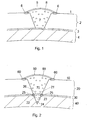
- Example of such a cartilage membrane is shown in figure 1 reference 5 and 2 reference 50, respectively.
- the embodiments of the membranes found in figure 1 and 2 are examples of membranes and the invention should not be limited thereto.
- the invention relates to a cartilage membrane, having at least one surface part, carrying a composition comprising at least one stimulation molecule, which is capable of inducing a signal transduction in chondroblasts/chondrocytes.
- the cartilage membrane is a non-immunogenic, non-toxic, biodegradable membrane, more preferably, the cartilage membrane is porous or substantially porous, even more preferably, the cartilage membrane has a structure allowing chondroblasts/chondrocytes in a chondroblast/chondrocyte suspension to adhere and invade the entire membrane and most preferably the cartilage membrane is a natural collagen type and/or III membrane or a synthetic membrane or part thereof.
- Example of a cartilage membrane is a periosteal flap excised from the patient's bone such as for instance proximal part of the tibia.
- the cartilage membrane may be impregnated with the stimulation molecule prior to or directly after application of the membrane to a surface to be treated, such as the cartilage free cavity found in figure 1, reference 7.
- the cartilage membrane is impregnated using conventional techniques, such as spraying, painting or immersion or any other conventional technique.
- the stimulation molecule comprises at least one RGD motif.
- the stimulation molecule is a natural or synthetic protein or peptide or a fusion or a mixture thereof, more preferably the stimulation molecule is selected from the group consisting of collagen proteins such as collagen types II, VI, IX, and XI, proteoglycans such as aggregans, decorin, fibromodulin and biglycan, and non collageneous proteins such as cryoprecipitate, fibronectin, vitronectin, fibronogen, fibrillin, kistrin, echistatin, von Willebrand factor, tenascin and anchorin CII, and even more preferably, the stimulation molecule is selected from the group consisting of collagen type II and fibronectin. Additionally, the stimulation molecule is attached to a support.
- the final concentration of the stimulation molecule is not critical and is dependent on the degree of the cartilage defect to be treated.
- the stimulation molecule may be added to a final concentration within the range of nM to mM, preferably between, 1 nM to 100 mM and more preferably 10mM.
- the above described cartilage membrane is used for the preparation of a kit containing a cartilage membrane alone or in combination with at least one interface membrane as defined hereinafter for the treatment of cartilage defects or bone and cartilage defects.
- the kit may comprise other components useful for the treatment of cartilage or bone and cartilage defects in mammals, such as chondral leasions or osteochondral lesions, osteochondritis dissecans (OCD), chondromalacia and osteoathritis.
- the invention relates to an interface membrane as defined above carrying a stimulation molecule for the treatment of a mammal having a cartilage and bone defect, such as chondral leasions or osteochondreal lesions, osteochondritis dissecans (OCD), chondromalacia and osteoathritis.
- a stimulation molecule for the treatment of a mammal having a cartilage and bone defect, such as chondral leasions or osteochondreal lesions, osteochondritis dissecans (OCD), chondromalacia and osteoathritis.
- chondral leasions or osteochondreal lesions such as chondral leasions or osteochondreal lesions, osteochondritis dissecans (OCD), chondromalacia and osteoathritis.
- OCD osteochondritis dissecans
- the invention relates to an interface membrane with at least two different surface parts, a first and a second surface part, carrying at least one stimulation molecule capable of inducing a signal transduction in chondroblasts/chondrocytes and osteoblasts/osteocytes.
- the first surface part of the interface membrane has the ability to bind osteoblasts/osteocytes, for example figure 2, reference 22 and the second surface part of the interface membrane has the ability to bind chondroblasts/chondrocytes, for example figure 2, reference 26.
- the interface membrane is a non-immunogenic, non-toxic, biodegradable membrane, more preferably the interface membrane is porous or substantially porous even more preferably the interface membrane has a structure allowing the chondroblasts/chondrocytes in the chondroblast/chondrocyte suspension to adhere and invade one side of the the interface membrane, but not to pass through the interface membrane and osteoblasts/osteocytes in the osteoblast/osteocyte suspension to the other side of the interface membrane but not to pass through the interface membrane and even more preferably the interface membrane is a natural or synthetic collagene type I and/or III membrane or part thereof.
- Example of an interface membrane is a periosteal flap excised from the patient's bone such as for instance proximal part of the tibia.
- the interface membrane may be impregnated with the stimulation molecule prior to or after application of the interface membrane to a surface to be treated, such as for example the bone free cavity found in figure 2, reference 23.
- the interface membrane is impregnated using conventional techniques, such as spraying, painting or immersion or any other conventional technique.
- the stimulation molecule comprises at least one RGD motif
- the stimulation molecule is a natural or synthetic protein or peptide or a fusion or a mixture thereof, more preferably the stimulation molecule is selected from the group consisting of collagen proteins such as types II, VI, IX, and XI, proteoglycans such as aggregans, decorin, fibromodulin and biglycan, and non-collageneous proteins such as cryoprecipitate, fibronectin, vitronectin, fibronogen, fibrillin, kistrin, echistatin, von Willebrand factor, tenascin and anchorin CII, and even more preferably, the stimulation molecule is selected from the group consisting of collagen type II and fibronectin. Additionally, the stimulation molecule is attached to a support.
- the final concentration of the stimulation molecule is not critical and is dependent on the degree of the defect to be treated.
- the stimulation molecule may be added to a final concentration within the range of nM to mM, preferably between, 1 nM to 100 mM and more preferably 10 mM
- the interface membrane may be used for the treatment of bone and cartilage defects using arthroscopy.
- the interface membrane used for arthroscopy comprising of at least one first sealing portion component as defined hereinafter.
- interface membrane for use in arthroscopy is as defined above for the interface membrane used for treatment of bone and cartilage defects, such as chondral leasions or osteochondreal lesions, osteochondritis dissecans (OCD), chondromalacia and osteoathritis
- the interface membrane is used for the preparation of a kit containing an interface membrane alone or in combination with at least one cartilage membrane for the treatment of bone defects or bone and cartilage defects.
- the kit may comprise other components useful for the treatment of cartilage defects or bone and cartilage defects in mammals, such as chondral leasions or ostochondreal lesions, osteochondritis dissecans (OCD), chondromalacia and osteoathritis.
- Method for the propagation of multipotent mesenchymal mammal stem cells to be used for the treatment of cartilage or cartilage and bone defects in mammals.
- the invention in another aspect, relates to a method for the propagation of multipotent mesenchymal mammal cells and/or precursor cells and their differentiation into chondroblasts/chondrocytes, osteoblasts/osteocytes, fibroblasts, myoblasts/myocytes and adipoblasts/adipocytes without the use of enzymes, such as collagenase.
- the differentiated cells may be used for the treatment of cartilage and/or bone and cartilage defects in mammals.
- the method comprises harvesting mesenchymal and/or precursor cells from a source such as bone marrow, perichondrium, periosteum, blood, blood vessels or muscle; adding the harvested cells to a cell culture flask comprising at least one growth medium as defined above under the term "growth medium"; growing the harvested cells until colony forming units with a cell number size in the ranging order of 10-20 000 cells /clones are formed with fibroblastic phenotype (CFU-f); transferring the CFU-f cells into a new cell culture flask comprising at least one selection medium as defined above for further differentiation into chondroblasts/chondrocytes, osteocytes/osteoblasts or myoblasts/myotubes; harvesting of the differentiated cells and preparation of chondroblast/chondrocyte or osteocyte/osteoblast suspensions.
- a source such as bone marrow, perichondrium, periosteum, blood, blood vessels or muscle
- growth medium
- the chondroblast/chondrocyte or osteocyte/osteoblast suspensions may be used for the treatment of cartilage and/or bone and cartilage defects in mammals.
- the period of cultivation is dependent on the stage and conditions of the harvested mesenchymal and/or precursor cells from mammals and it is therefore difficult to define the time period used for the propagation of the mesenchymal and/or precursor cells in the growth and selection medium to be differentiated into chondroblast/chondrocytes or osteoblasts/osteocytes.
- the immature cells are primed by selection medium, in which the composition of amino acids, hormonal, growth- and differentiation factors are more specific for selection than for growth.
- medium with fetal calf serum identified as sufficient "selection quality" leads to certain differentiations of the precursor cells.
- Other factors such as physical factors, e.g. adhesion to certain plastic materials or adhesion to cellular or non-cellular adhesion factors are also aiding to the differentiation of the cells.
- the chondroblast/chondrocyte and the osteoblast/osteocyte suspensions contain conventional culture medium such as for example DMEM/F12, serum such as fetal calf serum or autologous serum, preferably a serum, which is non-immunogenic and more preferably the chondroblast/chondrocyte and the osteoblast/osteocyte suspensions comprise chondroblast/chondrocyte respectively osteoblast/osteocyte cells within the range of 100,000 to 10 million cells/ml of the total cell suspensions.
- DMEM/F12 serum
- serum such as fetal calf serum or autologous serum
- a serum which is non-immunogenic
- the chondroblast/chondrocyte and the osteoblast/osteocyte suspensions comprise chondroblast/chondrocyte respectively osteoblast/osteocyte cells within the range of 100,000 to 10 million cells/ml of the total cell suspensions.
- Another embodiment according to the invention relates to a cell cultivating system built on the concept of using the cartilage matrix as the "binding and delivery" (or reservoir) system of growth and differentiation factors to the various cells present in the extracellular matrix, without using enzymatic treated and dissected cartilage pieces.
- Such a binding and delivery system might increase the half life time of the various growth and differentiation factors present in the culture medium with fetal calf serum meaning that growth and differentiation factors are much more protected from proteolysis when first bound to natural intermediate matrix molecules the delivery "vehicles" to the various cell receptors.
- a higher stimulation of the cells by growth and differentiation factors could be speculated, because of a higher local accumulation (higher concentration) of the growth and differentiation factors around the pericellular rim, in the territorial as well as in the interterritorial compartments, compared to the growth and differentiation factors present in conventional medium.
- cartilage derived cells in the explants maintain their differentiated state in the matrix while cell stimulation and cell propagation proceed.
- the cells are not exposed to high concentration of proteolytic activity used for instance by collagenase digestion of cartilage matrix. This means that the extracellular matrix in the process of propagating the cartilage-derived cells is conserved and close to in vivo conditions.
- the sealing portion may be used to entrap a chondroblast/chondrocyte suspension into a cartilage free cavity and/or an osteoblast/osteocyte suspension into a bone free cavity.
- Examples of the sealing portions are found in figure 1, reference 6 entrapping the chondroblast/chondrocyte suspension (8) into the cartilage free cavity (7), and the sealing portion in figure 2, reference (25) entrapping the osteoblast/osteocyte suspension (24) into the bone free cavity (23).
- Example of a sealing portion is TisseelTM (Baxter Immuno Austria, a lyophilised virus-inactivated substance that consist of fibrinogen, plasmafibrinonectin, factor VIII and plasminogen), which during application is mixed with aprotinin solution, thrombin 4, thrombin 500 and calcium chloride solution, using a dual syringe system connected to one blunt injection needle.
- the "sealing portion” may comprise a chondroblast/chondrocyte or an osteoblast/osteocyte suspension with or without a stimulation molecule as defined above under the definitions.
- a sealing agent is a CoSeal (Cohesion Technologies, Palo Alto, California, U.S.A), which also is a two-component biological glue, resembling Tisseel, can be used instead of Tisseel.
- CoSeal Cohesion Technologies, Palo Alto, California, U.S.A
- the sealing portion is glue comprising two different components
- two components that together form glue may be applied to cover the complete area of at least one side of the membrane.
- the other component is added between the sealing portion part of the membrane and the edge of the defected cartilage or bone defect after the membrane is placed onto the cartilage or bone defect.
- the invention relates to method and materials for in vivo repair of cartilage defects in mammals, such as such as chondral leasions or osteochondreal lesions, osteochondritis dissecans (OCD), chondromalacia and osteoathritis.
- mammals such as such as chondral leasions or osteochondreal lesions, osteochondritis dissecans (OCD), chondromalacia and osteoathritis.
- the cartilage-defect part is normally a surface part where the original cartilage has been torn apart.
- the defect is debrided so that the cartilage defect surface part appears as a cartilage free cavity 7, which often extending down to the interface 3, the interface in between the cartilage 2 and the bone 4, the cartilage-free cavity 7 being surrounded by healthy cartilage 2.
- the cartilage membrane 5 as defined above is applied over the cartilage free cavity 7.
- the cartilage membrane 5 is normally adapted in size so that the portion part of the membrane can be sutured to the surrounding articular surface 1.
- the edge of the membrane is normally sealed to the surrounding articular surface 1 by means of suitable sealing portions 6.
- the sealing is not completed; a small part of the circumference is left unsealed to allow the implantation of a chondroblast/chondrocyte suspension 8.
- the cartilage membrane 5 is completely sealed by means of a suitable sealing portion 6.
- the cartilage membrane 5 having at least one surface part, carrying a composition comprising at least one stimulation molecule which is capable of inducing a signal transduction in chondroblasts/chondrocytes resulting in the chondroblasts/chondrocytes producing and secreting matrix components which form hyalin or hyalin articular cartilage.
- the surface part of the cartilage membrane 5 carrying the stimulation molecule is localised against the articular surface 1 and covering the cartilage free cavity 7 comprising the chondroblast/chondrocyte suspension 8 as defined above. Further characteristics of the "cartilage membrane" as defined above under Cartilage membrane carrying a stimulation molecule.
- a composition is applied on the cartilage defect surface forming the walls surrounding the cartilage free cavity 7 prior to application of the cartilage membrane 5.
- the composition comprise at least one stimulation molecule as defined above to increase the exposure of the implanted chondroblasts/chondrocytes in the chondroblast/chondrocyte suspension 8 to the stimulation molecule, as well as the capability of the implanted chondro-blasts/chondrocytes cells to bind to the surrounding cartilage 2.
- the chondroblast/chondrocyte suspension 8 is applied simultaneously with or without a suspension comprising the stimulation molecule as defined above.
- the chondroblast/chondrocyte suspension is a suspension of autologous chondrocytes/chondroblasts.
- the chondroblasts/chondrocytes is included in a composition carried by the cartilage membrane 5 and optionally applied directly onto the cartilage free cavity 7 in order to obtain a lining of cells resulting in a normal cartilage surface layer and transitional layer.
- the chondroblast/chondrocyte suspension contain conventional culture medium such as for example DMEM/F12, serum such as fetal calf serum or autologous serum, preferably a serum, which is non-immunogenic and more preferably the chondroblast/chondrocyte suspension comprise chondroblast/chondrocyte cells within the range of 100.000 to 10 millions of cells/ml of the total chondroblast/chondrocyte suspension.
- conventional culture medium such as for example DMEM/F12
- serum such as fetal calf serum or autologous serum
- a serum which is non-immunogenic
- the chondroblast/chondrocyte suspension comprise chondroblast/chondrocyte cells within the range of 100.000 to 10 millions of cells/ml of the total chondroblast/chondrocyte suspension.
- Examples of conditions resulting in cartilage defects which may be treated according to the invention are chondreal leasions or osteochondral lesions, osteochondritis dissecans (OCD), chondromalacia and osteoathritis.
- OCD osteochondritis dissecans
- a particular embodiment of the invention relates to method and materials for in vivo repair of both bone and cartilage defects in mammals, such as such as chondral leasions or ostochondreal lesions, osteochondritis dissecans (OCD), chondromalacia and osteoathritis using two membranes.
- mammals such as such as chondral leasions or ostochondreal lesions, osteochondritis dissecans (OCD), chondromalacia and osteoathritis using two membranes.
- the bone and cartilage defect parts is debrided so that the bone and cartilage defect parts appears as bone and cartilage free cavities 23, 70 extending all way through the cartilage 20, interface 30 and down to the bone 40.
- the bone and cartilage free cavities 23, 70 being surrounded by healthy cartilage 20, interface 30 and bone 40.
- the interface membrane 21 is applied over the bone free cavity 23 at the level of the interface 30 for the ability to separate the cartilage 70 and the bone 23 free cavities.
- the interface membrane 21 is applied so that the first surface part 22 of the interface membrane 21, facing the bone free cavity 23 and the second surface part 26 facing the cartilage free cavity 70.
- the interface membrane 21 is normally adapted in size so that the portion part of the membrane can be sutured to the surrounding area of the interface 30.
- the edge of the membrane is normally sealed to the surrounding interface 30 by means of suitable sealing portions 25.
- the sealing is not completed a small part of the circumference is left unsealed to allow the implantation of an osteoblast/osteocyte suspension 24.
- the interface membrane is completely sealed to the surrounding interface 30 by means of a suitable sealing portion 25.
- the interface membrane 21 having at least two different surface parts, a first 22 and a second 26 surface part, which carries at least one stimulation molecule capable of inducing a signal transduction.
- the first surface part 22 of the interface membrane 21 has the ability to bind osteoblasts/osteocytes and the second surface part 26, of the interface membrane 21 has the ability to bind chondroblasts/chondrocytes.
- the osteoblast/osteocyte suspension is applied simultaneously with or without a suspension comprising the stimulation molecule as defined above.
- the osteoblast/osteocyte suspension is a suspension of autologous osteoblasts/osteocytes.
- the osteoblast/osteocyte suspension contain conventional culture medium such as for example DMEM/F12, serum such as fetal calf serum or autologous serum, preferably a serum, which is non-immunogenic and more preferably the osteoblast/osteocyte suspension comprise osteoblast/osteocyte cells within the range of 100.000 to 10 millions of cells/ml of the total osteoblast/osteocyte suspension.
- conventional culture medium such as for example DMEM/F12
- serum such as fetal calf serum or autologous serum
- a serum which is non-immunogenic
- the osteoblast/osteocyte suspension comprise osteoblast/osteocyte cells within the range of 100.000 to 10 millions of cells/ml of the total osteoblast/osteocyte suspension.
- the osteoblasts/osteocytes in the osteoblast/osteocyte suspension 24 is included in the composition carried by the surface part 22 of the interface membrane 21 and optionally applied directly on the bone free cavity 23 in order to obtain a lining of cells resulting in a normal bone surface layer and transitional layer.
- the cartilage membrane 50 as defined above is applied over the cartilage free cavity 70.
- the cartilage membrane 50 is normally adapted in size so that the portion part of the membrane can be sutured to the surrounding articular surface 10.
- the edge of the membrane is normally sealed to the surrounding articular surface 10 by means of suitable sealing portions 60.
- the sealing is not completed a small part of the circumference is left unsealed to allow the implantation of a chondroblast/chondrocyte suspension 80. Thereafter the implantation is completely sealed to the surrounding articular surface 10 by means of a suitable sealing portion 60.
- the cartilage membrane 50 having at least one surface part, carrying a composition comprising at least one stimulation molecule which is capable of inducing a signal transduction in chondroblasts/chondrocytes resulting in the chondroblasts/chondrocytes producing and secreting matrix components which form hyalin or hyalin articular cartilage.
- the surface part of the cartilage membrane 50 carrying the stimulation molecule is localised against the articular surface 10 and covering the cartilage free cavity 70 comprising the chondroblast/chondrocyte suspension 80 as defined above. Further characteristics of the cartilage membrane as defined above under Cartilage membrane carrying a stimulation molecule.
- the chondroblast/chondrocyte suspension may be applied simultaneously with or without a suspension comprising of the stimulation molecule as defined above.
- the chondroblasts/chondrocytes in the chondroblast/chondrocyte suspension 80 included in a composition carried by the cartilage membrane 50 and optionally applied directly on the cartilage free cavity 70 in order to obtain a lining of cells resulting in a normal cartilage surface layer and transitional layer.
- the chondroblast/chondrocyte suspension contain conventional culture medium such as for example DMEM/F12, serum such as fetal calf serum or autologous serum, preferably a serum, which is non-immunogenic and more preferably the chondroblast/chondrocyte suspension comprise chondroblasts/chondrocytes within the range of 100.000 to 10 millions of cells/ml of the total chondroblast/chondrocyte suspension.
- conventional culture medium such as for example DMEM/F12
- serum such as fetal calf serum or autologous serum
- a serum which is non-immunogenic
- the chondroblast/chondrocyte suspension comprise chondroblasts/chondrocytes within the range of 100.000 to 10 millions of cells/ml of the total chondroblast/chondrocyte suspension.
- Examples of conditions resulting in bone and cartilage defects, which may be treated according to the invention are chondral lesions or osteochondral lesions, osteochondritis dissecans (OCD), chondromalacia and osteoarthritis.
- OCD osteochondritis dissecans
- the bone and cartilage defect is treated using arthroscopy using a two component sealing portion, component 1 and 2.
- a two component sealing portion is Tisseel Duo Quick (Baxter).
- the interface membrane 21 is pretreated with a sealing portion component 1, for example a thrombin solution which contains: 500 IU thrombin, 50 mg mammal plasma protein, 40 mol calcium chloride, 10 mg sodium chloride, 3 mg glycine, and water for injection to 1 ml, such as Tisseel Duo Quick (Baxter)
- the sealing portion may comprise a stimulation molecule.
- the bone and cartilage defect parts is debrided so that the bone and cartilage defect parts appears as a bone and cartilage free cavity 23, 70 extending all way through the cartilage, interface and down to the bone.
- the bone and cartilage free cavity being surrounded by healthy cartilage 20, interface 30 and bone 40.
- a sealing portion component 2 is applied to the surrounding edges of the interface area 30.
- the interface membrane 21 is applied using arthroscopy over the bone free cavity 23 at the level of the interface 30 for the ability to separate the cartilage 70 and the bone 23 free cavities.
- the interface membrane 21 is applied so that the first surface part 22 of the interface membrane 21, facing the bone free cavity part 23 and the second surface part 26 facing the cartilage free cavity 70.
- the interface membrane 21 is normally adapted in size so that the portion part of the membrane can be glued to the surrounding area of the interface 30.
- the sealing portion component 1 and 2 react with each other and from a glue sealing the interface membrane 21 to the surrounding interface area 30.
- the sealing is not completed a small part of the circumference is left unsealed to allow the implantation of an osteoblast/osteocyte suspension 24.
- the interface membrane is completely sealed to the surrounding interface 30 by means of a suitable portion 25.
- the chondroblast/chondrocyte suspension 80 is implantated and the articular surface sealed by means of a suitable portion 60.
- a cell cultivation method for preparation of chondroblast/chondrocyte without enzymatic treatment comprising harvesting cartilage explants from a mammal;
- cartilage explants are used for the treatment of cartilage and/or bone and cartilage defects in mammals.
- a method for preparation of chondroblast/chondrocyte or osteocyte/osteoblast suspensions comprising
- suspensions are used for the treatment of cartilage and/or bone and cartilage defects in mammals.
- the selection medium comprises components more specific for selection than for growth.
- the selection medium comprises fetal calf serum at a concentration of over 10% v/v, such as e.g. over 20% v/v, over 25% v/v.
- chondroblast/chondrocyte or osteocyte/osteoblast suspensions have a chondroblast/chondrocyte or osteocyte/osteoblasts concentration from 100 000 to 10 million cells/ml such as e.g. from 500 000 to 5 million, from 1 million to 2 million of the total cell suspension.
- collagen proteins such as collagen types II, VI, IX, and XI
- proteoglycans such as aggregans, decorin, fibromodulin and biglycan
- non-collageneous proteins such as cryoprecipitate, fibronectin, vitronectin, fibronogen, fibrillin, kistrin, echistatin, von Wille
- a cartilage membrane as described herein which is a non-immunogenic, non-toxic, biodegradable membrane.
- collagen proteins such as collagen types II, VI, IX, and XI
- proteoglycans such as aggregans, decorin, fibromodulin and biglycan
- non-collageneous proteins such as cryoprecipitate, fibronectin, vitronectin, fibronogen, fibrillin,
- An interface membrane as described herein which is a non-immunogenic, non-toxic, biodegradable membrane.
- a kit for cartilage repair comprising at least one cartilage membrane as described herein and at least one stimulation molecule as described herein.
- a kit as described herein comprising at least one interface membrane aas described herein.
- DMEM/F12 containing 20 % fetal calf serum.
- DMEM/F12 obtained from Life Technologies Inc., Rockville, MD, USA and fetal calf serum from Life Technologies Inc., Rockville, MD, USA.
- Chondrocyte suspension used for the treatment of cartilage defects.
- Biopsy of healthy looking cartilage is obtained through an arthroscope from a minor load bearing area of the joint under aseptic conditions and placed in a sterile flask containing "Transport medium” (DMEM/F12 with 20% fetal calf serum).
- the Flask containing Transport medium is delivered within 48 hours to the Cell Culturing Laboratory for further processing into cell culture.
- the method described in example 3 was used for the propagation of the chondrocytes.
- the chondrocytes were expanded in tissue culture flasks in a CO 2 incubator (5% CO 2 ) at 37°C in growth medium for 3 to 6 weeks, and later on maintained in DMEM/F12 with the patient's (mammal) own heat inactivated serum (range 10-20%) for 3 to 10 days.
- the cells are harvested by trypsinization in 0.25% trypsin in 1mM EDTA, washed in medium containing fetal calf serum (10 20%) and centrifuged at 900 x g for 10 minutes at room temperature, and resuspended to cell numbers between 0.5 to 2 x 10 6 cells per 0.1 ml growth medium (5 to 20 x 10 6 cells per 1 ml growth medium).
- the optimum cell count per 0.1 ml growth medium for implantation is around 1 million cells.
- a cartilage defect has room for 0.1 ml cell suspension per 1 cm 2 defect.
- the chondrocytes are delivered to the orthopedic surgeon, who, within 24 hours implants the chondrocytes into a cartilage defect. Prior to the implantation the surgeon has either placed a periosteal flap or a suitable biodegradable cover over the defect, sutured and sealed the flap, except for the injection site. The cells are then injected under the cover, and the injection site is sealed using Tisseel Duo Quick (Baxter).
- Collagen II was purified from large scale culturing of porcine chondrocytes as well as proteoglycans derived from the same porcine cartilage source. Collagen II was obtained from large scale culturing of mammalian chondrocytes (preferably species specific to the recipient of the cells), cultured as explants whereby the adequate amount of Collagen II is present.
- a cartilage biopsy is harvested from the patients knee and immediately transferred into aseptic growth medium, supplemented with L-ascorbic acid [50 ⁇ g/ml (300 ⁇ mol/l)] and gentamicin sulfate [50 ⁇ g/ml (10 mmol/l)], Fungizone [2 ⁇ g/ml (2.2 ⁇ mol/l) in tissue culture flasks.
- the cartilage biopsy is then washed carefully with new growth medium and dissected in growth medium into 2-4 mm cartilage pieces (explants). When initially placed in the culture, it takes the cartilage explants approximately several days depending on donor material, to reach a constant metabolic state.
- steady state is a balance between synthesis and catabolism
- the pre chondroblasts/chondroblasts are now stimulated by growth factors present in the growth medium, which diffundate through the cartilage matrix and bind to various binding proteins present in the matrix as well as binding directly to the selective cell receptors
- the stimulated and proliferating cells remain in the explants usually for one to two weeks (depending on the donor material) and then "leave" the explants, via cell migration, into the culture medium for further attachment to the culture dish (monolayer culture).
- the cartilage explants are transferred to separate culture dishes with new growth medium, and the first established monolayer culture is split by conventional trypsination method, described earlier. This step is important in the process of propagating the cell culture to a high cell number. After each trypsination process, the monolayer cells new growth medium is added to the monolayer cells as described above.
- a membrane collagen type I/(III) is used, purchased from Geistlich Biomaterials, Wolhusen, Switzerland or from Baxter (Denmark).
- the membrane is cut into a size, which is somewhat larger than the cartilage defect, so that the membrane can cover the cartilage defect and extend around 1 ⁇ 2 cm beyond the rim of the defect.
- the membrane is sutured together with the cartilage rim.
- the border between the membrane and the cartilage is sealed using Tisseel Due Quick (Baxter).
- the collagen type I membrane is pretreated with a 1-5 ml collagen type II solution (3-10 mg collagen II/ml (Dep. Material Engineering, Drexel University, Philadelphia, Pennsylvania) for 5-15 minutes at room temperature prior to use for implantation.
- the above described thin layer membrane coated as described with "II” is then inserted over the defect (the size of the membrane is somewhat larger in diameter than the defect) is placed with the coat facing the defect.
- the two types of coating (coating "I” and coating “II”, which is the components in Tisseel) will result in a biological glue and thereby attaching the membrane to the defect.
- the membrane is held in place by inserting small absorbable pins into the periphery of the defect anchoring the "sealed” membrane to the cartilage in a more solid manner.
- the chondrocyte suspension is then injected via a needle in one area of the membrane near the rim into the defect, and the membrane hole is sealed, when pulling the needle, with the two component biological glue such as for instance Tisseel Duo Quick as shown below.
- component "II” is used as coat on the membrane facing the defect and where component “I” is used as coat on the cartilage rim surrounding the defect.
- Component “II” will react with component “I”, but will not react as glue with medium containing the autologous serum component, but as RGD coat towards the medium, and thereby induce adherence and matrix production of the injected chondrocytes.
- the Tisseel 2 component glue can also be used as coat on the membrane part facing the defect, but will be more difficult to handle when inserted.
- Another alternative is to coat the cartilage rim surrounding the defect with the two component glue. In this case it will also be difficult to handle the placement of the membrane over the defect.
- the growth medium was used s the control medium incapable of inducing chondrocyte formation in the cambium layer of a periosteal flap.
- periosteal flap 1 ⁇ 2 ⁇ 1 ⁇ 2 cm in size obtained from the medial part of tibia of a pig were placed in two tissue culture dishes.
- the conditioned medium was thawed, centrifuged at 3000 rpm for 20 minutes and sterile filtered through a 0.8 my filter and subsequently applied to one of the samples of periost. Incubated at 37° C, in a CO 2 incubator for a period of 4 weeks.
- a periosteal flap,1 ⁇ 2 ⁇ 1 ⁇ 2 cm in size obtained from the medial part of tibia of the same animal was used for the control experiment. These samples were incubated at 37°C, in a CO 2 incubator for a period of 4 weeks.
- the two samples of periosteal flap was fixated by conventional fixating technique in formaldehyde and stained with various routine staining including Safranin and submitted to microscopic examination.
- aspirate from spongiosa of femur containing both hematopoetic precursor cells and marrow stromal stem and/or precursor cells were plated into cell culture tissue bottles at approximate dilutions. Single and colony forming marrow cells were stimulated 24 hours (primary incubation) with growth medium. After primary incubation, non-adherent cells such as hematopoetic as well as non-hematopoetic cells were gently removed by replacing supernatant with new media. Adherent cells was further stimulated 1 week with above growth medium which induced several colony forming units with cell number sizes ranging in the order of 10-20.000 cells/clone and with fibroblastic phenotype (CFU-f).
- CFU-f fibroblastic phenotype
- the selection media After proliferation of these cells in growth medium for another week or more (depending on donor material) some cell cultures were transferred the selection media, are more specific for selection than for growth. It could be a certain batch of fetal calf serum at a higher concentration than conventionally used (over 10%), up to 30% v/v., which has been tested for its ability to differentiate cells into chondroblasts/chondrocytes, or tested for its ability to differentiate cells into osteoblasts/osteocytes. We have internally identified said batches of fetal calf serum that exhibits these selection patterns. Until further a new batch or new batches of fetal calf serum has to be tested for the differentiation of cells.
- the cells are identified by quantitative PCR of the mRNA (Lane Smith, R., et al., J. Rehab. Research and Development March/April 2000, vol 37), and in case of chondrocyte lines, the cells are tested using monoclonal antibodies such as collagen type II Ab-1 (Clone 5B2.5) mouse monoclonal antibody (Lab. Vision Corp, Fremont, California) for further differentiation and "cell maturation" (2-4 weeks in selection media).
- monoclonal antibodies such as collagen type II Ab-1 (Clone 5B2.5) mouse monoclonal antibody (Lab. Vision Corp, Fremont, California) for further differentiation and "cell maturation" (2-4 weeks in selection media).
- developed bone cells were shown to produce mineralized matrix, chondroblast/chondrocytes to form characteristic lacunas where matrix was stained metachomatic with dye such as Safranin-O.
Landscapes
- Health & Medical Sciences (AREA)
- Life Sciences & Earth Sciences (AREA)
- Engineering & Computer Science (AREA)
- Biomedical Technology (AREA)
- Chemical & Material Sciences (AREA)
- General Health & Medical Sciences (AREA)
- Cell Biology (AREA)
- Zoology (AREA)
- Transplantation (AREA)
- Animal Behavior & Ethology (AREA)
- Public Health (AREA)
- Veterinary Medicine (AREA)
- Oral & Maxillofacial Surgery (AREA)
- Medicinal Chemistry (AREA)
- Dermatology (AREA)
- Epidemiology (AREA)
- Chemical Kinetics & Catalysis (AREA)
- Botany (AREA)
- Organic Chemistry (AREA)
- Bioinformatics & Cheminformatics (AREA)
- Wood Science & Technology (AREA)
- Biotechnology (AREA)
- Genetics & Genomics (AREA)
- Orthopedic Medicine & Surgery (AREA)
- Rheumatology (AREA)
- Vascular Medicine (AREA)
- Urology & Nephrology (AREA)
- Microbiology (AREA)
- General Engineering & Computer Science (AREA)
- Biochemistry (AREA)
- Cardiology (AREA)
- Heart & Thoracic Surgery (AREA)
- Biophysics (AREA)
- Developmental Biology & Embryology (AREA)
- Immunology (AREA)
- Physical Education & Sports Medicine (AREA)
- General Chemical & Material Sciences (AREA)
- Nuclear Medicine, Radiotherapy & Molecular Imaging (AREA)
- Pharmacology & Pharmacy (AREA)
- Materials For Medical Uses (AREA)
Abstract
Description
introducing, in the interstice between the interface membrane first surface part and the bone, an osteoblast/osteocyte suspension,
joining a portion part of the first surface part of the interface membrane to the surrounding interface surface so as to sealingly entrap the osteoblast/osteocyte suspension in the bone free cavity using a sealing portion, thereby allowing the osteoblast/osteocyte suspension to produce and secrete matrix components characteristic for bone tissue; applying, over the cartilage free cavity, a cartilage membrane with a first surface part facing the second surface part of the interface membrane, the first surface part of the cartilage membrane carries a composition comprising at least one stimulation molecule which is capable of inducing a signal transduction in chondroblasts/chondrocytes resulting in the chondroblasts/chondrocytes producing and secreting matrix components which form hyalin cartilage,
introducing, in the cartilage free cavity between the interface membrane, the cartilage membrane and the cartilage, a chondroblast/chondrocyte suspension,
joining a portion part of the cartilage membrane to the surrounding articular surface so as to sealingly entrap the chondroblast/chondrocyte suspension in the cartilage free cavity, thereby allowing the chondroblast/chondrocyte suspension to produce and secrete matrix components which form hyalin.
joining a portion part of the first surface part of the interface membrane to the surrounding interface surface so as to sealingly entrap the osteoblast/osteocyte suspension in the bone free cavity using a second sealing portion component, thereby allowing the osteoblast/osteocyte suspension to produce and secrete components characteristic for bone tissue;
introducing, in the cartilage free cavity between the interface membrane, and the articular surface, a chondroblast/chondrocyte suspension, thereby allowing the chondroblast/chondrocyte suspension to produce and secrete components characteristic for hyalin.
growing the harvested cells until colony forming units with a cell number size in the ranging order of 10-20 000 cells /clones are formed with fibroblastic phenotype (CFU-f); transferring the CFU-f cells into a new cell culture flask comprising at least one selection medium as defined above for further differentiation into chondroblasts/chondrocytes, osteocytes/osteoblasts or myoblasts/myotubes;
harvesting of the differentiated cells and preparation of chondroblast/chondrocyte or osteocyte/osteoblast suspensions.
Example of a sealing portion is Tisseel™ (Baxter Immuno Austria, a lyophilised virus-inactivated substance that consist of fibrinogen, plasmafibrinonectin, factor VIII and plasminogen), which during application is mixed with aprotinin solution,
It is clear that embodiments of the present invention are as follows:
The chondrocytes were expanded in tissue culture flasks in a CO2 incubator (5% CO2) at 37°C in growth medium for 3 to 6 weeks, and later on maintained in DMEM/F12 with the patient's (mammal) own heat inactivated serum (range 10-20%) for 3 to 10 days. When the chondrocyte culture has been expanded to the amount of cells needed for the repair of a cartilage lesion(s) of a given patient (mammal), the cells are harvested by trypsinization in 0.25% trypsin in 1mM EDTA, washed in medium containing fetal calf serum (10 20%) and centrifuged at 900 x g for 10 minutes at room temperature, and resuspended to cell numbers between 0.5 to 2 x 10⁶ cells per 0.1 ml growth medium (5 to 20 x 106 cells per 1 ml growth medium). The optimum cell count per 0.1 ml growth medium for implantation is around 1 million cells. In general, a cartilage defect has room for 0.1 ml cell suspension per 1 cm2 defect.
Claims (10)
- A cell cultivation method for production of chondroblasts/chondrocytes comprising harvesting cartilage explants from a mammal;i. adding the harvested cartilage explants to a culturing flask comprising at least one growth medium;ii. growing the cartilage explants in the growth medium to propagate chondroblasts/chondrocytes into a mono-layer culture;iii. propagation of the chondroblasts/chondrocytes into a mono-layer culture with a high cell numberiv. transferring chondroblasts/chondrocytes from the mono-layer culture into an autologous growth medium.
- A method according to claim 1, wherein enzymatic treatment is not used.
- A method according to claim 1, wherein the explant-cultured chondroblasts/chondrocytes are used for the treatment of cartilage and/or bone defects in mammals.
- A method according to claims 1-2, wherein the growth medium comprises at least one of; DMEM/F12, growth factors, L-ascorbic acid, gentamicin sulfate, fungizone, Fetal Calf serum (FCS) and Human serum (HS).
- A method according to any of the preceding claims, wherein the cell number is between 0.5×10⁶ and 2×10⁶ per 0.1ml growth medium.
- A method for preparation of chondroblast/chondrocyte or osteocyte/osteoblast suspensions comprisingi. harvesting non-embryonic mesenchymal and/ or mesenchymal precursor cells from an source selected from the group consisting of bone marrow, perichondrium, periosteum, blood, blood vessels or muscle,ii. adding the harvested cells to a cell culture flask comprising at least one growth medium,iii. growing the harvested cells until colony forming units with a cell number size in the ranging order of 10-20000 cells /unit are formed with fibroblastic phenotype (CFU-f),iv. transferring the CFU-f cells into a new cell culture flask comprising at least one selection medium for differentiation of the CFU-f's into chondroblast/chondrocytes, osteocytes/osteoblasts or myoblasts/myocytes andv. harvesting of the differentiated cells.
- A method according to claim 6, wherein the suspensions are used for the treatment of cartilage and/or bone defects in mammals.
- A method according to claims 6 or 7, wherein the selection medium comprises components more specific for selection than for growth.
- A method according to claim 8, wherein the selection medium comprises fetal calf serum at a concentration of over 10% v/v and up to 30% v/v.
- A method according to any of claims 6 - 9, wherein the chondroblast/chondrocyte or osteocyte/osteoblast suspensions have a chondroblast/chondrocyte or osteocyte/osteoblasts concentration from 100 000 to 10 million cells/ml of the total cell suspension.
Applications Claiming Priority (5)
| Application Number | Priority Date | Filing Date | Title |
|---|---|---|---|
| SE9902807 | 1999-07-28 | ||
| SE9902808 | 1999-07-28 | ||
| SE9902808A SE9902808D0 (en) | 1999-07-28 | 1999-07-28 | A method for in vivo repair of cartilage defects or bone and cartilage defects in non-human mammal joints |
| SE9902807A SE9902807D0 (en) | 1999-07-28 | 1999-07-28 | A method for in vivo repair of cartilage defects or bone and cartilage dedects in joints in humans |
| EP00953086A EP1206225B1 (en) | 1999-07-28 | 2000-07-25 | In vitro repair of bone and/or cartilage defects |
Related Parent Applications (1)
| Application Number | Title | Priority Date | Filing Date |
|---|---|---|---|
| EP00953086A Division EP1206225B1 (en) | 1999-07-28 | 2000-07-25 | In vitro repair of bone and/or cartilage defects |
Publications (2)
| Publication Number | Publication Date |
|---|---|
| EP1441028A2 true EP1441028A2 (en) | 2004-07-28 |
| EP1441028A3 EP1441028A3 (en) | 2005-01-05 |
Family
ID=26663626
Family Applications (2)
| Application Number | Title | Priority Date | Filing Date |
|---|---|---|---|
| EP00953086A Expired - Lifetime EP1206225B1 (en) | 1999-07-28 | 2000-07-25 | In vitro repair of bone and/or cartilage defects |
| EP04002205A Withdrawn EP1441028A3 (en) | 1999-07-28 | 2000-07-25 | Cell cultivation method for the preparation of chondrocytes/chondroblasts |
Family Applications Before (1)
| Application Number | Title | Priority Date | Filing Date |
|---|---|---|---|
| EP00953086A Expired - Lifetime EP1206225B1 (en) | 1999-07-28 | 2000-07-25 | In vitro repair of bone and/or cartilage defects |
Country Status (12)
| Country | Link |
|---|---|
| US (1) | US20020082623A1 (en) |
| EP (2) | EP1206225B1 (en) |
| JP (1) | JP2003505143A (en) |
| AT (1) | ATE290353T1 (en) |
| AU (1) | AU779472B2 (en) |
| CA (1) | CA2378079C (en) |
| CZ (1) | CZ2002341A3 (en) |
| DE (1) | DE60018576T2 (en) |
| ES (1) | ES2239608T3 (en) |
| NO (1) | NO20020439D0 (en) |
| NZ (1) | NZ517199A (en) |
| WO (1) | WO2001006949A2 (en) |
Families Citing this family (37)
| Publication number | Priority date | Publication date | Assignee | Title |
|---|---|---|---|---|
| US7645739B2 (en) * | 2001-02-21 | 2010-01-12 | Alavita Pharmaceuticals, Inc. | Modified annexin compositions and methods of using same |
| US7635676B2 (en) | 2001-02-21 | 2009-12-22 | Alavita Pharmaccuticals, Inc. | Modified annexin proteins and methods for their use in organ transplantation |
| US7635680B2 (en) * | 2001-02-21 | 2009-12-22 | Alavita Pharmaceuticals, Inc. | Attenuation of reperfusion injury |
| US20090291086A1 (en) * | 2001-02-21 | 2009-11-26 | Alavita Pharmaceuticals, Inc. | Compositions and Methods for Treating Cerebral Thrombosis and Global Cerebral Ischemia |
| US20050222030A1 (en) * | 2001-02-21 | 2005-10-06 | Anthony Allison | Modified annexin proteins and methods for preventing thrombosis |
| US20040151705A1 (en) | 2002-03-22 | 2004-08-05 | Shuichi Mizuno | Neo-cartilage constructs and a method for preparation thereof |
| AU2002366585A1 (en) * | 2001-12-13 | 2003-06-23 | Andrew Stevens | Von willebrand factor-coated biocompatible implants |
| JP4067419B2 (en) * | 2002-03-04 | 2008-03-26 | 株式会社ネクスト21 | Closed cell culture system |
| US7537780B2 (en) * | 2002-03-22 | 2009-05-26 | Histogenics Corporation | Method for preparing and implanting a cartilage construct to treat cartilage lesions |
| EP1635887A2 (en) * | 2003-06-12 | 2006-03-22 | Interface Biotech A/S | A method for cell implantation |
| ATE417636T1 (en) * | 2003-10-10 | 2009-01-15 | Kw2 Implantattechnologie Gmbh | CARTILAGE REGENERATION WITH GENERATION OF CHONDRONES UNDER HIGH CONCENTRATION OF MAGNESIUM |
| ES2396689T3 (en) | 2003-12-11 | 2013-02-25 | Isto Technologies Inc. | Particle Cartilage System |
| WO2005079881A1 (en) * | 2004-02-19 | 2005-09-01 | Interface Biotech A/S | Arthroscopic method for cell implantation in mammals |
| EP1753860B1 (en) * | 2004-02-20 | 2012-04-11 | Isto Technologies Inc. | Intervertebral disc repair and methods therefor |
| US20060275273A1 (en) * | 2004-02-20 | 2006-12-07 | Seyedin Mitchell S | Intervertebral Disc Repair, Methods and Devices Therefor |
| EP1796592A4 (en) * | 2004-07-30 | 2008-01-02 | Histogenics Corp | Method for in situ repair of injured, damaged, diseased or aged articular cartilage using neo-cartilage constructs and a method for preparation thereof |
| US8697139B2 (en) | 2004-09-21 | 2014-04-15 | Frank M. Phillips | Method of intervertebral disc treatment using articular chondrocyte cells |
| GB0421298D0 (en) * | 2004-09-24 | 2004-10-27 | Univ Bristol | Cellular bandage |
| EP1916964A4 (en) | 2005-08-26 | 2015-11-04 | Zimmer Inc | IMPLANTS AND METHODS FOR THE TREATMENT, REPLACEMENT AND TREATMENT OF JOINT DISEASES |
| JP2009508596A (en) | 2005-09-19 | 2009-03-05 | ヒストジェニックス コーポレイション | Cell support substrate and preparation method thereof |
| EP1764117A1 (en) | 2005-09-20 | 2007-03-21 | Zimmer GmbH | Implant for the repair of a cartilage defect and method for manufacturing the implant |
| JP4920964B2 (en) * | 2005-12-16 | 2012-04-18 | 光則 石本 | Dental materials and composite dental materials formed using hydroxyapatite |
| US8163549B2 (en) | 2006-12-20 | 2012-04-24 | Zimmer Orthobiologics, Inc. | Method of obtaining viable small tissue particles and use for tissue repair |
| KR101503939B1 (en) * | 2007-01-23 | 2015-03-18 | 고리츠다이가쿠호진 요코하마시리츠다이가쿠 | Cartilage cell preparation method |
| AU2008240191B2 (en) | 2007-04-12 | 2013-09-19 | Zimmer, Inc. | Compositions and methods for tissue repair |
| US9421304B2 (en) | 2007-07-03 | 2016-08-23 | Histogenics Corporation | Method for improvement of differentiation of mesenchymal stem cells using a double-structured tissue implant |
| US8685107B2 (en) | 2007-07-03 | 2014-04-01 | Histogenics Corporation | Double-structured tissue implant and a method for preparation and use thereof |
| US20090054984A1 (en) | 2007-08-20 | 2009-02-26 | Histogenics Corporation | Method For Use Of A Double-Structured Tissue Implant For Treatment Of Tissue Defects |
| EP2367595A4 (en) * | 2008-11-24 | 2014-11-19 | Georgia Tech Res Inst | SYSTEMS AND METHODS FOR MODIFYING ANATOMICAL STRUCTURES |
| US9375303B1 (en) | 2010-04-15 | 2016-06-28 | Zimmer, Inc. | Methods of ordering and manufacturing orthopedic components |
| AU2013290152B2 (en) | 2012-07-11 | 2016-11-03 | Osiris Therapeutics, Inc. | Porated cartilage products |
| US20140178343A1 (en) | 2012-12-21 | 2014-06-26 | Jian Q. Yao | Supports and methods for promoting integration of cartilage tissue explants |
| US10077420B2 (en) | 2014-12-02 | 2018-09-18 | Histogenics Corporation | Cell and tissue culture container |
| KR101722509B1 (en) * | 2015-01-26 | 2017-04-03 | 가톨릭관동대학교산학협력단 | Apparatus for cartilage-culture |
| KR101754342B1 (en) * | 2015-01-26 | 2017-07-06 | 가톨릭관동대학교산학협력단 | Member of 3-dimensional cartilage-culture and apparatus of cartilage-culture having the same |
| KR102477418B1 (en) * | 2015-05-08 | 2022-12-14 | 레오스 게엠베하 | Method for manufacturing a device for covering and/or restoring a bone defect site and a cap of the covering device for a bone defect site |
| US9861410B2 (en) * | 2016-05-06 | 2018-01-09 | Medos International Sarl | Methods, devices, and systems for blood flow |
Family Cites Families (19)
| Publication number | Priority date | Publication date | Assignee | Title |
|---|---|---|---|---|
| US4846835A (en) * | 1987-06-15 | 1989-07-11 | Grande Daniel A | Technique for healing lesions in cartilage |
| US5236447A (en) * | 1990-06-29 | 1993-08-17 | Nissho Corporation | Artificial tubular organ |
| US5853746A (en) * | 1991-01-31 | 1998-12-29 | Robert Francis Shaw | Methods and compositions for the treatment and repair of defects or lesions in cartilage or bone using functional barrier |
| US5206028A (en) * | 1991-02-11 | 1993-04-27 | Li Shu Tung | Dense collagen membrane matrices for medical uses |
| US5270300A (en) * | 1991-09-06 | 1993-12-14 | Robert Francis Shaw | Methods and compositions for the treatment and repair of defects or lesions in cartilage or bone |
| US5876452A (en) * | 1992-02-14 | 1999-03-02 | Board Of Regents, University Of Texas System | Biodegradable implant |
| US6080194A (en) * | 1995-02-10 | 2000-06-27 | The Hospital For Joint Disease Orthopaedic Institute | Multi-stage collagen-based template or implant for use in the repair of cartilage lesions |
| GB9721585D0 (en) * | 1997-10-10 | 1997-12-10 | Geistlich Soehne Ag | Chemical product |
| DE19520867C2 (en) * | 1995-06-08 | 1998-05-20 | Kirsch Axel | Covering membrane |
| US6066325A (en) * | 1996-08-27 | 2000-05-23 | Fusion Medical Technologies, Inc. | Fragmented polymeric compositions and methods for their use |
| US5989269A (en) * | 1996-08-30 | 1999-11-23 | Vts Holdings L.L.C. | Method, instruments and kit for autologous transplantation |
| US5759190A (en) * | 1996-08-30 | 1998-06-02 | Vts Holdings Limited | Method and kit for autologous transplantation |
| DE19648876C2 (en) * | 1996-11-16 | 1999-10-07 | Will Minuth | Method of making a natural implant |
| JPH10194987A (en) * | 1997-01-14 | 1998-07-28 | Youtai Iwamoto | Composition for ossification and chondrogenesis |
| AU6144698A (en) * | 1997-02-05 | 1998-08-25 | Case Western Reserve University | Stimulatory effects of bfgf and bmp-2 on osteogenic differentiation of mesenchymal stem cells |
| DE19803673A1 (en) * | 1998-01-30 | 1999-08-05 | Norbert M Dr Meenen | Biohybrid joint replacement |
| US6251143B1 (en) * | 1999-06-04 | 2001-06-26 | Depuy Orthopaedics, Inc. | Cartilage repair unit |
| WO2003007805A2 (en) * | 2001-07-16 | 2003-01-30 | Depuy Products, Inc. | Cartilage repair apparatus and method |
| US6790455B2 (en) * | 2001-09-14 | 2004-09-14 | The Research Foundation At State University Of New York | Cell delivery system comprising a fibrous matrix and cells |
-
2000
- 2000-07-25 CZ CZ2002341A patent/CZ2002341A3/en unknown
- 2000-07-25 ES ES00953086T patent/ES2239608T3/en not_active Expired - Lifetime
- 2000-07-25 CA CA002378079A patent/CA2378079C/en not_active Expired - Fee Related
- 2000-07-25 AU AU65660/00A patent/AU779472B2/en not_active Ceased
- 2000-07-25 DE DE60018576T patent/DE60018576T2/en not_active Expired - Fee Related
- 2000-07-25 EP EP00953086A patent/EP1206225B1/en not_active Expired - Lifetime
- 2000-07-25 AT AT00953086T patent/ATE290353T1/en not_active IP Right Cessation
- 2000-07-25 WO PCT/EP2000/007111 patent/WO2001006949A2/en not_active Ceased
- 2000-07-25 EP EP04002205A patent/EP1441028A3/en not_active Withdrawn
- 2000-07-25 NZ NZ517199A patent/NZ517199A/en unknown
- 2000-07-25 JP JP2001511842A patent/JP2003505143A/en active Pending
-
2002
- 2002-01-25 US US10/057,112 patent/US20020082623A1/en not_active Abandoned
- 2002-01-28 NO NO20020439A patent/NO20020439D0/en not_active Application Discontinuation
Also Published As
| Publication number | Publication date |
|---|---|
| JP2003505143A (en) | 2003-02-12 |
| DE60018576D1 (en) | 2005-04-14 |
| ES2239608T3 (en) | 2005-10-01 |
| EP1206225B1 (en) | 2005-03-09 |
| WO2001006949B1 (en) | 2001-08-09 |
| WO2001006949A2 (en) | 2001-02-01 |
| AU6566000A (en) | 2001-02-13 |
| NZ517199A (en) | 2003-11-28 |
| WO2001006949A3 (en) | 2001-05-10 |
| CA2378079C (en) | 2008-11-18 |
| NO20020439L (en) | 2002-01-28 |
| EP1441028A3 (en) | 2005-01-05 |
| DE60018576T2 (en) | 2006-04-13 |
| ATE290353T1 (en) | 2005-03-15 |
| CA2378079A1 (en) | 2001-02-01 |
| US20020082623A1 (en) | 2002-06-27 |
| NO20020439D0 (en) | 2002-01-28 |
| AU779472B2 (en) | 2005-01-27 |
| CZ2002341A3 (en) | 2002-09-11 |
| EP1206225A2 (en) | 2002-05-22 |
Similar Documents
| Publication | Publication Date | Title |
|---|---|---|
| EP1206225B1 (en) | In vitro repair of bone and/or cartilage defects | |
| AU2002247044B2 (en) | Compositions and methods for the treatment and repair of defects or lesions in articular cartilage using synovial-derived tissue or cells | |
| Shortkroff et al. | Healing of chondral and osteochondral defects in a canine model: the role of cultured chondrocytes in regeneration of articular cartilage | |
| EP0339607B1 (en) | Composition for repair of cartilage and bone and method for their preparation as skeletal tissue implant | |
| Lindahl et al. | Cartilage repair with chondrocytes: clinical and cellular aspects | |
| KR101183066B1 (en) | Method of constructing artificial joint | |
| CN102614546B (en) | Cell sheet for tissue repair and biological artificial tissue engineering and preparation method thereof | |
| JP2002522168A (en) | Methods, devices and materials for chondrocyte transplantation | |
| CN1612715A (en) | Method, instruments, and kit for autologous transplantation | |
| CA2434281A1 (en) | An improved in vitro method of culturing mammalian cells for autologous cell implantation/transplantation methods | |
| JP2002525043A (en) | Improved submucosal tissue graft composition | |
| EP1945756A2 (en) | Method for non-autologous cartilage regeneration | |
| AU2004200550B2 (en) | Cell cultivation method for the preparation of chondroblasts/chondrocytes | |
| CA1341078C (en) | Compositions and methods for repairs of cartilage and bone | |
| AU2007200495A1 (en) | Compositions and Methods for the Treatment and Repair of Defects or Lesions in Articular Cartilage Using Synovial-Derived Tissue or Cells | |
| Glowacki | M Affairs | |
| AU2002227888A1 (en) | An improved in vitro method of culturing mammalian cells for autologous cell implantation/transplantation methods |
Legal Events
| Date | Code | Title | Description |
|---|---|---|---|
| PUAI | Public reference made under article 153(3) epc to a published international application that has entered the european phase |
Free format text: ORIGINAL CODE: 0009012 |
|
| AC | Divisional application: reference to earlier application |
Ref document number: 1206225 Country of ref document: EP Kind code of ref document: P |
|
| AK | Designated contracting states |
Kind code of ref document: A2 Designated state(s): AT BE CH CY DE DK ES FI FR GB GR IE IT LI LU MC NL PT SE |
|
| PUAL | Search report despatched |
Free format text: ORIGINAL CODE: 0009013 |
|
| AK | Designated contracting states |
Kind code of ref document: A3 Designated state(s): AT BE CH CY DE DK ES FI FR GB GR IE IT LI LU MC NL PT SE |
|
| RIC1 | Information provided on ipc code assigned before grant |
Ipc: 7A 61K 38/18 B Ipc: 7A 61L 27/54 B Ipc: 7C 12N 5/06 B Ipc: 7C 12N 5/00 B Ipc: 7A 61F 2/30 A Ipc: 7A 61L 27/22 B Ipc: 7A 61K 38/39 B Ipc: 7A 61L 27/38 B Ipc: 7A 61L 27/24 B Ipc: 7A 61F 2/28 B |
|
| 17P | Request for examination filed |
Effective date: 20050705 |
|
| AKX | Designation fees paid |
Designated state(s): AT BE CH CY DE DK ES FI FR GB GR IE IT LI LU MC NL PT SE |
|
| RAP1 | Party data changed (applicant data changed or rights of an application transferred) |
Owner name: INTERFACE BIOTECH A/S |
|
| 17Q | First examination report despatched |
Effective date: 20070620 |
|
| STAA | Information on the status of an ep patent application or granted ep patent |
Free format text: STATUS: THE APPLICATION IS DEEMED TO BE WITHDRAWN |
|
| 18D | Application deemed to be withdrawn |
Effective date: 20071031 |