Technical Field
-
The present invention relates generally to methods for treating multiple
sclerosis by using peptide analogs of human myelin basic protein.
Background of the Invention
-
Multiple sclerosis (MS) is a chronic, inflammatory disease that affects
approximately 250,000 individuals in the United States. Although the clinical course
may be quite variable, the most common form is manifested by relapsing neurological
deficits, in particular, paralysis, sensory deficits, and visual problems.
-
The inflammatory process occurs primarily within the white matter of
the central nervous system and is mediated by T lymphocytes, B lymphocytes, and
macrophages. These cells are responsible for the demyelination of axons. The
characteristic lesion in MS is called the plaque due to its macroscopic appearance.
-
Multiple sclerosis is thought to arise from pathogenic T cells that
somehow evaded mechanisms establishing self-tolerance, and attack normal tissue. T
cell reactivity to myelin basic protein may be a critical component in the development
of MS. The pathogenic T cells found in lesions have restricted heterogeneity of antigen
receptors (TCR). The T cells isolated from plaques show rearrangement of a restricted
number of Vα and Vβ gene segments. In addition, the TCRs display several dominant
amino acid motifs in the third complementarity determining region (CDR), which is the
major antigen contact site. All together, three CDR3 motifs have been identified in T
cell clones known to recognize an epitope within amino acids 82-106 of myelin basic
protein. These motifs were found in 44% of rearranged TCR sequences involving one
particular Vβ gene rearranged in T cells isolated from brain of two patients with MS.
-
A definitive treatment for MS has not been established. Historically,
corticosteroids and ACTH have been used to treat MS. Basically, these drugs reduce
the inflammatory response by toxicity to lymphocytes. Recovery may be hastened from
acute exacerbations, but these drugs do not prevent future attacks or prevent
development of additional disabilities or chronic progression of MS (Carter and
Rodriguez, Mayo Clinic Proc. 64:664, 1989; Weiner and Hafler, Ann. Neurol. 23:211,
1988). In addition, the substantial side effects of steroid treatments make these drugs
undesirable for long-term use.
-
Other toxic compounds, such as azathioprine, a purine antagonist,
cyclophosphamide, and cyclosporine have been used to treat symptoms of MS. Like
corticosteroid treatment, these drugs are beneficial at most for a short term and are
highly toxic. Side effects include increased malignancies, leukopenias, toxic hepatitis,
gastrointestinal problems, hypertension, and nephrotoxicity (Mitchell, Cont. Clin.
Neurol. 77:231, 1993; Weiner and Hafler, supra). Antibody based therapies directed
toward T cells, such as anti-CD4 antibodies, are currently under study for treatment of
MS. However, these agents may cause deleterious side effects by
immunocompromising the patient.
-
More recently, cytokines such as IFN-γ and IFN-β have been
administered in attempts to alleviate the symptoms of MS. However, a pilot study
involving IFN-γ was terminated because 7 of 18 patients treated with this drug
experienced a clinical exacerbation within one month after initiation of treatment.
Moreover, there was an increase in the specific response to MBP (Weiner and Hafler,
supra).
-
Betaseron, a modified beta interferon, has recently been approved for use
in MS patients. Although Betaseron treatment showed some improvement in
exacerbation rates (Paty et al., Neurology 43:662, 1993), there was no difference in the
rate of clinical deterioration between treated and control groups (IFNB MS Study
Group, Neurology 43:655, 1993; Paty et al., supra). Side effects were commonly
observed. The most frequent of such side effects were fever (40%-58% of patients), flu-like
symptoms (76% of patients), chills (46% of patients), mylagias (41% of patients),
and sweating (23% of patients). In addition, injection site reactions (85%), including
inflammation, pain, hypersensitivity and necrosis, were common (IFNB MS Study
Group, supra; Connelly, Annals of Pharm. 28:610, 1994).
-
In view of the problems associated with existing treatments of MS, there
is a compelling need for improved treatments which are more effective and are not
associated with such disadvantages. The present invention exploits the use of peptide
analogs which antagonize a T cell response to human myelin basic protein to effectively
treat MS, while providing other related advantages.
Summary of the Invention
-
The present invention provides peptide analogs comprising at least 7
amino acids selected from residues 83 to 99 of human myelin basic protein in which
either L-lysine at position 91, L-threonine at position 95, or L-arginine at position 97 is
altered to another amino acid. In one embodiment, the peptide analog comprises at least
7 amino acids selected from residues 86-99, L-lysine at position 91 is altered and one to
three additional L-amino acids selected from residues 86, 87, 88, 95, 98 or 99 are
altered to another amino acid. In a second related embodiment, L-threonine at position
95 is altered and one to three additional amino acids selected from residues 86, 87, 88,
91, 98 and 99 or 86, 87, 88, 97, 98, and 99 are altered to another amino acid. In a third
related embodiment, L-arginine at position 97 is altered and one to three additional
amino acids selected from residues 86, 87, 88, 95, 98 or 99 are altered to another amino
acid.
-
Within another set of embodiments, the peptide analog comprises
residues 83-99 of human myelin basic protein, wherein the peptide analogs preferably
contain two to five alterations. In preferred aspects of the invention, the peptide analogs
have altered residues 89, 91, 95 or 97 to alanine and the additional amino acids are
altered to the corresponding D-form amino acid.
-
In other embodiments, peptide analogs comprise at least seven amino
acids selected from residues 86 to 99 of human myelin basic protein in which either
L-lysine at position 91, L-threonine at position 95, or L-arginine at position 97 is altered
to another amino acid, and in addition the N-terminal and C-terminal amino acids are
altered in order to reduce proteolysis upon administration of the peptide analog. In a
preferred aspect, the N- and/or C-terminal amino acids are of the D-form.
-
In other embodiments, the peptide analogs comprise at least seven amino
acids selected from residues 86 to 99 of human myelin basic protein in which either
L-lysine at position 91, L-threonine at position 95, or L-arginine at position 97 is altered
to another amino acid and in addition up to three other amino acid alterations are made.
Any residue within 86-99 may be altered except that in a peptide analog in which
residue 91 is altered, residue 97 may not be altered. Likewise, in a peptide analog in
which residue 97 is altered, residue 91 may not be altered.
-
Other embodiments provide peptide analogs comprising at least seven
amino acids selected from residues 86 to 99 of human myelin basic protein in which
either L-lysine at position 91, L-threonine at position 95, or L-arginine at position 97 is
altered to another amino acid. In preferred aspects, residue 91, 95 or 97 are altered to
either alanine or the corresponding D-amino acid.
-
Further aspects of the present invention provide a pharmaceutical
composition comprising a peptide analog according to the embodiments set out above
in which the peptide analog is contained in a physiologically acceptable carrier or
diluent.
-
Still additional aspects of the present invention provide methods of
treating multiple sclerosis by administering to a patient a therapeutically effective
amount of a pharmaceutical composition comprising a peptide analog as described
above in combination with a physiologically acceptable carrier or diluent
-
These and other aspects of the invention will become evident upon
reference to the following detailed description and attached drawings. In addition,
various references are set forth below which describe in more detail certain procedures
or compositions. Each of these references are incorporated herein by reference in their
entirety as if each were individually noted for incorporation.
Brief Description of the Drawings
-
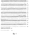
- Figure 1 depicts DNA and predicted amino acid sequence for human
myelin basic protein.
- Figure 2 depicts the response of draining lymph node cells from Lewis
rats immunized 9-10 days previously with MBP (87-99) to 10 µM of MBP (87-99),
medium, the unrelated peptide motilin, and six different MBP analogs. A, L-alanine; k,
D-Iysine; t, D-threonine; r, D-arginine.
- Figure 3 is a graph displaying the proliferative response of the T cell line
NBI to residue 91-substituted analogs of human myelin basic protein (87-99). Ten
different substitutions were tested. The proliferative response of the rat T cell line in
response to concentrations of peptide analogs ranging from 0 to 150 µM was
determined. The extent of proliferation is shown as counts per minute; standard errors
of the mean were less than ±10%. MOT, motilin, a peptide unrelated to MBP; MBP
(87-99), human myelin basic protein residues 87-99; K, lysine; R, arginine; N,
asparagine; H, histidine; L, leucine; S, serine; G, glycine; k, D-Lysine; E, glutamic acid;
F, phenylalanine; A, alanine.
- Figure 4 is a graph displaying the proliferative response of the T cell line
NBI to residue 95-substituted analogs of human myelin basic protein (87-99). Ten
different substitutions were tested. The proliferative response of the rat T cell line in
response to concentrations of peptide analogs ranging from 0 to 150 µM was
determined. The extent of proliferation is shown as counts per minute; standard errors
of the mean were less than ±10%. MOT, motilin, a peptide unrelated to MBP; MBP
(87-99), human myelin basic protein residues 87-99; T, threonine; A, alanine; t,
D-threonine; G, glycine; I, isoleucine; Y, tyrosine; Q, glutamine; S, serine; K, lysine; E,
glutamic acid; H, histidine.
- Figure 5 is a graph displaying the proliferative response of the T cell line
NBI to residue 97-substituted analogs of human myelin basic protein. Eleven different
substitutions were tested. The proliferative response of the T cells to concentrations of
peptide analogs ranging from 0 to 150 µM was determined. The extent of proliferation
is displayed as counts per minute. MBP 87-99, myelin basic protein (87-99); R,
arginine; a, D-alanine; r, D-arginine; G, glycine; K, lysine; Q, glutamine; E, glutamic
acid; T, threonine; L, leucine; F, phenylalanine; H, histidine; A, alanine.
- Figure 6 is a graph illustrating the ability of peptide analogs of MBP to
inhibit proliferation of rat T cells that are reactive to MBP. The proliferative response
of draining lymph node cells from rats immunized with MBP (87-99) to 16.7, 50, or
150 µM of each analog, or 5 µM of MBP (87-99) is displayed. Analogs were added in
the presence of 5 µM MBP (87-99). The extent of proliferation is shown as counts per
minute. Controls consisted of MBP (87-99) only at 5 µM and medium only. h88/A91
refers to a representative peptide analog of MBP (87-99) with D-histidine at residue 88
and alanine at residue 91; h88/A91/p99 refers to another representative peptide analog
of MBP (87-99) with D-histidine at 88, alanine at residue 91, and D-proline at residue
99.
- Figure 7 is a graph demonstrating the inhibition of EAE induction in
Lewis rats following injection of MBP (87-99). Arrows indicate days that either PBS
(control) or h88/A91 peptide analog were administered. EAE was recorded as 0, no
symptoms; 1, tail paralysis; 2, hind limb weakness; 3, hind limb paralysis; 4, hind and
front limb paralysis.
- Figure 8 depicts the amino acid sequence in single letter code for
residues 83 to 99 of human myelin basic protein and the amino acid sequences of the
peptide analogs NBI-5719, NBI-5748, NBI-5765, NBI-5788, and NBI-5789. A dash
indicates amino acid identity. MBP (83-99), myelin basic protein residues 83 to 99; a,
D-alanine; A, L-alanine; K, L-lysine; L, L-leucine.
- Figure 9 is a graph demonstrating the inhibition of EAE induction in
Lewis rats following injection of MBP (83-99). Lewis rats were injected with MBP
(83-99) at day 0. At day 9, rats were injected with either a control peptide, sperm whale
myoglobin (110-121) or the peptide analog, NBI-5788. Each data point represents the
average of the clinical score of six animals.
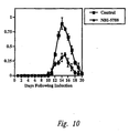
- Figure 10 is a graph demonstrating the inhibition of EAE induction in
Lewis rats following injection of MBP (83-99). Lewis rats were injected with MBP
(83-99) at day 0. At day 9, rats were injected with either a control peptide, sperm whale
myoglobin (110-121) or the peptide analog, NBI-5788. Each data point represents the
average of the clinical score of six animals.
- Figure 11 is a graph demonstrating the inhibition of EAE induction in
Lewis rats following injection of MBP (83-99). Lewis rats were injected with MBP
(83-99) at day 0. At day 9, rats were injected with either a control peptide, sperm whale
myoglobin (110-121) or the peptide analog, NBI-5765. Each data point represents the
average of the clinical score of six animals.
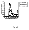
- Figure 12 is a graph demonstrating the inhibition of EAE induction in
SJL/J mice following injection of MBP (87-99). Groups of mice were injected
intraperitoneally on a weekly basis for four weeks with either a control peptide or the
peptide analog, NBI-5719 or NBI-5765. Each data point represents the average of the
clinical score for ten mice.
- Figure 13 is a graph illustrating the ability of a peptide analog of MBP to
inhibit proliferation of a Dr2a restricted human T cell clone, which is reactive to MBP.
The proliferative response of the T cell clone incubated with varying concentrations of
MBP (83-99) and 50 micromolar of either the peptide analog or sperm whale
myoglobin, the control peptide, is depicted.
- Figure 14 is a graph illustrating the ability of peptide analogs of MBP to
inhibit proliferation of a Dr2a restricted human T cell clone, which is reactive to MBP.
The proliferative response of the T cell clone incubated with varying concentrations of
MBP (83-99) and 50 micromolar of either the peptide analog or sperm whale
myoglobin, the control peptide, is depicted.
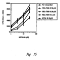
- Figure 15 is a graph illustrating the ability of a peptide analog of MBP to
inhibit proliferation of a Dr2a restricted human T cell clone, which is reactive to MBP.
The proliferative response of the T cell clone incubated with varying concentrations of
MBP (83-99) and 50 micromolar of either the peptide analog or sperm whale
myoglobin, the control peptide, is depicted.
- Figure 16 is a graph illustrating the ability of a peptide analog of MBP to
inhibit proliferation of a Dr2b restricted human T cell clone, which is reactive to MBP.
The proliferative response of the T cell clone incubated with varying concentrations of
MBP (83-99) and 50 micromolar of either the peptide analog or sperm whale
myoglobin, the control peptide, is depicted.
- Figure 17 is a graph illustrating the ability of a peptide analog of MBP to
inhibit proliferation of a Dr2b restricted human T cell clone, which is reactive to MBP.
The proliferative response of the T cell clone incubated with varying concentrations of
MBP (83-99) and 50 micromolar of either the peptide analog or sperm whale
myoglobin, the control peptide, is depicted.
- Figure 18 is a graph illustrating the ability of a peptide analog of MBP to
inhibit proliferation of a Dr2b restricted human T cell clone, which is reactive to MBP.
The proliferative response of the T cell clone incubated with varying concentrations of
MBP (83-99) and 50 micromolar of either the peptide analog or sperm whale
myoglobin, the control peptide, is depicted.
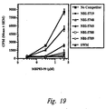
- Figure 19 is a graph illustrating the ability of a peptide analog of MBP to
inhibit proliferation of a Dr2b restricted human T cell clone, which is reactive to MBP.
The proliferative response of the T cell clone incubated with varying concentrations of
MBP (83-99) and 50 micromolar of either the peptide analog or sperm whale
myoglobin, the control peptide, is depicted.
- Figure 20 is a graph illustrating the ability of a peptide analog of MBP to
inhibit proliferation of a Dr2b restricted human T cell clone, which is reactive to MBP.
The proliferative response of the T cell clone incubated with varying concentrations of
MBP (83-99) and 50 micromolar of either the peptide analog or sperm whale
myoglobin, the control peptide, is depicted.
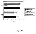
- Figure 21 is a graph displaying the production of TNF-α and IFN-γ by a
Dr 2b restricted human T cell clone, 5F6, which is reactive to MBP. The T cell clone
was incubated in the presence of 3 µM MBP (93-99) with either 10 µM of NBI-5788 or
sperm whale myoglobin or medium only. The expression level of TNF-α and IFN-γ are
displayed as pg/ml.
-
Detailed Description of the Invention
-
Prior to setting forth the invention, it may be helpful to an understanding
thereof to set forth definitions of certain terms and abbreviations that will be used
hereinafter.
-
"Human myelin basic protein" ("MBP") refers to a protein found in the
cytoplasm of human oligodendroglial cells. The nucleotide sequence and predicted
amino acid sequence of human MBP are presented in Figure 1 (SEQ. ID Nos. and
). Although not depicted in Figure 1, different molecular forms of human myelin
basic protein generated by differential splicing or post-translational modification are
also within the scope of this invention.
-
"Peptide analogs" of myelin basic protein are at least 7 amino acids in
length and contain at least one difference in amino acid sequence between the analog
and native human myelin basic protein, one of which is a difference at residue 91, 95 or
97. Unless otherwise indicated, a named amino acid refers to the L-form. An L-amino
acid from the native peptide may be altered to any other one of the 20 L-amino acids
commonly found in proteins, any one of the corresponding D-amino acids, rare amino
acids, such as 4-hydroxyproline, and hydroxylysine, or a non-protein amino acid, such
as β-alanine and homoserine. Also included with the scope of the present invention are
amino acids which have been altered by chemical means such as methylation (e.g.,
α-methylvaline), amidation of the C-terminal amino acid by an alkylamine such as
ethylamine, ethanolamine, and ethylene diamine, and acylation or methylation of an
amino acid side chain function (e.g., acylation of the epsilon amino group of lysine).
-
"Residue 83," "residue 89," "residue 91," "residue 95," and "residue 97"
(also called position 83, position 89, position 91, position 95, and position 97,
respectively), refer to amino acids 83, 89, 91, 95, and 97 of human myelin basic protein
as displayed in Figure 1 or the amino acid at a comparative position. More specifically,
the numbering system for these residues relates to the amino acid position within the
native human protein, regardless of the length of the peptide or the amino acid position
within that peptide.
-
The amino acids are referred to by their standard three-letter or one-letter
code. Unless otherwise specified, the L-form of the amino acid is intended. When the
1-letter code is used, a capital letter denotes the L-form and a small letter denotes the D-form.
The one letter code is as follows: A, alanine; C, cysteine; D, aspartic acid; E,
glutamic acid; F, phenylalanine; G, glycine; H, histidine; I, isoleucine; K, lysine; L,
leucine; M, methionine; N, asparagine; P, proline; Q, glutamine; R, arginine; S, serine;
T, threonine; V, valine; W, tryptophan; and Y, tyrosine.
Peptide Analogs of Myelin Basic Protein
-
As noted above, the present invention provides peptide analogs
comprising at least 7 amino acids selected from residues 83-99 of human myelin basic
protein and including an alteration of the naturally occurring L-lysine at position 91,
L-threonine at position 95, or L-arginine at position 97, to another amino acid. In one
aspect, the peptide analog includes additional alteration of one to three L-amino acids at
positions 86, 87, 88, 91, 95, 97, 98 and/or 99 of human myelin basic protein as long as
91 and 97 are not both altered in the same peptide analog. In another aspect, the peptide
analog additionally has the N-terminal and/or C-terminal residues altered to an amino
acid such that proteolysis is reduced upon administration to a patient compared to a
peptide analog without these additional alterations. In a further aspect, the peptide
analog of MBP comprises at least seven amino acids selected from residues 86-99 and
has one of the residues at position 91, 95 or 97 altered to an amino acid not present in
native MBP protein. In addition to such single alterations, one to three additional
alterations of residues 86 to 99 may be made, as long as residues 91 and 97 are not
altered in the same peptide analog. In yet a further aspect, the peptide analog of MBP
comprises residues 83 to 99, the L-lysine at position 91 is altered to another amino acid
and two to four additional amino acids selected from residues 83 to 90 and 92 to 99 are
altered to another amino acid. Preferably, at least one amino acid is substituted with a
charged amino acid. In addition, the N-terminal and/or C-terminal amino acids may be
altered to a D-amino acid.
-
The peptide analogs are preferably 7 to 17 amino acids, and usually not
longer than 20 amino acids. Particularly preferred peptide analogs are 14 to 17 amino
acids in length. Residues 83, 89, 91, 95, and 97, which are L-glutamic acid,
L-phenylalanine, L-lysine, L-threonine, and L-arginine, respectively, in the native
human protein, are the key residues. Within the subject invention, analogs must have
an amino acid other than L-lysine at position 91, an amino acid other than L-threonine
at position 95, or an amino acid other than L-arginine at position 97.
-
As noted above, any amino acid alteration at position 91 is within the
scope of this invention. Preferred peptide analogs include alteration of L-lysine to any
one of the following amino acids: D-lysine, alanine, glycine, glutamic acid,
phenylalanine, arginine, asparagine, histidine, leucine or serine. These amino acids
include both conservative (similar charge, polarity, hydrophobicity, and bulkiness) and
non-conservative amino acids. Although typically one might expect that only non-conservative
amino acid alterations would provide a therapeutic effect, unexpectedly
even conservative changes (e.g., arginine) greatly affect the function of the peptide
analog as compared to the native peptide. Such diversity of substitution is further
illustrated by the fact that the preferred amino acids noted above are hydrophobic and
hydrophilic, charged and uncharged, polar and non-polar.
-
In addition, any amino acid substitution at residue 95 is also within the
scope of this invention. Preferred peptide analogs contain alterations of L-threonine to
any one of the following amino acids: D-threonine, alanine, glycine, isoleucine,
tyrosine, glutamine, serine, lysine, glutamic acid and histidine. Other preferred
alterations are to non-conservative amino acids. Particularly preferred alterations are to
alanine or D-threonine.
-
Similarly, any amino acid alteration at position 97 is within the scope of
this invention. Preferred peptide analogs include alteration of L-arginine to D-alanine,
D-arginine, glycine, lysine, glutamine, glutamic acid, threonine, leucine, phenylalanine,
histidine or alanine. Other preferred alterations are to non-conservative amino acids.
Particularly preferred alterations are to alanine and D-arginine.
-
Further, any amino acid at position 83 and position 89 are within the
scope of this invention. Preferred peptide analogs contain alterations of L-glutamic acid
at residue 83 to any one of the following amino acids: D-alanine, L-alanine, D-glutamic
acid and L-phenylalanine at position 89 to alanine, leucine, valine, isoleucine.
-
In addition, in certain embodiments at least one other amino acid
selected from residues 86, 87, 88, 89, 95, 98, or 99 is altered. In such embodiments, if
two other amino acids are changed, one is preferably selected from residues 86, 87, 88,
or 89, and the other is selected from residues 98 or 99. Alternatively, up to three
alterations at any positions may be made. In other embodiments, at least two to four
amino acids (in addition to position 91) are altered. In such embodiments, the altered
amino acids are preferably selected from positions 83, 84, 89 and 98.
-
With these general considerations in mind, peptide analogs within the
scope of the invention have an alteration of residue 91, residue 95, or of residue 97.
One set of preferred peptide analogs have double alterations. In one embodiment,
residue 91 is altered as noted above, residue 87 is altered to D-valine, residue 88 to
D-histidine or residue 99 to D-proline. Similarly, in another embodiment, residue 97 is
altered as noted above, and either residue 87 is altered to D-valine, residue 88 to
D-histidine or residue 99 to D-proline. In yet another embodiment, residue 95 is altered
as noted above and residue 87 is altered to D-valine, residue 88 to D-histidine or
residue 99 to D-proline.
-
A second set of preferred peptide analogs have three substitutions. In
one embodiment, residue 91 is altered to alanine, residue 87 is altered to D-valine or
residue 88 is altered to D-histidine and residue 99 is altered to D-proline. In another
embodiment, residue 97 is altered to alanine, residue 88 is altered to D-histidine and
residue 99 to D-proline. In yet another embodiment, residue 95 is altered to alanine,
residue 88 is altered to D-histidine and residue 99 to D-proline. In still another
embodiment, residue 83 is altered to D-alanine, residue 89 is altered to alanine, and
residue 91 is altered to alanine.
-
A third set of preferred peptide analogs have four substitutions. In one
embodiment, residue 83 is altered to D-alanine, residue 84 is altered to lysine, residue
89 is altered to leucine and residue 91 is altered to alanine. In another embodiment,
residue 83 is altered to D-alanine, residue 84 is altered to lysine, and residues 89 and 91
are altered to alanine.
-
A fourth set of preferred peptide analogs have five substitutions. In one
embodiment, residues 83 and 98 are altered to D-alanine, residue 84 is altered to lysine,
and residues 89 and 91 are altered to alanine. In another embodiment, residues 83 and
89 are altered to D-alanine, residue 84 is altered to lysine, residue 89 is altered to
leucine and residue 91 is altered to alanine.
-
Peptide analogs may be synthesized by standard chemistry techniques,
including synthesis by automated procedure. In general, peptide analogs are prepared
by solid-phase peptide synthesis methodology which involves coupling each protected
amino acid residue to a resin support, preferably a 4-methyl-benzhydrylamine resin, by
activation with dicyclohexylcarbodimide to yield a peptide with a C-terminal amide.
Alternatively, a chloromethyl resin (Merrifield resin) may be used to yield a peptide
with a free carboxylic acid at the C-terminus. Side-chain functional groups are
protected as follows: benzyl for serine, threonine, glutamic acid, and aspartic acid;
tosyl for histidine and arginine; 2-chlorobenzyloxycarbonyl for lysine and 2,6-dichlorobenzyl
for tyrosine. Following coupling, the t-butyloxycarbonyl protecting
group on the alpha amino function of the added amino acid is removed by treatment
with trifluoroacetic acid followed by neutralization with di-isopropyl-ethylamine. The
next protected residue is then coupled onto the free amino group, propagating the
peptide chain. After the last residue has been attached, the protected peptide-resin is
treated with hydrogen fluoride to cleave the peptide from the resin, as well as deprotect
the side chain functional groups. Crude product can be further purified by gel filtration,
HPLC, partition chromatography, or ion-exchange chromatography.
-
Peptide analogs within the present invention should (a) compete for the
binding of native MBP peptide (e.g., 87-99 in rats; 83-99 in humans) to MHC; (b) not
cause proliferation of an MBP (87-99)-reactive T cell line; and (c) inhibit induction of
experimental allergic encephalomyelitis (EAE) by MBP (87-99) in rodents.
-
Thus, candidate peptide analogs may be screened for their ability to treat
MS by (1) an assay measuring competitive binding to MHC, (2) an assay measuring a
T cell proliferation, and (3) an assay assessing induction inhibition of EAE. Those
analogs that inhibit binding of the native peptides, do not stimulate proliferation of
MBP-reactive cell lines, and inhibit the development of EAE by native human MBP
(87-99), are useful therapeutics. Although not essential, a further safety assay may be
performed to demonstrate that the analog does not itself induce EAE.
-
Binding of peptides to MHC molecules may be assayed on whole cells.
Briefly, Lewis rat spleen cells are cultured for 3 hours to allow adherent cells to stick to
polystyrene petri dishes. Non-adherent cells are removed. Adherent cells, which
contain cells expressing MHC class II molecules, are collected by scraping the dishes.
The binding of peptide analogs to cells is measured by a fluorescence assay. In this
assay, splenic adherent cells are mixed with different concentrations of peptide analogs
and incubated for 1 hour at 37° in a CO2 incubator. Following incubation, biotin-labeled
MBP (87-99) is added to the culture wells. The cells are incubated for another
hour and then washed three times in medium. Phycoerythrin-conjugated or fluorescein-conjugated
streptavidin is added along with a fluorochrome-labeled OX-6 or OX-17
monoclonal antibody, which reacts with rat MHC Class II I-A and I-E, respectively.
The cells are washed twice before analysis by flow cytometry. Fluorescence intensity is
calculated by subtracting the fluorescence value obtained from cells stained with
phycoerythrin-streptavidin alone (control staining) from the fluorescence value obtained
from biotin-labeled MBP native peptide plus phycoerythrin-streptavidin (experimental
staining). Staining without analog establishes a 100% value. Percent inhibition is
calculated for each analog and expressed as IC50 values. A peptide analog with an IC50
value of less than 100 µM is suitable for further screenings.
-
Candidate peptide analogs are further tested for their property of causing
or inhibiting proliferation of T cell lines. Two different assays may be used as
alternatives. The first measures the ability of the analog to cause proliferation of T cells
in a direct fashion. The second measures the ability of the peptide analog to inhibit
proliferation of T cells induced by native MBP peptide.
-
In the direct proliferation assay, MBP (87-99) reactive T cell lines may
be used as target cells. T cell lines are established from lymph nodes taken from rats
injected with MBP (87-99). Lymph node cells are isolated and cultured for 5 to 8 days
with MBP (87-99) and IL-2 as a source of T cell growth factors. Viable cells are
recovered and a second round of stimulation is performed with MBP (87-99 or 83-99)
and irradiated splenocytes as a source of growth factors. After 5 to 6 passages in this
manner, the proliferative potential of the cell lines are determined. MBP-reactive lines
are used in the proliferation assay. In this assay, T cell lines are cultured for three days
with various concentrations of peptide analogs and irradiated, autologous splenocytes.
After three days, 0.5-1.0 µCi of [3H]-thymidine is added for 12-16 hours. Cultures are
harvested and incorporated counts determined. Mean CPM and standard error of the
mean are calculated from triplicate cultures.
-
As an alternative to the use of T cell lines described above, draining
lymph node cells from Lewis rats injected with MBP (87-99) may be used. Preferably,
this assay is used in combination with the proliferation assay using T cell lines. Briefly,
Lewis rats are injected subcutaneously with MBP (87-99) peptide in complete Freund's
adjuvant. Nine to ten days later, draining lymph node cells are isolated and single-cell
suspensions are prepared. Lymph node cells are incubated with various concentrations
of peptide analogs for three days in a humidified air chamber containing 6.5% CO2.
After incubation, the cultures are pulsed with 1-2 µCi of [3H]-thymidine for 12-18
hours. Cultures are harvested on fiberglass filters and counted in a scintillation counter.
Mean CPM and the standard error of the mean are calculated from data determined in
triplicate cultures. Peptide analogs yielding results that are more than three standard
deviations below the mean response from a comparable concentration of MBP (87-99)
are considered non-stimulatory. Peptide analogs which do not stimulate proliferation at
concentrations of less than or equal to 50 µM are suitable for further screenings.
-
The second or alternative assay is a competition assay for T cell
proliferation. In this assay, antigen presenting spleen cells are first irradiated and then
incubated with native MBP (87-99) peptide for 2-4 hours. These cells are then washed
and further cultured with T cells reactive to MBP (87-99). Various concentrations of
candidate peptide analogs are included in cultures for an additional 3 days. Following
this incubation period, each culture is pulsed with 1 µCi of [3H]-thymidine for an
additional 12-18 hours. Cultures are then harvested on fiberglass filters and counted as
above. Mean CPM and standard error of the mean are calculated from data determined
in triplicate cultures. Peptide analogs which inhibit proliferation to approximately 25%
at a concentration of 50 µM or greater are suitable for further screening.
-
Human T cells reactive to MBP (83-99) may alternatively be used to
measure the ability of the peptide analog to inhibit proliferation of T cells induced by
native MBP (83-99) peptide. MBP-specific T cells may be obtained as previously
described by Martin et al., J. Immunol. 148:1359-1366, 1992. Briefly, T cell lines are
established by culture of human T cells with irradiated, DR-matched peripheral blood
cells in MEM supplemented with 2 mM L-glutamine, 50 µg/ml gentamicin, penicillin
and streptomycin, 100 U/ml rIL-2, and 10% human AB negative serum. Proliferation
of these T cell lines is stimulated by culturing a clone with varying concentration (1.1-30
µM) of native MBP (89-99) peptide, 50 µM of the peptide analog or SWM peptide,
in the presence of irradiated, DR matched peripheral blood cells, following incubation
for approximately 60 hours, the cells are pulsed with 3H-thymidine for 12 hours and
harvested. The amount of incorporated 3H-thymidine is measured.
-
As discussed in detail below, the production of cytokines may also be
assessed. In particular, TNF-α and IFN-γ production are especially interesting. These
pro-inflammatory cytokines are thought to play a role in the pathogenesis of the disease.
Briefly, T cell clone is incubated in the presence of stimulating MBP peptide and
peptide analog or control peptide (SWM) or medium only. After a 24 hour incubation,
the levels of TNF-α and IFN-γ in the supernatant are determined using commercially
available EIA kits (Endogen, Cambridge, MA).
-
Candidate peptides that compete for binding of MBP (87-99) to MHC
and do not cause direct proliferation of T cell line or can inhibit proliferation by MBP
(87-99), are further tested for their ability to inhibit the induction of EAE by MBP
(87-99). Briefly, 500 µg of MBP (87-99) is injected as an emulsion in complete
Freund's adjuvant supplemented with heat killed Mycobacterium tuberculosis (H37Ra).
Rats are injected subcutaneously at the base of the tail with 200 µl of the emulsion.
Rats are divided into two groups. Approximately 2 days prior to disease induction
(usually 10 days following injection of MBP (87-99)) rats are injected intraperitoneally
either with PBS or peptide analogs in PBS. Animals are monitored for clinical signs on
a daily basis by an observer blind to the treatment protocol. EAE is scored on a scale of
0-4: 0, clinically normal; 1, flaccid tail paralysis; 2, hind limb weakness; 3, hind limb
paralysis; 4, front and hind limbs affected. Peptide analogs injected at 5 mg/kg or less
(approximately 1 mg per rat) are considered to inhibit the development of EAE if there
is a 50% reduction in the mean cumulative score over seven days following onset of
disease symptoms in the control group.
-
In addition, as a safety measure, but not essential to this invention,
suitable peptide analogs may be tested for direct induction of EAE. As described in
detail in Example 2, various amounts of peptide analogs are injected at the base of the
tail of rats, and the rats examined daily for signs of EAE. A peptide analog which is not
considered to cause EAE has a mean cumulative score of less than or equal to 1 over
seven days when 1 mg (5 mg/kg) in complete Freund's adjuvant is injected.
Treatment and Prevention of Multiple Sclerosis
-
As noted above, the present invention provides methods for treating and
preventing multiple sclerosis by administering to the patient a therapeutically effective
amount of a peptide analog of human myelin basic protein as described herein. Patients
suitable for such treatment may be identified by criteria establishing a diagnosis of
clinically definite MS as defined by the workshop on the diagnosis of MS (Poser et al.,
Ann. Neurol. 13:227, 1983). Briefly, an individual with clinically definite MS has had
two attacks and clinical evidence of either two lesions or clinical evidence of one lesion
and paraclinical evidence of another, separate lesion. Definite MS may also be
diagnosed by evidence of two attacks and oligoclonal bands of IgG in cerebrospinal
fluid or by combination of an attack, clinical evidence of two lesions and oligoclonal
band of IgG in cerebrospinal fluid. Slightly lower criteria are used for a diagnosis of
clinically probable MS.
-
Effective treatment of multiple sclerosis may be examined in several
different ways. Satisfying any of the following criteria evidences effective treatment.
Three main criteria are used: EDSS (extended disability status scale), appearance of
exacerbations or MRI (magnetic resonance imaging).
-
The EDSS is a means to grade clinical impairment due to MS (Kurtzke,
Neurology 33:1444, 1983). Eight functional systems are evaluated for the type and
severity of neurologic impairment. Briefly, prior to treatment, patients are evaluated for
impairment in the following systems: pyramidal, cerebella, brainstem, sensory, bowel
and bladder, visual, cerebral, and other. Follow-ups are conducted at defined intervals.
The scale ranges from 0 (normal) to 10 (death due to MS). A decrease of one full step
defines an effective treatment in the context of the present invention (Kurtzke, Ann.
Neurol. 36:573-79, 1994).
-
Exacerbations are defined as the appearance of a new symptom that is
attributable to MS and accompanied by an appropriate new neurologic abnormality
(IFNB MS Study Group, supra). In addition, the exacerbation must last at least 24
hours and be preceded by stability or improvement for at least 30 days. Briefly, patients
are given a standard neurological examination by clinicians. Exacerbations are either
mild, moderate, or severe according to changes in a Neurological Rating Scale
(Sipe et al., Neurology 34:1368, 1984). An annual exacerbation rate and proportion of
exacerbation-free patients are determined. Therapy is deemed to be effective if there is
a statistically significant difference in the rate or proportion of exacerbation-free
patients between the treated group and the placebo group for either of these
measurements. In addition, time to first exacerbation and exacerbation duration and
severity may also be measured. A measure of effectiveness as therapy in this regard is a
statistically significant difference in the time to first exacerbation or duration and
severity in the treated group compared to control group.
-
MRI can be used to measure active lesions using gadolinium-DTPA-enhanced
imaging (McDonald et al. Ann. Neurol. 36:14, 1994) or the location and
extent of lesions using T2-weighted techniques. Briefly, baseline MRIs are obtained.
The same imaging plane and patient position are used for each subsequent study.
Positioning and imaging sequences are chosen to maximize lesion detection and
facilitate lesion tracing. The same positioning and imaging sequences are used on
subsequent studies. The presence, location and extent of MS lesions are determined by
radiologists. Areas of lesions are outlined and summed slice by slice for total lesion
area. Three analyses may be done: evidence of new lesions, rate of appearance of
active lesions, percentage change in lesion area (Paty et al., Neurology 43:665, 1993).
Improvement due to therapy is established when there is a statistically significant
improvement in an individual patient compared to baseline or in a treated group versus
a placebo group.
-
Candidate patients for prevention may be identified by the presence of
genetic factors. For example, a majority of MS patients have HLA-type DR2a and
DR2b. The MS patients having genetic dispositions to MS who are suitable for
treatment fall within two groups. First are patients with early disease of the relapsing
remitting type. Entry criteria would include disease duration of more than one year,
EDSS score of 1.0 to 3.5, exacerbation rate of more than 0.5 per year, and free of
clinical exacerbations for 2 months prior to study. The second group would include
people with disease progression greater than 1.0 EDSS unit/year over the past two
years.
-
Efficacy of the peptide analog in the context of prevention is judged
based on the following criteria: frequency of MBP reactive T cells determined by
limiting dilution, proliferation response of MBP reactive T cell lines and clones,
cytokine profiles of T cell lines and clones to MBP established from patients. Efficacy
is established by decrease in frequency of reactive cells, a reduction in thymidine
incorporation with altered peptide compared to native, and a reduction in TNF and
IFN-α. Clinical measurements include the relapse rate in one and two year intervals,
and a change in EDSS, including time to progression from baseline of 1.0 unit on the
EDSS which persists for six months. On a Kaplan-Meier curve, a delay in sustained
progression of disability shows efficacy. Other criteria include a change in area and
volume of T2 images on MRI, and the number and volume of lesions determined by
gadolinium enhanced images.
-
Peptide analogs of the present invention may be administered either
alone, or as a pharmaceutical composition. Briefly, pharmaceutical compositions of the
present invention may comprise one or more of the peptide analogs described herein, in
combination with one or more pharmaceutically or physiologically acceptable carriers,
diluents or excipients. Such compositions may comprise buffers such as neutral
buffered saline, phosphate buffered saline and the like, carbohydrates such as glucose,
mannose, sucrose or dextrans, mannitol, proteins, polypeptides or amino acids such as
glycine, antioxidants, chelating agents such as EDTA or glutathione, adjuvants (e.g.,
aluminum hydroxide) and preservatives. In addition, pharmaceutical compositions of
the present invention may also contain one or more additional active ingredients, such
as, for example, cytokines like β-interferon.
-
Compositions of the present invention may be formulated for the manner
of administration indicated, including for example, for oral, nasal, venous, intracranial,
intraperitoneal, subcutaneous, or intramuscular administration. Within other
embodiments of the invention, the compositions described herein may be administered
as part of a sustained release implant. Within yet other embodiments, compositions of
the present invention may be formulated as a lyophilizate, utilizing appropriate
excipients which provide stability as a lyophilizate, and subsequent to rehydration.
-
Pharmaceutical compositions of the present invention may be
administered in a manner appropriate to the disease to be treated (or prevented). The
quantity and frequency of administration will be determined by such factors as the
condition of the patient, and the type and severity of the patient's disease. Within
particularly preferred embodiments of the invention, the peptide analog or
pharmaceutical compositions described herein may be administered at a dosage ranging
from 5 to 50 mg/kg, although appropriate dosages may be determined by clinical trials.
Patients may be monitored for therapeutic effectiveness by MRI, EDSS, and signs of
clinical exacerbation, as described above.
-
The following examples are offered by way of illustration and not by
way of limitation.
EXAMPLE 1
Preparation of Peptides
-
The peptides were synthesized by solid phase methodology on a peptide
synthesizer (Beckman model 990). Peptides with an amidated carboxyl-terminus were
prepared with a p-methylbenzhydrylamine resin (MBHA resin); for peptides with a free
carboxyl-terminus, a Merrifield resin coupled with the appropriately protected amino
acid was used. Both resins were obtained from Bachem Fine Chemicals (Torrance,
CA). Derivatized amino acids (Bachem Fine Chemicals) used in the synthesis were of
the L-configuration unless specified otherwise, and the N-alpha-amino function
protected exclusively with the t-butyloxycarbonyl group. Side-chain functional groups
were protected as follows: benzyl for serine, threonine, glutamic acid, and aspartic acid;
tosyl for histidine and arginine; 2-chlorobenzyloxycarbonyl for lysine and
2,6-dichlorobenzyl for tyrosine. Coupling of the carboxyl-terminal amino acid to the
MBHA resin was carried out with dicyclohexylcarbodiimide and the subsequent amino
acids were coupled with dicyclohexylcarbodiimide according to Ling et al. (Proc. Natl.
Acad. Sci. USA 81:4302, 1984). After the last amino acid was incorporated, the
t-butyoxycarbonyl protecting group was removed and the peptide-resin conjugate
treated with a mixture of 14 ml hydrofluoric acid (HF), 1.4 ml anisole, and 0.28 ml
methylethyl sulfide per gram of resin conjugate at -20°C for 0.5 hr and at 0°C for 0.5 hr.
HF was removed in vacuum at 0°C, and the resulting peptide and resin mixture was
washed twice with diethyl ether and twice with chloroform and diethyl ether alternately.
The peptide was extracted five times with 2 M acetic acid, and the extract lyophilized.
The lyophilized product was first purified on a column of Sephadex G-25 fine
(Pharmacia-LKB, Piscataway, NJ) developed in 30% acetic acid to remove the
truncated fragments and inorganic salts (Ling et al., 1984). Next, peptides were further
purified by CM-32 carboxymethylcellulose cation-exchange chromatography (Ling et
al., 1984). Final purification was achieved by partition chromatography on Sephadex
G-25 fine (Ling et al., 1984). The synthetic product was characterized by amino acid
analysis, mass spectrometric analysis, and reversed-phase HPLC.
EXAMPLE 2
Immunizations and EAE induction
-
MBP peptide and peptide analogs were dissolved in phosphate-buffered
saline (PBS) and emulsified with an equal volume of incomplete Freund's adjuvant
supplemented with 4 mg/ml heat-killed Mycobacterium tuberculosis H37Ra in oil
(Difco Laboratories, Inc., Detroit, MI). Rats were immunized subcutaneously at the
base of the tail with 0.1-0.2 ml containing 500 µg of peptide in the emulsion and were
monitored for clinical signs daily. EAE was scored on a scale of 0-4, as follows: 0,
clinically normal; 1, flaccid tail; 2, hind limb weakness; 3, hind limb paralysis; 4, front
and hind limbs affected.
EXAMPLE 3
Long-term T cell lines
-
Antigen specific long-term T cell lines were derived using the method
developed by Ben-Nun et al. (Eur. J. Immunol. 11:195, 1981). Lewis rats were injected
with MBP (87-99) or MBP (83-99) as described above. Nine to ten days later draining
lymph node cells were cultured (107/ml) for 5-8 days in stimulation medium
(Dulbecco's modified Eagle's medium supplemented with 5x10⁻⁵M 2-mercaptoethanol,
2mM L-glutamine, 1 mM sodium pyruvate, 100 µg/ml penicillin, 100 µg/ml
streptomycin and 10% fetal bovine serum (Hyclone Laboratories, Logan, UT)) together
with 10-20 µM of the MBP (87-99) peptide and 15 U/ml IL-2. After 5 to 8 days of
culture, viable cells were collected from the interface after Ficoll-Hypaque separation
and washed three times. These cells were recultured at 1 x 10⁷ cells/ml in medium with
5 x 10⁵ irradiated (3000 rad) autologous splenocytes as accessory cells and 10-20 µM of
MBP (87-99). After 5 to 6 stimulation cycles, plates were screened by the ability of
cells to proliferate in response to MBP (87-99). Positive lines were transferred to
24-well flat bottom plates and restimulated.
EXAMPLE 4
MHC binding assay
-
The ability of MBP peptides and peptide analogs to bind MHC was
measured. An assay which characterizes the binding of peptides to MHC molecules on
antigen presenting cells (APC) was employed (Mozes et al., EMBO J. 8:4049, 1989;
Gautam et al., PNAS 91:767, 1994). Spleen cells were cultured in Dulbecco's modified
Eagle's medium supplemented with 10% fetal bovine serum (Hyclone Laboratories,
Logan, UT) in standard polystyrene petri dishes (100 x 15 mm) in a 37°C incubator
containing 6.5% CO2 for 3 hours. Thereafter, non-adherent cells were removed, and the
plates were washed three times with PBS. Adherent cells were collected using a cell
scraper. The binding of MBP (87-99) analogs was measured using a fluorescence
assay. Briefly, 5 x 10⁵ splenic adherent cells in staining buffer (PBS containing 0.1%
bovine serum albumin) were mixed with different concentrations ranging from
0-400 µM of MBP analogs in individual wells of U-shape 96-well microculture plates
and incubated for 1 hr at 37°C in a 6.5% CO2 incubator. Following incubation, 10 µM
of biotin-labeled MBP native peptide was added to culture wells for 1 h. Cells were
washed three times with the staining buffer. Phycoerythrin-conjugated or fluorescein-conjugated
streptavidin (Becton Dickinson, San Jose, CA) was added as a second step
reagent (1 µg/well) along with 1 µg/well of fluorochrome-labeled OX-6 or OX-17
monoclonal antibody (Pharmingen, San Diego, CA), which reacts with rat MHC class II
I-A or I-E, respectively. The cells were washed twice before cytofluorographic analysis
on a FACScan (Becton Dickinson). Fluorescence intensity for each sample was
calculated by subtracting the fluorescence obtained from OX positive cells stained with
phycoerythrin-streptavidin alone (control staining) from the fluorescence obtained from
OX positive cells stained with biotin-labeled MBP plus phycoerythrin-streptavidin.
Percent inhibition was calculated for each analog and expressed as IC50 values.
-
The peptide analog, h88/A91, which contains D-histidine at position 88
and alanine at position 91 competed as effectively as MBP (87-99) for MHC against
MBP (87-99). At 200 µM, MBP (87-99) inhibited binding by 68.4% and h88/A91
inhibited binding by 67.64%. At 100 µM, MBP (87-89) inhibited binding by 40% and
a83, A89, A91 inhibited binding by 25%.
EXAMPLE 5
Antigen-specific lymph node cell proliferation assay
-
Female Lewis rates, approximately six weeks old, were purchased from
Harlan Sprague, Indianapolis, IN. MBP peptides were dissolved in phosphate-buffered
saline (PBS) and emulsified with an equal volume of complete Freund's adjuvant (Difco
Laboratories, Inc., Detroit, MI) supplemented with 2 mg/ml of heat-killed
Myobacterium tuberculosis H37Ra in oil (Difco). Rats were immunized
subcutaneously in the base of the tail with 0.1 ml containing 100 µg of the peptide in
the emulsion. Nine to ten days following immunization, rats were sacrificed, their
draining lymph node removed and a single cell suspension made. Cells were
resuspended to 5 x 10⁶ cells per ml in stimulation medium containing Dulbecco's
modified Eagle's medium (Gibco BRL, Gaithersburg, MD) supplemented with 2
mercaptoethanol (5 x 10⁻⁵ M), L-glutamine (2 mM), sodium pyruvate (1 mM),
penicillin (100 µg/ml), streptomycin (100 µg/ml), and 1% normal rat serum.
-
For the assay, 100 µl of the lymph node cell suspension was added to 96-well
flat-bottom wells in the presence of an equal volume of medium containing 10 µM
of various peptides (including: motilin as a negative control; MBP87-99; medium only
or alanine or D-amino acid substituted at position 91, 95, or 97). Cultures were then
incubated at 37°C in humidified air containing 7.5% CO2. After 3 days of incubation,
1.0 µCi of tritiated thymidine (20 Ci/mM; New England Nuclear) was added to each
well and the plates reincubated for an additional 12-16 hours. The plates were then
harvested with a Matrix filtermate harvester (Packard) and counted using an Automatic
Direct Beta Counter (Packard). Mean cpm and the standard error of the mean were
calculated from triplicate wells.
-
As seen in Figure 2, MBP (87-99) stimulated lymph node cells in
contrast to the peptide analogs. Alanine alterations at positions 95 and 97 and D-amino
acid alterations at residues 91, 95, and 97 failed to stimulate cells above the control
peptide, motilin.
EXAMPLE 6
Antigen-specific T cell line proliferation assays
-
Assays for the antigen-specific proliferation assay of T cell lines were
performed in 96-well flat bottom microtiter plates as described (Zamvil et al., Nature
317:355-358, 1985; Offner et al., J. Immunol. 148:1706-1711, 1992; Gold et al., J.
Immunol. 148:1712-1717, 1992; Karin et al., J. Exp. Med. 180:2227-2237, 1994). T
cell lines were established as described in Example 3. An initial 1:10 dilution of a 1.5
mM stock solution of MBP or the peptide analogs were added into tissue culture
medium. The samples were diluted by three-fold serial dilutions (final volume 100 µl).
The responding continuous T cell lines were resuspended to 4 x 10⁵ cells per ml and 50
µl aliquots added to each well (5 x 10⁴ cells per well). Approximately 1 x 10⁶ irradiated
(3000R) splenocyte feeder cells were also added to each well. Cultures were then
incubated at 37°C in humidified air containing 7.5% CO2 for 3 days. Twelve to sixteen
hours prior to harvesting, 0.5-1.0 µCi of [3H]-thymidine (20 Ci/mM; New England
Nuclear) was added to each well and the cultures reincubated. Plates were then
harvested with a Matrix filtermate harvester (Packard) and counted using an Automatic
Direct Beta Counter (Packard). Mean cpm and the standard error of the mean were
calculated from triplicate wells.
-
As can be seen in Figures 3, 4, and 5 a peptide analog with any
substitution of position 91, 95, or 97 failed to stimulate proliferation of a MBP (87-99)-reactive
T cell line. The effect was dramatic as even 150 µM of peptide analog was 1 to
2 logs less effective at causing proliferation.
EXAMPLE 7
Antagonism of T cell proliferation assay
-
T cell antagonism was detected in a prepulsed proliferation assay as
described by De Magistris et al. (Cell 58:625, 1992) with minor modifications. Antigen
presenting spleen cells were γ-irradiated (3000 rad) and incubated with shaking at a
concentration of 107 cells/well with 0.2-2.0 µM of the native peptide MBP (87-99) in
stimulation medium in 10 ml tissue culture plates for 2 to 4 hours at 37°C in
humidified air containing 6.5% CO2. Spleen cells were then washed and re-cultured at
a concentration of 5 x 10⁵ cells/well in U-shape 96-well microculture plates together
with 5 x 10⁴ resting MBP (87-99) reactive T cells. Various concentrations of antagonist
peptides, ranging from 5-150 µM, were added for an additional 72 hours. Each well
was pulsed with 0.5-1 µCi of [3H]-thymidine (specific activity 10 Ci/mmol) for the final
12-16 hours. The cultures were then harvested on fiberglass filters and the proliferative
response expressed as CPM±SEM.
-
The data presented in Figure 6 demonstrates that the double altered
peptide analog, h88/A91, and the triple altered peptide analog, h88/A91/p99,
significantly inhibited proliferation of a MBP reactive T cell line. The triple altered
analog caused inhibition at 50 µM and higher concentration, while the double altered
analog caused inhibition at 150 µM.
EXAMPLE 8
Treatment of 87-99 Induced EAE in Lewis Rats
-
Female Lewis rats, which were 6-8 weeks old, were injected with 500 µg
of MBP (87-99) in CFA containing 500 µg of Mycobacterium tuberculosis at the base
of the tail in 200 µl volume. Rats were divided in groups of 5. The control group
received 0.5 ml of PBS and the treatment group received the h88/A91 peptide analog (1
mg/0.5 ml PBS) intraperitoneally, twice, on days 9 and 10 after immunization. Animals
were monitored for disease symptoms on a daily basis. EAE was recorded on the
following scale: 0, no symptoms; 1, tail paralysis; 2, hind limb weakness; 3, hind limb
paralysis; 4, hind and front limbs affected.
-
Data from two different experiments was obtained as mean cumulative
score of 5 animals (Figure 7). Untreated control animals went on to develop high level
of disease whereas h88/A91 analog of the MBP peptide 87-99 was effective in
preventing significantly the development of EAE in two experiments. Though the
analog was given just before the onset of overt symptoms, it was able to arrest the
development of EAE.
EXAMPLE 9
Induction of EAE by Peptide Analog
-
The ability of peptide analogs to cause EAE is assessed in vivo. Rats
were injected with MBP (87-99) or h88/A91 peptide analog as described in Example 2.
Animals were monitored daily for evidence of EAE. Rats receiving MBP (87-99) had
100% incidence (18/18 rats) of EAE with a mean maximum clinical score of 2.4 ± 0.2.
In contrast, 0/12 rats receiving the peptide analog h88/A91 had EAE. Therefore, this
peptide analog does not induce EAE.
EXAMPLE 10
Treatment of EAE With Peptide Analogs
-
The 91K>A peptide analog is capable of inhibiting the adoptive transfer
of disease by immune T cells in the Lewis rat strain (Karin et al., 1994). Further
characterization of the effects of the APL on the immune system was investigated in
Lewis rates injected with HBP.
-
In this system, experimental allergic encephalomyelitis (EAE) was
induced in twelve female Lewis rats by injection of MBP(83-99) peptide in complete
Freund's adjuvant (CFA) at the base of the tail. Nine days later, rats were divided into
two groups of six animals and subcutaneously injected with 13.2 mg/kg of either
peptide analog or a control peptide, sperm whale myoglobin (SWM) (110-121).
Animals were monitored daily for disease symptoms and scored in a blinded fashion on
a nonlinear ascending scale of 0-4 with increments denoting increasing paralysis. Each
individual score was averaged with group cohorts to obtain the mean clinical score.
The results from one such experiment are shown in Figure 9.
-
As seen in Figure 9, the disease severity in those animals treated with the
APL NBI-5788 (Figure 8) was about 50% reduced compared to the control group.
Figure 10 shows the average disease severity of the results from three separate therapy
experiments. The APL NBI-5788 significantly reduced the severity and duration of the
disease in this model system. Figure 11 shows the results from treatment using another
APL, NBI-5765 (Figure 8). This APL also significantly reduced the magnitude of the
disease in the treated group over the control animals.
-
Although these results clearly demonstrate that APL inhibits the
development of EAE, a murine animal model system of EAE has also been developed.
The SJL/J (H-25) mouse develops a chronic relapsing form of EAE in response to
immunization with MBP(83-99) peptide in the presence of pertussis vaccine. The
ability of the peptide analogs NBI-5719 and NBI-5765 to inhibit the disease was
evaluated (see Figure 12).
-
Groups of 10 animals were injected intraperitoneally weekly for 4 weeks
with 20 mg/kg of either a control peptide or the peptide analog. The animals were then
monitored for disease over the next 2-3 months. As can be seen in Figure 12, SJL/J
mice developed symptoms of EAE beginning around day 20 in the control group that
lasted for approximately 3 weeks. Beginning around day 70, a relapse occurred reaching
a mean clinical score of about 1. However, weekly injection with the APL NBI-5765 or
NBI-5719 for four weeks not only reduced the level of the disease in the first phase, but
also reduced the severity of the relapse. This is particularly striking since the animals
had not been exposed to the APL for approximately one month.
EXAMPLE 11
Effects of Peptide Analogs on Human T Cell Proliferation
-
The ability of the peptide analogs NBI-5719, 5748, 5765, 5788 and 5789
to affect human T cell proliferation was assessed. A constant amount of peptide analog
or the control peptide SWM (50 µM) was cultured with varying concentrations of native
MBP(83-99) peptide (1.1-30 µM) in the presence of irradiated, DR matched peripheral
blood cells and T cell clones derived from various MS patients. Human T cells (1 x
106) were cultured with DR matched, irradiated peripheral blood cells (PBL, 5 x 10⁶) in
medium containing IMDM supplemented with 3 µM MBP83-99, 2 mM L-glutamine,
50 µg/ml gentamicin penicillin/streptomycin, 100 U/ml rIL-2 and 10% human
AB-negative serum. Cells were cultured for approximately 60 hours, pulsed with
tritiated thymidine for 12 hours, and harvested. The amount of tritiated thymidine
incorporated was measured, and the data represented as the mean plus or minus the
standard error of the mean of replicate samples. Representative results are shown in
Figures 13 and 14.
-
As seen in Figure 13, the peptide analog NBI-5788 corresponding to
MBP(83-99) (83E>a, 84N>K, 89F>L, and 91K>A) inhibited the ability of a human
Dr2a restricted T cell clone to respond to varying concentrations of MBP(83-99), where
the irrelevant peptide (sperm whale myoglobin, SWM 110-121) had little effect on the
proliferative capacity of the T cells. Figure 14 shows that all the peptides inhibited the
ability of the Dr2a restricted T cells to respond to native MBP peptide in a
concentration dependent fashion.
-
The potency of NBI-5788 was then determined by varying
concentrations of the APL (2, 10, or 50 µM) in the presence of varying amounts of the
native MBP(83-99) (1.1-30 µM). As seen in Figure 15, at both 10 and 50 µM, NBI-5788
significantly altered the ability of the Dr2a T cell line to respond to MBP(83-99),
but no significant inhibition was seen with the irrelevant peptide SWM.
-
The ability of the peptide analog to inhibit the proliferative response of
MBP-reactive T cells isolated from Dr2b (DrB1*1501) individuals was determined. A
constant amount of NBI-5788 (50 µM) was cultured with varying concentrations of
native peptide (1.1-30 µM) in the presence of irradiated, DR matched peripheral blood
cells and T cell clones derived from various MS patients.
-
Figures 16, 17, and 18 depict results using three different T cell lines.
Each T cell clone varies in the amount of thymidine incorporated in response to MBP
peptide. Nevertheless, NBI-5788 inhibited the ability of the T cell clones to respond to
MBP peptide in a concentration dependent fashion. The irrelevant peptide SWM had
little influence on the ability of the T cells to respond to MBP peptide.
-
Figure 19 depicts the ability of NBI-5719, NBI-5748, NBI-5765, NBI-5788,
and NBI-5789 to inhibit the MBP-dependent proliferation of the Dr2b restricted
human T cell clone 5F6. As seen above with the Dr2b restricted T cells (Figure 14), the
APL inhibited the MBP-dependent proliferation in a concentration dependent fashion.
However, the control peptide SWM had little effect on the proliferative response.
-
Figure 20 depicts the ability of NBI-5719 and NBI-5765 to inhibit the
MBP-dependent proliferation of the Dr4 Dw4 restricted human T cell clone MS-1. As
seen above with the Dr2 restricted T cells, the APL inhibited the MBP-dependent
proliferation in a concentration dependent fashion.
-
The ability of the peptide analog ligand to influence cytokine production
was next measured. The Dr2b-restricted T cell clone 5F6 was incubated in the presence
of 3 µM MBP peptide with either 10 µM of NBI-5788 or SWM or medium only. As a
control, cells were cultured in the presence of medium alone. After 24 hours,
supernatants were removed and the levels of tumor necrosis factor alpha (TNF-α) and
interferon-γ (IFN-γ) determined using commercially available EIA kits.
-
As can be seen in Figure 21, MBP stimulated the production of both
TNF-α and IFN-γ (approximately 200 and 160 pg/ml, respectively). However, the
peptide analog ligand NBI-5788 dramatically inhibited the production of both pro-inflammatory
cytokines to approximately levels achieved with medium only. The
irrelevant peptide SWM had minimal effect on cytokine production. None of the
peptide analogs NBI-5719, NBI-5748, NBI-5765, NBI-5788, or NBI-5789 stimulated
cytokine production over background, even at concentrations of 50 µM (data not
shown).
-
From the foregoing, it will be evident that although specific
embodiments of the invention have been described herein for the purpose of illustrating
the invention, various modifications may be made without deviating from the spirit and
scope of the invention.
THE PRESENT INVENTION WILL NOW BE DESCRIBED BY WAY OF
REFERENCE TO THE CLAUSES BELOW:
-
- 1. A peptide analog comprising at least seven amino acids selected from
residues 86 to 99 of human myelin basic protein, including residue 91, wherein the L-lysine
at position 91 is altered to another amino acid, and one to three L-amino acids selected from
the group consisting of valine at position 86, valine at position 87, histidine at position 88,
threonine at position 95, threonine at position 98 and proline at position 99 are altered to an
amino acid other than the amino acid present in the native protein at that position.
- 2. The peptide analog of clause 1 wherein L-lysine at position 91 is altered
to a non-conservative amino acid.
- 3. The peptide analog of clause 1 wherein residue 91 is altered to D-lysine.
- 4. The peptide analog of clause 1 wherein residue 91 is altered to an
amino acid selected from the group consisting of arginine, asparagine, histidine, leucine,
serine, glycine, glutamic acid, phenylalanine, alanine and D-lysine.
- 5. The peptide analog of clause 1 wherein residue 91 is altered to alanine
and residue 87 is altered to D-valine.
- 6. The peptide analog of clause 1 wherein residue 91 is altered to alanine
and residue 88 is altered to D-histidine.
- 7. The peptide analog of clause 1 wherein residue 91 is altered to alanine
and residue 99 is altered to D-proline.
- 8. The peptide analog of clause 1 wherein residue 91 is altered to alanine,
residue 87 is altered to D-valine, and residue 99 is altered to D-proline.
- 9. The peptide analog of clause 1 wherein residue 91 is altered to alanine,
residue 88 is altered to D-histidine, and residue 99 is altered to D-proline.
- 10. The peptide analog of clause 1 wherein residue 88 is altered to an
amino acid selected from the group consisting of serine, glutamic acid, tyrosine, leucine, D-histidine,
glutamine, phenylalanine and lysine.
- 11. A peptide analog comprising at least seven amino acids selected from
residues 86 to 99 of human myelin basic protein, including residue 97, wherein the L-arginine
at position 97 is altered to another amino acid and one to three L-amino acids selected from
the group consisting of valine at position 86, valine at position 87, histidine at position 88,
threonine at position 95, threonine at position 98 and proline at position 99 are altered to an
amino acid other than the amino acid present in the native protein at that position.
- 12. The peptide analog of clause 11 wherein the L-arginine at position 97 is
altered to a non-conservative amino acid.
- 13. The peptide analog of clause 11 wherein residue 97 is altered to
D-arginine.
- 14. The peptide analog of clause 11 wherein residue 97 is altered to an
amino acid selected from the group of D-alanine, D-arginine, glycine, lysine, glutamine,
glutamic acid, threonine, leucine, phenylalanine, histidine and alanine.
- 15. The peptide analog of clause 11 wherein residue 97 is altered to alanine
and residue 87 is altered to D-valine.
- 16. The peptide analog of clause 11 wherein residue 97 is altered to alanine
and residue 88 is altered to D-histidine.
- 17. The peptide analog of clause 11 wherein residue 97 is altered to alanine
and residue 99 is altered to D-proline.
- 18. The peptide analog of clause 11 wherein residue 97 is altered to alanine,
residue 87 is altered to D-valine, and residue 99 is altered to D-proline.
- 19. The peptide analog of clause 11 wherein residue 97 is altered to alanine,
residue 88 is altered to D-histidine and residue 99 is altered to D-proline.
- 20. The peptide analog of clause 11 wherein residue 88 is altered to an
amino acid selected from the group consisting of serine, glutamic acid, tyrosine, leucine, D-histidine,
glutamine, phenylalanine and lysine.
- 21. A peptide analog comprising at least seven amino acids selected from
residues 86 to 99 of human myelin basic protein, including residue 95, wherein the
L-threonine at position 95 is altered to another amino acid and one to three L-amino acids
selected from the group consisting of valine at position 86, valine at position 87, histidine at
position 88, threonine at position 98 and proline at position 99 are altered to an amino acid
other than the amino acid present in the native protein at that position.
- 22. The peptide analog of clause 21 wherein the L-threonine at position 95
is altered to a non-conservative amino acid.
- 23. The peptide analog of clause 21 wherein residue 95 is altered to
D-threonine.
- 24. The peptide analog of clause 21 wherein residue 95 is altered to an
amino acid selected from the group consisting of alanine, D-threonine, glycine, isoleucine,
tyrosine, glutamine, serine, lysine, glutamic acid and histidine.
- 25. The peptide analog of clause 21 wherein residue 95 is altered to alanine
and residue 87 is altered to D-valine.
- 26. The peptide analog of clause 21 wherein residue 95 is altered to alanine
and residue 88 is altered to D-histidine.
- 27. The peptide analog of clause 21 wherein residue 95 is altered to alanine
and residue 99 is altered to D-proline.
- 28. The peptide analog of clause 21 wherein residue 95 is altered to alanine,
residue 87 is altered to D-valine, and residue 99 is altered to D-proline.
- 29. The peptide analog of clause 21 wherein residue 95 is altered to alanine,
residue 88 is altered to D-histidine, and residue 99 is altered to D-proline.
- 30. A peptide analog comprising at least seven amino acids selected from
residues 86 to 99 of human myelin basic protein, including residue 91, wherein the L-lysine
at position 91 is altered to another amino acid and the N-terminal amino acid and the
C-terminal amino acid are altered to another amino acid, such that upon administration of the
peptide analog in vivo proteolysis is reduced.
- 31. The peptide analog of clause 30 wherein the N-terminal and/or
C-terminal amino acids are D-amino acids.
- 32. The peptide analog of clause 30 wherein L-lysine at position 91 is
altered to a non-conservative amino acid.
- 33. The peptide analog of clause 30 wherein residue 91 is altered to D-lysine.
- 34. The peptide analog of clause 30 wherein residue 91 is altered to an
amino acid selected from the group consisting of arginine, asparagine, histidine, leucine,
serine, glycine, glutamic acid, phenylalanine, alanine and D-lysine.
- 35. A peptide analog comprising at least seven amino acids selected from
residues 86 to 99 of human myelin basic protein, including residue 95, wherein the L-lysine
at position 91 is altered to another amino acid and the N-terminal amino acid and the
C-terminal amino acid are altered to another amino acid, such that upon administration of the
peptide analog in vivo proteolysis is reduced.
- 36. The peptide analog of clause 35 wherein the N-terminal and/or
C-terminal amino acids are D-amino acids.
- 37. The peptide analog of clause 35 wherein the L-threonine at position 95
is altered to a non-conservative amino acid.
- 38. The peptide analog of clause 35 wherein residue 95 is altered to
D-threonine.
- 39. The peptide analog of clause 35 wherein residue 95 is altered to an
amino acid selected from the group consisting of alanine, D-threonine, glycine, isoleucine,
tyrosine, glutamine, serine, lysine, glutamic acid and histidine.
- 40. A peptide analog comprising at least seven amino acids selected from
residues 86 to 99 of human myelin basic protein, including residue 97, wherein the L-lysine
at position 91 is altered to another amino acid and the N-terminal amino acid and the
C-terminal amino acid are altered to another amino acid, such that upon administration of the
peptide analog in vivo proteolysis is reduced.
- 41. The peptide analog of clause 40 wherein the N-terminal and/or
C-terminal amino acids are D-amino acids.
- 42. The peptide analog of clause 40 wherein the L-arginine at position 97 is
altered to a non-conservative amino acid.
- 43. The peptide analog of clause 40 wherein residue 97 is altered to
D-arginine.
- 44. The peptide analog of clause 40 wherein residue 97 is altered to an
amino acid selected from the group of D-alanine, D-arginine, glycine, lysine, glutamine,
glutamic acid, threonine, leucine, phenylalanine, histidine and alanine.
- 45. A peptide analog comprising at least seven amino acids selected from
residues 86 to 99 of human myelin basic protein, including residue 91, wherein the L-lysine
at position 91 is altered to another amino acid.
- 46. The peptide analog of clause 45 comprising seven to twelve amino
acids.
- 47. The peptide analog of clause 45, further comprising altering one to
three additional residues selected from residues 86-90, 92-96, 98 and 99 to another amino
acid.
- 48. The peptide analog of clause 45 wherein L-lysine at position 91 is
altered to a non-conservative amino acid.
- 49. The peptide analog of clause 45 wherein residue 91 is altered to D-lysine.
- 50. The peptide analog of clause 45 wherein residue 91 is altered to an
amino acid selected from the group consisting of arginine, asparagine, histidine, leucine,
serine, glycine, glutamic acid, phenylalanine, alanine and D-lysine.
- 51. A peptide analog comprising at least seven amino acids selected from
residues 86 to 99 of human myelin basic protein, including residue 95, wherein the
L-threonine at position 95 is altered to another amino acid.
- 52. The peptide analog of clause 45, further comprising altering one to
three additional residues selected from residues 86-90, 92-94, and 96-99 to another amino
acid.
- 53. The peptide analog of clause 45, further comprising altering one to
three additional residues selected from residues 86-94, 96, 98 and 99 to another amino acid.
- 54. A peptide analog comprising at least seven amino acids selected from
residues 86 to 99 of human myelin basic protein, including residue 97, wherein the L-arginine
at position 97 is altered to another amino acid.
- 55. The peptide analog of clause 45, further comprising altering one to
three additional residues selected from residues 86-90, 92-96, 98 and 99 to another amino
acid.
- 56. A pharmaceutical composition comprising a peptide analog according
to any one of clauses 1, 11, 21, 30, 35, 40, 45, 51, and 54 in combination with a
physiologically acceptable carrier or diluent.
- 57. A method of treating multiple sclerosis, comprising:
- administering to a patient a therapeutically effective amount of a
pharmaceutical composition comprising a peptide analog according to any one of clauses 1,
11, 21, 30, 35, 40, 45, 51, and 54 in combination with a physiologically acceptable carrier or
diluent.
- 58. The method of clause 57 wherein the peptide analog comprises 14
amino acids selected from residues 86 to 90 and residue 91 is alanine, residue 88 is
D-histidine and residue 99 is D-proline.
- 59. The method of clause 57 wherein the peptide analog comprises 14
amino acids selected from residues 86 to 90 and residue 91 is alanine, residue 87 is D-valine
and residue 99 is D-proline.
- 60. The method of clause 57 wherein the peptide analog comprises 14
amino acids selected from residues 86 to 90 and residue 91 is alanine and residue 88 is
D-histidine.
- 61. The method of clause 57 wherein the peptide analog comprises 14
amino acids selected from residues 86 to 90 and residue 91 is alanine and residue 87 is
D-valine.
- 62. The method of clause 57 wherein the peptide analog comprises 14
amino acids selected from residues 86 to 90 and residue 91 is alanine and residue 99 is
D-proline.
- 63. The method of clause 57 wherein the peptide analog comprises 14
amino acids selected from residues 86 to 90 and residue 95 is alanine, residue 87 is D-valine,
and residue 99 is D-proline.
- 64. The method of clause 57 wherein the peptide analog comprises 14
amino acids selected from residues 86 to 90 and residue 95 is alanine, residue 88 is
D-histidine and residue 99 is D-proline.
- 65. The method of clause 57 wherein the peptide analog comprises 14
amino acids selected from residues 86 to 90 and residue 95 is alanine and residue 88 is
D-histidine.
- 66. The method of clause 57 wherein the peptide analog comprises 14
amino acids selected from residues 86 to 90 and residue 95 is alanine and residue 99 is
D-proline.
- 67. The method of clause 57 wherein the peptide analog comprises 14
amino acids selected from residues 86 to 90 and residue 95 is alanine and residue 87 is
D-histidine.
- 68. The method of clause 57 wherein the peptide analog comprises 14
amino acids selected from residues 86 to 90 and residue 97 is alanine, residue 87 is D-valine,
and residue 99 is D-proline.
- 69. The method of clause 57 wherein the peptide analog comprises 14
amino acids selected from residues 86 to 90 and residue 97 is alanine, residue 88 is
D-histidine and residue 99 is D-proline.
- 70. The method of clause 57 wherein the peptide analog comprises 14
amino acids selected from residues 86 to 90 and residue 97 is alanine and residue 87 is
D-valine.
- 71. The method of clause 57 wherein the peptide analog comprises 14
amino acids selected from residues 86 to 90 and residue 97 is alanine and residue 88 is
D-histidine.
- 72. The method of clause 57 wherein the peptide analog comprises 14
amino acids selected from residues 86 to 90 and residue 97 is alanine and residue 99 is
D-proline.
- 73. A peptide analog comprising residues 83 to 99 of human myelin basic
protein, wherein the L-lysine at position 91 is altered to another amino acid, and two to four
additional L-amino acids selected from residues 83 to 90 and 92 to 99 are altered to an amino
acid other than the amino acid present in the native protein at that position.
- 74. The peptide analog of clause 73 wherein L-lysine at position 91 is
altered to alanine.
- 75. The peptide analog of clause 73, including a substitution of the
phenylalanine at position 89 with another amino acid.
- 76. The peptide analog of clause 73 wherein the N-terminal amino acid
and/or the C-terminal amino acid are altered to another amino acid.
- 77. The peptide analog of clause 76 wherein the N-terminal and/or
C-terminal amino acids are altered to a D-amino acid.
- 78. The peptide analog of clause 77 wherein the N-terminal amino acid is
residue 83 of human myelin basic protein.
- 79. The peptide analog of clause 73 wherein at least one of the additional
L-amino acids selected from residues 83 to 90 and 92 to 99 is substituted with a charged
amino acid.
- 80. A peptide analog comprising residues 83 to 99 of human myelin basic
protein, wherein the L-glutamic acid at position 83 is altered to D-alanine, L-asparagine at
position 84 is altered to L-lysine, L-phenylalanine at position 89 is altered to L-leucine, and
L-lysine at position 91 is altered to L-alanine.
- 81. A peptide analog comprising residues 83 to 99 of human myelin basic
protein, wherein L-glutamic acid at position 83 is altered to D-alanine, L-phenylalanine at
position 89 is altered to L-alanine and L-lysine at position 91 is altered to L-alanine.
- 82. A peptide analog comprising residues 83 to 99 of human myelin basic
protein, wherein the L-lysine at position 91 and the L-phenylalanine at position 89 are altered
to other amino acids.
- 83. A pharmaceutical composition comprising a peptide analog according
to any one of clauses 73 to 82 in combination with a physiologically acceptable carrier or
diluent.
- 84. A method of treating multiple sclerosis, comprising:
- administering to a patient a therapeutically effective amount of a
pharmaceutical composition according to clause 83.
-