Technical Field
The present invention relates to a method for
preparing a substrate for immobilizing ligands which is
useful for preparation of a substrate having immobilized
ligands such as a DNA chip, a saccharide chip or a protein
chip.
Background Art
With the progress in techniques for analyzing
genomes, structures of genomes of various organisms have
been (are being) elucidated. In parallel, development of
techniques for analyzing functions of genomes or encoded
functional molecules (e.g., proteins) has been promoted.
Among these, a DNA chip (DNA microarray), a saccharide chip
and a protein chip are noticeable techniques. A DNA chip
is a microarray in which a number of different genes or
fragments thereof (DNA fragments) are arrayed and
immobilized onto a surface of a solid phase substrate such
as a slide glass. It is very useful as a technique that
remarkably promotes analyses of expression, mutation,
polymorphism and the like of a gene. A saccharide chip is
a microarray in which monosaccharides or oligosaccharides
are arrayed and immobilized onto a surface of a solid phase
substrate such as a slide glass. It is very useful as a
technique that remarkably promotes analyses of saccharides,
saccharide targets, interactions between them and the like.
A protein chip is a microarray in which proteins,
antibodies or fragments thereof are arrayed and immobilized.
It is very useful as a technique that remarkably promotes
analyses of proteins, antibodies, interactions between them
and the like.
Immobilization of DNAs onto a substrate is a
fundamental technique for preparing a DNA chip. Known
methods are generally divided into two groups. One is a
group of methods in which DNAs are chemically synthesized
at the tips of linkers covalently attached to a substrate.
Such methods are disclosed, for example, in Science,
251:767-773 (1991) and Nucleic Acids Research, 20:1679-1684
(1992). DNAs that can be immobilized by such methods are
restricted to oligonucleotides, and the methods require
special equipment for controlling the reactions. Thus, it
cannot be said that they are general methods.
The other is a group of methods in which DNAs
that have been synthesized beforehand or DNAs prepared by
polymerase chain reactions (PCRs) (PCR amplification
products) are covalently or noncovalently attached to a
substrate. Such methods are disclosed, for example, in
Science, 270:467-470 (1995) and Nucleic Acids Research,
22:5456-5465 (1994).
In an exemplary known immobilization method by
noncovalent binding, a DNA dissolved in a salt solution
such as SSC (sodium chloride/sodium citrate) is spotted, as
it is or after denaturation, onto a slide glass (substrate)
coated with a basic polycation (e.g., polylysine or
polyethyleneimine), a basic silane-coupling agent having an
amino group or the like, and the slide glass is then
subjected to UV irradiation for immobilization. It is
supposed that one can immobilize any DNAs according to such
a method.
However, it is difficult to immobilize an
oligonucleotide or a short DNA (0.3 kb or less) using this
method. Furthermore, if a long DNA is to be immobilized
onto a substrate via a noncovalent bond, the immobilized
DNA tends to be detached from the substrate during a step
of washing or hybridization with a target nucleic acid.
This phenomenon is a factor that reduces the sensitivity of
detection of a target nucleic acid. In addition, the
method has a drawback that the background tends to be high
due to nonspecific binding between a basic functional group
(cation) remaining on a surface of a substrate and a target
nucleic acid.
On the other hand, any DNAs including
oligonucleotides can be immobilized according to a method
of immobilization by covalent binding. However, since a
substrate having a cationic group (e.g., an amino group) as
a functional group is generally used, this method has a
drawback that the background tends to be high like the
above-mentioned one, resulting in reduction in detection
sensitivity. A method in which the remaining amino groups
are blocked by acetylation or the like is known, although
the effect is insufficient.
A method in which a substrate having an anion
group (e.g., a carboxyl group) as a functional group on its
surface is used to reduce background is known (WO 01/02538).
A substrate for immobilizing nucleic acids prepared
according to this method can be used to detect a nucleic
acid of interest with high sensitivity since background can
be reduced as described above and because a number of DNA
molecules can be covalently immobilized due to the surface
treatment with a polyanion. According to this method, DNAs
are immobilized onto a substrate via covalent bonds by
surface treatment of the substrate with a polyanion (e.g.,
polyacrylic acid), generation of an active ester derivative
of the polyanion on the surface, and spotting of DNAs.
Many steps are required for immobilizing DNAs
onto a substrate via covalent bonds according to the above-mentioned
method. Thus, a lot of labor and large amounts
of reagents are required for using this method to prepare a
substrate for immobilizing nucleic acids that enables
detection of a nucleic acid of interest with high
sensitivity. Accordingly, it has been disadvantageous in
view of costs to actually prepare such a substrate and
supply it on the market.
A method for preparing a DNA chip is described in
WO 01/02538. According to this method, polyacrylic acid is
fixed to an aminopropylsilanized slide glass by dehydration
condensation using 1-[3-(dimethylamino)propyl]-3-ethylcarbodiimide
hydrochloride. After washing, a
dehydration condensation reaction is carried out using 1-[3-(-dimethylamino)propyl]-3-ethylcarbodiimide
hydrochloride to form an N-hydroxysuccinimide ester. Thus,
two steps of solid phase condensation reactions are
conducted. An expensive reagent 1-[3-(-dimethylamino)propyl]-3-ethylcarbodiimide
hydrochloride is
required for the reactions. In addition, a lot of
operations are required because washings with organic
solvents are frequently conducted. Furthermore, the
efficiencies of the condensation reactions all of which are
conducted in a solid phase are inferior to those of liquid
phase reactions. There has been a further problem that use
of 1-[3-(-dimethylamino)propyl]-3-ethylcarbodiimide
hydrochloride results in a low efficiency of the
condensation reaction because it is weakly acidic in liquid.
For immobilization of a saccharide onto a
substrate, it is possible to effectively utilize a
glycosylamine which is also a very important intermediate
for the preparation of a derivative of a monosaccharide or
an oligosaccharide. An amino group specifically introduced
to carbon at 1-position of a residue at its reducing end is
very reactive, and can be coupled with an active ester of
carboxylic acid or a compound having an isothiocyanate
group or the like under mild conditions. By appropriately
selecting a compound to be coupled with a glycosylamine, it
is possible to introduce a monosaccharide or an
oligosaccharide into a synthetic polymer (e.g.,
polyacrylamide), to immobilize a monosaccharide or an
oligosaccharide onto a solid phase support, to
fluorescently label a monosaccharide or an oligosaccharide,
or to confer a hydrophobic property on a monosaccharide or
an oligosaccharide that is originally hydrophilic
(Neoglycoconjugate, pp. 199-223, Y.C. Lee and Reiko C. Lee
(eds.), Academic Press, 1994).
One feature of a glycosylamine is the fact that
it can be conveniently prepared. Preparation of a
glycosylamine does not require reactions of
blocking/deblocking of hydroxyl groups which are frequently
utilized for general saccharide synthesis reactions. It
can be prepared only by dissolving a monosaccharide or an
oligosaccharide in a saturated ammonium bicarbonate aqueous
solution, and allowing the resulting solution to stand for
3 to 6 days (Journal of Carbohydrate Chemistry, 8:597-611
(1989)).
A protein can be immobilized onto a substrate via
a functional group of an N-terminal, C-terminal or internal
amino acid residue of the protein, or a sugar chain
attached to the protein (Biophys. J., 70:2437-2441 (1996)).
Alternatively, a protein may be immobilized as a fusion
protein with a linker containing a residue highly reactive
with a functional group on a surface of a substrate (also
called an affinity tag) (WO 00/04389).
Biological molecules (e.g., lipids) or other
compounds (e.g., drugs) other than the above-mentioned ones
can be immobilized onto a substrate and then used.
Furthermore, it is known that a cell (a microorganism, an
animal cell, a plant cell) can be immobilized onto a
substrate and used for various objects.
As described above, there are various ligands
desired to be bound to a substrate. A high-performance
substrate for immobilizing ligands is necessary and
indispensable for a material having such ligands being
immobilized thereon (e.g., a chip) to exert its ability
sufficiently.
Objects of Invention
The main object of the present invention is to
provide a substrate for immobilizing ligands which can be
used to prepare a chip that has an ability of detecting a
ligand with high sensitivity equivalent to one prepared
using a substrate for immobilizing ligands prepared
according to the above-mentioned method and which is so
practical in view of costs that it can be actually produced
and supplied on the market, as well as a method for
preparing the substrate and a material having immobilized
ligands obtained using the substrate.
Summary of Invention
The present inventors have intensively searched
for a preparation method that is so practical in view of
costs that it can be used to actually prepare and supply on
the market a substrate for immobilizing ligands that can be
used to detect a receptor of interest with high sensitivity.
As a result, the present inventors have found a method for
preparing a substrate for immobilizing ligands. According
to the method, a substrate of which the surface has been
aminated beforehand is directly treated with a polyacrylate
ester reaction mixture. The number of steps and the
amounts of reagents required for the method are much less
than those required for a conventional method. The method
can be used to prepare a chip that has an ability of
detecting a ligand with high sensitivity equivalent to that
of a substrate for immobilizing ligands prepared according
to a conventional method. The method is so practical in
view of costs that it can be used to actually prepare and
supply on the market the substrate. Thus, the present
invention has been completed.
The present invention is outlined as follows.
The first aspect of the present invention relates to a
substrate for immobilizing ligands coated with an activated
polyanion which has been activated beforehand.
According to the first aspect, it is preferable
that the activated polyanion is an activated carboxylate
having a functional group susceptible to substitution or
elimination. Polyacrylate succinimide ester can be
preferably used as the activated carboxylate.
The second aspect of the present invention
relates to a method for preparing a substrate for
immobilizing ligands, the method comprising activating a
polyanion into an activated polyanion on a polyanion-coated
substrate for immobilizing ligands.
According to the second aspect, it is preferable
that the activated polyanion is an activated carboxylate
having a functional group susceptible to substitution or
elimination. Polyacrylate succinimide ester can be
preferably used as the activated carboxylate.
The third aspect of the present invention relates
to a material having immobilized ligands in which nucleic
acids are immobilized in predetermined regions on a surface
of the substrate for immobilizing ligands of the first
aspect.
Detailed Description of the Invention
There is no specific limitation concerning a
ligand according to the present invention as long as it is
a molecule recognized and bound by a specific receptor.
For example, the ligand is a nucleic acid, a sugar chain, a
lipid, a peptide or a protein. The nucleic acid may be a
short nucleic acid, a long nucleic acid, a single-stranded
nucleic acid, a double-stranded nucleic acid, a DNA or an
RNA. The DNA may be a double-stranded DNA, a single-stranded
DNA or a cDNA. The RNA may be an mRNA. A short
nucleic acid is defined to be a nucleic acid consisting of
2 to 100 nucleotides, and a long nucleic acid is defined to
be a nucleic acid consisting of more than 100 nucleotides.
Examples of the ligands include agonists and
antagonists of cell membrane receptors, toxins and venoms,
epitopes of viruses, hormones (e.g., sedatives, opiates and
steroids), hormone receptors, peptides, enzymes, substrates
for enzymes, cofactors, drugs, lectins, saccharides,
oligonucleotides, polynucleotides, nucleic acids,
oligosaccharides, proteins, antigens, monoclonal antibodies,
lectins, cells, tumor cells, bacteria, viruses, chemical
substances and avidin.
There is no specific limitation concerning a
receptor according to the present invention as long as it
is capable of binding to the corresponding ligand. It may
be a naturally occurring molecule or an artificial molecule.
Examples of the receptors include nucleic acids, sugar
chains, proteins, peptides, enzymes, cell surface proteins
(receptors), glycoproteins and antibodies.
The properties of the contacting surfaces of a
ligand and a receptor capable of binding to the ligand
according to the present invention are complementary to
each other. For example, the receptor may be a nucleic
acid, a saccharide, a peptide or a protein that is capable
of binding to a nucleic acid, a saccharide, a peptide or a
protein. A labeled receptor may be used as the receptor.
There is no specific limitation concerning a
polyanion according to the present invention as long as it
is a compound that has two or more acidic functional groups
and it can be used to attach a nucleic acid.
Hereinafter, the present invention will be
described in detail.
(1) The substrate for immobilizing ligands of the
present invention
There is no specific limitation concerning a
substrate used to immobilize ligands according to the
present invention as long as it can be used as a substrate
for a DNA chip, a protein chip, a saccharide chip, a
biosensor or the like. For example, a non-porous material
that has a smooth surface and that is not fluorescent (e.g.,
glass such as a fluorescence-free slide glass can be
preferably used.
It is preferable that the substrate is subjected
to surface treatment to retain a compound having an anionic
functional group beforehand. A substrate treated with a
silane-coupling agent or the like may be used if the
treatment does not result in any inconvenience for the
surface treatment.
A polymer having an acidic functional group such
as a carboxyl group, a phosphate group or a sulfate group
which is effective in preventing nonspecific binding of
nucleic acids can be preferably used as a polyanion for the
surface treatment of the substrate. Examples of such
polymers which can be preferably used include, but are not
limited to, polyacrylic acid, polyglutamic acid,
polyaspertic acid and polyphosphoric acid.
The substrate for immobilizing nucleic acids of
the present invention can be subjected to surface treatment
after the acidic functional group of the polymer is
activated. A method of attachment of a functional group
susceptible to substitution or elimination can be
preferably used for activating the functional group.
Although there is no specific limitation concerning a
leaving group, one described as a good leaving group in
Cram & Hammond, Organic Chemistry, 4th edition, 1981,
McGraw-Hill International Book Company may be preferably
used. For example, a halogen group (e.g., an I, Br or Cl
group), or an ester of an alkoxy group (e.g., a methoxy or
ethoxy group) or an imide group (e.g., succinimide) can be
preferably used. Alternatively, a phenolic substance (e.g.,
p-nitrophenol or pentafluorophenol) or a hydroxyamine
substance (e.g., N-hydroxybenzotriazole or 3,4-dihydro-3-hydroxy-4-oxo-1,2,3-benzotriazine)
may be used for
activation.
(2) The method for preparing a substrate for
immobilizing ligands of the present invention
One embodiment of the method for preparing a
substrate or immobilizing ligands of the present invention
is shown below. A conventional method for preparing a
substrate for immobilizing ligands comprises the following
12 steps: (1) amination of a slide glass → (2) coating with
polyacrylic acid → (3) washing in dimethylformamide (DMF) →
(4) washing in methanol → (5) washing in 0.3 N NaOH → (6)
washing in distilled water (D.W.) → (7) washing in acetone
→ (8) drying (5 hours) → (9) esterification reaction → (10)
washing in DMF → (11) washing in dichloromethane → (12)
drying. One embodiment of the method for preparing a
substrate for immobilizing ligands of the present invention
comprises the following 6 steps: (1) amination of a slide
glass → (2) synthesis of polyacrylate ester → (3) coating
with a solution of polyacrylate ester → (4) washing in DMF
→ (5) washing in dichloromethane → (6) drying. The
conventional preparation method comprises 12 steps. On the
other hand, the preparation method of the present invention
which comprises 6 steps can be used to prepare a substrate
for immobilizing ligands that has an ability of detecting a
ligand with high sensitivity. Thus, a chip that has an
ability of detecting a ligand with high sensitivity of
interest can be prepared according to the method of which
the number of steps is a half of that of the conventional
method.
According to the method for preparing a substrate
for immobilizing ligands of the present invention, it is
preferable to use polyacrylic acid as a polyanion, and a
non-porous substrate (in particular, glass) as a substrate.
There is no specific limitation concerning a
method for preparing a DNA to be immobilized onto the
substrate for immobilizing ligands of the present invention.
For example, a nucleic acid chemically synthesized using a
DNA synthesizer or a nucleic acid enzymatically synthesized
according to the PCR method, the ICAN method (WO 00/56877)
or the like can be preferably used. Although it is not
intended to limit the present invention, for example, the
chemical synthesis method using a DNA synthesizer may be
preferably used in case of a short nucleic acid, and the
enzymatic method may be preferably used in case of a long
nucleic acid (DNA).
One embodiment of the method for preparing a
substrate for immobilizing ligands of the present invention
is described below. A substrate for immobilizing ligands
of interest can be obtained using the following six steps.
According to the preparation method of the present
invention, an aminopropylsilanized slide glass is coated
using a reaction of substituting amide of polyacrylate
succinimide ester. Thus, the amount of the expensive
reagent EDC to be used can be decreased. Furthermore, the
amounts of solvents used for washing and the like can be
decreased because the number of preparation steps is reduced
by half. Thus, the method for preparing a substrate for
immobilizing ligands is very useful in view of the total
cost of preparation method, the cost of reagents, and
reduction in the burden on environment.
Generation of polyacrylate succinimide ester by
conversion of polyacrylic acid into an active ester in step
2 of the preparation method of the present invention can be
verified as follows.
The reaction mixture obtained in step 2 is added
dropwise to ice-cold water. The resulting precipitate is
collected by filtration to obtain a white powder. The
infrared absorption spectrum of the white powder is then
examined for verification. Specifically, an unreacted
carboxyl group results in carboxylate-specific absorption at
1,610-1,550 cm-1 and 1,400 cm-1. On the other hand,
polyacrylate succinimide ester as the product results in
ester carbonyl-specific absorption at 1,736 cm-1, 1,205 cm-1
and 1,069 cm-1 instead of the above-mentioned absorption.
Thus, conversion of polyacrylic acid into an active ester
can be verified.
Furthermore, generation of polyacrylate
succinimide ester of interest can be verified by subjecting
polyacrylic acid and N-hydroxysuccinimide which are not
subjected to reaction as well as the white powder as a
reaction product obtained after reaction to development on
thin-layer chromatography (TLC) and examining absorption
upon exposure to ultraviolet ray at 254 nm. Specifically,
the mobility (Rf value) of polyacrylic acid on thin-layer
chromatography is unchanged even if it is converted into a
succinimide ester because of the high molecular weight.
However, since attachment of succinimide increases
ultraviolet absorption, unreacted polyacrylic acid can be
distinguished from the reaction product. Thus, generation
of polyacrylate succinimide ester can be verified.
Polyacrylate succinimide ester used according to
the preparation method of the present invention can be
obtained as a white powder by reacting polyacrylic acid with
N-hydroxysuccinimide, adding the reaction mixture dropwise
to ice-cold water and filtering the resulting precipitate.
Polyacrylate succinimide ester as the white powder is then
dissolved in dimethylformamide, and the solution is used to
treat a surface of an aminopropylsilanized slide glass.
Then, a substrate for immobilizing ligands which enables
preparation of a chip that has an ability of detecting a
ligand with high sensitivity of interest can be prepared.
Thus, polyacrylate succinimide ester can be stored in a form
of powder stably for a long period of time.
(3) Immobilization of ligands
The following method can be used for immobilizing
DNAs, saccharides or proteins onto a substrate for
immobilizing ligands prepared according to the method of the
present invention.
The method as described in WO 01/02538 can be
applied if a DNA is to be immobilized. Specifically, a
derivative of a DNA to be immobilized that is modified at
its 5' end with an amino group, a thiol group, a phosphate
group, an aldehyde group or the like, or a derivative
having a crosslinking agent or a linker as a spacer (e.g.,
alkylamine) being attached at the modified 5' end is
prepared. The modification at the 5' end is particularly
useful for immobilization of a short nucleic acid such as
an oligonucleotide. Furthermore, immobilization may be
carried out by internally introducing an appropriate
functional group into a DNA strand. DNA synthesis may be
conducted in the presence of an appropriate modified
nucleotide in this case. Alternatively, a DNA may be
chemically modified. For example, the Label It reagent
(Mirus) may be used for modifying a nucleic acid. A
nucleic acid of interest can be immobilized onto a
substrate as follows: the modified nucleic acid is
dissolved in a solution suitable for immobilization (e.g.,
20 mM 3-morpholinopropanesulfonic acid (MOPS) buffer (pH
7.5) or 50 mM carbonate buffer (pH 9.5)) at a concentration
of 0.01-2.0 mg/ml, preferably 0.1-1.0 mg/ml; and the
solution is contacted, as it is or after denaturation, with
the substrate of the present invention in which a
carboxylic acid has been converted into an activated ester
by surface treatment. Specifically, a given amount of the
DNA solution is spotted on a substrate having the activated
ester on the surface using an instrument for preparing DNA
chips (a DNA arrayer). After incubating it in a humidified
incubator for 30-60 minutes, the substrate is then washed
in 2% SDS (sodium dodecyl sulfate) followed by distilled
water. Furthermore, the substrate having immobilized
nucleic acids may optionally be immersed in 0.3 N NaOH at
room temperature for 5 minutes or in boiling water for 2
minutes to denature nucleic acids, washed in distilled
water followed by absolute ethanol, and dried. By this
treatment, activated esters that have not reacted with a
nucleic acid are hydrolyzed into carboxylate groups (i.e.,
negatively charged). Since the acidic groups prevent
nonspecific electrostatic binding between a probe nucleic
acid and the substrate, a procedure of blocking with
succinic anhydride which is generally required for a
polylysine slide glass can be omitted. Furthermore, the
acidic groups reduce background signals.
In addition to DNAs, various ligands can be
immobilized onto the substrate for immobilizing ligands of
the present invention. The most efficient method for the
ligand may be selected. Immobilization may be carried out
using a method suitable for the properties of the ligand
and the activated solid phase substrate. There is no
specific limitation concerning a solvent that can be used
for dissolving or suspending a ligand upon ligand
immobilization. For example, an aqueous solution, dimethyl
sulfoxide, dimethylformamide or a mixture thereof is
preferable. The ligand solution or suspension may contain
a detergent. For example, addition of glycerol,
polyethylene glycol, a saccharide or a salt may be
effective for adjusting the shape of spots or for
preventing spots from drying.
A saccharide having an amino group or a thiol
group can be immobilized onto a solid phase substrate
having a carboxylic acid activated, for example, using an
N-hydroxysuccinimide ester group or a halogen group. For
example, a glycosylamine which has a highly reactive amino
group is preferable for immobilization onto the substrate
of the present invention.
In addition, it is possible to form a covalent
bond between a saccharide target and a solid phase
substrate using a crosslinking agent and/or a reaction
promoter.
Many crosslinking agents are commercially
available. For example, compounds having various reactive
groups or molecular lengths such as ethylene glycol-bis-(succinimidyl
succinate) or N-(ε-maleimidocaproyloxy)succinimide
ester are commercially
available from Pierce. A crosslinking agent can be
appropriately selected according to the present invention
from such crosslinking agents depending on the combination
of the solid phase substrate and the saccharide target.
Although it is not intended to limit the present
invention, for example, 1-ethyl-3-(3-dimethylaminopropyl)carbodiimide,
dicyclohexylcarbodiimide
or a salt thereof can be preferably used as a reaction
promoter.
There is no specific limitation concerning the
sequence of contacts among three or four substances (i.e.,
a ligand, a solid phase substrate, and a crosslinking agent
and/or a reaction promoter) upon immobilization of a ligand
onto a solid phase substrate. For example, the sequence of
contacts may be as follows.
If the ligand is a cell such as a Gram-positive
bacterium, a Gram-negative bacterium, a fungus, a yeast, a
protozoan, a helminth, a cultured animal cell or a cultured
plant cell, the cell may be either living or dead. The
above-mentioned method can be used for immobilization onto
a surface of a solid phase. For example, the cell can be
immobilized onto the substrate for immobilizing ligands of
the present invention utilizing amino groups on the surface
of the cell.
A cell can be fixed using a commonly used
microbiological or histochemical fixation method. Fixation
means inactivation of a cell, and should be conceptually
distinguished from immobilization onto a solid phase
substrate or solid phase immobilization. Examples of
microbiological or histochemical fixation methods include
fixation by drying, fixation using 70% to 90% ethanol and
fixation using glutaraldehyde or formaldehyde. The
microbiological or histochemical fixation may be carried
out prior to or following immobilization onto a solid phase
substrate or solid phase immobilization.
A protein can be immobilized onto the substrate
for immobilizing ligands of the present invention via an
activated ester-reactive functional group of an N-terminal,
C-terminal or internal amino acid residue of the protein,
or a sugar chain attached to the protein. Alternatively, a
protein to be immobilized can be immobilized as a fusion
protein with a linker containing a residue highly reactive
with an activated ester on a surface of the substrate (also
called an affinity tag). Attachment via a linker is
advantageous in that inactivation of a protein on a surface
of a substrate can be suppressed. In particular, if an
antibody or a fragment thereof is to be immobilized,
immobilization as a fusion protein with a linker is
preferable.
There is no specific limitation concerning a
linker. Any hetero- or homopeptide consisting of 2 to 100
residues may be used. Examples of the homopeptides include
polycysteine, polylysine, polyarginine and polyhistidine.
The linker may contain one or more nonnatural
amino acid(s). A method in which suppressor tRNA that
recognizes amber codon is used may be used for introducing
a nonnatural amino acid (Science, 244:182-188 (1989);
Methods Enzym., 202:301-336 (1991); Chem. Biol., 3:1033-1038
(1996)).
If biotin or an antigen is to be used as a linker,
it is chemically crosslinked to a protein.
There is no specific limitation concerning a
signal-emitting substance used for detecting a receptor
bound to a ligand according to the present invention. A
fluorescent substance or a luminescent substance may be
used. For example, a chemiluminescent substance such as
AMPPD or a fluorescent substance such as fluorescein,
Cascade Blue, Oregon Green, BODIPY, Rhodamine Green, Alexa
Fluor, Texas Red, Cy3 or Cy5 can be preferably used.
As described above, the substrate for
immobilizing ligands of the present invention has an
ability of detecting a receptor with high sensitivity
equivalent to that for a substrate prepared according to a
conventional method (e.g., the method as described in WO
01/02538). Since the method for preparing a substrate for
immobilizing ligands of the present invention comprises a
less number of steps than a conventional method, the total
cost of preparation method and the cost of reagents to be
used can be reduced, and a high-quality and low-cost
substrate for immobilizing ligands can be provided on the
market. In addition, since the amounts of reagents to be
used can be reduced, the burden on global environment can be
reduced.
Examples
The following Examples further illustrate the
present invention in detail but are not to be construed to
limit the scope thereof.
Example 1: Preparation of polyacrylic acid
converted into active ester
(1) 1.0 g of polyacrylic acid (molecular weight:
1,000,000; Wako Pure Chemical Industries) was dissolve in
200 ml of dimethylformamide. 4.8 g of N-hydroxysuccinimide
(Nacalai Tesque), 8.0 g of N-ethyl-N'-dimethylaminopropylcarbodiimide
hydrochloride (Peptide Institute) and 52 mg of
4-dimethylaminopyridine (Nacalai Tesque) were added thereto
while cooling on ice. After the reagents were dissolved,
the pH was adjusted to 7 to 8 using triethylamine. The
resulting mixture was stirred overnight at room temperature.
This solution is designated as a polyacrylate ester
solution 1.
(2) The reaction mixture obtained in Example 1-(1)
was added dropwise to three volumes of ice-cold
distilled water. The resulting precipitate was collected
by filtration, washed in water followed by ethanol, and
dried under reduced pressure to obtain 1.7 g of
polyacrylate succinimide ester as a white powder (yield:
72.6%).
(3) The infrared absorption spectrum of
polyacrylate succinimide ester as the reaction product
obtained in Example 1-(2) was measured. An unreacted
carboxyl group results in carboxylate-specific absorption at
1,610-1,550 cm-1 and 1,400 cm-1. The white powder resulted
in ester carbonyl-specific absorption at 1,735.8 cm-1,
1,205.4 cm-1 and 1,068.5 cm-1 instead of the above-mentioned
absorption. Furthermore, polyacrylate succinimide ester was
analyzed using thin-layer chromatography (TLC). Polyacrylic
acid, N-hydroxysuccinimide and the white powder which had
been dissolved in dimethylformamide were spotted onto a
silica gel thin-layer plate (MERCK TLC aluminiumsheets
Silicagel 60F254, Merck). After air-drying, development was
carried out using chloroform/methanol/acetic acid (9:1:1,
v/v).
The mobility (Rf value) of polyacrylic acid on
thin-layer chromatography is unchanged even if it is
converted into a succinimide ester because of the high
molecular weight. However, since attachment of succinimide
increases ultraviolet absorption, unreacted polyacrylic acid
can be distinguished from the reaction product. After
development, the thin-layer plate was air-dried and exposed
to ultraviolet ray at 254 nm. Then, intense absorption was
observed around the center for N-hydroxysuccinimide.
Intense absorption was observed at the origin (Rf value = 0)
for the white powder. No absorption was observed for
polyacrylic acid. Based on these results, generation of
polyacrylate succinimide ester was verified.
Example 2: Surface treatment of slide glass with
polyacrylic acid converted into active ester
(1) A microscopic slide glass (Matsunami Glass)
was immersed sequentially in 2 M nitric acid, 2 M sodium
hydroxide solution and distilled water and sonicated each
for 10 minutes. 3-Aminopropyltriethoxysilane (Nacalai
Tesque) was added to 95% ethanol solution at a final
concentration of 8%. The treated slide glass was immersed
in this solution for 2 minutes, and then treated in an oven
at 100°C for 10 minutes to aminopropylsilanize the surface
of the slide glass. The aminopropylsilanized slide glass
was immersed overnight in the polyacrylate ester solution 1,
or a solution of the white powder of polyacrylate
succinimide ester obtained in Example 1 in 99.5%
dimethylformamide (hereinafter referred to as a polyacrylate
ester solution 2). The slide glass was washed sequentially
by sonication in dimethylformamide and methylene chloride,
and then dried under reduced pressure.
(2) A human model DNA chip was prepared using the
substrate for immobilizing nucleic acids prepared in Example
2-(1), and assessed as follows. Twenty human genes listed
in Tables 1 and 2 were used as test nucleic acids. The
nucleic acid fragments were prepared using PCRs. Nucleotide
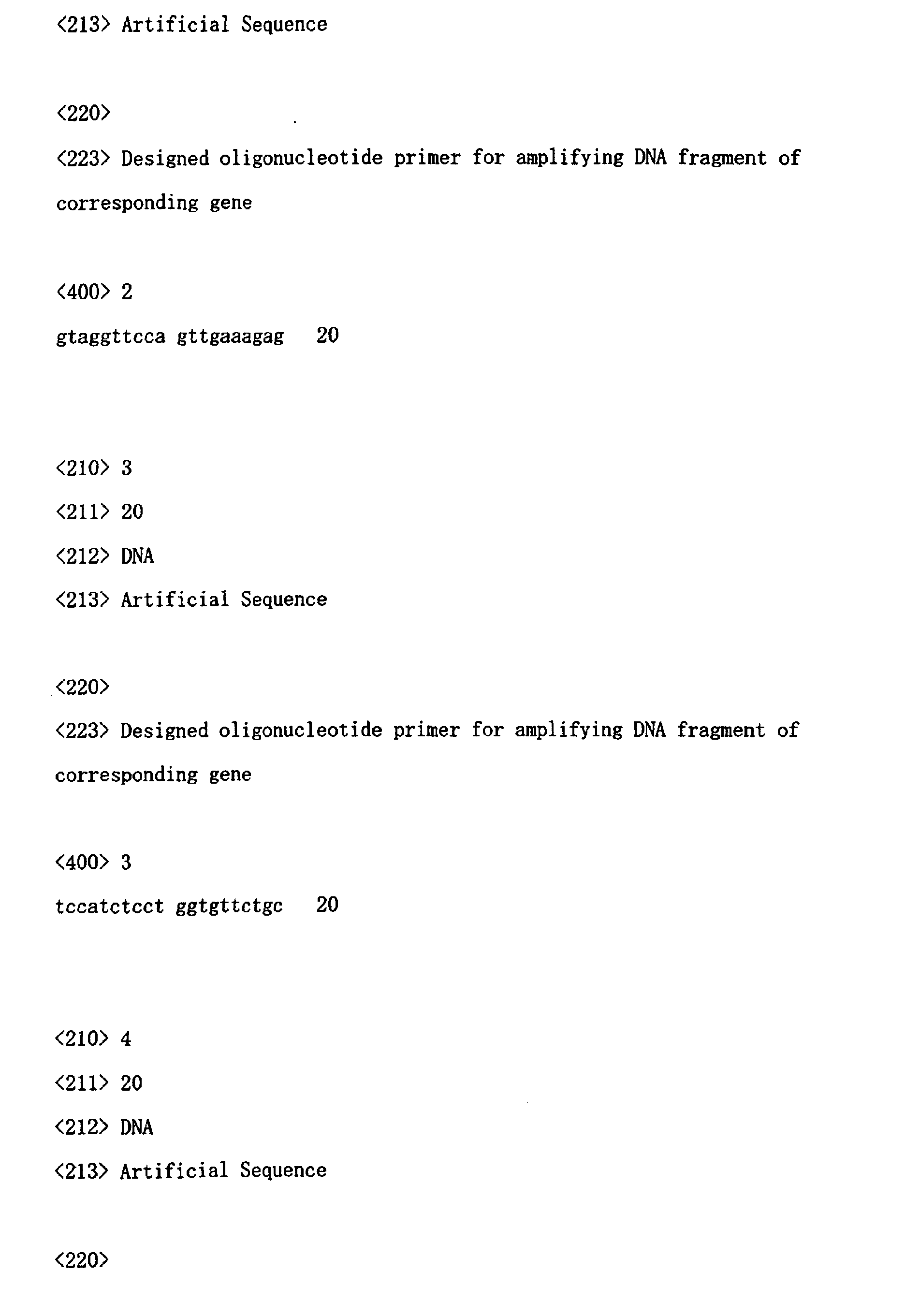
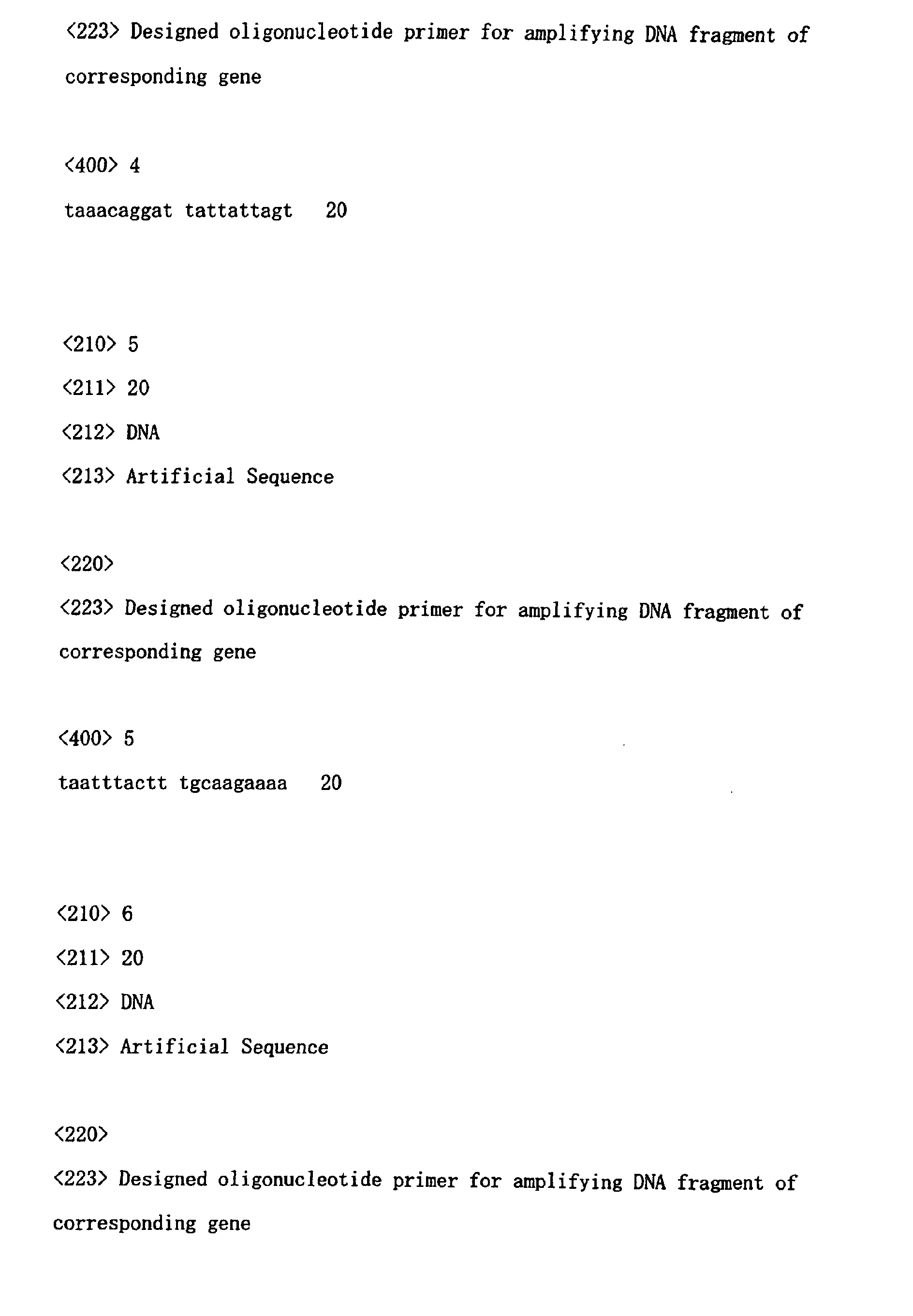
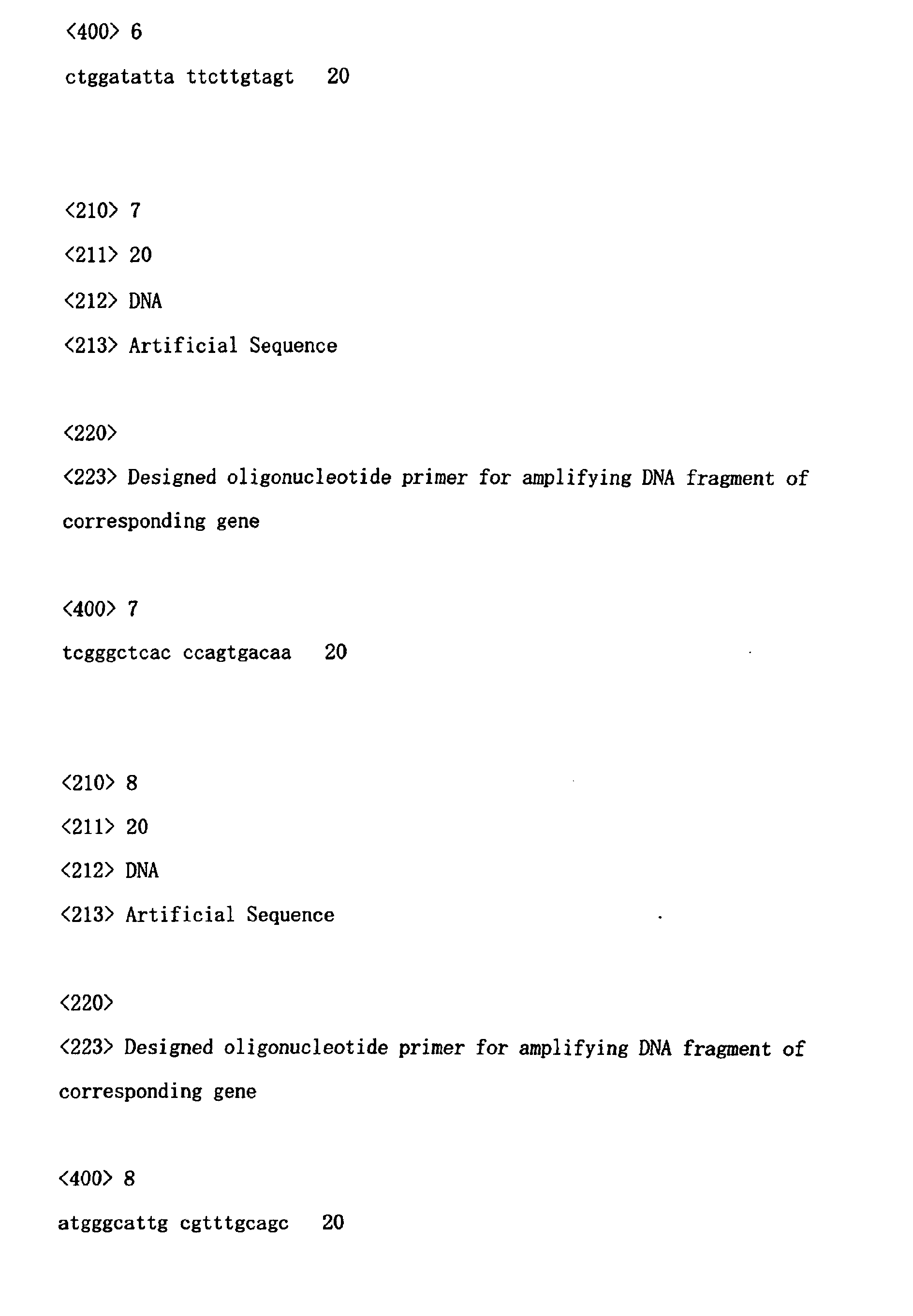
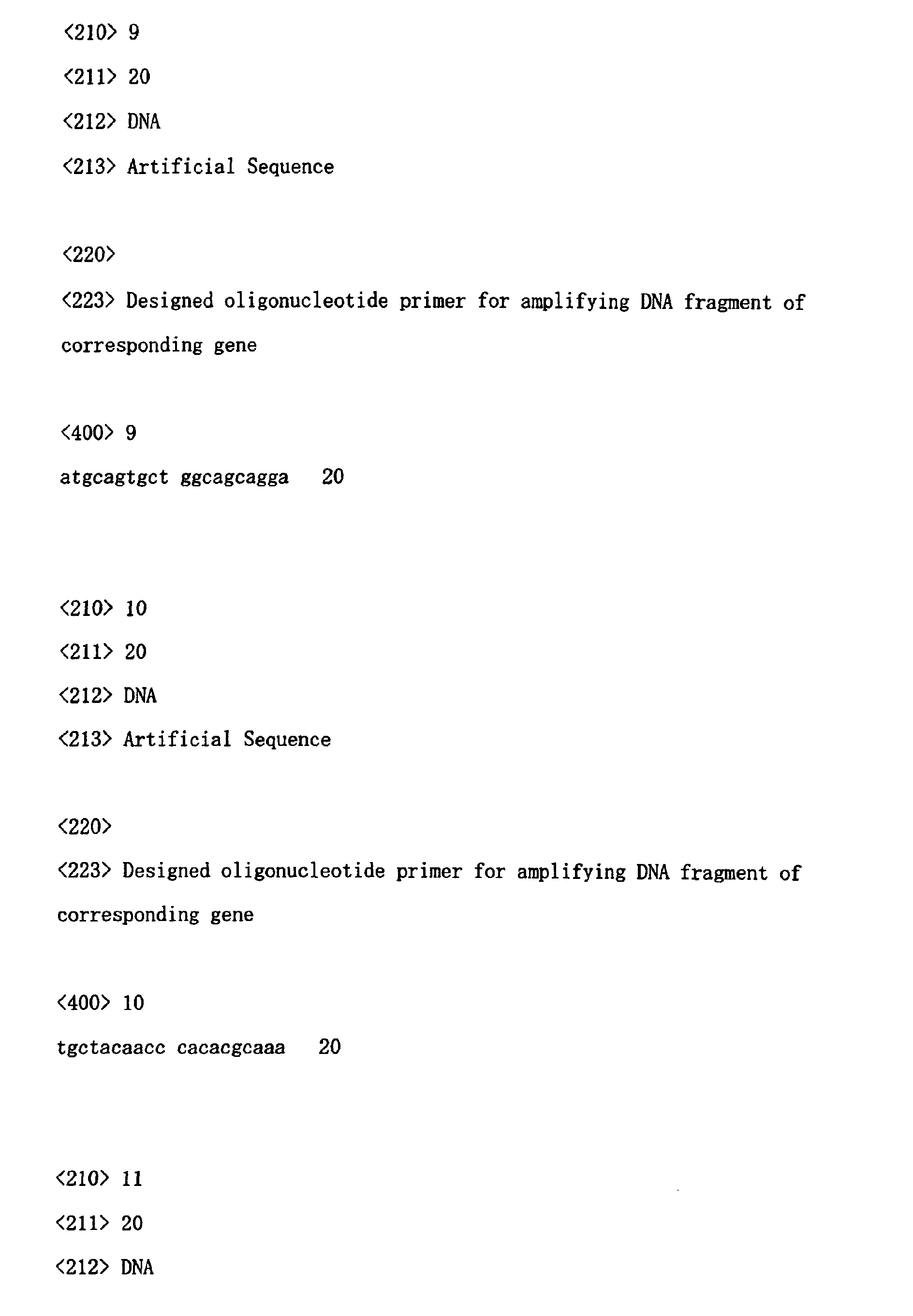
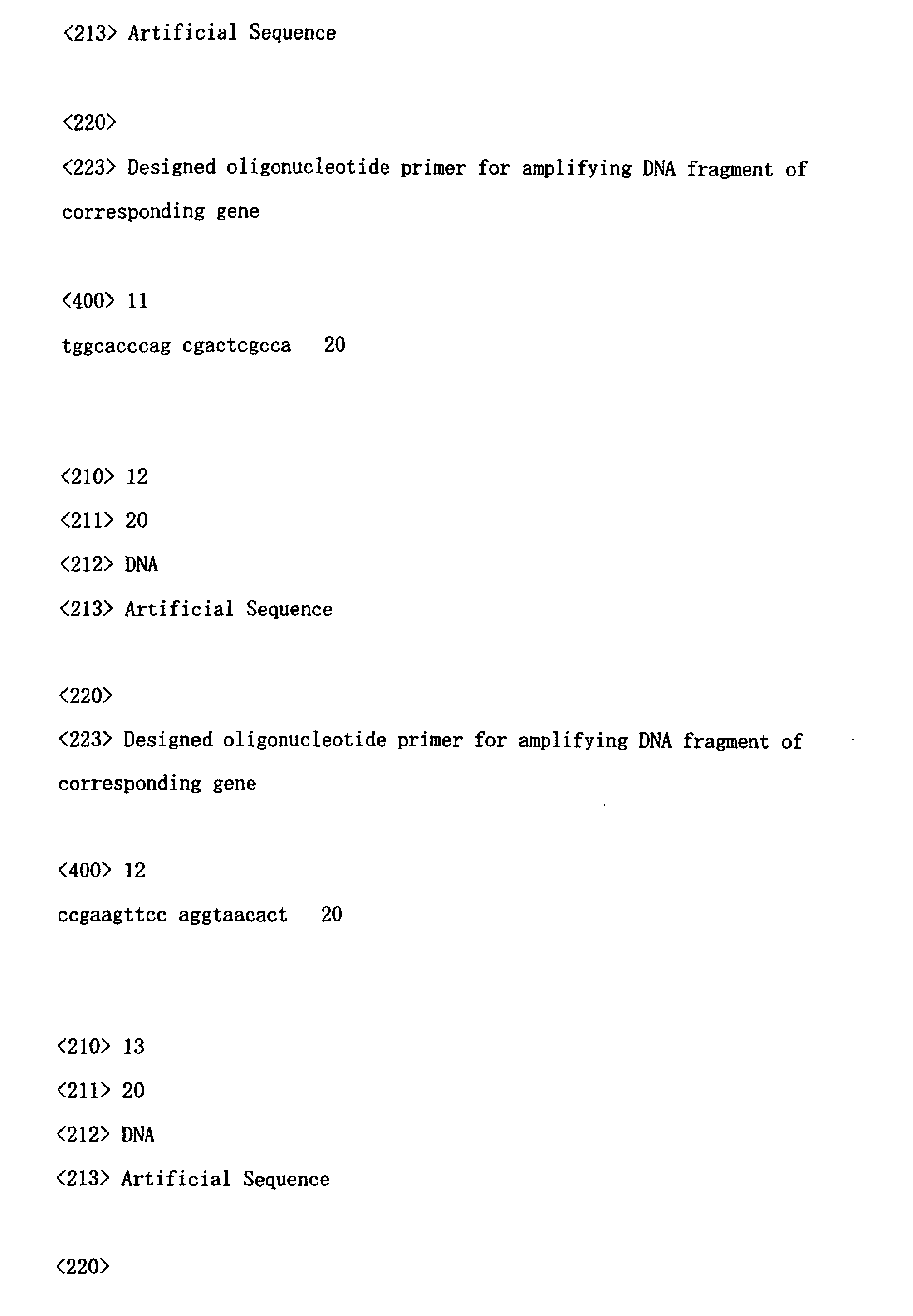
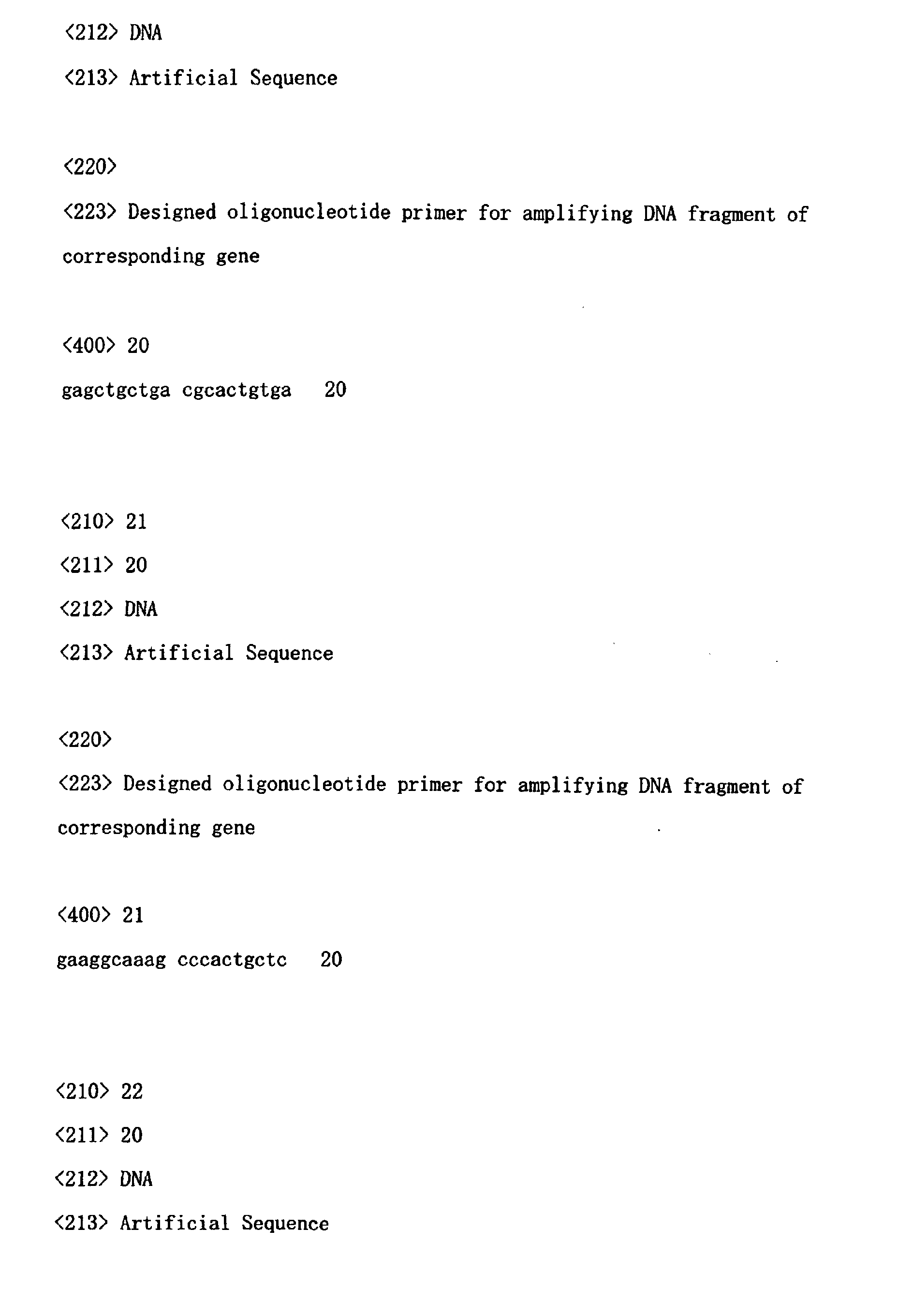
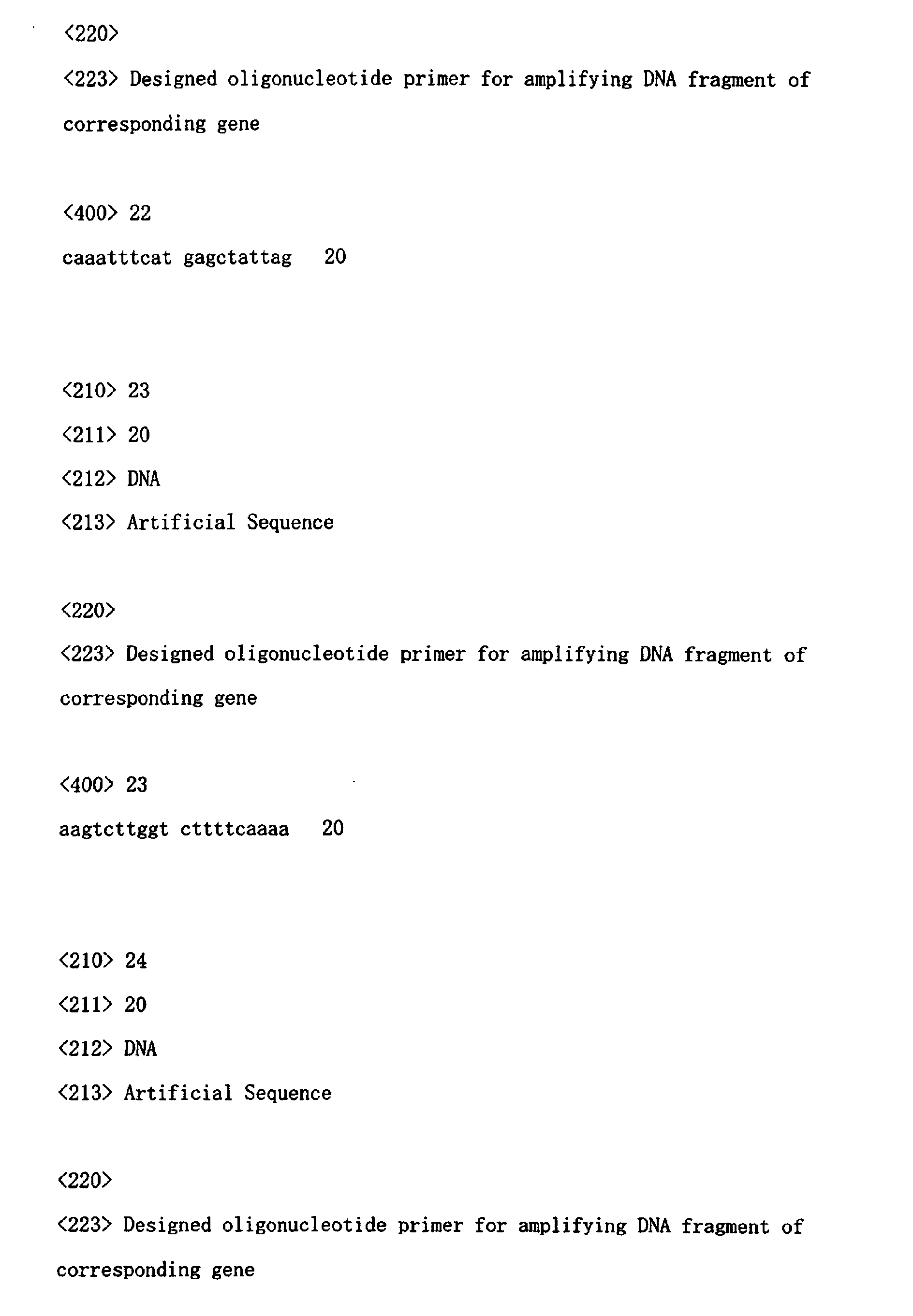
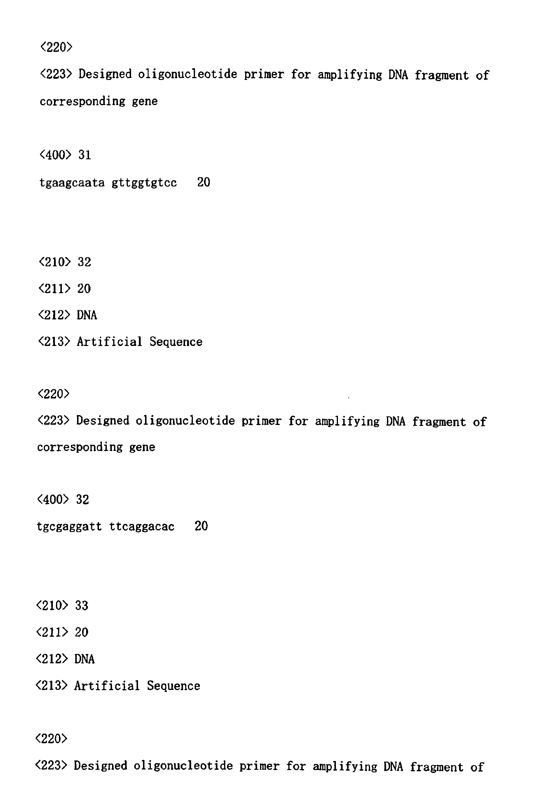
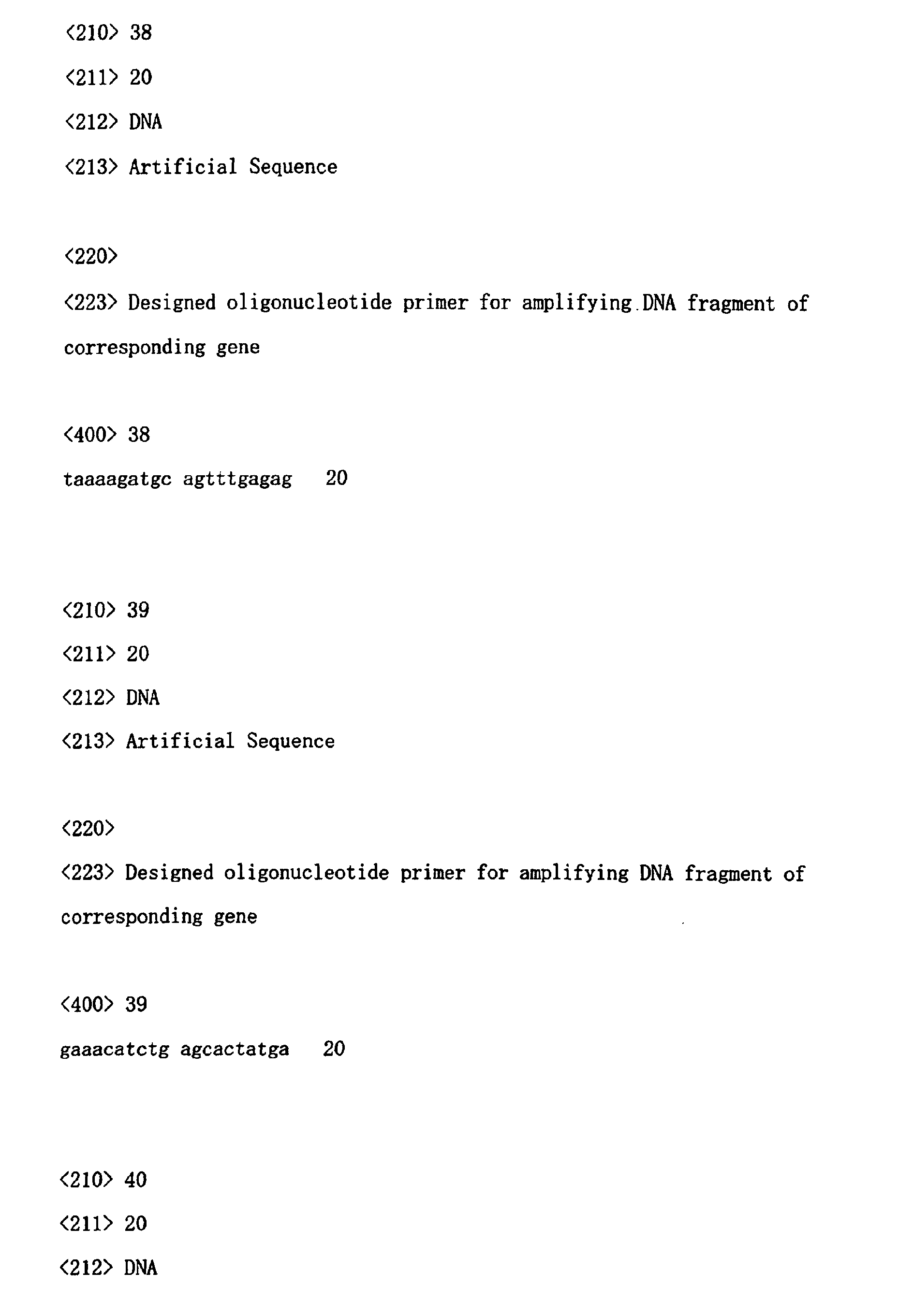
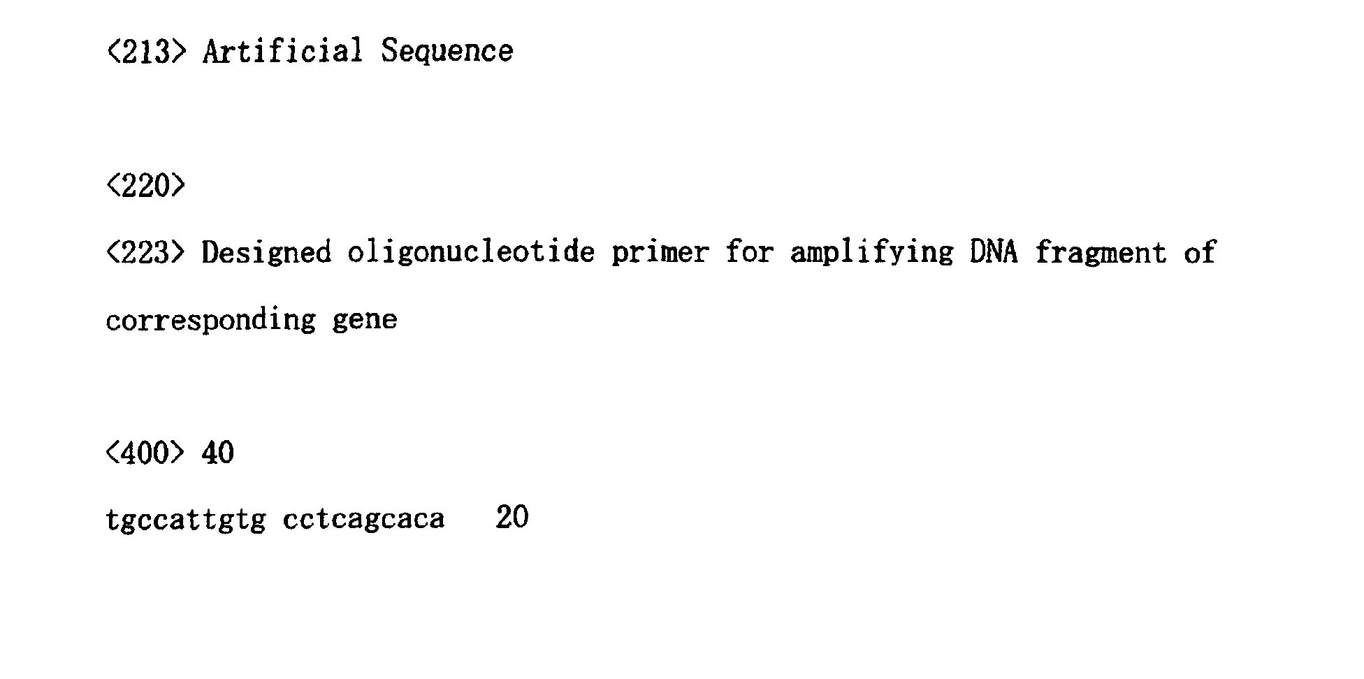
sequences of primers used for PCR are shown in SEQ ID NOS:1
to 40. The primer pairs used for PCRs for the respective
genes are shown in Tables 1 and 2. The 5' primers were
modified at their 5' ends with amino groups.
| | Gene name | GenBank accession no. | SEQ ID NO |
| | | | 5' | 3' |
| 1 | Zona pellucida glycoprotein 3A (sperm receptor) | NM_007155 | 1 | 21 |
| 2 | Wingless-type MMTV integration site family, member 5A | NM_003392 | 2 | 22 |
| 3 | Wingless-type MMTV integration site family member 2 | NM_003391 | 3 | 23 |
| 4 | Villin 2 (ezrin) | NM_003379 | 4 | 24 |
| 5 | Vascular cell adhesion molecule 1 | NM_001078 | 5 | 25 |
| 6 | v-kit Hardy-Zuckerman 4 feline sarcoma viral oncogene homolog | NM_000222 | 6 | 26 |
| 7 | v-akt murine thymoma viral oncogene homolog 1 | NM_005163 | 7 | 27 |
| 8 | Transforming growth factor, beta receptor III (betaglycan, 300kD) | NM_003243 | 8 | 28 |
| 9 | EphA2 | NM_004431 | 9 | 29 |
| 10 | Tumor necrosis factor, alpha-induced protein 6 | NM_007115 | 10 | 30 |
| | Gene name | GenBank accession no. | SEQ ID NO |
| | | | 5' | 3' |
| 11 | Transforming growth factor, beta 1 | NM_000660 | 11 | 31 |
| 12 | Interferon-stimulated transcription factor 3, gamma (48kD) | NM_006084 | 12 | 32 |
| 13 | v-rel avian reticuloendotheliosis viral oncogene homolog A (nuclear factor of kappa light polypeptide gene enhancer in B-cells 3 (p65)) | NM_021975 | 13 | 33 |
| 14 | Transcription elongation factor A (SII), 1 | NM_006756 | 14 | 34 |
| 15 | Selectin L (lymphocyte adhesion molecule 1) | NM_000655 | 15 | 35 |
| 16 | ribosomal protein S5 | NM_001009 | 16 | 36 |
| 17 | ras homolog gene family, member G (rho G) | NM_001665 | 17 | 37 |
| 18 | retinoic acid receptor, beta | NM_000965 | 18 | 38 |
| 19 | RAD52 (S. cerevisiae) homolog | NM_002879 | 19 | 39 |
| 20 | tyrosine kinase 2 | NM_003331 | 20 | 40 |
After PCR, the 20 amplified fragments were
purified according to a conventional method, and dissolved
in TaKaRa Solution T (Takara Bio) at concentrations of 0.3
µg/µl. The thus obtained solutions were used for spotting.
The solutions were spotted onto the activated polyacrylate
ester-coated slide glass prepared in Example 2-(1) using
Affymetrix 417 Arrayer (Affymetrix). The slide glass was
washed in 0.2% SDS and twice in distilled water and treated
with 0.3 N sodium hydroxide solution at room temperature for
5 minutes. Then, the slide glass was washed in distilled
water for 5 minutes, heated at 95°C for 2 minutes, rinsed
with ice-cold 100% ethanol, and dried by centrifugation.
The thus obtained substrate having immobilized nucleic acids
was used as a DNA chip in the subsequent reaction.
(3) Confirmation by hybridization was carried out
as follows. Briefly, salmon sperm DNA was fluorescently
labeled with Cy3 using Label IT Cy3 Label Kit (Mirus)
according to the instructions attached to the kit, and then
purified. The fluorescently labeled salmon sperm DNA
fragment was diluted with a hybridization solution to a
final concentration of 0.1 µg/µl. Denaturation was carried
out using distilled water, and Label IT buffers D1 and N1.
Then, the DNA was dissolved in a hybridization buffer at a
final concentration of 0.01 µg/µl.
About 9 µl of the hybridization solution was
dripped onto a cover glass. The cover glass was covered
with the slide glass with the DNA-immobilized side down so
carefully that no bubble was included. The covered slide
glass was turned upside down so carefully that the cover
glass did not shift, placed in a humidified incubator, and
incubated at 37°C for 30 minutes. After incubation, the
cover glass was removed in 2 x SSC at room temperature. The
slide glass was washed for 5 minutes in 0.2 x SSC containing
0.1% SDS followed by 0.2 x SSC, subjected to low-speed
centrifugation at about 1,000 rpm to remove water, and air-dried.
After air-drying, the slide glass was subjected to
measurement at an excitation wavelength of 532 nm and an
emission wavelength of 570 nm using Affymetrix 418™ Array
Scanner (Affymetrix). As a result, fluorescent signals
were observed at all spots on the slide glass.
Example 3
(1) A substrate for immobilizing nucleic acids
prepared using the polyacrylate ester solution 1 according
to the method as described in Example 2 and a substrate for
immobilizing nucleic acids prepared according to the method
as described in WO 01/02538 were compared to each other
based on the detection sensitivities observed for human
model DNA chips prepared using them. Specifically,
spotting solutions for the 20 human genes as described in
Example 2 prepared using the same test nucleic acids as
those described in Example 2 were used. The solutions were
spotted onto the slide glass for immobilizing nucleic acids
prepared in Example 2 or a slide glass for immobilizing
nucleic acids prepared according to the conventional method
using Affymetrix 417 Arrayer. The slide glasses were washed
in 0.2% SDS and twice in distilled water and treated with
0.3 N sodium hydroxide solution at room temperature for 5
minutes. Then, the slide glasses were washed in distilled
water for 5 minutes, heated at 95°C for 2 minutes, rinsed
with ice-cold 100% ethanol, and dried by centrifugation.
The thus obtained substrates having immobilized nucleic
acids were used as DNA chips in the subsequent reactions.
(2) Preparation of labeled cDNAs for detection and
hybridization were carried out as follows. Briefly,
polyA(+) RNAs were prepared from human HL-60 cells
(Dainippon Pharmaceutical) and human A431 cells (Dainippon
Pharmaceutical) using TRIzol Reagent (Gibco BRL) and
Oligotex-dT30 <Super> (Takara Bio) according to the
protocols for the kits. A Cy3-labeled cDNA and a Cy5-labeled
cDNA were prepared using 1 µg of the polyA(+) RNA
from HL-60 cells and 1 µg of the polyA(+) RNA from A431
cells as templates, respectively, as well as RNA
Fluorescence Labeling Core Kit (M-MLV Version) (Takara Bio)
according to the protocol for the kit, and then purified.
Hybridization between the two DNA chips prepared in Example
3-(1) and a mixture of the Cy3-labeled cDNA and the Cy5-labeled
cDNA, washing and drying were carried out according
to the instructions attached to IntelliGene (Takara Bio).
Fluorescence images for Cy3 (an excitation wavelength of
532 nm and an emission wavelength of 570 nm) and Cy5 (an
excitation wavelength of 635 nm and an emission wavelength
of 660 nm) and were obtained using Affymetrix 428 Array
Scanner (Affymetrix). The signal intensities for the
respective spots and the background signal intensity were
quantified using an image quantification analysis software
ImaGene 4.1 (Biodiscovery). Averages of values obtained by
subtracting the background signal intensity from the signal
intensities for the respective spots (hybridization signal
intensities) and averages of ratios of the signal
intensities for the respective spots to the background
signal intensity (signal to noise ratios) were calculated
for the 20 genes. As a result, the hybridization signal
intensities and the signal to noise ratios observed for the
DNA chip prepared using the slide glass having immobilized
nucleic acids according to the preparation method of the
present invention were equivalent to or greater than those
observed for the DNA chip prepared using the slide glass
having immobilized nucleic acids according to the
conventional method. Based on these results, it was
confirmed that, like the substrate for immobilizing nucleic
acid prepared according to the conventional method, the
substrate for immobilizing nucleic acids prepared according
to the preparation method of the present invention can be
preferably used as a substrate for immobilizing nucleic
acids that enables preparation of a DNA chip that has an
ability of detecting a nucleic acid with high sensitivity.
Example 4
(1) Conversion of polyacrylic acid into active
ester
1.0 g of polyacrylic acid (molecular weight:
1,000,000) was dissolve in 200 ml of dimethylformamide.
4.8 g of N-hydroxysuccinimide, 8.0 g of N-ethyl-N'-dimethylaminopropyl-carbodiimide
hydrochloride and 52 mg of
4-dimethylaminopyridine were added to the solution while
cooling on ice. After the reagents were dissolved, the pH
was adjusted to 7 to 8 using triethylamine. The resulting
mixture was stirred overnight at room temperature.
(2) Preparation of coating agent
The reaction mixture obtained in Example 4-(1)
was filtered through a Buchner funnel equipped with a
filter paper and covered with Celite 545 which had been
washed with dimethylformamide. The volume of the filtrate
was adjusted to 200 ml with dimethylformamide to obtain a
coating agent.
(3) Surface treatment of slide glass
A microscopic slide glass was subjected to 10-minutes
sonications in 2 M nitric acid, 2 M sodium hydroxide
and distilled water. 3-Aminopropyltriethoxysilane was added
to 95% ethanol solution at a final concentration of 4%. The
sonicated slide glass was immersed in this solution for 30
minutes, and then treated in an oven at 100°C for 10 minutes
to aminopropylsilanize the surface of the slide glass.
The aminopropylsilanized slide glass was immersed
for 1 hour in the coating agent obtained in Example 4-(2).
The slide glass was washed sequentially by sonication in
dimethylformamide and acetonitrile, and then dried under
reduced pressure.
(4) Determination of immobilization rates
A Cy3-labeled and end-aminated DNA fragment and a
nonlabeled and end-aminated DNA fragment were prepared by
PCRs using a Cy3-labeled primer having an amino group at its
end and a nonlabeled primer having an amino group at its end
which had been synthesized beforehand. Solutions of the
Cy3-labeled and end-aminated DNA fragment and the nonlabeled
and end-aminated DNA fragment were mixed with TaKaRa
Solution I (Takara Bio).
The resulting mixture was spotted onto the slide
glass prepared in Example 4-(3) using Affymetrix 417 Arrayer.
Measurement was carried out at an excitation wavelength of
532 nm and an emission wavelength of 570 nm using
Affymetrix 428 Array Scanner. As a result, fluorescent
signals were observed at all spots on the slide glass.
After measurement, the slide glass was washed
sequentially in 0.2% SDS, Milli-Q water, 0.3 N sodium
hydroxide, Milli-Q water, Milli-Q water at 100°C and ice-cold
ethanol, subjected to low-speed centrifugation at about
1,000 rpm to remove water, and air-dried. After air-drying,
the slide glass was subjected to measurement at an
excitation wavelength of 532 nm and an emission wavelength
of 570 nm using Affymetrix 428 Array Scanner. The image
obtained by the measurement was subjected to quantification
using a microarray quantification software ImaGene 4.2, and
DNA immobilization rates were calculated using a clustering
software GeneSight V3.0.7.
As a result, the rates of DNA immobilization onto
the slide glass of Example 4-(3) ranged from 13 to 26%, and
the average value was about 20%. The rates of DNA
immobilization onto the slide glass of Example 3 ranged
from 23 to 34%, and the average value was about 28%. Equal
or more DNA immobilization rates were observed even if the
period of immersion in the coating agent was shortened from
16 hours to 1 hour.
Example 5
(1) Conversion of polyacrylic acid into active
ester
3.25 g of polyacrylic acid (molecular weight:
1,000,000) was dissolve in 650 ml of dimethylformamide.
14.6 g of N-hydroxysuccinimide and 22.75 ml of N-ethyl-N'-dimethylaminopropyl-carbodiimide
were added to the solution
while cooling on ice. After the reagents were dissolved,
the resulting mixture was stirred overnight at room
temperature.
(2) Preparation of coating agent
The reaction mixture obtained in Example 5-(1)
was filtered through a Buchner funnel equipped with a
filter paper and covered with Celite 545 which had been
washed with dimethylformamide. The volume of the filtrate
was adjusted to 1.3 1 with dimethylformamide to obtain a
coating agent.
(3) Surface treatment of slide glass
A microscopic slide glass was subjected to 10-minutes
sonications in 2 M nitric acid, 2 M sodium hydroxide
and distilled water. 3-Aminopropyltriethoxysilane was added
to 95% ethanol solution at a final concentration of 4%. The
sonicated slide glass was immersed in this solution for 30
minutes, and then treated in an oven at 100°C for 10 minutes
to aminopropylsilanize the surface of the slide glass.
The aminopropylsilanized slide glass was immersed
for 1 hour in the coating agent obtained in Example 5-(2).
The slide glass was washed sequentially by sonication in
dimethylformamide and acetonitrile, and then dried under
reduced pressure.
(4) Determination of immobilization rates
A Cy3-labeled and end-aminated DNA fragment and a
nonlabeled and end-aminated DNA fragment were prepared by
PCRs using a Cy3-labeled primer having an amino group at its
end and a nonlabeled primer having an amino group at its end
which had been synthesized beforehand. Solutions of the
Cy3-labeled and end-aminated DNA fragment and the nonlabeled
and end-aminated DNA fragment were mixed with TaKaRa
Solution I (Takara Bio).
The resulting mixture was spotted onto the slide
glass prepared in Example 5-(3) using Affymetrix 417 Arrayer.
Measurement was carried out at an excitation wavelength of
532 nm and an emission wavelength of 570 nm using
Affymetrix 428 Array Scanner. As a result, fluorescent
signals were observed at all spots on the slide glass.
After measurement, the slide glass was washed
sequentially in 0.2% SDS, Milli-Q water, 0.3 N sodium
hydroxide, Milli-Q water, Milli-Q water at 100°C and ice-cold
ethanol, subjected to low-speed centrifugation at about
1,000 rpm to remove water, and air-dried. After air-drying,
the slide glass was subjected to measurement at an
excitation wavelength of 532 nm and an emission wavelength
of 570 nm using Affymetrix 428 Array Scanner. The image
obtained by the measurement was subjected to quantification
using a microarray quantification software ImaGene 4.2, and
DNA immobilization rates were calculated using a clustering
software GeneSight V3.0.7.
As a result, the rates of DNA immobilization onto
the slide glass of Example 5-(3) ranged from 37 to 50%.
Equal or more DNA immobilization rates were observed even
if N-ethyl-N'-dimethylaminopropyl-carbodiimide
hydrochloride was replaced by N-ethyl-N'-dimethylaminopropyl-carbodiimide.
Example 6
(1) Conversion of polyacrylic acid into active
ester
Polyacrylate ester was prepared as described in
Example 5-(1).
(2) Preparation of coating agent
A coating agent was prepared as described in
Example 5-(2).
(3) Surface treatment of slide glass
A microscopic slide glass was subjected to 10-minutes
sonications in 2 M nitric acid, 2 M sodium hydroxide
and distilled water. 3-Aminopropyltriethoxysilane was added
to 95% ethanol solution at a final concentration of 4%. The
sonicated slide glass was immersed in this solution for 30
minutes, and then treated in an oven at 100°C for 10 minutes
to aminopropylsilanize the surface of the slide glass.
The aminopropylsilanized slide glass was immersed
for 1 hour in the coating agent obtained in Example 6-(2).
The slide glass was washed sequentially in dimethylformamide
and acetonitrile, and then dried under reduced pressure.
(4) Repetitive use of coating agent - 1
A newly prepared aminopropylsilanized slide glass
was immersed for 1 hour in the coating agent used in Example
6-(3). The slide glass was washed sequentially by
sonication in dimethylformamide and acetonitrile, and then
dried under reduced pressure.
(5) Repetitive use of coating agent - 2
A newly prepared aminopropylsilanized slide glass
was immersed for 1 hour in the coating agent used in Example
6-(4). The slide glass was washed sequentially by
sonication in dimethylformamide and acetonitrile, and then
dried under reduced pressure.
(6) Repetitive use of coating agent - 3
A newly prepared aminopropylsilanized slide glass
was immersed for 1 hour in the coating agent used in Example
6-(5). The slide glass was washed sequentially by
sonication in dimethylformamide and acetonitrile, and then
dried under reduced pressure.
(7) Determination of immobilization rates
A Cy3-labeled and end-aminated DNA fragment and a
nonlabeled and end-aminated DNA fragment were prepared by
PCRs using a Cy3-labeled primer having an amino group at its
end and a nonlabeled primer having an amino group at its end
which had been synthesized beforehand. Solutions of the
Cy3-labeled and end-aminated DNA fragment and the nonlabeled
and end-aminated DNA fragment were mixed with TaKaRa
Solution I (Takara Bio).
The resulting mixture was spotted onto the slide
glasses prepared in Example 6-(3), (4), (5) and (6) using
Affymetrix 417 Arrayer. Measurements were carried out at an
excitation wavelength of 532 nm and an emission wavelength
of 570 nm using Affymetrix 428 Array Scanner. As a result,
fluorescent signals were observed at all spots on all the
slide glasses.
After measurement, the slide glasses were washed
sequentially in 0.2% SDS, Milli-Q water, 0.3 N sodium
hydroxide, Milli-Q water, Milli-Q water at 100°C and ice-cold
ethanol, subjected to low-speed centrifugation at about
1,000 rpm to remove water, and air-dried. After air-drying,
the slide glasses were subjected to measurements at an
excitation wavelength of 532 nm and an emission wavelength
of 570 nm using Affymetrix 428 Array Scanner. The images
obtained by the measurements were subjected to
quantification using a microarray quantification software
ImaGene 4.2, and DNA immobilization rates were calculated
using a clustering software GeneSight V3.0.7.
As a result, the rates of DNA immobilization onto
the slide glasses of Example 6-(3), Example 6-(4), Example
6-(5) and Example 6-(6) ranged from 41 to 45%, from 42 to
48%, from 39 to 42% and from 39 to 46%, respectively. The
average values were about 40% or more in all cases. Thus,
it was confirmed that the ability of immobilizing DNAs
equivalent to the initial one was retained after the
coating agent was repetitively used.
Industrial Applicability
The present invention provides a low-cost
substrate for immobilizing ligands that enables detection of
a receptor of interest with high sensitivity. Furthermore,
a material having immobilized ligands which can be used to
detect and analyze a target receptor with high sensitivity
is provided using the substrate.
Sequence Listing Free Text
SEQ ID NO:1: Designed oligonucleotide primer for
amplifying DNA fragment of corresponding gene
SEQ ID NO:2: Designed oligonucleotide primer for
amplifying DNA fragment of corresponding gene
SEQ ID NO:3: Designed oligonucleotide primer for
amplifying DNA fragment of corresponding gene
SEQ ID NO:4: Designed oligonucleotide primer for
amplifying DNA fragment of corresponding gene
SEQ ID NO:5: Designed oligonucleotide primer for
amplifying DNA fragment of corresponding gene
SEQ ID NO:6: Designed oligonucleotide primer for
amplifying DNA fragment of corresponding gene
SEQ ID NO:7: Designed oligonucleotide primer for
amplifying DNA fragment of corresponding gene
SEQ ID NO:8: Designed oligonucleotide primer for
amplifying DNA fragment of corresponding gene
SEQ ID NO:9: Designed oligonucleotide primer for
amplifying DNA fragment of corresponding gene
SEQ ID NO:10: Designed oligonucleotide primer for
amplifying DNA fragment of corresponding gene
SEQ ID NO:11: Designed oligonucleotide primer for
amplifying DNA fragment of corresponding gene
SEQ ID NO:12: Designed oligonucleotide primer for
amplifying DNA fragment of corresponding gene
SEQ ID NO:13: Designed oligonucleotide primer for
amplifying DNA fragment of corresponding gene
SEQ ID NO:14: Designed oligonucleotide primer for
amplifying DNA fragment of corresponding gene
SEQ ID NO:15: Designed oligonucleotide primer for
amplifying DNA fragment of corresponding gene
SEQ ID NO:16: Designed oligonucleotide primer for
amplifying DNA fragment of corresponding gene
SEQ ID NO:17: Designed oligonucleotide primer for
amplifying DNA fragment of corresponding gene
SEQ ID NO:18: Designed oligonucleotide primer for
amplifying DNA fragment of corresponding gene
SEQ ID NO:19: Designed oligonucleotide primer for
amplifying DNA fragment of corresponding gene
SEQ ID NO:20: Designed oligonucleotide primer for
amplifying DNA fragment of corresponding gene
SEQ ID NO:21: Designed oligonucleotide primer for
amplifying DNA fragment of corresponding gene
SEQ ID NO:22: Designed oligonucleotide primer for
amplifying DNA fragment of corresponding gene
SEQ ID NO:23: Designed oligonucleotide primer for
amplifying DNA fragment of corresponding gene
SEQ ID NO:24: Designed oligonucleotide primer for
amplifying DNA fragment of corresponding gene
SEQ ID NO:25: Designed oligonucleotide primer for
amplifying DNA fragment of corresponding gene
SEQ ID NO:26: Designed oligonucleotide primer for
amplifying DNA fragment of corresponding gene
SEQ ID NO:27: Designed oligonucleotide primer for
amplifying DNA fragment of corresponding gene
SEQ ID NO:28: Designed oligonucleotide primer for
amplifying DNA fragment of corresponding gene
SEQ ID NO:29: Designed oligonucleotide primer for
amplifying DNA fragment of corresponding gene
SEQ ID NO:30: Designed oligonucleotide primer for
amplifying DNA fragment of corresponding gene
SEQ ID NO:31: Designed oligonucleotide primer for
amplifying DNA fragment of corresponding gene
SEQ ID NO:32: Designed oligonucleotide primer for
amplifying DNA fragment of corresponding gene
SEQ ID NO:33: Designed oligonucleotide primer for
amplifying DNA fragment of corresponding gene
SEQ ID NO:34: Designed oligonucleotide primer for
amplifying DNA fragment of corresponding gene
SEQ ID NO:35: Designed oligonucleotide primer for
amplifying DNA fragment of corresponding gene
SEQ ID NO:36: Designed oligonucleotide primer for
amplifying DNA fragment of corresponding gene
SEQ ID NO:37: Designed oligonucleotide primer for
amplifying DNA fragment of corresponding gene
SEQ ID NO:38: Designed oligonucleotide primer for
amplifying DNA fragment of corresponding gene
SEQ ID NO:39: Designed oligonucleotide primer for
amplifying DNA fragment of corresponding gene
SEQ ID NO:40: Designed oligonucleotide primer for
amplifying DNA fragment of corresponding gene