EP1432729B1 - Peyers's patch and/or m-cell targeting ligands - Google Patents
Peyers's patch and/or m-cell targeting ligands Download PDFInfo
- Publication number
- EP1432729B1 EP1432729B1 EP02760458A EP02760458A EP1432729B1 EP 1432729 B1 EP1432729 B1 EP 1432729B1 EP 02760458 A EP02760458 A EP 02760458A EP 02760458 A EP02760458 A EP 02760458A EP 1432729 B1 EP1432729 B1 EP 1432729B1
- Authority
- EP
- European Patent Office
- Prior art keywords
- seq
- ligand
- peptide
- synthetic polypeptide
- polypeptide ligand
- Prior art date
- Legal status (The legal status is an assumption and is not a legal conclusion. Google has not performed a legal analysis and makes no representation as to the accuracy of the status listed.)
- Expired - Lifetime
Links
Images


Classifications
-
- C—CHEMISTRY; METALLURGY
- C07—ORGANIC CHEMISTRY
- C07K—PEPTIDES
- C07K7/00—Peptides having 5 to 20 amino acids in a fully defined sequence; Derivatives thereof
- C07K7/04—Linear peptides containing only normal peptide links
- C07K7/08—Linear peptides containing only normal peptide links having 12 to 20 amino acids
-
- C—CHEMISTRY; METALLURGY
- C07—ORGANIC CHEMISTRY
- C07K—PEPTIDES
- C07K14/00—Peptides having more than 20 amino acids; Gastrins; Somatostatins; Melanotropins; Derivatives thereof
- C07K14/001—Peptides having more than 20 amino acids; Gastrins; Somatostatins; Melanotropins; Derivatives thereof by chemical synthesis
-
- C—CHEMISTRY; METALLURGY
- C07—ORGANIC CHEMISTRY
- C07K—PEPTIDES
- C07K14/00—Peptides having more than 20 amino acids; Gastrins; Somatostatins; Melanotropins; Derivatives thereof
- C07K14/435—Peptides having more than 20 amino acids; Gastrins; Somatostatins; Melanotropins; Derivatives thereof from animals; from humans
- C07K14/46—Peptides having more than 20 amino acids; Gastrins; Somatostatins; Melanotropins; Derivatives thereof from animals; from humans from vertebrates
- C07K14/47—Peptides having more than 20 amino acids; Gastrins; Somatostatins; Melanotropins; Derivatives thereof from animals; from humans from vertebrates from mammals
- C07K14/4701—Peptides having more than 20 amino acids; Gastrins; Somatostatins; Melanotropins; Derivatives thereof from animals; from humans from vertebrates from mammals not used
- C07K14/4725—Proteoglycans, e.g. aggreccan
-
- C—CHEMISTRY; METALLURGY
- C07—ORGANIC CHEMISTRY
- C07K—PEPTIDES
- C07K7/00—Peptides having 5 to 20 amino acids in a fully defined sequence; Derivatives thereof
- C07K7/02—Linear peptides containing at least one abnormal peptide link
-
- C—CHEMISTRY; METALLURGY
- C07—ORGANIC CHEMISTRY
- C07K—PEPTIDES
- C07K7/00—Peptides having 5 to 20 amino acids in a fully defined sequence; Derivatives thereof
- C07K7/04—Linear peptides containing only normal peptide links
- C07K7/06—Linear peptides containing only normal peptide links having 5 to 11 amino acids
-
- A—HUMAN NECESSITIES
- A61—MEDICAL OR VETERINARY SCIENCE; HYGIENE
- A61K—PREPARATIONS FOR MEDICAL, DENTAL OR TOILETRY PURPOSES
- A61K38/00—Medicinal preparations containing peptides
-
- C—CHEMISTRY; METALLURGY
- C07—ORGANIC CHEMISTRY
- C07K—PEPTIDES
- C07K2319/00—Fusion polypeptide
Definitions
- This invention relates to novel targeting ligands which permit or facilitate the transport of drugs, macromolecules, or particles, such as biodegradable nanoparticles and microparticles, or bacterial carriers or viral carriers through the intestinal epithelium, M-cells located in gut associated lymphoid tissue, and/or Peyer's Patch tissue of the intestinal epithelium.
- the epithelial cells lining the lumenal side of the gastrointestinal tract are a major barrier to drug delivery following oral administration.
- GIT gastrointestinal tract
- transport pathways which can be exploited to facilitate drug delivery and transport: the transcellular, paracellular, carrier-mediated and transcytotic transport pathways.
- the ability of a conventional drug, peptide, protein, macromolecule, nanoparticulate system or microparticulate system to interact with one of these transport pathways may result in increased delivery of that drug or particle from the GIT to the underlying circulation.
- M-cells are antigen sampling cells that are found in the epithelium of the gut-associated lymphoid tissue, or Peyer's Patch.
- the transcytotic capacity of M-cells and the downstream processing of the antigen sampled would suggest that targeting vaccines to M-cells would enhance oral immunization ( Foster et al., 15 Vaccine 546-71 (1998 )).
- no human M-cell marker has been identified as a target for delivery of vaccines and/or other drugs through the M-cell route.
- U.S. Patent No. 6,060,082 to Chen et al discloses modified polymerized liposomes that contain a molecule or ligand on their surfaces in order to target the liposomes to a specific site or cell type fororal/mucosal drug delivery. Also disclosed is an embodiment in which the liposomes are modified with carbohydrate moities or lectins that specifically target M-cells or Peyer's Patches in mice. However, this reference only teaches transport of liposomes.
- M-cell and/or Peyer's Patch specific ligands that are particularly effective in transporting drugs, including drug-loaded nanoparticles and microparticles, or bacterial or viral carries coding for vaccines into or across a human or animal intestinal epithelium.
- the invention is a purified synthetic polypeptide ligand comprising a 12-mer peptide selected from the group consisting of SEQ 10 NOs:1-34, SEQ ID NOs:38-39, and SEQ ID NO:42 and wherein said 12mer peptide when integrated as an N-terminal PIII fusion peptide of an M13 phage confers an ability to bind the phage to either Caco-2 cell, IEC-6 cell, rat, mouse, pig or dog homogenate membrane fractions, said ability being at least as great as that conferred by a similarly integrated 12-mer peptide of SEQ ID NO:67.
- the invention is a purified synthetic polypeptide ligand, said ligand comprising a L-peptide sequence, D-peptide version thereof, or retro-inverted version thereof, said L-peptide sequence being selected from the group consisting of HESSH (SEQ ID NO:97) and NVYTXXXXSPXP (SEQ ID NO:98), wherein said L-peptide sequence, a D-peptide version thereof, or a retro inverted version thereof when integrated as an N-terminal PIII fusion peptide of an M13 phage confers an ability to bind the phage to either Caco-2 cell, IEC-6 cell, rat, mouse, pig or dog homogenate membrane fractions, said ability being at least as great as that conferred by a similarly integrated 12-mer peptide of SEQ ID NO:67.
- the invention is a purified synthetic polypeptide ligand, not more than 200 amino acids in length, comprising a peptide selected from the group consisting of SEQ ID NOs:74 through SEQ ID NO:96 wherein said peptide when integrated as an N-terminal PIII fusion peptide of an M13 phage confers an ability to bind the phage to either Caco-2 cell, IEC-6 cell, rat, mouse, pig or dog homogenate membrane fractions, said ability being at least as great as that conferred by a similarly integrated 12-mer peptide of SEQ ID NO:67.
- the length of the aforementioned purified synthetic polypeptide ligands be not more than 200 amino acids, more preferably not more than 100 amino acids, most preferably not more than 50 amino acids. Conversely, it is preferred that their length be at least 12 amino acids, more preferably at least 20 amino acids, most preferably at least 30 amino acids.
- the polypeptide comprises a zinc-binding domain.
- Nucleic acid molecules that code for the aforementioned purified synthetic polypeptide ligands are also aspects of the invention. Preferred are those that are not more than 600 nucleotides in length. Highly preferred are those that code for a purified synthetic polypeptide that comprises one of the specific 12-mer peptides, motifs, or naturally occurring homologues.
- one of the aforementioned purified synthetic polypeptides ligands is integrated into the protein of a phage.
- one of the aforementioned purified synthetic polypeptide ligands is covalently or non-covalently bound to a carrier entity comprising a pharmaceutical agent.
- the carrier entity is selected from the group consisting of a nanoparticle, microparticle, liposome, bacterium, phage (bacteriophage) and virus (preferably a mammalian virus, most preferably a human virus; especially non-pathogenic forms made by recombinant or other technologies). It is preferred that the nanoparticle, microparticle or liposome have a largest dimension that is in the range of 10nm to 500 ⁇ m, as discussed in more detail elsewhere herein.
- the pharmaceutical agent is a drug or therapeutic agent. In other specific embodiments, the pharmaceutical agent is a pathogen antigen.
- Certain aspects of the invention involve the use of the purified synthetic polypeptide ligands to target delivery of pharmaceutical agents.
- the invention is a method of administering a pharmaceutical agent to an organism having intestinal epithelium, said method comprising contacting said intestinal epithelium with one of the aforementioned purified synthetic polypeptide ligands that is covalently, or non-covalently bound to, a carrier entity.
- the organism is a mammal. Most preferably, the mammal is a human.
- the carrier entity is selected from the group consisting of a nanoparticle, microparticle, liposome, bacterium, phage and virus.
- a preferred embodiment is where the polypeptide ligand is expressed on the surface of a phage or bacterium further comprising an antigen or a gene encoding the antigen also expressed on the surface.
- the microparticle, nanoparticle or liposome has its major dimension in the range of 10 nm to 500 ⁇ m.
- the carrier entity is loaded with a pharmaceutical agent.
- the preferred route of administration for delivery of the ligand-carrier entity is the oral route.
- Other possible routes are the rectal, subcutaneous, intramuscular, nasal and intravenous routes.
- the purified synthetic polypeptide ligand is a 12-mer integrated into a coat protein of a phage.
- the purified synthetic polypeptide ligand comprises a zinc-binding motif, and said ligand is contacted with said epithelium in the presence of zinc.
- the present invention relates to targeted polypeptide ligands for mucosal delivery of agents through the intestinal epithelium.
- the polypeptide ligands are targeted to M-cells or Peyer's patch tissue of the intestinal epithelium.
- purified synthetic polypeptide ligand is intended to distinguish polypeptides of the invention from (1) those that consist of a naturally occurring amino acid sequence; and (2) those that naturally occur but have not been purified.
- polypeptides that naturally occur but which have not been purified are fragments of polypeptides that exist as intermediates during the translational process that elongates fragments into complete polypeptides, and proteolytic breakdown products which occur from time to time.
- polypeptide component of a protein such as mouse keratinocyte growth factor, identified in the Blast homology search below would be an example of a naturally occurring polypeptide.
- a polypeptide ligand that may naturally occur in a eukaryotic cell is a purified synthetic polypeptide ligand if it occurs (but does not naturally occur) on a phage surface or a bacterial surface, or if it occurs on the surface of a nanoparticle, microparticle or liposome, or bacterial or viral carrier, or if it occurs as a result of genetic recombination technologies in a cell, virus or phage where it does not naturally occur.
- polypeptide and peptide do not have an intrinsic difference as to biochemical meaning.
- a 12-mer peptide can qualify as a polypeptide.
- purified synthetic polypeptide ligand where one or more amino acids have been derivatized (e.g. glycosylation, acetylation, amidation, biotinylation, dansylation) the term purified synthetic polypeptide ligand is intended to apply to the polypeptide component of the ligand.
- dansylation comprises the addition of a dansyl-lysine group
- the polypeptide absent the lysine of the dansyl-lysine group is the purified synthetic polypeptide ligand.
- the test for functionality of a 12-mer, fragment or homologue is exemplified by "wherein said 12-mer L-peptide, fragment or homologue thereof, when integrated as an N-terminal PIII fusion peptide of an M13 phage confers an ability to bind the phage to a Caco-2 cell homogenate membrane fraction, said ability being at least as great as that conferred by a similarly integrated 12-mer peptide of SEQ ID NO:67.”
- a cloning vector useful for accomplishing the binding test is M13KE, which is available from New England Labs Inc., as are the details for integrating the peptide ⁇ See Technical Bulletin #8101(4/1/00), which is incorporated by reference herein in its entirety ⁇ .
- Peptides larger than 20-30 amino acids, if so integrated have deleterious effect on the infectivity of the M13 virus. If it is desired to test the binding functionality of peptides too large to be tested in the phage binding test, such larger peptides in biotinylated form can be tested in the Caco-2 membrane binding assay described herein, in order to see if that larger peptide retains detectable binding activity.
- a Caco-2 cell homogenate membrane fraction and simply "Caco-2 cell homogenate” are used interchangeably herein unless otherwise indicated.
- In vivo phage display library screening was used to determine polypeptide ligands that bind to intestinal Peyer's Patch and non-Peyer's Patch tissue homogenates of several species. DNA from one-hundred phage clones with the highest binding affinities (O.D. > 0.75) was sequenced to identify the sequence of the peptide insert. Thirty unique sequences were identified, of which there were several common tripeptide motifs. More than one copy of several clones was isolated and several clones were isolated from different rats (See Table 1 below).
- the 12-mer peptides and related peptides were synthesized and used as ligands in binding studies.
- the related peptides included selected homologues, D-forms and retro-inverted forms of the 12-mers, as well as a zinc-binding chimeric peptide (SEQ ID NO:43).
- the inventors have identified several polypeptide ligands, which mediate binding to intestinal epithelium of several species, including rat, dog, mouse, pig and/or human intestinal epithelium tissue.
- the invention encompasses the following ligands (Tables 1 & 2): Table 1: Amino Acid Sequences for Ligands No.
- the invention also encompasses these motifs and polypeptide ligands containing the motifs, wherein the polypeptide ligands facilitate transport of a pharmaceutical agent into or across the intestinal epithelium, M-cells or Peyer's patch tissue.
- the motifs PPY, PVT, LGT and NVY have no previously defined receptor.
- the motif TPPP has been described as a low affinity omega-opioid peptide antagonist. Certain opioid receptors have been observed on intestinal epithelium.
- An additional motif of the invention is NVYTXXXXSPXP (SEQ ID NO:98) wherein X is any amino acid.
- a first such group contains ligands comprising an amino acid sequence selected from the group consisting of: LETTCASLCYPS (SEQ ID NO:8), LETTAASLCYPS (SEQ ID NO:31), LETTCASLAYPS (SEQ ID NO:32), LETTAASLAYPS (SEQ ID NO:33), LETTSASLSYPS (SEQ ID NO:34), spyclsacttel (SEQ ID NO:35) and lettsaslsyps (SEQ ID NO:36).
- LETTCASLCYPS SEQ ID NO:8
- LETTAASLCYPS SEQ ID NO:31
- LETTCASLAYPS SEQ ID NO:32
- LETTAASLAYPS SEQ ID NO:33
- LETTSASLSYPS SEQ ID NO:34
- spyclsacttel SEQ ID NO:35
- lettsaslsyps SEQ ID NO:36
- a second such group contains ligands selected from the group consisting of: VPPHPMTYSCQY (SEQ ID NO:25), yqcsytmphppv (SEQ ID NO:40), VPPHPMTYSSQY (SEQ ID NO:39) and VPPHPMTYSAQY (SEQ ID NO:38).
- a third such group contains ligands comprising an amino acid sequence selected from the group consisting of: VCSNMYFSCRLS (SEQ ID NO:24), vcsnmyfscrls (SEQ ID NO:41) and VSSNMYFSSRLS (SEQ ID NO:42).
- Ligands of the invention are useful for transporting a carrier entity or pharmaceutical agent into or across the intestinal epithelium, M-cells or Peyer's patch tissue.
- the invention not only provides novel ligands, but also provides a method to transport a carrier entity or pharmaceutical agent into or across the intestinal epithelium, or M-cells or Peyer's patch tissue, as well as novel ligand-entity complexes.
- carrier entity is defined as a particle, droplet, bacterium, phage or virus that can carry a pharmaceutical agent.
- carrier entity is also defined as a bacterium, phage or virus that can code for a pharmaceutical agent
- a microparticle is defined as a particle whose "major dimension” is in the range 1 to 5 ⁇ m, most preferably in the range 1 to 3 ⁇ m.
- a nanoparticle is defined as a particle whose major dimension is less than 1 ⁇ , preferably in the range 1nm to 500nm, most preferably in the range 10nm to 500nm.
- the major dimension of a spherical particle is its diameter, and that of a rod-shaped particle, its length. For other particles, it is the longest dimension possible for the particle.
- Nano- and microparticles that are loaded with, or encapsulate, pharmaceutical agents can be coated with the polypeptide ligands, such as those of the present invention, that target intestinal epithelium tissue, such as M-cell or Peyer's patch tissue.
- the coating can be effected by covalent or non-covalent bonding.
- the covalent bonding can be achieved by adsorption or any other coating process. In either case, the bonding can be made to completed particles or to particle components that subsequently form part of the particles.
- Biodegradable particles are preferred.
- Pharmaceutical agents can, in the alternative, be directly linked to polypeptide ligands. If the agent is itself a polypeptide or peptide, the product is a chimeric polypeptide comprising both an agent and a targeting portion.
- Bacterial vectors can express a targeting ligand on their surface and also express an antigen on their surface or carry a gene coding for the antigen.
- Viral vectors can express a targeting ligand on their surface and also express an antigen on their surface or carry a gene coding for the antigen.
- a "pharmaceutical agent” is a therapeutic or diagnostic agent.
- Therapeutic agents are those that are administered either to treat an existing disease or prophylactically to protect against a potential future disease.
- Diagnostic agents are any agents that are administered as part of a diagnostic procedure.
- therapeutic agents are drugs, genes, gene-delivery vectors, DNA vaccines, antigens and recombinant viruses.
- Drugs include, for example, analgesics, anti-migraine agents, anti-coagulant agents, anti-emetic agents, cardiovascular agents, anti-hypertensive agents, narcotic antagonists, chelating agents, anti-anginal agents, chemotherapy agents, sedatives, anti-neoplastics, prostaglandins and antidiuretic agents, antisense oligonucleotides, gene-correcting hybrid oligonucleotides, ribozymes, RNA interference (RNA i ) oligonucleotides, silencing RNA (siRNA) oligonucleotides, aptameric oligonucleotides and triple-helix forming oligonucleotides.
- analgesics include, for example, analgesics, anti-migraine agents, anti-coagulant agents, anti-emetic agents, cardiovascular agents, anti-hypertensive agents, narcotic antagonists, chelating agents, anti-ang
- gene-delivery vectors are DNA molecules, viral vectors (E.g. adenovirus, adeno-associated virus, retrovirues, herpes simplex virus, and Sindbus virus), and cationic lipid-coated DNA and DNA-dendrimers.
- viral vectors E.g. adenovirus, adeno-associated virus, retrovirues, herpes simplex virus, and Sindbus virus
- cationic lipid-coated DNA and DNA-dendrimers are examples of gene-delivery vectors.
- drugs are as insulin, calcitonin, calcitonin gene regulating protein, atrial natriuretic protein, colony stimulating factor, betaseron, erythropoietin (EPO), interferons (E.g. ⁇ , ⁇ or ⁇ interferon), somatropin, somatotropin, somatostatin, insulin-like growth factor (somatomedins), luteinizing hormone releasing hormone (LHRH), tissue plasminogen activator (TPA), growth hormone releasing hormone (GHRH), oxytocin, estradiol, growth hormones, leuprolide acetate, factor VIII and interleukins (E.g. interleukin-2).
- Representative drugs also include: analgesics (E.g. fentanyl, sufentanil, butorphanol, buprenorphine, levorphanol, morphine, hydromorphone, hydrocodone, oxymorphone, methadone, lidocaine, bupivacaine, diclofenac, naproxen and paverin); anti-migraine agents (E.g. sumatriptan and ergot alkaloids); anti-coagulant agents (E.g. heparin and hirudin); anti-emetic agents (E.g.
- analgesics E.g. fentanyl, sufentanil, butorphanol, buprenorphine, levorphanol, morphine, hydromorphone, hydrocodone, oxymorphone, methadone, lidocaine, bupivacaine, diclofenac, naproxen and paverin
- anti-migraine agents E.g. sumatript
- scopolamine, ondansetron, domperidone and metoclopramide cardiovascular agents, anti-hypertensive agents and vasodilators (E.g. diltizem, clonidine, nifedipine, verapamil, isosorbide-5-mononitrate, organic nitrates and agents used in treatment of heart disorders); sedatives (E.g. benzodiazepines and phenothiozines); narcotic antagonists (E.g. naltrexone and naloxone); chelating agents (E.g. deferoxamine); anti-diuretic agents (E.g.
- desmopressin and vasopressin desmopressin and vasopressin
- anti-anginal agents E.g. nitroglycerine
- anti-neoplastics E.g. 5-fluorouracil and bleomycin
- prostaglandins E.g. vincristine
- antigens that are therapeutic agents are tumor antigens, pathogen antigens and allergen antigens.
- a vaccine preparation will contain at least one antigen.
- "Pathogen antigens” are those characteristic of pathogens, such as antigens derived from viruses, bacteria, parasites or fungi.
- pathogens examples include vibrio choleras, enterotoxigenic E . Coli , rotavirus, Clostridium difficile , Shigella species, Salmonella typhi , parainfluenza virus, influenza virus, Streptococcus mutans , Plasmodium falciparum, Staphylococcus aureus, rabies virus and Epstein-Barr virus.
- Viruses in general include the following families: picronaviridae; caliciviridae, togaviridae; flaviviridae; coronaviridae; rhabodviridae; filoviridae; paramyxoviridae; orthomyxoviridae; bunyaviridae; arenaviridae; reoviridae; retroviridae; hepadnaviridae; parvoviridae; papovaviridae; adenoviridae; herpesviridae and poxyviridae.
- viruses especially attenuated versions or otherwise modified versions that are not pathogenic, can also be modified to express targeting ligands on their surface and thus allow for enhanced vaccination.
- Bacteria in general include but are not limited to: P . aeruginosa; E . coli; Klebsiella sp.; Serratia sp; Pseudomanas sp.; P . cepacia; Acinetobacter sp.; S. epidermis; E. faecalis; S. pneumonias; S. aureus; Haemophilus sp.; Neisseria sp.; N . meningitidis; Bacterodies sp.; Citrobacter sp.; Branhamella sp.; Salmonelia sp.; Shigella sp.; S.
- Parasites include but are not limited to: Plasmodium falciparum , P . vivax , P . ovale , P . malaria ; Toxoplasma gondii ; Leishmania mexicana , L . tropica , L . major , L . aethiopica , L . donovani, Trypanosoma cruzi, T. brucei, Schistosoma mansoni , S. haematobium , S. japonium ; Trichinella spiralis ; Wuchereria bancrofti ; Brugia malayli ; Entamoeba histolytica ; Enterobus vermiculoarus; Taenia solium, T.
- Fungi in general include but are not limited to: Crytpococcus neoformans ; Blastomyces dematitidis ; Aiellomyces dermatitidis Histoplasfrai capsulatum ; Coccidiodes immitis ; Candids species, including C . albicans, C . tropicalis , C . parapsilosis , C . guilliermondii and C . krusei, Aspergillus species, including A . fumigatus , A . flavus and A . niger, Rhizopus species; Rhizomucor species; Cunnighammella species; Apophysomyces species, including A .
- Antigens that are allergens can be haptens, or antigens derived from pollens, dust, molds, spores, dander, insects and foods. Specific examples include the urusiols of Toxicodendron species and the sesquiterpenoid lactones.
- Adjuvants can, if desired, be delivered by the carrier entity or with a carrier entity.
- adjuvants are Freund's Complete Adjuvant, Freund's Incomplete Adjuvant, Hunter's Titermax, Gerbu Adjuvant, Ribi's Adjuvant, Montanide ISA Adjuvant, Aluminum Salt Adjuvants and Nitrocellulose adsorbed protein.
- Diagnostic agents include antibodies, nucleic acids and imaging agents, as well as molecules that are needed to make such antibodies, nucleic acids or imaging agents detectable.
- a preferred method of the invention for administering a carrier entity to an organism having intestinal epithelium comprises contacting the intestinal epithelium with a polypeptide ligand of the invention in the presence of the carrier entity, such that the carrier entity is transported into or across the intestinal epithelium or into or across a preferred region of the intestine such as M-cells or Peyer's patches.
- the carrier entity and the polypeptide ligand can be administered together (E.g., as part of an entity-ligand complex or discretely) or separately. Oral administration is most preferred, but other modes of administration requiring transepithelial transport to reach the target tissue are also acceptable (E.g., rectal administration).
- the ability of the ligands of the invention to target certain cells of the intestinal epithelium also makes the ligands suitable for targeting pharmaceutical agents to the cells themselves for therapy or prophylaxis.
- nucleic acid sequences encoding the ligands of the invention include the actual coding sequence or a complementary strand.
- Preferred nucleic acid sequences of the invention are shown in the following table: Table 3: Nucleic Acid Sequences of the Targeting Ligands SEQ ID NO:44 ATACTGCCTAGGATGAGAAGTCAACGTAGTATGCTG SEQ ID NO:45 ATTAGTCTAAGCCACACTCGCACCCAACGTCGGAGA SEQ ID NO:46 CCTCGAATCAAGCGGACGAGGAAACTTCACCATAGA SEQ ID NO:47 AGGATTCCGCCACCTCATATCCGTAGTCGGCTCACC SEQ ID NO:48 AAGCGGCATAATATTCCGCTTATGAATCGAACCATC SEQ ID NO:49 ATGAAGAGTAGAACGCCAAGAAAGCGAATCATAATC SEQ ID NO:50 ATACGTCGAAGCCGG
- PEG polyethylene glycol
- Phage titres (pfu/ml) in crude tissue homogenates Rat No. Peyer's Patch 1 1.0 x 10 6 2 5.6 x 10 5 3 4.0 x 10 3 4 2.6 x 10 6 5 4.6 x 10 4
- the phage pools obtained in Example 1 were plated out on LB agar plates with top agar and phage clones were selected for evaluation by an ELISA analysis of binding to Peyer's patch tissue from various species, along with Caco-2 and IEC-6 cell models.
- the ELISA was run with 5 ⁇ g/ml of phage homogenates, Blocking buffer: 1% BSA-TBS, wash buffer:TBS-Tween (0.05%), anti-M13 biotin conjugate (Research Diagnostics RDI-PRO61597) at a 1:5000 dilution, ExtrAvidin Alkaline Phosphatase (Sigma E-2636) at a 1:5000 dilution and pNPP substrate.
- a Biotin-ExtrAvidin Alkaline Phosphatase assay was established for high throughput screening of the phage clones.
- the initial screens identified 55 out of the 500 clones as high-binding clones (an absorbency reading of > 0.75).
- the rat tissue homogenates were prepared by harvesting rat GI and Peyer's patch and storing them on ice until needed, or 1-2 hours. The tissue was then put into homogenization buffer (250mM Sucrose, 12 mM Tris, 16mM EDTA) with protease inhibitor cocktail. A hand-held homogenizer was used to break up the tissue for 3-4 minutes. The contents of the homogenizer were then transferred to microfuge tubes and spun at 1500rpm for 1 minute. The supernatant was taken off and measured for protein content using the Bio-Rad Assay. Specificity studies were then run to allow differentiation between Peyer's patch specific and non-specific binding properties.
- homogenization buffer 250mM Sucrose, 12 mM Tris, 16mM EDTA
- a hand-held homogenizer was used to break up the tissue for 3-4 minutes.
- the contents of the homogenizer were then transferred to microfuge tubes and spun at 1500rpm for 1 minute. The supernatant was taken off and measured for protein content
- the 55 high-binding clones were assayed for binding to rat small intestinal homogenates (i.e., homogenate membrane fractions) with and without Peyer's Patch tissue (i.e., tissue homogenate membrane fractions) present.
- the negative control was M13mp18 with no peptide insert. This negative control consistently showed absorbance readings of ⁇ 0.200. All of the clones exhibited significantly higher binding to both tissue types as compared to the control. However, there was a negligible difference between binding to Peyer's patch and non-Peyer's Patch tissue, which suggests that the clones are binding to factors common to both tissue types.
- IEC-6 and Caco-2 homogenates Seventy clones were assayed for binding to cell homogenates (IEC-6 and Caco-2 homogenates).
- IEC-6 cells are a rat normal small intestinal epithelial cell line and Caco-2 cells are a human colon epithelial adenocarcinoma cell line believed to display properties of human small intestinal epithelial cells.
- Caco-2 cell membrane and cytosolic fractions In order to prepare the Caco-2 cell membrane and cytosolic fractions, confluent Caco-2 cell monolayers (grown in 75cm 2 flasks for up to 1 week at 37°C and 5% CO 2 ) were washed twice in Dulbecco's PBS (DPBS). The cell monolayers were then treated with 10mM EDTA-DPBS for 5-10 minutes at 37°C and the cells were harvested by centrifugation at 1000rpm for 5 minutes. The cells were then washed 3x in DPBS.
- DPBS Dulbecco's PBS
- the cell pellet was resuspended in 3 volumes of ice-cold HED buffer (20mM HEPES (pH 7.67), 1mM EGTA, 0,5mM dithiothreitol, 1mM phenylmethylsuphonyl fluoride (PMSF)) and the cells were allowed to swell for 5 minutes on ice. The cells were then homogenized for 30 seconds. The cell homogenates were then centrifuged in hard walled tubes at 40,000rpm for 45 minutes at 4°C (ultracentrifuge Ti90 rotor).
- HED buffer 20mM HEPES (pH 7.67), 1mM EGTA, 0,5mM dithiothreitol, 1mM phenylmethylsuphonyl fluoride (PMSF)
- the supernatant was removed and the pellet resuspended in HEDG buffer (20mM HEPES (pH 7.67), 1mM EGTA, 0,5mM dithiothreithol, 100mM NaCl, 10% glycerol, 1mM PMSF). Three volumes of buffer were then added and the pellet was resuspended and centrifuged again at 1000rpm for 2 minutes. The supernatant was removed and stored on ice. The procedure was repeated adding the second supernatant to the first and then the procedure was repeated 2-3 more times. The protein concentration was determined using the Bio-Rad protein assay. All fractions were stored at -80°C.
- the IE-6 cell homogenates were prepared in the same way as the Caco-2 homogenates as described above.
- the phage were incubated with biotinylated mouse anti-M13 MAb (1:5000 dilution in 1.5% BSA-TBS; RDI; 100 ⁇ l/well) for one hour at room temperature.
- the plates were washed three times prior to incubation with extravidin AP (1:5000 dilution in 1.5% BSA-TBS; Sigma; 100 ⁇ l/well) at room temperature for one hour.
- the plates were again washed 3 times in TBS/Tween 20.
- Alkaline phosphatase activity was detected using the substrate p-NPP (p-nitrophenyl phosphate). After 30 minutes, development of the enzymatic reaction was stopped by addition of 3M NaOH (100 ⁇ l/well). The plates were read at 405nm using an ELISA plate reader.
- the clones showed a broad range of activity with high binders exhibiting concentration-dependent binding.
- the binding profiles showed non-differentiated and differentiated Caco-2 cell fractions giving similar results.
- the absorbance readings varied between the different tissues and cell types, however, the overall binding profile remained unchanged. (See Figures 11 and 12 ).
- the 100 phage clones from Example 3 including all high-binding clones and a selection of medium- and low-binding clones were sequenced to determine the nature of the peptide inserts.
- the phage DNA was isolated using Qiagen's Quiaprep M13 spin kits.
- the isolated DNA was precipitated and subsequently sequenced with a 96 gIII sequencing primer situated 117 base pairs 3' of the peptide insert.
- a BLAST search using the Swissprot database was performed on thirty of the unique sequences in order to compare the predicted peptide sequence to the protein/peptide sequence database.
- a summary of BLAST alignments is as follows: Table 6:Alignment of most relevant Blast homologues.
- the homologue for SEQ ID NO:4 was found to be keratinocyte growth factor receptor (KGFR) which is expressed by intestinal as well as other epithelial cells. It interacts with KGF, a member of the fibroblast growth factor (FGF) family of mitogens which is produced by stromal cells and results in epithelial cell proliferation.
- KGF keratinocyte growth factor receptor
- FGF fibroblast growth factor
- the KGF-KGFR interaction is thought to play a role in the epithelial repair processes. See Werner, Cytokine Growth Factor Rev, 2:153-65, 1998 ; Bajaj-Elliott M. et al., J Clin Invest 102:1473-80, 1998 .
- the homologue for SEQ ID NO:5 is urokinase plasminogen activator (u-PA) which is one of the mediators of the plasminogen activator system, that also includes tissue-type plasminogen activator (t-PA) and plasminogen activator inhibitor type-1 (PAI-1).
- u-PA cleaves plasminogen to the active plasmin which can degrade components of the extracellular matrix (ECM).
- ECM extracellular matrix
- the u-PA receptor (u-PAR) has been shown to be expressed on different types of epithelial cells including intestinal epithelium. See Gibson P. et al., Gut 7:969-75, 1994 . Targeting of the u-PAR has also been shown to enhance gene delivery. See Drapkin P.T. et al., J Clin Invest 105:589-96, 2000 .
- Cadherins are epithelial adhesion molecules which allow an intact, selectively permeable, epithelial layer to be formed. They are transmembrane glycoproteins that form a complex with cytoplasmic proteins, termed catenins because they link cadherin to the actin cytoskeleton. The E-cadherin/catenin interaction is important in intestinal epithelial cells and tight junction integrity. See Jawhari A. et al., Gut 5:581-4, 1997 .
- the homologue of SEQ ID NO:7 is a high affinity Fc ⁇ RI alpha subunit which is a type I transmembrane protein that binds to the Fc region of IgE.
- Fc ⁇ RI plays a role in the activation of mast cells and basophils, and participates in IgE-mediated antigen presentation. Fc ⁇ RI is therefore central to the induction and maintenance of an allergic response and may confer physiological protection in parasitic infections. See Turner H. and Kinet J.P., Nature 402:B24-30, 1999 . This protein is expressed on mast cells, eosinophils, Langerhans cells, dendritic cells and monocytes.
- SEQ ID NO: 8 shows a strong homology with a lectin-related protein however, unlike true lectins previously shown to bind glycocalyx on enterocytes, this protein is devoid of carbohydrate binding activity. See Van Damme E.J. et al., Plant Mol Biol 3:579-98, 1995 .
- the homologue of SEQ ID NO:9 is a zonadhesion precursor which promotes adhesion of spermatozoa to egg extracellular matrix. Hardy D.M. et al., J Biol Chem. 44:26025-8, 1995 . It contains adhesive glycoprotein von Willebrand's factor domains which share similarity to intestinal mucin, muc2. It is within one of these domains that homology to the selected peptide is observed.
- the homologue of SEQ ID NO:10 is elastin, a major structural protein of extracellular matrix.
- elastin a major structural protein of extracellular matrix.
- the matrix metalloproteinase matrilysin, for which elastin is a substrate, is found in epithelial cells of the uterus, small intestine and extra-testicular ducts. See Wilson C.L. and Matrisian L.M., Int J Biochem Cell Biol 2:123-36, 1996 .
- the 6-residue peptide motif that shares homology with mouse elastin is repeated five times within the protein while a 4-residue motif within this sequence is repeated thirteen times.
- SEQ ID NO:12 shows homology to stromelysin, or MMP-3, which is responsible for the breakdown of ECM collagen as well as the cleavage of u-PA. See Ugwu F. et al., Biochemistry 2:7231-6, 1998 . It plays an important role together with other members of the MMP family in intestinal tissue remodeling and repair. See Pender S.L. et al., Ann NY Acad Sci 878:581-2, 1999 . Peptide SEQ ID NO:25 showed homology to TIMP-1 (tissue inhibitor of MMPs). This protein forms irreversible complexes with MMPs thus inactivating them. It is present in the intestine where, together with MMPs, it is important in the ongoing repair and renewal that takes place in the intestine.
- TIMP-1 tissue inhibitor of MMPs
- the homologue of SEQ ID NO:15 was intestinal sucrase-isomaltase which is a brush border hydrolase expressed in epithelial cells located on villi. The greatest amount of the hydrolase is located at the crypt-villus junction and in the lower to mid-villus region. See Traber P.G., Biochem Biophys Res Commun 173:765-73, 1990 . It has been shown to be down regulated on M-cells in an in vitro co-culture model. See Kerneis S. et al., Science 277:949-52, 1997 .
- Casein is a milk protein and has been shown to bind to small intestinal brush border membranes. See Bolte G. et al., J Biochem Biophys Methods 34:189-203, 1997 .
- SEQ ID NO:20 shares homology with mouse tight junction protein, ZO-1. See Itoh M. et al., J Cell Biol 3:491-502, 1993 .
- the N-terminus may be involved in transducing a signal required for tight junction assembly, while the C-terminus may have specific properties of tight junctions.
- ZO-1 has been shown in vitro to interact with cadherins.
- SEQ ID NOS:23, 26 and 27 show regions of homology with ECM proteins including versican and collagen. These proteins and proteoglycans are important in tissue integrity acting not only as the glue connecting cells together but also acting in cell motility, growth and differentiation.
- SEQ ID NO:29 shows homology to high affinity insulin-like growth factor I receptor (IGF IR) from several species.
- IGF IR has wide tissue expression including epithelial cells of the intestine where it acts as an epithelial cell growth factor. See Wolpert S.I. et al., J Surg Res 63:345-8, 1996 . IGF is a candidate for total parenteral nutrition.
- Peptide sequences (SEQ ID NOs: 3, 4, 5, 7, 8, 9, 10, 11, 14, 15, 16, 17, 19, 20, 26, 28, 29, 30) were synthesized with biotin tags at the amino terminal for all peptides and additionally at the carboxyl terminal for SEQ ID NOs:8 and 14. Binding of these peptides to Caco-2 homogenates and/or to rat intestinal tissue homogenates was tested in an ELISA-based assay with streptavidin-peroxidase detection. High binding to both tissue types was observed with the following peptides: SEQ ID NOs:3, 8, 25 and 24. ( See Figures 13A-13N ).
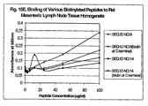
- Peptide sequences (SEQ ID NOs:3, 7, 8, 9 and 14) were synthesized with biotin tags at the amino terminal for all peptides and additionally at the carboxyl terminal for SEQ ID NOs:8 and 14. Binding of these peptides to intestinal tissue of different species namely dog, mouse, pig, and rat was performed. No major differences in binding profiles to tissue of different species were observed. ( See Figures 14A-14H ). Binding of peptides (SEQ ID NOs:8 and 14) to rat tissue from different organs, namely liver, lung, mesenteric lymph nodes, spleen and kidney, was performed. No major differences in binding profiles to these tissues were observed. ( See Figures 15A-15E ).
- Three synthetic peptides (SEQ ID NO:8, 25 and 14) derived from isolated clones were biotinylated and tested for binding to human Peyer's patch tissue sections.
- a known negative binding peptide was included as a negative control.
- Paraffin sections of human Peyer's patch were deparaffinized and dehydrated. The sections were rinsed in PBS. The antigenic determinants on the tissue were unmasked by microwaving in 2.1g/L acetic acid for 5 minutes and allowed to cool at room temperature for 20 minutes while covered in plastic wrap. After 20 minutes, the sections were washed in PBS and then blocked in endogenous peroxidase in 1% hydrogen peroxide in methanol for 10 minutes.
- the sections were rinsed with PBS/Tween.
- the slides were mounted using aqueous mounting medium and a cover slip.
- the negative control peptide showed no binding.
- SEQ ID NO:14 (a low to medium binder as determined by ELISA) was also negative for binding in this study. Positive binding to human Peyer's patch was observed with SEQ ID NO: 8 and 25. Both peptides gave positive staining on the apical side of human enterocytes.
- Peptide SEQ ID NO:8 at a concentration of 6.25 ⁇ g/ml, 12.5 ⁇ g/ml and 25 ⁇ g/ml, was co-incubated with L-cysteine over the concentration range of 100mM to 0.003mM.
- Peptide SEQ ID NO:25 at a concentration of 6.25 ⁇ g/ml was co-incubated with L-cysteine over the concentration range of 100mM to 0.003mM.
- the presence of free L-cysteine prevented binding therefore demonstrating that the cysteine groups are also involved in the binding of these peptides (See Figures 16A-16D ).
- SEQ ID NO:8 is a medium binder to intestinal epithelial tissue.
- SEQ ID NOs: 31 and 32 When either of the two cysteine residues are substituted with an alanine residue (SEQ ID NOs: 31 and 32), binding is still retained.
- both cysteines are substituted with an alanine residue (SEQ ID NO:33), binding to the epithelium is abrogated.
- a biotin tag is added to either the amino or carboxyl end, no difference in the binding affinities is observed (See Figure 17A ).
- SEQ ID NO:25 is the highest binder of the phage-derived peptides.
- cysteine residue is substituted with an alanine residue (SEQ ID NO:38)
- binding to the epithelium is abrogated.
- the stabilized D-form (SEQ ID NO:37) and retro-inverted D-form (SEQ ID NO:40) retained high binding (See Figure 17B ).
- SEQ ID NO:24 is a medium binder. When the cysteine residue is substituted with an alanine residue, the binding to the epithelium is abrogated.
- Biotinylated SEQ ID NO:40 was adsorbed to the surface of fluorescent polystyrene particles (0.289 ⁇ m) using routine methodologies at room temperature.
- Mouse intestinal loops containing one or more polystyrene suspensions typically 300 ⁇ l containing 5.0 x 10 10 particles per ml
- the Peyer's patches were excised, fixed in methanol and the M cells were counter-stained with UEA1-rhodamine for subsequent analysis by confocal microscopy. Stained tissues were examined on a BioRad MRC 600 confocal laser-scanning microscope.
- Fluorescent particles coated with peptide SEQ ID NO:40 exhibited binding and uptake into M-cells. No binding or uptake was visible using the control streptavidin particles.
- High-binding phage clones numbers 1.009, 5.074, 2.078 and 4.009 were injected into rat intestinal loops as described earlier. There were 3 mice per group. Blood samples were taken at 0, 30, 60, 90 and 120 minutes. The animals were then sacrificed and the loops excised. 100 ⁇ l of each blood sample was serially diluted in LB and plated out in top agar plates containing IPTG/Xgal. Blue plaques were counted after incubation at 37°C overnight. There was a PBS control and an m13mp19 control. Table 8. Titration of blood samples for translocation of phage from the gut into the blood Phage Corresponding Blood No. of Total phage per Clone No. SEQ ID No.
- SEQ ID NO:8 is one of the preferred phage-derived peptides. It is a medium binder. When either of the two cysteine residues are substituted, binding is abrogated. However, when this double-mutant peptide ligand is adsorbed to polystyrene particles binding to approximately 60% of that of the parent peptide is observed. This may indicate that conformation of the peptides is important.
- This 12-mer has been synthesized with a biotin tag at either the amino or carboxy end. No differences in binding affinities were observed with the addition of the biotin tag. The stabilized D-form and retro-inverted D form retained high-binding. Table 9.
- SEQ ID NO:25 is the highest binder of the phage derived peptides. When the cysteine residue is substituted with an alanine residue binding is abrogated. The stabilized D-form and retro-inverted D-form of SEQ ID NO:25 retained high binding. Table 10.
Abstract
Description
- This invention relates to novel targeting ligands which permit or facilitate the transport of drugs, macromolecules, or particles, such as biodegradable nanoparticles and microparticles, or bacterial carriers or viral carriers through the intestinal epithelium, M-cells located in gut associated lymphoid tissue, and/or Peyer's Patch tissue of the intestinal epithelium.
- The epithelial cells lining the lumenal side of the gastrointestinal tract (GIT) are a major barrier to drug delivery following oral administration. However, there are four recognized transport pathways which can be exploited to facilitate drug delivery and transport: the transcellular, paracellular, carrier-mediated and transcytotic transport pathways. The ability of a conventional drug, peptide, protein, macromolecule, nanoparticulate system or microparticulate system to interact with one of these transport pathways may result in increased delivery of that drug or particle from the GIT to the underlying circulation.
- M-cells are antigen sampling cells that are found in the epithelium of the gut-associated lymphoid tissue, or Peyer's Patch. The transcytotic capacity of M-cells and the downstream processing of the antigen sampled would suggest that targeting vaccines to M-cells would enhance oral immunization (Foster et al., 15 Vaccine 546-71 (1998)). However, to date, no human M-cell marker has been identified as a target for delivery of vaccines and/or other drugs through the M-cell route.
- In
U.S. Patent No. 6,117,632 to O'Mahony , one of the present inventors disclosed a method of identifying peptides which permit or facilitate the transport of an active agent through human or animal epithelial tissue. This method uses in vivo phage display screening to identify ligands. -
U.S. Patent No. 6,060,082 to Chen et al . discloses modified polymerized liposomes that contain a molecule or ligand on their surfaces in order to target the liposomes to a specific site or cell type fororal/mucosal drug delivery. Also disclosed is an embodiment in which the liposomes are modified with carbohydrate moities or lectins that specifically target M-cells or Peyer's Patches in mice. However, this reference only teaches transport of liposomes. - Other approaches include: drug delivery through the epithelium by a carrier molecule selected from transferrin receptor ligands conjugated to an active agent and a transport enhancing agent (
U. S. Patent No. 5,254, 342 to Shen et al .); and coupling the antigen to ligands that bind the FcRn receptor. (U. S. Patent No. 6,030, 613 to Blumberg et al .). - All references cited herein are incorporated herein by reference in their entireties.
- Thus, there still exists a need for M-cell and/or Peyer's Patch specific ligands that are particularly effective in transporting drugs, including drug-loaded nanoparticles and microparticles, or bacterial or viral carries coding for vaccines into or across a human or animal intestinal epithelium.
- In an aspect directly related to specific 12-mer peptides, the invention is a purified synthetic polypeptide ligand comprising a 12-mer peptide selected from the group consisting of
SEQ 10 NOs:1-34, SEQ ID NOs:38-39, and SEQ ID NO:42 and wherein said 12mer peptide when integrated as an N-terminal PIII fusion peptide of an M13 phage confers an ability to bind the phage to either Caco-2 cell, IEC-6 cell, rat, mouse, pig or dog homogenate membrane fractions, said ability being at least as great as that conferred by a similarly integrated 12-mer peptide of SEQ ID NO:67. - The above noted functional test is "wherein said 12-mer peptide when integrated as an N-terminal PIII fusion peptide of an M13 phage confers an ability to bind the phage either to Caco-2 cell, IEC-6 cell, rat, mouse, pig or dog homogenate membrane fractions, said ability being at least as great as that conferred by a similarly integrated 12-mer peptide of SEQ ID NO:67", and is also specified below for other aspects and embodiments of the invention. For all agents and embodiments, preferred ligands are those that satisfy the functional test when Caco-2 cell homogenate membrane fractions are used.
- In an aspect related to specific peptide motifs, the invention is a purified synthetic polypeptide ligand, said ligand comprising a L-peptide sequence, D-peptide version thereof, or retro-inverted version thereof, said L-peptide sequence being selected from the group consisting of HESSH (SEQ ID NO:97) and NVYTXXXXSPXP (SEQ ID NO:98), wherein said L-peptide sequence, a D-peptide version thereof, or a retro inverted version thereof when integrated as an N-terminal PIII fusion peptide of an M13 phage confers an ability to bind the phage to either Caco-2 cell, IEC-6 cell, rat, mouse, pig or dog homogenate membrane fractions, said ability being at least as great as that conferred by a similarly integrated 12-mer peptide of SEQ ID NO:67.
- In an aspect related to naturally occurring homologues of the specific peptides, the invention is a purified synthetic polypeptide ligand, not more than 200 amino acids in length, comprising a peptide selected from the group consisting of SEQ ID NOs:74 through SEQ ID NO:96 wherein said peptide when integrated as an N-terminal PIII fusion peptide of an M13 phage confers an ability to bind the phage to either Caco-2 cell, IEC-6 cell, rat, mouse, pig or dog homogenate membrane fractions, said ability being at least as great as that conferred by a similarly integrated 12-mer peptide of SEQ ID NO:67.
- In the above inventions, related to purified synthetic polypeptide ligands there are preferred and even highly preferred embodiments.
- As to all of the aforementioned purified synthetic polypeptide ligands, it is preferred that their length be not more than 200 amino acids, more preferably not more than 100 amino acids, most preferably not more than 50 amino acids. Conversely, it is preferred that their length be at least 12 amino acids, more preferably at least 20 amino acids, most preferably at least 30 amino acids.
- In particular embodiments of all of the aforementioned purified synthetic polypeptide ligands, the polypeptide comprises a zinc-binding domain.
- Nucleic acid molecules that code for the aforementioned purified synthetic polypeptide ligands are also aspects of the invention. Preferred are those that are not more than 600 nucleotides in length. Highly preferred are those that code for a purified synthetic polypeptide that comprises one of the specific 12-mer peptides, motifs, or naturally occurring homologues.
- In particular embodiments of the invention, one of the aforementioned purified synthetic polypeptides ligands is integrated into the protein of a phage.
- In particular embodiments of the invention, one of the aforementioned purified synthetic polypeptide ligands is covalently or non-covalently bound to a carrier entity comprising a pharmaceutical agent. For example, the carrier entity is selected from the group consisting of a nanoparticle, microparticle, liposome, bacterium, phage (bacteriophage) and virus (preferably a mammalian virus, most preferably a human virus; especially non-pathogenic forms made by recombinant or other technologies). It is preferred that the nanoparticle, microparticle or liposome have a largest dimension that is in the range of 10nm to 500µm, as discussed in more detail elsewhere herein. In particular embodiments of the invention, the pharmaceutical agent is a drug or therapeutic agent. In other specific embodiments, the pharmaceutical agent is a pathogen antigen.
- Certain aspects of the invention involve the use of the purified synthetic polypeptide ligands to target delivery of pharmaceutical agents.
- In one aspect, the invention is a method of administering a pharmaceutical agent to an organism having intestinal epithelium, said method comprising contacting said intestinal epithelium with one of the aforementioned purified synthetic polypeptide ligands that is covalently, or non-covalently bound to, a carrier entity. In preferred the embodiments, the organism is a mammal. Most preferably, the mammal is a human.
- In particular embodiments of the method, the carrier entity is selected from the group consisting of a nanoparticle, microparticle, liposome, bacterium, phage and virus. A preferred embodiment is where the polypeptide ligand is expressed on the surface of a phage or bacterium further comprising an antigen or a gene encoding the antigen also expressed on the surface.
- Preferably, the microparticle, nanoparticle or liposome has its major dimension in the range of 10 nm to 500 µm. In preferred embodiments, the carrier entity is loaded with a pharmaceutical agent. The preferred route of administration for delivery of the ligand-carrier entity is the oral route. Other possible routes are the rectal, subcutaneous, intramuscular, nasal and intravenous routes. In particular embodiments the purified synthetic polypeptide ligand is a 12-mer integrated into a coat protein of a phage. In other particular embodiments, the purified synthetic polypeptide ligand comprises a zinc-binding motif, and said ligand is contacted with said epithelium in the presence of zinc.
-
-
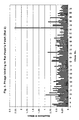
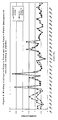
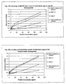
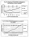
Fig. 1A is a graph of the binding of the phage fromRat 1 to rat Peyer's patch tissue as a function of absorbance at 405nm; -
Fig. 1B is a graph of the binding of the phage fromRat 1 to rat Peyer's patch tissue as a function of absorbance at 405nm; -
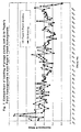
Fig. 2 is a graph of the binding of the phage fromRat 2 to rat Peyer's patch tissue as a function of absorbance at 405nm; -
Fig. 3 is a graph of the binding of the phage fromRat 3 to rat Peyer's patch tissue as a function of absorbance at 405nm; -
Fig. 4 is a graph of the binding of the phage fromRat 4 to rat Peyer's patch tissue as a function of absorbance at 405nm; -
Fig. 5 is a graph of the binding of thephage form Rat 5 to rat Peyer's patch tissue as a function of the absorbance at 405nm; -
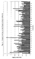
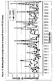
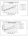
Fig. 6 is a graph of the binding of the 55 high binding clones to rat small intestinal homogenates with and without Peyer's patch tissue present as a function of absorbance at 405nm; -
Fig. 7 is a graph of the binding of the remaining 55 high binding clones to rat small intestinal homogenates with and without Peyer's patch tissue present as a function of absorbnce at 405nm; -
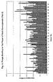
Fig. 8 is a graph of the binding of the clones to dog small intestinal homogenates with and without Peyer's patch tissue present as a function of absorbance at 405nm; -
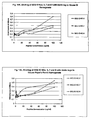
Fig. 9 is a graph of the binding of the clones to pig small intestinal homogenates with and without Peyer's patch tissue present as a function of absorbance at 405nm; -
Fig. 10 is a graph of the binding of the clones to mouse small intestinal homogenates with and without Peyer's patch tissue present as a function of absorbance at 405nm; -
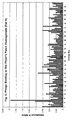
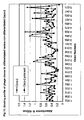
Fig. 11 is a graph of the binding of the phage clones to rat Peyer's patch, Caco-2 cells and IEC-6 cells as a function of absorbance at 405nm; -
Fig. 12 is a graph of the binding of the phage clones to differentiated and non-differentiated Caco-2 cells as a function of absorbance at 405nm; -
Fig. 13A is a graph of the binding of SEQ ID NO:3 with a biotin tag at the amino terminal end to Caco-2 homogenates and rat GI homogenates as a function of absorbance at 650nm. Results were obtained using an ELISA-based assay with streptavidin-peroxidase detection. -
Fig. 13B is a graph of the binding of SEQ ID NO:4 with a biotin tag at the amino terminal end to Caco-2 homogenates and rat GI homogenates as a function of absorbance at 650nm. Results were obtained using an ELISA-based assay with streptavidin-peroxidase detection. -
Fig. 13C is a graph of the binding of SEQ ID NO:7 with a biotin tag at the amino terminal end to Caco-2 homogenates and rat GI homogenates as a function of absorbance at 650nm. Results were obtained using an ELISA-based assay with streptavidin-peroxidase detection. -
Fig. 13D is a graph of the binding of SEQ ID NO:8 with a biotin tag at the amino and carboxyl terminal end to Caco-2 homogenates and rat GI homogenates as a function of absorbance at 650nm. Results were obtained using an ELISA-based assay with streptavidin-peroxidase detection. -
Fig. 13E is a graph of the binding of SEQ ID NO:5 with a biotin tag at the amino terminal end to Caco-2 homogenates and rat GI homogenates as a function of absorbance at 650nm. Results were obtained using an ELISA-based assay with streptavidin-peroxidase detection. -
Fig. 13F is a graph of the binding of SEQ ID NO:14 with a biotin tag at the amino and carboxyl terminal end to Caco-2 homogenates and rat GI homogenates as a function of absorbance at 650nm. Results were obtained using an ELISA-based assay with streptavidin-peroxidase detection. -
Fig. 13G is a graph of the binding of SEQ ID NO:9 with a biotin tag at the amino terminal end to Caco-2 homogenates and rat GI homogenates as a function of absorbance at 650nm. Results were obtained using an ELISA-based assay with streptavidin-peroxidase detection. -
Fig. 13H is a graph of the binding of SEQ ID NO:20 with a biotin tag at the amino terminal end to Caco-2 homogenates and rat GI homogenates as a function of absorbance at 650nm. Results were obtained using an ELISA-based assay with streptavidin-peroxidase detection. -
Fig. 13I is a graph of the binding of SEQ ID NO:17 with a biotin tag at the amino terminal end to Caco-2 homogenates and rat GI homogenates as a function of absorbance at 650nm. Results were obtained using an ELISA-based assay with streptavidin-peroxidase detection. -
Fig. 13J is a graph of the binding of SEQ ID NO:28 with a biotin tag at the amino terminal end to Caco-2 homogenates and rat GI homogenates as a function of absorbance at 650nm. Results were obtained using an ELISA-based assay with streptavidin-peroxidase detection. -
Fig. 13K is a graph of the binding of SEQ ID NO:26 with a biotin tag at the amino terminal end to Caco-2 homogenates and rat GI homogenates as a function of absorbance at 650nm. Results were obtained using an ELISA-based assay with streptavidin-peroxidase detection. -
Fig. 13L is a graph of the binding of SEQ ID NO:11 with a biotin tag at the amino terminal end to Caco-2 homogenates and rat GI homogenates as a function of absorbance at 650nm. Results were obtained using an ELISA-based assay with streptavidin-peroxidase detection. -
Fig. 13M is a graph of the binding of SEQ ID NO:10 with a biotin tag at the amino terminal end to Caco-2 homogenates as a function of absorbance at 650nm. Results were obtained using an ELISA-based assay with streptavidin-peroxidase detection. -
Fig. 13N is a graph of the binding of SEQ ID NO:16 with a biotin tag at the amino terminal end to Caco-2 homogenates as a function of absorbance at 650nm. Results were obtained using an ELISA-based assay with streptavidin-peroxidase detection. -
Fig. 13O is a graph of the binding of SEQ ID NO:19 with a biotin tag at the amino terminal end to Caco-2 homogenates and rat GI homogenates as a function of absorbance at 650nm. Results were obtained using an ELISA-based assay with streptavidin-peroxidase detection. -
Fig. 13P is a graph of the binding of SEQ ID NO:29 with a biotin tag at the amino terminal end to Caco-2 homogenates and rat GI homogenates as a function of absorbance at 650nm. Results were obtained using an ELISA-based assay with streptavidin-peroxidase detection. -
Fig. 13Q is a graph of the binding of SEQ ID NO:30 with a biotin tag at the amino terminal end to Caco-2 homogenates as a function of absorbance at 650nm. Results were obtained using an ELISA-based assay with streptavidin-peroxidase detection. -
Fig. 13R is a graph of the binding of SEQ ID NO:15 with a biotin tag at the amino terminal end to Caco-2 homogenates as a function of absorbance at 650nm. Results were obtained using an ELISA-based assay with streptavidin-peroxidase detection. -
Fig. 14A is a graph of the binding of SEQ ID NOs: 8 and 14 with a biotin tag at both the amino and carboxyl terminal end to pig GI homogenate as a function of absorbance at 650nm. -
Fig. 14B is a graph of the binding of SEQ ID NOs: 8 and 14 with a biotin tag at both the amino and carboxyl terminal end to pig Peyer's Patch homogenate as a function of absorbance at 650nm. -
Fig. 14C is a graph of the binding of SEQ ID NOs: 8 and 14 with a biotin tag at both the amino and carboxyl terminal end to rat GI homogenate as a function of absorbance at 650nm. -
Fig. 14D is a graph of the binding of SEQ ID NOs: 8 and 14 with a biotin tag at both the amino and carboxyl terminal end to rat Peyer's patch homogenate as a function of absorbance at 650nm. -
Fig. 14E is a graph of the binding of SEQ ID NOs: 8 and 14 with a biotin tag at both the amino and carboxyl terminal end to dog GI homogenate as a function of absorbance at 650nm. -
Fig. 14F is a graph of the binding of SEQ ID NOs: 8 and 14 with a biotin tag at both the amino and carboxyl terminal end to dog Peyer's patch homogenate as a function of absorbance at 650nm. -
Fig. 14G is a graph of the binding of SEQ ID NOs: 8 and 14 with a biotin tag at both the amino and carboxyl terminal end to mouse GI homogenate as a function of absorbance at 650nm. -
Fig. 14H is a graph of the binding of SEQ ID NOs: 8 and 14 with a biotin tag at both the amino and carboxyl terminal end to mouse Peyer's patch homogenate as a function of absorbance at 650nm. -
Fig. 14I is a graph of the binding of SEQ ID NOs: 3, 7 and 9 with a biotin tag at the amino terminal end to dog GI homogenate as a function of absorbance at 650nm. -
Fig. 14J is a graph of the binding of SEQ ID NOs: 3, 7 and 9 with a biotin tag at the amino terminal end to dog Peyer's patch homogenate as a function of absorbance at 650nm. -
Fig. 14K is a graph of the binding of SEQ ID NOs: 3, 7 and 9 with a biotin tag at the amino terminal end to mouse GI homogenate as a function of absorbance at 650nm. -
Fig. 14L is a graph of the binding of SEQ ID NOs: 3, 7 and 9 with a biotin tag at the amino terminal end to mouse Peyer's patch homogenate as a function of absorbance at 650nm. -
Fig. 15A is a graph of the binding of SEQ ID NOs: 8 and 14 with a biotin tag at the amino and carboxyl terminal end to rat liver tissue homogenate as a function of absorbance at 650nm. -
Fig. 15B is a graph of the binding of SEQ ID NOs: 8 and 14 with a biotin tag at the amino and carboxyl terminal end to rat spleen tissue homogenate as a function of absorbance at 650nm. -
Fig. 15C is a graph of the binding of SEQ ID NOs: 8 and 14 with a biotin tag at the amino and carboxyl terminal end to rat lung tissue homogenate as a function of absorbance at 650nm. -
Fig. 15D is a graph of the binding of SEQ ID NOs: 8 and 14 with a biotin tag at the amino and carboxyl terminal end to rat kidney tissue homogenate as a function of absorbance at 650nm. -
Fig. 15E is a graph of the binding of SEQ ID NOs: 8 and 14 with a biotin tag at the amino and carboxyl terminal end to rat mesenteric lymph node tissue homogenate as a function of absorbance at 650nm. -
Fig. 16A is a graph of the binding of SEQ ID NO:8 at a concentration of 25µg/ml to intestinal epithelial tissue in the presence of varying concentrations of free L-cysteine as a function of absorbance at 650nm. -
Fig. 16B is a graph of the binding of SEQ ID NO:8 at a concentration of 12.5µg/ml to intestinal epithelial tissue in the presence of varying concentrations of free L-cysteine as a function of absorbance at 650nm. -
Fig. 16C is a graph of the binding of SEQ ID NO:8 at a concentration of 6.25µg/ml to intestinal epithelial tissue in the presence of varying concentrations of free L-cysteine as a function of absorbance at 650nm. -
Fig. 16D is a graph of the binding of SEQ ID NO:25 at a concentration of 6.25µg/ml to intestinal epithelial tissue in the presence of varying concentrations of free L-cysteine as a function of absorbance at 650nm. -
Fig. 17A is a graph of the binding of derivatives of SEQ ID NO:8, including SEQ ID NOs: 31, 32 and 33, to Caco-2 homogenates as a function of absorbance at 650nm. -
Fig. 17B is a graph of the binding of derivatives of SEQ ID:25, including SEQ ID NOs: 37, 38 and 40, to Caco-2 homogenates as a function of absorbance at 650nm. -
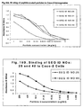
Fig. 18 is a graph of the binding of SEQ ID NO:3 with a zinc-binding motif, SEQ ID NO:43, to Caco-2 homogenates as a function of absorbance at 650nm. -
Fig. 19A is a graph of the binding of SEQ ID NOs: 25, 31, 32 and 33 that were adsorbed to streptavidin particles to Caco-2 cells as a function of absorbance at 650nm. -
Fig. 19B is a graph of the binding of SEQ ID NOs: 25 and 40 that were adsorbed to streptavidin particles to Caco-2 cells as a function of absorbance at 650nm. - The present invention relates to targeted polypeptide ligands for mucosal delivery of agents through the intestinal epithelium. In one embodiment of the invention, the polypeptide ligands are targeted to M-cells or Peyer's patch tissue of the intestinal epithelium.
- As used herein, the term "purified synthetic polypeptide ligand" is intended to distinguish polypeptides of the invention from (1) those that consist of a naturally occurring amino acid sequence; and (2) those that naturally occur but have not been purified.
- Examples of polypeptides that naturally occur but which have not been purified are fragments of polypeptides that exist as intermediates during the translational process that elongates fragments into complete polypeptides, and proteolytic breakdown products which occur from time to time.
- The polypeptide component of a protein, such as mouse keratinocyte growth factor, identified in the Blast homology search below would be an example of a naturally occurring polypeptide.
- A population of synthetic polypeptide ligands in solution, wherein most or all of the polypeptides in solution are a particular synthetic polypeptide ligand, is one example of a purified synthetic polypeptide ligand.
- A polypeptide ligand that may naturally occur in a eukaryotic cell is a purified synthetic polypeptide ligand if it occurs (but does not naturally occur) on a phage surface or a bacterial surface, or if it occurs on the surface of a nanoparticle, microparticle or liposome, or bacterial or viral carrier, or if it occurs as a result of genetic recombination technologies in a cell, virus or phage where it does not naturally occur.
- As used herein, the terms "polypeptide" and "peptide" do not have an intrinsic difference as to biochemical meaning. As indicated herein, a 12-mer peptide can qualify as a polypeptide. In a purified synthetic polypeptide ligand where one or more amino acids have been derivatized (e.g. glycosylation, acetylation, amidation, biotinylation, dansylation) the term purified synthetic polypeptide ligand is intended to apply to the polypeptide component of the ligand. In the case where dansylation comprises the addition of a dansyl-lysine group the polypeptide absent the lysine of the dansyl-lysine group is the purified synthetic polypeptide ligand.
- The test for functionality of a 12-mer, fragment or homologue is exemplified by "wherein said 12-mer L-peptide, fragment or homologue thereof, when integrated as an N-terminal PIII fusion peptide of an M13 phage confers an ability to bind the phage to a Caco-2 cell homogenate membrane fraction, said ability being at least as great as that conferred by a similarly integrated 12-mer peptide of SEQ ID NO:67." A cloning vector useful for accomplishing the binding test is M13KE, which is available from New England Labs Inc., as are the details for integrating the peptide {See Technical Bulletin #8101(4/1/00), which is incorporated by reference herein in its entirety}. Peptides larger than 20-30 amino acids, if so integrated have deleterious effect on the infectivity of the M13 virus. If it is desired to test the binding functionality of peptides too large to be tested in the phage binding test, such larger peptides in biotinylated form can be tested in the Caco-2 membrane binding assay described herein, in order to see if that larger peptide retains detectable binding activity.
- The terms " a Caco-2 cell homogenate membrane fraction" and simply "Caco-2 cell homogenate" are used interchangeably herein unless otherwise indicated. In vivo phage display library screening was used to determine polypeptide ligands that bind to intestinal Peyer's Patch and non-Peyer's Patch tissue homogenates of several species. DNA from one-hundred phage clones with the highest binding affinities (O.D. > 0.75) was sequenced to identify the sequence of the peptide insert. Thirty unique sequences were identified, of which there were several common tripeptide motifs. More than one copy of several clones was isolated and several clones were isolated from different rats (See Table 1 below).
- The 12-mer peptides and related peptides, a total of 43 in all (See Tables 1 and 2) were synthesized and used as ligands in binding studies. The related peptides included selected homologues, D-forms and retro-inverted forms of the 12-mers, as well as a zinc-binding chimeric peptide (SEQ ID NO:43).
- By employing the foregoing techniques, the inventors have identified several polypeptide ligands, which mediate binding to intestinal epithelium of several species, including rat, dog, mouse, pig and/or human intestinal epithelium tissue. Thus, the invention encompasses the following ligands (Tables 1 & 2):
Table 1: Amino Acid Sequences for Ligands No. of copies SEQ ID Sequence of each clone isolated SEQ ID NO:1 ATPPPWLLRTAP 1 SEQ ID NO:2 DGSIHKRNIMPL 1 SEQ ID NO:3 DYDSLSWRSTLH 1 SEQ ID NO:4 GEPTTDMRWRNP 1 SEQ ID NO:5 GLWPWNPVTVLP 5 SEQ ID NO:6 HMLNDPTPPPYW 2 SEQ ID NO:7 KPAYTHEYRWLA 3 SEQ ID NO:8 LETTCASLCYPS 1 SEQ ID NO:9 LGTDWHSVSYTL 1 SEQ ID NO:10 LGTLNAGVPGFP 1 SEQ ID NO:11 LTHSKNPVFLST 1 SEQ ID NO:12 LVPTTHRHWPVT 1 SEQ ID NO:13 LVSNARGFNNLS 1 SEQ ID NO:14 NTRIPEPIRFYM 1 SEQ ID NO:15 NVYTFHSMSPMP 1 SEQ ID NO:16 QHTTLTSHPRQY 1 SEQ ID NO:17 SDFSDTMPHRPS 2 SEQ ID NO:18 SIDTIQILSLRS 3 SEQ ID NO:19 SISWASQPPYSL 1 SEQ ID NO:20 SMVKFPRPLDSR 2 SEQ ID NO:21 LRRWVRVWLRL 1 SEQ ID NO:22 TMSPNVYYTAFG 1 SEQ ID NO:23 TQIPSRPQTPSQ 1 SEQ ID NO:24 VCSNMYFSCRLS 1 SEQ ID NO:25 VPPHPMTYSCQY 1 SEQ ID NO:26 VPRLEATMVPDI 1 SEQ ID NO:27 VPTKPELPVNFT 1 SEQ ID NO:28 WSSDLPQPASTY 1 SEQ ID NO:29 YITPYAHLRGGN 5 SEQ ID NO:30 NVYTDNTLSPTP 1 Table 2: Stabilized versions (D-form, retro-D form) and homologues of these sequences were also synthesized. (L-form amino acid residues are given as capital letters while D-form amino acids are given as lower case letters.) SEQ ID Sequence SEQ ID NO:31 LETTAASLCYPS SEQ ID NO:32 LETTCASLAYPS SEQ ID NO:33 LETTAASLAYPS SEQ ID NO:34 LETTSASLSYPS SEQ ID NO:35 spyclsacttel SEQ ID NO:36 lettcaslcyps SEQ ID NO:37 vpphpmtyscqy SEQ ID NO:38 VPPHPMTYSAQY SEQ ID NO:39 VPPHPMTYSSQY SEQ ID NO:40 yqcsytmphppv SEQ ID NO:41 vcsnmyfscrls SEQ ID NO:42 VSSNMYFSSRLS SEQ ID NO:43 DYDSLSWRSTLHGGHESSH - Analysis of the 12-mer peptide sequences revealed that several peptides contain common motifs. Thus, the invention also encompasses these motifs and polypeptide ligands containing the motifs, wherein the polypeptide ligands facilitate transport of a pharmaceutical agent into or across the intestinal epithelium, M-cells or Peyer's patch tissue.
- The motifs PPY, PVT, LGT and NVY have no previously defined receptor. The motif TPPP has been described as a low affinity omega-opioid peptide antagonist. Certain opioid receptors have been observed on intestinal epithelium. An additional motif of the invention is NVYTXXXXSPXP (SEQ ID NO:98) wherein X is any amino acid.
- There are several groups of preferred synthetic polypeptide ligands of the invention, wherein the members of each group contain related amino acid sequences. A first such group contains ligands comprising an amino acid sequence selected from the group consisting of: LETTCASLCYPS (SEQ ID NO:8), LETTAASLCYPS (SEQ ID NO:31), LETTCASLAYPS (SEQ ID NO:32), LETTAASLAYPS (SEQ ID NO:33), LETTSASLSYPS (SEQ ID NO:34), spyclsacttel (SEQ ID NO:35) and lettsaslsyps (SEQ ID NO:36). A second such group contains ligands selected from the group consisting of: VPPHPMTYSCQY (SEQ ID NO:25), yqcsytmphppv (SEQ ID NO:40), VPPHPMTYSSQY (SEQ ID NO:39) and VPPHPMTYSAQY (SEQ ID NO:38). A third such group contains ligands comprising an amino acid sequence selected from the group consisting of: VCSNMYFSCRLS (SEQ ID NO:24), vcsnmyfscrls (SEQ ID NO:41) and VSSNMYFSSRLS (SEQ ID NO:42).
- Ligands of the invention are useful for transporting a carrier entity or pharmaceutical agent into or across the intestinal epithelium, M-cells or Peyer's patch tissue. Thus, the invention not only provides novel ligands, but also provides a method to transport a carrier entity or pharmaceutical agent into or across the intestinal epithelium, or M-cells or Peyer's patch tissue, as well as novel ligand-entity complexes.
- As used herein, the term "carrier entity" is defined as a particle, droplet, bacterium, phage or virus that can carry a pharmaceutical agent. As used herein, the term "carrier entity" is also defined as a bacterium, phage or virus that can code for a pharmaceutical agent A microparticle is defined as a particle whose "major dimension" is in the
range 1 to 5µm, most preferably in therange 1 to 3µm. A nanoparticle is defined as a particle whose major dimension is less than 1µ, preferably in the range 1nm to 500nm, most preferably in the range 10nm to 500nm. - As used herein, the major dimension of a spherical particle is its diameter, and that of a rod-shaped particle, its length. For other particles, it is the longest dimension possible for the particle.
- Nano- and microparticles that are loaded with, or encapsulate, pharmaceutical agents, can be coated with the polypeptide ligands, such as those of the present invention, that target intestinal epithelium tissue, such as M-cell or Peyer's patch tissue. The coating can be effected by covalent or non-covalent bonding. The covalent bonding can be achieved by adsorption or any other coating process. In either case, the bonding can be made to completed particles or to particle components that subsequently form part of the particles.
- Biodegradable particles are preferred.
- Pharmaceutical agents can, in the alternative, be directly linked to polypeptide ligands. If the agent is itself a polypeptide or peptide, the product is a chimeric polypeptide comprising both an agent and a targeting portion. Bacterial vectors can express a targeting ligand on their surface and also express an antigen on their surface or carry a gene coding for the antigen. Viral vectors can express a targeting ligand on their surface and also express an antigen on their surface or carry a gene coding for the antigen.
- A "pharmaceutical agent" is a therapeutic or diagnostic agent. Therapeutic agents are those that are administered either to treat an existing disease or prophylactically to protect against a potential future disease. Diagnostic agents are any agents that are administered as part of a diagnostic procedure.
- Examples of therapeutic agents are drugs, genes, gene-delivery vectors, DNA vaccines, antigens and recombinant viruses.
- Drugs include, for example, analgesics, anti-migraine agents, anti-coagulant agents, anti-emetic agents, cardiovascular agents, anti-hypertensive agents, narcotic antagonists, chelating agents, anti-anginal agents, chemotherapy agents, sedatives, anti-neoplastics, prostaglandins and antidiuretic agents, antisense oligonucleotides, gene-correcting hybrid oligonucleotides, ribozymes, RNA interference (RNAi) oligonucleotides, silencing RNA (siRNA) oligonucleotides, aptameric oligonucleotides and triple-helix forming oligonucleotides.
- Examples of gene-delivery vectors are DNA molecules, viral vectors (E.g. adenovirus, adeno-associated virus, retrovirues, herpes simplex virus, and sindbus virus), and cationic lipid-coated DNA and DNA-dendrimers.
- Examples of drugs are as insulin, calcitonin, calcitonin gene regulating protein, atrial natriuretic protein, colony stimulating factor, betaseron, erythropoietin (EPO), interferons (E.g. α, β or γ interferon), somatropin, somatotropin, somatostatin, insulin-like growth factor (somatomedins), luteinizing hormone releasing hormone (LHRH), tissue plasminogen activator (TPA), growth hormone releasing hormone (GHRH), oxytocin, estradiol, growth hormones, leuprolide acetate, factor VIII and interleukins (E.g. interleukin-2). Representative drugs also include: analgesics (E.g. fentanyl, sufentanil, butorphanol, buprenorphine, levorphanol, morphine, hydromorphone, hydrocodone, oxymorphone, methadone, lidocaine, bupivacaine, diclofenac, naproxen and paverin); anti-migraine agents (E.g. sumatriptan and ergot alkaloids); anti-coagulant agents (E.g. heparin and hirudin); anti-emetic agents (E.g. scopolamine, ondansetron, domperidone and metoclopramide); cardiovascular agents, anti-hypertensive agents and vasodilators (E.g. diltizem, clonidine, nifedipine, verapamil, isosorbide-5-mononitrate, organic nitrates and agents used in treatment of heart disorders); sedatives (E.g. benzodiazepines and phenothiozines); narcotic antagonists (E.g. naltrexone and naloxone); chelating agents (E.g. deferoxamine); anti-diuretic agents (E.g. desmopressin and vasopressin); anti-anginal agents (E.g. nitroglycerine); anti-neoplastics (E.g. 5-fluorouracil and bleomycin); prostaglandins; and chemotherapy agents (E.g. vincristine).
- Examples of antigens that are therapeutic agents are tumor antigens, pathogen antigens and allergen antigens. A vaccine preparation will contain at least one antigen. "Pathogen antigens" are those characteristic of pathogens, such as antigens derived from viruses, bacteria, parasites or fungi.
- Examples of important pathogens include vibrio choleras, enterotoxigenic E. Coli, rotavirus, Clostridium difficile, Shigella species, Salmonella typhi, parainfluenza virus, influenza virus, Streptococcus mutans, Plasmodium falciparum, Staphylococcus aureus, rabies virus and Epstein-Barr virus.
- Viruses in general include the following families: picronaviridae; caliciviridae, togaviridae; flaviviridae; coronaviridae; rhabodviridae; filoviridae; paramyxoviridae; orthomyxoviridae; bunyaviridae; arenaviridae; reoviridae; retroviridae; hepadnaviridae; parvoviridae; papovaviridae; adenoviridae; herpesviridae and poxyviridae.
- These viruses, especially attenuated versions or otherwise modified versions that are not pathogenic, can also be modified to express targeting ligands on their surface and thus allow for enhanced vaccination.
- Bacteria in general include but are not limited to: P. aeruginosa; E. coli; Klebsiella sp.; Serratia sp; Pseudomanas sp.; P. cepacia; Acinetobacter sp.; S. epidermis; E. faecalis; S. pneumonias; S. aureus; Haemophilus sp.; Neisseria sp.; N. meningitidis; Bacterodies sp.; Citrobacter sp.; Branhamella sp.; Salmonelia sp.; Shigella sp.; S. Lesteria sp., Pasteurella multocida; Streptobacillus sp.; S. pyogenes; Proteus sp.; Clostridium sp.; Erysipelothrix sp.; Spirillum sp.; Fusospirocheta sp.; Treponema pallidum; Borrelia sp.; Actinomycetes; Mycoplasma sp.; Chlamydia sp.; Rickettsia sp., Spirchaeta; Legionella sp.; Mycobacteria sp.; Urealplasma sp.; Streptomyces sp.; Trichomoras sp.; and P. mirabilis
- Parasites include but are not limited to: Plasmodium falciparum, P. vivax, P. ovale, P. malaria; Toxoplasma gondii; Leishmania mexicana, L. tropica, L.major, L. aethiopica, L. donovani, Trypanosoma cruzi, T. brucei, Schistosoma mansoni, S. haematobium, S. japonium; Trichinella spiralis; Wuchereria bancrofti; Brugia malayli; Entamoeba histolytica; Enterobus vermiculoarus; Taenia solium, T. saginata, Trichomonas vaginitis, T. hominis, T. tenax; Giardia lamblia; Cryptosporidium parvum; Pneumocytis carinii, Babesia bovis, B. divergens, B. microti, Isospore belli, L. hominis; Dientamoeba fragiles; Onchocerca volvulus; Ascaris lumbricoides; Necator americanis; Ancylostoma duodenale; Strongyloides stercoralis; Capillaria philippinensis; Angiostrongylys cantonensis; Hymenolepis nan; Diphyllobothrium latum; Echinococcus granulosus, E.multilocularis; Paragonimus westermani, P. caliensis; Chlonorchis sinensis; Opisthorchis felineas, G. Viverini, Fasciola hepatica, Sarcoptes scabiei, Pediculus humanus; Phtirius pubis; and Dermatobia hominis.
- Fungi in general include but are not limited to: Crytpococcus neoformans; Blastomyces dematitidis; Aiellomyces dermatitidis Histoplasfrai capsulatum; Coccidiodes immitis; Candids species, including C. albicans, C. tropicalis, C. parapsilosis, C. guilliermondii and C. krusei, Aspergillus species, including A. fumigatus, A. flavus and A. niger, Rhizopus species; Rhizomucor species; Cunnighammella species; Apophysomyces species, including A. saksenaea, A. mucor and A. absidia; Sporothrix schenckii, Paracoccidioides brasiliensis; Pseudallescheria boydii, Torulopsis glabrata; and Dermatophyres species.
- Antigens that are allergens can be haptens, or antigens derived from pollens, dust, molds, spores, dander, insects and foods. Specific examples include the urusiols of Toxicodendron species and the sesquiterpenoid lactones.
- Adjuvants can, if desired, be delivered by the carrier entity or with a carrier entity. Examples of adjuvants are Freund's Complete Adjuvant, Freund's Incomplete Adjuvant, Hunter's Titermax, Gerbu Adjuvant, Ribi's Adjuvant, Montanide ISA Adjuvant, Aluminum Salt Adjuvants and Nitrocellulose adsorbed protein.
- Diagnostic agents include antibodies, nucleic acids and imaging agents, as well as molecules that are needed to make such antibodies, nucleic acids or imaging agents detectable.
- A preferred method of the invention for administering a carrier entity to an organism having intestinal epithelium comprises contacting the intestinal epithelium with a polypeptide ligand of the invention in the presence of the carrier entity, such that the carrier entity is transported into or across the intestinal epithelium or into or across a preferred region of the intestine such as M-cells or Peyer's patches.
- The carrier entity and the polypeptide ligand can be administered together (E.g., as part of an entity-ligand complex or discretely) or separately. Oral administration is most preferred, but other modes of administration requiring transepithelial transport to reach the target tissue are also acceptable (E.g., rectal administration).
- Of course, the ability of the ligands of the invention to target certain cells of the intestinal epithelium also makes the ligands suitable for targeting pharmaceutical agents to the cells themselves for therapy or prophylaxis.
- In addition to the aforementioned ligands and methods, the invention also encompasses nucleic acid sequences encoding the ligands of the invention. As stated herein, the term "encodes" includes the actual coding sequence or a complementary strand. Preferred nucleic acid sequences of the invention are shown in the following table:
Table 3: Nucleic Acid Sequences of the Targeting Ligands SEQ ID NO:44 ATACTGCCTAGGATGAGAAGTCAACGTAGTATGCTG SEQ ID NO:45 ATTAGTCTAAGCCACACTCGCACCCAACGTCGGAGA SEQ ID NO:46 CCTCGAATCAAGCGGACGAGGAAACTTCACCATAGA SEQ ID NO:47 AGGATTCCGCCACCTCATATCCGTAGTCGGCTCACC SEQ ID NO:48 AAGCGGCATAATATTCCGCTTATGAATCGAACCATC SEQ ID NO:49 ATGAAGAGTAGAACGCCAAGAAAGCGAATCATAATC SEQ ID NO:50 ATACGTCGAAGCCGGCTGCGGCAGATCAGACGACCA SEQ ID NO:51 AGTAGAAAGAAACACAGGATTCTTAGAATGCGTAAG SEQ ID NO:52 AGACAGACGACAAGAAAAATACATATTCGAACAAAC SEQ ID NO:53 CTGAGAAGGAGTCTGCGGCCTAGACGGAATCTGAGT SEQ ID NO:54 ATTCCCCCCACGCAAATGAGCATAAGGAGTAATATA SEQ ID NO:55 CATATAAAACCTAATCGGCTCAGGAATCCTCGTATT SEQ ID NO:56 AGAACGAAGCGAAAGAATCTAAATCGTATCAATACT SEQ ID NO:57 AGCAAGCCAACGATACTCATGCGTATACGCCGGCTT SEQ ID NO:58 CGGCATAGGAGACATAGAATGAAACGTATACACATT SEQ ID NO:59 CGTAAAATTAACCGGAAGCTCCGGCTTAGTCGGCAC SEQ ID NO:60 AGGAAGAACCGTAACAGGATTCCAAGGCCACAGACC SEQ ID NO:61 CCAATAAGGAGGAGGCGTAGGATCATTCAGCATATG SEQ ID NO:62 CGACGGATAACACAAACTAGCACAAGTCGTCTCAAG SEQ ID NO:63 CGACAGATTATTAAACCCACGAGCATTAGAAACAAG SEQ ID NO:64 CGAAGGCCGATGAGGCATAGTATCCGAAAAATCAGA SEQ ID NO:65 CATATCAGCAAGAATACGTCATAGGATGAGGAGGCAC SEQ ID NO:66 CGGCGTAGGAGACAGAGTATTATCCGTATACACATT SEQ ID NO:67 AGGCGCAGTCCGCAGAAGCCAAGGCGGAGGAGTAGC SEQ ID NO:68 ACCAAAAGCCGTATAATACACATTCGGAGACATAGT SEQ ID NO:69 AAGAGTATACGACACAGAATGCCAATCCGTCCCCAA SEQ ID NO:70 AATATCCGGAACCATCGTCGCCTCAAGACGAGGCAC SEQ ID NO:71 CGGAAAACCCGGCACACCAGCATTCAACGTCCCAAG SEQ ID NO:72 CGTCACAGGCCAATGACGATGAGTCGTCGGCACAAG SEQ ID NO:73 CAAAGAATAAGGAGGCTGCGACGCCCAAGAAATAGA - In addition, arising from the degeneracy of the genetic code all variations of these DNA sequences resulting in identical amino acid sequences are included in this invention.
- The invention will be illustrated in more detail with reference to the following Examples, but it should be understood that the present invention is not deemed to be limited thereto.
- A Phage Display Peptide Library (12-mer at 1.5 x 10¹¹ pfu) was inoculated intraduodenally into a rat loop model (n=5). Blood samples were taken from the rat loop at time periods of 0, 30, 60, 90 and 120 minutes. The animals were then sacrificed and the loops excised. From these loops, Peyer's patch and non-Peyer's patch tissues were isolated, washed and homogenized. The bacteriophage present in the tissue samples were amplified in E. Coli and isolated by polyethylene glycol (PEG) precipitation. Peyer's patch specific phage were titred and selected for use in subsequent screening cycles.
- Four screening cycles were completed and phage titres (in pfu/ml) were obtained at
cycle 4.Table 4. Phage titres (pfu/ml) in crude tissue homogenates (rat loop model; cycle 4) Rat No. Peyer's Patch 1 1.0 x 10⁶ 2 5.6 x 10⁵ 3 4.0 x 10³ 4 2.6 x 10⁶ 5 4.6 x 10⁴ - The phage pools obtained in Example 1 were plated out on LB agar plates with top agar and phage clones were selected for evaluation by an ELISA analysis of binding to Peyer's patch tissue from various species, along with Caco-2 and IEC-6 cell models.
- The ELISA was run with 5µg/ml of phage homogenates, Blocking buffer: 1% BSA-TBS, wash buffer:TBS-Tween (0.05%), anti-M13 biotin conjugate (Research Diagnostics RDI-PRO61597) at a 1:5000 dilution, ExtrAvidin Alkaline Phosphatase (Sigma E-2636) at a 1:5000 dilution and pNPP substrate.
- Five hundred clones (Rats 1-5 from Cycle 4) were subsequently assayed using the above method. (See
Figures 1A-5 for binding profiles). The clones exhibited a broad range of activity with concentration-dependent binding clearly detectable for all high-binding clones.Table 5: Table of phage clone numbers & SEQ ID NOs. No. of Clone No. SEQ ID of copies corresponding DNA SEQ ID NO: Amino Acid of each sequence: Sequence clone isolated SEQ ID NO:1 ATPPPWLLRTAP 1 3.030 SEQ ID NO:67 SEQ ID NO:2 DGSIHKRNIMPL 1 1.010 SEQ ID NO:48 SEQ ID NO:3 DYDSLSWRSTLH 1 1.016 SEQ ID NO:49 SEQ ID NO:4 GEPTTDMRWRNP 1 1.008 SEQ ID NO:47 SEQ ID NO:5 GLWPWNPVTVLP 5 2.054, 3.003,
3.056, 5.006,
5.074SEQ ID NO:60 SEQ ID NO:6 HMLNDPTPPPYW 2 2.061, 4.075 SEQ ID NO:61 SEQ ID NO:7 KPAYTHEYRWLA 3 2.025, 2.068,
3.083SEO ID NO:57 SEQ ID NO:8 LETTCASLCYPS 1 2.078 SEQ ID NO:62 SEQ ID NO:9 LGTDWHSVSYTL 1 4.009 SEQ ID NO:69 SEQ ID NO:10 LGTLNAGVPGFP 1 5.039 SEQ ID NO:71 SEQ ID NO:11 LTHSKNPVFLST 1 1.049 SEQ ID NO:51 SEQ ID NO:12 LVPTTHRHWPVT 1 5.049 SEQ ID NO:72 SEQ ID NO:13 LVSNARGFNNLS 1 2.081 SEQ ID NO:63 SEQ ID NO:14 NTRIPEPIRFYM 1 2.014 SEQ ID NO:55 SEQ ID NO:15 NVYTFHSMSPMP 1 2.045 SEQ ID NO:58 SEQ ID NO:16 QHTTLTSHPRQY 1 1.002 SEQ ID NO:44 SEQ ID NO:17 SDFSDTMPHRPS 2 3.006, 3.090 SEQ ID NO:64 SEQ ID NO:18 SIDTIQILSLRS 3 2.016, 3.014,
3.031SEQ ID NO:56 SEQ ID NO:19 SISWASQPPYSL 1 5.078 SEQ ID NO:73 SEQ ID NO:20 SMVKFPRPLDSR 2 1.005,1.076 SEQ ID NO:46 SEQ ID NO:21 LRRWVRVWLRL 1 1.004 SEQ ID NO:45 SEQ ID NO:22 TMSPNVYYTAFG 1 3.062 SEQ ID NO:68 SEQ ID NO:23 TQIPSRPQTPSQ 1 1.099 SEQ ID NO:53 SEQ ID MO:24 VCSNMYFSCRLS 1 1.083 SEQ ID NO:52 SEQ ID NO:25 VPPHPMTYSCQY 1 3.020 SEQ ID NO:65 SEQ ID NO:26 VPRLEATMVPDI 1 4.098 SEQ ID NO:70 SEQ ID NO:27 VPTKPELPVNFT 1 2.049 SEQ ID NO:59 SEQ ID NO:28 WSSDLPQPASTY 1 1.038 SEQ ID NO:50 SEQ ID NO:29 YITPYAHLRGGN 5 2.012,3.005,
3.013,3.035,
5.033SEQ ID NO:54 SEQ ID NO:30 NVYTDNTLSPTP 1 1.009 SEQ ID NO:66 - A Biotin-ExtrAvidin Alkaline Phosphatase assay was established for high throughput screening of the phage clones. The initial screens identified 55 out of the 500 clones as high-binding clones (an absorbency reading of > 0.75).
- The rat tissue homogenates were prepared by harvesting rat GI and Peyer's patch and storing them on ice until needed, or 1-2 hours. The tissue was then put into homogenization buffer (250mM Sucrose, 12 mM Tris, 16mM EDTA) with protease inhibitor cocktail. A hand-held homogenizer was used to break up the tissue for 3-4 minutes. The contents of the homogenizer were then transferred to microfuge tubes and spun at 1500rpm for 1 minute. The supernatant was taken off and measured for protein content using the Bio-Rad Assay. Specificity studies were then run to allow differentiation between Peyer's patch specific and non-specific binding properties. The 55 high-binding clones were assayed for binding to rat small intestinal homogenates (i.e., homogenate membrane fractions) with and without Peyer's Patch tissue (i.e., tissue homogenate membrane fractions) present. (See
Figures 6 and7 ). The negative control was M13mp18 with no peptide insert. This negative control consistently showed absorbance readings of <0.200. All of the clones exhibited significantly higher binding to both tissue types as compared to the control. However, there was a negligible difference between binding to Peyer's patch and non-Peyer's Patch tissue, which suggests that the clones are binding to factors common to both tissue types. - These results were reproducible when using a further 50 clones with an absorbance reading between 0.5-0.75.
- One hundred high-binding clones were assayed for their binding properties to pig, dog and mouse small intestinal homogenates that were with and without Peyer's patch tissue homogenates. (See
Figures 8 ,9 and10 ). These homogenates were prepared in the same way as the method for obtaining rat homogenates described above. As was observed with the rat tissues, all the clones exhibited a negligible difference between binding to Peyer's patch and non-Peyer's patch tissue suggesting that the clones were binding to sites specific to both tissue types. Several of the clones that had exhibited the highest binding in the rat model also were categorized as high-binding clones in the dog and pig model systems. These clones, numbered 1.002, 1.009, 1.016, 1.038, 1.083, 2.078, 2.080, 3.084, 3.087 and 5.074, along with 100 clones representing high, medium and low binders were selected for sequencing to determine the nature of the peptide inserts that may be implicated in their binding function. - Seventy clones were assayed for binding to cell homogenates (IEC-6 and Caco-2 homogenates). IEC-6 cells are a rat normal small intestinal epithelial cell line and Caco-2 cells are a human colon epithelial adenocarcinoma cell line believed to display properties of human small intestinal epithelial cells.
- In order to prepare the Caco-2 cell membrane and cytosolic fractions, confluent Caco-2 cell monolayers (grown in 75cm2 flasks for up to 1 week at 37°C and 5% CO2) were washed twice in Dulbecco's PBS (DPBS). The cell monolayers were then treated with 10mM EDTA-DPBS for 5-10 minutes at 37°C and the cells were harvested by centrifugation at 1000rpm for 5 minutes. The cells were then washed 3x in DPBS. The cell pellet was resuspended in 3 volumes of ice-cold HED buffer (20mM HEPES (pH 7.67), 1mM EGTA, 0,5mM dithiothreitol, 1mM phenylmethylsuphonyl fluoride (PMSF)) and the cells were allowed to swell for 5 minutes on ice. The cells were then homogenized for 30 seconds. The cell homogenates were then centrifuged in hard walled tubes at 40,000rpm for 45 minutes at 4°C (ultracentrifuge Ti90 rotor). The supernatant was removed and the pellet resuspended in HEDG buffer (20mM HEPES (pH 7.67), 1mM EGTA, 0,5mM dithiothreithol, 100mM NaCl, 10% glycerol, 1mM PMSF). Three volumes of buffer were then added and the pellet was resuspended and centrifuged again at 1000rpm for 2 minutes. The supernatant was removed and stored on ice. The procedure was repeated adding the second supernatant to the first and then the procedure was repeated 2-3 more times. The protein concentration was determined using the Bio-Rad protein assay. All fractions were stored at -80°C.
- The IE-6 cell homogenates were prepared in the same way as the Caco-2 homogenates as described above.
- Analysis of binding of phage clones to Caco-2 cell membrane fractions by ELISA was done as follows: 96-well ELISA plates were coated overnight at 4°C with Caco-2 cell membrane fractions (10µg/ml in 0.05M carbonate buffer (pH9.6); 100µl/well). The plates were then blocked in 1.5% BSA-TBS for 1 hour at room temperature (100µl/well) prior to washing 3X in TBS/Tween 20 (0.05%). Phage clones (1:2 dilution in 1.5% BSA-TBS) were serially diluted down the plate and incubated for one-two hours at room temperature. After 3 washes in TBS/
Tween 20 the phage were incubated with biotinylated mouse anti-M13 MAb (1:5000 dilution in 1.5% BSA-TBS; RDI; 100µl/well) for one hour at room temperature. The plates were washed three times prior to incubation with extravidin AP (1:5000 dilution in 1.5% BSA-TBS; Sigma; 100µl/well) at room temperature for one hour. The plates were again washed 3 times in TBS/Tween 20. Alkaline phosphatase activity was detected using the substrate p-NPP (p-nitrophenyl phosphate). After 30 minutes, development of the enzymatic reaction was stopped by addition of 3M NaOH (100µl/well). The plates were read at 405nm using an ELISA plate reader. - The clones showed a broad range of activity with high binders exhibiting concentration-dependent binding. The binding profiles showed non-differentiated and differentiated Caco-2 cell fractions giving similar results. The absorbance readings varied between the different tissues and cell types, however, the overall binding profile remained unchanged. (See
Figures 11 and12 ). - The 100 phage clones from Example 3 including all high-binding clones and a selection of medium- and low-binding clones were sequenced to determine the nature of the peptide inserts.
- The phage DNA was isolated using Qiagen's Quiaprep M13 spin kits. The isolated DNA was precipitated and subsequently sequenced with a 96 gIII sequencing primer situated 117 base pairs 3' of the peptide insert.
- Of the 100 inserts sequenced, 53% did not contain a detectable insert in gene III and all of these clones represented the low binding clones with an absorbance of < 0.4. This appeared to correspond with the library phage (MP13KE) and may have represented loss of the insert during final amplification of selected clones or may have been selected by their ability to be taken up as particulate matter in either Peyer's patch M-cells or enterocytes or alternatively these clones may have a mutation elsewhere in gene III, gene VII or another gene of M13 which was selected during the screening program for binding to/uptake into intestinal epithelium or Peyer's patch tissue in vivo.
- A BLAST search using the Swissprot database was performed on thirty of the unique sequences in order to compare the predicted peptide sequence to the protein/peptide sequence database. A summary of BLAST alignments is as follows:
Table 6:Alignment of most relevant Blast homologues. SEQ ID Sequence Homologue of
interestAlignment SEQ ID GEPTTDMRWRNP MOUSE KERATINOCYTE Query: 1 GEPTTDMRW 9 NO:4 GROWTH FACTOR G PT+ MRW RECEPTOR Sbjct: 183 GNPTSTMRW 191 (SEQ ID NO: 74) SEQ ID GLWPWNPVTVLP HUMAN UROKINASE-TYPE Query: 4 PWNPVTVL 11 NO:5 PLASMINOGEN PWN TVL ACTIVATOR PRECURSOR Sbjct: 93 PWNSATVL 100 (u-PA) (SEQ ID NO:75) SEQ ID HMLNDPTPPPY XENLA EPITHELIAL- Query: 4 NDPTPPPY 11 NO:6 CADHERIN PRECURSOR NDPT PPY (E-CADHERIN) Sbjct: 811 NDPTAPPY 818 (SEQ ID NO: 76) SEQ ID KPAYTHEYRWLA PRECURSOR (FCεRI) Query: 4 YTHEYRWL 11 NO:7 (IGE FC RECEPTOR, YT EYRWL ALPHA-SUBUNIT) Sbjct: 196 YTIEYRWL 203 (FC-EPSILON RI- (SEQ ID NO:77) ALPHA) SEQ ID LETTCASLCYPS CLALU LECTIN-RELATED Query: 2 ETTCASLCYPS 12 NO:8 PROTEIN PRECURSOR ET ASL YPS (CLLRP) (LRPCL) Sbjct:210 ETLIASLTYPS 220 (SEQ ID NO:78) CAVPO CASEIN A PRECURSOR Query: 2 ETTCASLC 9 ET CASLC Sbjct: 48 ETICASLC 55 (SEQ ID NO:79) SEQ ID LGTDWHSVSYTL PIG ZONADHESIN Query: 1 LGTDWHSVSYT 11 NO:9 PRECURSOR LGTDW S + T Sbjct: 750 LGTDWFSPNCT 760 (SEQ ID NO:80) SEQ ID LGTLNAGVPGFP MOUSE ELASTIN Query: 4 LNAGVPGF 11 NO:10 PRECURSOR L AGVPGF (TROPOELASTIN) Sbjct: 612 LGAGVPGF 619 (SEQ ID NO:81) Query: 6 AGVPGF 11 AGVPGF Sbjct: 623 AGVPGF 628 (SEQ ID NO:82) Query: 6 AGVPGF 11 AGVPGF Sbjct: 632 AGVPGF 637 (SEQ ID NO:83) Query: 6 AGVPGF 11 AGVPGF Sbjct: 641 AGVPGF 645 (SEQ ID NO:84) Query: 6 AGVPGF 11 AGVPGF Sbjct: 650 AGVPGF 655 (SEQ ID NO:85) SEQ ID LVPTTHRHWPVT MOUSE STROMELYSIN-3 Query: 3 PTTHRHWEV 11 NO:12 PRECURSOR (MATRIX P +HRH PV METALLOPROTEINASE- Sbjct: 40 PESHRHHPV 48 11) (MMP-11) (SEQ ID NO:86) SEQ ID:15 NVYTFHSMSPMP RAT SUCRASE- Query: 1 NVYTFHSMSPMP 12 ISOMALTASE, N YT S+ P+P INTESTINAL Sbjct:987 NPYTLTSIQPLP 99 (SEQ ID NO:87) SEQ ID QHTTLTSHPRQY HUMAN PLACENTAL- Query: 2 HTTLTSHP 9 NO:16 CADHERIN PRECURSOR H T+T+HP (P-CADHERIN) Sbjct: 376 HFTITTHP 383 (SEQ ID NO:88) SEQ ID SISWASQPPYSL CAPHI BETA CASEIN Query: 3 SWASQPPYSL 12 NO:19 PRECURSOR SW QPP L Sbjct: 157 SWMHQPPQPL 166 (SEQ ID NO:89) SEQ ID SMVKFPRPLDSR Z01 MOUSE TIGHT Query: 6 PRPLDSR 12 NO:20 JUNCTION PROTEIN ZO- PR LDSR 1 (TIGHT JUNCTION Sbjct: 1110 PRDLDSR (SEQ ID PROTEIN 1) NO:90) SEQ ID TQIPSRPQTPSQ MOUSE VERSICAN CORE Query: 1 TQIPSRPQTPS 11 NO:23 PROTEIN PRECURSOR T++P P TPS (LARGE FIBROBLAST Sbjct:1173 TELPKFPSTPS1183 PROTEOGLYCAN) (SEQ ID NO:91) (CHONDROITIN SULFATE PROTEOGLYCAN Query: 1 TQIPSRPQTPS 11 CORE PROTEIN 2) (PG- T IPS PQ P+ M) Sbjct:307 TGIPSTPQKPT 317 (SEQ ID NO:92) SEQ ID VPPHPMTYSCQY PAPCY Query: 1 VPPHPMTYSC 10 NO:25 METALLOPROTEINASE VPPHP T C INHIBITOR 1 Sbjct: 27 VPPHPQTAFC 36 PRECURSOR (TIMP-1) (SEQ ID NO:93) SEQ ID VPRLEATMVPDI HUMAN VERSICAN CORE NO:26 PROTEIN PRECURSOR Query:1 VPRLEATMVPDI12 (LARGE FIBROBLAST +PR AT++P+I PROTEOGLYCAN) Sbjct:2695 IPRKSATVIPEI (CHONDROITIN SULFATE 2706 (SEQ ID NO:94) PROTEOGLYCAN CORE PROTEIN 2) (GLIAL HYALURONATE-BINDING PROTEIN) (GHAP) SEQ ID VPTKPELPVNFT HUMAN COLLAGEN ALPHA Query: 1 VPTKPELPVN 10 NO:27 1(VII) CHAIN VPT PELPV+ PRECURSOR (LONG- Sbjct: 500 VPTGPELPVS 509 CHAIN COLLAGEN) (SEQ ID NO:95) SEQ ID YITPYAHLRGNN RAT INSULIN-LIKE Query: 4 PYAHLRGG 11 NO:29 GROWTH FACTOR I PYAH+ GG RECEPTOR Sbjct: 1348 PYAHMNGG 1355 (SEQ ID NO:96) - The homologue for SEQ ID NO:4 was found to be keratinocyte growth factor receptor (KGFR) which is expressed by intestinal as well as other epithelial cells. It interacts with KGF, a member of the fibroblast growth factor (FGF) family of mitogens which is produced by stromal cells and results in epithelial cell proliferation. The KGF-KGFR interaction is thought to play a role in the epithelial repair processes. See Werner, Cytokine Growth Factor Rev, 2:153-65, 1998; Bajaj-Elliott M. et al., J Clin Invest 102:1473-80, 1998.
- The homologue for SEQ ID NO:5 is urokinase plasminogen activator (u-PA) which is one of the mediators of the plasminogen activator system, that also includes tissue-type plasminogen activator (t-PA) and plasminogen activator inhibitor type-1 (PAI-1). u-PA cleaves plasminogen to the active plasmin which can degrade components of the extracellular matrix (ECM). The u-PA receptor (u-PAR) has been shown to be expressed on different types of epithelial cells including intestinal epithelium. See Gibson P. et al., Gut 7:969-75, 1994. Targeting of the u-PAR has also been shown to enhance gene delivery. See Drapkin P.T. et al., J Clin Invest 105:589-96, 2000.
- SEQ ID NO:6 was found to be homologous to cadherin precursors. Cadherins are epithelial adhesion molecules which allow an intact, selectively permeable, epithelial layer to be formed. They are transmembrane glycoproteins that form a complex with cytoplasmic proteins, termed catenins because they link cadherin to the actin cytoskeleton. The E-cadherin/catenin interaction is important in intestinal epithelial cells and tight junction integrity. See Jawhari A. et al., Gut 5:581-4, 1997.
- The homologue of SEQ ID NO:7 is a high affinity FcεRI alpha subunit which is a type I transmembrane protein that binds to the Fc region of IgE. In humans, FcεRI plays a role in the activation of mast cells and basophils, and participates in IgE-mediated antigen presentation. FcεRI is therefore central to the induction and maintenance of an allergic response and may confer physiological protection in parasitic infections. See Turner H. and Kinet J.P., Nature 402:B24-30, 1999. This protein is expressed on mast cells, eosinophils, Langerhans cells, dendritic cells and monocytes.
- SEQ ID NO: 8 shows a strong homology with a lectin-related protein however, unlike true lectins previously shown to bind glycocalyx on enterocytes, this protein is devoid of carbohydrate binding activity. See Van Damme E.J. et al., Plant Mol Biol 3:579-98, 1995.
- The homologue of SEQ ID NO:9 is a zonadhesion precursor which promotes adhesion of spermatozoa to egg extracellular matrix. Hardy D.M. et al., J Biol Chem. 44:26025-8, 1995. It contains adhesive glycoprotein von Willebrand's factor domains which share similarity to intestinal mucin, muc2. It is within one of these domains that homology to the selected peptide is observed.
- The homologue of SEQ ID NO:10 is elastin, a major structural protein of extracellular matrix. In mouse, the matrix metalloproteinase matrilysin, for which elastin is a substrate, is found in epithelial cells of the uterus, small intestine and extra-testicular ducts. See Wilson C.L. and Matrisian L.M., Int J Biochem Cell Biol 2:123-36, 1996. Interestingly, the 6-residue peptide motif that shares homology with mouse elastin is repeated five times within the protein while a 4-residue motif within this sequence is repeated thirteen times.
- SEQ ID NO:12 shows homology to stromelysin, or MMP-3, which is responsible for the breakdown of ECM collagen as well as the cleavage of u-PA. See Ugwu F. et al., Biochemistry 2:7231-6, 1998. It plays an important role together with other members of the MMP family in intestinal tissue remodeling and repair. See Pender S.L. et al., Ann NY Acad Sci 878:581-2, 1999. Peptide SEQ ID NO:25 showed homology to TIMP-1 (tissue inhibitor of MMPs). This protein forms irreversible complexes with MMPs thus inactivating them. It is present in the intestine where, together with MMPs, it is important in the ongoing repair and renewal that takes place in the intestine.
- The homologue of SEQ ID NO:15 was intestinal sucrase-isomaltase which is a brush border hydrolase expressed in epithelial cells located on villi. The greatest amount of the hydrolase is located at the crypt-villus junction and in the lower to mid-villus region. See Traber P.G., Biochem Biophys Res Commun 173:765-73, 1990. It has been shown to be down regulated on M-cells in an in vitro co-culture model. See Kerneis S. et al., Science 277:949-52, 1997.
- Two different peptides, SEQ ID NO:8 and SEQ ID NO:19, show homology with casein A and casein B, respectively. Casein is a milk protein and has been shown to bind to small intestinal brush border membranes. See Bolte G. et al., J Biochem Biophys Methods 34:189-203, 1997.
- One of the selected peptides, SEQ ID NO:20, shares homology with mouse tight junction protein, ZO-1. See Itoh M. et al., J Cell Biol 3:491-502, 1993. The N-terminus may be involved in transducing a signal required for tight junction assembly, while the C-terminus may have specific properties of tight junctions. ZO-1 has been shown in vitro to interact with cadherins.
- Several peptides, SEQ ID NOS:23, 26 and 27, show regions of homology with ECM proteins including versican and collagen. These proteins and proteoglycans are important in tissue integrity acting not only as the glue connecting cells together but also acting in cell motility, growth and differentiation.
- SEQ ID NO:29 shows homology to high affinity insulin-like growth factor I receptor (IGF IR) from several species. IGF IR has wide tissue expression including epithelial cells of the intestine where it acts as an epithelial cell growth factor. See Wolpert S.I. et al., J Surg Res 63:345-8, 1996. IGF is a candidate for total parenteral nutrition.
- Peptide sequences (SEQ ID NOs: 3, 4, 5, 7, 8, 9, 10, 11, 14, 15, 16, 17, 19, 20, 26, 28, 29, 30) were synthesized with biotin tags at the amino terminal for all peptides and additionally at the carboxyl terminal for SEQ ID NOs:8 and 14. Binding of these peptides to Caco-2 homogenates and/or to rat intestinal tissue homogenates was tested in an ELISA-based assay with streptavidin-peroxidase detection. High binding to both tissue types was observed with the following peptides: SEQ ID NOs:3, 8, 25 and 24. (See
Figures 13A-13N ). - Peptide sequences (SEQ ID NOs:3, 7, 8, 9 and 14) were synthesized with biotin tags at the amino terminal for all peptides and additionally at the carboxyl terminal for SEQ ID NOs:8 and 14. Binding of these peptides to intestinal tissue of different species namely dog, mouse, pig, and rat was performed. No major differences in binding profiles to tissue of different species were observed. (See
Figures 14A-14H ). Binding of peptides (SEQ ID NOs:8 and 14) to rat tissue from different organs, namely liver, lung, mesenteric lymph nodes, spleen and kidney, was performed. No major differences in binding profiles to these tissues were observed. (SeeFigures 15A-15E ). - Three synthetic peptides (SEQ ID NO:8, 25 and 14) derived from isolated clones were biotinylated and tested for binding to human Peyer's patch tissue sections. In addition, a known negative binding peptide was included as a negative control. Paraffin sections of human Peyer's patch were deparaffinized and dehydrated. The sections were rinsed in PBS. The antigenic determinants on the tissue were unmasked by microwaving in 2.1g/L acetic acid for 5 minutes and allowed to cool at room temperature for 20 minutes while covered in plastic wrap. After 20 minutes, the sections were washed in PBS and then blocked in endogenous peroxidase in 1% hydrogen peroxide in methanol for 10 minutes. The rinse was repeated and the sections were blocked with 2% BSA in PBS for 20 minutes at room temperature. The sections were incubated with peptide at 50µg/ml in 2% BSA in PBS for 1 hour at room temperature. Control tissue was treated with BSA alone. The sections were rinsed with 0.05% Tween in PBS. Streptavidin-HRP at 1/500 in 2% BSA was added for 30-60 minutes at room temperature. Again, the sections were rinsed with PBS/Tween. DAB substrate was added for up to 5 minutes, and the reaction was stopped by immersing the slides in water. The sections were counterstained using Haematoxlin for 50 seconds and then rinsed in water. The slides were differentiated in 1% acid alcohol for 5-10 seconds and then rinsed in water. The slides were mounted using aqueous mounting medium and a cover slip. The negative control peptide showed no binding. SEQ ID NO:14 (a low to medium binder as determined by ELISA) was also negative for binding in this study. Positive binding to human Peyer's patch was observed with SEQ ID NO: 8 and 25. Both peptides gave positive staining on the apical side of human enterocytes.
- Peptide SEQ ID NO:8, at a concentration of 6.25µg/ml, 12.5µg/ml and 25µg/ml, was co-incubated with L-cysteine over the concentration range of 100mM to 0.003mM. Peptide SEQ ID NO:25, at a concentration of 6.25µg/ml was co-incubated with L-cysteine over the concentration range of 100mM to 0.003mM. The presence of free L-cysteine prevented binding therefore demonstrating that the cysteine groups are also involved in the binding of these peptides (See
Figures 16A-16D ). - SEQ ID NO:8 is a medium binder to intestinal epithelial tissue. When either of the two cysteine residues are substituted with an alanine residue (SEQ ID NOs: 31 and 32), binding is still retained. When both cysteines are substituted with an alanine residue (SEQ ID NO:33), binding to the epithelium is abrogated. When a biotin tag is added to either the amino or carboxyl end, no difference in the binding affinities is observed (See
Figure 17A ). - SEQ ID NO:25 is the highest binder of the phage-derived peptides. When the cysteine residue is substituted with an alanine residue (SEQ ID NO:38), binding to the epithelium is abrogated. The stabilized D-form (SEQ ID NO:37) and retro-inverted D-form (SEQ ID NO:40) retained high binding (See
Figure 17B ). - SEQ ID NO:24 is a medium binder. When the cysteine residue is substituted with an alanine residue, the binding to the epithelium is abrogated.
- A derivative of SEQ ID NO:3 with an added Zinc-binding motif (HESSH) at the carboxyl terminal (SEQ ID NO:43) was tested for binding to Caco-2 homogenates. Enhanced binding was observed with this additional motif (See
Figure 18 ) - Several phage-derived peptides (SEQ ID NOs: 25, 31, 32, 33, and 40) were adsorbed to streptavidin particles (0.289µm). Peptide coated particles were tested in an ELISA-based assay for binding to Caco-2 homogenates. Binding was retained in all the cases that were examined. In addition, when one double-mutant peptide ligand (SEQ ID NO:33), is adsorbed to polystyrene particles binding that is approximately 60% of that of the parent peptide is observed (See
Figures 19A-19B ). - Biotinylated SEQ ID NO:40 was adsorbed to the surface of fluorescent polystyrene particles (0.289µm) using routine methodologies at room temperature. Mouse intestinal loops containing one or more polystyrene suspensions (typically 300µl containing 5.0 x 10¹⁰ particles per ml) were incubated for 30 minutes. The Peyer's patches were excised, fixed in methanol and the M cells were counter-stained with UEA1-rhodamine for subsequent analysis by confocal microscopy. Stained tissues were examined on a BioRad MRC 600 confocal laser-scanning microscope.
- Fluorescent particles coated with peptide SEQ ID NO:40 exhibited binding and uptake into M-cells. No binding or uptake was visible using the control streptavidin particles.
- High-binding phage clones numbers 1.009, 5.074, 2.078 and 4.009 were injected into rat intestinal loops as described earlier. There were 3 mice per group. Blood samples were taken at 0, 30, 60, 90 and 120 minutes. The animals were then sacrificed and the loops excised. 100µl of each blood sample was serially diluted in LB and plated out in top agar plates containing IPTG/Xgal. Blue plaques were counted after incubation at 37°C overnight. There was a PBS control and an m13mp19 control.
Table 8. Titration of blood samples for translocation of phage from the gut into the blood Phage Corresponding Blood No. of Total phage per Clone No. SEQ ID No. Dilution plaques 100µl of blood 1.009 SEQ ID NO:30 Neat 19 19 5.074 SEQ ID NO:5 Neat 0 0 2.078 SEQ ID NO:8 10-6 45 4.5 x 10⁷ 4.009 SEQ ID NO:9 Neat 34 34 M13mp18 - 10-1 18 180 PBS control - Neat 1 1 - SEQ ID NO:8 is one of the preferred phage-derived peptides. It is a medium binder. When either of the two cysteine residues are substituted, binding is abrogated. However, when this double-mutant peptide ligand is adsorbed to polystyrene particles binding to approximately 60% of that of the parent peptide is observed. This may indicate that conformation of the peptides is important. This 12-mer has been synthesized with a biotin tag at either the amino or carboxy end. No differences in binding affinities were observed with the addition of the biotin tag. The stabilized D-form and retro-inverted D form retained high-binding.
Table 9. Results of further studies with SEQ ID NO:8 SEQ ID NO: Comment Sequence Binding Assay results (Caco- 2) 8 dansyl-lysine LETTCASLCYPS High binder derivative (Kd- 1µg/ml) 8 biotinylated LETTCASLCYPS Medium binder derivative (Kd∼8µg/ml) 8 LETTCASLCYPS Medium binder 31 biotinylated LETTAASLCYPS Medium binder derivative (Kd∼9µg/ml) 32 biotinylated LETTCASLAYPS Medium binder derivative (Kd∼8µ/ml) 33 biotinylated LETTAASLAYPS No binding derivative 35 inverted D- spyclsacttel Medium binder form; (Kd-13.91µg/ml) biotinylated derivative 36 D-form; lettcaslcyps (Kd∼30.53µg/ml) biotinylated derivative 34 biotinylated LETTSASLSYPS No binding derivative - SEQ ID NO:25 is the highest binder of the phage derived peptides. When the cysteine residue is substituted with an alanine residue binding is abrogated. The stabilized D-form and retro-inverted D-form of SEQ ID NO:25 retained high binding.
Table 10. Results of further studies with SEQ ID NO:25 SEQ ID NO: Comment Sequence Binding Assay results (Caco- 2) 25 Dansyl-lysine VPPHPMTYSCQY High binder derivative (Kd<1µg/ml) 25 biotinylated VPPHPMTYSCQY High binder derivative (Kd<5µg/ml) 38 biotinylated VPPHPMTYSAQY No binding derivative 37 D-form; vpphpmtyscqy (Kd∼10µg/ml) biotinylated derivative 40 retro D-form yqcsytmphppv (Kd∼10µg/ml) of SEQ ID NO: 25; biotinylated derivative 39 biotinylated VPPHPMTYSSQY No binding derivative
Claims (46)
- A purified synthetic polypeptide ligand comprising a 12-mer peptide selected from the group consisting of SEQ ID NOs:1-34, SEQ ID NOs:38-39, and SEQ ID NO:42 and wherein said 12-mer peptide when integrated as an N-terminal PIII fusion peptide of an M13 phage confers an ability to bind the phage to either Caco-2 cell, IEC-6 cell, rat, mouse, pig or dog homogenate membrane fractions, said ability being at least as great as that conferred by a similarly integrated 12-mer peptide of SEQ ID NO:67.
- A purified synthetic polypeptide ligand as claimed in claim 1 wherein the 12-mer peptide is selected from the group consisting of 12-mer L-peptides, D-forms of said 12-mer L-peptides and retro-inverted forms of said 12-mer L-peptides.
- A purified synthetic polypeptide ligand of claim 1 or claim 2, wherein said ligand comprises a zinc-binding domain.
- A purified synthetic polypeptide ligand of any of the preceding claims, wherein said ligand consists of an amino acid sequence selected from the group consisting of SEQ ID NOs:1-34, SEQ ID NOs:38-39, SEQ ID NO:42.
- A purified synthetic polypeptide ligand of any of the preceding claims, wherein said ligand consists of a 12-mer peptide selected from the group consisting of SEQ ID NOs:1-34, SEQ ID NOs:38-39, SEQ ID NO:42 or the retro-inverted forms thereof.
- A purified synthetic polypeptide ligand of any of the preceding claims, wherein said ligand is at most 200 amino acids in length.
- A purified synthetic polypeptide ligand of any of the preceding claims, wherein said polypeptide ligand is at least 30 amino acids in length.
- A purified synthetic polypeptide ligand of any of the preceding claims, wherein the 12-mer peptide is selected from the group consisting of LETTCASLCYPS (SEQ ID NO:8), LETTAASLCYPS (SEQ ID NO:31), LETTCASLAYPS (SEQ ID NO:32), LETTAASLAYPS (SEQ ID NO:34).
- A purified synthetic polypeptide ligand of any of the preceding claims, wherein the 12-mer peptide is LETTCASLCYPS (SEQ ID NO:8).
- A purified synthetic polypeptide ligand of claims any of 1 to 7, wherein the 12-mer peptide is selected from the group consisting of VPPHPMTYSCQY (SEQ ID NO:25), VPPHPMTYSSQY (SEQ ID NO:39) and VPPHPMTYSAQY (SEQ ID NO:38).
- A purified synthetic polypeptide ligand of any of claims 1 to 7, wherein the 12-mer peptide is VPPHPMTYSCQY (SEQ ID NO:25).
- A purified synthetic polypeptide ligand of any of claims 1 to 7, wherein the 12-mer peptide is selected from the group consisting of VCSNMYFSCRLS (SEQ ID NO:24) and VSSNMYFSSRLS (SEQ ID NO:42).
- A purified synthetic polypeptide ligand of any of claims 1 to7, wherein the 12-mer peptide is VCSNMYFSCRLS (SEQ ID NO:24).
- A purified synthetic polypeptide ligand of any of claims 1 to 7, wherein said peptide is selected from the group consisting of the D-form of 12-mer L-peptides of SEQ ID NOs:8, 25, and 24.
- A purified synthetic polypeptide ligand of any of claims 1 to 7 wherein said peptide is selected from the group consisting of the retro-inverted form of 12-mer L-peptides SEQ ID NOs: 8 and 25.
- A purified synthetic polypeptide ligand, said ligand comprising a L-peptide sequence, a D-peptide version thereof, or a retro-inverted version thereof, said L-peptide sequence being selected from the group consisting of HESSH (SEQ ID NO:97) and NVYTXXXXSPXP (SEQ ID NO:98), wherein said L-peptide sequence, D-peptide version thereof, or retro-inverted version thereof when integrated as an N-terminal PIII fusion peptide of an M13 phage confers an ability to bind the phage to either Caco-2 cell, IEC-6 cell, rat, mouse, pig or dog homogenate membrane fractions, said ability being at least as great as that conferred by a similarly integrated 12-mer peptide of SEQ ID NO:67.
- A purified synthetic polypeptide ligand of claim 16, said ligand not more than 200 amino acids in length and preferably not more than 50 amino acids in length.
- A purified synthetic polypeptide ligand, not more than 200 amino acids in length, comprising a peptide being selected from the group consisting of SEQ ID NOs:74 through SEQ ID NO:96, wherein said peptide when integrated as an N-terminal PIII fusion peptide of an M13 phage confers an ability to bind the phage to either Caco-2 cell, IEC-6 cell, rat, mouse, pig or dog homogenate membrane fractions, said ability being at least as great as that conferred by a similarly integrated 12-mer peptide of SEQ ID NO:67.
- A purified synthetic polypeptide ligand of claim 18, wherein the peptide is an L-form, a D-form or a retro-inverted form of a peptide selected from the group consisting of SEQ ID NOs:74 through SEQ ID NO:96.
- A purified synthetic polypeptide ligand of claim 18 or claim 19, wherein said polypeptide ligand is at most 50 amino acids in length.
- A purified synthetic polypeptide ligand of any of the preceding claims, wherein said polypeptide ligand is integrated into the protein of a phage.
- The ligand of claim 21, wherein said polypeptide ligand is expressed on the surface of a phage further comprising an antigen and/or a gene encoding the antigen also expressed on the surface.
- The ligand of claim 21, wherein said polypeptide ligand is expressed on the surface of a bacterium further comprising an antigen and/or a gene encoding the antigen also expressed on the surface.
- A purified synthetic polypeptide ligand of any of the preceding claims, wherein said polypeptide ligand is covalently or non-covalently bound to a carrier entity comprising a pharmaceutical agent.
- The purified synthetic polypeptide ligand of claim 24, wherein said carrier entity is selected from the group consisting of a nanoparticle, a microparticle, a liposome, a bacterium, a phage and a virus.
- The purified synthetic polypeptide ligand of claim 24, wherein said carrier entity is selected from the group consisting of a nanoparticle, a microparticle and a liposome and said carrier entity has a largest dimension that is in the range of 10 nm to 500 µm.
- The ligand of claim 24, wherein said pharmaceutical agent is a drug or therapeutic agent.
- The ligand of claim 24, wherein said pharmaceutical agent is a pathogen antigen.
- The ligand of claim 24, wherein said pharmaceutical agent is an adjuvant.
- The purified synthetic polypeptide ligand of claim 24, wherein said carrier entity is selected from the group consisting of a phage and a virus.
- A purified nucleic acid sequence encoding for a purified synthetic polypeptide ligand of claim 1, said nucleotide sequence is not more than 600 nucleotides in length.
- The use of a purified synthetic polypeptide ligand of any of the preceding claims, in preparing a pharmaceutical agent for administering to an organism having intestinal epithelium, said ligand being covalently or non-covalently bound to a carrier entity.
- Use according to claim 32, wherein said organism is a mammal.
- Use according to claim 33, wherein said mammal is a human.
- Use according to claim 32, wherein said carrier entity is selected from the group consisting of a nanoparticle, a microparticle, a liposome, a bacterium, a phage and a viral carrier.
- Use according to claim 35, wherein said nanoparticle, microparticle or liposome carrier entity has its major dimension in the range of 10 nm to 500 µm.
- Use according to claim 35 or claim 36, wherein said nanoparticle, microparticle, or liposome is loaded with a pharmaceutical agent or encapsulated with a pharmaceutical agent.
- Use according to any of claims 32 to 37, wherein said pharmaceutical agent is administered via the oral route.
- Use according to any of claims 32 to 37, wherein said pharmaceutical agent is administered via either a rectal, subcutaneous, intramuscular, nasal or intravenous route.
- Use according to any of claims 32 to 39, wherein said pharmaceutical agent is a vaccine.
- Use according to any of claims 32 to 40, wherein said purified synthetic polypeptide ligand is a peptide integrated into the protein of a phage which is coated, adsorbed or covalently bonded to a surface of a carrier that is either a nanoparticle or microparticle.
- Use according to claim 41, wherein said phage is modified to contain DNA encoding an antigen.
- Use according to claim 41, wherein said nanoparticle or microparticle is loaded with a pharmaceutical agent or encapsulated with a pharmaceutical agent.
- Use according to claim 41, wherein the purified synthetic polypeptide ligand comprises a zinc-binding motif, and said ligand is contacted with said epithelium in the presence of zinc.
- Use according to claim 41, wherein said carrier entity is selected from the group consisting of a phage and a viral carrier.
- Use according to claim 45, wherein said phage comprises a targeting ligand on its surface.
Applications Claiming Priority (3)
| Application Number | Priority Date | Filing Date | Title |
|---|---|---|---|
| US30259101P | 2001-07-02 | 2001-07-02 | |
| US302591P | 2001-07-02 | ||
| PCT/IB2002/003401 WO2003004517A2 (en) | 2001-07-02 | 2002-06-28 | Peyers's patch and/or m-celle targeting ligands |
Publications (2)
| Publication Number | Publication Date |
|---|---|
| EP1432729A2 EP1432729A2 (en) | 2004-06-30 |
| EP1432729B1 true EP1432729B1 (en) | 2008-11-12 |
Family
ID=23168408
Family Applications (1)
| Application Number | Title | Priority Date | Filing Date |
|---|---|---|---|
| EP02760458A Expired - Lifetime EP1432729B1 (en) | 2001-07-02 | 2002-06-28 | Peyers's patch and/or m-cell targeting ligands |
Country Status (8)
| Country | Link |
|---|---|
| US (3) | US6916789B2 (en) |
| EP (1) | EP1432729B1 (en) |
| JP (1) | JP4383855B2 (en) |
| AT (1) | ATE414100T1 (en) |
| AU (1) | AU2002326070A1 (en) |
| CA (1) | CA2451741C (en) |
| DE (1) | DE60229867D1 (en) |
| WO (1) | WO2003004517A2 (en) |
Families Citing this family (11)
| Publication number | Priority date | Publication date | Assignee | Title |
|---|---|---|---|---|
| US6521737B1 (en) * | 1995-11-10 | 2003-02-18 | Elan Corporation, Plc | Peptides which enhance transport across tissues and methods of identifying and using the same |
| DE60229867D1 (en) * | 2001-07-02 | 2008-12-24 | Merrion Res Iii Ltd | TARGETED LIGANDS AGAINST PEYER'S FLAKE AND / OR M CELLS |
| JP5698426B2 (en) * | 2003-06-10 | 2015-04-08 | ユニバーシティ オブ サスカチュワン | Chimeric adenovirus capsid protein |
| AU2012201102B2 (en) * | 2004-06-23 | 2014-05-08 | Ferguson, Ian Andrew | Agents and methods for early diagnosis and monitoring of Alzheimer's disease and other neurological disorders |
| DE102005031755B9 (en) * | 2005-07-01 | 2010-04-08 | Technische Universität Dresden | Salmonella spp. binding peptides, nucleic acids encoding them, their uses and methods and kits for enrichment, immobilization and detection of Salmonella spp. |
| WO2007137174A2 (en) * | 2006-05-18 | 2007-11-29 | The Ohio State University Research Foundation | Compositions and methods for the detection of vulnerable plaque |
| MX2015003643A (en) | 2012-09-21 | 2016-04-06 | Intensity Therapeutics Inc | Method of treating cancer. |
| JP6618892B2 (en) | 2013-04-23 | 2019-12-11 | プレジデント アンド フェローズ オブ ハーバード カレッジ | Genetic reprogramming of bacterial biofilms |
| US20180258435A1 (en) | 2015-04-06 | 2018-09-13 | President And Fellows Of Harvard College | Biosynthetic amyloid-based materials displaying functional protein sequences |
| WO2017201428A1 (en) | 2016-05-19 | 2017-11-23 | President And Fellows Of Harvard College | Methods of making gels and film using curli nanofibers |
| WO2018081355A1 (en) | 2016-10-26 | 2018-05-03 | Chen, Chung-Chin | Fusion polypeptide for immuno-enhancement and method for enhancing stimulation of immune response using the same |
Family Cites Families (21)
| Publication number | Priority date | Publication date | Assignee | Title |
|---|---|---|---|---|
| JPH088870B2 (en) * | 1985-02-05 | 1996-01-31 | サイナ−ゲン バイオロジカルズ,インコ−ポレ−テツド | Recombinant vector system having a metalloproteinase inhibitor sequence and recombinant DNA for the production of metalloproteinase inhibitor |
| IL84167A (en) * | 1986-10-24 | 1991-04-15 | Southern Res Inst | Oral delivery of bioactive agents to and through the peyer's patch by use of microencapsulation |
| US5254342A (en) | 1991-09-30 | 1993-10-19 | University Of Southern California | Compositions and methods for enhanced transepithelial and transendothelial transport or active agents |
| GB9201139D0 (en) * | 1992-01-20 | 1992-03-11 | Connaught Lab | Synthetic peptides for a rubella vaccine |
| AU3937693A (en) | 1992-04-10 | 1993-11-18 | Brigham And Women's Hospital | Methods and compositions for oral delivery of therapeutic agents |
| US5534496A (en) | 1992-07-07 | 1996-07-09 | University Of Southern California | Methods and compositions to enhance epithelial drug transport |
| WO1995024419A1 (en) * | 1994-03-11 | 1995-09-14 | Ariad Pharmaceuticals, Inc. | Methods and materials for identifying inhibitors of molecular interactions mediated by sh3 domains |
| US5831008A (en) * | 1994-08-18 | 1998-11-03 | La Jolla Cancer Research Foundation | Retinoblastoma protein-interacting zinc finger proteins |
| US6086875A (en) | 1995-01-17 | 2000-07-11 | The Brigham And Women's Hospital, Inc. | Receptor specific transepithelial transport of immunogens |
| US6030613A (en) | 1995-01-17 | 2000-02-29 | The Brigham And Women's Hospital, Inc. | Receptor specific transepithelial transport of therapeutics |
| DK0859959T3 (en) * | 1995-11-10 | 2003-11-24 | Elan Corp Plc | Peptides that enhance tissue transport and methods for identifying and using them |
| IE80466B1 (en) | 1995-11-10 | 1998-07-29 | Elan Corp Plc | Peptides which enhance transport across tissues and methods of identifying and using the same |
| US6261787B1 (en) | 1996-06-03 | 2001-07-17 | Case Western Reserve University | Bifunctional molecules for delivery of therapeutics |
| US6060082A (en) | 1997-04-18 | 2000-05-09 | Massachusetts Institute Of Technology | Polymerized liposomes targeted to M cells and useful for oral or mucosal drug delivery |
| CA2290756A1 (en) * | 1997-05-15 | 1998-11-19 | Cytogen Corporation | Random peptides that bind to gastro-intestinal tract (git) transport receptors and related methods |
| DE60027030D1 (en) * | 1999-09-27 | 2006-05-18 | Elan Corp | MEMBRANE TRANSLLATING PEPTIDE AS AN ACTIVE TRANSPORT SYSTEM |
| JP2001245675A (en) * | 1999-12-28 | 2001-09-11 | Kyogo Ito | Tumor antigen |
| WO2002072015A2 (en) * | 2001-03-12 | 2002-09-19 | Montana State University-Bozeman | M cell directed vaccines |
| CN1323825A (en) * | 2000-05-16 | 2001-11-28 | 上海博德基因开发有限公司 | New polypeptide-human clathrin light chain 9 and polynucleotides for coding same |
| EP1419252A2 (en) * | 2001-04-04 | 2004-05-19 | Merrion Research I Limited | Genetic analysis of peyers's patches and m cells and methods and compositions targeting peyer's patches and m cell receptors |
| DE60229867D1 (en) * | 2001-07-02 | 2008-12-24 | Merrion Res Iii Ltd | TARGETED LIGANDS AGAINST PEYER'S FLAKE AND / OR M CELLS |
-
2002
- 2002-06-28 DE DE60229867T patent/DE60229867D1/en not_active Expired - Lifetime
- 2002-06-28 WO PCT/IB2002/003401 patent/WO2003004517A2/en active Application Filing
- 2002-06-28 JP JP2003510683A patent/JP4383855B2/en not_active Expired - Fee Related
- 2002-06-28 AU AU2002326070A patent/AU2002326070A1/en not_active Abandoned
- 2002-06-28 AT AT02760458T patent/ATE414100T1/en not_active IP Right Cessation
- 2002-06-28 EP EP02760458A patent/EP1432729B1/en not_active Expired - Lifetime
- 2002-06-28 CA CA2451741A patent/CA2451741C/en not_active Expired - Fee Related
- 2002-06-28 US US10/185,815 patent/US6916789B2/en not_active Expired - Fee Related
-
2005
- 2005-06-07 US US11/147,083 patent/US7485321B2/en not_active Expired - Fee Related
-
2008
- 2008-12-19 US US12/340,019 patent/US20100016549A1/en not_active Abandoned
Also Published As
| Publication number | Publication date |
|---|---|
| ATE414100T1 (en) | 2008-11-15 |
| CA2451741C (en) | 2014-02-18 |
| US20100016549A1 (en) | 2010-01-21 |
| EP1432729A2 (en) | 2004-06-30 |
| WO2003004517A3 (en) | 2004-04-29 |
| JP2005503782A (en) | 2005-02-10 |
| US20050227924A1 (en) | 2005-10-13 |
| US6916789B2 (en) | 2005-07-12 |
| US7485321B2 (en) | 2009-02-03 |
| AU2002326070A1 (en) | 2003-01-21 |
| US20030096354A1 (en) | 2003-05-22 |
| JP4383855B2 (en) | 2009-12-16 |
| CA2451741A1 (en) | 2003-01-16 |
| DE60229867D1 (en) | 2008-12-24 |
| WO2003004517A2 (en) | 2003-01-16 |
Similar Documents
| Publication | Publication Date | Title |
|---|---|---|
| US7485321B2 (en) | Peyer's patch and/or M-cell targeting ligands | |
| JP6698756B2 (en) | Methods for delivering molecules to cells and carrier complexes | |
| JP6832901B2 (en) | Compositions and Methods for the Treatment of Burkholderia Infections | |
| EP2061886B1 (en) | Small cationic immunomodulatory peptides | |
| TWI541020B (en) | Biologically active peptides | |
| WO2010091294A2 (en) | New targeted antimicrobial moieties | |
| AU2018385697A1 (en) | Stabilized peptide-mediated targeted protein degradation | |
| CA2230160A1 (en) | Antimicrobial cationic peptides and methods of screening for the same | |
| KR20110117038A (en) | Bipodal peptide binder | |
| US20170088591A1 (en) | Modified antibiotic peptides having variable systemic release | |
| JP2013535954A (en) | Peptides, structures and uses thereof | |
| US20120321687A1 (en) | Compositions and methods for using and identifying antimicrobial agents | |
| EP1131344B1 (en) | Retro-inversion peptides that target gastro-intestinal tract transport receptors and use thereof | |
| JPH06504260A (en) | Amphipathic peptide compositions and analogs thereof | |
| WO2005090570A9 (en) | Therapeutic compositions and methods for treating diseases that involve angiogenesis | |
| WO2018130170A1 (en) | High-affinity peptide for tumor necrosis factor alpha and application thereof | |
| WO2018144817A1 (en) | Modified cxcl12 polypeptides and uses thereof | |
| CN116322738A (en) | Vasopressin-2 receptor antagonistic peptide and use thereof | |
| AU2022335128A1 (en) | Human transferrin receptor-binding antibody-peptide conjugate | |
| Solanas et al. | Synthesis and biological activity of gramicidin S analogues containing constrained phenylalanines | |
| WO2013113755A1 (en) | Reagents and methods for the treatment of diseases based on the inhibition of calcineurin - nfat signalling pathway | |
| JP2001517201A (en) | Soluble peptide |
Legal Events
| Date | Code | Title | Description |
|---|---|---|---|
| PUAI | Public reference made under article 153(3) epc to a published international application that has entered the european phase |
Free format text: ORIGINAL CODE: 0009012 |
|
| 17P | Request for examination filed |
Effective date: 20040202 |
|
| AK | Designated contracting states |
Kind code of ref document: A2 Designated state(s): AT BE CH CY DE DK ES FI FR GB GR IE IT LI LU MC NL PT SE TR |
|
| AX | Request for extension of the european patent |
Extension state: AL LT LV MK RO SI |
|
| RAP1 | Party data changed (applicant data changed or rights of an application transferred) |
Owner name: MERRION RESEARCH I LIMITED |
|
| 17Q | First examination report despatched |
Effective date: 20050818 |
|
| GRAP | Despatch of communication of intention to grant a patent |
Free format text: ORIGINAL CODE: EPIDOSNIGR1 |
|
| GRAS | Grant fee paid |
Free format text: ORIGINAL CODE: EPIDOSNIGR3 |
|
| RAP1 | Party data changed (applicant data changed or rights of an application transferred) |
Owner name: MERRION RESEARCH III LIMITED |
|
| GRAA | (expected) grant |
Free format text: ORIGINAL CODE: 0009210 |
|
| AK | Designated contracting states |
Kind code of ref document: B1 Designated state(s): AT BE CH CY DE DK ES FI FR GB GR IE IT LI LU MC NL PT SE TR |
|
| REG | Reference to a national code |
Ref country code: GB Ref legal event code: FG4D |
|
| REG | Reference to a national code |
Ref country code: CH Ref legal event code: EP |
|
| REG | Reference to a national code |
Ref country code: IE Ref legal event code: FG4D |
|
| REF | Corresponds to: |
Ref document number: 60229867 Country of ref document: DE Date of ref document: 20081224 Kind code of ref document: P |
|
| PG25 | Lapsed in a contracting state [announced via postgrant information from national office to epo] |
Ref country code: ES Free format text: LAPSE BECAUSE OF FAILURE TO SUBMIT A TRANSLATION OF THE DESCRIPTION OR TO PAY THE FEE WITHIN THE PRESCRIBED TIME-LIMIT Effective date: 20090223 Ref country code: AT Free format text: LAPSE BECAUSE OF FAILURE TO SUBMIT A TRANSLATION OF THE DESCRIPTION OR TO PAY THE FEE WITHIN THE PRESCRIBED TIME-LIMIT Effective date: 20081112 |
|
| NLV1 | Nl: lapsed or annulled due to failure to fulfill the requirements of art. 29p and 29m of the patents act | ||
| PG25 | Lapsed in a contracting state [announced via postgrant information from national office to epo] |
Ref country code: FI Free format text: LAPSE BECAUSE OF FAILURE TO SUBMIT A TRANSLATION OF THE DESCRIPTION OR TO PAY THE FEE WITHIN THE PRESCRIBED TIME-LIMIT Effective date: 20081112 Ref country code: NL Free format text: LAPSE BECAUSE OF FAILURE TO SUBMIT A TRANSLATION OF THE DESCRIPTION OR TO PAY THE FEE WITHIN THE PRESCRIBED TIME-LIMIT Effective date: 20081112 |
|
| PG25 | Lapsed in a contracting state [announced via postgrant information from national office to epo] |
Ref country code: BE Free format text: LAPSE BECAUSE OF FAILURE TO SUBMIT A TRANSLATION OF THE DESCRIPTION OR TO PAY THE FEE WITHIN THE PRESCRIBED TIME-LIMIT Effective date: 20081112 Ref country code: DK Free format text: LAPSE BECAUSE OF FAILURE TO SUBMIT A TRANSLATION OF THE DESCRIPTION OR TO PAY THE FEE WITHIN THE PRESCRIBED TIME-LIMIT Effective date: 20081112 |
|
| PG25 | Lapsed in a contracting state [announced via postgrant information from national office to epo] |
Ref country code: SE Free format text: LAPSE BECAUSE OF FAILURE TO SUBMIT A TRANSLATION OF THE DESCRIPTION OR TO PAY THE FEE WITHIN THE PRESCRIBED TIME-LIMIT Effective date: 20090212 Ref country code: PT Free format text: LAPSE BECAUSE OF FAILURE TO SUBMIT A TRANSLATION OF THE DESCRIPTION OR TO PAY THE FEE WITHIN THE PRESCRIBED TIME-LIMIT Effective date: 20090413 |
|
| PLBE | No opposition filed within time limit |
Free format text: ORIGINAL CODE: 0009261 |
|
| STAA | Information on the status of an ep patent application or granted ep patent |
Free format text: STATUS: NO OPPOSITION FILED WITHIN TIME LIMIT |
|
| 26N | No opposition filed |
Effective date: 20090813 |
|
| PG25 | Lapsed in a contracting state [announced via postgrant information from national office to epo] |
Ref country code: MC Free format text: LAPSE BECAUSE OF NON-PAYMENT OF DUE FEES Effective date: 20090630 |
|
| REG | Reference to a national code |
Ref country code: CH Ref legal event code: PL |
|
| PG25 | Lapsed in a contracting state [announced via postgrant information from national office to epo] |
Ref country code: CH Free format text: LAPSE BECAUSE OF NON-PAYMENT OF DUE FEES Effective date: 20090630 Ref country code: LI Free format text: LAPSE BECAUSE OF NON-PAYMENT OF DUE FEES Effective date: 20090630 |
|
| PG25 | Lapsed in a contracting state [announced via postgrant information from national office to epo] |
Ref country code: GR Free format text: LAPSE BECAUSE OF FAILURE TO SUBMIT A TRANSLATION OF THE DESCRIPTION OR TO PAY THE FEE WITHIN THE PRESCRIBED TIME-LIMIT Effective date: 20090213 |
|
| PG25 | Lapsed in a contracting state [announced via postgrant information from national office to epo] |
Ref country code: IT Free format text: LAPSE BECAUSE OF FAILURE TO SUBMIT A TRANSLATION OF THE DESCRIPTION OR TO PAY THE FEE WITHIN THE PRESCRIBED TIME-LIMIT Effective date: 20081112 |
|
| PG25 | Lapsed in a contracting state [announced via postgrant information from national office to epo] |
Ref country code: LU Free format text: LAPSE BECAUSE OF NON-PAYMENT OF DUE FEES Effective date: 20090628 |
|
| PG25 | Lapsed in a contracting state [announced via postgrant information from national office to epo] |
Ref country code: TR Free format text: LAPSE BECAUSE OF FAILURE TO SUBMIT A TRANSLATION OF THE DESCRIPTION OR TO PAY THE FEE WITHIN THE PRESCRIBED TIME-LIMIT Effective date: 20081112 |
|
| PG25 | Lapsed in a contracting state [announced via postgrant information from national office to epo] |
Ref country code: CY Free format text: LAPSE BECAUSE OF FAILURE TO SUBMIT A TRANSLATION OF THE DESCRIPTION OR TO PAY THE FEE WITHIN THE PRESCRIBED TIME-LIMIT Effective date: 20081112 |
|
| PGFP | Annual fee paid to national office [announced via postgrant information from national office to epo] |
Ref country code: IE Payment date: 20140627 Year of fee payment: 13 Ref country code: GB Payment date: 20140627 Year of fee payment: 13 |
|
| PGFP | Annual fee paid to national office [announced via postgrant information from national office to epo] |
Ref country code: DE Payment date: 20140627 Year of fee payment: 13 |
|
| PGFP | Annual fee paid to national office [announced via postgrant information from national office to epo] |
Ref country code: FR Payment date: 20140617 Year of fee payment: 13 |
|
| REG | Reference to a national code |
Ref country code: DE Ref legal event code: R119 Ref document number: 60229867 Country of ref document: DE |
|
| GBPC | Gb: european patent ceased through non-payment of renewal fee |
Effective date: 20150628 |
|
| REG | Reference to a national code |
Ref country code: IE Ref legal event code: MM4A |
|
| REG | Reference to a national code |
Ref country code: FR Ref legal event code: ST Effective date: 20160229 |
|
| PG25 | Lapsed in a contracting state [announced via postgrant information from national office to epo] |
Ref country code: DE Free format text: LAPSE BECAUSE OF NON-PAYMENT OF DUE FEES Effective date: 20160101 Ref country code: GB Free format text: LAPSE BECAUSE OF NON-PAYMENT OF DUE FEES Effective date: 20150628 Ref country code: IE Free format text: LAPSE BECAUSE OF NON-PAYMENT OF DUE FEES Effective date: 20150628 |
|
| PG25 | Lapsed in a contracting state [announced via postgrant information from national office to epo] |
Ref country code: FR Free format text: LAPSE BECAUSE OF NON-PAYMENT OF DUE FEES Effective date: 20150630 |