EP1431308A1 - Enzyme phosphorylating ser46 of p53 - Google Patents
Enzyme phosphorylating ser46 of p53 Download PDFInfo
- Publication number
- EP1431308A1 EP1431308A1 EP02777794A EP02777794A EP1431308A1 EP 1431308 A1 EP1431308 A1 EP 1431308A1 EP 02777794 A EP02777794 A EP 02777794A EP 02777794 A EP02777794 A EP 02777794A EP 1431308 A1 EP1431308 A1 EP 1431308A1
- Authority
- EP
- European Patent Office
- Prior art keywords
- enzyme
- ser46
- antibodies
- p53dinp1
- amino acid
- Prior art date
- Legal status (The legal status is an assumption and is not a legal conclusion. Google has not performed a legal analysis and makes no representation as to the accuracy of the status listed.)
- Withdrawn
Links
Images


Classifications
-
- C—CHEMISTRY; METALLURGY
- C12—BIOCHEMISTRY; BEER; SPIRITS; WINE; VINEGAR; MICROBIOLOGY; ENZYMOLOGY; MUTATION OR GENETIC ENGINEERING
- C12N—MICROORGANISMS OR ENZYMES; COMPOSITIONS THEREOF; PROPAGATING, PRESERVING, OR MAINTAINING MICROORGANISMS; MUTATION OR GENETIC ENGINEERING; CULTURE MEDIA
- C12N9/00—Enzymes; Proenzymes; Compositions thereof; Processes for preparing, activating, inhibiting, separating or purifying enzymes
- C12N9/10—Transferases (2.)
- C12N9/12—Transferases (2.) transferring phosphorus containing groups, e.g. kinases (2.7)
- C12N9/1205—Phosphotransferases with an alcohol group as acceptor (2.7.1), e.g. protein kinases
-
- A—HUMAN NECESSITIES
- A61—MEDICAL OR VETERINARY SCIENCE; HYGIENE
- A61P—SPECIFIC THERAPEUTIC ACTIVITY OF CHEMICAL COMPOUNDS OR MEDICINAL PREPARATIONS
- A61P35/00—Antineoplastic agents
-
- C—CHEMISTRY; METALLURGY
- C07—ORGANIC CHEMISTRY
- C07K—PEPTIDES
- C07K14/00—Peptides having more than 20 amino acids; Gastrins; Somatostatins; Melanotropins; Derivatives thereof
- C07K14/435—Peptides having more than 20 amino acids; Gastrins; Somatostatins; Melanotropins; Derivatives thereof from animals; from humans
- C07K14/46—Peptides having more than 20 amino acids; Gastrins; Somatostatins; Melanotropins; Derivatives thereof from animals; from humans from vertebrates
- C07K14/47—Peptides having more than 20 amino acids; Gastrins; Somatostatins; Melanotropins; Derivatives thereof from animals; from humans from vertebrates from mammals
- C07K14/4701—Peptides having more than 20 amino acids; Gastrins; Somatostatins; Melanotropins; Derivatives thereof from animals; from humans from vertebrates from mammals not used
- C07K14/4747—Apoptosis related proteins
-
- A—HUMAN NECESSITIES
- A61—MEDICAL OR VETERINARY SCIENCE; HYGIENE
- A61K—PREPARATIONS FOR MEDICAL, DENTAL OR TOILETRY PURPOSES
- A61K38/00—Medicinal preparations containing peptides
Definitions
- This invention relates to the enzyme which phosphorylate the serine residue at amino acid position 46 (hereinafter called "Ser46”) of tumor suppressor protein p53.
- p53 induces G1 arrest in some cases, whereas it induces apoptosis in other cases.
- the mechanism how these two different processes are selected has been important to elucidate but remained unexplained.
- the inventors found, for the first time in the world, that newly phosphorylated Ser46 of p53 regulates apoptosis (Oda, K. et al., Cell, 102, 849-862, 2000).
- a kind of DNA damage may induce phosphorylation of a certain position of p53, which could result in binding of p53 to promoter region of the genes involved in G1 arrest, such as p21 Waf1 .
- Such promoter-p53 binding should induce expression of the genes to cause G1 arrest.
- another kind of DNA damage may induce phosphorylation of other parts of p53, which could result in binding of p53 to promoter region of genes involved in apoptosis.
- Such promoter-p53 binding must induce expression of the apoptosis-related genes and finally apoptosis.
- region of p53 may regulate inducibility of apoptosis (Experimental Medicine, 18: 2212-2218, 2000).
- Two domains of p53 were expected to be involved in the region.
- One is proline-rich domain (amino acid 64 to 101), closed toN-terminal region of the DNA binding domain.
- the other is a domain consisting of only 20 amino acids (amino acid 43 to 63) , which is located outside the N-terminal region of the proline-rich domain.
- Ser46 might be phosphorylated by cycline-dependent kinase or MAP kinase, since the next amino acid to Ser46 is Pro.
- the inventors started experiments to verify our prediction using antibodies, which recognize specifically these phosphorylated sites.
- the inventors found that the phopholylation of Ser46 always took place at later time points than that of Ser15 or Ser20, after cells were exposed to ⁇ irradiation, UV light or adriamycin to induce DNA damage.
- the induction of phosphorylation of Ser46 needs stronger intensity of UV light than that for Ser15 or Ser20, when cells were exposed to UV with changed intensity. Therefore, the inventors thought that the phosphorylation may coincide with apoptosis.
- p53AIP1 p53 regulated apoptosis inducing protein 1
- p21 waf1 p53 regulated apoptosis inducing protein 1
- DNA damage might induce the phosphorylation of Ser15 and Ser20, leading to the stabilization and activation of p53, and p53 may bind to the promoters of the genes related to G1 arrest, such as p21 waf1 or the genes related to DNA repair, such as p53R2, to induce these expression.
- G1 arrest such as p21 waf1
- p53R2 the genes related to DNA repair, such as p53R2
- severe DNA damage could not be recovered by G1 arrest and DNA repair, instead, could induce activation of Ser46-kinase and phosphorylation of Ser46 of p53, which increased affinity to promoter region of p53AIPI gene, and accordingly activate expression of p53AIP1, which leads to apoptotic cell death.
- p53DINP1 p53 damage inducible nuclear protein 1
- Ser46-kinase phosphorylating Ser46 of p53 (i.e., Ser46-kinase)
- the invention is to provide a completely new enzyme, obtained during the research on the enzymatic phosphorylation of the serine residue at amino acid position 46 of tumor suppressor protein p53 and further on the elucidation of the mechanism in regulation of inducibility of apoptosis by p53.
- the inventors succeeded for the first time in isolation of the enzyme, which phosphorylate Ser46 and elucidated their characteristics: The inventors clarified that the enzyme contains casein kinase 2 (hereinafter also called "CK2") and that the enzyme is a protein complex of CK2 and p53DINP1 (Molecular Cell, 8, 85-94, 2001).
- CK2 casein kinase 2
- the invention is an enzyme comprising casein kinase 2 and p53DINP1.
- CK2 is a commonly known kinase, being an ubiquitous serine / threonine kinase present in eukaryotic cells.
- CK2 is composed of ⁇ or ⁇ ' subunit and ⁇ subunit in the tetrameric form (i.e. ⁇ ⁇ ⁇ ⁇ or ⁇ ⁇ ' ⁇ ⁇ ) and is involved in the regulation of cell cycle and various signal transduction, phosphorylating various substrates, such as oncoproteins, transcription factors and enzymes related to DNA metabolism (Litchfield, D.W., et al. Mol. Cell. Biochem.
- p53DINP1 is one of the commonly known target proteins of p53, identified in 2001 and is a nuclear protein (there exist two types, a and b), inducible by DNA damage. P53DINP1 plays an important role in p53-dependent apoptosis, but the molecular mechanism remained unexplained (S. Okamura, et al. Molecular Cell 2001 Vol8: p85-94).
- the relevant enzyme is a protein complex of CK2 and P53DINP1 and phosphorylates Ser46 of p53.
- the size of this enzyme is about 250 - 350kDa, as described in later examples.
- the size of CK2 is about 130 - 144kDa ( ⁇ subunit is about 44kDa; ⁇ ' subunit is about 37 kDa; ⁇ subunit is about 28kDa) and that of p53DINP1a and b are about 42kDa and 38 kDa, respectively. Therefore, although the enzyme is a protein complex of CK2 and P53DINP1, the protein complex contains other factors than CK2 and p53DINP1.
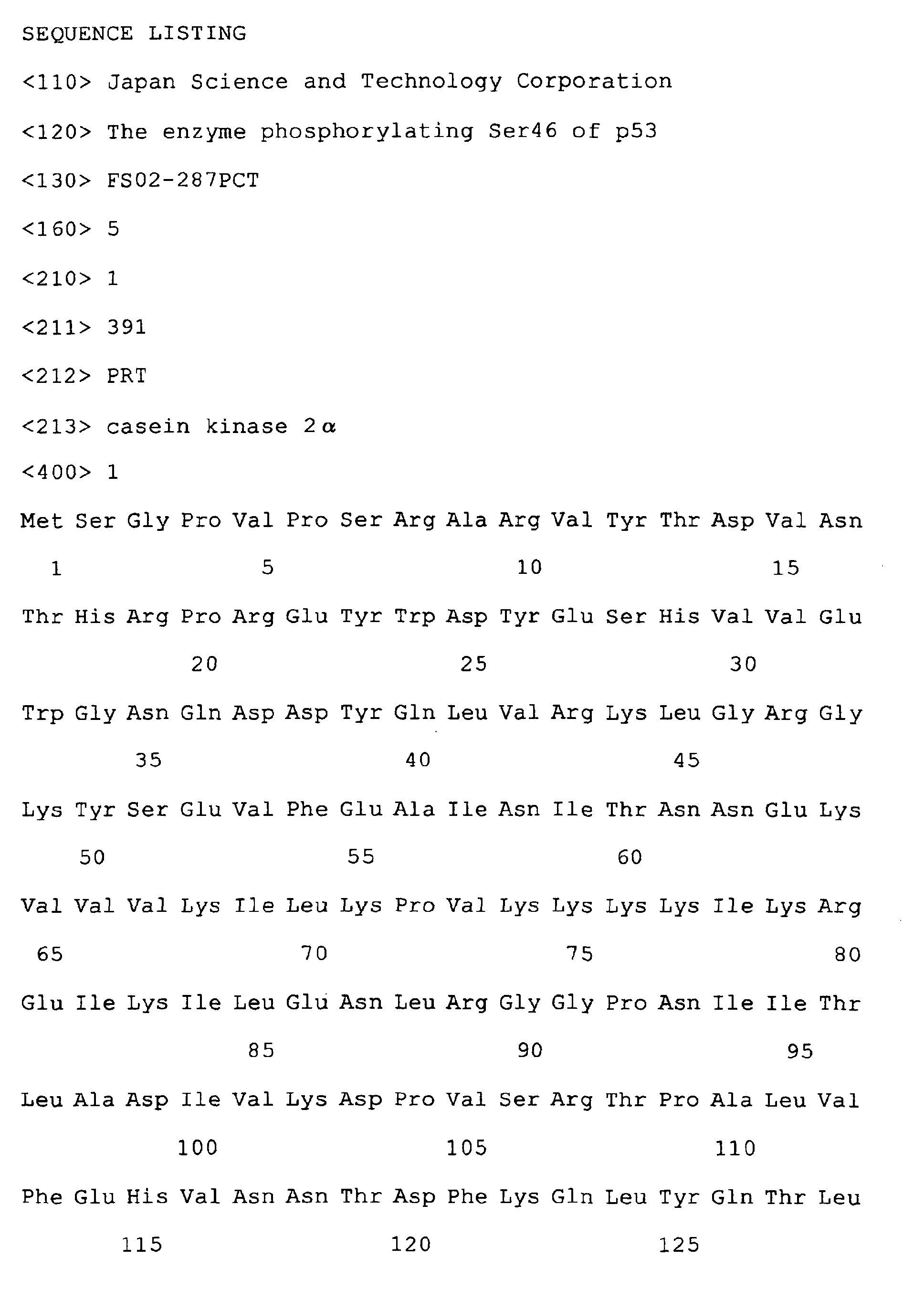
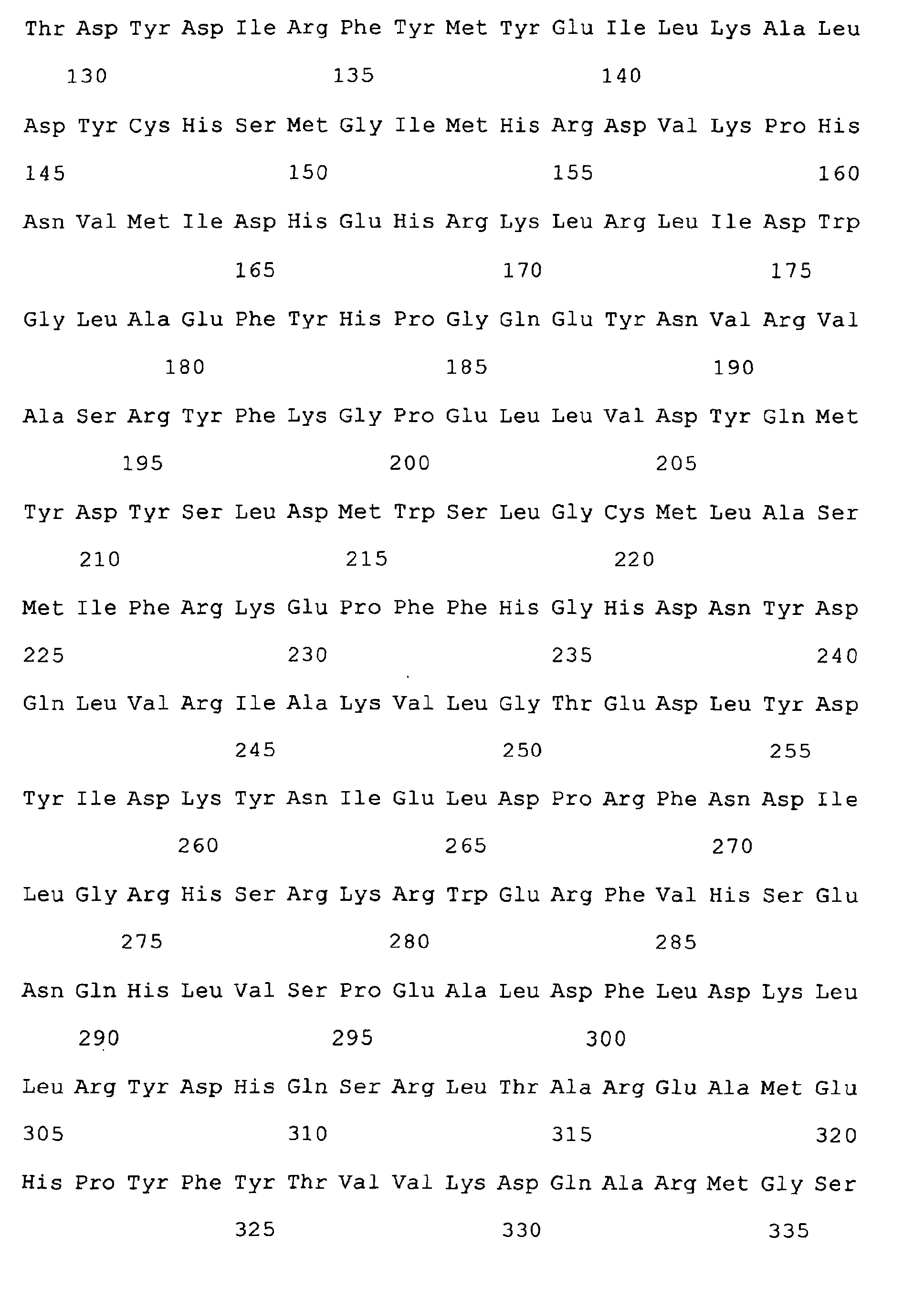
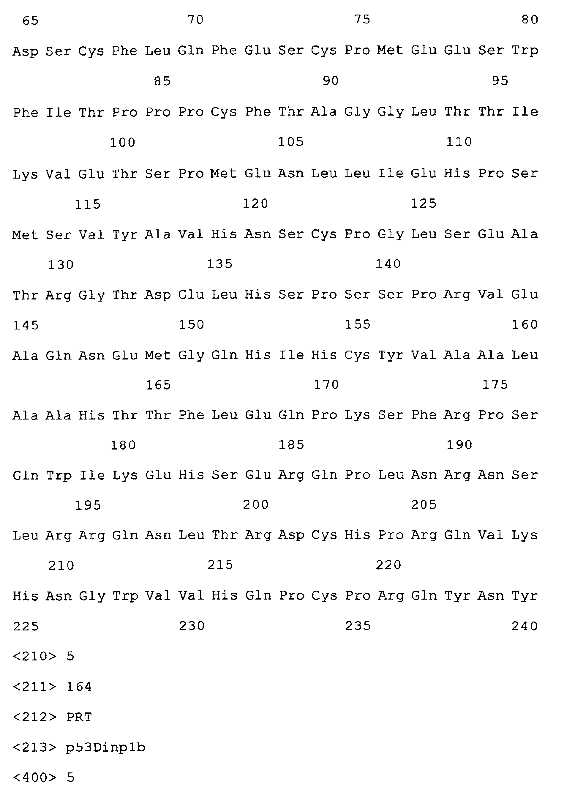
- the invention is an enzyme comprising at least one selected from the group consisting of proteins having one of the amino acid sequences of sequence numbers 1 to 3 and also at least one selected from the group consisting of proteins having one of the amino acid sequences of sequence numbers 4 and 5.
- the proteins having the amino acid sequences of sequence numbers 1 to 5 correspond to ⁇ , ⁇ ' and ⁇ subunits of CK2, and p53DINPla and b, respectively.
- this invention is an enzyme, which is a nuclear fraction obtained during purification of cells with activated p53, said nuclear fraction reacting with an antibody specific to an antigen that comprises a peptide fragment of p53 containing Ser46, said Ser46 being phosphorylated, when p53 being added to said nuclear fraction, and reacts with an antibody specific to casein kinase 2.
- the invention is also the enzyme, which further reacts with an antibody specific to p53DINP1.
- the nuclear fractions may be recovered from objective cells by appropriate known method.
- p53 As for p53 to be added to the nuclear fractions, p53 recombinant protein purified in E. coli. is preferred.
- Antibodies specific to CK2 or p53DINP1 may be prepared using known methods or obtained as commercial products.
- the nuclear fractions contain activated p53, it is judged depending on whether the nuclear fraction may react with an antibody specific to an antigen that comprises a peptide fragment of p53 containing Ser46, said Ser46 being phosphorylated.
- This peptide fragment may comprise at least 4 to 5 amino acids containing Ser46 wherein this Ser46 is phosphorylated.
- the peptide fragment may generally be used by coupling with carrier proteins, such as Keyhole Limpet Hemocyanine (KLH) , which is separated about 5 residues from the Ser residue.
- KLH Keyhole Limpet Hemocyanine
- the isolation or purification of the enzyme could be done by any known methods. It is preferable to use at least one process of the column separation processes such as hydroxyapatite column separation, gel-filtration column separation or anion-exchange column separation, and further to use the process of either cation-exchange column purification or hydrophorbic column purification. It is preferred to combine at least 2 or 3 different processes among these isolation or purification processes
- the invention also provides an anti-tumor agent comprising one of these enzymes as an effective component, a screening agents for drugs activating specifically p53, comprising one of these enzymes as an effective component, and a screening agents for drugs inhibiting one of the enzymes, comprising said enzyme as an effective component.
- the enzyme of this invention can be formulated by known methods to form these drugs.
- the inventions are also use of any of the enzymes as an anti-tumor agent, use of any of the enzymes as a screening agents for drugs activating specifically p53, and use of any of the enzymes as a screening agents for drugs inhibiting the activity of said enzyme.
- This invention is one of the results obtained during the research by the inventors, which successfully revealed that the enzyme phosphorylating the serine residue at amino acid position 46 of p53 is the enzyme comprising CK2 and p53DINP1 and clarified the regulatory mechanism in induction of apoptosis by p53. As a result, this invention provides a new enzyme. This new enzyme was purified through a series of difficult purification processes and its structure was determined.
- the enzyme of this invention can be applied to the development of new therapeutic methods for cancer by introducing selective apoptotic death of tumor cells. Furthermore, the enzyme can be applied for screening of agents activating specifically kinase activity of the enzyme. Such activating agents can be used as new anti-tumor drugs. Furthermore, the enzyme can be applied for screening of agents inhibiting specifically kinase activity of the enzyme. Such inhibiting agents can be used as new alleviating drugs for chemotherapy using anti-tumor drugs.
- MCF-7 Human breast cancer cells
- ATCC Human breast cancer cells
- DMEM medium Sigma
- GIBCO BRL 10 % fetal bovine serum
- doxorubicin Sigma, the same to adriamycin
- the harvested cells from one dish were suspended and solubilized in 500 ⁇ l of cytoplasm extraction buffer (10mM HEPES pH7.9, 2mM MgCl 2 , 0.1mM EDTA, 0.1mM EGTA, 1% Nonidet NP-40, DTT 1mM, Na 3 VO 4 1mM, NaF 5mM, PMSF 0.5mM, KCl 10mM, leupeptin 5 ⁇ g/ml, antipain 5 ⁇ g/ml, chymostatin 5 ⁇ g/ml, pepstatinA 2 ⁇ g/ml).
- the solbilized cells were centrifuged at 15, 000 rpm for 15min.
- the pellets obtained were mixed with 100 ⁇ l nuclear extraction buffer (10mM HEPES pH7. 9, 2mM MgCl 2 , 0.1mM EDTA, 0.1mM EGTA, 1% Nonidet NP-40, DTT 1mM, Na 3 VO 4 1mM, NaF 5mM, PMSF 0.5mM, KCl 10mM, NaCl 500mM, leupeptin 5 ⁇ g/ml, antipain 5 ⁇ g/ml, chymostatin 5 ⁇ g/ml, pepstatinA 2 ⁇ g/ml), suspended well and centrifuged at 15,000 rpm for 15 min. The supernatant obtained is adjusted to 10 mg protein/ ml buffer and is called as the nuclear extracts.
- nuclear extraction buffer 10mM HEPES pH7. 9, 2mM MgCl 2 , 0.1mM EDTA, 0.1mM EGTA, 1% Nonidet NP-40, DTT 1mM,
- the inventors prepared the following polypeptides containing a phosphorylated serine residue at amino acid position 46 of p53 by a polypeptide synthesizer:
- S(PO 3 H 2 ) indicates a phosphorylated serine residue.
- the phosphorylated peptides were coupled covalently to KLH and used as antigens. Immunization was carried out by injecting these antigens together with adjuvant under a rabbit back skin. After five weeks, venous blood was collected from the rabbit earlobe. The antiserum was purified by two steps affinity chromatography and is called as anti-p53-phosphorylated-Ser46 antibody (Oda, K et al., Cell, 102,849-862, 2000).
- a fusion protein (GST-p53) between the amino acid of the whole p53 and glutathione-s-transferase (GST) was used as a substrate for the enzyme reaction.
- the reaction mixture containing 10 ⁇ g of nuclear extracts and 1 ⁇ g of GST-p53 in 40 ⁇ l of kinase reaction buffer (25mM Tris-HCl (pH 7.5), 5 mM ⁇ -glycerophosphate, 2mM DTT, 0.1 mM Na 3 VO 4 , 10mMMgCl 2 , 5mMATP) was kept at 30 °C for 30 min.
- ADR-6h shows the reaction between the nuclear extracts, isolated at 6 hours after doxorubicin treatment, and the substrate GTS-p53.
- ADR-12h shows that between the nuclear extracts, isolated at 12 hours after the doxorubicin treatment, and the substrate GTS-p53.
- Substrate (-) shows the reaction without the substrate
- Lysate (-) shows the reaction without the nuclear extracts. It can be understood that only the reaction between the nuclear extracts, isolated after the doxorubicin treatment, and the substrate GST-p53 shows the presence of enzyme activity phosphorylating Ser46.
- AKTA explorer chromatography system (Amersham-Pharmacia) was used for purification by FPLC. Connecting Superose 6 HR (Amersham-Pharmacia) to the system, the inventors eluted 5 mg of the nuclear extracts by the elution buffer (50 mM Tris pH 7.4, 1 mMEDTA, 2.5m MEGTA, 150mM Nacl, 0.1mM Sodium vanadate, 5%Glyecerol) at a flow rate of 0.2 ml/min, collecting 0.5 ml for a fraction. The activity to phosphorylate Se46 of p53 was measured for each fraction and the active fractions were used for the next purification.
- the elution buffer 50 mM Tris pH 7.4, 1 mMEDTA, 2.5m MEGTA, 150mM Nacl, 0.1mM Sodium vanadate, 5%Glyecerol
- Fig. 2 The enzyme activity for each fraction was shown in Fig. 2 (Fig. 2, p-Ser46 Activity). As a result, a peak of the activity can be seen at fr. 25, where the salt concentration is around 330 mM.
- p-Ser15 Activity and p-Ser33 Activity were the results obtained using a similar procedure to p-Ser46 Activity using anti-phosphorylated Ser15 antibodies and anti-phosphorylated Ser33 antibodies, respectively, for comparison.
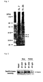
- Figure 4 shows silver staining of the enzyme phosphorylating Ser46 in purified fraction (shown as “a” and "b” in Fig. 4).
- Fragments a and b were cut out from SDS-PAGE gel, were washed two times in 25 mM Sodium bicarbonate/ 50% acetonitril, were added Tris-buffer (pH8.0) containing trypsin, were digested at 35 C for 20 hrs and the 70% portion of the digested samples were analyzed by LC-MS/MS (The analytical condition.
- CK2 is included in the purified enzyme by immunoprecipitation method.
- the fraction 25 described in (2) was immunoprecipitated with anti-CK2 ⁇ antibody (Santacruse).
- the supernatant and the immune complexes were assayed their Ser46 phosphorylating activity and the results are shown in Fig. 5A.
- Figure 5A shows that the activity was disappeared in the supernatant and was transferred to the pellet, which includes CK2.
- Immunoprecipitation by non-specific antibodies (IgG) does not show any Ser46 phosphorylating activity.
- MCF-7 cells were harvested at various time points after the exposure to 30 Gy of ⁇ ray and the cell lysates were prepared. Each sample was immunoprecipitated with anti-p53 DINP1 antibodies. Kinase assay was performed using the immunoprecipitation as the enzyme source and GST-p53 as substrates. The immune complex was separated by SDS-PAGE and the Western blotting using anti-p53-phosphorylating antibodies are shown in Fig. 6. It shows that the phospholulating enzyme includes p53DINP1.
- the anti-p53PDINP1 antibodies were prepared as follows:p53DIPN1a recombinant protein adding His residue at the N-terminal was expressed in E. coli. The protein was purified using a Ni-chelating sepharose column (Amersham-Pharmacia). Polyclonal, anti-p53DINP1 antibodies were prepared by injecting the purified protein as antigens together with adjuvants into rabbit.
- the supernatant of the crushed cells by centrifugation is called as cell extracts after adjusting the concentration as final 10 mg proteins/ ml.
- Each 1 mg of the cell extracts was added 5 ⁇ g each of primary antibodies (anti-p53DINP1 antibody, anti-CK2 ⁇ antibodies or nonspecific antibodies (IgG)).
- the mixtures were rotated slowly at 4 °C for 1 hr.
- the mixtures were rotated slowly at 4 °C for 1 hr after adding 20 ⁇ g of proteinG-sepharose 4B (Amersham-Pharmacia).
- the pellets of the mixture after centrifugation were washed four times with the immunoprecipitation buffer.
- the immune complex was eluted from the pellets by the sample buffer (125 mM Trs-HCl (pH 6.5), 45 % Glycerol, 5 % SDS, 5 % ⁇ -mercaptoethanol, 0.125 % BPB) after heating at 100 °C for 5 min.
- the obtained immune complex was separated by SDS-PAGE and the Western blotting probed with anti-p53DINP1 antibodies, anti-CK2 ⁇ antibodies or nonspecific antibodies (IgG) are shown in Fig. 7.
- Figure 7 shows that all of the antibodies as shown for anti-CK2 ⁇ antibodies (Fig. 7 A), anti-CK2 ⁇ antibodies (Fig. 7B) or anti-p53DINP1 antibodies (Fig.
- the enzyme phosphorylating Ser46 of p53 is a protein complex composed of CK2 and p53DINP1.
Landscapes
- Health & Medical Sciences (AREA)
- Chemical & Material Sciences (AREA)
- Life Sciences & Earth Sciences (AREA)
- Organic Chemistry (AREA)
- Molecular Biology (AREA)
- Genetics & Genomics (AREA)
- Zoology (AREA)
- General Health & Medical Sciences (AREA)
- Medicinal Chemistry (AREA)
- Biochemistry (AREA)
- Engineering & Computer Science (AREA)
- Wood Science & Technology (AREA)
- Bioinformatics & Cheminformatics (AREA)
- Proteomics, Peptides & Aminoacids (AREA)
- Toxicology (AREA)
- Gastroenterology & Hepatology (AREA)
- General Engineering & Computer Science (AREA)
- Biophysics (AREA)
- Microbiology (AREA)
- Biotechnology (AREA)
- Biomedical Technology (AREA)
- Veterinary Medicine (AREA)
- Pharmacology & Pharmacy (AREA)
- Animal Behavior & Ethology (AREA)
- Chemical Kinetics & Catalysis (AREA)
- General Chemical & Material Sciences (AREA)
- Public Health (AREA)
- Nuclear Medicine, Radiotherapy & Molecular Imaging (AREA)
- Enzymes And Modification Thereof (AREA)
- Medicines That Contain Protein Lipid Enzymes And Other Medicines (AREA)
- Investigating Or Analysing Biological Materials (AREA)
- Measuring Or Testing Involving Enzymes Or Micro-Organisms (AREA)
Abstract
The inventors clarified the enzyme phosphorylating the serine
residue at amino acid position 46 of tumor suppressor protein p53
and further elucidated the mechanism in regulation of inducibility
of apoptosis by p53. As the results, the inventors provide a
completely new enzyme comprising casein kinase 2 and p53DINP1
Description
- This invention relates to the enzyme which phosphorylate the serine residue at amino acid position 46 (hereinafter called "Ser46") of tumor suppressor protein p53.
- p53 induces G1 arrest in some cases, whereas it induces apoptosis in other cases. The mechanism how these two different processes are selected has been important to elucidate but remained unexplained. The inventors found, for the first time in the world, that newly phosphorylated Ser46 of p53 regulates apoptosis (Oda, K. et al., Cell, 102, 849-862, 2000).
- The above story is based on our hypothesis advocated three years ago. The hypothesis is such as in the followings: A kind of DNA damage may induce phosphorylation of a certain position of p53, which could result in binding of p53 to promoter region of the genes involved in G1 arrest, such as p21Waf1. Such promoter-p53 binding should induce expression of the genes to cause G1 arrest. In contrast, another kind of DNA damage may induce phosphorylation of other parts of p53, which could result in binding of p53 to promoter region of genes involved in apoptosis. Such promoter-p53 binding must induce expression of the apoptosis-related genes and finally apoptosis.
- The inventors also inferred which region of p53 may regulate inducibility of apoptosis (Experimental Medicine, 18: 2212-2218, 2000). Two domains of p53 were expected to be involved in the region. One is proline-rich domain (amino acid 64 to 101), closed toN-terminal region of the DNA binding domain. The other is a domain consisting of only 20 amino acids (amino acid 43 to 63) , which is located outside the N-terminal region of the proline-rich domain.
- Comparing the amino acid sequence of these two domains, the inventors noticed three possible in vivo phosphorylation sites (Ser46, thr81 and Ser90). Probably Ser46 might be phosphorylated by cycline-dependent kinase or MAP kinase, since the next amino acid to Ser46 is Pro.
- Then, the inventors started experiments to verify our prediction using antibodies, which recognize specifically these phosphorylated sites. In the beginning, the inventors found that the phopholylation of Ser46 always took place at later time points than that of Ser15 or Ser20, after cells were exposed to γ irradiation, UV light or adriamycin to induce DNA damage. Furthermore, the induction of phosphorylation of Ser46 needs stronger intensity of UV light than that for Ser15 or Ser20, when cells were exposed to UV with changed intensity. Therefore, the inventors thought that the phosphorylation may coincide with apoptosis.
- Last July, the inventors noticed the late expression of p53AIP1 (p53 regulated apoptosis inducing protein 1), compared with p21waf1, and the similarity between the expression of p53AIP1 and Ser46 phosphorylation. Then, the inventors started new research.
- The research gave us much interesting development unexpectedly. Changing Ser46 to Ala resulted in marked reduction in inducibility of apoptosis by p53. Furthermore, among many target genes of p53, only expression of p53AIP1 was found to be effectively influenced by Ser46 substitution. On the contrary, there are no influence of Ser46 substitution found on Bax, PIG3 or NOXA, which are suggested by other groups to be involved in apoptosis.
- According to our literature review after getting the above results, the inventors found some reports on mutation of Ser46 and Pro47 to other amino acids in some cases for human cancer. The inventors felt that the authors had reported the results without understanding the meaning. To the inventor's interpretation based on the test result, these human cases may show mutations lacking in phosphorylation of Ser46.
- Based on these results, the inventors constructed the following model (Experimental Medicine, 18: 2338-9, 2000): DNA damage might induce the phosphorylation of Ser15 and Ser20, leading to the stabilization and activation of p53, and p53 may bind to the promoters of the genes related to G1 arrest, such as p21waf1 or the genes related to DNA repair, such as p53R2, to induce these expression. However, severe DNA damage could not be recovered by G1 arrest and DNA repair, instead, could induce activation of Ser46-kinase and phosphorylation of Ser46 of p53, which increased affinity to promoter region of p53AIPI gene, and accordingly activate expression of p53AIP1, which leads to apoptotic cell death.
- Recently, it has been suggested that p53DINP1 (p53 damage inducible nuclear protein 1) could be a constituent of the enzyme phosphorylating Ser46 of p53 (i.e., Ser46-kinase) (Molecular Cell, 8, 85-94, 2001), since not only the expression of p53DINP1 but also phosphorylation of Ser46 are repressed when cells are treated with antisense oligonucleotides corresponding to the p53DINP1 gene.
- Current radiation therapy or chemotherapy by adriamycin, for example, could be interpreted as inducing apoptotic death of tumor cells, by damaging DNA and activating this pathway. Therefore, DNA in normal cells could also be damaged and side effects might be emerged. However, the mechanism, which the inventors elucidated, could be expected to apply to a development of a new therapy, in which only tumor cells could be induced to apoptotic death without damaging DNA.
- The invention is to provide a completely new enzyme, obtained during the research on the enzymatic phosphorylation of the serine residue at amino acid position 46 of tumor suppressor protein p53 and further on the elucidation of the mechanism in regulation of inducibility of apoptosis by p53.
- The inventors succeeded for the first time in isolation of the enzyme, which phosphorylate Ser46 and elucidated their characteristics: The inventors clarified that the enzyme contains casein kinase 2 (hereinafter also called "CK2") and that the enzyme is a protein complex of CK2 and p53DINP1 (Molecular Cell, 8, 85-94, 2001).
- Therefore, the invention is an enzyme comprising
casein kinase 2 and p53DINP1. CK2 is a commonly known kinase, being an ubiquitous serine / threonine kinase present in eukaryotic cells. CK2 is composed of α or α' subunit and β subunit in the tetrameric form (i.e. α α β β or α α' β β) and is involved in the regulation of cell cycle and various signal transduction, phosphorylating various substrates, such as oncoproteins, transcription factors and enzymes related to DNA metabolism (Litchfield, D.W., et al. Mol. Cell. Biochem. 1993 Vol127, :p187-199, Allende, J.E. et al., FASEB J. 1995, Vol9:p313-323) . On the other hand, p53DINP1 is one of the commonly known target proteins of p53, identified in 2001 and is a nuclear protein (there exist two types, a and b), inducible by DNA damage. P53DINP1 plays an important role in p53-dependent apoptosis, but the molecular mechanism remained unexplained (S. Okamura, et al. Molecular Cell 2001 Vol8: p85-94). The relevant enzyme is a protein complex of CK2 and P53DINP1 and phosphorylates Ser46 of p53. - The size of this enzyme is about 250 - 350kDa, as described in later examples. Whereas, the size of CK2 is about 130 - 144kDa (α subunit is about 44kDa; α' subunit is about 37 kDa; β subunit is about 28kDa) and that of p53DINP1a and b are about 42kDa and 38 kDa, respectively. Therefore, although the enzyme is a protein complex of CK2 and P53DINP1, the protein complex contains other factors than CK2 and p53DINP1.
- The invention is an enzyme comprising at least one selected from the group consisting of proteins having one of the amino acid sequences of
sequence numbers 1 to 3 and also at least one selected from the group consisting of proteins having one of the amino acid sequences ofsequence numbers 4 and 5. The proteins having the amino acid sequences ofsequence numbers 1 to 5 correspond to α, α' and β subunits of CK2, and p53DINPla and b, respectively. - Viewed from a different standpoint, this invention is an enzyme, which is a nuclear fraction obtained during purification of cells with activated p53, said nuclear fraction reacting with an antibody specific to an antigen that comprises a peptide fragment of p53 containing Ser46, said Ser46 being phosphorylated, when p53 being added to said nuclear fraction, and reacts with an antibody specific to
casein kinase 2. The invention is also the enzyme, which further reacts with an antibody specific to p53DINP1. - The nuclear fractions may be recovered from objective cells by appropriate known method.
- As for p53 to be added to the nuclear fractions, p53 recombinant protein purified in E. coli. is preferred.
- Antibodies specific to CK2 or p53DINP1 may be prepared using known methods or obtained as commercial products.
- As for whether the nuclear fractions contain activated p53, it is judged depending on whether the nuclear fraction may react with an antibody specific to an antigen that comprises a peptide fragment of p53 containing Ser46, said Ser46 being phosphorylated. This peptide fragment may comprise at least 4 to 5 amino acids containing Ser46 wherein this Ser46 is phosphorylated. The peptide fragment may generally be used by coupling with carrier proteins, such as Keyhole Limpet Hemocyanine (KLH) , which is separated about 5 residues from the Ser residue.
- Exposure of target cells to UV irradiation, γ ray irradiation, oxidative stress of various anti-tumor drugs such as cis-Platinum, etoposide or adramycin, but not to osmotic shock, induces DNA damage and activates p53.
- The isolation or purification of the enzyme could be done by any known methods. It is preferable to use at least one process of the column separation processes such as hydroxyapatite column separation, gel-filtration column separation or anion-exchange column separation, and further to use the process of either cation-exchange column purification or hydrophorbic column purification. It is preferred to combine at least 2 or 3 different processes among these isolation or purification processes
- Injection, in some way, of the enzyme of this invention into tumor cells could induce apoptosis of the cell and lead to cell death. The drugs activating specifically the kinase activity of the enzyme, if obtained by screening, could be useful as anti-tumor agent. The drugs inhibiting specifically the activity of the enzyme, if obtained by screening, could be useful as alleviating drugs for side effects of chemotherapy using anti-tumor agents. Therefore, the invention also provides an anti-tumor agent comprising one of these enzymes as an effective component, a screening agents for drugs activating specifically p53, comprising one of these enzymes as an effective component, and a screening agents for drugs inhibiting one of the enzymes, comprising said enzyme as an effective component. The enzyme of this invention can be formulated by known methods to form these drugs. The inventions are also use of any of the enzymes as an anti-tumor agent, use of any of the enzymes as a screening agents for drugs activating specifically p53, and use of any of the enzymes as a screening agents for drugs inhibiting the activity of said enzyme.
-
- Figure 1 shows Western blotting as detected by anti-p53-phosphorylated Ser46 antibodies for the enzyme reaction mixture using the nuclear extracts with damaged DNA.
- Figure 2 shows Western blotting as detected by anti-p53-phosphorylated Ser46 antibodies for enzyme reaction mixture using purified enzyme through Mono Q column after purified the nuclear extracts with damaged DNA through Superose 6 column.
- Figure 3 shows In gel assay for the purified nuclear extracts with damaged DNA.
- Figure 4 shows a silver stained SDS-PAGE gel for the nuclear extracts with damaged DNA.
- Figure 5 shows Western blotting as probed by anti-p53-phosphorylated Ser46 antibodies for the enzyme reaction mixture obtained by immunoprecipitation of purified nuclear extracts (active fraction for phosphorylation of Ser46) by anti-CK2α antibodies.
- Figure 6 shows Western blotting as probed by anti-p53-phosphorylated Ser46 antibodies for enzyme reaction mixture obtained by immunoprecipitation of purified nuclear extracts by anti-p53-phosphorylated Ser46 antibodies.
- Figure 7 shows Western blotting as probed by anti-CK2 α antibodies (A), by anti-CK2 β antibodies (B) and by anti-p53DINP1 antibodies (C) for immune complex of nuclear extracts precipitated by anti-p53DINP1 antibodies (IgG), by anti-CK2 α antibodies and by anti-CK2 β antibodies, respectively.
-
- This invention is one of the results obtained during the research by the inventors, which successfully revealed that the enzyme phosphorylating the serine residue at amino acid position 46 of p53 is the enzyme comprising CK2 and p53DINP1 and clarified the regulatory mechanism in induction of apoptosis by p53. As a result, this invention provides a new enzyme. This new enzyme was purified through a series of difficult purification processes and its structure was determined.
- The enzyme of this invention can be applied to the development of new therapeutic methods for cancer by introducing selective apoptotic death of tumor cells. Furthermore, the enzyme can be applied for screening of agents activating specifically kinase activity of the enzyme. Such activating agents can be used as new anti-tumor drugs. Furthermore, the enzyme can be applied for screening of agents inhibiting specifically kinase activity of the enzyme. Such inhibiting agents can be used as new alleviating drugs for chemotherapy using anti-tumor drugs.
- In the following paragraph, the inventors illustrate the practical examples of this invention, but do not intend to restrict the scope of the invention.
- Human breast cancer cells MCF-7 (hereinafter called "MCF-7", ATCC) were cultured in DMEM medium (Sigma) containing 10 % fetal bovine serum (GIBCO BRL) on 15 cm2 dishes (Corning) up to 90 % confluent state. These cells were treated with final 3 µM doxorubicin (Sigma, the same to adriamycin) by addition of its aqueous solution to the medium. After allowed to stand in an incubator, the cells were harvested at various time points.
- The harvested cells from one dish were suspended and solubilized in 500 µl of cytoplasm extraction buffer (10mM HEPES pH7.9, 2mM MgCl2, 0.1mM EDTA, 0.1mM EGTA, 1% Nonidet NP-40, DTT 1mM, Na3VO4 1mM, NaF 5mM, PMSF 0.5mM, KCl 10mM, leupeptin 5 µg/ml, antipain 5 µg/ml, chymostatin 5 µg/ml,
pepstatinA 2 µg/ml). The solbilized cells were centrifuged at 15, 000 rpm for 15min. The pellets obtained were mixed with 100 µl nuclear extraction buffer (10mM HEPES pH7. 9, 2mM MgCl2, 0.1mM EDTA, 0.1mM EGTA, 1% Nonidet NP-40, DTT 1mM, Na3VO4 1mM, NaF 5mM, PMSF 0.5mM, KCl 10mM, NaCl 500mM, leupeptin 5µg/ml, antipain 5 µg/ml, chymostatin 5 µg/ml, pepstatinA 2µg/ml), suspended well and centrifuged at 15,000 rpm for 15 min. The supernatant obtained is adjusted to 10 mg protein/ ml buffer and is called as the nuclear extracts. - On the other hand, the inventors prepared the following polypeptides containing a phosphorylated serine residue at amino acid position 46 of p53 by a polypeptide synthesizer:

- In the sequence, S(PO3H2) indicates a phosphorylated serine residue.
The phosphorylated peptides were coupled covalently to KLH and used as antigens. Immunization was carried out by injecting these antigens together with adjuvant under a rabbit back skin. After five weeks, venous blood was collected from the rabbit earlobe. The antiserum was purified by two steps affinity chromatography and is called as anti-p53-phosphorylated-Ser46 antibody (Oda, K et al., Cell, 102,849-862, 2000). - Next, the enzyme activity of the nuclear extracts was assayed using the antibodies. As a substrate for the enzyme reaction, a fusion protein (GST-p53) between the amino acid of the whole p53 and glutathione-s-transferase (GST) was used. The reaction mixture containing 10 µg of nuclear extracts and 1 µg of GST-p53 in 40 µl of kinase reaction buffer (25mM Tris-HCl (pH 7.5), 5 mM β -glycerophosphate, 2mM DTT, 0.1 mM Na3VO4, 10mMMgCl2, 5mMATP) was kept at 30 °C for 30 min. After addition of SDS sample buffer (125mM Tris-HCl (pH 6.5) , 45% Glycerol, 5 %SDS, 5% β-mercaptoethanol, 0.125% BPB), the reaction mixture was heated at 100 °C for 5 min and was separated on a 10 % SDS-PEGE gel. The phosphorylation of GST-p53 was detected in Western blotting probed with anti-p53-phosphorylated-Ser46 antibodies.
- The results were shown in Fig.1. In this figure, ADR-6h shows the reaction between the nuclear extracts, isolated at 6 hours after doxorubicin treatment, and the substrate GTS-p53. ADR-12h shows that between the nuclear extracts, isolated at 12 hours after the doxorubicin treatment, and the substrate GTS-p53. Substrate (-) shows the reaction without the substrate and Lysate (-) shows the reaction without the nuclear extracts. It can be understood that only the reaction between the nuclear extracts, isolated after the doxorubicin treatment, and the substrate GST-p53 shows the presence of enzyme activity phosphorylating Ser46.
- AKTA explorer chromatography system (Amersham-Pharmacia) was used for purification by FPLC. Connecting Superose 6 HR (Amersham-Pharmacia) to the system, the inventors eluted 5 mg of the nuclear extracts by the elution buffer (50 mM Tris pH 7.4, 1 mMEDTA, 2.5m MEGTA, 150mM Nacl, 0.1mM Sodium vanadate, 5%Glyecerol) at a flow rate of 0.2 ml/min, collecting 0.5 ml for a fraction. The activity to phosphorylate Se46 of p53 was measured for each fraction and the active fractions were used for the next purification.
- Then, connecting MonoQ HR5/5 (Amasham-Pharmacia) to the system, equilibrating the column with starting buffer (50 mM Tris pH 7.4, 1 mM EDTA, 2.5 mM EGTA, 250 mM NaCl, 0.1 mM Sodium vanadate, 5 % Glyecerol) the inventors eluted the 500 µg of the active fractions by the elution buffer (50 mM Tris pH 7.4, 1 mM EDTA, 2.5 mM EGTA, 500 mM NaCl, 0.1 mM Sodium vanadate, 5 % Glyecerol) at a flow rate of 0.3 ml/ min, collecting 0.5ml for a fraction by gradient elution. The enzyme activity for each fraction was shown in Fig. 2 (Fig. 2, p-Ser46 Activity). As a result, a peak of the activity can be seen at fr. 25, where the salt concentration is around 330 mM. In Fig. 2, p-Ser15 Activity and p-Ser33 Activity were the results obtained using a similar procedure to p-Ser46 Activity using anti-phosphorylated Ser15 antibodies and anti-phosphorylated Ser33 antibodies, respectively, for comparison.
- In the beginning, the molecules responsible for the activity phosphorylating Ser46 was identified by In Gel assay. Several fractions (1, 7, 15 and 25) from anion exchange column chromatography (MonoQ) as shown in Fig. 2 (2) were separated by electrophoresis through SDS-PAGE gels containing GST-p53 and Myelin basic proteins, respectively, as substrates for kinase activity. After the electrophoresis, SDS was removed from the gels. The kinase activity of the separated proteins was detected by autoradiography after In Gel assay in the presence of 32P-ATP. The results are shown in Fig. 3. As shown in Fig. 3, using GST-p53 as substrates, strong p53 phosphorylating activity was detected in the
fraction 25, in which the enzyme phosphorylating Ser46 of p53 was detected using anti-p53-phosphorylated Ser46 antibodies. However, kinase activity was not detected without using substrates (substrate (-) ) or with Myelin basic protein, not containing p53, as substrates. It can be understood that the enzyme induced after DNA damage shows the phosphorylating activity only when the enzyme encounters p53. - Gel filtration chromatography of the enzyme shows that the molecular weight of the purified enzyme is around 250 - 350 Kda.
- Precise examination of the active portion of the enzyme revealed that the portion is consists of two parts. Figure 4 shows silver staining of the enzyme phosphorylating Ser46 in purified fraction (shown as "a" and "b" in Fig. 4).
- Fragments a and b were cut out from SDS-PAGE gel, were washed two times in 25 mM Sodium bicarbonate/ 50% acetonitril, were added Tris-buffer (pH8.0) containing trypsin, were digested at 35 C for 20 hrs and the 70% portion of the digested samples were analyzed by LC-MS/MS (The analytical condition. HPLC apparatus: MAGIC 2002 (Michrom BioResources, Inc., USA) Column : MAGIC 2002 (Michrom BioResources, Inc., USA); Solvent A: 2% acetnitril/ 0.1% formic acid; Solvent B: 90% anetnitril/ 0.1% formic acid; Flow rate: 400~500 1/min; Gradient: 0 min, 5 (B%), 25 min, 65 (B%), 26 min, 100 (B%), 27 min, 100 (B%), 28 min, 5 (B%); Mass sectrometer: Q-Tof2 (Micromass, UK); Ionaization method: Nanoflow-LC ES1; Ionization mode: Positive mode, Capillary voltage: 2 Kv; Collision energy: 20-35 eV.) The amino acid sequence of the Ser46 phosphorylating enzyme was determined by this LC-MS/MS analysis (The part a corresponds to the
sequence number 1; the part b corresponds to the sequence number 2) and fragments a and b in Fig. 4 were proved as α and α' subunits of CK2, respectively. - The
fraction 25 described in (2) was immunoprecipitated with anti-CK2α antibody (Santacruse). The supernatant and the immune complexes were assayed their Ser46 phosphorylating activity and the results are shown in Fig. 5A. Figure 5A shows that the activity was disappeared in the supernatant and was transferred to the pellet, which includes CK2. Immunoprecipitation by non-specific antibodies (IgG) does not show any Ser46 phosphorylating activity. - MCF-7 cells were harvested at various time points after the exposure to 30 Gy of γ ray and the cell lysates were prepared. Each sample was immunoprecipitated with anti-p53 DINP1 antibodies. Kinase assay was performed using the immunoprecipitation as the enzyme source and GST-p53 as substrates. The immune complex was separated by SDS-PAGE and the Western blotting using anti-p53-phosphorylating antibodies are shown in Fig. 6. It shows that the phospholulating enzyme includes p53DINP1.
- The anti-p53PDINP1 antibodies were prepared as follows:p53DIPN1a recombinant protein adding His residue at the N-terminal was expressed in E. coli. The protein was purified using a Ni-chelating sepharose column (Amersham-Pharmacia). Polyclonal, anti-p53DINP1 antibodies were prepared by injecting the purified protein as antigens together with adjuvants into rabbit.
- Twelve hours after the treatment of MCF-7 cells with adiramycin, cells were harvested, were suspended in 500 µl of immunoprecipitation buffer (50 mM Hepes pH 7.4, 150 mM NaCl, 1 mM EDTA, 2.5 mM EGTA, 0.1 % Tween 20, 10 % glycerol, 1 mM NaF, 0.1 mM Sodium Vanadate, 0.5 mM PMSF, 5 µg/ml Leupeptin, 5 µg/ml chymostatin, 2 µg/ml pepstatin) per a dish cells in ice box and were crushed by sonication. The supernatant of the crushed cells by centrifugation is called as cell extracts after adjusting the concentration as final 10 mg proteins/ ml. Each 1 mg of the cell extracts was added 5 µg each of primary antibodies (anti-p53DINP1 antibody, anti-CK2α antibodies or nonspecific antibodies (IgG)). The mixtures were rotated slowly at 4 °C for 1 hr. Then, the mixtures were rotated slowly at 4 °C for 1 hr after adding 20 µg of proteinG-sepharose 4B (Amersham-Pharmacia). The pellets of the mixture after centrifugation were washed four times with the immunoprecipitation buffer. Then, the immune complex was eluted from the pellets by the sample buffer (125 mM Trs-HCl (pH 6.5), 45 % Glycerol, 5 % SDS, 5 % β-mercaptoethanol, 0.125 % BPB) after heating at 100 °C for 5 min. The obtained immune complex was separated by SDS-PAGE and the Western blotting probed with anti-p53DINP1 antibodies, anti-CK2 α antibodies or nonspecific antibodies (IgG) are shown in Fig. 7. Figure 7 shows that all of the antibodies as shown for anti-CK2 α antibodies (Fig. 7 A), anti-CK2 β antibodies (Fig. 7B) or anti-p53DINP1 antibodies (Fig. 7C) reacted with the immune complex precipitated both by anti-p53DINP1 antibodies and anti-CK2 α antibodies in Western blotting. This result means that CK2α, CK2 β and p53DINPl are composing a protein complex.
- From the above embodiments, it was elucidated that the enzyme phosphorylating Ser46 of p53 is a protein complex composed of CK2 and p53DINP1.



Claims (11)
- An enzyme comprising casein kinase 2 and p53DINP1.
- An enzyme comprising at least one selected from the group consisting of proteins having one of the amino acid sequences of sequence numbers 1 to 3 and also at least one selected from the group consisting of proteins having one of the amino acid sequences of sequence numbers 4 and 5.
- The enzyme as in claim 2, wherein one or a few amino acid(s) is/are deleted, substituted or added in said amino acid sequences of sequence numbers 1 to 5.
- An enzyme, which is a nuclear fraction obtained during purification of cells with activated p53, said nuclear fraction reacting with an antibody specific to an antigen that comprises a peptide fragment of p53 containing Ser46, said Ser46 being phosphorylated, when p53 being added to said nuclear fraction, and reacts with an antibody specific to casein kinase 2.
- The enzyme as in claim 4, which further reacts with an antibody specific to p53DINP1.
- An anti-tumor agent comprising the enzyme as in any one of claims 1 to 5 as an effective component.
- A screening agents for drugs activating specifically p53, comprising the enzyme as in any one of claims 1 to 5 as an effective component.
- A screening agents for drugs inhibiting the enzyme as in any one of claims 1 to 5, comprising said enzyme as an effective component.
- Use of the enzyme as in any one of claims 1 to 5 as an anti-tumor agent.
- Use of the enzyme as in any one of claims 1 to 5 as a screening agents for drugs activating specifically p53.
- Use of the enzyme as in any one of claims 1 to 5 as a screening agents for drugs inhibiting the activity of said enzyme.
Applications Claiming Priority (3)
| Application Number | Priority Date | Filing Date | Title |
|---|---|---|---|
| JP2001292953 | 2001-09-26 | ||
| JP2001292953A JP2003093056A (en) | 2001-09-26 | 2001-09-26 | Enzyme that phosphorylates Ser53 of p53 |
| PCT/JP2002/009914 WO2003027144A1 (en) | 2001-09-26 | 2002-09-25 | ENZYME PHOSPHORYLATING Ser46 OF p53 |
Publications (2)
| Publication Number | Publication Date |
|---|---|
| EP1431308A1 true EP1431308A1 (en) | 2004-06-23 |
| EP1431308A4 EP1431308A4 (en) | 2006-05-24 |
Family
ID=19114828
Family Applications (1)
| Application Number | Title | Priority Date | Filing Date |
|---|---|---|---|
| EP02777794A Withdrawn EP1431308A4 (en) | 2001-09-26 | 2002-09-25 | PHOSPHORYLANT ENZYME A SER46 RESIN OF PROTEIN P53 |
Country Status (5)
| Country | Link |
|---|---|
| US (1) | US20050232912A1 (en) |
| EP (1) | EP1431308A4 (en) |
| JP (1) | JP2003093056A (en) |
| CA (1) | CA2455117A1 (en) |
| WO (1) | WO2003027144A1 (en) |
Cited By (2)
| Publication number | Priority date | Publication date | Assignee | Title |
|---|---|---|---|---|
| WO2005098443A1 (en) * | 2004-04-08 | 2005-10-20 | INSERM (Institut National de la Santé et de la Recherche Médicale) | Methods for the diagnosis of cancers or of precancerous lesions |
| US9089565B2 (en) | 2009-03-06 | 2015-07-28 | Medical Research Council | Compositions and methods for causing neurite outgrowth |
Families Citing this family (2)
| Publication number | Priority date | Publication date | Assignee | Title |
|---|---|---|---|---|
| US8062887B2 (en) | 2005-04-01 | 2011-11-22 | National University Corporation, Hokkaido University | Monoclonal antibody specifically recognizing modification site after translation of p53 and kit for assaying modification site containing the same |
| EP4403581A4 (en) * | 2022-03-30 | 2025-06-18 | Miwako Homma | ANTI-CK2 ALPHA ANTIBODY OR FRAGMENT THEREOF |
Family Cites Families (1)
| Publication number | Priority date | Publication date | Assignee | Title |
|---|---|---|---|---|
| US6436703B1 (en) * | 2000-03-31 | 2002-08-20 | Hyseq, Inc. | Nucleic acids and polypeptides |
-
2001
- 2001-09-26 JP JP2001292953A patent/JP2003093056A/en active Pending
-
2002
- 2002-09-25 US US10/490,454 patent/US20050232912A1/en not_active Abandoned
- 2002-09-25 WO PCT/JP2002/009914 patent/WO2003027144A1/en not_active Ceased
- 2002-09-25 CA CA002455117A patent/CA2455117A1/en not_active Abandoned
- 2002-09-25 EP EP02777794A patent/EP1431308A4/en not_active Withdrawn
Non-Patent Citations (2)
| Title |
|---|
| See also references of WO03027144A1 * |
| TANAKA TOMOAKI ET AL: "Phosphorylation of Ser46 regulates apoptosis-inducing ability of p53" JOURNAL OF BIOMOLECULAR STRUCTURE AND DYNAMICS, vol. 17, no. 6, June 2000 (2000-06), page 1156, XP009064496 & CONFERENCE ON DNA STRUCTURE AND INTERACTIONS: THEIR BIOLOGICAL ROLES AND IMPLICATIONS IN BIOMEDICINE; BRNO, CZECH REPUBLIC; JULY 19-23, 2000 ISSN: 0739-1102 * |
Cited By (2)
| Publication number | Priority date | Publication date | Assignee | Title |
|---|---|---|---|---|
| WO2005098443A1 (en) * | 2004-04-08 | 2005-10-20 | INSERM (Institut National de la Santé et de la Recherche Médicale) | Methods for the diagnosis of cancers or of precancerous lesions |
| US9089565B2 (en) | 2009-03-06 | 2015-07-28 | Medical Research Council | Compositions and methods for causing neurite outgrowth |
Also Published As
| Publication number | Publication date |
|---|---|
| CA2455117A1 (en) | 2003-04-03 |
| WO2003027144A1 (en) | 2003-04-03 |
| JP2003093056A (en) | 2003-04-02 |
| EP1431308A4 (en) | 2006-05-24 |
| US20050232912A1 (en) | 2005-10-20 |
Similar Documents
| Publication | Publication Date | Title |
|---|---|---|
| Xiong et al. | p21 is a universal inhibitor of cyclin kinases | |
| Polans et al. | Recoverin, but not visinin, is an autoantigen in the human retina identified with a cancer-associated retinopathy. | |
| Bischoff et al. | Mitotic regulator protein RCC1 is complexed with a nuclear ras-related polypeptide. | |
| Lee et al. | Cloning of p57KIP2, a cyclin-dependent kinase inhibitor with unique domain structure and tissue distribution. | |
| US6140058A (en) | Activation of p53 protein | |
| Furuta et al. | Heterochromatin protein HP1Hsβ (p25β) and its localization with centromeres in mitosis | |
| CA2173978A1 (en) | A novel nuclear mitotic phosphoprotein: mitosin | |
| Götz et al. | Protein kinase CK2 interacts with a multi-protein binding domain of p53 | |
| EP1308462A1 (en) | Hepatitis b virus (hbv) antigenic polypeptide-heat shock protein complex and use thereof | |
| AU2001265947B2 (en) | Enzymatic assays for screening anti-cancer agents | |
| US20050232912A1 (en) | Enzyme phosphorylating ser46 of p53 | |
| US6025480A (en) | Isolated nucleic acid molecules encoding P57KIP2 | |
| AU2001274026A1 (en) | Protein complexes and assays for screening anti-cancer agents | |
| JP2001510684A (en) | Assays, methods of treatment and therapeutic means | |
| CA2169459A1 (en) | Novel proteins which bind to retinoblastoma protein and their encoding dna sequences | |
| JP4270548B2 (en) | Cdc7-ASK kinase complex, substrate for the kinase complex, antibody specific for the substrate, and screening method for compounds having Cdc7-ASK kinase inhibitory activity using them | |
| Shen et al. | Molecular cloning and characterization of a novel casein kinase II substrate, HASPP28, from rat brain | |
| JP2000135090A (en) | Human H37 protein and cDNA encoding this protein | |
| JP2003505064A (en) | Kinase blocking polypeptide and uses thereof | |
| NZ278745A (en) | Tumour suppressor gene encoding retinoblastoma binding protein | |
| US6232442B1 (en) | Purified eukaryotic-initiation factor 4E having altered RNA binding affinity | |
| WO2001023557A1 (en) | Novel polypeptides and genes encoding the same | |
| Yang et al. | Methylation of MATCAP by PRMT5 regulates microtubule detyrosination to ensure mitotic fidelity | |
| EP1159430A1 (en) | Histidin protein-phosphatase | |
| Bogojević et al. | p53-Like protein binding affinity to the hormone responsive element of the haptoglobin gene in fetal rat liver |
Legal Events
| Date | Code | Title | Description |
|---|---|---|---|
| PUAI | Public reference made under article 153(3) epc to a published international application that has entered the european phase |
Free format text: ORIGINAL CODE: 0009012 |
|
| 17P | Request for examination filed |
Effective date: 20040112 |
|
| AK | Designated contracting states |
Kind code of ref document: A1 Designated state(s): AT BE BG CH CY CZ DE DK EE ES FI FR GB GR IE IT LI LU MC NL PT SE SK TR |
|
| A4 | Supplementary search report drawn up and despatched |
Effective date: 20060406 |
|
| 17Q | First examination report despatched |
Effective date: 20061026 |
|
| STAA | Information on the status of an ep patent application or granted ep patent |
Free format text: STATUS: THE APPLICATION IS DEEMED TO BE WITHDRAWN |
|
| 18D | Application deemed to be withdrawn |
Effective date: 20070306 |