Technical Field
-
This invention relates to nucleic acids derived
from genes expressed in neuroblastoma and gene
expression products encoded by the nucleic acids. More
particularly, the invention relates to nucleic acids
and their fragments derived from the marker genes whose
expression is enhanced in neuroblastomas with favorable
prognosis based on comparison between neuroblastomas
with favorable prognosis and neuroblastomas with
unfavorable prognosis as well as to their utility in
the diagnosis for the prognosis of neuroblastomas.
Background Art
(Tumorgenesis and Genes)
-
Individual tumors exhibit distinct characteristic
natures, and their biological properties are not
necessarily identical even though the basic principle
of oncogenesis is the same. Rapid advances in the
understanding of cancer from a molecular biological and
molecular genetic perspective in recent years have
opened the way to an explanation of oncogenesis and
tumor cell biology on the genetic level.
(Neuroblastomas)
-
Neuroblastoma is a pediatric cancer occurring in
sympathetic gangliocytes and adrenal medullary cells
which originate from cells of the peripheral
sympathetic nervous system. Of these sympathetic
nervous system cells, neural crest cells in the initial
stage of development migrate to the abdomen,
differentiating and maturing at sites where sympathetic
ganglia are formed. Some of these cells migrate
further to the adrenal bodies, penetrating through the
adrenal cortex which is already in the process of
formation, and reaching the medulla and forming
medullary substance there. The neural crest cells also
serve as a source of other peripheral nerve cells,
differentiating into dorsal root ganglia (sensory
nerves), skin pigment cells, thyroid C cells, some
pulmonary cells, intestinal gangliocytes, and the like.
(Prognosis of neuroblastoma)
-
Neuroblastoma is characterized by a varied
clinical profile (Nakagawara, Shinkeigashu no Hassei to
Sono Bunshi Kiko [Neuroblastoma Development and
Molecular Mechanism], Shoni Naika 30, 143, 1998). For
example, neuroblastoma occurring at less than one year
of age has very favorable prognosis, with the majority
undergoing differentiation and cell death, and
spontaneous regression. Currently, most neuroblastomas
discovered by a positive result in the commonly
performed mass screening of 6-month-old infant urine
are of the type which tend to undergo this spontaneous
regression. On the other hand, neuroblastoma occurring
at age 1 or higher is highly malignant and leads to
death of the infant in the majority of cases. It is
also hypothesized that a somatic mutation occurs in
highly malignant neuroblastomas in infants older than
one year of age, which are of monoclonal nature,
whereas in naturally regressing neuroblastomas, the
genetic mutation remains at only a germline mutation.
See Knudson AG, et al.: Regression of neuroblastoma IV-S:
A genetic hypothesis, N. Engl. J. Med. 302, 1254
(1980)).
(Genes which allow the diagnosis for prognosis of
neuroblastoma)
-
With recent advances in molecular biology research,
it has become clear that expression of the high
affinity nerve growth factor (NGF) receptor TrkA is
closely connected with control of differentiation and
cell death. See Nakagawara A., The NGF story and
neuroblastoma, Med. Pediatr. Oncol., 31, 113 (1998).
Trk is a membrane-spanning receptor, existing as the
three main types, Trk-A, -B and -C.
-
These Trk family receptors play an important role
in specific nerve cell differentiation and survival in
the central nervous and peripheral nervous systems.
See Nakagawara, et al., Shinkeigasaiboushu ni Okeru
Neurotrophin Juyoutai no Hatsugen to Yogo [Expression
of Neurotrophin Receptors and Prognosis in
Neuroblastoma], Shoni Geka (Pediatric Surgery), 29:
425-432, 1997. The survival and differentiation of
tumor cells is controlled by signals from Trk tyrosine
kinase and Ret tyrosine kinase. In particular, the
role of TrkA receptor is most significant, with TrkA
expression being notably high in neuroblastomas with
favorable prognosis, and its signals exerting a
powerful control over survival and differentiation of
tumor cells, and cell death (apoptosis). In
neuroblastomas with unfavorable prognosis, on the other
hand, TrkA expression is significantly suppressed,
while tumor development is aided by a mechanism in
which survival is promoted by signals from TrkB and Ret.
-
It has become clear that amplification of the
neural oncogene N-myc has become clearly associated
with the prognosis of neuroblastoma. See Nakagawara,
Nou-shinkeishuyo no Tadankai Hatsugan [Multistage
Oncogenesis of Cerebral and Neural Tumors], Molecular
Medicine, 364, 366 (1999). This gene, first cloned in
neuroblastoma, is ordinarily only present in a single
copy per haploid set in normal cells and neuroblastomas
with favorable prognosis, whereas it has been found to
be amplified several dozen times in neuroblastomas with
unfavorable prognosis.
-
Up till the present time, however, no oncogene
other than N-myc is known to be expressed in
neuroblastomas, and absolutely no genetic information
other than that of N-myc has been known in relation to
favorable or unfavorable prognosis.
Disclosure of the Invention
-
This invention has been accomplished in light of
these circumstances, and its object is to identify the
base sequences of genes which are related to favorable
or unfavorable prognosis of neuroblastoma, and to allow
the diagnosis for the prognosis of neuroblastoma
(whether favorable or unfavorable) based on their
genetic information. Its object is also to provide the
information on the functions of proteins which are the
transcripts of the aforementioned genes.
-
As a result of conducting diligent research, the
present inventors examined the prognoses of
neuroblastomas and succeeded in constructing cDNA
libraries from the respective clinical tissues with
favorable prognosis and with unfavorable prognosis.
Approximately 2400 clones were respectively obtained
from these two types of cDNA libraries and were
classified according to the prognosis of neuroblastomas
and carried out profiling of the respective subsets.
-
Thus, the present inventors found that a group of
genes showed differential expression levels among the
abovementioned subsets and showed enhanced expression
levels only in clinical tissues of neuroblastoma with
favorable prognosis; one of the genes was designated
"NEDL-1 (nbla0078)." Moreover, the present inventors
sequenced the whole length of NEDL-1 gene, and
conducted the functional analysis of NEDL-1 protein
encoded by the gene: the protein was found to be a
ubiquitin ligase of the HECT type.
-
Based on this finding the present inventors have
made it possible to provide genetic information (base
sequence data etc.) which allowed the detection and
cloning of genes whose expression is enhanced only in
the clinical tissues of neuroblastoma with favorable
prognosis. Further based on the base sequence data,
the present inventors made it possible to provide
methods of diagnosis for prognosis and diagnostic
agents therefor and thus completed this invention.
-
Specifically, this invention provides a nucleic
acid probe comprising nucleic acid (a) or nucleic acid
(b) described below:
- (a) a nucleic acid having a portion of a base sequence
set forth in SEQ ID NO:2 in the Sequence Listing or a
base sequence complementary thereto; or
- (b) a nucleic acid capable of hybridizing to the
nucleic acid comprising a base sequence set forth in
SEQ ID NO:2 in the Sequence Listing, or having a base
sequence complementary to said base sequence.
-
-
Preferably, the nucleic acid is DNA in the nucleic
acid probe described above.
-
Also preferably, the nucleic acid has a base
length of at least 20 bases in the nucleic acid probe.
-
Further preferably, the base sequence set forth in
SEQ ID NO:2 is its full-length in the nucleic acid
probe.
-
This invention also provides a diagnostic agent
for the prognosis of neuroblastoma comprising the
nucleic acid probe described above as the effective
ingredient.
-
This invention further provides a primer
containing DNA (a) or DNA (b) as described below:
- (a) DNA having a portion of a base sequence set forth
in SEQ ID NO:2 in the Sequence Listing or a base
sequence complementary thereto; or
- (b) DNA capable of hybridizing to the DNA comprising a
base sequence set forth in SEQ ID NO:2 in the Sequence
Listing, or having a base sequence complementary to
said base sequence.
-
-
This invention also provides a kit for the
prognosis of neuroblastoma comprising the primer
described above as the effective ingredient.
-
This invention further provides a method for
diagnosing the prognosis of neuroblastoma, the method
comprising detecting the presence or absence of a
nucleic acid comprising a base sequence set forth in
SEQ ID NO:2 in the Sequence Listing in a clinical
tissue sample of neuroblastoma.
-
This invention also provides a method for
diagnosing the prognosis of neuroblastoma, the method
comprising detecting the presence or absence of a
protein comprising an amino acid sequence set forth in
SEQ ID NO:1 in the Sequence Listing in a clinical
tissue sample of neuroblastoma.
-
This invention additionally provides a method for
diagnosing the prognosis of neuroblastoma, the method
comprising contacting with a clinical tissue sample of
neuroblastoma, (a) a nucleic acid having a portion of a
base sequence set forth in SEQ ID NO:2 in the Sequence
Listing or a base sequence complementary thereto or (b)
a nucleic acid capable of hybridizing to the nucleic
acid comprising a base sequence set forth in SEQ ID
NO:2 in the Sequence Listing, or having a base sequence
complementary to said base sequence; and
analyzing the expression of a protein comprising an
amino acid sequence set forth in SEQ ID NO:1 in the
Sequence Listing or a level thereof.
-
Accordingly, the nucleic acids and the proteins
are derived from the marker genes whose expression is
enhanced in neuroblastomas with favorable prognosis
based on comparison between neuroblastomas with
favorable prognosis and neuroblastomas with unfavorable
prognosis. The information on the sequences of the
nucleic acids and proteins will characteristically
enable the diagnosis for the prognosis of neuroblastoma.
-
Further, this invention provides a
polyubiqutination agent comprising as the effective
ingredient, a protein comprising a base sequence set
forth in SEQ ID NO: 1 in the Sequence Listing.
-
In the polyubiquitination agent, the substrate to
be ubiquitinated is preferably β-amyloid precursor
protein (βAPP), β-amyloid precursor protein
intracellular region (AICD) or a superoxide dismutase
mutant (SOD1).
-
This invention also provides a composition for
modulating β-amyloid precursor protein (βAPP), the
composition comprising an effective amount of a protein
comprising an amino acid sequence set forth in SEQ ID
NO:1 in the Sequence Listing to modulate the expression,
the production or the formation of β-amyloid precursor
protein in a cell.
-
This invention also provides a composition for
modulating β-amyloid precursor protein (βAPP), the
composition comprising an effective amount of a nucleic
acid comprising a base sequence set forth in SEQ ID
NO:2 in the Sequence Listing to modulate the expression,
the production or the formation of β-amyloid precursor
protein in a cell.
-
Further, this provides a method for modulating the
expression, the production or the formation of β-amyloid
precursor protein in a cell, the method
comprising administering an effective amount of a
protein comprising an amino acid sequence set forth in
SEQ ID NO:1 in the Sequence Listing to modulate the
expression, the production or the formation of β-amyloid
precursor protein in a cell.
-
Still further, this provides a method for
modulating the expression, the production or the
formation of β-amyloid precursor protein in a cell,
the method comprising administering an effective amount
of a nucleic acid comprising a base sequence set forth
in SEQ ID NO:2 in the Sequence Listing to modulate the
expression, the production or the formation of β-amyloid
precursor protein in a cell.
-
This invention also provides a composition for
modulating superoxide dismutase (SOD1) activity, the
composition comprising an effective amount of the
protein comprising an amino acid sequence set forth in
SEQ ID NO:1 in the Sequence Listing to modulate the
superoxide dismutase (SOD1) activity in a cell.
-
This invention also provides a composition for
modulating superoxide dismutase (SOD1) activity, the
composition comprising an effective amount of a nucleic
acid comprising a base sequence set forth in SEQ ID
NO:1 in the Sequence Listing to modulate the superoxide
dismutase (SOD1) activity in a cell.
-
This invention also provides a method for
modulating superoxide dismutase (SOD1) activity in a
cell, the method comprising administering an effective
amount of a protein comprising an amino acid sequence
set forth in SEQ ID NO:1 in the Sequence Listing to
modulate the superoxide dismutase (SOD1) activity.
-
Further, this invention provides a method for
modulating superoxide dismutase (SOD1) activity in a
cell, the method comprising administering an effective
amount of a nucleic acid comprising a base sequence set
forth in SEQ ID NO:2 in the Sequence Listing to
modulate the superoxide dismutase (SOD1) activity.
-
In the composition for modulating superoxide
dismutase (SOD1) activity as well as in the method for
modulating superoxide dismutase (SOD1) activity in a
cell, SOD1 is preferably a mutant type thereof.
Brief Description of the Drawings
-
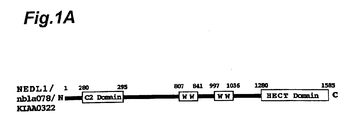
- Fig. 1A is a schematic representation of the
protein structure of ubiquitin ligases of the HECT type
showing that each has a HECT domain at its C-terminus,
plural WW domains at its center and a C2 domain at its
N-terminus.
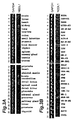
- Fig. 1B is an alignment diagram showing the
homology analysis between the amino acid sequence of
NEDL-1 protein and the amino acid sequence of NEDL-2
protein, where each of the domains is underlined or
boxed and conservative amino acids are indicated by
asterisks.
- Fig. 2 is an electropherogram showing the results
of determination of the expression levels of the NEDL-1
gene in clinical samples of neuroblastomas with
favorable prognosis and with unfavorable prognosis by
semi-quantitative PCR.
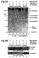
- Fig. 3A is a figure corresponding to an
electropherogram showing the results of determination
of the expression levels of the NEDL-1 gene in normal
human tissues by semi-quantitative PCR.
- Fig. 3B is a figure corresponding to an
electropherogram showing the results of determination
of the expression levels of the NEDL-1 gene in various
neuroblastoma cell lines by semi-quantitative PCR.
- Fig. 4 is a figure representing autoradiography of
different tissue expression of the NEDL-1 gene in
normal human tissues as analyzed by Northern blot.
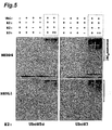
- Fig. 5 is an immunoblotted electropherogram
showing the ubiquitin ligase activity of the NEDL-1
protein.
- Fig. 6A is a Western blot showing cellular
localization of the NEDL-1 gene (Cos7 cell).
- Fig. 6B is a Western blot showing cellular
localization of the NEDL-1 gene (CHP134 cell).
- Fig. 7 is an immunoblotted electropherogram
showing the interaction between the NEDL-1 protein and
ACID as obtained by immunoprecipitation with anti-NEDL-1
antibody and detection with anti-FLAG antibody.
- Fig. 8 is an immunoblotted electropherogram
showing the interaction between the NEDL-1 protein and
ACID as obtained by immunoprecipitation with anti-FLAG
antibody and detection with anti-NEDL-1 antibody.
- Fig. 9A is an immunoblotted electropherogram
showing the ubiquitination of βAPP and ACID by the
NEDL-1 protein as obtained by immunoprecipitation with
anti-HA antibody and detection with anti-ubiquitin
antibody.
- Fig. 9B is an immunoblotted electropherogram
showing the ubiquitination of FLAG-ACID by the NEDL-1
protein as obtained by immunoprecipitation with anti-FLAG
antibody and detection with anti-ubiquitin
antibody.
- Fig. 10 is an immunoblotted electropherogram
showing the ubiquitination of βAPP and ACID by the
NEDL-1 protein as obtained by immunoprecipitation with
anti-HA antibody and detection with an antibody that
recognizes ACID.
- Fig. 11 is an immunoblotted electropherogram
showing the interaction between the NEDL-1 protein and
SOD1 mutants as obtained by immunoprecipitation with
anti-NEDL-1 antibody and detection with anti-FLAG
antibody.
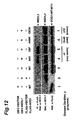
- Fig. 12 is an immunoblotted electropherogram
showing the interaction between the NEDL-1 protein and
SOD1 mutants as obtained by immunoprecipitation with
anti-FLAG antibody and detection with anti-NEDL-1
antibody.
- Fig. 13 is an immunoblotted electropherogram
showing the ubiquitination of SOD1 and SOD1 mutants by
the NEDL-1 protein.
-
Best Mode for Carrying Out the Invention
-
The nucleic acids (which will be referred to as
"the nucleic acid of this invention") derived from the
gene which is highly expressed in neuroblastomas with
favorable prognosis (which will be referred to as "the
NEDL-1 gene of this invention" or simply as "the NEDL-1
gene") and the protein encoded by the gene will be
described in detail by referring to the preferred
embodiments of the invention.
-
As stated above, the nucleic acids of this
invention are derived from the NEDL-1 gene of the
invention and they make up the gene or are obtained
from the gene by an in vivo or in vitro process. There
are no limitations on the base lengths of the nucleic
acids and here they will be referred to as the nucleic
acids of the invention, which include nucleic acid
fragments corresponding to parts of the gene. When the
base lengths are short, they can be synthesized by
chemical techniques. The term "nucleic acid(s)" as
used in this specification refers to, for example, DNA
or RNA, or polynucleotides derived therefrom which are
active as DNA or RNA, and preferably refers to DNA
and/or RNA. The particularly preferred nucleic acid
has a base sequence that is identical with the human
cDNA sequence disclosed in this specification or that
is complementary to the sequence.
-
The term "hybridize under stringent conditions" as
used in this specification means that two nucleic acid
fragments hybridize to each other under the
hybridization conditions described by Sambrook, J. et
al. in "Expression of cloned genes in E. coli",
Molecular Cloning: A Laboratory Manual (1989), Cold
Spring Harbor Laboratory Press, New York, USA, 9.47-9.62
and 11.45-11.61.
-
More specifically, the "stringent conditions"
refers to hybridization at approximately 45 °C, 6.0 x
SSC, followed by washing at 50 °C, 2.0 x SSC. The
stringency may be selected by choosing a salt
concentration in the washing step from approximately
2.0 x SSC, 50 °C as low stringency to approximately 0.2
x SSC, 50 °C as high stringency. Also, the temperature
in the washing step may be increased from room
temperature, or approximately 22 °C as low stringency
conditions, to approximately 65 °C as high stringency
conditions.
-
The term "nucleic acid(s)" as used in this
specification refers to an isolated nucleic acid(s) and
to a nucleic acid or a polynucleotide containing
substantially no cellular substances or culture medium,
if prepared by recombinant DNA techniques, or
containing substantially no precursor chemical
substances or other chemical substances, if prepared by
chemical synthesis.
-
The term "favorable prognosis" as used in this
specification refers to a condition of neuroblastoma in
which the tumor is localized or has become a regressing
or benign sympathetic ganglion neoplasm, and is judged
to have low malignancy based on N-myc or other tumor
markers (TrkA, chromosomal aberration). According to a
preferred embodiment of the invention, a favorable
prognosis is a case of stage 1 or 2, with an onset age
of less than one year and survival without recurrence
for 5 or more years after surgery, and with no noted
amplification of N-myc in the clinical tissue; however,
there is no limitation to such specific cases. The
term "unfavorable prognosis" as used in this
specification refers to a condition of neuroblastoma in
which progression of the tumor has been observed, and
it is judged to have high malignancy based on N-myc or
other tumor markers. According to a preferred
embodiment of the invention, an unfavorable prognosis
is a case of stage 4, with an onset age of greater than
one year, death within 3 years after surgery and noted
amplification of N-myc in the clinical tissue; however,
there is no limitation to such specific cases.
-
Neuroblastoma is a tumor consisting of actual
nerve cells, of which only two types of tumor are known
in humans, and analysis of the genes expressed therein
is expected to provide very useful knowledge for
understanding the biology of nerve cells. Specifically,
it is extremely difficult, and practically impossible,
to obtain site-specific homogeneous tissue from the
brain or peripheral nerves. On the other hand, a
neuroblastoma consists of an almost homogeneous nerve
cell population (though tumorized) derived from
peripheral sympathetic nerve cells, and thus offers the
high possibility of obtaining homogeneous expression of
neuro-related genes. Furthermore, since neuroblastoma
is a type of cancer, it will characteristically have
many important genes expressed in the immature stage of
neurogenesis.
-
Clinically and biologically, neuroblastoma can be
neatly classified into favorable prognosis and
unfavorable prognosis types. Cancer cells from
neuroblastoma with favorable prognosis are
characterized by having a very slow rate of
proliferation, with spontaneous regression beginning at
some point. Findings to date have confirmed that nerve
cell differentiation and apoptosis (nerve cell death)
occur in the spontaneous regression, and that the
differentiation which occurs in the maturation stages
of normal nerve cells and programmed cell death are
phenomena very closely resembling each other.
Consequently, it is highly probable that the analysis
of genes expressed in such tumors will lead to
obtaining important genetic information relating to
nerve cell differentiation and apoptosis.
-
NEDL-1 gene from which the useful genetic
information can be obtained and the NEDL-1 protein
encoded by the gene are found in clinical tissues of
human neuroblastomas with favorable prognosis. These
gene and protein are provided with the characteristics
described below.
-
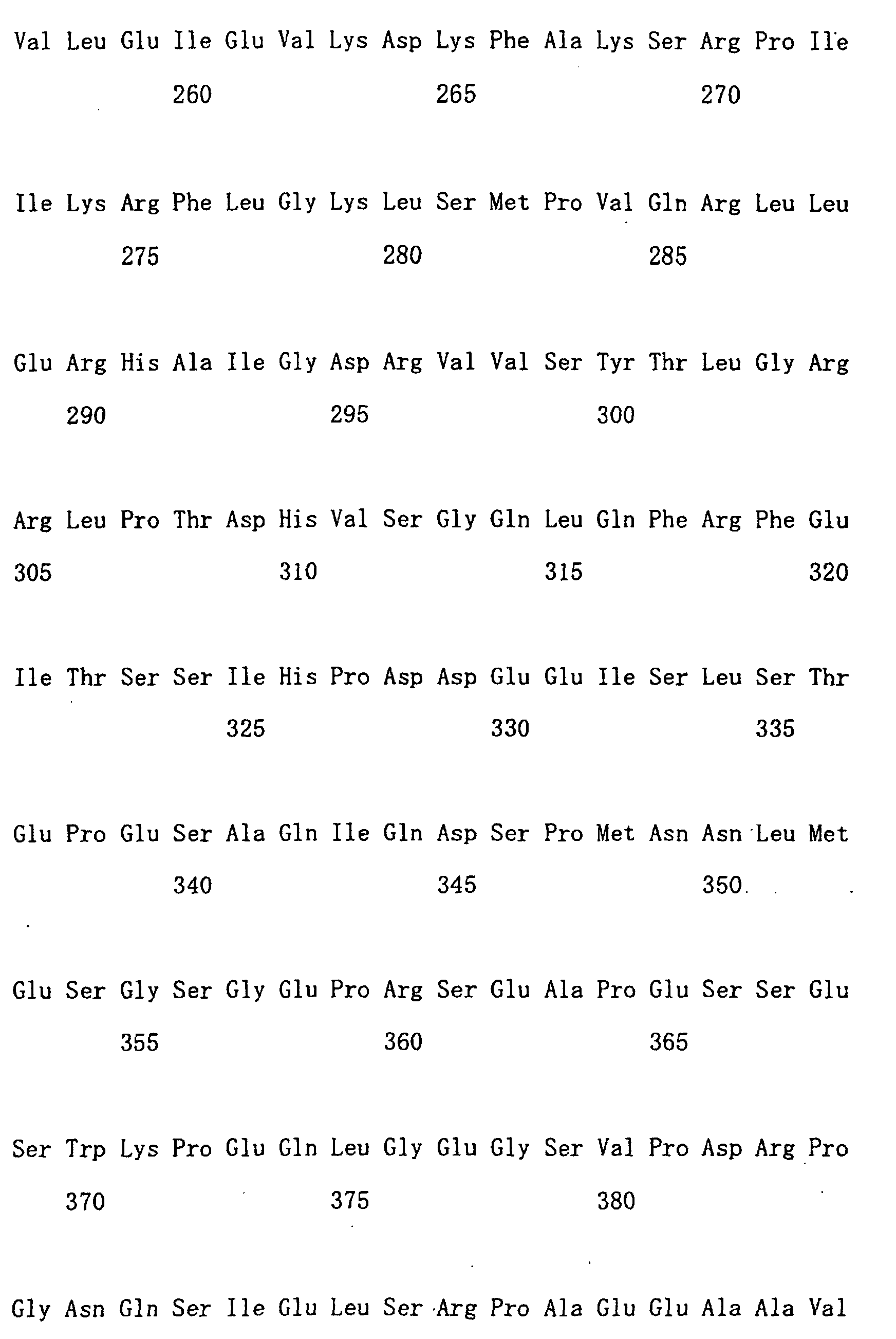
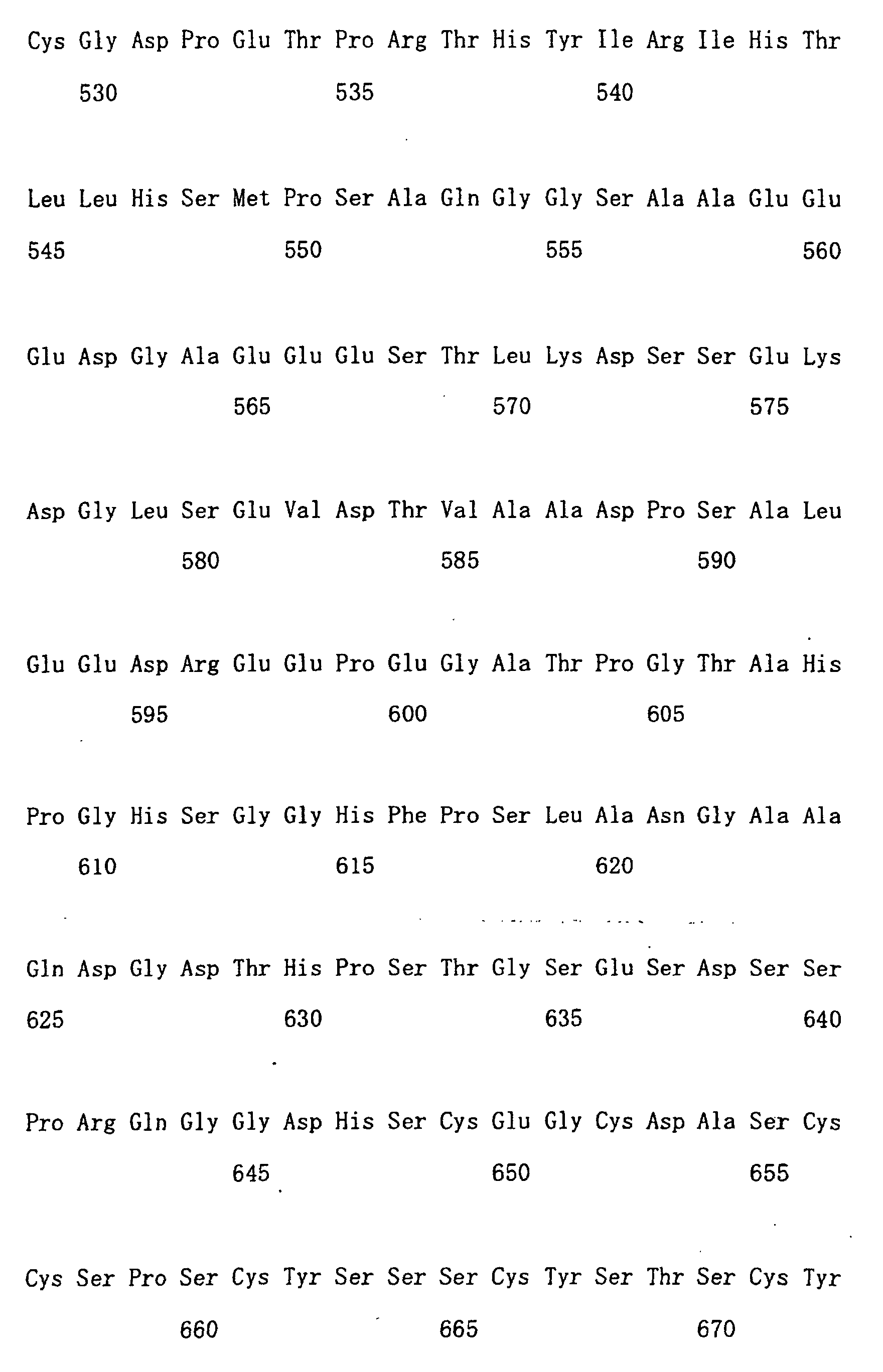
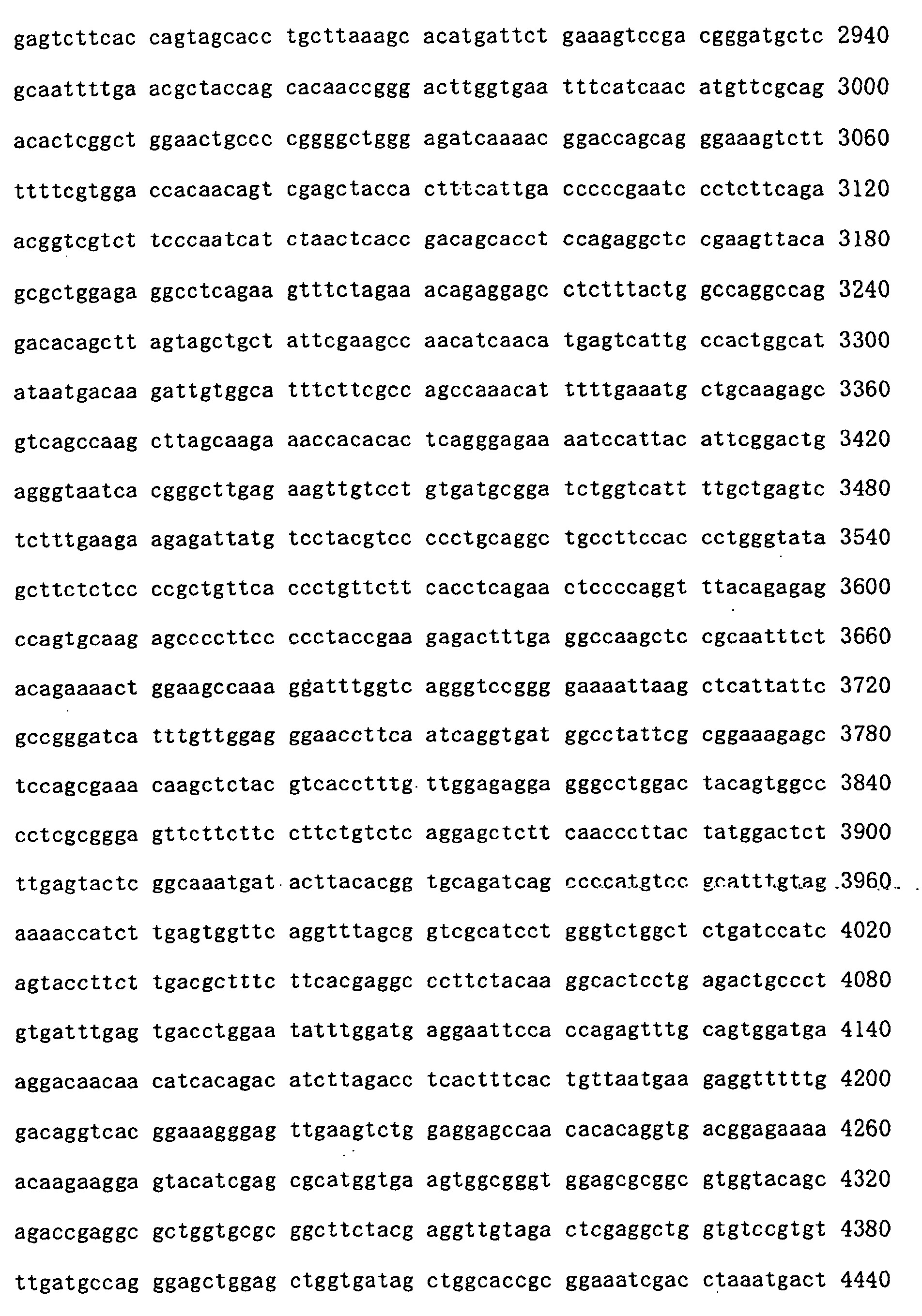
The NEDL-1 gene of this invention is a gene having
the full length of 6,200 bases (coding region of 4,755
bases) and its base sequence is shown in SEQ ID NO:2 in
the Sequence Listing. NEDL-1 protein encoded by the
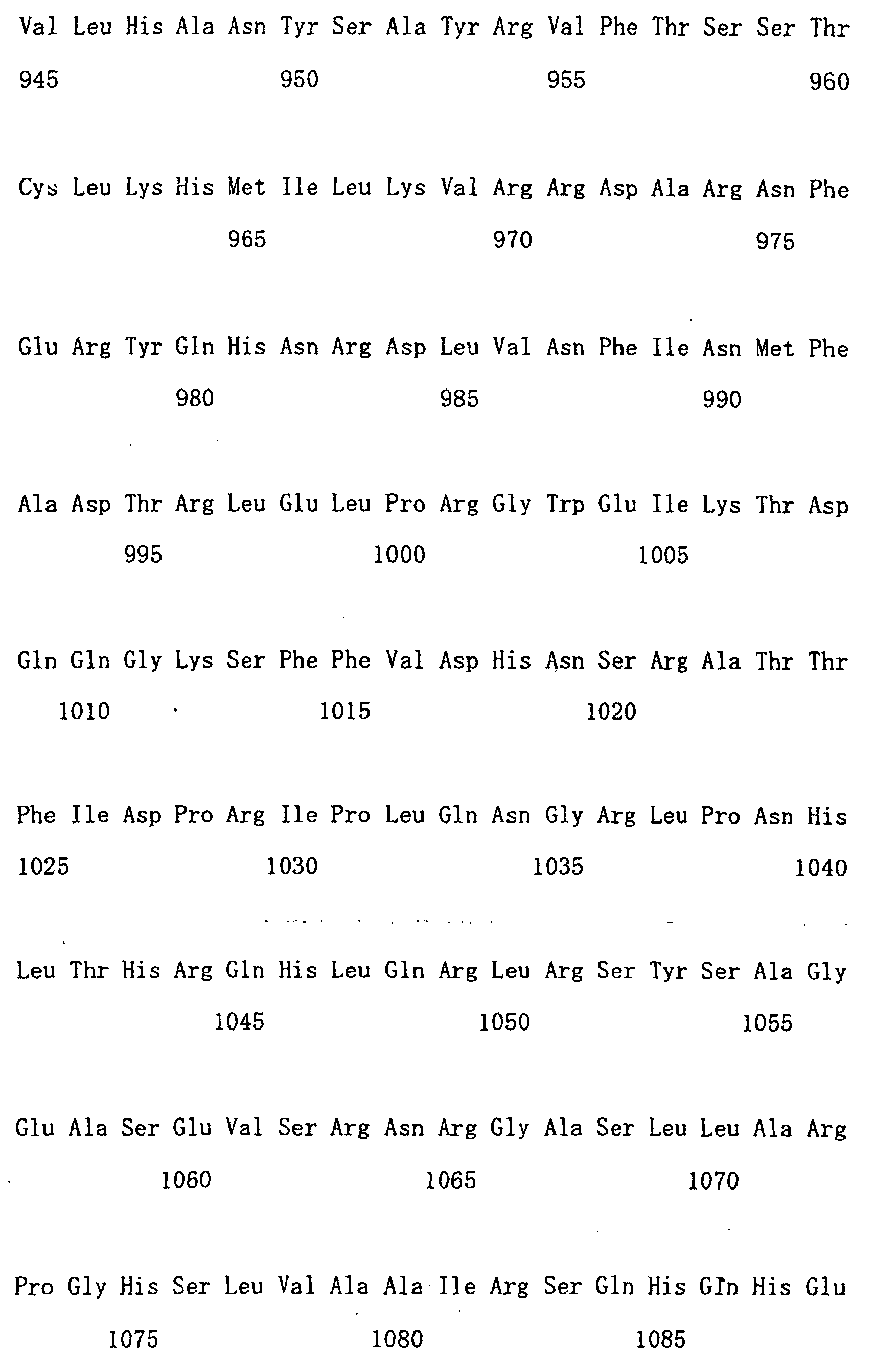
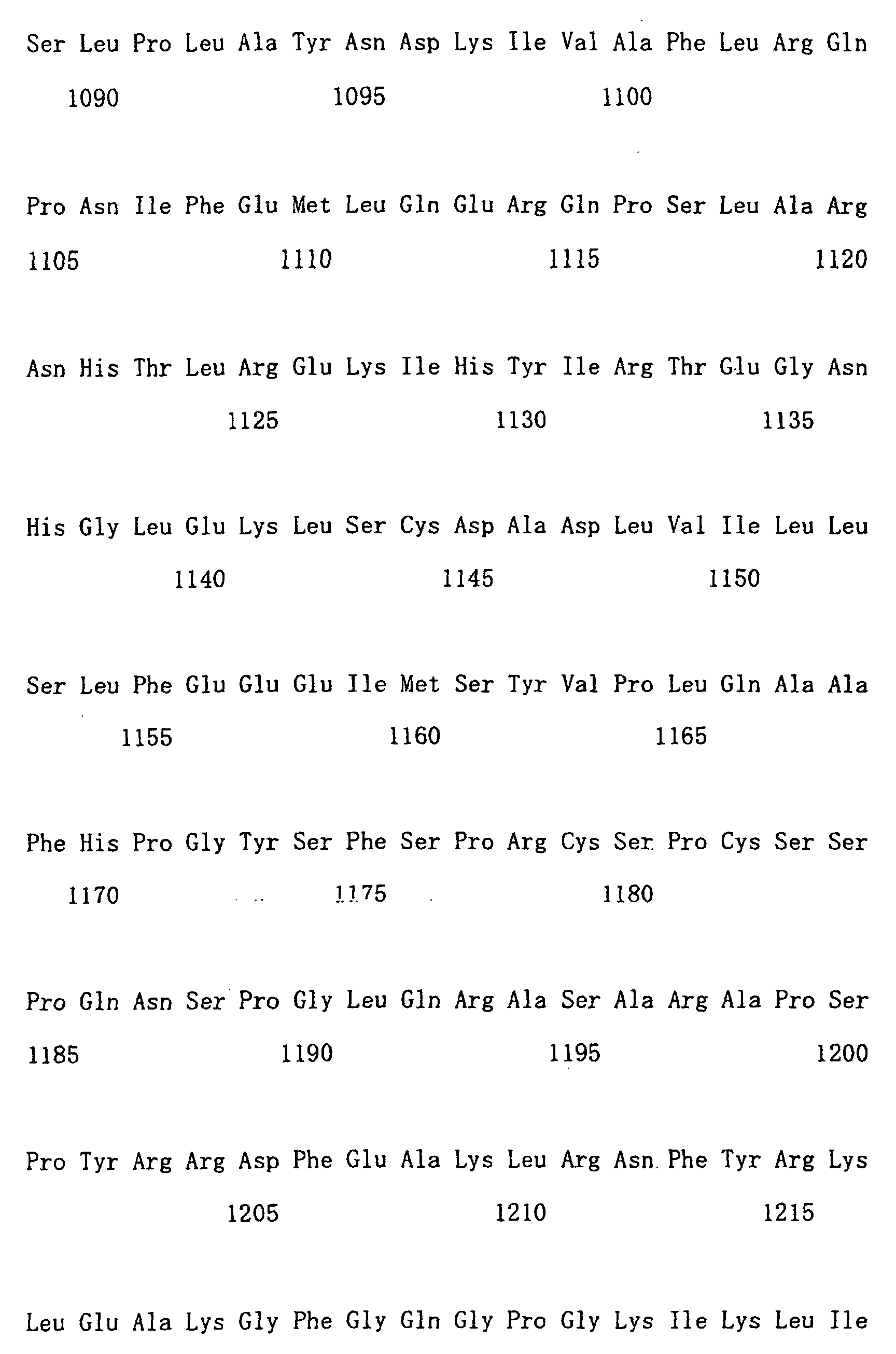
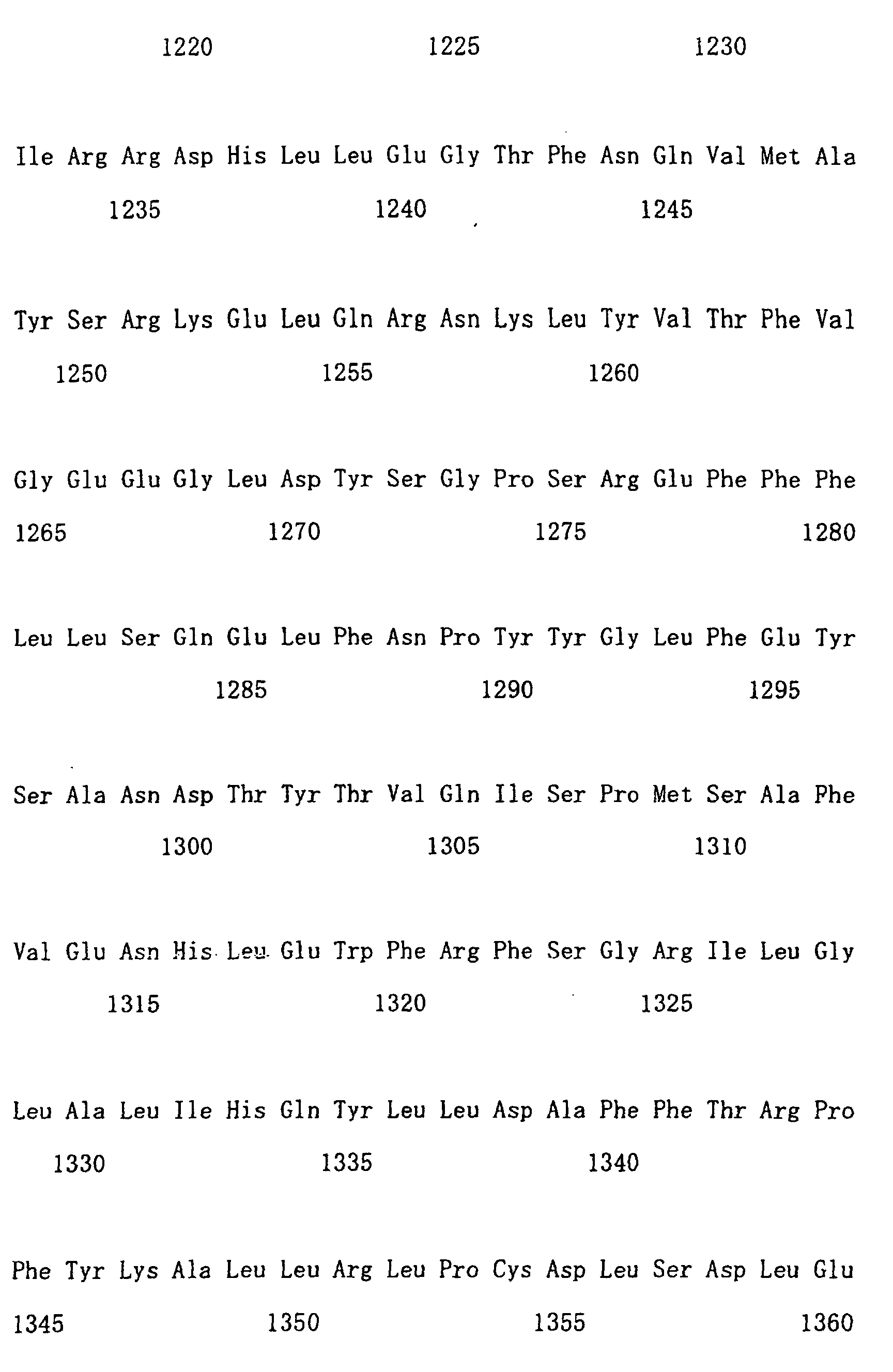
gene comprises 1585 amino acids and its full length is
shown in SEQ ID NO:1 in the Sequence Listing. The base
sequence and the amino acid sequence have been
registered with GeneBank (HYPERLINK
http://www.ncbi.nlm.nih.gov.) as Accession No. AB048365.
-
The present inventors found that as a result of
the structural and functional analysis of the NEDL-1
gene and the NEDL-1 protein, NEDL-1 is a ubiquitin
ligase of the HECT type. Fig. 1 shows the results from
the homology analysis between the NEDL-1 protein and
KIAA03222 protein (NEDL-2) which is a known member of
the HECT type ubiquitin ligase family. The NEDL-1
protein has the domains characteristic of HECT type
ubiquitin ligase. Specifically, these are (1) HECT
domain (about 300 amino acids) at the C-terminus, which
is positions 1280-1585 in NEDL-1; (2) plural WWW
domains at the central part (about 35 to 40 amino
acids), which is positions 807-841 and positions 998-1030
in NEDL-1 and positions 806-840 in NEDL-2; (3) C2
domain at the N-terminus binding to membrane lipid in a
Ca-dependent manner, which is positions 185-295 in
NEDL-1 and positions 186-295 in NEDL-2. In addition,
the NEDL-1 protein was found to possess ubiquitin
ligase activity at the same level as does Nedd4 which
is a HECT type ubiquitin ligase.
-
Because proteins are decomposed in the ubiquitin-protease
system, this system is essential to an
adequate cellular process. In brief, the system allows
a number of ubiquitin molecules (Ub) to be bound to a
target protein (what is called "polyubiquitination or
ubiquitination) and the ubiquitinated protein is
decomposed by 26S proteasome. It has been elucidated
that the ubiquitination of proteins progresses through
the catalytic action of a series of enzyme groups, that
is ubiquitin-activating enzymes (E1), ubiquitin-conjugating
enzymes (E2), and ubiquitin-ligating
enzymes (ubiquitin ligases). See, for example, a
review of Keiji Tanaka, "Ubiquitin and Proteasome" in
Experimental Medicine, Vol. 18, No. 11, pp. 1452-1456
(2000) by Yodosha. Among those enzymes ubiquitin
ligase (E3) receives Ub from E2-Ub and ligate this Ub
to the target protein (substrate). Thus, ubiquitin
ligase is thought to be most heavily involved in the
specificity with which a specific protein is
ubiquitinated.
-
It has been pointed out that the anomaly in the
ubiquitin-proteasome system is related to many diseases
(R. J. Mayer et al., Biochem. Biophs. Acta 1089: 141-157
(1991)). Recently, the relation between
neurodegenerative diseases and the anomaly in ubiquitin
metabolism has attracted attention; and there has been
a report that E6-AP, which is known as a ubiquitin
ligase, is one of the responsible genes for Angelman
syndrome (Nobutomi Honda et al., "HECT type ubiquitin-ligating
enzymes: physiological functions and disease
state" in Experimental Medicine, Vol. 18, No. 11, pp.
1483-1490 (2000) by Yodosha). The NEDL-1 protein of
this invention, which is one of HECT type ubiquitin
ligases, is highly expressed in nerve tissues. It is,
therefore, well anticipated that the protein uses as a
substrate the product of a causative gene of a
neurodegenerative disease. These causative gene
products are believed to be beta amyloid precursor
protein (βAPP), preselinin protein (PS) and others.
-
As will be described in the Examples, it was
actually found that NEDL-1 interacted with amyloid beta
precursor intracellular domain (AICD) which was the
coding region of amyloid precursor protein. It was
further determined that this interaction resulted from
ubiquitination of BAPP and ACID by NEDL-1.
-
Amyloid is a protein that deposits in cerebral
blood vessels and senile plaques of an Alzheimer
patient: it is comprised principally of β-protein with
a molecular weight of 4 kDa and is produced when
amyloid precursor protein is cleaved by secrease. The
fact that NEDL-1 directly interacts with ACID leads to
the possibility that NEDL-1 regulates the production of
βAPP (then β amyloid) directly or indirectly. It will
be a finding that is extremely important to planning a
strategy for Alzheimer treatment which targets lowered
production of β-amyloid on a molecular level.
-
As will also be described in the Examples, it was
also found that NEDL-1 interacted with superoxide
dismutase mutants (SOD1). It was further determined
that this resulted from ubiquitination of the SOD1
mutants by NEDL-1.
-
Amyotrophic lateral sclerosis (ALS) is a
neurodegenerative diseases with unfavorable prognosis
that involves muscular atrophy due to the degeneration
or deciduation of motor neurons. Currently, familial
ALS is seen in a frequency of 5-10% of the total ALS.
The causative gene has been identified as the Cu/Zn
superoxide dismutase (SOD1) in some of the families.
SOD1 is an enzyme that inactivates superoxide dismutase
which is one type of active enzymes produced in a cell
during an aerobic process. There is the possibility
that its lowered level causes degeneration of nerve
cells; however the detail of mechanism is unknown. The
cause for other general type of amyotrophic lateral
sclerosis is also unknown.
-
Presently, the virus theory, the poisoning theory,
the nerve nutrient factor depletion theory, the
autoimmune theory, the excessive glutamate theory, the
free radical theory and others are proposed. However,
the decisive pathological mechanism by which only motor
nerve degenerates in ALS has not been elucidated. The
aggregate hypothesis assumes that mutant SOD1 forms an
aggregate in a cell and exhibits cytotoxity; and this
will be becoming the most convincing one in recent
years.
-
Thus far about 80 of SOD1 mutants have been
reported. Intracellular signal transduction remain
unknown for any of these mutants. There has been a
report on interacting molecules for two types of SOD1
mutants (G85R/G93A) and only two types, lysyl-tRNA
synthetase and translocon-associated protein delta,
have been identified as a protein factor that only
binds to those mutants and that does not bind to normal
SOD1. (Kunst CB, Mezey E, Brownstein MJ, Patterson D,
"Mutations in SOD1 associated with amyotrophic lateral
sclerosis cause novel protein interactions" Nat. Genet.
1997 Jan; 15(1) 91-4.) The details have not yet been
elucidated. Thus, about 130 years have passed since
the first report of ALS, but even now the situation may
be that the elucidation of the intracellular signal
transduction for cytotoxity which the mutant SOD1 has
newly acquired. In consideration of the present
circumstance described above, the results obtained from
this invention are believed to provide very useful
information for the elucidation of the mechanism of ALS
crisis that has not been hitherto clarified.
-
The NEDL-1 protein has an amino acid sequence set
forth in SEQ ID NO:1 in the Sequence Listing but this
invention also encompasses a protein having an amino
acid sequence comprising a deletion, a substitution, an
insertion or an addition of one or more amino acids in
the amino acid sequence set forth in SEQ ID NO:1 in the
Sequence Listing.
-
This invention also encompasses salts of the NEDL-1
protein and others. These salts are not particularly
limited and, for example, preferred are a sodium salt,
a potassium salt, a magnesium salt, a lithium salt and
an ammonium salt.
-
Sugar chains are added to many proteins and the
addition of a sugar chain may be adjusted by converting
one or more amino acids. Therefore, this invention
encompasses proteins the sugar chain addition of which
has been adjusted in the amino acid sequence set forth
in SEQ NO:1 in the Sequence Listing.
-
This invention further encompasses a nucleic acid
having a base sequence encoding the NEDL-1 protein.
The term "encoding a protein" as used herein means that
either of complementary double strands has a base
sequence encoding the protein when DNA is doublestranded.
The nucleic acids of this invention embrace
a nucleic acid comprising a base sequence directly
encoding the amino acid sequence set forth in SEQ ID
NO:1 in the Sequence Listing and a nucleic acid
comprising a base sequence complementary to said
nucleic acid.
-
Further, the nucleic acid of the invention may be
a nucleic acid hybridizing to the nucleic acid
comprising a base sequence set forth in SEQ ID NO:2
under stringent conditions. The base sequence is not
particularly limited insofar as it satisfies this
condition. Still further, the nucleic acids of the
invention encompass a nucleic acid comprising a base
sequence complementary to the nucleic acid hybridizable
under the stringent conditions mentioned above.
Specifically there is mentioned a nucleic acid
comprising deletions, substitutions, insertions or
additions in some bases of the nucleic acid comprising
a base sequence set forth in SEQ ID NO:2 or a nucleic
acid complementary to said nucleic acid. As used
herein, the deletion, the substitution, the insertion
and the addition include not only a short deletion,
substitution, insertion and addition with 1 to 10 bases,
but also a long deletion, substitution, insertion and
addition with 10 to 100 bases.
-
As a result of comparing levels of expression of
the NED-1 gene according to this invention in clinical
tissues from neuroblastomas with favorable prognosis
and with unfavorable prognosis, a highly significant
difference was found. That is, expression of this gene
was enhanced in neuroblastomas with favorable prognosis.
Thus, in addition to providing the useful genetic
information described above, the nucleic acid sequence
set forth in SEQ ID NO:2 can also be utilized as data
for tumor markers to diagnose favorable or unfavorable
prognosis of neuroblastoma, by detecting the nucleic
acid (DNA or RNA) having that sequence. The amino acid
sequence in SEQ ID NO:1 can also be utilized as data
for tumor markers to diagnose favorable or unfavorable
prognosis of neuroblastoma, by detecting the NEDL-1
protein based on the sequence information.
-
Specifically, by using the NEDL-1 gene and the
NEDL-1 protein according to this invention, the
invention will make it possible to obtain various
genetic information on or relating to neuroblastoma
through the following means.
(1) Probes for use in hybridization
-
According to one embodiment of this invention, the
nucleic acid of the invention can be used as a probe
(i.e., the probe of this invention) for hybridization
to detect the NEDL-1 gene expressed in neuroblastoma.
The nucleic acid according to this invention can also
be used as probes for hybridization in order to
determine gene expression in a variety of tumors and
normal tissues, to identify the distribution of the
gene expression.
-
When the nucleic acid according to this invention
is used as a probe for hybridization, there are no
particular limitations on the actual method of
hybridization. As preferred methods there may be
mentioned, for example, Northern hybridization,
Southern hybridization, colony hybridization, dot
hybridization, fluorescence in situ hybridization
(FISH), in situ hybridization (ISH), DNA chip methods,
and microarray methods.
-
As one application example of the hybridization,
the nucleic acid according to this invention can be
used as a probe for Northern hybridization to measure
the length of mRNA or to quantitatively detect the
expression of the NEDL-1 gene of this invention in a
clinical tissue sample to be assayed.
-
As another application example, the nucleic acid
according to this invention can be used as a probe for
Southern hybridization to detect the presence or
absence of the DNA sequence in the genomic DNA of a
clinical tissue sample to be assayed.
-
As still another application example, the nucleic
acid according to this invention can also be used as a
probe for fluorescence in situ hybridization (FISH) to
identify the location of the NEDL-1 gene of this
invention on a chromosome.
-
As a further application example, the nucleic acid
according to this invention can also be used as a probe
for in situ hybridization (ISH) to identify the tissue
distribution of expression of the NEDL-1 gene of this
invention.
-
When the nucleic acid according to this invention
is used as a probe for hybridization, a base length of
at least 20 is necessary; and among the nucleic acids
according to this invention, a nucleic acid having 20
or more contiguous bases is preferably used. More
preferably, the nucleic acid having 40 or more bases is
used and most preferably the nucleic acid having 60 or
more bases is used. Further, the nucleic acid having
the full-length of the base sequence set forth in SEQ
ID NO:2 may be used.
-
Nucleic acid probe techniques are well known to
one skilled in the art, and for example, conditions
suitable for hybridization between a probe of specific
length according to the invention and the target
polynucleotide may be readily determined. In order to
obtain hybridization conditions optimal to probes of
varying lengths, Sambrook et al. "Molecular Cloning: A
Laboratory Manual, 2nd Edition, Cold Spring Harbor
(1989) may be followed for such manipulations which are
well known to one skilled in the art.
-
The probe according to this invention may
preferably be labeled for use in an easily detectable
fashion. The detectable label may be any type and any
element or compound which can be detected either
visually or using devices. As commonly used detectable
labels, there may be mentioned radioactive isotopes,
avidin and biotin and fluorescent substances (FITC or
Rhodamins). The radioactive isotopes are 32P, 14C, 125I,
3H, 35S etc. Biotin-labeled nucleotides may be
incorporated into DNA or RNA by nick translation, or
chemical or enzymatic means. The biotin-labeled probes
are detected after hybridization using labeling means
such as avidin/streptavidin, fluorescent labels,
enzymes, gold colloidal complexes or the like. The
nucleic acid probe of this invention may also be
labeled by binding with a protein. For this purpose, a
radioactive or fluorescent histone single-stranded
binding protein may also be used. In this manner, a
suitably labeled probe constitutes a diagnostic agent
for prognosis according to this invention.
(2) Primers for use in PCR
-
For methods of detecting the NEDL-1 gene according
to this invention other than the hybridization, primers
can be designed after any nucleic acid (DNA) sequence
contained in the nucleic acid according to this
invention and the polymerase chain reaction (PCR)
method can be used. For example, RNA may be extracted
from a clinical tissue sample to be assayed, and the
gene expression can be semi-quantitatively measured by
RT-PCR. This may be carried out by a method well known
to one skilled in the art. For example, "Molecular
Cloning: A Laboratory Manual," (T. Maniatis, Cold
Spring Harbor Laboratory Press) or Idenshibyo Nyumon
[Introduction to Genetic Diseases] (Takahisa, S.:
Nankodo Publishing) may be followed.
-
When the nucleic acid according to this invention
(DNA) is used as a PCR primer (i.e., the primer of the
invention), a base length of 10 to 60 is necessary; and
among portions of the base sequences according to the
invention, the nucleic acid having 10 to 60 contiguous
bases is preferably used. More preferably, one having
15 to 30 bases is used. Generally, a primer sequence
with a GC content of 40-60% is preferred. Also, there
is preferably no difference in the Tm values of the two
primers used for amplification. The primer has such
base sequence that there is no annealing at the 3' ends
of the primers and no secondary structure is formed in
the primers.
(3) Gene screening
-
The nucleic acid according to this invention can
also be used to detect the expression distribution of
the NEDL-1 gene which is expressed in various tissues
or cells. This can be accomplished, for example, by
using the nucleic acid according to this invention as a
probe for hybridization or as a primer for PCR.
-
The expression distribution of the gene can also
be detected using a DNA chip, microarray or the like.
That is, the nucleic acid according to the invention
may be directly attached to the chip or array. There
is known a method by which nucleic acids (DNA) are
spotted to a substrate for the purpose of attaching
them to a chip or array by using a high precision
dispenser (for example, see United State Patent No.
5807522). mRNA extracted from a clinical tissue sample
may be labeled with a fluorescent substance or the like,
hybridized thereto, and an analysis can be made of the
type of tissue cells with high expression of the gene.
The DNA attached to the chip or the array may be the
reaction product of PCR using the nucleic acid or its
fragment according to the invention. As an alternative
method, the nucleic acid fragment of this invention
(DNA fragment) may be directly synthesized on a
substrate to form a DNA chip or a DNA array (See, for
example, United State Patent No. 5424186).
(5) Methods of diagnosing tumor prognosis and
tumor markers to be used therefor
-
As mentioned above, the NEDL-1 gene of this
invention has its expression enhanced in neuroblastomas
with favorable prognosis. Therefore, the nucleic acid
according to this invention can be used as a probe for
hybridization, or as a primer for PCR to determine the
presence or absence of enhancement in the gene
expression in a sample containing the clinical tissue
taken from the subject, which enables the
identification of prognosis. The methods of detecting
the gene include Northern blot hybridization, in situ
hybridization and RT-PCR, as mentioned above among
others.
-
When hybridization is employed, prognosis may be
diagnosed as favorable if the amount of nucleic acid
hybridizing to the probe is increased in the sample.
When RT-PCR is employed, mRNA is extracted from the
sample and reverse transcribed into DNA, amplification
is performed using the aforementioned primer, and the
gene expression is semi-quantitatively measured. The
prognosis may be diagnosed as favorable if the gene
expression is then found to be enhanced. For the
purpose of such specific diagnosis it is preferred to
utilize a diagnosis kit containing a pair of such
primers as essential components. In addition to the
primer components, the diagnosis kit also include known
components such as PCR buffer, detergent solution and
enzymes.
(6) Antisense oligonucleotides
-
According to another embodiment of this invention
there are provided antisense oligonucleotides to the
nucleic acids of the invention. The antisense
oligonucleotides are capable of hybridizing to the
nucleic acids of the invention, and include antisense
DNAs and antisense RNAs. Antisense DNA inhibits
transcription of mRNA from DNA, while antisense RNA
inhibits translation of mRNA. These antisense
oligonucleotides may be synthesized using an automated
synthesizer or by PCR using the nucleic acid of the
invention as templates. The antisense oligonucleotides
also encompass antisense oligonucleotide derivatives
having improved binding affinity for DNA or mRNA,
tissue selectivity, cell permeability, nuclease
resistance and intracellular stability. These
derivatives may be synthesized using antisense
technology known in the art.
-
Antisense oligonucleotides having sequences
complementary to the sequences near the translation
initiation codon of the mRNA, those of the ribosome-binding
site, and those of the capping site or the
splicing site are capable of inhibiting synthesis of
the RNA and therefore will exhibit a particularly
notable inhibitory effect on gene expression. This
invention therefore encompasses such antisense
oligonucleotides.
(7) Gene therapy
-
According to a further embodiment of this
invention, there are provided nucleic acid sequences
encoding the therapeutic genes to be used in gene
therapy. Thus, the nucleic acid of the invention can
be transferred into a vector for use in gene
transportation, whereby the transgene (i.e., the NEDL-1
gene of the invention) can be expressed by an arbitrary
expression promoter and can be used in the gene therapy
for neurodegenerative diseases, for example.
1. Vectors
-
The transferable viral vectors may be prepared
from DNA viruses or RNA viruses. They may be any viral
vector of an MoMLV vector, a herpes virus vector, an
Adenovirus vector, an AAV vector, a HIV vector, a SIV
vector, a Seidai virus vector and the like. One or
more proteins among the constituent protein group of a
viral vector are substituted by the constituent
proteins of a different species of virus, or
alternatively a part of the nucleic acid sequence
constituting genetic information is substituted by the
nucleic acid sequence of a different species of virus
to form a viral vector of the pseudo-type which can
also be used in this invention. For example, there is
mentioned a pseudo-type viral vector wherein the Env
protein (an envelop protein of HIV) is substituted by
the VSV-G protein (an envelop protein of vesicular
stomatitis virus or VSV) (Naldini L., et al., Science
272, 263- 1996). Further, viruses having a host
spectrum other than human are usable as the viral
vector insofar as they are efficacious. As for the
vectors other than those of viral origin, there may be
used complexes of calcium phosphate and nucleic acid,
ribosomes, cation-lipid complexes, Seidai virus
liposomes, polymer carriers having polycation as the
backbone main chain and others. In addition, methods
such as electroporation and gene guns may be used as a
gene transfer system.
2. Expression promoters
-
As for the expression cassettes to be used for the
therapeutic gene, any cassettes without any particular
limitations may be used insofar as they can cause genes
to express in the target cells. One skilled in the art
can readily select such expression cassettes.
Preferably, they are expression cassettes capable of
gene expression in the cells derived from an animal,
more preferably, expression cassettes capable of gene
expression in the cells derived from a mammal, and most
preferably expression cassettes capable of gene
expression in the cells derived from a human. The gene
promoters that can be used as expression cassettes
include: for example, virus-derived promoters from an
Adenovirus, a cytomegalovirus, a human immunodeficiency
virus, a simian virus 40, a Rous sarcoma virus, a
herpes simplex virus, a murine leukemia virus, a sinbis
virus, a hepatitis type A virus, a hepatitis type B
virus, a hepatitis type C virus, a papilloma virus, a
human T cell leukemia virus, an influenza virus, a
Japanese encephalitis virus, a JC virus, parbovirus B19,
a poliovirus, and the like; mammal-derived promoters
such as albumin, SRα, a heat shock protein, and an
elongation factor; chimera type promoters such as a CAG
promoter; and the promoters whose expression can be
induced by tetracyclines, steroids and the like.
(8) Drugs
-
According to a still further embodiment of this
invention, there are provided therapeutic proteins and
peptides as drugs. As will be considered in practicing
this invention, the NEDL-1 protein of the invention and
its partial peptide may be prepared according to the
formulation method of choice and may be used through
any desired route of administration and at any desired
dosage age in the treatment of malignant tumors or
neurodegenerative diseases (e.g., Alzheimer disease) of
different types, for example.
1. Preparation method
-
The drug may be prepared as a recombinant viral
vector containing a therapeutic gene that is designed
for therapeutic purposes as described above. More
specifically, a recombinant virus vector comprising the
NEDL-1 gene may be prepared by dissolving it in an
appropriate solvent such as water, physiological saline
or an isotonized buffer solution. Alternatively, the
NEDL-1 protein produced by any desired method may be
dissolved in an appropriate solvent such as water,
physiological saline or an isotonized buffer solution
to prepare the vector similarly. Here, polyethylene
glycol, glucose, various amino acids, collagen, albumin
or the like may be then added as protective materials
for the preparation.
2. Administration method and dosage
-
There are no particular limitations on the method
of administrating the drug mentioned above to the
living body. For example, parental administration,
including injection is preferably carried out. The use
level of the drug varies depending on the method of use,
the purpose of use, etc.; and one skilled in the art
can easily select as appropriate and optimize it. In
the case of injection, for example, the daily dosage is
preferably administered at about 0.1 µg/kg to 1000
mg/kg per day, and more preferably at about 1 µg/kg to
100 mg/kg per day.
(9) Antibodies, Antisense, Ribozymes and TFO
-
In accordance with a still another embodiment of
this invention, an antibody to suppress the ubiquitin
activity of the NEDL-1 protein of the invention and
base sequences, including antisense, ribozyme or TFO,
to suppress the expression of the NEDL-1 gene of the
invention are provided. As will be considered in
practicing this invention, nucleic acids encoding
antisenses, ribozymes and TFOs can be transferred into
a vector used as a gene carrier; the transgene can be
expressed by any suitable expression promoter and can
be used, for example, to establish a primary culture
cell line or to construct a cancer model animal.
(10) Genetically modified animals
-
In accordance with a yet another embodiment of
this invention, a nucleic acid sequence to knock out
the expression of the NEDL-1 gene of the invention and
a knockout animal (e.g., knockout mouse) are provided.
There are provided a transgenic animal (e.g.,
transgenic mouse) where the gene has been forcedly
expressed and a genetically modified animal having an
introduced mutant gene obtained by introducing an
arbitrary mutation (such as a point mutation or
deletion) into the gene. This genetically modified
animal can be used to construct a model animal for a
neurodegenerative disease, for example.
-
As described above, by utilizing the NEDL-1 gene
or the NEDL-1 protein according to this invention or
the information obtainable therefrom, it will be
possible to detect the NEDL-1 gene in a clinical tissue
sample, which then will allow the diagnosis of
neuroblastoma whether favorable or unfavorable
prognosis. Further, by utilizing the gene, the protein
or the information obtainable therefrom, it will be
possible to design tumor markers that can be used in
the diagnosis for prognosis and the aforementioned
method.
-
This invention will now be explained in greater
detail by way of the examples; however, the technical
scope of invention will not be restricted to those
examples.
(Examples)
(Preparation Example 1) Construction of cDNA library
from neuroblastoma
1. Obtaining samples
-
The clinical tissue samples of neuroblastoma were
quasi-aseptically frozen immediately after surgical
extraction and then preserved at -80 °C.
2. Selecting samples with favorable prognosis
-
Prognosis of the samples obtained in 1. above was
carried out based on the following criteria.
| Favorable prognosis | Unfavorable prognosis |
| · Stage 1 or 2 | · Stage 4 |
| · Age of onset: <1 | · Age of onset: ≥1 |
| · Survival for ≥5 years after surgery without recurrence | · Death within 3 years after surgery |
| · No amplification of N-myc | · Amplification of N-myc |
-
Amplification of N-myc in the aforementioned two
sample types was confirmed in the following manner.
-
The samples obtained in 1. above was thinly sliced
with a scalpel and then thoroughly homogenized after
addition of 5 ml of TEN buffer (50 mM Tris-HCl (pH =
8.0)/1 mM EDTA/100 mM NaCl). Upon adding 750 µl of
SDS (10%) and 125 µl of proteinase K (20 mg/ml) to the
mixture, it was gently stirred and allowed to stand at
50 °C for 8 hours. This was followed by
phenol/chloroform treatment and finally ethanol
precipitation to obtain purified genomic DNA. A 5 µg
portion of the obtained genomic DNA was completely
digested with the restriction endonuclease EcoRI (NEB
Inc.), and an N-myc probe was used to determine
amplification of N-myc by Southern hybridization.
3. Preparation of mRNA from clinical tissue of
neuroblastoma with favorable prognosis
-
A 2-3 g portion of the clinical tissue samples of
neuroblastoma judged to have favorable prognosis in 2.
above was treated using a Total RNA Extraction Kit
(QIAGEN Inc.) and the total RNA was extracted. The
extracted total RNA was purified using an oligo dT
cellulose column (Collaborative Research, Inc.) to
obtain a pool of mRNA with a polyA structure.
4. Dephosphorylation of mRNA
-
A 100-200 µg portion of the mRNA pool prepared in
3. above was dissolved in 67.3 µl of distilled sterile
water containing 0.1% diethyl pyrocarbonate (DEPC), and
then 20 µl of 5XBAP buffer (Tris-HCl (500 mM, pH =
7.0)/mercaptoethanol (50 mM)), 2.7 µl of RNasin (40
unit/µl: Promega Inc.) and 10 µl of BAP (0.25 unit/µl,
bacteria-derived alkali phosphatase: Takara Shuzo Co.
Ltd.) were added. The mixture was reacted at 37 °C for
1 hour to effect dephosphorylation of the 5' end of the
mRNA. This was followed by phenol/chloroform treatment
two times, and finally ethanol precipitation to obtain
a purified dephosphorylated mRNA pool.
5. Decapping of dephosphorylated mRNA
-
The total amount of the dephosphorylated mRNA pool
prepared in 4. above was dissolved in 75.3 µl of
distilled sterile water containing 0.1% DEPC, and then
20 µl of 5XTAP buffer (sodium acetate (250 mM, pH =
5.5)/mercaptoethanol (50 mM), EDTA (5 mM, pH = 8.0)),
2.7 µl of RNasin (40 unit/µl) and 2 µl of TAP
(tobacco acid pyrophosphatase: 20 unit/µl) were added.
The mixture was reacted at 37 °C for 1 hour to effect
decapping treatment of the 5' end of the
dephosphorylated mRNA. The dephosphorylated mRNA of
incomplete length with no capped structure remained
without decapping, and with the 5' end dephosphorylated.
This was followed by phenol/chloroform treatment and
ethanol precipitation to obtain a purified decapped
mRNA pool.
6. Preparation of oligo-capped mRNA
-
The total amount of the decapped mRNA pool
prepared in 5. above was dissolved in 11 µl of
distilled sterile water containing 0.1% DEPC, and then
4 µl of 5'-oligo RNA (5'-AGCAUCGAGUCGGCCUUGGCCUACUGG-3':
100 ng/µl), 10 µl of 10X ligation buffer (Tris-HCl
(500 mM, pH = 7.0)/mercaptoethanol (100 mM)), 10 µ
l of magnesium chloride (50 mM), 2.5 µl of ATP (24 mM),
2.5µl of RNasin (40 unit/µl), 10 µl of T4 RNA ligase
(25 unit/µl: Takara Shuzo Co. Ltd.) and 50 µl of
polyethylene glycol (50% w/v, PEG8000: Sigma
Corporation) were added. The mixture was reacted at
20 °C for 3 hours for ligation of the 5'-oligo RNA to
the 5' end of the decapped mRNA. The dephosphorylated
mRNA of incomplete length with no capped structure
resulted in no ligation to the 5'-oligo RNA. This was
followed by phenol/chloroform treatment and ethanol
precipitation to obtain a purified oligo-capped mRNA
pool.
7. Removal of DNA from oligo-capped mRNA
-
The oligo-capped mRNA pool prepared in 6. above
was dissolved in 70.3 µl of distilled sterile water
containing 0.1% DEPC, and then 4 µl of Tris-HCl (1 M,
pH = 7.0), 5.0 µl of DTT (0.1 M), 16 µl of magnesium
chloride (50 mM) , 2.7 µl of RNasin (40 unit/µl) and 2
µl of DNaseI (5 unit/µl: Takara Shuzo Co. Ltd.) were
added. The mixture was reacted at 37 °C for 10 minutes
to dissolve the excess DNA. This was followed by
phenol/chloroform treatment and ethanol precipitation
and column purification (S-400HR: Pharmacia Biotech
Inc.), to obtain a purified DNA(-) oligo-capped mRNA
pool.
8. Preparation of 1st strand cDNA
-
The DNA(-) oligo-capped mRNA pool prepared in 7.
above was reverse transcribed using SuperScript II (kit
by Life Tech Oriental, Inc.) to obtain a pool of 1st
strand cDNA. The pool of DNA(-) oligo-capped mRNA was
dissolved in 21 µl of sterile distilled water, and
then 10 µl of 10X First Strand buffer (kit accessory),
8 µl of dNTP mix (5 mM, kit accessory), 6 µl of DTT
(0.1 M, kit accessory), 2.5 µl of oligo-dT adapter
primer (5 pmol/ µ 1, 5'-GCGGCTGAAGACGGCCTATGTGGCCTTTTTTTTTTTTTTTTT-3'),
2.0 µl
of RNasin (40 unit/µl) and 2 µl of SuperScript II
RTase (kit accessory) were added. The mixture was
reacted at 42 °C for 3 hours to effect reverse
transcription. This was followed by phenol/chloroform
treatment, alkali treatment and neutralization
treatment to dissolve all the RNA and purification was
carried out by ethanol precipitation.
9. Preparation of 2nd strand cDNA
-
The 1st strand cDNA pool prepared in 8. above was
subjected to PCR amplification using Gene Amp (kit by
Perkin Elmer Inc.). The pool of 1st strand cDNA was
dissolved in 52.4 µl of sterile distilled water, and
then 30 µl of 3.3X Reaction buffer (kit accessory), 8
µl of dNTP mix (2.5 mM, kit accessory), 4.4 µl of
magnesium acetate (25 mM, kit accessory), 1.6 µl of
Primer F (10 pmol/µ1, 5'-AGCATCGAGTCGGCCTTGTTG-3'),
1.6 µl of Primer R (10 pmol/ µ l, 5'-GCGCTGAAGACGGCCTATGT-3')
and 2 µl of rTth (kit
accessory) were added. A 100µl portion of mineral oil
was gently added to the mixture and overlayed thereon.
After denaturing the reaction solution at 94 °C for 5
minutes, a cycle of 94 °C for 1 minute, 52 °C for 1
minute and 72 °C for 10 minutes was repeated 12 times,
and then the solution was allowed to stand at 72 °C for
10 minutes to complete the PCR reaction. This was
followed by phenol/chloroform treatment and ethanol
precipitation to obtain a 2nd strand cDNA pool.
10. SfiI treatment of 2nd strand cDNA
-
The 2nd strand cDNA pool prepared in 9. above was
dissolved in 87 µl of sterile distilled water, and
then 10XNEB buffer (NEB Inc.), 100XBSA (bovine serum
albumin available from NEB Inc.) and 2 µl of SfiI
(restriction endonuclease, 20 unit/µl, NEB Inc.) were
added. The mixture was reacted overnight at 50 °C to
effect SfiI restriction endonuclease treatment. This
was followed by phenol/chloroform treatment and ethanol
precipitation to obtain a pool of cDNA which had been
SfiI-treated at both ends.
11. Size fractionation of SfiI-treated cDNA
-
The SfiI-treated cDNA pool prepared in 10. above
was electrophoresed on 1% agarose gel and a fraction
with >2 kb was purified using Geneclean II (Bio101
Inc.). The purified cDNA pool was dissolved in 100 µl
of sterile distilled water and allowed to stand at
37 °C for 6 hours. This was followed by
phenol/chloroform treatment and ethanol precipitation
to obtain a long-chain cDNA pool.
12. cDNA library
-
The long-chain cDNA pool prepared in 11. above was
ligated into the cloning vector pME18S-FL3 (provided by
Prof. Sumio Kanno of the Institute of Medical Science,
Tokyo University) using a DNA Ligation Kit ver.1 (kit
by Takara Shuzo Co. Ltd.). The long-chain cDNA pool
was dissolved in 8 µl of sterile distilled water, and
then 1 µl of pME18S-FL3 pretreated with restriction
endonuclease DraIII, 80 µl of Solution A (kit
accessory) and 10 µl of Solution B (kit accessory)
were added and reaction was conducted at 16 °C for 3
hours. This was followed by phenol/chloroform
treatment and ethanol precipitation for purification to
obtain a cDNA library.
(Example 2) Transformation into E. coli
1. Cloning
-
The cDNA library prepared in Example 1-12. above
was used for transformation into E. coli (TOP-10:
Invitrogen Corporation). The cDNA library was
dissolved in 10µl of sterile distilled water and mixed
with TOP-10. The mixture was then incubated on ice for
30 minutes, at 40 °C for 1 minute and on ice for 5
minutes. After adding 500 µl of SOB medium, shake
culturing was performed at 37 °C for 60 minutes.
Appropriate amounts thereof were seeded onto
ampicillin-containing agar media and culturing was
continued at 37 °C for a day and a night to obtain E.
coli clones.
2. Preservation of E. coli clones (Preparation of
glycerol stock)
-
The E. coli clones on agar media obtained in 1.
above were collected with toothpick and suspended in
120 µl of LB medium prepared in a 96-well plate. The
96-well plate was then allowed to stand overnight at
37 °C for culturing of the E. coli. A 72 µl portion of
60% glycerol solution was then added and preserved at -
20°C (glycerol stock).
(Example 2) Sequencing
1. Preparation of plasmid
-
The 10 µl of glycerol stock prepared in Example
1-2 above was transferred to a 15 ml centrifugation
tube, and then 3 ml of LB medium and 50 µg/ml of
ampicillin were added and shaking was carried out
overnight at 37 °C for culturing of the E. coli. A
QIAprep Spin Miniprep Kit (QIAGEN Inc.) was then used
to extract and purify a plasmid DNA from the E. coli.
2. Analysis of both end sequences
-
Both end sequences of the plasmid DNA prepared in
1. above were determined using a DNA Sequencing Kit
(kit by ABI). There were combined 600 ng of plasmid
DNA, 8 µl of premix (kit accessory) and 3.2 pmol of
primers, and sterile distilled water was added to a
total of 20 µl. After denaturing the mixture at 96 °C
for 2 minutes, a cycle of 96 °C for 10 seconds, 50 °C
for 5 seconds and 60 °C for 4 minutes was repeated 25
times for reaction. The product was then purified by
ethanol precipitation. Sequence determination was
carried out by polyaqcrylamide gel electrophoresis
under denaturing conditions, using ABI377 (ABI).
(Example 3) Homology search of database
-
An internet-mediated base sequence homology search
was conducted for the base sequence data obtained from
the both end-sequence analysis in Example 2. The
search was conducted using the BLAST database of the
NCBI (National Center of Biotechnology Information,
http://www.ncbi.nblm.nih.gov/BLAST). As a result of
the homology search, nbla0078 (one of the cDNA samples)
showed high homology to the genomic sequence on human
chromosome No. 9 (GeneBank Accession No. AL161625).
(Example 4) Cloning of the full-length nbla0078
-
For the genomic sequence obtained in Example 3,
its gene transcription sequence was deduced using
GENESCAN (Burge C et al.: 1997, 1998) and FGENESH
(Salamov AA et al.: 1999). Based on the putative
sequence the cloning of the full-length of nbla0078 was
conducted according to the method described below.
-
Specifically, 15 µg of total RNA extracted from a
clinical tissue of neuroblastoma with favorable
prognosis was reverse transcribed to cDNA using
superscript II reverse transcriptase (GIBCO). The
reverse-transcribed cDNA (2µl), 5 µl of sterile
distilled water, 1 µl of 10XrTaq buffer (Takara Shuzo
Co., Ltd.), 1 µl of 2 mM dNTPs, 0.5 µl each of the
synthesized primer set and 0.5 µl of rTaq (Takara
Shuzo Co., Ltd.) were combined. After denaturing the
mixture at 95 °C for 2 minutes, a cycle of 95 °C for 15
seconds, 58 °C for 15 seconds and 72 °C for 20 seconds
was repeated 35 times, and then the mixture was allowed
to stand at 72 °C for 20 minutes for PCR reaction. The
bands amplified by PCR were subcloned into a pGEM-T
easy vector (Promega Corporation) and the base
sequences were determined according to a standard
method (Sanger F. et al.: Proc. Natl. Acad Sci. USA 74:
5463-5467 (1977)). AB1377 (ABI) was used for analysis
and both strands of all the base sequence were analyzed.
-
The gene sequence of NEDL-1 obtained was
registered with DDBJ, GeneBank, EMBL. The accession
number was AB048365.
(Example 5) Comparison of gene expression levels in
human neuroblastomas with favorable prognosis and
unfavorable prognosis by semi-quantitative PCR
-
All semi-quantitative RT-PCR reactions were
performed in the manner described below.
1. Reverse transcription (RT)
-
The extracted total RNA (5 µg) was reverse-transcribed
into cDNA using a Superscript II reverse
transcriptase (GIBCO).
2. PCR
-
PCR was performed with rTaq (Takara Shuzo Co.,
Ltd.). The reverse-transcribed cDNA (2µl), 5 µl of
sterile distilled water, 1 µl of 10XrTaq buffer, 1 µl
of 2 mM dNTPs, 0.5 µl each of the synthesized primer
set and 0.5 µl of rTaq were combined. After
denaturing the mixture at 95 °C for 2 minutes, a cycle
of 95 °C for 15 seconds, 58 °C for 15 seconds and 72 °C
for 20 seconds was repeated 35 times, and then the
mixture was allowed to stand at 72 °C for 20 minutes
for PCR reaction.
-
GAPDH was used as the positive control. Primers
are shown below.
3. Comparison of NEDL-1 gene expression levels
-
RT-PCR was performed on the total RNAs of
neuroblastomas with favorable prognosis and with
unfavorable prognosis obtained in Preparation Example
1-3 under the conditions described above. These
reaction solutions were electrophoresed on 2.5% agarose
gel. The results confirmed that the expression of the
NEDL-1 gene was specific for the neuroblastoma clinical
tissues with favorable prognosis. Results are shown in
Fig. 2. Here, in Fig. 2 the samples in each lane are
as follows:
- Lanes F1-16 (left): neuroblastoma clinical samples with
favorable prognosis
- Lanes UF1-16 (right): neuroblastoma clinical samples
with unfavorable prognosis
- Control: GAPDH
- Positive control (favorable prognosis): TrkA
- Negative control (unfavorable prognosis): NMYC
-
(Example 6) Tissue-dependent gene expression levels by
semi-quantitative PCR
-
mRNAs of normal human tissues (Clontech) were used
to perform RT-PCR under the conditions described in
Example 5. These reaction solutions were
electrophoresed on 2.5% agarose gel. The results
confirmed that the expression of the NEDL-1 gene
expression was tissue-specific among the normal human
tissues. Results are shown in Fig. 3. The expression
of NEDL-1 was restricted in the brain, the fetal brain,
the cerebellum and the kidney.
(Example 7) Gene expression levels that are dependent
on neuroblastoma cell lines by semi-quantitative PCR
-
RT-PCR was performed on the total RNAs of various
neuroblastoma cell lines under the conditions described
in Example 5. These reaction solutions were
electrophoresed on 2.5% agarose gel. The results
confirmed that the distribution of NEDL-1 gene
expression was tissue-specific. Results are shown in
Fig. 3B. Those with which NEDL-1 expression was
observed were SKN-DZ, TGW, KAN, KCN+8, and LAN-5.
(Example 8) Northern hybridization
-
A multi tissue Northern blot on which poly(A)+RNA
of different human tissues had been blotted was used
together with NEDL-1 cDNA (labeled with 32P) as a probe
to carry out hybridization. A β-actin cDNA probe was
used as control. Results are shown in Fig. 4. Two
transcripts with about 10.0 kb and about 7.0 kb were
observed in the brain, the kidney and the fetal brain.
(Example 9) Ubiquitin ligase activity
-
Equivalent amounts of a bacterial lysis product
expressing E2 (UbcH5c or UbcH7) were incubated with
ubiquitin, yeast E1 and E3 (Nedd4, NEDL-1 or NEDL-2) at
37 °C for 2 hours. Subsequently, the product was
separated on SDS-PAGE under reductive conditions and
blotted with anti-ubiquitin antibody. Purified
recombinant GST-Nedd1, GST-NEDL-1/HECT and GST-NEDL2/HECT
were respectively used as E3 (ubiquitin
ligase). Results are shown in Fig. 5. Ubiquitination
increased depending on the amount of E3 (regions
enclosed by dotted line in the figure). NEDL-1
displayed ubiquitin ligase activity at the same level
as Nedd4 which served as positive control.
(Example 10) Cellular localization of NEDL-1
-
The full-length NEDL-1 gene was transfected into
Cos 7 cells transiently. Forty eight hours later, the
cells were lysed, subjected to SDS-PAGE on 6%
polyaqcrylamide and analyzed with NEDL-1 antibody.
Each gene product was detected at the position of about
220 kD. Results are shown in Fig. 6A. In endogenous
expression (CHP134 cells) and exogenous expression (Cos
7 cells) NEDL-1 was mainly localized in the cytoplasm
and the cell membrane. The result is well in accord
with those from the other members of the Nedd4 family
(Fig. 6B).
(Example 11) Interaction between NED-1 and AICD
-
A typical yeast two-hybrid screening was performed
using a MATCHMAKER GAL4 Two-HYBRID SYSTEM2 (K1604-1:
Clontech Company) with the NEDL-1 WW domain region as a
DNA binding domain fusion protein. Specifically, PCR
cloning was carried out in pAS2-1 (GenBank Accession No.
U3-4907) in frame and sequencing was carried out with a
DNA sequencer ABI PRISM 377 (Perkin Elmer/Applied
Biosystems). CG-1945 cell line was used as a directing
yeast cell line and a Human fetal Brain MATCHMAKER cDNA
Library (Priming Method: Xho I-(dT)15/Vector:
pACT2/Cat.#HL4028AH) was used as a library.
Proliferation potency was assayed in a SD(-His,
-Trp)TPD plate and library screening was performed in a
YPD medium with the addition of 3-amino-1,2,4-triazole
(20 mM) which was an inhibitor of HIS3. HIS+ colonies
were picked up and assayed for their β-galactosidase
activity according to a standard method, where positive
clones were selected. Plasmid DNAs were collected from
these positive clones according to a standard method.
Following the above procedure, a clone having the AICD
region was found in a plurality of positive clones that
had been discovered by performing the yeast two-hybrid
screening. The clone was focused and investigation
proceeded.
-
A FLAG sequence (DYKDDDDK) was appended to the
ACID region of the clone and a mammalian cell
expression vector capable of expression under a CMV
promoter was constructed. After sequence confirmation
with the sequencer described above, the vector was
transiently coexpressed with a CMV-NEDL-1 expression
vector in the cells. Thus investigation was carried
out to see if the physical interaction could be
reproduced in a cell.
-
Cos7 cells were maintained at 80% confluency in a
Dulbecco's modified Eagle's medium containing 10% FBS.
Each 6 µg of DNA was used to carry out transient gene
transfer according to the Lipofection method.
LipofectAMINE plus (Life technologies, Inc.) was used
as a liposome agent. Forty eight hours later cells
were washed twice with PBS on ice and 1 ml of TNEBuffer
(10 mM Tris-HCl, pH 7.8/1% NP40/0.15 M NaCl/l mM
EDTA/10 µl aprotinin) was added thereto. Incubation
was carried out on ice for 10 minutes. The cells were
then transferred to an Eppendorf tube and after
addition of 20 µl of Protein B-Sepharose (50% slurry),
the cells were revolved at 4 °C for 30 minutes to
eliminate non-specific bonding. Then cells were
centrifuged at 15,000 rpm/30 minutes at 4 °C. The
supernatant was transferred to a new Eppendorf tube by
decantation and after addition of 30 µl of Protein B-Sepharose
and 10µl of anti-NED 1 antibody, the cells
were revolved at 4 °C for 3 hours. The cells were
then spanned down and were washed with THE Buffer four
times. Subsequently, 25 µl of TNE buffer and 25 µl
of x2sample buffer were added to the cells and the
mixture was boiled for 5 minutes to prepare samples for
electrophoresis. The same amounts of protein were
developed on Tricine SDS-PAGE using 15% aqcrylamide gel,
transcribed onto a PVDF membrane and blocked with 3%
BSA. After antibody reaction with anti-FLAG-M2
antibody (Sigma) as a primary antibody and with antimouse
IgG antibody labeled with HRP as a secondary
antibody, the proteins were detected with an ECL
Western Blotting Detection Reagent (code no. RPN2106).
-
Fig. 7 shows the immunoprecipitation with anti-NEDL-1
antibody followed by the detection with anti-FLAG
antibody. Lanes 2 and 3 and Lanes 6 and 7 are a
set; Lanes 4 and 5 and Lanes 8 and 9 are a set. The
respective sets resulted from two different,
independent clones. FLAG-ACID coprecipitated with
NEDL-1 (Lanes 2 and 3). The FLAG-AICD protein band
expressed is weak in a normal Western Blotting and this
is due to the instability of the protein (Lanes 4 and
5). Lanes 4 and 5/8 and 9 are negative controls.
According to the normal Western Blotting strong
expression was observed in Lanes 8 and 9, while
coprecipitation with NEDL-1 was not observed in Lanes 4
and 5. Fig. 8 shows the immunoprecipitation with anti-FLAG
antibody followed by the detection with anti-NEDL-antibody.
Only where NEDL-1 and FLAG-ACID were coexisted,
strong coprecipitation band was obtained
(Lanes 4 and 5), which shows direct interaction of the
two.
(Example 12) Ubiquitination of BAPP and ACID by NEDL-1
-
Yeast two-hybrid screening as described in Example
1 was performed, except that prior to harvesting
treatment with MG132 (20 µM), a proteasome inhibitor,
was done for 2 hours. The other experimental
procedures followed almost Example 11. Figs. 9 and 10
show the results of immunoprecipitation.
-
Fig. 9A shows the immunoprecipitation with anti-HA
antibody followed by blotting with anti-ubiquitin
antibody. It can be seen that the absolute amount of
the ubiquitinated molecules in the cell has increased
in the presence of NEDL-1. Fig. 10 shows the
immunoprecipitation with anti-FLAG antibody followed by
blotting with anti-ubiquitin antibody. Fig. 9B shows
the immunoprecipitation with anti-HA antibody followed
by blotting with an antibody recognizing ACID. A high
molecular weight smear band was observed upward
starting from βAPP. This suggests the ubiquitination
of βAPP. From Figs. 9A and 9B it is obvious that β
APP has been subjected to ubiquitination in the
presence of NEDL-1. However, its degree is not related
to the mutation of βAPP (whether WT or MT) ( Lanes 1 and
2, and Lanes 3 and 4 both in Fig. 9A and Fig. 9B).
Lanes 5 and 6 in Fig. 9A and Lanes 7 and 8 in Fig. 10
show the ubiquitination of AICD. FLG-ACID has a
molecular weight of about 7 KD (Fig. 9B). Anti-ubiquitin
antibody was used for detection in Fig. 9A
and Fig. 10, where FLG-ACID that was not ubiquitinated
does not appear (lowest column). As a ubiquitin
molecule with about 9 kD adds to FLAG-AICD, bands
increasing by the about 9 kD are detected with anti-ubiquitin
antibody (indicated as asterisks in the
figure). It is understood that ACID alone is subjected
to ubiquitination by NEDL-1. In the figure, bands that
look two-lines represent the differential molecular
weight between the exogenous ubiquitin with a HA tag
added and the endogenous ubiquitin with no tag.
(Example 13) Interaction between NEDL-1 and SOD1
mutants
-
The yeast two-hybrid screening as described in
Example 11 was performed on SOD1 genes. The
experimental procedure generally followed Example 11.
Specifically, SOD1 genes (wild type and mutant types)
were transiently coexpressed with CMV-NEDL-1 in cells.
-
After immunoprecipitation with anti-NEDL-1
antibody, the cells were washed to prepare samples for
electrophoresis. The same amounts of protein were
developed on 15% SDS-PAGE, transcribed onto a PVDF
membrane and allowed for antibody reaction with anti-FLAG
antibody, after which detection by ECL was carried
out. Results are shown in Fig. 11. Similarly, after
immunoprecipitation with anti-FLAG antibody, the cells
were washed to prepare samples for electrophoresis.
The same amounts of protein were developed on 6% SDS-PAGE,
transcribed onto a PVDF membrane and allowed for
antibody reaction with anti-NEDL-1 antibody, after
which detection by ECL was carried out. Results are
shown in Fig. 12. In Lane 3 NEDL-1 and SOD1(WT) did
not coprecipitate. In Lanes 4 and 5 SOD1(A4V) and
SOD1(C6F) strongly interacted with NEDL-1 and
coprecipitated: SOD1(A4V) and SOD1(C6F) were mutants
that caused rapid clinical progress after crisis and
death within one year. In Lane 6 SOD1(H46R) showed
very week interaction with NEDL-1: SOD1(H46R) displayed
slow clinical progress after crisis and made survival
of nearly 40 years possible. In Lane 7 SOD1(G93A)
showed medium interaction with NEDL-1: SOD1(G93A) was a
mutant that displayed s peculiar neural symptom after
crisis.
-
The test results shown in Figs. 11 and 12 have
revealed that NEDL-1 does not interact with SOD1 of the
wild type but interacts with the SOD1 mutants which are
responsible for familial ALS. Further, it has been
found that the degree of interaction roughly correlates
with the degree of clinical malignancy.
(Example 14) Ubiquitination of SOD1 mutants by NEDL-1
-
The yeast two-hybrid screening as described in
Example 1 was performed, except that prior to
harvesting treatment with MG132 (20 µM), a proteasome
inhibitor, was done for 2 hours. The other
experimental procedures almost followed Example 11.
Figs. 13 show the results of immunoprecipitation, where
the products were immunoprecipitated with FLAG and then
blotted with anti-ubiquitin antibody. The SOD1 mutants
were ubiquitinated in the absence of NEDL-1. See Lanes
1, 3, 5 and 7 in Fig. 13. The degree of ubiquitination
is 3>5>7>1; it correlates with the clinical severity of
the mutants (as explained above). This suggests the
possibility that a ubiquitin ligase (such as dorfin)
existing in a cell to manage quality control is
involved. (Niwa J., Ishigaki S., Doyu M., Suzuki T.,
Tanaka K., Sobue G. A., Novel centrosomal ring-finger
protein, dorfin, mediates ubiquitin ligase activity.
Biochem. Biophys. Res Commun. 2001 Mar 2; 281 (3): 706-13.)
-
It is also understood that the degree of
ubiquitination drastically increases in the presence of
NEDL-1. See Lanes 2, 4, 6, and 8 in Fig. 13. The
ubiquitination degree is again 4>6>8>2 and correlates
with the clinical severity of the mutants. It is thus
understood that NEDL-1 has the function, as ubiquitin
ligase of the quality control type, of strongly
exhibiting the ubiquitination power against SOD1
mutants as opposed to mutant BAPP.
Industrial Applicability
-
As described above, the nucleic acid probes and
primers according to this invention may be used for
various types of hybridization or PCR, and permit
detection of the expression of the NEDL-1 gene in not
only neuroblastomas but also other human tissues and
cells, as well as the analysis of its structure and
function. Production of the NEDL-1 protein encoded by
the gene through genetic engineering is also possible
according to the invention. The protein has been
confirmed for its ubiquitin ligase activity and has
been shown to be a ubiquitin ligase of the HECT type
based on its structure. Accordingly, the
identification of the substrate for the NEDL-1 protein
in the ubiquitin-proteasome system has been possible
and it will lead to the possibility of treating
neurodegenerative diseases involving the protein. In
reality, it was determined that NEDL-1 interacted with
β APP, AICD or SOD1 (mutant type). Further, this
interaction was identified to be through ubiquitination.
-
The nucleic acids according to this invention are
those derived from the NEDL-1 gene whose expression is
enhanced in neuroblastoma with favorable prognosis, and
therefore allow the diagnosis for the prognosis of
neuroblastoma based on this genetic information from
these nucleic acids. Unlike the N-myc gene which is a
factor for unfavorable prognosis, these genes are
considered factors for favorable prognosis, similarly
to the TrkA gene, and therefore can serve as markers
(tumor markers) for neuroblastoma malignancy and
sensitivity to anti-cancer agents. Specifically, the
nucleic acid probes of this invention or the primers of
the invention may be used to construct the diagnostic
agents or diagnostic kits for the prognosis of
neuroblastoma and to detect the NEDL-1 protein or the
NEDL-1 protein in clinical tissue samples, whereby the
prognosis can be diagnosed.