EP1428882A1 - Nucleic acids showing difference in expression between hepatoblastoma anc normal liver - Google Patents
Nucleic acids showing difference in expression between hepatoblastoma anc normal liver Download PDFInfo
- Publication number
- EP1428882A1 EP1428882A1 EP02762859A EP02762859A EP1428882A1 EP 1428882 A1 EP1428882 A1 EP 1428882A1 EP 02762859 A EP02762859 A EP 02762859A EP 02762859 A EP02762859 A EP 02762859A EP 1428882 A1 EP1428882 A1 EP 1428882A1
- Authority
- EP
- European Patent Office
- Prior art keywords
- nucleic acid
- hepatoblastoma
- cancer
- tumor
- seq
- Prior art date
- Legal status (The legal status is an assumption and is not a legal conclusion. Google has not performed a legal analysis and makes no representation as to the accuracy of the status listed.)
- Withdrawn
Links
Images


Classifications
-
- G—PHYSICS
- G01—MEASURING; TESTING
- G01N—INVESTIGATING OR ANALYSING MATERIALS BY DETERMINING THEIR CHEMICAL OR PHYSICAL PROPERTIES
- G01N33/00—Investigating or analysing materials by specific methods not covered by groups G01N1/00 - G01N31/00
- G01N33/48—Biological material, e.g. blood, urine; Haemocytometers
- G01N33/50—Chemical analysis of biological material, e.g. blood, urine; Testing involving biospecific ligand binding methods; Immunological testing
- G01N33/53—Immunoassay; Biospecific binding assay; Materials therefor
- G01N33/575—Immunoassay; Biospecific binding assay; Materials therefor for cancer
- G01N33/5758—Immunoassay; Biospecific binding assay; Materials therefor for cancer involving compounds serving as markers for tumours, cancers or neoplasias, e.g. cellular determinants, receptors, heat shock/stress proteins, A-protein, oligosaccharides or metabolites
- G01N33/57595—Immunoassay; Biospecific binding assay; Materials therefor for cancer involving compounds serving as markers for tumours, cancers or neoplasias, e.g. cellular determinants, receptors, heat shock/stress proteins, A-protein, oligosaccharides or metabolites involving intracellular compounds
-
- C—CHEMISTRY; METALLURGY
- C07—ORGANIC CHEMISTRY
- C07K—PEPTIDES
- C07K14/00—Peptides having more than 20 amino acids; Gastrins; Somatostatins; Melanotropins; Derivatives thereof
- C07K14/435—Peptides having more than 20 amino acids; Gastrins; Somatostatins; Melanotropins; Derivatives thereof from animals; from humans
- C07K14/46—Peptides having more than 20 amino acids; Gastrins; Somatostatins; Melanotropins; Derivatives thereof from animals; from humans from vertebrates
- C07K14/47—Peptides having more than 20 amino acids; Gastrins; Somatostatins; Melanotropins; Derivatives thereof from animals; from humans from vertebrates from mammals
- C07K14/4701—Peptides having more than 20 amino acids; Gastrins; Somatostatins; Melanotropins; Derivatives thereof from animals; from humans from vertebrates from mammals not used
- C07K14/4748—Tumour specific antigens; Tumour rejection antigen precursors [TRAP], e.g. MAGE
-
- C—CHEMISTRY; METALLURGY
- C12—BIOCHEMISTRY; BEER; SPIRITS; WINE; VINEGAR; MICROBIOLOGY; ENZYMOLOGY; MUTATION OR GENETIC ENGINEERING
- C12Q—MEASURING OR TESTING PROCESSES INVOLVING ENZYMES, NUCLEIC ACIDS OR MICROORGANISMS; COMPOSITIONS OR TEST PAPERS THEREFOR; PROCESSES OF PREPARING SUCH COMPOSITIONS; CONDITION-RESPONSIVE CONTROL IN MICROBIOLOGICAL OR ENZYMOLOGICAL PROCESSES
- C12Q1/00—Measuring or testing processes involving enzymes, nucleic acids or microorganisms; Compositions therefor; Processes of preparing such compositions
- C12Q1/68—Measuring or testing processes involving enzymes, nucleic acids or microorganisms; Compositions therefor; Processes of preparing such compositions involving nucleic acids
- C12Q1/6876—Nucleic acid products used in the analysis of nucleic acids, e.g. primers or probes
- C12Q1/6883—Nucleic acid products used in the analysis of nucleic acids, e.g. primers or probes for diseases caused by alterations of genetic material
- C12Q1/6886—Nucleic acid products used in the analysis of nucleic acids, e.g. primers or probes for diseases caused by alterations of genetic material for cancer
-
- C—CHEMISTRY; METALLURGY
- C12—BIOCHEMISTRY; BEER; SPIRITS; WINE; VINEGAR; MICROBIOLOGY; ENZYMOLOGY; MUTATION OR GENETIC ENGINEERING
- C12Q—MEASURING OR TESTING PROCESSES INVOLVING ENZYMES, NUCLEIC ACIDS OR MICROORGANISMS; COMPOSITIONS OR TEST PAPERS THEREFOR; PROCESSES OF PREPARING SUCH COMPOSITIONS; CONDITION-RESPONSIVE CONTROL IN MICROBIOLOGICAL OR ENZYMOLOGICAL PROCESSES
- C12Q2600/00—Oligonucleotides characterized by their use
- C12Q2600/118—Prognosis of disease development
-
- C—CHEMISTRY; METALLURGY
- C12—BIOCHEMISTRY; BEER; SPIRITS; WINE; VINEGAR; MICROBIOLOGY; ENZYMOLOGY; MUTATION OR GENETIC ENGINEERING
- C12Q—MEASURING OR TESTING PROCESSES INVOLVING ENZYMES, NUCLEIC ACIDS OR MICROORGANISMS; COMPOSITIONS OR TEST PAPERS THEREFOR; PROCESSES OF PREPARING SUCH COMPOSITIONS; CONDITION-RESPONSIVE CONTROL IN MICROBIOLOGICAL OR ENZYMOLOGICAL PROCESSES
- C12Q2600/00—Oligonucleotides characterized by their use
- C12Q2600/158—Expression markers
Definitions
- This invention relates to nucleic acids having differential expression levels in hepatoblastoma and normal liver based on comparison therebetween.
- Hepatoblastoma is a malignant hepatoma occurring in infants with the highest frequency and 70% of the infants develop it before they reach two years of age.
- the infants who have been afflicted with hepatoblastoma generally experience a systemic decline in health and exhibit a large mass in the right upper abdomen.
- hepatoblastoma if its detection is at an early stage, there will be some hope of long-term survival through chemotherapy and surgical operation. When the detection is late, complete cure will be difficult; therefore, early detection is desired.
- AFP alpha-fetoprotein
- This invention has been made in light of the problem inherent in the above-stated prior art, and its object is to identify the genes displaying differential expression levels between hepatoblastoma and normal liver, and to allow the provision of their genetic information as well as the diagnosis of the tumor.
- the present inventors have succeeded in constructing cDNA libraries from both clinical tissues of human hepatoblastoma and normal liver.
- the genes of 6000 hepatoblastoma clones and 3000 normal liver clones from these two types of cDNA libraries were subjected to end sequencing and their expression profiles were analyzed.
- the present inventors found that some among the analyzed genes had differential expression levels between the hepatoblastoma clinical tissue and the normal liver clinical tissue and they made it possible to provide the base sequence information that would allow the detection and cloning of the genes.
- the nucleic acid of this invention is a nucleic acid having differential expression levels in hepatoblastoma and normal liver based on comparison therebetween, the nucleic acid comprising any one of base sequences set forth in SEQ ID NO:1 to NO:79 in the Sequence Listing.
- nucleic acid of the invention may be a nucleic acid comprising a portion of any one of base sequences set forth in SEQ ID NO:1 to NO:79 in the Sequence Listing.
- nucleic acid of the invention may be an isolated nucleic acid characterized in that it hybridizes to the nucleic acid comprising any one of base sequences set forth in SEQ ID NO:1 to NO:79 in the Sequence Listing or its fragment, or its complementary nucleic acid under stringent conditions.
- the probe for tumor detection of this invention is characterized in that it is a nucleic acid comprising any one of base sequences set forth in SEQ ID NO:1 to NO:104 in the Sequence Listing.
- the probe for tumor detection may be a nucleic acid comprising a portion of said nucleic acid.
- the probe for tumor detection of the invention may also be an isolated nucleic acid hybridizing to the nucleic acid comprising any one of base sequences set forth in SEQ ID NO:1 to NO:104 in the Sequence Listing or its fragment, or its complementary nucleic acid under stringent conditions.
- the PCR primer for tumor detection of this invention is characterized by being able to amplify a nucleic acid comprising any one of base sequences set forth in SEQ ID NO:1 to NO:104 in the Sequence Listing.
- the marker protein for tumor detection of this invention is characterized in that it is a protein which can be encoded by a nucleic acid comprising any one of base sequences set forth in SEQ ID NO:1 to NO:104 in the Sequence Listing.
- the diagnoses of various tumors will be feasible.
- the diagnostic agent for tumor detection of this invention contains at least one nucleic acid comprising a portion or the whole of any one of the base sequences set forth in SEQ ID NO:1 to SEQ ID NO:104 in the Sequence Listing.
- tumor detection diagnostic agents include DNA chips and microarrays both of which are produced using the above-mentioned nucleic acids, for example.
- the PCR primer for tumor detection As used herein, as a tumor that is targeted by the probe for tumor detection, the PCR primer for tumor detection, the marker protein for tumor detection and the diagnostic agent for tumor detection, there may be mentioned liver cancer, colorectal cancer, breast cancer, kidney cancer, gastric cancer, ovarian cancer, thyroid cancer and the like, but the tumor is preferably hepatoblastoma.
- nucleic acid(s) refers to, for example, DNA or RNA, or polynucleotides derived therefrom which may be active as DNA or RNA, and preferably it refers to DNA and/or RNA.
- hybridize under stringent conditions means that two nucleic acid fragments hybridize to each other under the hybridization conditions described by Sambrook, J. et al. in "Expression of cloned genes in E. coli ,” Molecular Cloning: A Laboratory Manual (1989), Cold Spring Harbor Laboratory Press, New York, USA, 9.47-9.62 and 11.45-11.61.
- the "stringent conditions” refers to hybridization at approximately 45 °C with 6.0 x SSC, followed by washing at 50 °C with 2.0 x SSC.
- the stringency may be selected by choosing a salt concentration in the washing step from approximately 2.0 x SSC, 50 °C as low stringency to approximately 0.2 x SSC, 50 °C as high stringency.
- the temperature in the washing step may be increased from room temperature, or approximately 22 °C as low stringency conditions, to approximately 65 °C as high stringency conditions.
- isolated nucleic acid(s) refers to a nucleic acid or a polynucleotide containing substantially no cellular substances or culture medium, if prepared by recombinant DNA techniques, or containing substantially no precursor chemical substances or other chemical substances, if prepared by chemical synthesis.
- marker protein for tumor detection refers to a protein having an amino acid sequence that is endowed with the phenotype whose expression in a tumor cell or a tumor tissue allows the judgment on the tumor cell or the tumor tissue as to whether the tumor cell or the tumor tissue is derived from the tumor.
- the nucleic acid of this invention is a nucleic acid having differential expression levels in hepatoblastoma and normal liver based on comparison therebetween, the nucleic acid comprising any one of base sequences set forth in SEQ ID NO:1 to NO:79 in the Sequence Listing.
- the nucleic acid of the invention may also be a nucleic acid comprising a portion of any one of base sequences set forth in SEQ ID NO:1 to NO:79 in the Sequence Listing.
- the nucleic acid of the invention may be a nucleic acid hybridizing to the nucleic acid comprising any one of base sequences set forth in SEQ ID NO:1 to NO:79 in the Sequence Listing or its fragment, or its complementary nucleic acid under stringent conditions.
- the base sequence is not particularly limited insofar as it satisfies this condition.
- Such base sequences specifically include a nucleic acid comprising deletions, substitutions, insertions or additions in some bases of the nucleic acid comprising any one of base sequences set forth in SEQ ID NO:1 to NO:79 or its complementary nucleic acid thereof.
- the deletion, the substitution, the insertion and the addition include not only a short deletion, substitution, insertion and addition with 1 to 10 bases, but also a long deletion, substitution, insertion and addition with 10 to 100 bases.
- the nucleic acid comprising any one of base sequences set forth in SEQ ID NO:80 to NO:102 in the Sequence Listing has been isolated as the gene having differential expression levels in hepatoblastoma and normal liver based on comparison therebetween similarly to the case of the nucleic acid comprising any one of base sequences set forth in SEQ ID NO:1 to NO:79 in the Sequence Listing.
- the former nucleic acid differs from the nucleic acid comprising any one of base sequences set forth in SEQ ID NO:1 to NO:79 in that it has been determined to have the same base sequence as does a known gene whose base sequence is already publicly disclosed during a homology search for its base sequence.
- the nucleic acid comprising any one of base sequences set forth in SEQ ID NO:1 to NO:104 can be obtained from a clinical tissue of hepatoblastoma or normal liver. An example of the method for obtaining those nucleic acids will be described below.
- CDNA libraries are first constructed from a hepatoblastoma clinical tissue and a normal liver clinical tissue according to a known method, and the base sequences of the genes cloned from the respective cDNA libraries are determined.
- Primers for PCR are designed based on the determined base sequences, and the cDNA libraries derived respectively from the hepatoblastoma and the normal liver are used as templates to carry out PCR semi-quantitatively. Screening is conducted for the genes whose expression levels are differentially noted in hepatoblastoma and normal liver based on comparison therebetween.
- the semi-quantitative PCR may be performed according to a known method, and specifically, it may be performed by adjusting the concentration of cDNA, which serves as the template, and thereafter carrying out normal PCR. In this case, the PCR conditions may adequately be determined depending on the base numbers of primers for use or Tm values.
- PCR products may be electrophoresed on agarose gel or acryl amide gel and determination may be made as to whether there is a noted difference in the expression level between hepatoblastoma and normal liver.
- a method of screening a gene for which expression levels are different in hepatoblastoma and normal liver based on comparison therebetween there may be mentioned a method of cloning genes from plural specimens based on the differential expression levels as an index, including differential display other than the method by differential screening as described above.
- nucleic acids of this invention have been found as the genes having differential expression levels in hepatoblastoma and normal liver based on comparison therebetween. Such nucleic acids are provided with the characteristics described below.
- Hepatoblastoma is one of liver cancers that develops only at infancy and this suggests that the analysis of genes whose expression levels are enhanced or lowered therein provides information very useful for the understanding of the biology of hepatic cells. That is, the nucleic acids of this invention are the genes involved in the early phase of hepatic development, and at the same time, are the important genes involved in the proliferation and differentiation of hepatic cells, which can be mentioned as characteristics of the nucleic acids.
- Hepatoblastoma is also characteristically a pediatric tumor, and because of the very low possibility of effects by acquired factors, it is expected that analysis of the mechanism of cancerization will also yield embryological information with high probability.
- nucleic acids of this invention are those for which differential expression levels are noted in hepatoblastoma and normal liver based on comparison therebetween. Therefore, the nucleic acids can also be utilized as data for tumor markers to diagnose hepatoblastoma by detecting DNA and/or RNA having any of their base sequences and by detecting a protein encoded by the DNA.
- Wnt is a notation originating in wingless which was found as one of the segment polarity genes of drosophila (Rijsewijk F. et al., Cell, 50: 649, 1987) and in int-1 which was identified in murine breast cancer (Nusse R. et al., Cell, 31: 99, 1982): similar genes are also included and generally termed "Wnt.”
- Wnt shows time- and location-dependent expression at various phases in all animals including nematodes, insects, mice and humans; Wnt functions as an inducing factor of morphogenesis, a polarity-determining factor of cell and a modulating factor of proliferation and differentiation.
- the Wnt signal transduction path is connected to oncogenesis since cancer suppressor genes such as APC (which is the causative gene of familial colorectal adenoma) are present in the path.
- cancer suppressor genes such as APC (which is the causative gene of familial colorectal adenoma) are present in the path.
- catenin existing in cytoplasm increases due to the anomaly in the Wnt signal transduction path and the catenin binds to transcription factor TCF/LEF, which is believed to contribute to oncogenesis.
- nucleic acid of this invention comprising any one of base sequences set forth in SEQ ID NO:1 to SEQ ID NO:104 in the Sequence Listing is in the group of genes for which differential expression levels are noted in hepatoblastoma and normal liver based on comparison therebetween.
- the nucleic acid therefore, is believed to be more or less involved in the anomaly in the Wnt signal.
- one or more genes in the gene group are analyzed for their expression levels and they can be used as markers for detection of any anomaly in the Wnt family or the Wnt signal transduction path in addition to as cancer markers for various cancers such as colorectal cancer, breast cancer, kidney cancer, gastric cancer, ovarian cancer and typhoid cancer, not to mention hepatoblastoma.
- the nucleic acids of this invention further encompass a cancer gene known as Plk-1 (polo-like kinase-1).
- Plk-1 displays noted differential expression levels in hepatoblastoma and normal liver based on comparison therebetween similarly to the case of the nucleic acid of the invention comprising any one of base sequences set forth in SEQ ID NO:1 to SEQ ID NO:101 and SEQ ID NO:103 to SEQ ID NO:104 in the Sequence Listing.
- the base sequence of Plk-1 is shown in SEQ ID NO:102 in the Sequence Listing (Gene Bank Accession No. X73458).
- the cancer targeted for diagnosis is not particularly limited insofar as it is regarded as resulting from the anomaly in the Wnt signal, particularly the anomaly of ⁇ -catenin gene.
- it is any of hepatoblastoma described above (to begin with), liver cancer, colorectal cancer, breast cancer, kidney cancer, gastric cancer, ovarian cancer and thyroid cancer.
- HCC hepatoblastoma
- HNPCC hereditary non-polyposis colorectal cancer
- desmoid tumor ovarian cancer, anaplastic thyroid carcinoma and Willms' tumor.
- this invention will make it possible to obtain various kinds of information on hepatoblastoma or related thereto through the following means.
- nucleic acid comprising a portion or the whole of a base sequence disclosed in this specification (or which may be referred to as "nucleic acid(s) of the invention") may be at least used as a probe for hybridization in order to detect genes expressed in human hepatoblastoma.
- the nucleic acids of the invention can also be used as probes for hybridization in order to determine gene expression in various tumors and normal tissues, to identify the distribution of the gene expression.
- nucleic acid of the invention When the nucleic acid of the invention is used as a probe for hybridization, there are no particular limitations on the actual method of hybridization. As preferred methods there may be mentioned, for example, Northern hybridization, Southern hybridization, colony hybridization, dot hybridization, fluorescence in situ hybridization (FISH), in situ hybridization (ISH), DNA chip methods, and microarray methods.
- FISH fluorescence in situ hybridization
- ISH in situ hybridization
- DNA chip methods DNA chip methods, and microarray methods.
- the nucleic acid of this invention can be used as a probe for Northern hybridization to measure the length of mRNA or to quantitatively detect gene expression in an assayed sample.
- the nucleic acid of the invention When used as a probe for Southern hybridization, it enables the detection of the presence or absence of the nucleic acid in the genomic DNA of an assayed sample.
- the nucleic acid of the invention can also be used as a probe for fluorescence in situ hybridization (FISH) to identify the location of the gene on a chromosome.
- FISH fluorescence in situ hybridization
- the nucleic acid of the invention can also be used as a probe for in situ hybridization (ISH) to identify the tissue distribution of gene expression.
- ISH in situ hybridization
- a base length of at least 40 is necessary; and among the nucleic acids of the invention, a nucleic acid having a base length of 40 or more contiguous bases is preferably used. More preferably, one having a base length of 60 or more bases is used.
- Nucleic acid probe techniques are well known to one skilled in the art, and for example, conditions suitable for hybridization between a probe of specific length according to this invention and the target polynucleotide may be readily determined.
- hybridization conditions optimal to probes of various lengths Sambrook et al. "Molecular Cloning: A Laboratory Manual, 2nd Edition, Cold Spring Harbor (1989)” may be followed for such manipulations which are well known to one skilled in the art.
- the probe of the invention is preferably labeled in an easily detectable fashion.
- the detectable label may be any type or portion which can be detected either visually or using devices.
- radioactive isotopes such as 32 P, 14 C, 125 I, 3 H and 35 S.
- Biotin-labeled nucleotides may be incorporated into DNA or RNA by nick translation, or chemical or enzymatic means.
- the biotin-labeled probes are detected after hybridization using labeling means such as avidin/streptavidin, fluorescent labels, enzymes, gold colloidal complexes or the like.
- the nucleic acid may also be labeled by binding with a protein. Nucleic acid cross-linked to a radioactive or fluorescent histone single-stranded DNA binding protein may also be used.
- a primer may be designed after any sequence contained in a nucleic acid of this invention and can be used in a polymerase chain reaction (PCR).
- PCR polymerase chain reaction
- RNA may be extracted from a sample to be assayed, and the gene expression can be semi-quantitatively measured by RT-PCR.
- PCR polymerase chain reaction
- Such method may be carried out by a technique well known to one skilled in the art. For example, "Molecular Cloning: A Laboratory Manual,” (T. Maniatis, Cold Spring Harbor Laboratory Press) or Idenshibyo Nyumon [Introduction to Genetic Diseases] (Takahisa, S.: Nankodo Publishing) may be followed.
- a base length of 10 to 60 is necessary; and among the nucleic acids of the invention, the nucleic acid having 10 to 60 contiguous bases is preferably used. More preferably, one having 15 to 30 bases is used. Generally, a primer sequence with a GC content of 40-60% is preferred. Also, there is preferably no difference in the Tm values of the two primers used for amplification. Preferably there is no annealing at the 3' ends of the primers and no secondary structure is formed in the primers.
- the nucleic acid of this invention can also be used to detect the expression distribution of a target gene which is expressed in various tissues or cells.
- the detection of expression distribution of the target gene can be accomplished, for example, by using the nucleic acid of the invention as a probe for hybridization or as a primer for PCR.
- the expression distribution of a gene can also be detected using a DNA chip, microarray or the like. That is, the nucleic acid of the invention may be directly attached to the chip or array.
- a method by which nucleic acids or others (DNA) are spotted to a substrate for the purpose of attaching them to a chip or array by using a high-precision dispenser for example, see United State Patent No. 5807522.
- RNA extracted from a clinical tissue may be labeled with a fluorescent substance or the like, hybridized, and an analysis can be made of the type of tissue cells with high expression of the gene.
- the DNA attached to the chip or the array may be the reaction product of PCR using the nucleic acid of the invention or a fragment thereof.
- the nucleic acid fragment of the invention (DNA fragment) may be directly synthesized on a substrate to form a DNA chip or a DNA array (See, for example, United State Patent No. 5424186).
- nucleic acids of this invention By utilizing the aforementioned technology, it is possible to use as a diagnostic agent, at least one among the nucleic acids of this invention.
- the genetic information possessed by individuals governs their susceptibility to a certain disease as well as governs the effectiveness of a particular drug.
- the DNA chips or microarrays produced using the aforementioned nucleic acids may be used to clarify the cause-effect relationship between the disease of a subject and the nucleic acid, which will enable not only the diagnosis of that disease but also the selection of a drug to be administered.
- the expression level of one nucleic acid among the nucleic acids of the invention can be examined, but also the expression levels of two or more nucleic acids can be comparatively examined to select the drug to be administered, which will then enable more accurate judgment.
- the disease is not particularly limited insofar as it is that which can be diagnosed by the nucleic acids of this invention.
- the disease is preferably a cancer-related disorder, more preferably liver cancer, breast cancer, kidney cancer, gastric cancer, ovarian cancer and typhoid cancer, and most preferably hepatoblastoma.
- the nucleic acid of invention can be at least used for cloning a gene which is expressed in human hepatoblastoma. For example, by using the nucleic acid of the invention as a probe for Northern hybridization or colony hybridization, or as a primer for PCR, cloning of a gene containing the nucleic acid of the invention is possible.
- the nucleic acid of this invention can be used as a probe for hybridization, or as a primer for PCR to determine the presence or absence of enhancement in expression of the target gene in sample cells, which enables the diagnosis of hepatoblastoma.
- any method that utilizes probes capable of hybridizing to any portion contained in the nucleic acid of the invention is provided for use.
- a sample cell can be diagnosed as a cell derived from hepatoblastoma if the amount of nucleic acid hybridizing to the probe sequence in the sample cell is in accord with the expression profile of the probe.
- RNA is extracted from the sample to be assayed and the gene expression can be semi-quantitatively measured by the RT-PCR method, for example.
- antisense oligonucleotides against the nucleic acids of the invention are capable of hybridizing to the nucleic acids of the invention, and include antisense DNAs and antisense RNAs.
- Antisense DNA inhibits transcription of mRNA from DNA, while antisense RNA inhibits translation of mRNA.
- Such antisense oligonucleotides may be synthesized using an automated synthesizer or by PCR using the nucleic acid of the invention as a template.
- antisense oligonucleotides also encompass antisense oligonucleotide derivatives having improved binding affinity for DNA or mRNA, tissue selectivity, cell permeability, nuclease resistance and intracellular stability. Such derivatives may be synthesized using antisense technology known in the art.
- Antisense oligonucleotides having sequences complementary to the sequences near the translation initiation codon of the mRNA, those of the ribosome-binding site, and those of the capping site or the splicing site are capable of inhibiting synthesis of the RNA and therefore will exhibit a particularly notable inhibitory effect on gene expression.
- This invention therefore, encompasses such antisense oligonucleotides.
- nucleic acid encoding the gene according to this invention may be transferred into a vector for use in gene transportation, whereby the transgene can be expressed by an arbitrary expression promoter and can be used for the gene therapy of cancers, for example.
- the transferable viral vectors may be prepared from DNA viruses or RNA viruses. They may be any viral vector of an MoMLV vector, a herpes virus vector, an Adenovirus vector, an AAV vector, a HIV vector, a SIV vector, a Seidai virus vector and the like. One or more proteins among the constituent protein group of a viral vector are substituted by the constituent proteins of a different species of virus, or alternatively a part of the base sequence constituting genetic information is substituted by the base sequence of a different species of virus to form a viral vector of the pseudo-type which can also be used in this invention.
- a pseudo-type viral vector wherein the Env protein (an envelop protein of HIV) is substituted by the VSV-G protein (an envelop protein of vesicular stomatitis virus or VSV) (Naldini L., et al., Science 272, 263-267, 1996).
- VSV-G protein an envelop protein of vesicular stomatitis virus or VSV
- a virus having a host spectrum other than human is usable as the viral vector insofar as it is efficacious.
- the vectors other than those of viral origin there may be used complexes of calcium phosphate and nucleic acid, ribosomes, cation-lipid complexes, Seidai virus liposomes, polymer carriers having polycation as the backbone main chain and others.
- methods such as electroporation and gene guns may be used as a gene transfer system.
- any cassettes without any particular limitations may be used insofar as they can cause genes to express in the target cells.
- One skilled in the art can readily select such expression cassettes.
- they are expression cassettes capable of gene expression in the cells derived from an animal, more preferably, expression cassettes capable of gene expression in the cells derived from a mammal, and most preferably expression cassettes capable of gene expression in the cells derived from a human.
- the gene promoters that can be used as expression cassettes include: for example, virus-derived promoters from an Adenovirus, a cytomegalovirus, a human immunodeficiency virus, a simian virus 40, a Rous sarcoma virus, a herpes simplex virus, a murine leukemia virus, a sinbis virus, a hepatitis type A virus, a hepatitis type B virus, a hepatitis type C virus, a papilloma virus, a human T cell leukemia virus, an influenza virus, a Japanese encephalitis virus, a JC virus, parbovirus B19, a poliovirus, and the like; mammal-derived promoters such as albumin, SR ⁇ , a heat shock protein, and an elongation factor; chimera type promoters such as a CAG promoter; and the promoters whose expression can be induced by tetracyclines, steroids and
- the protein encoded by the nucleic acid of this invention comprising any one of base sequences set forth in SEQ ID NO:1 to SEQ ID NO:104 in the Sequence Listing can be used as a marker protein for hepatoblastoma by detecting the protein in blood or a liver tissue of a patient suspected of having hepatoblastoma or by measuring the expression level of the protein.
- the protein in the blood collected from the patient may be detected according to a known method that is commonly used for protein detection.
- the nucleic acid of this invention can be utilized to diagnose whether or not the clinical tissue to be an assayable sample has been derived from hepatoblastoma.
- the gene, the protein or the information obtained from the foregoing can be utilized to design a tumor marker useful for the diagnosis of hepatoblastoma.
- the clinical tissues of hepatoblastoma and normal liver were provided by the tissue bank from JPLT (Japanese Pediatric Liver Tumor Study Group), which were used.
- the normal liver was prepared by extracting a normal portion of the same liver from which the hepatoblastoma tissue had been extracted.
- the patients were treated at different hospitals and facilities under the coordination of JPLT during 1991 to 2001. Tissue specimens were frozen during surgery and then preserved at -80 °C prior to use.
- Total RNA Extraction Kit (QIAGEN Inc.) was used to extract the total RNAs from the human hepatoblastoma clinical tissue and the normal liver clinical tissue. The extracted total RNAs were purified with phenol/chloroform, after which their concentrations were determined.
- cDNA libraries were constructed from the total RNAs which had been prepared from the clinical tissues in Preparation Example 1, according to the oligo capping method (Suzuki et al., Gene. 1997 Oct. 24; 200(1-2): 149-56). They were two libraries (HMFT and HYST) derived from a human hepatoblastoma tissue which secreted AFP, a library (HKMT) derived from a human hepatoblastoma tissue which did not secret AFP, and a library (HMFN) derived from the corresponding infant normal liver tissue. The obtained cDNA libraries were used for transformation into E. coli (TOP-10, Invitrogen Corporation).
- E. coli cell prepared in Example 1 approximately 3000 clones each were picked up from the three types of cDNA libraries derived from the hepatoblastoma and from one type of cDNA library derived from the normal liver and end-sequencing was performed. Plasmid DNAs were extracted from the picked up E. coli . cell and both end sequences of the cDNAs were determined using a DNA Sequencing Kit (ABI). Specifically, there were combined 600 ng of plasmid DNA, 8 ⁇ l of premix (kit accessory) and 3.2 pmol of primers, and sterile distilled water was added to a total of 20 ⁇ l.
- ABSI DNA Sequencing Kit
- the mutation screening of ⁇ -catenin was carried out on the genomic DNAs obtained from the hepatoblastoma clinical tissue and the normal liver clinical tissue according to the method by Takayasu et al. (Takayasu et al., Clin. Cancer Res. 2001, Apr.; 7(4): 901-908). Specifically, these genomic DNAs were used as templates to perform PCR that amplified a part of Exon 3 of ⁇ -catenin.
- the primers used were 5'-AAAAATCCAGCGTGGACAATGG-3' (ex3-2f : SEQ ID NO:105) and 5'-TGTGGCAAGTTCTGCATCATC-3' (ex3-2r: SEQ ID NO:106).
- RNA for RT-PCR 30 ⁇ l of a mixed transcriptase solution containing 5 ⁇ g of total RNA prepared from the hepatoblastoma clinical tissue or the normal liver clinical tissue, 200 units of Superscript II reversetranscriptase (Life Technologies. Inc., Gaithersburg, MD), 160 pmol of random primer (Takara Shuzo Co. Ltd.) was subjected to dissociation reaction at 65 °C for 15 minutes, after which incubation was carried out at 42 °C for 90 minutes. Subsequently, five kinds of primer sets described in Table 1 were, respectively, used to perform PCR.
- PCR was performed by subjecting a solution of 1 ⁇ M primer, 200 ⁇ M dNTPs, 50 mM KCl, 10 mM Tris-HCl (pH 8.3), 1.5 mM MgCl 2 , and 1 unit Taq Polymerase (Boehringer Mannheim AG.) to a 35-cylce reaction with one cycle of 94 °C for 15 seconds, 60 °C for 15 seconds and 72 °C for 45 seconds.
- the PCR products were electrophoresed on 2% agarose gel or 5% polyacrylamide gel to analyze the presence or the absence of deletion mutation. Concurrently, sequencing was conducted on all the PCR products to determine point mutation.
- RNA samples Four kinds of samples (Cases 10, 58, 77 and 85) for which the mutation in ⁇ -catenin was observed in Example 4 and four kinds of samples (Cases 14, 67, 78 and 81) for which the mutation in ⁇ -catenin was not observed in Example 4 were subjected to RT-PCR and cDNAs were synthesized, which were to be used as templates for later differential screening.
- RT-PCR was performed using a SuperScript II kit (Life Technologies Inc., Gaithersburg, MD). Specifically, a solution containing 5 ⁇ g of RNA, 2 ⁇ l of random primers (10 ⁇ g/ ⁇ l),and DEPC water added as required was incubated at 65 °C for 15 minutes and was placed on ice after reaction was completed.
- the prepared cDNAs were diluted 50-fold with DDW and then concentrations were adjusted with GAPDH primers.
- the base sequences of the GAPDH primers were as follows: 5'-ACCTGACCTGCCGTCTAGAA-3' (forward: SEQ ID NO:117) and 5'-TCCACCACCCTGTTGCTGTA-3' (reverse: SEQ ID NO:118).
- To 1 ⁇ l of cDNA were added each 5 ⁇ l of 10 ⁇ M the forward primer and the reverse primer, 1 ⁇ l of dNTPs, 6 ⁇ l of DDW, and 0.1 ⁇ l of rTaq polymerase, respectively, whereby a mixed solution was prepared.
- a 27-cylce PCR was performed at 95 °C for 15 seconds, 58 °C for 15 seconds and 72 °C for 20 seconds.
- the PCR products were electrophoresed on 1.5% agarose gel for 20 minutes to determine the concentrations.
- Primers were designed for novel genes obtained from the cDNA libraries and differential screening was conducted using 8 pairs (or 16 samples) with the adjusted concentrations.
- For the reaction solution of PCR to 1 ⁇ l of cDNA were mixed 10 ⁇ M of the forward and reverse primers respectively in 5 ⁇ l and 1 ⁇ l of 10Xreaction buffers, 1 ⁇ l of dNTPs, 6 ⁇ l of DDW, and 0.1 ⁇ l of rTaq polymerase, which mixed solution was used.
- PCR annealing temperature As for PCR annealing temperature, the reaction was subjected to PCR at three levels of annealing temperatures (57 °C, 59 °C and 61 °C) and in 35 cycles, and then, the temperature at which the deepest color band appeared was employed as the annealing temperature of the primer. When no band appeared, the conditions were examined in a similar manner by subjecting the reaction to 40 cycles. Thus, the respective annealing temperatures and cycle numbers for the primers were determined.
- PCR was performed at the thus determined annealing temperature.
- the PCR products obtained were electrophoresed on 1.5% agarose gel for 20 minutes and were stained with ethidium bromide, whereby the band concentrations were compared between the hepatoblastoma and the normal liver of the same patient.
- Fig. 1 shows one example of the electrophoresis patterns for four types of genes for which differential expression levels are noted in hepatoblastoma and normal liver based on comparison therebetween.
- Figs. 2 and 3 show other examples.
- the expression level of each gene is normalized against the expression of GAPDH.
- Lane N corresponds to a normal liver tissue
- Lane T corresponds to a hepatoblastoma tissue
- Case 77 corresponds to a hepatocellular carcinoma tissue.
- Tables 7-10 are known genes.
- Table 11 describes the names of genes for which homologies to the named genes were recognized as a result of homology search.
- genes described in Table 12 are also novel and are genes for which differential expression levels are noted hepatoblastoma and normal liver based on comparison therebetween in accordance with the above-mentioned criteria.
- the full length of each of these genes was not sequenced except for HMFN0839. However, as a result of both end sequencing and homology search, they were found to have homology to the respective known genes shown in Table 13.
- Example 6 a semi-quantitative RT-PCR was performed and the expression of Plk-1 was confirmed in 8 pairs (16 samples). Results are shown in Figure 4.
- the expression levels of the Plk-1 gene were normalized against the expression of GAPDH.
- Lane N corresponds to the normal liver tissue
- Lane T corresponds to the hepatoblastoma.
- Case 77 corresponds to the hepatocellular carcinoma tissue.
- the expression level of Plk-1 was clearly greater in the tumor tissue than in the normal tissue.
- RNA 25 ⁇ g was prepared from human hepatoblastoma, hepatocellular carcinoma and normal liver according to a conventional method (Chomczynski P., Sacchi N., Anal. Biochem., 162: 156-9, 1987). This was electrophoresed on 1% agarose gel containing formaldehyde and fixed through UV cross-linking. Hybridization was conducted in the presence of a human Plk-1 cDNA fragment labeled with ⁇ -32P-dCTP Random (976 bases) using random priming: 65 ° C in a solution containing 1M NaCl, 1% SDS, 7.5% dextran sulfate, 100 ⁇ g/ml thermally denatured salmon serum.
- Results are shown in Fig. 5.
- 67t, 93t and 58t are the human hepatoblastoma tissue and 67h, 93h and 58h are the corresponding normal liver tissues, 77t is the hepatocellular carcinoma tissue, and 77h is the corresponding normal liver tissue. It is understood that the expression of Plk-1 is higher in both the human hepatoblastoma and the hepatocellular carcinoma tissues than the normal liver tissue.
- this invention discloses gene sequences displaying differential expression levels between hepatoblastoma and normal liver and will enable the provision of their genetic information and the diagnosis of hepatoblastoma by utilizing the nucleic acids and proteins according to the invention derivable from the genetic information as probes and PCR primers.
Landscapes
- Health & Medical Sciences (AREA)
- Life Sciences & Earth Sciences (AREA)
- Chemical & Material Sciences (AREA)
- Immunology (AREA)
- Engineering & Computer Science (AREA)
- Organic Chemistry (AREA)
- Molecular Biology (AREA)
- Proteomics, Peptides & Aminoacids (AREA)
- Genetics & Genomics (AREA)
- Zoology (AREA)
- Pathology (AREA)
- General Health & Medical Sciences (AREA)
- Biochemistry (AREA)
- Analytical Chemistry (AREA)
- Wood Science & Technology (AREA)
- Medicinal Chemistry (AREA)
- Physics & Mathematics (AREA)
- Hematology (AREA)
- Biotechnology (AREA)
- Urology & Nephrology (AREA)
- Biomedical Technology (AREA)
- Microbiology (AREA)
- Biophysics (AREA)
- Gastroenterology & Hepatology (AREA)
- Toxicology (AREA)
- General Physics & Mathematics (AREA)
- Cell Biology (AREA)
- Hospice & Palliative Care (AREA)
- Oncology (AREA)
- Food Science & Technology (AREA)
- Bioinformatics & Cheminformatics (AREA)
- General Engineering & Computer Science (AREA)
- Measuring Or Testing Involving Enzymes Or Micro-Organisms (AREA)
- Peptides Or Proteins (AREA)
Abstract
A nucleic acid comprising any one of base
sequences set forth in SEQ ID NO:1 to NO:104 in the
Sequence Listing and a protein encoded by the nucleic
acid, particularly a nucleic acid displaying
differential expression levels in hepatoblastoma and
normal liver based on comparison therebetween and a
protein encoded by the nucleic acid as well as tumor
detection utilizing the foregoing.
Description
This invention relates to nucleic acids having
differential expression levels in hepatoblastoma and
normal liver based on comparison therebetween.
Individual tumors exhibit distinct characteristic
natures, and their biological properties are not
necessarily identical even though the basic principle
of oncogenesis is the same. Rapid advances in the
understanding of cancer from a molecular biological and
molecular genetic perspective in recent years have
opened the way to an explanation of oncogenesis and
tumor cell biology on the genetic level.
Hepatoblastoma is a malignant hepatoma occurring
in infants with the highest frequency and 70% of the
infants develop it before they reach two years of age.
The infants who have been afflicted with hepatoblastoma
generally experience a systemic decline in health and
exhibit a large mass in the right upper abdomen. In
hepatoblastoma if its detection is at an early stage,
there will be some hope of long-term survival through
chemotherapy and surgical operation. When the
detection is late, complete cure will be difficult;
therefore, early detection is desired.
AFP (alpha-fetoprotein) in blood is the only
hepatoblastoma marker in the diagnosis of
hepatoblastoma that has been known to date.
Hepatoblastoma has thus been diagnosed by detection or
quantification of AFP. However, it is known that the
blood concentration of AFP rises not only in
hepatoblastoma but also in hepatoma. There is also a
drawback that its specificity is low and its values
have sometimes been elevated in diseases other than
hepatoblastoma or hepatoma (such as liver cirrhosis).
Accordingly, AFP is insufficient for the use as a tumor
marker specific for hepatoblastoma. A problem has
existed that there is no other way but the ultimate
collection of part of the carcinoma tissue followed by
its pathological and histological diagnosis.
This invention has been made in light of the
problem inherent in the above-stated prior art, and its
object is to identify the genes displaying differential
expression levels between hepatoblastoma and normal
liver, and to allow the provision of their genetic
information as well as the diagnosis of the tumor.
As a result of conducting diligent research in
order to accomplish the above-mentioned objects, the
present inventors have succeeded in constructing cDNA
libraries from both clinical tissues of human
hepatoblastoma and normal liver. The genes of 6000
hepatoblastoma clones and 3000 normal liver clones from
these two types of cDNA libraries were subjected to end
sequencing and their expression profiles were analyzed.
Moreover, the present inventors found that some
among the analyzed genes had differential expression
levels between the hepatoblastoma clinical tissue and
the normal liver clinical tissue and they made it
possible to provide the base sequence information that
would allow the detection and cloning of the genes.
Furthermore, based on the aforementioned base
sequence information it has been made possible to
design tumor markers which can be used for the
diagnosis of hepatoblastoma, and this invention has
thereupon been completed.
Specifically, the nucleic acid of this invention
is a nucleic acid having differential expression levels
in hepatoblastoma and normal liver based on comparison
therebetween, the nucleic acid comprising any one of
base sequences set forth in SEQ ID NO:1 to NO:79 in the
Sequence Listing.
Here, the nucleic acid of the invention may be a
nucleic acid comprising a portion of any one of base
sequences set forth in SEQ ID NO:1 to NO:79 in the
Sequence Listing.
Also, the nucleic acid of the invention may be an
isolated nucleic acid characterized in that it
hybridizes to the nucleic acid comprising any one of
base sequences set forth in SEQ ID NO:1 to NO:79 in the
Sequence Listing or its fragment, or its complementary
nucleic acid under stringent conditions.
The probe for tumor detection of this invention is
characterized in that it is a nucleic acid comprising
any one of base sequences set forth in SEQ ID NO:1 to
NO:104 in the Sequence Listing. Here, the probe for
tumor detection may be a nucleic acid comprising a
portion of said nucleic acid.
The probe for tumor detection of the invention may
also be an isolated nucleic acid hybridizing to the
nucleic acid comprising any one of base sequences set
forth in SEQ ID NO:1 to NO:104 in the Sequence Listing
or its fragment, or its complementary nucleic acid
under stringent conditions.
Further, the PCR primer for tumor detection of
this invention is characterized by being able to
amplify a nucleic acid comprising any one of base
sequences set forth in SEQ ID NO:1 to NO:104 in the
Sequence Listing.
Also, the marker protein for tumor detection of
this invention is characterized in that it is a protein
which can be encoded by a nucleic acid comprising any
one of base sequences set forth in SEQ ID NO:1 to
NO:104 in the Sequence Listing.
By utilizing the probe for tumor detection, the
PCR primer for tumor detection, or the marker protein
for tumor detection, the diagnoses of various tumors
will be feasible.
Still further, the diagnostic agent for tumor
detection of this invention contains at least one
nucleic acid comprising a portion or the whole of any
one of the base sequences set forth in SEQ ID NO:1 to
SEQ ID NO:104 in the Sequence Listing. Specifically,
such tumor detection diagnostic agents include DNA
chips and microarrays both of which are produced using
the above-mentioned nucleic acids, for example.
As used herein, as a tumor that is targeted by the
probe for tumor detection, the PCR primer for tumor
detection, the marker protein for tumor detection and
the diagnostic agent for tumor detection, there may be
mentioned liver cancer, colorectal cancer, breast
cancer, kidney cancer, gastric cancer, ovarian cancer,
thyroid cancer and the like, but the tumor is
preferably hepatoblastoma.
The term "nucleic acid(s)" as used in this
invention refers to, for example, DNA or RNA, or
polynucleotides derived therefrom which may be active
as DNA or RNA, and preferably it refers to DNA and/or
RNA.
The term "hybridize under stringent conditions"
means that two nucleic acid fragments hybridize to each
other under the hybridization conditions described by
Sambrook, J. et al. in "Expression of cloned genes in E.
coli," Molecular Cloning: A Laboratory Manual (1989),
Cold Spring Harbor Laboratory Press, New York, USA,
9.47-9.62 and 11.45-11.61.
More specifically, the "stringent conditions"
refers to hybridization at approximately 45 °C with 6.0
x SSC, followed by washing at 50 °C with 2.0 x SSC.
The stringency may be selected by choosing a salt
concentration in the washing step from approximately
2.0 x SSC, 50 °C as low stringency to approximately 0.2
x SSC, 50 °C as high stringency. Also, the temperature
in the washing step may be increased from room
temperature, or approximately 22 °C as low stringency
conditions, to approximately 65 °C as high stringency
conditions.
The term "isolated nucleic acid(s)" as used in the
present specification refers to a nucleic acid or a
polynucleotide containing substantially no cellular
substances or culture medium, if prepared by
recombinant DNA techniques, or containing substantially
no precursor chemical substances or other chemical
substances, if prepared by chemical synthesis.
The term, "marker protein for tumor detection" as
used in this invention refers to a protein having an
amino acid sequence that is endowed with the phenotype
whose expression in a tumor cell or a tumor tissue
allows the judgment on the tumor cell or the tumor
tissue as to whether the tumor cell or the tumor tissue
is derived from the tumor.
The nucleic acids of this invention will be first
described.
The nucleic acid of this invention is a nucleic
acid having differential expression levels in
hepatoblastoma and normal liver based on comparison
therebetween, the nucleic acid comprising any one of
base sequences set forth in SEQ ID NO:1 to NO:79 in the
Sequence Listing.
The nucleic acid of the invention may also be a
nucleic acid comprising a portion of any one of base
sequences set forth in SEQ ID NO:1 to NO:79 in the
Sequence Listing.
Further, the nucleic acid of the invention may be
a nucleic acid hybridizing to the nucleic acid
comprising any one of base sequences set forth in SEQ
ID NO:1 to NO:79 in the Sequence Listing or its
fragment, or its complementary nucleic acid under
stringent conditions. The base sequence is not
particularly limited insofar as it satisfies this
condition. Such base sequences specifically include a
nucleic acid comprising deletions, substitutions,
insertions or additions in some bases of the nucleic
acid comprising any one of base sequences set forth in
SEQ ID NO:1 to NO:79 or its complementary nucleic acid
thereof. As used herein, the deletion, the
substitution, the insertion and the addition include
not only a short deletion, substitution, insertion and
addition with 1 to 10 bases, but also a long deletion,
substitution, insertion and addition with 10 to 100
bases.
On the other hand, the nucleic acid comprising any
one of base sequences set forth in SEQ ID NO:80 to
NO:102 in the Sequence Listing has been isolated as the
gene having differential expression levels in
hepatoblastoma and normal liver based on comparison
therebetween similarly to the case of the nucleic acid
comprising any one of base sequences set forth in SEQ
ID NO:1 to NO:79 in the Sequence Listing. However, the
former nucleic acid differs from the nucleic acid
comprising any one of base sequences set forth in SEQ
ID NO:1 to NO:79 in that it has been determined to have
the same base sequence as does a known gene whose base
sequence is already publicly disclosed during a
homology search for its base sequence.
The nucleic acid comprising any one of base
sequences set forth in SEQ ID NO:1 to NO:104 can be
obtained from a clinical tissue of hepatoblastoma or
normal liver. An example of the method for obtaining
those nucleic acids will be described below.
CDNA libraries are first constructed from a
hepatoblastoma clinical tissue and a normal liver
clinical tissue according to a known method, and the
base sequences of the genes cloned from the respective
cDNA libraries are determined.
Primers for PCR are designed based on the
determined base sequences, and the cDNA libraries
derived respectively from the hepatoblastoma and the
normal liver are used as templates to carry out PCR
semi-quantitatively. Screening is conducted for the
genes whose expression levels are differentially noted
in hepatoblastoma and normal liver based on comparison
therebetween. Here, the semi-quantitative PCR may be
performed according to a known method, and specifically,
it may be performed by adjusting the concentration of
cDNA, which serves as the template, and thereafter
carrying out normal PCR. In this case, the PCR
conditions may adequately be determined depending on
the base numbers of primers for use or Tm values. The
thus obtained PCR products may be electrophoresed on
agarose gel or acryl amide gel and determination may be
made as to whether there is a noted difference in the
expression level between hepatoblastoma and normal
liver. As for the method of screening a gene for which
expression levels are different in hepatoblastoma and
normal liver based on comparison therebetween, there
may be mentioned a method of cloning genes from plural
specimens based on the differential expression levels
as an index, including differential display other than
the method by differential screening as described above.
The thus obtained nucleic acids of this
invention have been found as the genes having
differential expression levels in hepatoblastoma and
normal liver based on comparison therebetween. Such
nucleic acids are provided with the characteristics
described below.
Hepatoblastoma is one of liver cancers that
develops only at infancy and this suggests that the
analysis of genes whose expression levels are enhanced
or lowered therein provides information very useful for
the understanding of the biology of hepatic cells.
That is, the nucleic acids of this invention are the
genes involved in the early phase of hepatic
development, and at the same time, are the important
genes involved in the proliferation and differentiation
of hepatic cells, which can be mentioned as
characteristics of the nucleic acids.
Hepatoblastoma is also characteristically a
pediatric tumor, and because of the very low
possibility of effects by acquired factors, it is
expected that analysis of the mechanism of
cancerization will also yield embryological information
with high probability.
Furthermore, the nucleic acids of this invention
are those for which differential expression levels are
noted in hepatoblastoma and normal liver based on
comparison therebetween. Therefore, the nucleic acids
can also be utilized as data for tumor markers to
diagnose hepatoblastoma by detecting DNA and/or RNA
having any of their base sequences and by detecting a
protein encoded by the DNA.
In addition, it has been reported in recent years
that there is anomaly in the Wnt signal transduction
path in hepatoblastoma. Wnt is a notation originating
in wingless which was found as one of the segment
polarity genes of drosophila (Rijsewijk F. et al., Cell,
50: 649, 1987) and in int-1 which was identified in
murine breast cancer (Nusse R. et al., Cell, 31: 99,
1982): similar genes are also included and generally
termed "Wnt."
Wnt shows time- and location-dependent expression
at various phases in all animals including nematodes,
insects, mice and humans; Wnt functions as an inducing
factor of morphogenesis, a polarity-determining factor
of cell and a modulating factor of proliferation and
differentiation.
Further, in many cancers such as colorectal cancer,
breast cancer, kidney cancer, gastric cancer and
thyroid cancer, 20 to 30% of the cancers are knowingly
due to the anomaly of Wnt signal. There has also been
a report that a majority of such anomalies of the Wnt
signal is caused by mutation in β-catenin gene (Jikken
Igaku: Experimental Medicine Vol. 19, No. 6, 2001).
Mutation in β-catenin at the somatic cell level is also
noted in 65% of Japanese hepatoblastoma patients.
Immunohistochemical staining recognizes the nuclear
accumulation of β-catenin in approximately 85% of
hepatoblastoma patients. These suggest that the
anomaly of β-catenin or the anomaly in the Wnt signal
is responsible for the crisis of hepatoblastoma.
It is also suggested that the Wnt signal
transduction path is connected to oncogenesis since
cancer suppressor genes such as APC (which is the
causative gene of familial colorectal adenoma) are
present in the path. Specifically, catenin existing in
cytoplasm increases due to the anomaly in the Wnt
signal transduction path and the catenin binds to
transcription factor TCF/LEF, which is believed to
contribute to oncogenesis.
The nucleic acid of this invention comprising any
one of base sequences set forth in SEQ ID NO:1 to SEQ
ID NO:104 in the Sequence Listing is in the group of
genes for which differential expression levels are
noted in hepatoblastoma and normal liver based on
comparison therebetween. The nucleic acid, therefore,
is believed to be more or less involved in the anomaly
in the Wnt signal. Accordingly, one or more genes in
the gene group are analyzed for their expression levels
and they can be used as markers for detection of any
anomaly in the Wnt family or the Wnt signal
transduction path in addition to as cancer markers for
various cancers such as colorectal cancer, breast
cancer, kidney cancer, gastric cancer, ovarian cancer
and typhoid cancer, not to mention hepatoblastoma.
The nucleic acids of this invention further
encompass a cancer gene known as Plk-1 (polo-like
kinase-1). (Clay F. J. et al., Proc. Natl Acad Sci USA.,
90: 4882-6, 1993.) Plk-1 displays noted differential
expression levels in hepatoblastoma and normal liver
based on comparison therebetween similarly to the case
of the nucleic acid of the invention comprising any one
of base sequences set forth in SEQ ID NO:1 to SEQ ID
NO:101 and SEQ ID NO:103 to SEQ ID NO:104 in the
Sequence Listing. The base sequence of Plk-1 is shown
in SEQ ID NO:102 in the Sequence Listing (Gene Bank
Accession No. X73458).
Thus, when the nucleic acid of the invention
comprising any one of base sequences set forth in SEQ
ID NO:1 to SEQ ID NO:104 in the Sequence Listing is
used as a cancer marker, the cancer targeted for
diagnosis is not particularly limited insofar as it is
regarded as resulting from the anomaly in the Wnt
signal, particularly the anomaly of β-catenin gene.
Preferably, it is any of hepatoblastoma described above
(to begin with), liver cancer, colorectal cancer,
breast cancer, kidney cancer, gastric cancer, ovarian
cancer and thyroid cancer. More preferably, it is any
of hepatoblastoma, hepatocellular carcinoma (HCC),
hereditary non-polyposis colorectal cancer (HNPCC),
desmoid tumor, ovarian cancer, anaplastic thyroid
carcinoma and Willms' tumor.
As stated above, this invention will make it
possible to obtain various kinds of information on
hepatoblastoma or related thereto through the following
means.
The nucleic acid comprising a portion or the whole
of a base sequence disclosed in this specification (or
which may be referred to as "nucleic acid(s) of the
invention") may be at least used as a probe for
hybridization in order to detect genes expressed in
human hepatoblastoma. The nucleic acids of the
invention can also be used as probes for hybridization
in order to determine gene expression in various tumors
and normal tissues, to identify the distribution of the
gene expression.
When the nucleic acid of the invention is used as
a probe for hybridization, there are no particular
limitations on the actual method of hybridization. As
preferred methods there may be mentioned, for example,
Northern hybridization, Southern hybridization, colony
hybridization, dot hybridization, fluorescence in situ
hybridization (FISH), in situ hybridization (ISH), DNA
chip methods, and microarray methods.
As one application example of the hybridization,
the nucleic acid of this invention can be used as a
probe for Northern hybridization to measure the length
of mRNA or to quantitatively detect gene expression in
an assayed sample.
When the nucleic acid of the invention is used as
a probe for Southern hybridization, it enables the
detection of the presence or absence of the nucleic
acid in the genomic DNA of an assayed sample.
The nucleic acid of the invention can also be used
as a probe for fluorescence in situ hybridization
(FISH) to identify the location of the gene on a
chromosome.
The nucleic acid of the invention can also be used
as a probe for in situ hybridization (ISH) to identify
the tissue distribution of gene expression.
When the nucleic acid of the invention is used as
a probe for hybridization, a base length of at least 40
is necessary; and among the nucleic acids of the
invention, a nucleic acid having a base length of 40 or
more contiguous bases is preferably used. More
preferably, one having a base length of 60 or more
bases is used.
Nucleic acid probe techniques are well known to
one skilled in the art, and for example, conditions
suitable for hybridization between a probe of specific
length according to this invention and the target
polynucleotide may be readily determined. In order to
obtain hybridization conditions optimal to probes of
various lengths, Sambrook et al. "Molecular Cloning: A
Laboratory Manual, 2nd Edition, Cold Spring Harbor
(1989)" may be followed for such manipulations which
are well known to one skilled in the art.
The probe of the invention is preferably labeled
in an easily detectable fashion. The detectable label
may be any type or portion which can be detected either
visually or using devices. As commonly used detectable
labels there may be mentioned radioactive isotopes such
as 32P, 14C, 125I, 3H and 35S. Biotin-labeled nucleotides
may be incorporated into DNA or RNA by nick translation,
or chemical or enzymatic means. The biotin-labeled
probes are detected after hybridization using labeling
means such as avidin/streptavidin, fluorescent labels,
enzymes, gold colloidal complexes or the like. The
nucleic acid may also be labeled by binding with a
protein. Nucleic acid cross-linked to a radioactive or
fluorescent histone single-stranded DNA binding protein
may also be used.
For other possible methods of detecting target
genes, a primer may be designed after any sequence
contained in a nucleic acid of this invention and can
be used in a polymerase chain reaction (PCR). For
example, RNA may be extracted from a sample to be
assayed, and the gene expression can be semi-quantitatively
measured by RT-PCR. Such method may be
carried out by a technique well known to one skilled in
the art. For example, "Molecular Cloning: A Laboratory
Manual," (T. Maniatis, Cold Spring Harbor Laboratory
Press) or Idenshibyo Nyumon [Introduction to Genetic
Diseases] (Takahisa, S.: Nankodo Publishing) may be
followed.
When a nucleic acid of this invention is used as a
PCR primer, a base length of 10 to 60 is necessary; and
among the nucleic acids of the invention, the nucleic
acid having 10 to 60 contiguous bases is preferably
used. More preferably, one having 15 to 30 bases is
used. Generally, a primer sequence with a GC content
of 40-60% is preferred. Also, there is preferably no
difference in the Tm values of the two primers used for
amplification. Preferably there is no annealing at the
3' ends of the primers and no secondary structure is
formed in the primers.
The nucleic acid of this invention can also be
used to detect the expression distribution of a target
gene which is expressed in various tissues or cells.
The detection of expression distribution of the target
gene can be accomplished, for example, by using the
nucleic acid of the invention as a probe for
hybridization or as a primer for PCR.
The expression distribution of a gene can also be
detected using a DNA chip, microarray or the like.
That is, the nucleic acid of the invention may be
directly attached to the chip or array. There is known
a method by which nucleic acids or others (DNA) are
spotted to a substrate for the purpose of attaching
them to a chip or array by using a high-precision
dispenser (for example, see United State Patent No.
5807522). RNA extracted from a clinical tissue may be
labeled with a fluorescent substance or the like,
hybridized, and an analysis can be made of the type of
tissue cells with high expression of the gene. The DNA
attached to the chip or the array may be the reaction
product of PCR using the nucleic acid of the invention
or a fragment thereof. As an alternative method, the
nucleic acid fragment of the invention (DNA fragment)
may be directly synthesized on a substrate to form a
DNA chip or a DNA array (See, for example, United
State Patent No. 5424186).
By utilizing the aforementioned technology, it is
possible to use as a diagnostic agent, at least one
among the nucleic acids of this invention. In recent
years, it has becoming clear that the genetic
information possessed by individuals governs their
susceptibility to a certain disease as well as governs
the effectiveness of a particular drug. The DNA chips
or microarrays produced using the aforementioned
nucleic acids may be used to clarify the cause-effect
relationship between the disease of a subject and the
nucleic acid, which will enable not only the diagnosis
of that disease but also the selection of a drug to be
administered. Specifically, if the result detected
using the DNA chip or microarray is employed as an
indicator of selection of a drug to be administered,
the expression level of one nucleic acid among the
nucleic acids of the invention can be examined, but
also the expression levels of two or more nucleic acids
can be comparatively examined to select the drug to be
administered, which will then enable more accurate
judgment. As used herein, the disease is not
particularly limited insofar as it is that which can be
diagnosed by the nucleic acids of this invention. The
disease is preferably a cancer-related disorder, more
preferably liver cancer, breast cancer, kidney cancer,
gastric cancer, ovarian cancer and typhoid cancer, and
most preferably hepatoblastoma.
The nucleic acid of invention can be at least used
for cloning a gene which is expressed in human
hepatoblastoma. For example, by using the nucleic acid
of the invention as a probe for Northern hybridization
or colony hybridization, or as a primer for PCR,
cloning of a gene containing the nucleic acid of the
invention is possible.
The nucleic acid of this invention can be used as
a probe for hybridization, or as a primer for PCR to
determine the presence or absence of enhancement in
expression of the target gene in sample cells, which
enables the diagnosis of hepatoblastoma. To determine
the presence or absence of enhancement in the gene
expression, any method that utilizes probes capable of
hybridizing to any portion contained in the nucleic
acid of the invention is provided for use.
Specifically, a sample cell can be diagnosed as a cell
derived from hepatoblastoma if the amount of nucleic
acid hybridizing to the probe sequence in the sample
cell is in accord with the expression profile of the
probe. When the nucleic acid is used as a primer for
PCR, RNA is extracted from the sample to be assayed and
the gene expression can be semi-quantitatively measured
by the RT-PCR method, for example.
According to another embodiment of this invention
there are provided antisense oligonucleotides against
the nucleic acids of the invention. The antisense
oligonucleotides are capable of hybridizing to the
nucleic acids of the invention, and include antisense
DNAs and antisense RNAs. Antisense DNA inhibits
transcription of mRNA from DNA, while antisense RNA
inhibits translation of mRNA. Such antisense
oligonucleotides may be synthesized using an automated
synthesizer or by PCR using the nucleic acid of the
invention as a template. The antisense
oligonucleotides also encompass antisense
oligonucleotide derivatives having improved binding
affinity for DNA or mRNA, tissue selectivity, cell
permeability, nuclease resistance and intracellular
stability. Such derivatives may be synthesized using
antisense technology known in the art.
Antisense oligonucleotides having sequences
complementary to the sequences near the translation
initiation codon of the mRNA, those of the ribosome-binding
site, and those of the capping site or the
splicing site are capable of inhibiting synthesis of
the RNA and therefore will exhibit a particularly
notable inhibitory effect on gene expression. This
invention, therefore, encompasses such antisense
oligonucleotides.
According to a further embodiment of this
invention, there are provided therapeutic genes to be
used in gene therapy. As will be considered in
practicing this invention, the nucleic acid encoding
the gene according to this invention may be transferred
into a vector for use in gene transportation, whereby
the transgene can be expressed by an arbitrary
expression promoter and can be used for the gene
therapy of cancers, for example.
The transferable viral vectors may be prepared
from DNA viruses or RNA viruses. They may be any viral
vector of an MoMLV vector, a herpes virus vector, an
Adenovirus vector, an AAV vector, a HIV vector, a SIV
vector, a Seidai virus vector and the like. One or
more proteins among the constituent protein group of a
viral vector are substituted by the constituent
proteins of a different species of virus, or
alternatively a part of the base sequence constituting
genetic information is substituted by the base sequence
of a different species of virus to form a viral vector
of the pseudo-type which can also be used in this
invention. For example, there is mentioned a pseudo-type
viral vector wherein the Env protein (an envelop
protein of HIV) is substituted by the VSV-G protein (an
envelop protein of vesicular stomatitis virus or VSV)
(Naldini L., et al., Science 272, 263-267, 1996).
Further, a virus having a host spectrum other than
human is usable as the viral vector insofar as it is
efficacious. As for the vectors other than those of
viral origin, there may be used complexes of calcium
phosphate and nucleic acid, ribosomes, cation-lipid
complexes, Seidai virus liposomes, polymer carriers
having polycation as the backbone main chain and others.
In addition, methods such as electroporation and gene
guns may be used as a gene transfer system.
As for the expression cassettes to be used for the
therapeutic gene, any cassettes without any particular
limitations may be used insofar as they can cause genes
to express in the target cells. One skilled in the art
can readily select such expression cassettes.
Preferably, they are expression cassettes capable of
gene expression in the cells derived from an animal,
more preferably, expression cassettes capable of gene
expression in the cells derived from a mammal, and most
preferably expression cassettes capable of gene
expression in the cells derived from a human. The gene
promoters that can be used as expression cassettes
include: for example, virus-derived promoters from an
Adenovirus, a cytomegalovirus, a human immunodeficiency
virus, a simian virus 40, a Rous sarcoma virus, a
herpes simplex virus, a murine leukemia virus, a sinbis
virus, a hepatitis type A virus, a hepatitis type B
virus, a hepatitis type C virus, a papilloma virus, a
human T cell leukemia virus, an influenza virus, a
Japanese encephalitis virus, a JC virus, parbovirus B19,
a poliovirus, and the like; mammal-derived promoters
such as albumin, SR α, a heat shock protein, and an
elongation factor; chimera type promoters such as a CAG
promoter; and the promoters whose expression can be
induced by tetracyclines, steroids and the like.
The protein encoded by the nucleic acid of this
invention comprising any one of base sequences set
forth in SEQ ID NO:1 to SEQ ID NO:104 in the Sequence
Listing can be used as a marker protein for
hepatoblastoma by detecting the protein in blood or a
liver tissue of a patient suspected of having
hepatoblastoma or by measuring the expression level of
the protein. Specifically, the protein in the blood
collected from the patient may be detected according to
a known method that is commonly used for protein
detection.
As described above, the nucleic acid of this
invention, the protein encoded by the nucleic acid, or
the information obtained from the foregoing can be
utilized to diagnose whether or not the clinical tissue
to be an assayable sample has been derived from
hepatoblastoma. The gene, the protein or the
information obtained from the foregoing can be utilized
to design a tumor marker useful for the diagnosis of
hepatoblastoma.
This invention will now be explained in greater
detail by way of the examples; however, the invention
will not be restricted to those example.
The clinical tissues of hepatoblastoma and normal
liver were provided by the tissue bank from JPLT
(Japanese Pediatric Liver Tumor Study Group), which
were used. The normal liver was prepared by extracting
a normal portion of the same liver from which the
hepatoblastoma tissue had been extracted. The patients
were treated at different hospitals and facilities
under the coordination of JPLT during 1991 to 2001.
Tissue specimens were frozen during surgery and then
preserved at -80 °C prior to use.
Total RNA Extraction Kit (QIAGEN Inc.) was used to
extract the total RNAs from the human hepatoblastoma
clinical tissue and the normal liver clinical tissue.
The extracted total RNAs were purified with
phenol/chloroform, after which their concentrations
were determined.
cDNA libraries were constructed from the total
RNAs which had been prepared from the clinical tissues
in Preparation Example 1, according to the oligo
capping method (Suzuki et al., Gene. 1997 Oct. 24;
200(1-2): 149-56). They were two libraries (HMFT and
HYST) derived from a human hepatoblastoma tissue which
secreted AFP, a library (HKMT) derived from a human
hepatoblastoma tissue which did not secret AFP, and a
library (HMFN) derived from the corresponding infant
normal liver tissue. The obtained cDNA libraries were
used for transformation into E. coli (TOP-10,
Invitrogen Corporation).
With respect to the E. coli cell prepared in
Example 1, approximately 3000 clones each were picked
up from the three types of cDNA libraries derived from
the hepatoblastoma and from one type of cDNA library
derived from the normal liver and end-sequencing was
performed. Plasmid DNAs were extracted from the picked
up E. coli. cell and both end sequences of the cDNAs
were determined using a DNA Sequencing Kit (ABI).
Specifically, there were combined 600 ng of plasmid DNA,
8 µl of premix (kit accessory) and 3.2 pmol of primers,
and sterile distilled water was added to a total of 20
µl. After denaturing the mixture at 96 °C for 2
minutes, a cycle of 96 °C for 10 seconds, 50 °C for 5
seconds and 60 °C for 4 minutes was repeated 25 times
for reaction. The products were then purified by
ethanol precipitation. Sequencing was carried out by
polyacrylamide gel electrophoresis under denaturing
conditions, using ABI377 (ABI).
An Internet-mediated DNA sequence homology search
was conducted for the cDNA samples of which the both
end-sequences were analyzed in Example 2. The search
was conducted using the BLAST of the NCBI (National
Center of Biotechnology Information, USA). Primers
were designed for the clones that turned out to be
novel genes as a result of the homology search.
The results obtained through Examples 1-3 are
summarized in Table 1.
| cDNA library | the number of clones | the number of genes with completed both end sequencing | the number of novel genes (percentage) |
| HMFT, HYST | 6,000 | 5,126 | 323 (6.3%) |
| HKMT | 3,000 | 2,537 | 262 (10.3%) |
| HMFN | 3,000 | 2,768 | 262 (9.5%) |
| total number | 12,000 | 10,431 | 847 (8.1%) |
Based on the obtained results from the homology
search, the expression profiles of the known genes were
compared between the hepatoblastoma tissue and the
normal liver tissue. In both of the hepatoblastoma
(HMFT and HYST in the presence of AFP) and the normal
liver (HMFN), albumin was expressed most abundantly.
Needless to indicate, in the former case AFP was
expressed next to albumin; in the latter case
cytochrome P450 was expressed next to albumin. The
hepatoblastoma that did not secrete AFP (HKMT) and the
hepatoblastoma that secreted AFP (HMFT and HYST) have
different expression profiles. While Wnt inhibiting
factor 1, which is a Wnt signal inhibiting factor 1,
(Accession No. NM_007191) and dickkopf-1 (Accession No.
BC001539) were expressed in the former case, they were
scarcely expressed in the latter case.
The mutation screening of β-catenin was carried
out on the genomic DNAs obtained from the
hepatoblastoma clinical tissue and the normal liver
clinical tissue according to the method by Takayasu et
al. (Takayasu et al., Clin. Cancer Res. 2001, Apr.;
7(4): 901-908). Specifically, these genomic DNAs were
used as templates to perform PCR that amplified a part
of Exon 3 of β-catenin. The primers used were 5'-AAAAATCCAGCGTGGACAATGG-3'
(ex3-2f : SEQ ID NO:105) and
5'-TGTGGCAAGTTCTGCATCATC-3' (ex3-2r: SEQ ID NO:106).
To conduct mutation screening for the coding
region of β-catenin five kinds of primer sets described
below were used to perform RT-PCR.










For RT-PCR 30 µl of a mixed transcriptase solution
containing 5 µg of total RNA prepared from the
hepatoblastoma clinical tissue or the normal liver
clinical tissue, 200 units of Superscript II
reversetranscriptase (Life Technologies. Inc.,
Gaithersburg, MD), 160 pmol of random primer (Takara
Shuzo Co. Ltd.) was subjected to dissociation reaction
at 65 °C for 15 minutes, after which incubation was
carried out at 42 °C for 90 minutes. Subsequently,
five kinds of primer sets described in Table 1 were,
respectively, used to perform PCR. PCR was performed by
subjecting a solution of 1 µM primer, 200 µM dNTPs, 50
mM KCl, 10 mM Tris-HCl (pH 8.3), 1.5 mM MgCl2, and 1
unit Taq Polymerase (Boehringer Mannheim AG.) to a 35-cylce
reaction with one cycle of 94 °C for 15 seconds,
60 °C for 15 seconds and 72 °C for 45 seconds.
The PCR products were electrophoresed on 2% agarose gel or 5% polyacrylamide gel to analyze the presence or the absence of deletion mutation. Concurrently, sequencing was conducted on all the PCR products to determine point mutation.
The PCR products were electrophoresed on 2% agarose gel or 5% polyacrylamide gel to analyze the presence or the absence of deletion mutation. Concurrently, sequencing was conducted on all the PCR products to determine point mutation.
Four kinds of samples (Cases 10, 58, 77 and 85)
for which the mutation in β-catenin was observed in
Example 4 and four kinds of samples (Cases 14, 67, 78
and 81) for which the mutation in β-catenin was not
observed in Example 4 were subjected to RT-PCR and
cDNAs were synthesized, which were to be used as
templates for later differential screening. RT-PCR was
performed using a SuperScript II kit (Life Technologies
Inc., Gaithersburg, MD). Specifically, a solution
containing 5 µg of RNA, 2 µl of random primers (10 µg/
µl),and DEPC water added as required was incubated at
65 °C for 15 minutes and was placed on ice after
reaction was completed. To this solution was added a
mixed solution containing 6 µl of 5Xbuffer, 1 µl of
0.1 mM DTT, 7.5 µl of dNTPs and 1 µl of SuperScript II
per sample. Reaction was allowed at 42 °C for 2 hours
and then 95 °C for 5 minutes.
The prepared cDNAs were diluted 50-fold with DDW
and then concentrations were adjusted with GAPDH
primers. The base sequences of the GAPDH primers were
as follows: 5'-ACCTGACCTGCCGTCTAGAA-3' (forward: SEQ ID
NO:117) and 5'-TCCACCACCCTGTTGCTGTA-3' (reverse: SEQ ID
NO:118). To 1 µl of cDNA were added each 5 µl of 10
µM the forward primer and the reverse primer, 1 µl of
dNTPs, 6 µl of DDW, and 0.1 µl of rTaq polymerase,
respectively, whereby a mixed solution was prepared. A
27-cylce PCR was performed at 95 °C for 15 seconds,
58 °C for 15 seconds and 72 °C for 20 seconds. The PCR
products were electrophoresed on 1.5% agarose gel for
20 minutes to determine the concentrations.
Consequently, the samples diluted 50-fold with DDW
were respectively further diluted to be almost even
concentrations: 6-fold in Case 14t (hepatoblastoma), 2-fold
in Case 14h (normal liver), 5-fold in Case 67t, 6-fold
in Case 67h, 10-fold in Case 78t, 12-fold in Case
78h, 8-fold in Case 81t, 9-fold in Case 81h, 2-fold in
Case 10t, 1-fold in Case 10h, 20-fold in Case 58t, 4-fold
in Case 58h, 15-fold in Case 77t, 6-fold in Case
85t, and 2-fold in Case 85h.
Primers were designed for novel genes obtained
from the cDNA libraries and differential screening was
conducted using 8 pairs (or 16 samples) with the
adjusted concentrations. For the reaction solution of
PCR, to 1 µl of cDNA were mixed 10 µM of the forward
and reverse primers respectively in 5 µl and 1 µl of
10Xreaction buffers, 1 µl of dNTPs, 6 µl of DDW, and
0.1 µl of rTaq polymerase, which mixed solution was
used. As for PCR annealing temperature, the reaction
was subjected to PCR at three levels of annealing
temperatures (57 °C, 59 °C and 61 °C) and in 35 cycles,
and then, the temperature at which the deepest color
band appeared was employed as the annealing temperature
of the primer. When no band appeared, the conditions
were examined in a similar manner by subjecting the
reaction to 40 cycles. Thus, the respective annealing
temperatures and cycle numbers for the primers were
determined.
PCR was performed at the thus determined annealing
temperature. The PCR products obtained were
electrophoresed on 1.5% agarose gel for 20 minutes and
were stained with ethidium bromide, whereby the band
concentrations were compared between the hepatoblastoma
and the normal liver of the same patient.
Fig. 1 shows one example of the electrophoresis
patterns for four types of genes for which differential
expression levels are noted in hepatoblastoma and
normal liver based on comparison therebetween. Figs. 2
and 3 show other examples. The expression level of
each gene is normalized against the expression of GAPDH.
In each figure Lane N corresponds to a normal liver
tissue and Lane T corresponds to a hepatoblastoma
tissue. However, Case 77 corresponds to a
hepatocellular carcinoma tissue.
As the table below shows, in not less than 5 cases
out of 8 cases reproducibility was confirmed on the
genes for which strong expression had been observed in
the hepatoblastoma (regardless of the β-catenin
mutation) or the normal liver. The genes for which
reproducibility was confirmed were judged to be the
genes having differences in hepatoblastoma and normal
liver based on comparison therebetween. In the tables
below N<T shows that expression is high in the
hepatoblastoma tissue; N>T shows that expression is
high in the normal liver tissue; and N=T shows that the
expression levels are almost the same in the
hepatoblastoma tissue and the normal liver tissue.


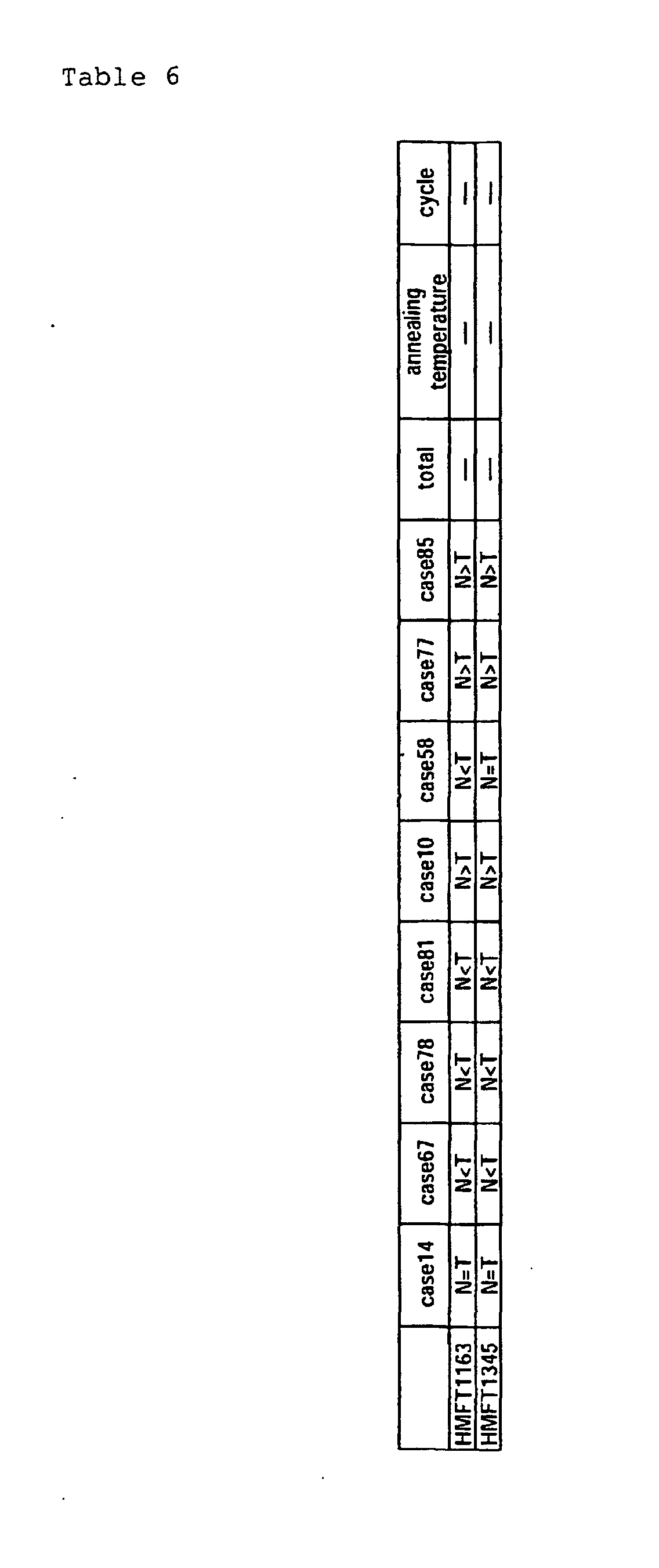
In not less than 3 cases out of 4 cases where no
mutation was observed in the β-catenin gene
differential expressions were noted and in not less
than 3 cases out of 4 cases where mutation was observed
in the β-catenin gene differential expressions were
noted in a manner opposite to the foregoing cases where
no mutation was observed. Such genes were judged to
have differential expressions depending on the presence
of the β-catenin mutation (Table 6). That is, the
genes described in Table 6 show such genotype that N<T
holds when no mutation was observed in β-catenin and
N>T holds when mutation was observed in β-catenin.
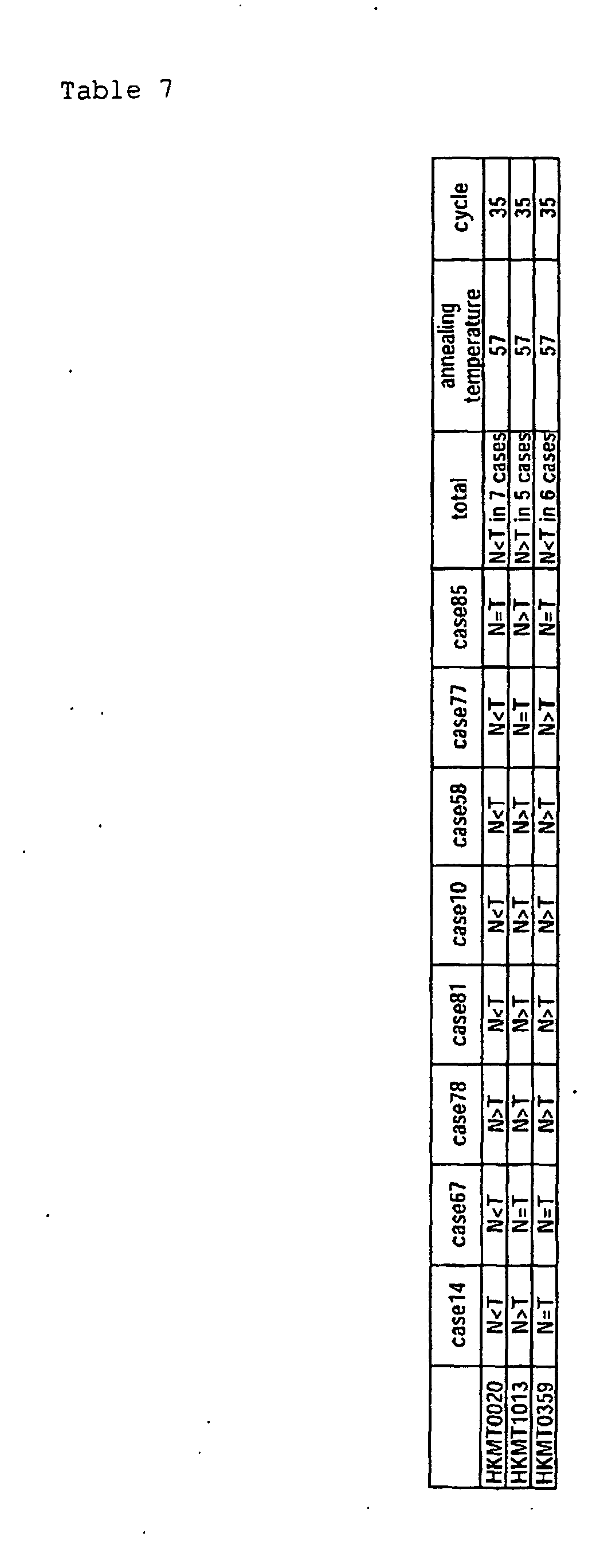
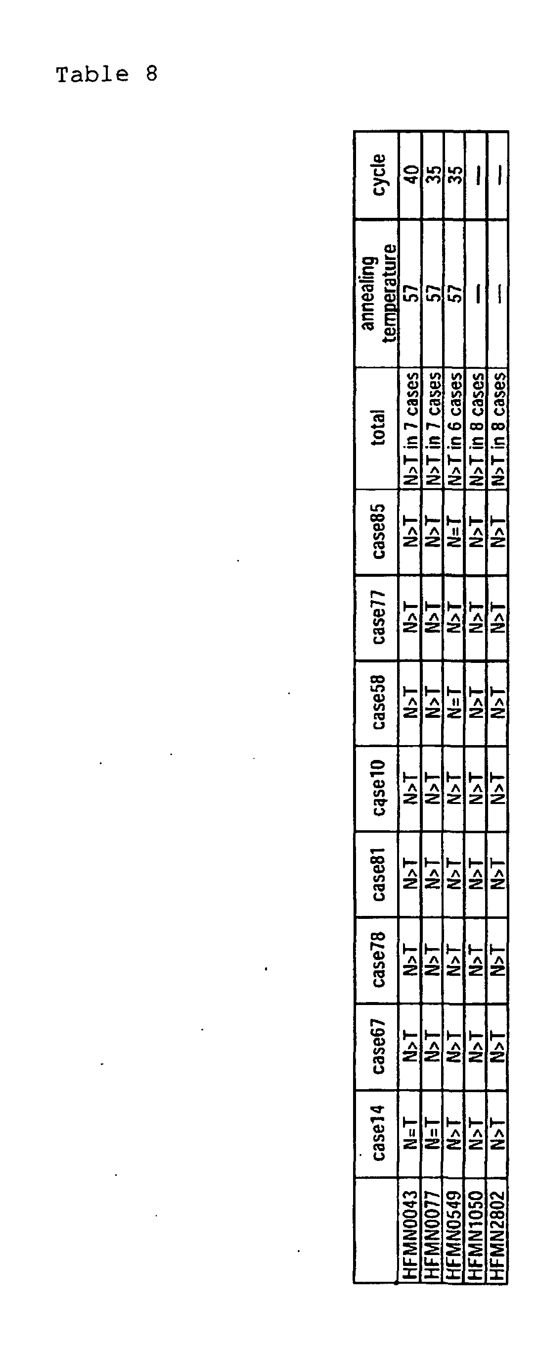


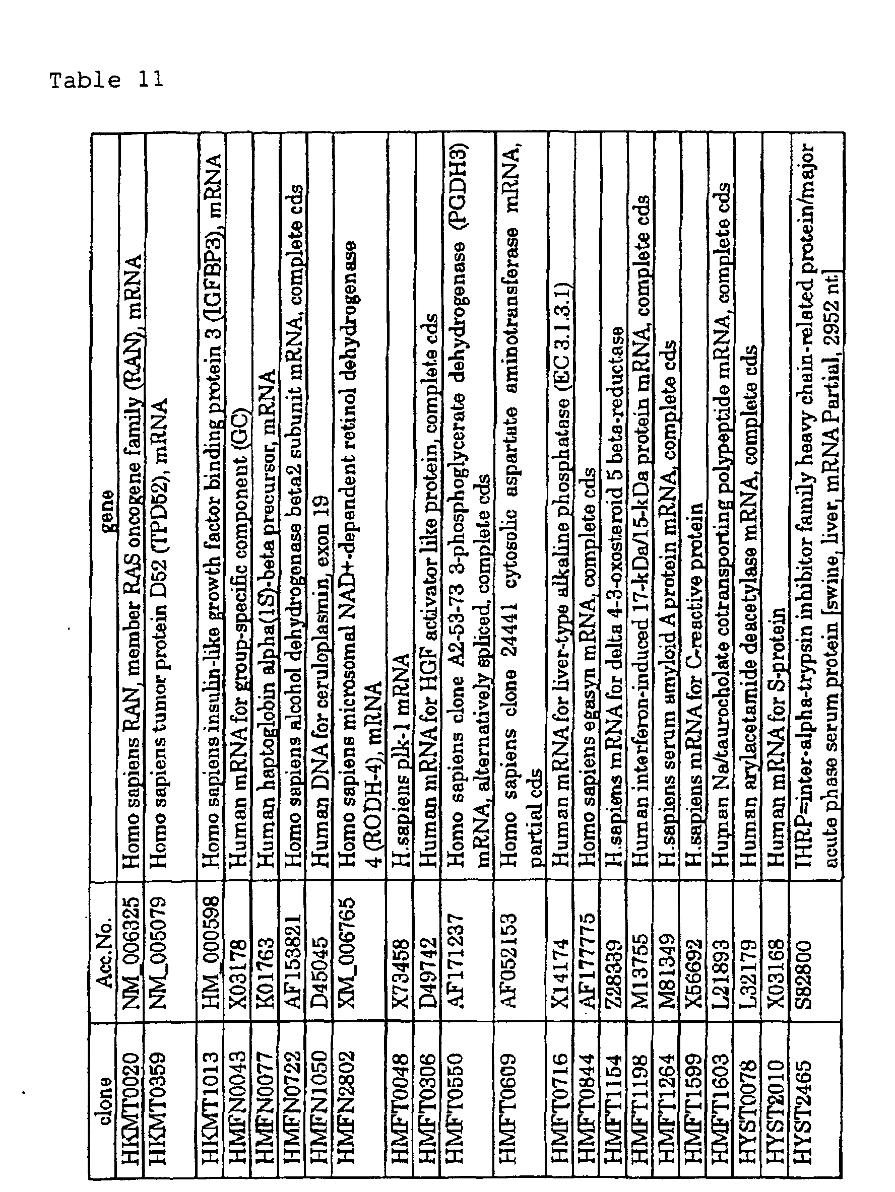
The genes described in Tables 7-10 are known genes.
Table 11 describes the names of genes for which
homologies to the named genes were recognized as a
result of homology search.
The genes described in Table 12 are also novel and
are genes for which differential expression levels are
noted hepatoblastoma and normal liver based on
comparison therebetween in accordance with the above-mentioned
criteria. The full length of each of these
genes was not sequenced except for HMFN0839. However,
as a result of both end sequencing and homology search,
they were found to have homology to the respective
known genes shown in Table 13.
| Case 14 | Case 67 | Case 78 | Case 81 | Case 10 | Case 58 | Case 77 | Case 85 | |
| HMFN 0217 | N=T | N=T | N>T | N>T | N>T | N>T | N>T | N>T |
| HMFN 0668 | N=T | N>T | N>T | N>T | N>T | N>T | N=T | N>T |
| HMFN 0839 | N=T | N>T | N>T | N>T | N>T | N>T | N=T | N>T |
| HMFN 2700 | N>T | N>T | N>T | N>T | N>T | N>T | N=T | N>T |
| HKMT 2698 | N>T | N=T | N>T | N=T | N>T | N=T | N>T | N>T |
| HYST 2198 | N<T | N<T | N<T | N<T | N=T | N>T | N<T | N>T |
| HYST 2935 | N<T | N<T | N<T | N=T | N<T | N<T | N=T | N=T |
| HMFT 1511 | N>T | N=T | N>T | N>T | N>T | N>T | N>T | N>T |
| clone | length | Acc. No. | gene |
| HMFN0217f | 510 | AP000356 | Homo sapiens genomic DNA, chromosome 22q11.2, clone KB1995A5 |
| HMFN0668f | 666 | 548898|sp| P36536 | SARA_MOUSE GTP-binding protein SARA |
| HMFN0668r | 597 | AW976405 | EST388514 MAGE resequences, MAGN Homo sapiens cDNA, mRNA sequence |
| HMFN0839f | 531 | T24032 | hypothetical protein R07E3.5 Caenorhabditis elegans |
| HMFN0839r | 607 | BE349224 | Homo sapiens cDNA clone |
| HMFN2700f | 429 | ||
| HKMT2698f | 420 | AR083267 | Sequence 19 in USP No. 5976837 |
| HKMT2698r | 517 | AR083267 | Sequence 19 in USP No. 5976837 |
| HYST2198f | 484 | AK024066 | Homo sapiens cDNA FLJ14004 fis., clone Y79AA1002351 |
| HYST2935f | 564 | BF345917 | Homo sapiens cDNA clone 602017918F1 |
| HMFT1511f | 401 | AW786459 | Sus scrofa cDNA 5', mRNA sequence |
| HMFT1511r | 466 | AP000359 | Homo sapiens genomic DNA, chromosome 22q11.2, clone KB63E7 |
Similarly to Example 6, a semi-quantitative RT-PCR
was performed and the expression of Plk-1 was confirmed
in 8 pairs (16 samples). Results are shown in Figure 4.
Here, the expression levels of the Plk-1 gene were
normalized against the expression of GAPDH. In the
figure Lane N corresponds to the normal liver tissue
and Lane T corresponds to the hepatoblastoma. However,
Case 77 corresponds to the hepatocellular carcinoma
tissue. The expression level of Plk-1 was clearly
greater in the tumor tissue than in the normal tissue.
Total RNA (25 µg) was prepared from human
hepatoblastoma, hepatocellular carcinoma and normal
liver according to a conventional method (Chomczynski
P., Sacchi N., Anal. Biochem., 162: 156-9, 1987). This
was electrophoresed on 1% agarose gel containing
formaldehyde and fixed through UV cross-linking.
Hybridization was conducted in the presence of a human
Plk-1 cDNA fragment labeled with α-32P-dCTP Random (976
bases) using random priming: 65 ° C in a solution
containing 1M NaCl, 1% SDS, 7.5% dextran sulfate, 100 µ
g/ml thermally denatured salmon serum. Results are
shown in Fig. 5. In the figure 67t, 93t and 58t are
the human hepatoblastoma tissue and 67h, 93h and 58h
are the corresponding normal liver tissues, 77t is the
hepatocellular carcinoma tissue, and 77h is the
corresponding normal liver tissue. It is understood
that the expression of Plk-1 is higher in both the
human hepatoblastoma and the hepatocellular carcinoma
tissues than the normal liver tissue.
A Kaplan-Meier analysis of 59 cases of
hepatoblastoma patients was attempted using the
expression levels of Plk-1 as indices around the
central value of Plk-1 expression level=13. The
results of analysis are shown in Fig. 6. The patients
with high Plk-1 expression showed more unfavorable
prognosis than do the patients with low PLk-1
expression. The survival rate of the former group is
55.9% while that of the latter group is 87.0% (with
statistical significance being p=0.042). Therefore, it
has been demonstrated that the expression level of the
Plk-1 gene is an excellent marker for the diagnosis of
prognosis of human hepatoblastoma.
As described above, this invention discloses gene
sequences displaying differential expression levels
between hepatoblastoma and normal liver and will enable
the provision of their genetic information and the
diagnosis of hepatoblastoma by utilizing the nucleic
acids and proteins according to the invention derivable
from the genetic information as probes and PCR primers.

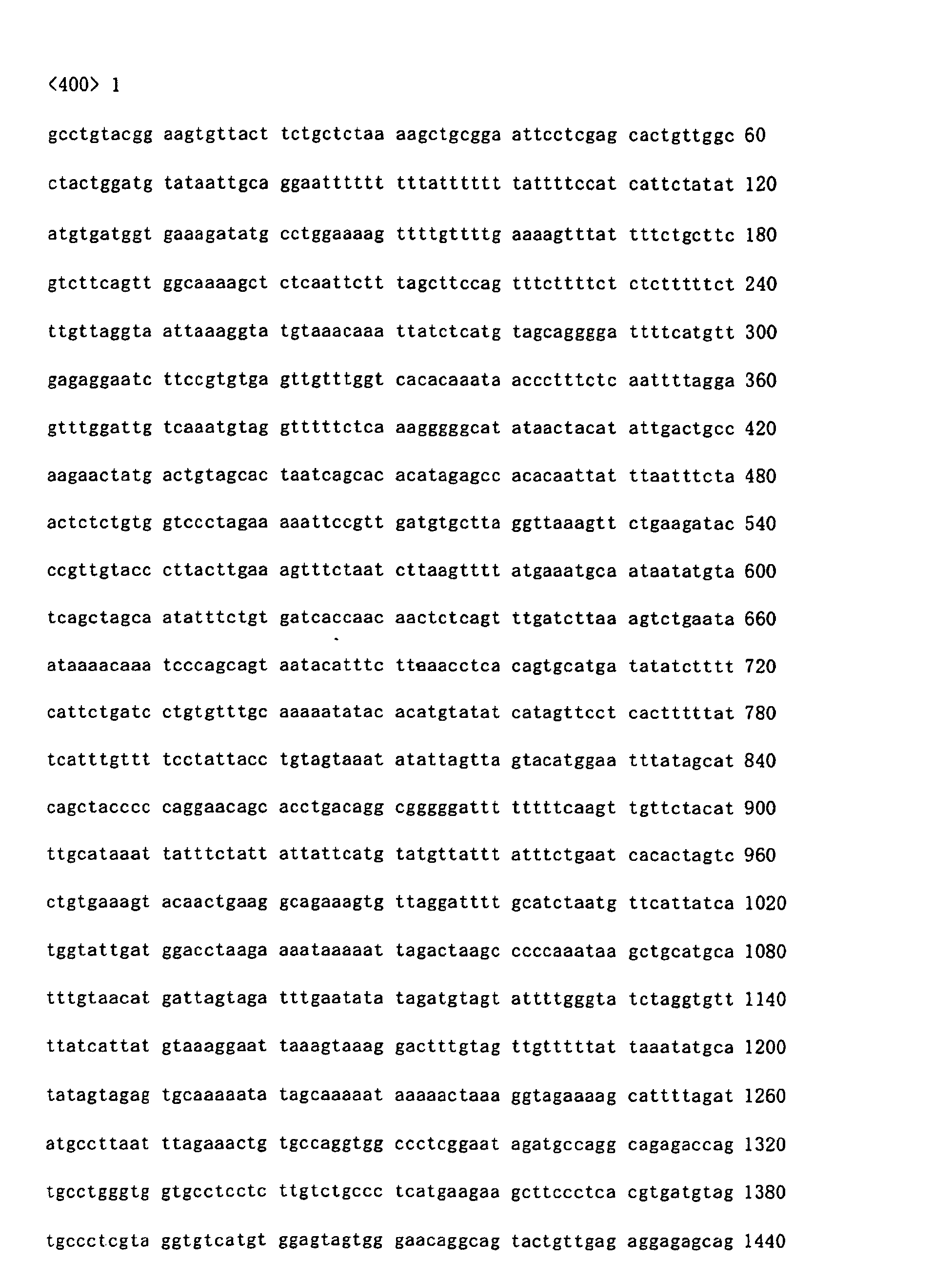

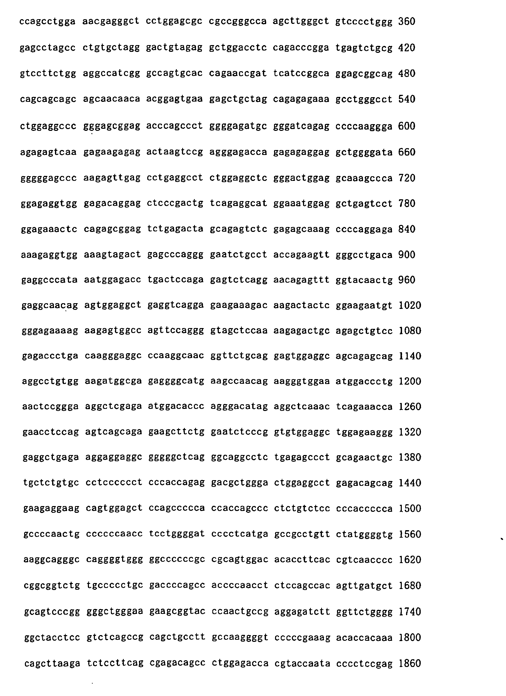
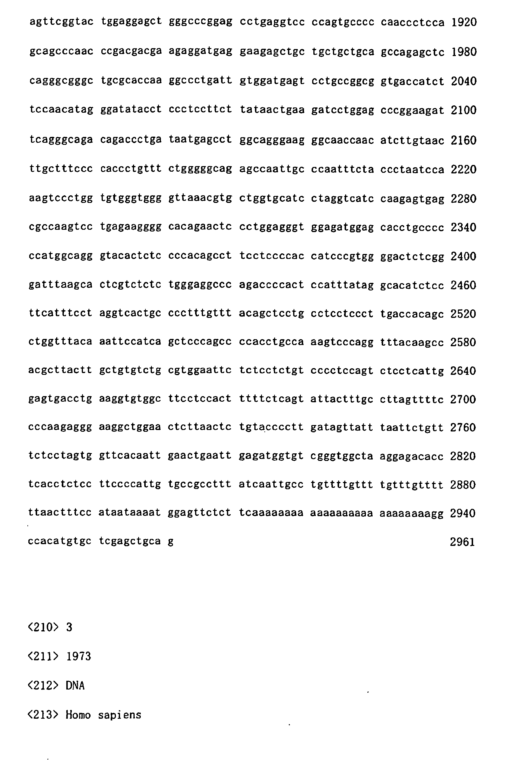
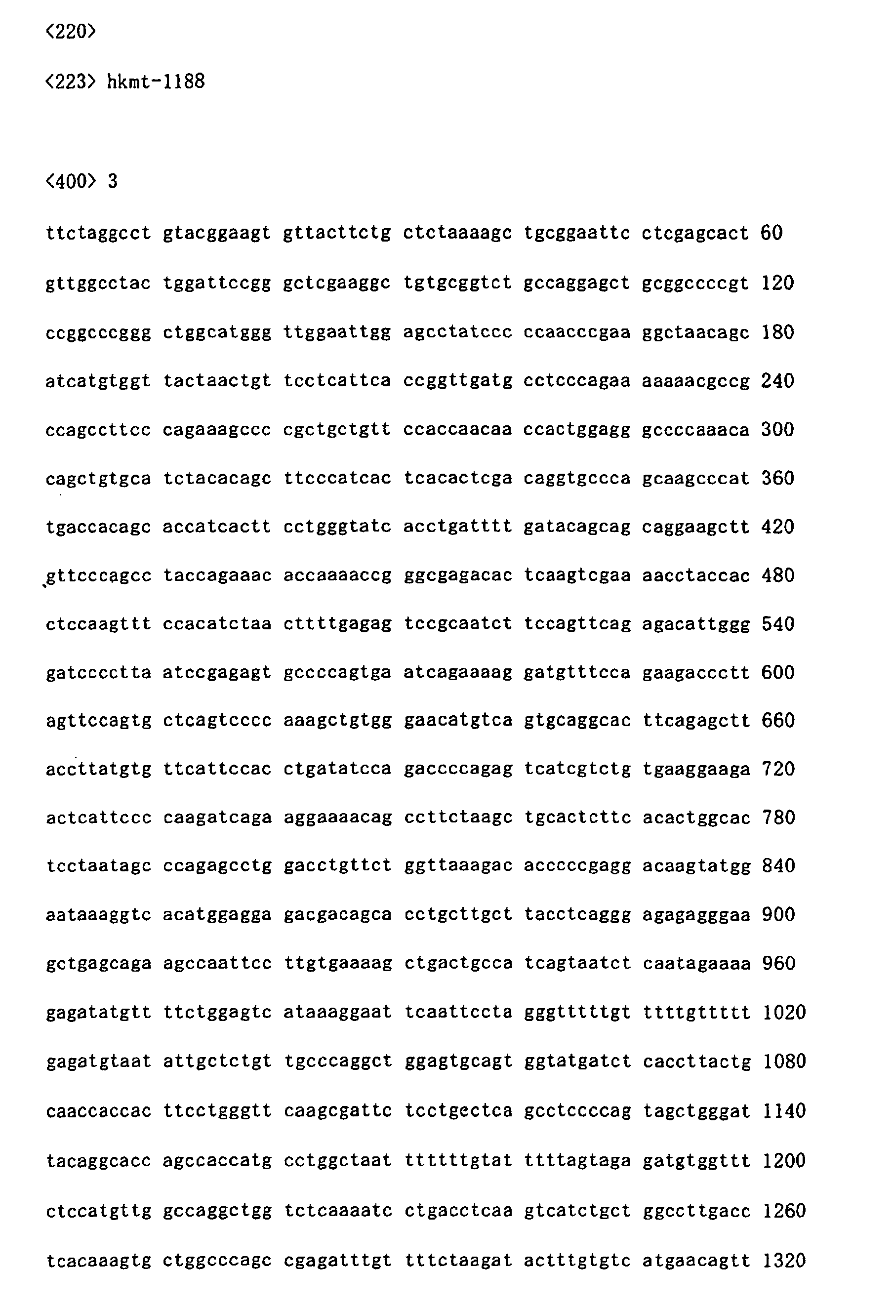
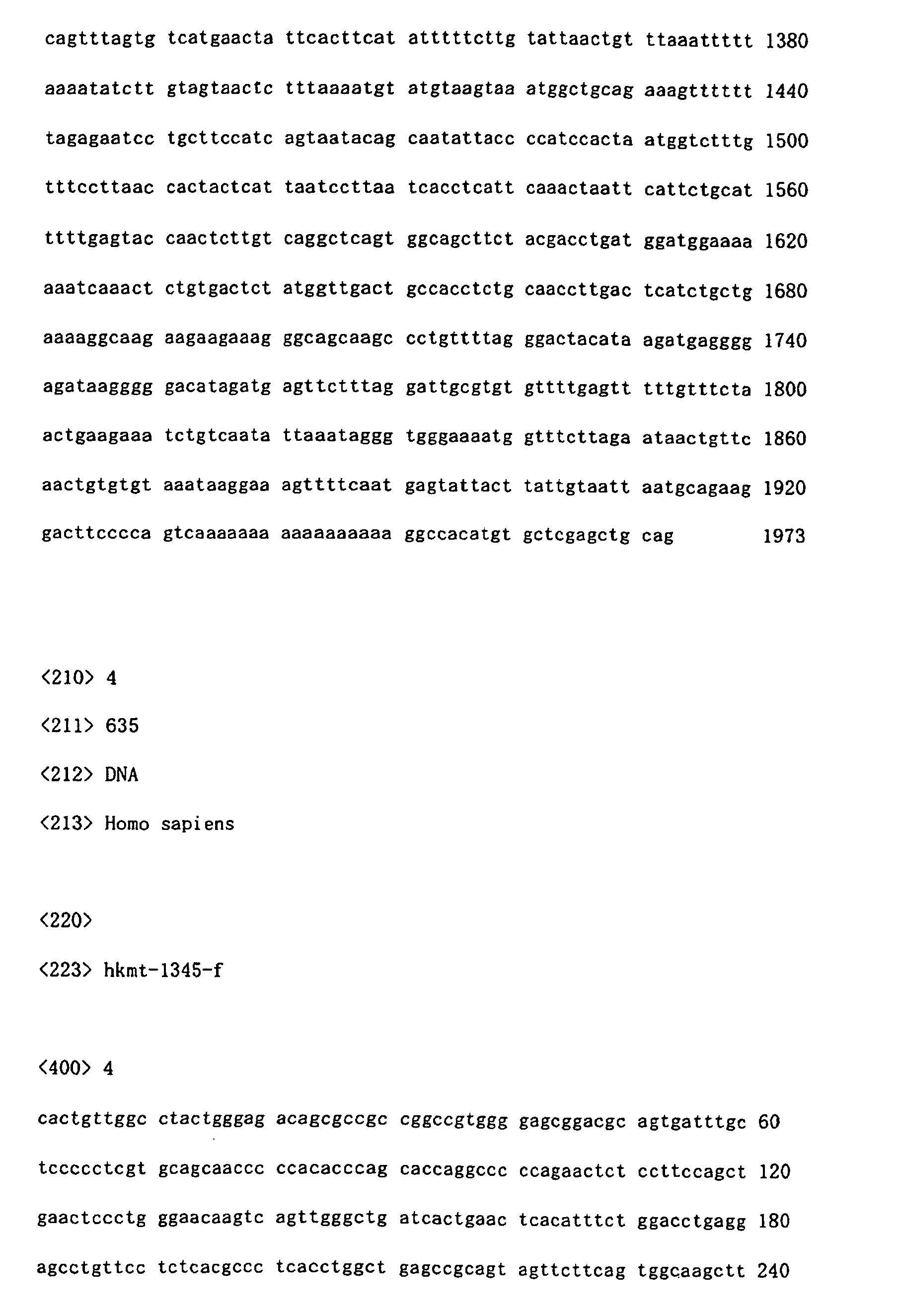
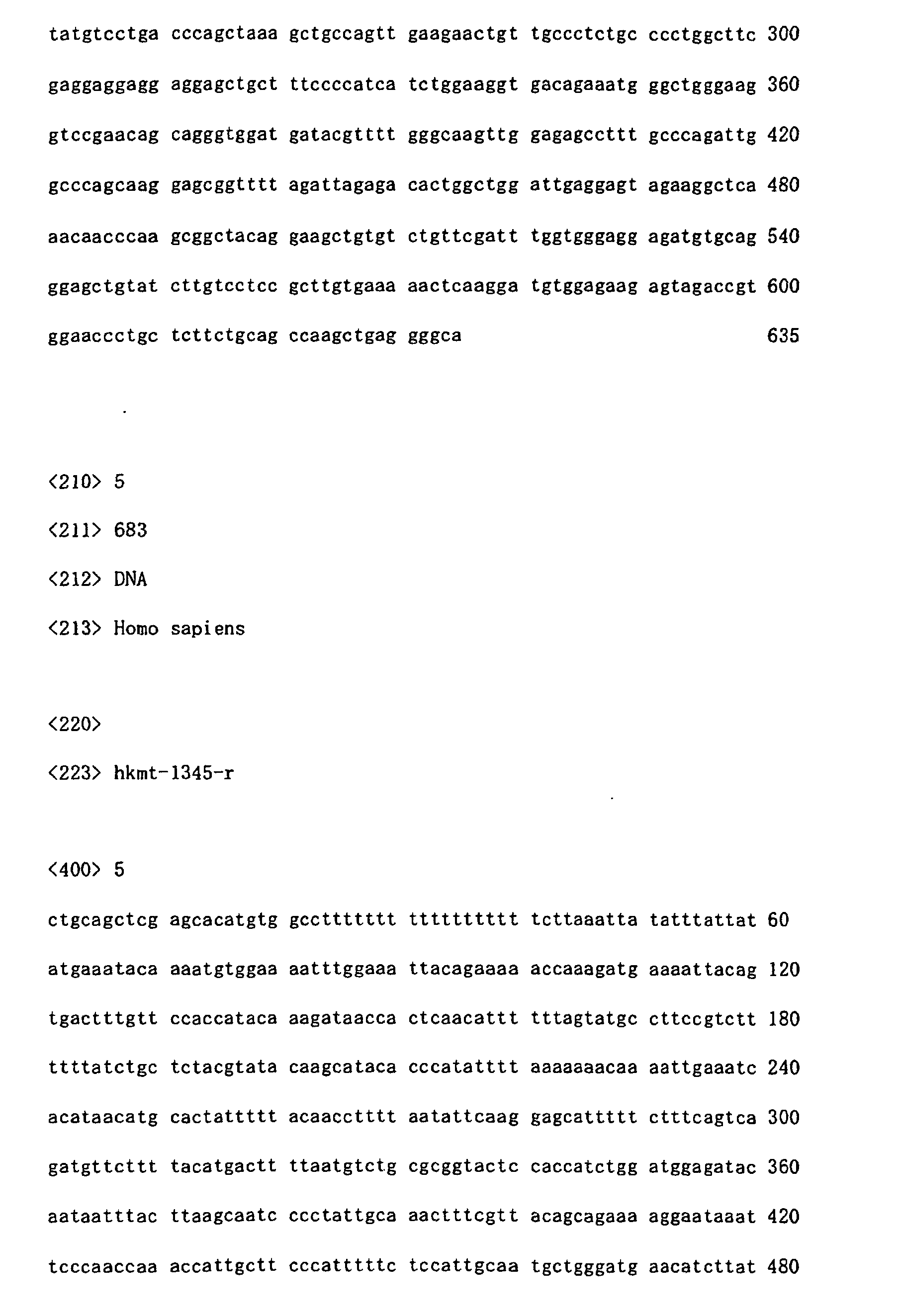
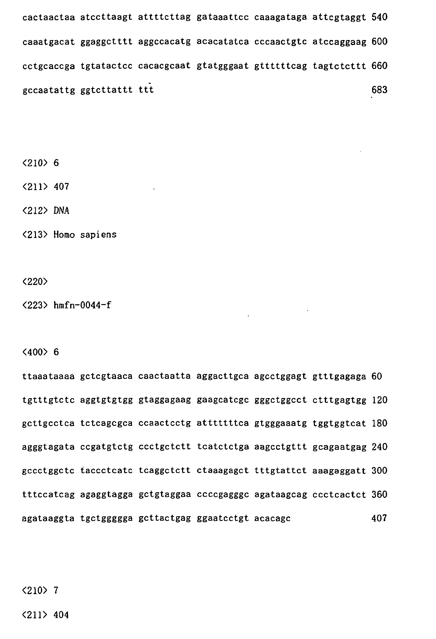
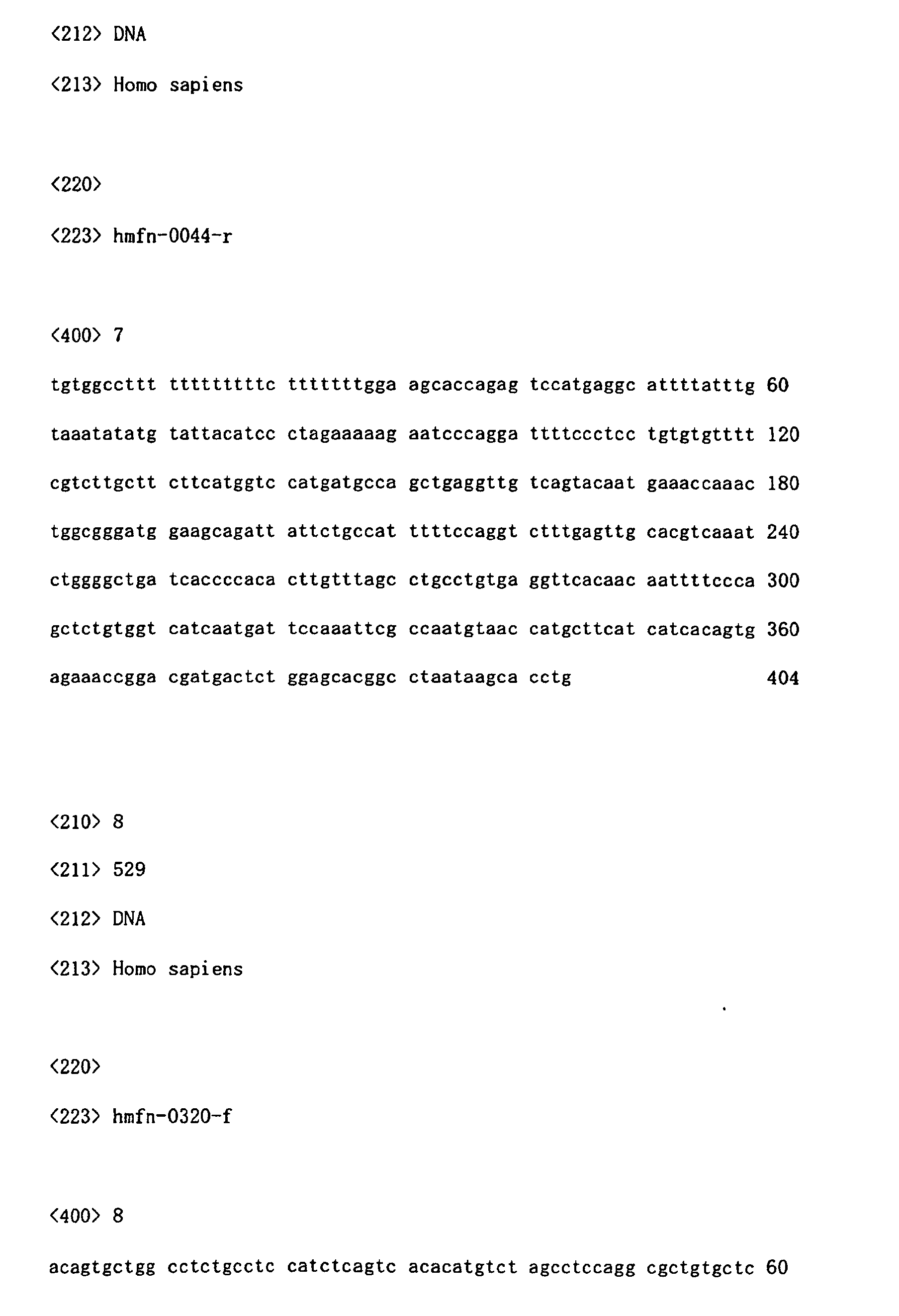
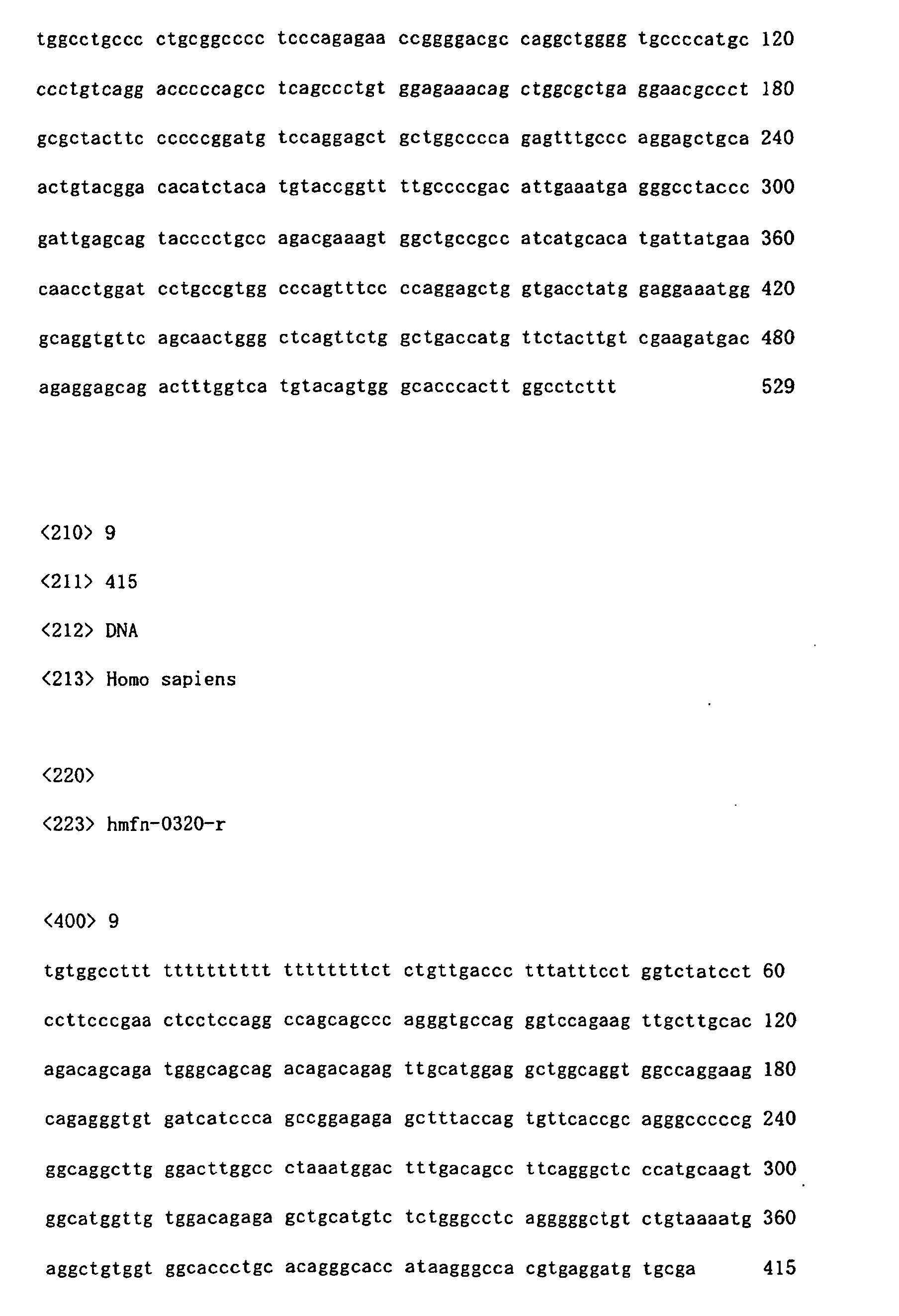
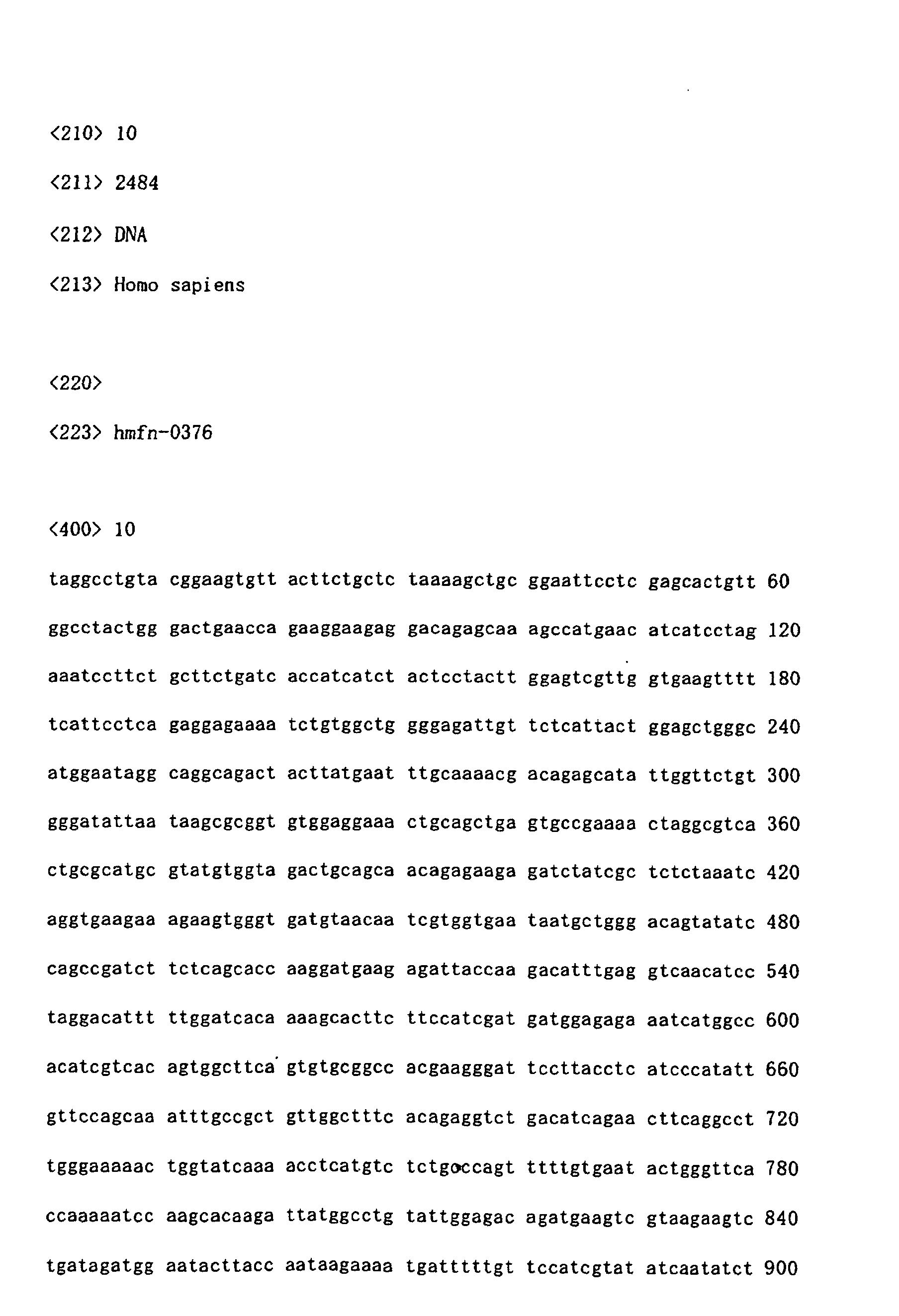

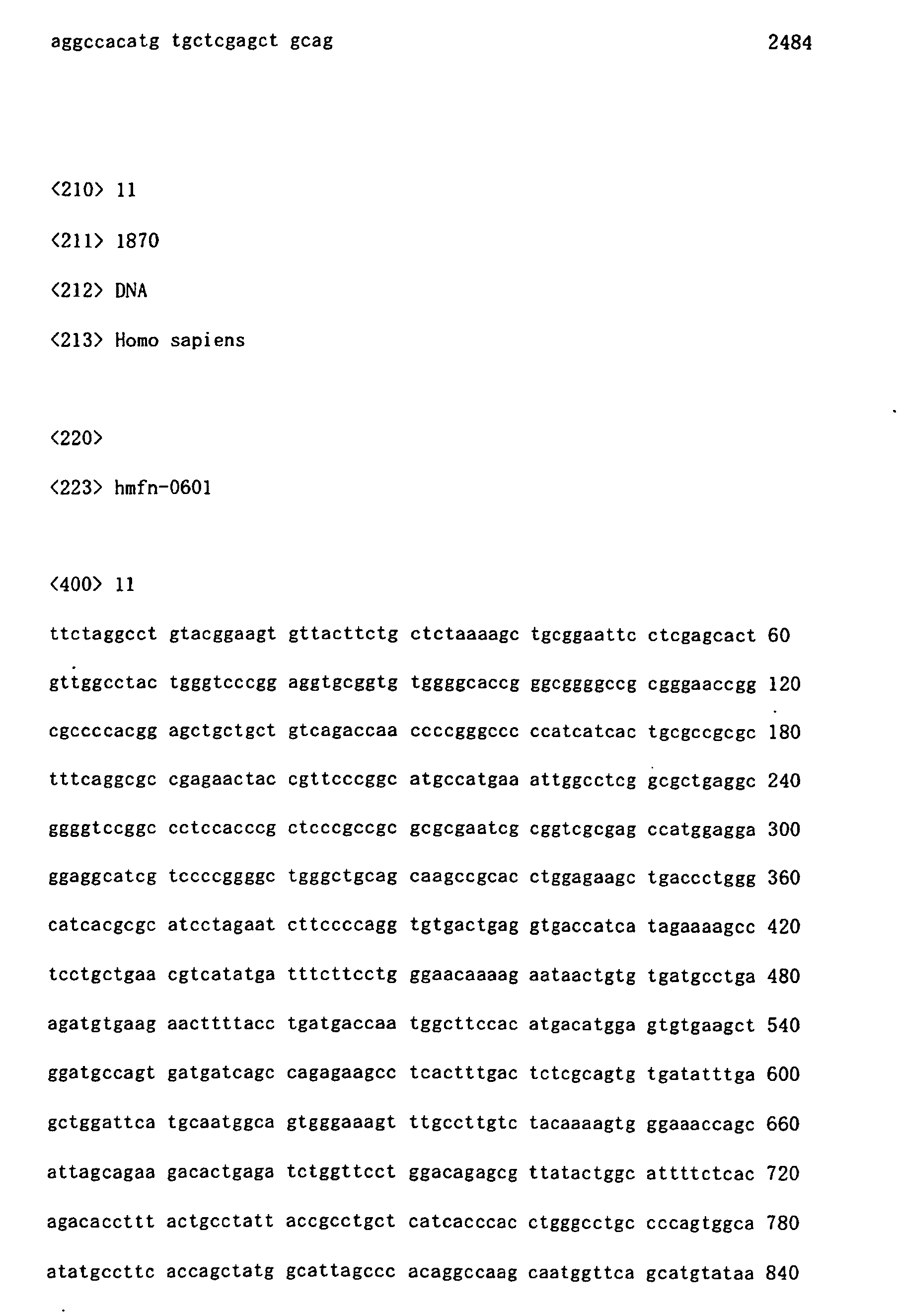


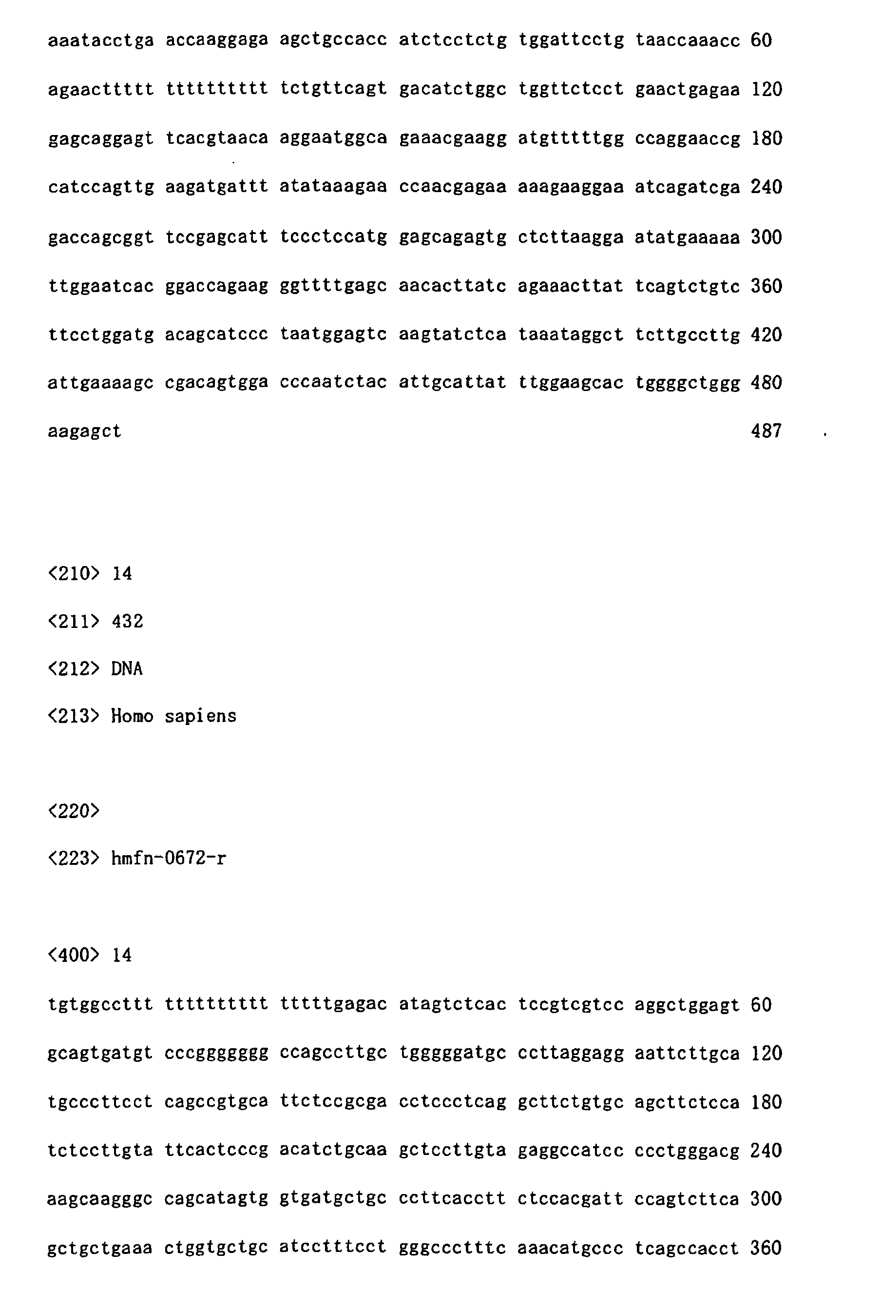
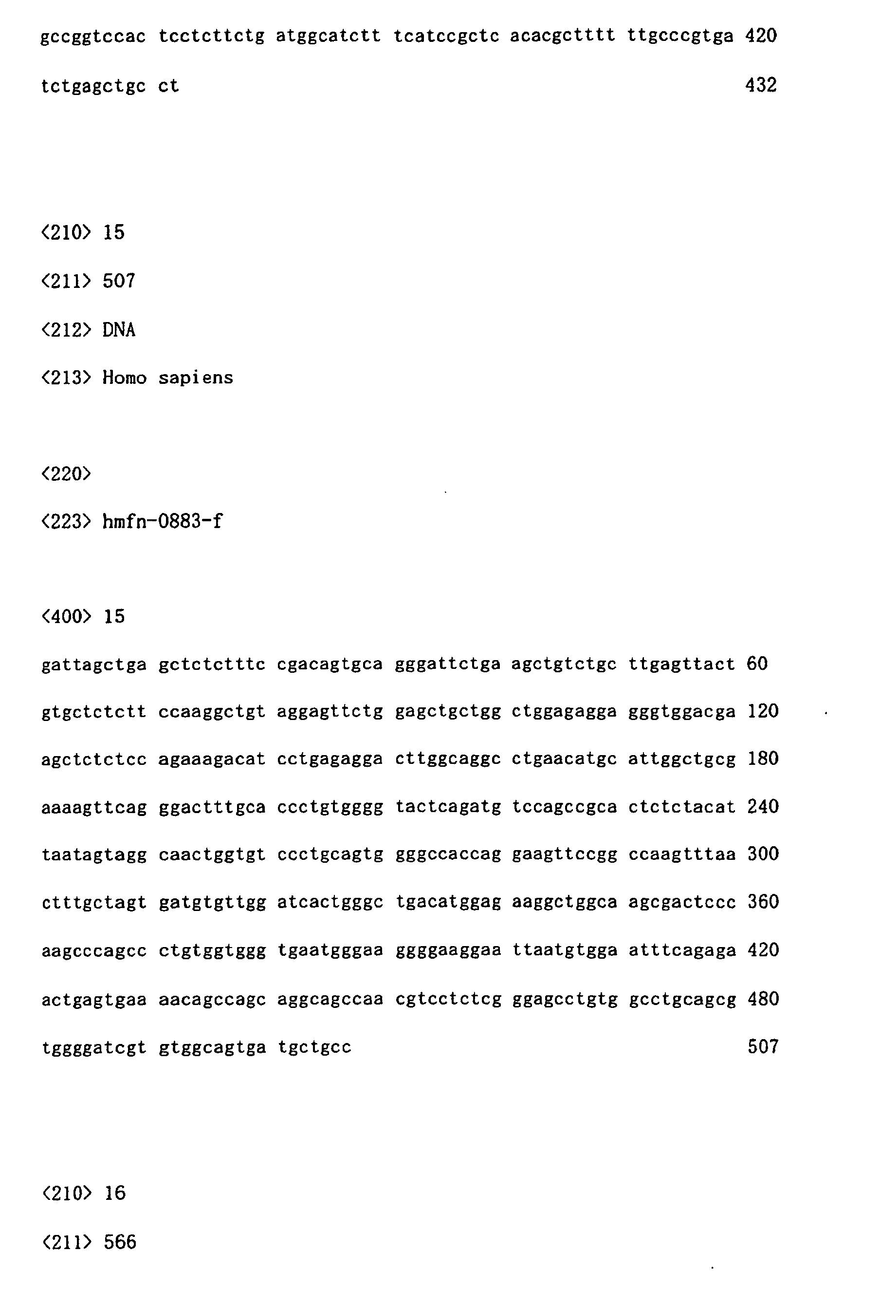

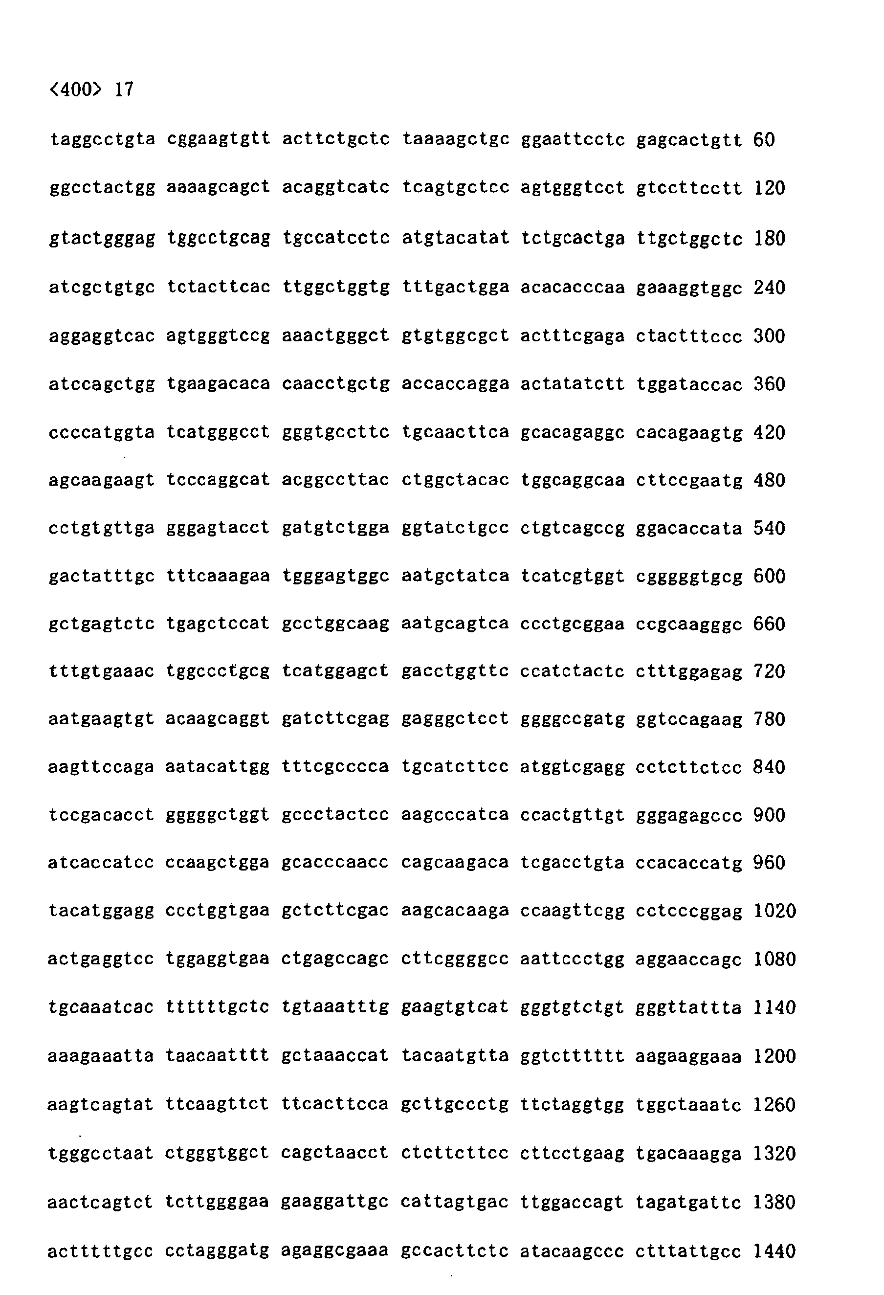


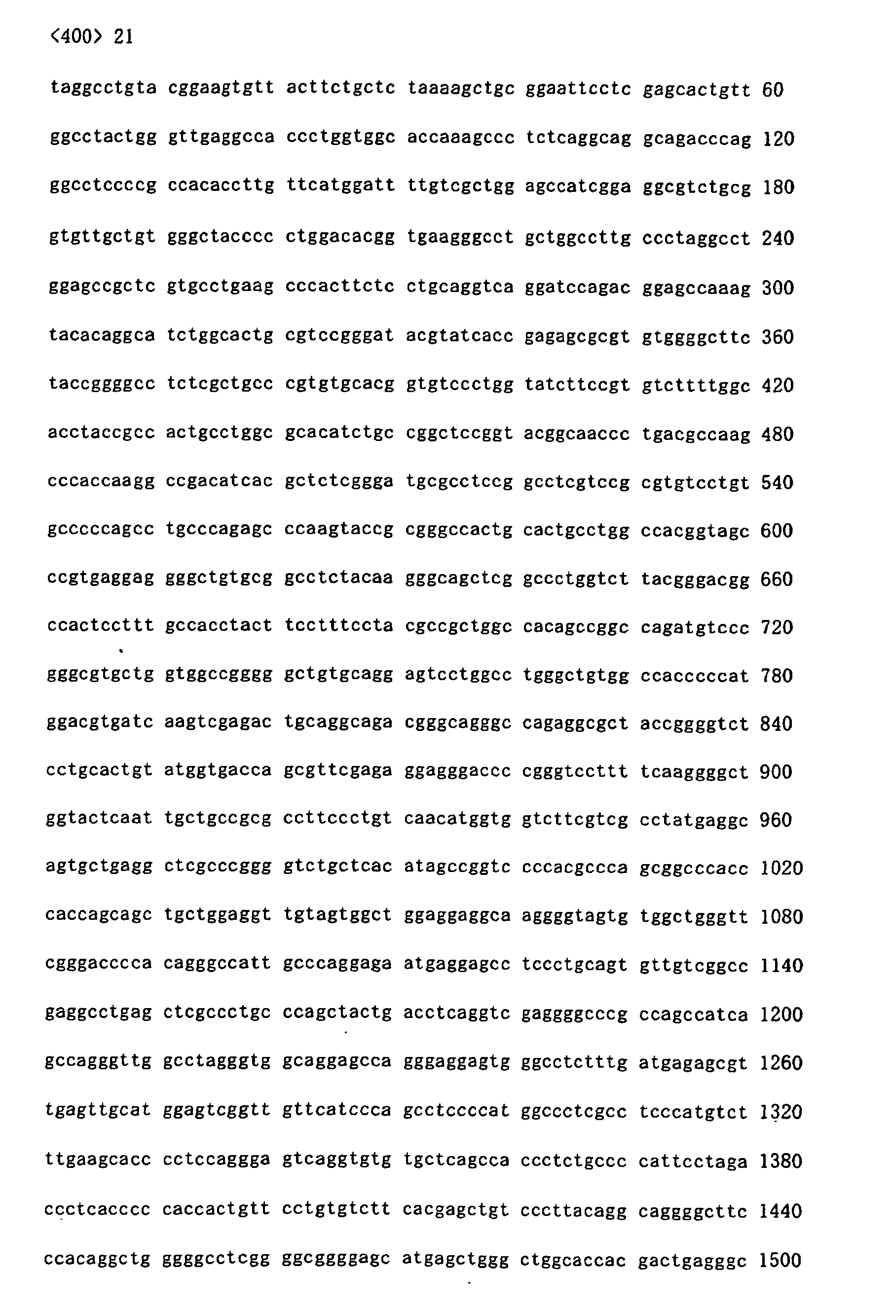

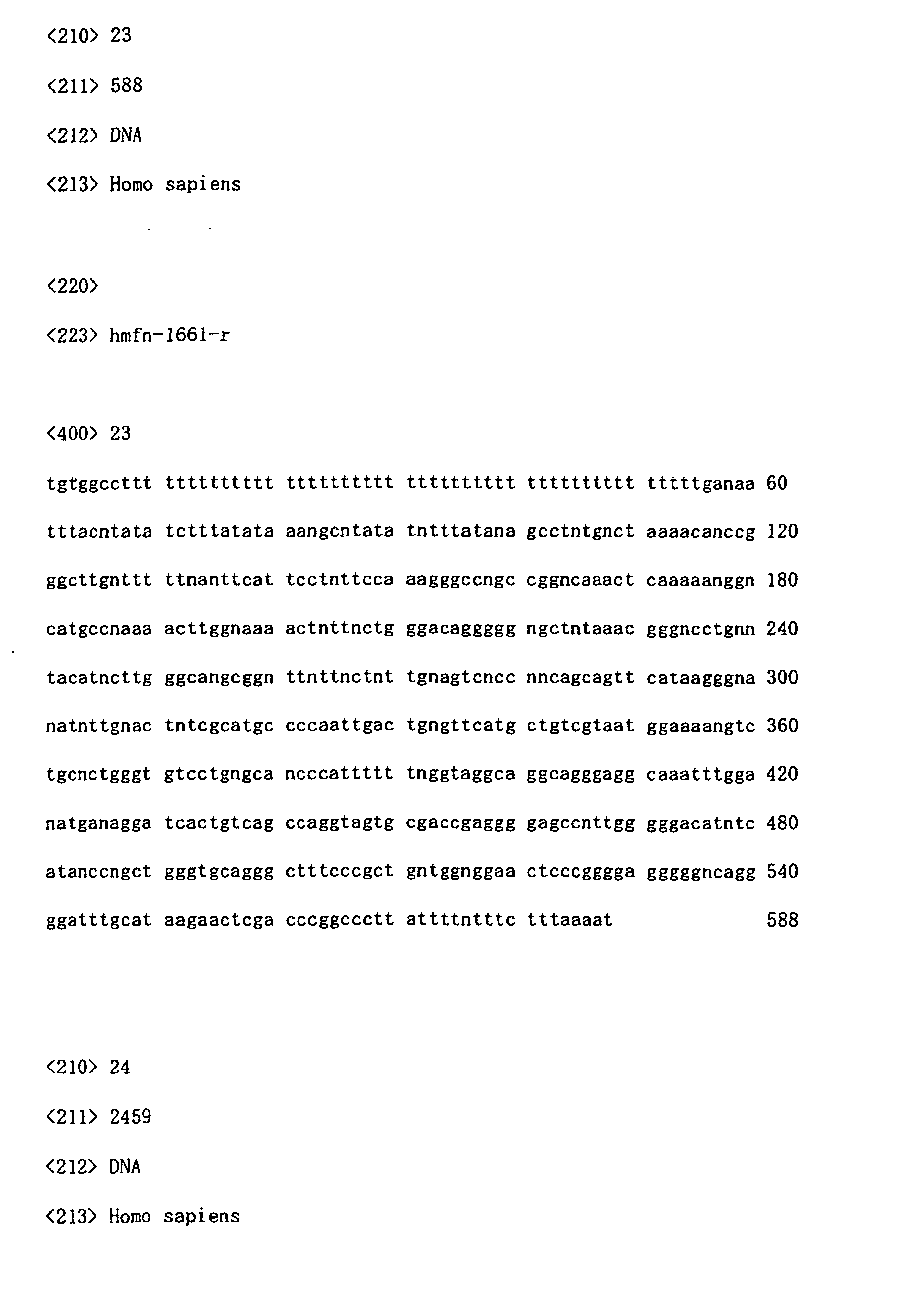


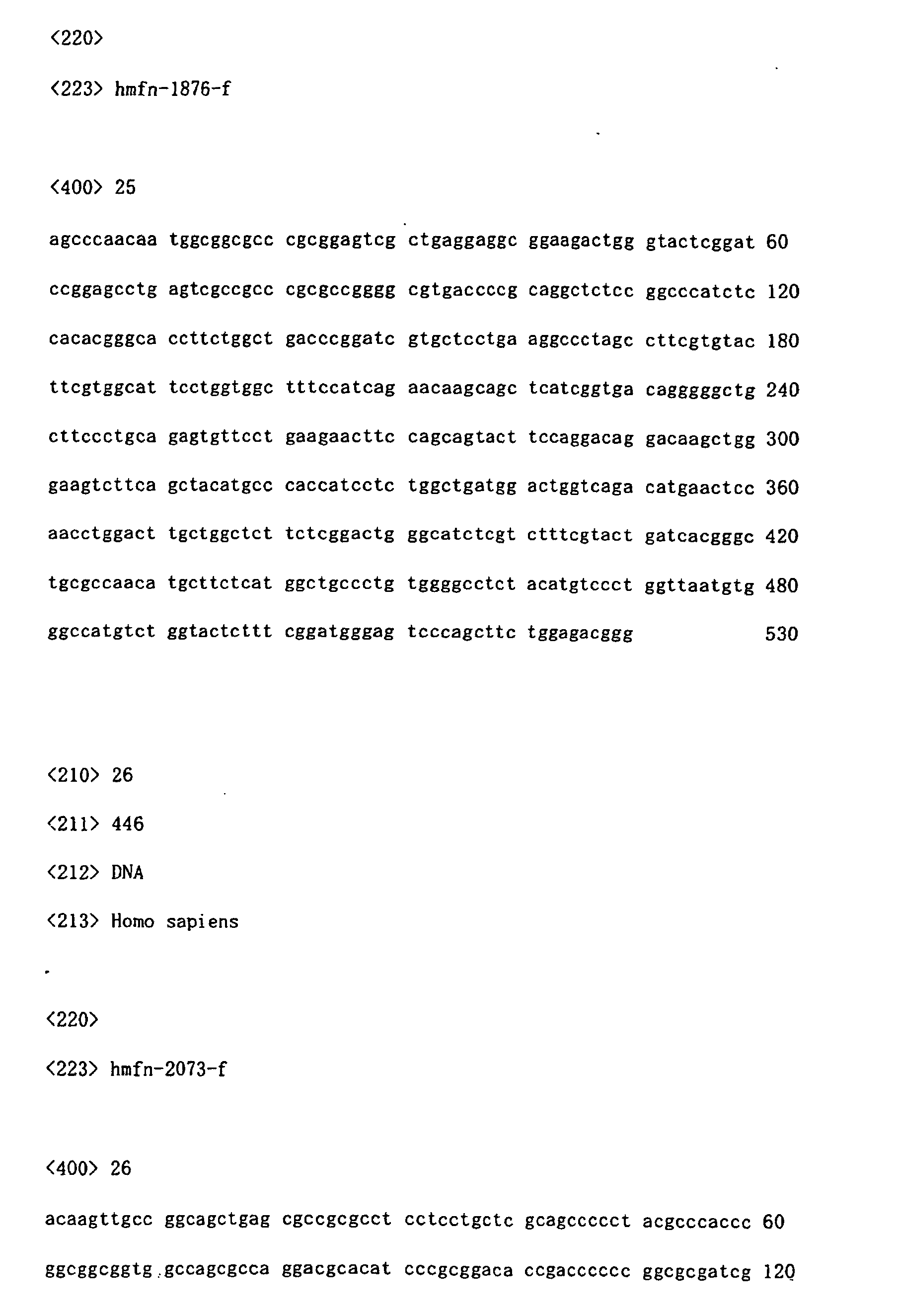
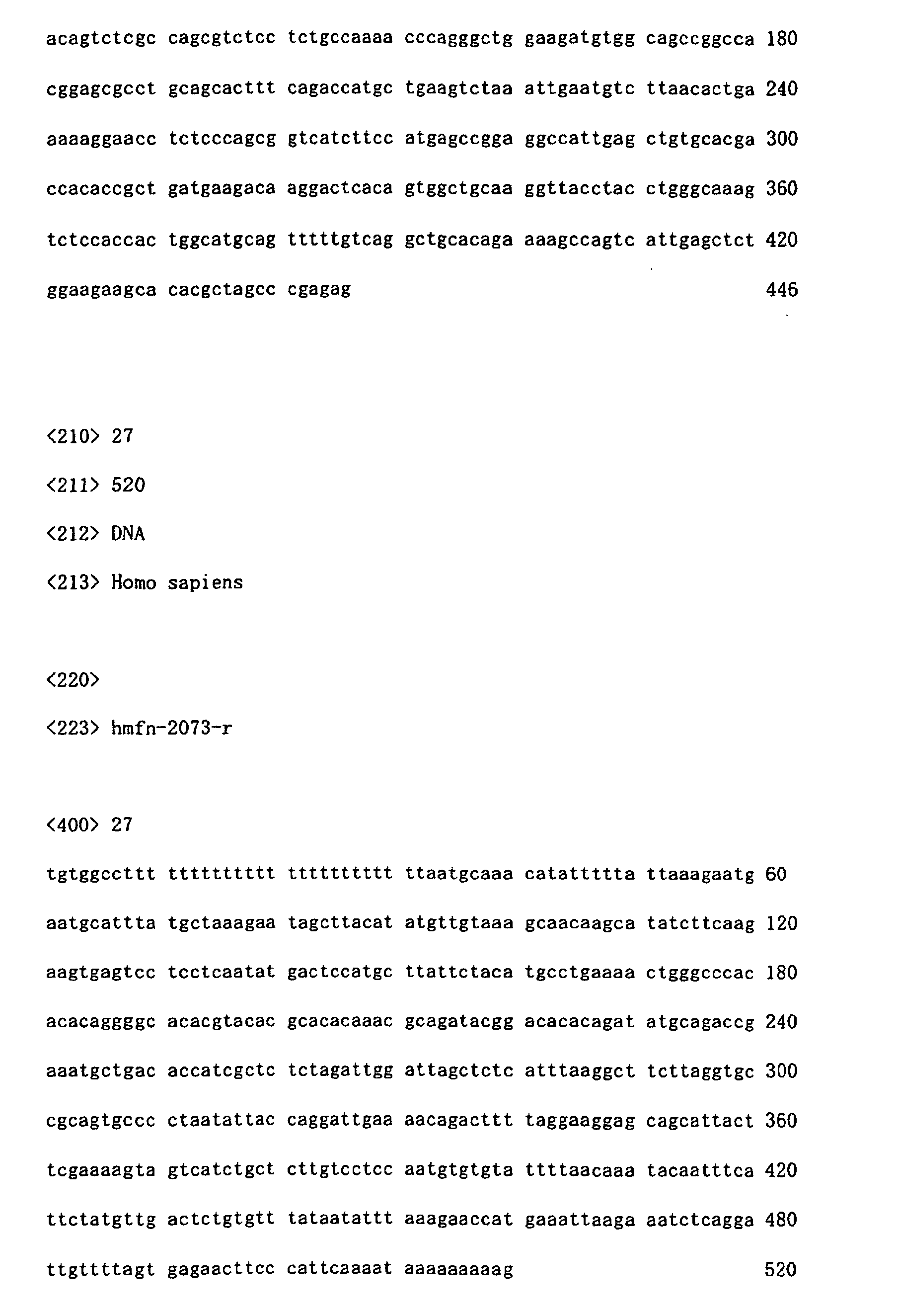


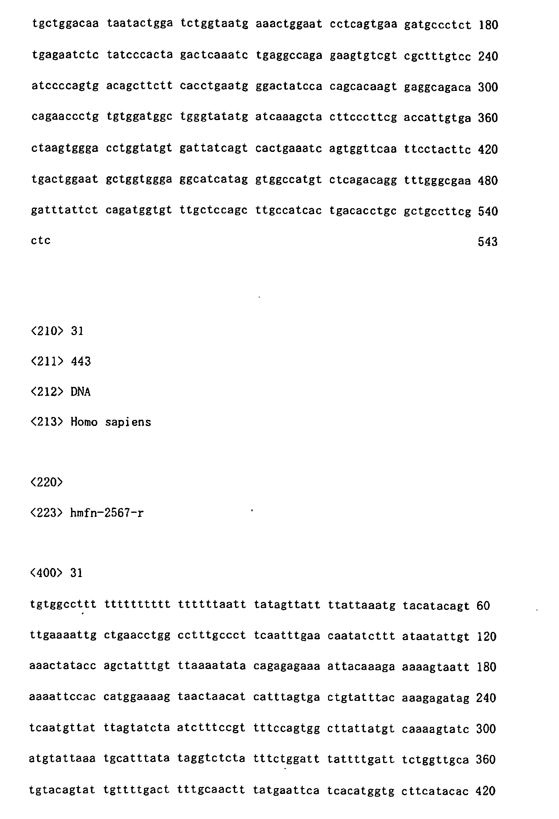

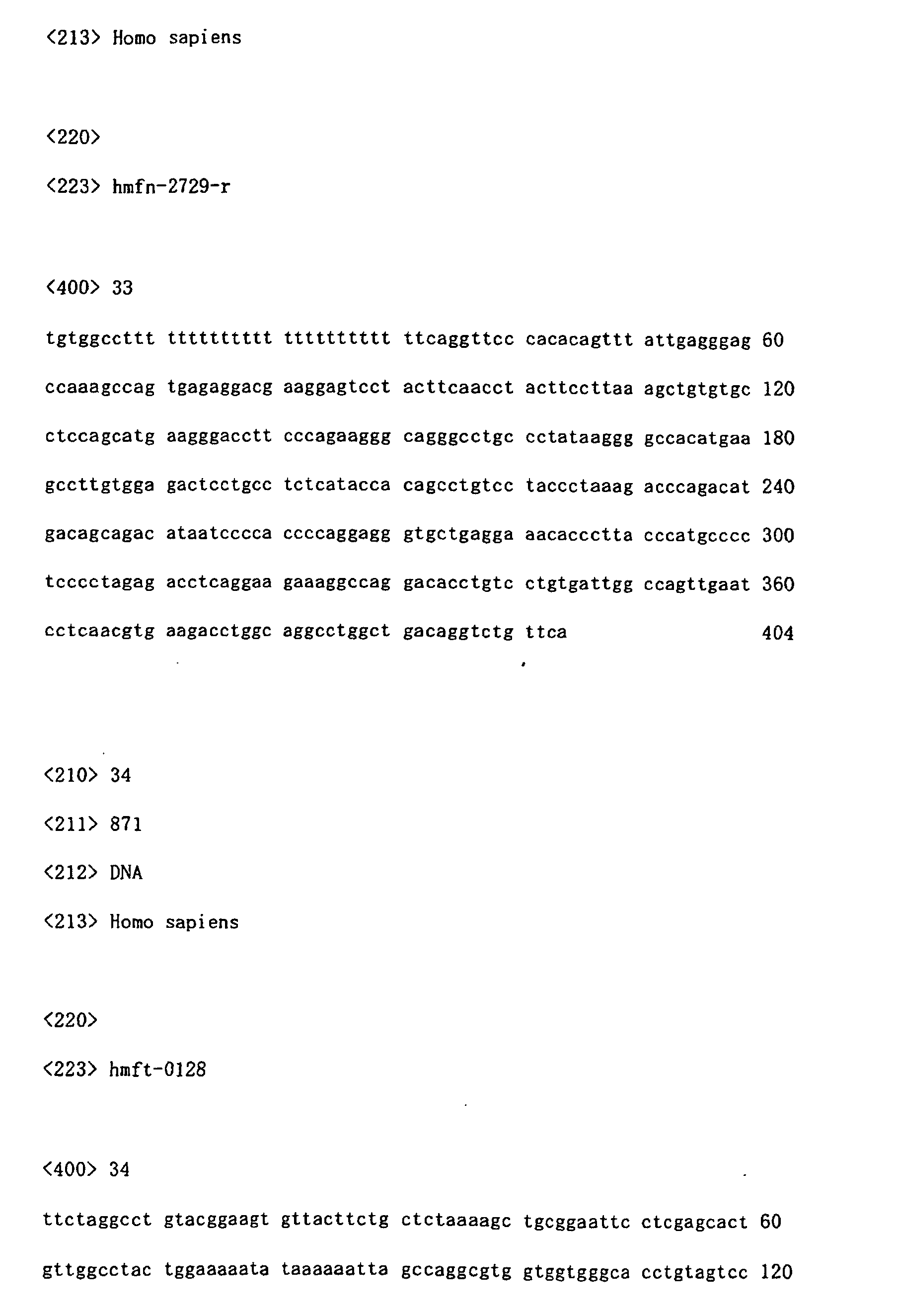

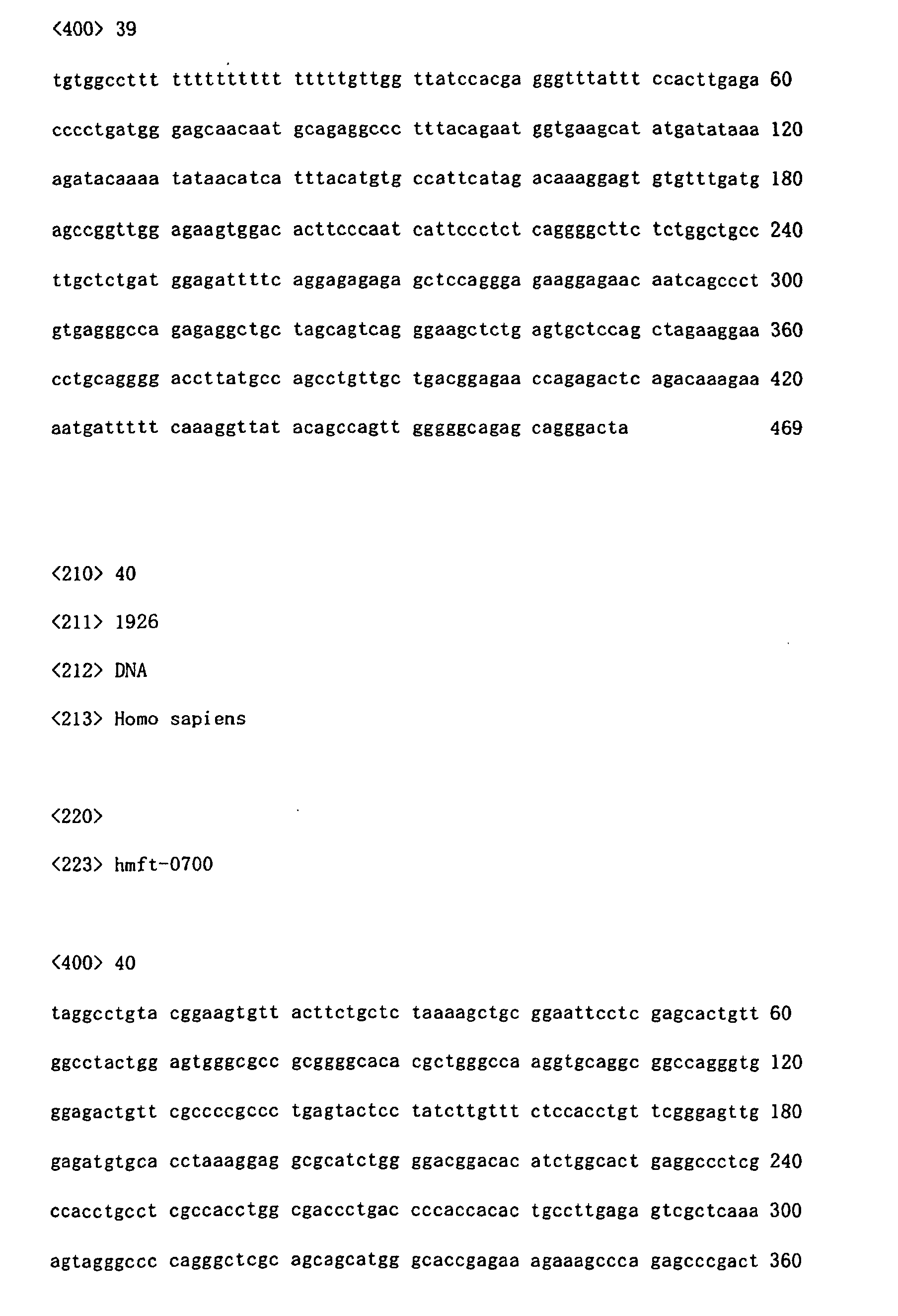
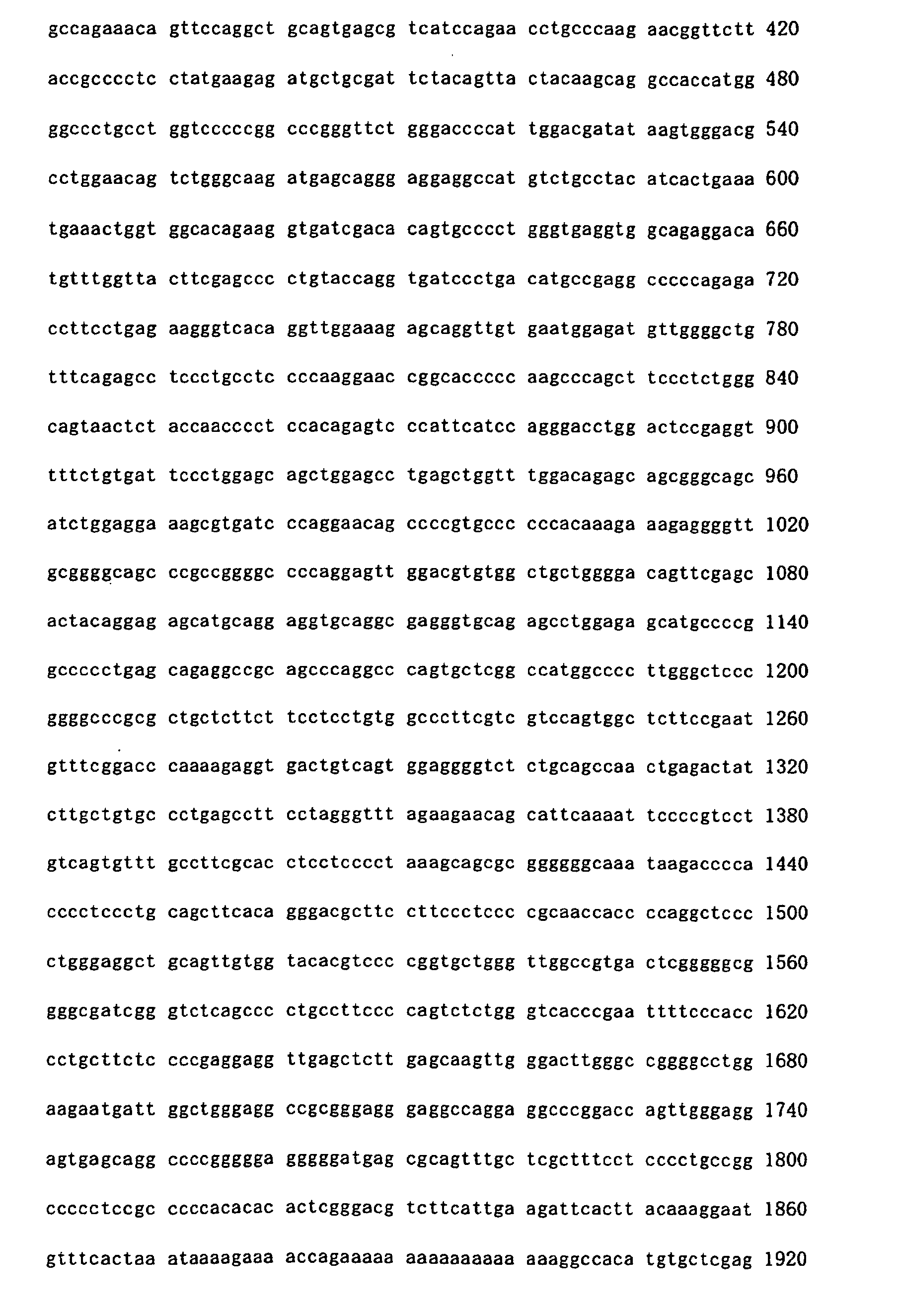
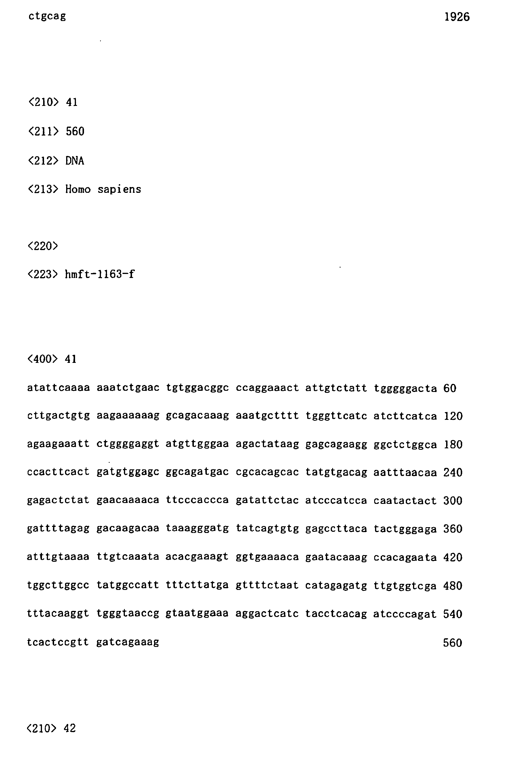


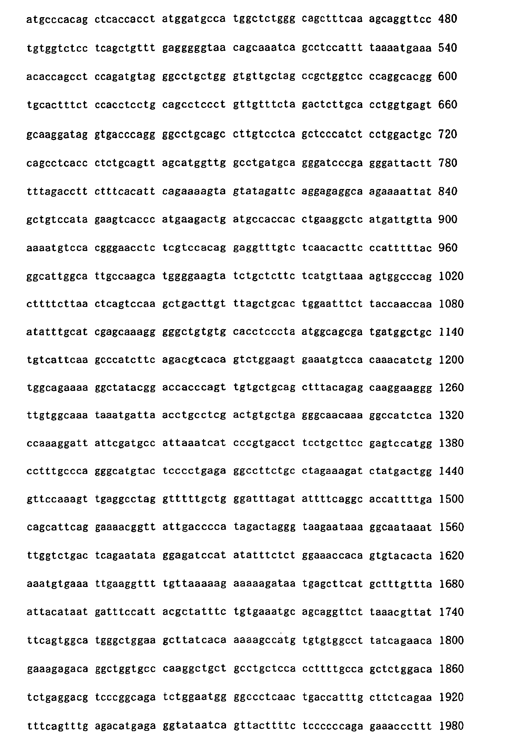


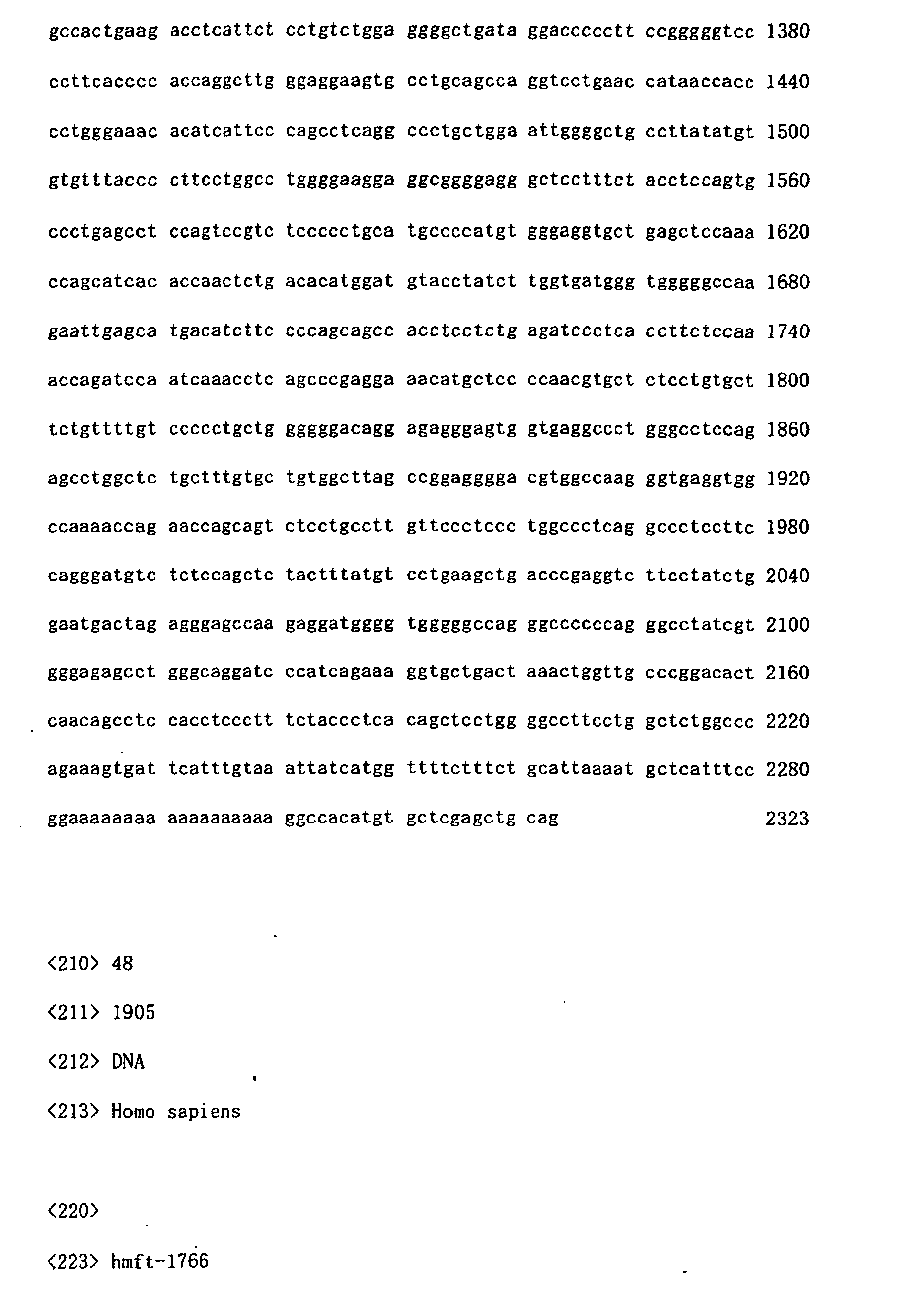
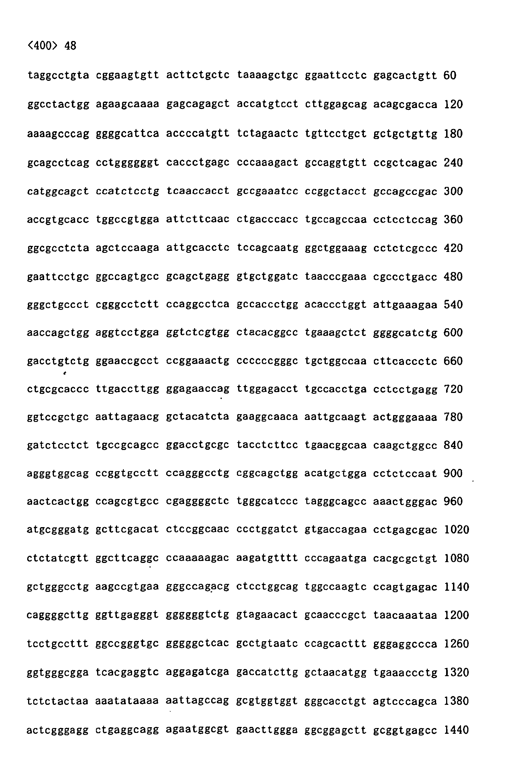
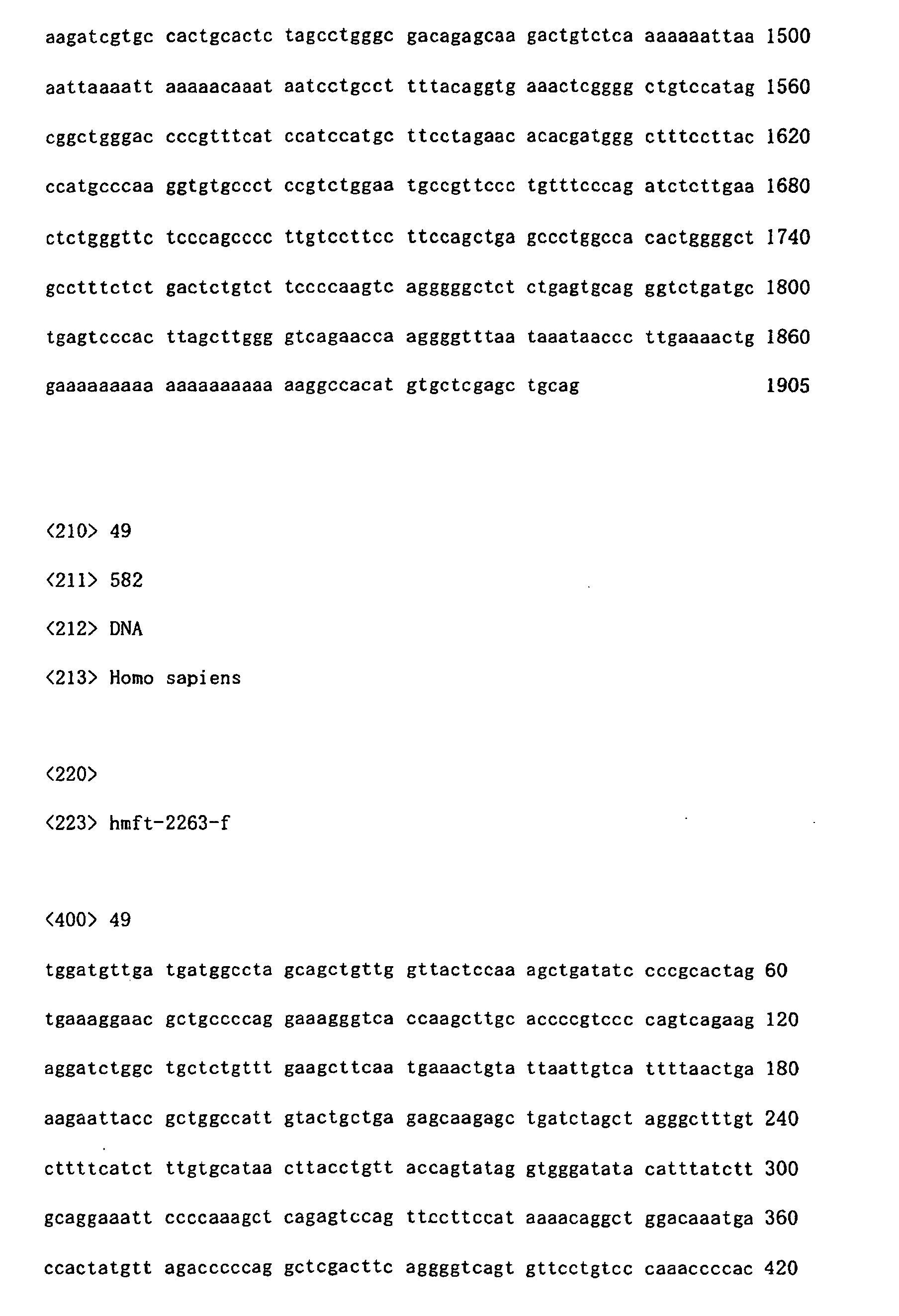
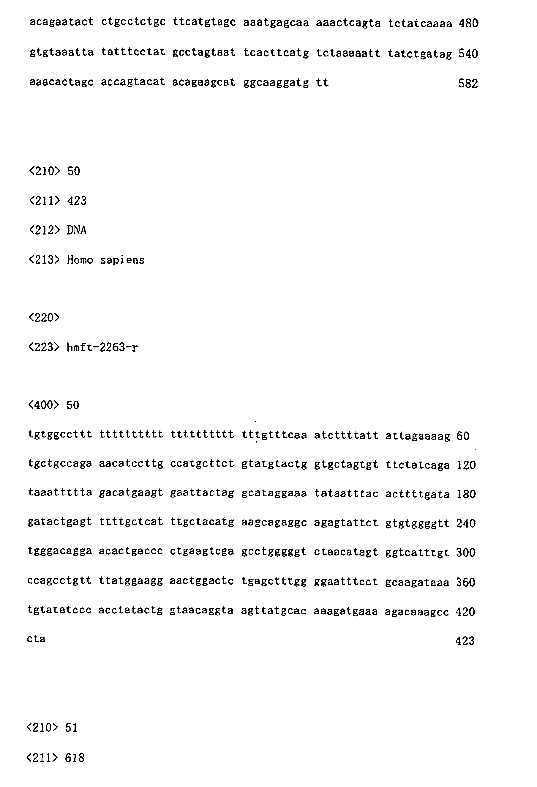
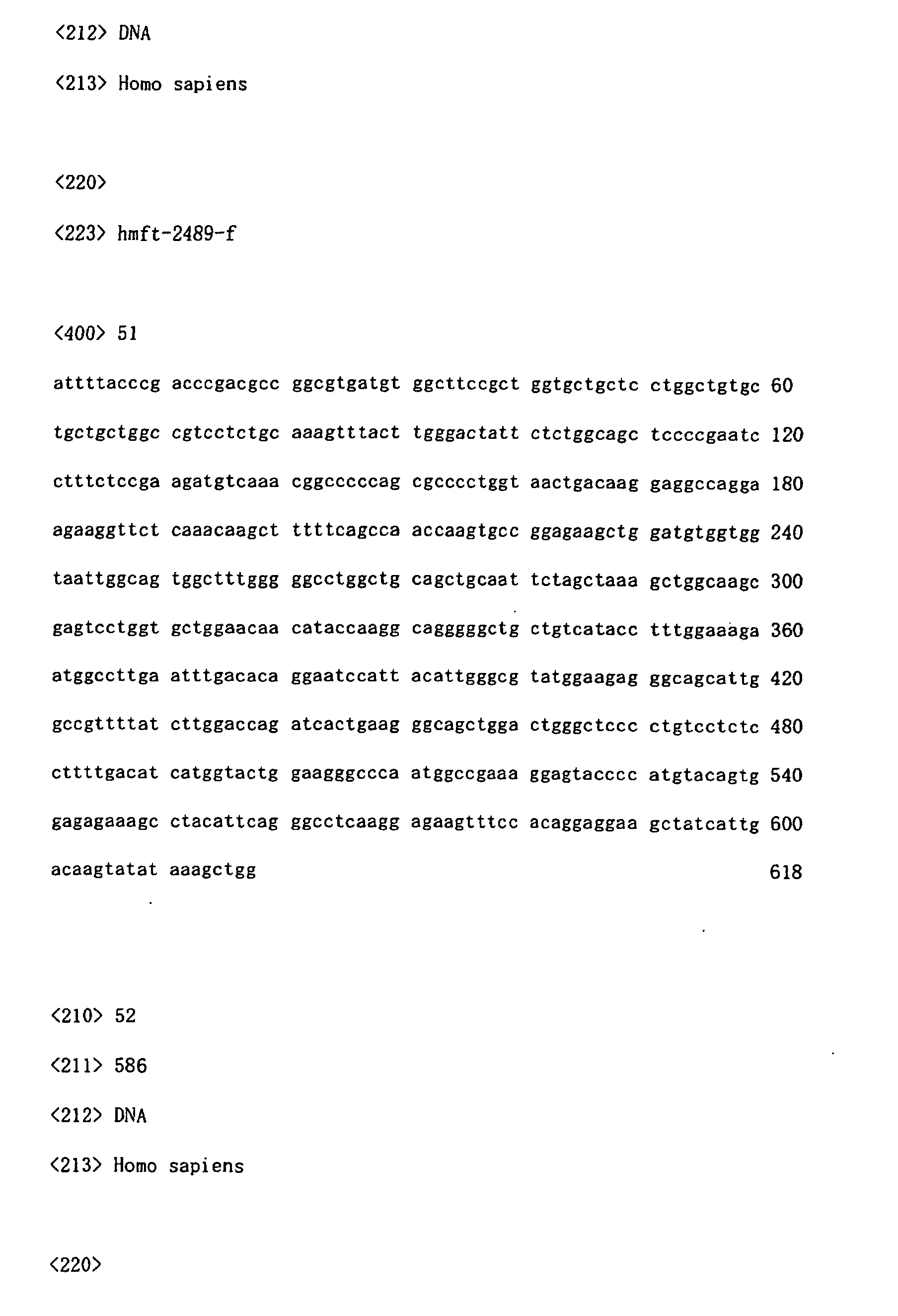


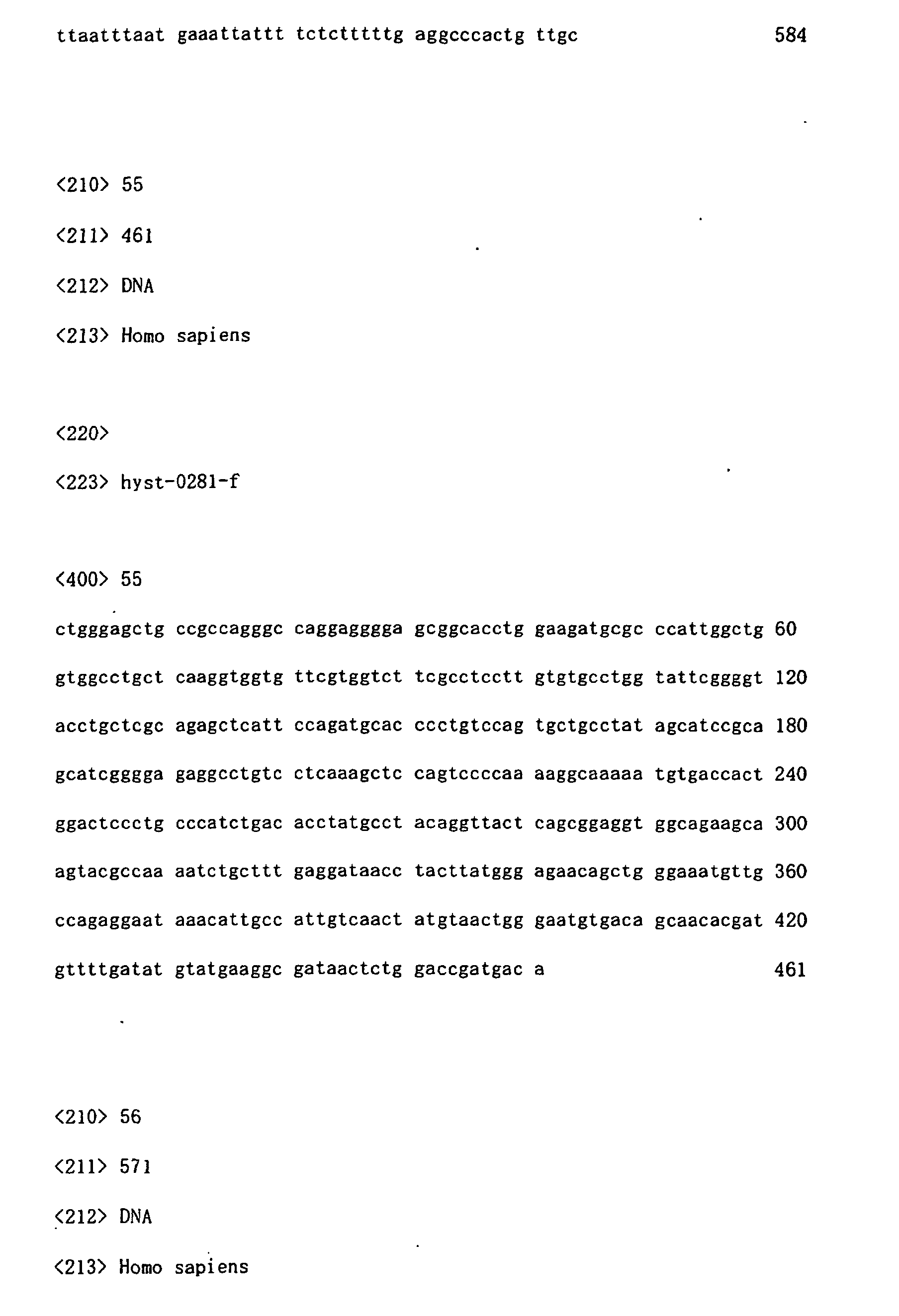

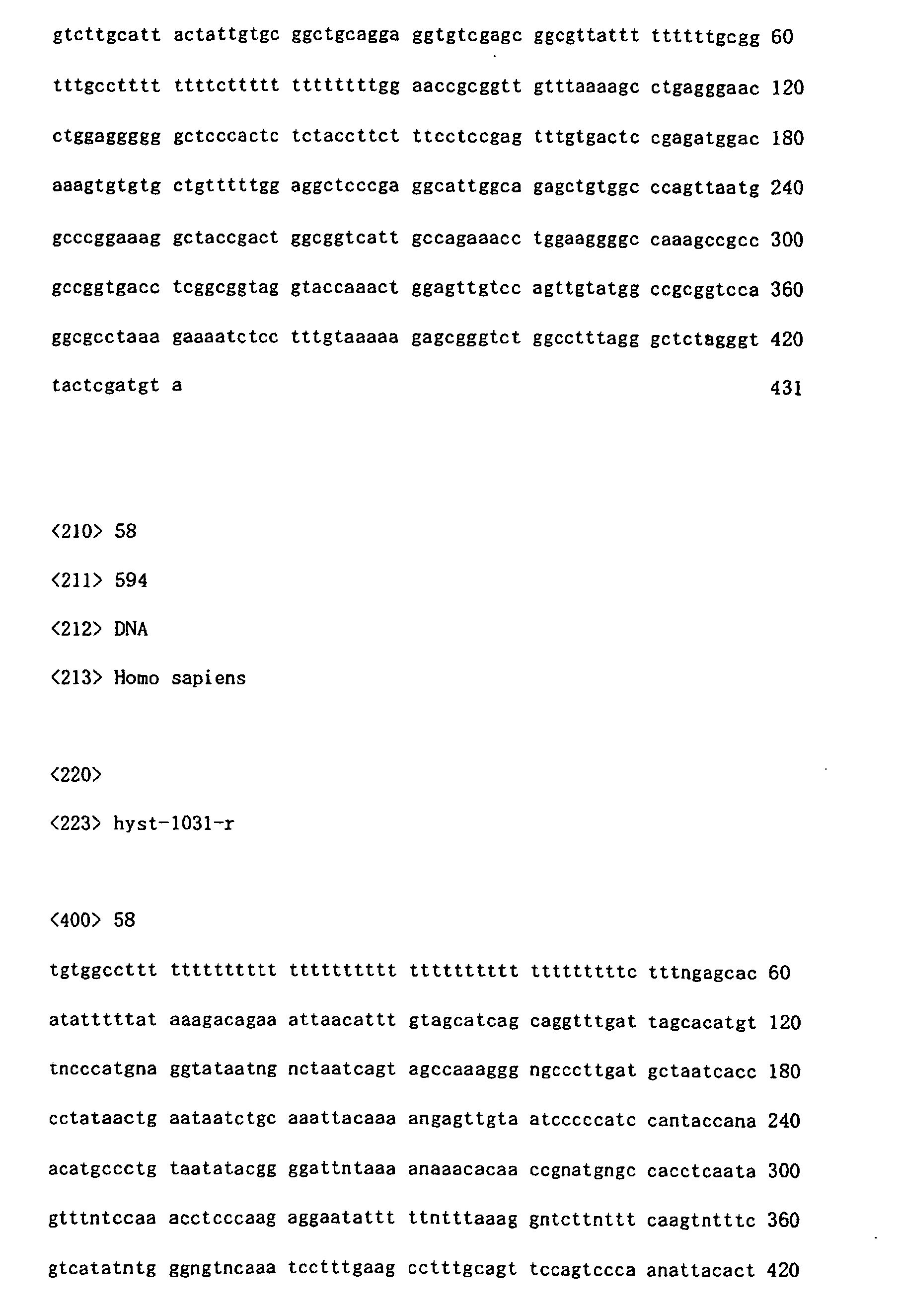


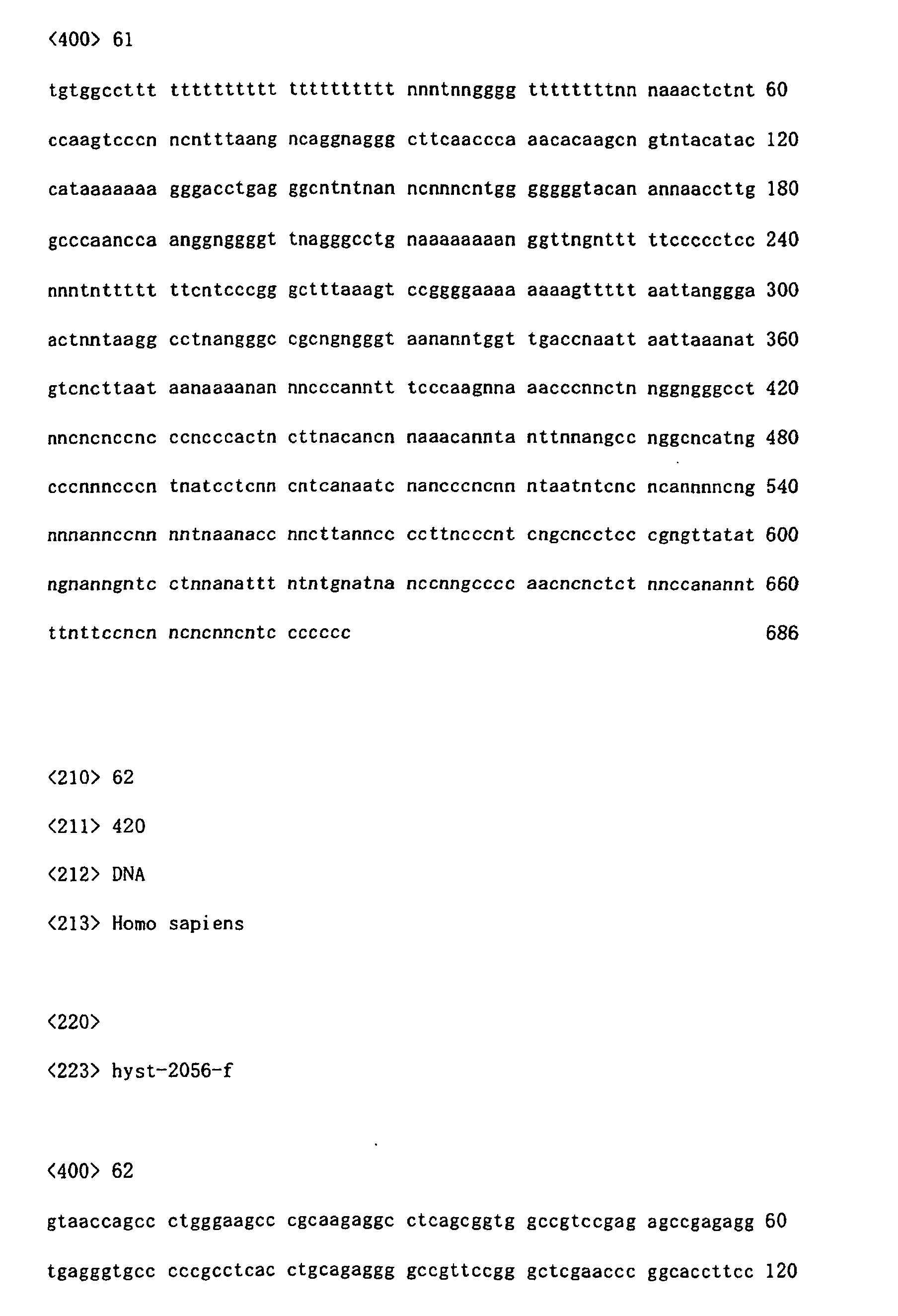


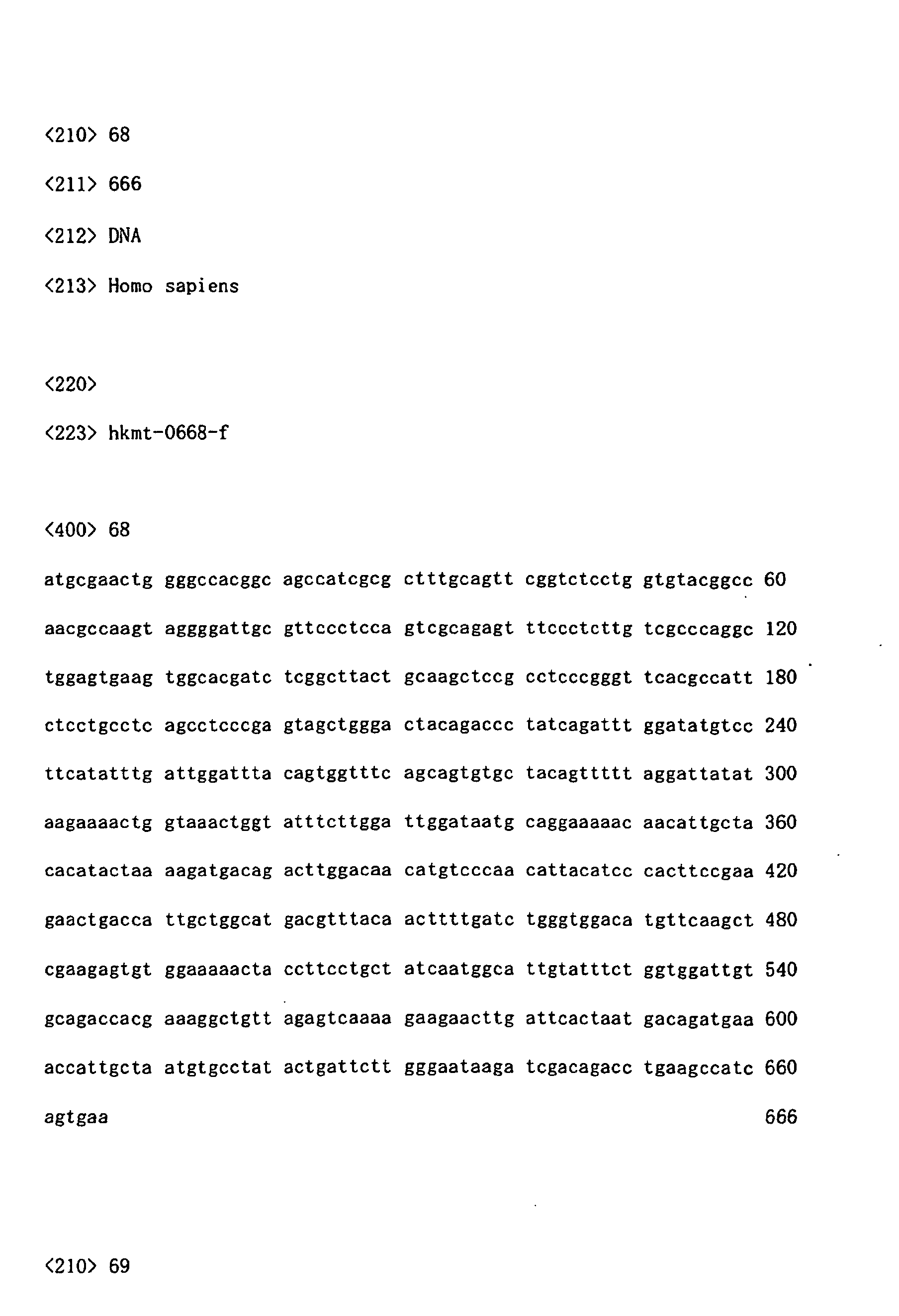


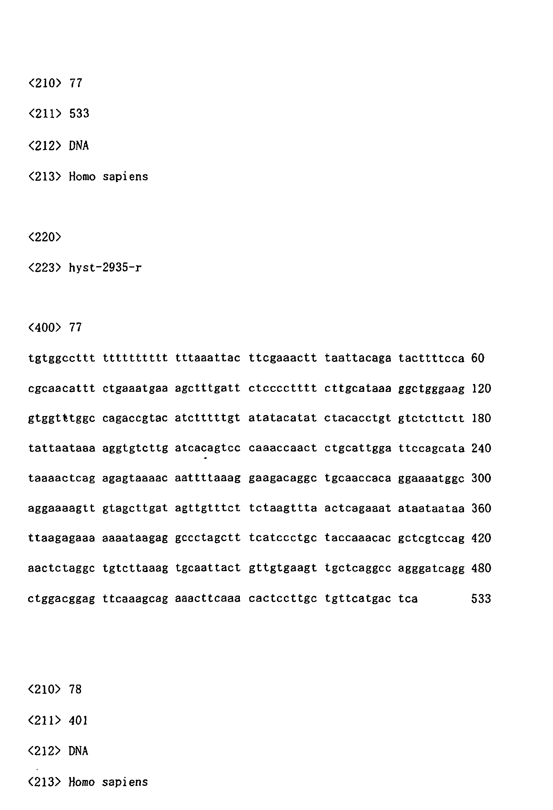
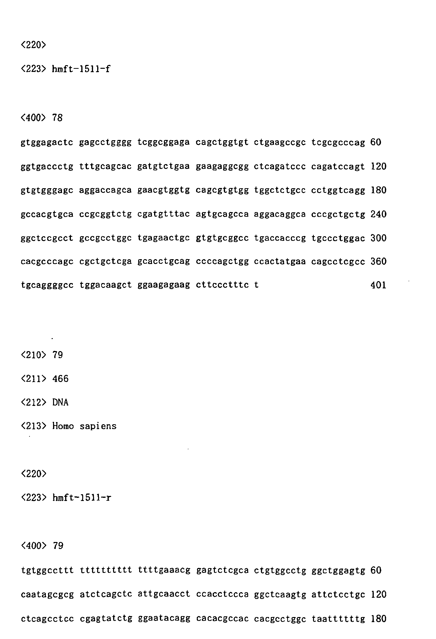
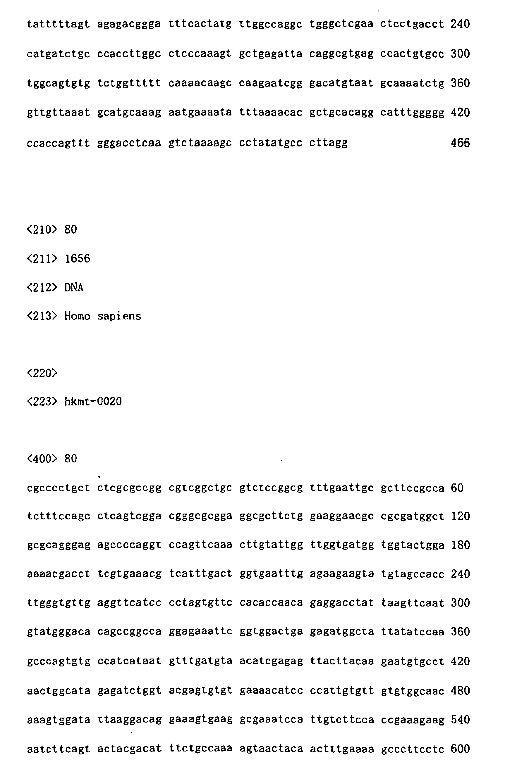

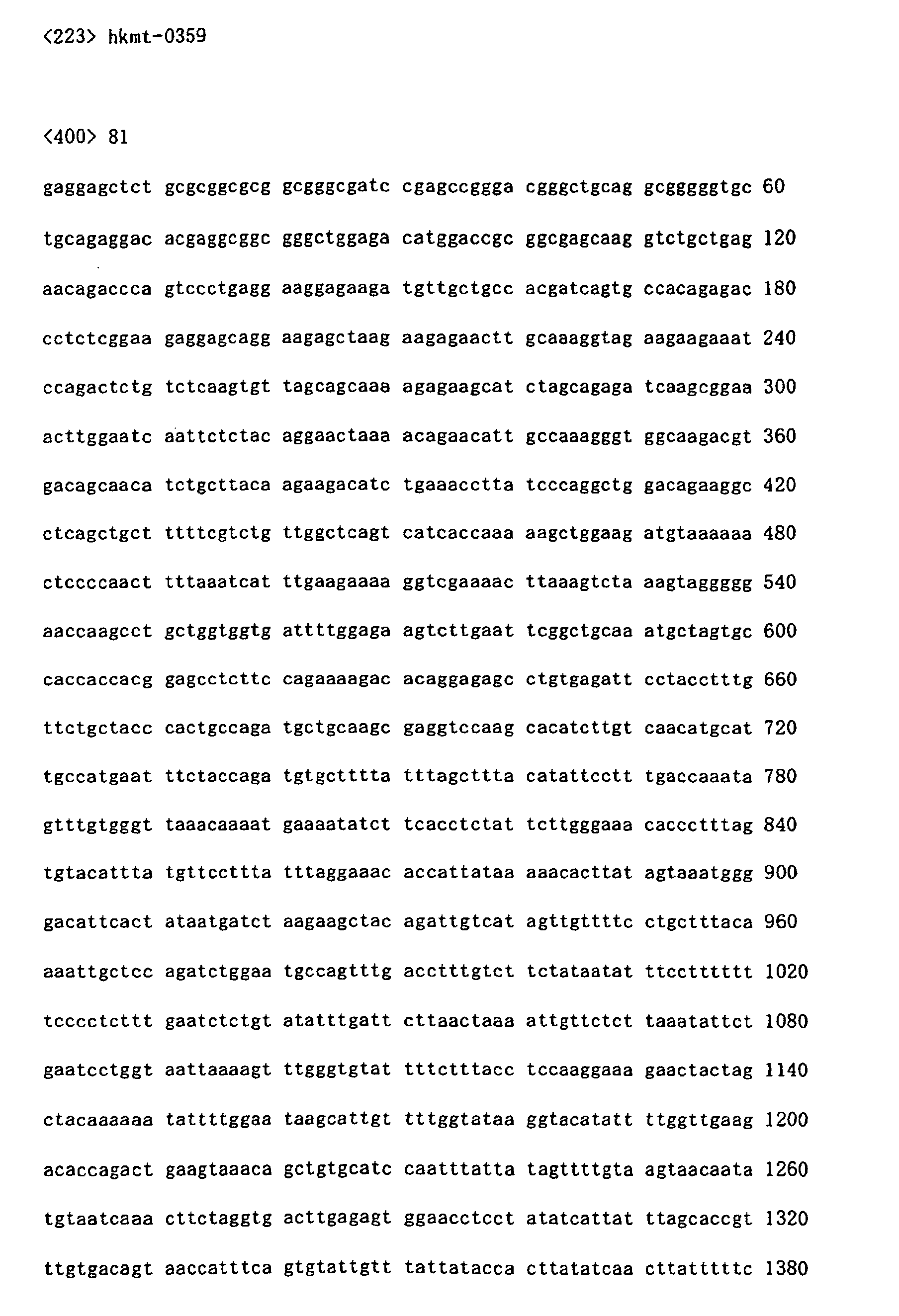


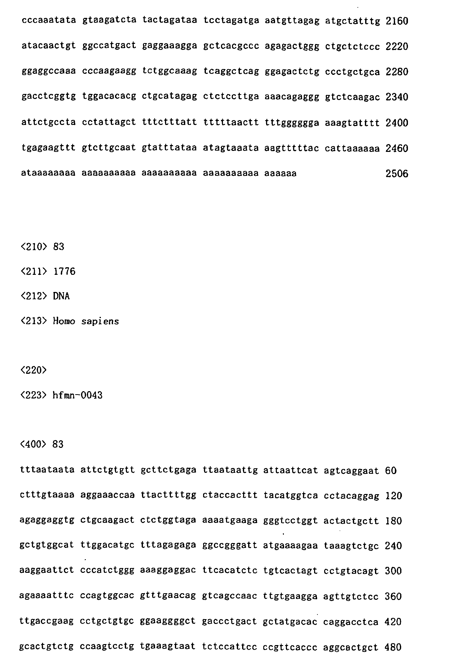
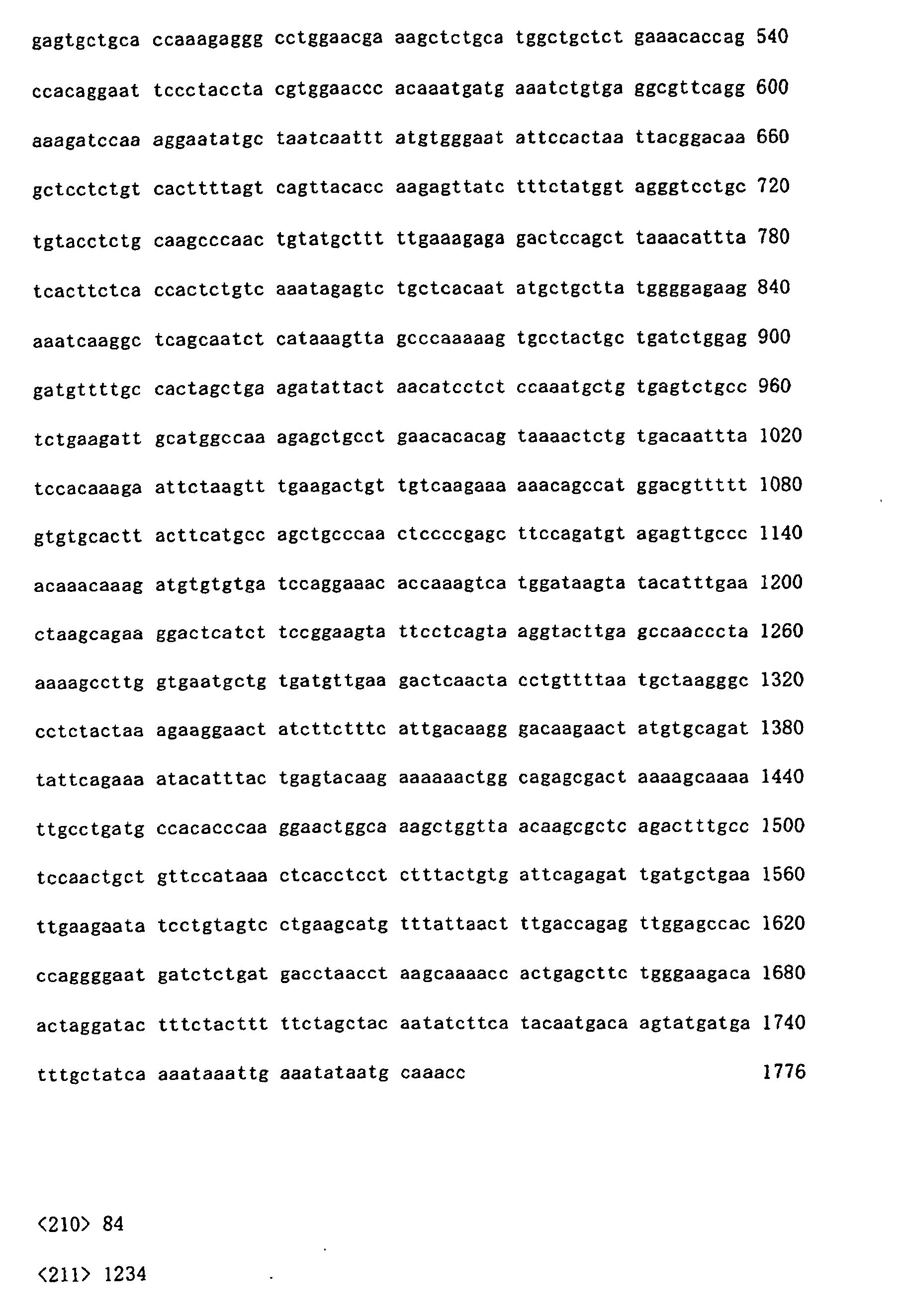


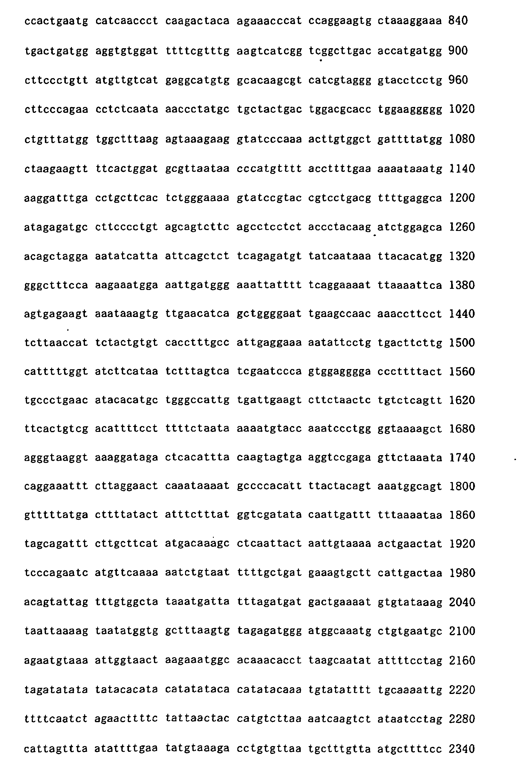


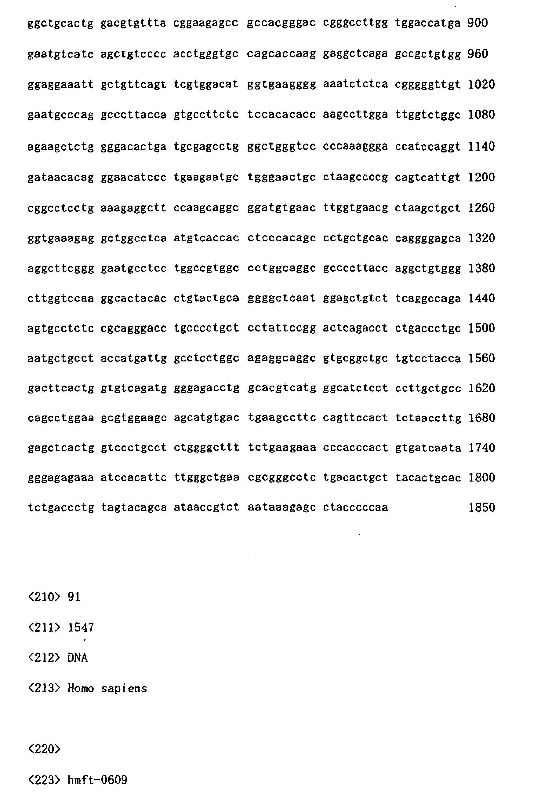


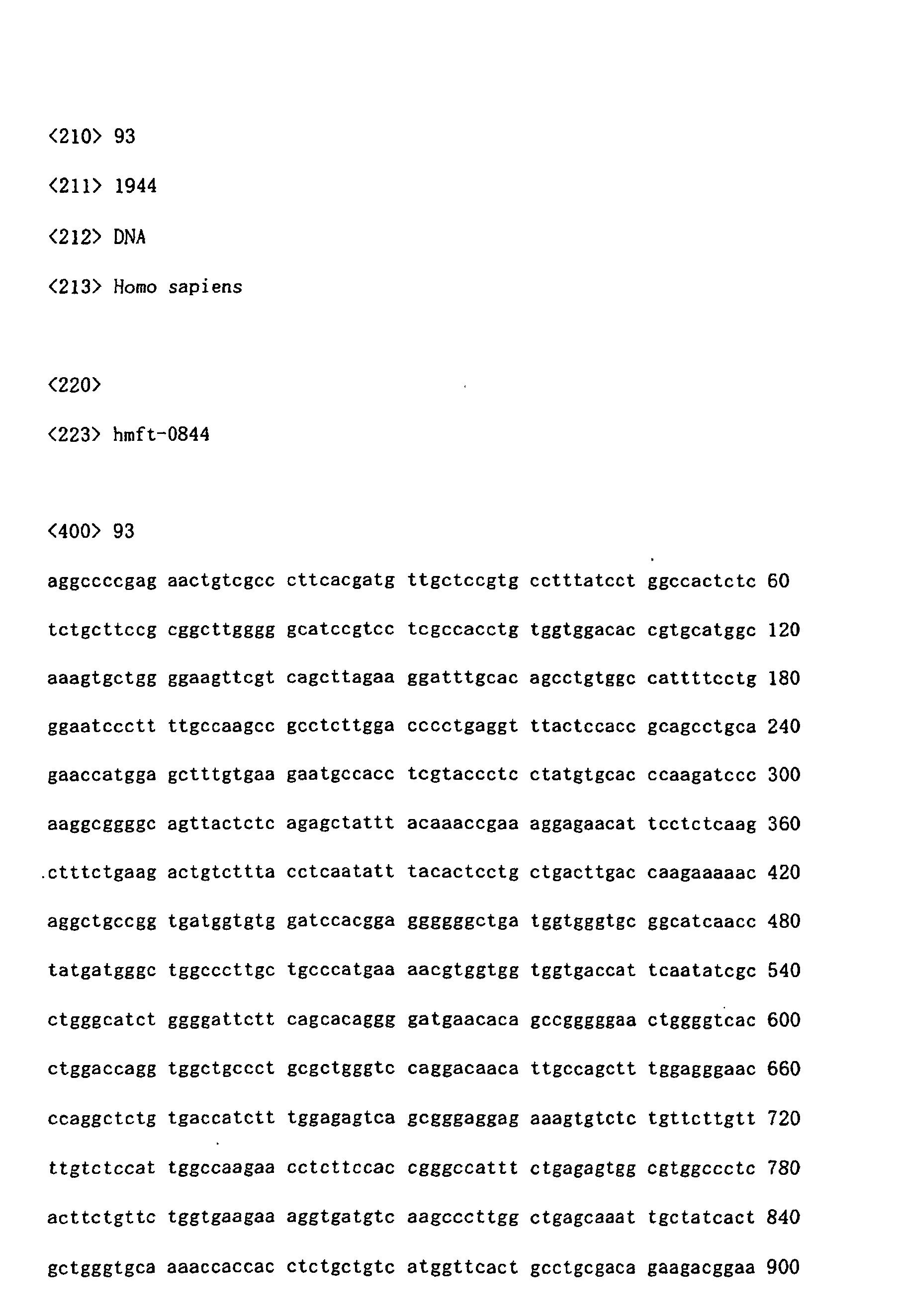
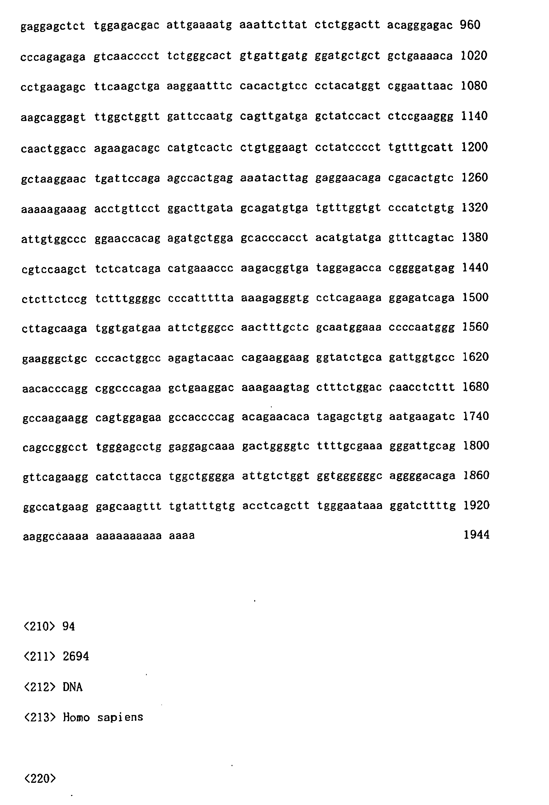
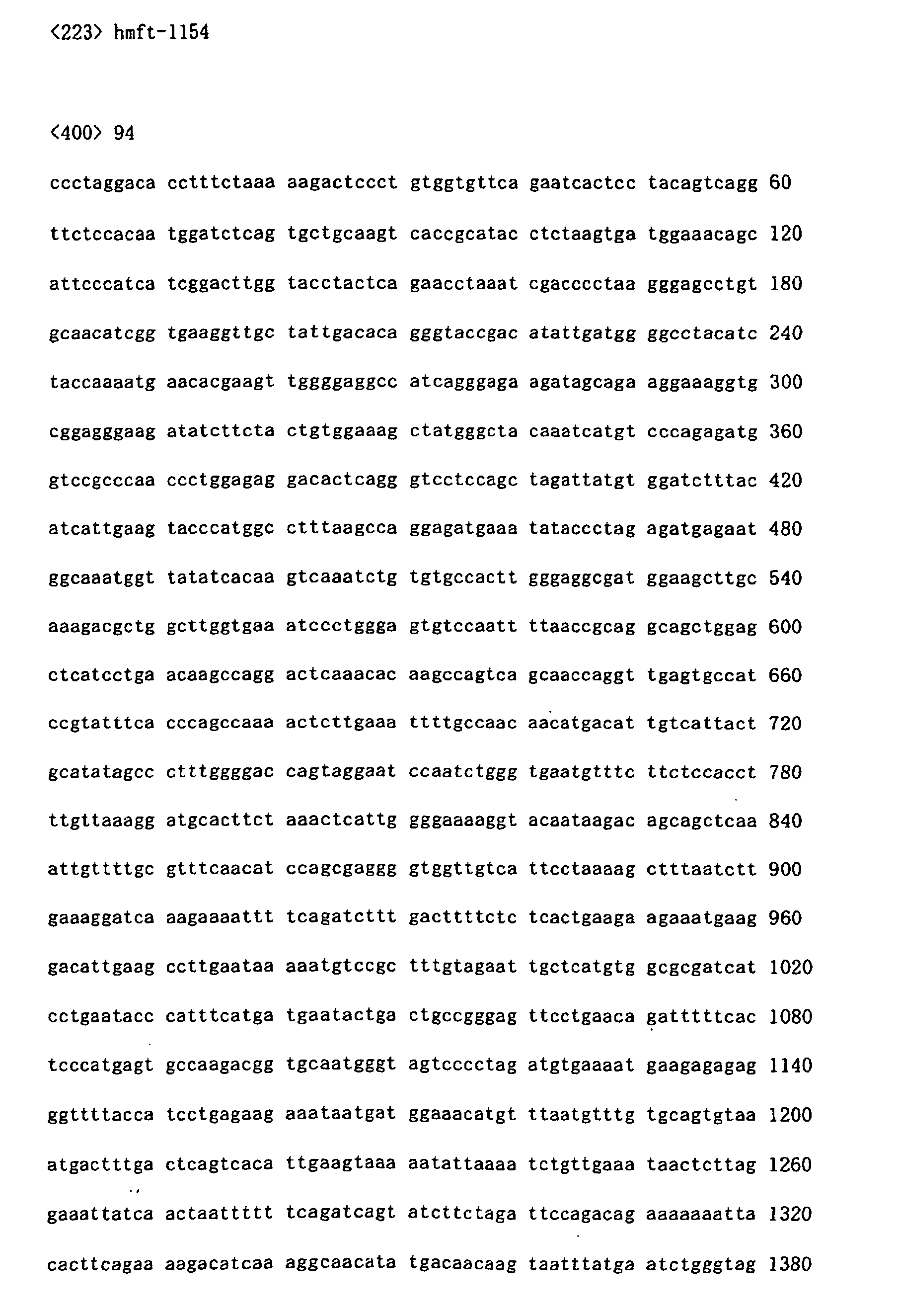

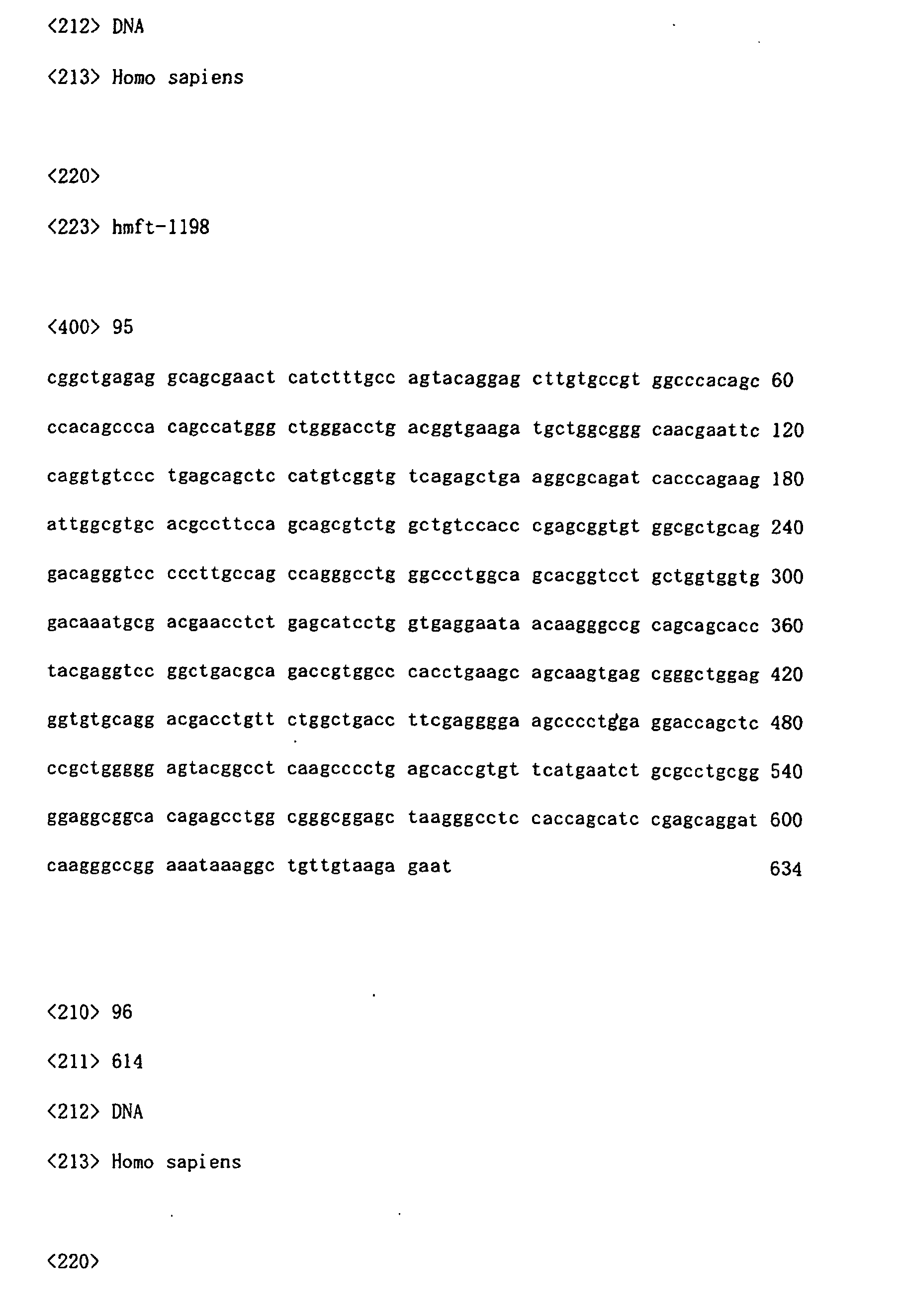

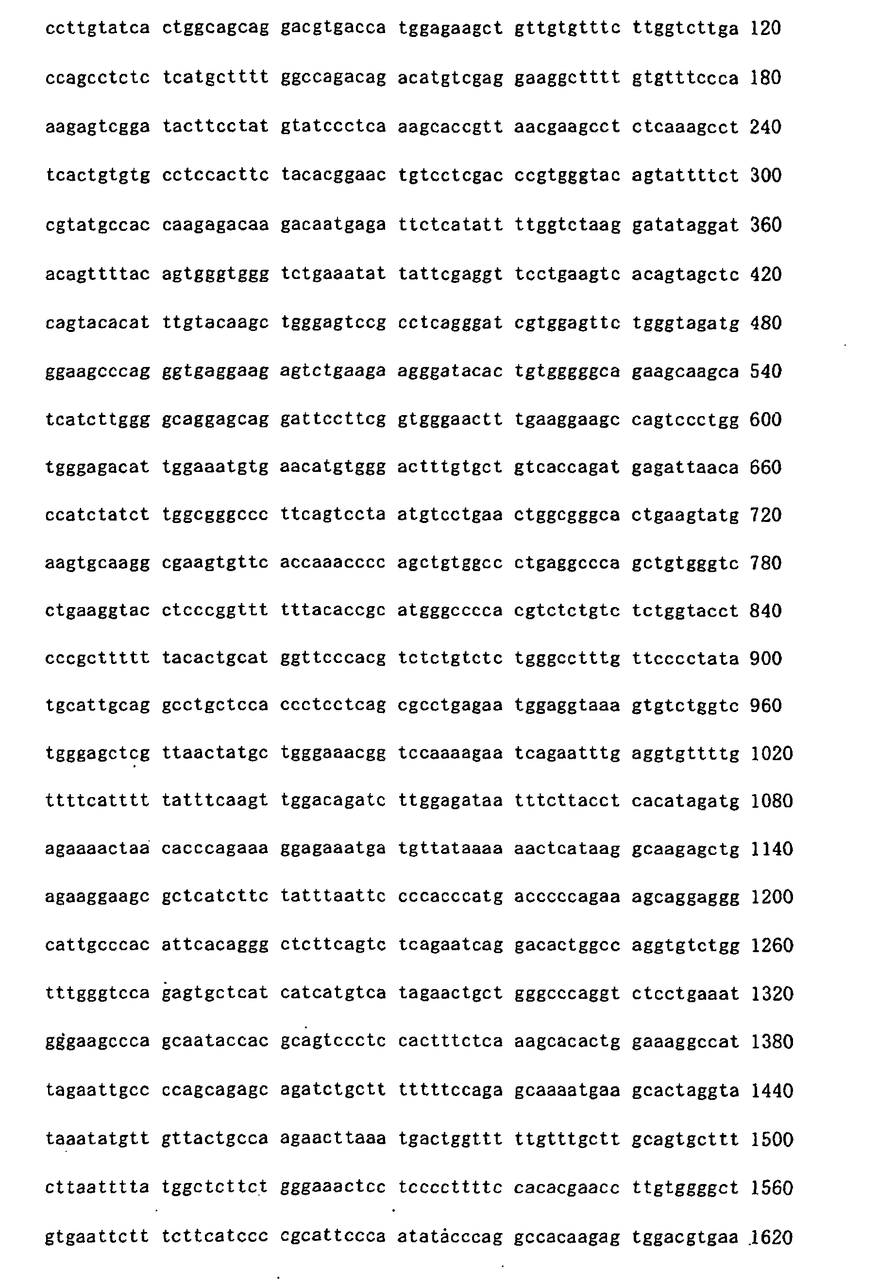

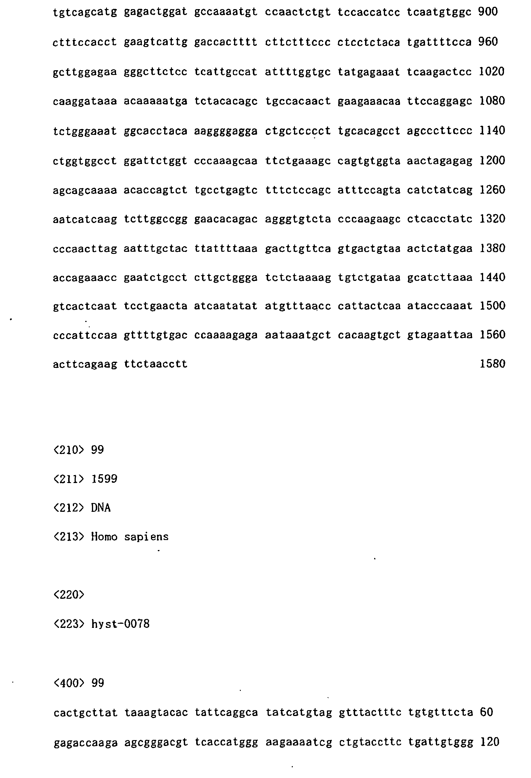

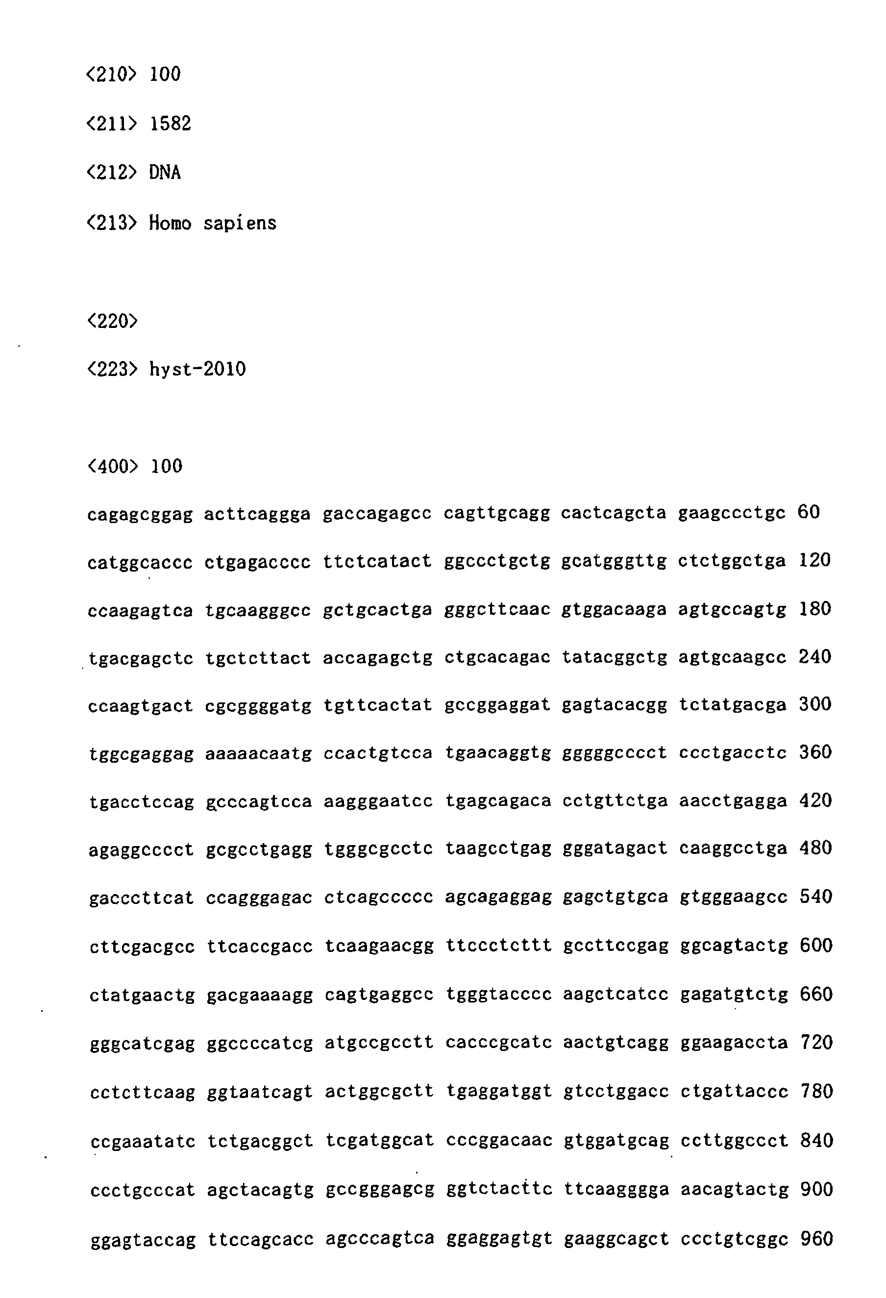

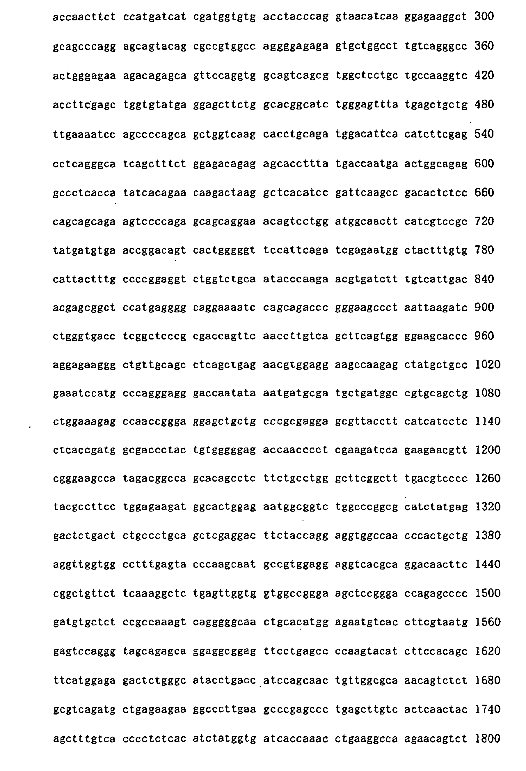


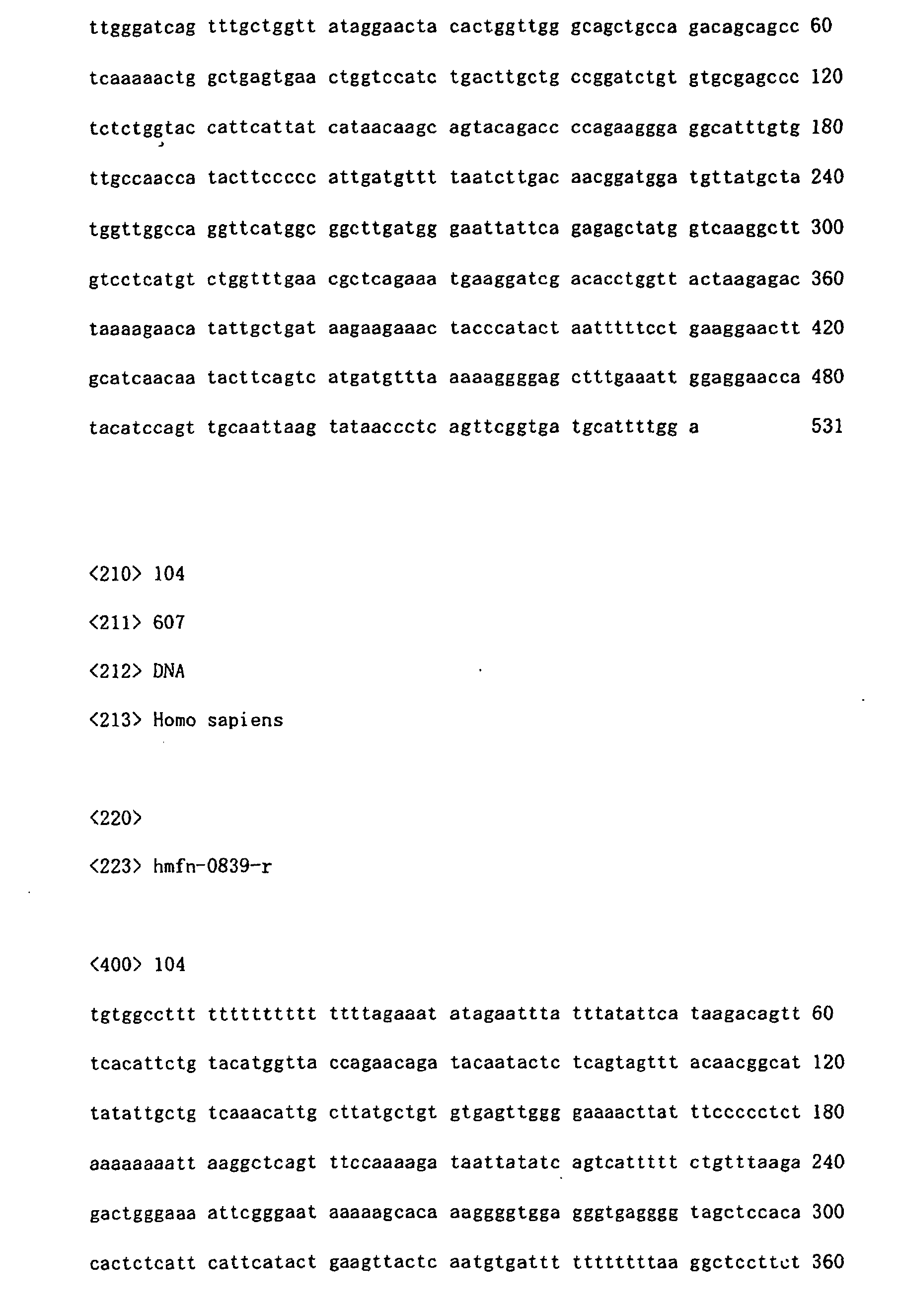
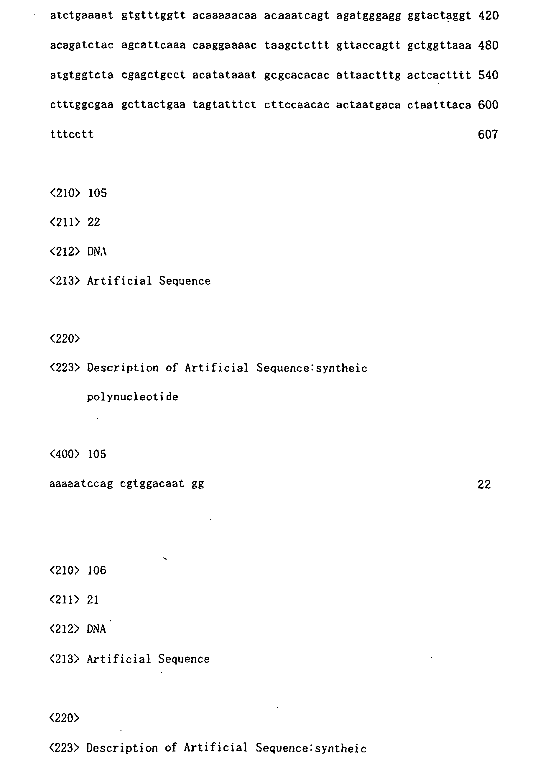
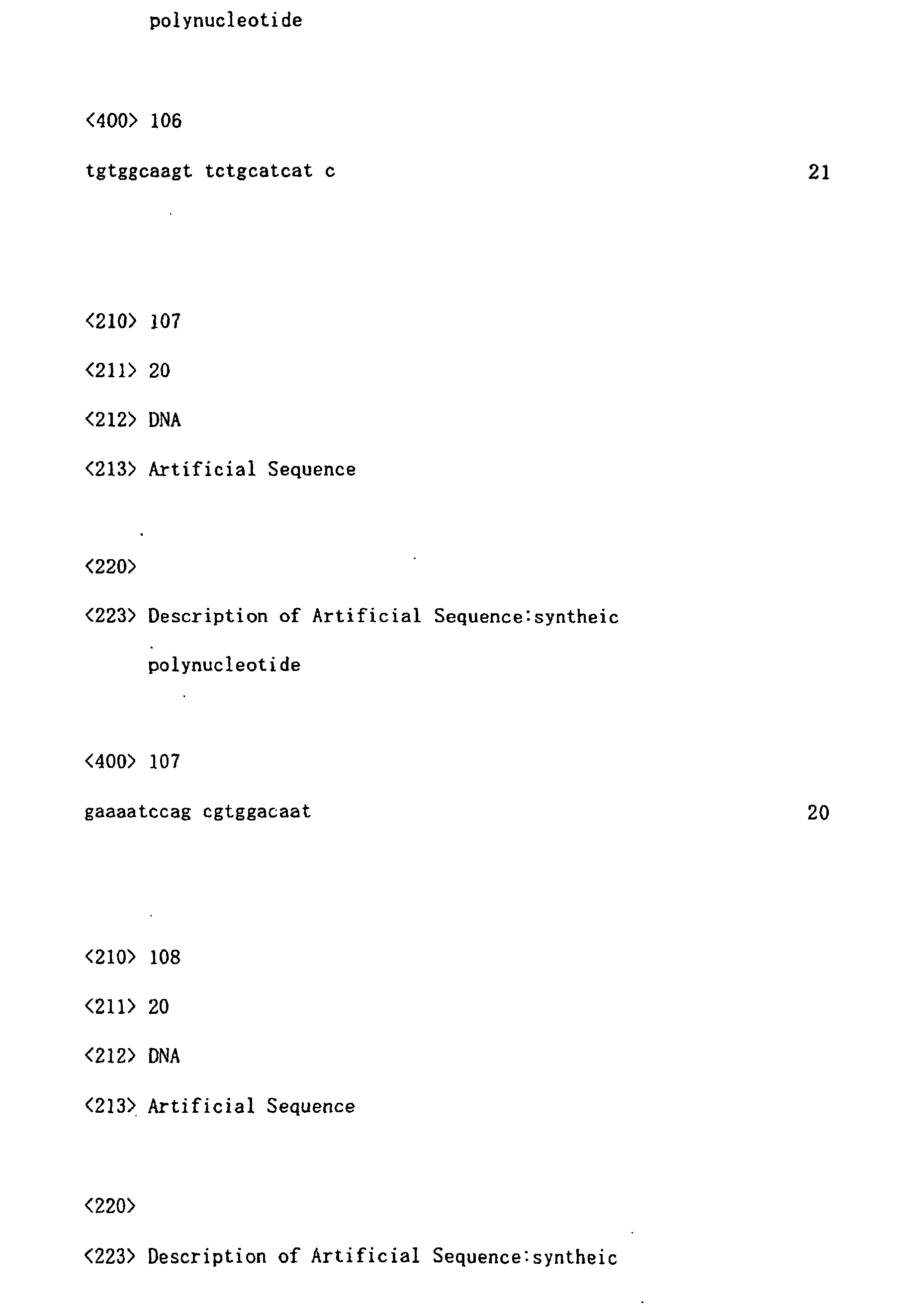


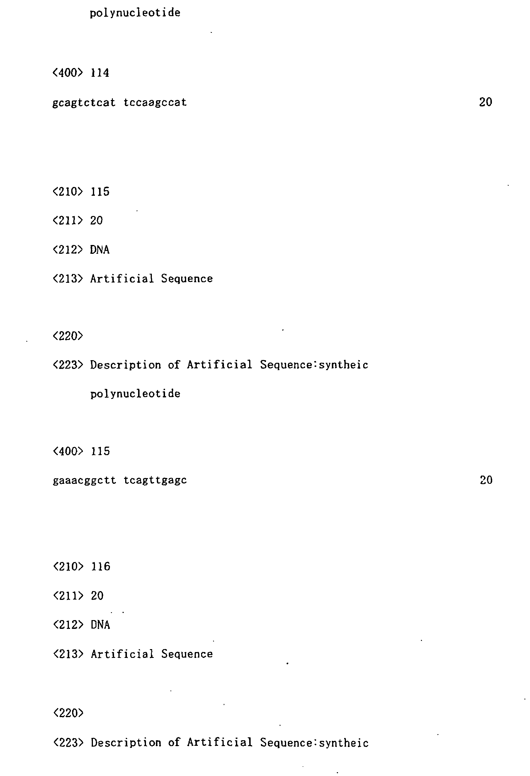

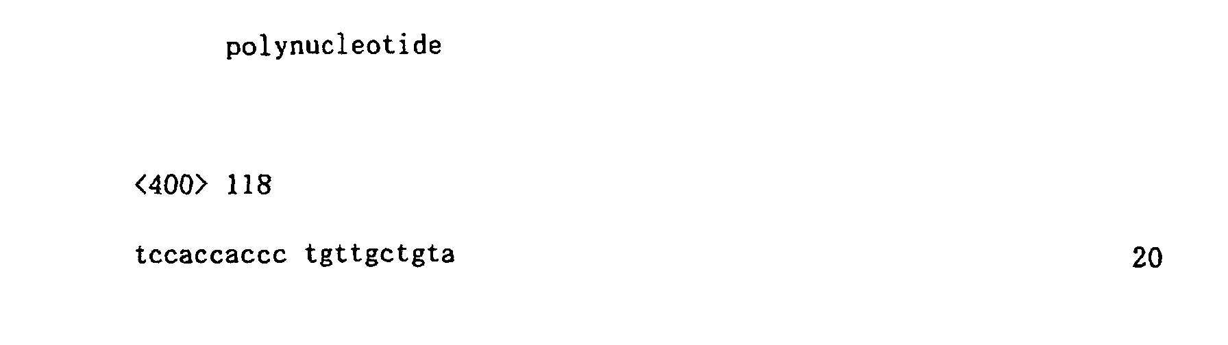
Claims (15)
- A nucleic acid having differential expression levels in hepatoblastoma and normal liver based on comparison therebetween, the nucleic acid comprising any one of base sequences set forth in SEQ ID NO:1 to NO:79 in the Sequence Listing.
- A nucleic acid comprising a portion of any one of base sequences set forth in SEQ ID NO:1 to NO:79 in the Sequence Listing.
- An isolated nucleic acid capable of hybridizing to the nucleic acid according to claim 1 or 2, or a complementary nucleic acid thereof under stringent conditions.
- A probe for tumor detection comprising a nucleic acid, the nucleic acid comprising any one of base sequences set forth in SEQ ID NO:1 to NO:104 in the Sequence Listing.
- A probe for tumor detection comprising a nucleic acid, the nucleic acid comprising a portion of any one of base sequences set forth in SEQ ID NO:1 to NO:104 in the Sequence Listing.
- A probe for tumor detection comprising an nucleic acid capable of hybridizing to the nucleic acid comprising any one of base sequences set forth in SEQ ID NO:1 to NO:104 in the Sequence Listing a fragment thereof, or a complementary nucleic acid thereof under stringent conditions.
- The probe for tumor detection according to any one of claims 4-6, wherein the tumor is selected from the group of consisting of liver cancer, colorectal cancer, breast cancer, kidney cancer, gastric cancer, ovarian cancer and thyroid cancer.
- The probe for tumor detection according to claim 7, wherein the tumor is hepatoblastoma.
- A PCR primer for tumor detection capable of amplifying a nucleic acid comprising any one of base sequences set forth in SEQ ID NO:1 to NO:104 in the Sequence Listing.
- The PCR primer for tumor detection according to claim 9, wherein the tumor is selected from the group of consisting of liver cancer, colorectal cancer, breast cancer, kidney cancer, gastric cancer, ovarian cancer and thyroid cancer.
- The PCR primer for tumor detection according to claim 10 wherein the tumor is hepatoblastoma.
- A marker protein for tumor detection, the protein capable of being encoded by a nucleic acid comprising any one of base sequences set forth in SEQ ID NO:1 to NO:104 in the Sequence Listing.
- The marker protein for tumor detection according to claim 12, wherein the tumor is selected from the group of consisting of liver cancer, colorectal cancer, breast cancer, kidney cancer, gastric cancer, ovarian cancer and thyroid cancer.
- The marker protein for tumor detection according to claim 13, wherein the tumor is hepatoblastoma.
- A diagnostic agent for tumor detection containing at least one nucleic acid comprising a portion or the whole of any one of bases sequence set forth in SEQ ID NO:1 to SEQ ID NO:104 in the Sequence Listing.
Priority Applications (1)
| Application Number | Priority Date | Filing Date | Title |
|---|---|---|---|
| EP06015501A EP1757693A3 (en) | 2001-08-24 | 2002-08-26 | Nucleic acids showing difference in expression between hepatoblastoma and normal liver |
Applications Claiming Priority (3)
| Application Number | Priority Date | Filing Date | Title |
|---|---|---|---|
| JP2001255225 | 2001-08-24 | ||
| JP2001255225 | 2001-08-24 | ||
| PCT/JP2002/008580 WO2003018807A1 (en) | 2001-08-24 | 2002-08-26 | Nucleic acids showing difference in expression between hepatoblastoma anc normal liver |
Related Child Applications (1)
| Application Number | Title | Priority Date | Filing Date |
|---|---|---|---|
| EP06015501A Division EP1757693A3 (en) | 2001-08-24 | 2002-08-26 | Nucleic acids showing difference in expression between hepatoblastoma and normal liver |
Publications (2)
| Publication Number | Publication Date |
|---|---|
| EP1428882A1 true EP1428882A1 (en) | 2004-06-16 |
| EP1428882A4 EP1428882A4 (en) | 2005-05-25 |
Family
ID=19083240
Family Applications (2)
| Application Number | Title | Priority Date | Filing Date |
|---|---|---|---|
| EP06015501A Withdrawn EP1757693A3 (en) | 2001-08-24 | 2002-08-26 | Nucleic acids showing difference in expression between hepatoblastoma and normal liver |
| EP02762859A Withdrawn EP1428882A4 (en) | 2001-08-24 | 2002-08-26 | NUCLEIC ACIDS HAVING A DIFFERENCE OF EXPRESSION IN HEPATOBLASTOMA AND NORMAL LIVER |
Family Applications Before (1)
| Application Number | Title | Priority Date | Filing Date |
|---|---|---|---|
| EP06015501A Withdrawn EP1757693A3 (en) | 2001-08-24 | 2002-08-26 | Nucleic acids showing difference in expression between hepatoblastoma and normal liver |
Country Status (4)
| Country | Link |
|---|---|
| US (1) | US7358349B2 (en) |
| EP (2) | EP1757693A3 (en) |
| JP (1) | JP4098236B2 (en) |
| WO (1) | WO2003018807A1 (en) |
Families Citing this family (2)
| Publication number | Priority date | Publication date | Assignee | Title |
|---|---|---|---|---|
| AU775272B2 (en) | 1999-10-28 | 2004-07-29 | Agensys, Inc. | 36P6D5: secreted tumor antigen |
| US20050186577A1 (en) | 2004-02-20 | 2005-08-25 | Yixin Wang | Breast cancer prognostics |
Family Cites Families (12)
| Publication number | Priority date | Publication date | Assignee | Title |
|---|---|---|---|---|
| US5474796A (en) * | 1991-09-04 | 1995-12-12 | Protogene Laboratories, Inc. | Method and apparatus for conducting an array of chemical reactions on a support surface |
| AU736506B2 (en) * | 1997-12-03 | 2001-07-26 | Genentech Inc. | Polypeptides and nucleic acids encoding the same |
| AU3094999A (en) * | 1998-03-18 | 1999-10-11 | Corixa Corporation | Compounds and methods for therapy and diagnosis of lung cancer |
| US6312695B1 (en) | 1998-03-18 | 2001-11-06 | Corixa Corporation | Compounds and methods for therapy of lung cancer |
| AU3395900A (en) | 1999-03-12 | 2000-10-04 | Human Genome Sciences, Inc. | Human lung cancer associated gene sequences and polypeptides |
| MXPA01009962A (en) | 1999-04-02 | 2002-08-20 | Corixa Corp | Compounds and methods for therapy and diagnosis of lung cancer. |
| EP1200576A2 (en) * | 1999-06-25 | 2002-05-02 | Genset | Dna's encoding proteins with signal peptides |
| US6152986A (en) | 1999-07-07 | 2000-11-28 | Ppg Industries Ohio, Inc. | Method of enriching chlorine gas |
| WO2001022920A2 (en) * | 1999-09-29 | 2001-04-05 | Human Genome Sciences, Inc. | Colon and colon cancer associated polynucleotides and polypeptides |
| AU775272B2 (en) | 1999-10-28 | 2004-07-29 | Agensys, Inc. | 36P6D5: secreted tumor antigen |
| AU2001241541A1 (en) * | 2000-02-17 | 2001-08-27 | Millennium Predictive Medicine, Inc. | Novel genes, compositions, kits, and methods for identification, assessment, prevention, and therapy of human prostate cancer |
| US6436703B1 (en) * | 2000-03-31 | 2002-08-20 | Hyseq, Inc. | Nucleic acids and polypeptides |
-
2002
- 2002-08-26 EP EP06015501A patent/EP1757693A3/en not_active Withdrawn
- 2002-08-26 EP EP02762859A patent/EP1428882A4/en not_active Withdrawn
- 2002-08-26 WO PCT/JP2002/008580 patent/WO2003018807A1/en not_active Ceased
- 2002-08-26 US US10/487,422 patent/US7358349B2/en not_active Expired - Fee Related
- 2002-08-26 JP JP2003523655A patent/JP4098236B2/en not_active Expired - Fee Related
Also Published As
| Publication number | Publication date |
|---|---|
| US20050042613A1 (en) | 2005-02-24 |
| EP1757693A2 (en) | 2007-02-28 |
| EP1428882A4 (en) | 2005-05-25 |
| WO2003018807A1 (en) | 2003-03-06 |
| JPWO2003018807A1 (en) | 2004-12-09 |
| EP1757693A3 (en) | 2007-05-09 |
| JP4098236B2 (en) | 2008-06-11 |
| US7358349B2 (en) | 2008-04-15 |
Similar Documents
| Publication | Publication Date | Title |
|---|---|---|
| US6797471B2 (en) | Detection and diagnosis of smoking related cancers | |
| Casson et al. | Clinical implications of p53 gene mutation in the progression of Barrett's epithelium to invasive esophageal cancer | |
| JPH08504081A (en) | Method for detecting mammalian nucleic acid isolated from fecal sample, and reagent for detecting the same | |
| CA2501131A1 (en) | Molecular sub-classification of kidney tumors and the discovery of new diagnostic markers | |
| AU720500B2 (en) | Diagnostic methods and gene therapy using reagents derived from the human metastasis suppressor gene KAI1 | |
| Ishii et al. | Absence of p53 gene mutations in a tumor panel representative of pilocytic astrocytoma diversity using a p53 functional assay | |
| US20110195070A1 (en) | Detection of granulosa-cell tumors | |
| US20020115057A1 (en) | Process for identifying anti-cancer therapeutic agents using cancer gene sets | |
| US20020165180A1 (en) | Process for identifying anti-cancer therapeutic agents using cancer gene sets | |
| US7358349B2 (en) | Nucleic acids having expression differentials between hepatoblastoma and normal liver | |
| US6617104B2 (en) | Predisposition to breast cancer by mutations at the ataxia-telangiectasia genetic locus | |
| EP1354962B1 (en) | Use of the slug gene or of the transcription or expression products thereof in the detection and/or treatment of cancerous cells | |
| US20040161760A1 (en) | Method of molecular diagnosis of chronic myelogenous leukemia | |
| US20040115625A1 (en) | Cancer gene determination and therapeutic screening using signature gene sets | |
| US7429451B2 (en) | Nucleic acids isolated from stage 4 neuroblastoma | |
| US20020102532A1 (en) | Cancer gene determination and therapeutic screening using signature gene sets | |
| JP2003528630A (en) | Human genes and expression products | |
| KR101148825B1 (en) | A protein associated with a colon cancer, a polynucleotide associated with a colon cancer comprising single nucleotide polymorphism, microarray and diagnostic kit comprising the same and method for diagnosing a colon cancer using the same | |
| JP2006166789A (en) | New diagnostic method for cancer | |
| JP2007082433A (en) | Malignant brain tumor marker gene and use thereof | |
| CN103540654A (en) | Applications of 11-19 lysine-rich leukemia (ELL) gene and expression product thereof | |
| US20020150877A1 (en) | Cancer gene determination and therapeutic screening using signature gene sets | |
| JP5089172B2 (en) | Cancer tissue detection method and cancer tissue detection reagent used in the method | |
| US20020110821A1 (en) | Cancer gene determination and therapeutic screening using signature gene sets | |
| KR20250033434A (en) | Diagnostic methods for prognosis of non-small-cell lung cancer using MLKL SNP |
Legal Events
| Date | Code | Title | Description |
|---|---|---|---|
| PUAI | Public reference made under article 153(3) epc to a published international application that has entered the european phase |
Free format text: ORIGINAL CODE: 0009012 |
|
| 17P | Request for examination filed |
Effective date: 20040323 |
|
| AK | Designated contracting states |
Kind code of ref document: A1 Designated state(s): AT BE BG CH CY CZ DE DK EE ES FI FR GB GR IE IT LI LU MC NL PT SE SK TR |
|
| AX | Request for extension of the european patent |
Extension state: AL LT LV MK RO SI |
|
| A4 | Supplementary search report drawn up and despatched |
Effective date: 20050406 |
|
| STAA | Information on the status of an ep patent application or granted ep patent |
Free format text: STATUS: THE APPLICATION IS DEEMED TO BE WITHDRAWN |
|
| 18D | Application deemed to be withdrawn |
Effective date: 20060727 |