EP1426053A1 - Use of amphiphilic lipids for reducing tumor metastasis - Google Patents
Use of amphiphilic lipids for reducing tumor metastasis Download PDFInfo
- Publication number
- EP1426053A1 EP1426053A1 EP02026897A EP02026897A EP1426053A1 EP 1426053 A1 EP1426053 A1 EP 1426053A1 EP 02026897 A EP02026897 A EP 02026897A EP 02026897 A EP02026897 A EP 02026897A EP 1426053 A1 EP1426053 A1 EP 1426053A1
- Authority
- EP
- European Patent Office
- Prior art keywords
- amphiphilic lipids
- tumor
- phospholipid
- cancer
- amphiphilic
- Prior art date
- Legal status (The legal status is an assumption and is not a legal conclusion. Google has not performed a legal analysis and makes no representation as to the accuracy of the status listed.)
- Withdrawn
Links
- 206010028980 Neoplasm Diseases 0.000 title claims abstract description 76
- 150000002632 lipids Chemical class 0.000 title claims abstract description 59
- 206010027476 Metastases Diseases 0.000 title claims abstract description 30
- 230000009401 metastasis Effects 0.000 title claims abstract description 20
- 208000005718 Stomach Neoplasms Diseases 0.000 claims abstract description 23
- 201000011510 cancer Diseases 0.000 claims abstract description 23
- 206010017758 gastric cancer Diseases 0.000 claims abstract description 23
- 201000011549 stomach cancer Diseases 0.000 claims abstract description 23
- 239000008194 pharmaceutical composition Substances 0.000 claims abstract description 20
- 238000002360 preparation method Methods 0.000 claims abstract description 17
- 238000007912 intraperitoneal administration Methods 0.000 claims abstract description 11
- 206010009944 Colon cancer Diseases 0.000 claims abstract description 8
- 208000029742 colonic neoplasm Diseases 0.000 claims abstract description 8
- 210000001072 colon Anatomy 0.000 claims abstract description 6
- 238000011321 prophylaxis Methods 0.000 claims abstract description 6
- 206010033128 Ovarian cancer Diseases 0.000 claims abstract description 5
- 206010061535 Ovarian neoplasm Diseases 0.000 claims abstract description 5
- 206010017993 Gastrointestinal neoplasms Diseases 0.000 claims abstract description 4
- 206010061902 Pancreatic neoplasm Diseases 0.000 claims abstract description 4
- 230000002496 gastric effect Effects 0.000 claims abstract description 4
- 208000015486 malignant pancreatic neoplasm Diseases 0.000 claims abstract description 4
- 201000002528 pancreatic cancer Diseases 0.000 claims abstract description 4
- 208000008443 pancreatic carcinoma Diseases 0.000 claims abstract description 4
- 210000002229 urogenital system Anatomy 0.000 claims abstract description 4
- 206010005003 Bladder cancer Diseases 0.000 claims abstract description 3
- 208000007097 Urinary Bladder Neoplasms Diseases 0.000 claims abstract description 3
- 201000005112 urinary bladder cancer Diseases 0.000 claims abstract description 3
- 150000003904 phospholipids Chemical class 0.000 claims description 87
- 210000004027 cell Anatomy 0.000 claims description 77
- 210000004881 tumor cell Anatomy 0.000 claims description 40
- 230000021164 cell adhesion Effects 0.000 claims description 20
- 238000000034 method Methods 0.000 claims description 17
- 230000005764 inhibitory process Effects 0.000 claims description 16
- 239000006185 dispersion Substances 0.000 claims description 15
- 150000001875 compounds Chemical class 0.000 claims description 13
- 239000007864 aqueous solution Substances 0.000 claims description 8
- 239000000243 solution Substances 0.000 claims description 7
- 239000002246 antineoplastic agent Substances 0.000 claims description 6
- 229940041181 antineoplastic drug Drugs 0.000 claims description 6
- 229940079593 drug Drugs 0.000 claims description 5
- 239000003814 drug Substances 0.000 claims description 5
- 238000000265 homogenisation Methods 0.000 claims description 4
- 239000007924 injection Substances 0.000 claims description 4
- 238000002347 injection Methods 0.000 claims description 4
- 230000002285 radioactive effect Effects 0.000 claims description 4
- 230000012010 growth Effects 0.000 claims description 3
- 238000002271 resection Methods 0.000 claims description 3
- 241000124008 Mammalia Species 0.000 claims description 2
- 230000001225 therapeutic effect Effects 0.000 claims description 2
- 238000011282 treatment Methods 0.000 description 26
- 230000009467 reduction Effects 0.000 description 20
- 238000002474 experimental method Methods 0.000 description 16
- 230000004083 survival effect Effects 0.000 description 16
- PEDCQBHIVMGVHV-UHFFFAOYSA-N Glycerine Chemical compound OCC(O)CO PEDCQBHIVMGVHV-UHFFFAOYSA-N 0.000 description 15
- 241001465754 Metazoa Species 0.000 description 15
- 230000000694 effects Effects 0.000 description 13
- 238000001356 surgical procedure Methods 0.000 description 12
- XLYOFNOQVPJJNP-UHFFFAOYSA-N water Substances O XLYOFNOQVPJJNP-UHFFFAOYSA-N 0.000 description 12
- 230000008033 biological extinction Effects 0.000 description 11
- 239000002609 medium Substances 0.000 description 10
- 102000010834 Extracellular Matrix Proteins Human genes 0.000 description 9
- 108010037362 Extracellular Matrix Proteins Proteins 0.000 description 9
- 239000012980 RPMI-1640 medium Substances 0.000 description 9
- 241000700159 Rattus Species 0.000 description 9
- 238000003556 assay Methods 0.000 description 9
- 210000002744 extracellular matrix Anatomy 0.000 description 9
- 239000000203 mixture Substances 0.000 description 9
- 238000007619 statistical method Methods 0.000 description 9
- 206010054827 Peritoneal lesion Diseases 0.000 description 8
- FAPWRFPIFSIZLT-UHFFFAOYSA-M Sodium chloride Chemical compound [Na+].[Cl-] FAPWRFPIFSIZLT-UHFFFAOYSA-M 0.000 description 8
- 238000004458 analytical method Methods 0.000 description 8
- 102000008186 Collagen Human genes 0.000 description 7
- 108010035532 Collagen Proteins 0.000 description 7
- 241000699670 Mus sp. Species 0.000 description 7
- 230000037396 body weight Effects 0.000 description 7
- 238000004113 cell culture Methods 0.000 description 7
- 229920001436 collagen Polymers 0.000 description 7
- 102000016359 Fibronectins Human genes 0.000 description 6
- 108010067306 Fibronectins Proteins 0.000 description 6
- OKKJLVBELUTLKV-UHFFFAOYSA-N Methanol Chemical compound OC OKKJLVBELUTLKV-UHFFFAOYSA-N 0.000 description 6
- 210000004534 cecum Anatomy 0.000 description 6
- 201000002628 peritoneum cancer Diseases 0.000 description 6
- UCSJYZPVAKXKNQ-HZYVHMACSA-N streptomycin Chemical compound CN[C@H]1[C@H](O)[C@@H](O)[C@H](CO)O[C@H]1O[C@@H]1[C@](C=O)(O)[C@H](C)O[C@H]1O[C@@H]1[C@@H](NC(N)=N)[C@H](O)[C@@H](NC(N)=N)[C@H](O)[C@H]1O UCSJYZPVAKXKNQ-HZYVHMACSA-N 0.000 description 6
- LFQSCWFLJHTTHZ-UHFFFAOYSA-N Ethanol Chemical compound CCO LFQSCWFLJHTTHZ-UHFFFAOYSA-N 0.000 description 5
- 239000008215 water for injection Substances 0.000 description 5
- 108091003079 Bovine Serum Albumin Proteins 0.000 description 4
- WZUVPPKBWHMQCE-UHFFFAOYSA-N Haematoxylin Chemical compound C12=CC(O)=C(O)C=C2CC2(O)C1C1=CC=C(O)C(O)=C1OC2 WZUVPPKBWHMQCE-UHFFFAOYSA-N 0.000 description 4
- YQEZLKZALYSWHR-UHFFFAOYSA-N Ketamine Chemical compound C=1C=CC=C(Cl)C=1C1(NC)CCCCC1=O YQEZLKZALYSWHR-UHFFFAOYSA-N 0.000 description 4
- 239000004793 Polystyrene Substances 0.000 description 4
- 208000015634 Rectal Neoplasms Diseases 0.000 description 4
- 102000004142 Trypsin Human genes 0.000 description 4
- 108090000631 Trypsin Proteins 0.000 description 4
- 210000003815 abdominal wall Anatomy 0.000 description 4
- 230000001464 adherent effect Effects 0.000 description 4
- 238000005119 centrifugation Methods 0.000 description 4
- 238000010790 dilution Methods 0.000 description 4
- 239000012895 dilution Substances 0.000 description 4
- 238000011156 evaluation Methods 0.000 description 4
- 239000012091 fetal bovine serum Substances 0.000 description 4
- 238000011534 incubation Methods 0.000 description 4
- 230000009545 invasion Effects 0.000 description 4
- 239000002502 liposome Substances 0.000 description 4
- 239000007788 liquid Substances 0.000 description 4
- 230000003287 optical effect Effects 0.000 description 4
- 239000008188 pellet Substances 0.000 description 4
- 229920002223 polystyrene Polymers 0.000 description 4
- 206010038038 rectal cancer Diseases 0.000 description 4
- 201000001275 rectum cancer Diseases 0.000 description 4
- 239000011780 sodium chloride Substances 0.000 description 4
- 210000001519 tissue Anatomy 0.000 description 4
- 239000012588 trypsin Substances 0.000 description 4
- 208000001333 Colorectal Neoplasms Diseases 0.000 description 3
- 229930182555 Penicillin Natural products 0.000 description 3
- JGSARLDLIJGVTE-MBNYWOFBSA-N Penicillin G Chemical compound N([C@H]1[C@H]2SC([C@@H](N2C1=O)C(O)=O)(C)C)C(=O)CC1=CC=CC=C1 JGSARLDLIJGVTE-MBNYWOFBSA-N 0.000 description 3
- HEMHJVSKTPXQMS-UHFFFAOYSA-M Sodium hydroxide Chemical compound [OH-].[Na+] HEMHJVSKTPXQMS-UHFFFAOYSA-M 0.000 description 3
- 208000027418 Wounds and injury Diseases 0.000 description 3
- 230000003187 abdominal effect Effects 0.000 description 3
- 238000005299 abrasion Methods 0.000 description 3
- 239000011248 coating agent Substances 0.000 description 3
- 238000000576 coating method Methods 0.000 description 3
- 230000003902 lesion Effects 0.000 description 3
- 210000004185 liver Anatomy 0.000 description 3
- 210000005033 mesothelial cell Anatomy 0.000 description 3
- 229940049954 penicillin Drugs 0.000 description 3
- 210000003200 peritoneal cavity Anatomy 0.000 description 3
- 210000004303 peritoneum Anatomy 0.000 description 3
- 238000000746 purification Methods 0.000 description 3
- 238000011160 research Methods 0.000 description 3
- 229960005322 streptomycin Drugs 0.000 description 3
- 238000012360 testing method Methods 0.000 description 3
- 230000004614 tumor growth Effects 0.000 description 3
- IIZPXYDJLKNOIY-JXPKJXOSSA-N 1-palmitoyl-2-arachidonoyl-sn-glycero-3-phosphocholine Chemical compound CCCCCCCCCCCCCCCC(=O)OC[C@H](COP([O-])(=O)OCC[N+](C)(C)C)OC(=O)CCC\C=C/C\C=C/C\C=C/C\C=C/CCCCC IIZPXYDJLKNOIY-JXPKJXOSSA-N 0.000 description 2
- JKMHFZQWWAIEOD-UHFFFAOYSA-N 2-[4-(2-hydroxyethyl)piperazin-1-yl]ethanesulfonic acid Chemical compound OCC[NH+]1CCN(CCS([O-])(=O)=O)CC1 JKMHFZQWWAIEOD-UHFFFAOYSA-N 0.000 description 2
- 101710179734 6,7-dimethyl-8-ribityllumazine synthase 2 Proteins 0.000 description 2
- 206010002091 Anaesthesia Diseases 0.000 description 2
- 241000766026 Coregonus nasus Species 0.000 description 2
- 238000002965 ELISA Methods 0.000 description 2
- WSFSSNUMVMOOMR-UHFFFAOYSA-N Formaldehyde Chemical compound O=C WSFSSNUMVMOOMR-UHFFFAOYSA-N 0.000 description 2
- 101710186609 Lipoyl synthase 2 Proteins 0.000 description 2
- 101710122908 Lipoyl synthase 2, chloroplastic Proteins 0.000 description 2
- 101710101072 Lipoyl synthase 2, mitochondrial Proteins 0.000 description 2
- 206010051676 Metastases to peritoneum Diseases 0.000 description 2
- 229920002385 Sodium hyaluronate Polymers 0.000 description 2
- 229920004890 Triton X-100 Polymers 0.000 description 2
- 239000013504 Triton X-100 Substances 0.000 description 2
- 210000001015 abdomen Anatomy 0.000 description 2
- 125000002252 acyl group Chemical group 0.000 description 2
- 230000002411 adverse Effects 0.000 description 2
- 230000037005 anaesthesia Effects 0.000 description 2
- 238000010171 animal model Methods 0.000 description 2
- 239000006285 cell suspension Substances 0.000 description 2
- YRQNKMKHABXEJZ-UVQQGXFZSA-N chembl176323 Chemical compound C1C[C@]2(C)[C@@]3(C)CC(N=C4C[C@]5(C)CCC6[C@]7(C)CC[C@@H]([C@]7(CC[C@]6(C)[C@@]5(C)CC4=N4)C)CCCCCCCC)=C4C[C@]3(C)CCC2[C@]2(C)CC[C@H](CCCCCCCC)[C@]21C YRQNKMKHABXEJZ-UVQQGXFZSA-N 0.000 description 2
- 230000002596 correlated effect Effects 0.000 description 2
- 239000013078 crystal Substances 0.000 description 2
- 238000012325 curative resection Methods 0.000 description 2
- 230000006378 damage Effects 0.000 description 2
- 230000001419 dependent effect Effects 0.000 description 2
- 229960000633 dextran sulfate Drugs 0.000 description 2
- 201000010099 disease Diseases 0.000 description 2
- 208000037265 diseases, disorders, signs and symptoms Diseases 0.000 description 2
- 238000006073 displacement reaction Methods 0.000 description 2
- 239000012153 distilled water Substances 0.000 description 2
- 239000000839 emulsion Substances 0.000 description 2
- YQGOJNYOYNNSMM-UHFFFAOYSA-N eosin Chemical compound [Na+].OC(=O)C1=CC=CC=C1C1=C2C=C(Br)C(=O)C(Br)=C2OC2=C(Br)C(O)=C(Br)C=C21 YQGOJNYOYNNSMM-UHFFFAOYSA-N 0.000 description 2
- 238000004299 exfoliation Methods 0.000 description 2
- 239000008098 formaldehyde solution Substances 0.000 description 2
- 238000009472 formulation Methods 0.000 description 2
- 238000002695 general anesthesia Methods 0.000 description 2
- 125000002887 hydroxy group Chemical group [H]O* 0.000 description 2
- 238000000338 in vitro Methods 0.000 description 2
- 238000001727 in vivo Methods 0.000 description 2
- 230000036512 infertility Effects 0.000 description 2
- 230000008595 infiltration Effects 0.000 description 2
- 238000001764 infiltration Methods 0.000 description 2
- 208000014674 injury Diseases 0.000 description 2
- 239000007927 intramuscular injection Substances 0.000 description 2
- 238000010255 intramuscular injection Methods 0.000 description 2
- 210000001630 jejunum Anatomy 0.000 description 2
- 239000000787 lecithin Substances 0.000 description 2
- 229940067606 lecithin Drugs 0.000 description 2
- 235000010445 lecithin Nutrition 0.000 description 2
- 231100000636 lethal dose Toxicity 0.000 description 2
- 238000001325 log-rank test Methods 0.000 description 2
- 230000001404 mediated effect Effects 0.000 description 2
- 238000000386 microscopy Methods 0.000 description 2
- 238000001543 one-way ANOVA Methods 0.000 description 2
- 235000016236 parenteral nutrition Nutrition 0.000 description 2
- 230000001936 parietal effect Effects 0.000 description 2
- 210000000505 parietal peritoneum Anatomy 0.000 description 2
- 239000002245 particle Substances 0.000 description 2
- 239000008363 phosphate buffer Substances 0.000 description 2
- 230000002980 postoperative effect Effects 0.000 description 2
- 230000008569 process Effects 0.000 description 2
- 230000002035 prolonged effect Effects 0.000 description 2
- 230000001681 protective effect Effects 0.000 description 2
- 102000004169 proteins and genes Human genes 0.000 description 2
- 108090000623 proteins and genes Proteins 0.000 description 2
- 229940010747 sodium hyaluronate Drugs 0.000 description 2
- YWIVKILSMZOHHF-QJZPQSOGSA-N sodium;(2s,3s,4s,5r,6r)-6-[(2s,3r,4r,5s,6r)-3-acetamido-2-[(2s,3s,4r,5r,6r)-6-[(2r,3r,4r,5s,6r)-3-acetamido-2,5-dihydroxy-6-(hydroxymethyl)oxan-4-yl]oxy-2-carboxy-4,5-dihydroxyoxan-3-yl]oxy-5-hydroxy-6-(hydroxymethyl)oxan-4-yl]oxy-3,4,5-trihydroxyoxane-2- Chemical compound [Na+].CC(=O)N[C@H]1[C@H](O)O[C@H](CO)[C@@H](O)[C@@H]1O[C@H]1[C@H](O)[C@@H](O)[C@H](O[C@H]2[C@@H]([C@@H](O[C@H]3[C@@H]([C@@H](O)[C@H](O)[C@H](O3)C(O)=O)O)[C@H](O)[C@@H](CO)O2)NC(C)=O)[C@@H](C(O)=O)O1 YWIVKILSMZOHHF-QJZPQSOGSA-N 0.000 description 2
- 210000000952 spleen Anatomy 0.000 description 2
- 238000004659 sterilization and disinfection Methods 0.000 description 2
- 238000007492 two-way ANOVA Methods 0.000 description 2
- 210000000504 visceral peritoneum Anatomy 0.000 description 2
- NHBKXEKEPDILRR-UHFFFAOYSA-N 2,3-bis(butanoylsulfanyl)propyl butanoate Chemical compound CCCC(=O)OCC(SC(=O)CCC)CSC(=O)CCC NHBKXEKEPDILRR-UHFFFAOYSA-N 0.000 description 1
- LRYZPFWEZHSTHD-HEFFAWAOSA-O 2-[[(e,2s,3r)-2-formamido-3-hydroxyoctadec-4-enoxy]-hydroxyphosphoryl]oxyethyl-trimethylazanium Chemical class CCCCCCCCCCCCC\C=C\[C@@H](O)[C@@H](NC=O)COP(O)(=O)OCC[N+](C)(C)C LRYZPFWEZHSTHD-HEFFAWAOSA-O 0.000 description 1
- WQTCTMDKQPZJES-UHFFFAOYSA-N 2-aminoethyl dihydrogen phosphate;nonane Chemical compound NCCOP(O)(O)=O.CCCCCCCCC.CCCCCCCCC WQTCTMDKQPZJES-UHFFFAOYSA-N 0.000 description 1
- 101150026868 CHS1 gene Proteins 0.000 description 1
- 229920002307 Dextran Polymers 0.000 description 1
- 239000006144 Dulbecco’s modified Eagle's medium Substances 0.000 description 1
- 241000196324 Embryophyta Species 0.000 description 1
- CEAZRRDELHUEMR-URQXQFDESA-N Gentamicin Chemical compound O1[C@H](C(C)NC)CC[C@@H](N)[C@H]1O[C@H]1[C@H](O)[C@@H](O[C@@H]2[C@@H]([C@@H](NC)[C@@](C)(O)CO2)O)[C@H](N)C[C@@H]1N CEAZRRDELHUEMR-URQXQFDESA-N 0.000 description 1
- 229930182566 Gentamicin Natural products 0.000 description 1
- 244000068988 Glycine max Species 0.000 description 1
- 235000010469 Glycine max Nutrition 0.000 description 1
- 241000282412 Homo Species 0.000 description 1
- 101000868273 Homo sapiens CD44 antigen Proteins 0.000 description 1
- 206010020751 Hypersensitivity Diseases 0.000 description 1
- 208000022120 Jeavons syndrome Diseases 0.000 description 1
- 208000007433 Lymphatic Metastasis Diseases 0.000 description 1
- 206010027459 Metastases to lymph nodes Diseases 0.000 description 1
- 241000699666 Mus <mouse, genus> Species 0.000 description 1
- 241000699660 Mus musculus Species 0.000 description 1
- 206010061309 Neoplasm progression Diseases 0.000 description 1
- 206010030113 Oedema Diseases 0.000 description 1
- 244000258044 Solanum gilo Species 0.000 description 1
- 206010052428 Wound Diseases 0.000 description 1
- 210000000683 abdominal cavity Anatomy 0.000 description 1
- 239000013543 active substance Substances 0.000 description 1
- 239000000654 additive Substances 0.000 description 1
- 208000009956 adenocarcinoma Diseases 0.000 description 1
- 239000000853 adhesive Substances 0.000 description 1
- 230000001070 adhesive effect Effects 0.000 description 1
- 150000001298 alcohols Chemical group 0.000 description 1
- 230000003872 anastomosis Effects 0.000 description 1
- 210000004102 animal cell Anatomy 0.000 description 1
- -1 anti-tumor drugs Chemical class 0.000 description 1
- 230000000259 anti-tumor effect Effects 0.000 description 1
- 239000000427 antigen Substances 0.000 description 1
- 102000036639 antigens Human genes 0.000 description 1
- 108091007433 antigens Proteins 0.000 description 1
- 210000002469 basement membrane Anatomy 0.000 description 1
- 230000015572 biosynthetic process Effects 0.000 description 1
- 244000309464 bull Species 0.000 description 1
- 230000004709 cell invasion Effects 0.000 description 1
- 230000015271 coagulation Effects 0.000 description 1
- 238000005345 coagulation Methods 0.000 description 1
- 201000010897 colon adenocarcinoma Diseases 0.000 description 1
- 210000002808 connective tissue Anatomy 0.000 description 1
- 238000003235 crystal violet staining Methods 0.000 description 1
- 230000034994 death Effects 0.000 description 1
- 231100000517 death Toxicity 0.000 description 1
- 230000003247 decreasing effect Effects 0.000 description 1
- 238000001514 detection method Methods 0.000 description 1
- 238000011161 development Methods 0.000 description 1
- 229960002086 dextran Drugs 0.000 description 1
- 235000014113 dietary fatty acids Nutrition 0.000 description 1
- 230000001079 digestive effect Effects 0.000 description 1
- 239000003995 emulsifying agent Substances 0.000 description 1
- 229930195729 fatty acid Natural products 0.000 description 1
- 239000000194 fatty acid Substances 0.000 description 1
- 150000004665 fatty acids Chemical class 0.000 description 1
- 238000005194 fractionation Methods 0.000 description 1
- 210000000569 greater omentum Anatomy 0.000 description 1
- 230000035876 healing Effects 0.000 description 1
- BHEPBYXIRTUNPN-UHFFFAOYSA-N hydridophosphorus(.) (triplet) Chemical compound [PH] BHEPBYXIRTUNPN-UHFFFAOYSA-N 0.000 description 1
- 238000002513 implantation Methods 0.000 description 1
- 230000006872 improvement Effects 0.000 description 1
- 238000011081 inoculation Methods 0.000 description 1
- 102000006495 integrins Human genes 0.000 description 1
- 108010044426 integrins Proteins 0.000 description 1
- 238000011462 intraperitoneal chemotherapy Methods 0.000 description 1
- 239000007928 intraperitoneal injection Substances 0.000 description 1
- 238000001990 intravenous administration Methods 0.000 description 1
- 229960003299 ketamine Drugs 0.000 description 1
- 238000002350 laparotomy Methods 0.000 description 1
- 239000000314 lubricant Substances 0.000 description 1
- 230000007257 malfunction Effects 0.000 description 1
- 238000004519 manufacturing process Methods 0.000 description 1
- 239000003550 marker Substances 0.000 description 1
- 201000001441 melanoma Diseases 0.000 description 1
- 210000004379 membrane Anatomy 0.000 description 1
- 239000012528 membrane Substances 0.000 description 1
- 230000001394 metastastic effect Effects 0.000 description 1
- 206010061289 metastatic neoplasm Diseases 0.000 description 1
- 230000005012 migration Effects 0.000 description 1
- 238000013508 migration Methods 0.000 description 1
- 238000010899 nucleation Methods 0.000 description 1
- 238000011580 nude mouse model Methods 0.000 description 1
- 210000002747 omentum Anatomy 0.000 description 1
- 210000000056 organ Anatomy 0.000 description 1
- 230000002611 ovarian Effects 0.000 description 1
- 239000008251 pharmaceutical emulsion Substances 0.000 description 1
- 239000007971 pharmaceutical suspension Substances 0.000 description 1
- 210000004224 pleura Anatomy 0.000 description 1
- 229920005862 polyol Polymers 0.000 description 1
- 150000003077 polyols Chemical class 0.000 description 1
- 239000000843 powder Substances 0.000 description 1
- 238000001556 precipitation Methods 0.000 description 1
- 230000002265 prevention Effects 0.000 description 1
- 239000000047 product Substances 0.000 description 1
- 230000017854 proteolysis Effects 0.000 description 1
- 230000009257 reactivity Effects 0.000 description 1
- 230000001105 regulatory effect Effects 0.000 description 1
- 229940069575 rompun Drugs 0.000 description 1
- 238000000638 solvent extraction Methods 0.000 description 1
- 150000003408 sphingolipids Chemical class 0.000 description 1
- 238000010186 staining Methods 0.000 description 1
- 238000010972 statistical evaluation Methods 0.000 description 1
- 239000000126 substance Substances 0.000 description 1
- 239000006228 supernatant Substances 0.000 description 1
- 230000005751 tumor progression Effects 0.000 description 1
- 230000029663 wound healing Effects 0.000 description 1
- QYEFBJRXKKSABU-UHFFFAOYSA-N xylazine hydrochloride Chemical compound Cl.CC1=CC=CC(C)=C1NC1=NCCCS1 QYEFBJRXKKSABU-UHFFFAOYSA-N 0.000 description 1
Images


Classifications
-
- A—HUMAN NECESSITIES
- A61—MEDICAL OR VETERINARY SCIENCE; HYGIENE
- A61K—PREPARATIONS FOR MEDICAL, DENTAL OR TOILETRY PURPOSES
- A61K45/00—Medicinal preparations containing active ingredients not provided for in groups A61K31/00 - A61K41/00
- A61K45/06—Mixtures of active ingredients without chemical characterisation, e.g. antiphlogistics and cardiaca
-
- A—HUMAN NECESSITIES
- A61—MEDICAL OR VETERINARY SCIENCE; HYGIENE
- A61K—PREPARATIONS FOR MEDICAL, DENTAL OR TOILETRY PURPOSES
- A61K31/00—Medicinal preparations containing organic active ingredients
- A61K31/66—Phosphorus compounds
- A61K31/683—Diesters of a phosphorus acid with two hydroxy compounds, e.g. phosphatidylinositols
- A61K31/685—Diesters of a phosphorus acid with two hydroxy compounds, e.g. phosphatidylinositols one of the hydroxy compounds having nitrogen atoms, e.g. phosphatidylserine, lecithin
-
- A—HUMAN NECESSITIES
- A61—MEDICAL OR VETERINARY SCIENCE; HYGIENE
- A61K—PREPARATIONS FOR MEDICAL, DENTAL OR TOILETRY PURPOSES
- A61K31/00—Medicinal preparations containing organic active ingredients
- A61K31/66—Phosphorus compounds
- A61K31/683—Diesters of a phosphorus acid with two hydroxy compounds, e.g. phosphatidylinositols
- A61K31/688—Diesters of a phosphorus acid with two hydroxy compounds, e.g. phosphatidylinositols both hydroxy compounds having nitrogen atoms, e.g. sphingomyelins
-
- A—HUMAN NECESSITIES
- A61—MEDICAL OR VETERINARY SCIENCE; HYGIENE
- A61K—PREPARATIONS FOR MEDICAL, DENTAL OR TOILETRY PURPOSES
- A61K31/00—Medicinal preparations containing organic active ingredients
- A61K31/70—Carbohydrates; Sugars; Derivatives thereof
-
- A—HUMAN NECESSITIES
- A61—MEDICAL OR VETERINARY SCIENCE; HYGIENE
- A61K—PREPARATIONS FOR MEDICAL, DENTAL OR TOILETRY PURPOSES
- A61K9/00—Medicinal preparations characterised by special physical form
- A61K9/10—Dispersions; Emulsions
- A61K9/127—Synthetic bilayered vehicles, e.g. liposomes or liposomes with cholesterol as the only non-phosphatidyl surfactant
-
- A—HUMAN NECESSITIES
- A61—MEDICAL OR VETERINARY SCIENCE; HYGIENE
- A61P—SPECIFIC THERAPEUTIC ACTIVITY OF CHEMICAL COMPOUNDS OR MEDICINAL PREPARATIONS
- A61P35/00—Antineoplastic agents
- A61P35/04—Antineoplastic agents specific for metastasis
Definitions
- the present invention relates to the use of amphiphilic lipids for the preparation of a pharmaceutical composition for reducing tumor metastasis.
- Cancerous dieseases and tumors in general are among the major causes for human deaths and severe illness. Even after surgical removal of a tumor, patients frequently suffer from tumor disease, mostly from tumor metastases. The development of peritoneal carcinosis after surgical treatment of digestive cancers for example still is a frequent cause of recurrence. In gastric cancer with serosal invasion up to 50% of patients develop peritoneal carcinosis even if curative resection is performed (Boku T et al., Br J Surg 1990; 77:436-9; and Jansen M et al., Chirurg 2001; 72:561-5).
- Peritoneal tumor recurrence may be a result of residual, intraperitoneal free tumor cells caused by serosal invasion of the primary tumor or intraoperative exfoliation.
- the invasion of tumor cells into the peritoneal wall is a complex process including attachment, proteolysis and migration (Schwartz OK, Semin Oncol 1996; 23:316-24).
- the most important step in developing peritoneal dissemination seems to reside in the adhesion of tumor cells to mesothelial cells or extracellular matrix components (Kiyasu Y et al., Cancer Res 1981; 41:1236-9; Koga S et al., Gann 1980; 71:8-13; and Schwartz OK, Semin Oncol 1996; 23:316-24).
- Experimental studies suggest that peritoneal metastases primarily tend to occur at sites of injured peritoneum (Yashiro M et al., Cancer 1996; 77:1668-75).
- Intraperitoneal chemotherapy is a reliable method for treating peritoneal seeding, but frequent complications have been described (Hagiwara et al., Surgery 1988; 104:874-881; Fass et al., Langenbecks Arch Chir Suppl Kongress bd 1998; 115:1363-1366; Sayag et al., Oncology 1993; 50:333-337; Fujimura et al., Int Surg 1999; 84:60-66; Fujimoto et al., Ann Surg 1990; 212:592-596; and Koga et al., Cancer 1988; 61:232-237).
- the present invention relates to the problem of medical treatment of cancerous diseases, specifically to the inhibition of tumor recurrence and metastases.
- this problem is solved by the use of amphiphilic lipids for the preparation of a pharmaceutical composition for reducing tumor metastasis.
- the present inventors have surprisingly shown that tumor metastases can significantly be reduced by administration of an amphiphilic lipid, preferably administration of a phospholipid, which results in an increase of the survival rates.
- the amphiphilic lipid is safe and causes no undesired side effects.
- amphiphilic lipid is used in this application to refer to an organic molecule with a glycerol (or polyol, such as alcohol) backbone substituted with two acyl chains coupled via ester bonds and a polar group substituted to the remaining hydroxyl group of the alcohol.
- the term amphiphilic clarifies that respective compounds contain both hydrophilic and lipophilic groups at the same time.
- the amphiphilic lipids to be used in accordance with the present invention form bilayers, preferably liposomes, upon dispersion in a liquid. For the medical use of the present invention it is necessary that the amphiphilic lipids are pharmaceutically acceptable. Any amphiphilic lipid which meets these requirements can be used in accordance with the present invention.
- pharmaceutical composition is used in the present application to refer amongst others to pharmaceutical compositions which have to be approved by a regulatory authority (EMEA, FDA, etc.).
- EMEA regulatory authority
- FDA FDA
- pharmaceutical composition is not limited to these compositions but encompasses any composition or medical device which contains amphiphilic lipids and is suitable for the claimed medical use.
- the present invention encompasses the use of a single specific type of amphiphilic lipid for the preparation of a pharmaceutical composition for reducing tumor metastasis as well as the use of a combination of various amphiphilic lipids for this purpose.
- the use of phospholipids is preferred for all embodibments of the present invention. More specifically, the present invention includes the medical use of known phospholipids for reducing tumor metastasis which have sofar been used as emulsifiers for the preparation of fett emulsions for parenteral nutrition.
- phospholipid is used to refer to a glycerol backbone containing two acyl chains coupled via ester bonds and a polar, phosphorous containing group substituted to the remaining hydroxyl group of the glycerol.
- Respective polar lipid derivatives can be of natural, semi-synthetic or synthetic origin. Phospholipids can thus be provided by and be prepared from natural sources, since they are a common membrane component in cells of plant and animal origin. The naturally occuring phospholipids can be extracted and purified. Respective phospholipids have sofar for example been used for the preparation of fett emulsions for parenteral nutrition.
- Phospholipids of more defined molecular composition can be provided by a combination of purification and fractionation of endogenous phospholipids including a final synthetic step (semisynthetic phospholipids) or by a purely synthetic route (synthetic phospholipids).
- Lipids and more specifically phospholipids are natural components of the abdominal cavity. It was speculated that this substance may form a lubricant layer on the peritoneal surface (Beavis et al., Perit Dial Int 1994; 14:348-355; and Chailley-Heu et al., Biochem J 1997; 328(Pt1):251-256).
- tumor metastases are reduced amongst others due to inhibition of tumor cell adhesion, inhibition of adhesion of metastastatic cells and/or inhibition of tumor metastasis growth.
- amphiphilic lipids can be used for reduction of any tumor metastasis, such as recurrence of peritoneal carcinosis and various other cancers, for example, gastric, colon, ovarian or pancreatic cancer.
- the medical use of the present invention aims, of course, at reducing the occurance of metastases to zero.
- a medical use of amphiphilic lipids which achieves a significant reduction of the number of metastases and/or an increase in the survival rate already has to be considered as a significant improvement and is covered by the present invention.
- the amphiphilic lipids can be administered for prophylactic treatment.
- the amphiphilic lipids are used for prophylactic treatment of intraperitoneal recurrence of tumors, specifically malignant tumors, e.g. gastro-intestinal cancer and cancer of the genito-urinary system such as ovarian cancer or bladder cancer.
- the patient may have undergone curative resection before treatment or be subject to a respective surgical treatment shortly after administration of amphiphilic lipids.
- the amphiphilic lipids may be present as the only active compound for reducing tumor metastasis in the pharmaceutical composition or may be present in combination with other active compounds, such as anti-tumor drugs, radioactive drugs, etc.
- active compound is used in the present application to refer to that compound in a composition which is responsible for the observed effect (reduction of tumor metastasis).
- amphiphilic lipids are used to treat a mammal.
- the treatment of humans is especially preferred.
- the amphiphilic lipids can be administered in any form and concentration which is clinically effective.
- a phospholipid is dispersed an aqueous solution.
- the solution preferably contains the phospholipid in a concentration of 0.05% to 25% (w/v), more preferably in a concentration of 0.5% to 20% (weight/weigt) and most preferably in a concentration of 1,5% to 10% (weight/weight).
- amphiphilic lipids may be formulated into any suitable pharmaceutical composition.
- the amphiphilic lipids are administered dispersed in an aqueous liquid and the amount of liquid administered is above the minimal coating volume, i.e. above the volume containing sufficient amounts of amphiphilic lipids necessary to coat all internal surfaces.
- the water will be absorbed by the organs and the amphiphilic lipids will form a coating on the internal surfaces.
- the present invention relates to methods for preparing pharmaceutical compositions for treating tumor metastasis, which method comprises stepes wherein amphiphilic lipids are mixed with an aqueous solution.
- the amphiphilic lipids are preferably dispersed in an aqueous solution.
- the dispersion can be obtained by a number of alternatives well known in the art such as high pressure homogenization.
- the dispersion is prepared in a manner such that liposomes with a particle size of 40 to 100 nm are obtained.
- the method for preparation of the pharmaceutical composition further preferably also comprises a step, wherein the dispersion or the components used for the preparation thereof are sterilized.
- the amphiphilic lipid can for example be a phospholipid, a sphingomyelin and/or a galactolipid. Further, the amphiphilic lipids can be a single specific type of amphiphilic lipid or a combination of different amphiphilic lipids.
- the pharmaceutical composition may but need not contain active compounds used for treatment of metastasis, such as anti-tumor drugs or radioactive drugs. According to an especially preferred embodiment the pharmaceutical composition does not contain any active compounds presently used for the treatment of tumors and metastasis. According to a further embodiment, the amphiphilic lipids are the only active drug compound in the pharmaceutical composition.
- the method for preparing the PLs may depend on the chosen source of the PLs and the molecules to be obtained.
- the level of purification can be of particular importance for PLs of natural origin. Purification methods to achieve quality readily acceptable for intravenous use, based on e.g. repeated solvent extraction/precipitation processes, are known to those skilled in the art (F Nielloud, G Marti Mestres, Pharmaceutical Emulsions and Suspensions, Chs 1.VB , 6.VI Marcel Dekker, NY 2000; with further references).
- the phospholipids are regularly formulated to a liposomal dispersion which can be manufactured and stored until use.
- a liposomal dispersion will readily adsorb onto the internal surfaces of the patient, such as the internal surfaces of the peritoneum, and thus form mono- or multilamellar layers which will prevent adhesion of tumor cells to the coated surfaces.
- the dispersion should preferably be prepared using high pressure homogenisation or other methods that are suitable for obtaining very small particulate size (sub-micron).
- the solution should be sterile. Apropriate sterility assurance levels are reached by thermal sterilisation which is used as the preferred choice for achieving sterility. This puts, however, strains on the physiochemical stability of the formulation.
- Optimised formulations containing appropriately balanced phospholipids with good bilayer forming properties, or formulated with additives which could help to provide a stable liposomal dispersion can redily be prepared using industrial methods for preparation and sterilisation (I Hanin and G Pepeu (eds.): “Phospholipids” p 115-122, Plenum Press NY 1990; M J Groves, "Parenteral Products", Heinemann 1973).
- the PL powder was stirred in the re-cited liquid and subjected to high pressure homogenization until a very fine dispersion of liposomes containing particles having a size in the range of 50-100nm are obtained.
- the dispersion thus obtained was sterilized by autoclaving and stored until further use.
- Developing peritoneal carcinosis after intraperitoneal injection of tumor cells (cell line: DHD/K12/TRb) into BD IX rats is a well established animal model (Dunnington et al., Int J Cancer 1987; 39:248-254; Reisser et al., Bull Cancer 1991; 78:249-252; and Qin et al., Int J Cancer 1991; 47:765-770) that was used in the present experiment to see if the phospholipids have a therapeutic effect.
- the cell line DHD/K12/TRb was used.
- the cells were obtained from a colon adenocarcinoma induced in syngenic BD-IX rats (Garcia-Olmo et al., Cancer Lett 1998; 132:127-133).
- the cell line was purchased from the European Collection of Animal Cell Cultures (ecacc, Salisbury, UK).
- the cells were cultivated in monolayers in a tissue culture flask (75 cm 2 Falcon, Becton Dicidson-Gambil, Heidelberg, Germany) in DMEM and Ham's F10 (1:1; GIBCO) supplemented with 10% fetal bovine serum (GIBCO) and 0.005% gentamycin (GIBCO). Cell cultures were incubated at 37°C in a humidified atmosphere of 5% CO 2 in air. Cells were passaged after treatment with 0.125% trypsin for 2 min. The cells were pelleted after centrifugation for 10 min at 200g, suspended in 20 ml PBS, and pelleted again. The cell pellet was resuspended in 30 ml complete medium and seeded with a splitting ratio of 1:3. Only cells from three passages were used for the experiments. On the day of Operation 2x10 6 cells were suspended in 100 ⁇ l complete medium.
- Tumor cells were injected together with phospholipid solution via a 2 cm midline incision into the peritoneal cavity.
- Group 2 additionally an abrasion of parietal and visceral peritoneum in the right flank and at the cecum was performed using a scalpell. (Muller et al., J Surg Res 2001; 96:68-74). Before surgery and at the end of the observation period the body weight (g) was measured in all animals.
- PCI Peritoneal Cancer Index
- the mean reduction of body weight was 18 ⁇ 9 g in the control group (C).
- the body weight remained nearly constant with differences of 1 ⁇ 2.8 g and -2 ⁇ 5 g, respectively. Those differences were not significant.
- the tumor volume in the control group reached 9.27 ⁇ 1.5 ml with deperitonealisation (C+dp) and 14.45 ⁇ 0.75 ml without peritoneal lesions (C).
- C+dp deperitonealisation
- C peritoneal lesions
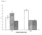
- the mean area of tumor attachment in the control group after 30 days was 640 ⁇ 112.5) mm 2 (C+dp), and 1691 ⁇ 123.6 mm 2 (C).
- C+dp The mean area of tumor attachment in the control group after 30 days was 640 ⁇ 112.5) mm 2 (C+dp), and 1691 ⁇ 123.6 mm 2 (C).
- PL75 group we measured 1082.43 ⁇ 169.4 mm 2 (PL75+dp) and 1048.42 ⁇ 209.3 mm 2 (PL75).
- PL150 group 480 ⁇ 41.6 mm 2 (PL150+dp) and 836.49 ⁇ 155.9 mm 2 (PL150) were proofed. A significant reduction of the area of tumor attachment was found only in the PL 150 group (Fig. 2).
- PCI Peritoneal Cancer Index
- the gastric cancer cell line NUGC-4 was derived from lymph node metastases from a 35 year old female.
- the histology of the original tumor described a poorly differentiated adenocarcinoma (Schwartz OK, Semin Oncol 1996; 23:316-24; and Akiyama S et al., Jpn J Surg 1988; 18:438-46).
- the experimental model of peritoneal dissemination in nude mice is well established (Nakashio T et al. Int J Cancer 1997; 70:612-8, Satta T et al., Gastroenterol Jpn 1993; 28:580) and was used in the present study.
- the human gastric cancer cell line NUGC-4 was purchased from the Japanese Cancer Research Resources Bank (Tokyo, Japan). The cells were maintained in monolayers in a tissue culture flask (75 cm 2 , Falcon, Becton Dikinson-Gambil, Heidelberg, Germany) in RPMI 1640 medium (GIBCO, Paisley, UK), supplemented with 10% fetal bovine serum (GIBCO), penicillin and streptomycin (GIBCO). Cell cultures were incubated at 37°C in a humidified atmosphere of 5% CO 2 in air. Cells were passaged after treatment with 0.125% trypsin for 6 min. The cells were pelleted after centrifugation for 10 min at 200g, suspended in 20 ml PBS, and pelleted again. The cell pellet was resuspended in 30 ml complete medium and seeded with a splitting ratio of 1:3. Only cells from three passages were used for the experiments. On the day of Operation 1x10 6 cells were suspended in 100 ⁇ l complete medium.
- mice A total of 90 female BALB/C nu/nu mice (mean body weight 20.2 +/- 2 g) were included in this study. The animals were kept in protective cages in pairs with unlimited access to water and standard mice chow. The mice were randomly assigned to nine different groups of equal numbers (Table 1). The surgical procedure was performed under sterile conditions and general anesthesia by intramuscular injection of ketamine 40 mg/kg (Ketamin 10%, Sanofi-Ceva, Düsseldorf, Germany) and 0,1 mg/kg KG Medetomidin (Dormitor).
- Tumor cells together with phospholipid solution were injected via a 1 cm midline incision into the peritoneal cavity.
- Group 2 additionally an abrasion of parietal and visceral peritoneum in the right flank and at the cecum was performed with a scalpell Before surgery and at the end of the observation period the body weight (g) was measured in all animals.
- the animals were sacrificed by a lethal dose of Isofluran by inhalation.
- the abdomen was opened by two paramedian incisions for complete exploration.
- the expanse of peritoneal carcinosis (mm 2 ) was measured using a digitizer board and calculated by custom-made Software on a personal computer. After subtle resection the tumor volume (ml) was measured by water displacement.
- PCI Peritoneal Cancer Index
- mice developed peritoneal carcinosis, regardless of treatment. No adverse side effects occurred after injection of phospholipids.
- PCI peritoneal cancer index
- the tumor volume in the control group registered 30 days postoperatively was 0.9 +/- 0.1 ml (dp) and 0.9 +/- 0.17 ml.
- the respective values in the PL75 group reached 0.67 +/- 0.14 ml (dp) and 1.0 +/- 0.13 ml.
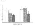
- Statistical analysis revealed no significant difference in both subgroups. After administration of PL150 the tumor volume in the subgroup with peritoneal lesions could be reduced to a value of 0.48 +/- 0.09 ml (dp) and 0.6 ⁇ 0.16. Both results were not significantly different to those in the control group. However, the p-values were 0.04 ( Figure 8).
- the mean area of tumor cell adhesion in the control group amounted to 217 +/- 31.5 mm 2 (dp) and 245 +/- 29.3 mm 2 in comparison with 184 +/- 28.3 mm 2 (dp) and 201 +/- 30.3 mm 2 in the PL75 group.
- the PL150 group developed an respective area of only 145 +/- 17 mm 2 (dp) and 164 +/- 32.8 mm 2 .
- the human rectal cancer cell line HRT-18 was purchased from the Japanese Cancer Research Resources Bank (Tokyo, Japan). The cells were maintained in monolayers in tissue culture flask (75 cm 2 , Falcon, Becton Dickinson-Gambil, Heidelberg, Germany) in RPMI 1640 medium (GIBCO, Düsseldorf, Germany), supplemented with 10% fetal bovine serum (GIBCO), penicillin and streptomycin (GIBCO). Cell cultures were incubated at 37°C in a humidified atmosphere of 5% CO2 in air. Cells were passaged after treatment with 0.125% trypsin for 6 min. The cells were pelleted after centrifugation for 10 min at 200g, suspended in 20 ml PBS, and pelleted. The cell pellet was resuspended in 30 ml complete medium and seeded with a splitting ratio of 1:3. Only cells from three passages were used for the experiments.
- colonic cancer cells were detached with collagenase I (15 min, 37°C, Worthington, Freehold, USA), washed once with RPMI 1640, centrifuged (200 g for 10 min), resuspended in RPMI 1640, and preincubated for 30 min in a humidified atmosphere of 5% CO2 in air (37°C). Seventy thousand tumor cells in 100 ⁇ l medium were seeded in ln and fn coated wells and 30.000 cells in coll IV coated wells according to dilution series. Evaluation of adherent cells was performed using cristal violet staining according to the method described by Aumeilley et al., and Tietze et al..
- the PL solution was added with the following concentrations: 0.1, 0.5, 0.75, 1 and 1.5 mg/100 ⁇ l medium. The concentrations used were correlated to our in vivo experiments.
- the human gastric cancer cell line NUGC-4 was purchased from the Japanese Cancer Research Resources Bank (Tokyo, Japan). The cells were maintained in monolayers in tissue culture flasks (75 cm 2 , Falcon, Becton Dickinson-GmbH, Heidelberg, Germany) in RPMI 1640 medium (GIBCO, Düsseldorf, Germany), supplemented with 10% fetal bovine serum (GIBCO), penicillin and streptomycin (GIBCO). Cell cultures were incubated at 37°C in a humidified atmosphere of 5% CO 2 in air. Cells were passaged after treatment with 0.125% trypsin for 6 min. The cells were pelleted after centrifugation for 10 min at 200g, suspended in 20 ml PBS, and pelleted again. The cell pellet was resuspended in 30 ml complete medium and seeded with a splitting ratio of 1:3. Only cells from three passages were used for the experiments.
- gastric cancer cells were detached with collagenase I (Worthington, Freehold, USA) for 15 min at 37°C, washed once with RPMI 1640, centrifuged (200 g for 10 min), resuspended in RPMI 1640, and pre-incubated for 30 min in a humidified atmosphere of 5% CO 2 in air at 37°C. 50.000 tumor cells in 100 ⁇ l medium were seeded into each well. Evaluation of adherent cells was performed using crystal violet staining according to the method described by Aumeilley et al., and Tietze et al.
- the phospholipid solution was added with the following concentrations: 0.05, 0.1, 0.5, 0.75, and 1 mg/100 ⁇ l medium. The concentrations used were correlated to the subsequent in vivo experiments.
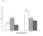
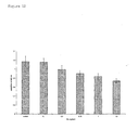
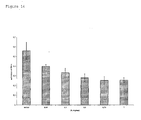
- Treatment with 0.1 or 0.5 mg PL/100 ⁇ l revealed extinction values of 0.33 ⁇ 0.05 and 0.28 ⁇ 0,04, respectively.
- the maximal effect could be demonstrated with 0.75 mg PL/100 ⁇ l with an extinction of 0.25 ⁇ 0.04.
- the relative reduction of tumor cell adhesion on laminin compared to the control amounts 59%.
- Treatment with 1 mg PL/100 ⁇ l showed no further decrease of tumor cell adhesion.
- the mean extinction was 0.26 ⁇ 0.02.
- Statistical evaluation revealed significant differences for all phospholipid concentrations compared to the control group (p ⁇ 0.0001) ( Figure 13 a,b; 14);.
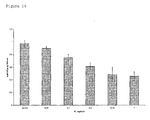
- the tumor cell adhesion on fn could not be reduced significantly with low phospholipid concentrations.
- Addition of 0.05 mg PL/100 ⁇ l and 0.1 mg PL/100 ⁇ l resulted in a slightly reduction of the extinction with mean values of 0.59 ⁇ 0.018 and 0.6 ⁇ 0.009.
- a significant reduction of tumor cell adhesion could be observed after treatment with 0.5 mg PL/100 ⁇ l, (ext. 0.44 ⁇ 0.023), as well as with 0.75 mg PL/100 ⁇ l, (ext. 0.41 ⁇ 0.025), and 1 mg PL/100 ⁇ l (ext. 0.39 ⁇ 0.02).
- the experimental evidence provided demonstrates that intraperitoneal PLs have an effect in reducing the adhesion of colorectal tumor cells in rats, gastric cancer cells in nuked mice and inhibit the adhesion of gastric cancer cells as well as rectal cancer cells on microtiter plates coated with collagen IV, laminin and fibronectin.
Landscapes
- Health & Medical Sciences (AREA)
- Life Sciences & Earth Sciences (AREA)
- Chemical & Material Sciences (AREA)
- Veterinary Medicine (AREA)
- Medicinal Chemistry (AREA)
- Pharmacology & Pharmacy (AREA)
- Animal Behavior & Ethology (AREA)
- General Health & Medical Sciences (AREA)
- Public Health (AREA)
- Epidemiology (AREA)
- Molecular Biology (AREA)
- Oncology (AREA)
- Dispersion Chemistry (AREA)
- Chemical Kinetics & Catalysis (AREA)
- General Chemical & Material Sciences (AREA)
- Nuclear Medicine, Radiotherapy & Molecular Imaging (AREA)
- Organic Chemistry (AREA)
- Pharmaceuticals Containing Other Organic And Inorganic Compounds (AREA)
- Acyclic And Carbocyclic Compounds In Medicinal Compositions (AREA)
- Medicinal Preparation (AREA)
- Organic Low-Molecular-Weight Compounds And Preparation Thereof (AREA)
Abstract
The present invention relates to the use of amphiphilic lipids
for the preparation of a pharmaceutical composition for reducing
tumor metastasis, preferably by prophylactic treatment of intraperitoneal
recurrence of tumors, specifically malignant tumors,
e.g. gastro-intestinal cancer, such as gastric, colon, or
pancreatic cancer, and cancer of the genito-urinary system such
as ovarian cancer and bladder cancer.
Description
- The present invention relates to the use of amphiphilic lipids for the preparation of a pharmaceutical composition for reducing tumor metastasis.
- Cancerous dieseases and tumors in general are among the major causes for human deaths and severe illness. Even after surgical removal of a tumor, patients frequently suffer from tumor disease, mostly from tumor metastases. The development of peritoneal carcinosis after surgical treatment of digestive cancers for example still is a frequent cause of recurrence. In gastric cancer with serosal invasion up to 50% of patients develop peritoneal carcinosis even if curative resection is performed (Boku T et al., Br J Surg 1990; 77:436-9; and Jansen M et al., Chirurg 2001; 72:561-5). Several studies demonstrated that detection of free, isolated tumor cells in the peritoneal cavity can serve as a prognostic marker for postoperative survival of colorectal and gastric cancer patients (Koga S et al., J Cancer Res Clin Oncol 1984; 108:236-8; Schott A et al., Ann Surg 1998; 227:372-9; Bando E et al., Am J Surg 1999; 178:256-62; and Broll R et al., Langenbecks Arch Chir 1996; 381:51-8).
- Peritoneal tumor recurrence may be a result of residual, intraperitoneal free tumor cells caused by serosal invasion of the primary tumor or intraoperative exfoliation. The invasion of tumor cells into the peritoneal wall is a complex process including attachment, proteolysis and migration (Schwartz OK, Semin Oncol 1996; 23:316-24). The most important step in developing peritoneal dissemination seems to reside in the adhesion of tumor cells to mesothelial cells or extracellular matrix components (Kiyasu Y et al., Cancer Res 1981; 41:1236-9; Koga S et al., Gann 1980; 71:8-13; and Schwartz OK, Semin Oncol 1996; 23:316-24). Experimental studies suggest that peritoneal metastases primarily tend to occur at sites of injured peritoneum (Yashiro M et al., Cancer 1996; 77:1668-75).
- Attachment of gastric cancer cells to the peritoneal surface represents the first step necessary in tumor cell invasion (Schwartz OK, Semin Oncol 1996; 23:316-24). In a scanning electron microscopic study Kiyasii et al. could demonstrate that cancer cells did not adhere to the mesothelial cells but to the free lying submesothelial connective tissue. The mesothelial cells, however, became hemispherical and exfoliation occurred in the presence of gastric cancer cells (Kiyasu Y et al., Cancer Res 1981; 41:1236-9).
- Since surgery alone remains an incomplete cure, numerous additional treatments have been evaluated. Intraperitoneal chemotherapy is a reliable method for treating peritoneal seeding, but frequent complications have been described (Hagiwara et al., Surgery 1988; 104:874-881; Fass et al., Langenbecks Arch Chir Suppl Kongress bd 1998; 115:1363-1366; Sayag et al., Oncology 1993; 50:333-337; Fujimura et al., Int Surg 1999; 84:60-66; Fujimoto et al., Ann Surg 1990; 212:592-596; and Koga et al., Cancer 1988; 61:232-237).
- Attempts were made to inhibit tumor cell attachment. Since the peritoneal tumor cell adhesion is mediated by adhesion molecules, antibodies with specificity for adhesion molecules were used to inhibit dissemination of human gastric cancer cells (Chailley-Heu B et al., Biochem J 1997; 328(Pt1):251-6). The main problem for the clinical use of respective antibodies seems to be the different expression of these antigens in tumors of different origin and other cell lines.
- Further, tests to reduce peritoneal metastases using dextran sulfate and sodium hyaluronate in animal models have been reported (Hagiwara A et al., Anticancer Drugs 1997; 8:894-7; and Haverlag R et al., Eur J Surg 1999; 165:791-5).
- Dextran sulfate led to a reduced tumor implantation at the sites of injury in the abdominal wall in mice (Hagiwara A et al., Anticancer Drugs 1997; 8:894-7; and Hagiwara A et al., Anticancer Drugs 2000; 11:873-7). At the same time the authors reported a prolonged survival in the treatment group after inoculation of melanoma cells. However, several side effects were observed when using dextran for adhesion prevention. The main problems were edema, pleura effusion, life-threatening malfunction of coagulation and severe allergic reactions (diZerega GS, Curr Opin Obstet Gynecol 1996; 8:230-7; and Treutner KH et al., Chirurg 2000; 71:510-7).
- Sodium hyaluronate proved to enhance colorectal tumor cell metastatic potential, probably mediated by the CD44 receptor (Tan et al., Br J Surg 2001; 88:246-250). It was thus concluded that this treatment leads to exponentiation of intraperitoneal tumor growth.
- In view of the above prior art, the present invention relates to the problem of medical treatment of cancerous diseases, specifically to the inhibition of tumor recurrence and metastases.
- According to the present invention this problem is solved by the use of amphiphilic lipids for the preparation of a pharmaceutical composition for reducing tumor metastasis.
- The present inventors have surprisingly shown that tumor metastases can significantly be reduced by administration of an amphiphilic lipid, preferably administration of a phospholipid, which results in an increase of the survival rates. The amphiphilic lipid is safe and causes no undesired side effects.
- The term "amphiphilic lipid" is used in this application to refer to an organic molecule with a glycerol (or polyol, such as alcohol) backbone substituted with two acyl chains coupled via ester bonds and a polar group substituted to the remaining hydroxyl group of the alcohol. The term amphiphilic clarifies that respective compounds contain both hydrophilic and lipophilic groups at the same time. The amphiphilic lipids to be used in accordance with the present invention form bilayers, preferably liposomes, upon dispersion in a liquid. For the medical use of the present invention it is necessary that the amphiphilic lipids are pharmaceutically acceptable. Any amphiphilic lipid which meets these requirements can be used in accordance with the present invention.
- Examples of preferred amphiphilic lipids to be used in accordance with the present invention comprise normal phospholipids, sphingomyelins (sphingolipids), galactolipids and other glycosylglycerides.
- The term "pharmaceutical composition" is used in the present application to refer amongst others to pharmaceutical compositions which have to be approved by a regulatory authority (EMEA, FDA, etc.). However, the term "pharmaceutical composition" is not limited to these compositions but encompasses any composition or medical device which contains amphiphilic lipids and is suitable for the claimed medical use.
- The present invention encompasses the use of a single specific type of amphiphilic lipid for the preparation of a pharmaceutical composition for reducing tumor metastasis as well as the use of a combination of various amphiphilic lipids for this purpose. The use of phospholipids is preferred for all embodibments of the present invention. More specifically, the present invention includes the medical use of known phospholipids for reducing tumor metastasis which have sofar been used as emulsifiers for the preparation of fett emulsions for parenteral nutrition.
- In the present application, the term phospholipid (PL) is used to refer to a glycerol backbone containing two acyl chains coupled via ester bonds and a polar, phosphorous containing group substituted to the remaining hydroxyl group of the glycerol. Respective polar lipid derivatives can be of natural, semi-synthetic or synthetic origin. Phospholipids can thus be provided by and be prepared from natural sources, since they are a common membrane component in cells of plant and animal origin. The naturally occuring phospholipids can be extracted and purified. Respective phospholipids have sofar for example been used for the preparation of fett emulsions for parenteral nutrition.
- There are several different sources of pharmaceutical grade phospholipids, e.g. egg and soybean. Depending on their origin, the detailed composition in terms of fatty acid composition as well as polar group composition will vary. Phospholipids of more defined molecular composition can be provided by a combination of purification and fractionation of endogenous phospholipids including a final synthetic step (semisynthetic phospholipids) or by a purely synthetic route (synthetic phospholipids).
- Lipids and more specifically phospholipids are natural components of the abdominal cavity. It was speculated that this substance may form a lubricant layer on the peritoneal surface (Beavis et al., Perit Dial Int 1994; 14:348-355; and Chailley-Heu et al., Biochem J 1997; 328(Pt1):251-256). Intraperitoneal application of phospholipids led to a significant decrease of adhesion formation, especially at sites of peritoneal injury, without any side effect on healing of anastomoses, laparotomy wounds, and liver incisions (Muller et al., J Surg Res 2001; 96:68-74; Muller et al., Langenbecks Arch Surg 2001; 386:278-284; and WO 98/535800). As of today, however, lipids as such, have not been suggested as active substance for treatment of cancerous diseases, in particular for reduction of metastasis.
- In the medical use of amphiphilic lipids according to the present invention tumor metastases are reduced amongst others due to inhibition of tumor cell adhesion, inhibition of adhesion of metastastatic cells and/or inhibition of tumor metastasis growth.
- In accordance with the present invention the amphiphilic lipids can be used for reduction of any tumor metastasis, such as recurrence of peritoneal carcinosis and various other cancers, for example, gastric, colon, ovarian or pancreatic cancer.
- The medical use of the present invention aims, of course, at reducing the occurance of metastases to zero. However, a medical use of amphiphilic lipids which achieves a significant reduction of the number of metastases and/or an increase in the survival rate already has to be considered as a significant improvement and is covered by the present invention.
- The amphiphilic lipids can be administered for prophylactic treatment. According to a specifically preferred embodiment, the amphiphilic lipids are used for prophylactic treatment of intraperitoneal recurrence of tumors, specifically malignant tumors, e.g. gastro-intestinal cancer and cancer of the genito-urinary system such as ovarian cancer or bladder cancer. The patient may have undergone curative resection before treatment or be subject to a respective surgical treatment shortly after administration of amphiphilic lipids.
- In the medical use of the present invention, the amphiphilic lipids may be present as the only active compound for reducing tumor metastasis in the pharmaceutical composition or may be present in combination with other active compounds, such as anti-tumor drugs, radioactive drugs, etc. The term "active compound" is used in the present application to refer to that compound in a composition which is responsible for the observed effect (reduction of tumor metastasis). Specific advantages will be achieved if the amphiphilic lipids are used as the only active compounds, since amphiphilic lipids do not cause any side effects in contrast to the majority of anti-tumor compounds and radioactive drugs, which are all known cause significant side effects.
- In one aspect of the present invention the amphiphilic lipids are used to treat a mammal. The treatment of humans is especially preferred.
- The amphiphilic lipids can be administered in any form and concentration which is clinically effective. According to one embodiment of the present invention a phospholipid is dispersed an aqueous solution. The solution preferably contains the phospholipid in a concentration of 0.05% to 25% (w/v), more preferably in a concentration of 0.5% to 20% (weight/weigt) and most preferably in a concentration of 1,5% to 10% (weight/weight).
- For administration the amphiphilic lipids may be formulated into any suitable pharmaceutical composition. The preparation of a dispersion for local administration, such as instillation or injection, wherein intraperitoneal administration is especially preferred.
- According to an especially preferred embodiment, the amphiphilic lipids are administered dispersed in an aqueous liquid and the amount of liquid administered is above the minimal coating volume, i.e. above the volume containing sufficient amounts of amphiphilic lipids necessary to coat all internal surfaces. The water will be absorbed by the organs and the amphiphilic lipids will form a coating on the internal surfaces.
- In a further aspect the present invention relates to methods for preparing pharmaceutical compositions for treating tumor metastasis, which method comprises stepes wherein amphiphilic lipids are mixed with an aqueous solution. The amphiphilic lipids are preferably dispersed in an aqueous solution. The dispersion can be obtained by a number of alternatives well known in the art such as high pressure homogenization. Preferably the dispersion is prepared in a manner such that liposomes with a particle size of 40 to 100 nm are obtained.
- The method for preparation of the pharmaceutical composition further preferably also comprises a step, wherein the dispersion or the components used for the preparation thereof are sterilized.
- Also in this embodiment of the present invention, the amphiphilic lipid can for example be a phospholipid, a sphingomyelin and/or a galactolipid. Further, the amphiphilic lipids can be a single specific type of amphiphilic lipid or a combination of different amphiphilic lipids. The pharmaceutical composition may but need not contain active compounds used for treatment of metastasis, such as anti-tumor drugs or radioactive drugs. According to an especially preferred embodiment the pharmaceutical composition does not contain any active compounds presently used for the treatment of tumors and metastasis. According to a further embodiment, the amphiphilic lipids are the only active drug compound in the pharmaceutical composition.
-
- Fig.1
- shows the results of analysis of tumor volume in the presence of various concentrations of PL as described in example 2.
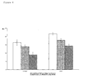
- Fig.2
- shows the results of analysis of mean area of tumor attachment in the presence of various concentrations of PL as described in example 2.
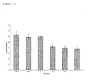
- Fig.3
- shows the results of analysis of the Peritoneal Cancer Index (PCI) in the presence of various concentrations of PL as described in example 2.
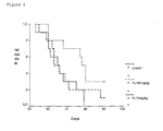
- Fig.4
- shows the results of the analysis of the relative survival of the test animals used in example 2.
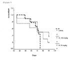
- Fig.5
- shows the results of the relative survival of the test animals used in example 3.
- Fig.6
- shows the results of analysis of the Peritoneal Cancer Index (PCI) in the presence of various concentrations of PL as described in example 3.
- Fig.7
- shows cecum with abrasion of peritoneum
- a. 30 days after PL150
- b. 30 days after NaCl
- Fig.8
- shows the results of analysis of tumor volume in the presence of various concentrations of PL as described in example 3.
- Fig.9
- shows the results of analysis of mean area of tumor attachment in the presence of various concentrations of PL as described in example 3.
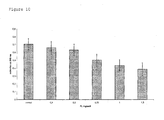
- Fig.10
- shows the results of the adhesion assay of example 4, i.e. inhibition of adhesion of rectal cancer cells on microtiter plates coated with ln in the presence of various concentrations of PL.
- Fig.11
- shows the results of the adhesion assay of example 4, i.e. inhibition of adhesion of rectal cancer cells on microtiter plates coated with coll IV in the presence of various concentrations of PL.
- Fig.12
- shows the results of the adhesion assay of example 4, i.e. inhibition of adhesion of gastric cancer cells on microtiter plates coated with fn in the presence of various concentrations of PL.
- Fig.13
- shows the results of the adhesion assay of example 5,
- a) adhesion of gastric cancer cells on laminin in the presence of 1.0 g PL.
- b) adhesion of gastric cancer cells on laminin in the control group.
- Fig.14
- shows the results of the adhesion assay of example 5, i.e. inhibition of adhesion of gastric cancer cells on microtiter plates coated with ln in the presence of various concentrations of PL.
- Fig.15
- shows the results of the adhesion assay of example 5, i.e. inhibition of adhesion of gastric cancer cells on microtiter plates coated with fn in the presence of various concentrations of PL.
- Fig.16
- shows the results of the adhesion assay of example 5, i.e. inhibition of adhesion of gastric cancer cells on microtiter plates coated with coll IV in the presence of various concentrations of PL.
- The method for preparing the PLs may depend on the chosen source of the PLs and the molecules to be obtained. The level of purification can be of particular importance for PLs of natural origin. Purification methods to achieve quality readily acceptable for intravenous use, based on e.g. repeated solvent extraction/precipitation processes, are known to those skilled in the art (F Nielloud, G Marti Mestres, Pharmaceutical Emulsions and Suspensions, Chs 1.VB , 6.VI Marcel Dekker,
NY 2000; with further references). - To provide an PLs in a form suitable for the intended purpose, the phospholipids are regularly formulated to a liposomal dispersion which can be manufactured and stored until use. A liposomal dispersion will readily adsorb onto the internal surfaces of the patient, such as the internal surfaces of the peritoneum, and thus form mono- or multilamellar layers which will prevent adhesion of tumor cells to the coated surfaces.
- From a pharmaceutical point of view such a liposome dispersion should fulfil several conditions. For stability and functionality reasons, the dispersion should preferably be prepared using high pressure homogenisation or other methods that are suitable for obtaining very small particulate size (sub-micron). In addition, the solution should be sterile. Apropriate sterility assurance levels are reached by thermal sterilisation which is used as the preferred choice for achieving sterility. This puts, however, strains on the physiochemical stability of the formulation.
- Optimised formulations containing appropriately balanced phospholipids with good bilayer forming properties, or formulated with additives which could help to provide a stable liposomal dispersion, can redily be prepared using industrial methods for preparation and sterilisation (I Hanin and G Pepeu (eds.): "Phospholipids" p 115-122, Plenum Press NY 1990; M J Groves, "Parenteral Products", Heinemann 1973).
- The following phospholipid dispersions were prepared:
- 1. 30 g egg phospolipids
25 g glycerol
NaOH to pH = 8
Water for injection (WFI) to 1000 ml - 2. 20 g lecithin
NaCl/phosphate buffer to pH = 7 and isotonicity
WFI to 1000 ml - 3. 20 g lecithin (phosphatidylcholin)
0,1 g natriumoleate
25 g glycerol
WFI to 1000 ml - 4. 25 g phosphatidylcholin
5 g phosphatidylethanolamin
NaCl phosphate buffer to pH = 7 and isotonicity
WFI to 1000 ml -
- The PL powder was stirred in the re-cited liquid and subjected to high pressure homogenization until a very fine dispersion of liposomes containing particles having a size in the range of 50-100nm are obtained. The dispersion thus obtained was sterilized by autoclaving and stored until further use.
- In some of the following examples the dispersion 1., above, has been used.
- Developing peritoneal carcinosis after intraperitoneal injection of tumor cells (cell line: DHD/K12/TRb) into BD IX rats is a well established animal model (Dunnington et al., Int J Cancer 1987; 39:248-254; Reisser et al., Bull Cancer 1991; 78:249-252; and Qin et al., Int J Cancer 1991; 47:765-770) that was used in the present experiment to see if the phospholipids have a therapeutic effect.
- The cell line DHD/K12/TRb was used. The cells were obtained from a colon adenocarcinoma induced in syngenic BD-IX rats (Garcia-Olmo et al., Cancer Lett 1998; 132:127-133). The cell line was purchased from the European Collection of Animal Cell Cultures (ecacc, Salisbury, UK).
- The cells were cultivated in monolayers in a tissue culture flask (75 cm2 Falcon, Becton Dicidson-Gambil, Heidelberg, Germany) in DMEM and Ham's F10 (1:1; GIBCO) supplemented with 10% fetal bovine serum (GIBCO) and 0.005% gentamycin (GIBCO). Cell cultures were incubated at 37°C in a humidified atmosphere of 5% CO2 in air. Cells were passaged after treatment with 0.125% trypsin for 2 min. The cells were pelleted after centrifugation for 10 min at 200g, suspended in 20 ml PBS, and pelleted again. The cell pellet was resuspended in 30 ml complete medium and seeded with a splitting ratio of 1:3. Only cells from three passages were used for the experiments. On the day of Operation 2x10⁶ cells were suspended in 100µl complete medium.
- A total of 90 female BD-IX rats (mean
body weight 250 +/- 28 g) were included in this study. The rats were kept in protective cages in pairs with unlimited access to water and standard rat chow. The animals were randomly assigned to nine different groups of equal numbers (Table 1). The surgical procedure was performed under sterile conditions and general anesthesia by intramuscular injection ofketamin 100 mg/kg (Ketamin 10%, Sanofi-Ceva, Düsseldorf, Germany) andxylacine 5 mg/kg (Rompun 2%, Bayer, Leverkusen, Germany).30 days - peritoneal lesions 30 days + peritoneal lesions 90 days Phospholipids 75 mg/kg (PL75) 10 10 10 Phospholipids 150 mg/kg (PL150)10 10 10 NaCl 0,9%10 10 10 - Tumor cells were injected together with phospholipid solution via a 2 cm midline incision into the peritoneal cavity. In
Group 2 additionally an abrasion of parietal and visceral peritoneum in the right flank and at the cecum was performed using a scalpell. (Muller et al., J Surg Res 2001; 96:68-74). Before surgery and at the end of the observation period the body weight (g) was measured in all animals. - After intervals of 30 and 90 days, respectively, the animals were sacrificed by a lethal dose of Isofluran by inhalation. The abdomen was opened by two paramedian incisions for complete exploration. The expanse of peritoneal carcinosis (mm2) was measured using a digitizer board and calculated by custom-made Software on a personal Computer (Treutner KH et al., J Surg Res 1995; 59:764-71). After subtile resection the tumor volume (ml) was measured by water displacement.
- Furthermore, the Peritoneal Cancer Index (PCI), as described by Sugarbaker et al. (Sugarbaker PH et al., Cancer Treat Res 1996; 81:149-68), was determined. The PCI was adapted to tumor sizes in rats. The following lesion size scores were used; LS-I: Tumorsize < 2 mm, LS-2: 2.1-5 mm; LS-3: > 5 mm or confluence. The abdominal exploration was carried out by a independent, blinded observer.
- Finally, specimens from the abdominal wall, cecum, jejunum, liver, spleen and omentum majus were taken and fixed in formaldehyde solution. Sections with a thickness of 5 µm were stained with hematoxylin and eosin for light microscopy.
- All data are expressed as means +/- Standard error of the mean (SEM). Statistical analysis was performed with a two way ANOVA with contrasts according to pairwise comparison. The level of significance was adjusted according to Bonferroni to alpha/18 = 0,00278. Survival curves were compared using the Log-Rank-Test.
- Performance of the initial surgical procedure was uneventful in all animals. All rats developed peritoneal carcinosis regardless of the treatment. No adverse side effects were noted after injection of phospholipids.
- After 30 days, the mean reduction of body weight was 18 ± 9 g in the control group (C). In the PL75 group as well as in the PL150 group the body weight remained nearly constant with differences of 1 ± 2.8 g and -2 ± 5 g, respectively. Those differences were not significant.
- The tumor volume in the control group reached 9.27 ± 1.5 ml with deperitonealisation (C+dp) and 14.45 ± 0.75 ml without peritoneal lesions (C). In the trial group without deperitonealisation, we found a significant reduction of the tumor volume after treatment with both higher phospholipid concentrations to 7 ± 0.59 ml (PL75) and 7.13 ± 0.91 ml (PL150) respectively (p<0.0001). Although a marked reduction of the tumor volume could be found in the PL150+dp, the difference was not significant compared to the control group (p = 0.0817) (Fig.1).
- The mean area of tumor attachment in the control group after 30 days was 640 ± 112.5) mm2 (C+dp), and 1691 ± 123.6 mm2 (C). In the PL75 group we measured 1082.43 ± 169.4 mm2 (PL75+dp) and 1048.42 ± 209.3 mm2 (PL75). In the PL150 group 480 ± 41.6 mm2 (PL150+dp) and 836.49 ± 155.9 mm2 (PL150) were proofed. A significant reduction of the area of tumor attachment was found only in the
PL 150 group (Fig. 2). - Concerning the Peritoneal Cancer Index (PCI) the control group reached a value of 39 ± 2.03 (C+dp) and 43 ± 1.125 (C), respectively. In comparison to the control group, we found a significant reduction in the PL150 group with 29.6 ± 1.97 (PL150+dp) , and 25.7 ± 2.29 (PL150). The PCI registered in the PL75 group was equal in the subgroup with peritoneal lesions (38.2 ± 1.14, PL75+dp) and significantly reduced in the second subgroup (33 ± 1.62, PL75) (Fig. 3).
- During the 90 day observation period only one animal in the control group survived. The median survival time was 68.5 ± 3.56. All rats in the PL75 group died because of tumor progression until the eighties postoperative day with a median survival time of 66.5 ± 2.79. Survival in the PL150 group (median 74.3 ± 2.71), however, was obviously prolonged compared to the control group although the difference was not significant (p = 0.13). (Fig. 4).
- Infiltration of colonic cancer could be proofed in all animals histologically. The evidence of tumor infiltration was restricted to the serosa. Invasion into deeper layers could not be detected. A exophytic tumor growth was found. From the histological point no differences occurred in the intraperitoneal tumor growth in the control group compared to the trial groups.
- The gastric cancer cell line NUGC-4 was derived from lymph node metastases from a 35 year old female. The histology of the original tumor described a poorly differentiated adenocarcinoma (Schwartz OK, Semin Oncol 1996; 23:316-24; and Akiyama S et al., Jpn J Surg 1988; 18:438-46). The experimental model of peritoneal dissemination in nude mice is well established (Nakashio T et al. Int J Cancer 1997; 70:612-8, Satta T et al., Gastroenterol Jpn 1993; 28:580) and was used in the present study.
- The human gastric cancer cell line NUGC-4 was purchased from the Japanese Cancer Research Resources Bank (Tokyo, Japan). The cells were maintained in monolayers in a tissue culture flask (75 cm2, Falcon, Becton Dikinson-Gambil, Heidelberg, Germany) in RPMI 1640 medium (GIBCO, Paisley, UK), supplemented with 10% fetal bovine serum (GIBCO), penicillin and streptomycin (GIBCO). Cell cultures were incubated at 37°C in a humidified atmosphere of 5% CO2 in air. Cells were passaged after treatment with 0.125% trypsin for 6 min. The cells were pelleted after centrifugation for 10 min at 200g, suspended in 20 ml PBS, and pelleted again. The cell pellet was resuspended in 30 ml complete medium and seeded with a splitting ratio of 1:3. Only cells from three passages were used for the experiments. On the day of Operation 1x10⁶ cells were suspended in 100µl complete medium.
- A total of 90 female BALB/C nu/nu mice (mean body weight 20.2 +/- 2 g) were included in this study. The animals were kept in protective cages in pairs with unlimited access to water and standard mice chow. The mice were randomly assigned to nine different groups of equal numbers (Table 1). The surgical procedure was performed under sterile conditions and general anesthesia by intramuscular injection of
ketamine 40 mg/kg (Ketamin 10%, Sanofi-Ceva, Düsseldorf, Germany) and 0,1 mg/kg KG Medetomidin (Dormitor). - Tumor cells together with phospholipid solution were injected via a 1 cm midline incision into the peritoneal cavity. In
Group 2 additionally an abrasion of parietal and visceral peritoneum in the right flank and at the cecum was performed with a scalpell Before surgery and at the end of the observation period the body weight (g) was measured in all animals. - After intervals of 30 and 90 days, respectively, the animals were sacrificed by a lethal dose of Isofluran by inhalation. The abdomen was opened by two paramedian incisions for complete exploration. The expanse of peritoneal carcinosis (mm2) was measured using a digitizer board and calculated by custom-made Software on a personal computer. After subtle resection the tumor volume (ml) was measured by water displacement.
- Further, the Peritoneal Cancer Index (PCI), as described by Sugarbaker et al., was determined (Sugarbaker PH et al., Cancer Treat Res 1996; 81:149-68). The PCI was adapted to tumor sizes in mice. The following lesion size scores were used; LS-I: Tumorsize < 2 mm, LS-2: 2.1-5 mm; LS-3: > 5 mm or confluence. The abdominal exploration was carried out by an independent, blinded observer.
- Finally, specimens from the abdominal wall, cecum, jejunum, liver, spleen and omentum were taken and fixed in formaldehyde solution. Sections with a thickness of 5 µm were stained with hematoxylin and eosin for light microscopy.
- All data are expressed as means +/- Standard error of the mean (SEM). Statistical analysis was performed with a two way ANOVA with contrasts according to pairwise comparison. The level of significance was adjusted according to Bonferroni to alpha/18 = 0,00278. Survival curves were compared using the Log-Rank-Test.
- All mice developed peritoneal carcinosis, regardless of treatment. No adverse side effects occurred after injection of phospholipids.
- In the control group no mouse lived longer than 75 days. The median survival time was 69 ± 3 days. In the PL150 group the relative survival was longer with a median survival time of 73 ± 5 days. In the PL75 group four animals survived the complete 90 days observation period. However, the median survival time was only 59 ± 6 days (Figure 5).
- After 30 days the peritoneal cancer index (PCI) in the control group was 16.4 +/- 1.7 without and 21.7 +/- 0.77 with peritoneal lesions (dp). Minor changes occurred after treatment with PL75. The PCI was 14 +/- 1 and 17.8 +/- 1.09 (dp), respectively. However, the differences were not significant compared to the control values. A significant lower PCI was achieved in the PL150 group with 9 +/- 1.68 (p = 0.0002) and 14.3 +/- 1.07 (p = 0.0001) after local deperitonealization (Figure 6).
- Tumor cell attachment was predominantly found at the site of abdominal incision. If standard peritoneal lesions had been performed, peritoneal carcinosis was focused at these sites (Figure 7 a, b). The mean PCI "Colon" in the control group amounted nearly the maximal value (2.9 +/- 0.09), whereas PCI registered at those sites was reduced in the treatment groups. But, as a consequence of evaluating the complete colon instead of the cecum, we could not find a significant influence of PL on the tumor adhesion compared to the control group in this area. At the PCI "right flank" in both trial groups was decreased significantly. 30 days after treatment the values were 1.7 +/-0.44 in the PL75 group and 0.9 +/- 0.44 in PL150 group, respectively, compared to 2.8 ± 0.13 in the control group.
- The tumor volume in the control group registered 30 days postoperatively was 0.9 +/- 0.1 ml (dp) and 0.9 +/- 0.17 ml. The respective values in the PL75 group reached 0.67 +/- 0.14 ml (dp) and 1.0 +/- 0.13 ml. Statistical analysis revealed no significant difference in both subgroups. After administration of PL150 the tumor volume in the subgroup with peritoneal lesions could be reduced to a value of 0.48 +/- 0.09 ml (dp) and 0.6 ± 0.16. Both results were not significantly different to those in the control group. However, the p-values were 0.04 (Figure 8). The mean area of tumor cell adhesion in the control group amounted to 217 +/- 31.5 mm2 (dp) and 245 +/- 29.3 mm2 in comparison with 184 +/- 28.3 mm2 (dp) and 201 +/- 30.3 mm2 in the PL75 group. The PL150 group developed an respective area of only 145 +/- 17 mm2 (dp) and 164 +/- 32.8 mm2. The difference between the control group versus PL150 in both groups was not significant, but the p-values were very low with p = 0.049 in the trial without peritoneal lesions and p = 0.08 in the trial with lesions (Figure 9).
- These results thus show that PL can be used to inhibit peritoneal tumor cell adhesion especially in the abdominal wall.
- In this in vitro study the influence of phospholipids on adhesion of colonic cancer cells to extracellular matrix components was determined. Specifically, the reactivity of the cancer cells with several integrins, namely main basement membrane components Collagen IV (coll IV) and laminin (ln) as well as fibronectin (fn), which plays an important role in wound healing, were analyzed.
- The human rectal cancer cell line HRT-18 was purchased from the Japanese Cancer Research Resources Bank (Tokyo, Japan). The cells were maintained in monolayers in tissue culture flask (75 cm2, Falcon, Becton Dickinson-Gambil, Heidelberg, Germany) in RPMI 1640 medium (GIBCO, Karlsruhe, Germany), supplemented with 10% fetal bovine serum (GIBCO), penicillin and streptomycin (GIBCO). Cell cultures were incubated at 37°C in a humidified atmosphere of 5% CO2 in air. Cells were passaged after treatment with 0.125% trypsin for 6 min. The cells were pelleted after centrifugation for 10 min at 200g, suspended in 20 ml PBS, and pelleted. The cell pellet was resuspended in 30 ml complete medium and seeded with a splitting ratio of 1:3. Only cells from three passages were used for the experiments.
- Flat-bottom polystyrene microtiter plates (Becton Dickinson, Heidelberg, Germany) were coated for adhesion experiments. The purified ECM components were dissolved in PBS with the following concentrations: Collagen IV (coll IV) - 2,5 µg/ml (Biomol, Hamburg, Germany), Fibronektin (fn) - 10 µg/ml (Boehringer, Mannheim, Germany) and Laminin (In) - 50 µg/ml (Boehringer, Mannheim, Germany). We found these concentrations to be optimal in foregoing dilution series. They were added to the wells and incubated at 4°C for 24 hours (coll IV, fn), or at 37°C for 45 min in a humidified atmosphere of 5% CO2 in air (ln) respectively. Nonspecific protein binding of the coated wells was blocked by adding 1% (w/v) BSA (4°C, 12h) and rinsing the wells with Hepes buffer.
- For adhesion experiments colonic cancer cells were detached with collagenase I (15 min, 37°C, Worthington, Freehold, USA), washed once with RPMI 1640, centrifuged (200 g for 10 min), resuspended in RPMI 1640, and preincubated for 30 min in a humidified atmosphere of 5% CO2 in air (37°C). Seventy thousand tumor cells in 100 µl medium were seeded in ln and fn coated wells and 30.000 cells in coll IV coated wells according to dilution series. Evaluation of adherent cells was performed using cristal violet staining according to the method described by Aumeilley et al., and Tietze et al.. After the incubation time of 30 min the supernatant with non adherent cells was removed by two washes with warmed RPMI 1640. Attached cells were fixed with 30% (v/v) methanol/ethanol for 15 min at room temperature. Cells were stained with 0.1% (w/v) crystal violet (Sigma, Hamburg, Germany), extensively washed with distilled water, and dried at room temperature. The dye was resuspended with 50 µl of 0.2% (v/v) Triton X-100/well and color yields were then measured in an ELISA reader at 590 nm (Titertek Multiscan Plus MKII, Flow Laboratories GmbH, Meckenheim, Germany). Optical density (OD) showed a linear relationship to the amount of cells between 1 x 10³ and 1 x 10⁵ cells per well, as determined by dilution series.
- Control dying of BSA/polystyrene without cells led to minimal Optical Density (OD) values of 0.01-0.07. These values were subtracted from those obtained in the experiments Phospholipids
- After complete preparation of the tumor cell suspension the PL solution was added with the following concentrations: 0.1, 0.5, 0.75, 1 and 1.5 mg/100 µl medium. The concentrations used were correlated to our in vivo experiments.
- All experiments were performed three times in quadruplicate. The data are expressed as means +/- standard error of the mean (SEM). One way ANOVA with contrasts according to pairwise comparison was used for statistical analysis. The level of significance was adjusted according to Bonferroni to alpha/10 = 0,005.
- In the adhesionessay of human rectal cells (HRT-18) on laminin after an incubation time of 30 min a mean extinction of 0.7 ± 0.07 was detected in the control group. Addition of phospholipids with a concentration of 0.75 (ext. 0.5 ± 0.08), 1 (ext. 0.44 ±0.07) and 1.5 (ext. 0.39 ± 0.08) mg/well led to a significant reduction of tumor cell adhesion (p < 0.0001). Treatment with a concentration of 1.5 mg/well reduced the cell attachment to 46% compared to the control group (Figure 10).
- The extinction after incubation of 30.000 tumor cells on collagen IV was 0.34 ± 0.03. While treatment with the lowest concentration of phospholipids (0.1 mg/well) led to an increase of tumor cell adhesion (ext. 0.5 ± 0.03) a reduction of 24 % could be reached with 1.5 mg Phospholipids/well (ext. 0.26 ± 0.02). The differences observed in this trial were compared to the control group were only significant with the maximum concentration (p < 0.0001) (Figure 11).
- On fibronectin we observed a reduction of tumor cell adhesion analog to the experiments on laminin. Already the treatment with 0.5 mg phospholipids/well (ext. 0.99 ± 0.09) led to a significant reduction of cell attachment compared to the extinction in the control group with a value of 1.17 ± 0.1 (p = 0.0005). The influence of higher phospholipid concentrations (0.75, 1, 1,5 mg/well) was also significant (p < 0.0001). The maximum reduction of tumor cell adhesion on fibronectin was observed after addition of 1.5 mg phospholipids/well (37%) (Figure 12).
- In this in vitro study the influence of phospholipids on adhesion of gastric cancer cells to extracellular matrix components was determined.
- The human gastric cancer cell line NUGC-4 was purchased from the Japanese Cancer Research Resources Bank (Tokyo, Japan). The cells were maintained in monolayers in tissue culture flasks (75 cm2, Falcon, Becton Dickinson-GmbH, Heidelberg, Germany) in RPMI 1640 medium (GIBCO, Karlsruhe, Germany), supplemented with 10% fetal bovine serum (GIBCO), penicillin and streptomycin (GIBCO). Cell cultures were incubated at 37°C in a humidified atmosphere of 5% CO2 in air. Cells were passaged after treatment with 0.125% trypsin for 6 min. The cells were pelleted after centrifugation for 10 min at 200g, suspended in 20 ml PBS, and pelleted again. The cell pellet was resuspended in 30 ml complete medium and seeded with a splitting ratio of 1:3. Only cells from three passages were used for the experiments.
- Flat bottom polystyrene microtiter plates (Becton Dickinson, Heidelberg, Germany) were coated for adhesion experiments. The purified ECM components were dissolved in PBS with the following concentrations: coll IV - 2,5 µg/ml (Biomol, Hamburg, Germany), fn - 10 µg/ml (Boehringer, Mannheim, German), In - 50 µg/ml (Boehringer, Mannheim, Germany). They were added to the wells and incubated at 4°C for 24 hours (coll IV, fn) or at 37°C for 45 minutes in a humidified atmosphere of 5% CO2 in air (ln), respectively. Non-specific protein binding of the coated wells was blocked by adding 1% (w/v) BSA (4°C, 12 h) and rinsing the wells with hepes buffer.
- For adhesion experiments gastric cancer cells were detached with collagenase I (Worthington, Freehold, USA) for 15 min at 37°C, washed once with RPMI 1640, centrifuged (200 g for 10 min), resuspended in RPMI 1640, and pre-incubated for 30 min in a humidified atmosphere of 5% CO2 in air at 37°C. 50.000 tumor cells in 100 µl medium were seeded into each well. Evaluation of adherent cells was performed using crystal violet staining according to the method described by Aumeilley et al., and Tietze et al. (Aumailley et al., Exp Cell Res 1989; 181(2) :463-474; and Tietze et al., Exp Mol Pathol 1999; 66(2):131-139). After the incubation time of 30 minutes the supernatent with non-adherent cells was removed by two washes with warmed RPMI 1640.
- Attached cells were fixed with 30% (v/v) methanol/ethanol for 15 min at room temperature. Cells were stained with 0.1% (w/v) crystal violet (Sigma, Hamburg, Germany), extensively washed with distilled water, and dried at room temperature. The dye was resuspended with 50 µl of 0.2% (v/v) Triton X-100/well and color yields were then measured in an ELISA reader at 590 nm (Titertek Multiscan Plus MKII, Flow Laboratories GmbH, Meckenheim, Germany). Optical density (OD) showed a linear relationship to the amount of cells between 1x10³ and 5x10⁴ cells per well, as determined by dilution series. Control dying of BSA/polystyrene without cells led to Optical Density (OD) values of 0.01-0.07. These values were subtracted from those obtained in the experiments.
- After complete preparation of the tumor cell suspension, the phospholipid solution was added with the following concentrations: 0.05, 0.1, 0.5, 0.75, and 1 mg/100 µl medium. The concentrations used were correlated to the subsequent in vivo experiments.
- All experiments were performed three times in quadruplicate. The data are expressed as means +/- Standard error of the mean (SEM). One way ANOVA with contrasts according to pairwise comparison was used for statistical analysis. The level of significance was adjusted according to Bonferroni to alpha/10 = 0,005.
- The analysis of tumor cell adhesion to
BSA 1% led to a mean extinction of 0.27 ± 0.013 at 590 nm. Coating with laminin (ln) and fibronectin (fn) led to a nearly twofold increase of tumor cell adhesion with mean values of 0.56 ± 0.09 (ln) and 1.17 ± 0.1 (fn), respectively. The cancer cells showed a most pronounced adhesion to collagen IV (coll IV) with a mean extinction of 0.97 ± 0.05.
The tumor cell adhesion on ln registered after addition of PL was significantly reduced. The effect was concentration dependent compared to the control. Even the minimal amount of 0.05 mg PL/100µl led to a reduced extinction of 0.4 ± 0,02. Treatment with 0.1 or 0.5 mg PL/100µl revealed extinction values of 0.33 ± 0.05 and 0.28 ± 0,04, respectively. The maximal effect could be demonstrated with 0.75 mg PL/100µl with an extinction of 0.25 ± 0.04. The relative reduction of tumor cell adhesion on laminin compared to the control amounts 59%. Treatment with 1 mg PL/100µl showed no further decrease of tumor cell adhesion. The mean extinction was 0.26 ± 0.02. Statistical evaluation revealed significant differences for all phospholipid concentrations compared to the control group (p < 0.0001) (Figure 13 a,b; 14);. - The tumor cell adhesion on fn could not be reduced significantly with low phospholipid concentrations. Addition of 0.05 mg PL/100µl and 0.1 mg PL/100µl resulted in a slightly reduction of the extinction with mean values of 0.59 ± 0.018 and 0.6 ± 0.009. However, a significant reduction of tumor cell adhesion could be observed after treatment with 0.5 mg PL/100µl, (ext. 0.44 ± 0.023), as well as with 0.75 mg PL/100µl, (ext. 0.41 ± 0.025), and 1 mg PL/100µl (ext. 0.39 ± 0.02). We found a similar situation compared to ln with equal effects of both 0.75 mg/100µl and 1 mg/100µl PL indicating that the maximum influence on adhesion is reached. The relative reduction of tumor cell adhesion compared to the control values amounts 35% (Figure 15).
NUGC-4 gastric cancer cells adhere to collagen IV increasingly compared to all other examined extracellularmatrix components. The influence of phospholipids on cell adhesion to coll IV was also concentration dependent. The reduction ranged from a mean extinction of 0.9 ± 0.02 after administration of 0.05 mg PL/100µl to a maximal effect after treatment with 1 mg PL/100µl, with a mean value of 0.46 ± 0.03. Significant differences were found from 0.1 to 1 mg PL/well (p < 0.0001). In comparison to the control value, this means a maximal reduction of adhesive tumor cells of 53% (Figure 16). - In summary, the experimental evidence provided demonstrates that intraperitoneal PLs have an effect in reducing the adhesion of colorectal tumor cells in rats, gastric cancer cells in nuked mice and inhibit the adhesion of gastric cancer cells as well as rectal cancer cells on microtiter plates coated with collagen IV, laminin and fibronectin. These results thus show that PLs inhibit peritoneal tumor cell adhesion and are thus suitable to reduce tumor metastasis.
Claims (20)
- Use of amphiphilic lipids for preparation of a pharmaceutical composition for reducing tumor metastasis.
- Use of amphiphilic lipids according to claim 1, wherein the amphiphilic lipid is a phospholipid, a sphingomyelin and/or a galactolipid.
- Use of amphiphilic lipids according to claim 2, wherein a single specific type of amphiphilic lipid or a combination of different amphiphilic lipids are used for the preparation of the pharmaceutical composition.
- Use of amphiphilic lipids according to claim 1, wherein tumor metastases are reduced due to inhibition of tumor cell adhesion, due to inhibition of adhesion of metastastatic cells and/or inhibition of tumor metastasis growth.
- Use of amphiphilic lipids according to one of claims 1 to 4, wherein tumor metastases are reduced by prophylactic treatment of intraperitoneal recurrence of tumors, specifically malignant tumors, e.g. gastro-intestinal cancer, such as gastric, colon, or pancreatic cancer, and cancer of the genito-urinary system such as ovarian cancer or bladder cancer.
- Use of amphiphilic lipids according to claim 5, wherein the amphiphilic lipids are used as the only active compound for preparation of the pharmaceutical composition for reducing tumor metastasis or the amphiphilic lipids are used in combination with one or more further active compounds for the preparation of the pharmaceutical composition.
- Use of amphiphilic lipids according to one of claims 1 to 6, wherein the further one or more further active compounds for the preparation of the pharmaceutical composition are selected from anti-tumor drugs and radioactive drugs.
- Use of amphiphilic lipids according to one of claims 1 to 7, wherein the phospholipid is used for therapeutic or prophylactic treatment of patients.
- Use of amphiphilic lipids according to claim 8, wherein the patient has undergone curative or palliative resection.
- Use of amphiphilic lipids according to claims 8 or 9, wherein the patient is a mammal.
- Use of amphiphilic lipids according to claim 10, wherein the patient is a human.
- Use of amphiphilic lipids according to one of claims 1 to 11, wherein the amphiphilic lipid is a phospholipid which is present as an aqueous solution containing 0.05% to 25% (w/v) of a phospholipid.
- Use according to claim 12, wherein the phospholipid is present as an aqueous solution containing 0.5% to 20% (w/v) of a phospholipid.
- Use according to claim 13, wherein the phospholipid is present as an aqueous solution containing 1.5% to 10% (w/v) of a phospholipid.
- Use of a phospholipid according to one of claims 12 to 14, wherein the phospholipid is formulated into a solution for local administration, such as instillation or injection, preferably for intraperitoneal administration.
- Method for preparing a pharmaceutical composition for reducing tumor metastasis, which method comprises steps, wherein amphiphilic lipids are mixed with an aqueous solution.
- Method according to claim 16, wherein the amphiphilic lipids are dispersed in the aqueous solution.
- Method according to claim 16 or 17, wherein the dispersion is obtained by high pressure homogenization.
- Method according to any of claims 16 or 18, wherein the tumor metastases are reduced due to inhibition of tumor cell adhesion, due to inhibition of adhesion of metastastatic cells and/or inhibition of tumor metastasis growth.
- Method according to claim 16 or 17, wherein tumor metastases are reduced by prophylactic treatment of intraperitoneal recurrence of tumors, specifically malignant tumors, e.g. gastro-intestinal cancer, such as gastric, colon, or pancreatic cancer, and cancer of the genito-urinary system such as ovarian cancer.
Priority Applications (14)
| Application Number | Priority Date | Filing Date | Title |
|---|---|---|---|
| EP02026897A EP1426053A1 (en) | 2002-12-03 | 2002-12-03 | Use of amphiphilic lipids for reducing tumor metastasis |
| BR0316977-4A BR0316977A (en) | 2002-12-03 | 2003-12-01 | Use of amphiphilic lipids for the preparation of a pharmaceutical composition for tumor metastasis reduction |
| KR1020117013834A KR20110091010A (en) | 2002-12-03 | 2003-12-01 | Use of Amphiphilic Lipids to Prepare Pharmaceutical Compositions for Lowering Tumor Metastasis |
| MXPA05005759A MXPA05005759A (en) | 2002-12-03 | 2003-12-01 | Use of amphiphilic lipids for the preparation of a pharmaceutical composition for reducing tumor metastasis. |
| KR1020057009811A KR101060442B1 (en) | 2002-12-03 | 2003-12-01 | Use of Amphiphilic Lipids to Prepare Pharmaceutical Compositions for Lowering Tumor Metastasis |
| PT03782263T PT1569657E (en) | 2002-12-03 | 2003-12-01 | Use of amphiphilic lipids for the preparation of a pharmaceutical composition for reducing tumor metastasis |
| EP03782263A EP1569657B1 (en) | 2002-12-03 | 2003-12-01 | Use of amphiphilic lipids for the preparation of a pharmaceutical composition for reducing tumor metastasis |
| ES03782263T ES2365218T3 (en) | 2002-12-03 | 2003-12-01 | Use of amphiphilic lipids for the preparation of a pharmaceutical composition to reduce the tumor metastasis. |
| CN2003801094171A CN1744901B (en) | 2002-12-03 | 2003-12-01 | Use of amphiphilic lipids for the preparation of a pharmaceutical composition for reducing tumor metastasis |
| HK06101629.1A HK1078804B (en) | 2002-12-03 | 2003-12-01 | Use of amphiphilic lipids for the preparation of a pharmaceutical composition for reducing tumor metastasis |
| DE60336842T DE60336842D1 (en) | 2002-12-03 | 2003-12-01 | USE OF AMPHIPHILIC LIPIDES FOR PREPARING A PHARMACEUTICAL COMPOSITION FOR INHIBITING TUMOR METASTASES |
| AU2003289924A AU2003289924A1 (en) | 2002-12-03 | 2003-12-01 | Use of amphiphilic lipids for the preparation of a pharmaceutical composition for reducing tumor metastasis |
| AT03782263T ATE506067T1 (en) | 2002-12-03 | 2003-12-01 | USE OF AMPHIPHIL LIPIDS FOR PREPARING A PHARMACEUTICAL COMPOSITION FOR INHIBITING TUMOR METASTASIS |
| PCT/EP2003/013507 WO2004050097A1 (en) | 2002-12-03 | 2003-12-01 | Use of amphiphilic lipids for the preparation of a pharmaceutical composition for reducing tumor metastasis |
Applications Claiming Priority (1)
| Application Number | Priority Date | Filing Date | Title |
|---|---|---|---|
| EP02026897A EP1426053A1 (en) | 2002-12-03 | 2002-12-03 | Use of amphiphilic lipids for reducing tumor metastasis |
Publications (1)
| Publication Number | Publication Date |
|---|---|
| EP1426053A1 true EP1426053A1 (en) | 2004-06-09 |
Family
ID=32309363
Family Applications (2)
| Application Number | Title | Priority Date | Filing Date |
|---|---|---|---|
| EP02026897A Withdrawn EP1426053A1 (en) | 2002-12-03 | 2002-12-03 | Use of amphiphilic lipids for reducing tumor metastasis |
| EP03782263A Expired - Lifetime EP1569657B1 (en) | 2002-12-03 | 2003-12-01 | Use of amphiphilic lipids for the preparation of a pharmaceutical composition for reducing tumor metastasis |
Family Applications After (1)
| Application Number | Title | Priority Date | Filing Date |
|---|---|---|---|
| EP03782263A Expired - Lifetime EP1569657B1 (en) | 2002-12-03 | 2003-12-01 | Use of amphiphilic lipids for the preparation of a pharmaceutical composition for reducing tumor metastasis |
Country Status (11)
| Country | Link |
|---|---|
| EP (2) | EP1426053A1 (en) |
| KR (2) | KR20110091010A (en) |
| CN (1) | CN1744901B (en) |
| AT (1) | ATE506067T1 (en) |
| AU (1) | AU2003289924A1 (en) |
| BR (1) | BR0316977A (en) |
| DE (1) | DE60336842D1 (en) |
| ES (1) | ES2365218T3 (en) |
| MX (1) | MXPA05005759A (en) |
| PT (1) | PT1569657E (en) |
| WO (1) | WO2004050097A1 (en) |
Cited By (3)
| Publication number | Priority date | Publication date | Assignee | Title |
|---|---|---|---|---|
| WO2007057090A1 (en) * | 2005-11-18 | 2007-05-24 | Farmaleis, S.L. | A combination comprising squalene, a phospholipid and an omega 3 fatty acid for the treatment of cancer |
| EP2342976A2 (en) | 2005-12-23 | 2011-07-13 | N.V. Nutricia | Composition for improving membrane composition and functioning of cells |
| EP3478296A4 (en) * | 2016-06-29 | 2020-03-25 | Machavert Pharmaceuticals LLC | PHOSPHOLIPID COMPOSITIONS |
Families Citing this family (2)
| Publication number | Priority date | Publication date | Assignee | Title |
|---|---|---|---|---|
| RU2364397C1 (en) * | 2008-06-16 | 2009-08-20 | Государственное образовательное учреждение высшего профессионального образования "БАШКИРСКИЙ ГОСУДАРСТВЕННЫЙ МЕДИЦИНСКИЙ УНИВЕРСИТЕТ Федерального Агентства по здравоохранению и социальному развитию" (ГОУ ВПО БГМУ РОСЗДРАВА) | Chemotherapy mode in hepatic metastases of gastric carcinoma |
| WO2024049792A1 (en) * | 2022-08-29 | 2024-03-07 | University Of Cincinnati | Immunotherapeutic phospholipids for cancer treatment |
Citations (2)
| Publication number | Priority date | Publication date | Assignee | Title |
|---|---|---|---|---|
| US4797479A (en) * | 1985-04-15 | 1989-01-10 | Toyo Jozo Kabushiki Kaisha | Nucleoside-phospholipid conjugate |
| EP0381514A2 (en) * | 1989-02-03 | 1990-08-08 | The Biomembrane Institute | Use of N-methyl-sphingosine as inhibitor of cell growth |
Family Cites Families (1)
| Publication number | Priority date | Publication date | Assignee | Title |
|---|---|---|---|---|
| GB9807298D0 (en) * | 1998-04-03 | 1998-06-03 | Britannia Pharmaceuticals Ltd | Medicament |
-
2002
- 2002-12-03 EP EP02026897A patent/EP1426053A1/en not_active Withdrawn
-
2003
- 2003-12-01 WO PCT/EP2003/013507 patent/WO2004050097A1/en not_active Ceased
- 2003-12-01 KR KR1020117013834A patent/KR20110091010A/en not_active Ceased
- 2003-12-01 EP EP03782263A patent/EP1569657B1/en not_active Expired - Lifetime
- 2003-12-01 AU AU2003289924A patent/AU2003289924A1/en not_active Abandoned
- 2003-12-01 AT AT03782263T patent/ATE506067T1/en active
- 2003-12-01 BR BR0316977-4A patent/BR0316977A/en not_active Application Discontinuation
- 2003-12-01 MX MXPA05005759A patent/MXPA05005759A/en active IP Right Grant
- 2003-12-01 KR KR1020057009811A patent/KR101060442B1/en not_active Expired - Fee Related
- 2003-12-01 ES ES03782263T patent/ES2365218T3/en not_active Expired - Lifetime
- 2003-12-01 CN CN2003801094171A patent/CN1744901B/en not_active Expired - Fee Related
- 2003-12-01 PT PT03782263T patent/PT1569657E/en unknown
- 2003-12-01 DE DE60336842T patent/DE60336842D1/en not_active Expired - Lifetime
Patent Citations (2)
| Publication number | Priority date | Publication date | Assignee | Title |
|---|---|---|---|---|
| US4797479A (en) * | 1985-04-15 | 1989-01-10 | Toyo Jozo Kabushiki Kaisha | Nucleoside-phospholipid conjugate |
| EP0381514A2 (en) * | 1989-02-03 | 1990-08-08 | The Biomembrane Institute | Use of N-methyl-sphingosine as inhibitor of cell growth |
Non-Patent Citations (4)
| Title |
|---|
| HAKOMORI SEN-ITIROH ET AL: "Glycosphingolipid antigens and cancer therapy.", CHEMISTRY & BIOLOGY (LONDON), vol. 4, no. 2, 1997, pages 97 - 104, XP009010432, ISSN: 1074-5521 * |
| KILLION J J ET AL: "Systemic targeting of liposome-encapsulated immunomodulators to macrophages for treatment of cancer metastasis.", IMMUNOMETHODS. UNITED STATES JUN 1994, vol. 4, no. 3, June 1994 (1994-06-01), pages 273 - 279, XP002240547, ISSN: 1058-6687 * |
| SAKAI ATSUSHI ET AL: "Deacylation-reacylation cycle: A possible absorption mechanism for the novel lymphotropic antitumor agent dipalmitoylphosphatidylfluorourid ine in rats.", JOURNAL OF PHARMACEUTICAL SCIENCES, vol. 82, no. 6, 1993, pages 575 - 578, XP002240546, ISSN: 0022-3549 * |
| TALSMA H ET AL: "THE SIZE REDUCTION OF LIPOSOMES WITH A HIGH PRESSURE HOMOGENIZER MICROFLUIDIZER CHARACTERIZATION OF PREPARED DISPERSIONS AND COMPARISON WITH CONVENTIONAL METHODS", DRUG DEVELOPMENT AND INDUSTRIAL PHARMACY, vol. 15, no. 2, 1989, pages 197 - 208, XP009010461, ISSN: 0363-9045 * |
Cited By (15)
| Publication number | Priority date | Publication date | Assignee | Title |
|---|---|---|---|---|
| WO2007057090A1 (en) * | 2005-11-18 | 2007-05-24 | Farmaleis, S.L. | A combination comprising squalene, a phospholipid and an omega 3 fatty acid for the treatment of cancer |
| EP2342976A2 (en) | 2005-12-23 | 2011-07-13 | N.V. Nutricia | Composition for improving membrane composition and functioning of cells |
| EP2342977A2 (en) | 2005-12-23 | 2011-07-13 | N.V. Nutricia | Composition for improving membrane composition and functioning of cells |
| EP2342975A2 (en) | 2005-12-23 | 2011-07-13 | N.V. Nutricia | Composition for improving membrane composition and functioning of cells |
| EP2342974A2 (en) | 2005-12-23 | 2011-07-13 | N.V. Nutricia | Composition for improving membrane composition and functioning of cells |
| EP2342979A2 (en) | 2005-12-23 | 2011-07-13 | N.V. Nutricia | Composition for improving membrane composition and functioning of cells |
| EP2342978A2 (en) | 2005-12-23 | 2011-07-13 | N.V. Nutricia | Composition for improving membrane composition and functioning of cells |
| EP2356914A2 (en) | 2005-12-23 | 2011-08-17 | N.V. Nutricia | Composition for improving membrane composition and functioning of cells |
| EP2359701A1 (en) | 2005-12-23 | 2011-08-24 | N.V. Nutricia | Composition for improving membrane composition and functioning of cells |
| US8076282B2 (en) | 2005-12-23 | 2011-12-13 | N.V. Nutricia | Composition for improving membrane composition and functioning of cells |
| US8497238B2 (en) | 2005-12-23 | 2013-07-30 | N.V. Nutricia | Composition for improving membrane composition and functioning cells |
| US8546325B2 (en) | 2005-12-23 | 2013-10-01 | N.V. Nutricia | Composition for improving membrane composition and functioning of cells |
| US9446014B2 (en) | 2005-12-23 | 2016-09-20 | N. V. Nutricia | Composition for improving membrane composition and functioning of cells |
| US9463176B2 (en) | 2005-12-23 | 2016-10-11 | N.V. Nutricia | Composition for improving membrane composition and functioning of cells |
| EP3478296A4 (en) * | 2016-06-29 | 2020-03-25 | Machavert Pharmaceuticals LLC | PHOSPHOLIPID COMPOSITIONS |
Also Published As
| Publication number | Publication date |
|---|---|
| PT1569657E (en) | 2011-07-08 |
| KR20050085230A (en) | 2005-08-29 |
| ATE506067T1 (en) | 2011-05-15 |
| DE60336842D1 (en) | 2011-06-01 |
| KR20110091010A (en) | 2011-08-10 |
| AU2003289924A1 (en) | 2004-06-23 |
| ES2365218T3 (en) | 2011-09-26 |
| EP1569657B1 (en) | 2011-04-20 |
| MXPA05005759A (en) | 2005-08-16 |
| BR0316977A (en) | 2005-10-25 |
| CN1744901A (en) | 2006-03-08 |
| HK1078804A1 (en) | 2006-03-24 |
| KR101060442B1 (en) | 2011-08-29 |
| CN1744901B (en) | 2012-11-07 |
| EP1569657A1 (en) | 2005-09-07 |
| WO2004050097A1 (en) | 2004-06-17 |
Similar Documents
| Publication | Publication Date | Title |
|---|---|---|
| US7901707B2 (en) | Biodegradable biocompatible implant and method of manufacturing same | |
| Elnaggar et al. | Anionic versus cationic bilosomes as oral nanocarriers for enhanced delivery of the hydrophilic drug risedronate | |
| CA2003692C (en) | Methods and compositions employing unique mixtures of polar and neutral lipids for protecting the gastrointestinal tract and additional applications for surfactant replacement therapy and other conditions requiring rapid phospholipid _absorption to surfaces | |
| WO2010009186A1 (en) | Liposome formulation having hydrophilic and hydrophobic pharmaceutical compounds co-encapsulated therein | |
| JP2015524411A (en) | Liposome composition of epoxy ketone-based proteasome inhibitor | |
| US20100203163A1 (en) | Injectable polymer-lipid blend for localized drug delivery | |
| CN114712520B (en) | Nanocrystalline drug stabilization system, preparation method, pharmaceutical composition and application | |
| EP1569657B1 (en) | Use of amphiphilic lipids for the preparation of a pharmaceutical composition for reducing tumor metastasis | |
| KR20090115856A (en) | Pharmaceutical Compositions Containing Campotesine Derivatives | |
| US20140105829A1 (en) | Therapeutic nanoemulsion formulation for the targeted delivery of docetaxel and methods of making and using the same | |
| CA2597699C (en) | Bisphosphonates for treating endometriosis | |
| US20150118289A1 (en) | Nanoparticle, method of preparating the same, and use of the nanoparticle | |
| HK1078804B (en) | Use of amphiphilic lipids for the preparation of a pharmaceutical composition for reducing tumor metastasis | |
| JP2922017B2 (en) | Oral lipid membrane structure | |
| WO2022153211A1 (en) | Liposomal composition of a camptothecin derivative | |
| JP2009530318A (en) | Lipid-based drug delivery system containing phospholipase A2-degradable lipid that undergoes intramolecular cyclization during hydrolysis | |
| CN110200920B (en) | A kind of reduction-sensitive pharmaceutical composition and its preparation and application | |
| JPWO1991007973A1 (en) | fat emulsion | |
| CN1446086A (en) | Use of estramustine phosphate in the treatment of bone metastases | |
| Guo et al. | Dual-mechanism inhalable nanocomposite restores the alveolar mucus barrier and enables deep-lesion delivery for synergistic treatment of pulmonary fibrosis | |
| Lu et al. | Delivery of Multifunctional Microspheres via Intravesical Instillation for Bladder Carcinoma: Therapeutic Potential and Mechanistic Insights | |
| AU2022212571A9 (en) | Oral liposomal compositions | |
| CN118986925A (en) | Polysaccharide nanoparticle composition for reversing tumor multi-drug resistance, preparation method and application thereof | |
| WO2009149537A1 (en) | Injectable polymer-lipid blend | |
| US20120015023A1 (en) | Treatment of tumors prostate with arsonoliposomes |
Legal Events
| Date | Code | Title | Description |
|---|---|---|---|
| PUAI | Public reference made under article 153(3) epc to a published international application that has entered the european phase |
Free format text: ORIGINAL CODE: 0009012 |
|
| AK | Designated contracting states |
Kind code of ref document: A1 Designated state(s): AT BE BG CH CY CZ DE DK EE ES FI FR GB GR IE IT LI LU MC NL PT SE SI SK TR |
|
| AX | Request for extension of the european patent |
Extension state: AL LT LV MK RO |
|
| AKX | Designation fees paid | ||
| REG | Reference to a national code |
Ref country code: DE Ref legal event code: 8566 |
|
| STAA | Information on the status of an ep patent application or granted ep patent |
Free format text: STATUS: THE APPLICATION IS DEEMED TO BE WITHDRAWN |
|
| 18D | Application deemed to be withdrawn |
Effective date: 20041210 |