EP1418807B1 - A method of inducing the formation of neurofibrillary tangles in transgenic animals - Google Patents
A method of inducing the formation of neurofibrillary tangles in transgenic animals Download PDFInfo
- Publication number
- EP1418807B1 EP1418807B1 EP02796273A EP02796273A EP1418807B1 EP 1418807 B1 EP1418807 B1 EP 1418807B1 EP 02796273 A EP02796273 A EP 02796273A EP 02796273 A EP02796273 A EP 02796273A EP 1418807 B1 EP1418807 B1 EP 1418807B1
- Authority
- EP
- European Patent Office
- Prior art keywords
- neurofibrillary tangles
- rodent
- anyone
- app
- amyloid
- Prior art date
- Legal status (The legal status is an assumption and is not a legal conclusion. Google has not performed a legal analysis and makes no representation as to the accuracy of the status listed.)
- Expired - Lifetime
Links
- 210000002682 neurofibrillary tangle Anatomy 0.000 title claims abstract description 101
- 230000015572 biosynthetic process Effects 0.000 title claims abstract description 42
- 238000000034 method Methods 0.000 title claims abstract description 39
- 230000009261 transgenic effect Effects 0.000 title claims description 24
- 241001465754 Metazoa Species 0.000 title claims description 17
- 230000001939 inductive effect Effects 0.000 title claims description 4
- 108010026424 tau Proteins Proteins 0.000 claims abstract description 80
- 238000002347 injection Methods 0.000 claims abstract description 59
- 239000007924 injection Substances 0.000 claims abstract description 59
- 210000004556 brain Anatomy 0.000 claims abstract description 45
- 208000024827 Alzheimer disease Diseases 0.000 claims abstract description 40
- 210000004727 amygdala Anatomy 0.000 claims abstract description 36
- 230000004770 neurodegeneration Effects 0.000 claims abstract description 18
- 208000015122 neurodegenerative disease Diseases 0.000 claims abstract description 17
- 238000004519 manufacturing process Methods 0.000 claims abstract description 16
- 239000003795 chemical substances by application Substances 0.000 claims abstract description 13
- 238000002560 therapeutic procedure Methods 0.000 claims abstract description 12
- 238000005462 in vivo assay Methods 0.000 claims abstract description 10
- 102000013498 tau Proteins Human genes 0.000 claims description 76
- 241000283984 Rodentia Species 0.000 claims description 37
- 108010090849 Amyloid beta-Peptides Proteins 0.000 claims description 30
- 102000013455 Amyloid beta-Peptides Human genes 0.000 claims description 30
- 108090000623 proteins and genes Proteins 0.000 claims description 21
- 239000012634 fragment Substances 0.000 claims description 17
- 201000011240 Frontotemporal dementia Diseases 0.000 claims description 12
- 210000001320 hippocampus Anatomy 0.000 claims description 11
- 238000010171 animal model Methods 0.000 claims description 8
- 210000002511 neuropil thread Anatomy 0.000 claims description 8
- 230000001965 increasing effect Effects 0.000 claims description 7
- 210000002241 neurite Anatomy 0.000 claims description 4
- 230000007850 degeneration Effects 0.000 claims description 3
- 238000011830 transgenic mouse model Methods 0.000 abstract description 28
- 241000699660 Mus musculus Species 0.000 abstract description 23
- 210000005056 cell body Anatomy 0.000 abstract description 6
- 238000012360 testing method Methods 0.000 abstract description 5
- 108010064539 amyloid beta-protein (1-42) Proteins 0.000 abstract description 3
- DZHSAHHDTRWUTF-SIQRNXPUSA-N amyloid-beta polypeptide 42 Chemical compound C([C@@H](C(=O)N[C@@H](C)C(=O)N[C@@H](CCC(O)=O)C(=O)N[C@@H](CC(O)=O)C(=O)N[C@H](C(=O)NCC(=O)N[C@@H](CO)C(=O)N[C@@H](CC(N)=O)C(=O)N[C@@H](CCCCN)C(=O)NCC(=O)N[C@@H](C)C(=O)N[C@H](C(=O)N[C@@H]([C@@H](C)CC)C(=O)NCC(=O)N[C@@H](CC(C)C)C(=O)N[C@@H](CCSC)C(=O)N[C@@H](C(C)C)C(=O)NCC(=O)NCC(=O)N[C@@H](C(C)C)C(=O)N[C@@H](C(C)C)C(=O)N[C@@H]([C@@H](C)CC)C(=O)N[C@@H](C)C(O)=O)[C@@H](C)CC)C(C)C)NC(=O)[C@H](CC=1C=CC=CC=1)NC(=O)[C@@H](NC(=O)[C@H](CC(C)C)NC(=O)[C@H](CCCCN)NC(=O)[C@H](CCC(N)=O)NC(=O)[C@H](CC=1N=CNC=1)NC(=O)[C@H](CC=1N=CNC=1)NC(=O)[C@@H](NC(=O)[C@H](CCC(O)=O)NC(=O)[C@H](CC=1C=CC(O)=CC=1)NC(=O)CNC(=O)[C@H](CO)NC(=O)[C@H](CC(O)=O)NC(=O)[C@H](CC=1N=CNC=1)NC(=O)[C@H](CCCNC(N)=N)NC(=O)[C@H](CC=1C=CC=CC=1)NC(=O)[C@H](CCC(O)=O)NC(=O)[C@H](C)NC(=O)[C@@H](N)CC(O)=O)C(C)C)C(C)C)C1=CC=CC=C1 DZHSAHHDTRWUTF-SIQRNXPUSA-N 0.000 abstract description 3
- 238000010200 validation analysis Methods 0.000 abstract description 2
- 102400000574 Amyloid-beta protein 42 Human genes 0.000 abstract 1
- 102100040243 Microtubule-associated protein tau Human genes 0.000 abstract 1
- 241000699670 Mus sp. Species 0.000 description 22
- 210000002569 neuron Anatomy 0.000 description 17
- 108090000765 processed proteins & peptides Proteins 0.000 description 15
- 108010029485 Protein Isoforms Proteins 0.000 description 13
- 102000001708 Protein Isoforms Human genes 0.000 description 13
- 230000026731 phosphorylation Effects 0.000 description 13
- 238000006366 phosphorylation reaction Methods 0.000 description 13
- 241000699666 Mus <mouse, genus> Species 0.000 description 11
- 230000035772 mutation Effects 0.000 description 11
- 230000002159 abnormal effect Effects 0.000 description 10
- 208000037259 Amyloid Plaque Diseases 0.000 description 9
- 102000004196 processed proteins & peptides Human genes 0.000 description 9
- 102000029749 Microtubule Human genes 0.000 description 8
- 108091022875 Microtubule Proteins 0.000 description 8
- MTCFGRXMJLQNBG-UHFFFAOYSA-N Serine Natural products OCC(N)C(O)=O MTCFGRXMJLQNBG-UHFFFAOYSA-N 0.000 description 8
- 210000004688 microtubule Anatomy 0.000 description 8
- 150000001413 amino acids Chemical class 0.000 description 7
- 150000001875 compounds Chemical class 0.000 description 7
- 238000001493 electron microscopy Methods 0.000 description 7
- 230000007170 pathology Effects 0.000 description 7
- 229920001184 polypeptide Polymers 0.000 description 7
- 102000004169 proteins and genes Human genes 0.000 description 7
- 238000010186 staining Methods 0.000 description 7
- 101000891579 Homo sapiens Microtubule-associated protein tau Proteins 0.000 description 6
- 238000004458 analytical method Methods 0.000 description 6
- 208000037265 diseases, disorders, signs and symptoms Diseases 0.000 description 6
- 102000057063 human MAPT Human genes 0.000 description 6
- 210000004092 somatosensory cortex Anatomy 0.000 description 6
- AYFVYJQAPQTCCC-UHFFFAOYSA-N Threonine Natural products CC(O)C(N)C(O)=O AYFVYJQAPQTCCC-UHFFFAOYSA-N 0.000 description 5
- 239000004473 Threonine Substances 0.000 description 5
- 210000004027 cell Anatomy 0.000 description 5
- 201000010099 disease Diseases 0.000 description 5
- 239000003814 drug Substances 0.000 description 5
- 229920002307 Dextran Polymers 0.000 description 4
- 208000034799 Tauopathies Diseases 0.000 description 4
- 108700019146 Transgenes Proteins 0.000 description 4
- 230000032683 aging Effects 0.000 description 4
- 230000002981 neuropathic effect Effects 0.000 description 4
- MPLHNVLQVRSVEE-UHFFFAOYSA-N texas red Chemical compound [O-]S(=O)(=O)C1=CC(S(Cl)(=O)=O)=CC=C1C(C1=CC=2CCCN3CCCC(C=23)=C1O1)=C2C1=C(CCC1)C3=[N+]1CCCC3=C2 MPLHNVLQVRSVEE-UHFFFAOYSA-N 0.000 description 4
- 241000283707 Capra Species 0.000 description 3
- 238000000585 Mann–Whitney U test Methods 0.000 description 3
- BQCADISMDOOEFD-UHFFFAOYSA-N Silver Chemical compound [Ag] BQCADISMDOOEFD-UHFFFAOYSA-N 0.000 description 3
- 230000002776 aggregation Effects 0.000 description 3
- 238000004220 aggregation Methods 0.000 description 3
- VREFGVBLTWBCJP-UHFFFAOYSA-N alprazolam Chemical compound C12=CC(Cl)=CC=C2N2C(C)=NN=C2CN=C1C1=CC=CC=C1 VREFGVBLTWBCJP-UHFFFAOYSA-N 0.000 description 3
- 210000001130 astrocyte Anatomy 0.000 description 3
- 210000003050 axon Anatomy 0.000 description 3
- 210000003169 central nervous system Anatomy 0.000 description 3
- 238000003776 cleavage reaction Methods 0.000 description 3
- 230000001419 dependent effect Effects 0.000 description 3
- 230000001605 fetal effect Effects 0.000 description 3
- 238000003364 immunohistochemistry Methods 0.000 description 3
- 238000001727 in vivo Methods 0.000 description 3
- 239000000203 mixture Substances 0.000 description 3
- 230000001936 parietal effect Effects 0.000 description 3
- 238000002360 preparation method Methods 0.000 description 3
- 230000007017 scission Effects 0.000 description 3
- 238000000926 separation method Methods 0.000 description 3
- 229910052709 silver Inorganic materials 0.000 description 3
- 239000004332 silver Substances 0.000 description 3
- 239000000725 suspension Substances 0.000 description 3
- 229940124597 therapeutic agent Drugs 0.000 description 3
- 230000001225 therapeutic effect Effects 0.000 description 3
- 210000001519 tissue Anatomy 0.000 description 3
- 239000013598 vector Substances 0.000 description 3
- 241000282412 Homo Species 0.000 description 2
- 101001071233 Homo sapiens PHD finger protein 1 Proteins 0.000 description 2
- PXHVJJICTQNCMI-UHFFFAOYSA-N Nickel Chemical compound [Ni] PXHVJJICTQNCMI-UHFFFAOYSA-N 0.000 description 2
- 102100036879 PHD finger protein 1 Human genes 0.000 description 2
- 229930040373 Paraformaldehyde Natural products 0.000 description 2
- 108091081062 Repeated sequence (DNA) Proteins 0.000 description 2
- 210000001056 activated astrocyte Anatomy 0.000 description 2
- 239000000556 agonist Substances 0.000 description 2
- 230000003321 amplification Effects 0.000 description 2
- 230000003941 amyloidogenesis Effects 0.000 description 2
- 230000003376 axonal effect Effects 0.000 description 2
- 210000000349 chromosome Anatomy 0.000 description 2
- 239000002299 complementary DNA Substances 0.000 description 2
- 230000001054 cortical effect Effects 0.000 description 2
- 238000011161 development Methods 0.000 description 2
- 230000018109 developmental process Effects 0.000 description 2
- 239000000032 diagnostic agent Substances 0.000 description 2
- 229940039227 diagnostic agent Drugs 0.000 description 2
- 238000010790 dilution Methods 0.000 description 2
- 239000012895 dilution Substances 0.000 description 2
- 238000009826 distribution Methods 0.000 description 2
- 238000002474 experimental method Methods 0.000 description 2
- 238000000198 fluorescence anisotropy Methods 0.000 description 2
- 230000014509 gene expression Effects 0.000 description 2
- 238000010166 immunofluorescence Methods 0.000 description 2
- 238000005470 impregnation Methods 0.000 description 2
- 230000006698 induction Effects 0.000 description 2
- 230000003447 ipsilateral effect Effects 0.000 description 2
- 210000000274 microglia Anatomy 0.000 description 2
- 230000002025 microglial effect Effects 0.000 description 2
- 239000003068 molecular probe Substances 0.000 description 2
- 230000001537 neural effect Effects 0.000 description 2
- 230000002887 neurotoxic effect Effects 0.000 description 2
- 238000003199 nucleic acid amplification method Methods 0.000 description 2
- 229920002866 paraformaldehyde Polymers 0.000 description 2
- 230000001717 pathogenic effect Effects 0.000 description 2
- 230000001575 pathological effect Effects 0.000 description 2
- 230000008569 process Effects 0.000 description 2
- 210000001176 projection neuron Anatomy 0.000 description 2
- 230000007342 reactive astrogliosis Effects 0.000 description 2
- 230000007441 retrograde transport Effects 0.000 description 2
- 210000002966 serum Anatomy 0.000 description 2
- JQWHASGSAFIOCM-UHFFFAOYSA-M sodium periodate Chemical compound [Na+].[O-]I(=O)(=O)=O JQWHASGSAFIOCM-UHFFFAOYSA-M 0.000 description 2
- 239000000126 substance Substances 0.000 description 2
- 238000013518 transcription Methods 0.000 description 2
- 230000035897 transcription Effects 0.000 description 2
- 238000013519 translation Methods 0.000 description 2
- 239000003656 tris buffered saline Substances 0.000 description 2
- 238000002255 vaccination Methods 0.000 description 2
- 238000005406 washing Methods 0.000 description 2
- 239000012099 Alexa Fluor family Substances 0.000 description 1
- 101710137189 Amyloid-beta A4 protein Proteins 0.000 description 1
- 101710151993 Amyloid-beta precursor protein Proteins 0.000 description 1
- 102100022704 Amyloid-beta precursor protein Human genes 0.000 description 1
- 238000011740 C57BL/6 mouse Methods 0.000 description 1
- RZZPDXZPRHQOCG-OJAKKHQRSA-O CDP-choline(1+) Chemical compound O[C@@H]1[C@H](O)[C@@H](COP(O)(=O)OP(O)(=O)OCC[N+](C)(C)C)O[C@H]1N1C(=O)N=C(N)C=C1 RZZPDXZPRHQOCG-OJAKKHQRSA-O 0.000 description 1
- OKTJSMMVPCPJKN-UHFFFAOYSA-N Carbon Chemical compound [C] OKTJSMMVPCPJKN-UHFFFAOYSA-N 0.000 description 1
- 241000700199 Cavia porcellus Species 0.000 description 1
- 108091035707 Consensus sequence Proteins 0.000 description 1
- 208000011990 Corticobasal Degeneration Diseases 0.000 description 1
- 206010012289 Dementia Diseases 0.000 description 1
- 208000018672 Dilatation Diseases 0.000 description 1
- 108010067770 Endopeptidase K Proteins 0.000 description 1
- 108700024394 Exon Proteins 0.000 description 1
- SXRSQZLOMIGNAQ-UHFFFAOYSA-N Glutaraldehyde Chemical compound O=CCCCC=O SXRSQZLOMIGNAQ-UHFFFAOYSA-N 0.000 description 1
- 208000023105 Huntington disease Diseases 0.000 description 1
- YQEZLKZALYSWHR-UHFFFAOYSA-N Ketamine Chemical compound C=1C=CC=C(Cl)C=1C1(NC)CCCCC1=O YQEZLKZALYSWHR-UHFFFAOYSA-N 0.000 description 1
- 108090001090 Lectins Proteins 0.000 description 1
- 102000004856 Lectins Human genes 0.000 description 1
- 241000282560 Macaca mulatta Species 0.000 description 1
- 102000009664 Microtubule-Associated Proteins Human genes 0.000 description 1
- 108010020004 Microtubule-Associated Proteins Proteins 0.000 description 1
- 208000026072 Motor neurone disease Diseases 0.000 description 1
- 208000001089 Multiple system atrophy Diseases 0.000 description 1
- 102000007474 Multiprotein Complexes Human genes 0.000 description 1
- 108010085220 Multiprotein Complexes Proteins 0.000 description 1
- 241001529936 Murinae Species 0.000 description 1
- 108091034117 Oligonucleotide Proteins 0.000 description 1
- 238000010222 PCR analysis Methods 0.000 description 1
- 206010033799 Paralysis Diseases 0.000 description 1
- 208000018737 Parkinson disease Diseases 0.000 description 1
- 208000027089 Parkinsonian disease Diseases 0.000 description 1
- 206010034010 Parkinsonism Diseases 0.000 description 1
- 102000003992 Peroxidases Human genes 0.000 description 1
- 208000000609 Pick Disease of the Brain Diseases 0.000 description 1
- 108010076504 Protein Sorting Signals Proteins 0.000 description 1
- COQLPRJCUIATTQ-UHFFFAOYSA-N Uranyl acetate Chemical compound O.O.O=[U]=O.CC(O)=O.CC(O)=O COQLPRJCUIATTQ-UHFFFAOYSA-N 0.000 description 1
- JLCPHMBAVCMARE-UHFFFAOYSA-N [3-[[3-[[3-[[3-[[3-[[3-[[3-[[3-[[3-[[3-[[3-[[5-(2-amino-6-oxo-1H-purin-9-yl)-3-[[3-[[3-[[3-[[3-[[3-[[5-(2-amino-6-oxo-1H-purin-9-yl)-3-[[5-(2-amino-6-oxo-1H-purin-9-yl)-3-hydroxyoxolan-2-yl]methoxy-hydroxyphosphoryl]oxyoxolan-2-yl]methoxy-hydroxyphosphoryl]oxy-5-(5-methyl-2,4-dioxopyrimidin-1-yl)oxolan-2-yl]methoxy-hydroxyphosphoryl]oxy-5-(6-aminopurin-9-yl)oxolan-2-yl]methoxy-hydroxyphosphoryl]oxy-5-(6-aminopurin-9-yl)oxolan-2-yl]methoxy-hydroxyphosphoryl]oxy-5-(6-aminopurin-9-yl)oxolan-2-yl]methoxy-hydroxyphosphoryl]oxy-5-(6-aminopurin-9-yl)oxolan-2-yl]methoxy-hydroxyphosphoryl]oxyoxolan-2-yl]methoxy-hydroxyphosphoryl]oxy-5-(5-methyl-2,4-dioxopyrimidin-1-yl)oxolan-2-yl]methoxy-hydroxyphosphoryl]oxy-5-(4-amino-2-oxopyrimidin-1-yl)oxolan-2-yl]methoxy-hydroxyphosphoryl]oxy-5-(5-methyl-2,4-dioxopyrimidin-1-yl)oxolan-2-yl]methoxy-hydroxyphosphoryl]oxy-5-(5-methyl-2,4-dioxopyrimidin-1-yl)oxolan-2-yl]methoxy-hydroxyphosphoryl]oxy-5-(6-aminopurin-9-yl)oxolan-2-yl]methoxy-hydroxyphosphoryl]oxy-5-(6-aminopurin-9-yl)oxolan-2-yl]methoxy-hydroxyphosphoryl]oxy-5-(4-amino-2-oxopyrimidin-1-yl)oxolan-2-yl]methoxy-hydroxyphosphoryl]oxy-5-(4-amino-2-oxopyrimidin-1-yl)oxolan-2-yl]methoxy-hydroxyphosphoryl]oxy-5-(4-amino-2-oxopyrimidin-1-yl)oxolan-2-yl]methoxy-hydroxyphosphoryl]oxy-5-(6-aminopurin-9-yl)oxolan-2-yl]methoxy-hydroxyphosphoryl]oxy-5-(4-amino-2-oxopyrimidin-1-yl)oxolan-2-yl]methyl [5-(6-aminopurin-9-yl)-2-(hydroxymethyl)oxolan-3-yl] hydrogen phosphate Polymers Cc1cn(C2CC(OP(O)(=O)OCC3OC(CC3OP(O)(=O)OCC3OC(CC3O)n3cnc4c3nc(N)[nH]c4=O)n3cnc4c3nc(N)[nH]c4=O)C(COP(O)(=O)OC3CC(OC3COP(O)(=O)OC3CC(OC3COP(O)(=O)OC3CC(OC3COP(O)(=O)OC3CC(OC3COP(O)(=O)OC3CC(OC3COP(O)(=O)OC3CC(OC3COP(O)(=O)OC3CC(OC3COP(O)(=O)OC3CC(OC3COP(O)(=O)OC3CC(OC3COP(O)(=O)OC3CC(OC3COP(O)(=O)OC3CC(OC3COP(O)(=O)OC3CC(OC3COP(O)(=O)OC3CC(OC3COP(O)(=O)OC3CC(OC3COP(O)(=O)OC3CC(OC3COP(O)(=O)OC3CC(OC3COP(O)(=O)OC3CC(OC3CO)n3cnc4c(N)ncnc34)n3ccc(N)nc3=O)n3cnc4c(N)ncnc34)n3ccc(N)nc3=O)n3ccc(N)nc3=O)n3ccc(N)nc3=O)n3cnc4c(N)ncnc34)n3cnc4c(N)ncnc34)n3cc(C)c(=O)[nH]c3=O)n3cc(C)c(=O)[nH]c3=O)n3ccc(N)nc3=O)n3cc(C)c(=O)[nH]c3=O)n3cnc4c3nc(N)[nH]c4=O)n3cnc4c(N)ncnc34)n3cnc4c(N)ncnc34)n3cnc4c(N)ncnc34)n3cnc4c(N)ncnc34)O2)c(=O)[nH]c1=O JLCPHMBAVCMARE-UHFFFAOYSA-N 0.000 description 1
- 238000009825 accumulation Methods 0.000 description 1
- 230000021736 acetylation Effects 0.000 description 1
- 238000006640 acetylation reaction Methods 0.000 description 1
- 230000004913 activation Effects 0.000 description 1
- 230000007791 alzheimer disease like pathology Effects 0.000 description 1
- 230000007792 alzheimer disease pathology Effects 0.000 description 1
- 206010002026 amyotrophic lateral sclerosis Diseases 0.000 description 1
- 239000005557 antagonist Substances 0.000 description 1
- 238000013459 approach Methods 0.000 description 1
- 238000003556 assay Methods 0.000 description 1
- 208000037875 astrocytosis Diseases 0.000 description 1
- 230000008335 axon cargo transport Effects 0.000 description 1
- 230000007844 axonal damage Effects 0.000 description 1
- 230000008901 benefit Effects 0.000 description 1
- 230000004071 biological effect Effects 0.000 description 1
- 238000001574 biopsy Methods 0.000 description 1
- HOQPTLCRWVZIQZ-UHFFFAOYSA-H bis[[2-(5-hydroxy-4,7-dioxo-1,3,2$l^{2}-dioxaplumbepan-5-yl)acetyl]oxy]lead Chemical compound [Pb+2].[Pb+2].[Pb+2].[O-]C(=O)CC(O)(CC([O-])=O)C([O-])=O.[O-]C(=O)CC(O)(CC([O-])=O)C([O-])=O HOQPTLCRWVZIQZ-UHFFFAOYSA-H 0.000 description 1
- 210000001124 body fluid Anatomy 0.000 description 1
- 239000010839 body fluid Substances 0.000 description 1
- 210000004899 c-terminal region Anatomy 0.000 description 1
- 229910052799 carbon Inorganic materials 0.000 description 1
- 239000000969 carrier Substances 0.000 description 1
- 208000013677 cerebrovascular dementia Diseases 0.000 description 1
- 238000006243 chemical reaction Methods 0.000 description 1
- 230000004186 co-expression Effects 0.000 description 1
- 208000010877 cognitive disease Diseases 0.000 description 1
- 230000003920 cognitive function Effects 0.000 description 1
- 230000003436 cytoskeletal effect Effects 0.000 description 1
- 230000006378 damage Effects 0.000 description 1
- 230000006735 deficit Effects 0.000 description 1
- 208000037765 diseases and disorders Diseases 0.000 description 1
- 208000035475 disorder Diseases 0.000 description 1
- 229940079593 drug Drugs 0.000 description 1
- 230000000694 effects Effects 0.000 description 1
- 210000002257 embryonic structure Anatomy 0.000 description 1
- 239000003822 epoxy resin Substances 0.000 description 1
- 230000003628 erosive effect Effects 0.000 description 1
- 239000013604 expression vector Substances 0.000 description 1
- 239000000284 extract Substances 0.000 description 1
- 238000002060 fluorescence correlation spectroscopy Methods 0.000 description 1
- 238000000475 fluorescence cross-correlation spectroscopy Methods 0.000 description 1
- 238000002866 fluorescence resonance energy transfer Methods 0.000 description 1
- 108020001507 fusion proteins Proteins 0.000 description 1
- 102000037865 fusion proteins Human genes 0.000 description 1
- 230000002518 glial effect Effects 0.000 description 1
- 230000013595 glycosylation Effects 0.000 description 1
- 238000006206 glycosylation reaction Methods 0.000 description 1
- 230000036541 health Effects 0.000 description 1
- 230000000971 hippocampal effect Effects 0.000 description 1
- 230000002209 hydrophobic effect Effects 0.000 description 1
- 230000006951 hyperphosphorylation Effects 0.000 description 1
- 238000003119 immunoblot Methods 0.000 description 1
- 238000010185 immunofluorescence analysis Methods 0.000 description 1
- 238000003125 immunofluorescent labeling Methods 0.000 description 1
- 238000003126 immunogold labeling Methods 0.000 description 1
- 238000012744 immunostaining Methods 0.000 description 1
- 230000001771 impaired effect Effects 0.000 description 1
- 238000000338 in vitro Methods 0.000 description 1
- 238000011534 incubation Methods 0.000 description 1
- 208000015181 infectious disease Diseases 0.000 description 1
- 230000028709 inflammatory response Effects 0.000 description 1
- 230000003993 interaction Effects 0.000 description 1
- 230000029226 lipidation Effects 0.000 description 1
- 239000006166 lysate Substances 0.000 description 1
- 230000035800 maturation Effects 0.000 description 1
- 238000005259 measurement Methods 0.000 description 1
- 230000007246 mechanism Effects 0.000 description 1
- 230000001404 mediated effect Effects 0.000 description 1
- 238000001000 micrograph Methods 0.000 description 1
- 238000000520 microinjection Methods 0.000 description 1
- 238000000386 microscopy Methods 0.000 description 1
- 208000027061 mild cognitive impairment Diseases 0.000 description 1
- 230000000626 neurodegenerative effect Effects 0.000 description 1
- 231100000189 neurotoxic Toxicity 0.000 description 1
- 229910052759 nickel Inorganic materials 0.000 description 1
- 108020004707 nucleic acids Proteins 0.000 description 1
- 102000039446 nucleic acids Human genes 0.000 description 1
- 150000007523 nucleic acids Chemical class 0.000 description 1
- 210000004248 oligodendroglia Anatomy 0.000 description 1
- 230000003287 optical effect Effects 0.000 description 1
- 229910000489 osmium tetroxide Inorganic materials 0.000 description 1
- 208000021090 palsy Diseases 0.000 description 1
- 239000012188 paraffin wax Substances 0.000 description 1
- 239000004031 partial agonist Substances 0.000 description 1
- 230000008506 pathogenesis Effects 0.000 description 1
- 230000001991 pathophysiological effect Effects 0.000 description 1
- 230000007030 peptide scission Effects 0.000 description 1
- 210000001428 peripheral nervous system Anatomy 0.000 description 1
- 108040007629 peroxidase activity proteins Proteins 0.000 description 1
- RLZZZVKAURTHCP-UHFFFAOYSA-N phenanthrene-3,4-diol Chemical compound C1=CC=C2C3=C(O)C(O)=CC=C3C=CC2=C1 RLZZZVKAURTHCP-UHFFFAOYSA-N 0.000 description 1
- IYDGMDWEHDFVQI-UHFFFAOYSA-N phosphoric acid;trioxotungsten Chemical compound O=[W](=O)=O.O=[W](=O)=O.O=[W](=O)=O.O=[W](=O)=O.O=[W](=O)=O.O=[W](=O)=O.O=[W](=O)=O.O=[W](=O)=O.O=[W](=O)=O.O=[W](=O)=O.O=[W](=O)=O.O=[W](=O)=O.OP(O)(O)=O IYDGMDWEHDFVQI-UHFFFAOYSA-N 0.000 description 1
- 229920000647 polyepoxide Polymers 0.000 description 1
- 238000006116 polymerization reaction Methods 0.000 description 1
- 210000000063 presynaptic terminal Anatomy 0.000 description 1
- 208000037821 progressive disease Diseases 0.000 description 1
- 230000000750 progressive effect Effects 0.000 description 1
- 230000001737 promoting effect Effects 0.000 description 1
- 238000003259 recombinant expression Methods 0.000 description 1
- 230000001105 regulatory effect Effects 0.000 description 1
- 230000004044 response Effects 0.000 description 1
- 238000012552 review Methods 0.000 description 1
- 230000037432 silent mutation Effects 0.000 description 1
- 150000003384 small molecules Chemical class 0.000 description 1
- 239000012064 sodium phosphate buffer Substances 0.000 description 1
- 210000000225 synapse Anatomy 0.000 description 1
- 238000003786 synthesis reaction Methods 0.000 description 1
- 231100000331 toxic Toxicity 0.000 description 1
- 230000002588 toxic effect Effects 0.000 description 1
- 238000011820 transgenic animal model Methods 0.000 description 1
- 238000004627 transmission electron microscopy Methods 0.000 description 1
- BPICBUSOMSTKRF-UHFFFAOYSA-N xylazine Chemical compound CC1=CC=CC(C)=C1NC1=NCCCS1 BPICBUSOMSTKRF-UHFFFAOYSA-N 0.000 description 1
Images






Classifications
-
- C—CHEMISTRY; METALLURGY
- C07—ORGANIC CHEMISTRY
- C07K—PEPTIDES
- C07K14/00—Peptides having more than 20 amino acids; Gastrins; Somatostatins; Melanotropins; Derivatives thereof
- C07K14/435—Peptides having more than 20 amino acids; Gastrins; Somatostatins; Melanotropins; Derivatives thereof from animals; from humans
- C07K14/46—Peptides having more than 20 amino acids; Gastrins; Somatostatins; Melanotropins; Derivatives thereof from animals; from humans from vertebrates
- C07K14/47—Peptides having more than 20 amino acids; Gastrins; Somatostatins; Melanotropins; Derivatives thereof from animals; from humans from vertebrates from mammals
- C07K14/4701—Peptides having more than 20 amino acids; Gastrins; Somatostatins; Melanotropins; Derivatives thereof from animals; from humans from vertebrates from mammals not used
- C07K14/4711—Alzheimer's disease; Amyloid plaque core protein
-
- A—HUMAN NECESSITIES
- A01—AGRICULTURE; FORESTRY; ANIMAL HUSBANDRY; HUNTING; TRAPPING; FISHING
- A01K—ANIMAL HUSBANDRY; AVICULTURE; APICULTURE; PISCICULTURE; FISHING; REARING OR BREEDING ANIMALS, NOT OTHERWISE PROVIDED FOR; NEW BREEDS OF ANIMALS
- A01K67/00—Rearing or breeding animals, not otherwise provided for; New or modified breeds of animals
- A01K67/027—New or modified breeds of vertebrates
- A01K67/0275—Genetically modified vertebrates, e.g. transgenic
-
- A—HUMAN NECESSITIES
- A01—AGRICULTURE; FORESTRY; ANIMAL HUSBANDRY; HUNTING; TRAPPING; FISHING
- A01K—ANIMAL HUSBANDRY; AVICULTURE; APICULTURE; PISCICULTURE; FISHING; REARING OR BREEDING ANIMALS, NOT OTHERWISE PROVIDED FOR; NEW BREEDS OF ANIMALS
- A01K67/00—Rearing or breeding animals, not otherwise provided for; New or modified breeds of animals
- A01K67/027—New or modified breeds of vertebrates
- A01K67/0275—Genetically modified vertebrates, e.g. transgenic
- A01K67/0278—Knock-in vertebrates, e.g. humanised vertebrates
-
- A—HUMAN NECESSITIES
- A61—MEDICAL OR VETERINARY SCIENCE; HYGIENE
- A61P—SPECIFIC THERAPEUTIC ACTIVITY OF CHEMICAL COMPOUNDS OR MEDICINAL PREPARATIONS
- A61P25/00—Drugs for disorders of the nervous system
-
- C—CHEMISTRY; METALLURGY
- C12—BIOCHEMISTRY; BEER; SPIRITS; WINE; VINEGAR; MICROBIOLOGY; ENZYMOLOGY; MUTATION OR GENETIC ENGINEERING
- C12N—MICROORGANISMS OR ENZYMES; COMPOSITIONS THEREOF; PROPAGATING, PRESERVING, OR MAINTAINING MICROORGANISMS; MUTATION OR GENETIC ENGINEERING; CULTURE MEDIA
- C12N15/00—Mutation or genetic engineering; DNA or RNA concerning genetic engineering, vectors, e.g. plasmids, or their isolation, preparation or purification; Use of hosts therefor
- C12N15/09—Recombinant DNA-technology
- C12N15/63—Introduction of foreign genetic material using vectors; Vectors; Use of hosts therefor; Regulation of expression
- C12N15/79—Vectors or expression systems specially adapted for eukaryotic hosts
- C12N15/85—Vectors or expression systems specially adapted for eukaryotic hosts for animal cells
- C12N15/8509—Vectors or expression systems specially adapted for eukaryotic hosts for animal cells for producing genetically modified animals, e.g. transgenic
-
- A—HUMAN NECESSITIES
- A01—AGRICULTURE; FORESTRY; ANIMAL HUSBANDRY; HUNTING; TRAPPING; FISHING
- A01K—ANIMAL HUSBANDRY; AVICULTURE; APICULTURE; PISCICULTURE; FISHING; REARING OR BREEDING ANIMALS, NOT OTHERWISE PROVIDED FOR; NEW BREEDS OF ANIMALS
- A01K2207/00—Modified animals
- A01K2207/15—Humanized animals
-
- A—HUMAN NECESSITIES
- A01—AGRICULTURE; FORESTRY; ANIMAL HUSBANDRY; HUNTING; TRAPPING; FISHING
- A01K—ANIMAL HUSBANDRY; AVICULTURE; APICULTURE; PISCICULTURE; FISHING; REARING OR BREEDING ANIMALS, NOT OTHERWISE PROVIDED FOR; NEW BREEDS OF ANIMALS
- A01K2217/00—Genetically modified animals
-
- A—HUMAN NECESSITIES
- A01—AGRICULTURE; FORESTRY; ANIMAL HUSBANDRY; HUNTING; TRAPPING; FISHING
- A01K—ANIMAL HUSBANDRY; AVICULTURE; APICULTURE; PISCICULTURE; FISHING; REARING OR BREEDING ANIMALS, NOT OTHERWISE PROVIDED FOR; NEW BREEDS OF ANIMALS
- A01K2217/00—Genetically modified animals
- A01K2217/05—Animals comprising random inserted nucleic acids (transgenic)
-
- A—HUMAN NECESSITIES
- A01—AGRICULTURE; FORESTRY; ANIMAL HUSBANDRY; HUNTING; TRAPPING; FISHING
- A01K—ANIMAL HUSBANDRY; AVICULTURE; APICULTURE; PISCICULTURE; FISHING; REARING OR BREEDING ANIMALS, NOT OTHERWISE PROVIDED FOR; NEW BREEDS OF ANIMALS
- A01K2227/00—Animals characterised by species
- A01K2227/10—Mammal
- A01K2227/105—Murine
-
- A—HUMAN NECESSITIES
- A01—AGRICULTURE; FORESTRY; ANIMAL HUSBANDRY; HUNTING; TRAPPING; FISHING
- A01K—ANIMAL HUSBANDRY; AVICULTURE; APICULTURE; PISCICULTURE; FISHING; REARING OR BREEDING ANIMALS, NOT OTHERWISE PROVIDED FOR; NEW BREEDS OF ANIMALS
- A01K2267/00—Animals characterised by purpose
- A01K2267/03—Animal model, e.g. for test or diseases
- A01K2267/0306—Animal model for genetic diseases
- A01K2267/0312—Animal model for Alzheimer's disease
-
- A—HUMAN NECESSITIES
- A01—AGRICULTURE; FORESTRY; ANIMAL HUSBANDRY; HUNTING; TRAPPING; FISHING
- A01K—ANIMAL HUSBANDRY; AVICULTURE; APICULTURE; PISCICULTURE; FISHING; REARING OR BREEDING ANIMALS, NOT OTHERWISE PROVIDED FOR; NEW BREEDS OF ANIMALS
- A01K2267/00—Animals characterised by purpose
- A01K2267/03—Animal model, e.g. for test or diseases
- A01K2267/0306—Animal model for genetic diseases
- A01K2267/0318—Animal model for neurodegenerative disease, e.g. non- Alzheimer's
Definitions
- the present invention relates to a method of inducing the formation of neurofibrillary tangles in transgenic rodents and said rodents as such.
- An in-vivo assay is provided whereby compounds and modifying agents can be tested and validated for their potential efficacy as therapeutics for the treatment of disorders associated with neurofibrillary tangles, in particular Alzheimer's disease and other tauopathies.
- Alzheimer's disease has a severely debilitating impact on a patient's life. Furthermore, these diseases constitute an enormous health, social, and economic burden. Alzheimer's disease is the most common age-related neurodegenerative condition affecting about 10 % of the population over 65 years of age and up to 45 % over age 85 (for a recent review see Vickers et al., Progress in Neurobiology 2000, 60:139). Presently, this amounts to an estimated 12 million cases in the US, Europe, and Japan. This situation will inevitably worsen with the demographic increase in the number of old people ("aging of the baby boomers") in developed countries.
- AD Alzheimer's disease
- amyloid- ⁇ protein amyloid- ⁇ protein
- cytoskeletal changes coinciding with the appearance of abnormal filamentous structures and the formation of neurofibrillary tangles.
- AD is a progressive disease that is associated with early deficits in memory formation and ultimately leads to the complete erosion of higher cognitive function.
- There is no cure for AD nor is there an effective treatment to halt the progression of AD or even a method to diagnose AD ante-mortem with high probability.
- the late onset and complex pathogenesis of neurodegenerative disorders pose a daunting challenge to the development of therapeutic and diagnostic agents. Therefore, it is very important to develop suitable animal models and in-vivo assay systems of neurodegenerative disease which may be useful in the development and subsequent validation of such therapeutic and diagnostic agents.
- Alzheimer's disease is characterized by two hallmarks: senile plaques composed of proteinaceous deposits of ⁇ -amyloid peptide and neurofibrillary tangles composed of tau protein.
- senile plaques composed of proteinaceous deposits of ⁇ -amyloid peptide
- neurofibrillary tangles composed of tau protein.
- tauopathies in general, are characterized by neuronal and/or glial tau inclusions, insoluble deposits comprising tau or tau-similar structures, the appearance of abnormal filamentous neuronal structures, structures of straight tau filaments, and the formation of neurofibrillary tangles composed of paired helical filaments.
- tau protein is higher phosphorylated as in adult CNS.
- Some of the phosphorylation sites are located within the microtubule binding domains of tau, and it has been shown that an increase of tau phosphorylation negatively regulates the binding of microtubules.
- Ser262 and Ser396, which lie within or adjacent to microtubule binding motifs are phosphorylated in fetal tau and are hyperphosphorylated in the tau proteins of the abnormal paired helical filaments (PHFs) which form the neurofibrillary tangles (NFTs) present in the brains of AD patients.
- PHFs abnormal paired helical filaments
- NFTs neurofibrillary tangles
- NFTs are abundant in several other neurodegenerative diseases, including frontotemporal dementia with Parkinsonism linked to chromosome 17 (FTDP-17), caused by mutations in the tau gene (Hutton et al., Nature 1998, 393: 702; Poorkaj et al., Ann Neurol 1998, 43: 815; Spillantini et al., Proc Natl Acad Sci USA 1998, 95: 7737).
- FTDP-17 frontotemporal dementia with Parkinsonism linked to chromosome 17
- ⁇ -amyloid is the principal histopathological hallmark of AD.
- ⁇ -amyloid consists of hydrophobic A ⁇ peptides which have a strong tendency to aggregate into various oligomeric and filamentous states.
- SDS-stable oligomers and protofibrils appear to be particularly neurotoxic (Hartley et al., J Neurosci 1999, 19:8876-8884).
- Frautschy and coworkers investigated the significance of extracellular ⁇ -amyloid deposits and their impact on AD pathology by injecting amyloid cores into rat brain (Frautschy et al., Proc Natl Acad Sci USA 1991, 88: 8362).
- transgenic mice expressing pathogenic human tau mutations form abnormal tau-containing filaments in brains (Lewis et al., Nat Genet 2000, 25:402; Götz et al., J Biol Chem 2001, 276:529), and that these filaments display similarities with neurofibrillary tangles (NFTs) of several human neurodegenerative diseases including Alzheimer's disease (AD) and frontotemporal dementia with parkinsonism linked to chromosome 17 (FTDP-17), their numbers are considerably lower than the ones commonly found in human diseases (Götz, Brain Res Brain Res Rev 2001, 35:266). This quantitative limitation precludes the use of these transgenic mice as a model system for said diseases. Thus, there is an urgent need for a suitable animal model system for the formation of genuine NFTs.
- AD Alzheimer's disease
- FTDP-17 frontotemporal dementia with parkinsonism linked to chromosome 17
- an animal model system characterized by an accelerated production and an increased number of NFTs as well as by specific features of NFTs which are similar to those seen in human AD patients. Therefore, it is an object of the present invention to provide a non-human animal model and an in-vivo assay system useful for testing of therapies, compounds and moduiaiing agents of neurodegenerative diseases associated with the formation of neurofibrillary tangles, in particular Alzheimer's disease.
- the present invention shows that injection of ⁇ -amyloid A ⁇ 42 fibrils into brains of P301 L mutant tau transgenic mice cause a several-fold increase in the numbers of NFTs in cell bodies within the amygdala from where neurons project to the injection sites. Another feature of the present invention is the accelerated production of NFTs.
- variants shall include any shorter or longer version of a polypeptide or protein. Variants comprise proteins and polypeptides which can be isolated from nature or be produced by recombinant and/or synthetic means. Native proteins or polypeptides refer to naturally occurring truncated or secreted forms, naturally occurring variant forms (e.g. splice-variants) and naturally occurring allelic variants.
- agent modulating agent
- compound refer to any substance, chemical, composition or extract that have a positive or negative biological effect on an animal, a cell, tissue, body fluid, or within the context of any biological system, or any assay system examined. They can be agonists, antagonists, partial agonists or inverse agonists of a target.
- Such agents, modulating agents, or compounds may be nucleic acids, natural or synthetic peptides or protein complexes, or fusion proteins. They may also be antibodies, organic or anorganic molecules or compositions, small molecules, drugs and any combinations of any of said agents above. They may be used for testing, for diagnostic or for therapeutic purposes.
- Neurodegenerative diseases or disorders according to the present invention comprise Alzheimer's disease, Pick's disease, fronto-temporal dementia with parkinsonism (FTDP-17), progressive nuclear palsy, corticobasal degeneration, parkinsonism-dementia complex of Guam, and other tauopathies.
- Further conditions involving neurodegenerative processes are, for instance, Parkinson's disease, Huntington's disease, amyotrophic lateral sclerosis and other motor neuron diseases, cerebro-vascular dementia, multiple system atrophy, and mild cognitive impairment.
- tau-related diseases and disorders can be found in a publication by Lee et al. ( Annu Rev Neurosci, 2001, 24:1122).
- the non-human transgenic rodent is preferably a mouse, a guinea pig, or a rat.
- the transgenic rodent in which neurofibrillary tangles are to be produced is preferably a mouse.
- said transgenic mouse expresses a recombinant gene coding for human tau, preferably the tau protein isoform containing four microtubule binding motifs, also referred to as the four-repeat isoform.
- said transgenic mouse expresses a recombinant gene coding for P301 L mutant tau.
- other tau gene sequences having a mutation linked to tau pathology may also be expressed as a trans-gene in said animal.
- ⁇ -amyloid is admitted, or administered, through injection into the brain of said animal, in particular through injection into the hippocampus or the cortex.
- a preferred method is stereotaxic injection (Paxinos, The Mouse Brain in Stereotaxic Coordinates, Academic Press, San Diego, 1997).
- a proper control animal for instance, can be an animal which has been sham-injected, or mock-injected, or control-injected, for instance with an A ⁇ 42-1 reverse peptide.
- the increase of neurofibrillary tangles is at least five-fold as compared to a control animal to which no APP or a fragment thereof, in particular ⁇ -amyloid, has been administered.
- the herein described methods can further be useful for the induction of other neuropathological features similar to the features observed in Alzheimer's disease, such as the formation of neuropil threads and/or the degeneration of neurites.
- the invention features a transgenic rodent which expresses a recombinant gene coding for tau protein wherein said rodent is capable of producing neurofibrillary tangles. Furthermore, a feature of the present invention is a transgenic rodent which expresses a recombinant gene coding for tau protein wherein said rodent is capable of producing an increased number of neurofibrillary tangles. Additionally, another feature of the invention is a transgenic rodent wich expresses a recombinant gene coding for tau protein wherein said rodent is capable of an accelerated production of neurofibrillary tangles.
- the herein described rodents comprise APP or a ⁇ -amyloid fragment, thereof, in an amount sufficient to induce the production of neurofibrillary tangles.
- the instant invention claims a transgenic rodent which expresses a recombinant gene coding for tau protein wherein the increase in the number of neurofibrillary tangles and/or the accelerated production of neurofibrillary tangles is induced through injection of APP or a ⁇ -amyloid fragment thereof.
- the ⁇ -amyloid is preferably injected into the brain of said rodent, in particular through injection into the hippocampus or the cortex.
- a recombinant gene coding for APP or can be coexpressed thereof can be coexpressed said APP being enzymatically processed to generate a cleavage product, said cleavage product being ⁇ -amyloid.
- said co-expressed APP is a mutant APP, particularly a mutant associated with Alzheimer's disease pathology.
- the co-expression can be carried out, for instance, by recombinant expression methods as described in the state of the art.
- the transgenic rodents according to the present invention provide an in-vivo assay to determine or validate the efficacy of therapies, or modulatory agents, or compounds for the treatment of neurodegenerative diseases, in particular Alzheimer's disease, frontotemporal dementia (FTD), and other neurodegenerative diseases accompanied by neurofibrillary tangle formation.
- said in-vivo assay is useful for determining or validating the efficacy of amyloid-lowering therapies, such as ⁇ -amyloid vaccination, or NFT-reducing therapies.
- an in-vivo assay comprises, for instance, the following steps: (i) providing a transgenic rodent which produces neurofibrillary tangles, said rodent being generated by the methods of the instant invention, (ii) contacting said rodent with a compound or modulatory agent, or alternatively, applying a therapy in said rodent, and (iii) assessing the ability of said compound or modulatory agent, or said therapy to modulate the formation of neurofibrillary tangles, and/or the formation of neuropil threads, and/or the degeneration of neurites, or other AD-related pathology in said rodent.
- Determining the formation of neurofibrillary tangles in said in-vivo assay can be accomplished by a variety of methods, for instance immunohistochemistry and electron microscopy, and it can comprise the utilization of conformation-dependent antibodies that are capable of recognizing and discriminating a tau molecule in the context of neurofibrillary tangles from tau molecules existing in other states of aggregation.
- said conformation-dependent antibodies are optically labeled, preferably flourescently labeled, and can be detected, for instance, by a number of optical methodologies such as fluorescence polarisation spectroscopy, fluorescence correlation spectroscopy, fluorescence cross-correlation spectroscopy, fluorescence intensity distribution analysis, fluorescence lifetime measurements, fluorescence anisotropy measurements, fluorescence resonance energy transfer, or combinations thereof.
- optical methodologies such as fluorescence polarisation spectroscopy, fluorescence correlation spectroscopy, fluorescence cross-correlation spectroscopy, fluorescence intensity distribution analysis, fluorescence lifetime measurements, fluorescence anisotropy measurements, fluorescence resonance energy transfer, or combinations thereof.
- antibodies that are capable of recognizing phosphorylated epitopes of tau in particular epitopes whose degree of phosphorylation correlates with the state of tau aggregation and neurofibrillary tangle formation as observed in Alzheimer's disease, in particular the tau epitope S422, can be used.
- a ⁇ 42 fibrils were injected into the somatosensory cortex and the hippocampus of five to six month-old P301L tau transgenic mice and non-transgenic littermates, (refer to methods section (i)-(iii)).
- a ⁇ 42-1 the reversed sequence, derived from the identical source was used (methods section (ii)).
- a ⁇ 42 fibrils were generated by shaking incubation at 37°C and were confirmed by electron microscopy (Fig. 2 A,B) (methods section (ii) and (iii)).
- This reaction may be related to the fact that neurons in the amygdala project to the injection sites, as shown by retrograde transport of Texas red-conjugated dextran from the injection site in the somatosensory cortex to cell bodies in the amygdala (Fig. 3D) (methods section (iv)).
- the neuropil threads were similar to those known in AD (Braak et al., Neurosci Lett 1986, 65: 351; McKee et al., Ann Neurol 1989, 26: 652; Velasco et al., Brain Res 1998, 813: 329).
- a subset of Gallyas-positive neurofibrillary tangles in the mice were also stained with thioflavin-S, consistent with the histopathology of AD (Fig. 5D).
- the filaments had a width of 20-25 nm and a periodicity of 90 nm, and are best described as twisted ribbons (Fig. 7).
- the P301L mutation causes predominant expression of 4 repeat (4R) isoforms, with a small amount of wild-type 3R isoforms, resulting in 15 nm wide twisted filaments with a periodicity of greater than 130 nm (Spillantini et al., Am J Pathol 1998, 153: 1359). Because mice endogenously express only 4R tau isoforms, and the transgene was designed to express 4R human P301 L tau, the filaments observed here contained no 3R tau. Importantly, the human intronic FTDP-17 mutations that reduce the formation of 3R tau also cause twisted ribbons mainly composed of 4R tau. It is therefore possible that the relative amounts of 3R and 4R isoforms contribute to the ultrastructural morphology of the filaments.
- NFTs in AD are associated with hyperphosphorylation and conformational changes of tau (Grundke-lqbal et al., Proc Natl Acad Sci U S A 1986, 83: 4913; Braak and Braak, Neurobiol Aging 1995, 16: 271; Jicha et al., J Neurosci Res 1997, 48: 128).
Landscapes
- Life Sciences & Earth Sciences (AREA)
- Health & Medical Sciences (AREA)
- Engineering & Computer Science (AREA)
- Chemical & Material Sciences (AREA)
- Zoology (AREA)
- Genetics & Genomics (AREA)
- Organic Chemistry (AREA)
- General Health & Medical Sciences (AREA)
- Environmental Sciences (AREA)
- Biotechnology (AREA)
- Biomedical Technology (AREA)
- Veterinary Medicine (AREA)
- Animal Behavior & Ethology (AREA)
- Bioinformatics & Cheminformatics (AREA)
- Animal Husbandry (AREA)
- Biodiversity & Conservation Biology (AREA)
- General Engineering & Computer Science (AREA)
- Wood Science & Technology (AREA)
- Molecular Biology (AREA)
- Biochemistry (AREA)
- Biophysics (AREA)
- Neurology (AREA)
- Medicinal Chemistry (AREA)
- Microbiology (AREA)
- Plant Pathology (AREA)
- Proteomics, Peptides & Aminoacids (AREA)
- Physics & Mathematics (AREA)
- Toxicology (AREA)
- Gastroenterology & Hepatology (AREA)
- General Chemical & Material Sciences (AREA)
- Pharmacology & Pharmacy (AREA)
- Nuclear Medicine, Radiotherapy & Molecular Imaging (AREA)
- Public Health (AREA)
- Chemical Kinetics & Catalysis (AREA)
- Neurosurgery (AREA)
- Micro-Organisms Or Cultivation Processes Thereof (AREA)
- Medicines That Contain Protein Lipid Enzymes And Other Medicines (AREA)
- Measuring Or Testing Involving Enzymes Or Micro-Organisms (AREA)
- Catching Or Destruction (AREA)
- Investigating Or Analysing Biological Materials (AREA)
Abstract
Description
- The present invention relates to a method of inducing the formation of neurofibrillary tangles in transgenic rodents and said rodents as such. An in-vivo assay is provided whereby compounds and modifying agents can be tested and validated for their potential efficacy as therapeutics for the treatment of disorders associated with neurofibrillary tangles, in particular Alzheimer's disease and other tauopathies.
- Neurodegenerative diseases, in particular Alzheimer's disease, have a severely debilitating impact on a patient's life. Furthermore, these diseases constitute an enormous health, social, and economic burden. Alzheimer's disease is the most common age-related neurodegenerative condition affecting about 10 % of the population over 65 years of age and up to 45 % over age 85 (for a recent review see Vickers et al., Progress in Neurobiology 2000, 60:139). Presently, this amounts to an estimated 12 million cases in the US, Europe, and Japan. This situation will inevitably worsen with the demographic increase in the number of old people ("aging of the baby boomers") in developed countries. The neuropathological hallmarks that occur in the brains of individuals suffering from Alzheimer's disease are senile plaques, composed of amyloid-β protein, and profound cytoskeletal changes coinciding with the appearance of abnormal filamentous structures and the formation of neurofibrillary tangles. AD is a progressive disease that is associated with early deficits in memory formation and ultimately leads to the complete erosion of higher cognitive function. Currently, there is no cure for AD, nor is there an effective treatment to halt the progression of AD or even a method to diagnose AD ante-mortem with high probability. The late onset and complex pathogenesis of neurodegenerative disorders pose a formidable challenge to the development of therapeutic and diagnostic agents. Therefore, it is very important to develop suitable animal models and in-vivo assay systems of neurodegenerative disease which may be useful in the development and subsequent validation of such therapeutic and diagnostic agents.
- The pathology of Alzheimer's disease is characterized by two hallmarks: senile plaques composed of proteinaceous deposits of β-amyloid peptide and neurofibrillary tangles composed of tau protein. Currently, it is still controversial which of these histopathological features more closely relates to the primary cause of neurodegeneration in Alzheimer's disease. Tauopathies, in general, are characterized by neuronal and/or glial tau inclusions, insoluble deposits comprising tau or tau-similar structures, the appearance of abnormal filamentous neuronal structures, structures of straight tau filaments, and the formation of neurofibrillary tangles composed of paired helical filaments. Tau is a microtubule-associated protein of low molecular weight and is predominantly expressed in the axons of the CNS and to a lesser extent in axons of the peripheral nervous system, whereas in astrocytes and oligodendrocytes they are hardly expressed at all. Alternative splicing of the gene coding for tau results in a number of isoforms present in the adult human brain. The splicing event is developmentally regulated. In fetal brain only the shortest isoforms are expressed, whereas áll six tau-isoforms of 352 up to 441 amino acids in length exist in the postnatal human brain. They differ in the extent of tandem repeat sequences in the C-terminal region. For instance, the 3R-tau and 4R-tau isoforms consist of either three or four repeat sequences of 31 and 32 amino acids each. Tau proteins bind to and stabilize microtubules and promote their polymerization. Binding of microtubules by tau is due to highly conserved amino acid motifs localized in the carboxy-terminal part of the protein. The 4R-tau isoforms have a higher binding affinity for microtubules and are more active in promoting their assembly than the 3R-tau isoforms. One of the most striking features of tau proteins is phosphorylation, which occurs at about 30 of 79 potential serine (Ser) and threonine (Thr) phosphorylation sites. The degree of phosphorylation declines with age. Consequently, in fetal CNS tau protein is higher phosphorylated as in adult CNS. Some of the phosphorylation sites are located within the microtubule binding domains of tau, and it has been shown that an increase of tau phosphorylation negatively regulates the binding of microtubules. In this context, it is noteworthy that, for example, Ser262 and Ser396, which lie within or adjacent to microtubule binding motifs, are phosphorylated in fetal tau and are hyperphosphorylated in the tau proteins of the abnormal paired helical filaments (PHFs) which form the neurofibrillary tangles (NFTs) present in the brains of AD patients. Paired helical filaments are ultrastructural components of neurofibrillary tangles (NFTs). PHFs are filamentous aggregates of tau proteins which are abnormally hyperphosphorylated and can be stained with specific anti-tau antibodies and detected by light microscopy. The same holds true for so called straight tau filaments. PHFs form twisted ribbons consisting of two filaments twisted around one another with a periodicity of about 80 nm. These pathological features are commonly referred to as "tau-pathology" or "tau-related pathology". For a more detailed description of neuropathological features of tauopathies refer to Lee et al., Annu Rev Neurosci 2001, 24: 1121 and Götz J, Brain Res Brain Res Rev 2001, 35: 266. In addition to Alzheimer's disease (AD), NFTs are abundant in several other neurodegenerative diseases, including frontotemporal dementia with Parkinsonism linked to chromosome 17 (FTDP-17), caused by mutations in the tau gene (Hutton et al., Nature 1998, 393: 702; Poorkaj et al., Ann Neurol 1998, 43: 815; Spillantini et al., Proc Natl Acad Sci USA 1998, 95: 7737).
- Besides NFTs, β-amyloid is the principal histopathological hallmark of AD. β-amyloid consists of hydrophobic Aβ peptides which have a strong tendency to aggregate into various oligomeric and filamentous states. SDS-stable oligomers and protofibrils appear to be particularly neurotoxic (Hartley et al., J Neurosci 1999, 19:8876-8884). Frautschy and coworkers investigated the significance of extracellular β-amyloid deposits and their impact on AD pathology by injecting amyloid cores into rat brain (Frautschy et al., Proc Natl Acad Sci USA 1991, 88: 8362). The authors reported neurotoxic effects of amyloid and observed a microglial inflammatory response and neuronal loss, but no tau-related pathology such as abnormal tau aggregation was observed. An induction of microtubule-associated tau phosphorylation at the AT8 and 12E8/Ab31 epitopes was observed after injection of Aβ42 into brains of aged rhesus monkeys but not after injection into rat brains (Geula et al., Nat Med 1998, 4: 827). β-amyloid plaques and neurofibrillary tangles (NFTs) are the defining neuropathological hallmarks of Alzheimer's disease, but their pathophysiological relation was up to now unclear. Previous attempts in generating animal model systems for the formation of fibrillar aggregates of tau and neurofibrillary tangles were without success in respect to the formation of aggregates and tangles with characteristic features observed in human disease. Likewise, transgenic mouse models based on mutations in the amyloid precursor protein exhibit only partial AD-like pathology. Such animals develop amyloid plaques, which are immunoreactive for hyperphosphorylated tau, resembling those in AD diseased brains, but lack any neurofibrillary or tangle-like features (Sturchler-Pierrat et al., Proc Natl Acad Sci USA 1997, 94: 13287). These APP animal models are subject to patent applications claiming their use for testing therapeutic agents for the treatment of neurodegenerative diseases (Sommer and Staufenbiel, patent application WO 9803644). Another patent application (Hutton et al., WO 9957129) describes a transgenic animal model expressing a tau polypeptide. The tau gene sequences used for generating those transgenic animals carry mutations which are linked to tau pathologies in humans. However, a significant number of neurofibrillary tangles, or any neurofibrillary tangles at all, were not observed. Thus, this animal model seems to be of only limited use as a model for a disease with pathologic features related to NFT formation, such as AD. Although recently it has been reported that transgenic mice expressing pathogenic human tau mutations form abnormal tau-containing filaments in brains (Lewis et al., Nat Genet 2000, 25:402; Götz et al., J Biol Chem 2001, 276:529), and that these filaments display similarities with neurofibrillary tangles (NFTs) of several human neurodegenerative diseases including Alzheimer's disease (AD) and frontotemporal dementia with parkinsonism linked to chromosome 17 (FTDP-17), their numbers are considerably lower than the ones commonly found in human diseases (Götz, Brain Res Brain Res Rev 2001, 35:266). This quantitative limitation precludes the use of these transgenic mice as a model system for said diseases. Thus, there is an urgent need for a suitable animal model system for the formation of genuine NFTs.
- Now, with respect to the present invention, it is possible to generate an animal model system, characterized by an accelerated production and an increased number of NFTs as well as by specific features of NFTs which are similar to those seen in human AD patients. Therefore, it is an object of the present invention to provide a non-human animal model and an in-vivo assay system useful for testing of therapies, compounds and moduiaiing agents of neurodegenerative diseases associated with the formation of neurofibrillary tangles, in particular Alzheimer's disease. The present invention shows that injection of β-amyloid Aβ42 fibrils into brains of P301 L mutant tau transgenic mice cause a several-fold increase in the numbers of NFTs in cell bodies within the amygdala from where neurons project to the injection sites. Another feature of the present invention is the accelerated production of NFTs.
- The singular forms "a", "an", and "the" as used herein and in the claims include plural reference unless the context dictates otherwise. For example, "an animal" means as well a plurality of animals, and so forth. The term "fragment" as used herein is meant to comprise e.g. an alternatively spliced, or truncated, or otherwise cleaved transcription product or translation product. The term "derivative" as used herein refers to a mutant, or an RNA-edited, or a chemically modified, or otherwise altered transcription product, or to a mutant, or chemically modified, or otherwise altered translation product. For instance, a "derivative" may be generated by processes such as altered phosphorylation, or glycosylation, or, acetylation, or lipidation, or by altered signal peptide cleavage or other types of maturation cleavage. These processes may occur post-translationally. The term "variant" as used herein refers to any polypeptide or protein, in reference to polypeptides and proteins disclosed in the present invention, in which one or more amino acids are added and/or substituted and/or deleted and/or inserted at the N-terminus, and/or the C-terminus, and/or within the native amino acid sequences of the native polypeptides or proteins of the present invention. Furthermore, the term "variant" shall include any shorter or longer version of a polypeptide or protein. Variants comprise proteins and polypeptides which can be isolated from nature or be produced by recombinant and/or synthetic means. Native proteins or polypeptides refer to naturally occurring truncated or secreted forms, naturally occurring variant forms (e.g. splice-variants) and naturally occurring allelic variants. The terms "agent", "modulating agent", or "compound" refer to any substance, chemical, composition or extract that have a positive or negative biological effect on an animal, a cell, tissue, body fluid, or within the context of any biological system, or any assay system examined. They can be agonists, antagonists, partial agonists or inverse agonists of a target. Such agents, modulating agents, or compounds may be nucleic acids, natural or synthetic peptides or protein complexes, or fusion proteins. They may also be antibodies, organic or anorganic molecules or compositions, small molecules, drugs and any combinations of any of said agents above. They may be used for testing, for diagnostic or for therapeutic purposes.
- Neurodegenerative diseases or disorders according to the present invention comprise Alzheimer's disease, Pick's disease, fronto-temporal dementia with parkinsonism (FTDP-17), progressive nuclear palsy, corticobasal degeneration, parkinsonism-dementia complex of Guam, and other tauopathies. Further conditions involving neurodegenerative processes are, for instance, Parkinson's disease, Huntington's disease, amyotrophic lateral sclerosis and other motor neuron diseases, cerebro-vascular dementia, multiple system atrophy, and mild cognitive impairment. A list of tau-related diseases and disorders can be found in a publication by Lee et al. (Annu Rev Neurosci, 2001, 24:1122).
- The present invention features a method of increasing the number of neurofibrillary tangles in a transgenic rodent which expresses a recombinant gene coding for tau protein wherein the increase of neurofibrillary tangles is induced through injection of APP or a β-amyloid fragment, thereof. In another aspect the present invention features a method of accelerating the production of neurofibrillary tangles in a transgenic rodent which expresses a recombinant gene coding for tau protein wherein the accelerated production of neurofibrillary tangles is induced through injection of APP or a β-amyloid fragment, thereof.
- In a preferred embodiment of the featured methods said injected β-amyloid is Aβ42. It is further preferred that the admitted Aβ42 is fibrillar, pre-aggregated, or aggregated.
- The non-human transgenic rodent is preferably a mouse, a guinea pig, or a rat. According to the instant invention, the transgenic rodent in which neurofibrillary tangles are to be produced is preferably a mouse. In a preferred embodiment, said transgenic mouse expresses a recombinant gene coding for human tau, preferably the tau protein isoform containing four microtubule binding motifs, also referred to as the four-repeat isoform. In another preferred embodiment, said transgenic mouse expresses a recombinant gene coding for P301 L mutant tau. However, other tau gene sequences having a mutation linked to tau pathology may also be expressed as a trans-gene in said animal.
- In the present inventive methods, β-amyloid is admitted, or administered, through injection into the brain of said animal, in particular through injection into the hippocampus or the cortex. A preferred method is stereotaxic injection (Paxinos, The Mouse Brain in Stereotaxic Coordinates, Academic Press, San Diego, 1997).
- In one preferred embodiment of the present inventive methods, the increase and/or the accelerated production of neurofibrillary tangles is predominantly in the amygdala of the animal's brain, in particular the basolateral amygdala. However, such an increase and/or accelerated production of neurofibrillary tangles shall not be limited to the amygdala. Other regions of the brain, for instance, the parietal cortex, are also capable of displaying the desired effect. It is preferred that the increase of neurofibrillary tangles is several-fold, preferably at least two-fold, as compared to a control animal to which no APP or a fragment, or derivative, or variant thereof, in particular β-amyloid, has been administered. A proper control animal, for instance, can be an animal which has been sham-injected, or mock-injected, or control-injected, for instance with an Aβ42-1 reverse peptide. In another preferred example, the increase of neurofibrillary tangles is at least five-fold as compared to a control animal to which no APP or a fragment thereof, in particular β-amyloid, has been administered.
- The herein described methods can further be useful for the induction of other neuropathological features similar to the features observed in Alzheimer's disease, such as the formation of neuropil threads and/or the degeneration of neurites.
- The invention features a transgenic rodent which expresses a recombinant gene coding for tau protein wherein said rodent is capable of producing neurofibrillary tangles. Furthermore, a feature of the present invention is a transgenic rodent which expresses a recombinant gene coding for tau protein wherein said rodent is capable of producing an increased number of neurofibrillary tangles. Additionally, another feature of the invention is a transgenic rodent wich expresses a recombinant gene coding for tau protein wherein said rodent is capable of an accelerated production of neurofibrillary tangles.
- It is one particular embodiment of the invention that all of the above featured transgenic rodents produce neurofibrillary tangles which are comparable to those commonly found in human neurodegenerative diseases.
- The herein described rodents comprise APP or a β-amyloid fragment, thereof, in an amount sufficient to induce the production of neurofibrillary tangles.
- In a preferred embodiment, the instant invention claims a transgenic rodent which expresses a recombinant gene coding for tau protein wherein the increase in the number of neurofibrillary tangles and/or the accelerated production of neurofibrillary tangles is induced through injection of APP or a β-amyloid fragment thereof. The β-amyloid is preferably injected into the brain of said rodent, in particular through injection into the hippocampus or the cortex. Alternatively, a recombinant gene coding for APP or can be coexpressed thereof, can be coexpressed said APP being enzymatically processed to generate a cleavage product, said cleavage product being β-amyloid. Preferentially, said co-expressed APP is a mutant APP, particularly a mutant associated with Alzheimer's disease pathology. The co-expression can be carried out, for instance, by recombinant expression methods as described in the state of the art.
- The transgenic rodents according to the present invention provide an in-vivo assay to determine or validate the efficacy of therapies, or modulatory agents, or compounds for the treatment of neurodegenerative diseases, in particular Alzheimer's disease, frontotemporal dementia (FTD), and other neurodegenerative diseases accompanied by neurofibrillary tangle formation. In a preferred embodiment of the present invention, said in-vivo assay is useful for determining or validating the efficacy of amyloid-lowering therapies, such as β-amyloid vaccination, or NFT-reducing therapies.
- According to the present invention, an in-vivo assay comprises, for instance, the following steps: (i) providing a transgenic rodent which produces neurofibrillary tangles, said rodent being generated by the methods of the instant invention, (ii) contacting said rodent with a compound or modulatory agent, or alternatively, applying a therapy in said rodent, and (iii) assessing the ability of said compound or modulatory agent, or said therapy to modulate the formation of neurofibrillary tangles, and/or the formation of neuropil threads, and/or the degeneration of neurites, or other AD-related pathology in said rodent. Determining the formation of neurofibrillary tangles in said in-vivo assay can be accomplished by a variety of methods, for instance immunohistochemistry and electron microscopy, and it can comprise the utilization of conformation-dependent antibodies that are capable of recognizing and discriminating a tau molecule in the context of neurofibrillary tangles from tau molecules existing in other states of aggregation. In a preferred embodiment, said conformation-dependent antibodies are optically labeled, preferably flourescently labeled, and can be detected, for instance, by a number of optical methodologies such as fluorescence polarisation spectroscopy, fluorescence correlation spectroscopy, fluorescence cross-correlation spectroscopy, fluorescence intensity distribution analysis, fluorescence lifetime measurements, fluorescence anisotropy measurements, fluorescence resonance energy transfer, or combinations thereof. Alternatively, in another embodiment, antibodies that are capable of recognizing phosphorylated epitopes of tau, in particular epitopes whose degree of phosphorylation correlates with the state of tau aggregation and neurofibrillary tangle formation as observed in Alzheimer's disease, in particular the tau epitope S422, can be used.
- Other features and advantages of the invention wiii be apparent from the following detailed description of the figures and examples, which are illustrative only and not intended to limit the remainder of the disclosure or the scope of the invention in the claims.
- Figure 1
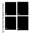
Stereotaxic injections of suspensions of Aβ42 fibrils or Aβ42-1 into the somatosensory cortex of one hemisphere, as well as the CA1 region of the hippocampus of the contralateral hemisphere of mouse brains. Sites of injections are indicated with symbolic syringes. - Figure 2
Stereotaxic injections of Aβ42 or Aβ42-1 fibrils into the somatosensory cortex and the CA1 region of the hippocampus of mouse brains. Electron microscopy confirmed that aggregated synthetic Aβ42 formed fibrils (A), whereas a reverse control peptide, Aβ42-1 (B), did not. Mice brains were analyzed 18-60 day post- injection. Scale bars: A,B: 0.4 µm. - Figure 3
Suspensions of Aβ42 fibrils or Aβ42-1 were injected stereotaxically into the somatosensory cortex of one hemisphere, as well as the CA1 region of the hippocampus of the contralateral hemisphere of mouse brains. Antibody 6E10 revealed that Aβ42 fibrils were present at the injection sites at least for 45 days following the injection (A). As staining controls, amyloid plaque-containing APPsw transgenic (Hsiao et al., Neuron 1995, 15: 1203) and human AD brain sections were included. Aβ42 and Aβ42-1 increased the occurrence of reactive astrocytes both around the injection site (B), and in the amygdala (C). Reactive astrocytosis persisted for at least 45 days. Stereotaxic application of Texas red-coupled dextran into identical coordinates stained a subset of neurons in the amygdala confirming that these projected to the injection sites (D). A-D: Seven month-old P301L female analyzed 45 days after injection. Scale bars: A-D: 50 µm. - Figure 4
Presence of neurofibrillary tangles (NFTs) in P301L tau transgenic mice induced by Aβ42 fibrils. Aβ42 fibrils induced the Gallyas-positive formation of numerous NFTs and neuropil threads in the amygdala and, occasionally, the cortex as early as 18 days following the injection. Amygdala of a six month-old female P301L transgenic mouse analyzed 18 days post injection. Scale bar: 25 µm. - Figure 5
Aβ42 fibrils trigger the formation of numerous Gallyas-positive NFTs and neuropil threads in the amygdala and, occasionally, the cortex of P301L transgenic mice as early as 18 days following the injection. Gallyas silver impreganations of NFTs in the amygdala and parietal cortex of Aβ42- (A,C) and Aβ42-1-injected (B) P301 L tau transgenic mice. A subset of NFTs was also stained by thioflavin-S (D). A: Eight month-old P301 L male analyzed 40 days post injection; B: 6.5 month-old P301L female analyzed 40 days post injection; C: Six month-old female P301 L transgenic mouse analyzed 18 days post injection D: Seven month-old P301L female analyzed 45 days post injection. Scale bars: A,B: 25 µm; C,D: 12.5 µm - Figure 6
Neurofibrillary tangles (NFTs) in P301 L tau transgenic mice induced by Aβ42 fibrils. Aβ42 fibrils trigger the formation of numerous Gallyas-positive NFTs and neuropil threads in the amygdala and, occasionally, the cortex of P301 L transgenic mice (A). The induced NFTs were very similar to those found in brains obtained from AD patients (B). Both slides were stained in parallel by the same protocol. A: 5.25 month-old P301L male analyzed 21 days post injection. B: Human 86 year-old female AD patient. - Figure 7
Aβ42 fibrils trigger the formation of neurofibrillary tangles (NFTs) in P301 L tau transgenic mice. Immuno-electron microscopy of thin sections of the amygdala reveals AD-like tau filaments (using antibody AT8). Scale bars:: low magnification view: 800 µm (A); high magnification view: 100 µm (B). - Figure 8
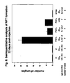
Gallyas-positive NFTs in the amygdala. NFTs were counted onday 22 post-injection in Aβ42-, Aβ42-1-, and uninjected P301L transgenic mice, non-transgenic litter mate controls as well as transgenic mice expressing wild-type human tau (Probst et al., Acta Neuropathol (Berl) 2000, 99: 469). The mean age in months ± SD at the time of analyses is indicated. Gallyas-positive NFTs were counted according to the herein described methods and represent the sum in 20 standardized frontal sections comprising both the ipsilateral and the contralateral amygdala. Mann-Whitney U-test: P = 0.007 (2-tailed exact significance) comparing Aβ42- with Aβ42-1-injected P301 L mice. - Figure 9
Abnormal phospho-epitopes of tau induced by Aβ42 fibrils. The R145d epitope S422 was not phosphorylated in the hippocampus and cortex (A), but specifically induced by Aβ42 fibrils in the amygdala (B), high magnification view). In contrast to R145d and AT100, the AT8 epitope was phosphorylated in many neurons of the hippocampus, cortex (C) and amygdala (D, high magnification view). A-D: Six month-old P301L male analyzed 18 days post injection. Scale bars: A,C: 100 µm; B,D: 25 µm. Schematic map of a mouse brain hemisphere indicating relevant brain regions (E). - Figure 10
Abnormal phospho-epitopes of tau induced by Aβ42 fibrils. Double immunofluorescence staining with R145d (tau phospho-epitope S422) and AT100 (phospho-epitope S212/T214) revealed that R145d-positive neurons in the amygdala were AT100-positive (A, B, merge: C). Scale bar: 40 µm. - Figure 11
Abnormal phospho-epitopes of tau induced by Aβ42 fibrils. Approximately half of the R145d-positive neurons (A,B) beared Gallyas-positive neurofibrillary tangles (C,D), and generally these included the neurons that were most intensely stained by R145d (phospho-S422) (indicated by arrows). A-D: Seven month-old P301 L female analyzed 45 days post injection. Scale bars: 40 µm. - To determine whether β-amyloid can accelerate NFT formation, synthetic Aβ42 fibrils were injected into the somatosensory cortex and the hippocampus of five to six month-old P301L tau transgenic mice and non-transgenic littermates, (refer to methods section (i)-(iii)). As a control peptide, the reversed sequence, Aβ42-1, derived from the identical source was used (methods section (ii)). Aβ42 fibrils were generated by shaking incubation at 37°C and were confirmed by electron microscopy (Fig. 2 A,B) (methods section (ii) and (iii)). Aβ42 fibrils were stable in vivo in both P301L transgenic and wild-type control mice and were readily detectable at least until 45 days following the injections (Fig. 3A). Brain amyloid deposits were accompanied by significant reactive astrogliosis at both the injection sites (Fig. 3B) and the amygdala (Fig. 3C) (for methods refer to section (iv)); these deposits were observed in both Aβ42- and in control-injected transgenic mice and persisted for at least 45 days post-injection. This reaction may be related to the fact that neurons in the amygdala project to the injection sites, as shown by retrograde transport of Texas red-conjugated dextran from the injection site in the somatosensory cortex to cell bodies in the amygdala (Fig. 3D) (methods section (iv)).
- Eighteen days following the injections of Aβ42, Gallyas silver impregnation (section (iv)) revealed numerous NFTs (Fig. 5A-C, Fig. 6A), along with neuropil threads and degenerating neurites (Fig. 4) in the amygdala of P301L, but not wild-type mice. Occasional NFTs were also present in the parietal cortex (Fig. 5C). The NFTs in mice (Fig. 6A) were very similar to those in AD brains stained in parallel by the same protocol (Fig. 6B). Moreover, the neuropil threads were similar to those known in AD (Braak et al., Neurosci Lett 1986, 65: 351; McKee et al., Ann Neurol 1989, 26: 652; Velasco et al., Brain Res 1998, 813: 329). A subset of Gallyas-positive neurofibrillary tangles in the mice were also stained with thioflavin-S, consistent with the histopathology of AD (Fig. 5D). The filaments had a width of 20-25 nm and a periodicity of 90 nm, and are best described as twisted ribbons (Fig. 7). In human carriers, the P301L mutation causes predominant expression of 4 repeat (4R) isoforms, with a small amount of wild-type 3R isoforms, resulting in 15 nm wide twisted filaments with a periodicity of greater than 130 nm (Spillantini et al., Am J Pathol 1998, 153: 1359). Because mice endogenously express only 4R tau isoforms, and the transgene was designed to express 4R human P301 L tau, the filaments observed here contained no 3R tau. Importantly, the human intronic FTDP-17 mutations that reduce the formation of 3R tau also cause twisted ribbons mainly composed of 4R tau. It is therefore possible that the relative amounts of 3R and 4R isoforms contribute to the ultrastructural morphology of the filaments.
- Quantitative analyses revealed five times more Gallyas-positive NFTs in the Aβ42-injected P301 L mice as compared to Aβ42-1- or uninjected P301L mice (Fig. 5A,B; Fig. 8). Cross-sectional time-course analyses of NFT formation showed initial NFTs 18 days after Aβ42 injection, with further increases in numbers (n=58) at least until 60 days following the injection. NFT formation in both hemispheres in the Aβ42-injected P301 L mice did not vary with gender (females: 23 ± 21; males: 23 ± 4; n=7, P=0.86, Mann-Whitney U-test). In contrast, Aβ42-1-injected P301L males developed few NFTs and P301L females no NFTs, at 6 to 8.5 months of age. This difference was statistically significant (females: 0; males: 5.8 ± 1.9; n=7, P< 0.01, Mann-Whitney U-test). The presence of the tau mutation is preferred for inducing NFT formation.
- A surprising and remarkable finding was the spatial separation of the site of Aβ42 injection and remote NFT formation in the amygdala, with no significant differences between the ipsilateral and the contralateral amygdala (11.4 ± 10.13 and 9.4 ± 8.0; n=7, P=0.058, Wilcoxon Signed Ranks Test). This suggests the possibility that damage to presynaptic terminals or axons of neurons that project to the injection site caused NFT formation in the respective cell bodies. The anatomical separation of amyloid deposition and NFT formation is therefore consistent with Aβ42-induced axonal damage, and, possibly, impaired axonal transport of tau (Varadarajan et al., J Struct Biol 2000, 130: 184). We confirmed that the affected neuronai population in the amygdala projected to the cortical injection sites by showing retrograde transport of Texas red-conjugated dextran from the injection sites to the amygdala (Fig. 3D). Other mechanisms of somatodendritic accumulation of tau are less likely: First, we excluded a direct exposure to Aβ42 fibrils of the cell bodies in the amygdala by immunohistochemistry. Second, increases in synthesis of tau protein levels are unlikely as indicated by the absence of axonal dilatations or spheroids in amygdala neurons. Third, a diffusible toxic factor would hardly explain the failure of neurons adjacent to the injection sites to develop NFTs. In human patients with AD, an anatomical separation of amyloid plaques and NFTs is frequently found; with amyloid deposits around synapses and NFTs in the respective cell bodies of projection neurons (Price and Sisodia, Ann Rev Med 1994, 45: 435). In addition, the amygdala is among the most vulnerable areas affected early by NFT formation in human patients (Arnold et al., Cereb Cortex 1991, 1: 103). High vulnerability of the amygdala in our P301L mice is supported by the fact that neurons in the amygdala expressed similar levels of the transgene as compared to cortical or hippocampal pyramidal neurons, yet these developed hardly any NFTs (Götz et al., J Biol Chem 2001, 276: 529). Although our experiments did not formally address the involvement of astrocytes and microglia in NFT formation, activation of these cells alone clearly was not sufficient for NFT formation because Aβ42 and Aβ42-1 similarly activated astrocytes and microglial cells both around the injection sites and in the amygdala (Fig. 3B,C).
- The formation of NFTs in AD is associated with hyperphosphorylation and conformational changes of tau (Grundke-lqbal et al., Proc Natl Acad Sci U S A 1986, 83: 4913; Braak and Braak, Neurobiol Aging 1995, 16: 271; Jicha et al., J Neurosci Res 1997, 48: 128). To determine whether the Aβ42-induced NFT formation in P301L mice was associated with altered phosphorylation and conformation of tau, we used antibodies directed against abnormal phospho-epitopes (R145d, pS422, AT100, TG3) (Goedert et al., Biochem J 1994, 301: 871; Dickson et al., Neurobiol Aging 1995, 16: 285; Tanaka et al., FEBS Lett 1998, 426: 248), hyperphosphorylated epitopes (AT8, S199P, AT180, 12E8, AD2, PHF1) (Goedert et al., Biochem J 1994, 301: 871; Greenberg et al., J Biol Chem 1992, 267: 564; Goedert et al., Neurosci Lett 1995, 189: 167; Seubert et al., J Biol Chem 1995, 270: 18917; Buee-Scherrer et al., Brain Res Mol Brain Res 1996, 39: 79; Delacourte et al., Ann Neurol 1998, 43, 193; Sergeant et al., J Neurochem 1999, 72: 1243), as well as conformation-dependent antibodies (TG3, MC1) (Jicha et al., J Neurosci Res 1997, 48: 128; Dickson et al., Neurobiol Aging 1995, 16: 285), by using standard published procedures (see methods section (iv) and Götz et al., Proc Natl Acad Sci USA 1998, 95: 12370).
- Whereas several antibodies, including AT8, detected phosphorylated tau throughout the brains of P301 L mice independently of the injections, R145d/pS422 and AT100 directed against phospho-epitopes S422 and S212/T214, respectively, specifically detected NFTs and neurons only in response to Aβ42 (Fig. 9A,B; Fig. 10A-C). Remarkably, the spatial distribution pattern of these abnormally phosphorylated forms of tau was identical to that observed by Gallyas stainings, and occurred, again, predominantly in the amygdala (Fig. 9A,B). Co-staining revealed that neurons stained by R145d/pS422 were also stained by AT100 (Fig. 10A-C). Neither R145d nor AT100 immunostained any cells in non-transgenic mice. The specificity of R145d, pS422, and AT100 immunoreactivity for Aβ42-associated abnormal phosphorylation was remarkable because these antisera revealed hardly any signals in uninjected or Aβ42-1-injected P301 L tau transgenic mice, and none in transgenic mice expressing wild-type human tau. Moreover, all Gallyas-positive NFTs were also stained by R145d, as indicated by sequential immunofluorescence and Gallyas silver impregnation protocols, strongly suggesting that the NFTs in P301 L mice contained S422-phosphorylated tau. Semi-quantitative analyses revealed that approximately one half of the R145d-positive neurons (Fig. 11A,B) were Gallyas-positive (Fig. 11C,D), and R145d stained these neurons generally more intensely than cells without NFTs.
- Together, the result of our immunostaining is consistent with the possibility that phosphorylation of epitopes S212/T214 and S422 is tightly associated with NFT formation. The herein disclosed data extend prior findings that Aβ42 induced tau phosphorylation in vitro and in vivo at the AT8 and 12E8/Ab31 epitopes (Busciglio et al., Neuron 1995, 14: 879; Geula et al., Nat Med 1998, 4: 827). In our P301 L mice, tau was phosphorylated at these epitopes, even in the absence of injected Aβ42. Therefore, these epitopes may be necessary but clearly were not sufficient for NFT formation in P301L mice. By using R145d/ pS422 and AT100, we found that Aβ42 injections were followed by phosphorylation of tau at S212/T214 and S422 suggesting a role of these epitopes in NFT formation.
- In summary, the herein disclosed data establish that Aβ42 fibrils can significantly accelerate NFT formation in P301L mice. The neurofibrillary tangles generated according to the inventive method closely resemble those extracted from brains of AD patients and the formation of which was achieved already within 18 days post-injection. The herein disclosed data demonstrate that, in transgenic mice, the interaction of β-amyloid with the P301 L mutation was required for NFT formation - neither β-amyloid, nor the mutation alone, were sufficient to generate high numbers of NFTs. The mice generated according to the instant invention provide an in vivo assay to determine whether such amyloid-lowering therapies as Aβ vaccination are effective in preventing NFT formation in vivo.
-
- (i) Generation of transgenic mice: To generate transgenic mice, the human pathogenic tau mutation P301 L was introduced into the cDNA encoding the longest human brain tau isoform by a PCR-mediated approach. This isoform contained
exons - (ii) Production of Aβ42 and reverse Aβ42-1 fibrils: Aβ42 (Bachem, No. H-1368, Lot No. 524548) and Aβ42-1 (Bachem, No. H-3976, Lot No. 536763) were reconstituted in PBS at final concentrations of 250µM, shaken at 1000 rpm for 84 hours at 37°C in an Eppendorf thermomixer and analyzed by electron microscopy. Preparations were placed on carbon-coated 300-mesh grids and stained with 2% phosphotungstic acid. The preparation of the reverse peptide Aβ42-1 consisted of insoluble aggregates that were not readily detectable by negative contrasting in electron microscopy (Fig. 2B). Micrographs were recorded at an operating voltage of 80 to 100 kV and at nominal magnifications of x40,000 on a Philips model CM12 electron microscope.
- (iii) Stereotaxic injection: Stereotaxic injections into mouse brain were carried out according to the protocols of Paxinos (Paxinos KB, The mouse brain in stereotaxic coordinates, Academic Press, San Diego, 1997). Mice were anaesthesized with a mixture of 2% Xylazin and 10% Ketamin, and stereotaxically injected with 1.5 µl of peptide suspension into the CA1 region of the hippocampal formation of the right hemisphere (coordinates: AP -1.9 mm from the bregma, LAT -1.0 mm, DV +1.9 mm), and into the cortex of the left hemisphere (coordinates: AP -1.9 mm from the bregma, LAT +2.0 mm, DV +1.2 mm; includes the trunk region of the somatosensory cortex 1, S1Tr), using a 10 µl Hamilton syringe driven by a mini pump (Motorized Stereotaxic Injector, Stoelting Co.), with an injection speed of 0.15µl/min. The needle was kept in the injection site for another ten minutes, and then slowly withdrawn. Operated animals were kept under daily control. No mouse developed infections or died during the experiment. Injected mice were perfused transcardially with 4% paraformaldehyde in sodium phosphate buffer, pH 7.4, on days 18, 26, 45 and 60, respectively, and processed for immunohistochemistry. To determine axonal uptake, we injected 1.5 µl of 12.5 mg/ml Texas red-coupled dextran (70,000 MW, Molecular Probes) in PBS, and perfused the mice five to eight days later.
- (iv) Immunohistological stainings, immunofluorescence analysis and antibodies: Immunohistological stainings were performed on 4 µm coronal paraffin sections from brain, by using standard published procedures. Serial frontal sections were taken, and brain sections from AD patients and healthy humans were included as controls. Some of the sections were pretreated with 5 µg/ml proteinase K in Tris-buffered saline or PBS at 37°C for 2.5 minutes for signal enhancement. Sections were stained with thioflavin-S and silver-impregnated by the Gallyas protocol (Gallyas F, Acta Morphol Acad Sci Hung 1971, 19:1). For Gallyas stainings, every 20th section was analyzed from positions AP -1.5 mm to AP -2.4 mm (from the bregma), whereas every fifth section was analyzed around the injection site (position AP-1.9 mm) from positions AP -1.8 mm to AP -2.0 mm. Gallyas-positive NFTs were counted in 20 standardized frontal sections of total brain from each mouse, and added. Averages and standard deviations were determined for the numbers of mice indiciated in Fig. 8. Tau14 (Zymed Inc, South San Francisco, CA, amino acids 83-120, used at 1:2 dilutions) and HT7 (Innogenetics Inc, Belgium, amino acids 159-163, 1:200) were used to detect human tau specifically. The following antibodies were used to detect distinct tau phospho-epitopes (in brackets): AT270 (Innogenetics Inc., 1:500, threonine 181), AT8 (Innogenetics Inc., 1:50, serine 202/threonine 205), AT100 (Innogenetics Inc., 1:100, serine 212/threonine 214), 12E8 (Dr. Peter Seubert, 1:100, serine 262/serine 356), AT180 and TG3 (Dr. Peter Davies, 1:100 and 1:20, resp., threonine 231/serine 235), PHF1 (Dr. Peter Davies, 1:50); S199P (Dr. Andre Delacourte, 1:100, serine199), R145d (Dr. Khalid Iqbal, 1:30, serine422) and pS422 (Biosource Inc., 1:50, serine422), and AD2 (Dr. Chantal Mourton-Gilles, 1:10.000, serine 396/serine 404). MC1 detected the conformational Alz-50 epitope (Dr. Peter Davies, 1:20). Monoclonal antibody 6E10 (Serotec, 1:500) was used to detect Aβ peptide. Anti-GFAP polyclonal antibody (Sigma, 1:400), was used to detect activated astrocytes and Isolectin B4 (Vector Laboratories, 2µg/ml) was used to detect microglia. Secondary antibodies were obtained from Vector Laboratories (Vectastain ABC kits PK-6101 and PK-6102) for peroxidase/DAB stainings, and from Molecular Probes (ALEXA-FLUOR series) for immunofluorescence.
- (v) Transmission electron microscopy: Areas containing the amygdala were excised from 40-µm-thick vibratome sections and fixed in 4% paraformaldehyde and 0.1% glutaraldehyde in PBS. After fixation with OsO4, tissue samples for routine electron microscopy were embedded in epoxy resin (Epon, Sigma, Bornem, Belgium). For immunogold labeling, ultrathin sections from epoxy-resin-embedded tissues on formvar-coated nickel grids were treated with 6% sodium metaperiodate for 10 minutes and with 5% normal goat serum in PBS for 30 minutes. This was followed by incubating the sections with monoclonal antibody AT8 in 1% normal goat serum in PBS at a dilution of 1:50 for 2 hours. After washing, 10-nm colloidal gold-tagged secondary antibody in Tris-buffered saline (goat anti-mouse; British Biocell, Cardiff, UK) was applied for 1 hour. Then, after washing, sections were stained with lead citrate and uranyl acetate. Control sections were stained following the same procedure, but with omission of the primary antibody. AT8 appeared to label frequently "ends" of filaments due to the fact that filaments sectioned at the surfaces of the epoxy-embedded preparations were more easily accessible for the antibodies than deeper layers.
Claims (19)
- A method for increasing the number of neurofibrillary tangles in a transgenic rodent which expresses a recombinant gene coding for human P301L mutant tau protein wherein the increase of neurofibrillary tangles is induced through injection of APP or a β-amyloid fragment thereof.
- A method for accelerating the production of neurofibrillary tangles in a transgenic rodent which expresses a recombinant gene coding for human P301L mutant tau protein wherein the accelerated production of neurofibrillary tangles is induced through injection of APP or a β-amyloid fragment thereof.
- The method according to anyone of the claims 1 or 2 wherein said β-amyloid fragment is Aβ42.
- The method according to claim 3 wherein said Aβ42 is fibrillar, pre-aggregated or aggregated.
- The method according to anyone of the claims 1 to 4 wherein said transgenic rodent is a mouse.
- The method according to anyone of the claims 1 to 5 wherein the β-amyloid fragment is injected into the brain of said rodent, in particular into the hippocampus or the cortex.
- The method according to anyone of the claims 1 to 6 wherein the increase of neurofibrillary tangles is predominantly in the amygdala of the rodent's brain, in particular the basolateral amygdala.
- The method according to anyone of the claims 1 to 7 wherein the increase of neurofibrillary tangles is several-fold, preferably at least two-fold, as compared to a control animal into which no APP or β-amyloid fragment has been injected.
- The method according to anyone of the claims 1 to 8 wherein the increase of neurofibrillary tangles is at least five-fold as compared to a control animal to which no APP fragment β-amyloid has been injected.
- The method according to anyone of the claims 1 to 9 for inducing neuropil threads and/or degeneration of neurites.
- A transgenic rodent which expresses a recombinant gene coding for human P301L mutant tau protein having an increased number of neurofibrillary tangles or accelerated production of neurofibrillary tangles, wherein rodent's increased number of neurofibrillary tangles and/or the accelerated production of neurofibrillary tangles is obtained through injection of the APP or the β-amyloid fragment according to anyone of the claims 1 to 10.
- The rodent according to claim 11 wherein neurofibrillary tangles are produced which are comparable to those commonly found in human neurodegenerative diseases.
- The rodent according to anyone of claims 11 and/or 12 comprising the APP or the β-amyloid fragment, in an amount sufficient to induce the production of neurofibrillary tangles.
- The rodent according to anyone of the claims 11 to 13, wherein the β-amyloid fragment is injected into the brain of said rodent, in particular into the hippocampus or the cortex.
- The rodent according to anyone of the claims 11 to 14 co-expressing a recombinant gene coding for APP, said APP being enzymatically processed to generate the β-amyloid fragment.
- The rodent of claim 15 wherein said APP is a mutant APP.
- The rodent according to anyone of claims 11 to 16 which is a mouse.
- Use of the rodent according to anyone of the claims 11 to 17 as an animal model in an in-vivo assay to determine or validate the efficacy of modulating agents, or therapies, in particular amyloid-lowering therapies or NFT-reducing therapies.
- The use according to claim 18 wherein said modulating agents, or therapies are for neurodegenerative diseases, in particular Alzheimer's disease, frontotemporal dementia (FTD), and other neurodegenerative diseases accompanied by neurofibrillary tangle formation.
Priority Applications (1)
| Application Number | Priority Date | Filing Date | Title |
|---|---|---|---|
| EP02796273A EP1418807B8 (en) | 2001-08-24 | 2002-08-24 | A method of inducing the formation of neurofibrillary tangles in transgenic animals |
Applications Claiming Priority (6)
| Application Number | Priority Date | Filing Date | Title |
|---|---|---|---|
| US31430301P | 2001-08-24 | 2001-08-24 | |
| EP01120348 | 2001-08-24 | ||
| US314303P | 2001-08-24 | ||
| EP01120348 | 2001-08-24 | ||
| PCT/EP2002/009491 WO2003017918A2 (en) | 2001-08-24 | 2002-08-24 | A method of inducing the formation of neurofibrillary tangles in transgenic animals |
| EP02796273A EP1418807B8 (en) | 2001-08-24 | 2002-08-24 | A method of inducing the formation of neurofibrillary tangles in transgenic animals |
Publications (3)
| Publication Number | Publication Date |
|---|---|
| EP1418807A2 EP1418807A2 (en) | 2004-05-19 |
| EP1418807B1 true EP1418807B1 (en) | 2007-05-02 |
| EP1418807B8 EP1418807B8 (en) | 2008-04-23 |
Family
ID=34042746
Family Applications (1)
| Application Number | Title | Priority Date | Filing Date |
|---|---|---|---|
| EP02796273A Expired - Lifetime EP1418807B8 (en) | 2001-08-24 | 2002-08-24 | A method of inducing the formation of neurofibrillary tangles in transgenic animals |
Country Status (7)
| Country | Link |
|---|---|
| US (2) | US20050015819A1 (en) |
| EP (1) | EP1418807B8 (en) |
| JP (1) | JP2005500076A (en) |
| AT (1) | ATE360988T1 (en) |
| AU (1) | AU2002333704A1 (en) |
| DE (1) | DE60219925T2 (en) |
| WO (1) | WO2003017918A2 (en) |
Families Citing this family (5)
| Publication number | Priority date | Publication date | Assignee | Title |
|---|---|---|---|---|
| JP5511144B2 (en) * | 2008-02-12 | 2014-06-04 | 学校法人福岡大学 | Epilepsy model non-human mammal |
| SG189174A1 (en) | 2010-10-11 | 2013-05-31 | Biogen Idec Internat Neuroscience Gmbh | Human anti-tau antibodies |
| HRP20201422T1 (en) | 2012-12-21 | 2021-02-19 | Biogen Ma Inc. | Human anti-tau antibodies |
| ZA201608812B (en) | 2014-06-26 | 2019-08-28 | Janssen Vaccines & Prevention Bv | Antibodies and antigen-binding fragments that specifically bind to microtubule-associated protein tau |
| NZ727020A (en) * | 2014-06-26 | 2022-02-25 | Janssen Vaccines & Prevention Bv | Antibodies and antigen-binding fragments that specifically bind to microtubule-associated protein tau |
Family Cites Families (3)
| Publication number | Priority date | Publication date | Assignee | Title |
|---|---|---|---|---|
| US6172277B1 (en) * | 1997-10-28 | 2001-01-09 | The Miriam Hospital | Non-transgenic rodent model of alzheimer's disease |
| US6664443B1 (en) * | 1998-05-01 | 2003-12-16 | Mayo Foundation For Medical Education And Research | Pathogenic Tau mutations in transgenic mice |
| EP1077990A1 (en) * | 1998-05-01 | 2001-02-28 | Mayo Foundation For Medical Education And Research | Pathogeniv tau mutations |
-
2002
- 2002-08-24 US US10/487,546 patent/US20050015819A1/en not_active Abandoned
- 2002-08-24 JP JP2003522441A patent/JP2005500076A/en active Pending
- 2002-08-24 DE DE60219925T patent/DE60219925T2/en not_active Expired - Lifetime
- 2002-08-24 AT AT02796273T patent/ATE360988T1/en not_active IP Right Cessation
- 2002-08-24 WO PCT/EP2002/009491 patent/WO2003017918A2/en not_active Ceased
- 2002-08-24 AU AU2002333704A patent/AU2002333704A1/en not_active Abandoned
- 2002-08-24 EP EP02796273A patent/EP1418807B8/en not_active Expired - Lifetime
-
2007
- 2007-10-23 US US11/976,318 patent/US7550648B2/en not_active Expired - Fee Related
Also Published As
| Publication number | Publication date |
|---|---|
| AU2002333704A1 (en) | 2003-03-10 |
| EP1418807B8 (en) | 2008-04-23 |
| US20050015819A1 (en) | 2005-01-20 |
| DE60219925D1 (en) | 2007-06-14 |
| JP2005500076A (en) | 2005-01-06 |
| ATE360988T1 (en) | 2007-05-15 |
| US20080261878A1 (en) | 2008-10-23 |
| WO2003017918A3 (en) | 2003-12-04 |
| DE60219925T2 (en) | 2008-01-17 |
| WO2003017918A2 (en) | 2003-03-06 |
| EP1418807A2 (en) | 2004-05-19 |
| US7550648B2 (en) | 2009-06-23 |
Similar Documents
| Publication | Publication Date | Title |
|---|---|---|
| Götz et al. | Oligodendroglial tau filament formation in transgenic mice expressing G272V tau | |
| Götz et al. | Tau filament formation in transgenic mice expressing P301L tau | |
| Duyckaerts et al. | Alzheimer disease models and human neuropathology: similarities and differences | |
| Götz et al. | Transgenic animal models of Alzheimer's disease and related disorders: histopathology, behavior and therapy | |
| Schwab et al. | Transgenic mice overexpressing amyloid beta protein are an incomplete model of Alzheimer disease | |
| Lewis et al. | Propagation of tau pathology: hypotheses, discoveries, and yet unresolved questions from experimental and human brain studies | |
| Eisele et al. | Propagation of Aß pathology: hypotheses, discoveries, and yet unresolved questions from experimental and human brain studies | |
| Wen et al. | Overexpression of wild type but not an FAD mutant presenilin-1 promotes neurogenesis in the hippocampus of adult mice | |
| Moechars et al. | Premature death in transgenic mice that overexpress a mutant amyloid precursor protein is preceded by severe neurodegeneration and apoptosis | |
| EP3080611B1 (en) | Soluble high molecular weight (hmw) tau species and applications thereof | |
| US7632816B2 (en) | Treatment of Alzheimer amyloid deposition | |
| Ubhi et al. | Neurofibrillary and neurodegenerative pathology in APP-transgenic mice injected with AAV2-mutant TAU: neuroprotective effects of Cerebrolysin | |
| Spencer et al. | Selective targeting of 3 repeat Tau with brain penetrating single chain antibodies for the treatment of neurodegenerative disorders | |
| US7550648B2 (en) | Inducing neurofibrillary tangles in transgenic mice expressing a mutant tau protein | |
| Ajit et al. | A unique tau conformation generated by an acetylation-mimic substitution modulates P301S-dependent tau pathology and hyperphosphorylation | |
| DeArmond et al. | Neuropathology of prion diseases | |
| JP2000512141A (en) | Perlecan transgenic animals and methods for identifying compounds for treating amyloidosis | |
| EP1255824B1 (en) | Transgenic mice for the study of neurodegenerative syndromes | |
| Hajimohammadreza et al. | β-Amyloid precursor protein fragments and lysosomal dense bodies are found in rat brain neurons after ventricular infusion of leupeptin | |
| US20040038302A1 (en) | Methods and compounds for treating brain amyloidosis | |
| Vitali et al. | Soluble amyloid β-protein is increased in frontotemporal dementia with tau gene mutations | |
| KR102775835B1 (en) | A preparation method for animal model of alzheimer's disease and animal model of alzheimer's disease using the same | |
| Shoji et al. | Age‐related amyloid β protein accumulation induces cellular death and macrophage activation in transgenic mice | |
| US7135610B2 (en) | Mouse showing neurofibril change due to senile dementia | |
| US20260132176A1 (en) | Compositions and methods for treating neurodegenerative disease |
Legal Events
| Date | Code | Title | Description |
|---|---|---|---|
| PUAI | Public reference made under article 153(3) epc to a published international application that has entered the european phase |
Free format text: ORIGINAL CODE: 0009012 |
|
| 17P | Request for examination filed |
Effective date: 20040303 |
|
| AK | Designated contracting states |
Kind code of ref document: A2 Designated state(s): AT BE BG CH CY CZ DE DK EE ES FI FR GB GR IE IT LI LU MC NL PT SE SK TR |
|
| AX | Request for extension of the european patent |
Extension state: AL LT LV MK RO SI |
|
| RIN1 | Information on inventor provided before grant (corrected) |
Inventor name: GOETZ, JUERGEN 2 PSYCHIATRISCHE UNIV.KLINIKEN ZUER Inventor name: NITSCH, ROGER Inventor name: CHEN, FENG PSYCHIATRISCHE UNIV.KLINIKEN ZUERICH |
|
| 17Q | First examination report despatched |
Effective date: 20040809 |
|
| GRAP | Despatch of communication of intention to grant a patent |
Free format text: ORIGINAL CODE: EPIDOSNIGR1 |
|
| GRAS | Grant fee paid |
Free format text: ORIGINAL CODE: EPIDOSNIGR3 |
|
| GRAA | (expected) grant |
Free format text: ORIGINAL CODE: 0009210 |
|
| AK | Designated contracting states |
Kind code of ref document: B1 Designated state(s): AT BE BG CH CY CZ DE DK EE ES FI FR GB GR IE IT LI LU MC NL PT SE SK TR |
|
| PG25 | Lapsed in a contracting state [announced via postgrant information from national office to epo] |
Ref country code: FI Free format text: LAPSE BECAUSE OF FAILURE TO SUBMIT A TRANSLATION OF THE DESCRIPTION OR TO PAY THE FEE WITHIN THE PRESCRIBED TIME-LIMIT Effective date: 20070502 |
|
| REG | Reference to a national code |
Ref country code: GB Ref legal event code: FG4D |
|
| RIN1 | Information on inventor provided before grant (corrected) |
Inventor name: CHEN, FENG Inventor name: NITSCH, ROGER Inventor name: GOETZ, JUERGEN B |
|
| REG | Reference to a national code |
Ref country code: CH Ref legal event code: EP |
|
| REG | Reference to a national code |
Ref country code: IE Ref legal event code: FG4D |
|
| REF | Corresponds to: |
Ref document number: 60219925 Country of ref document: DE Date of ref document: 20070614 Kind code of ref document: P |
|
| RIN2 | Information on inventor provided after grant (corrected) |
Inventor name: GOETZ, JUERGEN B Inventor name: NITSCH, ROGER Inventor name: CHEN, FENG |
|
| PG25 | Lapsed in a contracting state [announced via postgrant information from national office to epo] |
Ref country code: SE Free format text: LAPSE BECAUSE OF FAILURE TO SUBMIT A TRANSLATION OF THE DESCRIPTION OR TO PAY THE FEE WITHIN THE PRESCRIBED TIME-LIMIT Effective date: 20070802 |
|
| PG25 | Lapsed in a contracting state [announced via postgrant information from national office to epo] |
Ref country code: ES Free format text: LAPSE BECAUSE OF FAILURE TO SUBMIT A TRANSLATION OF THE DESCRIPTION OR TO PAY THE FEE WITHIN THE PRESCRIBED TIME-LIMIT Effective date: 20070813 |
|
| REG | Reference to a national code |
Ref country code: CH Ref legal event code: NV Representative=s name: PATENTANWAELTE SCHAAD, BALASS, MENZL & PARTNER AG |
|
| NLV1 | Nl: lapsed or annulled due to failure to fulfill the requirements of art. 29p and 29m of the patents act | ||
| PG25 | Lapsed in a contracting state [announced via postgrant information from national office to epo] |
Ref country code: AT Free format text: LAPSE BECAUSE OF FAILURE TO SUBMIT A TRANSLATION OF THE DESCRIPTION OR TO PAY THE FEE WITHIN THE PRESCRIBED TIME-LIMIT Effective date: 20070502 |
|
| EN | Fr: translation not filed | ||
| PG25 | Lapsed in a contracting state [announced via postgrant information from national office to epo] |
Ref country code: BE Free format text: LAPSE BECAUSE OF FAILURE TO SUBMIT A TRANSLATION OF THE DESCRIPTION OR TO PAY THE FEE WITHIN THE PRESCRIBED TIME-LIMIT Effective date: 20070502 |
|
| PG25 | Lapsed in a contracting state [announced via postgrant information from national office to epo] |
Ref country code: NL Free format text: LAPSE BECAUSE OF FAILURE TO SUBMIT A TRANSLATION OF THE DESCRIPTION OR TO PAY THE FEE WITHIN THE PRESCRIBED TIME-LIMIT Effective date: 20070502 Ref country code: DK Free format text: LAPSE BECAUSE OF FAILURE TO SUBMIT A TRANSLATION OF THE DESCRIPTION OR TO PAY THE FEE WITHIN THE PRESCRIBED TIME-LIMIT Effective date: 20070502 Ref country code: CZ Free format text: LAPSE BECAUSE OF FAILURE TO SUBMIT A TRANSLATION OF THE DESCRIPTION OR TO PAY THE FEE WITHIN THE PRESCRIBED TIME-LIMIT Effective date: 20070502 Ref country code: PT Free format text: LAPSE BECAUSE OF FAILURE TO SUBMIT A TRANSLATION OF THE DESCRIPTION OR TO PAY THE FEE WITHIN THE PRESCRIBED TIME-LIMIT Effective date: 20071002 Ref country code: BG Free format text: LAPSE BECAUSE OF FAILURE TO SUBMIT A TRANSLATION OF THE DESCRIPTION OR TO PAY THE FEE WITHIN THE PRESCRIBED TIME-LIMIT Effective date: 20070802 |
|
| PG25 | Lapsed in a contracting state [announced via postgrant information from national office to epo] |
Ref country code: SK Free format text: LAPSE BECAUSE OF FAILURE TO SUBMIT A TRANSLATION OF THE DESCRIPTION OR TO PAY THE FEE WITHIN THE PRESCRIBED TIME-LIMIT Effective date: 20070502 |
|
| PLBE | No opposition filed within time limit |
Free format text: ORIGINAL CODE: 0009261 |
|
| STAA | Information on the status of an ep patent application or granted ep patent |
Free format text: STATUS: NO OPPOSITION FILED WITHIN TIME LIMIT |
|
| 26N | No opposition filed |
Effective date: 20080205 |
|
| PG25 | Lapsed in a contracting state [announced via postgrant information from national office to epo] |
Ref country code: IT Free format text: LAPSE BECAUSE OF FAILURE TO SUBMIT A TRANSLATION OF THE DESCRIPTION OR TO PAY THE FEE WITHIN THE PRESCRIBED TIME-LIMIT Effective date: 20070502 Ref country code: FR Free format text: LAPSE BECAUSE OF FAILURE TO SUBMIT A TRANSLATION OF THE DESCRIPTION OR TO PAY THE FEE WITHIN THE PRESCRIBED TIME-LIMIT Effective date: 20071228 Ref country code: GR Free format text: LAPSE BECAUSE OF FAILURE TO SUBMIT A TRANSLATION OF THE DESCRIPTION OR TO PAY THE FEE WITHIN THE PRESCRIBED TIME-LIMIT Effective date: 20070803 Ref country code: MC Free format text: LAPSE BECAUSE OF NON-PAYMENT OF DUE FEES Effective date: 20070831 |
|
| REG | Reference to a national code |
Ref country code: CH Ref legal event code: PK Free format text: DER VERTRETER SCHAAD BALASS MENZL & PARTNER WURDE IRRTUEMLICH EINGETRAGEN. Ref country code: CH Ref legal event code: NV Representative=s name: SCHMAUDER & PARTNER AG PATENTANWALTSBUERO |
|
| PG25 | Lapsed in a contracting state [announced via postgrant information from national office to epo] |
Ref country code: IE Free format text: LAPSE BECAUSE OF NON-PAYMENT OF DUE FEES Effective date: 20070824 |
|
| PG25 | Lapsed in a contracting state [announced via postgrant information from national office to epo] |
Ref country code: FR Free format text: LAPSE BECAUSE OF FAILURE TO SUBMIT A TRANSLATION OF THE DESCRIPTION OR TO PAY THE FEE WITHIN THE PRESCRIBED TIME-LIMIT Effective date: 20070502 |
|
| PG25 | Lapsed in a contracting state [announced via postgrant information from national office to epo] |
Ref country code: EE Free format text: LAPSE BECAUSE OF FAILURE TO SUBMIT A TRANSLATION OF THE DESCRIPTION OR TO PAY THE FEE WITHIN THE PRESCRIBED TIME-LIMIT Effective date: 20070502 |
|
| PG25 | Lapsed in a contracting state [announced via postgrant information from national office to epo] |
Ref country code: CY Free format text: LAPSE BECAUSE OF FAILURE TO SUBMIT A TRANSLATION OF THE DESCRIPTION OR TO PAY THE FEE WITHIN THE PRESCRIBED TIME-LIMIT Effective date: 20070502 |
|
| REG | Reference to a national code |
Ref country code: CH Ref legal event code: PCAR Free format text: SCHMAUDER & PARTNER AG PATENT- UND MARKENANWAELTE VSP;ZWAENGIWEG 7;8038 ZUERICH (CH) |
|
| PG25 | Lapsed in a contracting state [announced via postgrant information from national office to epo] |
Ref country code: LU Free format text: LAPSE BECAUSE OF NON-PAYMENT OF DUE FEES Effective date: 20070824 |
|
| PG25 | Lapsed in a contracting state [announced via postgrant information from national office to epo] |
Ref country code: TR Free format text: LAPSE BECAUSE OF FAILURE TO SUBMIT A TRANSLATION OF THE DESCRIPTION OR TO PAY THE FEE WITHIN THE PRESCRIBED TIME-LIMIT Effective date: 20070502 |
|
| PGFP | Annual fee paid to national office [announced via postgrant information from national office to epo] |
Ref country code: GB Payment date: 20150824 Year of fee payment: 14 Ref country code: CH Payment date: 20150824 Year of fee payment: 14 |
|
| PGFP | Annual fee paid to national office [announced via postgrant information from national office to epo] |
Ref country code: DE Payment date: 20151019 Year of fee payment: 14 |
|
| REG | Reference to a national code |
Ref country code: DE Ref legal event code: R119 Ref document number: 60219925 Country of ref document: DE |
|
| REG | Reference to a national code |
Ref country code: CH Ref legal event code: PL |
|
| GBPC | Gb: european patent ceased through non-payment of renewal fee |
Effective date: 20160824 |
|
| PG25 | Lapsed in a contracting state [announced via postgrant information from national office to epo] |
Ref country code: CH Free format text: LAPSE BECAUSE OF NON-PAYMENT OF DUE FEES Effective date: 20160831 Ref country code: LI Free format text: LAPSE BECAUSE OF NON-PAYMENT OF DUE FEES Effective date: 20160831 |
|
| PG25 | Lapsed in a contracting state [announced via postgrant information from national office to epo] |
Ref country code: DE Free format text: LAPSE BECAUSE OF NON-PAYMENT OF DUE FEES Effective date: 20170301 Ref country code: GB Free format text: LAPSE BECAUSE OF NON-PAYMENT OF DUE FEES Effective date: 20160824 |