EP1414996B1 - Methods for genetic modification of hematopoietic progenitor cells and uses of the modified cells - Google Patents
Methods for genetic modification of hematopoietic progenitor cells and uses of the modified cells Download PDFInfo
- Publication number
- EP1414996B1 EP1414996B1 EP02746963A EP02746963A EP1414996B1 EP 1414996 B1 EP1414996 B1 EP 1414996B1 EP 02746963 A EP02746963 A EP 02746963A EP 02746963 A EP02746963 A EP 02746963A EP 1414996 B1 EP1414996 B1 EP 1414996B1
- Authority
- EP
- European Patent Office
- Prior art keywords
- cells
- cell
- hiv
- transduced
- composition
- Prior art date
- Legal status (The legal status is an assumption and is not a legal conclusion. Google has not performed a legal analysis and makes no representation as to the accuracy of the status listed.)
- Expired - Lifetime
Links
- 210000004027 cell Anatomy 0.000 title claims abstract description 646
- 210000003958 hematopoietic stem cell Anatomy 0.000 title claims abstract description 136
- 238000000034 method Methods 0.000 title claims abstract description 123
- 238000012239 gene modification Methods 0.000 title description 3
- 230000005017 genetic modification Effects 0.000 title description 3
- 235000013617 genetically modified food Nutrition 0.000 title description 3
- 239000000203 mixture Substances 0.000 claims abstract description 88
- 208000031886 HIV Infections Diseases 0.000 claims abstract description 41
- 208000037357 HIV infectious disease Diseases 0.000 claims abstract description 34
- 208000033519 human immunodeficiency virus infectious disease Diseases 0.000 claims abstract description 34
- 238000011282 treatment Methods 0.000 claims abstract description 29
- 102100031573 Hematopoietic progenitor cell antigen CD34 Human genes 0.000 claims description 265
- 101000777663 Homo sapiens Hematopoietic progenitor cell antigen CD34 Proteins 0.000 claims description 265
- 239000013598 vector Substances 0.000 claims description 158
- 108090000623 proteins and genes Proteins 0.000 claims description 153
- 238000010361 transduction Methods 0.000 claims description 122
- 230000026683 transduction Effects 0.000 claims description 120
- 108090000994 Catalytic RNA Proteins 0.000 claims description 71
- 210000004369 blood Anatomy 0.000 claims description 70
- 239000008280 blood Substances 0.000 claims description 70
- 102000053642 Catalytic RNA Human genes 0.000 claims description 67
- 108091092562 ribozyme Proteins 0.000 claims description 67
- 230000001177 retroviral effect Effects 0.000 claims description 57
- 230000003612 virological effect Effects 0.000 claims description 50
- 102000004127 Cytokines Human genes 0.000 claims description 47
- 108090000695 Cytokines Proteins 0.000 claims description 47
- 239000003795 chemical substances by application Substances 0.000 claims description 40
- 108091030071 RNAI Proteins 0.000 claims description 31
- 238000002617 apheresis Methods 0.000 claims description 31
- 230000009368 gene silencing by RNA Effects 0.000 claims description 31
- 230000037396 body weight Effects 0.000 claims description 26
- 239000012634 fragment Substances 0.000 claims description 25
- 239000002259 anti human immunodeficiency virus agent Substances 0.000 claims description 23
- 229940124411 anti-hiv antiviral agent Drugs 0.000 claims description 23
- 210000000265 leukocyte Anatomy 0.000 claims description 23
- 230000008569 process Effects 0.000 claims description 22
- 108010067306 Fibronectins Proteins 0.000 claims description 21
- 241000700605 Viruses Species 0.000 claims description 21
- 239000002773 nucleotide Substances 0.000 claims description 21
- 125000003729 nucleotide group Chemical group 0.000 claims description 21
- 230000000692 anti-sense effect Effects 0.000 claims description 18
- 239000003937 drug carrier Substances 0.000 claims description 13
- 238000000338 in vitro Methods 0.000 claims description 13
- 238000002955 isolation Methods 0.000 claims description 8
- 108091032973 (ribonucleotides)n+m Proteins 0.000 claims description 5
- 102000016359 Fibronectins Human genes 0.000 claims 3
- 238000001415 gene therapy Methods 0.000 abstract description 38
- 230000003394 haemopoietic effect Effects 0.000 abstract description 15
- 230000002035 prolonged effect Effects 0.000 abstract description 7
- 230000002265 prevention Effects 0.000 abstract description 3
- 238000001802 infusion Methods 0.000 description 73
- 210000001744 T-lymphocyte Anatomy 0.000 description 66
- 108020004414 DNA Proteins 0.000 description 55
- 210000003819 peripheral blood mononuclear cell Anatomy 0.000 description 49
- 241000725303 Human immunodeficiency virus Species 0.000 description 45
- 102000036693 Thrombopoietin Human genes 0.000 description 45
- 108010041111 Thrombopoietin Proteins 0.000 description 45
- 241000713772 Human immunodeficiency virus 1 Species 0.000 description 44
- 210000001185 bone marrow Anatomy 0.000 description 44
- 210000000130 stem cell Anatomy 0.000 description 44
- 239000000047 product Substances 0.000 description 38
- 230000007774 longterm Effects 0.000 description 36
- 239000011886 peripheral blood Substances 0.000 description 36
- 210000005259 peripheral blood Anatomy 0.000 description 36
- 238000002360 preparation method Methods 0.000 description 36
- 238000003556 assay Methods 0.000 description 34
- 102000004269 Granulocyte Colony-Stimulating Factor Human genes 0.000 description 33
- 108010017080 Granulocyte Colony-Stimulating Factor Proteins 0.000 description 33
- 230000014509 gene expression Effects 0.000 description 33
- 239000000523 sample Substances 0.000 description 31
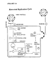
- 230000010076 replication Effects 0.000 description 30
- 102100036011 T-cell surface glycoprotein CD4 Human genes 0.000 description 27
- 101001078385 Homo sapiens Haptoglobin Proteins 0.000 description 26
- 102000050796 human HP Human genes 0.000 description 26
- 239000002953 phosphate buffered saline Substances 0.000 description 25
- 238000001514 detection method Methods 0.000 description 24
- 102000004169 proteins and genes Human genes 0.000 description 24
- 210000004296 naive t lymphocyte Anatomy 0.000 description 23
- NQDJXKOVJZTUJA-UHFFFAOYSA-N nevirapine Chemical compound C12=NC=CC=C2C(=O)NC=2C(C)=CC=NC=2N1C1CC1 NQDJXKOVJZTUJA-UHFFFAOYSA-N 0.000 description 23
- 230000004083 survival effect Effects 0.000 description 23
- 239000000243 solution Substances 0.000 description 22
- 108091003079 Bovine Serum Albumin Proteins 0.000 description 21
- 239000012091 fetal bovine serum Substances 0.000 description 21
- 241001529936 Murinae Species 0.000 description 20
- 210000003714 granulocyte Anatomy 0.000 description 20
- 238000003776 cleavage reaction Methods 0.000 description 19
- 239000013615 primer Substances 0.000 description 19
- 108010056030 retronectin Proteins 0.000 description 19
- 230000001965 increasing effect Effects 0.000 description 18
- 102100037362 Fibronectin Human genes 0.000 description 17
- 108091006905 Human Serum Albumin Proteins 0.000 description 17
- 102000008100 Human Serum Albumin Human genes 0.000 description 17
- 238000004113 cell culture Methods 0.000 description 17
- 108700014844 flt3 ligand Proteins 0.000 description 17
- 210000001616 monocyte Anatomy 0.000 description 17
- 241001430294 unidentified retrovirus Species 0.000 description 17
- 230000008901 benefit Effects 0.000 description 16
- 210000004698 lymphocyte Anatomy 0.000 description 16
- 230000007017 scission Effects 0.000 description 16
- XLYOFNOQVPJJNP-UHFFFAOYSA-N water Substances O XLYOFNOQVPJJNP-UHFFFAOYSA-N 0.000 description 16
- 238000004458 analytical method Methods 0.000 description 15
- 238000006243 chemical reaction Methods 0.000 description 15
- 239000001963 growth medium Substances 0.000 description 15
- 210000005087 mononuclear cell Anatomy 0.000 description 15
- 238000005406 washing Methods 0.000 description 15
- 238000012360 testing method Methods 0.000 description 14
- IAZDPXIOMUYVGZ-UHFFFAOYSA-N Dimethylsulphoxide Chemical compound CS(C)=O IAZDPXIOMUYVGZ-UHFFFAOYSA-N 0.000 description 13
- 102000000646 Interleukin-3 Human genes 0.000 description 13
- 108010002386 Interleukin-3 Proteins 0.000 description 13
- 230000003321 amplification Effects 0.000 description 13
- 230000000694 effects Effects 0.000 description 13
- 238000001943 fluorescence-activated cell sorting Methods 0.000 description 13
- 239000002609 medium Substances 0.000 description 13
- 238000003199 nucleic acid amplification method Methods 0.000 description 13
- 239000011550 stock solution Substances 0.000 description 13
- 208000030507 AIDS Diseases 0.000 description 12
- 241000713869 Moloney murine leukemia virus Species 0.000 description 12
- 102000007327 Protamines Human genes 0.000 description 12
- 108010007568 Protamines Proteins 0.000 description 12
- 239000002299 complementary DNA Substances 0.000 description 12
- 239000000499 gel Substances 0.000 description 12
- 229940076264 interleukin-3 Drugs 0.000 description 12
- 238000004806 packaging method and process Methods 0.000 description 12
- 210000001519 tissue Anatomy 0.000 description 12
- 239000013603 viral vector Substances 0.000 description 12
- 238000011161 development Methods 0.000 description 11
- 230000018109 developmental process Effects 0.000 description 11
- 238000004519 manufacturing process Methods 0.000 description 11
- 229960000689 nevirapine Drugs 0.000 description 11
- 238000010186 staining Methods 0.000 description 11
- 239000006228 supernatant Substances 0.000 description 11
- 239000011534 wash buffer Substances 0.000 description 11
- 108020000999 Viral RNA Proteins 0.000 description 10
- 239000003814 drug Substances 0.000 description 10
- BRZYSWJRSDMWLG-CAXSIQPQSA-N geneticin Natural products O1C[C@@](O)(C)[C@H](NC)[C@@H](O)[C@H]1O[C@@H]1[C@@H](O)[C@H](O[C@@H]2[C@@H]([C@@H](O)[C@H](O)[C@@H](C(C)O)O2)N)[C@@H](N)C[C@H]1N BRZYSWJRSDMWLG-CAXSIQPQSA-N 0.000 description 10
- 239000008188 pellet Substances 0.000 description 10
- 108010017213 Granulocyte-Macrophage Colony-Stimulating Factor Proteins 0.000 description 9
- 102100039620 Granulocyte-macrophage colony-stimulating factor Human genes 0.000 description 9
- 241000282412 Homo Species 0.000 description 9
- 102100034343 Integrase Human genes 0.000 description 9
- 102000004889 Interleukin-6 Human genes 0.000 description 9
- 108090001005 Interleukin-6 Proteins 0.000 description 9
- 241000204031 Mycoplasma Species 0.000 description 9
- FAPWRFPIFSIZLT-UHFFFAOYSA-M Sodium chloride Chemical compound [Na+].[Cl-] FAPWRFPIFSIZLT-UHFFFAOYSA-M 0.000 description 9
- HEMHJVSKTPXQMS-UHFFFAOYSA-M Sodium hydroxide Chemical compound [OH-].[Na+] HEMHJVSKTPXQMS-UHFFFAOYSA-M 0.000 description 9
- 238000011225 antiretroviral therapy Methods 0.000 description 9
- 239000000872 buffer Substances 0.000 description 9
- 238000005119 centrifugation Methods 0.000 description 9
- 239000002158 endotoxin Substances 0.000 description 9
- 210000004700 fetal blood Anatomy 0.000 description 9
- 239000012595 freezing medium Substances 0.000 description 9
- 208000015181 infectious disease Diseases 0.000 description 9
- 229940100601 interleukin-6 Drugs 0.000 description 9
- 210000002540 macrophage Anatomy 0.000 description 9
- 230000001225 therapeutic effect Effects 0.000 description 9
- 238000012546 transfer Methods 0.000 description 9
- 239000013607 AAV vector Substances 0.000 description 8
- 108010008951 Chemokine CXCL12 Proteins 0.000 description 8
- HEDRZPFGACZZDS-UHFFFAOYSA-N Chloroform Chemical compound ClC(Cl)Cl HEDRZPFGACZZDS-UHFFFAOYSA-N 0.000 description 8
- 108091028043 Nucleic acid sequence Proteins 0.000 description 8
- 102100021669 Stromal cell-derived factor 1 Human genes 0.000 description 8
- 239000003153 chemical reaction reagent Substances 0.000 description 8
- 230000000295 complement effect Effects 0.000 description 8
- 238000012258 culturing Methods 0.000 description 8
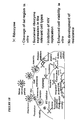
- 230000004069 differentiation Effects 0.000 description 8
- 208000037265 diseases, disorders, signs and symptoms Diseases 0.000 description 8
- 239000003102 growth factor Substances 0.000 description 8
- 239000003550 marker Substances 0.000 description 8
- 102000005962 receptors Human genes 0.000 description 8
- 108020003175 receptors Proteins 0.000 description 8
- 210000001541 thymus gland Anatomy 0.000 description 8
- 108010092799 RNA-directed DNA polymerase Proteins 0.000 description 7
- 241000251131 Sphyrna Species 0.000 description 7
- 230000022131 cell cycle Effects 0.000 description 7
- 230000008045 co-localization Effects 0.000 description 7
- 201000010099 disease Diseases 0.000 description 7
- 229940079593 drug Drugs 0.000 description 7
- 238000003306 harvesting Methods 0.000 description 7
- 230000011132 hemopoiesis Effects 0.000 description 7
- 230000001483 mobilizing effect Effects 0.000 description 7
- 239000013642 negative control Substances 0.000 description 7
- LFQSCWFLJHTTHZ-UHFFFAOYSA-N Ethanol Chemical compound CCO LFQSCWFLJHTTHZ-UHFFFAOYSA-N 0.000 description 6
- 239000007760 Iscove's Modified Dulbecco's Medium Substances 0.000 description 6
- -1 MIP-1 Proteins 0.000 description 6
- 101710149951 Protein Tat Proteins 0.000 description 6
- 239000006146 Roswell Park Memorial Institute medium Substances 0.000 description 6
- PXIPVTKHYLBLMZ-UHFFFAOYSA-N Sodium azide Chemical compound [Na+].[N-]=[N+]=[N-] PXIPVTKHYLBLMZ-UHFFFAOYSA-N 0.000 description 6
- 108700019146 Transgenes Proteins 0.000 description 6
- 239000007983 Tris buffer Substances 0.000 description 6
- 206010058874 Viraemia Diseases 0.000 description 6
- 239000006143 cell culture medium Substances 0.000 description 6
- 230000001413 cellular effect Effects 0.000 description 6
- 230000002596 correlated effect Effects 0.000 description 6
- 230000000875 corresponding effect Effects 0.000 description 6
- 230000001419 dependent effect Effects 0.000 description 6
- 210000000777 hematopoietic system Anatomy 0.000 description 6
- 210000000987 immune system Anatomy 0.000 description 6
- 238000001727 in vivo Methods 0.000 description 6
- 230000006698 induction Effects 0.000 description 6
- 230000002688 persistence Effects 0.000 description 6
- 229940048914 protamine Drugs 0.000 description 6
- 229950008679 protamine sulfate Drugs 0.000 description 6
- 125000006850 spacer group Chemical group 0.000 description 6
- 108700004027 tat Genes Proteins 0.000 description 6
- 101150098170 tat gene Proteins 0.000 description 6
- LENZDBCJOHFCAS-UHFFFAOYSA-N tris Chemical compound OCC(N)(CO)CO LENZDBCJOHFCAS-UHFFFAOYSA-N 0.000 description 6
- 108090001102 Hammerhead ribozyme Proteins 0.000 description 5
- 108091092195 Intron Proteins 0.000 description 5
- 238000010222 PCR analysis Methods 0.000 description 5
- ISWSIDIOOBJBQZ-UHFFFAOYSA-N Phenol Chemical compound OC1=CC=CC=C1 ISWSIDIOOBJBQZ-UHFFFAOYSA-N 0.000 description 5
- QAOWNCQODCNURD-UHFFFAOYSA-L Sulfate Chemical compound [O-]S([O-])(=O)=O QAOWNCQODCNURD-UHFFFAOYSA-L 0.000 description 5
- XSQUKJJJFZCRTK-UHFFFAOYSA-N Urea Chemical compound NC(N)=O XSQUKJJJFZCRTK-UHFFFAOYSA-N 0.000 description 5
- 230000036436 anti-hiv Effects 0.000 description 5
- 239000000427 antigen Substances 0.000 description 5
- 108091007433 antigens Proteins 0.000 description 5
- 102000036639 antigens Human genes 0.000 description 5
- 238000013459 approach Methods 0.000 description 5
- 210000001772 blood platelet Anatomy 0.000 description 5
- 239000004202 carbamide Substances 0.000 description 5
- 230000003197 catalytic effect Effects 0.000 description 5
- 238000005516 engineering process Methods 0.000 description 5
- 238000000605 extraction Methods 0.000 description 5
- 230000006870 function Effects 0.000 description 5
- 230000002068 genetic effect Effects 0.000 description 5
- 230000005764 inhibitory process Effects 0.000 description 5
- 239000012139 lysis buffer Substances 0.000 description 5
- 238000011177 media preparation Methods 0.000 description 5
- 230000001404 mediated effect Effects 0.000 description 5
- 102000039446 nucleic acids Human genes 0.000 description 5
- 108020004707 nucleic acids Proteins 0.000 description 5
- 150000007523 nucleic acids Chemical class 0.000 description 5
- 210000004940 nucleus Anatomy 0.000 description 5
- 239000002245 particle Substances 0.000 description 5
- 239000013612 plasmid Substances 0.000 description 5
- 238000003753 real-time PCR Methods 0.000 description 5
- 238000011084 recovery Methods 0.000 description 5
- 230000002829 reductive effect Effects 0.000 description 5
- 229910021653 sulphate ion Inorganic materials 0.000 description 5
- 230000002992 thymic effect Effects 0.000 description 5
- 238000013518 transcription Methods 0.000 description 5
- 230000035897 transcription Effects 0.000 description 5
- 230000002463 transducing effect Effects 0.000 description 5
- 230000029812 viral genome replication Effects 0.000 description 5
- 102000040650 (ribonucleotides)n+m Human genes 0.000 description 4
- IJGRMHOSHXDMSA-UHFFFAOYSA-N Atomic nitrogen Chemical compound N#N IJGRMHOSHXDMSA-UHFFFAOYSA-N 0.000 description 4
- 102000017420 CD3 protein, epsilon/gamma/delta subunit Human genes 0.000 description 4
- 108050005493 CD3 protein, epsilon/gamma/delta subunit Proteins 0.000 description 4
- 101710205625 Capsid protein p24 Proteins 0.000 description 4
- 241000701022 Cytomegalovirus Species 0.000 description 4
- 238000002965 ELISA Methods 0.000 description 4
- 102000003951 Erythropoietin Human genes 0.000 description 4
- 108090000394 Erythropoietin Proteins 0.000 description 4
- HTTJABKRGRZYRN-UHFFFAOYSA-N Heparin Chemical compound OC1C(NC(=O)C)C(O)OC(COS(O)(=O)=O)C1OC1C(OS(O)(=O)=O)C(O)C(OC2C(C(OS(O)(=O)=O)C(OC3C(C(O)C(O)C(O3)C(O)=O)OS(O)(=O)=O)C(CO)O2)NS(O)(=O)=O)C(C(O)=O)O1 HTTJABKRGRZYRN-UHFFFAOYSA-N 0.000 description 4
- 101001027128 Homo sapiens Fibronectin Proteins 0.000 description 4
- 101001018097 Homo sapiens L-selectin Proteins 0.000 description 4
- 101000946889 Homo sapiens Monocyte differentiation antigen CD14 Proteins 0.000 description 4
- 102000000589 Interleukin-1 Human genes 0.000 description 4
- 108010002352 Interleukin-1 Proteins 0.000 description 4
- 102000003815 Interleukin-11 Human genes 0.000 description 4
- 108090000177 Interleukin-11 Proteins 0.000 description 4
- 102000013462 Interleukin-12 Human genes 0.000 description 4
- 108010065805 Interleukin-12 Proteins 0.000 description 4
- 102000003812 Interleukin-15 Human genes 0.000 description 4
- 108090000172 Interleukin-15 Proteins 0.000 description 4
- 102000004388 Interleukin-4 Human genes 0.000 description 4
- 108090000978 Interleukin-4 Proteins 0.000 description 4
- 102100039897 Interleukin-5 Human genes 0.000 description 4
- 108010002616 Interleukin-5 Proteins 0.000 description 4
- 102100021592 Interleukin-7 Human genes 0.000 description 4
- 108010002586 Interleukin-7 Proteins 0.000 description 4
- 102000000585 Interleukin-9 Human genes 0.000 description 4
- 108010002335 Interleukin-9 Proteins 0.000 description 4
- 101150008942 J gene Proteins 0.000 description 4
- 102100033467 L-selectin Human genes 0.000 description 4
- 108090000581 Leukemia inhibitory factor Proteins 0.000 description 4
- 102000004058 Leukemia inhibitory factor Human genes 0.000 description 4
- 102000007651 Macrophage Colony-Stimulating Factor Human genes 0.000 description 4
- 108010046938 Macrophage Colony-Stimulating Factor Proteins 0.000 description 4
- TWRXJAOTZQYOKJ-UHFFFAOYSA-L Magnesium chloride Chemical compound [Mg+2].[Cl-].[Cl-] TWRXJAOTZQYOKJ-UHFFFAOYSA-L 0.000 description 4
- 102100035877 Monocyte differentiation antigen CD14 Human genes 0.000 description 4
- 229930040373 Paraformaldehyde Natural products 0.000 description 4
- 101710177166 Phosphoprotein Proteins 0.000 description 4
- 239000004743 Polypropylene Substances 0.000 description 4
- 102000009572 RNA Polymerase II Human genes 0.000 description 4
- 108010009460 RNA Polymerase II Proteins 0.000 description 4
- 102000014450 RNA Polymerase III Human genes 0.000 description 4
- 108010078067 RNA Polymerase III Proteins 0.000 description 4
- 101710149279 Small delta antigen Proteins 0.000 description 4
- 208000012827 T-B+ severe combined immunodeficiency due to gamma chain deficiency Diseases 0.000 description 4
- 102000004887 Transforming Growth Factor beta Human genes 0.000 description 4
- 108090001012 Transforming Growth Factor beta Proteins 0.000 description 4
- 102100022563 Tubulin polymerization-promoting protein Human genes 0.000 description 4
- 108060008682 Tumor Necrosis Factor Proteins 0.000 description 4
- 108010067390 Viral Proteins Proteins 0.000 description 4
- 201000007146 X-linked severe combined immunodeficiency Diseases 0.000 description 4
- 239000000443 aerosol Substances 0.000 description 4
- 230000000735 allogeneic effect Effects 0.000 description 4
- 230000015572 biosynthetic process Effects 0.000 description 4
- 210000002798 bone marrow cell Anatomy 0.000 description 4
- 230000010261 cell growth Effects 0.000 description 4
- 238000011109 contamination Methods 0.000 description 4
- 239000012228 culture supernatant Substances 0.000 description 4
- 229940105423 erythropoietin Drugs 0.000 description 4
- 238000000684 flow cytometry Methods 0.000 description 4
- 229960002897 heparin Drugs 0.000 description 4
- 229920000669 heparin Polymers 0.000 description 4
- 230000001900 immune effect Effects 0.000 description 4
- 230000002458 infectious effect Effects 0.000 description 4
- 230000003993 interaction Effects 0.000 description 4
- 229940074383 interleukin-11 Drugs 0.000 description 4
- 229940117681 interleukin-12 Drugs 0.000 description 4
- 229940028885 interleukin-4 Drugs 0.000 description 4
- 229940100602 interleukin-5 Drugs 0.000 description 4
- 229940100994 interleukin-7 Drugs 0.000 description 4
- 229940118526 interleukin-9 Drugs 0.000 description 4
- 208000032839 leukemia Diseases 0.000 description 4
- 238000012417 linear regression Methods 0.000 description 4
- 239000000463 material Substances 0.000 description 4
- 230000007246 mechanism Effects 0.000 description 4
- 230000004048 modification Effects 0.000 description 4
- 238000012986 modification Methods 0.000 description 4
- 229920002866 paraformaldehyde Polymers 0.000 description 4
- 238000009520 phase I clinical trial Methods 0.000 description 4
- 229920001155 polypropylene Polymers 0.000 description 4
- OXCMYAYHXIHQOA-UHFFFAOYSA-N potassium;[2-butyl-5-chloro-3-[[4-[2-(1,2,4-triaza-3-azanidacyclopenta-1,4-dien-5-yl)phenyl]phenyl]methyl]imidazol-4-yl]methanol Chemical compound [K+].CCCCC1=NC(Cl)=C(CO)N1CC1=CC=C(C=2C(=CC=CC=2)C2=N[N-]N=N2)C=C1 OXCMYAYHXIHQOA-UHFFFAOYSA-N 0.000 description 4
- 239000002243 precursor Substances 0.000 description 4
- 230000001105 regulatory effect Effects 0.000 description 4
- 238000010839 reverse transcription Methods 0.000 description 4
- 238000003757 reverse transcription PCR Methods 0.000 description 4
- 230000002441 reversible effect Effects 0.000 description 4
- 230000035945 sensitivity Effects 0.000 description 4
- 210000002966 serum Anatomy 0.000 description 4
- 239000011780 sodium chloride Substances 0.000 description 4
- 239000008223 sterile water Substances 0.000 description 4
- 239000000725 suspension Substances 0.000 description 4
- 230000002459 sustained effect Effects 0.000 description 4
- 230000008685 targeting Effects 0.000 description 4
- ZRKFYGHZFMAOKI-QMGMOQQFSA-N tgfbeta Chemical compound C([C@H](NC(=O)[C@H](C(C)C)NC(=O)CNC(=O)[C@H](CCC(O)=O)NC(=O)[C@H](CCCNC(N)=N)NC(=O)[C@H](CC(N)=O)NC(=O)[C@H](CC(C)C)NC(=O)[C@H]([C@@H](C)O)NC(=O)[C@H](CCC(O)=O)NC(=O)[C@H]([C@@H](C)O)NC(=O)[C@H](CC(C)C)NC(=O)CNC(=O)[C@H](C)NC(=O)[C@H](CO)NC(=O)[C@H](CCC(N)=O)NC(=O)[C@@H](NC(=O)[C@H](C)NC(=O)[C@H](C)NC(=O)[C@@H](NC(=O)[C@H](CC(C)C)NC(=O)[C@@H](N)CCSC)C(C)C)[C@@H](C)CC)C(=O)N[C@@H]([C@@H](C)O)C(=O)N[C@@H](C(C)C)C(=O)N[C@@H](CC=1C=CC=CC=1)C(=O)N[C@@H](C)C(=O)N1[C@@H](CCC1)C(=O)N[C@@H]([C@@H](C)O)C(=O)N[C@@H](CC(N)=O)C(=O)N[C@@H](CCC(O)=O)C(=O)N[C@@H](C)C(=O)N[C@@H](CC=1C=CC=CC=1)C(=O)N[C@@H](CCCNC(N)=N)C(=O)N[C@@H](C)C(=O)N[C@@H](CC(C)C)C(=O)N1[C@@H](CCC1)C(=O)N1[C@@H](CCC1)C(=O)N[C@@H](CCCNC(N)=N)C(=O)N[C@@H](CCC(O)=O)C(=O)N[C@@H](CCCNC(N)=N)C(=O)N[C@@H](CO)C(=O)N[C@@H](CCCNC(N)=N)C(=O)N[C@@H](CC(C)C)C(=O)N[C@@H](CC(C)C)C(O)=O)C1=CC=C(O)C=C1 ZRKFYGHZFMAOKI-QMGMOQQFSA-N 0.000 description 4
- 238000013519 translation Methods 0.000 description 4
- 102000003390 tumor necrosis factor Human genes 0.000 description 4
- 210000002845 virion Anatomy 0.000 description 4
- 102100031585 ADP-ribosyl cyclase/cyclic ADP-ribose hydrolase 1 Human genes 0.000 description 3
- 241000894006 Bacteria Species 0.000 description 3
- FGUUSXIOTUKUDN-IBGZPJMESA-N C1(=CC=CC=C1)N1C2=C(NC([C@H](C1)NC=1OC(=NN=1)C1=CC=CC=C1)=O)C=CC=C2 Chemical compound C1(=CC=CC=C1)N1C2=C(NC([C@H](C1)NC=1OC(=NN=1)C1=CC=CC=C1)=O)C=CC=C2 FGUUSXIOTUKUDN-IBGZPJMESA-N 0.000 description 3
- 102000013925 CD34 antigen Human genes 0.000 description 3
- 108050003733 CD34 antigen Proteins 0.000 description 3
- 102100025621 Cytochrome b-245 heavy chain Human genes 0.000 description 3
- 102000053602 DNA Human genes 0.000 description 3
- 102000016911 Deoxyribonucleases Human genes 0.000 description 3
- 108010053770 Deoxyribonucleases Proteins 0.000 description 3
- 241000724709 Hepatitis delta virus Species 0.000 description 3
- 229920000209 Hexadimethrine bromide Polymers 0.000 description 3
- 101000777636 Homo sapiens ADP-ribosyl cyclase/cyclic ADP-ribose hydrolase 1 Proteins 0.000 description 3
- 101100220044 Homo sapiens CD34 gene Proteins 0.000 description 3
- 229920002274 Nalgene Polymers 0.000 description 3
- 240000007594 Oryza sativa Species 0.000 description 3
- BELBBZDIHDAJOR-UHFFFAOYSA-N Phenolsulfonephthalein Chemical compound C1=CC(O)=CC=C1C1(C=2C=CC(O)=CC=2)C2=CC=CC=C2S(=O)(=O)O1 BELBBZDIHDAJOR-UHFFFAOYSA-N 0.000 description 3
- 238000011579 SCID mouse model Methods 0.000 description 3
- 108010006785 Taq Polymerase Proteins 0.000 description 3
- 108020004566 Transfer RNA Proteins 0.000 description 3
- 230000009471 action Effects 0.000 description 3
- 230000004913 activation Effects 0.000 description 3
- 230000000798 anti-retroviral effect Effects 0.000 description 3
- 230000001580 bacterial effect Effects 0.000 description 3
- 239000011575 calcium Substances 0.000 description 3
- 125000002091 cationic group Chemical group 0.000 description 3
- 210000000170 cell membrane Anatomy 0.000 description 3
- 230000012292 cell migration Effects 0.000 description 3
- 230000008859 change Effects 0.000 description 3
- 238000002512 chemotherapy Methods 0.000 description 3
- 208000016532 chronic granulomatous disease Diseases 0.000 description 3
- 238000012937 correction Methods 0.000 description 3
- 238000012864 cross contamination Methods 0.000 description 3
- 238000005138 cryopreservation Methods 0.000 description 3
- 230000009089 cytolysis Effects 0.000 description 3
- MXCPYJZDGPQDRA-UHFFFAOYSA-N dialuminum;2-acetyloxybenzoic acid;oxygen(2-) Chemical compound [O-2].[O-2].[O-2].[Al+3].[Al+3].CC(=O)OC1=CC=CC=C1C(O)=O MXCPYJZDGPQDRA-UHFFFAOYSA-N 0.000 description 3
- 238000010790 dilution Methods 0.000 description 3
- 239000012895 dilution Substances 0.000 description 3
- 210000003743 erythrocyte Anatomy 0.000 description 3
- 238000002474 experimental method Methods 0.000 description 3
- 238000001914 filtration Methods 0.000 description 3
- 238000007710 freezing Methods 0.000 description 3
- 230000008014 freezing Effects 0.000 description 3
- 230000002538 fungal effect Effects 0.000 description 3
- 108090001052 hairpin ribozyme Proteins 0.000 description 3
- 230000001976 improved effect Effects 0.000 description 3
- 230000036512 infertility Effects 0.000 description 3
- 230000003834 intracellular effect Effects 0.000 description 3
- 239000011777 magnesium Substances 0.000 description 3
- 238000002156 mixing Methods 0.000 description 3
- 238000010172 mouse model Methods 0.000 description 3
- 244000052769 pathogen Species 0.000 description 3
- 230000001717 pathogenic effect Effects 0.000 description 3
- 229960003531 phenolsulfonphthalein Drugs 0.000 description 3
- 229920002401 polyacrylamide Polymers 0.000 description 3
- 230000008488 polyadenylation Effects 0.000 description 3
- 239000013641 positive control Substances 0.000 description 3
- 108090000765 processed proteins & peptides Proteins 0.000 description 3
- 230000004044 response Effects 0.000 description 3
- 238000009781 safety test method Methods 0.000 description 3
- 238000012216 screening Methods 0.000 description 3
- 238000012163 sequencing technique Methods 0.000 description 3
- 238000001228 spectrum Methods 0.000 description 3
- 238000003860 storage Methods 0.000 description 3
- 239000000126 substance Substances 0.000 description 3
- 238000003786 synthesis reaction Methods 0.000 description 3
- 231100000041 toxicology testing Toxicity 0.000 description 3
- 238000001890 transfection Methods 0.000 description 3
- 230000009466 transformation Effects 0.000 description 3
- 238000002054 transplantation Methods 0.000 description 3
- 241000701161 unidentified adenovirus Species 0.000 description 3
- 230000035899 viability Effects 0.000 description 3
- QKNYBSVHEMOAJP-UHFFFAOYSA-N 2-amino-2-(hydroxymethyl)propane-1,3-diol;hydron;chloride Chemical compound Cl.OCC(N)(CO)CO QKNYBSVHEMOAJP-UHFFFAOYSA-N 0.000 description 2
- NALREUIWICQLPS-UHFFFAOYSA-N 7-imino-n,n-dimethylphenothiazin-3-amine;hydrochloride Chemical compound [Cl-].C1=C(N)C=C2SC3=CC(=[N+](C)C)C=CC3=NC2=C1 NALREUIWICQLPS-UHFFFAOYSA-N 0.000 description 2
- QTBSBXVTEAMEQO-UHFFFAOYSA-N Acetic acid Chemical compound CC(O)=O QTBSBXVTEAMEQO-UHFFFAOYSA-N 0.000 description 2
- 108091093088 Amplicon Proteins 0.000 description 2
- 108020005544 Antisense RNA Proteins 0.000 description 2
- 102100024222 B-lymphocyte antigen CD19 Human genes 0.000 description 2
- 206010065553 Bone marrow failure Diseases 0.000 description 2
- 241000167854 Bourreria succulenta Species 0.000 description 2
- OYPRJOBELJOOCE-UHFFFAOYSA-N Calcium Chemical compound [Ca] OYPRJOBELJOOCE-UHFFFAOYSA-N 0.000 description 2
- 102100025470 Carcinoembryonic antigen-related cell adhesion molecule 8 Human genes 0.000 description 2
- 206010053138 Congenital aplastic anaemia Diseases 0.000 description 2
- CMSMOCZEIVJLDB-UHFFFAOYSA-N Cyclophosphamide Chemical compound ClCCN(CCCl)P1(=O)NCCCO1 CMSMOCZEIVJLDB-UHFFFAOYSA-N 0.000 description 2
- 238000007400 DNA extraction Methods 0.000 description 2
- 239000003155 DNA primer Substances 0.000 description 2
- 108010014303 DNA-directed DNA polymerase Proteins 0.000 description 2
- 102000016928 DNA-directed DNA polymerase Human genes 0.000 description 2
- 206010059866 Drug resistance Diseases 0.000 description 2
- KCXVZYZYPLLWCC-UHFFFAOYSA-N EDTA Chemical compound OC(=O)CN(CC(O)=O)CCN(CC(O)=O)CC(O)=O KCXVZYZYPLLWCC-UHFFFAOYSA-N 0.000 description 2
- 241000876833 Emberizinae Species 0.000 description 2
- 101710088235 Envelope glycoprotein C homolog Proteins 0.000 description 2
- 102000004190 Enzymes Human genes 0.000 description 2
- 108090000790 Enzymes Proteins 0.000 description 2
- 241000588724 Escherichia coli Species 0.000 description 2
- 201000004939 Fanconi anemia Diseases 0.000 description 2
- 208000015872 Gaucher disease Diseases 0.000 description 2
- 241000713813 Gibbon ape leukemia virus Species 0.000 description 2
- 208000037262 Hepatitis delta Diseases 0.000 description 2
- 101000980825 Homo sapiens B-lymphocyte antigen CD19 Proteins 0.000 description 2
- 101000914320 Homo sapiens Carcinoembryonic antigen-related cell adhesion molecule 8 Proteins 0.000 description 2
- 101000917858 Homo sapiens Low affinity immunoglobulin gamma Fc region receptor III-A Proteins 0.000 description 2
- 101000917839 Homo sapiens Low affinity immunoglobulin gamma Fc region receptor III-B Proteins 0.000 description 2
- 101000581981 Homo sapiens Neural cell adhesion molecule 1 Proteins 0.000 description 2
- 101000738771 Homo sapiens Receptor-type tyrosine-protein phosphatase C Proteins 0.000 description 2
- 101000884271 Homo sapiens Signal transducer CD24 Proteins 0.000 description 2
- 101000934346 Homo sapiens T-cell surface antigen CD2 Proteins 0.000 description 2
- 241000713340 Human immunodeficiency virus 2 Species 0.000 description 2
- 208000026350 Inborn Genetic disease Diseases 0.000 description 2
- 102100020880 Kit ligand Human genes 0.000 description 2
- 102100029185 Low affinity immunoglobulin gamma Fc region receptor III-B Human genes 0.000 description 2
- FYYHWMGAXLPEAU-UHFFFAOYSA-N Magnesium Chemical compound [Mg] FYYHWMGAXLPEAU-UHFFFAOYSA-N 0.000 description 2
- XRYVAQQLDYTHCL-UHFFFAOYSA-N Marini Chemical compound O1C=2C(CC(CC=C(C)C)C(C)=C)=C(O)C=C(O)C=2C(=O)CC1C1=CC=C(O)C=C1O XRYVAQQLDYTHCL-UHFFFAOYSA-N 0.000 description 2
- 241001465754 Metazoa Species 0.000 description 2
- 101100335081 Mus musculus Flt3 gene Proteins 0.000 description 2
- 241001364944 Mus terricolor Species 0.000 description 2
- 108010021466 Mutant Proteins Proteins 0.000 description 2
- 102000008300 Mutant Proteins Human genes 0.000 description 2
- 206010028980 Neoplasm Diseases 0.000 description 2
- 102100027347 Neural cell adhesion molecule 1 Human genes 0.000 description 2
- 229940122313 Nucleoside reverse transcriptase inhibitor Drugs 0.000 description 2
- 108091034117 Oligonucleotide Proteins 0.000 description 2
- 101710160107 Outer membrane protein A Proteins 0.000 description 2
- 206010034133 Pathogen resistance Diseases 0.000 description 2
- 229940124158 Protease/peptidase inhibitor Drugs 0.000 description 2
- 101710149136 Protein Vpr Proteins 0.000 description 2
- 102100037422 Receptor-type tyrosine-protein phosphatase C Human genes 0.000 description 2
- 108020004511 Recombinant DNA Proteins 0.000 description 2
- 108091028664 Ribonucleotide Proteins 0.000 description 2
- 102100038081 Signal transducer CD24 Human genes 0.000 description 2
- 241000713311 Simian immunodeficiency virus Species 0.000 description 2
- 108020004682 Single-Stranded DNA Proteins 0.000 description 2
- 108010039445 Stem Cell Factor Proteins 0.000 description 2
- 102100025237 T-cell surface antigen CD2 Human genes 0.000 description 2
- 241000053227 Themus Species 0.000 description 2
- 108091036066 Three prime untranslated region Proteins 0.000 description 2
- 208000023940 X-Linked Combined Immunodeficiency disease Diseases 0.000 description 2
- 239000002253 acid Substances 0.000 description 2
- 150000007513 acids Chemical class 0.000 description 2
- 108700024685 ancestim Proteins 0.000 description 2
- 238000000137 annealing Methods 0.000 description 2
- 150000001540 azides Chemical class 0.000 description 2
- 239000011324 bead Substances 0.000 description 2
- 238000001574 biopsy Methods 0.000 description 2
- 238000004820 blood count Methods 0.000 description 2
- 238000010322 bone marrow transplantation Methods 0.000 description 2
- 229910052791 calcium Inorganic materials 0.000 description 2
- 230000015556 catabolic process Effects 0.000 description 2
- 230000034303 cell budding Effects 0.000 description 2
- 230000032823 cell division Effects 0.000 description 2
- 230000003915 cell function Effects 0.000 description 2
- 239000013592 cell lysate Substances 0.000 description 2
- 239000006285 cell suspension Substances 0.000 description 2
- 235000019693 cherries Nutrition 0.000 description 2
- 230000002860 competitive effect Effects 0.000 description 2
- 239000003184 complementary RNA Substances 0.000 description 2
- 239000003636 conditioned culture medium Substances 0.000 description 2
- 230000003750 conditioning effect Effects 0.000 description 2
- 229960004397 cyclophosphamide Drugs 0.000 description 2
- 210000000805 cytoplasm Anatomy 0.000 description 2
- 230000006378 damage Effects 0.000 description 2
- 230000007547 defect Effects 0.000 description 2
- 230000002950 deficient Effects 0.000 description 2
- 238000004925 denaturation Methods 0.000 description 2
- 230000036425 denaturation Effects 0.000 description 2
- 239000012153 distilled water Substances 0.000 description 2
- 231100000673 dose–response relationship Toxicity 0.000 description 2
- 239000000975 dye Substances 0.000 description 2
- 238000001962 electrophoresis Methods 0.000 description 2
- 108700004025 env Genes Proteins 0.000 description 2
- 230000002255 enzymatic effect Effects 0.000 description 2
- 230000008029 eradication Effects 0.000 description 2
- 229960004756 ethanol Drugs 0.000 description 2
- 230000002349 favourable effect Effects 0.000 description 2
- 239000012997 ficoll-paque Substances 0.000 description 2
- 239000000834 fixative Substances 0.000 description 2
- 230000004927 fusion Effects 0.000 description 2
- 238000001476 gene delivery Methods 0.000 description 2
- 208000016361 genetic disease Diseases 0.000 description 2
- 230000012010 growth Effects 0.000 description 2
- 238000012787 harvest procedure Methods 0.000 description 2
- 208000029570 hepatitis D virus infection Diseases 0.000 description 2
- 230000036737 immune function Effects 0.000 description 2
- 230000028993 immune response Effects 0.000 description 2
- 230000036039 immunity Effects 0.000 description 2
- 230000001939 inductive effect Effects 0.000 description 2
- 239000003112 inhibitor Substances 0.000 description 2
- 230000002401 inhibitory effect Effects 0.000 description 2
- 238000003780 insertion Methods 0.000 description 2
- 230000037431 insertion Effects 0.000 description 2
- 230000010354 integration Effects 0.000 description 2
- 230000016507 interphase Effects 0.000 description 2
- PHTQWCKDNZKARW-UHFFFAOYSA-N isoamylol Chemical compound CC(C)CCO PHTQWCKDNZKARW-UHFFFAOYSA-N 0.000 description 2
- 238000011005 laboratory method Methods 0.000 description 2
- 239000002502 liposome Substances 0.000 description 2
- 239000007788 liquid Substances 0.000 description 2
- 210000004185 liver Anatomy 0.000 description 2
- 229910052749 magnesium Inorganic materials 0.000 description 2
- 229910001629 magnesium chloride Inorganic materials 0.000 description 2
- 230000014759 maintenance of location Effects 0.000 description 2
- 210000004962 mammalian cell Anatomy 0.000 description 2
- 239000011159 matrix material Substances 0.000 description 2
- 230000035800 maturation Effects 0.000 description 2
- 239000012528 membrane Substances 0.000 description 2
- 210000003071 memory t lymphocyte Anatomy 0.000 description 2
- 108020004999 messenger RNA Proteins 0.000 description 2
- 239000011325 microbead Substances 0.000 description 2
- 230000011278 mitosis Effects 0.000 description 2
- 230000035772 mutation Effects 0.000 description 2
- 230000001400 myeloablative effect Effects 0.000 description 2
- 238000007857 nested PCR Methods 0.000 description 2
- 229910052757 nitrogen Inorganic materials 0.000 description 2
- 229940042402 non-nucleoside reverse transcriptase inhibitor Drugs 0.000 description 2
- 239000002726 nonnucleoside reverse transcriptase inhibitor Substances 0.000 description 2
- 239000000137 peptide hydrolase inhibitor Substances 0.000 description 2
- 239000013600 plasmid vector Substances 0.000 description 2
- 239000004033 plastic Substances 0.000 description 2
- 229920003023 plastic Polymers 0.000 description 2
- 229920000642 polymer Polymers 0.000 description 2
- 238000003752 polymerase chain reaction Methods 0.000 description 2
- 229920001184 polypeptide Polymers 0.000 description 2
- 230000001566 pro-viral effect Effects 0.000 description 2
- 102000004196 processed proteins & peptides Human genes 0.000 description 2
- 230000002062 proliferating effect Effects 0.000 description 2
- 230000001681 protective effect Effects 0.000 description 2
- 238000000746 purification Methods 0.000 description 2
- 230000002285 radioactive effect Effects 0.000 description 2
- 230000003362 replicative effect Effects 0.000 description 2
- 230000002629 repopulating effect Effects 0.000 description 2
- 238000011160 research Methods 0.000 description 2
- 239000002336 ribonucleotide Substances 0.000 description 2
- 125000002652 ribonucleotide group Chemical group 0.000 description 2
- 239000003419 rna directed dna polymerase inhibitor Substances 0.000 description 2
- 238000010187 selection method Methods 0.000 description 2
- 208000002491 severe combined immunodeficiency Diseases 0.000 description 2
- 238000010561 standard procedure Methods 0.000 description 2
- 238000000528 statistical test Methods 0.000 description 2
- 238000006467 substitution reaction Methods 0.000 description 2
- 230000001629 suppression Effects 0.000 description 2
- 238000010257 thawing Methods 0.000 description 2
- 238000002560 therapeutic procedure Methods 0.000 description 2
- 231100000331 toxic Toxicity 0.000 description 2
- 230000002588 toxic effect Effects 0.000 description 2
- 230000001052 transient effect Effects 0.000 description 2
- 230000007704 transition Effects 0.000 description 2
- 230000009385 viral infection Effects 0.000 description 2
- 238000003260 vortexing Methods 0.000 description 2
- DIGQNXIGRZPYDK-WKSCXVIASA-N (2R)-6-amino-2-[[2-[[(2S)-2-[[2-[[(2R)-2-[[(2S)-2-[[(2R,3S)-2-[[2-[[(2S)-2-[[2-[[(2S)-2-[[(2S)-2-[[(2R)-2-[[(2S,3S)-2-[[(2R)-2-[[(2S)-2-[[(2S)-2-[[(2S)-2-[[2-[[(2S)-2-[[(2R)-2-[[2-[[2-[[2-[(2-amino-1-hydroxyethylidene)amino]-3-carboxy-1-hydroxypropylidene]amino]-1-hydroxy-3-sulfanylpropylidene]amino]-1-hydroxyethylidene]amino]-1-hydroxy-3-sulfanylpropylidene]amino]-1,3-dihydroxypropylidene]amino]-1-hydroxyethylidene]amino]-1-hydroxypropylidene]amino]-1,3-dihydroxypropylidene]amino]-1,3-dihydroxypropylidene]amino]-1-hydroxy-3-sulfanylpropylidene]amino]-1,3-dihydroxybutylidene]amino]-1-hydroxy-3-sulfanylpropylidene]amino]-1-hydroxypropylidene]amino]-1,3-dihydroxypropylidene]amino]-1-hydroxyethylidene]amino]-1,5-dihydroxy-5-iminopentylidene]amino]-1-hydroxy-3-sulfanylpropylidene]amino]-1,3-dihydroxybutylidene]amino]-1-hydroxy-3-sulfanylpropylidene]amino]-1,3-dihydroxypropylidene]amino]-1-hydroxyethylidene]amino]-1-hydroxy-3-sulfanylpropylidene]amino]-1-hydroxyethylidene]amino]hexanoic acid Chemical compound C[C@@H]([C@@H](C(=N[C@@H](CS)C(=N[C@@H](C)C(=N[C@@H](CO)C(=NCC(=N[C@@H](CCC(=N)O)C(=NC(CS)C(=N[C@H]([C@H](C)O)C(=N[C@H](CS)C(=N[C@H](CO)C(=NCC(=N[C@H](CS)C(=NCC(=N[C@H](CCCCN)C(=O)O)O)O)O)O)O)O)O)O)O)O)O)O)O)N=C([C@H](CS)N=C([C@H](CO)N=C([C@H](CO)N=C([C@H](C)N=C(CN=C([C@H](CO)N=C([C@H](CS)N=C(CN=C(C(CS)N=C(C(CC(=O)O)N=C(CN)O)O)O)O)O)O)O)O)O)O)O)O DIGQNXIGRZPYDK-WKSCXVIASA-N 0.000 description 1
- PRDFBSVERLRRMY-UHFFFAOYSA-N 2'-(4-ethoxyphenyl)-5-(4-methylpiperazin-1-yl)-2,5'-bibenzimidazole Chemical compound C1=CC(OCC)=CC=C1C1=NC2=CC=C(C=3NC4=CC(=CC=C4N=3)N3CCN(C)CC3)C=C2N1 PRDFBSVERLRRMY-UHFFFAOYSA-N 0.000 description 1
- HNLXNOZHXNSSPN-UHFFFAOYSA-N 2-[2-[2-[2-[2-[2-[2-[4-(2,4,4-trimethylpentan-2-yl)phenoxy]ethoxy]ethoxy]ethoxy]ethoxy]ethoxy]ethoxy]ethanol Chemical compound CC(C)(C)CC(C)(C)C1=CC=C(OCCOCCOCCOCCOCCOCCOCCO)C=C1 HNLXNOZHXNSSPN-UHFFFAOYSA-N 0.000 description 1
- KDELTXNPUXUBMU-UHFFFAOYSA-N 2-[2-[bis(carboxymethyl)amino]ethyl-(carboxymethyl)amino]acetic acid boric acid Chemical compound OB(O)O.OB(O)O.OB(O)O.OC(=O)CN(CC(O)=O)CCN(CC(O)=O)CC(O)=O KDELTXNPUXUBMU-UHFFFAOYSA-N 0.000 description 1
- GNFTZDOKVXKIBK-UHFFFAOYSA-N 3-(2-methoxyethoxy)benzohydrazide Chemical compound COCCOC1=CC=CC(C(=O)NN)=C1 GNFTZDOKVXKIBK-UHFFFAOYSA-N 0.000 description 1
- 239000007991 ACES buffer Substances 0.000 description 1
- 102000009027 Albumins Human genes 0.000 description 1
- 108010088751 Albumins Proteins 0.000 description 1
- BTBUEUYNUDRHOZ-UHFFFAOYSA-N Borate Chemical compound [O-]B([O-])[O-] BTBUEUYNUDRHOZ-UHFFFAOYSA-N 0.000 description 1
- 206010006187 Breast cancer Diseases 0.000 description 1
- 208000026310 Breast neoplasm Diseases 0.000 description 1
- 101150111062 C gene Proteins 0.000 description 1
- 102000004274 CCR5 Receptors Human genes 0.000 description 1
- 108010017088 CCR5 Receptors Proteins 0.000 description 1
- 210000004366 CD4-positive T-lymphocyte Anatomy 0.000 description 1
- UXVMQQNJUSDDNG-UHFFFAOYSA-L Calcium chloride Chemical compound [Cl-].[Cl-].[Ca+2] UXVMQQNJUSDDNG-UHFFFAOYSA-L 0.000 description 1
- 108090000565 Capsid Proteins Proteins 0.000 description 1
- 102000000844 Cell Surface Receptors Human genes 0.000 description 1
- 108010001857 Cell Surface Receptors Proteins 0.000 description 1
- 241000282693 Cercopithecidae Species 0.000 description 1
- CHBRHODLKOZEPZ-UHFFFAOYSA-N Clotiazepam Chemical compound S1C(CC)=CC2=C1N(C)C(=O)CN=C2C1=CC=CC=C1Cl CHBRHODLKOZEPZ-UHFFFAOYSA-N 0.000 description 1
- 108091026890 Coding region Proteins 0.000 description 1
- 206010010099 Combined immunodeficiency Diseases 0.000 description 1
- 208000035473 Communicable disease Diseases 0.000 description 1
- 108091035707 Consensus sequence Proteins 0.000 description 1
- FBPFZTCFMRRESA-FSIIMWSLSA-N D-Glucitol Natural products OC[C@H](O)[C@H](O)[C@@H](O)[C@H](O)CO FBPFZTCFMRRESA-FSIIMWSLSA-N 0.000 description 1
- 238000001712 DNA sequencing Methods 0.000 description 1
- 102000004163 DNA-directed RNA polymerases Human genes 0.000 description 1
- 108090000626 DNA-directed RNA polymerases Proteins 0.000 description 1
- 206010061818 Disease progression Diseases 0.000 description 1
- 208000030453 Drug-Related Side Effects and Adverse reaction Diseases 0.000 description 1
- 101710091045 Envelope protein Proteins 0.000 description 1
- 241000714162 Feline endogenous virus Species 0.000 description 1
- 238000012413 Fluorescence activated cell sorting analysis Methods 0.000 description 1
- 102100020715 Fms-related tyrosine kinase 3 ligand protein Human genes 0.000 description 1
- 101710162577 Fms-related tyrosine kinase 3 ligand protein Proteins 0.000 description 1
- 208000025499 G6PD deficiency Diseases 0.000 description 1
- 239000001828 Gelatine Substances 0.000 description 1
- WQZGKKKJIJFFOK-GASJEMHNSA-N Glucose Natural products OC[C@H]1OC(O)[C@H](O)[C@@H](O)[C@@H]1O WQZGKKKJIJFFOK-GASJEMHNSA-N 0.000 description 1
- 102000003886 Glycoproteins Human genes 0.000 description 1
- 108090000288 Glycoproteins Proteins 0.000 description 1
- 238000003794 Gram staining Methods 0.000 description 1
- 108010051696 Growth Hormone Proteins 0.000 description 1
- 108010027044 HIV Core Protein p24 Proteins 0.000 description 1
- 102100021519 Hemoglobin subunit beta Human genes 0.000 description 1
- 108091005904 Hemoglobin subunit beta Proteins 0.000 description 1
- 108091080980 Hepatitis delta virus ribozyme Proteins 0.000 description 1
- 101000899111 Homo sapiens Hemoglobin subunit beta Proteins 0.000 description 1
- 101001112229 Homo sapiens Neutrophil cytosol factor 1 Proteins 0.000 description 1
- 101000851007 Homo sapiens Vascular endothelial growth factor receptor 2 Proteins 0.000 description 1
- 241000714260 Human T-lymphotropic virus 1 Species 0.000 description 1
- 241000714259 Human T-lymphotropic virus 2 Species 0.000 description 1
- 241000714192 Human spumaretrovirus Species 0.000 description 1
- 206010020751 Hypersensitivity Diseases 0.000 description 1
- 101150017040 I gene Proteins 0.000 description 1
- 102000008394 Immunoglobulin Fragments Human genes 0.000 description 1
- 108010021625 Immunoglobulin Fragments Proteins 0.000 description 1
- 102100034349 Integrase Human genes 0.000 description 1
- 101710203526 Integrase Proteins 0.000 description 1
- 108010061833 Integrases Proteins 0.000 description 1
- 102100032817 Integrin alpha-5 Human genes 0.000 description 1
- 108010008212 Integrin alpha4beta1 Proteins 0.000 description 1
- 108010041014 Integrin alpha5 Proteins 0.000 description 1
- 108010042918 Integrin alpha5beta1 Proteins 0.000 description 1
- 108010025815 Kanamycin Kinase Proteins 0.000 description 1
- 239000012480 LAL reagent Substances 0.000 description 1
- 241000713666 Lentivirus Species 0.000 description 1
- 208000015439 Lysosomal storage disease Diseases 0.000 description 1
- 102000003792 Metallothionein Human genes 0.000 description 1
- 108090000157 Metallothionein Proteins 0.000 description 1
- 206010048723 Multiple-drug resistance Diseases 0.000 description 1
- 241000699666 Mus <mouse, genus> Species 0.000 description 1
- 241000699670 Mus sp. Species 0.000 description 1
- 241000204003 Mycoplasmatales Species 0.000 description 1
- SQVRNKJHWKZAKO-PFQGKNLYSA-N N-acetyl-beta-neuraminic acid Chemical compound CC(=O)N[C@@H]1[C@@H](O)C[C@@](O)(C(O)=O)O[C@H]1[C@H](O)[C@H](O)CO SQVRNKJHWKZAKO-PFQGKNLYSA-N 0.000 description 1
- 241001045988 Neogene Species 0.000 description 1
- 108010038807 Oligopeptides Proteins 0.000 description 1
- 102000015636 Oligopeptides Human genes 0.000 description 1
- 208000001388 Opportunistic Infections Diseases 0.000 description 1
- 206010033128 Ovarian cancer Diseases 0.000 description 1
- 206010061535 Ovarian neoplasm Diseases 0.000 description 1
- 238000012408 PCR amplification Methods 0.000 description 1
- 238000002944 PCR assay Methods 0.000 description 1
- 108010021757 Polynucleotide 5'-Hydroxyl-Kinase Proteins 0.000 description 1
- 102000008422 Polynucleotide 5'-hydroxyl-kinase Human genes 0.000 description 1
- 229920001213 Polysorbate 20 Polymers 0.000 description 1
- 239000004793 Polystyrene Substances 0.000 description 1
- 241000288906 Primates Species 0.000 description 1
- 101710150344 Protein Rev Proteins 0.000 description 1
- 101710188315 Protein X Proteins 0.000 description 1
- 102000016611 Proteoglycans Human genes 0.000 description 1
- 108010067787 Proteoglycans Proteins 0.000 description 1
- 238000002123 RNA extraction Methods 0.000 description 1
- 239000013614 RNA sample Substances 0.000 description 1
- 238000010240 RT-PCR analysis Methods 0.000 description 1
- 108091081062 Repeated sequence (DNA) Proteins 0.000 description 1
- 241001068263 Replication competent viruses Species 0.000 description 1
- 206010038997 Retroviral infections Diseases 0.000 description 1
- 241000712907 Retroviridae Species 0.000 description 1
- 108091081021 Sense strand Proteins 0.000 description 1
- 229920005654 Sephadex Polymers 0.000 description 1
- 239000012507 Sephadex™ Substances 0.000 description 1
- 102000012010 Sialomucins Human genes 0.000 description 1
- 108010061228 Sialomucins Proteins 0.000 description 1
- 108091027967 Small hairpin RNA Proteins 0.000 description 1
- 108020004459 Small interfering RNA Proteins 0.000 description 1
- VMHLLURERBWHNL-UHFFFAOYSA-M Sodium acetate Chemical compound [Na+].CC([O-])=O VMHLLURERBWHNL-UHFFFAOYSA-M 0.000 description 1
- 102100038803 Somatotropin Human genes 0.000 description 1
- 241000713675 Spumavirus Species 0.000 description 1
- 101710137500 T7 RNA polymerase Proteins 0.000 description 1
- 101150052863 THY1 gene Proteins 0.000 description 1
- RZCIEJXAILMSQK-JXOAFFINSA-N TTP Chemical compound O=C1NC(=O)C(C)=CN1[C@H]1[C@H](O)[C@H](O)[C@@H](COP(O)(=O)OP(O)(=O)OP(O)(O)=O)O1 RZCIEJXAILMSQK-JXOAFFINSA-N 0.000 description 1
- 206010070863 Toxicity to various agents Diseases 0.000 description 1
- 102000046299 Transforming Growth Factor beta1 Human genes 0.000 description 1
- 101800002279 Transforming growth factor beta-1 Proteins 0.000 description 1
- 206010052779 Transplant rejections Diseases 0.000 description 1
- GLNADSQYFUSGOU-GPTZEZBUSA-J Trypan blue Chemical compound [Na+].[Na+].[Na+].[Na+].C1=C(S([O-])(=O)=O)C=C2C=C(S([O-])(=O)=O)C(/N=N/C3=CC=C(C=C3C)C=3C=C(C(=CC=3)\N=N\C=3C(=CC4=CC(=CC(N)=C4C=3O)S([O-])(=O)=O)S([O-])(=O)=O)C)=C(O)C2=C1N GLNADSQYFUSGOU-GPTZEZBUSA-J 0.000 description 1
- 108091023045 Untranslated Region Proteins 0.000 description 1
- 108091008605 VEGF receptors Proteins 0.000 description 1
- 102000009484 Vascular Endothelial Growth Factor Receptors Human genes 0.000 description 1
- 241000711975 Vesicular stomatitis virus Species 0.000 description 1
- 108010003533 Viral Envelope Proteins Proteins 0.000 description 1
- 208000036142 Viral infection Diseases 0.000 description 1
- JLCPHMBAVCMARE-UHFFFAOYSA-N [3-[[3-[[3-[[3-[[3-[[3-[[3-[[3-[[3-[[3-[[3-[[5-(2-amino-6-oxo-1H-purin-9-yl)-3-[[3-[[3-[[3-[[3-[[3-[[5-(2-amino-6-oxo-1H-purin-9-yl)-3-[[5-(2-amino-6-oxo-1H-purin-9-yl)-3-hydroxyoxolan-2-yl]methoxy-hydroxyphosphoryl]oxyoxolan-2-yl]methoxy-hydroxyphosphoryl]oxy-5-(5-methyl-2,4-dioxopyrimidin-1-yl)oxolan-2-yl]methoxy-hydroxyphosphoryl]oxy-5-(6-aminopurin-9-yl)oxolan-2-yl]methoxy-hydroxyphosphoryl]oxy-5-(6-aminopurin-9-yl)oxolan-2-yl]methoxy-hydroxyphosphoryl]oxy-5-(6-aminopurin-9-yl)oxolan-2-yl]methoxy-hydroxyphosphoryl]oxy-5-(6-aminopurin-9-yl)oxolan-2-yl]methoxy-hydroxyphosphoryl]oxyoxolan-2-yl]methoxy-hydroxyphosphoryl]oxy-5-(5-methyl-2,4-dioxopyrimidin-1-yl)oxolan-2-yl]methoxy-hydroxyphosphoryl]oxy-5-(4-amino-2-oxopyrimidin-1-yl)oxolan-2-yl]methoxy-hydroxyphosphoryl]oxy-5-(5-methyl-2,4-dioxopyrimidin-1-yl)oxolan-2-yl]methoxy-hydroxyphosphoryl]oxy-5-(5-methyl-2,4-dioxopyrimidin-1-yl)oxolan-2-yl]methoxy-hydroxyphosphoryl]oxy-5-(6-aminopurin-9-yl)oxolan-2-yl]methoxy-hydroxyphosphoryl]oxy-5-(6-aminopurin-9-yl)oxolan-2-yl]methoxy-hydroxyphosphoryl]oxy-5-(4-amino-2-oxopyrimidin-1-yl)oxolan-2-yl]methoxy-hydroxyphosphoryl]oxy-5-(4-amino-2-oxopyrimidin-1-yl)oxolan-2-yl]methoxy-hydroxyphosphoryl]oxy-5-(4-amino-2-oxopyrimidin-1-yl)oxolan-2-yl]methoxy-hydroxyphosphoryl]oxy-5-(6-aminopurin-9-yl)oxolan-2-yl]methoxy-hydroxyphosphoryl]oxy-5-(4-amino-2-oxopyrimidin-1-yl)oxolan-2-yl]methyl [5-(6-aminopurin-9-yl)-2-(hydroxymethyl)oxolan-3-yl] hydrogen phosphate Polymers Cc1cn(C2CC(OP(O)(=O)OCC3OC(CC3OP(O)(=O)OCC3OC(CC3O)n3cnc4c3nc(N)[nH]c4=O)n3cnc4c3nc(N)[nH]c4=O)C(COP(O)(=O)OC3CC(OC3COP(O)(=O)OC3CC(OC3COP(O)(=O)OC3CC(OC3COP(O)(=O)OC3CC(OC3COP(O)(=O)OC3CC(OC3COP(O)(=O)OC3CC(OC3COP(O)(=O)OC3CC(OC3COP(O)(=O)OC3CC(OC3COP(O)(=O)OC3CC(OC3COP(O)(=O)OC3CC(OC3COP(O)(=O)OC3CC(OC3COP(O)(=O)OC3CC(OC3COP(O)(=O)OC3CC(OC3COP(O)(=O)OC3CC(OC3COP(O)(=O)OC3CC(OC3COP(O)(=O)OC3CC(OC3COP(O)(=O)OC3CC(OC3CO)n3cnc4c(N)ncnc34)n3ccc(N)nc3=O)n3cnc4c(N)ncnc34)n3ccc(N)nc3=O)n3ccc(N)nc3=O)n3ccc(N)nc3=O)n3cnc4c(N)ncnc34)n3cnc4c(N)ncnc34)n3cc(C)c(=O)[nH]c3=O)n3cc(C)c(=O)[nH]c3=O)n3ccc(N)nc3=O)n3cc(C)c(=O)[nH]c3=O)n3cnc4c3nc(N)[nH]c4=O)n3cnc4c(N)ncnc34)n3cnc4c(N)ncnc34)n3cnc4c(N)ncnc34)n3cnc4c(N)ncnc34)O2)c(=O)[nH]c1=O JLCPHMBAVCMARE-UHFFFAOYSA-N 0.000 description 1
- 238000002835 absorbance Methods 0.000 description 1
- 229960000583 acetic acid Drugs 0.000 description 1
- DPXJVFZANSGRMM-UHFFFAOYSA-N acetic acid;2,3,4,5,6-pentahydroxyhexanal;sodium Chemical compound [Na].CC(O)=O.OCC(O)C(O)C(O)C(O)C=O DPXJVFZANSGRMM-UHFFFAOYSA-N 0.000 description 1
- 238000013019 agitation Methods 0.000 description 1
- 208000026935 allergic disease Diseases 0.000 description 1
- 230000007815 allergy Effects 0.000 description 1
- 150000001413 amino acids Chemical group 0.000 description 1
- FROZIYRKKUFAOC-UHFFFAOYSA-N amobam Chemical compound N.N.SC(=S)NCCNC(S)=S FROZIYRKKUFAOC-UHFFFAOYSA-N 0.000 description 1
- 230000003444 anaesthetic effect Effects 0.000 description 1
- 238000000540 analysis of variance Methods 0.000 description 1
- 238000010171 animal model Methods 0.000 description 1
- 229920006318 anionic polymer Polymers 0.000 description 1
- 229940127090 anticoagulant agent Drugs 0.000 description 1
- 239000003146 anticoagulant agent Substances 0.000 description 1
- 239000002246 antineoplastic agent Substances 0.000 description 1
- 239000003443 antiviral agent Substances 0.000 description 1
- 230000006907 apoptotic process Effects 0.000 description 1
- 239000013584 assay control Substances 0.000 description 1
- 238000000376 autoradiography Methods 0.000 description 1
- 230000004888 barrier function Effects 0.000 description 1
- 230000009286 beneficial effect Effects 0.000 description 1
- 238000004166 bioassay Methods 0.000 description 1
- 230000000903 blocking effect Effects 0.000 description 1
- 210000000601 blood cell Anatomy 0.000 description 1
- 230000017531 blood circulation Effects 0.000 description 1
- 210000000988 bone and bone Anatomy 0.000 description 1
- 210000004271 bone marrow stromal cell Anatomy 0.000 description 1
- 210000000481 breast Anatomy 0.000 description 1
- 239000001110 calcium chloride Substances 0.000 description 1
- 229910001628 calcium chloride Inorganic materials 0.000 description 1
- 238000004364 calculation method Methods 0.000 description 1
- 201000011510 cancer Diseases 0.000 description 1
- 229910052799 carbon Inorganic materials 0.000 description 1
- 108020001778 catalytic domains Proteins 0.000 description 1
- 229920006317 cationic polymer Polymers 0.000 description 1
- 230000008568 cell cell communication Effects 0.000 description 1
- 230000005779 cell damage Effects 0.000 description 1
- 230000030833 cell death Effects 0.000 description 1
- 230000011712 cell development Effects 0.000 description 1
- 238000011072 cell harvest Methods 0.000 description 1
- 208000037887 cell injury Diseases 0.000 description 1
- 108091092356 cellular DNA Proteins 0.000 description 1
- 238000004140 cleaning Methods 0.000 description 1
- 238000010367 cloning Methods 0.000 description 1
- 230000003021 clonogenic effect Effects 0.000 description 1
- 230000001332 colony forming effect Effects 0.000 description 1
- 238000010668 complexation reaction Methods 0.000 description 1
- 150000001875 compounds Chemical class 0.000 description 1
- 230000001010 compromised effect Effects 0.000 description 1
- 238000012790 confirmation Methods 0.000 description 1
- 239000000470 constituent Substances 0.000 description 1
- 238000010276 construction Methods 0.000 description 1
- 238000001816 cooling Methods 0.000 description 1
- 230000002338 cryopreservative effect Effects 0.000 description 1
- 239000013078 crystal Substances 0.000 description 1
- 210000004748 cultured cell Anatomy 0.000 description 1
- 230000001351 cycling effect Effects 0.000 description 1
- 208000030605 cytokine receptor deficiency Diseases 0.000 description 1
- 230000007711 cytoplasmic localization Effects 0.000 description 1
- 230000001086 cytosolic effect Effects 0.000 description 1
- 229940127089 cytotoxic agent Drugs 0.000 description 1
- SUYVUBYJARFZHO-RRKCRQDMSA-N dATP Chemical compound C1=NC=2C(N)=NC=NC=2N1[C@H]1C[C@H](O)[C@@H](COP(O)(=O)OP(O)(=O)OP(O)(O)=O)O1 SUYVUBYJARFZHO-RRKCRQDMSA-N 0.000 description 1
- SUYVUBYJARFZHO-UHFFFAOYSA-N dATP Natural products C1=NC=2C(N)=NC=NC=2N1C1CC(O)C(COP(O)(=O)OP(O)(=O)OP(O)(O)=O)O1 SUYVUBYJARFZHO-UHFFFAOYSA-N 0.000 description 1
- RGWHQCVHVJXOKC-SHYZEUOFSA-J dCTP(4-) Chemical compound O=C1N=C(N)C=CN1[C@@H]1O[C@H](COP([O-])(=O)OP([O-])(=O)OP([O-])([O-])=O)[C@@H](O)C1 RGWHQCVHVJXOKC-SHYZEUOFSA-J 0.000 description 1
- HAAZLUGHYHWQIW-KVQBGUIXSA-N dGTP Chemical compound C1=NC=2C(=O)NC(N)=NC=2N1[C@H]1C[C@H](O)[C@@H](COP(O)(=O)OP(O)(=O)OP(O)(O)=O)O1 HAAZLUGHYHWQIW-KVQBGUIXSA-N 0.000 description 1
- 230000003247 decreasing effect Effects 0.000 description 1
- 230000005860 defense response to virus Effects 0.000 description 1
- 230000007812 deficiency Effects 0.000 description 1
- 238000006731 degradation reaction Methods 0.000 description 1
- 229960000935 dehydrated alcohol Drugs 0.000 description 1
- 230000002939 deleterious effect Effects 0.000 description 1
- 238000013461 design Methods 0.000 description 1
- 239000010432 diamond Substances 0.000 description 1
- 229910003460 diamond Inorganic materials 0.000 description 1
- 230000029087 digestion Effects 0.000 description 1
- 238000007865 diluting Methods 0.000 description 1
- 229960001760 dimethyl sulfoxide Drugs 0.000 description 1
- LOKCTEFSRHRXRJ-UHFFFAOYSA-I dipotassium trisodium dihydrogen phosphate hydrogen phosphate dichloride Chemical compound P(=O)(O)(O)[O-].[K+].P(=O)(O)([O-])[O-].[Na+].[Na+].[Cl-].[K+].[Cl-].[Na+] LOKCTEFSRHRXRJ-UHFFFAOYSA-I 0.000 description 1
- 230000005750 disease progression Effects 0.000 description 1
- 208000035475 disorder Diseases 0.000 description 1
- 238000011833 dog model Methods 0.000 description 1
- 230000003828 downregulation Effects 0.000 description 1
- 238000001647 drug administration Methods 0.000 description 1
- 239000012636 effector Substances 0.000 description 1
- 230000008030 elimination Effects 0.000 description 1
- 238000003379 elimination reaction Methods 0.000 description 1
- 210000001671 embryonic stem cell Anatomy 0.000 description 1
- 239000003623 enhancer Substances 0.000 description 1
- 230000002708 enhancing effect Effects 0.000 description 1
- 101150030339 env gene Proteins 0.000 description 1
- 230000007717 exclusion Effects 0.000 description 1
- 210000002950 fibroblast Anatomy 0.000 description 1
- 239000012467 final product Substances 0.000 description 1
- MHMNJMPURVTYEJ-UHFFFAOYSA-N fluorescein-5-isothiocyanate Chemical compound O1C(=O)C2=CC(N=C=S)=CC=C2C21C1=CC=C(O)C=C1OC1=CC(O)=CC=C21 MHMNJMPURVTYEJ-UHFFFAOYSA-N 0.000 description 1
- 108020001507 fusion proteins Proteins 0.000 description 1
- 102000037865 fusion proteins Human genes 0.000 description 1
- 229920000159 gelatin Polymers 0.000 description 1
- 235000019322 gelatine Nutrition 0.000 description 1
- 239000010437 gem Substances 0.000 description 1
- 230000009395 genetic defect Effects 0.000 description 1
- 238000010353 genetic engineering Methods 0.000 description 1
- 238000003205 genotyping method Methods 0.000 description 1
- 239000012362 glacial acetic acid Substances 0.000 description 1
- 239000008103 glucose Substances 0.000 description 1
- 208000008605 glucosephosphate dehydrogenase deficiency Diseases 0.000 description 1
- 239000000122 growth hormone Substances 0.000 description 1
- 230000002489 hematologic effect Effects 0.000 description 1
- 208000034737 hemoglobinopathy Diseases 0.000 description 1
- 230000009097 homeostatic mechanism Effects 0.000 description 1
- 230000003054 hormonal effect Effects 0.000 description 1
- 230000005745 host immune response Effects 0.000 description 1
- 238000007849 hot-start PCR Methods 0.000 description 1
- 102000055590 human KDR Human genes 0.000 description 1
- 210000005260 human cell Anatomy 0.000 description 1
- 238000007654 immersion Methods 0.000 description 1
- 210000002865 immune cell Anatomy 0.000 description 1
- 230000008105 immune reaction Effects 0.000 description 1
- 230000008629 immune suppression Effects 0.000 description 1
- 230000005847 immunogenicity Effects 0.000 description 1
- 239000007943 implant Substances 0.000 description 1
- 238000002513 implantation Methods 0.000 description 1
- 230000000415 inactivating effect Effects 0.000 description 1
- 238000010348 incorporation Methods 0.000 description 1
- 229940041682 inhalant solution Drugs 0.000 description 1
- 208000018337 inherited hemoglobinopathy Diseases 0.000 description 1
- 238000002347 injection Methods 0.000 description 1
- 239000007924 injection Substances 0.000 description 1
- 239000012212 insulator Substances 0.000 description 1
- 238000002372 labelling Methods 0.000 description 1
- 239000003446 ligand Substances 0.000 description 1
- 230000033001 locomotion Effects 0.000 description 1
- 230000005923 long-lasting effect Effects 0.000 description 1
- 210000001165 lymph node Anatomy 0.000 description 1
- 239000006166 lysate Substances 0.000 description 1
- 238000002826 magnetic-activated cell sorting Methods 0.000 description 1
- 238000012423 maintenance Methods 0.000 description 1
- 238000013411 master cell bank Methods 0.000 description 1
- 210000003593 megakaryocyte Anatomy 0.000 description 1
- 210000002901 mesenchymal stem cell Anatomy 0.000 description 1
- 125000001360 methionine group Chemical group N[C@@H](CCSC)C(=O)* 0.000 description 1
- 229920000609 methyl cellulose Polymers 0.000 description 1
- 239000001923 methylcellulose Substances 0.000 description 1
- 244000005700 microbiome Species 0.000 description 1
- 239000003226 mitogen Substances 0.000 description 1
- 238000000329 molecular dynamics simulation Methods 0.000 description 1
- 238000009126 molecular therapy Methods 0.000 description 1
- 210000000066 myeloid cell Anatomy 0.000 description 1
- 108700004028 nef Genes Proteins 0.000 description 1
- 101150091879 neo gene Proteins 0.000 description 1
- 210000000440 neutrophil Anatomy 0.000 description 1
- 244000309711 non-enveloped viruses Species 0.000 description 1
- 210000000633 nuclear envelope Anatomy 0.000 description 1
- 230000003287 optical effect Effects 0.000 description 1
- 210000000056 organ Anatomy 0.000 description 1
- 230000036961 partial effect Effects 0.000 description 1
- 230000007170 pathology Effects 0.000 description 1
- 239000013610 patient sample Substances 0.000 description 1
- 210000004976 peripheral blood cell Anatomy 0.000 description 1
- 150000004713 phosphodiesters Chemical group 0.000 description 1
- 239000002504 physiological saline solution Substances 0.000 description 1
- 239000003058 plasma substitute Substances 0.000 description 1
- 210000004180 plasmocyte Anatomy 0.000 description 1
- 102000040430 polynucleotide Human genes 0.000 description 1
- 108091033319 polynucleotide Proteins 0.000 description 1
- 239000002157 polynucleotide Substances 0.000 description 1
- 239000000256 polyoxyethylene sorbitan monolaurate Substances 0.000 description 1
- 235000010486 polyoxyethylene sorbitan monolaurate Nutrition 0.000 description 1
- 229920002223 polystyrene Polymers 0.000 description 1
- 238000011176 pooling Methods 0.000 description 1
- 230000003334 potential effect Effects 0.000 description 1
- 239000000843 powder Substances 0.000 description 1
- 230000002028 premature Effects 0.000 description 1
- 210000004986 primary T-cell Anatomy 0.000 description 1
- 238000012545 processing Methods 0.000 description 1
- 230000035755 proliferation Effects 0.000 description 1
- 230000009696 proliferative response Effects 0.000 description 1
- 238000000275 quality assurance Methods 0.000 description 1
- 238000013441 quality evaluation Methods 0.000 description 1
- 238000012207 quantitative assay Methods 0.000 description 1
- MCJGNVYPOGVAJF-UHFFFAOYSA-N quinolin-8-ol Chemical compound C1=CN=C2C(O)=CC=CC2=C1 MCJGNVYPOGVAJF-UHFFFAOYSA-N 0.000 description 1
- 230000036647 reaction Effects 0.000 description 1
- 239000011541 reaction mixture Substances 0.000 description 1
- 108091008598 receptor tyrosine kinases Proteins 0.000 description 1
- 102000027426 receptor tyrosine kinases Human genes 0.000 description 1
- 230000006798 recombination Effects 0.000 description 1
- 238000005215 recombination Methods 0.000 description 1
- 230000009467 reduction Effects 0.000 description 1
- 230000001172 regenerating effect Effects 0.000 description 1
- 230000008929 regeneration Effects 0.000 description 1
- 238000011069 regeneration method Methods 0.000 description 1
- 238000000611 regression analysis Methods 0.000 description 1
- 230000022983 regulation of cell cycle Effects 0.000 description 1
- 230000022532 regulation of transcription, DNA-dependent Effects 0.000 description 1
- 102000037983 regulatory factors Human genes 0.000 description 1
- 108091008025 regulatory factors Proteins 0.000 description 1
- 238000012429 release testing Methods 0.000 description 1
- 108700004030 rev Genes Proteins 0.000 description 1
- 101150098213 rev gene Proteins 0.000 description 1
- MYFATKRONKHHQL-UHFFFAOYSA-N rhodamine 123 Chemical compound [Cl-].COC(=O)C1=CC=CC=C1C1=C2C=CC(=[NH2+])C=C2OC2=CC(N)=CC=C21 MYFATKRONKHHQL-UHFFFAOYSA-N 0.000 description 1
- 150000003839 salts Chemical class 0.000 description 1
- 239000013049 sediment Substances 0.000 description 1
- 238000000926 separation method Methods 0.000 description 1
- 238000002864 sequence alignment Methods 0.000 description 1
- 238000013207 serial dilution Methods 0.000 description 1
- 238000010008 shearing Methods 0.000 description 1
- 108091029842 small nuclear ribonucleic acid Proteins 0.000 description 1
- 239000001632 sodium acetate Substances 0.000 description 1
- 235000017281 sodium acetate Nutrition 0.000 description 1
- 239000001509 sodium citrate Substances 0.000 description 1
- NLJMYIDDQXHKNR-UHFFFAOYSA-K sodium citrate Chemical compound O.O.[Na+].[Na+].[Na+].[O-]C(=O)CC(O)(CC([O-])=O)C([O-])=O NLJMYIDDQXHKNR-UHFFFAOYSA-K 0.000 description 1
- 239000000600 sorbitol Substances 0.000 description 1
- 238000001179 sorption measurement Methods 0.000 description 1
- 238000012421 spiking Methods 0.000 description 1
- 238000011272 standard treatment Methods 0.000 description 1
- 238000013190 sterility testing Methods 0.000 description 1
- 230000000638 stimulation Effects 0.000 description 1
- 239000012089 stop solution Substances 0.000 description 1
- 239000013595 supernatant sample Substances 0.000 description 1
- 230000002195 synergetic effect Effects 0.000 description 1
- 230000005100 tissue tropism Effects 0.000 description 1
- 108091006106 transcriptional activators Proteins 0.000 description 1
- 230000001131 transforming effect Effects 0.000 description 1
- 229940099456 transforming growth factor beta 1 Drugs 0.000 description 1
- 230000007306 turnover Effects 0.000 description 1
- 230000024275 uncoating of virus Effects 0.000 description 1
- 210000003556 vascular endothelial cell Anatomy 0.000 description 1
- 108700026220 vif Genes Proteins 0.000 description 1
- 238000012447 xenograft mouse model Methods 0.000 description 1
Images


Classifications
-
- C—CHEMISTRY; METALLURGY
- C12—BIOCHEMISTRY; BEER; SPIRITS; WINE; VINEGAR; MICROBIOLOGY; ENZYMOLOGY; MUTATION OR GENETIC ENGINEERING
- C12N—MICROORGANISMS OR ENZYMES; COMPOSITIONS THEREOF; PROPAGATING, PRESERVING, OR MAINTAINING MICROORGANISMS; MUTATION OR GENETIC ENGINEERING; CULTURE MEDIA
- C12N15/00—Mutation or genetic engineering; DNA or RNA concerning genetic engineering, vectors, e.g. plasmids, or their isolation, preparation or purification; Use of hosts therefor
- C12N15/09—Recombinant DNA-technology
- C12N15/11—DNA or RNA fragments; Modified forms thereof; Non-coding nucleic acids having a biological activity
- C12N15/113—Non-coding nucleic acids modulating the expression of genes, e.g. antisense oligonucleotides; Antisense DNA or RNA; Triplex- forming oligonucleotides; Catalytic nucleic acids, e.g. ribozymes; Nucleic acids used in co-suppression or gene silencing
- C12N15/1131—Non-coding nucleic acids modulating the expression of genes, e.g. antisense oligonucleotides; Antisense DNA or RNA; Triplex- forming oligonucleotides; Catalytic nucleic acids, e.g. ribozymes; Nucleic acids used in co-suppression or gene silencing against viruses
- C12N15/1132—Non-coding nucleic acids modulating the expression of genes, e.g. antisense oligonucleotides; Antisense DNA or RNA; Triplex- forming oligonucleotides; Catalytic nucleic acids, e.g. ribozymes; Nucleic acids used in co-suppression or gene silencing against viruses against retroviridae, e.g. HIV
-
- A—HUMAN NECESSITIES
- A61—MEDICAL OR VETERINARY SCIENCE; HYGIENE
- A61P—SPECIFIC THERAPEUTIC ACTIVITY OF CHEMICAL COMPOUNDS OR MEDICINAL PREPARATIONS
- A61P31/00—Antiinfectives, i.e. antibiotics, antiseptics, chemotherapeutics
- A61P31/12—Antivirals
- A61P31/14—Antivirals for RNA viruses
- A61P31/18—Antivirals for RNA viruses for HIV
-
- A—HUMAN NECESSITIES
- A61—MEDICAL OR VETERINARY SCIENCE; HYGIENE
- A61K—PREPARATIONS FOR MEDICAL, DENTAL OR TOILETRY PURPOSES
- A61K35/00—Medicinal preparations containing materials or reaction products thereof with undetermined constitution
- A61K35/12—Materials from mammals; Compositions comprising non-specified tissues or cells; Compositions comprising non-embryonic stem cells; Genetically modified cells
- A61K2035/124—Materials from mammals; Compositions comprising non-specified tissues or cells; Compositions comprising non-embryonic stem cells; Genetically modified cells the cells being hematopoietic, bone marrow derived or blood cells
-
- A—HUMAN NECESSITIES
- A61—MEDICAL OR VETERINARY SCIENCE; HYGIENE
- A61K—PREPARATIONS FOR MEDICAL, DENTAL OR TOILETRY PURPOSES
- A61K38/00—Medicinal preparations containing peptides
-
- A—HUMAN NECESSITIES
- A61—MEDICAL OR VETERINARY SCIENCE; HYGIENE
- A61K—PREPARATIONS FOR MEDICAL, DENTAL OR TOILETRY PURPOSES
- A61K48/00—Medicinal preparations containing genetic material which is inserted into cells of the living body to treat genetic diseases; Gene therapy
-
- C—CHEMISTRY; METALLURGY
- C12—BIOCHEMISTRY; BEER; SPIRITS; WINE; VINEGAR; MICROBIOLOGY; ENZYMOLOGY; MUTATION OR GENETIC ENGINEERING
- C12N—MICROORGANISMS OR ENZYMES; COMPOSITIONS THEREOF; PROPAGATING, PRESERVING, OR MAINTAINING MICROORGANISMS; MUTATION OR GENETIC ENGINEERING; CULTURE MEDIA
- C12N2310/00—Structure or type of the nucleic acid
- C12N2310/10—Type of nucleic acid
- C12N2310/11—Antisense
- C12N2310/111—Antisense spanning the whole gene, or a large part of it
-
- C—CHEMISTRY; METALLURGY
- C12—BIOCHEMISTRY; BEER; SPIRITS; WINE; VINEGAR; MICROBIOLOGY; ENZYMOLOGY; MUTATION OR GENETIC ENGINEERING
- C12N—MICROORGANISMS OR ENZYMES; COMPOSITIONS THEREOF; PROPAGATING, PRESERVING, OR MAINTAINING MICROORGANISMS; MUTATION OR GENETIC ENGINEERING; CULTURE MEDIA
- C12N2310/00—Structure or type of the nucleic acid
- C12N2310/10—Type of nucleic acid
- C12N2310/12—Type of nucleic acid catalytic nucleic acids, e.g. ribozymes
- C12N2310/121—Hammerhead
-
- C—CHEMISTRY; METALLURGY
- C12—BIOCHEMISTRY; BEER; SPIRITS; WINE; VINEGAR; MICROBIOLOGY; ENZYMOLOGY; MUTATION OR GENETIC ENGINEERING
- C12N—MICROORGANISMS OR ENZYMES; COMPOSITIONS THEREOF; PROPAGATING, PRESERVING, OR MAINTAINING MICROORGANISMS; MUTATION OR GENETIC ENGINEERING; CULTURE MEDIA
- C12N2310/00—Structure or type of the nucleic acid
- C12N2310/10—Type of nucleic acid
- C12N2310/14—Type of nucleic acid interfering N.A.
Definitions
- the present invention relates to gene therapy, particularly as applied to hematopoietic progenitor (HP) cells, to transduced cells and methods of obtaining them, and to methods of using.
- HP hematopoietic progenitor
- Gene therapy refers to the use of genetic sequences and their introduction into cells to alter the genetic makeup of the cells and thereby change the properties or functioning of those cells. Gene therapy may be used, for example, to correct a genetic defect by providing to the cells a good copy of a gene that functions as desired, or to provide a gene that encodes an RNA or protein that inhibits an undesired cellular or pathogen activity.
- Gene therapy may be aimed at any of a variety of diseases in which there is a genetic aspect.
- diseases of the blood or immune systems since the hematopoietic cells are relatively easy to collect from a subject, allowing for ex vivo procedures to be used.
- diseases of the blood or immune systems since the hematopoietic cells are relatively easy to collect from a subject, allowing for ex vivo procedures to be used.
- These include hemoglobinopathies, defects of leukocyte production or function, immune deficiencies, lysosomal storage diseases and stem cell defects such as Fanconi's anemia, chronic granulomatous disease, Gaucher's disease, G6PD deficiency etc. Many of these disorders have been successfully treated by allogeneic bone marrow cell transplants (Parkman 1986).
- HS hematopoietic stem
- this study is unusual in that thymopoiesis in the SCID-X1 context results only from CD34 + cells that contain the exogenous gene (Cavazzana-Calvo et al 2001).
- this study is analogous to those where myeloablation is carried out in that the infused cells can fill the physiological space that is unoccupied in the SCID patient.
- Early studies with allogeneic bone marrow transplantation showed that HS cell engraftment was not sustained in patients that were not myeloablated, primarily because of the continued presence of the recipient HS cells (Parkman 1986). Therefore, conclusions drawn from prior engraftment studies using human HS cells in an ablative context cannot be simply transferred to the non-ablative system.
- a clinical trial including three patients with Gaucher disease showed presence of the gene-containing vector in peripheral blood and bone marrow up to 3 months post-infusion but at very low levels (Dunbar et al 1998).
- CCD Chronic Granulomatous Disease
- a trial with five patients suffering from Chronic Granulomatous Disease (CGD) was carried out whereby the p47phox gene was introduced into CD34 + cells from peripheral blood. Although corrected neutrophils were found in peripheral blood during the first few months after infusion, they were undetectable at 6 months post-infusion (Malech et al 1997).
- a trial to correct Fanconi Anemia where the complementation group C gene was inserted into CD34 + cells resulted in only transient detection of the gene in the patients post-infusion (Liu et al 1999).
- the poor results in these trials may reflect the lack of a survival advantage of the corrected cells compared to the uncorrected cells, in contrast to the X-linked SCID case.
- the manipulated cell populations were administered to patients with no or partial myeloablation, requiring that the transduced cells compete with the resident stem cells to engraft.
- HS cells can be reduced in number in patients with HIV infection (Marandin et al 1996), making it more difficult to obtain sufficient numbers of such cells. Moreover, HS cells of HIV-infected individuals are compromised in their replication and clonogenic capacities and show an enhanced propensity to apoptosis (Vignoli et al 1998, Zauli et al 1996). Mobilization of peripheral blood HP cells using granulocyte colony-stimulating factor (G-CSF) was demonstrated in HIV-infected individuals (Law et al 1999). Maximal mobilization was achieved after 4 days of G-CSF administration. The leukapheresis product contained approximately 3 x 10 6 CD34 + cells per kg.
- G-CSF granulocyte colony-stimulating factor
- Law et al did not transduce the isolated CD34 + cells nor show that the isolated CD34 + cells were capable of engrafting a subject long term. They merely speculate that gene therapy of HP cells might provide a cure for HIV infection. They also comment that discussion of the number of stem cells required for gene therapy of AIDS is premature because of many uncertainties, including the engraftment potential of the genetically modified cells, the need for chemotherapy, the need for myeloablation or not, the requirement to establish a niche for the infused cells, and the unknown response of the microenvironment in the marrow of AIDS patients after infusion of cells.
- the minimum number of CD34 + cells from peripheral blood required for efficient restoration of the hematopoietic system, particularly platelet recovery, in the context of myeloablation has been suggested to be 2.0 x 10 6 cells per kg of weight of a subject (Zimmerman 1995).
- the number required for efficient engraftment when not performing myeloablation was unknown prior to this invention. It was unknown whether a "niche" had to be established for the infused cells, or the effect of competing, resident cells in the marrow. As mentioned above, this was particularly true in the context of HIV infection.
- This invention provides a composition for use in the treatment of HIV infection in a human subject, comprising a pharmaceutically acceptable carrier and at least 1.63 x 10 6 CD34 + hematopoietic cells per kg of body weight of the human subject to whom the composition is to be administered, at least 0.52 x 10 6 of such CD34 + hematopoietic cells being transduced by a viral construct which expresses an anti-HIV agent and wherein the transduced CD34 + cells are capable of engraftment, giving rise to progeny cells for at least 12 months, in the subject.
- This description also discloses a method of inserting into hematopoietic cells of a human subject a gene of interest comprising:
- This invention further provides a use of the composition comprising a pharmaceutically acceptable carrier and at least 1.63 x 10 6 CD34 + hematopoietic cells per kg of body weight of a human subject to whom the composition is to be administered, at least 0.52 x 10 6 CD34 + of such cells per kg being transduced with a viral construct which expresses an anti-HIV agent, for the manufacture of a medicament for the treatment of the human subject infected with HIV, wherein the transduced CD34 + cells are capable of engraftment, giving rise to progeny cells for at least 12 months, in the subject.
- This invention provides a composition for use in the treatment of HIV infection in a human subject, comprising a pharmaceutically acceptable carrier and at least 1.63 x 10 6 CD34 + hematopoietic cells per kg of body weight of the human subject to whom the composition is to be administered, at least 0.52 x 10 6 of such CD34 + hematopoietic cells being transduced with a viral construct which expresses an anti-HIV agent and wherein the transduced CD34 + cells are capable of engraftment, giving rize to progeny alls for at least 12 months, in the subject.
- the composition comprises at least about 1.7 x 10 6 CD34 + hematopoietic cells per kg, at least about 0.5 x 10 6 of such cells per kg being transduced with the viral construct.
- the composition is suitable for administration to a human subject.
- the human subject may be an adult.
- the viral construct may be a retroviral construct.
- the composition may also be substantially free of cytokines, or substantially free of virus.
- This invention also provides a composition where at least 5 x 10 6 CD34 + hematopoietic cells per kg of body weight of a human subject to whom the composition is to be administered are transduced; or comprising at least 9.37 x 10 6 CD34 + hematopoietic cells per kg of body weight of a human subject, wherein at least 5 x 10 6 of such CD34 + hematopoietic cells are transduced; or comprising at least about 10 x 10 6 CD34 + hematopoietic cells per kg of body weight where at least 5 x 10 6 such cells are transduced; or where the anti-HIV agent is an RNA molecule; or where the anti-HIV agent is an RNAi molecule; or where the anti-HIV agent is an antisense molecule; or where the anti-HIV agent is a ribozyme.
- the ribozyme may comprise nucleotides having the sequence 5'- UUA GGA UCC UGA UGA GUC CGU GAG GAC
- the transduced CD34 + cells are capable of engraftment, and of giving rise to progeny cells for at least 12 months, in the subject.
- the cells may be in a primary cell culture.
- compositions comprising a pharmaceutically acceptable carrier and at least 1.63 x 10 6 CD34 + hematopoietic cells per kg of body weight of the subject to whom the composition is to be administered, at least 0.52 x 10 6 of such CD34 + hematopoietic cells being transduced with a viral construct which expresses an anti-HIV agent, wherein the composition is produced by a process comprising the steps of:
- the culturing of step (b) may be performed in the presence of at least one cytokine, at least two cytokines or only two cytokines.
- Step (c) may be performed in the presence of a recombinant fibronectin fragment.
- compositions comprising a pharmaceutically acceptable carrier and at least 1.63 x 10 6 CD34 + hematopoietic cells per kg of body weight of the human subject to whom the composition is to be administered, at least 0.52 x 10 6 CD34 + of such CD34 + hematopoietic cells being transformed with a gene of interest not found in the CD34 + cells prior to transformation.
- the composition is suitable for administration to a human subject.
- the numbers of cells can be as defined above.
- the subject may be an adult.
- the gene of interest may express an RNA agent.
- composition comprising a pharmaceutically acceptable carrier and at least 1.63 x 10 6 CD34 + hematopoietic cells per kg of body weight of a human subject to whom the composition is to be administered, at least 0.52 x 10 6 CD34 + of such CD34 + hematopoietic cells being transformed with a gene of interest not found in the CD34 + cells prior to transformation, wherein the composition is produced by a process comprising the steps of:
- This description further discloses a method of inserting into hematopoietic cells of a human subject a gene of interest comprising:
- the agent that colocalizes the cells with a transduction vector may be a fragment of fibronectin.
- step f) may be performed without myeloablation.
- Step a) of mobilizing hematopoietic progenitor cells in the subject may be performed by administering to the subject an amount of a cytokine sufficient to mobilize the hematopoietic progenitor cells.
- apheresis may be performed at least twice.
- the step of subjecting the CD34 + hematopoietic cells to a transduction process with a gene of interest is performed in the presence of a recombinant fibronectin fragment, which may be recombinant fibronectin fragment CH-296.
- the gene of interest may encode an anti-HIV agent.
- the anti-HIV agent may be an RNA molecule; or an RNAi molecule; or an antisense molecule; or a ribozyme.
- the ribozyme may comprise nucleotides having the sequence 5'- UUA GGA UCC UGA UGA GUC CGU GAG GAC GAA ACU GGC UCC -3' (Rz2)
- step e) if the total number of CD34 + hematopoietic cells after step d) is less than 1.63 x 10 6 cells per kg of body weight of the human subject, then further including a step of cryogenically storing the CD34 + hematopoietic cells from step d), repeating steps a)-d), and combining any cryogenically stored cells with the cells from step d).
- the specific number of cells to be obtained may be increased as described above.
- all or almost all of the CD34 + hematopoietic cells of step e) are delivered to the subject, for example at least 90% of the total number.
- the method may further comprise a step of culturing the isolated CD34 + hematopoietic cells of step c) in the presence of at least two cytokines or a cytokine mixture.
- the cytokine mixture may comprise one or more cytokines selected from the group consisting of stem cell factor (SCF), megakaryocyte growth and development factor (MGDF), Flt-3 ligand (FL, sometimes abbreviated Flt-3), interleukin 3 (IL-3), granulocyte-macrophage colony stimulating factor (GM-CSF) and thrombopoietin (TPO).
- SCF stem cell factor
- MGDF megakaryocyte growth and development factor
- Flt-3 ligand FL, sometimes abbreviated Flt-3
- IL-3 interleukin 3
- GM-CSF granulocyte-macrophage colony stimulating factor
- TPO thrombopoietin
- the cytokine mixture may further comprise one or more cytokines elected from the group consisting of interleukin 1 (IL-1), interleukin 4 (IL-4), interleukin 5 (IL-5), interleukin 6 (IL-6), interleukin 7 (IL-7), interleukin 9 (IL-9), interleukin 11 (IL-11), interleukin 12 (IL-12), interleukin 15 (IL-15), granulocyte colony stimulating factor (G-CSF), macrophage colony stimulating factor (M-CSF), erythropoietin (EPO), leukemia inhibitory factor (LIF), transforming growth factor beta (TGF- ⁇ ), macrophage inhibitory protein 1 (MIP-1), tumor necrosis factor (TNF) and stromal cell-derived factor 1 (SDF-1).
- IL-1 interleukin 1
- IL-4 interleukin 4
- IL-5 interleukin 5
- IL-6 interleukin 6
- IL-7 interleukin 7
- the cytokine mixture comprises one cytokine selected from a first group and one cytokine selected from a second group, wherein the first group consists of SCF, MGDF, FL, IL-3, GM-CSF, TPO, IL-1, IL-4, IL-5, IL-6, IL-7, IL-9, IL-11, IL-12, IL-15, G-CSF, M-CSF, EPO, LIF, TGF- ⁇ , MIP-1, TNF and SDF-1, and wherein the second group consists of MGDF, FL, GM-CSF, TPO, IL-1, IL-4, IL-5, IL-7, IL-9, IL-11, IL-12, IL-15, G-CSF, M-CSF, EPO, LIF, TGF- ⁇ , MIP-1, TNF and SDF-1.
- the first group consists of SCF, MGDF, FL, IL-3, GM-CSF, TPO, IL-1, IL-4
- This description further discloses a method of inserting into hematopoietic cells of a human subject a gene of interest comprising:
- the description further discloses a method of inserting into hematopoietic cells of a human subject a gene that expresses a ribozyme comprising nucleotides having the sequence 5' - UUA GGA UCC UGA UGA GUC CGU GAG GAC GAA ACU GGC UCC - 3' (Rz2) comprising:
- compositions described above comprising:
- compositions comprising a pharmaceutically acceptable carrier and at least 1.63 x 10 6 CD34 + hematopoietic cells per kg of body weight of a human subject to whom the composition is to be administered, at least 0.52 x 10 6 CD34 + of such cells per kg being transduced with a viral construct which expresses an anti-HIV agent and wherein the transduced CD34 + cells are capable of engraftment, giving rise to progeny cells for at least 12 months, in the subject, for the manufacture of a medicament for the treatment of the human subject infected with HIV.
- kits comprising elements for use in carrying out the described methods.
- a specific embodiment of a kit comprises
- kits comprising the described kits and instructions for the use of the kits.
- the total combined time taken for the steps of culturing and transducing the CD34 + hematopoietic cells is not more than about three days, that is, the time during which the cells are in a culture medium at 37°C in the presence of added cytokines (at normal levels) is not more than about three days.
- the time during which the cells are in culture media in the presence of more than one cytokine is not more than three days.
- the transduction of the cells may be performed in the presence of a recombinant fibronectin fragment CH-296 or an equivalent agent.
- compositions and methods of this invention can be used to treat AIDS/HIV infection
- the present invention relates to gene therapy, particularly as applied to hematopoietic progenitor (HP) cells, to transduced cells and methods of obtaining them, and to methods of using them to provide prolonged engraftment of modified hematopoietic cells in human subjects.
- the invention particularly relates to ex vivo gene therapy of HP cells for treatment of HIV infection.
- the invention provides compositions of transduced HP cells that comprise sufficient numbers of totipotent cells capable of providing therapeutic benefit.
- this invention provides compositions of transduced human HP cells and methods of gene therapy against HIV in order to give rise, in human subjects, to protected T-lymphocytes.
- the transduced human HP cells of the composition or system are capable of long-term engraftment when infused into a patient, giving rise to differentiated hematopoietic cells for at least 12 months after infusion, preferably at least 24 months and even more preferably at least 30 months after infusion.
- the transduced human HP cells are capable of long-term engraftment when infused into an autologous subject.
- the transduced human HP cells are capable of long-term engraftment when infused into a subject without myeloablation.
- composition or system comprising transduced human HP cells in sufficient numbers that, when delivered into a human subject, provide long term engraftment at a level such that at least 0.01% gene-modified cells of at least one cell type can be detected in the blood or bone marrow for example, by biopsy.
- the cell type be T-lymphocytes or macrophages/monocytes.
- the level of gene-modified cells is at least 0.1%, more preferably at least 1% and most preferably at least 10%. It is preferred that the transduced cells are delivered into an autologous subject. It is preferred that the transduced cells are delivered in the absence of myeloablation.
- Long term engraftment occurs for at least 12 months, preferably at least 24 months, more preferably, at least 30 months.
- the transduced gene is for treatment of HIV infection.
- the HP cells for transduction were preferably obtained from one subject.
- the CD34 + purity of the transduced human HP cells should be at least 65%, preferably at least 90% and more preferably at least 95%.
- the percentage transduction should be at least about 10%, preferably at least about 30% and more preferably at least about 50%.
- the transduced human HP cells are derived from CD34 + cells isolated from the blood of a human subject after mobilization of HP cells into the peripheral blood. Mobilization can be achieved by the use of cytokines, preferably one or more from the group consisting of granulocyte colony-stimulating factor (G-CSF), conjugated G-CSF, pegylated G-CSF and granulocyte-macrophage colony-stimulating factor (GM-CSF).
- the cytokine(s) may further comprise stem cell factor (SCF), interleukin 3 (IL-3), or stromal cell-derived factor-1 (SDF-1, Lataillade et al 2000) or similar acting cytokines.
- SCF stem cell factor
- IL-3 interleukin 3
- SDF-1 stromal cell-derived factor-1
- Mobilization may be assisted by the use of a short course of chemotherapy with agents such as cyclophosphamide. More preferably, mobilization is carried out using G-CSF or pegylated G-CSF.
- the cytokine(s) may be administered daily at an amount of at least about 10 ⁇ g per kg of weight of the subject and more preferably at about 30 ⁇ g per kg.
- the CD34 + cells may be collected by apheresis on days 3, 4, 5, 6 or later after beginning cytokine treatment.
- apheresis is carried out at least twice.
- the CD34 + cells may be selected by any of the clinical grade devices known in the art such as the Isolex 300i cell selection system or the CEPRATE SC Stem Cell Concentration System.
- the CD34 + cells are treated prior to transduction with a cytokine mixture, preferably comprising MGDF and SCF, or essentially MGDF and SCF, to induce entry into cell cycle, preferably at concentrations of about 100 ng/ml and 50 ng/ml, respectively. It is preferred that cell cycle induction occur in the absence of added cytokines IL-3, IL-6 or SCF, or the combination of the three of these.
- a cytokine mixture preferably comprising MGDF and SCF, or essentially MGDF and SCF, to induce entry into cell cycle, preferably at concentrations of about 100 ng/ml and 50 ng/ml, respectively. It is preferred that cell cycle induction occur in the absence of added cytokines IL-3, IL-6 or SCF, or the combination of the three of these.
- the transduced human HP cells contain an introduced gene which may encode one or more proteins or RNA molecules, for example antisense molecules, RNAi molecules, RNA decoys or ribozyme RNA (ie. RNA agents).
- the introduced gene may be any introduced gene provided that the encoded protein or RNA or both alter the properties of the transduced human HP cells in a desired way compared to the non-transduced HP cells.
- the introduced gene when expressed, provides resistance to the transduced HP cells or to differentiated progeny of these cells against HIV infection. More preferably, the introduced gene encodes antisense or ribozyme RNA capable of inhibiting HIV-1 replication in cells.
- the ribozymes are preferably hammerhead or hairpin ribozymes, more preferably hammerhead ribozymes. Such ribozymes are capable of cleaving RNA molecules associated with the disease.
- the invention includes the use of multiple ribozymes (eg. Ramezani et al 2002), for example a ribozyme with multiple catalytic domains, or a combination of types of ribozymes. This should reduce the likelihood of viral resistance in the case of treatment of virus infection. It is also preferred that the ribozyme cleavage site(s) is highly conserved in the viral target RNA, as is the case for the Rz2 cleavage site. Any combination of the above is also possible, providing more than one mechanism of effect.
- the transduced human HP cells of the composition or system are transduced by DNA or a plasmid or viral transfer vector. It is desired that the introduced gene is integrated into the cell genome, after reverse transcription if appropriate.
- the cells are transduced with a retroviral vector, for example a murine retroviral vector or a lentiviral vector. More preferably, the retroviral vector is derived from LNL6 (Bender et al. 1987) or other oncoretroviral vector.
- the cells are transduced with RRz2.
- the introduced gene is expressed in the transduced human HP cells or progeny cells from a promoter.
- the promoter may be constitutively expressed or inducible, for example being expressed preferentially under favorable conditions or circumstances.
- the gene may be transcribed by RNA polymerase II (RNA pol II promoters) or by RNA polymerase III.
- the composition is formulated to be ready for delivery into a human subject.
- the great majority of cells should be viable for example greater than 95% and preferably greater than 98%.
- the volume of the composition is preferably from about 10 ml to about 1000 ml, more preferably from about 100 ml to about 500 ml.
- the composition comprises a pharmaceutically acceptable carrier which is preferably a buffered salts solution comprising a protein agent such as an albumin or gelatine and/or a sugar such as glucose, which agents may act to stabilize the cells.
- the carrier may contain anticoagulant agents such as sodium citrate.
- the carrier may comprise a plasma expander, well known in the art.
- the composition is sterile (bacterial, fungal, mycoplasma), detectably free of bacteria, endotoxin, mycoplasma, HIV p24 antigen or replication-competent retrovirus, substantially free of free transducing vector, or any combination of these.
- the composition is substantially free of added cytokines.
- the composition is administered to the subject by parenteral means, preferably by infusion or injection on one or more occasions.
- the invention also discloses methods for gene therapy of hematopoietic cells, particularly hematopoietic progenitor cells, using the compositions as described herein.
- the invention also discloses methods of treatment or prevention of genetic or infectious diseases, for example HIV infection.
- the methods may comprise the use of the CH-296 fragment of human fibronectin (RetroNectin TM ) or equivalent, or one or more debulking steps to remove unwanted cells, or one or more washing steps.
- HP cells for use in the methods of the invention can be obtained from peripheral blood, bone marrow, umbilical cord blood, or from stem cells that give rise to hematopoietic cells. They are preferably obtained from peripheral blood after mobilization. HP cells can be mobilized into the peripheral blood by administering one or more cytokines, with or without administration of a chemotherapeutic agent.
- the cytokines may be selected from the group consisting of G-CSF, pegylated G-CSF, conjugated G-CSF, GM-CSF and any combination of the above.
- the cytokines may further comprise one or more selected from the group consisting of SCF, FL and IL-3.
- the methods of the invention are capable of providing at least 0.01% of gene-modified hematopoietic cells long term in a patient in the absence of myeloablation.
- Hematopoietic cells refer to cells normally found in the blood as well as cells that give rise to cells normally found in the blood, such as cells found in the bone marrow.
- "normally” includes the situation where a person is treated to alter the number or quality of cells in the blood or bone.marrow.
- Viral vector is used herein to mean a vector that comprises all or parts of a viral genome which is capable of being introduced into cells and expressed.
- Such viral vectors may include native, mutant or recombinant viruses.
- Such viruses may have an RNA or DNA genome.
- suitable viral vectors include retroviral vectors (including lentiviral vectors), adenoviral vectors, adeno-associated viral vectors and hybrid vectors.
- a retroviral vector is a viral vector where the virus is from the family retroviridae.
- a “construct” is used to mean recombinant nucleic acid which may be a recombinant DNA or RNA molecule, that has been generated for the purpose of the expression of a specific nucleotide sequence(s), or is to be used in the construction of other recombinant nucleic acids.
- "construct” is used herein to refer to an isolated, recombinant DNA or RNA molecule.
- an "anti-HIV agent” as used here refers to any agent that can be expressed by a mammalian cell and which inhibits the replication of HIV or the entry of HIV into the mammalian cell.
- agents may be nucleic acids or polypeptides.
- Capable of engraftment is used in here to refer to the ability of a hematopoietic cell to implant into the bone marrow for an extended period of time, i.e. at least one year. Implantation may be detected directly (e.g. by biopsy) or by the production of progeny cells in the blood.
- cytokine is used to refer to any number of hormone like, low-molecular weight proteins, whether secreted by various cell types or recombinant, that regulate the intensity and duration of cell growth or function, for example cell-to-cell communication. Cytokines are involved, for example, in mediating immunity, allergy, and in regulating maturation and growth of cells.
- An “adult” is used here to refer to a fully grown and physically mature human subject. Generally accepted age of a human “adult” is 18 years or more.
- Transduction is used to refer to the introduction of genetic material into a cell by using a viral vector.
- transduced cell results from a transduction process and contains genetic material it did not contain before the transduction process, whether stably integrated or not.
- transduced cells may refer to a population of cells which has resulted from a transduction process and which population includes cells containing the genetic material and cells not containing the genetic material, whether stably integrated or not.
- Transfection refers to the introduction of genetic material into a cell without using a viral vector. Examples of transfection include insertion of "naked" DNA or DNA in liposomes, that is without a viral coat or envelope.
- Myeloablation refers to treatment, generally chemical or radiological, which results in the destruction of at least a significant part of the myeloid compartment (which includes hematopoietic progenitor cells) in a patient. Myeloablation does not include conditioning treatments which may cause only a minor or unsubstantial destruction of cells of the myeloid compartment.
- pharmaceutically acceptable carrier is used to mean any of the standard pharmaceutically acceptable carriers. Examples include, but are not limited to, phosphate buffered saline, physiological saline, and water.
- RetroNectinTM is used to refer to an agent that functions to colocalize the cells with the vector during the transduction process and is based on the activity of fibronectin.
- RetroNectinTM TaKaRa Shuzo Co. Ltd.
- RetroNectinTM is a recombinant fibronectin fragment that contains three domains, a central cell binding domain that binds to integrin VLA-5, a high affinity heparin-binding domain that binds proteoglycans, and a CS-1 site within the alternatively splices IIICS region that binds integrin VLA-4 (Williams 1999).
- Equivalent retronectins contain three domains that are functionally equivalent to RetroNectinTM, while colocalization agents that are similar to RetroNectinTM contain at least two domains that are functionally equivalent.
- Nucleic acid sequence refers to an oligonucleotide, or polynucleotide, and fragments or portions thereof, and to DNA or RNA of genomic or synthetic origin which may be single- or double-stranded, and represent the sense or antisense strand.
- amino acid sequence refers to an oligopeptide, peptide, polypeptide, or protein sequence, and fragments or portions thereof, and to naturally occurring or synthetic molecules.
- antisense refers to nucleotide sequences which are complementary to a specific DNA or RNA sequence.
- antisense strand is used in reference to a nucleic acid strand that is complementary to the “sense” strand.
- Antisense molecules may be produced by any method, including synthesis by ligating the gene(s) of interest in a reverse orientation to a promoter which permits the synthesis of a complementary strand. Once introduced into a cell, this transcribed strand combines with natural sequences produced by the cell to form duplexes. These duplexes then block either the further transcription or translation. In this manner, mutant phenotypes may be generated.
- the designation “negative” is sometimes used in reference to the antisense strand, and "positive” is sometimes used in reference to the sense strand.
- the model selected to prove the principles of the invention is an HIV infected human.
- An effective, long term and practical treatment or eradication or prevention of HIV infection in a human subject has been an elusive goal.
- the advantages of the invention are exemplified in the context of a highly complex problem, i.e. therapy against HIV infection in a human subject.
- HIV Human Immunodeficiency Virus
- HIV-2 The Human Immunodeficiency Virus
- Replication of HIV-1 is now well understood.
- the current standard treatment uses a combination of antiretroviral drugs, often three or more, and may provide control of HIV replication in the short-term but is often associated with negative aspects such as drug toxicity, viral resistance, awkward dosing regimes, and cost of treatment.
- gene therapy for HIV/AIDS aims to replace a fraction of the HIV-infected cellular pool with cells engineered to inhibit virus replication.
- This strategy can potentially contribute to virus eradication by protecting CD4 + cells and by allowing the establishment of an antiviral response mediated by protected immune elements.
- it is essential that i) a degree of immune reconstitution occur in the setting of HIV infection, ii) the reconstituted immune system be protected against HIV-induced depletion, enabling it to recognize antigen and to protect the host against pathogens. It is desired that this strategy impact on viral load.
- HAART highly active antiretroviral therapy
- peripheral blood progenitor cells to undergo T-lymphocyte development in the adult thymus has not previously been elucidated in the setting of active HIV replication, as the emergence of T-lymphocytes after autologous transplantation of HIV patients could be ascribed to T-lymphocyte development arising from endogenous residual T-lymphocyte precursors (Gabarre et al 2000).
- uninfected adult patients receiving allogeneic hematopoietic progenitor cell transplantation using selected CD34 + cells display marked delay and suboptimal T-lymphocyte recovery, indicating subnormal thymic activity after intensive bone marrow suppression (Behringer et 1999; Martinez et al 1999).
- T-lymphoid development It should also be considered that potential factors inherent to the methods employed in genetic manipulation of hematopoietic progenitors might affect their ability to undergo T-lymphoid development. These previously identified factors include the induction of progenitors into cell cycle in preparation for transduction with murine retroviruses (Roe et al 1993), which could result in myeloid lineage commitment, and the presence of a constitutively expressed foreign gene that might interfere with the required processes of progenitor cell migration, homing and differentiation. Therefore we sought to determine whether genetically protected T-lymphocytes, including naive T-lymphocytes, could be produced in the context of adult HIV infection.
- Retroviral infection of a cell is initiated by the interaction of viral glycoproteins with cellular receptors (A) (see Figure 1 ). Following adsorption and uncoating, the viral RNA enters the target cell and is converted into cDNA by the action of reverse transcriptase, an enzyme brought within the virion (B). The cDNA adopts a circular form (C), is converted to double-stranded cDNA and then becomes integrated into the host cell's genomic DNA by the action of integrase (D). Once integrated, proviral cDNA is transcribed from the promoter within the 5' LTR (E).
- the transcribed RNA including the mRNAs for gag, pol and env and the regulatory factors tat, rev and vpr are translated to produce the viral proteins (F) or is left as nascent viral RNA.
- This viral RNA contains a Psi packaging sequence which is essential for its packaging into virions (G). Once the virion is produced, it is released from the cell by budding from the plasma membrane (H). In general, retroviruses do not cause lysis of the host cell; HIV is an exception to this.
- the proviral cDNA remains stably integrated in the host genome and is replicated with the host DNA so that progeny cells also inherit the provirus. Potential anti-viral agents may be targeted at any of these replicative control points. For example, down-regulation of the CCR5 receptor can inhibit HIV-1 replication (Bai et al 2000).
- RNAi eg. Novina et al 2002.
- transdominant proteins eg. Smythe et al. 1994
- intracellular antibodies eg. Marasco et al. 1998, Shaheen et al 1996)
- antisense ribonucleic acid eg. Sczakiel and Pawlita 1991
- viral decoys eg. Kohn et al. 1999
- catalytic ribozymes eg. Sarver et al. 1990; Sun et al. 1996)
- RNAi eg. Novina et al 2002.
- Transdominant (mutant) proteins act by binding to HIV RNA or factors required for HIV replication. They have an altered function compared to the non-mutant protein such that they interfere with the function of the non-mutant protein. They may be a fusion protein, combining two or more activities.
- the transdominant protein is the RevM10 protein (Ranga et al 1998), which has been shown to inhibit HIV-1 replication in primary T cells. RevM10 transduced CD34 + cells isolated from human umbilical cord blood or peripheral blood gave rise to mature thymocytes in a mouse model and protected T cells against HIV-1 (Bonyhadi et al 1997). Furthermore, retroviral delivery of RevM10 to CD4 + cells protected these cells in HIV-infected individuals (Ranga et al 1998).
- Intracellular antibodies generally of the single-chain type, such as that produced from the retroviral construct pSLXCMV (Shaheen et al 1996), can inhibit the HIV life cycle by binding or sequestering specific viral proteins.
- anti-reverse transcriptase (RT) antibody fragments inhibited HIV infection in vitro (Maciejewski et al 1995).
- Antisense RNA may bind to viral RNA, either genomic or transcription products, and destabilize the RNA or inhibit processes such as translation or export from the nucleus. Binding to the nascent viral RNA may also act to inhibit productive packaging of RNA into virions.
- the complementary region for an antisense molecule can be as short as 15 nucleotides, more preferably more than 30 nucleotides, and most preferably between 100 and 500 nucleotides in length. Inhibition of HIV-1 replication has been demonstrated for antisense RNAs targeted against several viral regulatory and structural genes including pol, gag, env, tat, vif, and psi (see Veres et al 1998).
- the retroviral vector HGTV43 encodes an antisense molecule targeting tar and two separate sites of the tat/rev region in the HIV-1 genome. This molecule has been shown to provide protection against HIV infection in vitro.
- RNA decoys such as RRE decoys and TAR decoys have also been used to protect cells against HIV (Lee et al 1994, Lisziewicz et al 1993) and are preferably used in a polymeric form to increase the ability to bind HIV-1 related proteins and sequester them.
- Ribozymes may act not only by binding viral RNAs but also by cleaving and inactivating them and so are attractive for use with this invention. They consist of one or more (usually two) regions of complementarity to the target RNA and a catalytic region that provides enzymatic activity. Ribozymes, particularly those with longer hybridizing arms, may also act through mechanisms similar to those used by antisense molecules.
- the most widely used ribozyme motifs are the hammerhead and hairpin types, which are described in US Patent No. 6,127,114 and US Patent No. 6,221,661 , respectively.
- the retroviral vector pTCAG encodes a hairpin ribozyme targeting the U5 region (position +111/112 from the cap site) of HIV-1 LTR fused with part of an RRE sequence and a ribozyme targeting the rev / env coding region (position 8629-8644 of HXB2 isolate), expressed from a tRNAval promoter (Gervaix et al 1997).
- RNAi molecules are those with double stranded RNA regions that trigger host cell RNA degradation mechanisms in a sequence-specific manner. They may therefore be used to inactivate endogenous RNAs or pathogen RNA such as HIV-1 RNA.
- Each double stranded region may be relatively short, for example 21-25 base pairs in length, preferably less than about 30 base pairs in length and more preferably with a double stranded region of 19 to 25 base pairs. It is preferred that there be not more than one mismatch (mismatches are defined as not including G:U pairs) in each double-stranded region, more preferably no mismatches, and most preferred that the double stranded region(s) be perfectly matched. Where the targeted molecule is variable (eg.
- RNAi duplexes may also be produced from longer RNA transcripts by splicing or self-cleaving means, for example by incorporating self-cleaving ribozymes between or flanking the duplex regions. RNAi molecules are easily formed from DNA molecules having an inverted repeat structure. Alternatively, RNAi duplexes may be formed from two RNA molecules with complementary regions.
- RNAi molecules with double-stranded regions of greater than 30 base pairs can be used if they are nuclear localized, eg. if they are made without signals for cytoplasmic export such as polyadenylated sequences.
- RNAi had not been shown to work in human cells.
- RNAi also called iRNA, or short siRNA, or hairpin RNA
- RNAi molecules targeted to the viral LTR, or the accessory vif and nef genes inhibited early and late steps of HIV replication in cell lines and primary lymphocytes (Jacque et al 2002).
- RNAi has also been successfully targeted to other viruses (eg. Gitlin et al 2002) and can be targeted against endogenous genes.
- RNA agents disclosed herein for use in the invention can be expressed from viral promoters (eg retroviral LTR, cytomegalovirus) or other promoters utilizing RNA polymerase II for high level expression.
- the RNA agent can be incorporated into longer transcripts, for example in the 3' untranslated region of a marker gene.
- the transcript may be engineered for self-cleavage for release of the agent.
- the RNA agent may also be expressed from RNA polymerase III promoters using gene constructs derived from tRNA genes, adenovirus VA1, U1 and U6 or other small nuclear RNA genes.
- the RNA agent may be provided with signals that aid in colocalizing the agent with the target molecule (for example, see Michienzi et al 2000).
- Hematopoietic cells include cells normally found in the blood as well as cells that give rise to cells normally found in the blood, such as cells found in the bone marrow.
- "normally” includes the situation where a person is treated to alter the number or quality of cells in the blood or bone marrow.
- the process of differentiation of hematopoietic cells is shown schematically in Figure 3 .
- Hematopoiesis is the process through which the blood-forming system is maintained. This process involves a balance between cell death and regeneration and differentiation of new cells.
- T-lymphocytes Production of mature lymphoid cells requires that precursors leave the bone marrow, pass through the selection mechanisms within the thymus and be exported as naive cells into the peripheral blood.
- the efficiency of this process is age related as the thymus involutes with age and its rate of CD4 + T-lymphocyte export decays accordingly. Survival and expansion of T-lymphocytes to give rise to activated and memory T-cells is dependent on natural homeostatic mechanisms.
- Hematopoiesis is maintained by a pool of pluripotent hematopoietic stem (HS) cells which have the long term capacity for self-renewal as well as giving rise to progeny which proliferate and differentiate into mature effector blood cells of both the myeloid and lymphoid groups (Ogawa et al 1993, Orlic and Bodine 1994).
- the numbers of HS cells are maintained by cell division so that these cells are effectively immortal. At least in theory, the whole hematopoietic system could be regenerated from a single HS cell. Many of the HS cells are quiescent in the body (Hodgson and Bradley 1979, Jones et al 1990).
- Hematopoietic progenitor (HP) cells are characterized by the presence of the CD34 cell surface antigen and their ability to give rise to multilineage progeny of both the myeloid and lymphoid types. Some CD34 + hematopoietic progenitor cells have the capacity for self-renewal and can be considered true stem cells, while other CD34 + hematopoietic cells may not have the capacity for self-renewal or only a limited capacity. The CD34 + antigen is absent on more mature hematopoietic cells.
- the CD34 + cells are themselves heterogenous (Bertolini et al 1998) and can be fractionated into subpopulations based on expression of other markers, for example CD38 (Hogan et al 2002).
- Human CD34 + /CD38 - cells representing about 5% of the CD34 + cell population, were shown to have better long-term reconstituting ability in the SCID mouse model than the CD38 + cells (Hogan et al 2002).
- 2.5 x 10 4 CD34 + /CD38 - (CD34 + /CD38low) cells may be equivalent to 5 x 10 5 CD34 + cells.
- markers that can be used to enrich the cell population for cells with long-term reconstituting ability include Thy-1 + , CD133 + , human KDR + (VEGF receptor), human C1QR p + , HLA-DR - , and low-level retention of vital dyes such as Rhodamine 123 or Hoechst 33342.
- the capacity of the HP cells, which are to be altered by gene therapy according to this invention, to engraft and give rise long term to multilineage differentiated progeny is a critical feature of this invention. This provides for persistence of gene-modified hematopoietic cells in the human subject. This capacity may be assayed by the ability to repopulate the hematopoietic systems of myeloablated animals (Harrison 1980, Harrison 1988) or preferably myeloablated humans, or more preferably non-myeloablated humans. Even more preferably, this capacity is assayed in the context of viral infection such as HIV-1 infection.
- HP cells for use in gene therapy according to the invention can be isolated from peripheral blood after mobilization, bone marrow, or umbilical cord blood. HP cells may also be obtained from stem cells that give rise to hematopoietic cells.
- HP cells are preferably obtained from peripheral blood after mobilization (Huss 2000). There are some advantages in isolating HP cells from mobilized peripheral blood. A higher absolute number of CD34 + cells can be collected from the peripheral blood after mobilization compared to bone marrow or umbilical cord blood, due to the relatively large amount of blood that can be processed. The procedure does not require a general anaesthetic and is associated with reduced hospitilization costs. As is well understood in the art (for example, Fu and Liesveld 2000) that mobilization can be performed by treatment with one or more cytokines, optionally adding a short course of chemotherapy with agents such as cyclophosphamide (Campos et al 1993).
- cytokines for example, Fu and Liesveld 2000
- agents such as cyclophosphamide
- HP cells can be mobilized into the peripheral blood using G-CSF (Ho 1993, Lane et al 1995), pegylated G-CSF, conjugated G-CSF, GM-CSF (Siena et al 1989), or any combination of these. Mobilization can be enhanced by combining one or more of these cytokines with others such as stem cell factor (SCF), Flt-3 ligand (Ho et al 1996 abbreviated as Flt3), or interleukin 3 (IL-3; Huhn et al 1996). Mobilization may be enhanced by counteracting stromal cell-derived factor-1 (SDF-1; Benboubker et al 2001) or other factors that act negatively to restrict mobilization.
- SCF stem cell factor
- Flt-3 ligand Ho et al 1996 abbreviated as Flt3
- IL-3 interleukin 3
- Mobilization may be enhanced by counteracting stromal cell-derived factor-1 (SDF-1; Benboubker et al 2001) or other
- HP cells may be obtained by apheresis on days 4, 5, 6 or later.
- Levels of CD34 + cells in the blood may be monitored from about day 3 onward, for example Complete Blood Counts (CBCs), differential and platelet count may be performed daily during cytokine administration to assess the extent of the leucocytosis.
- CBCs Complete Blood Counts
- the CD34 + cell count is preferably greater than 20 cells/mm 3 prior to the start of apheresis.
- Apheresis may be carried out with the Cobe Spectra (Gambra), Hemonetics (Domediac), Amicus (Baxter) or equivalent equipment. Apheresis results in a leukocyte population highly enriched in mononuclear cells and depleted for granulocytes, which is desired. If insufficient CD34 + cells are obtained from a first series of mobilization/apheresis, the procedure can be repeated with the same or modified mobilization regime. Alternatively, apheresis can be repeated. CD34 + cells from the first procedure can be cryopreserved and combined with those from subsequent procedures.
- HP cells can also be isolated from aspirated bone marrow by isolating mononuclear cells (MNC) and purifying CD34 + cells.
- HP cells can also be isolated from umbilical cord blood (Gluckman 2000). Up to about 200 ml of cord blood can be obtained at birth. Such cells can be cryopreserved and used for successful transduction and transplantation later (Huss 2000). There is evidence that HP cells from umbilical cord blood are more readily transduced and have greater self-renewal potential than those from peripheral blood (Moore and MacKenzie 1999).
- CD34 + cell surface antigen which is a transmembrane sialomucin that is expressed on HP cells and on vascular endothelial cells.
- the methods include immunoselective methods using antibodies with specificity for the CD34 antigen, which antibodies may be tagged with magnetic or fluorescent or other tags that allow selection.
- Cells may be expressing the CD34 protein internally but this would not allow immunoselection. Only cells expressing the CD34 antigen on the cell surface at same time, allowing access to the antibody, are considered CD34 + .
- Populations of hematopoietic cells that are highly enriched for CD34 + cells can also be obtained from the sources mentioned above by antigen-depletion strategies, for example to selectively deplete the population of cells expressing lineage-specific markers such as CD2, CD3, CD14, CD16, CD19, CD24, CD56, or CD66b glycoprotein A.
- This type of strategy allows the isolation of cell populations enriched for CD34 - HS cells as well as CD34 + cells.
- the enriched pool of CD34 + or lineage depleted cells preferably comprises at least 40%, more preferably at least 60% and most preferably at least 80% cells of this type. A balance must be struck between the purity and recovery of the desired cells.
- the proportion of CD34 + cells in samples can be determined by flow cytometry methods, for example as done by Bender et al 1991, or immunologic methods.
- the absolute number and proportion of CD34 + cells can be determined by standardized procedures (Barnett et al 1998, Sandhaus et al. 1998).
- Absolute nucleated cell counts can be determined by hematological analyzers, or more preferably in single-platform assays, where absolute CD34 counts are produced directly from a single flow cytometric analysis. Enumeration of CD34 + cells and some of the equipment that can be used has recently been reviewed (Reis 1999).
- CD34+ cells can be cultured in any suitable medium, well known in the art, in vessels such as flasks or bags, for example the gas-permeable polypropylene bags (Giarratana et al 1998).
- CD34 + cells are involved in short-term but not long term reconstitution, and that only a small fraction of all CD34 + cells have long term multilineage engraftment potential (see Bertolini et al 1998). This raises concern about the enumeration of CD34 + numbers in earlier reports and engraftment potential (Ducos et al 2000). We have shown here that the transduced human CD34 + cells and methods of the invention are capable of providing for long term multilineage engraftment.
- Efficient transduction of human HP cells with murine oncoretroviral vectors requires induction of cell cycle, for example with one or more cytokines (growth factors) (Dao and Nolta 1999) or inhibitors of cell cycle control.
- cytokines growth factors
- TPO thrombopoietin
- FL Flt-3 ligand
- Kit ligand KL, also known as SCF
- TPO thrombopoietin
- FL Flt-3 ligand
- Kit ligand Kit ligand
- the combination of MGDF, SCF and FL was used in repopulation assays in primates (Wu et al 2000).
- Amado et al showed that treatment of cells with MGDF and SCF better supported the survival of thymocyte precursor cells than other combinations of factors in a mouse model (Amado et al 1998).
- IL-3, IL-6, SCF or TPO or combinations thereof have been shown to have beneficial effects on HP cell transduction (Nolta et al 1992, Hennemann et al 1999).
- the combinations FL/SCF/IL-3/IL-6, SCF/G-CSF, FL/SCF/TPO/IL-6, FL/SCF/G-CSF, FL/SCF/TPO, and FL/SCF/GM-CSF have also been used in large animal models (Richter and Karlson 2001).
- SCF c-kit ligand
- Flt-3 ligand FL is a cytokine that binds to a class III receptor tyrosine kinase that is expressed on primitive hematopoietic cells (Lyman and Jacobsen 1998). FL has a synergistic effect with SCF on survival and proliferation of HP cells (Dao et al 1997).
- Thrombopoietin is a ligand for the c-Mpl receptor and is a growth factor involved in early hematopoiesis as well as megakaryocyte and platelet formation (Solar et al 1998).
- MGDF is a pegylated and truncated form of TPO and acts in a similar fashion to TPO; it may be regarded as functionally equivalent to TPO. Any of these cytokines may be modified, formulated differently or conjugated while still providing an equivalent effect.
- Ribozymes are enzymatic RNAs that can specifically cleave RNA (for example, Haseloff and Gerlach, 1988). Being catalytic, they exhibit turnover and can therefore cleave multiple target molecules. Ribozymes pair with the specific target RNA by virtue of complementary sequence and induce cleavage at specific sites along the phosphodiester backbone of RNA (Haseloff and Gerlach, 1988; Rossi et al., 1992; Hampel et al 1990; Ojwang et al 1992).
- the hammerhead ribozyme is small, simple and has an ability to maintain site-specific cleavage when incorporated into a variety of flanking sequence motifs (Haseloff and Gerlach, 1988; Rossi et al., 1992).
- the requirements for cleavage by a ribozyme are an accessible region of RNA and, in the case of the hammerhead ribozyme, a NUH target motif (where N is any ribonucleotide and H is A, C or U ribonucleotides). Cleavage occurs immediately 3' of the NUH target motif.
- ribozymes include the so-called hairpin ribozyme, hepatitis delta virus ribozyme (HDV), RNAse P, intervening sequence (IVS) Group I, IVS Group II, and motifs identified by in vitro selection methods.
- HDV hepatitis delta virus ribozyme
- IVS intervening sequence
- IVS Group II IVS Group II
- motifs identified by in vitro selection methods The hammerhead and hairpin types are among the smallest and most widely used.
- Rz2 ribozyme cleavage activity in test tube reactions, and protective effects in tissue culture systems against laboratory and clinical isolates of HIV-1 (Sarver et al. 1990; Sun et al. 1995; Wang et al. 1998).
- a particular hammerhead ribozyme denoted Rz2 is directed against a highly conserved region of the tat gene ( Figure 4 ) .
- the tat gene is essential for HIV-1 replication; it encodes and produces the Tat protein that is a transcriptional activator of integrated HIV-1 provirus.
- Sun et al (1995) used Rz2 to protect T lymphocytes against HIV-1 in vitro but did not describe results in patients.
- Figure 4 shows the structure of Rz2 and its target sequence at position 5833 to 5849 within the HIV-1 strain HXB2 (Genbank sequence K03455), where cleavage occurs after the GUA triplet at position 5842.
- the target sequences comprise nucleotides 5833-5849 (GGAGCCA GUA GAUCCUA) of reference strain HIV-HXB2 (Genbank accession number K03455) or nucleotides 5865 to 5882 (GGAGCCA GUA GAUCCUA) of HIV IIIB (Genbank accession number X01762) or the corresponding region from other HIV strains.
- DNA nucleotides with the sequence 5'-TTA GGA TCC TGA TGA GTC CGT GAG GAC GAA ACT GGC TC-3' corresponding to the Rz2 ribozyme were inserted into the SalI site in the 3' untranslated region of the neo R gene within the plasmid pLNL6, which contains the replication-incompetent retroviral vector LNL6 (Genbank accession number M63653) to generate a new virus, RRz2.
- the ribozyme sequence was expressed as a neo R -ribozyme fusion transcript from the Moloney Murine Leukemia Virus (MoMLV) Long Terminal Repeat (LTR) in RRz2.
- MoMLV Moloney Murine Leukemia Virus
- LTR Long Terminal Repeat
- nucleotide sequence immediately around the ribozyme cleavage site(s) is highly conserved in the viral target RNA. This can readily be determined by comparison of sequences available in sequence databases, or tested experimentally by multiple-passage assays (Wang et al 1998).
- the Rz2 target/cleavage site in HIV-1 is conserved in almost all naturally occurring infectious isolates.
- two sequence variants were observed at positions -4 and -1 relative to the GUA triplet at the cleavage site. However, these variants may represent less fit pseudotypes.
- CD4 + and CD8 + T-lymphocytes are the most susceptible to HIV infection
- genetic modification of these cells so that they express Rz2 leads to inhibition of HIV infection.
- the genetic modification is accomplished during the early stage of hematopoiesis.
- Retroviral vectors have been used widely so far in gene therapy (Chu et al 1998), particularly those based on Moloney murine leukemia virus (MoMLV), a member of the murine oncoretroviruses.
- Other murine retroviral vectors that can be used include those based on murine embryonic stem cell virus (MESV) and murine stem cell virus (MSCV).
- Vectors based on murine oncoretroviruses can be used for high efficiency transduction of cells, however, they require that the cells be active in cell division. Following entry into the cell cytoplasm and reverse transcription, transport of the preintegration complex to the nucleus requires the breakdown of the nuclear membrane during mitosis. Transduction of HP cells with murine retroviral based vectors therefore requires activation of the cells.
- Lentiviral vectors (Amado and Chen 1999), a subclass of the retroviral vectors, can also be used for high-efficiency transduction (Haas et al 2000, Miyoshi et al 1999, Case et al 1999) and are able to transduce non-dividing cells (Uchida et al 1998, Sutton et al 1998).
- the preintegration complex is able to enter the nucleus without mitosis, and therefore lentiviral transduction does not require the induction of HP cells into cell cycle. This increases the likelihood that the cells remain pluripotent.
- the use of lentiviral vectors in gene therapy against HIV-1 has been reviewed (Mautino and Morgan 2002).
- retroviruses such as spumaviruses, for example the foamy viruses (Vassilopoulos et al 2001) are also capable of efficiently transducing non-dividing cells.
- viral vectors that can be used in the invention include adenoviral vectors (Fan et al 2000, Knaan-Shanzer et al 2001, Marini et al 2000), adeno-associated viral (AAV) vectors (Fisher-Adams et al 1996), SV40 based vectors (Strayer et al 2000), or forms of hybrid vectors(for example Feng et al, 1997 or Lieber et al 1999).
- Adenoviral vectors can be readily produced at high titers, that can be easily concentrated (10 12 pfu/ml), and can transduce non-dividing cells. Large DNA inserts can be accommodated (7-8 kb). Immune reactions against adenovirus in vivo can be alleviated by removing genes encoding certain proteins.
- AAV vectors are non-pathogenic, transduce both proliferating and non-proliferating cells including CD34 + cells, and integrate stably into the cellular genome (Grimm and Kleinschmidt 1999). Moreover, they do not induce a host immune response and can be produced in helper-free systems to high titers of about 10 10 cfu per ml.
- AAV is a non-enveloped virus with a single-stranded DNA genome.
- AAV vectors can readily incorporate up to about 4 kilobases of new DNA, although recent studies have extended this. AAV vectors can effectively transduce CD34 + cells in long-term cultures (Chatterjee et al 1999).
- Retroviral vectors including lentiviral vectors, and AAV vectors.
- Integrating viral vectors are herein defined as those which result in the integration of all or part of their genetic material into the cellular genome. They include retroviral vectors and AAV vectors. They also include hybrid vectors such as adenoviral/retroviral vectors (for example, Feng et al 1997) and adenoviral/AAV vectors (for example Lieber et al 1999). However, vectors that replicate stably as episomes can also be used. It is also desired that the vector can be produced in cell lines to a high titre, in a cost-effective manner, and have minimal risk for patients, for example not giving rise to replication competent virus.
- retroviral vectors Methods for constructing and producing retroviral vectors are reviewed in Gambotto et al (2000).
- the vectors are packaged in packaging cell lines such as the PA317 or AM-12 cell lines which contain helper vector(s) that is itself defective in packaging.
- packaging cell lines such as the PA317 or AM-12 cell lines which contain helper vector(s) that is itself defective in packaging.
- helper vector(s) that is itself defective in packaging.
- Retroviruses packaged in murine amphotropic envelopes may not transduce primitive HP cells efficiently due to low levels of the amphotropic receptor (Bodine et al 1998). However, cell cycle induction has been shown to lead to increased expression of the amphotropic receptor with a concordant increase in gene transfer (Orlic et al 1999).
- pseudotype retroviral vectors with envelopes such as the envelope from gibbon ape leukemia virus (GALV) (Kiem et al 1997, Eglitis and Schneiderman 1997, Relander et al 2002), vesicular stomatitis virus (VSV-G protein) (Naldini et al 1996, von Laer et al 1998) or feline endogenous virus (Kelly et al 2002).
- GLV gibbon ape leukemia virus
- VSV-G protein vesicular stomatitis virus
- feline endogenous virus Korean et al 2002.
- Pseudo-typing vectors may allow concentration, for example by centrifugation.
- AAV vectors may be produced in packaging cell lines or cells expressing the AAV rep and cap genes either constitutively or transiently. Production of AAV vectors has been reviewed (Grimm and Kleinschmidt 1999) including the development of helper-free packaging methods and the establishment of vector producer lines. Adenoviral vectors can be produced and purified according to standard methods (eg. see Fan et al 2000).
- the biological titre of viral stocks can be readily determined (for example Tavoloni et al 1997).
- the introduced gene is expressed in the transduced human HP cells of this invention or progeny cells from a promoter.
- the promoter may be constitutively expressed or inducible, for example being expressed preferentially under favorable conditions or circumstances (for example Chang and Roninson 1996, Saylors et al 1999).
- the promoters/enhancers of viral vectors such as the MoMLV retroviral LTR promoter can be modified for improved expression (Robbins et al 1998, Halene et al 1999) or modified by insertion of elements such as insulators (Rivella et al 2000) or scaffold attachment regions (SAR) (Murray 2000).
- Preferred promoters and additional regulatory elements are those which should yield maximum expression in the cell type (eg T-lymphocytes) which the gene therapy agent is to be expressed in.
- a transcriptional control unit promoter and polyadenylation signal
- Preferred promoters are the cytomegalovirus (CMV) immediate early promoter, optionally used in conjunction with the growth hormone polyadenylation signals, and the promoter of the Moloney-MuLV LTR.
- an LTR promoter has the same tissue tropism as does the retrovirus of its origin.
- the CMV promoter is expressed in lymphocytes.
- Other promoters include VA1 and tRNA promoters which are dependent on RNA polymerase III.
- the metallothionein promoter has the advantage of inducability.
- the SV40 early promoter exhibits high level expression in vitro in bone marrow cells. Hematopoietic cell-specific promoters can be used instead of viral promoters (for example Malik et al 1995).
- the human HP cells may need to be treated with growth factors to induce cell cycle (see above). This may not be the case with other retroviral vectors. Following any such treatment, the cells need to be contacted with the transducing vector.
- the extracellular matrix protein fibronectin or chymotryptic fragments of fibronectin which enhances colocalization of cells and viral particles and increases transduction frequencies (Hanenberg et al 1996, Hanenberg et al 1997, Kramer et al 1999, Williams et al 1999), or more preferably the recombinant fibronectin fragment CH-296.
- Equivalent fragments containing the heparin-binding domain and the alternatively spliced type 3 connecting segment region can also be used (Kiem et al 1998).
- CH-296 may also aid in the maintenance of the regenerative potential of the HP cells as shown in a mouse xenograft model (Dao et al 1998). Use of CH-296 and growth factor combinations was used in a canine model (Goerner et al 1999) but it was not known how this would apply to humans. Other colocalization agents such as polybrene and protamine sulfate can also be used. These agents act by increasing the apparent titer of viral particles.
- Serum-free methods of preparing retroviral vectors for transduction of human HP cells can be used (for example Glimm et al 1998, Schilz et al 1998).
- the transduction frequency can be increased, particularly for CD34 + CD38low cells, in the presence of fibronectin fragment by reducing the concentration of the vector containing medium or preloading of the vector alone onto the fibronectin fragment (Relander et al 2001).
- Increased transduction frequency can also be achieved by enriching the virus preparations, for example with cationic and anionic polymers (LeDoux etal 2001).
- Transfection of cells by non-viral means can be achieved by the use of cationic liposomes, or DNA-protein complexes such as poly-lysine-DNA complexes, or other means known in the art.
- the frequency of transfer of genes into human HP cells can be determined by standard methods, for example PCR or fluorescent detection (Gerard et al 1996). Transduction frequencies of up to 70-100% have been obtained with retroviral vectors, but this was for relatively small cell samples (Halene et al 1999). Scaling up to clinically relevant levels of material generally results in lower transduction frequencies, particularly for the more primitive HP cells that are needed for long-term reconstitution (eg in the range 1-5% without colocalization agents).
- IL-3 and IL-6 were not preferred (Herrera et al 2001).
- culture of the cells post-transduction with SCF alone for two days can improve engraftment potential (Dunbar et al 2001). Treatment to de-activate the cells post-transduction may improve engraftment potential.
- the transduced human HP cells can also be enriched by introducing marker genes such as ones encoding cell-surface reporters (for example see Fehse et al 1997), however this may not be desirable in a clinical setting.
- Transduction frequencies can be measured by any of the methods well known in the art, for example by PCR, growth of colonies in the presence of selective agents such as G418 when a selectable marker is included in the construct, or fluorescence-activated sorting. It is preferred that the transduction frequency is measured on a truly representative sample of cells from the total population, for example by quantitative PCR methods (eg real-time PCR) on total DNA from a sample of the cell population. Analysis of transduction frequencies on individual colonies produced from cells in the population is not preferred, but not excluded.
- transduced human HP cells must be used for prolonged engraftment. Moreover, the transduced HP cells must be capable of undergoing thymopoiesis in order to give rise to differentiated multi-lineage leukocytes.
- Any gene can be introduced by transduction into human HP cells for this invention.
- expression of the gene can be controlled by regulatable promoters, well understood in the art.
- RNA products from the transgene such as ribozymes are expected to have negligible immunogenicity.
- the RevM10 gene encoding an anti-HIV protein did not inhibit the differentiation of transduced human CD34 + cells in SCID mice (Su et al 1997, also Yu et al 1995). Proteins such as INFalpha have been expressed in CD34 + cells without affecting engraftment and differentiation in NOD/SCID mice (Quan et al 1999).
- human HP cells can be modified to provide them with a selective advantage in vivo in certain circumstances (for example Kirby et al 2000) or in the presence of selective agents (Omori et al 1999, Ragg et al 2000).
- PBMC Cryopreservation Medium was used for the cryopreservation of PBMC cells for archival and safety testing purposes.
- the medium is constituted to provide maximum viable recovery of PBMC cells upon thaw. It contains 90% Fetal Bovine Serum (StemCell Technologies, Cat# HCC-6450) and 10% DMSO (Sigma, Cat# D-2650), filter sterilized and stored in 4 ml aliquots. Once opened, an aliquot was reserved for the exclusive use of one patient.
- the components were mixed thoroughly by swirling. To filter sterilize, the mixture was filtered through a Corning 150 ml filter system (Corning Cat#25932-200). Aliquots of 4ml in 5ml sterile Nunc tubes were stored at -20° C.
- One tube (4 ml) per patient was used for archival samples and co-cultivation samples.
- the cryopreservation medium was thawed and kept at 4° C until ready for use. (DMSO is toxic to cells at higher temperatures).
- the recombinant human pegylated Megakaryocyte Growth and Development Factor was used for stem cell culture to promote cell growth and retroviral transduction. It was prepared and aliquotted as a 100 mg/ml working stock solution and added to the stem cell culture medium at a final concentration of 100 ng/ml.
- MGDF vial Amgen Inc., 500 mg/ml recombinant human pegylated MGDF in 1 ml 10 mM sodium acetate containing 5% sorbitol, pH 5) and some IMDM culture medium (Iscove's Modified Dulbecco's Medium; Gibco BRL Cat# 12440-046) were warmed to room temperature.
- IMDM culture medium Iscove's Modified Dulbecco's Medium; Gibco BRL Cat# 12440-046
- 1 ml of MGDF solution was withdrawn from the vial and transferred to a sterile 15 ml tube (polypropylene conical tube; Corning Cat# 25319-15 or Falcon Cat# 352097).
- the MGDF was diluted to 5 ml final volume with 4 ml of IMDM to create a 100 mg/ml working stock solution. Aliquots (1 ml) of working stock solution were transferred to sterile screw cap microcentrifuge tubes (Sarstedt Cat# 72692005).
- the MGDF working stock solution has a limited shelf life of 3 days.
- Prepared MGDF aliquots were stored at 4° - 8° C for up to three days without freezing.
- a batch of MGDF was prepared fresh for each patient on the day of CD34 + cell preparation (day 0 of culture).
- working stock aliquots of MGDF were prepared from a separate vial of material that was discarded after use. Sufficient aliquots are prepared for at least five individual cell culture medium preparations.
- Stem Cell Factor 50 ⁇ g/ml Recombinant Methionyl Human Stem Cell Factor.
- the recombinant methionyl human Stem Cell Factor was used in stem cell culture medium to promote cell growth and retroviral transduction. It was prepared as a 50 mg/ml stock solution and used at a final concentration of 50 ng/ml.
- SCF vial Amgen Inc., 1875 mg lyophilized recombinant human methionyl SCF
- IMDM culture medium Iscove's Modified Dulbecco's Medium; Gibco BRL Cat# 12440-046
- SCF vial Amgen Inc., 1875 mg lyophilized recombinant human methionyl SCF
- IMDM culture medium Iscove's Modified Dulbecco's Medium; Gibco BRL Cat# 12440-046
- the SCF was reconstituted by swirling without shaking.
- 0.2 ml of the SCF solution was withdrawn and added to a 15 ml sterile conical tube containing 5.8 ml of IMDM. This made 6 ml of a 50 mg/ml working stock solution.
- a sterile 5ml pipette 1 ml aliquots of SCF working stock solution were transferred to sterile microcentrifuge tubes.
- Nevirapine (5 mg/ml Nevirapine-VirimmuneTM, 18.7 mM)
- Nevirapine (VirimmuneTM) was used to inhibit the replication of HIV in the CD34 stem cell cultures during the period of cell culture and retroviral transduction. Nevirapine was prepared and aliquotted as a 5 mg/ml (18.7 mM) stock solution and added to the cell culture medium at a final concentration of 500 nM. A single batch of nevirapine working stock was prepared for the entire clinical trial. This stock was aliquoted to provide three vials per patient for each day of culture medium preparation.
- Nevirapine anhydrous powder (Boehringer Ingelheim. Mfr# 43074) was weighed into a 50ml tube (BluemaxTM 50 ml sterile polypropylene centrifuge tube; Falcon Cat# 2098). Ethanol (200 Proof Dehydrated Alcohol USP, Punctilious; Quantum Chemical Corp) was added to the tube to make a 5mg/ml solution. 0.5 ml aliquots of 5mg/ml working stock solution were transferred to 1.5 ml sterile screw-capped microcentrifuge tubes (Sarstedt Cat# 72692005) and stored at -20° C.
- the Nevirapine stock solution was thawed at room temperature before use. Each aliquot was single-use for daily cell culture medium preparation.
- Fetal bovine serum was a constituent of the CD34 + culture medium and was used at a concentration of 20%.
- the viral supernatants used for transduction contained 10% FBS and therefore were supplemented with 10% FBS before use in transduction of the CD34 cells.
- FBS FBS, supplied in 500ml bottles, was aliquoted into 50ml volumes to minimize wastage.
- the serum Fetal bovine serum, 500 ml; Stem Cell Technologies HCC-6450
- the serum was thawed in a 37° C water bath, mixed by swirling without shaking until visibly homogeneous and aliquoted aseptically into 50 ml volumes in 50ml centrifuge tubes (BluemaxTM 50ml sterile polypropylene centrifuge tube; Falcon Cat# 2098).
- the aliquots were stored frozen. Each aliquot was single use for daily medium preparation.
- Isolated CD34 + cells were grown in this culture medium for at least one day before transduction.
- the medium was designed to maintain high viability of progenitor cells. This procedure is for the preparation of 500ml of medium:
- the CD34 + culture medium was prepared fresh when required. It was stored at room temperature in a light protected environment until approximately 30 minutes before use, then warmed to 37° C in a water bath. Medium in excess of immediate requirements was labeled with the patient CRF ID# and stored at 4° C. It was not used for any other patient and discarded when the patient cell culture/transduction/harvest procedure was completed.
- Protamine facilitates binding of the vector in viral conditioned medium to target CD34 + cells.
- Protamine Sulfate from ampoules (Elkin-Sinn, 5 ml, 10mg/ml) was aliquoted to minimise wastage.
- Each CD34 + transduction used 2 aliquots per patient so approximate 0.5 ml aliquots were dispensed into 10 x 1.5 ml sterile screw-capped microfuge tubes. These were stored at 4° C without freezing.
- One vial was used on each day of transduction (day 1 and day 2) for preparing the VCM transduction mix. Aliquots were single use and discarded after VCM preparation on each day.
- VCM Virus Conditioned Medium
- Magenta Corporation bIOrELIANCE Corp.
- the PA317/RRz2 VCM was of a lower titer than the PA317/LNL6 VCM, therefore 300ml RRz2 or 200 ml LNL6 was used per transduction to equalise the numbers of infectious viral particles in each transduction.
- VCM transduction proceeded over two consecutive days.
- each VCM was supplemented with growth factors and serum to match the culture medium of the first day as follows:
- the funnel was swirled gently to mix and vacuum applied to filter the mixture.
- the RRz2 VCM was prepared in the same fashion except that the following volumes were used:
- the remaining 100 ml RRz2 VCM was labeled with the CRF# and immediately re-frozen at -80 C. This was used on the second day of transduction.
- Each VCM transduction mix was prepared fresh on each day of transduction and stored in the biological safety cabinet until the CD34 + cells were ready for transduction.
- Retronectin® 25 mg/ml Retronectin in PBS.
- Retronectin® human fibronectin fragment CH-2966 solution was used to coat tissue culture vessels to facilitate retroviral transduction of CD34 + cells.
- 2% HSA was used to block the tissue culture vessels after they were coated with Retronectin. It was prepared by aseptically mixing 8 ml of 25% HSA solution (Albumarc25TM) with 92 ml PBS (calcium & magnesium free, 1x; Virus Core Lab or JRH Biosciences Cat #59321-78P). The reagent was generally used immediately.
- Retronectin Flasks coated with Retronectin were used for some patients to transduce CD34 + cells with retroviral vectors.
- Retronectin solution 25 ml was pipetted into 4x 175 ml flasks (Bacterial plastic flasks 175 cm 2 , with 0.2 ⁇ m vented closures, Sarstedt 83.1812.502) and let stand for 2 hours at room temperature.
- the solution was removed from the flasks and 25 ml of the 2% HSA added. After a further 30 min at room temperature, the solution was removed and the flasks washed with 25 ml of IMDM (Iscove's Modified Dulbecco's Medium; GIBCO BRL, Cat #12440-046).
- IMDM Iscove's Modified Dulbecco's Medium
- the LNL6 or RRz2 VCM Transduction Mixes were prepared by adding into a filter funnel (Nalgene 0.45 ⁇ m filter flask with 250 ml receiver, Nalge SFCA 162-0045) in this order:
- VCM transduction mixes used with Retronectin were prepared and used in the same way except that the volumes used in the preparation were doubled. Three rounds of transduction were preformed over 2 days, namely on the evening of day 1, and morning and afternoon of day 2.
- CD34 + cell Wash Buffer PBS with Ca 2+ & Mg 2+ , + 1% HSA.
- CD34 + wash buffer containing 1% (final concentration) HSA was used for washing of the cells prior to infusion of the transduced CD34 + cells. 1L wash buffer was used per patient and was prepared fresh or several days in advance.
- CD34 + Infusion buffer is designed to maintain viability of the transduced CD34 + cell harvest until transfused.
- the RPMI was free of phenol red as it was used for direct infusion into the patient. It contained human serum albumin at 5% final concentration.
- This buffer can be made fresh on the day of harvest/infusion or can be prepared several days in advance.
- phenol red-free RPMI Phenol Red free, Gibco-BRL, Cat 118-030
- 20 ml 25% HSA solution Albumarc25TM
- the mixture was swirled gently. If the reagent was prepared in advance, it was stored at 4° C, but if prepared fresh on the day of harvest, it was stored at room temperature until use. The mixture was prewarmed to 37° C before use. A 100ml batch was for the exclusive use of one patient and was single-use only.
- FACS PBS is a wash buffer that was used to wash the cells during the FACS staining procedure. Additionally, if FACS analysis was performed immediately after staining (ie within the next 4-6 hours) it was used to resuspend the stained cells.
- An Azide stock solution (10%) was prepared by dissolving 4 g of sodium azide in a 50-ml tube in distilled water.
- the FACS PBS solution was prepared by adding to 48.5 ml of PBS, in a 50 ml tube, 1 ml fetal bovine serum and 0.5 ml of the sodium azide solution. The solutions were stored at 4° C.
- the azide stock solution has an unlimited shelf life, the FACS PBS has a shelf life of 1 year.
- the FACS PBS was used chilled.
- the FACS Paraformaldehyde is a fixative solution that preserves cells after antibody staining for FACS anaysis. Used at 1% concentration to resuspend cells after FACS staining, the antibody staining will remain stable for up to at least 3 days. After this time, the background signal from the fluorescent antibodies may increase. It contained 10 ml of 10% paraformaldehyde (Polysciences #04018) mixed with 40 ml of PBS and was stored at 4° C. Cells were suspended in 200 ⁇ l of PBS and then fixed with 200 ⁇ l of this buffer to create a working concentration of 1%.
- Urea lysis buffer was used to prepare cell lysates for phenol extraction of DNA. It contains 84 g urea (Boehringer-Mannheim 1685 899), 4 g SDS (USB 21651), 1 ml 0.2M EDTA, 1 ml 1M Tris base, 1 ml 1M Tris HCl, 14 ml 5M NaCl in a final volume of 200 ml in water. This solution was filtered through a 0.45 ⁇ filter and stored at room temperature.
- RRz2 For transduction of cells with a ribozyme gene, RRz2 was used. RRz2 encodes the hammerhead ribozyme Rz2, which is directed against a highly conserved region of the tat gene of HIV-1. The DNA sequence encoding Rz2 was sub-cloned into a Sal-I site within the untranslated region of the neomycin phosphotransferase ( neo R ) gene in pLNL6 (Bender et al 1987) to make RRz2. The ribozyme is expressed as a neo -ribozyme transcript from the MoMLV LTR in RRz2.
- progenitor cells were also transduced with the control retroviral vector LNL6.
- HIV-1 infected patients with viremia less than 10,000 copies/ml and CD4 counts between 300 and 700 cells/mm 3 underwent mobilization of peripheral blood progenitor cells (PBPC) with the cytokine granulocyte colony stimulating factor (G-CSF) for 6 days.
- PBPC procurement was carried out by performing one blood volume of apheresis on days 5 and 6 of G-CSF treatment using the COBE ® Spectra TM Apheresis System (Gambro BCT, Lakewood, CO).
- CD34 + cell selection was performed using the CEPRATE ® SC Stem Cell Concentration System (CellPro Inc. Bothell, WA) (patients 1 to 7) and Isolex 300i cell selection system (Nexell Therapeutics, Irvine, CA) (patients 8 to 10).
- PBPC peripheral blood mononuclear cell
- Isolex 300i cell selection system Nexell Therapeutics, Irvine, CA
- MGDF megakaryocyte growth and development factor
- SCF stem cell factor
- CD34 + cells were transduced independently with the RRz2 and LNL6 vectors.
- the LNL6 and RRz2 producer cell lines were prepared in a two stage process by transfecting the cDNA constructs, pLNL6 or pRRz2, into the psi2 packaging cell line to produce two populations of ecotropic replication-incompetent virus. These two populations were then used to infect the PA317 amphotropic packaging cell line (Miller and Buttimore 1986).
- Clonal producer cell lines derived following selection in G418, were checked for integrity of the constructs and sent to BioReliance Corporation (Rockville, MD) for manufacture of a Master Cell Bank and subsequent manufacture of GMP virus with safety testing.
- Retroviral supernatant All batches of retroviral supernatant (LNL6 and RRz2) were tested for sterility, replication-competent retrovirus and general safety by BioReliance Corporation. Viral titers were confirmed by infecting the NIH 3T3 cell line using serial dilutions and scoring G418 resistance. LNL6 and RRz2 titers were 1.4 x 10 7 and 0.8 x 10 7 infectious viral particles/ml respectively. Retroviral supernatant (VCM Transduction Mix) was added once daily for two days for patients 1 to 3, twice in one day for patients 4-7, and 3 times over 2 days for patients 8 to 10.
- VCM Transduction Mix VCM Transduction Mix
- transductions were performed in flasks coated with the CH296 fragment of human fibronectin (RetroNectin TM , Takara Shuzo, obtained from BioWhittaker, Inc. Walkersville, MD).
- RetroNectin TM human fibronectin
- CD34 + cell cultures and transductions were carried out in the presence of Nevirapine at a concentration of 500nM (Boehringer Ingelheim, Ridgefield, CT). Absence of HIV replication was verified by measuring p24 antigen by ELISA in the final infusate (all 10 samples had undetectable p24 levels).
- Fungal, bacterial and mycoplasma cultures, as well as endotoxin assays were negative in the final cell product for all 10 patients.
- PBMC peripheral blood mononuclear cells
- RRz2 in PBMC ribozyme in bone marrow and peripheral blood mononuclear cells
- Table 1 Characteristics of the patients and of the CD34 + cell infusion product.
- the Table shows patient's age, gender, number of prior antiretroviral regimens (ART), use of retronectin to support transduction, CD34 + purity, number of infused CD34 + cells, percentage of transduction, and number of transduced CD34 + cells infused.
- transduction efficiency for LNL6 was 1.6 times higher than that obtained with RRz2, probably reflecting differences in vector titers.
- transgenes were detected in all patients at multiple time points and in multiple hematopoietic lineages. On average, gene presence was found in 0.1 to 0.01% of PBMC analyzed.
- Figure 7 shows long-term multilineage gene presence in a representative patient.
- Figure 7a shows LNL6 and RRz2 vector sequences in peripheral blood mononuclear cells (PBMC), bone marrow mononuclear cells (BMMC), T-lymphocytes and monocytes in patient 5 two years after infusion of transduced CD34 + cells.
- T-lymphocytes and monocytes were selected from PBMC to a purity >90%, as confirmed by flow cytometry. For each cell type, 4 replicates of a pool of samples are shown.
- Figure 7b shows short- and long-term vector expression of both LNL6 and RRz2 in PBMC up to 2 years post-infusion in 3 representative patients using a radiolabelled primer. For each sample, a reaction that did not contain reverse-transcriptase (-RT) was included.
- -RT reverse-transcriptase
- peripheral blood CD4 + and CD8 + cells for CD45RA and CD62L surface marker expression, which characterize naive T-lymphocytes (Sanders et al 1988; Tedder et 1985; Kansas 1996; Picker et al 1993), and we analyzed these T-lymphocyte subpopulations for the presence of LNL6 and RRz2.
- PCR analysis was performed in leukocyte subsets using primers directed against the neo R gene that overlap the Rz2 sequence in the RRz2 vector.
- DNA was extracted from cell populations using the exclusively Polymer extraction method (Ward et al 1998).
- Quadruple product samples were resolved on a 5% denaturing PAGE gel, and quantitated using Molecular Dynamics Imagequant software.
- PCR products for LNL6 and RRz2 were 174 and 216 base pairs respectively, and include a stretch of the untranslated terminus of the neo R gene. Results were included if the ratio control was within the acceptable limits (for a ratio of 1:3.5 LNL6:RRz2 in a 0.005% vector containing sample, accepted range was 1:1.1 to 1: 6.9).
- Figure 7c shows detection of vector sequences in naive T-lymphocytes.
- the gel shows PCR analysis for LNL6 and RRz2 vector sequences in CD4 + and CD8 + T-lymphocytes, and in naive T-lymphocytes subsets selected from peripheral blood in patient 7 two years after infusion of transduced CD34 + cells.
- Naive T-lymphocyte populations were selected to purity >90% from CD4 and CD8 selected populations.
- T-lymphocytes and monocytes were selected from PBMC using CD3 and CD14 MACS MicroBeads (Miltenyi Biotec Inc., Auburn, CA).
- Naive T-lymphocytes were selected by staining with a FITC-conjugated monoclonal IgG 1 anti-CD45RA antibody (Becton Dickinson, Franklin Lakes, NJ) followed by selection using an anti-FITC Multisort kit (Miltenyi Biotec Inc., Auburn, CA). Subsequently, CD62L selection was performed using a murine IgG 2a anti-CD62L antibody (Becton Dickinson, Franklin Lakes, NJ), followed by selection with rat anti-mouse IgG a+b Microbeads (Miltenyi Biotec Inc., Auburn, CA).
- Figure 7(D) shows vector sequences detected in naive T-cell subsets in patient 5 at 2.5 years.
- Figure 7(E) shows vector sequences detected in naive T-cell subsets in patient 7 at 2 years.
- Figure 7(F) shows vector sequences detected in naive T-cell subsets in patient 8 at 4 weeks post-infusion.
- Figure 8 shows a summary of detection of both ribozyme and control vector transgene by semi-quantitative PCR in bone marrow mononuclear cells (BMMC), PBMC, granulocytes, T-lymphocytes and monocytes in all 10 patients up to 3 years post-infusion.
- BMMC bone marrow mononuclear cells
- PBMC PBMC
- granulocytes T-lymphocytes
- monocytes monocytes in all 10 patients up to 3 years post-infusion.
- DNA extracted from PBMC 6 months and 1 year after transduced-CD34 + cell infusion was analyzed for the presence of amphotropic envelope sequences using the following primers: 5'-CTA TGT GAT CTG GTC GGA GA-3' and 5'-CCA CAG GCA ACT TTA GAG CA-3'.
- the assay allows the detection of replication competent retrovirus by amplifying a highly conserved region that encodes part of the host-determining region of the envelope gene, which is required for infection of cells through the amphotropic receptor. The amplified region is 289 base pair-long.
- the sensitivity of the assay is 1 positive cell in a background of 10 5 negative cells.
- the PA317-packaging cell line was run in each assay. PCR products were resolved on a 2.5% NuSieve gel.
- LNL6 marking is correlated with the quantity of LNL6-transduced cells reintroduced
- RRz2 marking is correlated with the quantity of RRz2-transduced cells reintroduced.
- the minimum number of transduced CD34 + cells that resulted in marking longer than one year was 0.5X10 6 cells/kg.
- Vector sequences were detected in naive cells up to 2.5 years post-infusion (the last time point evaluated).
- Figure 7c shows presence of vector sequences in highly enriched naive cells in a representative patient.
- Vector sequences were detected in naive cells up to 3 years post-infusion (the last time point evaluated).
- Figures 7D-F show vector sequences in highly enriched naive and memory cells from 4 to 130 weeks post-infusion in 3 patients.
- the average age and viral load of patients whose naive T-lymphocytes had detectable vector sequences were 41 years (range 32 to 48 years) and 3,680 copies /ml (range: undetectable to 22,628 copies/ml) respectively.
- a summary of vector detection is naive T-lymphocytes and viral load at the time of detection is shown in Table 2.
- Circulating naive T-lymphocyte populations were selected to purity >85% from CD4 and CD8 selected populations. Cells were analyzed by PCR. Viral load at the time point of vector analysis is shown under each symbol, ND: not determined. Two values indicate that two determinations are available from a time interval. NT: not tested; (+): LNL6, RRz2 or both vectors detected in CD4 + CD45 + CD62L + or CD8 + CD45 + CD62L + (naive) Tlymphocytes; (-): Neither vector detected in CD4 or CD8 naive T-cells.
- a more sustained level of RRz2 marking in HIV vulnerable cell types than in cell types not subject to HIV-induced depletion is consistent with Rz2-induced protection providing a selective survival advantage for RRz2-transduced cells.
- LNL6 marking is shown in Figure 9b .
- This analysis showed a decay rate of LNL6 copies of -0.716 for T-lymphocytes and -0.725 for BMMC. Both curves were significantly different from a zero decay rate curve (p values 0.0019 and 0.004 for T-lymphocytes and bone marrow respectively).
- the p value for the statistical test comparing the decay rates of RRz2 between both cell types was 0.97 reflecting near identical decay kinetics for the LNL6 vector. These comparisons were implemented as mixed effect regression models (Miller 1986).
- Single-stranded DNA for cycle sequencing was produced by a second PCR step using AmpliTaq (Perkin-Elmer) and Primer 3 (AGTTTTAGGCTGACTTCCTGG) for 25 cycles at 94° C for 20 sec., 55° C for 30 sec., and 68° C for 30 sec. Sequencing was performed on purified PCR products with the ABI PRISM Dye termination cycle sequencing Ready Reaction kit with AmpliTaq DNA polymerase (Perkin-Elmer) on an automated DNA sequencer (ABI Model 377, Applied Biosystems, Foster City, CA) using Primer 4 (TGGAAGCCATAATAAGAAT). Sequence alignment was performed with Sequence Navigator software (Perkin-Elmer) and manually proofread and edited. The resulting sequence was compared to the HXB2 clade B HIV-1 reference strain.
- the procedure used was based on the amplification of a mycoplasma-specific DNA sequence by PCR and subsequent detection of the amplicon by ELISA.
- the procedure used the Mycoplasma PCR ELISA test kit (Boehringer Mannheim, Cat #1 663 925).
- Test samples (1 sample per donor) and a negative control sample (1 ml aliquot of fresh RPMI culture media containing 5% human serum albumin) were centrifuged in microcentrifuge tubes at maximum speed for 10 minutes at 4° C to sediment any mycoplasma. To solubilize the pellet, 10 ⁇ l of sterile water and 10 ⁇ l Lysis Reagent (solution 1 of the kit) was added.
- the presence of endotoxin was determined in the cell cultures following culture and transduction for two reasons: the presence of low level endotoxin in the cultures would be an indication of a possible previous contamination by Gram negative micro-organisms, and secondly, high levels of endotoxin are toxic to cells in culture.
- This assay was carried out on the day of harvest after transduction, prior to infusion. The assay was carried out using the QCL-1000 Limulus Amebocyte Lysate kit (BioWhittaker # 50-647U) according to the manufacturers instructions. A stop solution consiting of 25% glacial acetic acid was prepared as it was not provided with the BioWhittaker kit. One kit was sufficient for 5 patient cultures. Results were analyzed with Softmax software.
- the infusion would not have been proceeded with. If the results of the undiluted infusion sample or undiluted VCM sample were greater than 0.3 EU/ml, the Gram stain results were referred to for confirmation of a possible contamination by bacteria in the infusion bag. Otherwise the infusion was proceeded with.
- Gram staining was used to test for Gram positive and Gram negative bacteria in cell cultures before infusion. Quality controlled slides, which have positive and negative controls incorporated (Fisherbrand Gramv QC slides cat #08-80) and the Fisher diagnostics Gram Stain Set (Cat #SG 100D) were used according to the manufacturers instructions. A 5 ⁇ l sample of each infusion mixture was smeared evenly on the slides. After staining, slides were examined under a 100x objective with immersion oil. Control squares in the first column marked with "+", containing Gram positive Staph. aureus appeared as dark purple round dots. Control squares in the first column marked with "-"containing Gram negative E. coli appeared as red-pink rods. If the controls did not look like this, the staining was repeated. If the infusion samples had contained any objects looking like the controls, infusion would not have proceeded. Cultured cells showed up as relatively large objects and cell membranes as pale wispy shreds.
- the patient retrovirus-transduced cells and the transduced cell culture supernatant were tested for replication competent retrovirus (RCR).
- RCR replication competent retrovirus
- FDA Per U.S. Food & Drug Administration
- 1% or 10 8 of the total transduced patient cells (whichever was less) for each patient was tested in a Mus dunni co-cultivation assay and 5% of the transduced cell culture supernatant was tested in the Mus dunni amplification assay.
- FDA Per U.S. Food & Drug Administration
- Amplification RCR Samples were prepared for storage as follows. Supernatant samples from the final harvested CD34 + cells were prepared in triplicate and consisted of alignots of clarified supernatant (5% total volume per tube). They were stored at -80° C until the final transduced patient amplification sample had been collected. Duplicate RCR Co-cultivation Samples were prepared for storage, using 2% of each CD34 + cell batch per sample, and resuspended in cryopreservation media. Samples were then stored in liquid nitrogen until time of shipment. Enough cells were included to assure that the correct number of viable cells (1%) would be achieved upon thawing of the sample at BioReliance Corp. laboratories.
- Blood samples and bone marrow samples were collected from patients at screening prior to infusion and at various time points up to at least 3 years after infusion.
- the blood was collected into 10ml ACD tubes, the volume collected depending on the tests required.
- plasma was collected and PBMC prepared as cell pellets or cryopreserved samples.
- BMMC were prepared from bone marrow and used fresh for CFC assay and the remainder cryopreserved. The procedures used were as follows.
- Plasma samples (10 ml, ACD-A vacutainers) were centrifuged for 10 minutes at 2000 rpm. The plasma fraction from each tube was carefully collected and pooled into a 50ml sterile tube. 2 ml volumes of plasma were aliquoted and stored at -80° C.
- PBMC PBMC Preparation of PBMC: Using the erythrocyte/leukocyte cell pellet after collection of the plasma, the cells were diluted to 3 times the initial starting volume with Wash Buffer and distributed in 30 ml lots in 50 ml centrifuge tubes. Each dilute cell suspension was underlaid with 10 ml Ficoll-Paque (Pharmacia Cat#17-0849-03) and centrifuged at 2000 rpm for 20 min at 20° C in a swinging bucket rotor. The upper layer was aspirated, leaving the mononuclear cell layer undisturbed at the interphase.
- Ficoll-Paque Pharmacia Cat#17-0849-03
- the interphase cells were transferred to a new 50 ml tube, pooled if appropriate, washed with Wash Buffer, centrifuged at 1500 rpm for 15 min, and the pellet resuspended in 5-10 ml Wash Buffer.
- a viable cell count was carried out on 50 ⁇ l cell mixture using a hemacytometer and Trypan Blue (1:25 dilution).
- the cells were aliquoted at 1-2 x 10 6 cells per tube and stored frozen if required, and lysed with 140 ⁇ l Urea Lysis Buffer. For cryopreservation, cells were resuspended at 1-5 x 10 6 cells per ml in PBMC Cryopreservative Medium and cooled gradually in liquid nitrogen for storage.
- BMMC Bone Marrow Mononuclear Cells
- CFU assays were performed on bone marrow from patients prior to infusion, thereby providing a baseline or control for all other colony assays performed after infusion. As the cells had not been exposed to a gene therapeutic or control, they were not selected on G418. All procedures were performed in a biocontainment hood and aseptic technique was applied at all times.
- BMMC prepared as described above were aliquoted into three 1.5ml sterile microfuge tubes at 1.5X10 6 cells, 7.5X10 5 cells, and 3X10 5 cells per tube. The samples were centrifuged at 2500 rpm for 2 min, the medium aspirated and the cell pellets resuspended thoroughly in 300 ⁇ l of RPMI (Gibco BRL, Cat # 118-030) + 1%FBS (Stem Cell technologies, Cat# HCC-6450). The cell mixtures were pipetted into tubes (6 ml polystyrene Falcon, Cat#2058) each containing 3 ml of Methocult GFH4434 (Stem Cell Technologies, Cat# HCC-4434).
- the contents were vortexed thoroughly for at least 15 seconds, let sit until bubbles settled, and 1.1 ml aliquots layered carefully onto grid dishes (Nunc Cat# 174926) arranged in a petri dish.
- the petri dishes had an additional grid dish containing sterile water, opened to maintain humidity during culture.
- the petri dishes with the grid dishes were incubated at 37° C in a humidified incubator and colonies observed after 10-14 days.
- CFC post infusion Colony Forming Cell
- BMMC BMMC were prepared as described above and aliquoted at 6x10 5 or 1.5x10 6 cells to sterile microfuge tubes. The cells were pelleted at -2500 rpm for 2min. The medium was aspirated and the cell pellets resuspended thoroughly in 600 ⁇ l of RPMI+1%FBS. 300 ⁇ l of each cell mixture was added to tubes containing 3 ml of Methocult GFH4434 (StemCell Technologies, Cat# HCC-4434), one +G418 at 0.9 mg/ml (G418, crystalline geneticin, Gibco-BRL Cat # 11811-031) and the other without - G418. The samples were then vortexed and further treated as described above for the Screening Sample Bone Marrow CFU Assay.
- Leucocytes were isolated from patient's blood at several time points after infusion. These cells were fractionated into 3 types (the T-cell, macrophage and granulocyte lineages) to follow RRz2 or LNL6 presence and HIV levels.
- the blood was first separated on 1-Step Polymorphs into erythrocytes, granulocytes, and peripheral blood mononuclear cells (PBMCs).
- PBMCs peripheral blood mononuclear cells
- the PBMCs are further fractionated on two columns: A CD3 column to yield lymphocytes and a CD14 column to yield monocytes.
- the granulocyte fraction was assessed for purity by Giemsa stain and the Lymphocyte and monocyte fractions were FACS stained to assess purity. All fractions were treated to prepare cell lysates for later DNA extraction and PCR analysis.
- the tubes were centrifuged at 1650 rpm for 30 minutes at room temp in a swinging-bucket rotor. After centrifugation, two leukocyte bands were visible.
- the top band at the plasma /1-Step interface consisted of mononuclear cells and the lower band of PMN cells (Granulocytes). The erythrocytes are pelleted. All but about 1 ml of plasma was aspirated and transferred to a "Plasma" tube, leaving the mononuclear cell layer undisturbed at the interface. All plasma collections were pooled for each patient, aliquoted in 2 ml lots and kept for later preparation of the granulocyte stain.
- the PBMC interface cells were carefully transferred to a "PBMC” tube, being careful not to pick up the lower band. All PBMC collections were pooled for each patient. The lower band was transferred to a "Granulocyte” tube and pooled. An equal volume of hypotonic PBS was added to the granulocyte tube. Both the "PBMC” and “Granulocyte” tubes were filled with wash buffer up to 50 ml, mixed and centrifuged at 1500 rpm for 15 minutes at room temperature. The supernatant was aspirated and the cells washed once with 50 ml of Wash Buffer. After pelleting, the cells were resuspended in 10 ml of PBS. A cell count was performed (1:20 dilution with PBMC cell suspension and a 1:5 with the Granulocyte suspension).
- Granulocyte cell pellet/lysate preparation and phenotyping The original suspension or cell pellet was resuspended to a final concentration of 1 x 10 7 cells/ml, if necessary repelleting the cells first. 100 ⁇ l was transferred to a microfuge tube for phenotype staining. These cells were pelleted in a microfuge at -3000 rpm for 1 minute, suspended in 10 ⁇ l of plasma fraction, and 5 ⁇ l of this concentrated suspension smeared onto each of two microscope slides. The slides were air dried, stained with Giemsa stain for 30 min, rinsed with distilled water and let air dry. They were examined under a 20x objective and the fraction of granulocytes counted. The remainder of the Granulocyte cells were pelleted in a microfuge at -3000 rpm for 2 minutes for late DNA exraction.
- Vacutainers (Hemogard, SerumSep, 6 ml. Cat# 369789) were used for some DNA extractions. This was a rapid way to extract genomic DNA from CD34 selected cells, methylcellulose colonies, and patient PBMCs. 1 or 2 million cells in a 1.5 ml microfuge tube were pelleted at 3000 RPM for 3 minutes in the microcentrifuge, washed once with 1 ml of PBS and then dispersed in 70 ⁇ l of water. 140 ⁇ l of Urea Lysis Buffer was added to each tube, and the phases mixed throughly by vortexing the tubes five to eight times. These tubes can be kept frozen at -70 C indefinitely.
- phenol solution Tris equilibrated United States Biochemical #20083, with 0.4 g of hydroxyquinoline hemisulfate added per 400 ml
- the mixture pumped 2 or 3 times using a 1 ml syringe with a 23 or 25 G needle, then squirted into a vacutainer containing 210 ⁇ l of water.
- 15 ⁇ l of chloroform was then added to each vacutainer. They were capped, centrifuged at 2400 rpm for 5 min, then 0.5 ml of phenol/chloroform added. They were shaken for 30 seconds and recentrifuged at 2000 rpm for 3 min.
- the phenol/chloroform extraction was repeated, followed by two extractions with chloroform/isoamyl alcohol.
- 400 ⁇ l of the extract above the plug was transferred to a microfuge tube with an aerosol-resistant tip, and the DNA precipitated with 25 ⁇ l 5 M NaCl and 850 ⁇ l absolute ethanol at -20° C.
- the DNA was recovered by centrifugation, washed once with 70% ethanol, air dried, and resuspended in 50 ⁇ l water or 5 mM Tris pH 9.
- 20 ⁇ l was used for each 10 6 cells.
- DNA preparations were stored at -70° C.
- the 210 ⁇ l water in the vacutainers contained 10 ⁇ g tRNA (Sigma R 9001) as carrier.
- DNA was also prepared from cells using the "Acest Protocol" and used in competitive PCR and PCR-RCR assays.
- Cell pellets of approximately 5 x 10 6 cells in a microfuge tube were resuspended in 300 ⁇ l lysis buffer (10 mM Tris-HCl, 50 mM KCl, 3mM CaCl 2 , 0.4% Triton x 100, pH 8.0, filter sterilized), 3 ⁇ l of PreTaq (Boehringer Mannheim Cat #1696491) added, the sample boiled for 5 min and centrifuged at 13000rpm for 2 min. The supernatant was transferred to a clean screw-capped 1.5 ml tube, 100 ⁇ l ACES Buffer (2.28 Aces (Sigma Cat. No.
- PCR analysis of cellular DNA was carried out. PCR primers were labelled with P32 to enable quantitative detection of the PCR product. Labelling was carried out with ⁇ 32 P-ATP (ICN # 3502005) and T4 Polynucleotide kinase (GIBCO-BRL Cat# 18004-010) by the recommended procedure. Excess unincorporated label was removed using G25 Sephadex spin columns.
- 10x buffer was used for PCRs, containing 250 mM Tris, 50 mM MgCl 2 , 500 mM NaCl, 2.5 mM each of dATP, dCTP, dGTP, TTP (Gibco BRL 10297-018), 1.0 mg/ml BSA (Sigma A-4378, made up as 100 mg/ml), pH 8.0.
- LNL6/RRz2 "High copies”: Method used for quantitating relatively high levels of LNL6 and RRz in preparations of DNA. Such DNA was derived from CD34 cells and hematopoietic colonies. In this protocol the PCR reactions were of 25 ⁇ l with no more than 10 4 copies of the human genome. Oligonucleotide primers were 5L1A, 3L1D, Taq polymerase from Fisher. Amplification was carried out at 94° C for 3 min, 68° C for 1 min, followed by 27 cycles of 94° C for 1 min and 68° C for 1 min using an MJ Research Programmable Thermal Controller.
- Ten standard (control) samples were also treated, containing 5000, 1000, 500, 100, 50, 10, 5, 0, 0, and 0 copies of RRz2 and 0, 0, 0, 5, 10, 50, 100, 500, 1000, and 5000 copies of LNL6, respectively, all in the presence of 5000 copies of the human genome.
- LNL6/RRz2 "Low copies” Method used for quantitating relatively low levels of LNL6 and RRz in preparations of DNA. Such DNA was derived from peripheral blood cells (lymphocytes, macrophages, and granulocytes). In this protocol the reaction was run on 50 ⁇ l samples with approximately 10 6 copies of the human genome. 20 ⁇ l of DNA samples were mixed with 30 ⁇ l containing primers 5L1A and 3L1D (one labeled), buffer and polymerase, and treated as for the "High copies” except that the 94° C steps were for 90 seconds. The standard samples were in the presence of 10 6 copies of the human genome.
- DNA was derived from CD34 + cells and hematopoietic colonies, and patient PBMCs, T-cells, and bone marrow.
- Amplification was carried out using oligonucleotide primers LX1 and LA2, and 25 cycles of 94° C for 1 min, 65° C for 2 min.
- a nested radioactive PCR method was also used to calculate the ratio of LNL6:RRz2 marking where less than approximately 0.01% of cells contain either construct.
- the two rounds of PCR provided increased sensitivity and the incorporation of radioactive label readily allowed quantitation using Imagequant software. Meticulous laboratory technique was used to avoid cross-contamination and appropriate controls carried out.
- the first round of PCR used 1 ⁇ g of template DNA, primers 5Nes1 and 3L2A, Buffer II (Perkin Elmer Cat #N808-0010) with 2 mM MgCl2, dNTPs and Taq DNA Polymerase (Perkin Elmer Cat #N801-0060) in 50 ⁇ l volumes with taqbeads (Perkin Elmer Cat #N808-0100).
- Amplification was carried out for ten replicates of each sample in Thermofast 96 PCR plates (Advanced Biotechnologies, Cat #AB0600) using 1 cycle at 94° C, 17 cycles of 30 sec/68° C and 30 sec/94° C, and cooling to 4° C. Products from the ten replicates were pooled and 5 ⁇ l pooled sample used for each second round amplification reaction.
- the second round PCR used labelled primer 5L1A and primer 3L2A under the same conditions as the first round except that 35 cycles of amplification were carried out. Products were analysed on polyacrylamide gels.
- the 216 bp product corresponded to RRz2, the 174 bp product to LNL6.
- the RCR-PCR assay allowed the detection of replication competent retrovirus by amplifying a highly conserved region of the env gene.
- the amplified sequence encodes part of the host-determining region of the envelope protein which is required for infection of cells through the amphotropic receptor.
- the amplified region was 289bp long.
- the sensitivity of the assay was one positive cell in a background of one million negative cells (10 -6 ).
- Amplification was carried out with 3 min at 94° C, followed by 45 cycles of 94° C for 30 secs, 63° C for 30 secs and 72° C for 30 secs. Amplified samples were analyzed on 2.5% NuSieve gels. Presence of the 289 bp band indicated the presence of RCR.
- a "no DNA” control containing water instead of sample DNA was run in each PCR experiment to verify that there was no contamination of any reagent.
- a negative control (CEMT4 DNA) was also run to ensure the specificity of the amplicons generated.
- a positive control (10 -5 PA317) was run in each PCR to verify that the sensitivity of the PCR was at least 1 positive in 100,000 negative cells.
- PA317 spiked samples referring to the addition of 10 ⁇ l of 10 -3 PA317 'spiking' DNA at 20 ng/ml, was also included in each experiment to all test samples in replicate PCR tubes.
- the CFC assay was performed on patient bone marrow cells, CD34 + enriched cells from apheresis, and the final transduced product. Colonies from the assay were analyzed by PCR for the LNL6 and RRz2 genes as described above. Cells from colonies after 14 days growth in methocellulose medium were isolated and lysed as follows. Under microscope, individual colonies were aspirated with P200 aerosol-resistant tips and flushed into microfuge tubes. Tips were rinsed with PBS to remove all methocellulose. The samples were vortexed at medium speed for 15 seconds to dissolve the methocellulose without shearing cells. DNA was isolated from the cells after lysis as described above.
- HP cells are harvested, transduced and re-infused as follows.
- the method comprises the following steps:
- Step 1 HP Cell Mobilization.
- the first step of this procedure uses an agent to mobilize HP cells from the bone marrow into the peripheral blood.
- An example here is the use of Granulocyte Colony Stimulating Factor (G-CSF, Neupogen TM ) which is administered to the patient subcutaneously, at least at 10 ⁇ g/kg/day and preferably at 30 ⁇ g/kg/day, once daily, for up to five consecutive days.
- G-CSF Granulocyte Colony Stimulating Factor
- CBCs Complete Blood Counts
- differential and platelet count are performed daily during G-CSF administration to assess the extent of the leucocytosis.
- a blood sample for CD34 + cell count is drawn on day 3 of G-CSF administration to ensure that the peripheral blood CD34 + count is greater than 20 cells/mm 3 prior to the start of apheresis. Failure to attain this CD34 + cell number does not however prevent apheresis on days 4, 5 and 6 of G-CSF administration.
- Step 2 Apheresis.
- Apheresis is a method of "blood filtration” to obtain the mononuclear cell fraction of the peripheral blood. It is conducted with a Cobe Spectra (Gambra), Hemonetics (Domedica) or Amicus (Baxter) machines on at least two separate occasions, (preferably on days 4, 5 or 6 following mobilization, where day 1 is the first day of induced mobilization), though in other examples this can be done on earlier or later days by determining the day at which the peripheral blood CD34 + count is greater than 5 cells/mm 3 or more preferably 10 cells/mm 3 and most preferably 20 cells/mm 3 .
- this apheresis yields cellular product from about 5 Liters (L) of blood flow through, preferably this will be 5-10 L, but more preferably 10-20 L, and more preferably still 20L or greater.
- Product from each apheresis is either treated separately or, in a preferred embodiment, pooled after the second apheresis.
- Total cell counts, and absolute CD34 + cell numbers are recorded.
- Use of Steps 1 & 2 will produce up to greater than 5x10 6 , preferably greater than 2x10 7 , more preferably greater than 4x10 7 HP (as measured by CD34 positivity) cells/kg
- Step 3 Washing Step #1 (preferably on days of apheresis).
- the pooled cells are washed. This is done by cell centrifugation or more preferably using an automated cell washer, in one example this cell washing is done by using a Nexell CytoMate washer.
- Step 4 De-Bulking Step (preferably on days of apheresis).
- the cells from the apheresis procedure(s) are "de-bulked" using a system like a Charter Medical DACS-SC TM system.
- a system like a Charter Medical DACS-SC TM system.
- the two apheresis products are de-bulked on the day of collection and the first product stored until the second product has been de-bulked.
- Step 5 Washing Step #2 (preferably Day 6).
- the cells are taken, pooled (in the embodiment where there are two products) and washed by centrifugation or by using a Nexell CytoMate device or similar. (If there are more than two products all will be pooled at the latest time point).
- Step 6 - CD34 + Cell Selection (preferably Day 6).
- CD34 + cells are selected from the post-washing product by using the Isolex 300i, Miltenyi or a lineage depletion strategy of cells expressing markers (e.g. CD2, CD3, CD14, CD16, CD19, CD24, CD56, CD66b glycoprotein A, StemSep).
- the enriched pool of CD34 + or lineage depleted cells preferably comprises at least 40%, more preferably at least 60% and most preferably at least 80% cells of this type.
- Step 7 Washing Step #3 (preferably Day 6).
- the cells are washed by centrifugation or by using the Nexell CytoMate or similar equipment.
- Step 8 Cell Culture (preferably Days 6-9).
- the cells are counted and placed at preferably 1x10 5 to 5x10 6 cells/ml into cell culture flasks, cell culture bags or in a preferred embodiment into 1,000ml (390cm 2 ) Nexell Lifecell X-Fold Culture Bag or similar with Iscove's Modified Dulbecco's Medium plus 10% Fetal Bovine Serum (FBS) containing cytokines/growth factors.
- this cytokine/ growth factor mixture consists of Stem Cell Factor (50ng/ml) and Megakaryocyte Growth and Development Factor (100ng/ml). Steps 3-9 will result in up to 12x10 7 HP cells or more (as assessed by CD34 positivity) per kg.
- Step 9 Transduction Procedure (preferably Day 8).
- the cells are harvested from the first flask, tissue culture bag, including a preferred embodiment of a Lifecell Culture Bag or similar and using the Cytomate device or similar, resuspended in retroviral supernatant (an example of this is a 200 ml aliquot) and transferred into a second tissue culture container, one type of which is the Lifecell X-Fold Culture Bag which have a retrovirus transduction facilitating agent.
- retroviral supernatant an example of this is a 200 ml aliquot
- Such agents include polybrene, protamine sulphate, cationic lipids or in a preferred embodiment, in a tissue culture container that has been pre-coated with RetroNectin at 1-4mcg/cm 2 .
- the transfer procedure will be repeated using the CytoMate or similar; for this second transduction cells are either transferred to a new tissue culture container (polybrene, protamine sulphate) or returned to the same or similar RetroNectin-coated container from which they came. In a preferred embodiment, this is done in a fresh aliquot of retroviral supernatant and cultured overnight. In other embodiments this is either not done or repeated several times for similar periods of time. An aliquot of the retroviral supernatant(s) is collected for sterility testing. This will result in up to 6x10 7 gene-containing HP cells or more (as assessed by CD34 positivity) per kg. This number is determined by a quantitative assay. The transduction efficiency will be at least 20%, and preferably in the range from 30-50%, and more preferably greater than 50%.
- Step 10 Harvest Cell Product (preferably on day 9).
- Step 11 Infusion Product (preferably Day 9).
- Cells are resuspended in a physiologic infusion buffer containing 5% human serum albumin or similar as carrier. Aliquot samples are removed for sterility (aerobic, anaerobic, fungal, mycoplasma). Infusion product is not released until the results of endotoxin (LAL) and Gram stain testing are available.
- LAL endotoxin
- Step 12 Infusion of Patient (preferably Day 9).
- the CD34 + cell preparation is administered to the patient pre-medicated as appropriate.
- the patient receives a single infusion of 0.5-6 x 10 7 transduced CD34 + cells per kilogram of body weight (cells/kg) in the physiologic infusion buffer containing 5% human serum albumin or similar as carrier.
- the dose of transduced CD34 + cells per patient will depend on the efficiency of each step of the mobilization, apheresis, isolation, culture and transduction procedures.
- the total number of CD34 + cells (transduced and non-transduced) is determined by cell counting and flow cytometry.
- the introduced gene-containing HP cells give rise to a chimeric hematopoietic system in which there is a percentage of gene-containing HP cells in the bone marrow.
- this percentage of gene-containing HP cells is at least 5%, preferably greater than 10% and more preferably than 20%.
- Example 5 Use of RNAi with multiple-targeting ability to inhibit HIV-1 replication.
- RNAi construct with multiple-targeting ability against HIV-1 is designed as follows.
- a cassette is made comprising three RNAi units each having 19-25 nucleotide segments corresponding to HIV-1 in sense orientation (1A, 2A, 3A) and antisense orientation (1B, 2B, 3B) see Figure 14 , such that 1B, 2B and 3B are complementary in sequence to 1A, 2A and 3A, respectively.
- sequences 1A, 2A and 3A are selected as being highly conserved in most HIV-1 strains, for example sequence position 5831-5849 (atggagccagtagatccta), sequence position 5852-5870 (ctagagccctggaagcatc), and sequence position 5971-5989 (tggcaggaagaagcggaga) in strain HXB2 or corresponding regions in other strains.
- sequences were calculated using the service located at the following web site: http://hiv-web.lanl.gov/content/hiv-db/NUM-HXB2/HXB2.Nuc.html.
- the sequences 1A, 2A and 3A preferably differ by not more than 1 nucleotide compared to the corresponding sequences in most HIV-1 strains.
- Each of the above nineteen nucleotide sequences are reasonably conserved within the tat gene over many HIV subtypes and very well conserved in Subtype B.
- Each of these nineteen nucleotide sequences have no more than one base pair deviation from the consensus sequence within Subtype B.
- the first sequence includes the target for Rz2. Differences close to the ends of the sequences may be better tolerated.
- the RNAi units are separated by spacers which may be 3-7 nucleotides in length. Spacers may be longer, for example comprising intron sequences to aid in cytoplasmic localization of the RNAi units.
- the cassette is flanked by self-cleaving ribozyme sequences to allow release of the multiple RNAi molecule.
- the 5' end may be processed by a hammerhead ribozyme where the catalytic domain is designed according to US Patent No. 6,127,114 , and the 3' end by an autocatalytic hairpin ribozyme designed according to US Patent No. 5,856,188 .
- Such a configuration allows basepaired (blunt) ends to the RNAi molecule without extra nucleotides, although these can be tolerated.
- Autocatalytic cleavage occurs at the arrowed positions ( Figure 14 ) to release the 3 RNAi containing molecule.
- Spacers 1/2 and 2/3 may comprise cleavable sequences, for example sequences cleavable by the hammerhead or hairpin ribozymes or additional ribozyme units, to allow separation of the RNAi units.
- sequences cleavable by the hammerhead or hairpin ribozymes or additional ribozyme units to allow separation of the RNAi units.
- single RNAi units can be used or multimers of up to six or even ten units.
- the cassette is assembled as a DNA molecule from overlapping annealed oligonucleotides and inserted into a plasmid vector under the control of a T7 promoter.
- a recombination-deficient E. coli strain that allows the stable replication of plasmids with inverted repeat sequences, well understood in the art, is used as a cloning host. Longer spacers (eg introns) also assist in this regard.
- the nucleotide sequence of the DNA insert is confirmed by DNA sequencing.
- T7 RNA polymerase is used to transcribe the DNA in vitro in the presence of radiolabelled UTP and the self-cleavage ability of the ribozyme units is assayed by electrophoresis of the transcription products on polyacrylamide gels and autoradiography. Self-cleavage occurs at greater than 90% efficiency during transcription at 37°C for 1 hour.
- the length and/or sequence of stems and loops in the ribozyme domains can be adjusted if cleavage is less efficient than desired.
- the cassette is inserted into the plasmid form of a retroviral vector such as pLNL6 under the control of an RNA polymerase II-dependent promoter. Alternatively, an RNA polymerase III-dependent promoter can be used.
- the cassette is inserted into a restriction site in the vector in the appropriate orientation.
- the resultant plasmid is introduced into packaging cell lines such as the AM-12 line and stably transfected cells used to produce retroviral vector.
- the CemT4 cell line or PBLs are transduced with the retroviral vector and the expression of the RNAi construct determined by RNAse protection assays or reverse transcription-PCR, well understood in the art. Significant protection of the transduced cells is observed after infection with any of several HIV-1 strains. A reduction of p24 production of more than 90% compared to the control (vector without RNAi cassette) is observed, indicating reduced HIV-1 replication.
- CD34 + cells are obtained from patients, transduced with the retroviral vector in the presence of RetroNectin by methods as described earlier in this application. At least 0.5 x 10 6 transduced CD34 + cells per kg (of weight of the patient) in a total cell population of more than 1.63 x 10 6 CD34 + such cells per kg are administered to the patients by infusion. Preferably, more than 5 x 10 6 transduced CD34 + cells per kg are administered. These cells engraft the patients' bone marrow and produce protected T-lymphocytes and macrophages/monocytes for more than three years post-infusion. These cells are relatively protected against HIV-1 infection and contribute to improved immune function.
- the non-transduced cells could play a role in enhancing the survival of the transduced cells in the peripheral blood and organs such as the liver as they are homing to the bone marrow compartment.
- Prolonged engraftment of transduced CD34 + hematopoietic cells required a minimum dose of 0.52 x 10 6 transduced cells in a total CD34 + cell population of at least 1.63 x 10 6 cells, in the context of the absence of myeloablative pre-conditioning.
- the degree of preferential survival was CD34 + cell dose-dependent, i.e. correlated positively with the number of infused transduced cells, which was unexpected. It is reasonable to expect an even greater degree of preferential survival of ribozyme-protected lymphocytes at higher levels of selection, and greater therapeutic benefit at higher cell doses.
Abstract
Description
- Throughout this application various publications are referenced in parenthesis. Full citations for these publications may be found listed alphabetically at the end of the specification immediately preceding the claims. The reference to these publications in this applications is in order to more fully describe- the state of the art to which this invention pertains.
- The present invention relates to gene therapy, particularly as applied to hematopoietic progenitor (HP) cells, to transduced cells and methods of obtaining them, and to methods of using.
- Gene therapy refers to the use of genetic sequences and their introduction into cells to alter the genetic makeup of the cells and thereby change the properties or functioning of those cells. Gene therapy may be used, for example, to correct a genetic defect by providing to the cells a good copy of a gene that functions as desired, or to provide a gene that encodes an RNA or protein that inhibits an undesired cellular or pathogen activity.
- Gene therapy may be aimed at any of a variety of diseases in which there is a genetic aspect. Of particular interest are diseases of the blood or immune systems since the hematopoietic cells are relatively easy to collect from a subject, allowing for ex vivo procedures to be used. These include hemoglobinopathies, defects of leukocyte production or function, immune deficiencies, lysosomal storage diseases and stem cell defects such as Fanconi's anemia, chronic granulomatous disease, Gaucher's disease, G6PD deficiency etc. Many of these disorders have been successfully treated by allogeneic bone marrow cell transplants (Parkman 1986). However, the requirement for immune suppression or the occurrence of immunologic effects such as graft rejection are a disadvantage of allogeneic bone marrow transplantation. Gene therapy of hematopoietic stem cells has been suggested as an alternative means of treating disease affecting the hematopoietic system in humans.
- Despite early promise of success in gene therapy in humans, clinical success has been very difficult to achieve despite a massive effort in the last decade (Mountain, 2000). This is due at least in part to low efficiencies of gene transfer; an inability to modify enough cells, an inability to target appropriate cell types, and a lack of persistence of the desired effect in human subjects.
- Gene therapy of human hematopoietic stem (HS) cells has proven to be difficult to carry out in practice (Kohn et al 1998, Halene and Kohn 2000, Kume et al 1999). In most trials in humans, the level of gene-containing peripheral blood leukocytes has been low and these have been short-lived, suggesting a failure to transduce reconstituting HS cells (Bordignon et al 1995, Kohn 1995, Kohn et al 1998, Dunbar et al 1995, Hoogerbrugge et al 1996). This is related in part to the relatively few HS and hematopoietic progenitor (HP) cells in the body (Bertolini et al 1998, Reis 1999) and the requirement that the cells be activated when using some murine retroviral vectors for transduction. This is related to the low level of amphotropic receptors in quiescent human HS cells (Bodine et al 1998). Most human HS cells are quiescent, are relatively slow to respond to stimulation (Hao et al 1996, Gothot et al 1998) and when induced to divide, tend to lose long term repopulating capacity (Traycoff et al 1998). Almost all gene therapy attempts in humans using HS cells have up to now suffered from these two basic problems: insufficient numbers of HS cells that are totipotent and capable of long term engraftment have been transduced in order to have a therapeutic effect, and, secondly, the transduced cells have not persisted to provide modified hematopoietic cells long term.
- The most promising trial of gene therapy into human HP cells involved the transfer of a gene into children with X-linked severe combined immunodeficiency (SCID) which led to the reconstitution of an immune system with gene-containing T-lymphocytes (Cavazzana-Calvo et al 2000; Hacein-Bey-Abina et al 2002). That trial used CD34+ cells from bone marrow of pediatric patients (< 12 months) and delivered more than 106 transduced cells per kg. The number of CD34+ cells (per kg weight) that can be isolated from children, particularly of low weight, is much higher than in adults. Thymopoiesis is also more active in children. Furthermore, this study is unusual in that thymopoiesis in the SCID-X1 context results only from CD34+ cells that contain the exogenous gene (Cavazzana-Calvo et al 2001). In some ways, this study is analogous to those where myeloablation is carried out in that the infused cells can fill the physiological space that is unoccupied in the SCID patient. Early studies with allogeneic bone marrow transplantation showed that HS cell engraftment was not sustained in patients that were not myeloablated, primarily because of the continued presence of the recipient HS cells (Parkman 1986). Therefore, conclusions drawn from prior engraftment studies using human HS cells in an ablative context cannot be simply transferred to the non-ablative system.
- Other reports of human clinical trials for gene therapy of hematopoietic progenitor cells are less positive. Kohn et al 1999 reported results of a clinical trial using bone-marrow derived CD34+ cells from pediatric patients (8-17 yrs) transduced with a gene encoding an RRE decoy (RNA molecule) against HIV. This trial failed to achieve significant transduction and engraftment of progenitor cells. In another trial, patients with breast or ovarian cancer were treated with HP cells after transduction with a marker gene, after myeloablation, but only transient presence of marked cells was observed (Bagnis et al 2002). A clinical trial including three patients with Gaucher disease showed presence of the gene-containing vector in peripheral blood and bone marrow up to 3 months post-infusion but at very low levels (Dunbar et al 1998). In another example, a trial with five patients suffering from Chronic Granulomatous Disease (CGD) was carried out whereby the p47phox gene was introduced into CD34+ cells from peripheral blood. Although corrected neutrophils were found in peripheral blood during the first few months after infusion, they were undetectable at 6 months post-infusion (Malech et al 1997). Further, a trial to correct Fanconi Anemia where the complementation group C gene was inserted into CD34+ cells resulted in only transient detection of the gene in the patients post-infusion (Liu et al 1999).
- The poor results in these trials may reflect the lack of a survival advantage of the corrected cells compared to the uncorrected cells, in contrast to the X-linked SCID case. Furthermore, in most of these examples, the manipulated cell populations were administered to patients with no or partial myeloablation, requiring that the transduced cells compete with the resident stem cells to engraft.
- Other factors may be operating as well. HS cells can be reduced in number in patients with HIV infection (Marandin et al 1996), making it more difficult to obtain sufficient numbers of such cells. Moreover, HS cells of HIV-infected individuals are compromised in their replication and clonogenic capacities and show an enhanced propensity to apoptosis (Vignoli et al 1998, Zauli et al 1996). Mobilization of peripheral blood HP cells using granulocyte colony-stimulating factor (G-CSF) was demonstrated in HIV-infected individuals (Law et al 1999). Maximal mobilization was achieved after 4 days of G-CSF administration. The leukapheresis product contained approximately 3 x 10⁶ CD34+ cells per kg. Law et al did not transduce the isolated CD34+ cells nor show that the isolated CD34+ cells were capable of engrafting a subject long term. They merely speculate that gene therapy of HP cells might provide a cure for HIV infection. They also comment that discussion of the number of stem cells required for gene therapy of AIDS is premature because of many uncertainties, including the engraftment potential of the genetically modified cells, the need for chemotherapy, the need for myeloablation or not, the requirement to establish a niche for the infused cells, and the unknown response of the microenvironment in the marrow of AIDS patients after infusion of cells.
- The minimum number of CD34+ cells from peripheral blood required for efficient restoration of the hematopoietic system, particularly platelet recovery, in the context of myeloablation has been suggested to be 2.0 x 10⁶ cells per kg of weight of a subject (Zimmerman 1995). However, the number required for efficient engraftment when not performing myeloablation was unknown prior to this invention. It was unknown whether a "niche" had to be established for the infused cells, or the effect of competing, resident cells in the marrow. As mentioned above, this was particularly true in the context of HIV infection.
- Many studies have used model animal systems, particularly in mice, to improve the methods for transduction and increase engraftment. However, although murine HS cells can be efficiently transduced with retroviral vectors, efforts to translate findings from the murine system to applications for human HS cells have revealed major difficulties (Halene and Kohn 2000; Richter and Karlson 2001).
- A further difficulty for therapeutic application of gene therapy is in scaling up procedures to obtain sufficient transduced cell numbers (Schilz et al, 2000). Schilz et al measured transduction efficiency and engraftment in a mouse model, but it is unclear how the conclusions might apply to human subjects.
- Each of these factors is addressed by the present invention.
- This invention provides a composition for use in the treatment of HIV infection in a human subject, comprising a pharmaceutically acceptable carrier and at least 1.63 x 10⁶ CD34+ hematopoietic cells per kg of body weight of the human subject to whom the composition is to be administered, at least 0.52 x 10⁶ of such CD34+ hematopoietic cells being transduced by a viral construct which expresses an anti-HIV agent and wherein the transduced CD34+ cells are capable of engraftment, giving rise to progeny cells for at least 12 months, in the subject. This description also discloses a method of inserting into hematopoietic cells of a human subject a gene of interest comprising:
- a) mobilizing CD34+ hematopoietic progenitor cells into the blood of the human subject;
- b) isolating leukocytes from the subject by apheresis;
- c) isolating CD34+ hematopoietic cells from the isolated leukocytes by an immunoselective method;
- d) subjecting the CD34+ hematopoietic cells of step c) to a transduction process with a gene of interest in the presence of an agent that colocalizes the cells with a transduction vector;
- e) determining the total number of CD34+ hematopoietic cells after step d), and if the total number is at least 1.63 x 10⁶ cells per kg of body weight of the human subject, then proceeding to step f), and if the total number of CD34+ hematopoietic cells after step d) is less than 1.63 x 10⁶ cells per kg of body weight of the human subject, then performing at least steps b)-d) and combining the CD34+ hematopoietic cells; and
- f) delivering to the subject the CD34+ hematopoietic cells,
- This invention further provides a use of the composition comprising a pharmaceutically acceptable carrier and at least 1.63 x 10⁶ CD34+ hematopoietic cells per kg of body weight of a human subject to whom the composition is to be administered, at least 0.52 x 10⁶ CD34+ of such cells per kg being transduced with a viral construct which expresses an anti-HIV agent, for the manufacture of a medicament for the treatment of the human subject infected with HIV, wherein the transduced CD34+ cells are capable of engraftment, giving rise to progeny cells for at least 12 months, in the subject.
- This description yet further discloses a kit comprising
- a) an amount of an agent capable of mobilizing hematopoietic progenitor cells in a human subject;
- b) a culture medium including at least one cytokine acceptable for culturing CD34+ hematopoietic cells;
- c) a retroviral vector comprising nucleotides having a sequence that in a cell gives rise to a ribozyme having the sequence 5' - UUA GGA UCC UGA UGA GUC CGU GAG GAC GAA ACU GGC UCC - 3' (Rz2); and
- d) tissue culture vessels coated on their inside with a recombinant fibronectin fragment. A package comprising the kit and instructions for its use is also provided by this ideation.
-
-
Figure 1 (A) . Replication cycle of a typical retrovirus.- (A) Virus binds to cell surface receptors on the target cell and the genomic RNA enters the target cell following fusion and viral uncoating.
- (B) Reverse transcription occurs resulting in the conversion of viral RNA into cDNA.
- (C) cDNA enters the nucleus and is converted into a circular form.
- (D) The cDNA then becomes integrated into the host cell genome.
- (E) Transcription of the provirus to produce viral RNA and mRNA.
- (F) Translation produces viral proteins.
- (G) The viral core is formed from the virally encoded proteins and viral RNA packaged.
- (H) The core obtains a membrane and exits the cell by budding through the cell membrane.
-
Figure 1 (B) . Proposed mode of action of invention. The ribozyme can act at any of several points in the life cycle of the HIV-1 virus. It can cleave the genomic RNA after uncoating and before reverse transcription, or it can cleave viral transcripts in the nucleus or cytoplasm to inhibit translation of viral proteins, or it can cleave newly-formed genomic RNA prior to or during assembly. -
Figure 2 . Scientific rationale for use of ribozyme gene transfer to treat HIV/AIDS. A. Normal CD34+ hematopoietic progenitor cells give rise to lymphocytes and monocytes/macrophages that can be infected by HIV-1 and these infected cells generate HIV-1 particles before dying. B. CD34+ hematopoietic progenitor cells transduced with the ribozyme gene give rise to lymphocytes and monocytes/macrophages that express the ribozyme gene. The therapeutic ribozyme cleaves HIV-1 RNA and inhibits HIV-1 replication in these two key cell types. -
Figure 3 . Schematic of hematopoiesis. The CD34+ hematopoietic progenitor cells give rise to cells of increasing maturity through intermediate progenitor cells. Key cells in terms of HIV/AIDS infection are CD4+ T-lymphocytes and the monocytes/macrophages (asterisked). All of the cells shown schematically are hematopoietic cells. -
Figure 4 . Location of Rz2 target site. A: Schematic of HIV-1 genome showing location of replicative, regulatory and accessory genes. B: Ribozyme sequence together with the complementary target and hybridizing sequence within the tat gene. Cleavage occurs immediately 3' of the triplet GUA. C: Location of the GUA target sequence in the genes encoding Tat and Vpr proteins. -
Figure 5 . Schematic Representation of CD34+ Phase I Clinical Trial. Ten subjects with HIV-1 infection were enrolled. The LNL6 and RRz2 vector were separately introduced into autologous CD34+ hematopoietic cells. Both populations of cells were infused into the patients without myeloablative treatment. -
Figure 6 . Effect of retronectin on transduction. This shows schematically how retronectin facilitates retroviral transduction by bringing the CD34+ cells into close proximity to the retroviral vector. -
Figure 7 . Long-term vector presence and expression. Semi-quantitative PCR analysis was performed in leukocyte subsets using primers directed against the neoR gene that overlap the Rz2 sequence in the RRz2 vector. PCR products for LNL6 and RRz2 are 174 and 216 base pairs respectively, and include a tract of the untranslated terminus of the neoR gene. Graph A shows LNL6 and RRz2 vector sequences in peripheral blood mononuclear cells (PBMC), bone marrow mononuclear cells (BMMC), T-lymphocytes and monocytes inpatient 5 two years after infusion of transduced CD34+ cells. Graph B shows short- and long-term expression of both LNL6 and RRz2 in PBMC in 3 representative patients, as measured by RT-PCR. Expression was assessed in a reverse-transcriptase (RT+) nested polymerase chain reaction using radiolabelled primer. For each sample, a reaction that did not contain reverse-transcriptase (-RT) was included. Graph C. Detection of vector sequences in naive T-lymphocytes. Gel shows PCR analysis for LNL6 and RRz2 vector sequences in CD4+ and CD8+ T-lymphocytes, and in naive T-lymphocytes subsets selected from peripheral blood inpatient 7 two years after infusion of transduced CD34+ cells. D, E and F show detection of vector sequences in naive T-lymphocytes. Gel shows PCR results for LNL6 and RRz2 vector sequences in CD4+ and CD8+ T-lymphocytes, and in naive T-lymphocytes subsets selected from peripheral blood in 3 patients. Vector sequences were detected in naive T-cell subsets as early as 4 weeks post-infusion (panel F), and long-term detection is shown in panels E and D, at 2.5 and 2 years after infusion of transduced CD34+ cells respectively. -
Figure 8 . Summary of vector detection by PCR in 10 patients. Cells were examined by PCR for LNL6 or RRz2 vector detection up to 36 months post-infusion. Cell types were bone marrow mononuclear cells, peripheral blood mononuclear cells (PBMC), granulocytes, T-lymphocytes and monocytes as indicated. Data are shown for each patient as labeled in the Y-axis. Longer-term gene marking was observed after the use of the fibronectin fragment (CH-296), which resulted in an increase in transduction efficiency (see table 1). The presence or absence of vector detection is indicated by circles, without regards to vector copy number: black, both vectors detected; open, neither vector detected; circle with vertical stripe, ribozyme vector only detected; circle with horizontal stripe, LNL6 control vector only detected. -
Figure 9 . Comparison between the kinetics of vector decay in T-lymphocytes and bone marrow mononuclear cells. Comparison of rate of decay of vector copies across T-lymphocytes and bone marrow mononuclear cells (BMMC) shows increased persistence of RRz2 marking in T-lymphocytes compared to that of LNL6 (A). LNL6 marking is shown in (B). Plots show vector marking level represented as percent of baseline, where baseline is defined as the average of vector copy numbers atweeks patient 7 at the time points indicated. Radioactivity volumes for each band were normalized to known standards run in the same PCR reaction (not shown), and the ratio of RRz2 to LNL6 (values shown under each gel) was plotted against time (panel F). As a negative control for RRz2 protection, the plot of BMMC (which contain mostly cells invulnerable to HIV infection) is shown in panel F (PCR gels not shown). Trends over time in the ratio of RRz2 marking to LNL6 marking were estimated by linear regression, with P values reflecting the difference from a change rate of 0 (expected if RRz2 and LNL6 marking decay at equivalent rates). In this patient, the ratio of RRz2 to LNL6 marking in BMMC remained approximately constant over time (slope= -0.0005, difference from 0, P = .281). In contrast, RRz2 marking increased relative to LNL6 marking over time in HIV-vulnerable T lymphocytes (slope=0.0036, difference from 0, P = .008). The difference between trend lines was statistically significant (P < .0006). To determine whether the magnitude of differential decay in LNL6 vs. RRz2 gene marking for a given patient was related to the number of RRz2-transduced cells infused, the difference between decay slopes of each vector for was correlated (spearman rank) with the number of RRz2-transduced CD34+ cells reintroduced. Patient-specific decay slopes for LNL6 and RRz2 marking were calculated by linear regression, and the difference between these slopes (RRz2 - LNL6) was taken as an indicator of RRz2-mediated protection. Panels G and H show the plots depicting this linear relationship and confidence intervals (dotted lines) between RRz2-transduced CD34+ cell dose, and difference between LNL6 and RRz2 copy number (protection index) in T-lymphocytes (plot H) and PBMC (plot G). -
Figure 10 . Absolute CD4+ cell counts (A) and viral loads (B) in study patients. Absolute CD4+ cell counts per mm3 (A) and viral loads (B) in HIV RNA copies per ml of blood are shown for patient Nos. 1-10 through the study. An initial increase in viral load was observed atday 1 post-infusion in some patients who discontinued antiretroviral therapy during the period of mobilization. Drug discontinuation or substitution of nucleoside reverse transcriptase inhibitors for non-nucleoside reverse transcriptase inhibitor or protease inhibitor was included in the protocol to prevent potential inhibition of MMLV reverse transcriptase during transduction. Occasional rises in viremia were corrected after modification of antiretroviral therapy. -
Figure 11 . Long-term marking of hematopoietic cell populations inPatient # 005 from the Phase I Autologous CD34+ study. Shown in the gel are PCR amplified bands from LNL6 and RRz2 marked cells in bone marrow andperipheral blood populations 2 years post-infusion. -
Figure 12 . Gene Expression in Peripheral Blood Mononuclear Cells in 4 patients from the Phase I autologous CD34+ cell study. Expression of both LNL6 and RRz2 is shown for 2 patients at 2 years post-infusion. Expression was assessed in a reverse transcriptase-nested PCR reaction using radiolabelled primer. For each sample, a reaction that did not contain RT (-RT) was included. Presence or absence of RT is indicated. -
Figure 13 . Long-term marking of T-lymphocyte (CD4+, CD8+) subpopulations inPatient # 007. Results show the marking in naive and memory CD4+ and CD8+ lymphocytes 1 year post-infusion of the autologous LNL6 or RRz2 transduced CD34+ cells. -
Figure 14 . Schematic design of an RNAi with multiple-targeting ability. The RNA transcript contains three RNAi units each containing sense (1A, 2A, 3A) and antisense (1B, 2B, 3B) segments separated by spacers (SP). The RNAi units are flanked by self cleaving hammerhead and hairpin ribozymes, which cleave at the positions indicated by arrows. - This invention provides a composition for use in the treatment of HIV infection in a human subject, comprising a pharmaceutically acceptable carrier and at least 1.63 x 10⁶ CD34+ hematopoietic cells per kg of body weight of the human subject to whom the composition is to be administered, at least 0.52 x 10⁶ of such CD34+ hematopoietic cells being transduced with a viral construct which expresses an anti-HIV agent and wherein the transduced CD34+ cells are capable of engraftment, giving rize to progeny alls for at least 12 months, in the subject. Alternatively, the composition comprises at least about 1.7 x 10⁶ CD34+ hematopoietic cells per kg, at least about 0.5 x 10⁶ of such cells per kg being transduced with the viral construct. The composition is suitable for administration to a human subject. The human subject may be an adult.
- The viral construct may be a retroviral construct. The composition may also be substantially free of cytokines, or substantially free of virus.
- This invention also provides a composition where at least 5 x 10⁶ CD34+ hematopoietic cells per kg of body weight of a human subject to whom the composition is to be administered are transduced; or comprising at least 9.37 x 10⁶ CD34+ hematopoietic cells per kg of body weight of a human subject, wherein at least 5 x 10⁶ of such CD34+ hematopoietic cells are transduced; or comprising at least about 10 x 106 CD34+ hematopoietic cells per kg of body weight where at least 5 x 10⁶ such cells are transduced; or where the anti-HIV agent is an RNA molecule; or where the anti-HIV agent is an RNAi molecule; or where the anti-HIV agent is an antisense molecule; or where the anti-HIV agent is a ribozyme. The ribozyme may comprise nucleotides having the sequence 5'- UUA GGA UCC UGA UGA GUC CGU GAG GAC GAA ACU GGC UCC -3' (Rz2)
- In the composition, the transduced CD34+ cells are capable of engraftment, and of giving rise to progeny cells for at least 12 months, in the subject. The cells may be in a primary cell culture.
- Also disclosed is a composition comprising a pharmaceutically acceptable carrier and at least 1.63 x 10⁶ CD34+ hematopoietic cells per kg of body weight of the subject to whom the composition is to be administered, at least 0.52 x 10⁶ of such CD34+ hematopoietic cells being transduced with a viral construct which expresses an anti-HIV agent,
wherein the composition is produced by a process comprising the steps of: - (a) isolating CD34+ hematopoietic cells from the subject;
- (b) culturing the CD34+ hematopoietic cells with at least one cytokine;
- (c) transducing the CD34+ hematopoietic cells with the viral construct which expresses the anti-HIV agent in the presence of an agent which enhances colocalization of the cells and the viral construct;
- (d) washing the CD34+ hematopoietic cells, and
- (e) mixing the CD34+ hematopoietic cells with a pharmaceutically acceptable carrier, to thereby obtain the composition. The composition is suitable for administration to a human subject.
- In the composition, the culturing of step (b) may be performed in the presence of at least one cytokine, at least two cytokines or only two cytokines. Step (c) may be performed in the presence of a recombinant fibronectin fragment.
- This description also discloses a composition comprising a pharmaceutically acceptable carrier and at least 1.63 x 10⁶ CD34+ hematopoietic cells per kg of body weight of the human subject to whom the composition is to be administered, at least 0.52 x 10⁶ CD34+ of such CD34+ hematopoietic cells being transformed with a gene of interest not found in the CD34+ cells prior to transformation. The composition is suitable for administration to a human subject. In this composition, the numbers of cells can be as defined above. The subject may be an adult. In this composition, the gene of interest may express an RNA agent.
- This description yet also discloses a composition comprising a pharmaceutically acceptable carrier and at least 1.63 x 10⁶ CD34+ hematopoietic cells per kg of body weight of a human subject to whom the composition is to be administered, at least 0.52 x 10⁶ CD34+ of such CD34+ hematopoietic cells being transformed with a gene of interest not found in the CD34+ cells prior to transformation,
wherein the composition is produced by a process comprising the steps of: - (a) isolating CD34+ hematopoietic cells from the subject;
- (b) culturing the CD34+ hematopoietic cells with at least one cytokine;
- (c) transforming the CD34+ hematopoietic cells with a vector which encodes a gene of interest in the presence of an agent which enhances colocalization of the cells and the vector;
- (d) washing the CD34+ hematopoietic cells, and
- (e) mixing the CD34+ hematopoietic cells with a pharmaceutically acceptable carrier, to thereby obtain the composition. The composition is suitable for administration to a human subject. In this composition, the numbers of cells can be as defined above. The subject may be an adult. In this composition, the gene of interest may express an RNA agent.
- This description further discloses a method of inserting into hematopoietic cells of a human subject a gene of interest comprising:
- a) mobilizing CD34+ hematopoietic progenitor cells into the blood of the human subject;
- b) isolating leukocytes from the subject's blood by apheresis;
- c) isolating CD34+ hematopoietic cells from the isolated leukocytes by an immunoselective method;
- d) subjecting the CD34+ hematopoietic cells of step c) to a transduction process with a gene of interest in the presence of an agent that colocalizes the cells with a transduction vector;
- e) determining the total number of CD34+ hematopoietic cells after step d), and if the total number is at least 1.63 x 10⁶ cells per kg of body weight of the human subject, then proceeding to step f), and if the total number of CD34+ hematopoietic cells after step d) is less than 1.63 x 10⁶ cells per kg of body weight of the human subject, then performing at least steps b)-d) and combining the CD34+ hematopoietic cells; and
- f) delivering to the subject the CD34+ hematopoietic cells,
- In the method, the agent that colocalizes the cells with a transduction vector may be a fragment of fibronectin.
- In the method, step f) may be performed without myeloablation. Step a) of mobilizing hematopoietic progenitor cells in the subject may be performed by administering to the subject an amount of a cytokine sufficient to mobilize the hematopoietic progenitor cells. In the step of isolating the leukocytes from the subject's blood, apheresis may be performed at least twice.
- In the method, the step of subjecting the CD34+ hematopoietic cells to a transduction process with a gene of interest is performed in the presence of a recombinant fibronectin fragment, which may be recombinant fibronectin fragment CH-296.
- In the method, the gene of interest may encode an anti-HIV agent. The anti-HIV agent may be an RNA molecule; or an RNAi molecule; or an antisense molecule; or a ribozyme. The ribozyme may comprise nucleotides having the sequence 5'- UUA GGA UCC UGA UGA GUC CGU GAG GAC GAA ACU GGC UCC -3' (Rz2)
- In an embodiment of the method, in step e), if the total number of CD34+ hematopoietic cells after step d) is less than 1.63 x 10⁶ cells per kg of body weight of the human subject, then further including a step of cryogenically storing the CD34+ hematopoietic cells from step d), repeating steps a)-d), and combining any cryogenically stored cells with the cells from step d). The specific number of cells to be obtained may be increased as described above.
- In the method, all or almost all of the CD34+ hematopoietic cells of step e) are delivered to the subject, for example at least 90% of the total number.
- The method may further comprise a step of culturing the isolated CD34+ hematopoietic cells of step c) in the presence of at least two cytokines or a cytokine mixture.
- The cytokine mixture may comprise one or more cytokines selected from the group consisting of stem cell factor (SCF), megakaryocyte growth and development factor (MGDF), Flt-3 ligand (FL, sometimes abbreviated Flt-3), interleukin 3 (IL-3), granulocyte-macrophage colony stimulating factor (GM-CSF) and thrombopoietin (TPO). The cytokine mixture may further comprise one or more cytokines elected from the group consisting of interleukin 1 (IL-1), interleukin 4 (IL-4), interleukin 5 (IL-5), interleukin 6 (IL-6), interleukin 7 (IL-7), interleukin 9 (IL-9), interleukin 11 (IL-11), interleukin 12 (IL-12), interleukin 15 (IL-15), granulocyte colony stimulating factor (G-CSF), macrophage colony stimulating factor (M-CSF), erythropoietin (EPO), leukemia inhibitory factor (LIF), transforming growth factor beta (TGF-β), macrophage inhibitory protein 1 (MIP-1), tumor necrosis factor (TNF) and stromal cell-derived factor 1 (SDF-1).
- In a further embodiment of the method, the cytokine mixture comprises one cytokine selected from a first group and one cytokine selected from a second group, wherein the first group consists of SCF, MGDF, FL, IL-3, GM-CSF, TPO, IL-1, IL-4, IL-5, IL-6, IL-7, IL-9, IL-11, IL-12, IL-15, G-CSF, M-CSF, EPO, LIF, TGF-β, MIP-1, TNF and SDF-1, and wherein the second group consists of MGDF, FL, GM-CSF, TPO, IL-1, IL-4, IL-5, IL-7, IL-9, IL-11, IL-12, IL-15, G-CSF, M-CSF, EPO, LIF, TGF-β, MIP-1, TNF and SDF-1.
- This description further discloses a method of inserting into hematopoietic cells of a human subject a gene of interest comprising:
- a) mobilizing CD34+ hematopoietic progenitor cells into the blood of the subject;
- b) isolating leukocytes from the subject's blood by apheresis;
- c) isolating CD34+ hematopoietic cells from the isolated leukocytes by an immunoselective method;
- d) determining the total number of CD34+ hematopoietic cells after step c), and if the total number is at least 1.63 x 10⁶ cells per kg of body weight of the human subject, then proceeding to step e), and if the total number of CD34+ hematopoietic cells after step c) is less than 1.63 x 10⁶ cells per kg of body weight of the human subject, then performing steps b)-c) and combining the CD34+ hematopoietic cells;
- e) subjecting the CD34+ hematopoietic cells of step c) to a transduction process with a gene of interest in the presence of an agent that colocalizes the cells with a transduction vector; and
- f) delivering to the subject the CD34+ hematopoietic cells,
- The description further discloses a method of inserting into hematopoietic cells of a human subject a gene that expresses a ribozyme comprising nucleotides having the sequence 5' - UUA GGA UCC UGA UGA GUC CGU GAG GAC GAA ACU GGC UCC - 3' (Rz2) comprising:
- a) mobilizing CD34+ hematopoietic progenitor cells into the blood of the subject by administering to the subject an amount of a cytokine sufficient to mobilize the hematopoietic progenitor cells;
- b) isolating leukocytes from the subject's blood by apheresis, which is performed at least twice;
- c) isolating CD34+ hematopoietic cells from the isolated leukocytes by an immunoselective method;
- d) culturing the isolated CD34+ hematopoietic cells of step c) for about one day in a culture medium in the presence of a cytokine;
- e) subjecting the CD34+ hematopoietic cells of step d) to a transduction process with a retrovirus comprising a vector that gives rise in the cell to a ribozyme comprising nucleotides having the sequence 5'- UUA GGA UCC UGA UGA GUC CGU GAG GAC GAA ACU GGC UCC -3' (Rz2) in the presence of a recombinant fibronectin fragment;
- f) determining the total number of CD34+ hematopoietic cells after step e), and if the total number is at least 1.63 x 10⁶ cells per kg of body weight of the human subject, then proceeding to step g), and if the total number of CD34+ hematopoietic cells after step e) is less than 1.63 x 10⁶ cells per kg of body weight of the human subject, then again performing steps b)-e) and combining the CD34+ hematopoietic cells; and
- g) delivering to the subject, without myeloablation, the CD34+ hematopoietic cells,
- Also provided is a method of preparing the compositions described above, comprising:
- a) isolating the CD34+ hematopoietic cells by an immunoselective method from the isolated leukocytes obtainable from a human subject having undergone mobilization of CD34+ hemato- poietic cells into the blood and isolation of leukocytes by apheresis;
- b) subjecting the CD34+ hematopoietic cells of step a) to a transduction process with a gene of interest encoding an anti-HIV agent in the presence of an agent that colocalizes the cells with a transduction vector; and
- c) determining the total number of CD34+ hematopoietic cells after step b), and if the total number of CD34+ hematopoietic cells after step b) is less than 1.63 x 10⁶ cells per kg of body weight of the human subject, then again performing steps a) - b) and combining the CD34+ hematopoietic cells.
- Also provided is a use of a composition comprising a pharmaceutically acceptable carrier and at least 1.63 x 10⁶ CD34+ hematopoietic cells per kg of body weight of a human subject to whom the composition is to be administered, at least 0.52 x 10⁶ CD34+ of such cells per kg being transduced with a viral construct which expresses an anti-HIV agent and wherein the transduced CD34+ cells are capable of engraftment, giving rise to progeny cells for at least 12 months, in the subject, for the manufacture of a medicament for the treatment of the human subject infected with HIV.
- Also disclosed is a kit comprising elements for use in carrying out the described methods. A specific embodiment of a kit comprises
- a) an amount of an agent capable of mobilizing hematopoietic progenitor cells in a human subject;
- b) a culture medium including at least one cytokine acceptable for culturing CD34+ hematopoietic cells;
- c) a retroviral vector comprising nucleotides having a sequence that in a cell gives rise to a ribozyme having the sequence 5'- UUA GGA UCC UGA UGA GUC CGU GAG GAC GAA ACU GGC UCC -3' (Rz2); and
- d) tissue culture vessels coated on their inside with a recombinant fibronectin fragment.
- Yet further disclosed is a package comprising the described kits and instructions for the use of the kits.
- In a further embodiment of the described method, the total combined time taken for the steps of culturing and transducing the CD34+ hematopoietic cells is not more than about three days, that is, the time during which the cells are in a culture medium at 37°C in the presence of added cytokines (at normal levels) is not more than about three days. Alternatively, the time during which the cells are in culture media in the presence of more than one cytokine is not more than three days. The transduction of the cells may be performed in the presence of a recombinant fibronectin fragment CH-296 or an equivalent agent.
- The compositions and methods of this invention can be used to treat AIDS/HIV infection
- The present invention relates to gene therapy, particularly as applied to hematopoietic progenitor (HP) cells, to transduced cells and methods of obtaining them, and to methods of using them to provide prolonged engraftment of modified hematopoietic cells in human subjects. The invention particularly relates to ex vivo gene therapy of HP cells for treatment of HIV infection.
- The invention provides compositions of transduced HP cells that comprise sufficient numbers of totipotent cells capable of providing therapeutic benefit. In one embodiment, this invention provides compositions of transduced human HP cells and methods of gene therapy against HIV in order to give rise, in human subjects, to protected T-lymphocytes.
- In the context of HIV infection, significant therapeutic benefit is provided by the invention through increased long term survival of modified T-lymphocytes in the human subject and thereby increased numbers of T-lymphocytes and improved immune function, leading to lower viral replication and viral load.
- The transduced human HP cells of the composition or system are capable of long-term engraftment when infused into a patient, giving rise to differentiated hematopoietic cells for at least 12 months after infusion, preferably at least 24 months and even more preferably at least 30 months after infusion. In a further embodiment, the transduced human HP cells are capable of long-term engraftment when infused into an autologous subject. In a further embodiment, the transduced human HP cells are capable of long-term engraftment when infused into a subject without myeloablation.
- Another disclosure provides a composition or system comprising transduced human HP cells in sufficient numbers that, when delivered into a human subject, provide long term engraftment at a level such that at least 0.01% gene-modified cells of at least one cell type can be detected in the blood or bone marrow for example, by biopsy. It is preferred that the cell type be T-lymphocytes or macrophages/monocytes. Preferably, the level of gene-modified cells is at least 0.1%, more preferably at least 1% and most preferably at least 10%. It is preferred that the transduced cells are delivered into an autologous subject. It is preferred that the transduced cells are delivered in the absence of myeloablation.
- Long term engraftment occurs for at least 12 months, preferably at least 24 months, more preferably, at least 30 months. The transduced gene is for treatment of HIV infection.
- The HP cells for transduction were preferably obtained from one subject. The CD34+ purity of the transduced human HP cells (% CD34+) should be at least 65%, preferably at least 90% and more preferably at least 95%. The percentage transduction should be at least about 10%, preferably at least about 30% and more preferably at least about 50%.
- In a further embodiment, the transduced human HP cells are derived from CD34+ cells isolated from the blood of a human subject after mobilization of HP cells into the peripheral blood. Mobilization can be achieved by the use of cytokines, preferably one or more from the group consisting of granulocyte colony-stimulating factor (G-CSF), conjugated G-CSF, pegylated G-CSF and granulocyte-macrophage colony-stimulating factor (GM-CSF). The cytokine(s) may further comprise stem cell factor (SCF), interleukin 3 (IL-3), or stromal cell-derived factor-1 (SDF-1, Lataillade et al 2000) or similar acting cytokines. Mobilization may be assisted by the use of a short course of chemotherapy with agents such as cyclophosphamide. More preferably, mobilization is carried out using G-CSF or pegylated G-CSF. The cytokine(s) may be administered daily at an amount of at least about 10 µg per kg of weight of the subject and more preferably at about 30 µg per kg. The CD34+ cells may be collected by apheresis on
days - Preferably, apheresis is carried out at least twice. The CD34+ cells may be selected by any of the clinical grade devices known in the art such as the Isolex 300i cell selection system or the CEPRATE SC Stem Cell Concentration System.
- In a further embodiment, the CD34+ cells are treated prior to transduction with a cytokine mixture, preferably comprising MGDF and SCF, or essentially MGDF and SCF, to induce entry into cell cycle, preferably at concentrations of about 100 ng/ml and 50 ng/ml, respectively. It is preferred that cell cycle induction occur in the absence of added cytokines IL-3, IL-6 or SCF, or the combination of the three of these.
- The transduced human HP cells contain an introduced gene which may encode one or more proteins or RNA molecules, for example antisense molecules, RNAi molecules, RNA decoys or ribozyme RNA (ie. RNA agents). The introduced gene may be any introduced gene provided that the encoded protein or RNA or both alter the properties of the transduced human HP cells in a desired way compared to the non-transduced HP cells. The introduced gene, when expressed, provides resistance to the transduced HP cells or to differentiated progeny of these cells against HIV infection. More preferably, the introduced gene encodes antisense or ribozyme RNA capable of inhibiting HIV-1 replication in cells.
- Types of ribozymes which may be directed against HIV-1 infection include the hammerhead, hairpin, RNAse P, hepatitis delta virus (HDV), intervening sequence ribozymes of the Group I or Group II type, or catalytic motifs selected by in vitro selection methods. The ribozymes are preferably hammerhead or hairpin ribozymes, more preferably hammerhead ribozymes. Such ribozymes are capable of cleaving RNA molecules associated with the disease.
- The invention includes the use of multiple ribozymes (eg. Ramezani et al 2002), for example a ribozyme with multiple catalytic domains, or a combination of types of ribozymes. This should reduce the likelihood of viral resistance in the case of treatment of virus infection. It is also preferred that the ribozyme cleavage site(s) is highly conserved in the viral target RNA, as is the case for the Rz2 cleavage site. Any combination of the above is also possible, providing more than one mechanism of effect.
- The transduced human HP cells of the composition or system are transduced by DNA or a plasmid or viral transfer vector. It is desired that the introduced gene is integrated into the cell genome, after reverse transcription if appropriate. Preferably, the cells are transduced with a retroviral vector, for example a murine retroviral vector or a lentiviral vector. More preferably, the retroviral vector is derived from LNL6 (Bender et al. 1987) or other oncoretroviral vector. In a particular embodiment, the cells are transduced with RRz2.
- The introduced gene is expressed in the transduced human HP cells or progeny cells from a promoter. The promoter may be constitutively expressed or inducible, for example being expressed preferentially under favorable conditions or circumstances. The gene may be transcribed by RNA polymerase II (RNA pol II promoters) or by RNA polymerase III.
- In another embodiment of the invention, the composition is formulated to be ready for delivery into a human subject. The great majority of cells should be viable for example greater than 95% and preferably greater than 98%. The volume of the composition is preferably from about 10 ml to about 1000 ml, more preferably from about 100 ml to about 500 ml. The composition comprises a pharmaceutically acceptable carrier which is preferably a buffered salts solution comprising a protein agent such as an albumin or gelatine and/or a sugar such as glucose, which agents may act to stabilize the cells. The carrier may contain anticoagulant agents such as sodium citrate. The carrier may comprise a plasma expander, well known in the art. In further aspects, the composition is sterile (bacterial, fungal, mycoplasma), detectably free of bacteria, endotoxin, mycoplasma, HIV p24 antigen or replication-competent retrovirus, substantially free of free transducing vector, or any combination of these. In a further aspect, the composition is substantially free of added cytokines. The composition is administered to the subject by parenteral means, preferably by infusion or injection on one or more occasions.
- The invention also discloses methods for gene therapy of hematopoietic cells, particularly hematopoietic progenitor cells, using the compositions as described herein. The invention also discloses methods of treatment or prevention of genetic or infectious diseases, for example HIV infection. The methods may comprise the use of the CH-296 fragment of human fibronectin (RetroNectin™) or equivalent, or one or more debulking steps to remove unwanted cells, or one or more washing steps.
- Gene therapy can be carried out ex vivo or in vivo. The methods described here preferably apply to the ex vivo approach but could also be applied to in vivo approaches (for example, Newbound et al., 2001). The invention is performed for subjects already having disease HP cells for use in the methods of the invention can be obtained from peripheral blood, bone marrow, umbilical cord blood, or from stem cells that give rise to hematopoietic cells. They are preferably obtained from peripheral blood after mobilization. HP cells can be mobilized into the peripheral blood by administering one or more cytokines, with or without administration of a chemotherapeutic agent. The cytokines may be selected from the group consisting of G-CSF, pegylated G-CSF, conjugated G-CSF, GM-CSF and any combination of the above. The cytokines may further comprise one or more selected from the group consisting of SCF, FL and IL-3.
- The methods of the invention are capable of providing at least 0.01% of gene-modified hematopoietic cells long term in a patient in the absence of myeloablation.
- The parameters and characteristics of each of the embodiments described above are interchangeable when applicable to each other, and are therefore not repeated. Thus, for example, any parameter or characteristic of the first embodiment may be employed in the other embodiments of the invention.
- Hematopoietic cells as used herein refer to cells normally found in the blood as well as cells that give rise to cells normally found in the blood, such as cells found in the bone marrow. In this context, "normally" includes the situation where a person is treated to alter the number or quality of cells in the blood or bone.marrow.
- Viral vector is used herein to mean a vector that comprises all or parts of a viral genome which is capable of being introduced into cells and expressed. Such viral vectors may include native, mutant or recombinant viruses. Such viruses may have an RNA or DNA genome. Examples of suitable viral vectors include retroviral vectors (including lentiviral vectors), adenoviral vectors, adeno-associated viral vectors and hybrid vectors.
- A retroviral vector is a viral vector where the virus is from the family retroviridae.
- A "construct" is used to mean recombinant nucleic acid which may be a recombinant DNA or RNA molecule, that has been generated for the purpose of the expression of a specific nucleotide sequence(s), or is to be used in the construction of other recombinant nucleic acids. In general, "construct" is used herein to refer to an isolated, recombinant DNA or RNA molecule.
- An "anti-HIV agent" as used here refers to any agent that can be expressed by a mammalian cell and which inhibits the replication of HIV or the entry of HIV into the mammalian cell. Such agents may be nucleic acids or polypeptides.
- The term "capable of engraftment" is used in here to refer to the ability of a hematopoietic cell to implant into the bone marrow for an extended period of time, i.e. at least one year. Implantation may be detected directly (e.g. by biopsy) or by the production of progeny cells in the blood.
- The terms "mobilize" and "mobilized" are used here to refer to hematopoietic cells being moved from the tissue stores in the bone marrow into the peripheral blood.
- The term "cytokine" is used to refer to any number of hormone like, low-molecular weight proteins, whether secreted by various cell types or recombinant, that regulate the intensity and duration of cell growth or function, for example cell-to-cell communication. Cytokines are involved, for example, in mediating immunity, allergy, and in regulating maturation and growth of cells.
- An "adult" is used here to refer to a fully grown and physically mature human subject. Generally accepted age of a human "adult" is 18 years or more.
- Transduction is used to refer to the introduction of genetic material into a cell by using a viral vector.
- As used herein a transduced cell results from a transduction process and contains genetic material it did not contain before the transduction process, whether stably integrated or not. As used in some prior art, but not as used herein, "transduced cells" may refer to a population of cells which has resulted from a transduction process and which population includes cells containing the genetic material and cells not containing the genetic material, whether stably integrated or not.
- Transfection refers to the introduction of genetic material into a cell without using a viral vector. Examples of transfection include insertion of "naked" DNA or DNA in liposomes, that is without a viral coat or envelope.
- Myeloablation refers to treatment, generally chemical or radiological, which results in the destruction of at least a significant part of the myeloid compartment (which includes hematopoietic progenitor cells) in a patient. Myeloablation does not include conditioning treatments which may cause only a minor or unsubstantial destruction of cells of the myeloid compartment.
- The phrase "pharmaceutically acceptable carrier" is used to mean any of the standard pharmaceutically acceptable carriers. Examples include, but are not limited to, phosphate buffered saline, physiological saline, and water.
- "Recombinant fibronectin fragment" is used to refer to an agent that functions to colocalize the cells with the vector during the transduction process and is based on the activity of fibronectin. For example, RetroNectin™, TaKaRa Shuzo Co. Ltd., is a recombinant fibronectin fragment that contains three domains, a central cell binding domain that binds to integrin VLA-5, a high affinity heparin-binding domain that binds proteoglycans, and a CS-1 site within the alternatively splices IIICS region that binds integrin VLA-4 (Williams 1999). Equivalent retronectins contain three domains that are functionally equivalent to RetroNectin™, while colocalization agents that are similar to RetroNectin™ contain at least two domains that are functionally equivalent.
- "Nucleic acid sequence" as used herein refers to an oligonucleotide, or polynucleotide, and fragments or portions thereof, and to DNA or RNA of genomic or synthetic origin which may be single- or double-stranded, and represent the sense or antisense strand. Similarly, "amino acid sequence" as used herein refers to an oligopeptide, peptide, polypeptide, or protein sequence, and fragments or portions thereof, and to naturally occurring or synthetic molecules.
- The term "antisense", as used herein, refers to nucleotide sequences which are complementary to a specific DNA or RNA sequence. The term "antisense strand" is used in reference to a nucleic acid strand that is complementary to the "sense" strand. Antisense molecules may be produced by any method, including synthesis by ligating the gene(s) of interest in a reverse orientation to a promoter which permits the synthesis of a complementary strand. Once introduced into a cell, this transcribed strand combines with natural sequences produced by the cell to form duplexes. These duplexes then block either the further transcription or translation. In this manner, mutant phenotypes may be generated. The designation "negative" is sometimes used in reference to the antisense strand, and "positive" is sometimes used in reference to the sense strand.
- Throughout this specification the word "comprise", or variations such as "comprises" or "comprising", will be understood to imply the inclusion of a stated element, integer or step, or group of elements, integers or steps, but not the exclusion of any other element, integer or step, or group of elements, integers or steps.
- The model selected to prove the principles of the invention is an HIV infected human. An effective, long term and practical treatment or eradication or prevention of HIV infection in a human subject has been an elusive goal. Thus, the advantages of the invention are exemplified in the context of a highly complex problem, i.e. therapy against HIV infection in a human subject.
- The Human Immunodeficiency Virus (HIV) group includes HIV-1 and HIV-2 types. Replication of HIV-1 is now well understood. The current standard treatment uses a combination of antiretroviral drugs, often three or more, and may provide control of HIV replication in the short-term but is often associated with negative aspects such as drug toxicity, viral resistance, awkward dosing regimes, and cost of treatment.
- Using hematopoietic progenitor cells as transduction targets, gene therapy for HIV/AIDS aims to replace a fraction of the HIV-infected cellular pool with cells engineered to inhibit virus replication. This strategy can potentially contribute to virus eradication by protecting CD4+ cells and by allowing the establishment of an antiviral response mediated by protected immune elements. For these strategies to have a positive impact on the course of HIV infection, it is essential that i) a degree of immune reconstitution occur in the setting of HIV infection, ii) the reconstituted immune system be protected against HIV-induced depletion, enabling it to recognize antigen and to protect the host against pathogens. It is desired that this strategy impact on viral load. With regard to the potential for immune reconstitution in HIV infection, several reports have addressed the effects of highly active antiretroviral therapy (HAART) on the immune system (Ho et al 1995; Zhang et al 1998). In essence, HAART is associated with increases in CD4+ cell counts, principally due to the expansion of memory cells during the first 4 months of HAART. This is followed by an increase in naive CD4+ cells, associated with a decrease in CD4 activation markers and an increase in proliferative responses to recall antigens (Autran et al 1997; Pakker et al 1998).
- Although in vitro studies have demonstrated that the adult uninfected thymus maintains the ability to support T-lymphopoiesis (Jamieson et al 1999; Poulin 1999), it has not previously been proven that hematopoietic progenitor cell gene therapy can result in the prolonged restoration of the immune system with cells engineered to inhibit HIV-1 replication. With regard to the absence of gene therapy, while the emergence of recent thymic emigrants in the periphery has been described for patients that were previously HAART naive, it was sustained only as long as viremia was kept in check (Douek et 1998; Zhang et al 1999). It is not known whether a similar response would occur in patients with more advanced HIV infection in the context of drug resistance and uncontrolled viremia. In addition, the source of progenitors that give rise to these recent thymic emigrants has not been elucidated; it is not known whether hematopoietic precursors responsible for the degree of thymopoiesis observed after HAART in adults migrate from the bone marrow to the thymus as a response to T-cell depletion, or whether T-lymphoid development after HAART derives from T-lymphoid progenitors that colonized the thymus earlier in life. Indeed, the ability of peripheral blood progenitor cells to undergo T-lymphocyte development in the adult thymus has not previously been elucidated in the setting of active HIV replication, as the emergence of T-lymphocytes after autologous transplantation of HIV patients could be ascribed to T-lymphocyte development arising from endogenous residual T-lymphocyte precursors (Gabarre et al 2000). Moreover, uninfected adult patients receiving allogeneic hematopoietic progenitor cell transplantation using selected CD34+ cells display marked delay and suboptimal T-lymphocyte recovery, indicating subnormal thymic activity after intensive bone marrow suppression (Behringer et 1999; Martinez et al 1999). It should also be considered that potential factors inherent to the methods employed in genetic manipulation of hematopoietic progenitors might affect their ability to undergo T-lymphoid development. These previously identified factors include the induction of progenitors into cell cycle in preparation for transduction with murine retroviruses (Roe et al 1993), which could result in myeloid lineage commitment, and the presence of a constitutively expressed foreign gene that might interfere with the required processes of progenitor cell migration, homing and differentiation. Therefore we sought to determine whether genetically protected T-lymphocytes, including naive T-lymphocytes, could be produced in the context of adult HIV infection.
- Interference with HIV-1 multiplication can occur at any stage of its replication cycle. Retroviral infection of a cell is initiated by the interaction of viral glycoproteins with cellular receptors (A) (see
Figure 1 ). Following adsorption and uncoating, the viral RNA enters the target cell and is converted into cDNA by the action of reverse transcriptase, an enzyme brought within the virion (B). The cDNA adopts a circular form (C), is converted to double-stranded cDNA and then becomes integrated into the host cell's genomic DNA by the action of integrase (D). Once integrated, proviral cDNA is transcribed from the promoter within the 5' LTR (E). The transcribed RNA including the mRNAs for gag, pol and env and the regulatory factors tat, rev and vpr are translated to produce the viral proteins (F) or is left as nascent viral RNA. This viral RNA contains a Psi packaging sequence which is essential for its packaging into virions (G). Once the virion is produced, it is released from the cell by budding from the plasma membrane (H). In general, retroviruses do not cause lysis of the host cell; HIV is an exception to this. The proviral cDNA remains stably integrated in the host genome and is replicated with the host DNA so that progeny cells also inherit the provirus. Potential anti-viral agents may be targeted at any of these replicative control points. For example, down-regulation of the CCR5 receptor can inhibit HIV-1 replication (Bai et al 2000). - Different types of approaches that can be used with this invention for gene therapy against HIV-1 including intracellular expression of transdominant proteins (eg. Smythe et al. 1994), intracellular antibodies (eg. Marasco et al. 1998, Shaheen et al 1996), antisense ribonucleic acid (RNA) (eg. Sczakiel and Pawlita 1991), viral decoys (eg. Kohn et al. 1999), catalytic ribozymes (eg. Sarver et al. 1990; Sun et al. 1996) and RNAi (eg. Novina et al 2002).
- Transdominant (mutant) proteins, particularly mutant Rev or Tat proteins, act by binding to HIV RNA or factors required for HIV replication. They have an altered function compared to the non-mutant protein such that they interfere with the function of the non-mutant protein. They may be a fusion protein, combining two or more activities. In one particular embodiment, the transdominant protein is the RevM10 protein (Ranga et al 1998), which has been shown to inhibit HIV-1 replication in primary T cells. RevM10 transduced CD34+ cells isolated from human umbilical cord blood or peripheral blood gave rise to mature thymocytes in a mouse model and protected T cells against HIV-1 (Bonyhadi et al 1997). Furthermore, retroviral delivery of RevM10 to CD4+ cells protected these cells in HIV-infected individuals (Ranga et al 1998).
- Intracellular antibodies, generally of the single-chain type, such as that produced from the retroviral construct pSLXCMV (Shaheen et al 1996), can inhibit the HIV life cycle by binding or sequestering specific viral proteins. In one particular embodiment, anti-reverse transcriptase (RT) antibody fragments inhibited HIV infection in vitro (Maciejewski et al 1995).
- Antisense RNA may bind to viral RNA, either genomic or transcription products, and destabilize the RNA or inhibit processes such as translation or export from the nucleus. Binding to the nascent viral RNA may also act to inhibit productive packaging of RNA into virions. As is well understood in the art, the complementary region for an antisense molecule can be as short as 15 nucleotides, more preferably more than 30 nucleotides, and most preferably between 100 and 500 nucleotides in length. Inhibition of HIV-1 replication has been demonstrated for antisense RNAs targeted against several viral regulatory and structural genes including pol, gag, env, tat, vif, and psi (see Veres et al 1998). Replication of the related simian immunodeficiency virus (SIV) was limited and disease progression was reduced in monkeys after treatment with lymphocytes containing an antisense tat/rev gene (Donahue et al 1998) showing that antisense expression can inhibit lentivirus replication in vivo. In one particular embodiment, the retroviral vector HGTV43 encodes an antisense molecule targeting tar and two separate sites of the tat/rev region in the HIV-1 genome. This molecule has been shown to provide protection against HIV infection in vitro.
- RNA decoys such as RRE decoys and TAR decoys have also been used to protect cells against HIV (Lee et al 1994, Lisziewicz et al 1993) and are preferably used in a polymeric form to increase the ability to bind HIV-1 related proteins and sequester them.
- Ribozymes may act not only by binding viral RNAs but also by cleaving and inactivating them and so are attractive for use with this invention. They consist of one or more (usually two) regions of complementarity to the target RNA and a catalytic region that provides enzymatic activity. Ribozymes, particularly those with longer hybridizing arms, may also act through mechanisms similar to those used by antisense molecules. The most widely used ribozyme motifs are the hammerhead and hairpin types, which are described in
US Patent No. 6,127,114 andUS Patent No. 6,221,661 , respectively. In one particular embodiment, the retroviral vector pTCAG encodes a hairpin ribozyme targeting the U5 region (position +111/112 from the cap site) of HIV-1 LTR fused with part of an RRE sequence and a ribozyme targeting the rev/env coding region (position 8629-8644 of HXB2 isolate), expressed from a tRNAval promoter (Gervaix et al 1997). - RNAi molecules are those with double stranded RNA regions that trigger host cell RNA degradation mechanisms in a sequence-specific manner. They may therefore be used to inactivate endogenous RNAs or pathogen RNA such as HIV-1 RNA. Each double stranded region may be relatively short, for example 21-25 base pairs in length, preferably less than about 30 base pairs in length and more preferably with a double stranded region of 19 to 25 base pairs. It is preferred that there be not more than one mismatch (mismatches are defined as not including G:U pairs) in each double-stranded region, more preferably no mismatches, and most preferred that the double stranded region(s) be perfectly matched. Where the targeted molecule is variable (eg. HIV-1 RNA), highly conserved regions should be targeted. A family of variants can be targeted provided they do not have more than one mismatch with one or other of the strands of the double-stranded region of the RNAi molecule. For longer RNAi molecules, several short duplexes may be joined, allowing targeting of multiple genes, which is preferred for targets with higher variablity. The RNAi duplexes may also be produced from longer RNA transcripts by splicing or self-cleaving means, for example by incorporating self-cleaving ribozymes between or flanking the duplex regions. RNAi molecules are easily formed from DNA molecules having an inverted repeat structure. Alternatively, RNAi duplexes may be formed from two RNA molecules with complementary regions. RNAi molecules with double-stranded regions of greater than 30 base pairs can be used if they are nuclear localized, eg. if they are made without signals for cytoplasmic export such as polyadenylated sequences. Until recently, RNAi had not been shown to work in human cells. Recently, however, RNAi (also called iRNA, or short siRNA, or hairpin RNA) has been shown to inhibit HIV-1 replication in T lymphocytes (Novina et al 2002). RNAi molecules targeted to the viral LTR, or the accessory vif and nef genes inhibited early and late steps of HIV replication in cell lines and primary lymphocytes (Jacque et al 2002). RNAi has also been successfully targeted to other viruses (eg. Gitlin et al 2002) and can be targeted against endogenous genes.
- The RNA agents disclosed herein for use in the invention can be expressed from viral promoters (eg retroviral LTR, cytomegalovirus) or other promoters utilizing RNA polymerase II for high level expression. The RNA agent can be incorporated into longer transcripts, for example in the 3' untranslated region of a marker gene. The transcript may be engineered for self-cleavage for release of the agent. The RNA agent may also be expressed from RNA polymerase III promoters using gene constructs derived from tRNA genes, adenovirus VA1, U1 and U6 or other small nuclear RNA genes. Furthermore, the RNA agent may be provided with signals that aid in colocalizing the agent with the target molecule (for example, see Michienzi et al 2000).
- The scientific rationale for the use of a ribozyme or other genes to treat HIV or other infection is shown schematically in
Figure 2 . - It is an object of this invention to provide therapeutic benefit by allowing for the long term emergence of protected T-lymphocytes from the thymus, with increased survival of the CD4+ cells, and the establishment of an increased immune response by protected immune elements.
- Hematopoietic cells include cells normally found in the blood as well as cells that give rise to cells normally found in the blood, such as cells found in the bone marrow. In this context, "normally" includes the situation where a person is treated to alter the number or quality of cells in the blood or bone marrow. The process of differentiation of hematopoietic cells is shown schematically in
Figure 3 . Hematopoiesis is the process through which the blood-forming system is maintained. This process involves a balance between cell death and regeneration and differentiation of new cells. - Production of mature lymphoid cells requires that precursors leave the bone marrow, pass through the selection mechanisms within the thymus and be exported as naive cells into the peripheral blood. The efficiency of this process is age related as the thymus involutes with age and its rate of CD4+ T-lymphocyte export decays accordingly. Survival and expansion of T-lymphocytes to give rise to activated and memory T-cells is dependent on natural homeostatic mechanisms.
- Hematopoiesis is maintained by a pool of pluripotent hematopoietic stem (HS) cells which have the long term capacity for self-renewal as well as giving rise to progeny which proliferate and differentiate into mature effector blood cells of both the myeloid and lymphoid groups (Ogawa et al 1993, Orlic and Bodine 1994). The numbers of HS cells are maintained by cell division so that these cells are effectively immortal. At least in theory, the whole hematopoietic system could be regenerated from a single HS cell. Many of the HS cells are quiescent in the body (Hodgson and Bradley 1979, Jones et al 1990).
- Hematopoietic progenitor (HP) cells are characterized by the presence of the CD34 cell surface antigen and their ability to give rise to multilineage progeny of both the myeloid and lymphoid types. Some CD34+ hematopoietic progenitor cells have the capacity for self-renewal and can be considered true stem cells, while other CD34+ hematopoietic cells may not have the capacity for self-renewal or only a limited capacity. The CD34+ antigen is absent on more mature hematopoietic cells.
- The CD34+ cells are themselves heterogenous (Bertolini et al 1998) and can be fractionated into subpopulations based on expression of other markers, for example CD38 (Hogan et al 2002). Human CD34+/CD38- cells, representing about 5% of the CD34+ cell population, were shown to have better long-term reconstituting ability in the SCID mouse model than the CD38+ cells (Hogan et al 2002). Thus, 2.5 x 10⁴ CD34+/CD38- (CD34+/CD38low) cells may be equivalent to 5 x 10⁵ CD34+ cells. Other markers that can be used to enrich the cell population for cells with long-term reconstituting ability include Thy-1+, CD133+, human KDR+ (VEGF receptor), human C1QRp +, HLA-DR-, and low-level retention of vital dyes such as Rhodamine 123 or Hoechst 33342.
- Recent reports indicate that there may be HS cells lacking the CD34 antigen for at least some of the time (Halene and Kohn 2000, Dao and Nolta 2000). Reversible expression of the CD34 marker on murine HS cells has been shown, suggesting that CD34 serves as an activation marker (Sato et al 1999). CD34- cells have been shown to be capable of multilineage engraftment and to give rise to CD34+ cells (Zanjani et al 1998).
- The capacity of the HP cells, which are to be altered by gene therapy according to this invention, to engraft and give rise long term to multilineage differentiated progeny is a critical feature of this invention. This provides for persistence of gene-modified hematopoietic cells in the human subject. This capacity may be assayed by the ability to repopulate the hematopoietic systems of myeloablated animals (Harrison 1980, Harrison 1988) or preferably myeloablated humans, or more preferably non-myeloablated humans. Even more preferably, this capacity is assayed in the context of viral infection such as HIV-1 infection.
- The isolation and purification of human HP cells has been reviewed recently (To et al 1997, Huss 2000, Thomas et al 1999, Sadelain et al 2000).
- HP cells for use in gene therapy according to the invention can be isolated from peripheral blood after mobilization, bone marrow, or umbilical cord blood. HP cells may also be obtained from stem cells that give rise to hematopoietic cells.
- HP cells are preferably obtained from peripheral blood after mobilization (Huss 2000). There are some advantages in isolating HP cells from mobilized peripheral blood. A higher absolute number of CD34+ cells can be collected from the peripheral blood after mobilization compared to bone marrow or umbilical cord blood, due to the relatively large amount of blood that can be processed. The procedure does not require a general anaesthetic and is associated with reduced hospitilization costs. As is well understood in the art (for example, Fu and Liesveld 2000) that mobilization can be performed by treatment with one or more cytokines, optionally adding a short course of chemotherapy with agents such as cyclophosphamide (Campos et al 1993). HP cells can be mobilized into the peripheral blood using G-CSF (Ho 1993, Lane et al 1995), pegylated G-CSF, conjugated G-CSF, GM-CSF (Siena et al 1989), or any combination of these. Mobilization can be enhanced by combining one or more of these cytokines with others such as stem cell factor (SCF), Flt-3 ligand (Ho et al 1996 abbreviated as Flt3), or interleukin 3 (IL-3; Huhn et al 1996). Mobilization may be enhanced by counteracting stromal cell-derived factor-1 (SDF-1; Benboubker et al 2001) or other factors that act negatively to restrict mobilization. Mobilization of peripheral blood HP cells using G-CSF in HIV-infected individuals has been demonstrated by Law et al 1999. Maximal mobilization was achieved after 4 days of G-CSF administration. HP cells may be obtained by apheresis on
days day 3 onward, for example Complete Blood Counts (CBCs), differential and platelet count may be performed daily during cytokine administration to assess the extent of the leucocytosis. The CD34+ cell count is preferably greater than 20 cells/mm3 prior to the start of apheresis. - Apheresis may be carried out with the Cobe Spectra (Gambra), Hemonetics (Domediac), Amicus (Baxter) or equivalent equipment. Apheresis results in a leukocyte population highly enriched in mononuclear cells and depleted for granulocytes, which is desired. If insufficient CD34+ cells are obtained from a first series of mobilization/apheresis, the procedure can be repeated with the same or modified mobilization regime. Alternatively, apheresis can be repeated. CD34+ cells from the first procedure can be cryopreserved and combined with those from subsequent procedures.
- It has been shown that primitive HP cells are reduced or lost in patients with HIV infection (Marandin et al 1996); this makes it more difficult to obtain sufficient numbers of cells in the context of HIV infection.
- HP cells can also be isolated from aspirated bone marrow by isolating mononuclear cells (MNC) and purifying CD34+ cells. HP cells can also be isolated from umbilical cord blood (Gluckman 2000). Up to about 200 ml of cord blood can be obtained at birth. Such cells can be cryopreserved and used for successful transduction and transplantation later (Huss 2000). There is evidence that HP cells from umbilical cord blood are more readily transduced and have greater self-renewal potential than those from peripheral blood (Moore and MacKenzie 1999).
- Devices have been developed that allow enrichment of CD34+ cells for clinical use, including the Isolex 300i or equivalent. These are based on the recognition of the CD34+ cell surface antigen, which is a transmembrane sialomucin that is expressed on HP cells and on vascular endothelial cells. The methods include immunoselective methods using antibodies with specificity for the CD34 antigen, which antibodies may be tagged with magnetic or fluorescent or other tags that allow selection. Cells may be expressing the CD34 protein internally but this would not allow immunoselection. Only cells expressing the CD34 antigen on the cell surface at same time, allowing access to the antibody, are considered CD34+.
- Populations of hematopoietic cells that are highly enriched for CD34+ cells can also be obtained from the sources mentioned above by antigen-depletion strategies, for example to selectively deplete the population of cells expressing lineage-specific markers such as CD2, CD3, CD14, CD16, CD19, CD24, CD56, or CD66b glycoprotein A. This type of strategy allows the isolation of cell populations enriched for CD34- HS cells as well as CD34+ cells. The enriched pool of CD34+ or lineage depleted cells preferably comprises at least 40%, more preferably at least 60% and most preferably at least 80% cells of this type. A balance must be struck between the purity and recovery of the desired cells.
- The proportion of CD34+ cells in samples can be determined by flow cytometry methods, for example as done by Bender et al 1991, or immunologic methods. The absolute number and proportion of CD34+ cells can be determined by standardized procedures (Barnett et al 1998, Sandhaus et al. 1998). Absolute nucleated cell counts can be determined by hematological analyzers, or more preferably in single-platform assays, where absolute CD34 counts are produced directly from a single flow cytometric analysis. Enumeration of CD34+ cells and some of the equipment that can be used has recently been reviewed (Reis 1999).
- Once isolated, CD34+ cells can be cultured in any suitable medium, well known in the art, in vessels such as flasks or bags, for example the gas-permeable polypropylene bags (Giarratana et al 1998).
- There is increasing evidence that most CD34+ cells are involved in short-term but not long term reconstitution, and that only a small fraction of all CD34+ cells have long term multilineage engraftment potential (see Bertolini et al 1998). This raises concern about the enumeration of CD34+ numbers in earlier reports and engraftment potential (Ducos et al 2000). We have shown here that the transduced human CD34+ cells and methods of the invention are capable of providing for long term multilineage engraftment.
- Efficient transduction of human HP cells with murine oncoretroviral vectors (for example, those based on MMLV) and some other retroviral vectors requires induction of cell cycle, for example with one or more cytokines (growth factors) (Dao and Nolta 1999) or inhibitors of cell cycle control. The combination of thrombopoietin (TPO), Flt-3 ligand (FL) and Kit ligand (KL, also known as SCF) has been used in vitro (Murray et al 1999, Ng et al 2002). The combination of MGDF, SCF and FL was used in repopulation assays in primates (Wu et al 2000). Amado et al showed that treatment of cells with MGDF and SCF better supported the survival of thymocyte precursor cells than other combinations of factors in a mouse model (Amado et al 1998). IL-3, IL-6, SCF or TPO or combinations thereof have been shown to have beneficial effects on HP cell transduction (Nolta et al 1992, Hennemann et al 1999). The combinations FL/SCF/IL-3/IL-6, SCF/G-CSF, FL/SCF/TPO/IL-6, FL/SCF/G-CSF, FL/SCF/TPO, and FL/SCF/GM-CSF have also been used in large animal models (Richter and Karlson 2001). There is evidence, however, that the combination of IL-3, IL-6 and SCF may impair engraftment (Peters et al 1996). Other approaches to induce cycling of HP cells include the use of inhibitors (eg antisense molecules or antibodies) of p27 (kip1) (Dao et al 1998, Cheng et al 2000) or transforming growth factor beta-1 (Ducos et al 2000, Imbert et al 1998) to increase cell numbers. However, the ability of cells stimulated in any of these ways and then transduced to confer long term engraftment in humans was unknown prior to this invention.
- SCF (c-kit ligand) is a cytokine produced mainly by marrow stromal cells and has an important role in the survival and self-renewal of HSC (Lyman and Jacobsen 1998). It also acts as a co-mitogen in the movement of HS cells out of the stem cell pool into progeny. Flt-3 ligand (FL) is a cytokine that binds to a class III receptor tyrosine kinase that is expressed on primitive hematopoietic cells (Lyman and Jacobsen 1998). FL has a synergistic effect with SCF on survival and proliferation of HP cells (Dao et al 1997). Thrombopoietin (TPO) is a ligand for the c-Mpl receptor and is a growth factor involved in early hematopoiesis as well as megakaryocyte and platelet formation (Solar et al 1998). MGDF is a pegylated and truncated form of TPO and acts in a similar fashion to TPO; it may be regarded as functionally equivalent to TPO. Any of these cytokines may be modified, formulated differently or conjugated while still providing an equivalent effect.
- Ribozymes are enzymatic RNAs that can specifically cleave RNA (for example, Haseloff and Gerlach, 1988). Being catalytic, they exhibit turnover and can therefore cleave multiple target molecules. Ribozymes pair with the specific target RNA by virtue of complementary sequence and induce cleavage at specific sites along the phosphodiester backbone of RNA (Haseloff and Gerlach, 1988; Rossi et al., 1992; Hampel et al 1990; Ojwang et al 1992). The hammerhead ribozyme is small, simple and has an ability to maintain site-specific cleavage when incorporated into a variety of flanking sequence motifs (Haseloff and Gerlach, 1988; Rossi et al., 1992). The requirements for cleavage by a ribozyme are an accessible region of RNA and, in the case of the hammerhead ribozyme, a NUH target motif (where N is any ribonucleotide and H is A, C or U ribonucleotides). Cleavage occurs immediately 3' of the NUH target motif. These features make it particularly well suited for gene suppression. Other types of ribozymes include the so-called hairpin ribozyme, hepatitis delta virus ribozyme (HDV), RNAse P, intervening sequence (IVS) Group I, IVS Group II, and motifs identified by in vitro selection methods. The hammerhead and hairpin types are among the smallest and most widely used.
- A number of studies have demonstrated ribozyme cleavage activity in test tube reactions, and protective effects in tissue culture systems against laboratory and clinical isolates of HIV-1 (Sarver et al. 1990; Sun et al. 1995; Wang et al. 1998). A particular hammerhead ribozyme denoted Rz2 is directed against a highly conserved region of the tat gene (
Figure 4 ). The tat gene is essential for HIV-1 replication; it encodes and produces the Tat protein that is a transcriptional activator of integrated HIV-1 provirus. Sun et al (1995) used Rz2 to protect T lymphocytes against HIV-1 in vitro but did not describe results in patients. They also did not disclose that a minimum number of transduced HP cells must be used for prolonged engraftment, or what that number might be. Amado et al (1999) describe in general terms the protocol used in a Phase I clinical trial to determine the feasibility and safety of transduction of CD34+ cells in HIV-1 infected individuals with an MoMLV-based retroviral vector. They did not describe results of the trial or that a minimum number of transduced HP cells should be used for long term engraftment. Objectives of the trial included determining the efficiency of transduction and safety and to test whether the ribozyme would confer a survival advantage (or disadvantage) to the progeny cells in vivo. -
Figure 4 shows the structure of Rz2 and its target sequence at position 5833 to 5849 within the HIV-1 strain HXB2 (Genbank sequence K03455), where cleavage occurs after the GUA triplet at position 5842. The target sequences comprise nucleotides 5833-5849 (GGAGCCA GUA GAUCCUA) of reference strain HIV-HXB2 (Genbank accession number K03455) or nucleotides 5865 to 5882 (GGAGCCA GUA GAUCCUA) of HIV IIIB (Genbank accession number X01762) or the corresponding region from other HIV strains. DNA nucleotides with the sequence 5'-TTA GGA TCC TGA TGA GTC CGT GAG GAC GAA ACT GGC TC-3' corresponding to the Rz2 ribozyme were inserted into the SalI site in the 3' untranslated region of the neo R gene within the plasmid pLNL6, which contains the replication-incompetent retroviral vector LNL6 (Genbank accession number M63653) to generate a new virus, RRz2. The ribozyme sequence was expressed as a neo R-ribozyme fusion transcript from the Moloney Murine Leukemia Virus (MoMLV) Long Terminal Repeat (LTR) in RRz2. - It is preferred that the nucleotide sequence immediately around the ribozyme cleavage site(s) is highly conserved in the viral target RNA. This can readily be determined by comparison of sequences available in sequence databases, or tested experimentally by multiple-passage assays (Wang et al 1998). The Rz2 target/cleavage site in HIV-1 is conserved in almost all naturally occurring infectious isolates. In a Phase I clinical trial, two sequence variants were observed at positions -4 and -1 relative to the GUA triplet at the cleavage site. However, these variants may represent less fit pseudotypes.
- Since CD4+ and CD8+ T-lymphocytes, monocytes and macrophages are the most susceptible to HIV infection, genetic modification of these cells so that they express Rz2 leads to inhibition of HIV infection. Preferably, the genetic modification is accomplished during the early stage of hematopoiesis.
- Different types of vectors can be used for transduction or transformation of HP cells. These include plasmid or viral vectors. Retroviral vectors have been used widely so far in gene therapy (Chu et al 1998), particularly those based on Moloney murine leukemia virus (MoMLV), a member of the murine oncoretroviruses. Other murine retroviral vectors that can be used include those based on murine embryonic stem cell virus (MESV) and murine stem cell virus (MSCV). Vectors based on murine oncoretroviruses can be used for high efficiency transduction of cells, however, they require that the cells be active in cell division. Following entry into the cell cytoplasm and reverse transcription, transport of the preintegration complex to the nucleus requires the breakdown of the nuclear membrane during mitosis. Transduction of HP cells with murine retroviral based vectors therefore requires activation of the cells.
- Lentiviral vectors (Amado and Chen 1999), a subclass of the retroviral vectors, can also be used for high-efficiency transduction (Haas et al 2000, Miyoshi et al 1999, Case et al 1999) and are able to transduce non-dividing cells (Uchida et al 1998, Sutton et al 1998). The preintegration complex is able to enter the nucleus without mitosis, and therefore lentiviral transduction does not require the induction of HP cells into cell cycle. This increases the likelihood that the cells remain pluripotent. The use of lentiviral vectors in gene therapy against HIV-1 has been reviewed (Mautino and Morgan 2002).
- Other groups of retroviruses such as spumaviruses, for example the foamy viruses (Vassilopoulos et al 2001) are also capable of efficiently transducing non-dividing cells.
- Other types of viral vectors that can be used in the invention include adenoviral vectors (Fan et al 2000, Knaan-Shanzer et al 2001, Marini et al 2000), adeno-associated viral (AAV) vectors (Fisher-Adams et al 1996), SV40 based vectors (Strayer et al 2000), or forms of hybrid vectors(for example Feng et al, 1997 or Lieber et al 1999). Adenoviral vectors can be readily produced at high titers, that can be easily concentrated (1012 pfu/ml), and can transduce non-dividing cells. Large DNA inserts can be accommodated (7-8 kb). Immune reactions against adenovirus in vivo can be alleviated by removing genes encoding certain proteins.
- AAV vectors are non-pathogenic, transduce both proliferating and non-proliferating cells including CD34+ cells, and integrate stably into the cellular genome (Grimm and Kleinschmidt 1999). Moreover, they do not induce a host immune response and can be produced in helper-free systems to high titers of about 1010 cfu per ml. AAV is a non-enveloped virus with a single-stranded DNA genome. AAV vectors can readily incorporate up to about 4 kilobases of new DNA, although recent studies have extended this. AAV vectors can effectively transduce CD34+ cells in long-term cultures (Chatterjee et al 1999).
- Vectors which result in integration of the introduced gene into the cell genome are preferred, to obtain a long lasting effect after return of cells into a patient, for example retroviral vectors including lentiviral vectors, and AAV vectors. Integrating viral vectors are herein defined as those which result in the integration of all or part of their genetic material into the cellular genome. They include retroviral vectors and AAV vectors. They also include hybrid vectors such as adenoviral/retroviral vectors (for example, Feng et al 1997) and adenoviral/AAV vectors (for example Lieber et al 1999). However, vectors that replicate stably as episomes can also be used. It is also desired that the vector can be produced in cell lines to a high titre, in a cost-effective manner, and have minimal risk for patients, for example not giving rise to replication competent virus.
- Methods for constructing and producing retroviral vectors are reviewed in Gambotto et al (2000). The vectors are packaged in packaging cell lines such as the PA317 or AM-12 cell lines which contain helper vector(s) that is itself defective in packaging. Several variations in the methods for producing high-titer retroviral supernatants have been described (Schilz et al 2001), including variations in the medium, packaging cells, temperature of harvest and concentration methods by centrifugation or complexation (Le Doux et al 2001). Any of these methods can be used with this invention.
- Retroviruses packaged in murine amphotropic envelopes may not transduce primitive HP cells efficiently due to low levels of the amphotropic receptor (Bodine et al 1998). However, cell cycle induction has been shown to lead to increased expression of the amphotropic receptor with a concordant increase in gene transfer (Orlic et al 1999). An alternative approach is to pseudotype retroviral vectors with envelopes such as the envelope from gibbon ape leukemia virus (GALV) (Kiem et al 1997, Eglitis and Schneiderman 1997, Relander et al 2002), vesicular stomatitis virus (VSV-G protein) (Naldini et al 1996, von Laer et al 1998) or feline endogenous virus (Kelly et al 2002). Pseudo-typing vectors may allow concentration, for example by centrifugation.
- AAV vectors may be produced in packaging cell lines or cells expressing the AAV rep and cap genes either constitutively or transiently. Production of AAV vectors has been reviewed (Grimm and Kleinschmidt 1999) including the development of helper-free packaging methods and the establishment of vector producer lines. Adenoviral vectors can be produced and purified according to standard methods (eg. see Fan et al 2000).
- The biological titre of viral stocks can be readily determined (for example Tavoloni et al 1997).
- The introduced gene is expressed in the transduced human HP cells of this invention or progeny cells from a promoter. The promoter may be constitutively expressed or inducible, for example being expressed preferentially under favorable conditions or circumstances (for example Chang and Roninson 1996, Saylors et al 1999). The promoters/enhancers of viral vectors such as the MoMLV retroviral LTR promoter can be modified for improved expression (Robbins et al 1998, Halene et al 1999) or modified by insertion of elements such as insulators (Rivella et al 2000) or scaffold attachment regions (SAR) (Murray 2000). Preferred promoters and additional regulatory elements, such as polyadenylation signals, are those which should yield maximum expression in the cell type (eg T-lymphocytes) which the gene therapy agent is to be expressed in. Thus, for example, HIV-1, HIV-2, HTLV-1 and HTLV-2 all infect lymphoid cells, and in order to efficiently express the gene therapy agent against these viruses, a transcriptional control unit (promoter and polyadenylation signal) are selected which provide efficient expression in hematopoietic, particularly lymphoid cells (or tissues). Preferred promoters are the cytomegalovirus (CMV) immediate early promoter, optionally used in conjunction with the growth hormone polyadenylation signals, and the promoter of the Moloney-MuLV LTR. A desirable feature of an LTR promoter is that it has the same tissue tropism as does the retrovirus of its origin. The CMV promoter is expressed in lymphocytes. Other promoters include VA1 and tRNA promoters which are dependent on RNA polymerase III. The metallothionein promoter has the advantage of inducability. The SV40 early promoter exhibits high level expression in vitro in bone marrow cells. Hematopoietic cell-specific promoters can be used instead of viral promoters (for example Malik et al 1995).
- Expression of several anti-HIV genes from MoMLV-based vectors was maintained long term (Austin et al 2000, Su et al 1997). Vectors based on retroviruses other than MoMLV have shown prolonged expression, for example for mouse stem cell virus (MSCV) vectors (Cherry et al 2000) or FrMLV (Cohen-Haguenauer et al 1998). Expression from lentiviral vectors also appears to be maintained in transduced cells (Case et al 1999). Loss of gene expression from retroviral vectors has sometimes been observed after transduction of murine hematopoietic cells (Challita and Kohn 1994, Lange and Blankenstein 1997) but has rarely if ever been observed in transduced human HP cells in humans.
- In the case of transduction with some murine retroviral vectors, the human HP cells may need to be treated with growth factors to induce cell cycle (see above). This may not be the case with other retroviral vectors. Following any such treatment, the cells need to be contacted with the transducing vector.
- In the transduction method of this invention, it is preferable to use the extracellular matrix protein fibronectin (or chymotryptic fragments of fibronectin) which enhances colocalization of cells and viral particles and increases transduction frequencies (Hanenberg et al 1996, Hanenberg et al 1997, Kramer et al 1999, Williams et al 1999), or more preferably the recombinant fibronectin fragment CH-296. Equivalent fragments containing the heparin-binding domain and the alternatively spliced
type 3 connecting segment region can also be used (Kiem et al 1998). Use of CH-296 may also aid in the maintenance of the regenerative potential of the HP cells as shown in a mouse xenograft model (Dao et al 1998). Use of CH-296 and growth factor combinations was used in a canine model (Goerner et al 1999) but it was not known how this would apply to humans. Other colocalization agents such as polybrene and protamine sulfate can also be used. These agents act by increasing the apparent titer of viral particles. - Physical colocalization of cells and vector can also be achieved on membrane filters (Hutchings et al 1998) or by centrifugation in fibronectin-coated tubes (Sanyal and Schuening 1999).
- Cocultivation of the HP cells on monolayers of the vector-producing murine fibroblasts leads to efficient gene transduction but is not clinically useful as it would expose patients to large numbers of infused murine cells (Halene and Kohn 2000). In contrast, human mesenchymal stem cells can provide stromal support for efficient CD34+ transduction (Reese et al 1999).
- Serum-free methods of preparing retroviral vectors for transduction of human HP cells can be used (for example Glimm et al 1998, Schilz et al 1998). The transduction frequency can be increased, particularly for CD34+CD38low cells, in the presence of fibronectin fragment by reducing the concentration of the vector containing medium or preloading of the vector alone onto the fibronectin fragment (Relander et al 2001). Increased transduction frequency can also be achieved by enriching the virus preparations, for example with cationic and anionic polymers (LeDoux etal 2001).
- Transfection of cells by non-viral means can be achieved by the use of cationic liposomes, or DNA-protein complexes such as poly-lysine-DNA complexes, or other means known in the art.
- Several authors have reviewed conditions for gene transfer into human hematopoietic cells (Moore and MacKenzie 1999, Sadelain et al 2000).
- The frequency of transfer of genes into human HP cells can be determined by standard methods, for example PCR or fluorescent detection (Gerard et al 1996). Transduction frequencies of up to 70-100% have been obtained with retroviral vectors, but this was for relatively small cell samples (Halene et al 1999). Scaling up to clinically relevant levels of material generally results in lower transduction frequencies, particularly for the more primitive HP cells that are needed for long-term reconstitution (eg in the range 1-5% without colocalization agents).
- It has been suggested that greater numbers of transduced human HP cells could be obtained by expansion in vitro. However, this can lead to loss of totipotency of the cells and stem cell damage (Bunting et al 1999, Briones et al 1999, Takatoku et al 2001). It is preferred that expansion in vitro be kept to a minimum, although some culture conditions allow some expansion of the HP cells without loss of repopulating potential (Kobari et al 2000, Lewis and Verfaillie 2000, Rosler et al 2000). For example, the combination of cytokines Flt3-Ligand, SCF and thrombopoietin (TPO) can be used (Ng et al 2002). Further addition of IL-3 and IL-6 was not preferred (Herrera et al 2001). Alternatively or additionally, culture of the cells post-transduction with SCF alone for two days can improve engraftment potential (Dunbar et al 2001). Treatment to de-activate the cells post-transduction may improve engraftment potential.
- The frequency of transduction of human HP cells isolated from umbilical cord blood with retroviral vectors was increased when the cord blood was first cryopreserved (Orlic et al 1999).
- The transduced human HP cells can also be enriched by introducing marker genes such as ones encoding cell-surface reporters (for example see Fehse et al 1997), however this may not be desirable in a clinical setting.
- Transduction frequencies can be measured by any of the methods well known in the art, for example by PCR, growth of colonies in the presence of selective agents such as G418 when a selectable marker is included in the construct, or fluorescence-activated sorting. It is preferred that the transduction frequency is measured on a truly representative sample of cells from the total population, for example by quantitative PCR methods (eg real-time PCR) on total DNA from a sample of the cell population. Analysis of transduction frequencies on individual colonies produced from cells in the population is not preferred, but not excluded.
- We have found in this invention that a minimum number of transduced human HP cells must be used for prolonged engraftment. Moreover, the transduced HP cells must be capable of undergoing thymopoiesis in order to give rise to differentiated multi-lineage leukocytes.
- Any gene can be introduced by transduction into human HP cells for this invention.
- If there is the potential for deleterious effects of expression of the gene on HP cell function where this is not desired, expression of the gene can be controlled by regulatable promoters, well understood in the art.
- It should be considered that the presence of a constitutively expressed foreign gene in transduced HP cells might interfere with the processes of stem cell migration, homing and differentiation. An immune response directed at a protein might also lead to elimination of gene-containing cells. This has been seen after adenovirus-mediated gene delivery but does not normally occur after retroviral-mediated gene delivery or introduction of genes into CD34+ cells. Immunologic reactions to the neo gene product are not generally observed. We have found in this invention that the introduction of a constitutively expressed foreign gene in two different retroviral vectors did not interfere with the processes of stem cell migration, homing and differentiation. Moreover, the use of human HP cells as the target for correction of genetic diseases is expected to be advantageous in that development of immunologic tolerance to the transgene product may be induced in such cells (Halene and Kohn 2000).
- Furthermore, RNA products from the transgene such as ribozymes are expected to have negligible immunogenicity. The RevM10 gene encoding an anti-HIV protein did not inhibit the differentiation of transduced human CD34+ cells in SCID mice (Su et al 1997, also Yu et al 1995). Proteins such as INFalpha have been expressed in CD34+ cells without affecting engraftment and differentiation in NOD/SCID mice (Quan et al 1999).
- Furthermore, human HP cells can be modified to provide them with a selective advantage in vivo in certain circumstances (for example Kirby et al 2000) or in the presence of selective agents (Omori et al 1999, Ragg et al 2000).
- All steps were performed aseptically in a Class II Biological Safety Cabinet.
- Stock DNAse solution was used in the preparation of CD34+ cryopreservation medium. 1.4ml sterile saline solution (Sterile saline inhalation solution USP (0.9% NaCl), Dey Corp. NDC# 49502 830) was added to DNAse (DNAse I Type IIS, Sigma Cat# D-4513) in a 1.5ml sterile screw-capped eppendorf tube (Sarstedt, Cat# 72692005) and dissolved by gently agitation. Stored at -20° C. 1ml stock DNAse was used for every 50ml cryopreservation medium.
- PBMC Cryopreservation Medium was used for the cryopreservation of PBMC cells for archival and safety testing purposes. The medium is constituted to provide maximum viable recovery of PBMC cells upon thaw. It contains 90% Fetal Bovine Serum (StemCell Technologies, Cat# HCC-6450) and 10% DMSO (Sigma, Cat# D-2650), filter sterilized and stored in 4 ml aliquots. Once opened, an aliquot was reserved for the exclusive use of one patient.
- This was used for the cryopreservation of CD34+ cells for archival and safety testing purposes. The medium is constituted to provide maximum viable recovery of CD34+ cells upon thawing. This procedure was used for the preparation of 50ml cryopreservation medium:
- The following were pipetted into a sterile 50ml tube, in this order:
- 31ml IMDM (Iscove's Modified Dulbecco's Medium, Gibco BRL, Cat 12440-046).
- 10ml DMSO (Dimethyl Sulphoxide; Sigma, Cat# D-2650)
- 8ml Albuminarc25™ (25% Human Serum Albumin (HSA); American Red Cross, Cat# 451-0513)
- 15 µl Heparin solution (Heparin 10,000U/ml; Elkins-Sinn Inc.)
- 1 ml DNAse stock solution (10mg/ml), see 1. above
- The components were mixed thoroughly by swirling. To filter sterilize, the mixture was filtered through a
Corning 150 ml filter system (Corning Cat#25932-200). Aliquots of 4ml in 5ml sterile Nunc tubes were stored at -20° C. - One tube (4 ml) per patient was used for archival samples and co-cultivation samples. The cryopreservation medium was thawed and kept at 4° C until ready for use. (DMSO is toxic to cells at higher temperatures).
- The recombinant human pegylated Megakaryocyte Growth and Development Factor was used for stem cell culture to promote cell growth and retroviral transduction. It was prepared and aliquotted as a 100 mg/ml working stock solution and added to the stem cell culture medium at a final concentration of 100 ng/ml.
- The contents of a MGDF vial (Amgen Inc., 500 mg/ml recombinant human pegylated MGDF in 1
ml 10 mM sodium acetate containing 5% sorbitol, pH 5) and some IMDM culture medium (Iscove's Modified Dulbecco's Medium; Gibco BRL Cat# 12440-046) were warmed to room temperature. Using aseptic technique, 1 ml of MGDF solution was withdrawn from the vial and transferred to a sterile 15 ml tube (polypropylene conical tube; Corning Cat# 25319-15 or Falcon Cat# 352097). The MGDF was diluted to 5 ml final volume with 4 ml of IMDM to create a 100 mg/ml working stock solution. Aliquots (1 ml) of working stock solution were transferred to sterile screw cap microcentrifuge tubes (Sarstedt Cat# 72692005). - Once prepared, the MGDF working stock solution has a limited shelf life of 3 days. Prepared MGDF aliquots were stored at 4° - 8° C for up to three days without freezing. A batch of MGDF was prepared fresh for each patient on the day of CD34+ cell preparation (
day 0 of culture). For each patient, working stock aliquots of MGDF were prepared from a separate vial of material that was discarded after use. Sufficient aliquots are prepared for at least five individual cell culture medium preparations. - The recombinant methionyl human Stem Cell Factor was used in stem cell culture medium to promote cell growth and retroviral transduction. It was prepared as a 50 mg/ml stock solution and used at a final concentration of 50 ng/ml.
- Vial of SCF vial (Amgen Inc., 1875 mg lyophilized recombinant human methionyl SCF) and IMDM culture medium (Iscove's Modified Dulbecco's Medium; Gibco BRL Cat# 12440-046) were warmed to room temperature. Using aseptic technique, 1.25 ml of sterile water was drawn up through a needle into a syringe and injected into the SCF vial. The SCF was reconstituted by swirling without shaking. Using a fresh needle and syringe, 0.2 ml of the SCF solution was withdrawn and added to a 15 ml sterile conical tube containing 5.8 ml of IMDM. This made 6 ml of a 50 mg/ml working stock solution. Using a sterile 5ml pipette, 1 ml aliquots of SCF working stock solution were transferred to sterile microcentrifuge tubes.
- Prepared SCF aliquots were stored at 4° - 8° C for up to three days without freezing. The 50 mg/ml stock was prepared fresh for each patient on the day of CD34+ cell preparation (
day 0 of culture). A separate vial of material was used for each patient. Each aliquot was single-use only for daily cell culture medium preparation. - Nevirapine (Virimmune™) was used to inhibit the replication of HIV in the CD34 stem cell cultures during the period of cell culture and retroviral transduction. Nevirapine was prepared and aliquotted as a 5 mg/ml (18.7 mM) stock solution and added to the cell culture medium at a final concentration of 500 nM. A single batch of nevirapine working stock was prepared for the entire clinical trial. This stock was aliquoted to provide three vials per patient for each day of culture medium preparation.
- Approximately 100 mg of Nevirapine anhydrous powder (Boehringer Ingelheim. Mfr# 43074) was weighed into a 50ml tube (
Bluemax™ 50 ml sterile polypropylene centrifuge tube; Falcon Cat# 2098). Ethanol (200 Proof Dehydrated Alcohol USP, Punctilious; Quantum Chemical Corp) was added to the tube to make a 5mg/ml solution. 0.5 ml aliquots of 5mg/ml working stock solution were transferred to 1.5 ml sterile screw-capped microcentrifuge tubes (Sarstedt Cat# 72692005) and stored at -20° C. - The Nevirapine stock solution was thawed at room temperature before use. Each aliquot was single-use for daily cell culture medium preparation.
- Fetal bovine serum was a constituent of the CD34+ culture medium and was used at a concentration of 20%. The viral supernatants used for transduction contained 10% FBS and therefore were supplemented with 10% FBS before use in transduction of the CD34 cells.
- FBS, supplied in 500ml bottles, was aliquoted into 50ml volumes to minimize wastage. The serum (Fetal bovine serum, 500 ml; Stem Cell Technologies HCC-6450) was thawed in a 37° C water bath, mixed by swirling without shaking until visibly homogeneous and aliquoted aseptically into 50 ml volumes in 50ml centrifuge tubes (Bluemax™ 50ml sterile polypropylene centrifuge tube; Falcon Cat# 2098). The aliquots were stored frozen. Each aliquot was single use for daily medium preparation.
- Isolated CD34+ cells were grown in this culture medium for at least one day before transduction. The medium was designed to maintain high viability of progenitor cells. This procedure is for the preparation of 500ml of medium:
- The following were pipetted into a filter funnel (0.45 µm filter flask with 500 ml receiver; Nalgene cat# SFCA 162-0045) in this order:
- 400 ml IMDM (Iscove's Modified Dulbecco's Medium; Gibco BRL, Cat #12440-046).
- 100 ml FBS (Fetal bovine serum 2 x 50ml aliquoted according to 1.2 above)
- 500 ml SCF (see 1.5 above)
- 500 ml MGDF (see 1.4 above)
- 13.3 ml nevirapine (see 1.6 above)
- Vacuum was applied until half had passed through, then the contents swirled gently to mix. Filtration was completed and the contents swirled again.
- The CD34+ culture medium was prepared fresh when required. It was stored at room temperature in a light protected environment until approximately 30 minutes before use, then warmed to 37° C in a water bath. Medium in excess of immediate requirements was labeled with the patient CRF ID# and stored at 4° C. It was not used for any other patient and discarded when the patient cell culture/transduction/harvest procedure was completed.
- Protamine facilitates binding of the vector in viral conditioned medium to target CD34+ cells.
- Protamine Sulfate from ampoules (Elkin-Sinn, 5 ml, 10mg/ml) was aliquoted to minimise wastage. Each CD34+ transduction used 2 aliquots per patient so approximate 0.5 ml aliquots were dispensed into 10 x 1.5 ml sterile screw-capped microfuge tubes. These were stored at 4° C without freezing. One vial was used on each day of transduction (
day 1 and day 2) for preparing the VCM transduction mix. Aliquots were single use and discarded after VCM preparation on each day. - Cultured CD34+ cells were transduced with Virus Conditioned Medium (VCM) made by Magenta Corporation (bIOrELIANCE Corp.) under GMP conditions. There were two VCM preparations corresponding to the ribozyme and control vectors. The PA317/RRz2 VCM was of a lower titer than the PA317/LNL6 VCM, therefore 300ml RRz2 or 200 ml LNL6 was used per transduction to equalise the numbers of infectious viral particles in each transduction.
- Transduction proceeded over two consecutive days. To make VCM transduction mixes, each VCM was supplemented with growth factors and serum to match the culture medium of the first day as follows:
- On
Day 1, one 200 ml bottle of PA317/LNL6 VCM, two 200 ml bottles of PA317/RRz2 VCM and 2 x 30ml aliquots of Fetal Bovine Serum were completely thawed at 37° C in a waterbath. An aliquot of Nevirapine was thawed at room temperature and other reagents warmed to room temperature: 1 aliquot Protamine Sulfate, 1 aliquot SCF, 1 aliquot MGDF. - Preparation of the LNL6 mix was completed before starting the RRz2 mix. To prepare the LNL6 VCM, the following were pipetted into a filter funnel (Nalgene 0.45 mm filter flask with 500 ml receiver, Nalge SFCA 162-0045) in this order:
- 20 ml FBS (see above)
- 88 ml protamine sulfate (see below)
- 220 ml SCF (see above)
- 220 ml MGDF (see above)
- 6 ml nevirapine (see above)
- 200 ml PA317/LNL6 (PA317/LNL6-3; Magenta Corporation, titre 1.4x10⁷ ivp/ml).
- The funnel was swirled gently to mix and vacuum applied to filter the mixture.
- The RRz2 VCM was prepared in the same fashion except that the following volumes were used:
- 30 ml FBS
- 132 ml protamine sulfate
- 330 ml SCF
- 330 ml MGDF
- 9 ml nevirapine
- 300 ml PA317/RRz2 (PA317/RRz2-17R'; Magenta Corporation, titre 0.8x10⁷ ivp/ml)
- The remaining 100 ml RRz2 VCM was labeled with the CRF# and immediately re-frozen at -80 C. This was used on the second day of transduction.
- The same procedure was followed on
Day 2 for preparation of the second LNL6 and RRz2 VCM transduction mixes except that one 200 ml bottle of the PA317/RRz2 and the remaining 100 ml fromDay 1 were used for the RRz2 mix. - Each VCM transduction mix was prepared fresh on each day of transduction and stored in the biological safety cabinet until the CD34+ cells were ready for transduction.
- Retronectin® (human fibronectin fragment CH-296) solution was used to coat tissue culture vessels to facilitate retroviral transduction of CD34+ cells.
- 1 vial containing 2500 mg lyophilized Retronectin® from Takara Shuzo Co. Ltd., code #T100B, ordered from BioWhittaker, was warmed to room temperature. 2.5 ml of sterile water was added, the material dissolved by gentle swirling, and removed with a syringe with needle. The mixture was filtered through a Millex filter (Millipore, cat # SLGV 013 0S) into a 50 ml tube (50 ml sterile tissue culture tube; Falcon Cat # 2098) and diluted with 4 ml of PBS split into two 50 ml tubes and made up to 100 ml total volume. 200 ml was removed for endotoxin testing at the same time as the final product, the rest was stored at 4° C. Generally, the reagent was coated onto vessels immediately.
- 2% HSA was used to block the tissue culture vessels after they were coated with Retronectin. It was prepared by aseptically mixing 8 ml of 25% HSA solution (Albumarc25™) with 92 ml PBS (calcium & magnesium free, 1x; Virus Core Lab or JRH Biosciences Cat #59321-78P). The reagent was generally used immediately.
- Flasks coated with Retronectin were used for some patients to transduce CD34+ cells with retroviral vectors.
- 25 ml of the Retronectin solution (see above) was pipetted into 4x 175 ml flasks (Bacterial plastic flasks 175 cm2, with 0.2µm vented closures, Sarstedt 83.1812.502) and let stand for 2 hours at room temperature. The solution was removed from the flasks and 25 ml of the 2% HSA added. After a further 30 min at room temperature, the solution was removed and the flasks washed with 25 ml of IMDM (Iscove's Modified Dulbecco's Medium; GIBCO BRL, Cat #12440-046). The flasks were sealed in plastic bags and stored at 4° C before use within 2-3 days.
- When retronectin-coated flasks were used for the transductions, protamine sulphate was omitted from the VCM Transduction Mixes. These were prepared in a similar fashion to those described in 1.10. above except that the following volumes were used:
- For the first transduction in the morning of
day - 10 ml FBS (see above)
- 110 ml SCF (see above)
- 110 ml MGDF (see above)
- 3 ml nevirapine (see above)
- 100 ml PA317/LNL6 or 100 ml PA317/RRz2 (see above)
- The components were mixed by gentle swirling and sterilized by filtration. Preparation of the LNL6 mix was completed before starting the RRz2 mix.
- For a second transduction in the evening, the remaining 100 ml of PA317/LNL6 and 100 ml of PA317/RRz2 were used in the same way to prepare VCM Transduction Mixes. These were stored at room temperature until used.
- 1.15 VCM transduction mixes used with Retronectin (patients 8-10). VCM transduction mixes for patients #8-10 were prepared and used in the same way except that the volumes used in the preparation were doubled. Three rounds of transduction were preformed over 2 days, namely on the evening of
day 1, and morning and afternoon ofday 2. - CD34+ wash buffer containing 1% (final concentration) HSA was used for washing of the cells prior to infusion of the transduced CD34+ cells. 1L wash buffer was used per patient and was prepared fresh or several days in advance.
- From a new 1 L bottle of PBS, 40 ml of PBS was removed with a sterile 25 ml pipette so that approx 960 ml remained. 40 ml of 25% HSA solution (25% HSA Solution, Albumarc25™) was aseptically transferred to the PBS bottle using a 50ml syringe with 18 gauge needle attached, and mixed well by swirling, without shaking. The Wash Buffer was stored at 4° C and warmed to 37° C before use. A 1L batch was for the exclusive use of one patient and was single-use only, any remainder was discarded.
- CD34+ Infusion buffer is designed to maintain viability of the transduced CD34+ cell harvest until transfused. The RPMI was free of phenol red as it was used for direct infusion into the patient. It contained human serum albumin at 5% final concentration.
- 100ml was used for suspension of each batch of harvested cells after washing. This buffer can be made fresh on the day of harvest/infusion or can be prepared several days in advance.
- Using a 25ml sterile pipette, 80 ml phenol red-free RPMI (Phenol Red free, Gibco-BRL, Cat 118-030) was transferred to a 250 ml conical centrifuge tube. 20 ml 25% HSA solution (Albumarc25™), American Red Cross, was added aseptically using a 25 cc syringe with 21 gauge needle attached. The mixture was swirled gently. If the reagent was prepared in advance, it was stored at 4° C, but if prepared fresh on the day of harvest, it was stored at room temperature until use. The mixture was prewarmed to 37° C before use. A 100ml batch was for the exclusive use of one patient and was single-use only.
- FACS PBS is a wash buffer that was used to wash the cells during the FACS staining procedure. Additionally, if FACS analysis was performed immediately after staining (ie within the next 4-6 hours) it was used to resuspend the stained cells.
- An Azide stock solution (10%) was prepared by dissolving 4 g of sodium azide in a 50-ml tube in distilled water. The FACS PBS solution was prepared by adding to 48.5 ml of PBS, in a 50 ml tube, 1 ml fetal bovine serum and 0.5 ml of the sodium azide solution. The solutions were stored at 4° C. The azide stock solution has an unlimited shelf life, the FACS PBS has a shelf life of 1 year. The FACS PBS was used chilled.
- This was used in the FACS antibody staining reaction to reduce non-specific background staining of the cells. It contained 5% human AB serum (Sigma cat #H-4522, stored at -20° C, thawed in a 37° C waterbath) diluted in sterile PBS (calcium & magnesium free, 1x; Virus Core Lab or JRH Biosciences cat #59321-78P). It was filter sterilized through an Acrodisc 0.45 µm filter (Gelman #4184) and stored as 1 ml aliquots at -20° C.
- The FACS Paraformaldehyde is a fixative solution that preserves cells after antibody staining for FACS anaysis. Used at 1% concentration to resuspend cells after FACS staining, the antibody staining will remain stable for up to at least 3 days. After this time, the background signal from the fluorescent antibodies may increase. It contained 10 ml of 10% paraformaldehyde (Polysciences #04018) mixed with 40 ml of PBS and was stored at 4° C. Cells were suspended in 200 µl of PBS and then fixed with 200 µl of this buffer to create a working concentration of 1%.
- Urea lysis buffer was used to prepare cell lysates for phenol extraction of DNA. It contains 84 g urea (Boehringer-Mannheim 1685 899), 4 g SDS (USB 21651), 1 ml 0.2M EDTA, 1 ml 1M Tris base, 1 ml 1M Tris HCl, 14 ml 5M NaCl in a final volume of 200 ml in water. This solution was filtered through a 0.45 µ filter and stored at room temperature.
- We performed a phase I gene therapy clinical study to investigate whether i) the introduction of an anti-HIV-1 ribozyme into circulating hematopoietic progenitor cells could result in the emergence of thymic emigrants bearing vector sequences, ii) normal T-lymphocyte maturation could take place in genetically modified cells, iii) vector presence and expression could persist long-term and iv) the ribozyme could confer a survival advantage to HIV-1 vulnerable cells (Amado et al. 1999).
- For transduction of cells with a ribozyme gene, RRz2 was used. RRz2 encodes the hammerhead ribozyme Rz2, which is directed against a highly conserved region of the tat gene of HIV-1. The DNA sequence encoding Rz2 was sub-cloned into a Sal-I site within the untranslated region of the neomycin phosphotransferase (neoR ) gene in pLNL6 (Bender et al 1987) to make RRz2. The ribozyme is expressed as a neo-ribozyme transcript from the MoMLV LTR in RRz2. To control for the potential ribozyme-specific effects on progenitor cell engraftment and T-lymphoid development, and to study potential effects on T-lymphocyte survival conferred by Rz2, progenitor cells were also transduced with the control retroviral vector LNL6.
- In this study, HIV-1 infected patients with viremia less than 10,000 copies/ml and CD4 counts between 300 and 700 cells/mm3 underwent mobilization of peripheral blood progenitor cells (PBPC) with the cytokine granulocyte colony stimulating factor (G-CSF) for 6 days. Patients received granulocyte colony stimulating factor (G-CSF) subcutaneously at a dose of 10 µg/kg daily for 6 days. PBPC procurement was carried out by performing one blood volume of apheresis on
days patients 1 to 7) and Isolex 300i cell selection system (Nexell Therapeutics, Irvine, CA) (patients 8 to 10). After purification of PBPC for CD34 surface marker expression, cells were cultured for only one day in CD34 Culture Medium for induction into cycle using the cytokine combination of megakaryocyte growth and development factor (MGDF) and stem cell factor (SCF) (Amado et al 1998). MGDF and SCF were supplied by Amgen Inc. (Thousand Oaks, CA) and used at a concentration of 100 ng/ml and 50 ng/ml respectively. - Approximately, equal numbers of CD34+ cells were transduced independently with the RRz2 and LNL6 vectors. The LNL6 and RRz2 producer cell lines were prepared in a two stage process by transfecting the cDNA constructs, pLNL6 or pRRz2, into the psi2 packaging cell line to produce two populations of ecotropic replication-incompetent virus. These two populations were then used to infect the PA317 amphotropic packaging cell line (Miller and Buttimore 1986). Clonal producer cell lines derived following selection in G418, were checked for integrity of the constructs and sent to BioReliance Corporation (Rockville, MD) for manufacture of a Master Cell Bank and subsequent manufacture of GMP virus with safety testing. All batches of retroviral supernatant (LNL6 and RRz2) were tested for sterility, replication-competent retrovirus and general safety by BioReliance Corporation. Viral titers were confirmed by infecting the NIH 3T3 cell line using serial dilutions and scoring G418 resistance. LNL6 and RRz2 titers were 1.4 x 10⁷ and 0.8 x 10⁷ infectious viral particles/ml respectively. Retroviral supernatant (VCM Transduction Mix) was added once daily for two days for
patients 1 to 3, twice in one day for patients 4-7, and 3 times over 2 days forpatients 8 to 10. Forpatients 4 to 10, transductions were performed in flasks coated with the CH296 fragment of human fibronectin (RetroNectin™, Takara Shuzo, obtained from BioWhittaker, Inc. Walkersville, MD). To inhibit potential HIV replication in vitro, CD34+ cell cultures and transductions were carried out in the presence of Nevirapine at a concentration of 500nM (Boehringer Ingelheim, Ridgefield, CT). Absence of HIV replication was verified by measuring p24 antigen by ELISA in the final infusate (all 10 samples had undetectable p24 levels). Fungal, bacterial and mycoplasma cultures, as well as endotoxin assays were negative in the final cell product for all 10 patients. - Following transduction, cells were pooled, tested for sterility, cell count, viability, CD34/CD38 phenotype, p24, and endotoxin, and then washed and infused into autologous recipient patients without myelosuppression. This treatment schema is illustrated in
Figure 5 . Samples were kept aside for later testing in CFC assays, RCR analysis and PCR analysis for transduction efficiency. - Ten patients were enrolled on this study (Table 1). The median age was 42 years (range 32 to 59). The median number of antiretroviral regimens used was 3 (
range 1 to 6). The total number of CD34+ cells infused ranged from 1.3 to 10.1 X 10⁶ cells per kilogram of body weight (kg) (median 3.2 +/- 1.1X 10⁶ cells/kg). Transduction efficiency for the first 3 patients, carried out in the presence of protamine sulphate, was low (range <1% to 4%), accounting for a number of transduced CD34+ cells infused ranging from 0.01 to 0.08 X 10⁶ cells/kg (Table 1). Cells carrying the transgene were detected up to 6 months in patient 1 (ribozyme in bone marrow and peripheral blood mononuclear cells (PBMC), LNL6 in granulocytes), up to 9 months in patient 2 (RRz2 in PBMC) and 12 months in patient 3 (LNL6 in PBMC and RRz2 in monocytes). - Table 1. Characteristics of the patients and of the CD34+ cell infusion product. The Table shows patient's age, gender, number of prior antiretroviral regimens (ART), use of retronectin to support transduction, CD34+ purity, number of infused CD34+ cells, percentage of transduction, and number of transduced CD34+ cells infused.
TABLE 1 Patient Age Gender ART Retronectin CD34+ Purity (%) Infused CD34+ cells (X106/kg) Transduction (%) Transduced CD34+ cells (X106/kg) 01 59 M I No 65 3.38 0.4 0.01 02 44 M 4 No 80 2.08 4 0.08 03 40 M 4 No 66 2.98 2 0.06 04 44 M 6 Yes 67 1.29 10 0.13 05 37 M 6 Yes 94 10.01 7 0.70 06 32 M 3 Yes 90 1.63 32 0.52 07 41 M 3 Yes 96 8.45 48 4.06 08 46 M 2 Yes 98 9.37 57 5.34 09 48 M 3 Yes 93 1.64 36 0.59 10 38 F 1 Yes 95 5.07 28 1.42 - To improve transduction efficiency, 7 subsequent patients received autologous CD34+ cells transduced in the presence of the CH296 fragment of human fibronectin (Hanenberg et 1997; Hanenberg et al 1996). In these patients, transduction efficiency increased to a median level of 32% +/- 6.9 (
range 7% to 57%) (Figure 6 ). Calculation of transduction efficiencies was carried out by performing competitive PCR in transduced CD34+ cells (Knop et al 1999). Efficiencies were also determined by performing PCR for vector sequences in single colonies grown from the final transduced CD34+ cell product. - On average, transduction efficiency for LNL6 was 1.6 times higher than that obtained with RRz2, probably reflecting differences in vector titers. After a median follow up of 30 months (
range 12 to 36 months), transgenes were detected in all patients at multiple time points and in multiple hematopoietic lineages. On average, gene presence was found in 0.1 to 0.01% of PBMC analyzed. -
Figure 7 shows long-term multilineage gene presence in a representative patient.Figure 7a shows LNL6 and RRz2 vector sequences in peripheral blood mononuclear cells (PBMC), bone marrow mononuclear cells (BMMC), T-lymphocytes and monocytes inpatient 5 two years after infusion of transduced CD34+ cells. T-lymphocytes and monocytes were selected from PBMC to a purity >90%, as confirmed by flow cytometry. For each cell type, 4 replicates of a pool of samples are shown. - We also analyzed expression of the gene constructs in PBMC using RT-PCR. RNA was prepared from PBMC selected by Ficoll-Hypaque centrifugation of blood samples, using the Qiagen RNeasy kit (Valencia, California), following the manufacturer's instructions. Residual DNA was removed by DNase digestion. RNA was reverse-transcribed using Gibco Superscript Reverse Transcriptase (Carlsbad, California) with 7 replicates of each sample. Samples were then pooled, and cDNA amplification was performed on 10 replicates.
Round 1 hot start PCR was performed using Promega Taq bead (Madison, WI).Round 2 was performed using Perkin Elmer Ampliwax gem (Boston, MA).Figure 7b shows short- and long-term vector expression of both LNL6 and RRz2 in PBMC up to 2 years post-infusion in 3 representative patients using a radiolabelled primer. For each sample, a reaction that did not contain reverse-transcriptase (-RT) was included. - To determine whether transduced CD34+ cells could undergo T-lymphocyte development in HIV-infected patients we selected peripheral blood CD4+ and CD8+ cells for CD45RA and CD62L surface marker expression, which characterize naive T-lymphocytes (Sanders et al 1988; Tedder et 1985; Kansas 1996; Picker et al 1993), and we analyzed these T-lymphocyte subpopulations for the presence of LNL6 and RRz2.
- Semi-quantitative PCR analysis was performed in leukocyte subsets using primers directed against the neoR gene that overlap the Rz2 sequence in the RRz2 vector. For PCR detection, DNA was extracted from cell populations using the Acest Polymer extraction method (Ward et al 1998). A DNA ratio control was constructed by diluting DNA from CEM T4 cells transduced with LNL6 & RRz2 at a ratio of 1:5 (where LNL6 =1) in a background of PBL (negative) DNA to a concentration of 0.005% marked cells. Nested (hot start) PCR was then performed in a 50 µl PCR reaction mixture. Primers used were 5L1A: CAC TCA TGA GAT GCC TGC AAG; 3L2A: GAG TTC TAC CGG CAG TGC AAA; 5Nes1: GAT CCC CTC GCG AGT TGG TTC A (Primers Round #1: 5Nes1 & 3L2A, Round #2: 3L2A and labeled: 5L1A). Ten replicates per sample were included in a
Round 1 PCR of 17 cycles annealing at 68 C and denaturation at 94 C. The replicate samples were then pooled and used as template for theRound 2 PCR of 35 cycles with annealing temperature at 68 C and denaturation temperature at 94 C. Quadruple product samples were resolved on a 5% denaturing PAGE gel, and quantitated using Molecular Dynamics Imagequant software. PCR products for LNL6 and RRz2 were 174 and 216 base pairs respectively, and include a stretch of the untranslated terminus of the neoR gene. Results were included if the ratio control was within the acceptable limits (for a ratio of 1:3.5 LNL6:RRz2 in a 0.005% vector containing sample, accepted range was 1:1.1 to 1: 6.9). -
Figure 7c shows detection of vector sequences in naive T-lymphocytes. The gel shows PCR analysis for LNL6 and RRz2 vector sequences in CD4+ and CD8+ T-lymphocytes, and in naive T-lymphocytes subsets selected from peripheral blood inpatient 7 two years after infusion of transduced CD34+ cells. Naive T-lymphocyte populations were selected to purity >90% from CD4 and CD8 selected populations. T-lymphocytes and monocytes were selected from PBMC using CD3 and CD14 MACS MicroBeads (Miltenyi Biotec Inc., Auburn, CA). Naive T-lymphocytes were selected by staining with a FITC-conjugated monoclonal IgG1 anti-CD45RA antibody (Becton Dickinson, Franklin Lakes, NJ) followed by selection using an anti-FITC Multisort kit (Miltenyi Biotec Inc., Auburn, CA). Subsequently, CD62L selection was performed using a murine IgG2a anti-CD62L antibody (Becton Dickinson, Franklin Lakes, NJ), followed by selection with rat anti-mouse IgGa+b Microbeads (Miltenyi Biotec Inc., Auburn, CA). -
Figure 7(D) shows vector sequences detected in naive T-cell subsets inpatient 5 at 2.5 years. -
Figure 7(E) shows vector sequences detected in naive T-cell subsets inpatient 7 at 2 years. -
Figure 7(F) shows vector sequences detected in naive T-cell subsets inpatient 8 at 4 weeks post-infusion. -
Figure 8 shows a summary of detection of both ribozyme and control vector transgene by semi-quantitative PCR in bone marrow mononuclear cells (BMMC), PBMC, granulocytes, T-lymphocytes and monocytes in all 10 patients up to 3 years post-infusion. - In all 10 patients, biological assays for replication competent retrovirus (RCR) at the end of transduction using both viral supernatant and cultured CD34+ enriched cells were negative. RCR testing of the final cell infusate was performed on 5% of the culture supernatant at the end of transduction as well as on 1% of the transduced CD34+ cells by co-cultivation, using a 2 passage amplification step in the Mus dunni cell line. The resulting Mus dunni cell culture supernatants were then tested for infectious retrovirus using the PG4 S+L- focus assay. Patient PBMC samples analyzed by PCR for
RCR 6 months and 1 year following CD34+ cell infusion revealed no evidence of RCR. For RCR detection in patient cells, DNA extracted fromPBMC 6 months and 1 year after transduced-CD34+ cell infusion was analyzed for the presence of amphotropic envelope sequences using the following primers: 5'-CTA TGT GAT CTG GTC GGA GA-3' and 5'-CCA CAG GCA ACT TTA GAG CA-3'. The assay allows the detection of replication competent retrovirus by amplifying a highly conserved region that encodes part of the host-determining region of the envelope gene, which is required for infection of cells through the amphotropic receptor. The amplified region is 289 base pair-long. The sensitivity of the assay is 1 positive cell in a background of 105 negative cells. As a positive control, the PA317-packaging cell line was run in each assay. PCR products were resolved on a 2.5% NuSieve gel.) - For both vectors, we found a strong linear correlation between the number of transduced CD34+ cells infused and the persistence of gene detection at 2 years post-infusion in both PBMC (LNL6 p=0.021; RRz2 p=0.034) and T-lymphocytes (p<0.0001 for both LNL6 and RRz2). Spearman rank correlation was used to quantify the relationship between the number of transduced cells reintroduced and subsequent marking of progeny PBMC and T lymphocytes. Analyses are based on values given in cells/kg for each patient. In these analyses, LNL6 marking is correlated with the quantity of LNL6-transduced cells reintroduced, and RRz2 marking is correlated with the quantity of RRz2-transduced cells reintroduced. The minimum number of transduced CD34+ cells that resulted in marking longer than one year was 0.5X10⁶ cells/kg.
- Vector sequences were detected in naive cells up to 2.5 years post-infusion (the last time point evaluated). For example,
Figure 7c shows presence of vector sequences in highly enriched naive cells in a representative patient. - Vector sequences were detected in naive cells up to 3 years post-infusion (the last time point evaluated).
Figures 7D-F show vector sequences in highly enriched naive and memory cells from 4 to 130 weeks post-infusion in 3 patients. The average age and viral load of patients whose naive T-lymphocytes had detectable vector sequences were 41 years (range 32 to 48 years) and 3,680 copies /ml (range: undetectable to 22,628 copies/ml) respectively. A summary of vector detection is naive T-lymphocytes and viral load at the time of detection is shown in Table 2. We also analyzed fine needle aspirates of lymph nodes from 4 patients for the presence of vector sequences. Both LNL6 and RRz2 were detected in 2 of the 4 patients (patient 7 at 2.5 years post-infusion andpatient - Circulating naive T-lymphocyte populations were selected to purity >85% from CD4 and CD8 selected populations. Cells were analyzed by PCR. Viral load at the time point of vector analysis is shown under each symbol, ND: not determined. Two values indicate that two determinations are available from a time interval. NT: not tested; (+): LNL6, RRz2 or both vectors detected in CD4+CD45+CD62L+ or CD8+CD45+CD62L+ (naive) Tlymphocytes; (-): Neither vector detected in CD4 or CD8 naive T-cells.
PATIENT NUMBER < 3 MONTHS 3 TO 6 MONTHS 6-12 MONTHS 12-24 MONTHS >24 MONTHS 1 NT NT +
660NT NT 2 NT NT +
12,893NT NT 3 NT NT -
782-
ND NT 4 +
590NT +
22,628NT NT 5 NT NT +
3,183+
3,899++
130/15,8006 NT ++
368/606-
2,905- NT 7 +
4,509Nr +
ND+
12,496+
ND 8 +
NDNT NT NT NT 9 +
NDNT +
ND+
ND NT 10 NT +
925+
1,384+
4,562NT - To determine whether the presence of the anti-HIV-1 ribozyme in CD4+ cells conferred protection against HIV infection, we measured LNL6 and RRz2 vector copy numbers by PCR in different cell types over time. Intra-construct comparisons of marking decay rate are implemented as mixed effect linear regression (Miller, 1986). Marking intensity is regressed on (a) (log) time since infusion, (b) an indicator of cell type, and (c) a time x cell type interaction term (multiplicative product). Mixed linear models analysis could be performed for these data because estimation algorithms consistently converged (models fit in SAS PROC MIXED). Intra-subject correlation of marking intensities was modeled using a "repeated measures" blocking structure for the data. Throughout these analyses, we fit models using either an unstructured variance-covariance matrix for residuals, or assuming a compound symmetric matrix. Substantively identical parameter estimates and significance tests emerged from each type of analysis.
- A more sustained level of RRz2 marking in HIV vulnerable cell types than in cell types not subject to HIV-induced depletion is consistent with Rz2-induced protection providing a selective survival advantage for RRz2-transduced cells.
- As shown in
Figure 9a , RRz2 marking decayed at approximately one eighth the rate in peripheral blood T-lymphocytes than in BMMC (-0.081 cells per log-week for T-lymphocytes vs. -0.643 cells for BMMC per log-week, difference p=0.0095). Unlike the decay rate of RRz2-containing BMMC, the rate of decay of RRz2-containing T-lymphocytes was not significantly different from zero, and the difference between both rates was significant (p<0.0001 for BMMC, p=0.55 for T- lymphocytes, p = 0.009 for the statistical test comparing decay rates of RRz2-containing cells between both cell types). To exclude the possibility that these results are due to intrinsic decay rate differences between the cell types, LNL6 marking is shown inFigure 9b . This analysis showed a decay rate of LNL6 copies of -0.716 for T-lymphocytes and -0.725 for BMMC. Both curves were significantly different from a zero decay rate curve (p values 0.0019 and 0.004 for T-lymphocytes and bone marrow respectively). The p value for the statistical test comparing the decay rates of RRz2 between both cell types was 0.97 reflecting near identical decay kinetics for the LNL6 vector. These comparisons were implemented as mixed effect regression models (Miller 1986). Consistent with the lack of protective activity against HIV conferred by LNL6, no differential decay was observed for LNL6 marking between these two cell types (p=0.9781), (Figure 9b ). These results show that statistically significant differences in marking decay rates between the two vectors were observed in favor of RRz2 in PBMC and T-lymphocytes, but equal decay rates were observed between vectors in the case of BMMC and granulocytes. Moreover, RRz2 containing naive T-lymphocytes increased over time, whereas LNL6 containing ones declined (+0.145 vs. -0.240 per log week for RRz2 and LNL6 respectively, difference p=0.033). These results indicate that RRz2 confers a selective survival advantage to HIV-vulnerable cells, including recent thymic emigrants, in patients with HIV-1 infection. - We next sought to determine whether the magnitude of differential decay between T-lymphocytes containing LNL6 and RRz2 was correlated with the number of RRz2-transduced CD34+ cells that each patient received. To this end, the difference between decay slopes between both vectors for each patient was correlated with the number of RRz2-transduced CD34+ cells that were infused. Patient-specific decay slopes for LNL6 and RRz2 marking were calculated by linear regression, and the difference in slopes (RRz2 - LNL6) was taken as an indicator of RRz2-mediated protection. Spearman rank correlation was used to examine relationships between differential decay rates and the numbers of transduced CD34+ cells infused.).
- Plots depicting this relationship for T-lymphocyte and PBMC decay slopes are shown in
Figure 9c &d respectively. These analyses demonstrate a strong linear relationship between the number of transduced CD34+ cells that were infused and the magnitude of differential decay of LNL6 vs. RRz2 in both PBMC and T-lymphocytes. When reinfused RRz2-transduced CD34+ cell numbers are taken as a continuous variable predicting the differential decay of marking over time in a regression analysis, results are statistically significant with p < .0001 (Regression coefficient t statistics for the interaction term in a regression of differential marking (RRz2 - LNL6) on (log) time, infused cell number, and their product-term interaction). These data indicate that there is an unexpected dose dependent effect on differential survival between protected and unprotected HIV-1-vulnerable cells, and that clinical benefit using hematopoietic progenitor cell gene therapy strategies will be dependent on the dose of transduced progenitors administered to patients. - All patients in this phase I study have been receiving antiretroviral therapy and to date, none of the patients have developed opportunistic infections. The kinetics of CD4+ cell count and viral load are illustrated in
Figure 10 . An initial increase in viral load was observed atday 1 post-infusion in some patients who discontinued antiretroviral therapy during the period of mobilization. Drug discontinuation or substitution of nucleoside reverse transcriptase inhibitors for non-nucleoside reverse transcriptase inhibitor or protease inhibitor was included in the protocol to prevent potential inhibition of MMLV reverse transcriptase during transduction (H. Bazin, et al., 1989). Occasional rises in viremia responded to modifications of antiretroviral therapy. - The average change in CD4+ T-lymphocyte count from entry to
year 3 was an increase of 10 cells per mm3 (range -40 to +80). Viral load decreased by an average of 2.25 logs in 6 patients (range 0.35 to 3.9), remained undetectable in 3 patients, and increased by 1 log in one patient. These changes did not correlate with the degree or persistence of vector detection or vector expression in any cell type, and are thought to be influenced by individual viral susceptibility to antiretroviral therapy. - Viral genotyping demonstrated multiple drug resistance mutations in all patients (data not shown). We also performed genotypic analysis of the Rz2 binding/cleavage region of HIV in all patients at study entry and at 12, 24, 52 and 104 weeks post treatment Ribozyme cleavage site analysis was done as previously described (Wang et al. 1998) with minor modifications. Briefly, viral RNA was extracted and reverse transcribed with the Access RT-PCR kit (Promega, Madison, WI) using Primer 1 (TGGCAATGAAAGCAACACT) for 45 minutes at 48° C. The resulting cDNA was PCR amplified by addition of Primer 2 (TTTAGAGGAGCTTAAGAATGA) for 25 cycles (94° C. for 20 sec., 55° C. for 30 sec., and 68° C. for 30 sec.). Single-stranded DNA for cycle sequencing was produced by a second PCR step using AmpliTaq (Perkin-Elmer) and Primer 3 (AGTTTTAGGCTGACTTCCTGG) for 25 cycles at 94° C for 20 sec., 55° C for 30 sec., and 68° C for 30 sec. Sequencing was performed on purified PCR products with the ABI PRISM Dye termination cycle sequencing Ready Reaction kit with AmpliTaq DNA polymerase (Perkin-Elmer) on an automated DNA sequencer (ABI Model 377, Applied Biosystems, Foster City, CA) using Primer 4 (TGGAAGCCATAATAAGAAT). Sequence alignment was performed with Sequence Navigator software (Perkin-Elmer) and manually proofread and edited. The resulting sequence was compared to the HXB2 clade B HIV-1 reference strain.
- Six of the 10 patients had viral loads that permitted sequence determination.
Patients Patient 4 had an A to C transition at position -1 from the GUA target triplet.Patient 7 had a G to T transition at position -4 from the GUA target triplet. Evidence indicates that the mutant RNAs are clearable by the ribozyme. The mutations detected in both these patients were present before treatment; hence they did not arise as a result of efficacy-induced resistance to the construct. - The studies described here were conducted in the absence of myelosuppression, therefore the engineered CD34+ cells contributed to form a chimeric hematopoietic system. Transduced CD34+ cells must compete with endogenous stem cells for hematopoietic reconstitution. Indeed our results indicate a correlation between cell dose and the length of engraftment with transduced cells. Because no survival advantage is expected to occur at the level of the transduced CD34+ cells, and given the established correlation of survival to numbers of infused CD34+ cells, future studies will aim to increase the number of gene modified cells administered. Recently, it was reported that genetic correction of the γc cytokine receptor deficiency that characterizes human severe combined immunodeficiency (SCID)-X1 disease leads to the development of a functional immune system (Cavazzana-Calvo et al. 2000). With regards to the application of gene correction strategies, the HIV-infection model is different from the SCID-X1 model. Unlike in the setting of HIV/AIDS, where both transduced and non-transduced CD34+ cells can contribute to thymopoiesis, in the SCID-X1 case, where the resulting functional receptor mediates survival signals, thymopoiesis results only from CD34+ cells that contain the exogenous gene. Thus, in HIV infection, a survival advantage at the level of the CD4+ T-lymphocyte resulting in an expansion of these ribozyme-carrying cells would presumably take place in the presence of HIV replication. In this case the virus provides the selective survival pressure, as unprotected cells would remain vulnerable. This hypothesis was not tested in our study, as our patients have remained on antiretroviral therapy. It is possible that a greater degree of preferential survival may occur in the presence of uncontrolled viral replication.
- These studies have shown that gene constructs can be retrovirally introduced into CD34+ hematopoietic progenitor cells, and that these cells will contribute long-term to multilineage hematopoiesis in HIV-infected patients. Our study represents the second report of a stem cell gene therapy trial in HIV infection. A previous trial employing a retroviral vector containing a rev-responsive element decoy gene in pediatric patients resulted in detection of the anti-HIV gene in 2 of the 4 patients only on one occasion at
day 1 following cell infusion. Control vector was detected at low levels in all 4 patients at 30 days, in 3 patients at 90 days and in 1 patient 250 and 330 days post-infusion (Kohn et al. 1999). These results contrast with our long-term reconstitution results. Based on our results, the short-term marking observed in the previous report seems to be due at least in part to low doses of transduced CD34 cells administered. Whereas previous HIV gene therapy studies using transduced T-lymphocytes have shown longer persistence of a therapeutic vector as compared to a control vector up to a year after infusion (Ranga et al. 1998), ours is the first report to indicate that T-lymphocyte development ensues long-term from genetically modified hematopoietic progenitors in the context of HIV infection, and to show evidence of cell protection of naive and memory T-lymphocytes against HIV-induced depletion. The finding that sustained production of transgene-containing naive T-lymphocytes occurs even in patients with detectable viremia is significant, given that naive thymocytes are known to be infected by HIV (Ostrowski et al. 1999), and that the thymus can act as a source of HIV-1 latency during T-lymphocyte differentiation (Brooks et al. 2001). As thymopoiesis continues in the adult patient, replacement of this naive T-lymphocyte-based latent pool with cells that are engineered to effectively inhibit virus replication should result in restoration of protected immune cells and in inhibition of viral rebound following withdrawal from antiretroviral therapy, or after the development of drug resistance. The presence of ribozyme sequences in other viral reservoirs such as monocytes could also contribute to control of virus replication in these settings. Such results justify further exploration of anti-HIV stem cell gene transfer as a form of anti-HIV therapy. - Following culture and transduction, the harvested cell cultures were tested for mycoplasma. The procedure used was based on the amplification of a mycoplasma-specific DNA sequence by PCR and subsequent detection of the amplicon by ELISA. The procedure used the Mycoplasma PCR ELISA test kit (Boehringer Mannheim,
Cat # 1 663 925). Test samples (1 sample per donor) and a negative control sample (1 ml aliquot of fresh RPMI culture media containing 5% human serum albumin) were centrifuged in microcentrifuge tubes at maximum speed for 10 minutes at 4° C to sediment any mycoplasma. To solubilize the pellet, 10 µl of sterile water and 10 µl Lysis Reagent (solution 1 of the kit) was added. Further processing was carried out according to the kit instructions. Cross-contamination of samples and reagents in the PCR procedure was avoided by using fresh aerosol tips for all pipetting steps. Each experiment included two negative controls and a positive assay control. The PCR amplification used 1 cycle of 5 min at 95° C, 39 cycles of 30 secs at 94° C; 30 secs at 62° C; 1 min at 72° C, ending with 10 min at 72° C. After the ELISA step according to the manufacturers instructions, negative controls were accepted if they were lower than 0.25 A450-A690-units, if not the assay was repeated. Positive assay controls were accepted if they were higher than 1.2 A450-A690-units, if lower the assay was repeated. Samples were regarded as positive for mycoplasma contamination if the absorbance was more than 0.2 A450-A690-units higher than the negative controls. - The presence of endotoxin was determined in the cell cultures following culture and transduction for two reasons: the presence of low level endotoxin in the cultures would be an indication of a possible previous contamination by Gram negative micro-organisms, and secondly, high levels of endotoxin are toxic to cells in culture. This assay was carried out on the day of harvest after transduction, prior to infusion. The assay was carried out using the QCL-1000 Limulus Amebocyte Lysate kit (BioWhittaker # 50-647U) according to the manufacturers instructions. A stop solution consiting of 25% glacial acetic acid was prepared as it was not provided with the BioWhittaker kit. One kit was sufficient for 5 patient cultures. Results were analyzed with Softmax software. If the results of the diluted infusion sample or diluted VCM sample were greater than 5 EU/ml, the infusion would not have been proceeded with. If the results of the undiluted infusion sample or undiluted VCM sample were greater than 0.3 EU/ml, the Gram stain results were referred to for confirmation of a possible contamination by bacteria in the infusion bag. Otherwise the infusion was proceeded with.
- Gram staining was used to test for Gram positive and Gram negative bacteria in cell cultures before infusion. Quality controlled slides, which have positive and negative controls incorporated (Fisherbrand Gramv QC slides cat #08-80) and the Fisher diagnostics Gram Stain Set (Cat #SG 100D) were used according to the manufacturers instructions. A 5 µl sample of each infusion mixture was smeared evenly on the slides. After staining, slides were examined under a 100x objective with immersion oil. Control squares in the first column marked with "+", containing Gram positive Staph. aureus appeared as dark purple round dots. Control squares in the first column marked with "-"containing Gram negative E. coli appeared as red-pink rods. If the controls did not look like this, the staining was repeated. If the infusion samples had contained any objects looking like the controls, infusion would not have proceeded. Cultured cells showed up as relatively large objects and cell membranes as pale wispy shreds.
- The patient retrovirus-transduced cells and the transduced cell culture supernatant were tested for replication competent retrovirus (RCR). Per U.S. Food & Drug Administration (FDA) requirements, 1% or 108 of the total transduced patient cells (whichever was less) for each patient was tested in a Mus dunni co-cultivation assay and 5% of the transduced cell culture supernatant was tested in the Mus dunni amplification assay. These assays were performed at BioReliance Corp., (formerly MA BioServices in Rockville, Maryland, USA). Samples for these assays were taken at the time of cell harvesting and preparation for infusion and stored until all patient tranduction/harvest procedures were completed and then shipped for testing.
- Amplification RCR Samples were prepared for storage as follows. Supernatant samples from the final harvested CD34+ cells were prepared in triplicate and consisted of alignots of clarified supernatant (5% total volume per tube). They were stored at -80° C until the final transduced patient amplification sample had been collected. Duplicate RCR Co-cultivation Samples were prepared for storage, using 2% of each CD34+ cell batch per sample, and resuspended in cryopreservation media. Samples were then stored in liquid nitrogen until time of shipment. Enough cells were included to assure that the correct number of viable cells (1%) would be achieved upon thawing of the sample at BioReliance Corp. laboratories.
- Blood samples and bone marrow samples were collected from patients at screening prior to infusion and at various time points up to at least 3 years after infusion. The blood was collected into 10ml ACD tubes, the volume collected depending on the tests required. From these blood samples, plasma was collected and PBMC prepared as cell pellets or cryopreserved samples. BMMC were prepared from bone marrow and used fresh for CFC assay and the remainder cryopreserved. The procedures used were as follows.
- Collection of plasma: Blood tubes (10 ml, ACD-A vacutainers) were centrifuged for 10 minutes at 2000 rpm. The plasma fraction from each tube was carefully collected and pooled into a 50ml sterile tube. 2 ml volumes of plasma were aliquoted and stored at -80° C.
- Preparation of PBMC: Using the erythrocyte/leukocyte cell pellet after collection of the plasma, the cells were diluted to 3 times the initial starting volume with Wash Buffer and distributed in 30 ml lots in 50 ml centrifuge tubes. Each dilute cell suspension was underlaid with 10 ml Ficoll-Paque (Pharmacia Cat#17-0849-03) and centrifuged at 2000 rpm for 20 min at 20° C in a swinging bucket rotor. The upper layer was aspirated, leaving the mononuclear cell layer undisturbed at the interphase. The interphase cells were transferred to a new 50 ml tube, pooled if appropriate, washed with Wash Buffer, centrifuged at 1500 rpm for 15 min, and the pellet resuspended in 5-10 ml Wash Buffer. A viable cell count was carried out on 50 µl cell mixture using a hemacytometer and Trypan Blue (1:25 dilution). The cells were aliquoted at 1-2
x 106 cells per tube and stored frozen if required, and lysed with 140 µl Urea Lysis Buffer. For cryopreservation, cells were resuspended at 1-5x 10⁶ cells per ml in PBMC Cryopreservative Medium and cooled gradually in liquid nitrogen for storage. - Preparation of Bone Marrow Mononuclear Cells (BMMC): Bone Marrow was diluted 1:1 with Wash Buffer, and 30 ml samples underlaid with Ficoll-Paque and treated as above for PBMC. Viable Cell counts were carried out on 30 µl samples and the volume required for CFC assay (2.5-3
x 106 cells) put aside for the assay. All remaining BMMC was cryopreserved at 1 x 10⁷ cells per ml of CD34+ Cryopreservation Medium. - CFU assays were performed on bone marrow from patients prior to infusion, thereby providing a baseline or control for all other colony assays performed after infusion. As the cells had not been exposed to a gene therapeutic or control, they were not selected on G418. All procedures were performed in a biocontainment hood and aseptic technique was applied at all times.
- BMMC prepared as described above were aliquoted into three 1.5ml sterile microfuge tubes at 1.5X10⁶ cells, 7.5X10⁵ cells, and 3X10⁵ cells per tube. The samples were centrifuged at 2500 rpm for 2 min, the medium aspirated and the cell pellets resuspended thoroughly in 300 µl of RPMI (Gibco BRL, Cat # 118-030) + 1%FBS (Stem Cell technologies, Cat# HCC-6450). The cell mixtures were pipetted into tubes (6 ml polystyrene Falcon, Cat#2058) each containing 3 ml of Methocult GFH4434 (Stem Cell Technologies, Cat# HCC-4434). The contents were vortexed thoroughly for at least 15 seconds, let sit until bubbles settled, and 1.1 ml aliquots layered carefully onto grid dishes (Nunc Cat# 174926) arranged in a petri dish. The petri dishes had an additional grid dish containing sterile water, opened to maintain humidity during culture. The petri dishes with the grid dishes were incubated at 37° C in a humidified incubator and colonies observed after 10-14 days.
- The post infusion Colony Forming Cell (CFC) assay included cultures with and without G418. This was used to assess transduced progenitor cell development following infusion. Two cell numbers/dish are used to ensure that colonies are at an optimum density when picked.
- BMMC were prepared as described above and aliquoted at 6x10⁵ or 1.5x10⁶ cells to sterile microfuge tubes. The cells were pelleted at -2500 rpm for 2min. The medium was aspirated and the cell pellets resuspended thoroughly in 600 µl of RPMI+1%FBS. 300 µl of each cell mixture was added to tubes containing 3 ml of Methocult GFH4434 (StemCell Technologies, Cat# HCC-4434), one +G418 at 0.9 mg/ml (G418, crystalline geneticin, Gibco-BRL Cat # 11811-031) and the other without - G418. The samples were then vortexed and further treated as described above for the Screening Sample Bone Marrow CFU Assay.
- Leucocytes were isolated from patient's blood at several time points after infusion. These cells were fractionated into 3 types (the T-cell, macrophage and granulocyte lineages) to follow RRz2 or LNL6 presence and HIV levels. The blood was first separated on 1-Step Polymorphs into erythrocytes, granulocytes, and peripheral blood mononuclear cells (PBMCs). The PBMCs are further fractionated on two columns: A CD3 column to yield lymphocytes and a CD14 column to yield monocytes. The granulocyte fraction was assessed for purity by Giemsa stain and the Lymphocyte and monocyte fractions were FACS stained to assess purity. All fractions were treated to prepare cell lysates for later DNA extraction and PCR analysis.
- All procedures were performed in a Class II Biological Containment cabinet. 5 ml of fresh, ACD anticoagulated, human blood in 10 ml tubes, collected less than 2 hours previously and kept at room temperature, was overlaid on 3.5 ml of 1-Step Polymorphs (Accurate Chemical & Scientific Corporation, Cat# AN221710, store at room temperature and protect from light).
- The tubes were centrifuged at 1650 rpm for 30 minutes at room temp in a swinging-bucket rotor. After centrifugation, two leukocyte bands were visible. The top band at the plasma /1-Step interface consisted of mononuclear cells and the lower band of PMN cells (Granulocytes). The erythrocytes are pelleted. All but about 1 ml of plasma was aspirated and transferred to a "Plasma" tube, leaving the mononuclear cell layer undisturbed at the interface. All plasma collections were pooled for each patient, aliquoted in 2 ml lots and kept for later preparation of the granulocyte stain. The PBMC interface cells were carefully transferred to a "PBMC" tube, being careful not to pick up the lower band. All PBMC collections were pooled for each patient. The lower band was transferred to a "Granulocyte" tube and pooled. An equal volume of hypotonic PBS was added to the granulocyte tube. Both the "PBMC" and "Granulocyte" tubes were filled with wash buffer up to 50 ml, mixed and centrifuged at 1500 rpm for 15 minutes at room temperature. The supernatant was aspirated and the cells washed once with 50 ml of Wash Buffer. After pelleting, the cells were resuspended in 10 ml of PBS. A cell count was performed (1:20 dilution with PBMC cell suspension and a 1:5 with the Granulocyte suspension).
- Granulocyte cell pellet/lysate preparation and phenotyping: The original suspension or cell pellet was resuspended to a final concentration of 1 x 10⁷ cells/ml, if necessary repelleting the cells first. 100 µl was transferred to a microfuge tube for phenotype staining. These cells were pelleted in a microfuge at -3000 rpm for 1 minute, suspended in 10 µl of plasma fraction, and 5 µl of this concentrated suspension smeared onto each of two microscope slides. The slides were air dried, stained with Giemsa stain for 30 min, rinsed with distilled water and let air dry. They were examined under a 20x objective and the fraction of granulocytes counted. The remainder of the Granulocyte cells were pelleted in a microfuge at -3000 rpm for 2 minutes for late DNA exraction.
- Vacutainers (Hemogard, SerumSep, 6 ml. Cat# 369789) were used for some DNA extractions. This was a rapid way to extract genomic DNA from CD34 selected cells, methylcellulose colonies, and patient PBMCs. 1 or 2 million cells in a 1.5 ml microfuge tube were pelleted at 3000 RPM for 3 minutes in the microcentrifuge, washed once with 1 ml of PBS and then dispersed in 70 µl of water. 140 µl of Urea Lysis Buffer was added to each tube, and the phases mixed throughly by vortexing the tubes five to eight times. These tubes can be kept frozen at -70 C indefinitely. For each sample, 0.5 ml phenol solution (Tris equilibrated United States Biochemical #20083, with 0.4 g of hydroxyquinoline hemisulfate added per 400 ml) was added, and the mixture pumped 2 or 3 times using a 1 ml syringe with a 23 or 25 G needle, then squirted into a vacutainer containing 210 µl of water. 15 µl of chloroform was then added to each vacutainer. They were capped, centrifuged at 2400 rpm for 5 min, then 0.5 ml of phenol/chloroform added. They were shaken for 30 seconds and recentrifuged at 2000 rpm for 3 min. The phenol/chloroform extraction was repeated, followed by two extractions with chloroform/isoamyl alcohol. 400 µl of the extract above the plug was transferred to a microfuge tube with an aerosol-resistant tip, and the DNA precipitated with 25 µl 5 M NaCl and 850 µl absolute ethanol at -20° C. The DNA was recovered by centrifugation, washed once with 70% ethanol, air dried, and resuspended in 50 µl water or 5
mM Tris pH 9. For PBMC fractions or bone marrow samples, 20 µl was used for each 106 cells. DNA preparations were stored at -70° C. For samples from colonies, the 210 µl water in the vacutainers contained 10 µg tRNA (Sigma R 9001) as carrier. - DNA was also prepared from cells using the "Acest Protocol" and used in competitive PCR and PCR-RCR assays. Cell pellets of approximately 5 x 10⁶ cells in a microfuge tube were resuspended in 300µl lysis buffer (10 mM Tris-HCl, 50 mM KCl, 3mM CaCl2, 0.4% Triton x 100, pH 8.0, filter sterilized), 3 µl of PreTaq (Boehringer Mannheim Cat #1696491) added, the sample boiled for 5 min and centrifuged at 13000rpm for 2 min. The supernatant was transferred to a clean screw-capped 1.5 ml tube, 100 µl ACES Buffer (2.28 Aces (Sigma Cat. No. A-7949), 12.5 ml 05M NaOH, 12.5 ml Tween-20, pH6.8, in total volume 50ml, filter sterilized) and 25 µl Polymer (Ward et al 1998) added, the sample mixed by vortexing briefly and then centrifuged for 2 min at 13000rpm. The pellet was resuspended in 50 µl of 20 mM NaOH and left at room temperature until thoroughly dissolved. The sample was boiled for 5 min and the DNA concentration determined by measuring the optical density at 260 nm. Extractions from post-infusion cells were carried out under PC3 containment due to HIV presence.
- For detection of LNL6 or RRz2 sequences in cells or cell colonies, PCR analysis of cellular DNA was carried out. PCR primers were labelled with P32 to enable quantitative detection of the PCR product. Labelling was carried out with γ32P-ATP (ICN # 3502005) and T4 Polynucleotide kinase (GIBCO-BRL Cat# 18004-010) by the recommended procedure. Excess unincorporated label was removed using G25 Sephadex spin columns. 10x buffer was used for PCRs, containing 250 mM Tris, 50 mM MgCl2, 500 mM NaCl, 2.5 mM each of dATP, dCTP, dGTP, TTP (Gibco BRL 10297-018), 1.0 mg/ml BSA (Sigma A-4378, made up as 100 mg/ml), pH 8.0.
- Standards of pLNL6 and pRRz2 DNA were diluted in 5 mM Tris,
pH 9 to give 1,000 and 100,000 copies per µl using human liver DNA as carrier and subsequently diluted to give a range of 5-5000 copies per 5 µl sample. For human beta globin analysis, human DNA standards were made from a 1mg/ml stock to make dilutions at 10,000, 3000, 1000, 300 and 100 gene copies per µl. - LNL6/RRz2 "High copies": Method used for quantitating relatively high levels of LNL6 and RRz in preparations of DNA. Such DNA was derived from CD34 cells and hematopoietic colonies. In this protocol the PCR reactions were of 25 µl with no more than 104 copies of the human genome. Oligonucleotide primers were 5L1A, 3L1D, Taq polymerase from Fisher. Amplification was carried out at 94° C for 3 min, 68° C for 1 min, followed by 27 cycles of 94° C for 1 min and 68° C for 1 min using an MJ Research Programmable Thermal Controller. Ten standard (control) samples were also treated, containing 5000, 1000, 500, 100, 50, 10, 5, 0, 0, and 0 copies of RRz2 and 0, 0, 0, 5, 10, 50, 100, 500, 1000, and 5000 copies of LNL6, respectively, all in the presence of 5000 copies of the human genome.
- LNL6/RRz2 "Low copies": Method used for quantitating relatively low levels of LNL6 and RRz in preparations of DNA. Such DNA was derived from peripheral blood cells (lymphocytes, macrophages, and granulocytes). In this protocol the reaction was run on 50 µl samples with approximately 106 copies of the human genome. 20 µl of DNA samples were mixed with 30 µl containing primers 5L1A and 3L1D (one labeled), buffer and polymerase, and treated as for the "High copies" except that the 94° C steps were for 90 seconds. The standard samples were in the presence of 106 copies of the human genome.
- Amplified samples were analyzed on 5% or 6% polyacrylamide gels by electrophoresis using Tris-Borate-EDTA buffer (10x TBE, 0.89M Tris borate pH 8.3 + 20mM EDTA) and radioactivity in bands quantitated using an AMBIS 4000 Radioimager.
- As an additional standard, beta globin DNA was quantitated in preparations of DNA where the number of copies of the human genome was =10000. Such DNA was derived from CD34+ cells and hematopoietic colonies, and patient PBMCs, T-cells, and bone marrow. Amplification was carried out using oligonucleotide primers LX1 and LA2, and 25 cycles of 94° C for 1 min, 65° C for 2 min.
- A nested radioactive PCR method was also used to calculate the ratio of LNL6:RRz2 marking where less than approximately 0.01% of cells contain either construct. The two rounds of PCR provided increased sensitivity and the incorporation of radioactive label readily allowed quantitation using Imagequant software. Meticulous laboratory technique was used to avoid cross-contamination and appropriate controls carried out. The first round of PCR used 1 µg of template DNA, primers 5Nes1 and 3L2A, Buffer II (Perkin Elmer Cat #N808-0010) with 2 mM MgCl2, dNTPs and Taq DNA Polymerase (Perkin Elmer Cat #N801-0060) in 50 µl volumes with taqbeads (Perkin Elmer Cat #N808-0100). Amplification was carried out for ten replicates of each sample in Thermofast 96 PCR plates (Advanced Biotechnologies, Cat #AB0600) using 1 cycle at 94° C, 17 cycles of 30 sec/68° C and 30 sec/94° C, and cooling to 4° C. Products from the ten replicates were pooled and 5 µl pooled sample used for each second round amplification reaction. The second round PCR used labelled primer 5L1A and primer 3L2A under the same conditions as the first round except that 35 cycles of amplification were carried out. Products were analysed on polyacrylamide gels. The 216 bp product corresponded to RRz2, the 174 bp product to LNL6.
- The RCR-PCR assay allowed the detection of replication competent retrovirus by amplifying a highly conserved region of the env gene. The amplified sequence encodes part of the host-determining region of the envelope protein which is required for infection of cells through the amphotropic receptor. The amplified region was 289bp long. The sensitivity of the assay was one positive cell in a background of one million negative cells (10-6).
- The PCR reaction used 7 µl DNA sample and the primers 5RCR6 = 5'-CTA TGT GAT CTG GTC GGA GA-3' and 3RCR6 = 5'-CCA CAG GCA ACT TTA GAG CA-3' with Buffer II (Perkin Elmer,Cat # N808-0010) and Mg2+ (Perkin Elmer, Cat# N808-0010), 0.25 mM dNTPs (Gibco BRL 10297-018), and Taq polymerase (Taqbead ™ DNA Polymerase, Promega, Cat # M5661). Amplification was carried out with 3 min at 94° C, followed by 45 cycles of 94° C for 30 secs, 63° C for 30 secs and 72° C for 30 secs. Amplified samples were analyzed on 2.5% NuSieve gels. Presence of the 289 bp band indicated the presence of RCR.
- A "no DNA" control containing water instead of sample DNA was run in each PCR experiment to verify that there was no contamination of any reagent. A negative control (CEMT4 DNA) was also run to ensure the specificity of the amplicons generated. A positive control (10-5 PA317) was run in each PCR to verify that the sensitivity of the PCR was at least 1 positive in 100,000 negative cells. "PA317 spiked" samples, referring to the addition of 10 µl of 10-3 PA317 'spiking' DNA at 20 ng/ml, was also included in each experiment to all test samples in replicate PCR tubes. The addition of this positive, 10-3 PA317 DNA verified that a negative PCR result was a true negative for RCR, not a false negative result due to unamplifiable DNA. All manipulations involved meticulous laboratory technique to avoid cross-contamination, for example cleaning benches and pipettes with 0.1 M sodium hydroxide, frequent changing of gloves and use of aerosol barrier tips.
- The CFC assay was performed on patient bone marrow cells, CD34+ enriched cells from apheresis, and the final transduced product. Colonies from the assay were analyzed by PCR for the LNL6 and RRz2 genes as described above. Cells from colonies after 14 days growth in methocellulose medium were isolated and lysed as follows. Under microscope, individual colonies were aspirated with P200 aerosol-resistant tips and flushed into microfuge tubes. Tips were rinsed with PBS to remove all methocellulose. The samples were vortexed at medium speed for 15 seconds to dissolve the methocellulose without shearing cells. DNA was isolated from the cells after lysis as described above.
- RNA was extracted from patient samples using the QIAmp RNA Blood Mini Kit (Qiagen Cat No 52304) by following the manufacturers instructions. RNA was extracted from 1-5
x 106 cells and resuspended in 50 µl RNase-free water. After DNase treatment of the RNA preparations using RQ-1 DNase (Promega, Cat No. M6101), synthesis of cDNA was carried out by using approximately 700-1000 ng RNA per reaction, primer 3L2A, and enzyme Superscript RNase H minus RT (Gibco Cat No. 18053-017) at 37C for 45 min. Seven replicates were performed for each RNA sample and the products pooled before use as template in the nested PCR method described above. - The HP cells are harvested, transduced and re-infused as follows. The method comprises the following steps:
- HP Cell Mobilization from the human subject's bone marrow into the peripheral blood;
- Apheresis of the peripheral blood of the individual to obtain the mobilized HP cells;
-
Washing Step # 1; washing of the unpurified peripheral blood mononuclear cells by using a cell washer in preparation for de-bulking; - De-bulking Step; to remove excess red cells, granulocytes, platelets, and T-lymphocytes;
-
Washing Step # 2; of the enriched HP cells using a cell washer; CD34+ Cell Selection or depletion of antigen positive cells from the HP cell population; -
Washing Stop # 3, washing of the purified HP cells using a cell washer; - Cell Culture by placing the purified HP cells into culture with cytokines/growth factors;
- Transduction Procedure of the HP cells by using a retroviral vector containing the gene construct in the presence of a transduction-facilitating agent, preferably introducing the viral vector introduced using a cell washer;
- Harvest Cell Product and wash the HP cells, including the transduced HP cells using a cell washer;
- Preparation of Infusion Product, placing the HP cells into an infusion bag and perform product safety release testing; and Infusion of Patient, delivering the cells back into the same subject.
- These steps are described in more detail with examples and other modifications as follows:
- The first step of this procedure uses an agent to mobilize HP cells from the bone marrow into the peripheral blood. An example here is the use of Granulocyte Colony Stimulating Factor (G-CSF, Neupogen™) which is administered to the patient subcutaneously, at least at 10 µg/kg/day and preferably at 30 µg/kg/day, once daily, for up to five consecutive days. Complete Blood Counts (CBCs), differential and platelet count are performed daily during G-CSF administration to assess the extent of the leucocytosis. A blood sample for CD34+ cell count is drawn on
day 3 of G-CSF administration to ensure that the peripheral blood CD34+ count is greater than 20 cells/mm3 prior to the start of apheresis. Failure to attain this CD34+ cell number does not however prevent apheresis ondays - Apheresis is a method of "blood filtration" to obtain the mononuclear cell fraction of the peripheral blood. It is conducted with a Cobe Spectra (Gambra), Hemonetics (Domedica) or Amicus (Baxter) machines on at least two separate occasions, (preferably on
days day 1 is the first day of induced mobilization), though in other examples this can be done on earlier or later days by determining the day at which the peripheral blood CD34+ count is greater than 5 cells/mm3 or more preferably 10 cells/mm3 and most preferably 20 cells/mm3. In a preferred embodiment, this apheresis yields cellular product from about 5 Liters (L) of blood flow through, preferably this will be 5-10 L, but more preferably 10-20 L, and more preferably still 20L or greater. Product from each apheresis is either treated separately or, in a preferred embodiment, pooled after the second apheresis. Total cell counts, and absolute CD34+ cell numbers are recorded. Use ofSteps 1 & 2 will produce up to greater than 5x10⁶, preferably greater than 2x10⁷, more preferably greater than 4x10⁷ HP (as measured by CD34 positivity) cells/kg - The pooled cells are washed. This is done by cell centrifugation or more preferably using an automated cell washer, in one example this cell washing is done by using a Nexell CytoMate washer.
- In one embodiment, the cells from the apheresis procedure(s) are "de-bulked" using a system like a Charter Medical DACS-SC™ system. In the embodiment where product is stored overnight from the first day for pooling with second day product, the two apheresis products are de-bulked on the day of collection and the first product stored until the second product has been de-bulked.
- The cells are taken, pooled (in the embodiment where there are two products) and washed by centrifugation or by using a Nexell CytoMate device or similar. (If there are more than two products all will be pooled at the latest time point).
- CD34+ cells are selected from the post-washing product by using the Isolex 300i, Miltenyi or a lineage depletion strategy of cells expressing markers (e.g. CD2, CD3, CD14, CD16, CD19, CD24, CD56, CD66b glycoprotein A, StemSep). The enriched pool of CD34+ or lineage depleted cells preferably comprises at least 40%, more preferably at least 60% and most preferably at least 80% cells of this type.
- The cells are washed by centrifugation or by using the Nexell CytoMate or similar equipment.
- The cells are counted and placed at preferably 1x10⁵ to 5x10⁶ cells/ml into cell culture flasks, cell culture bags or in a preferred embodiment into 1,000ml (390cm2) Nexell Lifecell X-Fold Culture Bag or similar with Iscove's Modified Dulbecco's Medium plus 10% Fetal Bovine Serum (FBS) containing cytokines/growth factors. In a preferred embodiment this cytokine/ growth factor mixture consists of Stem Cell Factor (50ng/ml) and Megakaryocyte Growth and Development Factor (100ng/ml). Steps 3-9 will result in up to 12x107 HP cells or more (as assessed by CD34 positivity) per kg.
- The cells are harvested from the first flask, tissue culture bag, including a preferred embodiment of a Lifecell Culture Bag or similar and using the Cytomate device or similar, resuspended in retroviral supernatant (an example of this is a 200 ml aliquot) and transferred into a second tissue culture container, one type of which is the Lifecell X-Fold Culture Bag which have a retrovirus transduction facilitating agent. Such agents include polybrene, protamine sulphate, cationic lipids or in a preferred embodiment, in a tissue culture container that has been pre-coated with RetroNectin at 1-4mcg/cm2. After 4-10 hours or up to 24 hours, the transfer procedure will be repeated using the CytoMate or similar; for this second transduction cells are either transferred to a new tissue culture container (polybrene, protamine sulphate) or returned to the same or similar RetroNectin-coated container from which they came. In a preferred embodiment, this is done in a fresh aliquot of retroviral supernatant and cultured overnight. In other embodiments this is either not done or repeated several times for similar periods of time. An aliquot of the retroviral supernatant(s) is collected for sterility testing. This will result in up to 6x10⁷ gene-containing HP cells or more (as assessed by CD34 positivity) per kg. This number is determined by a quantitative assay. The transduction efficiency will be at least 20%, and preferably in the range from 30-50%, and more preferably greater than 50%.
- On the morning of
day 9, cells are harvested and washed using standard cell centrifuge or automated systems such as the Cytomate samples of cell culture. This will yield up to 5.7 x 10⁷ gene-containing HP cells or more (as assessed by CD34 positivity) per kg. - Cells are resuspended in a physiologic infusion buffer containing 5% human serum albumin or similar as carrier. Aliquot samples are removed for sterility (aerobic, anaerobic, fungal, mycoplasma). Infusion product is not released until the results of endotoxin (LAL) and Gram stain testing are available.
- The CD34+ cell preparation is administered to the patient pre-medicated as appropriate. In a preferred embodiment, the patient receives a single infusion of 0.5-6
x 107 transduced CD34+ cells per kilogram of body weight (cells/kg) in the physiologic infusion buffer containing 5% human serum albumin or similar as carrier. The dose of transduced CD34+ cells per patient will depend on the efficiency of each step of the mobilization, apheresis, isolation, culture and transduction procedures. The total number of CD34+ cells (transduced and non-transduced) is determined by cell counting and flow cytometry. The introduced gene-containing HP cells give rise to a chimeric hematopoietic system in which there is a percentage of gene-containing HP cells in the bone marrow. In a preferred embodiment, the one for the treatment of HIV/AIDS, this percentage of gene-containing HP cells is at least 5%, preferably greater than 10% and more preferably than 20%. - An RNAi construct with multiple-targeting ability against HIV-1 is designed as follows. A cassette is made comprising three RNAi units each having 19-25 nucleotide segments corresponding to HIV-1 in sense orientation (1A, 2A, 3A) and antisense orientation (1B, 2B, 3B) see
Figure 14 , such that 1B, 2B and 3B are complementary in sequence to 1A, 2A and 3A, respectively. Thesequences sequences US Patent No. 6,127,114 , and the 3' end by an autocatalytic hairpin ribozyme designed according toUS Patent No. 5,856,188 . Such a configuration allows basepaired (blunt) ends to the RNAi molecule without extra nucleotides, although these can be tolerated. Autocatalytic cleavage occurs at the arrowed positions (Figure 14 ) to release the 3 RNAi containing molecule.Spacers 1/2 and 2/3 may comprise cleavable sequences, for example sequences cleavable by the hammerhead or hairpin ribozymes or additional ribozyme units, to allow separation of the RNAi units. Clearly, single RNAi units can be used or multimers of up to six or even ten units. - The cassette is assembled as a DNA molecule from overlapping annealed oligonucleotides and inserted into a plasmid vector under the control of a T7 promoter. A recombination-deficient E. coli strain that allows the stable replication of plasmids with inverted repeat sequences, well understood in the art, is used as a cloning host. Longer spacers (eg introns) also assist in this regard. The nucleotide sequence of the DNA insert is confirmed by DNA sequencing. T7 RNA polymerase is used to transcribe the DNA in vitro in the presence of radiolabelled UTP and the self-cleavage ability of the ribozyme units is assayed by electrophoresis of the transcription products on polyacrylamide gels and autoradiography. Self-cleavage occurs at greater than 90% efficiency during transcription at 37°C for 1 hour. The length and/or sequence of stems and loops in the ribozyme domains can be adjusted if cleavage is less efficient than desired.
- The cassette is inserted into the plasmid form of a retroviral vector such as pLNL6 under the control of an RNA polymerase II-dependent promoter. Alternatively, an RNA polymerase III-dependent promoter can be used. The cassette is inserted into a restriction site in the vector in the appropriate orientation. The resultant plasmid is introduced into packaging cell lines such as the AM-12 line and stably transfected cells used to produce retroviral vector. The CemT4 cell line or PBLs are transduced with the retroviral vector and the expression of the RNAi construct determined by RNAse protection assays or reverse transcription-PCR, well understood in the art. Significant protection of the transduced cells is observed after infection with any of several HIV-1 strains. A reduction of p24 production of more than 90% compared to the control (vector without RNAi cassette) is observed, indicating reduced HIV-1 replication.
- CD34+ cells are obtained from patients, transduced with the retroviral vector in the presence of RetroNectin by methods as described earlier in this application. At least 0.5 x 10⁶ transduced CD34+ cells per kg (of weight of the patient) in a total cell population of more than 1.63 x 10⁶ CD34+ such cells per kg are administered to the patients by infusion. Preferably, more than 5 x 10⁶ transduced CD34+ cells per kg are administered. These cells engraft the patients' bone marrow and produce protected T-lymphocytes and macrophages/monocytes for more than three years post-infusion. These cells are relatively protected against HIV-1 infection and contribute to improved immune function.
- In the clinical trial described herein, the introduction of a gene for expression of an anti-HIV agent into CD34+ cells ex vivo and infusion of these cells into autologous patients was shown to be technically feasible and safe. The presence and expression of the ribozyme construct in peripheral blood lymphoid and myeloid cells was found for at least three years. The degree of cell marking varied in the ten patients treated in this study, and this allowed the following conclusions. The relevant parameters to the degree of cell marking were found to be - the percentage of CD34+ cell transduction, the number of transduced CD34+ cells infused, and the total number of CD34+ cells infused. The actual number of transduced cells was found to be important. The non-transduced cells could play a role in enhancing the survival of the transduced cells in the peripheral blood and organs such as the liver as they are homing to the bone marrow compartment. Prolonged engraftment of transduced CD34+ hematopoietic cells required a minimum dose of 0.52 x 10⁶ transduced cells in a total CD34+ cell population of at least 1.63 x 10⁶ cells, in the context of the absence of myeloablative pre-conditioning. There was preferential survival of ribozyme-containing lymphocytes over control lymphocytes, even under relatively low levels of selection. The degree of preferential survival was CD34+ cell dose-dependent, i.e. correlated positively with the number of infused transduced cells, which was unexpected. It is reasonable to expect an even greater degree of preferential survival of ribozyme-protected lymphocytes at higher levels of selection, and greater therapeutic benefit at higher cell doses.
- This provides a basis for effective gene therapy of hematopoietic cells for treatment of AIDS/HIV infection and many other diseases. It provides important knowledge for effective quality assurance and evaluation of the procedure in a clinical setting.
-
Amado et al 1998 Human Gene Ther 9:173-183 Amado and Chen 1999 Science 285:674-676 Amado et al 1999 Human Gene Ther 10:2255-2270 Austin et al 2000 Blood 95:829-36 Autran et al 1997 Science 277: 112-116 Bagnis et al 2002 Exp Hematol 30:108-15 Bai et al 2000 Molecular Therapy 1:244-54 Barnett et al 1998 Brit J Hematol 102:553-65 Bazin et al 1989 Biochem Pharmacol 38:109-19 Behringer et al 1999 Bone Marrow Transplant 24:295-302 Benboubker et al 2001 Brit J Hematol 113:247-50 Bender et al 1987 J Virol 61:1639-46 Bender et al 1991 Blood 77:2591-6 Bertolini et al 1998 Bone Marrow Transplant 21:S5-7 Bodine et al 1998 Ann NY Acad Sci 850:139-50 Bonyhadi et al 1997 J Virol 71:4704-16. Bordignon et al 1995 Science 270:470-5 Briones et al 1999 Haematologica 84:483-8 Brooks et al 2001 Nature Med 7:459-64 Bunting et al 1999 Ann NY Acad Sci 872:125-40 Campos 1993 Leukemia 7:1409-15 Case et al 1999 Proc Natl Acad Sci USA 96:2988-93 Cavazzana-Calvo et al 2000 Science 288:669-672 Cavazzana-Calvo 2001 J Gene Med 3:201-6 Challita and Kohn 1994 Proc Natl Acad Sci USA 91:2567-71 Chang and Roninson 1996 Gene 183:137-42 Chatterjee et al 1999 Blood 93:1882-1894 Cheng et al, 2000 Nat Med 6:1235-40 Cherry et al, 2000 Mol Cell Biol 20:7419-26 Chu et al 1998 J Mol Med 76:184-192 Cohen-Haguenauer et al 1998 Hum Gene Ther 9:207-16 Crystal 1995 Science 270:404-10 Dao et al 1997 Blood 89:446-56 Dao et al 1998 Proc Natl Acad Sci USA 95:13006-11 Dao et al 1998 Blood 92:4612-21 Dao and Nolta 1999 Leukemia 13:1473-80 Dao and Nolta 2000 Leukemia 14:773-6 Donahue et al 1998 Nature Med 4:181-6 Douek et al 1998 Nature 396:690-695 Ducos et al 2000 Gene Ther 7:1790-4 Dunbar et al 1995 Blood 85:3048-3057 Dunbar et al 1998a Hum Gene Ther 9:2629-40 Dunbar et al 1998b Hum Gene Ther 7:231-53 Dunbar et al 2001 Ann NY Acad Sci 938:236-45 Eglitis and Schneiderman 1997 Biochem Biophys Res Commun 231:477-80 Fan et al, 2000 Hum Gene Ther 11:1313-27 Fehse et al, 1997 Hum Gene Ther 8:1815-24 Feng et al 1997 Nature Biotechnol 15: 866-870 Fisher-Adams et al 1996 Blood 88:492-504 Fu and Liesveld 2000 Blood Rev 14:205-18 Gabarre et al 2000 The Lancet 355:1071-2 Gambotto et al 2000 Methods Mol Biol 135:495-508 Gerard et al 1996 Hum Gene Ther 7:343-54 Gervaix et al 1997 Hum Gene Ther 8:2229-38 Giarratana et al 1998 Bone Marrow Transpl 22:707-15 Gitlin et al 2002 Nature advance online publicn. 26 June 2002 Glimm et al 1998 Hum Gene Ther 9:771-8 Gluckman 2000 Exp Hematol 28:1197-205 Goemer et al 1999 Blood 94:2287-92 Gothot et al 1998 Blood 92: 2641-2649 Grimm and Kleinschmidt 1999 Hum Gene Ther 10:2445-2450 Haas et al 2000 Mol Ther, 2:71-80 Hacein-Bey-Abina et al 2002 New Eng J Medicine 346:1185-93 Halene et al 1999 Blood 94:3349-57 Halene and Kohn 2000 Hum Gene Ther 11:1259-67 Hampel et al 1990 Nucleic Acids Res 18:299-304 Hanenberg et al 1996 Nat Med 2:876-82 Hanenberg et al 1997 Hum Gene Ther 8:2193-2206 Hao et al 1996 Blood 88:3306-3313 Garrison 1980 Blood 55:77-81 Harrison et al 1988 Proc Natl Acad Sci USA 85:822-6 Haseloff and Gerlach 1988 Nature 334:585-91 Hennemann et al 1999 Exp Hematol 27:817-825 Herrera et al 2001 Brit J Hematol 114:920-30 Hesforffer et al 1998 J Clin Oncol 16:165-72 Ho 1993 Leukemia 7:1738-46 Ho et al 1995 Nature 373:123-126 Ho et al 1996 Blood 88 (suppl 1):405a Hodgson and Bradley 1979 Nature 281:381-2 Hogan et al 2002 Proc Natl Acad Sci USA 99:413-8 Hoogerbrugge et al 1996 Gene Ther 3:179-183 Huhn et al 1996 Exp Hematol 24:839-47. Huss 2000 Stem Cells 18:1-9 Hutchings et al 1998 J Hematother 7:217-24 Imbert et al 1998 Exp Hematol 26:374-81 Jacque et al 2002 Nature advance online publicn 26 June 2002 Jamieson et al 1999 Immunity 10: 569-575 Jones et al 1990 Nature 347:188-9 Kansas 1996 Blood 88:3259-87 Kiem et al 1997 Blood 90:4638-4645 Kiem et al 1998 Blood 92:1878-1886 Kirby et al 2000 Blood 95:3710-5 Knaan-Shanzer et al 2001 Hum Gene Ther 12:1989-2005 Knop et al 1999 Gene Therapy 6:373-84 Kobari et al 2000 Exp Hematol 28:1470-80 Kohn 1995 Nature Medicine 1:1017-1023 Kohn et al 1998 Nature Medicine 4:775-780 Kohn et al 1999 Blood 94:368-71 Krämer et al 1999 Proc Natl Acad Sci USA 96:2087-92 Kume et al 1999 Int J Hematol 69:227-33 Lane et al 1995 Blood 85:275-82 Lange and Blankenstein 1997 Gene Therapy 4:303-8 Lataillade et al 2000 Blood 95:756-68 Law et al 1999 Exp Hematol 27:147-54 LeDoux et al 2001 Hum Gene Ther 12:1611-21 Lee et al 1994 J Virol 68:8254-64 Lewis and Verfaillie 2000 Exp Hematol 28:1087-95 Lieber et al 1999 J Virol 73:9314-24 Lisziewicz et al 1993 Proc Natl Acad Sci USA 90:8000-4 Liu et al 1999 Hum Gene Ther 10:2337-46 Lyman and Jacobsen 1998 Blood 91:1101-34 Maciejewski et al 1995 Nature Med 1:667-73 Malech et al 1997 Proc Natl Acad Sci USA 94:12133-8 Malik et al 1995 Blood 86:2993-3005 Marandin et al 1996 Blood 88:4568-78 Marasco et al 1998 Human Gene Ther 9:1627-42 Marini et al 2000 Caner Gene Ther 7:816-25 Martinez et al 1999 Exp Hematol 27:561-8 Mautino and Morgan 2002 AIDS Patient Care and Stds 16:11-26 Michienzi et al 2000 Proc Natl Acad Sci USA 97:8955-60 Miller 1986 Beyond ANOVA; New York; John Wiley and Sons Miller and Buttimore 1986 Mol Cell Biol 6:2895-902 Miyoshi et al 1999 Science 283:682-686 Moore and MacKenzie 1999 Prog Exp Tumor Res 36:20-49 Mountain 2000 TIBTECH 18: 119-128 Murray et al 1999 Exp Hematol 27:1019-28 Murray et al 2000 Hum Gene Ther 11:2039-50 Naldini et al 1996 Science 272:263-7 Newbound et al 2001 Exp Hematol 29:163-73 Ng et al 2002 Brit J Hematol 117:226-37 Nolta et al 1992 Exp Hematol 20:1065-1071 Novina et al 2002 Nature advance online publicn 3 June 2002 Ogawa et al 1993 Blood 81:2844-2853 Ojwang et al 1992 Proc Natl Acad Sci USA 89: 10802-6 Omori et al 1999 J Hematother Stem Cell Res 8:503-14 Orlic and Bodine 1994 Blood 84:3991-3994 Orlic et al 1999 Ann NY Acad Sci 872:115-24 Ostrowski et al 1999 J Virol 73:6430-5 Quan et al 1999 Exp Hematol 27:1511-8 Pakker et al 1998 Nature Medicine 4:208-14 Parkman 1986 Science 232:1373-78 Peters et al 1996 Blood 87:30-7 Picker et al 1993 J Immunol 150:1105-21 Poulin et al 1999 J Exp Med 190:479-86 Ragg et al 2000 Cancer Res 60:5187-95 Ramezani et al 2002 Frontiers in Bioscience 7:A29-36 Ranga et al 1998 Proc Natl Acad Sci USA 95:1201-6 Reese et al 1999 J Hematother Stem Cell Res 8:515-23 Reis 1999 Transplant Proc 31:2970-2 Relander et al 2001 J Gene Med 3:207-18 Relander et al 2002 J Gene Med 4:122-32 Richter and Karlson 2001 Int J Hematol 73:162-69 Rivella et al 2000 J Virology 74:4679-87 Robbins et al 1998 Proc Natl Acad Sci USA 95:10182-7 Roe et al 1993 EMBO J 12:2099-2108 Rosler et al 2000 Exp Hematol 28:841-52 Rossi et al 1992 AIDS Res and Human Retroviruses 8:183-9 Sadelain et al 2000 Curr Opin Hematol 7:364-77 Sanders et al 1988 Immunol Today 9:195-9 Sandhaus et al 1998 Exp Hematol 26:73-8 Sanyal and Schuening 1999 Hum Gene Ther 10:2859-68 Sarver et al 1990 Science 247:1222-5 Sato et al 1999 Blood 94:2548-2554 Saylors et al 1999 Gene Ther 6:944-6 Schilz et al 1998 Blood 92:3163-71 Schilz et al 2000 Mol Ther 2:609-18 Schilz et al 2001 J Gene Med 3:427-36 Sczakiel and Pawlita 1991 J Virol 65:468-72 Shaheen et al 1996 J Virol 70:3392-400 Siena et al 1989 Blood 74:1905-14 Smythe et al 1994 Proc Natl Acad Sci USA 91:3657-61 Solar et al 1998 Blood 92:4-10 Strayer et al 2000 Gene Ther 7:886-95 Su et al 1997 Blood 89:2283-90 Sun et al 1995 Proc Natl Acad Sci USA 92:7272-6 Sun et al 1995 Nucl Acids Research 23:2909-13 Sun et al 1996 Nucl Acids Mol Biol Catalytic RNA 10: 329-342 Sutton et al 1998 J Virol 72:5781-8 Takatoku et al 2001 J Clinical Investig 108:447-55 Tavolini 1997 Gene Ther 4:150-5 Thomas et al 1999 Methods 17:202-18 To et al 1997 Blood 85:2233-58 Traycoff et al 1998 Exp Hematol 26:53-62 Uchida et al 1998 Proc Natl Acad Sci USA 95:1939-44 Vassilopoulos et al 2001 Blood 98:604-609 Veres et al 1998 J Virol 72:1894- Verfaillie et al 2000 Exp Hematol 28:1071-9 Vignoli et al 1998 AIDS 12:999-1005 von Laer et al 1998 J Virol 72:1424-30 Wang et al 1998 Human Gene Ther 9:1283-91 Ward et al 1998 American J Pathology 153:373-379 Williams et al 1999 Ann NY Acad Sci 872:109-13 Yu et al 1995 Proc Natl Acad Sci USA 92:699-703 Zanjani et al 1998 Exp Hematol 26:353-60 Zauli et al 1996 J Exp Med 183:99-108 Zhang et al 1999 J Exp Med 190:725-32 Zhang et al 1998 Proc Natl Acad Sci USA 95:1154-9 Zimmermann et al 1995 Bone Marrow Transpl 9:439-44
Claims (17)
- A composition for use in the treatment of HIV infection in a human subject, comprising a pharmaceutically acceptable carrier and at least 1.63 x 10⁶ CD34+ hematopoietic cells per kg of body weight of a human subject to whom the composition is to be administered, at least 0.52 x10⁶ of such CD34+ hematopoietic cells per kg of body weight of said subject being transduced with a viral construct which expresses an anti-HIV agent and wherein the transduced CD34+ cells are capable of engraftment, giving rise to progeny cells for at least 12 months, in the subject.
- The composition of claim 1, comprising at least 9,37 x 10⁶ CD34+ hematopoietic cells per kg of body weight of a human subject, wherein at least 5 x 10⁶ CD34+ hematopoietic cells are transduced.
- The composition of claims 1-2, wherein the anti-HIV agent is an RNA.
- The composition of claims 1-2, wherein the anti-HIV agent is an RNAi molecule.
- The composition of claims 1-2, wherein the anti-HIV agent is an antisense molecule.
- The composition of claims 1-2, wherein the anti-HIV agent is a ribozyme.
- The composition of claim 6, wherein the anti-HIV agent is a ribozyme encoded by DNA nucleotides having the sequence 5'-TTA GGA TCC TGA TGA GTC CGT GAG GAC GAA ACT GGC TC-3'.
- The composition of any one of claims 1 to 7, wherein the cells are transduced with a retroviral vector.
- The composition of any one of claims 1 to 8, wherein the composition is substantially free of cytokines.
- The composition of any one of claims 1 to 9, wherein the composition is substantially free of virus.
- The composition of any one of claims 1 to 10, wherein the transduced CD34+ cells are capable of engraftment, and of giving rise to progeny cells for at least 24 months, in the subject.
- The composition of any one of claims 1 to 11, wherein the subject is an adult.
- The composition of any one of claims 1-12, wherein the CD34+ hematopoietic cells are autologous.
- A method of preparing in vitro the composition of claim 1 comprising: a) isolating the CD34+ hematopoietic cells by an immunoselective method from isolated leukocytes obtainable from a human subject having undergone mobilization of CD34+ hematopoietic cells into the blood and isolation of leukocytes by apheresis; b) subjecting the CD34+ hematopoietic cells of step a) to a transduction process with a gene of interest encoding an anti-HIV agent in the presence of an agent that colocalizes the cells with a transduction vector; and c) determining the total number of CD34+ hematopoietic cells after step b), and if the total number of CD34+ hematopoietic cells after step b) is less than 1.63 x 10⁶ cells per kg of body weight of the human subject, then again performing steps a) -b) and combining the CD34+ hematopoietic cells.
- The method according to claim 14, wherein the anti-HIV agent is a ribozyme encoded by DNA nucleotides having the sequence 5'-TTA GGA TCC TGA TGA GTC CGT GAG GAC GAA ACT GGC TC-3'.
- The method of claim 14, wherein the agent that colocalizes the cells with the viral construct is a fragment of fibronectin.
- The method of claim 16, wherein the fragment of fibronectin is the recombinant fibronectin fragment CH-296.
Priority Applications (1)
| Application Number | Priority Date | Filing Date | Title |
|---|---|---|---|
| EP10183035A EP2322618A1 (en) | 2001-07-10 | 2002-07-10 | Methods for genetic modification of hematopoietic progenitor cells and uses of the modified cells |
Applications Claiming Priority (9)
| Application Number | Priority Date | Filing Date | Title |
|---|---|---|---|
| US30412701P | 2001-07-10 | 2001-07-10 | |
| US30428301P | 2001-07-10 | 2001-07-10 | |
| US304127P | 2001-07-10 | ||
| US304283P | 2001-07-10 | ||
| US34348401P | 2001-12-21 | 2001-12-21 | |
| US343484P | 2001-12-21 | ||
| US38606302P | 2002-06-04 | 2002-06-04 | |
| US386063P | 2002-06-04 | ||
| PCT/US2002/021907 WO2003006691A1 (en) | 2001-07-10 | 2002-07-10 | Methods for genetic modification of hematopoietic progenitor cells and uses of the modified cells |
Related Child Applications (1)
| Application Number | Title | Priority Date | Filing Date |
|---|---|---|---|
| EP10183035.4 Division-Into | 2010-09-30 |
Publications (3)
| Publication Number | Publication Date |
|---|---|
| EP1414996A1 EP1414996A1 (en) | 2004-05-06 |
| EP1414996A4 EP1414996A4 (en) | 2007-03-28 |
| EP1414996B1 true EP1414996B1 (en) | 2010-12-01 |
Family
ID=27501852
Family Applications (2)
| Application Number | Title | Priority Date | Filing Date |
|---|---|---|---|
| EP10183035A Withdrawn EP2322618A1 (en) | 2001-07-10 | 2002-07-10 | Methods for genetic modification of hematopoietic progenitor cells and uses of the modified cells |
| EP02746963A Expired - Lifetime EP1414996B1 (en) | 2001-07-10 | 2002-07-10 | Methods for genetic modification of hematopoietic progenitor cells and uses of the modified cells |
Family Applications Before (1)
| Application Number | Title | Priority Date | Filing Date |
|---|---|---|---|
| EP10183035A Withdrawn EP2322618A1 (en) | 2001-07-10 | 2002-07-10 | Methods for genetic modification of hematopoietic progenitor cells and uses of the modified cells |
Country Status (16)
| Country | Link |
|---|---|
| US (2) | US7345025B2 (en) |
| EP (2) | EP2322618A1 (en) |
| JP (2) | JP5179697B2 (en) |
| KR (1) | KR101015913B1 (en) |
| CN (2) | CN101926824A (en) |
| AT (1) | ATE490322T1 (en) |
| AU (1) | AU2002316640C1 (en) |
| BR (1) | BR0211079A (en) |
| CA (1) | CA2453531A1 (en) |
| DE (1) | DE60238486D1 (en) |
| DK (1) | DK1414996T3 (en) |
| HK (1) | HK1065074A1 (en) |
| IL (2) | IL159637A0 (en) |
| MX (1) | MXPA04000268A (en) |
| NZ (1) | NZ530624A (en) |
| WO (1) | WO2003006691A1 (en) |
Families Citing this family (40)
| Publication number | Priority date | Publication date | Assignee | Title |
|---|---|---|---|---|
| US20020058636A1 (en) * | 1994-09-21 | 2002-05-16 | Geoffrey P. Symonds | Ribozymes targeting the retroviral packaging sequence expression constructs and recombinant retroviruses containing such constructs |
| CA2190304A1 (en) * | 1995-12-15 | 1997-06-16 | Elazar Rabbani | Property effecting and/or property exhibiting compositions for therapeutic and diagnostic uses |
| US6627442B1 (en) * | 2000-08-31 | 2003-09-30 | Virxsys Corporation | Methods for stable transduction of cells with hiv-derived viral vectors |
| MXPA04000268A (en) | 2001-07-10 | 2005-03-07 | Johnson & Johnson Res Pty Ltd | Methods for genetic modification of hematopoietic progenitor cells and uses of the modified cells. |
| US7994144B2 (en) | 2001-07-10 | 2011-08-09 | Johnson & Johnson Research Pty, Limited | Process for the preparation of a composition of genetically modified hematopoietic progenitor cells |
| WO2003006612A2 (en) * | 2001-07-10 | 2003-01-23 | J & J Research Pty Ltd | Production of transduced hematopoietic progenitor cells |
| US7737124B2 (en) | 2001-09-13 | 2010-06-15 | California Institute Of Technology | Method for expression of small antiviral RNA molecules with reduced cytotoxicity within a cell |
| EP1424896B1 (en) | 2001-09-13 | 2016-08-03 | California Institute Of Technology | Method for expression of small rna molecules within a cell |
| DE10212892A1 (en) * | 2002-03-20 | 2003-10-09 | Basf Plant Science Gmbh | Constructs and methods for regulating gene expression |
| DE10224750A1 (en) | 2002-06-04 | 2003-12-24 | Fresenius Medical Care De Gmbh | Device for the treatment of a medical fluid |
| WO2004071418A2 (en) * | 2003-02-06 | 2004-08-26 | Walter Reed Army Institute Of Research | Compounds for the treatment of systemic lupus erythematosus |
| AU2004227205B2 (en) * | 2003-04-08 | 2010-06-10 | Yeda Research And Development Co. Ltd | Stem cells having increased sensitivity to a chemoattractant and methods of generating and using same |
| GB0410130D0 (en) * | 2004-05-06 | 2004-06-09 | Molmed Spa | Cell preparation |
| US7935074B2 (en) * | 2005-02-28 | 2011-05-03 | Fresenius Medical Care Holdings, Inc. | Cassette system for peritoneal dialysis machine |
| US8197231B2 (en) | 2005-07-13 | 2012-06-12 | Purity Solutions Llc | Diaphragm pump and related methods |
| US20070041948A1 (en) * | 2005-07-20 | 2007-02-22 | Seoul National University Industry Foundation | Method for culturing and proliferating hematopoietic stem cells and progenitor cells using human endometrial cells |
| EP2032691A2 (en) * | 2006-06-15 | 2009-03-11 | Neostem, Inc | Processing procedure for peripheral blood stem cells |
| JP2011160661A (en) * | 2008-06-02 | 2011-08-25 | Kyowa Hakko Kirin Co Ltd | Reprogramming method of blood cell |
| US8112365B2 (en) * | 2008-12-19 | 2012-02-07 | Foster Scott C | System and method for online employment recruiting and evaluation |
| US20100210989A1 (en) | 2008-12-23 | 2010-08-19 | Janet Lesley Macpherson | Processing blood |
| US8192401B2 (en) | 2009-03-20 | 2012-06-05 | Fresenius Medical Care Holdings, Inc. | Medical fluid pump systems and related components and methods |
| US20120034197A1 (en) * | 2009-04-09 | 2012-02-09 | Stemcyte Inc. | Hiv-resistant stem cells and uses thereof |
| EP2453946B1 (en) | 2009-07-15 | 2013-02-13 | Fresenius Medical Care Holdings, Inc. | Medical fluid cassettes and related systems |
| US8720913B2 (en) | 2009-08-11 | 2014-05-13 | Fresenius Medical Care Holdings, Inc. | Portable peritoneal dialysis carts and related systems |
| JP5946775B2 (en) * | 2010-12-01 | 2016-07-06 | 日産化学工業株式会社 | Method for producing hematopoietic stem cells using pyrazole compounds |
| DE102010053973A1 (en) | 2010-12-09 | 2012-06-14 | Fresenius Medical Care Deutschland Gmbh | Medical device with a heater |
| US9694125B2 (en) | 2010-12-20 | 2017-07-04 | Fresenius Medical Care Holdings, Inc. | Medical fluid cassettes and related systems and methods |
| US9624915B2 (en) | 2011-03-09 | 2017-04-18 | Fresenius Medical Care Holdings, Inc. | Medical fluid delivery sets and related systems and methods |
| EP3006059B1 (en) | 2011-04-21 | 2017-09-27 | Fresenius Medical Care Holdings, Inc. | Medical fluid pumping systems and related devices and methods |
| US9186449B2 (en) | 2011-11-01 | 2015-11-17 | Fresenius Medical Care Holdings, Inc. | Dialysis machine support assemblies and related systems and methods |
| US9610392B2 (en) | 2012-06-08 | 2017-04-04 | Fresenius Medical Care Holdings, Inc. | Medical fluid cassettes and related systems and methods |
| US9500188B2 (en) | 2012-06-11 | 2016-11-22 | Fresenius Medical Care Holdings, Inc. | Medical fluid cassettes and related systems and methods |
| AU2013312359B8 (en) * | 2012-09-07 | 2019-08-15 | Institute For Research In Biomedicine (Irb) | Genetically modified non-human animals and methods of use thereof |
| RU2673156C2 (en) * | 2012-11-05 | 2018-11-22 | Ридженерон Фармасьютикалз, Инк. | Genetically modified non-human animals and methods for use thereof |
| EP2929015B1 (en) | 2012-12-06 | 2019-09-18 | Enlivex Therapeutics Ltd. | Therapeutic apoptotic cell preparations, method for producing same and uses thereof |
| US9561323B2 (en) | 2013-03-14 | 2017-02-07 | Fresenius Medical Care Holdings, Inc. | Medical fluid cassette leak detection methods and devices |
| US10117985B2 (en) | 2013-08-21 | 2018-11-06 | Fresenius Medical Care Holdings, Inc. | Determining a volume of medical fluid pumped into or out of a medical fluid cassette |
| CN103471896A (en) * | 2013-09-09 | 2013-12-25 | 上海兰卫临床检验有限公司 | Preparation method for gram staining solution quality control product |
| US20200199534A1 (en) * | 2017-04-27 | 2020-06-25 | Fred Hutchinson Cancer Research Center | Therapeutic formulations containing cd34+ stem cells derived from negative selection |
| CA3082404A1 (en) * | 2017-11-15 | 2019-05-23 | Weird Science Llc | Methods and compositions for non-myeloablative bone marrow reconstitution |
Family Cites Families (31)
| Publication number | Priority date | Publication date | Assignee | Title |
|---|---|---|---|---|
| CA1340831C (en) | 1987-12-15 | 1999-11-30 | Wayne Lyle Gerlach | Ribozymes |
| US5254678A (en) | 1987-12-15 | 1993-10-19 | Gene Shears Pty. Limited | Ribozymes |
| US5866701A (en) | 1988-09-20 | 1999-02-02 | The Board Of Regents For Northern Illinois University Of Dekalb | HIV targeted hairpin ribozymes |
| US4978514A (en) | 1989-09-12 | 1990-12-18 | Fuel Tech, Inc. | Combined catalytic/non-catalytic process for nitrogen oxides reduction |
| US5225337A (en) | 1989-09-25 | 1993-07-06 | Innovir Laboratories, Inc. | Ribozyme compositions and methods for use |
| US5500357A (en) * | 1990-11-02 | 1996-03-19 | Agency Of Industrial Science & Technology, Ministry Of International Trade & Industry | RNA transcription system using novel ribozyme |
| AU655279B2 (en) | 1991-04-05 | 1994-12-15 | Edison Animal Biotechnology Center | Retrovirus inhibition with antisense nucleic acids complementary to packaging sequences |
| US5525468A (en) * | 1992-05-14 | 1996-06-11 | Ribozyme Pharmaceuticals, Inc. | Assay for Ribozyme target site |
| US5693535A (en) * | 1992-05-14 | 1997-12-02 | Ribozyme Pharmaceuticals, Inc. | HIV targeted ribozymes |
| US5911983A (en) | 1992-06-26 | 1999-06-15 | University Of Pittsburgh | Gene therapy for Gaucher disease using retroviral vectors |
| ES2255703T3 (en) | 1992-06-29 | 2006-07-01 | Gene Shears Pty Limited | NUCLEIC ACIDS AND PROCEDURES FOR THE USE OF THE SAME FOR THE CONTROL OF VIRIC PATHOGENS. |
| ATE312188T1 (en) | 1993-01-22 | 2005-12-15 | Univ Research Corp | LOCALIZATION OF THERAPEUTIC AGENTS |
| IL108719A0 (en) | 1993-02-25 | 1994-08-26 | Ortho Pharma Corp | Expression constructs containing hiv inhibitory antisense and other nucleotide sequences, retroviralvectors and recombinant retroviruses containing them |
| WO1995004818A1 (en) | 1993-08-06 | 1995-02-16 | Ribozyme Pharmaceuticals, Inc. | Method and reagent for inhibiting human immunodeficiency virus replication |
| US5624803A (en) * | 1993-10-14 | 1997-04-29 | The Regents Of The University Of California | In vivo oligonucleotide generator, and methods of testing the binding affinity of triplex forming oligonucleotides derived therefrom |
| US5712384A (en) * | 1994-01-05 | 1998-01-27 | Gene Shears Pty Ltd. | Ribozymes targeting retroviral packaging sequence expression constructs and recombinant retroviruses containing such constructs |
| EP0753062A4 (en) | 1994-01-05 | 1999-10-20 | Gene Shears Pty Ltd | Ribozymes targeting the retroviral packaging sequence expression constructs and recombinant retroviruses containing such constructs |
| US20020058636A1 (en) | 1994-09-21 | 2002-05-16 | Geoffrey P. Symonds | Ribozymes targeting the retroviral packaging sequence expression constructs and recombinant retroviruses containing such constructs |
| WO1996033281A1 (en) | 1995-04-20 | 1996-10-24 | Chiron Corporation | High efficiency ex vivo transduction of hematopoietic stem cells by recombinant retroviral preparations |
| US5741706A (en) | 1996-06-13 | 1998-04-21 | Immusol, Incorporated | Anti-HIV ribozymes |
| TWI239352B (en) * | 1997-07-23 | 2005-09-11 | Takara Bio Inc | Gene transfer method with the use of serum-free medium |
| US6506559B1 (en) * | 1997-12-23 | 2003-01-14 | Carnegie Institute Of Washington | Genetic inhibition by double-stranded RNA |
| US6060317A (en) | 1998-08-11 | 2000-05-09 | The United States Of America As Represented By The Department Of Health And Human Services | Method of transducing mammalian cells, and products related thereto |
| AU2036700A (en) | 1998-12-04 | 2000-06-26 | City Of Hope | A method of genetically modifying very primitive quiescent human hematopoietic stem cells |
| WO2000075291A2 (en) * | 1999-06-08 | 2000-12-14 | Novartis Ag | Cells with enforced expression of bcl-xl |
| JP2004509120A (en) | 2000-09-13 | 2004-03-25 | バーテックス ファーマシューティカルズ インコーポレイテッド | Caspase inhibitors and their uses |
| CA2433680A1 (en) | 2000-12-28 | 2002-08-01 | Gregory M Arndt | Double-stranded rna-mediated gene suppression |
| US7994144B2 (en) | 2001-07-10 | 2011-08-09 | Johnson & Johnson Research Pty, Limited | Process for the preparation of a composition of genetically modified hematopoietic progenitor cells |
| MXPA04000268A (en) | 2001-07-10 | 2005-03-07 | Johnson & Johnson Res Pty Ltd | Methods for genetic modification of hematopoietic progenitor cells and uses of the modified cells. |
| WO2003006612A2 (en) | 2001-07-10 | 2003-01-23 | J & J Research Pty Ltd | Production of transduced hematopoietic progenitor cells |
| KR100646702B1 (en) * | 2001-08-16 | 2006-11-17 | 에스케이씨 주식회사 | Chemical mechanical polishing pad having holes and/or grooves |
-
2002
- 2002-07-10 MX MXPA04000268A patent/MXPA04000268A/en active IP Right Grant
- 2002-07-10 NZ NZ530624A patent/NZ530624A/en not_active IP Right Cessation
- 2002-07-10 EP EP10183035A patent/EP2322618A1/en not_active Withdrawn
- 2002-07-10 CN CN2010102736257A patent/CN101926824A/en active Pending
- 2002-07-10 CA CA002453531A patent/CA2453531A1/en not_active Abandoned
- 2002-07-10 AT AT02746963T patent/ATE490322T1/en not_active IP Right Cessation
- 2002-07-10 WO PCT/US2002/021907 patent/WO2003006691A1/en active Application Filing
- 2002-07-10 DE DE60238486T patent/DE60238486D1/en not_active Expired - Lifetime
- 2002-07-10 IL IL15963702A patent/IL159637A0/en unknown
- 2002-07-10 BR BR0211079-2A patent/BR0211079A/en not_active IP Right Cessation
- 2002-07-10 US US10/192,980 patent/US7345025B2/en not_active Expired - Fee Related
- 2002-07-10 EP EP02746963A patent/EP1414996B1/en not_active Expired - Lifetime
- 2002-07-10 JP JP2003512448A patent/JP5179697B2/en not_active Expired - Fee Related
- 2002-07-10 KR KR1020047000421A patent/KR101015913B1/en not_active IP Right Cessation
- 2002-07-10 CN CNA028177428A patent/CN1553963A/en active Pending
- 2002-07-10 AU AU2002316640A patent/AU2002316640C1/en not_active Ceased
- 2002-07-10 DK DK02746963.4T patent/DK1414996T3/en active
-
2003
- 2003-12-29 IL IL159637A patent/IL159637A/en not_active IP Right Cessation
-
2004
- 2004-10-14 HK HK04107955.4A patent/HK1065074A1/en not_active IP Right Cessation
-
2006
- 2006-08-18 US US11/506,722 patent/US7776595B2/en not_active Expired - Fee Related
-
2009
- 2009-09-14 JP JP2009211662A patent/JP2010077125A/en active Pending
Also Published As
| Publication number | Publication date |
|---|---|
| US7345025B2 (en) | 2008-03-18 |
| EP1414996A1 (en) | 2004-05-06 |
| ATE490322T1 (en) | 2010-12-15 |
| JP5179697B2 (en) | 2013-04-10 |
| MXPA04000268A (en) | 2005-03-07 |
| CA2453531A1 (en) | 2003-01-23 |
| AU2002316640C1 (en) | 2009-01-29 |
| US7776595B2 (en) | 2010-08-17 |
| CN1553963A (en) | 2004-12-08 |
| AU2002316640B2 (en) | 2008-06-12 |
| BR0211079A (en) | 2005-08-30 |
| JP2005528319A (en) | 2005-09-22 |
| KR20040032852A (en) | 2004-04-17 |
| EP2322618A1 (en) | 2011-05-18 |
| IL159637A (en) | 2013-07-31 |
| KR101015913B1 (en) | 2011-02-23 |
| US20040072771A1 (en) | 2004-04-15 |
| EP1414996A4 (en) | 2007-03-28 |
| NZ530624A (en) | 2009-02-28 |
| US20080044394A1 (en) | 2008-02-21 |
| DE60238486D1 (en) | 2011-01-13 |
| JP2010077125A (en) | 2010-04-08 |
| IL159637A0 (en) | 2004-06-01 |
| WO2003006691A1 (en) | 2003-01-23 |
| DK1414996T3 (en) | 2011-03-21 |
| HK1065074A1 (en) | 2005-02-08 |
| CN101926824A (en) | 2010-12-29 |
Similar Documents
| Publication | Publication Date | Title |
|---|---|---|
| EP1414996B1 (en) | Methods for genetic modification of hematopoietic progenitor cells and uses of the modified cells | |
| AU2002316640A1 (en) | Methods for genetic modification of hematopoietic progenitor cells and uses of the modified cells | |
| US20130344040A1 (en) | Process for the preparation of a composition of genetically modified hematopoietic progenitor cells | |
| JP6713073B2 (en) | Compositions and methods for treating abnormal hemoglobinopathy | |
| JP6247259B2 (en) | Dual vector for human immunodeficiency virus inhibition | |
| Mosca et al. | Mesenchymal stem cells as vehicles for gene delivery. | |
| OA10194A (en) | Ribozyme gene therapy for hiv infection and aids | |
| Trobridge et al. | Protection of stem cell-derived lymphocytes in a primate AIDS gene therapy model after in vivo selection | |
| Mitsuyasu et al. | Phase I/II clinical trials using gene-modified adult hematopoietic stem cells for HIV: lessons learnt | |
| Fanning et al. | Gene therapy for HIV/AIDS: the potential for a new therapeutic regimen | |
| US20030082158A1 (en) | Production of transduced hematopoietic progenitor cells | |
| Ngok et al. | Clinical gene therapy research utilizing ribozymes: application to the treatment of HIV/AIDS | |
| Bollard et al. | Gene marking studies of hematopoietic cells | |
| AU7681394A (en) | Methods of suppressing graft rejection | |
| Ho et al. | Ribozyme Gene Therapy Targeting Stem Cells for Human Immunodeficiency Virus Infection | |
| Trobridge et al. | Protection of Stem Cell-Derived Lymphocytes in a Primate AIDS Gene Therapy Model | |
| AU2002320365A1 (en) | Production of transduced hematopoietic progenitor cells | |
| Mitsuyasu et al. | Phase I/II Clinical Trials Using Gene-Modified Adult Hematopoietic Stem Cells for HIV |
Legal Events
| Date | Code | Title | Description |
|---|---|---|---|
| PUAI | Public reference made under article 153(3) epc to a published international application that has entered the european phase |
Free format text: ORIGINAL CODE: 0009012 |
|
| 17P | Request for examination filed |
Effective date: 20040209 |
|
| AK | Designated contracting states |
Kind code of ref document: A1 Designated state(s): AT BE BG CH CY CZ DE DK EE ES FI FR GB GR IE IT LI LU MC NL PT SE SK TR |
|
| AX | Request for extension of the european patent |
Extension state: AL LT LV MK RO SI |
|
| REG | Reference to a national code |
Ref country code: HK Ref legal event code: DE Ref document number: 1065074 Country of ref document: HK |
|
| RIC1 | Information provided on ipc code assigned before grant |
Ipc: C12N 15/11 20060101AFI20061117BHEP |
|
| A4 | Supplementary search report drawn up and despatched |
Effective date: 20070228 |
|
| 17Q | First examination report despatched |
Effective date: 20070808 |
|
| RIC1 | Information provided on ipc code assigned before grant |
Ipc: C12N 15/113 20100101AFI20100201BHEP Ipc: A61K 35/12 20060101ALI20100201BHEP Ipc: A61K 31/7105 20060101ALI20100201BHEP |
|
| GRAP | Despatch of communication of intention to grant a patent |
Free format text: ORIGINAL CODE: EPIDOSNIGR1 |
|
| GRAS | Grant fee paid |
Free format text: ORIGINAL CODE: EPIDOSNIGR3 |
|
| GRAA | (expected) grant |
Free format text: ORIGINAL CODE: 0009210 |
|
| AK | Designated contracting states |
Kind code of ref document: B1 Designated state(s): AT BE BG CH CY CZ DE DK EE ES FI FR GB GR IE IT LI LU MC NL PT SE SK TR |
|
| REG | Reference to a national code |
Ref country code: GB Ref legal event code: FG4D |
|
| REG | Reference to a national code |
Ref country code: CH Ref legal event code: EP |
|
| REG | Reference to a national code |
Ref country code: IE Ref legal event code: FG4D |
|
| REF | Corresponds to: |
Ref document number: 60238486 Country of ref document: DE Date of ref document: 20110113 Kind code of ref document: P |
|
| REG | Reference to a national code |
Ref country code: NL Ref legal event code: T3 |
|
| REG | Reference to a national code |
Ref country code: DK Ref legal event code: T3 |
|
| REG | Reference to a national code |
Ref country code: SE Ref legal event code: TRGR |
|
| REG | Reference to a national code |
Ref country code: CH Ref legal event code: NV Representative=s name: PATENTANWAELTE SCHAAD, BALASS, MENZL & PARTNER AG |
|
| REG | Reference to a national code |
Ref country code: ES Ref legal event code: FG2A Ref document number: 2358187 Country of ref document: ES Kind code of ref document: T3 Effective date: 20110425 |
|
| PG25 | Lapsed in a contracting state [announced via postgrant information from national office to epo] |
Ref country code: CY Free format text: LAPSE BECAUSE OF FAILURE TO SUBMIT A TRANSLATION OF THE DESCRIPTION OR TO PAY THE FEE WITHIN THE PRESCRIBED TIME-LIMIT Effective date: 20101201 Ref country code: FI Free format text: LAPSE BECAUSE OF FAILURE TO SUBMIT A TRANSLATION OF THE DESCRIPTION OR TO PAY THE FEE WITHIN THE PRESCRIBED TIME-LIMIT Effective date: 20101201 Ref country code: AT Free format text: LAPSE BECAUSE OF FAILURE TO SUBMIT A TRANSLATION OF THE DESCRIPTION OR TO PAY THE FEE WITHIN THE PRESCRIBED TIME-LIMIT Effective date: 20101201 Ref country code: BG Free format text: LAPSE BECAUSE OF FAILURE TO SUBMIT A TRANSLATION OF THE DESCRIPTION OR TO PAY THE FEE WITHIN THE PRESCRIBED TIME-LIMIT Effective date: 20110301 |
|
| PG25 | Lapsed in a contracting state [announced via postgrant information from national office to epo] |
Ref country code: GR Free format text: LAPSE BECAUSE OF FAILURE TO SUBMIT A TRANSLATION OF THE DESCRIPTION OR TO PAY THE FEE WITHIN THE PRESCRIBED TIME-LIMIT Effective date: 20110302 |
|
| REG | Reference to a national code |
Ref country code: HK Ref legal event code: GR Ref document number: 1065074 Country of ref document: HK |
|
| PG25 | Lapsed in a contracting state [announced via postgrant information from national office to epo] |
Ref country code: EE Free format text: LAPSE BECAUSE OF FAILURE TO SUBMIT A TRANSLATION OF THE DESCRIPTION OR TO PAY THE FEE WITHIN THE PRESCRIBED TIME-LIMIT Effective date: 20101201 Ref country code: BE Free format text: LAPSE BECAUSE OF FAILURE TO SUBMIT A TRANSLATION OF THE DESCRIPTION OR TO PAY THE FEE WITHIN THE PRESCRIBED TIME-LIMIT Effective date: 20101201 Ref country code: CZ Free format text: LAPSE BECAUSE OF FAILURE TO SUBMIT A TRANSLATION OF THE DESCRIPTION OR TO PAY THE FEE WITHIN THE PRESCRIBED TIME-LIMIT Effective date: 20101201 Ref country code: PT Free format text: LAPSE BECAUSE OF FAILURE TO SUBMIT A TRANSLATION OF THE DESCRIPTION OR TO PAY THE FEE WITHIN THE PRESCRIBED TIME-LIMIT Effective date: 20110401 |
|
| PG25 | Lapsed in a contracting state [announced via postgrant information from national office to epo] |
Ref country code: SK Free format text: LAPSE BECAUSE OF FAILURE TO SUBMIT A TRANSLATION OF THE DESCRIPTION OR TO PAY THE FEE WITHIN THE PRESCRIBED TIME-LIMIT Effective date: 20101201 |
|
| PLBE | No opposition filed within time limit |
Free format text: ORIGINAL CODE: 0009261 |
|
| STAA | Information on the status of an ep patent application or granted ep patent |
Free format text: STATUS: NO OPPOSITION FILED WITHIN TIME LIMIT |
|
| PGFP | Annual fee paid to national office [announced via postgrant information from national office to epo] |
Ref country code: DK Payment date: 20110720 Year of fee payment: 10 Ref country code: CH Payment date: 20110725 Year of fee payment: 10 |
|
| 26N | No opposition filed |
Effective date: 20110902 |
|
| REG | Reference to a national code |
Ref country code: DE Ref legal event code: R097 Ref document number: 60238486 Country of ref document: DE Effective date: 20110902 |
|
| PG25 | Lapsed in a contracting state [announced via postgrant information from national office to epo] |
Ref country code: MC Free format text: LAPSE BECAUSE OF NON-PAYMENT OF DUE FEES Effective date: 20110731 |
|
| REG | Reference to a national code |
Ref country code: IE Ref legal event code: MM4A |
|
| PG25 | Lapsed in a contracting state [announced via postgrant information from national office to epo] |
Ref country code: IE Free format text: LAPSE BECAUSE OF NON-PAYMENT OF DUE FEES Effective date: 20110710 |
|
| PGFP | Annual fee paid to national office [announced via postgrant information from national office to epo] |
Ref country code: SE Payment date: 20120725 Year of fee payment: 11 Ref country code: GB Payment date: 20120719 Year of fee payment: 11 |
|
| PGFP | Annual fee paid to national office [announced via postgrant information from national office to epo] |
Ref country code: IT Payment date: 20120727 Year of fee payment: 11 Ref country code: DE Payment date: 20120720 Year of fee payment: 11 Ref country code: FR Payment date: 20120806 Year of fee payment: 11 Ref country code: ES Payment date: 20120726 Year of fee payment: 11 |
|
| PGFP | Annual fee paid to national office [announced via postgrant information from national office to epo] |
Ref country code: NL Payment date: 20120719 Year of fee payment: 11 |
|
| PG25 | Lapsed in a contracting state [announced via postgrant information from national office to epo] |
Ref country code: LU Free format text: LAPSE BECAUSE OF NON-PAYMENT OF DUE FEES Effective date: 20110710 |
|
| PG25 | Lapsed in a contracting state [announced via postgrant information from national office to epo] |
Ref country code: TR Free format text: LAPSE BECAUSE OF FAILURE TO SUBMIT A TRANSLATION OF THE DESCRIPTION OR TO PAY THE FEE WITHIN THE PRESCRIBED TIME-LIMIT Effective date: 20101201 |
|
| REG | Reference to a national code |
Ref country code: NL Ref legal event code: V1 Effective date: 20140201 |
|
| REG | Reference to a national code |
Ref country code: DK Ref legal event code: EBP Effective date: 20130731 |
|
| REG | Reference to a national code |
Ref country code: CH Ref legal event code: PL |
|
| REG | Reference to a national code |
Ref country code: SE Ref legal event code: EUG |
|
| GBPC | Gb: european patent ceased through non-payment of renewal fee |
Effective date: 20130710 |
|
| REG | Reference to a national code |
Ref country code: FR Ref legal event code: ST Effective date: 20140331 |
|
| PG25 | Lapsed in a contracting state [announced via postgrant information from national office to epo] |
Ref country code: DE Free format text: LAPSE BECAUSE OF NON-PAYMENT OF DUE FEES Effective date: 20140201 Ref country code: SE Free format text: LAPSE BECAUSE OF NON-PAYMENT OF DUE FEES Effective date: 20130711 Ref country code: CH Free format text: LAPSE BECAUSE OF NON-PAYMENT OF DUE FEES Effective date: 20130731 Ref country code: GB Free format text: LAPSE BECAUSE OF NON-PAYMENT OF DUE FEES Effective date: 20130710 Ref country code: NL Free format text: LAPSE BECAUSE OF NON-PAYMENT OF DUE FEES Effective date: 20140201 Ref country code: LI Free format text: LAPSE BECAUSE OF NON-PAYMENT OF DUE FEES Effective date: 20130731 |
|
| REG | Reference to a national code |
Ref country code: DE Ref legal event code: R119 Ref document number: 60238486 Country of ref document: DE Effective date: 20140201 |
|
| PG25 | Lapsed in a contracting state [announced via postgrant information from national office to epo] |
Ref country code: FR Free format text: LAPSE BECAUSE OF NON-PAYMENT OF DUE FEES Effective date: 20130731 Ref country code: IT Free format text: LAPSE BECAUSE OF NON-PAYMENT OF DUE FEES Effective date: 20130710 |
|
| PG25 | Lapsed in a contracting state [announced via postgrant information from national office to epo] |
Ref country code: DK Free format text: LAPSE BECAUSE OF NON-PAYMENT OF DUE FEES Effective date: 20130731 |
|
| REG | Reference to a national code |
Ref country code: ES Ref legal event code: FD2A Effective date: 20150710 |
|
| PG25 | Lapsed in a contracting state [announced via postgrant information from national office to epo] |
Ref country code: ES Free format text: LAPSE BECAUSE OF NON-PAYMENT OF DUE FEES Effective date: 20130711 |