EP1414848B1 - Replikin peptides and uses thereof - Google Patents
Replikin peptides and uses thereof Download PDFInfo
- Publication number
- EP1414848B1 EP1414848B1 EP02752202A EP02752202A EP1414848B1 EP 1414848 B1 EP1414848 B1 EP 1414848B1 EP 02752202 A EP02752202 A EP 02752202A EP 02752202 A EP02752202 A EP 02752202A EP 1414848 B1 EP1414848 B1 EP 1414848B1
- Authority
- EP
- European Patent Office
- Prior art keywords
- replikin
- replikins
- influenza
- protein
- virus
- Prior art date
- Legal status (The legal status is an assumption and is not a legal conclusion. Google has not performed a legal analysis and makes no representation as to the accuracy of the status listed.)
- Expired - Lifetime
Links
- 108090000765 processed proteins & peptides Proteins 0.000 title claims abstract description 73
- 102000004196 processed proteins & peptides Human genes 0.000 title abstract description 33
- 230000010076 replication Effects 0.000 abstract description 58
- 201000010099 disease Diseases 0.000 abstract description 9
- 208000037265 diseases, disorders, signs and symptoms Diseases 0.000 abstract description 9
- 108090000623 proteins and genes Proteins 0.000 description 73
- 102000004169 proteins and genes Human genes 0.000 description 71
- 235000018102 proteins Nutrition 0.000 description 70
- 206010022000 influenza Diseases 0.000 description 60
- 241000712461 unidentified influenza virus Species 0.000 description 50
- 210000004027 cell Anatomy 0.000 description 44
- 235000001014 amino acid Nutrition 0.000 description 43
- 241000700605 Viruses Species 0.000 description 41
- 150000001413 amino acids Chemical class 0.000 description 40
- 229960005486 vaccine Drugs 0.000 description 38
- 206010028980 Neoplasm Diseases 0.000 description 35
- 239000000203 mixture Substances 0.000 description 35
- 238000000034 method Methods 0.000 description 32
- 208000032612 Glial tumor Diseases 0.000 description 29
- 206010018338 Glioma Diseases 0.000 description 29
- 101710154606 Hemagglutinin Proteins 0.000 description 28
- 101710093908 Outer capsid protein VP4 Proteins 0.000 description 28
- 101710135467 Outer capsid protein sigma-1 Proteins 0.000 description 28
- 101710176177 Protein A56 Proteins 0.000 description 28
- 201000011510 cancer Diseases 0.000 description 28
- 241000725303 Human immunodeficiency virus Species 0.000 description 21
- 239000000243 solution Substances 0.000 description 21
- 125000003275 alpha amino acid group Chemical group 0.000 description 20
- 239000000185 hemagglutinin Substances 0.000 description 20
- 208000037798 influenza B Diseases 0.000 description 20
- 230000006870 function Effects 0.000 description 19
- 108010031052 malignin Proteins 0.000 description 17
- 235000018977 lysine Nutrition 0.000 description 16
- 238000006467 substitution reaction Methods 0.000 description 16
- 230000001131 transforming effect Effects 0.000 description 13
- 241000195493 Cryptophyta Species 0.000 description 12
- KDXKERNSBIXSRK-UHFFFAOYSA-N Lysine Natural products NCCCCC(N)C(O)=O KDXKERNSBIXSRK-UHFFFAOYSA-N 0.000 description 12
- 235000014304 histidine Nutrition 0.000 description 11
- 210000000987 immune system Anatomy 0.000 description 11
- 229960003971 influenza vaccine Drugs 0.000 description 11
- 125000003588 lysine group Chemical group [H]N([H])C([H])([H])C([H])([H])C([H])([H])C([H])([H])C([H])(N([H])[H])C(*)=O 0.000 description 11
- 230000004083 survival effect Effects 0.000 description 11
- 239000000126 substance Substances 0.000 description 10
- 241000193738 Bacillus anthracis Species 0.000 description 9
- 241000894006 Bacteria Species 0.000 description 9
- 241000710198 Foot-and-mouth disease virus Species 0.000 description 9
- 239000004472 Lysine Substances 0.000 description 9
- 101710149951 Protein Tat Proteins 0.000 description 9
- 241000209140 Triticum Species 0.000 description 9
- 229940065181 bacillus anthracis Drugs 0.000 description 9
- HNDVDQJCIGZPNO-UHFFFAOYSA-N histidine Natural products OC(=O)C(N)CC1=CN=CN1 HNDVDQJCIGZPNO-UHFFFAOYSA-N 0.000 description 9
- 229940023041 peptide vaccine Drugs 0.000 description 9
- 230000008569 process Effects 0.000 description 9
- 241000196324 Embryophyta Species 0.000 description 8
- 230000000890 antigenic effect Effects 0.000 description 8
- 102000036639 antigens Human genes 0.000 description 8
- 108091007433 antigens Proteins 0.000 description 8
- 230000007423 decrease Effects 0.000 description 8
- 239000012634 fragment Substances 0.000 description 8
- 230000035772 mutation Effects 0.000 description 8
- 230000003612 virological effect Effects 0.000 description 8
- XLYOFNOQVPJJNP-UHFFFAOYSA-N water Substances O XLYOFNOQVPJJNP-UHFFFAOYSA-N 0.000 description 8
- 108010006232 Neuraminidase Proteins 0.000 description 7
- 235000021307 Triticum Nutrition 0.000 description 7
- 238000004458 analytical method Methods 0.000 description 7
- 239000000427 antigen Substances 0.000 description 7
- 230000008859 change Effects 0.000 description 7
- 239000012153 distilled water Substances 0.000 description 7
- 125000000487 histidyl group Chemical group [H]N([H])C(C(=O)O*)C([H])([H])C1=C([H])N([H])C([H])=N1 0.000 description 7
- 150000002669 lysines Chemical class 0.000 description 7
- 229910000162 sodium phosphate Inorganic materials 0.000 description 7
- 102000005348 Neuraminidase Human genes 0.000 description 6
- 108700001237 Nucleic Acid-Based Vaccines Proteins 0.000 description 6
- 240000004808 Saccharomyces cerevisiae Species 0.000 description 6
- 235000014680 Saccharomyces cerevisiae Nutrition 0.000 description 6
- 241000700647 Variola virus Species 0.000 description 6
- 230000027455 binding Effects 0.000 description 6
- 239000003795 chemical substances by application Substances 0.000 description 6
- 230000008034 disappearance Effects 0.000 description 6
- 230000007062 hydrolysis Effects 0.000 description 6
- 238000006460 hydrolysis reaction Methods 0.000 description 6
- 238000000338 in vitro Methods 0.000 description 6
- 241000233866 Fungi Species 0.000 description 5
- 101710151532 Replicating protein Proteins 0.000 description 5
- 208000001203 Smallpox Diseases 0.000 description 5
- 241000870995 Variola Species 0.000 description 5
- 230000015572 biosynthetic process Effects 0.000 description 5
- 230000006378 damage Effects 0.000 description 5
- 230000000694 effects Effects 0.000 description 5
- 210000002966 serum Anatomy 0.000 description 5
- 230000000638 stimulation Effects 0.000 description 5
- 210000001519 tissue Anatomy 0.000 description 5
- 102000014914 Carrier Proteins Human genes 0.000 description 4
- 108020004414 DNA Proteins 0.000 description 4
- 102000004190 Enzymes Human genes 0.000 description 4
- 108090000790 Enzymes Proteins 0.000 description 4
- 108010070875 Human Immunodeficiency Virus tat Gene Products Proteins 0.000 description 4
- 108010052285 Membrane Proteins Proteins 0.000 description 4
- 108091008324 binding proteins Proteins 0.000 description 4
- 238000001514 detection method Methods 0.000 description 4
- 238000000605 extraction Methods 0.000 description 4
- 238000009472 formulation Methods 0.000 description 4
- 208000005017 glioblastoma Diseases 0.000 description 4
- 230000028993 immune response Effects 0.000 description 4
- 238000001727 in vivo Methods 0.000 description 4
- 208000037797 influenza A Diseases 0.000 description 4
- 201000004792 malaria Diseases 0.000 description 4
- 238000004519 manufacturing process Methods 0.000 description 4
- 238000004949 mass spectrometry Methods 0.000 description 4
- 239000008363 phosphate buffer Substances 0.000 description 4
- 239000002243 precursor Substances 0.000 description 4
- 238000011084 recovery Methods 0.000 description 4
- 230000000241 respiratory effect Effects 0.000 description 4
- 238000003786 synthesis reaction Methods 0.000 description 4
- 230000001225 therapeutic effect Effects 0.000 description 4
- 230000001018 virulence Effects 0.000 description 4
- 241000219195 Arabidopsis thaliana Species 0.000 description 3
- 241000271566 Aves Species 0.000 description 3
- 241000713340 Human immunodeficiency virus 2 Species 0.000 description 3
- 241000227653 Lycopersicon Species 0.000 description 3
- 235000007688 Lycopersicon esculentum Nutrition 0.000 description 3
- 241000702489 Maize streak virus Species 0.000 description 3
- 102000018697 Membrane Proteins Human genes 0.000 description 3
- 241001465754 Metazoa Species 0.000 description 3
- 241000283973 Oryctolagus cuniculus Species 0.000 description 3
- 108090000412 Protein-Tyrosine Kinases Proteins 0.000 description 3
- 102000004022 Protein-Tyrosine Kinases Human genes 0.000 description 3
- 101710118046 RNA-directed RNA polymerase Proteins 0.000 description 3
- 241001135990 Tomato leaf curl virus Species 0.000 description 3
- 108091023040 Transcription factor Proteins 0.000 description 3
- 102000040945 Transcription factor Human genes 0.000 description 3
- 102000018478 Ubiquitin-Activating Enzymes Human genes 0.000 description 3
- 108010091546 Ubiquitin-Activating Enzymes Proteins 0.000 description 3
- 125000000539 amino acid group Chemical group 0.000 description 3
- 239000003443 antiviral agent Substances 0.000 description 3
- 238000006243 chemical reaction Methods 0.000 description 3
- 239000000470 constituent Substances 0.000 description 3
- 231100000433 cytotoxic Toxicity 0.000 description 3
- 230000001472 cytotoxic effect Effects 0.000 description 3
- 230000007123 defense Effects 0.000 description 3
- 238000011161 development Methods 0.000 description 3
- 239000003937 drug carrier Substances 0.000 description 3
- 150000002411 histidines Chemical class 0.000 description 3
- 230000036039 immunity Effects 0.000 description 3
- 230000036210 malignancy Effects 0.000 description 3
- 230000003211 malignant effect Effects 0.000 description 3
- 230000007246 mechanism Effects 0.000 description 3
- 239000002184 metal Substances 0.000 description 3
- 229910052751 metal Inorganic materials 0.000 description 3
- 239000002773 nucleotide Substances 0.000 description 3
- 125000003729 nucleotide group Chemical group 0.000 description 3
- 230000003362 replicative effect Effects 0.000 description 3
- 230000000405 serological effect Effects 0.000 description 3
- 230000009466 transformation Effects 0.000 description 3
- 210000004881 tumor cell Anatomy 0.000 description 3
- 241001529453 unidentified herpesvirus Species 0.000 description 3
- 230000000007 visual effect Effects 0.000 description 3
- 206010000830 Acute leukaemia Diseases 0.000 description 2
- 241000224489 Amoeba Species 0.000 description 2
- 241000701066 Bovine gammaherpesvirus 4 Species 0.000 description 2
- 208000003174 Brain Neoplasms Diseases 0.000 description 2
- 206010006187 Breast cancer Diseases 0.000 description 2
- 208000026310 Breast neoplasm Diseases 0.000 description 2
- 241000222120 Candida <Saccharomycetales> Species 0.000 description 2
- 101710132601 Capsid protein Proteins 0.000 description 2
- 101710197658 Capsid protein VP1 Proteins 0.000 description 2
- 241001337994 Cryptococcus <scale insect> Species 0.000 description 2
- 101710160937 DNA replication protein Proteins 0.000 description 2
- 241001464851 Entamoeba invadens Species 0.000 description 2
- 241000282324 Felis Species 0.000 description 2
- 201000010915 Glioblastoma multiforme Diseases 0.000 description 2
- 102000003886 Glycoproteins Human genes 0.000 description 2
- 108090000288 Glycoproteins Proteins 0.000 description 2
- 229940124873 Influenza virus vaccine Drugs 0.000 description 2
- 241001500350 Influenzavirus B Species 0.000 description 2
- 241000879605 Isolepis prolifera Species 0.000 description 2
- 101100193693 Kirsten murine sarcoma virus K-RAS gene Proteins 0.000 description 2
- 241000422392 Laurencia Species 0.000 description 2
- 241001529936 Murinae Species 0.000 description 2
- 241000204031 Mycoplasma Species 0.000 description 2
- 206010029260 Neuroblastoma Diseases 0.000 description 2
- 108700020796 Oncogene Proteins 0.000 description 2
- 241000221871 Ophiostoma Species 0.000 description 2
- 240000007594 Oryza sativa Species 0.000 description 2
- 235000007164 Oryza sativa Nutrition 0.000 description 2
- NBIIXXVUZAFLBC-UHFFFAOYSA-N Phosphoric acid Chemical compound OP(O)(O)=O NBIIXXVUZAFLBC-UHFFFAOYSA-N 0.000 description 2
- 241000235648 Pichia Species 0.000 description 2
- 241000223960 Plasmodium falciparum Species 0.000 description 2
- 241001505332 Polyomavirus sp. Species 0.000 description 2
- 101710088839 Replication initiation protein Proteins 0.000 description 2
- 101710203837 Replication-associated protein Proteins 0.000 description 2
- 101710090029 Replication-associated protein A Proteins 0.000 description 2
- 241000714474 Rous sarcoma virus Species 0.000 description 2
- 241000187747 Streptomyces Species 0.000 description 2
- 229940100514 Syk tyrosine kinase inhibitor Drugs 0.000 description 2
- 101710108545 Viral protein 1 Proteins 0.000 description 2
- 238000009825 accumulation Methods 0.000 description 2
- 230000001154 acute effect Effects 0.000 description 2
- 239000002671 adjuvant Substances 0.000 description 2
- 230000003416 augmentation Effects 0.000 description 2
- 208000005266 avian sarcoma Diseases 0.000 description 2
- 210000004369 blood Anatomy 0.000 description 2
- 239000008280 blood Substances 0.000 description 2
- 238000010367 cloning Methods 0.000 description 2
- 238000004440 column chromatography Methods 0.000 description 2
- 238000004590 computer program Methods 0.000 description 2
- 238000010276 construction Methods 0.000 description 2
- 230000003247 decreasing effect Effects 0.000 description 2
- 229940079593 drug Drugs 0.000 description 2
- 239000003814 drug Substances 0.000 description 2
- -1 histidine amino acids Chemical class 0.000 description 2
- 230000001900 immune effect Effects 0.000 description 2
- 208000015181 infectious disease Diseases 0.000 description 2
- 210000004072 lung Anatomy 0.000 description 2
- 239000012528 membrane Substances 0.000 description 2
- 238000001823 molecular biology technique Methods 0.000 description 2
- 229940023146 nucleic acid vaccine Drugs 0.000 description 2
- 230000002688 persistence Effects 0.000 description 2
- 230000002265 prevention Effects 0.000 description 2
- 230000003449 preventive effect Effects 0.000 description 2
- 238000012545 processing Methods 0.000 description 2
- 239000000047 product Substances 0.000 description 2
- 230000002062 proliferating effect Effects 0.000 description 2
- 238000000746 purification Methods 0.000 description 2
- 230000009467 reduction Effects 0.000 description 2
- 230000004044 response Effects 0.000 description 2
- 238000000926 separation method Methods 0.000 description 2
- 238000012163 sequencing technique Methods 0.000 description 2
- 208000000587 small cell lung carcinoma Diseases 0.000 description 2
- 229940126577 synthetic vaccine Drugs 0.000 description 2
- 238000002560 therapeutic procedure Methods 0.000 description 2
- UBCHPRBFMUDMNC-UHFFFAOYSA-N 1-(1-adamantyl)ethanamine Chemical compound C1C(C2)CC3CC2CC1(C(N)C)C3 UBCHPRBFMUDMNC-UHFFFAOYSA-N 0.000 description 1
- UHPMCKVQTMMPCG-UHFFFAOYSA-N 5,8-dihydroxy-2-methoxy-6-methyl-7-(2-oxopropyl)naphthalene-1,4-dione Chemical compound CC1=C(CC(C)=O)C(O)=C2C(=O)C(OC)=CC(=O)C2=C1O UHPMCKVQTMMPCG-UHFFFAOYSA-N 0.000 description 1
- 108091006112 ATPases Proteins 0.000 description 1
- 241000235389 Absidia Species 0.000 description 1
- 241000224422 Acanthamoeba Species 0.000 description 1
- 241000589220 Acetobacter Species 0.000 description 1
- 241000590020 Achromobacter Species 0.000 description 1
- 241000186046 Actinomyces Species 0.000 description 1
- 102100034540 Adenomatous polyposis coli protein Human genes 0.000 description 1
- 102000057290 Adenosine Triphosphatases Human genes 0.000 description 1
- 241001134800 Ahnfeltia Species 0.000 description 1
- 241000588986 Alcaligenes Species 0.000 description 1
- 241001492423 Amoebidae Species 0.000 description 1
- 241000219194 Arabidopsis Species 0.000 description 1
- 241000186063 Arthrobacter Species 0.000 description 1
- 241000512260 Ascophyllum Species 0.000 description 1
- 241000228212 Aspergillus Species 0.000 description 1
- 241000589151 Azotobacter Species 0.000 description 1
- 102000051618 BRCA1-associated protein Human genes 0.000 description 1
- 108700039023 BRCA1-associated protein Proteins 0.000 description 1
- 241000193830 Bacillus <bacterium> Species 0.000 description 1
- 241000283690 Bos taurus Species 0.000 description 1
- 241000186146 Brevibacterium Species 0.000 description 1
- 208000011691 Burkitt lymphomas Diseases 0.000 description 1
- 208000005623 Carcinogenesis Diseases 0.000 description 1
- 241001290342 Caulerpa Species 0.000 description 1
- 241001290348 Caulerpa mexicana Species 0.000 description 1
- 241000766401 Caulerpa verticillata Species 0.000 description 1
- 241001619326 Cephalosporium Species 0.000 description 1
- 241000206576 Chondrus Species 0.000 description 1
- 241000193403 Clostridium Species 0.000 description 1
- 206010009944 Colon cancer Diseases 0.000 description 1
- 241000186216 Corynebacterium Species 0.000 description 1
- GUBGYTABKSRVRQ-WFVLMXAXSA-N DEAE-cellulose Chemical compound OC1C(O)C(O)C(CO)O[C@H]1O[C@@H]1C(CO)OC(O)C(O)C1O GUBGYTABKSRVRQ-WFVLMXAXSA-N 0.000 description 1
- 102000052510 DNA-Binding Proteins Human genes 0.000 description 1
- 101710096438 DNA-binding protein Proteins 0.000 description 1
- 102000004163 DNA-directed RNA polymerases Human genes 0.000 description 1
- 108090000626 DNA-directed RNA polymerases Proteins 0.000 description 1
- 102100037840 Dehydrogenase/reductase SDR family member 2, mitochondrial Human genes 0.000 description 1
- 244000002639 Delonix regia Species 0.000 description 1
- 208000002699 Digestive System Neoplasms Diseases 0.000 description 1
- 206010059866 Drug resistance Diseases 0.000 description 1
- 241000224431 Entamoeba Species 0.000 description 1
- 241000588698 Erwinia Species 0.000 description 1
- 241000221787 Erysiphe Species 0.000 description 1
- 241001428166 Eucheuma Species 0.000 description 1
- 241000713800 Feline immunodeficiency virus Species 0.000 description 1
- 102000003969 Fibroblast growth factor 4 Human genes 0.000 description 1
- 108090000381 Fibroblast growth factor 4 Proteins 0.000 description 1
- 241000589565 Flavobacterium Species 0.000 description 1
- 208000007212 Foot-and-Mouth Disease Diseases 0.000 description 1
- 241000195480 Fucus Species 0.000 description 1
- 241000223218 Fusarium Species 0.000 description 1
- 241000206672 Gelidium Species 0.000 description 1
- 241000702463 Geminiviridae Species 0.000 description 1
- 241000206581 Gracilaria Species 0.000 description 1
- 208000031886 HIV Infections Diseases 0.000 description 1
- 208000037357 HIV infectious disease Diseases 0.000 description 1
- 241000606790 Haemophilus Species 0.000 description 1
- 241000248841 Halimeda Species 0.000 description 1
- 241000680944 Halimeda tuna Species 0.000 description 1
- 241000589989 Helicobacter Species 0.000 description 1
- 208000005176 Hepatitis C Diseases 0.000 description 1
- 101710121996 Hexon protein p72 Proteins 0.000 description 1
- 101710103773 Histone H2B Proteins 0.000 description 1
- 102100021639 Histone H2B type 1-K Human genes 0.000 description 1
- 101000924577 Homo sapiens Adenomatous polyposis coli protein Proteins 0.000 description 1
- 101000739160 Homo sapiens Secretoglobin family 3A member 1 Proteins 0.000 description 1
- 241000598436 Human T-cell lymphotropic virus Species 0.000 description 1
- 241000701806 Human papillomavirus Species 0.000 description 1
- 206010061598 Immunodeficiency Diseases 0.000 description 1
- 208000029462 Immunodeficiency disease Diseases 0.000 description 1
- 241001500351 Influenzavirus A Species 0.000 description 1
- 108010050904 Interferons Proteins 0.000 description 1
- 102000014150 Interferons Human genes 0.000 description 1
- 108010063738 Interleukins Proteins 0.000 description 1
- 102000015696 Interleukins Human genes 0.000 description 1
- 108020003285 Isocitrate lyase Proteins 0.000 description 1
- 208000007766 Kaposi sarcoma Diseases 0.000 description 1
- 241000186660 Lactobacillus Species 0.000 description 1
- 241001466453 Laminaria Species 0.000 description 1
- 101710128836 Large T antigen Proteins 0.000 description 1
- 241000589248 Legionella Species 0.000 description 1
- 208000007764 Legionnaires' Disease Diseases 0.000 description 1
- 241000713666 Lentivirus Species 0.000 description 1
- 206010025323 Lymphomas Diseases 0.000 description 1
- 241001491708 Macrocystis Species 0.000 description 1
- 108010058398 Macrophage Colony-Stimulating Factor Receptor Proteins 0.000 description 1
- 101710155214 Macrophage infectivity potentiator Proteins 0.000 description 1
- 101710125418 Major capsid protein Proteins 0.000 description 1
- 206010064912 Malignant transformation Diseases 0.000 description 1
- 241001502481 Meleagrid alphaherpesvirus 1 Species 0.000 description 1
- 241000589344 Methylomonas Species 0.000 description 1
- 241000192041 Micrococcus Species 0.000 description 1
- 101710202709 Middle T antigen Proteins 0.000 description 1
- 241000235395 Mucor Species 0.000 description 1
- 241000186359 Mycobacterium Species 0.000 description 1
- 108700026495 N-Myc Proto-Oncogene Proteins 0.000 description 1
- 102000055056 N-Myc Proto-Oncogene Human genes 0.000 description 1
- 241000224436 Naegleria Species 0.000 description 1
- 241000588653 Neisseria Species 0.000 description 1
- 108090000189 Neuropeptides Proteins 0.000 description 1
- 241000187654 Nocardia Species 0.000 description 1
- 108010020361 Oncogene Protein gp140(v-fms) Proteins 0.000 description 1
- 241000209094 Oryza Species 0.000 description 1
- 101710114693 Outer membrane protein MIP Proteins 0.000 description 1
- 206010033128 Ovarian cancer Diseases 0.000 description 1
- 206010061535 Ovarian neoplasm Diseases 0.000 description 1
- 241001236817 Paecilomyces <Clavicipitaceae> Species 0.000 description 1
- 241001631646 Papillomaviridae Species 0.000 description 1
- 101500013104 Pelophylax ridibundus Secretoneurin Proteins 0.000 description 1
- 241000228143 Penicillium Species 0.000 description 1
- 102000035195 Peptidases Human genes 0.000 description 1
- 108091005804 Peptidases Proteins 0.000 description 1
- 108010033276 Peptide Fragments Proteins 0.000 description 1
- 102000007079 Peptide Fragments Human genes 0.000 description 1
- 108091000080 Phosphotransferase Proteins 0.000 description 1
- 241000233622 Phytophthora infestans Species 0.000 description 1
- 239000004365 Protease Substances 0.000 description 1
- 101710188053 Protein D Proteins 0.000 description 1
- 101710150114 Protein rep Proteins 0.000 description 1
- 241000588769 Proteus <enterobacteria> Species 0.000 description 1
- 108700020978 Proto-Oncogene Proteins 0.000 description 1
- 102000052575 Proto-Oncogene Human genes 0.000 description 1
- 108010014608 Proto-Oncogene Proteins c-kit Proteins 0.000 description 1
- 102000016971 Proto-Oncogene Proteins c-kit Human genes 0.000 description 1
- 108010021833 Proto-Oncogene Proteins c-yes Proteins 0.000 description 1
- 102000007696 Proto-Oncogene Proteins c-yes Human genes 0.000 description 1
- 241000589516 Pseudomonas Species 0.000 description 1
- 102000044126 RNA-Binding Proteins Human genes 0.000 description 1
- 108700020471 RNA-Binding Proteins Proteins 0.000 description 1
- 101710152114 Replication protein Proteins 0.000 description 1
- 101710132893 Resolvase Proteins 0.000 description 1
- 241000589180 Rhizobium Species 0.000 description 1
- 241000235527 Rhizopus Species 0.000 description 1
- 241000206616 Rhodymenia Species 0.000 description 1
- 102000001332 SRC Human genes 0.000 description 1
- 108060006706 SRC Proteins 0.000 description 1
- 241000235070 Saccharomyces Species 0.000 description 1
- 241000607142 Salmonella Species 0.000 description 1
- 206010039491 Sarcoma Diseases 0.000 description 1
- 241000195474 Sargassum Species 0.000 description 1
- 241001282806 Sargassum natans Species 0.000 description 1
- 241000235346 Schizosaccharomyces Species 0.000 description 1
- 241000235347 Schizosaccharomyces pombe Species 0.000 description 1
- 102100037268 Secretoglobin family 3A member 1 Human genes 0.000 description 1
- 241000607720 Serratia Species 0.000 description 1
- 241000580858 Simian-Human immunodeficiency virus Species 0.000 description 1
- 101710185500 Small t antigen Proteins 0.000 description 1
- 241000191940 Staphylococcus Species 0.000 description 1
- 241000191967 Staphylococcus aureus Species 0.000 description 1
- 208000005718 Stomach Neoplasms Diseases 0.000 description 1
- 241000203590 Streptosporangium Species 0.000 description 1
- 210000001744 T-lymphocyte Anatomy 0.000 description 1
- 241001523006 Talaromyces marneffei Species 0.000 description 1
- 102000002689 Toll-like receptor Human genes 0.000 description 1
- 108020000411 Toll-like receptor Proteins 0.000 description 1
- 101710100570 Transforming protein Myb Proteins 0.000 description 1
- 241000223259 Trichoderma Species 0.000 description 1
- 241001286670 Ulmus x hollandica Species 0.000 description 1
- 241000607598 Vibrio Species 0.000 description 1
- 206010048215 Xanthomatosis Diseases 0.000 description 1
- 240000008042 Zea mays Species 0.000 description 1
- 235000016383 Zea mays subsp huehuetenangensis Nutrition 0.000 description 1
- 235000002017 Zea mays subsp mays Nutrition 0.000 description 1
- 230000003213 activating effect Effects 0.000 description 1
- 239000012190 activator Substances 0.000 description 1
- 229910000147 aluminium phosphate Inorganic materials 0.000 description 1
- DKNWSYNQZKUICI-UHFFFAOYSA-N amantadine Chemical compound C1C(C2)CC3CC2CC1(N)C3 DKNWSYNQZKUICI-UHFFFAOYSA-N 0.000 description 1
- 229960003805 amantadine Drugs 0.000 description 1
- 230000004099 anaerobic respiration Effects 0.000 description 1
- 230000001093 anti-cancer Effects 0.000 description 1
- 230000000078 anti-malarial effect Effects 0.000 description 1
- 239000003430 antimalarial agent Substances 0.000 description 1
- 229940121357 antivirals Drugs 0.000 description 1
- 108010090946 astrocytin Proteins 0.000 description 1
- 210000003719 b-lymphocyte Anatomy 0.000 description 1
- 230000001580 bacterial effect Effects 0.000 description 1
- 239000013060 biological fluid Substances 0.000 description 1
- 230000033228 biological regulation Effects 0.000 description 1
- 208000024055 brain glioblastoma Diseases 0.000 description 1
- 201000011609 brain glioblastoma multiforme Diseases 0.000 description 1
- 210000000481 breast Anatomy 0.000 description 1
- 201000008275 breast carcinoma Diseases 0.000 description 1
- 208000030270 breast disease Diseases 0.000 description 1
- 239000000872 buffer Substances 0.000 description 1
- 230000036952 cancer formation Effects 0.000 description 1
- 238000009566 cancer vaccine Methods 0.000 description 1
- 229940022399 cancer vaccine Drugs 0.000 description 1
- 125000000837 carbohydrate group Chemical group 0.000 description 1
- 231100000504 carcinogenesis Toxicity 0.000 description 1
- 238000004587 chromatography analysis Methods 0.000 description 1
- 210000000349 chromosome Anatomy 0.000 description 1
- 230000001684 chronic effect Effects 0.000 description 1
- 210000001072 colon Anatomy 0.000 description 1
- 208000029742 colonic neoplasm Diseases 0.000 description 1
- 239000000356 contaminant Substances 0.000 description 1
- 230000009260 cross reactivity Effects 0.000 description 1
- 208000035250 cutaneous malignant susceptibility to 1 melanoma Diseases 0.000 description 1
- 125000004122 cyclic group Chemical group 0.000 description 1
- 230000001086 cytosolic effect Effects 0.000 description 1
- 230000003013 cytotoxicity Effects 0.000 description 1
- 231100000135 cytotoxicity Toxicity 0.000 description 1
- 230000001419 dependent effect Effects 0.000 description 1
- 238000013461 design Methods 0.000 description 1
- 230000001066 destructive effect Effects 0.000 description 1
- 230000010460 detection of virus Effects 0.000 description 1
- 238000010586 diagram Methods 0.000 description 1
- 229940042406 direct acting antivirals neuraminidase inhibitors Drugs 0.000 description 1
- 238000005516 engineering process Methods 0.000 description 1
- 230000001747 exhibiting effect Effects 0.000 description 1
- 238000005194 fractionation Methods 0.000 description 1
- 229960002963 ganciclovir Drugs 0.000 description 1
- IRSCQMHQWWYFCW-UHFFFAOYSA-N ganciclovir Chemical compound O=C1NC(N)=NC2=C1N=CN2COC(CO)CO IRSCQMHQWWYFCW-UHFFFAOYSA-N 0.000 description 1
- 206010017758 gastric cancer Diseases 0.000 description 1
- 201000010231 gastrointestinal system cancer Diseases 0.000 description 1
- 238000001502 gel electrophoresis Methods 0.000 description 1
- 108091006104 gene-regulatory proteins Proteins 0.000 description 1
- 102000034356 gene-regulatory proteins Human genes 0.000 description 1
- 230000002068 genetic effect Effects 0.000 description 1
- 230000005484 gravity Effects 0.000 description 1
- 230000002489 hematologic effect Effects 0.000 description 1
- 208000006454 hepatitis Diseases 0.000 description 1
- 231100000283 hepatitis Toxicity 0.000 description 1
- 208000033519 human immunodeficiency virus infectious disease Diseases 0.000 description 1
- 230000003301 hydrolyzing effect Effects 0.000 description 1
- 230000007124 immune defense Effects 0.000 description 1
- 230000007813 immunodeficiency Effects 0.000 description 1
- 230000016784 immunoglobulin production Effects 0.000 description 1
- 230000006054 immunological memory Effects 0.000 description 1
- 230000000415 inactivating effect Effects 0.000 description 1
- 108700010900 influenza virus proteins Proteins 0.000 description 1
- 239000003112 inhibitor Substances 0.000 description 1
- 230000002401 inhibitory effect Effects 0.000 description 1
- 238000002347 injection Methods 0.000 description 1
- 239000007924 injection Substances 0.000 description 1
- 229940079322 interferon Drugs 0.000 description 1
- 238000010255 intramuscular injection Methods 0.000 description 1
- 239000007927 intramuscular injection Substances 0.000 description 1
- 238000010253 intravenous injection Methods 0.000 description 1
- 230000001788 irregular Effects 0.000 description 1
- 238000002955 isolation Methods 0.000 description 1
- 238000002372 labelling Methods 0.000 description 1
- 229940039696 lactobacillus Drugs 0.000 description 1
- 208000032839 leukemia Diseases 0.000 description 1
- 210000004185 liver Anatomy 0.000 description 1
- 238000012423 maintenance Methods 0.000 description 1
- 235000009973 maize Nutrition 0.000 description 1
- 229940124735 malaria vaccine Drugs 0.000 description 1
- 230000036212 malign transformation Effects 0.000 description 1
- 239000011159 matrix material Substances 0.000 description 1
- 201000001441 melanoma Diseases 0.000 description 1
- 150000002739 metals Chemical class 0.000 description 1
- 238000012544 monitoring process Methods 0.000 description 1
- 230000017066 negative regulation of growth Effects 0.000 description 1
- 210000004498 neuroglial cell Anatomy 0.000 description 1
- 230000007935 neutral effect Effects 0.000 description 1
- 108020004707 nucleic acids Proteins 0.000 description 1
- 102000039446 nucleic acids Human genes 0.000 description 1
- 150000007523 nucleic acids Chemical group 0.000 description 1
- 235000016709 nutrition Nutrition 0.000 description 1
- 229960003752 oseltamivir Drugs 0.000 description 1
- VSZGPKBBMSAYNT-RRFJBIMHSA-N oseltamivir Chemical compound CCOC(=O)C1=C[C@@H](OC(CC)CC)[C@H](NC(C)=O)[C@@H](N)C1 VSZGPKBBMSAYNT-RRFJBIMHSA-N 0.000 description 1
- 230000008506 pathogenesis Effects 0.000 description 1
- 230000002085 persistent effect Effects 0.000 description 1
- 239000008055 phosphate buffer solution Substances 0.000 description 1
- 102000020233 phosphotransferase Human genes 0.000 description 1
- 230000008635 plant growth Effects 0.000 description 1
- 229920000656 polylysine Polymers 0.000 description 1
- 238000002360 preparation method Methods 0.000 description 1
- 230000001737 promoting effect Effects 0.000 description 1
- 238000001243 protein synthesis Methods 0.000 description 1
- 238000004445 quantitative analysis Methods 0.000 description 1
- 230000001105 regulatory effect Effects 0.000 description 1
- 210000003705 ribosome Anatomy 0.000 description 1
- 235000009566 rice Nutrition 0.000 description 1
- 229960000888 rimantadine Drugs 0.000 description 1
- 230000001932 seasonal effect Effects 0.000 description 1
- 239000002911 sialidase inhibitor Substances 0.000 description 1
- 102000030938 small GTPase Human genes 0.000 description 1
- 108060007624 small GTPase Proteins 0.000 description 1
- MSXHSNHNTORCAW-MPGIDXPLSA-M sodium;(3s,4s,5s,6r)-3,4,5,6-tetrahydroxyoxane-2-carboxylate Chemical compound [Na+].O[C@@H]1OC(C([O-])=O)[C@@H](O)[C@H](O)[C@@H]1O MSXHSNHNTORCAW-MPGIDXPLSA-M 0.000 description 1
- 239000002904 solvent Substances 0.000 description 1
- 241000894007 species Species 0.000 description 1
- 230000004936 stimulating effect Effects 0.000 description 1
- 201000011549 stomach cancer Diseases 0.000 description 1
- 238000003860 storage Methods 0.000 description 1
- 239000006228 supernatant Substances 0.000 description 1
- 238000001356 surgical procedure Methods 0.000 description 1
- 230000008685 targeting Effects 0.000 description 1
- 208000001608 teratocarcinoma Diseases 0.000 description 1
- 238000012360 testing method Methods 0.000 description 1
- 229940021747 therapeutic vaccine Drugs 0.000 description 1
- 238000013518 transcription Methods 0.000 description 1
- 230000035897 transcription Effects 0.000 description 1
- 230000005029 transcription elongation Effects 0.000 description 1
- 230000005026 transcription initiation Effects 0.000 description 1
- 230000002103 transcriptional effect Effects 0.000 description 1
- 238000012546 transfer Methods 0.000 description 1
- 230000014616 translation Effects 0.000 description 1
- 241000701161 unidentified adenovirus Species 0.000 description 1
- 241001430294 unidentified retrovirus Species 0.000 description 1
- 238000002255 vaccination Methods 0.000 description 1
- 230000029812 viral genome replication Effects 0.000 description 1
- 230000009385 viral infection Effects 0.000 description 1
- 230000009447 viral pathogenesis Effects 0.000 description 1
- 238000004018 waxing Methods 0.000 description 1
- 230000003442 weekly effect Effects 0.000 description 1
- 229960001028 zanamivir Drugs 0.000 description 1
- ARAIBEBZBOPLMB-UFGQHTETSA-N zanamivir Chemical compound CC(=O)N[C@@H]1[C@@H](N=C(N)N)C=C(C(O)=O)O[C@H]1[C@H](O)[C@H](O)CO ARAIBEBZBOPLMB-UFGQHTETSA-N 0.000 description 1
Images


Classifications
-
- C—CHEMISTRY; METALLURGY
- C07—ORGANIC CHEMISTRY
- C07K—PEPTIDES
- C07K14/00—Peptides having more than 20 amino acids; Gastrins; Somatostatins; Melanotropins; Derivatives thereof
- C07K14/195—Peptides having more than 20 amino acids; Gastrins; Somatostatins; Melanotropins; Derivatives thereof from bacteria
-
- A—HUMAN NECESSITIES
- A61—MEDICAL OR VETERINARY SCIENCE; HYGIENE
- A61P—SPECIFIC THERAPEUTIC ACTIVITY OF CHEMICAL COMPOUNDS OR MEDICINAL PREPARATIONS
- A61P31/00—Antiinfectives, i.e. antibiotics, antiseptics, chemotherapeutics
- A61P31/04—Antibacterial agents
-
- A—HUMAN NECESSITIES
- A61—MEDICAL OR VETERINARY SCIENCE; HYGIENE
- A61P—SPECIFIC THERAPEUTIC ACTIVITY OF CHEMICAL COMPOUNDS OR MEDICINAL PREPARATIONS
- A61P31/00—Antiinfectives, i.e. antibiotics, antiseptics, chemotherapeutics
- A61P31/10—Antimycotics
-
- A—HUMAN NECESSITIES
- A61—MEDICAL OR VETERINARY SCIENCE; HYGIENE
- A61P—SPECIFIC THERAPEUTIC ACTIVITY OF CHEMICAL COMPOUNDS OR MEDICINAL PREPARATIONS
- A61P31/00—Antiinfectives, i.e. antibiotics, antiseptics, chemotherapeutics
- A61P31/12—Antivirals
-
- A—HUMAN NECESSITIES
- A61—MEDICAL OR VETERINARY SCIENCE; HYGIENE
- A61P—SPECIFIC THERAPEUTIC ACTIVITY OF CHEMICAL COMPOUNDS OR MEDICINAL PREPARATIONS
- A61P31/00—Antiinfectives, i.e. antibiotics, antiseptics, chemotherapeutics
- A61P31/12—Antivirals
- A61P31/14—Antivirals for RNA viruses
- A61P31/16—Antivirals for RNA viruses for influenza or rhinoviruses
-
- A—HUMAN NECESSITIES
- A61—MEDICAL OR VETERINARY SCIENCE; HYGIENE
- A61P—SPECIFIC THERAPEUTIC ACTIVITY OF CHEMICAL COMPOUNDS OR MEDICINAL PREPARATIONS
- A61P31/00—Antiinfectives, i.e. antibiotics, antiseptics, chemotherapeutics
- A61P31/12—Antivirals
- A61P31/14—Antivirals for RNA viruses
- A61P31/18—Antivirals for RNA viruses for HIV
-
- A—HUMAN NECESSITIES
- A61—MEDICAL OR VETERINARY SCIENCE; HYGIENE
- A61P—SPECIFIC THERAPEUTIC ACTIVITY OF CHEMICAL COMPOUNDS OR MEDICINAL PREPARATIONS
- A61P31/00—Antiinfectives, i.e. antibiotics, antiseptics, chemotherapeutics
- A61P31/12—Antivirals
- A61P31/20—Antivirals for DNA viruses
-
- A—HUMAN NECESSITIES
- A61—MEDICAL OR VETERINARY SCIENCE; HYGIENE
- A61P—SPECIFIC THERAPEUTIC ACTIVITY OF CHEMICAL COMPOUNDS OR MEDICINAL PREPARATIONS
- A61P33/00—Antiparasitic agents
- A61P33/02—Antiprotozoals, e.g. for leishmaniasis, trichomoniasis, toxoplasmosis
- A61P33/04—Amoebicides
-
- A—HUMAN NECESSITIES
- A61—MEDICAL OR VETERINARY SCIENCE; HYGIENE
- A61P—SPECIFIC THERAPEUTIC ACTIVITY OF CHEMICAL COMPOUNDS OR MEDICINAL PREPARATIONS
- A61P33/00—Antiparasitic agents
- A61P33/02—Antiprotozoals, e.g. for leishmaniasis, trichomoniasis, toxoplasmosis
- A61P33/06—Antimalarials
-
- A—HUMAN NECESSITIES
- A61—MEDICAL OR VETERINARY SCIENCE; HYGIENE
- A61P—SPECIFIC THERAPEUTIC ACTIVITY OF CHEMICAL COMPOUNDS OR MEDICINAL PREPARATIONS
- A61P35/00—Antineoplastic agents
-
- C—CHEMISTRY; METALLURGY
- C07—ORGANIC CHEMISTRY
- C07K—PEPTIDES
- C07K14/00—Peptides having more than 20 amino acids; Gastrins; Somatostatins; Melanotropins; Derivatives thereof
- C07K14/195—Peptides having more than 20 amino acids; Gastrins; Somatostatins; Melanotropins; Derivatives thereof from bacteria
- C07K14/205—Peptides having more than 20 amino acids; Gastrins; Somatostatins; Melanotropins; Derivatives thereof from bacteria from Campylobacter (G)
-
- C—CHEMISTRY; METALLURGY
- C07—ORGANIC CHEMISTRY
- C07K—PEPTIDES
- C07K14/00—Peptides having more than 20 amino acids; Gastrins; Somatostatins; Melanotropins; Derivatives thereof
- C07K14/195—Peptides having more than 20 amino acids; Gastrins; Somatostatins; Melanotropins; Derivatives thereof from bacteria
- C07K14/24—Peptides having more than 20 amino acids; Gastrins; Somatostatins; Melanotropins; Derivatives thereof from bacteria from Enterobacteriaceae (F), e.g. Citrobacter, Serratia, Proteus, Providencia, Morganella, Yersinia
- C07K14/245—Escherichia (G)
-
- C—CHEMISTRY; METALLURGY
- C07—ORGANIC CHEMISTRY
- C07K—PEPTIDES
- C07K14/00—Peptides having more than 20 amino acids; Gastrins; Somatostatins; Melanotropins; Derivatives thereof
- C07K14/195—Peptides having more than 20 amino acids; Gastrins; Somatostatins; Melanotropins; Derivatives thereof from bacteria
- C07K14/30—Peptides having more than 20 amino acids; Gastrins; Somatostatins; Melanotropins; Derivatives thereof from bacteria from Mycoplasmatales, e.g. Pleuropneumonia-like organisms [PPLO]
-
- C—CHEMISTRY; METALLURGY
- C07—ORGANIC CHEMISTRY
- C07K—PEPTIDES
- C07K14/00—Peptides having more than 20 amino acids; Gastrins; Somatostatins; Melanotropins; Derivatives thereof
- C07K14/195—Peptides having more than 20 amino acids; Gastrins; Somatostatins; Melanotropins; Derivatives thereof from bacteria
- C07K14/305—Peptides having more than 20 amino acids; Gastrins; Somatostatins; Melanotropins; Derivatives thereof from bacteria from Micrococcaceae (F)
- C07K14/31—Peptides having more than 20 amino acids; Gastrins; Somatostatins; Melanotropins; Derivatives thereof from bacteria from Micrococcaceae (F) from Staphylococcus (G)
-
- C—CHEMISTRY; METALLURGY
- C07—ORGANIC CHEMISTRY
- C07K—PEPTIDES
- C07K14/00—Peptides having more than 20 amino acids; Gastrins; Somatostatins; Melanotropins; Derivatives thereof
- C07K14/195—Peptides having more than 20 amino acids; Gastrins; Somatostatins; Melanotropins; Derivatives thereof from bacteria
- C07K14/32—Peptides having more than 20 amino acids; Gastrins; Somatostatins; Melanotropins; Derivatives thereof from bacteria from Bacillus (G)
-
- C—CHEMISTRY; METALLURGY
- C07—ORGANIC CHEMISTRY
- C07K—PEPTIDES
- C07K14/00—Peptides having more than 20 amino acids; Gastrins; Somatostatins; Melanotropins; Derivatives thereof
- C07K14/195—Peptides having more than 20 amino acids; Gastrins; Somatostatins; Melanotropins; Derivatives thereof from bacteria
- C07K14/35—Peptides having more than 20 amino acids; Gastrins; Somatostatins; Melanotropins; Derivatives thereof from bacteria from Mycobacteriaceae (F)
-
- C—CHEMISTRY; METALLURGY
- C07—ORGANIC CHEMISTRY
- C07K—PEPTIDES
- C07K14/00—Peptides having more than 20 amino acids; Gastrins; Somatostatins; Melanotropins; Derivatives thereof
- C07K14/37—Peptides having more than 20 amino acids; Gastrins; Somatostatins; Melanotropins; Derivatives thereof from fungi
-
- C—CHEMISTRY; METALLURGY
- C07—ORGANIC CHEMISTRY
- C07K—PEPTIDES
- C07K14/00—Peptides having more than 20 amino acids; Gastrins; Somatostatins; Melanotropins; Derivatives thereof
- C07K14/415—Peptides having more than 20 amino acids; Gastrins; Somatostatins; Melanotropins; Derivatives thereof from plants
-
- C—CHEMISTRY; METALLURGY
- C07—ORGANIC CHEMISTRY
- C07K—PEPTIDES
- C07K14/00—Peptides having more than 20 amino acids; Gastrins; Somatostatins; Melanotropins; Derivatives thereof
- C07K14/435—Peptides having more than 20 amino acids; Gastrins; Somatostatins; Melanotropins; Derivatives thereof from animals; from humans
- C07K14/44—Peptides having more than 20 amino acids; Gastrins; Somatostatins; Melanotropins; Derivatives thereof from animals; from humans from protozoa
- C07K14/445—Plasmodium
-
- C—CHEMISTRY; METALLURGY
- C12—BIOCHEMISTRY; BEER; SPIRITS; WINE; VINEGAR; MICROBIOLOGY; ENZYMOLOGY; MUTATION OR GENETIC ENGINEERING
- C12N—MICROORGANISMS OR ENZYMES; COMPOSITIONS THEREOF; PROPAGATING, PRESERVING, OR MAINTAINING MICROORGANISMS; MUTATION OR GENETIC ENGINEERING; CULTURE MEDIA
- C12N15/00—Mutation or genetic engineering; DNA or RNA concerning genetic engineering, vectors, e.g. plasmids, or their isolation, preparation or purification; Use of hosts therefor
- C12N15/09—Recombinant DNA-technology
- C12N15/63—Introduction of foreign genetic material using vectors; Vectors; Use of hosts therefor; Regulation of expression
- C12N15/79—Vectors or expression systems specially adapted for eukaryotic hosts
- C12N15/82—Vectors or expression systems specially adapted for eukaryotic hosts for plant cells, e.g. plant artificial chromosomes (PACs)
- C12N15/8241—Phenotypically and genetically modified plants via recombinant DNA technology
- C12N15/8261—Phenotypically and genetically modified plants via recombinant DNA technology with agronomic (input) traits, e.g. crop yield
-
- C—CHEMISTRY; METALLURGY
- C12—BIOCHEMISTRY; BEER; SPIRITS; WINE; VINEGAR; MICROBIOLOGY; ENZYMOLOGY; MUTATION OR GENETIC ENGINEERING
- C12N—MICROORGANISMS OR ENZYMES; COMPOSITIONS THEREOF; PROPAGATING, PRESERVING, OR MAINTAINING MICROORGANISMS; MUTATION OR GENETIC ENGINEERING; CULTURE MEDIA
- C12N9/00—Enzymes; Proenzymes; Compositions thereof; Processes for preparing, activating, inhibiting, separating or purifying enzymes
- C12N9/93—Ligases (6)
-
- A—HUMAN NECESSITIES
- A61—MEDICAL OR VETERINARY SCIENCE; HYGIENE
- A61K—PREPARATIONS FOR MEDICAL, DENTAL OR TOILETRY PURPOSES
- A61K39/00—Medicinal preparations containing antigens or antibodies
- A61K2039/505—Medicinal preparations containing antigens or antibodies comprising antibodies
-
- A—HUMAN NECESSITIES
- A61—MEDICAL OR VETERINARY SCIENCE; HYGIENE
- A61K—PREPARATIONS FOR MEDICAL, DENTAL OR TOILETRY PURPOSES
- A61K39/00—Medicinal preparations containing antigens or antibodies
- A61K2039/51—Medicinal preparations containing antigens or antibodies comprising whole cells, viruses or DNA/RNA
- A61K2039/53—DNA (RNA) vaccination
-
- A—HUMAN NECESSITIES
- A61—MEDICAL OR VETERINARY SCIENCE; HYGIENE
- A61K—PREPARATIONS FOR MEDICAL, DENTAL OR TOILETRY PURPOSES
- A61K39/00—Medicinal preparations containing antigens or antibodies
-
- Y—GENERAL TAGGING OF NEW TECHNOLOGICAL DEVELOPMENTS; GENERAL TAGGING OF CROSS-SECTIONAL TECHNOLOGIES SPANNING OVER SEVERAL SECTIONS OF THE IPC; TECHNICAL SUBJECTS COVERED BY FORMER USPC CROSS-REFERENCE ART COLLECTIONS [XRACs] AND DIGESTS
- Y02—TECHNOLOGIES OR APPLICATIONS FOR MITIGATION OR ADAPTATION AGAINST CLIMATE CHANGE
- Y02A—TECHNOLOGIES FOR ADAPTATION TO CLIMATE CHANGE
- Y02A40/00—Adaptation technologies in agriculture, forestry, livestock or agroalimentary production
- Y02A40/10—Adaptation technologies in agriculture, forestry, livestock or agroalimentary production in agriculture
- Y02A40/146—Genetically Modified [GMO] plants, e.g. transgenic plants
Definitions
- This invention relates to the identification and use of Replikins, a newly discovered class of peptides that share structural characteristics.
- this invention relates to Replikins which have been found in influence viruses and which consist of any one of the peptide sequences set forth in the variable sequence SEQ ID NO: 144.
- Rapid replication is characteristic of virulence in certain bacteria, viruses and malignancies, but no chemistry common to rapid replication in different organisms has been described previously.
- This patent application discloses a member of a new class of protein structures related to rapid replication. The new family of conserved small proteins related to rapid replication, named Replikins.
- the algorithm is based on the following: 1) Evidence that the immune system looks to parts rather than a whole protein in recognition. Protein chains are first hydrolyzed by the immune system into smaller pieces, frequently six (6) to ten (10) amino acids long, as part of the immune systems' process of recognition of foreign structures against which it may mount an immune defense. By way of example, the immune system recognizes the presence of disease by chopping up proteins of the disease agent into smaller peptide sequences and reading them.
- This principle is used as a basis for the algorithm with which to search for homologues of the malignin cancer epitope, once the structure of the epitope was known; 2) The specific structure of the malignin epitope, in which two of the three lysines (K's) are eight residues apart is in accordance with the apparent 'rules' used by the immune system for recognition referred to above (6-10 amino acids long); 3) The fact that the malignin cancer epitope was shown to be a very strong antigen, that is - a generator of a strong immune response; that there are three lysines (K's) in the 10-mer peptide glioma Replikin and that K's are known to bind frequently to DNA and RNA as potential anchors for the entry of viruses; and 4) One histidine (H) is included in the sequence of the malignin epitope, between the two K's which are eight (8) residues apart, suggesting a connection to the metals of redox systems which are required to provide the energy
- a functional basis for the Replikins' role in rapid replication is the study of data from the past 100 years on influenza virus hemagglutinin protein sequences and epidemiology of influenza epidemics and pandemics. To date, only serological hemagglutinin and antibody classification, but no strain-specific conserved peptide sequences have previously been described in influenza, and no changes in concentration and composition of any strain-specific peptide sequences have been described previously which correlate with epidemiologically documented epidemics or rapid replication.
- Synthetic Replikins are new vaccines. This high degree of conservation of Replikin structures observed whereby the identical structure can persist for 100 years, or reappear after an absence of from one to 64 years reappears indicates that what was previously thought to be change in virulence due to random substitution of amino acids in influenza proteins is more likely to be change due to an organized process of conservation of Replikins. In fact, if random substitutions of each amino acid occurred, the chance against an average length influenza Replikin sequence being conserved for one year (let alone 100) is calculated to be in the order of 2 to the 27 th power to 1.
- Replikin concentration and composition therefore provide new methods to detect and to control the process of replication, which is central to the survival and dominance of each biological population.
- the present invention provides peptide consisting of any one of the peptide sequences set forth in variable sequence SEQ ID No: 144
- the peptide was unveiled by use of the "3-point recognition" method, namely, peptides comprising from 7 to about 50 amino acids including (1) at least one lysine residue located six to ten amino acid residues from a second lysine residue; (2) at least one histidine residue; and (3) at least 6% lysine residues (Replikin) appplied on proteins extracted from influenza virus infected cells.
- Influenza is an acute respiratory illness of global importance. Despite international attempts to control influenza virus outbreaks through vaccination, influenza infections remain an important cause of morbidity and mortality. Worldwide influenza epidemics and pandemics have occurred at irregular and previously unpredictable intervals throughout history and it is expected that they will continue to occur in the future. The impact of both pandemic and epidemic influenza is substantial in terms of morbidity, mortality and economic cost.
- Influenza vaccines remain the most effective defense against influenza virus, but because of the ability of the virus to mutate and the availability of non-human host reservoirs, it is expected that influenza will remain an emergent or re-emergent infection.
- Global influenza surveillance indicates that influenza viruses may vary within a country and between countries and continents during an influenza season. Virological surveillance is of importance in monitoring antigenic shift and drift. Disease surveillance is also important in assessing the impact of epidemics. Both types of information have provided the basis of the vaccine composition and the correct use of antivirals. However, to date there has been only annual post hoc hematological classification of the increasing number of emerging influenza virus strains, and no specific chemical structure of the viruses has been identified as an indicator of approaching influenza epidemics or pandemics.
- influenza virus Currently, the only basis for annual classification of influenza virus as active, inactive or prevalent in a given year is the activities of the virus hemagglutinin and neuraminidase proteins. No influenza viral chemical structure has been identified prior to this application that can be used for quantitative warning of epidemics or pandemics or to design more effective and safer vaccines.
- influenza virus peptides containing a Replikin sequence set forth in variable sequence SEQ ID NO:144.
- a Replikin peptide as defined in claim 1 for the use in a process for stimulating the immune system of a subject to produce antibodies that bind specifically to an influenza virus Replikin sequence.
- the present invention also provides therapeutic compositions comprising one or more of isolated influenza virus peptides defined in claim 1.
- a preventive or therapeutic influenza virus vaccine comprising at least one isolated Replikin defined in claim 1, present in the hemagglutinin protein of an emerging strain of influenza virus and a pharmaceutically acceptable carrier and/or adjuvant.
- Also provided by the present invention is the use of at least one isolated Replikin present in the hemagglutinin protein of an emerging strain of influenza virus as defined in claim 1 for the preparation of a preventive or therapeutic vaccine for preventing or treating influenza virus infection comprising administering to a patient in need thereof
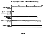
- Figure 1 is a bar graph depicting the frequency of occurrence of Replikins in various organisms.
- Figure 2 is a graph depicting the percentage of malignin per milligram total membrane protein during anaerobic replication of glioblastoma cells.
- Figure 3 is a bar graph showing amount of antimalignin antibody produced in response to exposure to the recognin 16-mer.
- Figure 4A is a photograph of a blood smear taken with ordinary and fluorescent light.
- Figure 4B is a photograph of a blood smear taken with ordinary and fluorescent light illustrating the presence of two leukemic cells.
- Figure 4C is a photograph of a dense layer of glioma cells in the presence of antimalignin antibody.
- Figure 4D and Figure 4E are photographs of the layer of cells in Figure 4C taken at 30 and 45 minutes following addition of antimalignin antibody
- Figure 4F is a bar graph showing the inhibition of growth of small cell lung carcinoma cells in vitro by antimalignin antibody.
- Figure 5 is a plot of the amount of antimalignin antibody present in the serum of patients with benign or malignant breast disease pre-and post surgery.
- Figure 6 is a box diagram depicting an embodiment of the invention wherein a computer is used to carry out the 3-point-recognition method of identifying Replikin sequences.
- Figure 7 is a graph showing the concentration of Replikins observed in hemagglutinin of influenza B and influenza A strain, H1N1, on a year by year basis from 1918 through 2001.
- Figure 8 is a graph of the Replikin concentration observed in hemagglutinin of influenza A strains, H2N2 and H3N2, as well as an emerging strain defined by its constituent Replikins, designated H3N2(R), on a year by year basis from 1950 to 2001.
- the inventors have identified a new family of small peptides related to the phenomenon of rapid replication, referred to herein as Replikins.
- Replikins a new family of small peptides related to the phenomenon of rapid replication.
- knowledge of and identification of this family of peptides enables development of effective therapies and vaccines for any organism that harbors Replikins.
- Identification of this family of peptides also provides for the detection of viruses and virus vaccine development.
- identification of this family of peptides provides for the detection of influenza virus and provides new targets for influenza treatment.
- the first functional basis for Replikins role to rapid replication is seen in glioma replication.
- glioma malignin is enriched ten-fold compared to the five-fold increase in cell number and membrane protein concentration in rapid replication of glioma cells suggests an integral relationship of the Replikins to replication.
- the glioma Replikin was synthesized in vitro and administered as a synthetic vaccine to rabbits, abundant antimalignin antibody was produced. This establishes the antigenic basis of the antimalignin antibody in serum (AMAS) test, and provides the first potential synthetic cancer vaccine and the prototype for Replikin vaccines in other organisms.
- antimalignin antibody The relationship between the presence of antimalignin antibody and survival in patients was shown in a study of 8,090 serum specimens from cancer patients. The study showed that the concentration of antimalignin antibody increases with age, as the incidence of cancer in the population increases, and increases further two to three-fold in early malignancy, regardless of cell type. In vitro, the antimalignin antibody is cytotoxic to cancer cells at picograms (femtomoles) per cancer cell, and in vivo the concentration of antimalignin antibody relates quantitatively to the survival of cancer patients.
- glioma glycoprotein 10B has a 50% reduction in carbohydrate residues when compared to the normal 1OB. This reduction is associated with virus entry in other instances, and so may be evidence of the attachment of virus for the delivery of virus Replikins to the 10B of glial cells as a step in the transformation to the malignant state.
- a varying structure provides an inconstant target, which is a good strategy for avoiding attackers, such as antibodies that have been generated specifically against the prior structure and thus are ineffective against the modified form. This strategy is used by influenza virus, for example, so that a previous vaccine may be quite ineffective against the current virulent virus.
- Replikin concentration and composition provide new quantitative methods to detect and control the process of replication, which is central to the survival and dominance of each biological population.
- the first Replikin sequence to be identified was the cancer cell Replikin found in a brain cancer protein, malignin, which was demonstrated to be enriched ten-fold during rapid anaerobic replication of glioblastoma multiforme (glioma) cells.
- Malignin is a 10KDa portion of the 250 KDa glycoprotein 10B, which was isolated in vivo and in vitro from membranes of glioblastoma multiforme (glioma) cells.
- the present invention provides a method for identifying nucleotide or amino acid sequences that include a Replikin sequence.
- the method is referred to herein as a 3-point-recognition method.
- the three point recognition method comprises: a peptide from 7 to about 50 amino acids including (1) at least one lysine residue located six to ten amino acid residues from a second lysine residue; (2) at least one histidine residue; and (3) at least 6% lysine residues. (Replikin).
- peptides or proteins constitute a new class of peptides in species including algae, yeast, fungi, amoebae, bacteria, plant, virus and cancer proteins having replication, transformation, or redox functions.
- Replikin peptides have been found to be concentrated in larger 'replicating' and 'transforming' proteins (so designated by their investigators, See Table 2) and cancer cell proteins. No sequences were found to be identical to the malignin 16-mer peptide.
- Table 2 Examples of Replikins in various organisms - prototype: Glioma Replikin* kagvaflhkk (SEQ ID No.:1) SEQ ID NO.
- Algae 34 Caldophera prolifera kaskftkh 35 Isolepisprolifera kaqaetiteikgh Yeast: 36 Schizosaccharomyces pombe ksfkypkkhk 37 Oryza sativa kkaygnelhk 2 Sacch.
- cerevisiae replication binding protein hsikrelgiifdk Fungi Isocitrate lyase ICI 1,Penicillium marneffei kvdivthqk 38 DNA-dependent RNA polymerase 11, Diseula dcstructiva kleedaayhrkk 39 Ophiostoma novo-ulm 1,RNA in Dutch elm disease kvilplrgnikgiffkh 40 fungus Amoeba: 41 Entamoeba invadens, histone H2B klilkgdlnkh Bacteria: 42 Pribosomal protein replication factor, Helicobacter Replication-associated protein Staph.
- aureus ksvhaflk 10 Mycoplasma pulmonic, chromosome replication kkektthnk 43 Macrophage infectivity potentiator, L. legionella kvhffqlkk 90 Bacillus anthracis kihlisvkk 91 Bacillus anthracis hvkkekeknk 92 Bacillus anthracis vicvkievk 93 Bacillus anthracis kkkkikdiygkdallh 94 Bacillus anthracis kwekikqh 95 Bacillus anthracis kklqippiepkkddiih 96 Bacillus anthracis hnryasnivesayllilnewknniqsdlikk 97 Bacillus anthracis havddyagylldkngsdlvtnskk 98 Bacillus anthracis haerlkvqknapk Plants: 44 Arabidops
- Tumor Viruses 48 Rous sarcoma virus tyrosine-protein kinase kklrhek 49 v-yes, avian sarcoma kklrhdk 50 c-yes, colon cancer, malignant melanoma kklrhdk 51 v-srcC, avian sarcoma kklrhek 52 c-src, colon, mammary, panrcreatic cancer kklrhek 53 Neuroblastoma RAS viral (v-ras) oncogene kqahelak 54 VPI (major capsid protein) [Polyamavirus sp.] kthrfskh 55 Sindbis knlhekik 56 EI [Human papilloamavirus type 71] khrpllqlk 57 v-erbB from AEV and c-erb kspnhvk 58 v-fms (feline s
- sequences have three elements: (1) at least one lysine residue located six to ten residues from another lysine residue; (2) at least one histidine residue; and (3) a composition of at least 6% lysine within an amino acid sequence of 7 to about 50 residues.
- viruses or viral peptides, as,adenovirus, lentivirus, a-virus, retrovirus, andeno-associated virus, human immunodeficiency virus, hepatitis virus, influenza virus, maize streak virus, herpes virus, bovine herpes virus, feline immunodeficiency virus, foot and mouth disease virus, small pox virus, rous sarcoma virus, neuroblastoma RAS viral oncogene, polyamavirus, Sindbis, human papilloma virus, myelomonocytic tumor virus, murine acute leukemia, T-cell lymphotropic virus, and tomato leaf curl virus.
- viruses or viral peptides, as,adenovirus, lentivirus, a-virus, retrovirus, andeno-associated virus, human immunodeficiency virus, hepatitis virus, influenza virus, maize streak virus, herpes virus, bovine herpes virus, feline immunodeficiency virus, foot and mouth disease virus, small pox virus,
- Replikins are present in such bacteria as, Acetobacter, Achromobacter, Actinomyces, Aerobacter, Alcaligenes, Arthrobacter, Azotobacter, Bacillus, Brevibacterium, Chainia, Clostridium, Corynebacterium, Erwinia, Escheria, Lebsiella, Lactobacillus, Haemophilus, Flavobacterium, Methylomonas, Micrococcus, Mycobacterium, Micronomspora, Mycoplasma, Neisseria, Nocardia, Proteus, Pseudomonas, Rhizobium, Salmonella, Serratia, Staphylococcus, Streptocossus, Streptomyces, Streptosporangium, Streptovirticillium, Vibrio, peptide, and Xanthomas.
- Replikins are present in such fungi as, Penicillium, Diseula, Ophiostoma novo-ulim, Mycophycophta, Phytophthora infestans, Absidia, Aspergillus, Candida, Cephalosporium, Fusarium, Hansenula, Mucor, Paecilomyces, Pichia, Rhizopus, Torulopsis, Trichoderma, and Erysiphe.
- Replikins are present in such yeast as, Saccharomyces, Cryptococcus, including Cryptococcus neoformas, Schizosaccharomyces, and Oryza.
- Replikins are present in algae such as, Caldophera, Isolepisprolifera, Chondrus, Gracilaria, Gelidium, Caulerpa, Laurencia, Cladophexa, Sargassum, Penicillos, Halimeda, Laminaria, Fucus, Ascophyllum, Undari, Rhodymenia, Macrocystis, Eucheuma, Ahnfeltia, and Pteroclasia.
- Replikins are present in amoeba sch as Entamoeba (including Entamoeba invadens), Amoebidae, Acanthamoeba and Naegleria.
- Replikins are present in plants such as, Arabidopsis, wheat, rice, and maize.
- auxiliary specifications to the basic "3-point-recognition” requirements may be added: (a) on a structural basis, such as the common occurrence of adjacent di- and polylysines in cancer cell proteins (e.g., transforming protein P21B(K-RAS 2B), lung, Table 2, SEQ ID NO.: 89), and other adjacent di-amino acids in TOLL-like receptors, or b) on a functional basis, such as exhibiting ATPase, tyrosine kinase or redox activity as seen in Table 2.
- a structural basis such as the common occurrence of adjacent di- and polylysines in cancer cell proteins (e.g., transforming protein P21B(K-RAS 2B), lung, Table 2, SEQ ID NO.: 89), and other adjacent di-amino acids in TOLL-like receptors
- b) on a functional basis such as exhibiting ATPase, tyrosine kinase or redox activity as seen in Table 2.
- “functional derivatives” of the Replikins as described herein are fragments, variants, analogs, or chemical derivatives of the Replikins, which retain at least a portion of the immunological cross reactivity with an antibody specific for the Replikin.
- a fragment of the Replikin peptide refers to any subset of the molecule.
- Variant peptides may be made by direct chemical synthesis, for example, using methods well known in the art.
- An analog of a Replikin to a non-natural protein substantially similar to either the entire protein or a fragment thereof.
- Chemical derivatives of a Replikin contain additional chemical moieties not normally a part of the peptide or peptide fragment.
- malignin is enriched. That is, malignin is found to increase not simply in proportion to the increase in cell number and total membrane proteins, but is enriched as much as ten-fold in concentration, starting with 3% at rest and reaching 30% of total membrane protein.
- This clear demonstration of a marked increase in Replikin concentration with glioma cell replication points to, and is consistent with, the presence of Replikins identified with the 3-point recognition method in various organisms.
- Replikins were identified in such proteins as "Saccharomyces cerevisiae replication binding protein” (SEQ ID NO.: 2) (hsikrelgiifdk); the “replication associated protein A of maize streak virus” (SEQ ID NO.: 8) (kyivcareahk) and (SEQ ID NO.: 9) (kekkpskdeimrdiish); the "replication-associated protein of Staphylococcus aureus” (SEQ ID NO.: 10) (kkektthnk); the "DNA replication protein of bovine herpes virus 4" (SEQ ID NO.: 11) (hkinitngqk); and the "Mealigrid herpes virus 1 replication binding protein” (SEQ ID NO.: 12) (hkdlyrllmk).
- Table 2 shows that Replikin-containing proteins also are associated frequently with redox functions, and protein synthesis or elongation, as well as with cell replication.
- composition ofReplikins observed were found to relate to the occurrence of influenza pandemics and epidemics.
- concentration of Replikins in influenza viruses was examined by visually scanning the hemagglutinin amino acid sequences published in the National Library of Medicine "PubMed" data base for influenza strains isolated world wide from human and animal reservoirs year by year over the past century, i.e., 1900 to 2001. These Replikin concentrations (number of Replikins per 100 amino acids, mean +/- SD) were then plotted for each strain.
- the concentration of Replikins was found to directly relate to the occurrence of influenza pandemics and epidemics.
- the concentration of Replikins found in influenza B hemagglutinin and influenza A strain, H1N1 is shown in Figure 7
- the concentration of Replikins found in the two other common influenza virus A strains, H2N2 and H3N2 is shown in Figure 8 (H2N2, H3N2).
- the data in Figure 8 also demonstrate an emerging new strain of influenza virus as defined by its constituent Replikins (H3N2(R)).
- Each influenza A strain has been responsible for one pandemic: in 1918, 1957, and 1968, respectively.
- the data in Figures 7 and 8 show that at least one Replikin per 100 amino acids is present in each of the influenza hemagglutinin proteins of all isolates of the four common influenza viruses examined, suggesting a function for Replikins in the maintenance of survival levels of replication.
- the H3N2 strain In the 1990s, during the decline of the H3N2 strain, there were no Replikins in many isolates of H3N2, but a high concentration of new Replikins appeared in H3N2 isolates, which define the emergence of the H3N2(R) strain.
- H1N1 Replikin concentration also declined between 1997 and 2000, and the presence of H1N1 strains decreased in isolates obtained during these years.
- H2N2 Replikins recovery from a 35 year decline has not occurred ( Figure 8 ), and this correlates with the absence of H2N2 from recent isolates.
- H3N2 the Replikin concentration of many isolates fell to zero during the period from 1996 to 2000, but other H3N2 isolates showed a significant, sharp increase in Replikin concentration. This indicates the emergence of a substrain of H3N2, which is designated herein as H3N2(R).
- Figures 7 and 8 demonstrate that frequently, a one to three year stepwise increase is observed before Replikin concentration reaches a peak. This stepwise increase proceeds the occurrence of an epidemic, which occurs concurrently with the Replikin peak. Thus, the stepwise increase in concentration of a particular strain is a signal that particular strain is the most likely candidate to cause an epidemic or pandemic.
- FIGURES 7 and 8 demonstrate a direct relationship between the presence and concentration of a particular Replikin in influenza protein sequences and the occurrence of pandemics and epidemics of influenza.
- analysis of the influenza virus hemagglutinin protein sequence for the presence and concentration of Replikins provides a predictor of influenza pandemics and/or epidemics, as well as a target for influenza vaccine formulation.
- composition of Replikins in Strains of Influenza Virus B Of a total of 26 Replikins identified in this strain (Table 3), the following ten Replikins are present in every influenza B isolate examined from 1902-2001. Overlapping Replikin sequences are listed separately. Lysines and histidines are in bold type to demonstrate homology consistent with the "3-point recognition.”
- Tables 3 and 4 indicate that there appears to be much greater stability of the Replikin structures in influenza B hemagglutinins compared with H1N1 Replikins. Influenza B has not been responsible for any pandemic, and it appears not to have an animal or avian reservoirs. (Stuart-Harris et al., Edward Arnold Ltd., London (1985)).
- Influenza H1N1 Replikins Only one Replikin "hp(v/i)tigecpkyv(r/k)(s/t)(t/a)k" is present in every H1N1 isolate for which sequences are available from 1918, when the strain first appeared and caused the pandemic of that year, through 2000. (Table 4). ("(v/i)" indicates that the amino acid v or i is present in the same position in different years.) Although H1N1 contains only one persistent Replikin, H1N1 appears to be more prolific than influenza B. There are 95 different Replikin structures in 82 years on H1N1 versus only 31 different Replikins in 100 years of influenza B isolates (Table 4). An increase in the number of new Replikin structures occurs in years of epidemics (Tables 3, 4, 5 and 6) and correlates with increased total Replikin concentration ( Figures 7 and 8 ).
- Influenza H2N2 Replikins Influenza H2N2 was responsible for the human pandemic of 1957. Three of the 20 Replikins identified in that strain for 1957 were conserved in each of the H2N2 isolates available for examination on PubMed until 1995 (Table 5).
- Influenza H3N2 Replikins Influenza H3N2 was responsible for the human pandemic of 1968. Five Replikins which appeared in 1968 disappeared after 1977, but reappeared in the 1990s (Table 6). The only Replikin structure which persisted for 22 years was hcd(g/q)f(q/r)nekwdlf(v/i)er(s/t)k, which appeared first in 1977 and persisted through 1998.
- Figures 1 and 2 show that influenza epidemics and pandemics correlate with the increased concentration of Replikins in influenza virus, which is due to the reappearance of at least one Replikin from one to 59 years after its disappearance. Also, in the A strain only, there is an emergence of new strain-specific Replikin compositions (Tables 4-6). Increase in Replikin concentration by repetition of individual Replikins within a single protein appears not to occur in influenza virus, but is seen in other organisms.
- influenza Replikin histidine appears never to be, and lysine (k) is rarely replaced.
- H1N1 Replikin hp(v/i)tigecpkyv(r/k)(s/t)(t/a)k
- H3N2 Replikin hcd(g/q)f(q,r)nekwdlf(v/i)er(s/t)k
- the amino acids which comprise the Replikin structure are substituted little or not at all, that is the Replikin structure is conserved.
- the Replikin (SEQ ID NO.: 3) "hkqkivapvk" was found to be conserved in 78% of the 236 isolates reported in PubMed, and each amino acid was found to be conserved in individual isolates as follows: his, 95,6%; lys, 91.8%; gln 92.3%; lys, 84.1%; ile, 90.7%; val, 91.8%; ala, 97.3%; pro, 96.2%; ala, 75.4%; and lys, 88.4%.
- the high rate of conservation suggests structural and functional stability of the Replikin structure and provides constant targets for treatment.
- sequence conservation was found in different isolates of HIV for its Replikins, such as (SEQ ID NO.: 5) “kcfncgkegh” or (SEQ ID NO.: 6) "kvylawvpahk” in HIV Type 1 and (SEQ ID NO.: 7) "kcwncgkegh” in HIV Type 2 (Table 2).
- Further examples of sequence conservation were found in the HIV tat proteins, such as (SEQ ID NO.: 613) "hclvckqkkglgisygrkk,” wherein the key lysine and histaidine amino acids are conserved. (See Table 7)
- sequence conservation was observed in plants, for example in wheat, such as in wheat ubiguitin activating enzyme E (SEQ ID NOs. 614-616).
- the Replikins in wheat even provided a reliable target for stimulation of plant growth as described within.
- Other examples of conservation are seen in the constant presence of malignin in successive generations, over ten years of tissue culture of glioma cells, and by the constancy of affinity of the glioma Replikin for antimalignin antibody isolated by immunoadsorption from 8,090 human sera from the U.S., U.K., Europe and Asia (e.g., Figure 5 and U.S. Patent 6,242,578 B1 ).
- Tat trans-activator proteins
- TAR transactivating response sequence
- Replikin structure has a specific survival function for the HIV virus which must be preserved and conserved, and cannot be sacrificed to the virus 'defense' maneuver of amino acid substitution crested to avoid antibody and other 'attack.' These 'defense' functions, although also essential, cannot 'compete' with the virus survival function of HIV replication.
- HIV Type 2 Further conservation was observed in different isolates of HIV for its Replikins such as “kcfncgkegh” (SEQ ID NO. 5) or “kvylawvpahk” (SEQ ID NO. 6) in HIV Type 1 and “kcwncgkegh” (SEQ ID NO. 7) in HIV Type 2.
- the Replikin in the wheat ubiquitin activating enzyme E is conserved.
- substitutions of amino acids (designated with an '*') adjacent to the Replikin variant forms in wheat ubiquitin activating enzyme E are common.
- the key k and h amino acids that form the Replikin structure do not vary whereas the 'unessential' k that is only 5 amino acids (from the first k on the left) is substituted.
- Vaccine formulations are changed twice yearly at international WHO arid CDC meetings. Vaccine formulations are based on serological evidence of the most current preponderance of influenza virus strain in a given region of the world. However, prior to the present invention there has been no correlation of influenza virus strain specific amino acid sequence changes with occurrence of influenza epidemics or pandemics.
- influenza virus pandemics and epidemics provide for production and timely administration of influenza vaccines tailored specifically to treat the prevalent emerging or re-emerging strain of influenza virus in a particular region of the world.
- influenza virus pandemics and epidemics can be predicted.
- the severity of such outbreaks of influenza can be significantly lessened by administering an influenza peptide vaccine based on the Replikin sequences found to be most abundant or shown to be on the rise in virus isolates over a given time period, such as about one to about three years.
- An influenza peptide vaccine of the invention may include a single Replikinpeptide sequence of SEQ ID No: 144 or may include a plurality of Replikin sequences observed in influenza virus strains.
- the peptide vaccine is based on Replikin sequence(s) shown to be increasing in concentration over a given time period and conserved for at least that period of time.
- a vaccine may include a conserved Replikin peptide(s) in combination with a new Replikin(s) peptide or may be based on new Replikin peptide sequences.
- the Replikin peptides can be synthesized by any method, including chemical synthesis or recombinant gene technology, and may include non-Replikin sequences, although vaccines based on peptides containing only Replikin sequences are preferred.
- vaccine compositions of the invention also contain a pharmaceutically acceptable carrier and/or adjuvant.
- influenza vaccines of the present invention can be administered alone or in combination with antiviral drugs, such as gancyclovir; interferon; interleukin; M2 inhibitors, such as, amantadine, rimantadine; neuraminidase inhibitors, such as zanamivir and oseltamivir; and the like, as well as with combinations of antiviral drugs.
- antiviral drugs such as gancyclovir; interferon; interleukin; M2 inhibitors, such as, amantadine, rimantadine; neuraminidase inhibitors, such as zanamivir and oseltamivir; and the like, as well as with combinations of antiviral drugs.
- Synthetic Replikin vaccines based on Replikins of SEQ ID No: 144 or preferably, an influenza vaccine of SEQ ID No: 144 based on conserved and/or emerging or re-emerging Replikin(s) over a given time period may be used to augment antibody concentration in order to lyse the respective virus infected cells and release virus extracellularly where chemical treatment can then be effective.
- Replikin peptides according to clam 1 may be administered to a subject to induce the immune system of the subject to produce anti-Replikin antibodies.
- a 0.5 to about 2 mg dosage, preferably a 1 mg dosage of each peptide is administered to the subject to induce an immune response. Subsequent dosages may be administered if desired.
- the Replikin sequence structure is associated with the function of replication.
- the Replikins of this invention are used for targeting sequences that contain Replikins for the purpose of diagnostic identification, promoting replication, or inhibiting or attacking replication, for example, the structure-function relationshipof the Replikin is fundamental.
- the larger protein sequence invariably has one or more epitopes (independent antigenic sequences that can induce specific antibody formation); Replikin structures usually comprise one of these potential epitopes.
- the presence of other epitopes within the larger protein may interfere with adequate formation of antibodies to the Replikin, by "flooding" the immune system with irrelevant antigenic stimuli that may preempt the Replikin antigens, See, e.g., Webster, R.G., J. Immunol., 97(2):177-183 (1966 ); and Webster et al., J. Infect.
- decoy epitopes may bind anti-Replikin antibodies, but may keep the antibodies away from histidine-bound respiratory enzymes. Treatment may therefore be most efficacious in two stages: 1) proteases to hydrolize decoys, then; 2) anti-Replikin antibodies or other anti-Replikin agents.
- Replikin sequences contain at least one histidine residue. Histidine is frequently involved in binding to redox centers.
- an antibody that specifically recognizes a Replikin sequence has a better chance of inactivating or destroying the cell in which the Replikin is located, as seen with anti-malignin antibody, which is perhaps the most cytotoxic anti-cancer antibody yet described, being active at picograms per cell.
- either epitopes other than Replikins present in the larger protein fragments may interfere according to the phenomenon of antigenic primacy referred to above, and/or because the hydrolysis of larger protein sequences into smaller sequences for processing to produce antibodies results in loss of integrity of any Replikin structure that is present, e.g., the Replikin is cut in two and/or the histidine residue is lost in the hydrolytic processing.
- the present studies suggest that for an effective vaccine to be produced, the Replikin sequences, and no other epitope, should be used as the vaccine.
- a vaccine of the invention can be generated using any one of the Replikin peptides of claim 1 identified by the three point recognition system.
- influenza vaccines include peptides of SEQ ID No: 144 that have been demonstrated to be conserved over a period of one or more years, preferably about three years or more, and/or which are present in a strain of influenza virus shown to have the highest increase in concentration of Replikins relative to Replikin concentration in other influenza virus strains, e.g., an emerging strain.
- the increase in Replikin concentration preferably occurs over a period of at least about six months to one year, preferably at least about two years or more, and most preferably about three years or more.
- preferred Replikin peptides for use in an influenza virus vaccine are those Replikins of claim 1 observed to "re- emerge" after an absence from the hemagglutinin amino acid sequence for one or more years.
- the Replikin peptides of the invention are administered to a subject, preferably by i.v. or intramuscular injection, in order to stimulate the immune system of the subject to produce antibodies to the peptide.
- the dosage of peptides is in the range of from about 0.1 ⁇ g to about 10 mg, preferably about 10 ⁇ g to about 1 mg, and most preferably about 50 ⁇ g to about 500 ug. The skilled practitioner can readily determine the dosage and number of dosages needed to produce an effective immune response.
- identified constituent Replikins are useful as vaccines, and also may be usefully targeted for diagnostic purposes.
- Three-point recognition methods described herein may be performed by a computer.
- the following algae were collected from Bermuda water sites and either extracted on the same day or frozen at -20 degrees C and extracted the next day.
- the algae were homogenized in a cold room (at 0 to 5 degrees C) in 1 gram aliquots in neutral buffer, for example 100 cc. of 0.005M phosphate buffer solution, pH 7 ("phosphate buffer”) for 15 minutes in a Waring blender, centrifuged at 3000 rpm, and the supernatant concentrated by perevaporation and dialyzed against phosphate buffer in the cold to produce a volume of approximately 15 ml. The volume of this extract solution was noted and an aliquot taken for protein analysis, and the remainder was fractionated to obtain the protein fraction having a pK range between 1 and 4.
- the preferred method of fractionation is chromatography as follows: The extract solution is fractionated in the cold room (4 degrees C) on a DEAE cellulose (Cellex-D) column 2.5x11.0 cm, which has been equilibrated with 0.005M phosphate buffer. Stepwise eluting solvent changes are made with the following solutions:
- the extract solution in 6 to 10 ml volume, is passed onto the column and overlayed with Solution 1, and a reservoir of 300 ml of Solution 1 is attached and allowed to drip by gravity onto the column.
- Three ml aliquots of eluant are collected and analyzed for protein content at OD 280 until all of the protein to be removed with Solution 1 has been removed from the column.
- Solution 2 is then applied to the column, followed in succession by Solutions 3, 4, 5, 6 aid 7 until all of the protein which can, be removed with each Solution is removed from the column.
- the eluates from Solution 7 are combined, dialyzed against phosphate buffer, the protein content determined of both dialysand and dialyzate, and both analyzed by gel electrophoresis.
- One or two bands of peptide or protein of molecular weight between 3,000 and 25,000 Daltons are obtained in Solution 7.
- the algae Caulerpa mexicana, Laurencia obtura, Cladophexa prolifera, Sargassum natans, Caulerpa verticillata, Halimeda tuna, and Penicillos capitatus after extraction and treatment as above, all demonstrated in Solution 7 eluates sharp peptide bands in this molecular weight region with no contaminants.
- These Solution 7 proteins or their eluted bands are hydrolyzed, and the amino acid composition determined.
- the peptides so obtained, which have a lysine composition of 6% or greater are Replikin precursors.
- Replikin peptide precursors are then determined for amino acid sequence and the Replikins are determined by hydrolysis and mass spectrometry as detailed in U.S. patent 6,242,578 B1 . Those which fulfill the criteria defined by the " 3-point-recognition" method are identified as Replikins. This procedure can also be applied to obtain yeast, bacterial and any plant Replikins.
- Replikins in tumor cells are isolated and identified.
- Replikin precursors of Astrocytin isolated from malignant brain tumors, Malignin (Aglyco 10B) isolated from glioblastoma tumor cells in tissue culture, MCF7 mammary carcinoma cells in tissue culture, and P3J Lymphoma cells in tissue culture each treated as above in a) yielded Replikin precursors with lysine content of 9.1%, 6.7%, 6.7%, and 6.5% respectively.
- Hydrolysis and mass spectrometry of Aglyco IOB as described in Example 10 U.S. 6,242,578 B1 produced the amino acid sequence, ykagvaflhkkndiide the 16-mer Replikin.
- Aglyco IOB or the 16-mer Repliken may be used as antigen to capture and quantify the amount of its corresponding antibody present in serum for diagnostic purposes are as shown in Figures 2 , 3 , 4 and 7 of U.S. 6,242,578 B1 .
- Analysis of sequence data of isolates of influenza virus hemagglutinin protein or neuraminidase protein for the presence and concentration of Replikins is carried out by visual scanning of sequences or through use of a computer program based on the 3-point recognition system described herein. Isolates of influenza virus are obtained and the amino acid sequence of the influenza hemagglutinin and/or neuraminidase protein is obtained by any art known method, such as by sequencing the hemagglutinin or neuraminidase gene and deriving the protein sequence therefrom. Sequences are scanned for the presence of new Replikins, conservation of Replikins over time and concentration of Replikins in each isolate.
- Comparison of the Replikin sequences and concentrations to the amino acid sequences obtained from isolates at an earlier time, such as about six months to about three years earlier, provides data that are used to predict the emergence of strains that are most likely to be the cause of influenza in upcoming flu seasons, and that form the basis for seasonal influenza peptide vaccines or nucleic acid based vaccines. Observation of an increase in concentration, particularly a stepwise increase in concentration of Replikins in a given strain of influenza virus for a period of about six months to about three years or more is a predictor of emergence of the strain as a likely cause of influenza epidemic or pandemic in the future.
- Peptide vaccines or nucleic acid-based vaccines based on the Replikins observed in the emerging strain are generated.
- An emerging strain is identified as the strain of influenza virus having the highest increase in concentration of Replikin sequences within the hemagglutinin and/or neuraminidase sequence during the time period.
- the peptide or nucleic acid vaccine is based on or includes any Replikin sequences that are observed to be conserved in the emerging strain.
- conserveed Replikins are preferably those Replikin sequences which are present in the hemagglutinin or neuraminidase protein sequence for about two years and preferably longer.
- the vaccines may include any combination of Replikin sequences identified in the emerging strain.
- the Replikin peptide or peptides identified as useful for an effective vaccine are synthesized by any method, including chemical synthesis and molecular biology techniques, including cloning, expression in a host cell and purification therefrom.
- the peptides are preferably admixed with a phannaceutically acceptable carrier in an amount determined to induce a therapeutic antibody reaction thereto.
- the dosage is about 0.1 ⁇ g to about 10 mg.
- influenza vaccine is preferably administered to a patient in need thereof prior to the onset of "flu season." Influenza flu season generally occurs in late October and lasts through late April. However, the vaccine may be administered at any time during the year. Preferably, the influenza vaccine is administered once yearly, and is based on Replikin sequences observed to be present, and preferably conserved in the emerging strain of influenza virus. Another preferred Replikin for inclusion in an influenza vaccine is a Replikin demonstrated to have re-emerged in a strain of influenza after an absence of one or more years.
- Peptide vaccines or nucleic acid-based vaccines based on the Replikins observed in the malaria causing organism are generated.
- the peptide or nucleic acid vaccine is based on or includes any Replikin sequences that are observed to be present on a surface antigen of the organism.
- the vaccines may include any combination of Replikin sequences identified in the malaria causing strain.
- the Replikin peptide or peptides identified as useful for an effective vaccine are synthesized by any method, including chemical synthesis and molecular biology techniques, including cloning, expression in a host cell and purification therefrom.
- the peptides are preferably admixed with a pharmaceutically acceptable carrier in an amount determined to induce a therapeutic antibody reaction thereto.
- the dosage is about 0.1 ⁇ g to about 10 mg.
- malaria vaccine is preferably administered to a patient in need thereof at any time during the year, and particularly prior to travel to a tropical environment.
Abstract
Description
- This invention relates to the identification and use of Replikins, a newly discovered class of peptides that share structural characteristics. In particular, this invention relates to Replikins which have been found in influence viruses and which consist of any one of the peptide sequences set forth in the variable sequence SEQ ID NO: 144.
- Rapid replication is characteristic of virulence in certain bacteria, viruses and malignancies, but no chemistry common to rapid replication in different organisms has been described previously. This patent application discloses a member of a new class of protein structures related to rapid replication. The new family of conserved small proteins related to rapid replication, named Replikins.
- We constructed an algorithm search for Replikins. In applying the algorithm not only was the function of the epitope revealed - rapid replication, but an entire family of homologues whose function is related to rapid replication was discovered, which we named Replikins.
- The algorithm is based on the following: 1) Evidence that the immune system looks to parts rather than a whole protein in recognition. Protein chains are first hydrolyzed by the immune system into smaller pieces, frequently six (6) to ten (10) amino acids long, as part of the immune systems' process of recognition of foreign structures against which it may mount an immune defense. By way of example, the immune system recognizes the presence of disease by chopping up proteins of the disease agent into smaller peptide sequences and reading them. This principle is used as a basis for the algorithm with which to search for homologues of the malignin cancer epitope, once the structure of the epitope was known; 2) The specific structure of the malignin epitope, in which two of the three lysines (K's) are eight residues apart is in accordance with the apparent 'rules' used by the immune system for recognition referred to above (6-10 amino acids long); 3) The fact that the malignin cancer epitope was shown to be a very strong antigen, that is - a generator of a strong immune response; that there are three lysines (K's) in the 10-mer peptide glioma Replikin and that K's are known to bind frequently to DNA and RNA as potential anchors for the entry of viruses; and 4) One histidine (H) is included in the sequence of the malignin epitope, between the two K's which are eight (8) residues apart, suggesting a connection to the metals of redox systems which are required to provide the energy for replication.
- A functional basis for the Replikins' role in rapid replication is the study of data from the past 100 years on influenza virus hemagglutinin protein sequences and epidemiology of influenza epidemics and pandemics. To date, only serological hemagglutinin and antibody classification, but no strain-specific conserved peptide sequences have previously been described in influenza, and no changes in concentration and composition of any strain-specific peptide sequences have been described previously which correlate with epidemiologically documented epidemics or rapid replication.
- A four to ten-fold increase in the concentration of strain-specific influenza Replikins in one of each of the four major strains, influenza B, (A)H1N1, (A)H2N2 and (A)H3N2 was found, and that such increase of Replikin concentration was related to influenza epidemics caused specifically by each strain from 1902 to 2001. These increases in concentration were then shown to be due to the reappearance of at least one specific Replikin composition from 1 to up to 64 years after its disappearance, plus the emergence of new strain-specific Replikin compositions. Previously, no strain-specific chemical structures were known with which to predict which strains would predominate in coming influenza seasons, nor to devise annual mixtures of whole-virus strains for vaccines. The recent sharp increase in H3N2 Replikin concentration (1997 to 2000), the largest in H3N2's history, and the reappearance of specific Replikin compositions which were last seen in the high mortality H3N2 pandemic of 1968 and in the two high mortality epidemics of 1975 and 1977, but were absent for 20-25 years, together may be a warning of coming epidemics.
- Synthetic Replikins are new vaccines. This high degree of conservation of Replikin structures observed whereby the identical structure can persist for 100 years, or reappear after an absence of from one to 64 years reappears indicates that what was previously thought to be change in virulence due to random substitution of amino acids in influenza proteins is more likely to be change due to an organized process of conservation of Replikins. In fact, if random substitutions of each amino acid occurred, the chance against an average length influenza Replikin sequence being conserved for one year (let alone 100) is calculated to be in the order of 2 to the 27th power to 1.
- Replikin concentration and composition therefore provide new methods to detect and to control the process of replication, which is central to the survival and dominance of each biological population.
- The present invention provides peptide consisting of any one of the peptide sequences set forth in variable sequence SEQ ID No: 144 The peptide was unveiled by use of the "3-point recognition" method, namely, peptides comprising from 7 to about 50 amino acids including (1) at least one lysine residue located six to ten amino acid residues from a second lysine residue; (2) at least one histidine residue; and (3) at least 6% lysine residues (Replikin) appplied on proteins extracted from influenza virus infected cells.
- Influenza is an acute respiratory illness of global importance. Despite international attempts to control influenza virus outbreaks through vaccination, influenza infections remain an important cause of morbidity and mortality. Worldwide influenza epidemics and pandemics have occurred at irregular and previously unpredictable intervals throughout history and it is expected that they will continue to occur in the future. The impact of both pandemic and epidemic influenza is substantial in terms of morbidity, mortality and economic cost.
- Influenza vaccines remain the most effective defense against influenza virus, but because of the ability of the virus to mutate and the availability of non-human host reservoirs, it is expected that influenza will remain an emergent or re-emergent infection. Global influenza surveillance indicates that influenza viruses may vary within a country and between countries and continents during an influenza season. Virological surveillance is of importance in monitoring antigenic shift and drift. Disease surveillance is also important in assessing the impact of epidemics. Both types of information have provided the basis of the vaccine composition and the correct use of antivirals. However, to date there has been only annual post hoc hematological classification of the increasing number of emerging influenza virus strains, and no specific chemical structure of the viruses has been identified as an indicator of approaching influenza epidemics or pandemics. Currently, the only basis for annual classification of influenza virus as active, inactive or prevalent in a given year is the activities of the virus hemagglutinin and neuraminidase proteins. No influenza viral chemical structure has been identified prior to this application that can be used for quantitative warning of epidemics or pandemics or to design more effective and safer vaccines.
- Because of the annual administration of influenza vaccines and the short period of time when a vaccine can be administered, strategies directed at improving vaccine coverage are of critical importance.
- In one aspect of the invention there are provided isolated or synthesized influenza virus peptides containing a Replikin sequence set forth in variable sequence SEQ ID NO:144.
- In another aspect of the invention, there is provided a Replikin peptide as defined in claim 1 for the use in a process for stimulating the immune system of a subject to produce antibodies that bind specifically to an influenza virus Replikin sequence.
- The present invention also provides therapeutic compositions comprising one or more of isolated influenza virus peptides defined in claim 1.
- In yet another aspect of the invention, there is provided a preventive or therapeutic influenza virus vaccine comprising at least one isolated Replikin defined in claim 1, present in the hemagglutinin protein of an emerging strain of influenza virus and a pharmaceutically acceptable carrier and/or adjuvant.
- Also provided by the present invention is the use of at least one isolated Replikin present in the hemagglutinin protein of an emerging strain of influenza virus as defined in claim 1 for the preparation of a preventive or therapeutic vaccine for preventing or treating influenza virus infection comprising administering to a patient in need thereof
-
Figure 1 is a bar graph depicting the frequency of occurrence of Replikins in various organisms. -
Figure 2 is a graph depicting the percentage of malignin per milligram total membrane protein during anaerobic replication of glioblastoma cells. -
Figure 3 is a bar graph showing amount of antimalignin antibody produced in response to exposure to the recognin 16-mer. -
Figure 4A is a photograph of a blood smear taken with ordinary and fluorescent light.Figure 4B is a photograph of a blood smear taken with ordinary and fluorescent light illustrating the presence of two leukemic cells.Figure 4C is a photograph of a dense layer of glioma cells in the presence of antimalignin antibody.Figure 4D and Figure 4E are photographs of the layer of cells inFigure 4C taken at 30 and 45 minutes following addition of antimalignin antibody -
Figure 4F is a bar graph showing the inhibition of growth of small cell lung carcinoma cells in vitro by antimalignin antibody. -
Figure 5 is a plot of the amount of antimalignin antibody present in the serum of patients with benign or malignant breast disease pre-and post surgery. -
Figure 6 is a box diagram depicting an embodiment of the invention wherein a computer is used to carry out the 3-point-recognition method of identifying Replikin sequences. -
Figure 7 is a graph showing the concentration of Replikins observed in hemagglutinin of influenza B and influenza A strain, H1N1, on a year by year basis from 1918 through 2001. -
Figure 8 is a graph of the Replikin concentration observed in hemagglutinin of influenza A strains, H2N2 and H3N2, as well as an emerging strain defined by its constituent Replikins, designated H3N2(R), on a year by year basis from 1950 to 2001. - The inventors have identified a new family of small peptides related to the phenomenon of rapid replication, referred to herein as Replikins. In general, knowledge of and identification of this family of peptides enables development of effective therapies and vaccines for any organism that harbors Replikins. Identification of this family of peptides also provides for the detection of viruses and virus vaccine development.
- For example, identification of this family of peptides provides for the detection of influenza virus and provides new targets for influenza treatment.
- Rapid replication is characteristic of virulence in certain bacteria, viruses and malignancies, but no chemistry common to rapid replication in different organisms has been described. We have found a family of conserved small protein sequences related to rapid replication, which we have named Replikins. Such Replikins offer new targets for developing effective detection methods and therapies. The first Replikin found was the glioma Replikin, which was identified in brain glioblastoma multiforme (glioma) cell protein called malignin.
- Hydrolysis and mass spectrometry of malignin revealed the novel 16 mer peptide sequence which contains the glioma Replikin. This Replikin was not found in databases for the normal healthy human genome and therefore appeared to be derived from some source outside the body.
- We have devised an algorithm to search for the glioma Replikin or homologue thereof. Homologues were not common in over 4,000 protein sequences, but were found, surprisingly, in all tumor viruses, and in the replicating proteins of algae, plants, fungi, viruses and bacteria.
- We have identified that both 1) Replikin concentration (number of Replikins per 100 amino acids) and 2) Replikin composition correlate with the functional phenomenon of rapid replication. These relationships provide functional basis for the determination that Replikins are related quantitatively as well as qualitatively to the rate of replication.
- The first functional basis for Replikins role to rapid replication is seen in glioma replication. The fact that glioma malignin is enriched ten-fold compared to the five-fold increase in cell number and membrane protein concentration in rapid replication of glioma cells suggests an integral relationship of the Replikins to replication. When the glioma Replikin was synthesized in vitro and administered as a synthetic vaccine to rabbits, abundant antimalignin antibody was produced. This establishes the antigenic basis of the antimalignin antibody in serum (AMAS) test, and provides the first potential synthetic cancer vaccine and the prototype for Replikin vaccines in other organisms. With the demonstration of this natural immune relationship of the Replikins to replication and this natural immune response to cancer Replikins, which overrides cell type, based upon the shared specificity of cancer Replikins and rapid replication, both passive augmentation of this immunity with antimalignin antibody and active augmentation with synthetic Replikin vaccines now is possible.
- The relationship between the presence of antimalignin antibody and survival in patients was shown in a study of 8,090 serum specimens from cancer patients. The study showed that the concentration of antimalignin antibody increases with age, as the incidence of cancer in the population increases, and increases further two to three-fold in early malignancy, regardless of cell type. In vitro, the antimalignin antibody is cytotoxic to cancer cells at picograms (femtomoles) per cancer cell, and in vivo the concentration of antimalignin antibody relates quantitatively to the survival of cancer patients. As shown in glioma cells, the stage in cancer at which cells have only been transformed to the immortal malignant state but remain quiescent or dormant, now can be distinguished from the more active life-threatening replicating state, which is characterized by the increased concentration of Replikins. In addition, clues to the viral pathogenesis of cancer may be found in the fact that glioma glycoprotein 10B has a 50% reduction in carbohydrate residues when compared to the normal 1OB. This reduction is associated with virus entry in other instances, and so may be evidence of the attachment of virus for the delivery of virus Replikins to the 10B of glial cells as a step in the transformation to the malignant state.
- Our study concerning influenza virus hemagglutinin protein sequences and influenza epidemiology over the past 100 years, has provided a second functional basis for the relations ofReplikins to rapid replication. Only serological hemagglutinin and antibody classification, but no strain-specific conserved peptide sequences have previously been described in influenza. Further, no changes in concentration and composition of any strain-specific peptide sequences have been described previously that correlate with epidemiologically documented epidemics or rapid replication. In this study, a four to ten-fold increase in the concentration of strain-specific influenza Replikins in one of each of the four major strains, influenza B, (A)HIN1, (A)H2N2 and (A)143N2, is shown to relate to influenza epidemics caused by each strain from 1902 to 2001.
- We then showed that these increases in concentration are due to the reappearance of at least one specific Replikin composition from 1 to up to 64 years after its disappearance, plus the emergence of new strain-specific Replikin compositions. Previously, no strain-specific chemical structures were known with which to predict the strains that would predominate in coming influenza seasons, nor to devise annual mixtures of whole-virus strains for vaccines. The recent sharp increase in H3N2 Replikin concentration (1997 to 2000), the largest in H3N2's history, and the reappearance of specific Replikin compositions that were last seen in the high mortality H3N2 pandemic of 1968, and in the two high mortality epidemics of 1975 and 1977, but were absent for 20-25 years, together may be a warning of coming epidemics. This high degree of conservation of Replikin structures observed, whereby the identical structure can persist for 100 years, or reappear after an absence of from one to 64 years, indicate that what was previously thought to be change due to random substitution of amino acids in influenza proteins is more likely to be change due to an organized process of conservation of Replikins.
- The conservation of Replikins is not unique to influenza virus but was also observed in other sources, for example in foot and mouth disease virus,
type 0, HIV tat, and wheat. - A third functional basis for Replikins' role in rapid replication is seen in the increase in rapid replication in HIV. Replikin concentration was shown to be related to rapid replication in HIV. We found the Replikin concentration in the slow growing low-titre strain of HIV (NS1, "Bru"), which is prevalent in early stage infection, to be one-sixth of the Replikin concentration in the rapidly-growing high-titre strain of HIV (SI, "Lai")(prevalent in late stage HIV infection).
- Further examples demonstrate the relationship of Replikins to rapid replication. In the "replicating protein," of tomato curl leaf gemini virus which devastates tomato crops, the first 161 amino acids, the sequence which has been shown to bind to DNA, was shown to contain five Replikins. In malaria, legendary for rapid replication when trypanosomes are released from the liver in the tens of thousands from one trypanosome, multiple, novel, almost 'flamboyant' Replikin structures have been found with concentrations of up to 36 overlapping Replikins per 100 amino acids.
- The conservation of any structure is critical to whether that structure provides a stable invariant target to attack and destroy or to stimulate. When a structure is tied in some way to a basic survival mechanism of the organism, the structures tend to be conserved. A varying structure provides an inconstant target, which is a good strategy for avoiding attackers, such as antibodies that have been generated specifically against the prior structure and thus are ineffective against the modified form. This strategy is used by influenza virus, for example, so that a previous vaccine may be quite ineffective against the current virulent virus.
- Both bacteria and HIV have both Replikin and non-Replikin amino acids. In HIV, for example, there has been a recent increase in drug-resistance from 9% to 13% due to mutation, that is substitution of non-Replikin amino acids. (See detailed analysis of TAT protein of HIV discussed herein). In bacteria, the development of 'resistant strains' is due to a similar mechanism. However, we have found that Replikin structures do not mutate or change to the same degree as non Replikin amino acids (see also discussion of foot and mouth disease virus conservation of Replikins discussed herein). The Replikin structures, as opposed to the non-Replikin structures are conserved and thus provide new constant targets for treatment.
- Certain structures too closely related to survival functions apparently cannot change constantly. Because an essential component of the Replikin structure is histidine (h), which is know for its frequent binding to metal groups in redox enzymes and probable source of energy needed for replication, and since this histidine structure remains constant, this structure remains all the more attractive a target for destruction or stimulation.
- From a proteomic point of view, inventors construction of a template based on the newly determined glioma peptide sequence led them to the discovery of a wide class of proteins with related conserved structures and a particular function, in this case replication. Examples of the increase in Replikin concentration with virulence of a disease include, influenza, HIV, cancer and tomato leaf curl virus. This newly recognized class of structures is related to the phenomenon of rapid replication in organisms as diverse as yeast, algae, plants, the gemini curl leaf tomato virus, HIV and cancer.
- Replikin concentration and composition provide new quantitative methods to detect and control the process of replication, which is central to the survival and dominance of each biological population. The sharing of immunological specificity by diverse members of the class, as demonstrated with antimalignin antibody for the glioma and related cancer Replikins, suggests that B cells and their product antibodies may recognize Replikins by means of a similar recognition language.
- The first Replikin sequence to be identified was the cancer cell Replikin found in a brain cancer protein, malignin, which was demonstrated to be enriched ten-fold during rapid anaerobic replication of glioblastoma multiforme (glioma) cells. (
Figure 2 ) Malignin is a 10KDa portion of the 250 KDa glycoprotein 10B, which was isolated in vivo and in vitro from membranes of glioblastoma multiforme (glioma) cells. Hydrolysis and mass spectroscopy of malignin revealed a 16-mer peptide sequence, ykagvaflhkkndide (SEQ ID NO.:4), which is referred to herein as the glioma Replikin and which includes the shorter peptide, kagvaflhkk (SEQ ID NO.: 1), both of which apparently are absent in the normal human genome.
- When the 16-mer glioma Replikin was synthesized and injected as a synthetic vaccine into rabbits, abundant antimalignin antibody was produced. (Bogoch et al., Cancer Detection and Prevention, 26 (Suppl. 1): 402 (2002)). The concentration of antimalignin antibody in serum in vivo has been shown to relate quantitatively to the survival of cancer patients. (Bogoch et al., Protides of Biological Fluids, 31:739-747 (1984). In vitro antimalignin antibodies have been shown to be cytotoxic to cancer cells at a concentration of picograms (femtomolar) per cancer cell. (Bogoch et al., Cancer Detection and Prevention, 26 (Suppl. 1): 402 (2002).
- Studies carried out by the inventors showed that the glioma Replikin is not represented in the normal healthy human genome. Consequently, a search for the origin and possible homologues of the Replikin sequence was undertaken by analysis of published sequences of various organisms.
- By using the 16-mer glioma Replikin sequence as a template and constructing a recognition proteomic system to visually scan the amino acid sequences of proteins of several different organisms, a new class of peptides, the Replikins, was identified. The present invention provides a method for identifying nucleotide or amino acid sequences that include a Replikin sequence. The method is referred to herein as a 3-point-recognition method. The three point recognition method comprises: a peptide from 7 to about 50 amino acids including (1) at least one lysine residue located six to ten amino acid residues from a second lysine residue; (2) at least one histidine residue; and (3) at least 6% lysine residues. (Replikin). These peptides or proteins constitute a new class of peptides in species including algae, yeast, fungi, amoebae, bacteria, plant, virus and cancer proteins having replication, transformation, or redox functions. Replikin peptides have been found to be concentrated in larger 'replicating' and 'transforming' proteins (so designated by their investigators, See Table 2) and cancer cell proteins. No sequences were found to be identical to the malignin 16-mer peptide.
Table 2: Examples of Replikins in various organisms - prototype: Glioma Replikin* kagvaflhkk
(SEQ ID No.:1)SEQ ID NO. Algae: 34 Caldophera prolifera kaskftkh 35 Isolepisprolifera kaqaetiteikgh Yeast: 36 Schizosaccharomyces pombe ksfkypkkhk 37 Oryza sativa kkaygnelhk 2 Sacch. cerevisiae replication binding protein hsikrelgiifdk Fungi: Isocitrate lyase ICI 1,Penicillium marneffei kvdivthqk 38 DNA-dependent RNA polymerase 11, Diseula dcstructiva kleedaayhrkk 39 Ophiostoma novo-ulm 1,RNA in Dutch elm disease kvilplrgnikgiffkh 40 fungus Amoeba: 41 Entamoeba invadens, histone H2B klilkgdlnkh Bacteria: 42 Pribosomal protein replication factor, Helicobacter Replication-associated protein Staph. aureus ksvhaflk 10 Mycoplasma pulmonic, chromosome replication kkektthnk 43 Macrophage infectivity potentiator, L. legionella kvhffqlkk 90 Bacillus anthracis kihlisvkk 91 Bacillus anthracis hvkkekeknk 92 Bacillus anthracis khivkievk 93 Bacillus anthracis kkkkikdiygkdallh 94 Bacillus anthracis kwekikqh 95 Bacillus anthracis kklqippiepkkddiih 96 Bacillus anthracis hnryasnivesayllilnewknniqsdlikk 97 Bacillus anthracis havddyagylldkngsdlvtnskk 98 Bacillus anthracis haerlkvqknapk Plants: 44 Arabidopsis thaliana, prolifera kdhdfdgdk 45 Arabidopsis thaliana, cytoplasmic ribosomal kmkglkqkkah 46 Arabidopsis thaliana, DNA binding protein kelssttqeksh Viruses: 9 Replication associated protein A [Maize streak virus] Kekkpskdeimrdiish 11 Bovine herpes virus 4, DNA replication protein hkinitngqk 12 Meleagrid herpesvirus 1, replication binding protein hkdlyrllmk 47 Feline immunodeficiency hikdyklvk 3 Foot and Mouth Disease (O) hkqkivapvk 5 HIV Type 1 kcfncgkegh 7 HIV Type 2kcwncgkegh 99 Small Pox Virus (Variola) khynnitwyk 100 Small Pox Virus (Variola) kysqtgkeliih 101 Small Pox Virus (Variola) hyddvrikndivvsrck 102 Small Pox Virus (Variola) hrfklildski 103 Small Pox Virus (Variola) kerghnyyfek. Tumor Viruses: 48 Rous sarcoma virus tyrosine-protein kinase kklrhek 49 v-yes, avian sarcoma kklrhdk 50 c-yes, colon cancer, malignant melanoma kklrhdk 51 v-srcC, avian sarcoma kklrhek 52 c-src, colon, mammary, panrcreatic cancer kklrhek 53 Neuroblastoma RAS viral (v-ras) oncogene kqahelak 54 VPI (major capsid protein) [Polyamavirus sp.] kthrfskh 55 Sindbis knlhekik 56 EI [Human papilloamavirus type 71] khrpllqlk 57 v-erbB from AEV and c-erb kspnhvk 58 v-fms (feline sarcoma) knihlekk 59 c-fms (acute and chronic myelomonocytic tumors) knihlekk 60 large t-antigen 1 [Polyomavinis sp.1 kphlaqslek 61 middle t-antigen [Polyomavirus sp,1- kqhrelkdk 62 small t-antigen [Polyomavirus spJ, kqhrelkdk 63 v-abl, murine acute leukemia kvpvlisptlkh 64 Human T-cell lymphotropic virus typo 2kslllevdkdish 65 c-kit, GI tumors, small cell lung carcinoma kagitimvkreyh 18 Hepatitis C hyppkpgcivpak Trans-Forming Proteins: 66 Transforming protein myb Ksgkhlgk 67 Transforming protein myc, Burkitt lymphoma krreqlkhk 68 Ras-related GTP-binding protein ksfevikvih 69 Transforming protein ras (teratocarcinoma) kkkhtvkk 70 TRAF-associated NF·kB activator TANK kaqkdhisk 71 RFP transforming protein hlkrvkdlkk 72 Transforming protein D (S.C.) kygspkhrlik 73 Papilloma virus type 11, transforming protein klkhilgkarfik 74 Protein tryosine kinase (EC 2.7.1.112slk kgdhvkhykirk 75 Transforming protein (ax1(-)) keklrdvmvdrhk 76 Transforming protein (N-myc) klqarqqqllkkieh 77 Fibroblast growth factor 4 (Kaposi sarcoma) kkgnrvsptmkvth Cancer Cell Proteins: 78 Matrix metaloproteinase 7 (uterine) keiplhfrk 79 Transcription factor 7-like kkkphikk 80 Breast cancer antigen NY-BR-87 ktrhdplak 81 BRCA-1-Associated Ring Domain Protein (breast) khhpkdnlik 82 Autoantigen from a breast tumor' khkrkkfrqk 83 Glioma Replikin (this study) kagvaflhkk 84 Ovarian cancer antigen khkrkkfrqk 85 EE L leukemia kkkskkhkdk 86 Proto-oncogene tyrosine-protein kinase C-ABLE hksekpalprk 87 Adenomatosis polyposis coli kkkkpsrlkgdnek 88 Gastric cancer transforming protein ktkkgnrvsptmkvth 89 Transforming protein (K-RAS 2B),lung khkekmskdkkkkkksk - Identification of an amino acid sequence as a Replikin or as containing a Replikin, i.e., a homologue of the glioma peptide, kagvaflhkk, requires that the three following requirements be met. According to the three point recognition system the sequences have three elements: (1) at least one lysine residue located six to ten residues from another lysine residue; (2) at least one histidine residue; and (3) a composition of at least 6% lysine within an amino acid sequence of 7 to about 50 residues.
- Databases were searched using the National Library of Medicine keyword "PubMed" descriptor for protein sequences containing Replikin sequences. Over 4,000 protein sequences were visually examined for homologues. Sequences of all individual proteins within each group of PubMed-classified proteins were visually scanned for peptides meeting the three above-listed requirements. An infrequent occurrence of homologues was observed in "virus peptides" as a whole (1.5%) (N=953), and in other peptides not designated as associated with malignant transformation or replication such as "brain peptides" and "neuropeptides" (together 8.5%) (N=845). However, surprisingly, homologues were significantly more frequently identified in large "replicating proteins," which were identified as having an established function in replication in bacteria, algae, and viruses. Even more surprising was the finding that Replikin homologues occurred in 100% of "tumor viruses" (N=250), in 97% of "cancer proteins" (N=401), and in 85% of "transforming viruses" (N=248). These results suggest that there are shared properties of cancer pathogenesis regardless of cell type and suggest a role of viruses in carcinogenesis, i.e., conversion of cells from a transformed albeit dormant state to a more virulent actively replicating state.
- Homologues of the following amino acid sequence, kagvaflhkk, as defined by the three point recognition method, were found in such viruses, or viral peptides, as,adenovirus, lentivirus, a-virus, retrovirus, andeno-associated virus, human immunodeficiency virus, hepatitis virus, influenza virus, maize streak virus, herpes virus, bovine herpes virus, feline immunodeficiency virus, foot and mouth disease virus, small pox virus, rous sarcoma virus, neuroblastoma RAS viral oncogene, polyamavirus, sindbis, human papilloma virus, myelomonocytic tumor virus, murine acute leukemia, T-cell lymphotropic virus, and tomato leaf curl virus.
- Replikins are present in such bacteria as, Acetobacter, Achromobacter, Actinomyces, Aerobacter, Alcaligenes, Arthrobacter, Azotobacter, Bacillus, Brevibacterium, Chainia, Clostridium, Corynebacterium, Erwinia, Escheria, Lebsiella, Lactobacillus, Haemophilus, Flavobacterium, Methylomonas, Micrococcus, Mycobacterium, Micronomspora, Mycoplasma, Neisseria, Nocardia, Proteus, Pseudomonas, Rhizobium, Salmonella, Serratia, Staphylococcus, Streptocossus, Streptomyces, Streptosporangium, Streptovirticillium, Vibrio, peptide, and Xanthomas.
- Replikins are present in such fungi as, Penicillium, Diseula, Ophiostoma novo-ulim, Mycophycophta, Phytophthora infestans, Absidia, Aspergillus, Candida, Cephalosporium, Fusarium, Hansenula, Mucor, Paecilomyces, Pichia, Rhizopus, Torulopsis, Trichoderma, and Erysiphe.
- Replikins are present in such yeast as, Saccharomyces, Cryptococcus, including Cryptococcus neoformas, Schizosaccharomyces, and Oryza.
- Replikins are present in algae such as, Caldophera, Isolepisprolifera, Chondrus, Gracilaria, Gelidium, Caulerpa, Laurencia, Cladophexa, Sargassum, Penicillos, Halimeda, Laminaria, Fucus, Ascophyllum, Undari, Rhodymenia, Macrocystis, Eucheuma, Ahnfeltia, and Pteroclasia.
- Replikins are present in amoeba sch as Entamoeba (including Entamoeba invadens), Amoebidae, Acanthamoeba and Naegleria.
- Replikins are present in plants such as, Arabidopsis, wheat, rice, and maize.
- To permit classification of subtypes of Replikins, additional or "auxiliary specifications" to the basic "3-point-recognition" requirements may be added: (a) on a structural basis, such as the common occurrence of adjacent di- and polylysines in cancer cell proteins (e.g., transforming protein P21B(K-RAS 2B), lung, Table 2, SEQ ID NO.: 89), and other adjacent di-amino acids in TOLL-like receptors, or b) on a functional basis, such as exhibiting ATPase, tyrosine kinase or redox activity as seen in Table 2.
- "functional derivatives" of the Replikins as described herein are fragments, variants, analogs, or chemical derivatives of the Replikins, which retain at least a portion of the immunological cross reactivity with an antibody specific for the Replikin. A fragment of the Replikin peptide refers to any subset of the molecule. Variant peptides may be made by direct chemical synthesis, for example, using methods well known in the art. An analog of a Replikin to a non-natural protein substantially similar to either the entire protein or a fragment thereof. Chemical derivatives of a Replikin contain additional chemical moieties not normally a part of the peptide or peptide fragment.
- As seen in
Figure 2 , during anaerobic respiration when the rate of cell replication is increased, malignin is enriched. That is, malignin is found to increase not simply in proportion to the increase in cell number and total membrane proteins, but is enriched as much as ten-fold in concentration, starting with 3% at rest and reaching 30% of total membrane protein. This clear demonstration of a marked increase in Replikin concentration with glioma cell replication points to, and is consistent with, the presence of Replikins identified with the 3-point recognition method in various organisms. For example, Replikins were identified in such proteins as "Saccharomyces cerevisiae replication binding protein" (SEQ ID NO.: 2) (hsikrelgiifdk); the "replication associated protein A of maize streak virus" (SEQ ID NO.: 8) (kyivcareahk) and (SEQ ID NO.: 9) (kekkpskdeimrdiish); the "replication-associated protein of Staphylococcus aureus" (SEQ ID NO.: 10) (kkektthnk); the "DNA replication protein of bovine herpes virus 4" (SEQ ID NO.: 11) (hkinitngqk); and the "Mealigrid herpes virus 1 replication binding protein" (SEQ ID NO.: 12) (hkdlyrllmk). Previous studies of tomato leaf curl gemini virus show that the regulation of virus accumulation appears to involve binding of amino acids 1-160 of the "replicating protein" of that virus to leaf DNA and to other replication protein molecules during virus replication. Analysis of this sequence showed that amino acids 1-163 of this "replicating protein" contain five Replikins, namely: (SEQ ID NO.: 13) kfrinaknyfltyph, (SEQ ID NO.: 14) knletpvnklfiricrefh, (SEQ ID NO.: 15) hpniqaaksstdvk, (SEQ ID NO.: 16) ksstdvkaymdkdgdvldh, and (SEQ ID NO.: 17) kasalnitrekapkdfvlqfh. - Table 2 shows that Replikin-containing proteins also are associated frequently with redox functions, and protein synthesis or elongation, as well as with cell replication. The association with metal-based redox functions, the enrichment of the Replikin-containing glioma malignin concentration during anaerobic replication, and the cytotoxicity of antimalignin at low concentrations (picograms/cell) (
Figure 4c-f ), all suggest that the Replikins are related to central respiratory survival functions, have been found less often subjected to the mutations characteristic of non-Replikin amino acids. - Of particular interest, it was observed that at least one Replikin per 100 amino acids was found to be present in the hemagglutinin proteins of almost all of the individual strains of influenza viruses examined. The Replikin sequences that were observed to occur in the hemagglutinin proteins of isolates of each of the four prevalent strains of influenza virus, influenza B, H1N1, H2N2, and H3N2, for each year that amino acid sequence data are available (1902-2001), are shown in Tables 3, 4, 5 and 6, below.


- Both the concentration and type, i.e., composition ofReplikins observed, were found to relate to the occurrence of influenza pandemics and epidemics. The concentration of Replikins in influenza viruses was examined by visually scanning the hemagglutinin amino acid sequences published in the National Library of Medicine "PubMed" data base for influenza strains isolated world wide from human and animal reservoirs year by year over the past century, i.e., 1900 to 2001. These Replikin concentrations (number of Replikins per 100 amino acids, mean +/- SD) were then plotted for each strain.
- The concentration of Replikins was found to directly relate to the occurrence of influenza pandemics and epidemics. The concentration of Replikins found in influenza B hemagglutinin and influenza A strain, H1N1, is shown in
Figure 7 , and the concentration of Replikins found in the two other common influenza virus A strains, H2N2 and H3N2 is shown inFigure 8 (H2N2, H3N2). The data inFigure 8 also demonstrate an emerging new strain of influenza virus as defined by its constituent Replikins (H3N2(R)). - Each influenza A strain has been responsible for one pandemic: in 1918, 1957, and 1968, respectively. The data in
Figures 7 and8 show that at least one Replikin per 100 amino acids is present in each of the influenza hemagglutinin proteins of all isolates of the four common influenza viruses examined, suggesting a function for Replikins in the maintenance of survival levels of replication. In the 1990s, during the decline of the H3N2 strain, there were no Replikins in many isolates of H3N2, but a high concentration of new Replikins appeared in H3N2 isolates, which define the emergence of the H3N2(R) strain. - Several properties of Replikin concentration are seen in
Figure 7 andFigure 8 to be common to all four influenza virus strains. First, the concentration is cyclic over the years, with a single cycle of rise and fall occurring over a period of two to thirty years. This rise and fall is consistent with the known waxing and waning of individual influenza virus strain predominance by hemagglutinin and neuraminidase classification. Second, peak Replikin concentrations of each influenza virus strain previously shown to be responsible for a pandemic were observed to relate specifically and individually to each of the three years of the pandemics. For example, for the pandemic of 1918, where the influenza virus strain, H1N1, was shown to be responsible, a peak concentration of the Replikins in H1N1 independently occurred (P1); for the pandemic of 1957, where H2N2 emerged and was shown to be responsible, a peak concentration of the Replikins in H2N2 occurred (P2); and for the pandemic of 1968, where H3N2 emerged and was shown to be the cause of the pandemic, a peak concentration of the Replikins in H3N2 occurred (P3). Third, in the years immediately following each of the above three pandemics, the specific Replikin concentration decreased markedly, perhaps reflecting the broadly distributed immunity generated in each case. Thus, this post-pandemic decline is specific for H1N1 immediately following the pandemic (P1) for which it was responsible, and is not a general property of all strains at the time. An increase of Replikin concentration in influenza B repeatedly occurred simultaneously with the decrease in Replikin concentration in H1N1, e.g., EB1 in 1951 and EB2 in 1976, both associated with influenza B epidemics having the highest mortality. (Stuart-Harris, et al., Edward Arnold Ltd. (1985). Fourth, a secondary peak concentration, which exceeded the primary peak increase in concentration, occurred 15 years after each of the three pandemics, and this secondary peak was accompanied by an epidemic: 15 years after the 1918 pandemic in an H1N1 'epidemic' year (E1); eight years after the 1957 pandemic in an H2N2 'epidemic' year (E2); and occurred seven years after the 1968 pandemic in an H3N2 'epidemic' year (E3). These secondary peak concentrations of specific Replikins may reflect recovery of the strain. Fifth, peaks of each strain's specific Replikin concentration frequently appear to be associated with declines in Replikin concentration of one or both other strains, suggesting competition between strains for host sites. Sixth, there is an apparent overall tendency for the Replikin concentration of each strain to decline over a period of 35 years (H2N2) to 60 years (influenza B). This decline cannot be ascribed to the influence of vaccines because it was evident in the case of influenza B from 1901 to 1964, prior to common use of influenza vaccines. In the case of influenza B, Replikin recovery from the decline is seen to occur after 1965, but Replikin concentration declined again between 1997 and 2000 (Figure 7 ). This correlates with the low occurrence of influenza B in recent case isolates. H1N1 Replikin concentration peaked in 1978-1979 (Figure 7 ) together with the reappearance and prevalence of the H1N1 strain, and then peaked in 1996 coincident with an H1N1 epidemic. (Figure 7 ). H1N1 Replikin concentration also declined between 1997 and 2000, and the presence of H1N1 strains decreased in isolates obtained during these years. For H2N2 Replikins, recovery from a 35 year decline has not occurred (Figure 8 ), and this correlates with the absence of H2N2 from recent isolates. For H3N2, the Replikin concentration of many isolates fell to zero during the period from 1996 to 2000, but other H3N2 isolates showed a significant, sharp increase in Replikin concentration. This indicates the emergence of a substrain of H3N2, which is designated herein as H3N2(R). -
Figures 7 and8 demonstrate that frequently, a one to three year stepwise increase is observed before Replikin concentration reaches a peak. This stepwise increase proceeds the occurrence of an epidemic, which occurs concurrently with the Replikin peak. Thus, the stepwise increase in concentration of a particular strain is a signal that particular strain is the most likely candidate to cause an epidemic or pandemic. - Currently, Replikin concentration in the H3N2(R) strain of influenza virus is increasing (
Figure 8 , 1997 to 2000). Three similar previous peak increases in H3N2 Replikin concentration are seen to have occurred in the H3N2-based pandemic of 1968 (Figure 8 ), when the strain first emerged, and in the H3N2-based epidemics of 1972 and 1975 (Figure 8 ). Each of these pandemic and epidemics was associated with excess mortality. (Ailing, et al., Am J. Epidemio1.,113(1):30-43 (1981). The rapid ascent in concentration of the H3N2(R) subspecies of the H3N2 Replikins in 1997-2000, therefore, statistically represents an early warning of an approaching severe epidemic or pandemic. An H3N2 epidemic occurred in Russia in 2000 (Figure 8 , E4); and the CDC report of December 2001 states that currently, H3N2 is the most frequently isolated strain of influenza virus world wide. (Morbidity and Mortality Weekly Reports (MMWR), Center for Disease Control; 50(48):1084-68 (Dec.7, 2001). - In each case of influenza virus pandemic or epidemic new Replikins emerge. There has been no observation of two of the same Replikins in a given hemagglutinin in a given isolate. To what degree the emergence of a new Replikin represents mutations versus transfer from another animal or avian pool is unknown. In some cases, each year one or more of the original Replikin structures is conserved, while at the same time, new Replikins emerge. For example, in influenza virus B hemagglutinin, five Replikins were constantly conserved between 1919 and 2001, whereas 26 Replikins came and went during the same period (some recurred after several years absence). The disappearance and re-emergence years later of a particular Replikin structure suggests that the Replikins return from another virus host pool rather than through de novo mutation.
- In the case of H1N1 Replikins, the two Replikins present in the P1 peak associated with the 1918 pandemic were not present in the recovery E1 peak of 1933, which contains 12 new Replikins. Constantly conserved Replikins, therefore, are the best choice for vaccines, either alone or in combination. However, even recently appearing Replikins accompanying one year's increase in concentration frequently persist and increase further for an additional one or more years, culminating in a concentration peak and an epidemic, thus providing both an early warning and time to vaccinate with synthetic Replikins (see for example, H1N1 in the early 1990's,
Figure 7 ). - The data in
FIGURES 7 and8 demonstrate a direct relationship between the presence and concentration of a particular Replikin in influenza protein sequences and the occurrence of pandemics and epidemics of influenza. Thus, analysis of the influenza virus hemagglutinin protein sequence for the presence and concentration of Replikins provides a predictor of influenza pandemics and/or epidemics, as well as a target for influenza vaccine formulation. - Composition of Replikins in Strains of Influenza Virus B: Of a total of 26 Replikins identified in this strain (Table 3), the following ten Replikins are present in every influenza B isolate examined from 1902-2001. Overlapping Replikin sequences are listed separately. Lysines and histidines are in bold type to demonstrate homology consistent with the "3-point recognition."
- kshfanlk (SEQ ID NO. 104)
- kshfanlkgtk (SEQ ID NO. 105)
- kshfanlkgtktrgklcpk (SEQ ID NO. 106)
- hekygglnk (SEQ ID NO. 107)
- hekygglnksk (SEQ ID NO. 108)
- hekygglnkskpyytgehak (SEQ ID NO. 10)
- hakaigncpiwvk (SEQ ID NO. 110)
- hakaigncpiwwkktpllclangtk (SEQ ID NO. 111)
- hakaigncpiwvktplklangtkyrppak (SEQ ID NO. 112)
- hakaigncpiwvktplklangtkyrppakllk (SEQ ID NO. 113)
- Tables 3 and 4 indicate that there appears to be much greater stability of the Replikin structures in influenza B hemagglutinins compared with H1N1 Replikins. Influenza B has not been responsible for any pandemic, and it appears not to have an animal or avian reservoirs. (Stuart-Harris et al., Edward Arnold Ltd., London (1985)).
- Influenza H1N1 Replikins: Only one Replikin "hp(v/i)tigecpkyv(r/k)(s/t)(t/a)k" is present in every H1N1 isolate for which sequences are available from 1918, when the strain first appeared and caused the pandemic of that year, through 2000. (Table 4). ("(v/i)" indicates that the amino acid v or i is present in the same position in different years.) Although H1N1 contains only one persistent Replikin, H1N1 appears to be more prolific than influenza B. There are 95 different Replikin structures in 82 years on H1N1 versus only 31 different Replikins in 100 years of influenza B isolates (Table 4). An increase in the number of new Replikin structures occurs in years of epidemics (Tables 3, 4, 5 and 6) and correlates with increased total Replikin concentration (
Figures 7 and8 ). - Influenza H2N2 Replikins: Influenza H2N2 was responsible for the human pandemic of 1957. Three of the 20 Replikins identified in that strain for 1957 were conserved in each of the H2N2 isolates available for examination on PubMed until 1995 (Table 5).
- ha(k/q/m)(d/n)ilekthngk (SEQ ID NO. 232)
- ha(k/q/m)(d/n)ilekthngklc(k/r) (SEQ ID NO. 233)
- kgsnyp(v/i)ak(g/r)synntsgeqmliiwq(v/i)h (SEQ ID No. 238)
- However, in contrast to H1N1, only 13 additional Replikins have been found in H2N2 beginning in 1961. This paucity of appearance of new Replikins correlates with the decline in the concentration of the H2N2 Replikins and the appearance of H2N2 in isolates over the years. (
Figure 8 ). - Influenza H3N2 Replikins: Influenza H3N2 was responsible for the human pandemic of 1968. Five Replikins which appeared in 1968 disappeared after 1977, but reappeared in the 1990s (Table 6). The only Replikin structure which persisted for 22 years was hcd(g/q)f(q/r)nekwdlf(v/i)er(s/t)k, which appeared first in 1977 and persisted through 1998. The emergence of twelve new H3N2 Replikins in the mid 1990s (Table 6) correlates with the increase in Replikin concentration at the same time (
Figure 8 ), and with the prevalence of the H3N2 strain in recent isolates together with the concurrent disappearance of all Replikins from some of these isolates (Figure 8 ), this suggests the emergence of the new substrain H3N2(R). -
Figures 1 and2 show that influenza epidemics and pandemics correlate with the increased concentration of Replikins in influenza virus, which is due to the reappearance of at least one Replikin from one to 59 years after its disappearance. Also, in the A strain only, there is an emergence of new strain-specific Replikin compositions (Tables 4-6). Increase in Replikin concentration by repetition of individual Replikins within a single protein appears not to occur in influenza virus, but is seen in other organisms. - It has been believed that changes in the activity of different influenza strains are related to sequence changes in influenza hemagglutinins, which in turn are the products of substitutions effected by one of two poorly understood processes: i) antigenic drift, thought to be due to the accumulation of a series of point mutations in the hemagglutinin molecule, or ii) antigenic shift, in which the changes are so great that genetic reassortment is postulated to occur between the viruses of human and non-human hosts. First, the present data suggests that the change in activity of different influenza strains, rather than being related to nonspecific sequence changes, are based upon, or relate to the increased concentration of strain-specific Replikins and strain-specific increases in the replication associated with epidemics. In addition, the data were examined for a possible insight into which sequence changes are due to "drift" or "shift", and which are due to conservation, storage in reservoirs, and reappearance. The data show that the epidemic-related increase in Replikin concentration is not due to the duplication of existing Replikins per hemagglutinin, but is due to the reappearance of at least one Replikin composition from 1 to up to 59 years after its disappearance, plus in the A strains only, the emergence of new strain-specific Replikin compositions (Tables 3-6). Thus the increase in Replikin concentration in the influenza B epidemics of 1951 and 1977 are not associated with the emergence of new Replikin compositions in the year of the epidemic but only with the reappearance of Replikin compositions which had appeared in previous years then disappeared (Table 3). In contrast, for the A strains, in addition to the reappearance of previously disappeared virus Replikins, new compositions appear (e.g. in H1N1 in the year of the epidemic of 1996, in addition to the reappearance of 6 earlier Replikins, 10 new compositions emerged). Since the A strains only, not influenza B, have access to non-human animal and avian reservoirs, totally new compositions probably derive from non-human host reservoirs rather than from mutations of existing human Replikins which appear to bear no resemblance to the new compositions other than the basic requirements of "3-point recognition" (Tables 2-5). The more prolific nature of H1N1 compared with B, and the fact that pandemics have been produced by the three A strains only, but not by the B strain, both may also be a function of the ability of the human A strains to receive new Replikin compositions from non-human viral reservoirs.
- Some Replikins have appeared in only one year, disappeared, and not reappeared to date (Tables 3-6). Other Replikins disappear from one to up to 81 years, when the identical Replikin sequence reappears. Key Replikin 'k' and 'h' amino acids, and the spaces between them, are conserved during the constant presence of particular Replikins over many years, as shown in Tables 23-6for the following strain-specific Replikins: ten of influenza B, the single Replikin of H1N1, and the single Replikin of H2N3, as well as for the reappearance of identical Replikins after an absence. Despite the marked replacement or substitution activity of other amino acids both inside the Replikin structure and outside it in the rest of the hemagglutinin sequences, influenza Replikin histidine (h) appears never to be, and lysine (k) is rarely replaced. Examples of this conservation are seen in the H1N1 Replikin"hp(v/i)tigecpkyv(r/k)(s/t)(t/a)k," (SEQ ID NO. 135) constant between 1918 and 2000, in the H3N2 Replikin "hcd(g/q)f(q,r)nekwdlf(v/i)er(s/t)k" (SEQ ID NO. 277) constant between 1975 and 1998 and in the H3N2 Replikin"hqn(s/e)(e/q)g(t/s)g(q/y)aad(l/q)kstq(a/n)a(i/l)d(q/g)I(n/t)(g/n)k,(l/v)n(r/s) vi(e/c)k" (SEQ ID NO. 276) which first appeared in 1975, disappeared for 25 years, and then reappeared in 2000. While many amino acids were substituted, the basic Replikin structure of 2 Lysines, 6 to 10 residues apart, one histidine, a minimum of 6% lysine in not more than approximately 50 amino acids, was conserved.
- Totally random substitution would not permit the persistence of these H1N1 and H3N2 Replikins, nor from 1902 to 2001 in influenza B the persistence of 10 Replikin structures, nor the reappearance in 1993 of a 1919 18mer Replikin after an absence of 74 years. Rather than a random type of substitution, the constancy suggests an orderly controlled process, or in the least, protection of the key Replikin residues so that they are fixed or bound in some way: lysines, perhaps bound to nucleic acids, and histidines, perhaps bound to respiratory redox enzymes. The mechanisms which control this conservation are at present unknown.
- Whether Replikin structures are conserved or are subject to extensive natural mutation was examined by scanning the protein sequences of various isolates of foot and mouth disease virus (FMDV), where mutations in proteins of these viruses have been well documented worldwide for decades. Protein sequences of FMDV isolates were visually examined for the presence of both the entire Replikin and each of the component Replikin amino acid residues observed in a particular Replikin.
- Rather than being subject to extensive substitution over time as occurs in neighboring amino acids, the amino acids which comprise the Replikin structure are substituted little or not at all, that is the Replikin structure is conserved.
- For example, in the protein VP1 of FMDV type O, the Replikin (SEQ ID NO.: 3) "hkqkivapvk" was found to be conserved in 78% of the 236 isolates reported in PubMed, and each amino acid was found to be conserved in individual isolates as follows: his, 95,6%; lys, 91.8%; gln 92.3%; lys, 84.1%; ile, 90.7%; val, 91.8%; ala, 97.3%; pro, 96.2%; ala, 75.4%; and lys, 88.4%. The high rate of conservation suggests structural and functional stability of the Replikin structure and provides constant targets for treatment.
- Similarly, sequence conservation was found in different isolates of HIV for its Replikins, such as (SEQ ID NO.: 5) "kcfncgkegh" or (SEQ ID NO.: 6) "kvylawvpahk" in HIV Type 1 and (SEQ ID NO.: 7) "kcwncgkegh" in HIV Type 2 (Table 2). Further examples of sequence conservation were found in the HIV tat proteins, such as (SEQ ID NO.: 613) "hclvckqkkglgisygrkk," wherein the key lysine and histaidine amino acids are conserved. (See Table 7)
- Similarly, sequence conservation was observed in plants, for example in wheat, such as in wheat ubiguitin activating enzyme E (SEQ ID NOs. 614-616). The Replikins in wheat even provided a reliable target for stimulation of plant growth as described within. Other examples of conservation are seen in the constant presence of malignin in successive generations, over ten years of tissue culture of glioma cells, and by the constancy of affinity of the glioma Replikin for antimalignin antibody isolated by immunoadsorption from 8,090 human sera from the U.S., U.K., Europe and Asia (e.g.,
Figure 5 andU.S. Patent 6,242,578 B1 ). - Similarly, conservation was observed in trans-activator (Tat) proteins in isolates of HV. Tat (trans-activator) proteins are early RNA binding proteins regulating lentiviral transcription. These proteins are necessary components in the life cycle of all known lentivirases, such as the human immunodeficiency viruses (HIV). Tat is a transcriptional regulator protein that acts by binding to the transactivating response sequence (TAR) RNA element and activates transcription Initiation and/or elongation from the LTR promoter. HN cannot replicate without tat, but the chemical basis of this has been unknown. In the HIV tat protein sequence from 89 to 102 residues, we have found a Replikin that is associated with rapid replication in other organisms. The amino acid sequence of this Replikin is "hclvcfqkkglgisygrkk." In fact, we found that this Replikin is present in every SHIV tat protein. Some tat amino acids are substituted frequently, as shown in Table 8, by alternate amino acids (in small size fonts lined up below the most frequent amino acid (Table 7), the percentage of conservation for the predominant Replikin "hclvcfqkkglgisygrkk"). These substitutions have appeared for most of the individual amino acids. However, the key lysine and histidine amino acids within the Replikin sequence, which define the Replikin structure, are conserved 100% in the sequence; while substitutions are common elsewhere in other amino acids, both within and outside the Replikin, none occurs on these key histidine amino acids.
- As shown in Table 7, it is not the case that lysines are not substituted in the tat protein amino acid sequence. From the left side of the table, the very first lysine in the immediate neighboring sequence, but outside the Replikin sequence, and the second lysine (k) in the sequence inside the Replikin, but "extra" in that it is not essential for the Replikin formation, are both substituted frequently. However, the 3rd, 4th and 5th lysines, and the one histidine, in parentheses, which together set up the Replikin structure, are never substituted. Thus, these key amino acid sequences are 100% conserved. As observed in the case of the influenza virus Replikins, random substitution would not permit this selective substitution and selective non-substitution to occur due to chance.

- The conservation of the Replikin structure suggests that the Replikin structure has a specific survival function for the HIV virus which must be preserved and conserved, and cannot be sacrificed to the virus 'defense' maneuver of amino acid substitution crested to avoid antibody and other 'attack.' These 'defense' functions, although also essential, cannot 'compete' with the virus survival function of HIV replication.
- Further conservation was observed in different isolates of HIV for its Replikins such as "kcfncgkegh" (SEQ ID NO. 5) or "kvylawvpahk" (SEQ ID NO. 6) in HIV Type 1 and "kcwncgkegh" (SEQ ID NO. 7) in
HIV Type 2. - The high rate of conservation observed in FMVD and HIV Replikins asuggests that conservation also observed in the Replikins of influenza Replikins is a general property of viral Replikins. This conservation makes them a constant and reliable tarted for either destruction, for example by using specific Replikins such as for influenza, FMVD or HN vaccines as illustrated for the glioma Replikin, or stimulation.
- Similarly, as provided in examples found in viruses including influenza viruses, FMDV, and HIV, where high rates of conservation in Replikins suggest that conservation is a general property of viral Replikins and thus making Replikins a constant and reliable target for destruction or stimulation, conservation of Replikin structures occurs in plants. For example, in wheat plants, Replikins are conserved and provide a reliable target for stimulation. Examples of conserved Replikins in wheat plants ubiquitin activating enzyme E include:
E3 hkdrltkkvvdiarevakvdvpeyrrh (SEQ ID NO. 614) E2 hkerldrkwdvarevakvevpsyrrh (SEQ ID NO. 615) E1 hkerldrkvvdvarevakmevpsyrrh (SEQ ID NO. 616) * * * ** * - Similarly to conservation found in the HIV tat protein, the Replikin in the wheat ubiquitin activating enzyme E is conserved. As with the HN tat protein, substitutions of amino acids (designated with an '*') adjacent to the Replikin variant forms in wheat ubiquitin activating enzyme E are common. The key k and h amino acids that form the Replikin structure, however, do not vary whereas the 'unessential' k that is only 5 amino acids (from the first k on the left) is substituted.
- Currently, vaccine formulations are changed twice yearly at international WHO arid CDC meetings. Vaccine formulations are based on serological evidence of the most current preponderance of influenza virus strain in a given region of the world. However, prior to the present invention there has been no correlation of influenza virus strain specific amino acid sequence changes with occurrence of influenza epidemics or pandemics.
- The observations of specific Replikins and their concentration in influenza virus proteins provides the first specific quantitative early chemical correlates of influenza pandemics and epidemics and provides for production and timely administration of influenza vaccines tailored specifically to treat the prevalent emerging or re-emerging strain of influenza virus in a particular region of the world. By analyzing the protein sequences of isolates of strains of influenza virus, such as the hemagglutinin protein sequence, for the presence, concentration and/or conservation of Replikins, influenza virus pandemics and epidemics can be predicted. Furthermore, the severity of such outbreaks of influenza can be significantly lessened by administering an influenza peptide vaccine based on the Replikin sequences found to be most abundant or shown to be on the rise in virus isolates over a given time period, such as about one to about three years.
- An influenza peptide vaccine of the invention may include a single Replikinpeptide sequence of SEQ ID No: 144 or may include a plurality of Replikin sequences observed in influenza virus strains. Preferably, the peptide vaccine is based on Replikin sequence(s) shown to be increasing in concentration over a given time period and conserved for at least that period of time. However, a vaccine may include a conserved Replikin peptide(s) in combination with a new Replikin(s) peptide or may be based on new Replikin peptide sequences. The Replikin peptides can be synthesized by any method, including chemical synthesis or recombinant gene technology, and may include non-Replikin sequences, although vaccines based on peptides containing only Replikin sequences are preferred. Preferably, vaccine compositions of the invention also contain a pharmaceutically acceptable carrier and/or adjuvant.
- The influenza vaccines of the present invention can be administered alone or in combination with antiviral drugs, such as gancyclovir; interferon; interleukin; M2 inhibitors, such as, amantadine, rimantadine; neuraminidase inhibitors, such as zanamivir and oseltamivir; and the like, as well as with combinations of antiviral drugs.
- Synthetic Replikin vaccines, based on Replikins of SEQ ID No: 144 or preferably, an influenza vaccine of SEQ ID No: 144 based on conserved and/or emerging or re-emerging Replikin(s) over a given time period may be used to augment antibody concentration in order to lyse the respective virus infected cells and release virus extracellularly where chemical treatment can then be effective.
- Replikin peptides according to clam 1 may be administered to a subject to induce the immune system of the subject to produce anti-Replikin antibodies. Generally, a 0.5 to about 2 mg dosage, preferably a 1 mg dosage of each peptide is administered to the subject to induce an immune response. Subsequent dosages may be administered if desired.
- The Replikin sequence structure is associated with the function of replication. Thus, whether the Replikins of this invention are used for targeting sequences that contain Replikins for the purpose of diagnostic identification, promoting replication, or inhibiting or attacking replication, for example, the structure-function relationshipof the Replikin is fundamental.
- It is preferable to utilize only the specific Replikin structure when seeking to induce antibodies that will recognize and attach to the Replikin fragment and thereby cause destruction of the cell. Even though the larger protein sequence may be known in the art as having a "replication associated function," vaccines using the larger protein often have failed or proven ineffective.
- Although the present inventors do not wish to be held to a single theory, the studies herein suggest that the prior art vaccines are ineffective because they are based on the use of the larger protein sequence. The larger protein sequence invariably has one or more epitopes (independent antigenic sequences that can induce specific antibody formation); Replikin structures usually comprise one of these potential epitopes. The presence of other epitopes within the larger protein may interfere with adequate formation of antibodies to the Replikin, by "flooding" the immune system with irrelevant antigenic stimuli that may preempt the Replikin antigens, See, e.g., Webster, R.G., J. Immunol., 97(2):177-183 (1966); and Webster et al., J. Infect. Dis., 134:48-58, 1976; Klenerman et al, Nature 394:421-422 (1998) for a discussion of this well-known phenomenon of antigenic primacy whereby the first peptide epitope presented and recognized by the immune system subsequently prevails and antibodies are made to it even though other peptide epitopes are presented at the same time. This is another reason that, in a vaccine formulation, it is important to present the constant Replikin peptide to the immune system first, before presenting other epitopes from the organism so that the Replikin is not pre-empted but lodged in immunological memory.
- The formation of an antibody to a non-Replikin epitope may allow binding to the cell, but not necessarily lead to cell destruction. The presence of structural "decoys" on the C-termini of malaria proteins is another aspect of this ability of other epitopes to interfere with binding of effective anti-Replikin antibodies, since the decoy epitopes have many lysine residues, but no histidine residues. Thus, decoy epitopes may bind anti-Replikin antibodies, but may keep the antibodies away from histidine-bound respiratory enzymes. Treatment may therefore be most efficacious in two stages: 1) proteases to hydrolize decoys, then; 2) anti-Replikin antibodies or other anti-Replikin agents.
- It is well known in the art that in the course of antibody production against a "foreign" protein, the protein is first hydrolyzed into smaller fragments. Usually fragments containing from about six to ten amino acids are selected for antibody formation. Thus, if hydrolysis of a protein does not result in Replikin-containing fragments, anti-Replikin antibodies will not be produced. In this regard, it is interesting that Replikins contain lysine residues located six to ten amino acids apart, since lysine residues are known to bind to membranes.
- Furthermore, Replikin sequences contain at least one histidine residue. Histidine is frequently involved in binding to redox centers. Thus, an antibody that specifically recognizes a Replikin sequence has a better chance of inactivating or destroying the cell in which the Replikin is located, as seen with anti-malignin antibody, which is perhaps the most cytotoxic anti-cancer antibody yet described, being active at picograms per cell.
- One of the reasons that vaccines directed towards a particular protein antigen of a disease causing agent have not been fully effective in providing protection against the disease (such as foot and mouth vaccine which has been developed against the VP1 protein or large segments of the VP1 protein) is that the best influenza antibodies have not been produced, that is - it is likey that the antibodies to the Replikins have not been produced. Replikins have not been produced. That is, either epitopes other than Replikins present in the larger protein fragments may interfere according to the phenomenon of antigenic primacy referred to above, and/or because the hydrolysis of larger protein sequences into smaller sequences for processing to produce antibodies results in loss of integrity of any Replikin structure that is present, e.g., the Replikin is cut in two and/or the histidine residue is lost in the hydrolytic processing. The present studies suggest that for an effective vaccine to be produced, the Replikin sequences, and no other epitope, should be used as the vaccine. For example, a vaccine of the invention can be generated using any one of the Replikin peptides of claim 1 identified by the three point recognition system.
- Particularly preferred influenza vaccines include peptides of SEQ ID No: 144 that have been demonstrated to be conserved over a period of one or more years, preferably about three years or more, and/or which are present in a strain of influenza virus shown to have the highest increase in concentration of Replikins relative to Replikin concentration in other influenza virus strains, e.g., an emerging strain. The increase in Replikin concentration preferably occurs over a period of at least about six months to one year, preferably at least about two years or more, and most preferably about three years or more. Among the preferred Replikin peptides for use in an influenza virus vaccine are those Replikins of claim 1 observed to "re- emerge" after an absence from the hemagglutinin amino acid sequence for one or more years.
- The Replikin peptides of the invention, alone or in various combinations are administered to a subject, preferably by i.v. or intramuscular injection, in order to stimulate the immune system of the subject to produce antibodies to the peptide. Generally the dosage of peptides is in the range of from about 0.1 µg to about 10 mg, preferably about 10 µg to about 1 mg, and most preferably about 50 µg to about 500 ug. The skilled practitioner can readily determine the dosage and number of dosages needed to produce an effective immune response.
- For influenza viruses , identified constituent Replikins are useful as vaccines, and also may be usefully targeted for diagnostic purposes.
- Visual scanning of over four thousand sequences was performed in developing the present 3-point-recognition methods. However, data banks comprising nucleotide and/or amino acid sequences can also be scanned by computer for the presence of sequences meeting the 3 point recognition requirements.
- Three-point recognition methods described herein may be performed by a computer.
- From a proteomic point of view the construction of a "3-point recognition" template based on the new glioma peptide sequence led directly to identification of a biology-wide class of proteins having related structures and functions. The operation of the 3-point-recognition method resembles identification by the use of a "keyword"search; but instead of using the exact spelling of the keyword "kagvaflhkk" (SEQ ID NO.: 1) as in a typical sequence homology search, or in the nucleotide specification of an amino acid, an abstraction of the keyword delimited by the "3-point-recognition" parameters is used. This delimited abstraction, although derived from a single relatively short amino acid sequence leads to identification of a class of proteins with structures that are defined by the same specifications. That particular functions, in this case transformation and replication, in addition to structures, turn out also to be shared by members of the exposed class suggests that these structures and functions are related. Thus, from this newly identified short peptide sequence, a molecular recognition 'language' has been formulated.
- The following algae were collected from Bermuda water sites and either extracted on the same day or frozen at -20 degrees C and extracted the next day. The algae were homogenized in a cold room (at 0 to 5 degrees C) in 1 gram aliquots in neutral buffer, for example 100 cc. of 0.005M phosphate buffer solution, pH 7 ("phosphate buffer") for 15 minutes in a Waring blender, centrifuged at 3000 rpm, and the supernatant concentrated by perevaporation and dialyzed against phosphate buffer in the cold to produce a volume of approximately 15 ml. The volume of this extract solution was noted and an aliquot taken for protein analysis, and the remainder was fractionated to obtain the protein fraction having a pK range between 1 and 4.
- The preferred method of fractionation is chromatography as follows: The extract solution is fractionated in the cold room (4 degrees C) on a DEAE cellulose (Cellex-D) column 2.5x11.0 cm, which has been equilibrated with 0.005M phosphate buffer. Stepwise eluting solvent changes are made with the following solutions:
- Solution 1- 4.04 g. NaH2P04 and 0.5g NaH2P04 are dissolved in 15 litres of distilled water (0.005 molar, pH 7);
- Solution 2 - 8.57 g. NaH2P04 is dissolved in 2,480 ml. of distilled water;
- Solution 3 - 17.1 g. of NaH2P04 is dissolved in 2480 ml of distilled water (0.05 molar, pH 4.7);
- Solution 4 - 59.65 g. of NaH2P04 is dissolved in 2470 ml distilled water (0.175 molar);
- Solution 5 - 101.6 g. of NaH2P04 is dissolved in 2455 ml distilled water (pH 4.3);
- Solution 6 - 340.2 g. of NaH2P04 is dissolved in 2465 of distilled water (1.0 molar, pX-i 4.1);
- Solution 7 - 283.63 g. of 80% phosphoric acid (H3P04) is made up in 2460 ml of distilled water (1.0 molar, pH 1.0).
- The extract solution, in 6 to 10 ml volume, is passed onto the column and overlayed with Solution 1, and a reservoir of 300 ml of Solution 1 is attached and allowed to drip by gravity onto the column. Three ml aliquots of eluant are collected and analyzed for protein content at OD 280 until all of the protein to be removed with Solution 1 has been removed from the column.
Solution 2 is then applied to the column, followed in succession bySolutions aid 7 until all of the protein which can, be removed with each Solution is removed from the column. The eluates fromSolution 7 are combined, dialyzed against phosphate buffer, the protein content determined of both dialysand and dialyzate, and both analyzed by gel electrophoresis. One or two bands of peptide or protein of molecular weight between 3,000 and 25,000 Daltons are obtained inSolution 7. For example the algae Caulerpa mexicana, Laurencia obtura, Cladophexa prolifera, Sargassum natans, Caulerpa verticillata, Halimeda tuna, and Penicillos capitatus, after extraction and treatment as above, all demonstrated inSolution 7 eluates sharp peptide bands in this molecular weight region with no contaminants. TheseSolution 7 proteins or their eluted bands are hydrolyzed, and the amino acid composition determined. The peptides so obtained, which have a lysine composition of 6% or greater are Replikin precursors. These Replikin peptide precursors are then determined for amino acid sequence and the Replikins are determined by hydrolysis and mass spectrometry as detailed inU.S. patent 6,242,578 B1 . Those which fulfill the criteria defined by the " 3-point-recognition" method are identified as Replikins. This procedure can also be applied to obtain yeast, bacterial and any plant Replikins. - Using the same extraction and column chromatography separation methods as above in a) for algae, Replikens in virus-infected cells are isolated and identified.
- Using the same extraction and column chromatography separation methods as above in a) for algae, Replikins in tumor cells are isolated and identified. For example, Replikin precursors of Astrocytin isolated from malignant brain tumors, Malignin (Aglyco 10B) isolated from glioblastoma tumor cells in tissue culture, MCF7 mammary carcinoma cells in tissue culture, and P3J Lymphoma cells in tissue culture each treated as above in a) yielded Replikin precursors with lysine content of 9.1%, 6.7%, 6.7%, and 6.5% respectively. Hydrolysis and mass spectrometry of Aglyco IOB as described in Example 10
U.S. 6,242,578 B1 produced the amino acid sequence, ykagvaflhkkndiide the 16-mer Replikin. - As an example of diagnostic use of Replikins: Aglyco IOB or the 16-mer Repliken may be used as antigen to capture and quantify the amount of its corresponding antibody present in serum for diagnostic purposes are as shown in
Figures 2 ,3 ,4 and7 ofU.S. 6,242,578 B1 . - As an example of the production of agents to attach to Replikins for labeling, nutritional or destructive purposes: Injection of the 16-mer Replikin into rabbits to produce the specific antibody to the 16-mer Replikin is shown in Example 6 and Figures 9A and 9B of
U.S. 6,242,578 B1 . - As an example of the use of agents to label Replikins: The use of antibodies to the 16-mer Replikin to label specific cells which contain this Replikin is shown in
Figure 5 and Example 6 ofU.S. 6,242,578 B1 . - As an example of the use of agents to destroy Replikins: The use of antibodies to the 16-mer Replikin to inhibit or destroy specific cells which contain this Replikin is shown in
Figure 6 ofU.S. 6,242,578 B1 . - Analysis of sequence data of isolates of influenza virus hemagglutinin protein or neuraminidase protein for the presence and concentration of Replikins is carried out by visual scanning of sequences or through use of a computer program based on the 3-point recognition system described herein. Isolates of influenza virus are obtained and the amino acid sequence of the influenza hemagglutinin and/or neuraminidase protein is obtained by any art known method, such as by sequencing the hemagglutinin or neuraminidase gene and deriving the protein sequence therefrom. Sequences are scanned for the presence of new Replikins, conservation of Replikins over time and concentration of Replikins in each isolate. Comparison of the Replikin sequences and concentrations to the amino acid sequences obtained from isolates at an earlier time, such as about six months to about three years earlier, provides data that are used to predict the emergence of strains that are most likely to be the cause of influenza in upcoming flu seasons, and that form the basis for seasonal influenza peptide vaccines or nucleic acid based vaccines. Observation of an increase in concentration, particularly a stepwise increase in concentration of Replikins in a given strain of influenza virus for a period of about six months to about three years or more is a predictor of emergence of the strain as a likely cause of influenza epidemic or pandemic in the future.
- Peptide vaccines or nucleic acid-based vaccines based on the Replikins observed in the emerging strain are generated. An emerging strain is identified as the strain of influenza virus having the highest increase in concentration of Replikin sequences within the hemagglutinin and/or neuraminidase sequence during the time period. Preferably, the peptide or nucleic acid vaccine is based on or includes any Replikin sequences that are observed to be conserved in the emerging strain. Conserved Replikins are preferably those Replikin sequences which are present in the hemagglutinin or neuraminidase protein sequence for about two years and preferably longer. The vaccines may include any combination of Replikin sequences identified in the emerging strain.
- For vaccine production, the Replikin peptide or peptides identified as useful for an effective vaccine are synthesized by any method, including chemical synthesis and molecular biology techniques, including cloning, expression in a host cell and purification therefrom. The peptides are preferably admixed with a phannaceutically acceptable carrier in an amount determined to induce a therapeutic antibody reaction thereto. Generally, the dosage is about 0.1 µg to about 10 mg.
- The influenza vaccine is preferably administered to a patient in need thereof prior to the onset of "flu season." Influenza flu season generally occurs in late October and lasts through late April. However, the vaccine may be administered at any time during the year. Preferably, the influenza vaccine is administered once yearly, and is based on Replikin sequences observed to be present, and preferably conserved in the emerging strain of influenza virus. Another preferred Replikin for inclusion in an influenza vaccine is a Replikin demonstrated to have re-emerged in a strain of influenza after an absence of one or more years.
- Analysis of sequence data of isolates of Plasmodium falciparum antigens for the presence and concentration of Replikins is carried out by visual scanning of sequences or through use of a computer program based on the 3-point recognition method described herein. Isolates of Plasmodium falciparum are obtained and the amino acid sequence of the protein is obtained by any art known method, such as by sequencing the gene and deriving the protein sequence therefrom. Sequences are scanned for the presence of Replikins, conservation of Replikins over time and concentration of Replikins in each isolate. This information provides data that are used to form the basis for anti-malarial peptide vaccines or nucleic acid based vaccines.
- Peptide vaccines or nucleic acid-based vaccines based on the Replikins observed in the malaria causing organism are generated. Preferably, the peptide or nucleic acid vaccine is based on or includes any Replikin sequences that are observed to be present on a surface antigen of the organism. The vaccines may include any combination of Replikin sequences identified in the malaria causing strain.
- For vaccine production, the Replikin peptide or peptides identified as useful for an effective vaccine are synthesized by any method, including chemical synthesis and molecular biology techniques, including cloning, expression in a host cell and purification therefrom. The peptides are preferably admixed with a pharmaceutically acceptable carrier in an amount determined to induce a therapeutic antibody reaction thereto. Generally, the dosage is about 0.1 µg to about 10 mg.
- Then malaria vaccine is preferably administered to a patient in need thereof at any time during the year, and particularly prior to travel to a tropical environment.
-
- <110> BOGOCH, SAMUEL BOGOCH, ELENORE S.
- <120> REPLIKIN PEPTIDES AND METHODS OF USE
- <130> 09425/469765
- <140> unassigned
<141> 2002-07-09 - <150>
PCT/US02/09240
<151> 2002-03-26 - <150> 60/278,761
<151> 2001-03-27 - <150> 60/303,396
<151> 2001-07-09 - <150> 09/984,056
<151> 2001-10-26 - <150> 09/984,057
<151> 2001-10-26 - <160> 1
- <170> PatentIn 2.1
- <210> 144
<211> 27
<212> PRT
<213> Influenza virus - <220>
<221> MOD_RES
<222> (3)
<223> asn, ser or thr - <220>
<221> MOD_RES
<222> (9)
<223> asn or ser - <220>
<221> MOD_RES
<222> (13)
<223> val or thr - <400> 144

Claims (1)
- A peptide consisting of any one of the peptide sequences set forth in the variable sequence SEQ ID NO: 144.
Priority Applications (2)
| Application Number | Priority Date | Filing Date | Title |
|---|---|---|---|
| EP10012944A EP2345664A3 (en) | 2001-07-09 | 2002-07-09 | Replikin peptides and uses thereof |
| EP10012945A EP2348040A1 (en) | 2001-07-09 | 2002-07-09 | Replikin peptides and uses thereof |
Applications Claiming Priority (9)
| Application Number | Priority Date | Filing Date | Title |
|---|---|---|---|
| US30339601P | 2001-07-09 | 2001-07-09 | |
| US303396P | 2001-07-09 | ||
| US09/984,056 US7176275B2 (en) | 1998-09-04 | 2001-10-26 | Anthrax and small pox replikins and methods of use |
| US984056 | 2001-10-26 | ||
| US09/984,057 US7420028B2 (en) | 2001-03-27 | 2001-10-26 | Replikins and methods of identifying replikin-containing sequences |
| US984057 | 2001-10-26 | ||
| PCT/US2002/009240 WO2002085093A2 (en) | 2001-03-27 | 2002-03-26 | Replikin peptides and uses thereof |
| WOPCT/US02/09240 | 2002-03-26 | ||
| PCT/US2002/021494 WO2003005880A2 (en) | 2001-07-09 | 2002-07-09 | Replikin peptides and uses thereof |
Related Child Applications (2)
| Application Number | Title | Priority Date | Filing Date |
|---|---|---|---|
| EP10012945.1 Division-Into | 2010-10-01 | ||
| EP10012944.4 Division-Into | 2010-10-01 |
Publications (3)
| Publication Number | Publication Date |
|---|---|
| EP1414848A2 EP1414848A2 (en) | 2004-05-06 |
| EP1414848A4 EP1414848A4 (en) | 2006-07-19 |
| EP1414848B1 true EP1414848B1 (en) | 2011-12-21 |
Family
ID=27485956
Family Applications (1)
| Application Number | Title | Priority Date | Filing Date |
|---|---|---|---|
| EP02752202A Expired - Lifetime EP1414848B1 (en) | 2001-07-09 | 2002-07-09 | Replikin peptides and uses thereof |
Country Status (4)
| Country | Link |
|---|---|
| EP (1) | EP1414848B1 (en) |
| JP (1) | JP2006500001A (en) |
| CA (1) | CA2453136A1 (en) |
| WO (1) | WO2003005880A2 (en) |
Families Citing this family (15)
| Publication number | Priority date | Publication date | Assignee | Title |
|---|---|---|---|---|
| US7774144B2 (en) | 2001-10-26 | 2010-08-10 | Samuel Bogoch | System and method for identifying complex patterns of amino acids |
| US7420028B2 (en) | 2001-03-27 | 2008-09-02 | Samuel Bogoch | Replikins and methods of identifying replikin-containing sequences |
| US7452963B2 (en) | 2001-03-27 | 2008-11-18 | Samuel Bogoch | Replikin peptides and antibodies therefore |
| US7894999B2 (en) | 2001-03-27 | 2011-02-22 | Samuel Bogoch | Systems and methods for identifying Replikin Scaffolds and uses of said Replikin Scaffolds |
| US7189800B2 (en) * | 2001-03-27 | 2007-03-13 | Samuel Bogoch | Replikin peptides in rapid replication of glioma cells and in influenza epidemics |
| US7442761B2 (en) * | 2003-06-06 | 2008-10-28 | Samuel Bogoch | Replikin peptides and uses thereof |
| US8494781B2 (en) | 2003-06-06 | 2013-07-23 | Samuel Bogoch | Systems and methods for identifying replikin scaffolds and uses of said replikin scaffolds |
| US7242270B2 (en) | 2003-10-30 | 2007-07-10 | Avago Technologies Wireless Ip (Singapore) Pte. Ltd. | Decoupled stacked bulk acoustic resonator-based band-pass filter |
| US9254315B2 (en) | 2004-04-28 | 2016-02-09 | Samuel Bogoch | Systems and methods for identifying replikin scaffolds and uses of said replikin scaffolds |
| SG196773A1 (en) * | 2004-04-28 | 2014-02-13 | Samuel Bogoch | System and method for identifying complex patterns of amino acids |
| US8050871B2 (en) | 2006-10-24 | 2011-11-01 | Samuel Bogoch | Method of predicting influenza outbreaks by correlating an increase in replikin count in shrimp white spot syndrome virus and/or taura syndrome virus |
| JP5675348B2 (en) | 2007-05-30 | 2015-02-25 | ボゴーク,サミュエル | Synthetic Replikin Peptides against Invertebrate Pathogenic Infections in Aquaculture |
| US9233148B2 (en) | 2009-01-09 | 2016-01-12 | Samuel Bogoch | Replikin-based compounds for prevention and treatment of influenza and methods of differentiating infectivity and lethality in influenza |
| CA2842345A1 (en) | 2011-07-20 | 2013-01-24 | Samuel Bogoch | Peptides shared among lethal cancers and therapeutic compositions comprising said peptides |
| WO2013151662A2 (en) | 2012-03-09 | 2013-10-10 | Samuel Bogoch | Therapies, vaccines, and predictive methods for infectious salmon anemia virus |
Family Cites Families (2)
| Publication number | Priority date | Publication date | Assignee | Title |
|---|---|---|---|---|
| US5104854A (en) * | 1989-02-01 | 1992-04-14 | Washington University | Antiviral peptides |
| US6242578B1 (en) | 1994-02-17 | 2001-06-05 | Samuel Bogoch | Aglyco products and methods of use |
-
2002
- 2002-07-09 EP EP02752202A patent/EP1414848B1/en not_active Expired - Lifetime
- 2002-07-09 JP JP2003511693A patent/JP2006500001A/en active Pending
- 2002-07-09 CA CA002453136A patent/CA2453136A1/en not_active Abandoned
- 2002-07-09 WO PCT/US2002/021494 patent/WO2003005880A2/en active Application Filing
Also Published As
| Publication number | Publication date |
|---|---|
| JP2006500001A (en) | 2006-01-05 |
| EP1414848A4 (en) | 2006-07-19 |
| EP1414848A2 (en) | 2004-05-06 |
| CA2453136A1 (en) | 2003-01-23 |
| WO2003005880A2 (en) | 2003-01-23 |
| WO2003005880A3 (en) | 2003-11-06 |
Similar Documents
| Publication | Publication Date | Title |
|---|---|---|
| US9133247B2 (en) | Replikin peptides in rapid replication of glioma cells and in influenza epidemics | |
| EP2292643B1 (en) | Replikin peptides and uses thereof | |
| US20100215680A1 (en) | Replikin peptides and antibodies therefor | |
| AU2008261122B2 (en) | Replikin peptides and uses thereof | |
| EP1414848B1 (en) | Replikin peptides and uses thereof | |
| EP1419175B1 (en) | Replikin peptides and uses thereof | |
| AU2002354559B2 (en) | Replikin peptides and uses thereof | |
| SG187264A1 (en) | Replikin peptides and uses thereof | |
| AU2002354559A1 (en) | Replikin peptides and uses thereof | |
| AU2012202207A1 (en) | Replikin peptides and uses thereof |
Legal Events
| Date | Code | Title | Description |
|---|---|---|---|
| PUAI | Public reference made under article 153(3) epc to a published international application that has entered the european phase |
Free format text: ORIGINAL CODE: 0009012 |
|
| 17P | Request for examination filed |
Effective date: 20040209 |
|
| AK | Designated contracting states |
Kind code of ref document: A2 Designated state(s): AT BE BG CH CY CZ DE DK EE ES FI FR GB GR IE IT LI LU MC NL PT SE SK TR |
|
| AX | Request for extension of the european patent |
Extension state: AL LT LV MK RO SI |
|
| RIC1 | Information provided on ipc code assigned before grant |
Ipc: 7C 07K 2/00 B Ipc: 7C 07K 4/02 A Ipc: 7C 07K 4/00 B |
|
| RIC1 | Information provided on ipc code assigned before grant |
Ipc: C07K 14/005 20060101AFI20060322BHEP |
|
| A4 | Supplementary search report drawn up and despatched |
Effective date: 20060620 |
|
| 17Q | First examination report despatched |
Effective date: 20071113 |
|
| GRAP | Despatch of communication of intention to grant a patent |
Free format text: ORIGINAL CODE: EPIDOSNIGR1 |
|
| GRAS | Grant fee paid |
Free format text: ORIGINAL CODE: EPIDOSNIGR3 |
|
| GRAA | (expected) grant |
Free format text: ORIGINAL CODE: 0009210 |
|
| AK | Designated contracting states |
Kind code of ref document: B1 Designated state(s): AT BE BG CH CY CZ DE DK EE ES FI FR GB GR IE IT LI LU MC NL PT SE SK TR |
|
| REG | Reference to a national code |
Ref country code: GB Ref legal event code: FG4D |
|
| REG | Reference to a national code |
Ref country code: CH Ref legal event code: EP |
|
| REG | Reference to a national code |
Ref country code: AT Ref legal event code: REF Ref document number: 538133 Country of ref document: AT Kind code of ref document: T Effective date: 20120115 |
|
| REG | Reference to a national code |
Ref country code: IE Ref legal event code: FG4D |
|
| REG | Reference to a national code |
Ref country code: CH Ref legal event code: NV Representative=s name: ING. MARCO ZARDI C/O M. ZARDI & CO. S.A. |
|
| REG | Reference to a national code |
Ref country code: DE Ref legal event code: R096 Ref document number: 60241805 Country of ref document: DE Effective date: 20120301 |
|
| REG | Reference to a national code |
Ref country code: NL Ref legal event code: T3 |
|
| PG25 | Lapsed in a contracting state [announced via postgrant information from national office to epo] |
Ref country code: GR Free format text: LAPSE BECAUSE OF FAILURE TO SUBMIT A TRANSLATION OF THE DESCRIPTION OR TO PAY THE FEE WITHIN THE PRESCRIBED TIME-LIMIT Effective date: 20120322 Ref country code: SE Free format text: LAPSE BECAUSE OF FAILURE TO SUBMIT A TRANSLATION OF THE DESCRIPTION OR TO PAY THE FEE WITHIN THE PRESCRIBED TIME-LIMIT Effective date: 20111221 |
|
| PG25 | Lapsed in a contracting state [announced via postgrant information from national office to epo] |
Ref country code: BE Free format text: LAPSE BECAUSE OF FAILURE TO SUBMIT A TRANSLATION OF THE DESCRIPTION OR TO PAY THE FEE WITHIN THE PRESCRIBED TIME-LIMIT Effective date: 20111221 Ref country code: CY Free format text: LAPSE BECAUSE OF FAILURE TO SUBMIT A TRANSLATION OF THE DESCRIPTION OR TO PAY THE FEE WITHIN THE PRESCRIBED TIME-LIMIT Effective date: 20111221 |
|
| PG25 | Lapsed in a contracting state [announced via postgrant information from national office to epo] |
Ref country code: SK Free format text: LAPSE BECAUSE OF FAILURE TO SUBMIT A TRANSLATION OF THE DESCRIPTION OR TO PAY THE FEE WITHIN THE PRESCRIBED TIME-LIMIT Effective date: 20111221 Ref country code: EE Free format text: LAPSE BECAUSE OF FAILURE TO SUBMIT A TRANSLATION OF THE DESCRIPTION OR TO PAY THE FEE WITHIN THE PRESCRIBED TIME-LIMIT Effective date: 20111221 Ref country code: CZ Free format text: LAPSE BECAUSE OF FAILURE TO SUBMIT A TRANSLATION OF THE DESCRIPTION OR TO PAY THE FEE WITHIN THE PRESCRIBED TIME-LIMIT Effective date: 20111221 Ref country code: BG Free format text: LAPSE BECAUSE OF FAILURE TO SUBMIT A TRANSLATION OF THE DESCRIPTION OR TO PAY THE FEE WITHIN THE PRESCRIBED TIME-LIMIT Effective date: 20120321 |
|
| PG25 | Lapsed in a contracting state [announced via postgrant information from national office to epo] |
Ref country code: PT Free format text: LAPSE BECAUSE OF FAILURE TO SUBMIT A TRANSLATION OF THE DESCRIPTION OR TO PAY THE FEE WITHIN THE PRESCRIBED TIME-LIMIT Effective date: 20120423 |
|
| REG | Reference to a national code |
Ref country code: AT Ref legal event code: MK05 Ref document number: 538133 Country of ref document: AT Kind code of ref document: T Effective date: 20111221 |
|
| PLBE | No opposition filed within time limit |
Free format text: ORIGINAL CODE: 0009261 |
|
| STAA | Information on the status of an ep patent application or granted ep patent |
Free format text: STATUS: NO OPPOSITION FILED WITHIN TIME LIMIT |
|
| PG25 | Lapsed in a contracting state [announced via postgrant information from national office to epo] |
Ref country code: DK Free format text: LAPSE BECAUSE OF FAILURE TO SUBMIT A TRANSLATION OF THE DESCRIPTION OR TO PAY THE FEE WITHIN THE PRESCRIBED TIME-LIMIT Effective date: 20111221 |
|
| 26N | No opposition filed |
Effective date: 20120924 |
|
| PG25 | Lapsed in a contracting state [announced via postgrant information from national office to epo] |
Ref country code: IT Free format text: LAPSE BECAUSE OF FAILURE TO SUBMIT A TRANSLATION OF THE DESCRIPTION OR TO PAY THE FEE WITHIN THE PRESCRIBED TIME-LIMIT Effective date: 20111221 |
|
| REG | Reference to a national code |
Ref country code: DE Ref legal event code: R097 Ref document number: 60241805 Country of ref document: DE Effective date: 20120924 |
|
| PG25 | Lapsed in a contracting state [announced via postgrant information from national office to epo] |
Ref country code: AT Free format text: LAPSE BECAUSE OF FAILURE TO SUBMIT A TRANSLATION OF THE DESCRIPTION OR TO PAY THE FEE WITHIN THE PRESCRIBED TIME-LIMIT Effective date: 20111221 |
|
| PG25 | Lapsed in a contracting state [announced via postgrant information from national office to epo] |
Ref country code: MC Free format text: LAPSE BECAUSE OF NON-PAYMENT OF DUE FEES Effective date: 20120731 |
|
| PG25 | Lapsed in a contracting state [announced via postgrant information from national office to epo] |
Ref country code: ES Free format text: LAPSE BECAUSE OF FAILURE TO SUBMIT A TRANSLATION OF THE DESCRIPTION OR TO PAY THE FEE WITHIN THE PRESCRIBED TIME-LIMIT Effective date: 20120401 |
|
| REG | Reference to a national code |
Ref country code: IE Ref legal event code: MM4A |
|
| PG25 | Lapsed in a contracting state [announced via postgrant information from national office to epo] |
Ref country code: FI Free format text: LAPSE BECAUSE OF FAILURE TO SUBMIT A TRANSLATION OF THE DESCRIPTION OR TO PAY THE FEE WITHIN THE PRESCRIBED TIME-LIMIT Effective date: 20111221 |
|
| PG25 | Lapsed in a contracting state [announced via postgrant information from national office to epo] |
Ref country code: IE Free format text: LAPSE BECAUSE OF NON-PAYMENT OF DUE FEES Effective date: 20120709 |
|
| PGFP | Annual fee paid to national office [announced via postgrant information from national office to epo] |
Ref country code: NL Payment date: 20130710 Year of fee payment: 12 Ref country code: CH Payment date: 20130712 Year of fee payment: 12 Ref country code: DE Payment date: 20130711 Year of fee payment: 12 |
|
| PGFP | Annual fee paid to national office [announced via postgrant information from national office to epo] |
Ref country code: FR Payment date: 20130724 Year of fee payment: 12 Ref country code: GB Payment date: 20130710 Year of fee payment: 12 |
|
| PG25 | Lapsed in a contracting state [announced via postgrant information from national office to epo] |
Ref country code: TR Free format text: LAPSE BECAUSE OF FAILURE TO SUBMIT A TRANSLATION OF THE DESCRIPTION OR TO PAY THE FEE WITHIN THE PRESCRIBED TIME-LIMIT Effective date: 20111221 |
|
| PG25 | Lapsed in a contracting state [announced via postgrant information from national office to epo] |
Ref country code: LU Free format text: LAPSE BECAUSE OF NON-PAYMENT OF DUE FEES Effective date: 20120709 |
|
| REG | Reference to a national code |
Ref country code: DE Ref legal event code: R119 Ref document number: 60241805 Country of ref document: DE |
|
| REG | Reference to a national code |
Ref country code: NL Ref legal event code: V1 Effective date: 20150201 |
|
| REG | Reference to a national code |
Ref country code: CH Ref legal event code: PL |
|
| GBPC | Gb: european patent ceased through non-payment of renewal fee |
Effective date: 20140709 |
|
| PG25 | Lapsed in a contracting state [announced via postgrant information from national office to epo] |
Ref country code: NL Free format text: LAPSE BECAUSE OF NON-PAYMENT OF DUE FEES Effective date: 20150201 |
|
| REG | Reference to a national code |
Ref country code: FR Ref legal event code: ST Effective date: 20150331 |
|
| PG25 | Lapsed in a contracting state [announced via postgrant information from national office to epo] |
Ref country code: LI Free format text: LAPSE BECAUSE OF NON-PAYMENT OF DUE FEES Effective date: 20140731 Ref country code: DE Free format text: LAPSE BECAUSE OF NON-PAYMENT OF DUE FEES Effective date: 20150203 Ref country code: CH Free format text: LAPSE BECAUSE OF NON-PAYMENT OF DUE FEES Effective date: 20140731 |
|
| REG | Reference to a national code |
Ref country code: DE Ref legal event code: R119 Ref document number: 60241805 Country of ref document: DE Effective date: 20150203 |
|
| PG25 | Lapsed in a contracting state [announced via postgrant information from national office to epo] |
Ref country code: GB Free format text: LAPSE BECAUSE OF NON-PAYMENT OF DUE FEES Effective date: 20140709 Ref country code: FR Free format text: LAPSE BECAUSE OF NON-PAYMENT OF DUE FEES Effective date: 20140731 |