FIELD OF THE INVENTION
-
The invention relates to novel spider silk protein analogs derived from the amino acid consensus sequence of repeating units found in the natural spider dragline of Nephila clavipes. More specifically, synthetic spider dragline has been produced from E. coli and Bacillus subtilis recombinant expression systems wherein expression from E. coli is at levels greater than 1 mg full-length polypeptide per gram of cell mass.
BACKGROUND
-
Ever increasing demands for materials and fabrics that are both light-weight and flexible without compromising strength and durability has created a need for new fibers possessing higher tolerances for such properties as elasticity, denier, tensile strength and modulus. The search for a better fiber has led to the investigation of fibers produced in nature, some of which possess remarkable qualities. The virtues of natural silk produced by Bombyx mori (silk worm) have been well known for years but it is only recently that other other naturally produced silks have been examined.
-
Spider silks have been demonstrated to have several desirable characteristics. The orb-web-spinning spiders can produce silk from six different types of glands. Each of the six fibers has different mechanical properties. However, they all have several features in common. They are (i) composed predominantly or completely of protein; (ii) undergo a transition from a soluble to an insoluble form that is virtually irreversible; (iii) composed of amino acids dominated by alanine, serine, and glycine and have substantial quantities of other amino acids, such as glutamine, tyrosine, leucine, and valine. The spider dragline silk fiber has been proposed to consist of pseudocrystaline regions of antiparallel, β-sheet structure interspersed with elastic amorphous segments.
-
The spider silks range from those displaying a tensile strength greater than steel (7.8 vs 3.4 G/denier) and those with an elasticity greater than wool, to others characterized by energy-to-break limits that are greater than KEVLAR® (1x10⁵ vs 3x10⁴ JKG-1). Given these characteristics spider silk could be used as a light-weight, high strength fiber for various textile applications.
-
Considerable difficulty has been encountered in attempting to solubilize and purify natural spider silk while retaining the molecular-weight integrity of the fiber. The silk fibers are insoluble except in very harsh agents such as LiSCN, LiClO4, or 88% (vol/vol) formic acid. Once dissolved, the protein precipitates if dialyzed or if diluted with typical buffers. Another disadvantage of spider silk protein is that only small amounts are available from cultivated spiders, making commercially useful quantities of silk protein unattainable at a reasonable cost. Additionally, multiple forms of spider silks are produced simultaneously by any given spider. The resulting mixture has less application than a single isolated silk because the different spider-silk proteins have different properties and, due to solubilization problems, are not easily separated by methods based on their physical characteristics. Hence the prospect of producing commercial quantities of spider silk from natural sources is not a practical one and there remains a need for an alternate mode of production. The technology of recombinant genetics provides one such mode.
-
By the use of recombinant DNA technology it is now possible to transfer DNA between different organisms for the purposes of expressing desired proteins in commercially useful quantities. Such transfer usually involves joining appropriate fragments of DNA to a vector molecule, which is then introduced into a recipient organism by transformation. Transformants are selected by a known marker on the vector, or by a genetic or biochemical screen to identify the cloned fragment. Vectors contain sequences that enable autonomous replication within the host cell, or allow integration into a chromosome in the host.
-
If the cloned DNA sequence encodes a protein, a series of events must occur to obtain synthesis of this foreign protein in an active form in the host cell. Promoter sequences must be present to allow transcription of the gene by RNA polymerase, and a ribosome binding site and initiation codon must be present in the transcribed mRNA for translation by ribosomes. These transcriptional and translational recognition sequences are usually optimized for effective binding by the host RNA polymerase and ribosomes, and by the judicious choice of vectors, it is often possible to obtain effective expression of many foreign genes in a host cell.
-
While many of the problems of efficient transcription and translation have been generally recognized and for the most part, overcome, the synthesis of fiber-forming foreign polypeptides containing high numbers of repeating units poses unique problems. Genes encoding proteins of this type are prone to genetic instability due to the repeating nucleic acid sequences. Ideally, they encode proteins of high molecular weight, consisting of at least 800 amino acid residues, and generally with restricted amino acid compositions. While E. coli produces endogenous proteins in excess of 1000 residues, production of long proteins of restricted amino acid composition appears to place an unbalanced strain on the biosynthetic system, resulting in the production of truncated products, probably due to abortive translation.
-
In spite of the above mentioned difficulties, recombinant expression of fiber forming proteins is known in the art. Chatellard et al., Gene, 81, 267, (1989) teach the cloning and expression of the trimeric fiber protein of human adenovirus type 2 from E. coli. The gene expression system relied upon bacteriophage T7 RNA polymerase and optimal gene expression was obtained at 30 °C where the foreign protein attained levels of 1% of total host protein.
-
Goldberg et al., Gene, 80, 305, (1989) disclose the cloning and expression in E. coli of a synthetic gene encoding a collagen analog (poly (Gly-Pro-Pro)). The largest DNA insert was on the order of 450 base pairs and it was suggested that large segments of highly-repeated DNA may be unstable in E. coli.
-
Ferrari et al. (WO 8803533) disclose methods and compositions for the production of polypeptides having repetitive oligomeric units such as those found in silk-like proteins and elastin-like proteins by the expression of synthetic structural genes. The DNA sequences of Ferrari encode peptides containing an oligopeptide repeating unit which contains at least 3 different amino acids and a total of 4-30 amino acids, there being at least 2 repeating units in the peptide and at least 2 identical amino acids in each repeating unit.
-
Cappello et al. (WO 9005177) teach the production of a proteinaceous polymer from transformed prokaryotic hosts comprising strands of repeating units which can be assembled into aligned strands and DNA sequences encoding the same. The repeating units are derived from natural polymers such as fibroin, elastin, keratin or collagen.
-
The cloning and expression of silk-like proteins is also known. Ohshima et al., Proc. Natl. Acad. Sci. U.S.A., 74, 5363, (1977) reported the cloning of the silk fibroin gene complete with flanking sequences of Bombyx mori into E. coli. Petty-Saphon et al. (EP 230702) disclose the recombinant production of silk fibroin and silk sericin from a variety of hosts including E. coli, Saccharomyces cerevisiae, Pseudomonas sp Rhodopseudomonas sp, Bacilus sp, and Streptomyces sp. In the preferred embodiments the expression of silk proteins derived from Bombyx mori is discussed.
-
Progress has also been made in the the cloning and expression of spider silk proteins. Xu et al., Proc. Natl, Acad. Sci. U.S.A., 87, 7120, (1990) report the determination of the sequence for a portion of the repetitive sequence of a dragline silk protein, Spidroin 1, from the spider Nephila clavipes, based on a partial cDNA clone. The repeating unit is a maximum of 34 amino acids long and is not rigidly conserved. The repeat unit is composed of two different segments: (i) a 10 amino acid segment dominated by a polyalanine sequence of 5-7 residues; (ii) a 24 amino acid segment that is conserved in sequence but has deletions of multiples of 3 amino acids in many of the repeats. The latter sequence consists predominantly of GlyXaaGly motifs, with Xaa being alanine, tyrosine, leucine, or glutamine. The codon usage for this DNA is highly selective, avoiding the use of cytosine or guanine in the third position.
-
Hinman and Lewis, J. Biol. Chem. 267, 19320 (1992) report the sequence of a partial cDNA clone encoding a portion of the repeating sequence of a second fibroin protein, Spidroin 2, from dragline silk of Nephila clavipes. The repeating unit of Spidroin 2 is a maximum of 51 amino acids long and is also not rigidly conserved. The frequency of codon usage of the Spidroin 2 cDNA is very similar to Spidroin 1.
-
Lewis et al. (EP 452925) disclose the expression of spider silk proteins including protein fragments and variants, of Nephila clavipes from transformed E. coli. Two distinct proteins were independently identified and cloned and were distinguished as silk protein 1 ((Spidroin 1) and silk protein 2 (Spidroin 2).
-
Lombardi et al. (WO 9116351) teach the production of recombinant spider silk protein comprising an amorphous domain or subunit and a crystalline domain or subunit where the domain or subunit refers to a portion of the protein containing a repeating amino acid sequence that provides a particular mechanostructural property.
-
The above mentioned expression systems are useful for the production of recombinant silks and silk variants, however all rely on the specific cloned gene of a silk producing organism. One detrimental effect of such systems is that codon usage is not optimized for the production of foreign proteins in a recombinant host. It is well known in the art that expression of a foreign gene is more efficient if codons not favored by the organism in which expression is desired are avoided. Foreign genes cloned into recombinant hosts often rely on a codon usage not typically found in the host. This often results in poor yields of foreign protein.
-
There remains a need therefore for a method to produce a spider silk protein in commercially useful quantities. It is the object of the present invention to meet such need by providing novel DNA sequences encoding variants of consensus sequences derived from spider silk proteins capable of being expressed in a foreign host having the ability to produce synthetic proteins in commercially useful amounts of 1% to 30% of total host protein.
SUMMARY OF THE INVENTION
-
The present invention provides novel synthetic spider dragline variant proteins produced by a process comprising the steps of:designing a DNA monomer sequence of between about 50 bp and 1000 bp which codes for an polypeptide monomer consisting of a variant of a consensus sequence derived from the fiber forming regions of spider dragline protein; assembling the DNA monomer; polymerizing the DNA monomer to form a synthetic gene encoding a full length silk variant protein; transforming a suitable host cell with a vector containing the synthetic gene; expressing the DNA polymer whereby the protein encoded by the DNA polymer is produced at levels greater than 1 mg full-length protein per gram of cell mass and; recovering the protein in a useful form.
-
The present invention provides novel plasmids containing DNA compositions encoding spider silk variant proteins and novel transformed host cells containing these plasmids which are capable of expressing the silk variant protein at levels greater than 1 mg full-length polypeptide per gram of cell mass.
-
Also included in the scope of the invention are transformed host cells capable of secreting full-length spider dragline protein analogs into the cell growth medium.
-
In a preferred embodiment, an artificial gene is constructed to encode an analog of a spider silk protein, one of the proteins of the dragline fiber of Nephila clavipes. Means are provided whereby such an artificial gene can be assembled and polymerized to encode a protein of approximately the same length as the natural protein. Further, means are provided whereby such an artificial gene can be expressed in a regulated fashion in a bacterial host, producing large quantities of its protein product. This protein product can be prepared in purified form suitable for forming into a fiber. While the subject of the current invention is a spider silk variant protein, it should be understood that the invention can be extended to encompass other highly repetitive fiber forming proteins or variant forms of such natural proteins.
-
The present invention provides methods for the production of commercially useful quantities of spider silk proteins in microorganisms, using recombinant DNA technology. Microbial methods of production of such proteins, would provide several advantages. For example microbial sources would provide the basis for production of f iber-forming proteins in large quantities at low enough cost for commercial applications. Microbial hosts would allow the application of recombinant DNA technology for the construction and production of variant forms of fiber-forming proteins, as well as novel proteins that could extend the utility of such fibers. Furthermore, microbial production would permit the rapid preparation of samples of variant proteins for testing. Such proteins would be free of other proteins found in the natural fiber, allowing the properties of the individual proteins to be studied separately.
BRIEF DESCRIPTION OF THE DRAWINGS, SEQUENCE LISTING AND BIOLOGICAL DEPOSITS
-
- Figure 1 illustrates the amino acid sequence (SEQ ID NO.:19) of natural spider dragline protein Spidroin 1 as disclosed by Xu et al., Proc. Natl, Acad. Sci. U.S.A., 87, 7120, (1990).
- Figure 2 illustrates the amino acid sequences for the monomer (SEQ ID NO.:20) and polymer (SEQ ID NO.:21) of the spider silk DP-1A.9 analog (SEQ ID NO.:80).
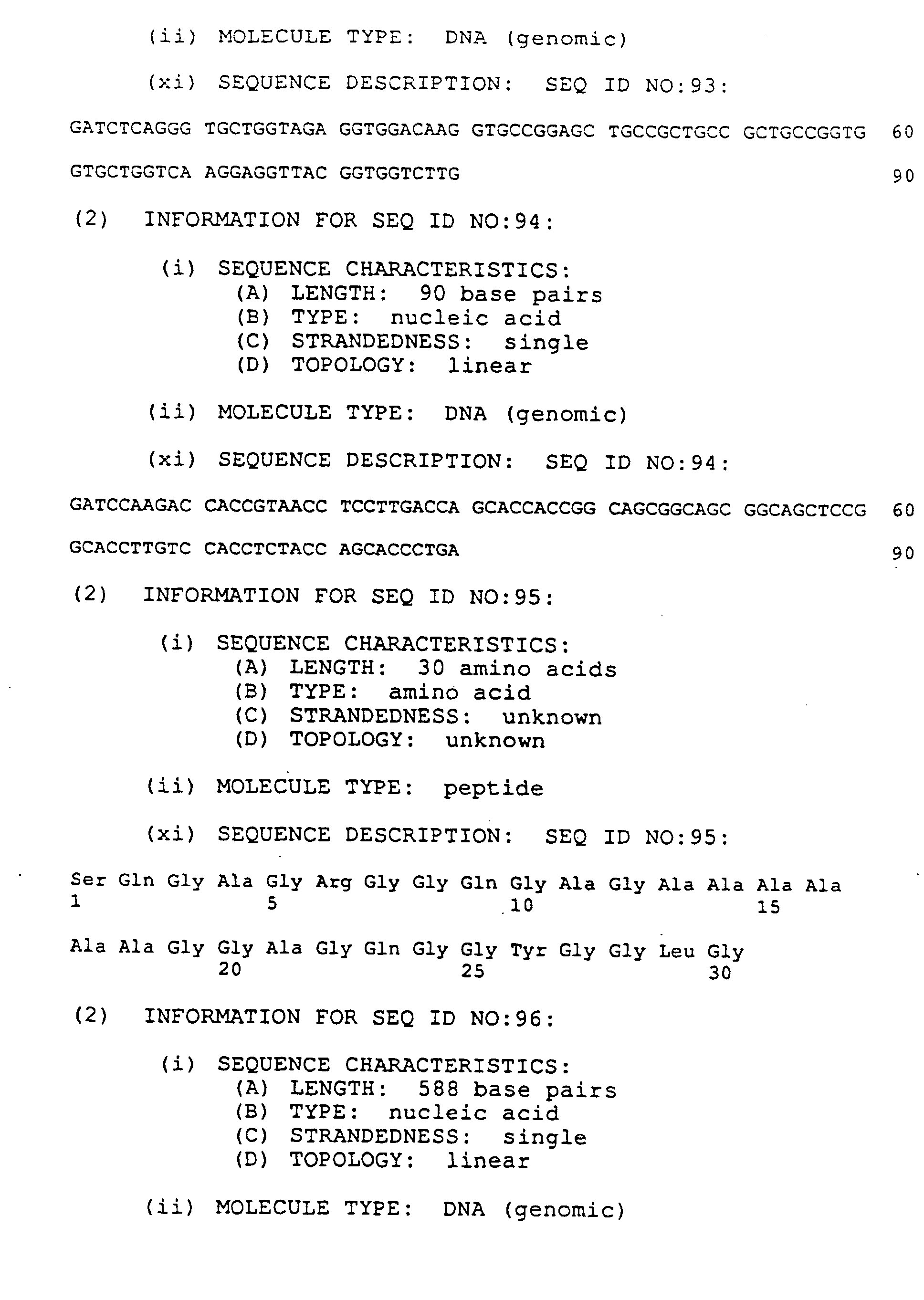
- Figure 3 illustrates the amino acid sequences for the monomer (SEQ ID NO.:22) and polymer (SEQ ID NO.:23) of the spider silk DP-1B.9 analog (SEQ ID NO.:81).
- Figure 4 illustrates the synthetic oligonucleotides L(SEQ ID NOs.:24-26), M1(SEQ ID NOs.:27-29), M2(SEQ ID NOs.:30-32), and S(SEQ ID NOs.:33-35) used in the construction of the DNA monomer for DP-1 protein expression.
- Figure 5 is a plasmid map illustrating the construction of plasmid pFP510 from pA126i. Plasmid pFP510 is used to construct plasmids for the assembly and polymerization of DNA monomers and genes encoding DP-1A analogs.
- Figure 6 is a plasmid map of plasmid pFP202 which is used to construct high level expression vectors.
- Figure 7 illustrates the six double stranded synthetic oligonucleotides, A(SEQ ID NOs.:41-43), B(SEQ ID NOs.:44-46), C(SEQ ID NOs.:47-49), D(SEQ ID NOs.:50-52), E(SEQ ID NOs.:53-55), and F(SEQ ID NOs.:56-58), used in the construction of the DNA monomer for DP-2 protein expression.
- Figure 8 illustrates the amino acid sequence (SEQ ID NO.:59) of the natural spider silk protein Spidroin 2 as described by Lewis et al. (EP 452925).
- Figure 9 illustrates the amino acid sequences of the amino acid monomer (SEQ ID NO.:60) and polymer (SEQ ID NO.:61) of the spider dragline protein 2 analog, DP-2A (SEQ ID NO.:83).
- Figure 10 illustrates the amino acid sequences of the amino acid monomer (SEQ ID NO.:62) and polymer (SEQ ID NO.:63) of the spider dragline protein 1 analog, DP-1B.16 (SEQ ID NO.:82).
- Figure 11 illustrates the four double stranded synthetic oligonucleotides 1(SEQ ID NOs.:64-66), 2(SEQ ID NOs.:67-69), 3(SEQ ID NOs.:70-72), and 4(SEQ ID NOs.:73-75) used to construct the synthetic genes encoding DP-1B.16 (SEQ ID NO.:82).
- Figure 12 is a plasmid map illustrating the construction of the plasmid pFP206 from pA126i. Plasmid pFP206 was used to construct plasmids used for the assembly and polymerization of the DNA monomer, and genes encoding DP-1B analogs.
- Figure 13 illustrates the full nucleic acid sequence (SEQ ID NO.:78) of plasmid pA126i.
- Figure 14 illustrates the complete DNA sequence (SEQ ID NO.:79) of pBE346.
- Figure 15 is a plasmid map illustrating the construction of the plasmid pFP191 which was used to transform B. subtilis cells for DP-1A analog protein expression and secretion.
- Figure 16 illustrates the four synthetic double-stranded oligonucleotides P1, P2, P3, and P4, used to construct the synthetic genes encoding DP-1B.33.
P1 corresponds to SEQ ID NOs.:84, 85, and 86.
P2 corresponds to SEQ ID NOs.:87, 88, and 89.
P3 corresponds to SEQ ID NOs.:90, 91, and 92.
P4 corresponds to SEQ ID NOs.:93, 94, and 95.
- Figure 17 is a plasmid map of plasmid pHIL-D4, used to construct vectors for intracellular protein expression in Pichia pastoris.
- Figure 18 is a plasmid map of plasmid pPIC9, used to construct vectors for extracellular protein production in P. pastoris.
- Figure 19 illustrates the DNA sequence of a portion of plasmid pFO734, an intermediate in the construction of vectors for extracellular protein production in P. pastoris.
- Figure 20 illustrates DP-1B production by P. pastoris strain YFP5028.
- Figure 21 illustrates DP-1B production by P. pastoris strain YFP5093.
-
Applicants have provided sequence listings 1-107 in conformity with "Rules for the standard representation of nucleotide and amino acid sequence in patent applications" (Annexes I and II to the Decision of the President of the EPO, published in Supplement No. 2 to OJ EPO 12/1992).
-
Applicants have made the following biological deposits under the terms of the Budapest Treaty.
| Deposit or Identification Reference | ATCC Designation | Deposit Date |
| Escherichia coli, FP 3227 | 69326 | 15 June 1993 |
| Escherichia coli, FP 2193 | 69327 | 15 June 1993 |
| Escherichia coli, FP 3350 | 69328 | 15 June 1993 |
-
As used herein, the designation "ATCC" refers to the American Type Culture Collection depository located in Rockville, Maryland at 12301 Parklawn Drive, Rockville, MD 20852, U.S.A. The "ATCC No." is the accession number to cultures on deposit at the ATCC.
DETAILED DESCRIPTION OF THE INVENTION
-
The following definitions are used herein and should be referred to for interpretation of the claims and the specification.
-
As used herein, the terms "promoter" and "promoter region" refer to a sequence of DNA, usually upstream of (5' to) the protein coding sequence of a structural gene, which controls the expression of the coding region by providing the recognition for RNA polymerase and/or other factors required for transcription to start at the correct site. Promoter sequences are necessary but not always sufficient to drive the expression of the gene.
-
A "fragment" constitutes a fraction of the DNA sequence of the particular region.
-
"Nucleic acid" refers to a molecule which can be single stranded or double stranded, composed of monomers (nucleotides) containing a sugar, phosphate and either a purine or pyrimidine. In bacteria, lower eukaryotes, and in higher animals and plants, "deoxyribonucleic acid" (DNA) refers to the genetic material while "ribonucleic acid" (RNA) is involved in the translation of the information from DNA into proteins.
-
The terms "peptide", "polypeptide" and "protein" are used interchangeably.
-
"Regulation" and "regulate" refer to the modulation of gene expression controlled by DNA sequence elements located primarily, but not exclusively upstream of (5' to) the transcription start of a gene. Regulation may result in an all or none response to a stimulation, or it may result in variations in the level of gene expression.
-
The term "coding sequence" refers to that portion of a gene encoding a protein, polypeptide, or a portion thereof, and excluding the regulatory sequences which drive the initiation of transcription. The coding sequence may constitute an uninterrupted coding region or it may include one or more introns bounded by appropriate splice junctions. The coding sequence may be a composite of segments derived from different sources, naturally occurring or synthetic.
-
The term "construction" or "construct" refers to a plasmid, virus, autonomously replicating sequence, phage or nucleotide sequence, linear or circular, of a single- or double-stranded DNA or RNA, derived from any source, in which a number of nucleotide sequences have been joined or recombined into a unique construction which is capable of introducing a promoter fragment and DNA sequence for a selected gene product along with appropriate 3' untranslated sequence into a cell.
-
As used herein, "transformation" is the acquisition of new genes in a cell by the incorporation of nucleic acid.
-
The term, "operably linked" refers to the chemical fusion of two fragments of DNA in a proper orientation and reading frame to lead to the transcription of functional RNA.
-
The term "expression" as used herein is intended to mean the transcription and translation to gene product from a gene coding for the sequence of the gene product. In the expression, a DNA chain coding for the sequence of gene product is first transcribed to a complementary RNA which is often a messenger RNA and, then, the thus transcribed messenger RNA is translated into the above-mentioned gene product if the gene product is a protein.
-
The term "translation initiation signal" refers to a unit of three nucleotides (codon) in a nucleic acid that specifies the initiation of protein synthesis.
-
The term "signal peptide" refers to an amino terminal polypeptide preceding the secreted mature protein. The signal peptide is cleaved from and is therefore not present in the mature protein. Signal peptides have the function of directing and trans-locating secreted proteins across cell membranes. The signal peptide is also referred to as signal sequence.
-
The term "mature protein" refers to the final secreted protein product without any part of the signal peptide attached.
-
The term "plasmid" or "vector" as used herein refers to an extra-chromosomal element often carrying genes which are not part of the central metabolism of the cell, and usually in the form of circular double-stranded DNA molecules.
-
The term "restriction endonuclease" refers to an enzyme which catalyzes hydrolytic cleavage within a specific nucleotide sequence in double-stranded DNA.
-
The term "compatible restriction sites" refers to different restriction sites that when cleaved yield nucleotide ends that can be ligated without any additional modification.
-
The term "suitable promoter" will refer to any eukaryotic or prokaryotic promoter capable of driving the expression of a synthetic spider silk variant gene.
-
The term "spider silk variant protein" will refer to a designed protein, the amino acid sequence of which is based on repetitive sequence motifs and variations thereof that are found in a known a natural spider silk.
-
The term "full length variant protein" will refer to any spider silk variant protein encoded by a synthetic gene which has been constructed by the assembly and polymerization of a DNA monomer.
-
The term "DNA monomer" will refer to a DNA fragment consisting of between 300 and 400 bp which encodes one or more repeating amino acid sequences of a spider silk variant protein. Examples of DNA monomers suitable for the present invention are illustrated in Figures 2, 3, 9 and 10.
-
The term "peptide monomer", "polypeptide monomer" or "amino acid monomer" will refer to the amino acid sequence encoded by a DNA monomer.
-
The term "commercial quantities" will refer to quantities of recombinantly produced desired proteins where at least 1% of the total protein produced by a microbial culture is the desired protein.
-
The term "desired protein" will refer to any protein considered a valuable product to be obtained from genetically engineered bacteria.
-
The term "DP-1 analog" will refer to any spider silk variant derived from the amino acid sequence of the natural Protein 1 (Spidroin 1) of Nephila calvipes as illustrated in Figure 1.
-
The term "DP-2 analog" will refer to any spider silk variant derived from the amino acid sequence of the natural Protein 2 (Spidroin 2) of Nephila calvipes as illustrated in Figure 8.
-
As used herein the following abbreviations will be used to identify specific amino acids:
| Amino Acid | Three-Letter Abbreviation | One-Letter Abbreviation |
| Alanine | Ala | A |
| Arginine | Arg | R |
| Asparagine | Asn | N |
| Aspartic acid | Asp | D |
| Asparagine or aspartic acid | Asx | B |
| Cysteine | Cys | C |
| Glutamine | Gln | Q |
| Glutamine acid | Glu | E |
| Glutamine or glutamic acid | Glx | Z |
| Glycine | Gly | G |
| Histidine | His | H |
| Leucine | Leu | L |
| Lysine | Lys | K |
| Methionine | Met | M |
| Phenylalanine | Phe | F |
| Proline | Pro | P |
| Serine | Ser | S |
| Threonine | Thr | T |
| Tryptophan | Trp | W |
| Tyrosine | Tyr | Y |
| Valine | Val | V |
-
The present invention also provides novel DNA sequences encoding spider silk protein variants that are suitable for expression of commercial quantities of silk protein in a recombinant host.
-
It will be appreciated that the advantages of such a protein and such a method are many. Spider silk, especially dragline silk, has a tensile strength of over 200 ksi with an elasticity of nearly 35%, which makes it more difficult to break than either KEVLAR or steel. When spun into fibers, spider silk of the present invention may have application in the bulk clothing industries as well as being applicable for certain kinds of high strength uses such as rope, surgical sutures, flexible tie downs for certain electrical components and even as a biomaterial for implantation (e.g., artificial ligaments or aortic banding). Additionally these fibers may be mixed with various plastics and/or resins to prepare a fiber-reinforced plastic and/or resin product. Furthermore, since spider silk is stable up to 100 °C, these fibers may be used to reinforce thermal injected plastics. These proteins may also be of value in the form of films or coatings. It will be appreciated by one of skill in the art that the properties of the silk fibers may be altered by altering the amino acid sequence of the protein.
-
The present invention provides a method for the production of analogs of natural spider silk proteins and variants using recombinant DNA technology. The method consists of (1) the design of analog protein sequences based on the amino acid sequence of the fiber forming regions of natural proteins; (2) the design of DNA sequences to encode such analog protein sequences, based on a DNA monomer of at least 50 bp with minimal internal repetitiveness, and making preferential use of codons matched to the preferences of a specific host organism; (3) assembly of the DNA monomer from cloned synthetic oligonucleotides; (4) polymerization of the DNA monomer to lengths of at least 800 bp, and preferably to lengths approximating the length of the gene encoding the natural protein; (5) inserting the polymerized artificial gene into an appropriate vector able to replicate in the host organism, in such a manner that the gene is operably linked to expression signals whereby its expression can be regulated; (6) producing the protein in the above mentioned microbial host carrying such an expression vector; (7) purifying the protein from the biomass and preparing it in a form suitable for forming into fibers, films, or coatings.
-
The expression of the desired silk variant protein in Escherichia coli is preferred since this host reliably produces high levels of foreign protein and the art is replete with suitable transformation and expression vectors. However, it is not outside the scope of the invention to provide alternative hosts and particularly hosts that facilitate the secretion of the desired protein into the growth medium. Such alternative hosts may include but are not limited to Bacillus subtilis, Saccharomyces cerevisiae, Schizosaccharomyces pombe, Pichia pastoris, Aspergillus spp., Hansenula spp., and Streptomyces spp. The expression host preferred for the secretion of silk variant protein is Bacillus subtilis.
-
The present invention provides a variety of plasmids or vectors suitable for the cloning of portions of the DNA required for the assembly and expression of the silk variant protein gene in E. coli. Suitable vectors for construction contain a selectable marker and sequences allowing autonomous replication or chromosomal integration. Additionally, suitable vectors for expression contain sequences directing transcription and translation of the heterologous DNA fragment. These vectors comprise a region 5' of the heterologous DNA fragment which harbors transcriptional initiation controls, and optionally a region 3' of the DNA fragment which controls transcriptional termination. It is most preferred when both control regions are derived from genes homologous to E. coli although it is to be understood that such control regions need not be derived from the genes native to the specific species chosen as a production host. Suitable vectors can be derived, for example, from a bacteria, a virus (such as bacteriophage T7 or a M-13 derived phage), a cosmid, a yeast or a plant. Protocols for obtaining and using such vectors are known to those in the art. (Sambrook et al., Molecular Cloning: A Laboratory Manual - volumes 1,2,3 (Cold Spring Harbor Laboratory: Cold Spring Harbor, New York, 1989))
-
Examples of bacteria-derived vectors include plasmid vectors such as pBR322, pUC19, pSP64, pUR278 and pORF1. Illustrative of suitable viral vectors are those derived from phage, vaccinia, retrovirus, baculovirus, or a bovine papilloma virus. Examples of phage vectors include λ+, λEMBL3, 12001, λgtlO, λgtll, Charon 4a, Charon 40, and λZAP/R. pXB3 and pSCll are exemplary of vaccinia vectors ( Chakrabarti et al., Molec. Cell. Biol. 5:3401-9 (1985) and Mackett et al., J. Virol. 49:857864 (1984). An example of a filamentous phage vector is an M13-derived vector like M13mpl8, and M13mpl9.
-
For the expression of spider silk variant proteins in E. coli bacteria-derived vectors are preferred where plasmids derived from pBR322 are most preferred.
-
Optionally it may be desired to produce the silk variant protein as a secretion product of a transformed host, such as B. subtilis. Secretion of desired proteins into the growth media has the advantage of simplified and less costly purification procedures. It is well known in the art that secretion signal sequences are often useful in facilitating the active transport of expressible proteins across cell membranes. The creation of a transformed Bacillus host capable of secretion may be accomplished by the incorporation of a DNA sequence that codes for a secretion signal functional in the Bacillus production host on the expression cassette, between the expression-controlling DNA and the DNA encoding the silk variant protein and in reading frame with the latter. Examples of vectors enabling the secretion of a number of different heterologous proteins by B. subtilis have been taught and are described in Nagarajan et al., U.S. Patent 4,801,537; Stephens et al., U.S. Patent 4,769,327; and Biotechnology Handbook 2, Bacillus, C. R. Harwood, Ed., Plenum Press, New York (1989).
-
Secretion vectors of this invention include a regulatable promoter sequence which controls transcription, a sequence for a ribosome binding site which controls translation, and a sequence for a signal peptide which enables translocation of the peptide through the bacterial membrane and the cleavage of the signal peptide from the mature protein. Suitable vectors will be those which are compatible with the bacterium employed. For example, for B. subtilis such suitable vectors include E. coli-B. subtilis shuttle vectors. They will have compatible regulatory sequences and origins of replication. They will be preferably multicopy and have a selective marker gene, for example, a gene coding for antibiotic resistance. An example of such a vector is pTZ18R phagemid, obtainable from Pharmacia, Piscataway, NJ 08854 which confers resistance to ampicillin in E. coli. The DNA sequences encoding the promoter, ribosome binding site and signal peptide may be from any single gene which encodes a secreted product.
-
The DNA sequences encoding the promoter and ribosome binding site may also be from a different gene than that encoding the signal peptide. The DNA sequences encoding the promoter, ribosome binding site and signal peptide can be isolated by means well known to those in the art and illustrative examples are documented in the literature. See Biotechnology Handbook 2 Bacillus, C. R. Harwood, Ed., Plenum Press, New York, New York (1989). The promoters in the DNA sequences may be either constitutive or inducible and thus permit the resulting secretion vectors to be differentially regulated.
-
Promoters which are useful to drive expression of heterologous DNA fragments in E. coli and Bacillus are numerous and familiar to those skilled in the art. Virtually any promoter capable of driving the gene encoding a silk variant protein is suitable for the present invention, where the T7 promoters are preferred in E. coli and promoters derived from the SacB gene are preferred in Bacillus.
-
Termination control regions may also be derived from various genes native to E. coli or Bacillus hosts, or optionally other bacterial hosts. It will be appreciated by one of skill in the art that a termination control region may be unnecessary.
-
For introducing a polynucleotide of the present invention into a bacterial cell, known procedures can be used according to the present invention such as by transformation, e.g., using calcium-permeabilized cells, electroporation, or by transfection using a recombinant phage virus. (Sambrook et al., Molecular Cloning: A Laboratory Manual - volumes 1,2,3 (Cold Spring Harbor Laboratory: Cold Spring Harbor, New York, 1989)). Other known procedures can also be employed to obtain a recombinant host cell that expresses a heterologous spider silk protein according to the present invention, as will be apparent to those skilled in the art.
Design of Spider Silk Variant Amino Acid Sequences:
-
The design of the spider silk variant proteins was based on consensus amino acid sequences derived from the fiber forming regions of the natural spider silk dragline proteins of Nephila clavipes. Natural spider dragline consists of two different proteins that are co-spun from the spider's major ampullate gland. The amino acid sequence of both dragline proteins has been disclosed by Xu et al., Proc. Natl, Acad. Sci. U.S.A., 87, 7120, (1990) and Hinman and Lewis, J. Biol. Chem. 267, 19320 (1992), and will be identified hereinafter as Dragline Protein 1 (DP-1) and Dragline Protein 2 (DP-2).
-
The amino acid sequence of a fragment of DP-1 is repetitive and rich in glycine and alanine, but is otherwise unlike any previously known amino acid sequence. The repetitive nature of the protein and the pattern of variation among the individual repeats are emphasized by rewriting the sequence as in Figure 1. The "consensus" sequence of a single repeat, viewed in this way, is:
where X may be S,G, or N.
-
Examination of Figure 1 shows that individual repeats differ from the consensus according to a pattern which can be generalized as follows: (1) The poly-alanine sequence varies in length from zero to seven residues. (2) When the entire poly-alanine sequence is deleted, so also is the surrounding sequence encompassing AGRGGLGGQGAGAnGG (SEQ ID NO:2). (3) Aside from the poly-alanine sequence, deletions generally encompass integral multiples of three consecutive residues. (4) Deletion of GYG is generally accompanied by deletion of GRG in the same repeat. (5) A repeat in which the entire poly-alanine sequence is deleted is generally preceded by a repeat containing six alanine residues.
-
Synthetic analogs of DP-1 were designed to mimic both the repeating consensus sequence of the natural protein and the pattern of variation among individual repeats. Two analogs of DP-1 were designed and designated DP-1A and DP-1B. DP-1A is composed of a tandemly repeated 101-amino acid sequence listed in Figure 2. The 101-amino acid "monomer" comprises four repeats which differ according to the pattern (1)-(5) above. This 101-amino acid long peptide monomer is repeated from 1 to 16 times in a series of analog proteins. DP-1B was designed by reordering the four repeats within the monomer of DP-1A. This monomer sequence, shown in Figure 3, exhibits all of the regularities of (1)-(5) above. In addition, it exhibits a regularity of the natural sequence which is not shared by DP-1A, namely that a repeat in which both GYG and GRG are deleted is generally preceded by a repeat lacking the entire poly-alanine sequence, with one intervening repeat. The sequence of DP-1B matches the natural sequence more closely over a more extended segment than does DP-1A.
-
The amino acid sequence of a fragment of DP-2 is also repetitive and also rich in glycine and alanine, but is otherwise unlike any previously known amino acid sequence, and, aside from a region of consecutive alanine residues, different from DP-1. The repetitive nature of the protein and the pattern of variation among the individual repeats are emphasized by rewriting the sequence as in Figure 8. The "consensus" sequence of a single repeat, viewed in this way, is:
-
Examination of Figure 8 shows that individual repeats differ from the consensus according to a pattern which can be generalized as follows: (1) The poly-alanine-rich sequence varies in length from six to ten residues. (2) Aside from the poly-alanine sequence, individual repeats differ from the consensus repeat sequence by deletions of integral multiples of five consecutive residues consisting of one or both of the pentapeptide sequences GPGGY (SEQ ID NO:3) or GPGQQ (SEQ ID NO:4).
-
Synthetic analogs of DP-2 were designed to mimic both the repeating consensus sequence of the natural protein and the pattern of variation among individual repeats. The analog DP-2A is composed of a tandemly repeated 119-amino acid sequence listed in Figure 9. The 119-amino acid "peptide monomer" comprises three repeats which differ according to the pattern (1)-(2) above. This 119-amino acid long peptide monomer is repeated from 1 to 16 times in a series of analog proteins.
Design of DNA encoding Spider Silk Variant Proteins:
-
DNA sequences encoding the designed analog amino acid sequences were devised according to the following criteria: (1) The DNA monomer was to be at least 300 bp in length; (2) within the monomer, repetitiveness of the sequence was minimized, with no repeated sequence longer than 17 bp and minimal repetitiveness of sequences longer than 10 bp; (3) where possible, codons were chosen from among the codons found preferentially in highly expressed genes of the intended host organism (E. coli) with preference for codons providing balanced A+T/G+C base ratios; and (4) predicted secondary structure of mRNA within the monomer was dominated by long-range interactions rather than shorter range base pairing. No attempt was made to minimize secondary structure of the mRNA.
Assembly of DP-1 and DP-2 Analog Genes:
-
Assembly of the synthetic dragline analog genes was accomplished by first assembling the appropriate DNA monomers followed by polymerization of these monomers to form the completed gene.
-
Synthetic DNA monomers, based on the consensus peptide monomers described above were assembled from four to six cloned double stranded synthetic oligonucleotides. Each oligonucleotide was designed to encode a different portion of the the peptide monomer. Briefly, the oligonucleotides were each cloned into separate suitable plasmid vectors containing an ampicillin resistance gene. A suitable E. coli host was transformed with the plasmids and screened for the presence of the correct vector by standard methods. After the oligonucleotides were cloned the DNA monomer was sequentially assembled. Vectors containing individual oligonucleotides were digested and the plasmid DNA was purified by gel electrophoresis. Purified plasmid DNA containing two different oligonucleotide sequences were then incubated under ligating conditions and the ligation products were used to transform a suitable E. coli host. These transformants comprised two of the oligonucleotide sequences linked in tandem. A similar procedure was followed for the creation of the full DNA monomer, comprising four to six of the oligonucleotides. Additional confirmation of the existence of the correct DNA insertions was obtained by direct DNA sequencing. The present invetion provides several DNA monomers useful for the production of DP-1A and DP-1B analogs. In general DNA monomers used to produce the the analog DP-1B.16 are preferred since this construct avoids codons rarely used by the E. coli production host.
-
The assembled DNA monomer was then polymerized by a method essentially as described by Kempe et al. ( Gene 39, 239, (1985). This method consists of a series of successive doublings of the sequence of interest. Briefly, the DNA monomer containing the cloned oligonucleotides was digested with suitable restriction enzymes and incubated under annealing conditions followed by ligation to produce a series of constructs containing multiple repeats of the monomer. Ligation products were used to transform a suitable E. coli host and intact plasmids were selected on the basis of ampicillin resistance. Subsequent analysis of plasmid DNA by gel electrophoresis resulted in the identification of transformants containing plasmids with 2, 4, 8, and 16 tandem repeats of the DNA monomer. These protein products were analyzed by SDS polyacrylamide gel electrophoresis and detected and quantitated by immunochemical staining using a polyclonal antiserum raised in rabbits against a synthetic peptide analogous to a fragment of the natural protein.
Expression and purification of Protein:
-
High level expression of the spider dragline protein analogs in E. coli was achieved by inserting the synthetic genes into plasmid vectors pFP202 and pFP204, which were derived from the well-known vector pET11a. In these vectors, the dragline protein-coding gene is inserted in such a manner as to be operably linked to a promoter derived from bacteriophage T7. This promoter is joined with sequences derived from the lac operator of E. coli, which confers regulation by lactose or analogs (IPTG). The E. coli host strain BL21(DE3) contains a lambda prophage which carries a gene encoding bacteriophage T7 RNA polymerase. This gene is controlled by a promoter which is also regulated by lactose or analogs. In addition to the phage T7 promoter, the vectors pFP202 and pFP204 provide sequences which encode a C-terminal tail containing six consecutive histidine resdues appended to the dragline protein-coding sequences. This tail provides a means of affinity purification of the protein under denaturing conditions through its adsorption to resins bearing immobilized Ni ions.
-
DP-1 analog protein was produced by E. coli at levels of approximately 5-20% of total protein. Of this, approximately 20-40% was recovered in purified form as full-length protein. DP-2 analog protein was produced at approximately 5% of total cell protein, of which approximately 30% was recovered in purified form as full-length protein.
-
The following examples are meant to illustrate the invention but should not be construed as limiting it in any way.
EXAMPLES
GENERAL METHODS
-
The position of the newly engineered restriction sites is indicated in the figures and any one skilled in the art can repeat these constructs with the available information.
-
The source of the genes and the various vectors described throughout this application are as follows.
-
The anti-DP-1 and anti-DP-2 antisera were prepared by Multiple Peptide Systems, San Diego, CA.
-
Restriction enzyme digestions, phosphorylations, ligations, transformations and other suitable methods of genetic engineering employed herein are described in Sambrook et al., Molecular Cloning: A Laboratory Manual - volumes 1,2,3 (Cold Spring Harbor Laboratory: Cold Spring Harbor, New York, 1989), and in the instructions accompanying commercially available kits for genetic engineering.
-
Bacterial cultures and plasmids to carry out the present invention are available either commercially (from Novagen, Inc., Madison, WI) or from the E. coli Genetic Stock Center, Yale University, New Haven, CT, the Bacillus Genetic Stock Center, Ohio State University, Columbus, OH, or the ATCC and, along with their sources, are identified in the text and examples which follow. Unless otherwise specified standard reagents and solutions used in the following examples were supplied by Sigma Chemical Co. (St. Louis, MO)
-
Isolation of restriction fragments from agarose gels used the GENECLEAN® procedure (Bio101, Inc., P.O. Box 2284, La Jolla, CA), and was performed as specified by the manufacturer.
EXAMPLE 1
CONSTRUCTION OF THE SYNTHETIC GENES DP-1A.9 AND DP-1B.9
Oligonucleotide design and cloning:
-
Synthetic genes encoding DP-1A.9 and DP-1B.9 were assembled from four double stranded synthetic oligonucleotides labled L (SEQ ID NOs.:24, 25, and 26), M1 (SEQ ID NOs.:27, 28, and 29), M2 (SEQ ID NOs.:30, 31, and 3), and S (SEQ ID NOs.:33, 34, and 35) whose sequences are shown in Figure 4. The oligonucleotides were provided by the manufacturer (Midland Certified Reagents, Midland, TX) in double stranded form with 5'-OH groups phosphorylated. Methods of oligonucleotide synthesis, purification, phosphorylation, and annealing to the double stranded form are well known to those skilled in the art.
-
The four double stranded oligonucleotides were separately cloned by inserting them into a plasmid vector pFP510 (Figure 5). This vector was derived from the plasmid pA126i (see Figure 13), the complete nucleotide sequence of which is provided in SEQ ID NO.:78 and Figure 13. Details of the structure of pA126i are not important for the construction, aside from the following essential features: (a) a replication origin active in E. coli; (b) a selectable genetic marker, in this case a gene conferring resistance to the antibiotic ampicillin; (c) sites for restriction endonucleases BamHI and BglII with no essential sequences between them; and (d) a third restriction site (PstI), located within the selectable marker, which produces cohesive ends incompatible with those produced by BamHI and BglII. For the construction of pFP510, DNA of plasmid pA126i was digested with endonucleases BamHI and BglII, then recovered by adsorption to glass beads in the presence of NaI GENECLEAN® procedure (Bio101, Inc., P.O. Box 2284, La Jolla, CA). To approximately 0.1 pmole of the eluted plasmid DNA was added 10 pmoles of the double stranded, phosphorylated oligonucleotide SF4/5 (Figure 5). The mixture was incubated under ligation conditions with T4 polynucleotide ligase for 19 h at 4 °C. Ligated DNA was then digested with endonuclease XmaI to linearize any remaining parental pA126i and used to transform E. coli SK2267 (obtained from the E. coli Genetic Stock Center, Yale University, New Haven, CT) which had been made competent by calcium treatment as described by Sambrook et al., op. cit. Plasmid DNA isolated from ampicillin resistant transformants was characterized by digestion separately with endonucleases ApaI and BamHI, and a transformant containing the desired plasmid was identified and designated pFP510.
-
DNA of plasmid pFP510 was digested with endonucleases SfiI and DraIII and purified by the GENECLEAN® procedure (Bio101, Inc., P.O. Box 2284, La Jolla, CA). To approximately 0.1 pmole of the eluted plasmid DNA was added 10 pmoles of one of the double stranded, phosphorylated oligonucleotides L, M1, M2, or S (Figure 4). The four plasmid-oligonucleotide mixtures were incubated under ligation conditions for 15 h at 4 °C, then for 20 min at 23 °C and finally ligation was terminated by incubation for 3 min at 65 °C. Aliquots of ligated DNA were used to transform E. coli SK2267 and ampicillin resistant transformants were selected. Clones containing oligonucleotides L, M1, and M2 shown in Figure 4 were identified by screening plasmid DNA isolated from individual transformants with endonuclease AlwNI, a recognition site for which is present in the oligonucleotides. Clones containing oligonucleotide S were identified by screening plasmid DNA isolated from individual transformants with endonucleases BglI and DraIII. Plasmid DNA from putative clones was further characterized by digestion with endonucleases EcoRI, SfiI, and DraIII in order to establish that the oligonucleotide sequences were oriented correctly in the plasmid. The inserts were excised with endonucleases BamHI and BglII and analyzed by electrophoresis in 4% NuSieve agarose (FMC) to verify that the plasmid had acquired only a single copy of the oligonucleotide. Correct clones were identified and their plasmids were designated pFP521 (oligonucleotide L), pFP533 (oligonucleotide M1), pFP523 (oligonucleotide M2), and pFP524 (oligonucleotide S). DNA sequences of all four cloned oligonucleotides were verified by DNA sequencing.
-
DNA sequencing was carried out essentially according to procedures provided by the supplier (U.S. Biochemicals) with the Sequenase 2.0 kit for DNA sequencing with 7-deaza-GTP. Plasmid DNA was prepared using the Magic Minipreps kit (Promega). Template DNA was denatured by incubating 20 µl miniprep DNA in 40 µl (total volume) 0.2 M NaOH for 5 min at 23 °C. The mixture was neutralized by adding 6 µl 2 M ammonium acetate (adjusted to pH 4.5 with acetic acid), and the DNA was precipitated by adding 0.15 mL ethanol, recovered by centrifugation, washed with cold 70% ethanol, and vacuum dried. Primers for sequencing were as follows:


Primers SI1 and SI5 anneal to sites on opposite strands in pA126i. SI5 primes synthesis into the sequences of interest from 31 bp beyond the BamHI site. SI1 primes synthesis on the opposite strand into the sequences of interest from 38 bp beyond the BglII site. For sequencing in the vector pFP206 (see below) the primer SI20, which anneals 25 bp beyond the BglII site, was substituted for SI1 (Figure 12). Polyacrylamide gels for DNA sequencing were run at 52 °C.
Assembly of the Gene:
-
For assembly of subsequence M2L, plasmid pFP523 (M2) was digested with endonucleases PstI and DraIII, and plasmid pFP521 (L) was digested with endonucleases PstI and SfiI. Digested plasmid DNA was fractionated by electrophoresis in a 1.2% agarose (low melting, BioRad) gel. Ethidium bromide-stained bands containing the oligonucleotide sequences, identified by their relative sizes, were excised, the excised bands combined, and the DNA recovered from melted agarose by the GENECLEAN® procedure (Bio101, Inc., P.O. Box 2284, La Jolla, CA). The eluted combined DNA fragments were incubated under ligation conditions and an aliquot was used to transform E. coli W3110 (available from the E. coli Genetic Stock Center, Yale University, New Haven, CT.). Ampicillin resistant transformants were selected. Plasmid DNA was isolated from several transformants, digested with endonucleases BamHI and BglII, and analyzed by agarose gel electrophoresis. Plasmid containing insert of the expected size was identified and designated pFP525.
-
Assembly of subsequence M1S was accomplished in the same manner, starting with plasmids pFP533 (digested with PstI and DraIII) and pFP524 (digested with PstI and SfiI). Plasmid containing the M1S subsequence was identified and designated pFP531.
-
For assembly of the DNA monomer (M2LM1S), plasmid pFP525 (M2L) was digested with endonucleases PstI and DraIII, and plasmid pFP531 (M1S) was digested with endonucleases PstI and SfiI. Digested plasmid DNA was fractionated by electrophoresis in a 1.2% low melting agarose gel. Ethidium bromide-stained bands containing the M2L and M1S sequences, respectively, identified by their relative sizes, were excised, the excised bands combined, and the DNA recovered from melted agarose by the GENECLEAN® procedure (Bio101, Inc., P.O. Box 2284, La Jolla, CA). The eluted combined DNA fragments were incubated under ligation conditions and an aliquot was used to transform E. coli W3110. Ampicillin resistant transformants were selected. Plasmid DNA was isolated from several transformants, digested with endonucleases BamHI and BglII, and analyzed by agarose gel electrophoresis. Plasmid containing insert of the expected size was identified and designated pFP534. The DNA inserts in plasmids pFP523, pFP521, pFP533, pFP524, pFP525, pFP531, and pFP534 were verified by direct DNA sequencing as previously described.
Polymerization of the Gene:
-
The synthetic gene was extended by sequential doubling, starting with the monomer sequence in pFP534. For doubling any insert sequence, an aliquot of plasmid DNA was digested with endonucleases PstI and DraIII, and a separate aliquot of the same plasmid was digested with endonucleases PstI and SfiI. Digests were fractionated by electrophoresis on low melting agarose, and ethidium bromide stained fragments containing insert sequences were identified by their relative sizes. In some cases, the two fragments were not adequately separated, so it was necessary to cut the non-insert-containing fragment with a third enzyme, usually MluI.
-
Each of the two insert sequence-containing fragments has one end generated by endonuclease PstI. Annealing of these compatible single stranded ends and ligation results in reconstitution of the gene that confers ampicillin resistance, part of which is carried on each fragment. The other end of each fragment displays a single stranded sequence generated by either DraIII or SfiI. These sequences are, by design, complementary, and annealing and ligation results in a head-to-tail coupling of two insert sequences, with concomitant loss of both sites at the junction. The principle of this method of insert sequence doubling was described by Kempe et al. ( Gene 39, 239-245 (1985)).
-
The two insert-containing fragments, purified by electrophoresis and recovered by the GENECLEAN® procedure (Bio101, Inc., P.O. Box 2284, La Jolla, CA), were combined and incubated under ligation conditions. An aliquot was used to transform E. coli W3110. Ampicillin resistant transformants were selected. Plasmid DNA was isolated from several transformants, digested with endonucleases BamHI and BglII, and analyzed by agarose gel electrophoresis. Plasmid containing insert of the expected size was identified.
-
By this procedure a series of plasmids was constructed containing 2, 4, 8, and 16 tandem repeats of the DNA monomer sequence M2LM1S, encoding the series of DP-1A analogs. In addition, analogous methods were used to construct genes encoding the series of DP-1B analogs. For this purpose, subsequences SL (from pFP524 and pFP521) and M1M2 (from pFP533 and pFP523) were first constructed, then combined to form the monomer SLM1M2, which was polymerized as described. It should be apparent that similar methods can be used to assemble any combination of subsequences carried in the vector pFP510, or any other appropriate vector, provided that the subsequences are bounded by cleavage sites for restriction endonucleases that generate compatible ends (complementary single stranded ends or blunt ends). In addition to various monomer sequences, polymers of any number of repeats of the monomer sequence can be assembled in the same way, starting with plasmids containing inserts of different sizes.
EXAMPLE 2
SYNTHETIC GENE DP-1B.16
-
A second set of genes encoding DP-1B, designated DP-1B.16 (SEQ ID NO.:82), were designed to reduce the number of codons which are rarely used in highly expressed E. coli genes, but at the same time encoding proteins of the same repeating sequence. The sequence of the DP-1B.16 peptide monomer is shown in Fig. 10 and in SEQ ID NO.:82.
Oligonucleotide Synthesis and Cloning:
-
Synthetic genes encoding DP-1B.16 (SEQ ID NO.:82) were assembled from four double stranded synthetic oligonucleotides whose sequences (SEQ ID NOs.:64, 65, 66; SEQ ID NOs.:67, 68, 69; SEQ ID NOs.:70, 71, 72; and SEQ ID NOs.:73, 74, 75) are shown in Figure 11. The oligonucleotides were provided by the manufacturer (Midland Certified Reagents, Midland, TX) in single stranded form with 5'-OH groups not phosphorylated. For annealing to the double stranded form, complementary single stranded oligonucleotides (667 pmoles each) were mixed in 0.2 mL buffer containing 0.01 M Tris-HCl, 0.01 M MgCl2, 0.05 M NaCl, 0.001 M dithiothreitol, pH 7.9. The mixture was heated in boiling water for 1 minute, then allowed to cool slowly to 23 °C over approximately 3 h.
-
The four double stranded oligonucleotides were separately cloned by inserting them into a plasmid vector pFP206 (Figure 12). This vector was derived from the plasmid pA126i as illustrated in Fig. 12. Briefly, DNA of plasmid pA126i was digested with endonucleases BamHI and EcoRI, and the two fragments were separated by electrophoresis in a 1.2% agarose (low melting, BioRad). The larger of the two fragments was excised from the ethidium bromide-stained gel and recovered by the GENECLEAN® procedure (Bio101, Inc., P.O. Box 2284, La Jolla, CA). To approximately 0.1 pmole of the eluted DNA fragment was added 10 pmoles of the double stranded, phosphorylated oligonucleotide SF31/32 (Figure 12). The mixture was incubated under ligation conditions with T4 polynucleotide ligase for 8.5 h at 4 °C. Ligated DNA was used to transform E. coli HB101, which had been made competent by calcium treatment. Plasmid DNA isolated from ampicillin resistant transformants was characterized by digestion separately with endonucleases HindIII, EcoRI, BglII, and BamHI, and a transformant containing the desired plasmid was identified and designated pFP206.
-
DNA of plasmid pFP206 was digested with endonucleases BamHI and BglII and purified by the GENECLEAN® procedure (Bio101, Inc., P.O. Box 2284, La Jolla, CA). To approximately 0.1 pmole of the eluted plasmid DNA was added 10 pmoles of one of the double stranded oligonucleotides 1 (SEQ ID NOs.:64, 65, 66) 2 (SEQ ID NOs.:67, 68, 69), 3 (SEQ ID NOs.:70, 71, 72), or 4 (SEQ ID NOs.:73, 74, 75). The four plasmid-oligonucleotide mixtures were incubated under ligation conditions for 15 h at 4 °C, then ligation was terminated by incubation for 3 min at 70 °C. Ligated DNA was then digested with endonuclease HindIII to linearize any remaining parental pFP206. Aliquots of ligated DNA were used to transform E. coli HB101 and ampicillin resistant transformants were selected. Clones containing oligonucleotides 1, 2, 3, or 4 were identified by screening plasmid DNA isolated from individual transformants with endonucleases BamHI and PstI. In plasmids with inserts in the desired orientation, the shorter of two BamHI-PstI fragments of pFP206 is lengthened by the length of the cloned oligonucleotide. Plasmid DNA from putative clones was further characterized by digestion with endonucleases BamHI and BglII and analysis by electrophoresis in 3% NuSieve agarose (FMC), 1% Agarose (Sigma Chemical Co.) to verify that the plasmid had acquired only a single copy of the oligonucleotide in the correct orientation. Correct clones were identified and their plasmids were designated pFP636 (oligonucleotide 1), pFP620 (oligonucleotide 2), pFP641 (oligonucleotide 3), and pFP631 (oligonucleotide 4). Sequences of all four cloned oligonucleotides were verified by DNA sequencing as described above.
Assembly of the Gene:
-
For assembly of subsequence 1,2, plasmid pFP636 (1) was digested with endonucleases PstI and BamHI, and plasmid pFP620 (2) was digested with endonucleases PstI and BglII. Digested plasmid DNA was fractionated by electrophoresis in a 1.2% agarose (low melting, BioRad) gel. Ethidium bromide-stained bands containing the oligonucleotide sequences, identified by their relative sizes, were excised, the excised bands combined, and the DNA recovered from melted agarose by the GENECLEAN® procedure (Bio101, Inc., P.O. Box 2284, La Jolla, CA). The eluted combined DNA fragments were incubated under ligation conditions and an aliquot was used to transform E. coli HB101. Ampicillin resistant transformants were selected. Plasmid DNA was isolated from several transformants, digested with endonucleases BamHI and BglII, and analyzed by agarose gel electrophoresis. Plasmid containing insert of the expected size was identified and designated pFP647.
-
Assembly of subsequence 3,4 was accomplished in the same manner, starting with plasmids pFP641 (digested with PstI and BamHI) and pFP631 (digested with PstI and BglII). Plasmid containing the 3,4 subsequence was identified and designated pFP649.
-
For assembly of the DNA monomer (1,2,3,4), plasmid pFP647 (1,2) was digested with endonucleases PstI and BamHI, and plasmid pFP640 (3,4) was digested with endonucleases PstI and BglII. Digested plasmid DNA was fractionated by electrophoresis in a 1.2% low melting agarose gel. Ethidium bromide-stained bands containing the 1,2 and 3,4 sequences, respectively, identified by their relative sizes, were excised, the excised bands combined, and the DNA recovered from melted agarose by the GENECLEAN® procedure (Bio101, Inc., P.O. Box 2284, La Jolla, CA). The eluted combined DNA fragments were incubated under ligation conditions and an aliquot was used to transform E. coli HB101. Ampicillin resistant transformants were selected. Plasmid DNA was isolated from several transformants, digested with endonucleases BamHI and BglII, and analyzed by agarose gel electrophoresis. Plasmid containing insert of the expected size was identified and designated pFP652. The DNA insert in plasmid pFP652 was verified by direct DNA sequencing as described above.
Polymerization of the Gene:
-
The synthetic gene was extended by sequential doubling, starting with the monomer sequence in pFP652. For doubling any insert sequence, an aliquot of plasmid DNA was digested with endonucleases PstI and BamHI, and a separate aliquot of the same plasmid was digested with endonucleases PstI and BglII. Digests were fractionated by electrophoresis on low melting agarose, and ethidium bromide stained fragments containing insert sequences were identified by their relative sizes. The two insert-containing fragments, purified by electrophoresis and recovered by the GENECLEAN® procedure (Bio101, Inc., P.O. Box 2284, La Jolla, CA), were combined and incubated under ligation conditions. At the third doubling, the two fragments in the BamHI digest were not adequately separated, so the eluted band contained both fragments. In this case a two-fold excess of the BglII-PstI fragment was used in the ligation. An aliquot of the ligated DNA was used to transform E. coli HB101. Ampicillin resistant transformants were selected. Plasmid DNA was isolated from several transformants, digested with endonucleases BamHI and BglII, and analyzed by agarose gel electrophoresis. Plasmid containing insert of the expected size was identified.
-
By this procedure a series of plasmids was constructed containing 2, 4, 8, and 16 tandem repeats of the DNA monomer sequence 1 (SEQ ID NOs.:64, 65, 66), 2 (SEQ ID NOs.:67, 68, 69), 3 (SEQ ID NOs.:70, 71, 72), 4 (SEQ ID NOs.:73, 74, 75), encoding the series of DP-1B.16 analogs. These plasmids were designated pFP656 (2 repeats), pFP661 (4 repeats), pFP662 (8 repeats), and pFP665 (16 repeats), respectively.
EXAMPLE 3
SYNTHETIC GENE DP-2A
Oligonucleotide Synthesis and Cloning:
-
Synthetic genes encoding DP-2A were assembled from six double stranded synthetic oligonucleotides whose sequences are shown in Figure 7. The oligonucleotides were provided by the manufacturer (Midland Certified Reagents, Midland, TX) in double stranded form with 5'-OH groups not phosphorylated. The six double stranded oligonucleotides were separately cloned by inserting them into the plasmid vector pFP206.
-
DNA of plasmid pFP206 was digested with endonucleases BamHI and BglII and purified by the GENECLEAN® procedure (Bio101, Inc., P.O. Box 2284, La Jolla, CA). To approximately 0.1 pmole of the eluted plasmid DNA was added 10 pmoles of one of the double stranded oligonucleotides A (SEQ ID NOs.:41, 42, 43), B (SEQ ID NOs.:44, 45, 46), C (SEQ ID NOs.:47, 48, 49), D (SEQ ID NOs.:50, 51, 52), E (SEQ ID NOs.:53, 54, 55), or F (SEQ ID NOs.:56, 57, 58). The six plasmid-oligonucleotide mixtures were incubated under ligation conditions for 15 h at 4 °C, then ligation was terminated by incubation for 3 min at 70 °C. Ligated DNA was then digested with endonuclease HindIII to linearize any remaining parental pFP206. Aliquots of ligated DNA were used to transform E. coli HB101 and ampicillin resistant transformants were selected. Clones containing oligonucleotides A, B, C, D, E, or F were identified by screening plasmid DNA isolated from individual transformants with endonucleases BamHI and PstI. In plasmids with inserts in the desired orientation, the shorter of two BamHI-PstI fragments of pFP206 is lengthened by the length of the cloned oligonucleotide. Plasmid DNA from putative clones was further characterized by digestion with endonucleases BamHI and BglII and analysis by electrophoresis in 3% NUSIEVE agarose (FMC), 1% Agarose (Sigma Chemical Co.) to verify that the plasmid had acquired only a single copy of the oligonucleotide in the correct orientation. Correct clones were identified and their plasmids were designated pFP193 (oligonucleotide A), pFP194 (oligonucleotide B), pFP195 (oligonucleotide C), pFP196 (oligonucleotide D), pFP197 (oligonucleotide E), and pFP198 (oligonucleotide F).
Assembly of the Gene:
-
For assembly of subsequence AB, plasmid pFP193 (A) was digested with endonucleases PstI and PvuII, and plasmid pFP194 (B) was digested with endonucleases PstI and SmaI. Digested plasmid DNA was fractionated by electrophoresis in a 1.2% agarose (low melting, BioRad) gel. Ethidium bromide-stained bands containing the oligonucleotide sequences, identified by their relative sizes, were excised, the excised bands combined, and the DNA recovered from melted agarose by the GENECLEAN® procedure (Bio101, Inc., P.O. Box 2284, La Jolla, CA). The eluted combined DNA fragments were incubated under ligation conditions and an aliquot was used to transform E. coli HB101. Ampicillin resistant transformants were selected. Plasmid DNA was isolated from several transformants, digested with endonucleases BamHI and BglII, and analyzed by agarose gel electrophoresis. Plasmid containing insert of the expected size was identified and designated pFP300 (AB).
-
Assembly of subsequence CD was accomplished in the same manner, starting with plasmids pFP195 (digested with PstI and SnaBI) and pFP196 (digested with PstI and SmaI). Plasmid containing the CD subsequence was identified and designated pFP578. Assembly of subsequence EF was accomplished in the same manner, starting with plasmids pFP197 (digested with PstI and SnaBI) and pFP198 (digested with PstI and SmaI). Plasmid containing the EF subsequence was identified and designated pFP583. The DNA inserts in plasmids pFP300, pFP578, and pFP583 were verified by direct DNA sequencing as described above.
-
Assembly of subsequence CDEF was accomplished similarly, starting with plasmids pFP578 (digested with PstI and PvuII) and pFP583 (digested with PstI and SmaI). Plasmid containing the CDEF subsequence was identified and designated pFP588.
-
For assembly of the DNA monomer (ABCDEF), plasmid pFP300 (AB) was digested with endonucleases PstI and PvuII, and plasmid pFP588 (CDEF) was digested with endonucleases PstI and SmaI. Digested plasmid DNA was fractionated by electrophoresis in a 1.2% low melting agarose gel. Ethidium bromide-stained bands containing the AB and CDEF sequences, respectively, identified by their relative sizes, were excised, the excised bands combined, and the DNA recovered from melted agarose by the GENECLEAN® procedure (Bio101, Inc., P.O. Box 2284, La Jolla, CA). The eluted combined DNA fragments were incubated under ligation conditions and an aliquot was used to transform E. coli HB101. Ampicillin resistant transformants were selected. Plasmid DNA was isolated from several transformants, digested with endonucleases BamHI and BglII, and analyzed by agarose gel electrophoresis. Plasmid containing insert of the expected size was identified and designated pFP303. The DNA insert in plasmid pFP303 was verified by direct DNA sequencing.
Polymerization of the Gene:
-
The synthetic gene was extended by sequential doubling, starting with the monomer sequence in pFP303. For doubling any insert sequence, an aliquot of plasmid DNA was digested with endonucleases PstI and PvuII, and a separate aliquot of the same plasmid was digested with endonucleases PstI and SmaI. Digests were fractionated by electrophoresis on low melting agarose, and ethidium bromide stained fragments containing insert sequences were identified by their relative sizes. The two insert-containing fragments, purified by electrophoresis and recovered by the GENECLEAN® procedure (Bio101, Inc., P.O. Box 2284, La Jolla, CA), were combined and incubated under ligation conditions. An aliquot of the ligated DNA was used to transform E. coli HB101. Ampicillin resistant transformants were selected. Plasmid DNA was isolated from several transformants, digested with endonucleases BamHI and BglII, and analyzed by agarose gel electrophoresis. Plasmid containing insert of the expected size was identified.
-
By this procedure a series of plasmids was constructed containing 2, 4, 8, and 16 tandem repeats of the DNA monomer sequence ABCDEF, encoding the series of DP-2A analogs. These plasmids were designated pFP304 (2 repeats), pFP596 (4 repeats), pFP597 (8 repeats), and pFP598 (16 repeats), respectively.
EXAMPLE 4
EXPRESSION OF DP-1 AND DP-2 ANALOG GENES IN E. COLI Immunoassay
-
For detection of DP-1 analog amino acid sequences, polyclonal antisera were raised in rabbits by immunization with a synthetic peptide matching the most highly conserved segment of the consensus repeat sequence of the natural protein. The peptide (sequence CGAGQGGYGGLGSQGAGRG-NH2) (SEQ ID NO:8) was synthesized by standard solid phase methods (Multiple Peptide Systems, San Diego, CA) and coupled through its terminal Cys thiol to Keyhole Lympet Hemocyanin via maleimido-benzoyl-N-hydroxysuccinimide ester. Similarly, for detection of DP-2 analog amino acid sequences, antisera were raised against a peptide of sequence CGPGQQGPGGYGPGQQGPS-NH2 (SEQ ID NO:9), which reflects the consensus repeat sequence of the natural protein DP-2.
-
For the growth of cultures to assess production levels, 20 mL L broth (per liter: 10 g Bacto-Tryptone (Difco), 5 g Bacto-Yeast Extract (Difco), 5 g NaCl, pH adjusted to 7.0 with NaOH) containing 0.1 mg/mL ampicillin in a 125 mL baffled Erlenmeyer flask was inoculated at an absorption (A600 nm) of approximately 0.05 with cells eluted from an L-agar plate containing 0.1 mg/mL ampicillin, which had been grown overnight at 37 °C . The culture was shaken at 37 °C until the A600 nm reached approximately 1.0, at which time IPTG was added to a final concentration of 1 mM. Samples (0.5 mL) were taken immediately before IPTG addition and after an additional 3 h at 37 °C. Cells were immediately recovered by centrifugation in a microfuge, supernatant was removed, and the cell pellet was frozen in dry ice and stored at -70 °C.
-
For analysis by polyacrylamide gel electrophoresis, cell pellets were thawed, suspended in 0.2 mL sample preparation buffer (0.0625 M Tris-HCl, pH 6.8, 2% w/v Na-dodecyl sulfate, 0.0025% w/v bromphenol blue, 10% v/v glycerol, 2.5% v/v 2-mercaptoethanol), and incubated in a boiling water bath for 5 min. Aliquots (15 µl) were applied to a 4-12% gradient polyacrylamide gel (Novex) and subjected to electrophoresis until the dye front was less than 1-cm from the bottom of the gel. The gel was stained with Coomassie Brilliant Blue. A second gel (6% acrylamide) was run with similar samples, then protein bands were transferred electrophoretically to a sheet of nitrocellulose, using an apparatus manufactured by Idea Scientific, Inc. The buffer for transfer contained (per liter) 3.03-g Trishydroxymethyl aminomethane, 14.4-g glycine, 0.1% w/v SDS, 25% v/v methanol.
-
The nitrocellulose blot was stained immunochemically as follows. Protein binding sites on the sheet were blocked by incubation with "Blotto" (3% nonfat dry milk, 0.05% TWEEN 20, in Tris-saline (0.1 M Tris-HCl, pH 8.0, 0.9% w/v NaCl)) for 30 min at room temperature on a rocking platform. The blot was then incubated for 1 h with anti DP-1 serum or anti DP-2 serum, diluted 1:1000 in "Blotto", washed with Tris-saline, and incubated for 1 h with horseradish peroxidase-conjugated goat anti-rabbit IgG serum (Kierkegaard and Perry Laboratories, Gaithersburg, MD), diluted 1:1000 in "Blotto". After again washing with Tris-saline, the blot was exposed to a solution of 18 mg 4-chloro-1-naphthol in 6 mL methanol, to which had been added 24 mL Tris-saline and 30 µl 30% H2O2.
-
For quantitation of DP-1 antigen production, cell extracts were prepared by either of two procedures.
-
Procedure 1: The cell pellet from 0.5 mL culture was resuspended in 0.084 mL 50 mM EDTA, pH 8.0, to which was then added 10 µl 10 mg/mL egg white lysozyme in the same buffer, 1 µl 2 mg/mL bovine pancreatic ribonuclease, and 5 µl 0.1 M phenyl methane sulfonyl fluoride in ethanol. After 15 min at 37 °C, 1 µl 1 mg/mL DNase I was added, along with 3 µl 1 M MgCl2, 1 M MgSO4, and incubation was continued for 10 min at 37 °C. The resulting lysate was clarified by centrifugation for 5 min in a microfuge, and the supernatant was diluted to 0.5 mL with Tris-saline.
-
Procedure 2: The cell pellet was resuspended in 0.5 mL of buffer 8.0G containing 6 M guanidine-HCl, 0.1 M NaH2PO4, 0.01 M Tris-HCl, 5 mM 2-mercaptoethanol, pH adjusted to 8.0 with NaOH. After thorough mixing and incubation for 1 h at 23 °C, cell debris was removed by centrifugation for 15 minutes in a microfuge.
-
Aliquots (1 µl) of serial dilutions in Tris saline (Procedure 1) or buffer 8.0G (Procedure 2) were spotted onto nitrocellulose, along with various concentrations of a standard solution of purified DP-1 8-mer (8 repeats of 101 amino acid residues). The nitrocellulose sheet was then treated as described above for the Western blot. The concentration of DP-1 antigen in each sample was estimated by matching the color intensity of one of the standard spots.
Production strains:
Vectors:
-
To construct bacterial strains for production of DP-1, cloned synthetic DP-1-coding DNA sequences were inserted into plasmid vector, pFP202 (Figure 6) or pFP204, which were derived from plasmid pFP200, which was, in turn, derived from the plasmids pET11a and pET9a of Studier et al., Methods in Enzymology, 185, 60 (1990). Plasmids pET9a and pET11a and host strains BL21, BL21(DE3), HMS174, and HMS174(DE3) were obtained from Novagen, Madison, WI.
-
To construct the plasmid pFP200, DNA of plasmids pET9a and pET11a were digested with endonucleases EcoRI and AlwNI and the digests fractionated separately by electrophoresis in low-melting agarose. The appropriate ethidium bromide-stained bands (from pET9a, the band carrying the gene that confers resistance to kanamycin, and from pET11a, the band carrying the T7 promoter) were identified by size, excised and recovered from melted gel slices by the GENECLEAN® procedure (Bio101, Inc., P.O. Box 2284, La Jolla, CA). Equivalent amounts of the purified DNA bands were combined and incubated under ligation conditions. An aliquot of the ligated DNA was used to transform E. coli BL21 and transformants were selected for resistance to kanamycin (50 µg/mL). Plasmid DNA from individual transformants was analyzed following digestion with endonuclease ClaI, and a correct one was identified and designated pFP200.
-
Next DNA sequences encoding six consecutive histidine residues were inserted into pFP200. Such sequences were carried on a synthetic double stranded oligonucleotide (SF25/26) with the following sequence:
-
The amino acid sequence encoded by this oligonucleotide when it is inserted in the correct orientation into the BamHI site of pFP200 is shown in one-letter code above the DNA sequence. DNA of pFP200 was digested with endonuclease BamHI and recovered by the GENECLEAN® procedure (Bio101, Inc., P.O. Box 2284, La Jolla, CA). An aliquot of this digested DNA (approximately 0.02 pmoles) was mixed with oligonucleotide SF25/26 (10 pmoles), the 5' termini of which had not been phosphorylated. After incubation under ligation conditions for 5 h at 4 °C and 20 min at 23 °C, an aliquot was used to transform E. coli BL21. Transformants were selected for kanamycin resistance and plasmid DNA of individual transformants was analyzed following digestion with endonucleases EcoRI and BamHI. A correct plasmid was identified by the presence in the digest of a DNA band indicative of restoration of the BamHI site at the promoter-proximal end of the oligonucleotide sequence, resulting from insertion in the desired orientation. This plasmid was designated pFP202. Correct insertion of the oligonucleotide was verified by direct DNA sequencing as described above.
-
The plasmid vector pFP204 was constructed in an analogous manner, by inserting into pFP200 a synthetic double stranded oligonucleotide (SF29/30) with the following sequence:
This oligonucleotide places a termination codon immediately following the six tandem His residues.
DP-1A.9 strains:
-
Next sequences encoding DP-1A were inserted into pFP202 at the BamHI site located between the T7 promoter and sequences encoding the His6 oligomer. DNA of plasmids pFP534 (encoding 101 aa DP-1A), pFP538 (encoding 2 repeats of 101 aa DP-1A), and pFP541 (8 repeats of 101 aa DP-1A) were digested with endonucleases BamHI and BglII, and pFP546 (16 repeats of 101 aa DP-1) was digested with BamHI, BglII, and EcoRI. The digests were fractionated by electrophoresis in low-melting agarose, and the ethidium bromide-stained band carrying the DP-1-encoding sequences was identified by size and excised. The excised gel bands were melted, and to each was added an aliquot of pFP202 DNA that had been digested with endonuclease BamHI. DNA was recovered by the GENECLEAN® procedure (Bio101, Inc., P.O. Box 2284, La Jolla, CA) and incubated under ligation conditions for 2 h at 4 °C, followed by 20 min at 23 °C. An aliquot of ligated DNA was used to transform E. coli BL21(DE3), and transformants were selected for resistance to kanamycin.
-
Individual transformants were patched onto a sheet of cellulose acetate on the surface of LB agar containing kanamycin. After overnight growth, the cellulose acetate was transferred to a second plate on which a sheet of nitrocellulose had been placed on the surface of LB agar containing 1mM IPTG. After incubation for 3 h at 37 °C, the nitrocellulose sheet was removed from under the cellulose acetate, blocked with "Blotto", and developed by immunochemical staining with anti-DP-1 serum as described below. Positive transformants, identified by blue color in this colony immunoassay, were picked from a replica master plate that had been inoculated at the same time as the immunoassay plate, with the same transformant colonies. The correct structure of plasmid DNA from positive transformants was verified following digestion with endonucleases BamHI and BglII. Transformants in which the DP-1-encoding insert was inserted backwards (as identified by the formation of appropriately sized bands in the digest) gave a positive reaction on colony immunoassay, but the color yield was markedly less intense than those in the correct orientation. Transformants containing plasmids with correctly oriented inserts were identified and designated FP3211 (1 repeat of 101 aa), FP3217 (2 repeats), FP3203 (8 repeats) and FP3206 (16 repeats).
-
DP-1 protein produced by strains FP3217, FP3203, and FP3206 was assayed by Western blot analysis as described below. All were shown to produce full-length protein of the expected size, detected by anti-DP-1 serum. In addition, a regular array of anti-DP-1-staining protein bands was observed, mainly at higher gel mobilities.
DP-1B.9 strains:
-
E. coli strains for the production of DP-1B.9 were constructed in a similar fashion by transferring DNA fragments encoding DP-1B.9 (SEQ ID NO.:81) (derived by digestion with BamHI and BglII of plasmids pFP156 and pFP158, containing 8 and 16 repeats of the 303 bp DNA monomer, respectively) into plasmid pFP202. The resulting production strains were designated FP2121 (8repeats) and FP2123 (16 repeats). Both strains were shown by Western Blot analysis to produce full-length protein of the expected size.
DP-1B.16 strains:
-
E. coli strains for the production of DP-1B.16 (SEQ ID NO.:82) were constructed in a similar fashion by transferring DNA fragments encoding DP-1B.16 (derived by digestion with BamHI and BglII of plasmids pFP662 and pFP665 containing 8 and 16 repeats of the 303 bp DNA monomer, respectively) into plasmid pFP204. The resulting production strains were designated FP3350 (8 repeats) and FP3356 (16 repeats). Both strains were shown by Western Blot analysis to produce full-length protein of the expected size. Host cell FP3350 has been deposited with the ATCC under the terms of the Budapest Treaty and is identified by the ATCC number ATCC 69328 (deposited 15 June 1993).
DP-2A strains:
-
E. coli strains for the production of DP-2A were constructed in a similar fashion by transferring DNA fragments encoding DP-2A (derived by digestion with BamHI and BglII of plasmids pFP597 and pFP598, containing 8 and 16 repeats of the 357 bp DNA monomer, respectively) into plasmid pFP204. The resulting production strains were designated FP3276 (8 repeats) and FP3284 (16 repeats). Both strains were shown by Western Blot analysis to produce full-length protein of the expected size.
EXAMPLE 5
LARGE SCALE PRODUCTION, PURIFICATION AND QUANTITATION OF RECOMBINANT SILK VARIANT PROTEINS
Purification of DP-1A.9 (SEQ ID NO.:80):
-
Strain FP3203 was grown at 36 °C in a Fermgen fermenter (New Brunswick Scientific, New Brunswick, NJ) in 10 1 of a medium containing:
| (NH4)2SO4 | 3.0 g |
| MgSO4 | 4.5 g |
| Na citrate · 2H2O | 0.47 g |
| FeSO4 · 7H2O | 0.25 g |
| CaCl2 · 2H2O | 0.26 g |
| Thiamine-HCl | 0.6 g |
| Casamino acids | 200 g |
| Biotin | 0.05 g |
| K2HPO4 | 19.5 g |
| NaH2PO4 | 9.0 g |
| Glycerol | 100 g |
| L-Alanine | 10.0 g |
| Glycine | 10.0 g |
| Glucose | 200 g |
| PPG |
| | 5 mL |
| ZnSO4 · 7H2O | 0.08 g |
| CuSO4 · 5H2O | 0.03 g |
| MnSO4 · H2O | 0.025 g |
| H3BO3 | 0.0015 g |
| (NH4)nMOx | 0.001 g |
| CoCl2 · 6H2O | 0.0006 g |
-
The fermenter was inoculated with 500 mL overnight culture of FP3203 in the same medium. The pH was maintained at 6.8 by addition of 5 N NaOH or 20% H3PO4. Dissolved O2 was maintained at approximately 50%. When the absorption at 600 nm had reached 10-15, production of DP-1 was induced by adding 5-g IPTG. After 3 h, cells were harvested by centrifugation and frozen. The yield was 314 g cell paste. Thawed cells (100 g paste) were suspended in 1000 mL buffer 8.0G containing 6 M guanidine-HCl, 0.1 M NaH2PO4, 0.01 M Tris-HCl, 5 mM 2-mercaptoethanol, pH adjusted to 8.0 with NaOH. After stirring for 1 h at 23 °C, the lysate was clarified by centrifugation at 10,000 x g for 30 min, and the supernatant was filtered through Whatman No. 3 paper. To the filtrate was added 200 mL packed volume of Ni-nitrilotriacetic acid (NTA)-agarose (Qiagen, Inc.), which had been equilibrated with buffer 8.0G, recovered by filtration, and drained. The lysate-resin slurry was stirred at 23 °C for 24 h, then the resin was recovered by filtration on Whatman No. 3 paper. The drained resin was suspended in 500 mL buffer 8.0G and packed into a chromatography column (5 cm diameter). The column was washed with 500 mL buffer 8.0G, then with successive 320 mL volumes of buffers of the same composition as buffer 8.0G, but with the pH adjusted with NaOH to the following values: pH 6.3, 6.1, 5.9, 5.7, and 5.5. Effluent fractions of 40 mL were collected. DP-1 protein was located by immunoassay, as described above. Positive fractions were pooled and the pH was adjusted to 8.0 with NaOH. Immunoassay and Western blot analysis revealed that approximately 50% of the material containing DP-1 sequences was adsorbed to the resin and recovered in the pooled fractions. The remaining material apparently lacks the C-terminal oligo-histidine affinity tail, presumably as a result of premature termination of protein synthesis.
-
The concentration of 2-mercaptoethanol was adjusted to 17 mM, and the pooled material was stirred for 5 h at 23 °C. This material was reapplied to the same Ni-NTA-agarose column, which had been re-equilibrated with buffer 8.0G. The column was then washed with 200 mL buffer 8.0G and 400 mL of buffer with a similar composition, but with a pH of 6.5, followed by 400 mL of a buffer composed of 0.1 M acetic acid adjusted to pH 6.5 with triethylamine, plus 5 mM 2-mercaptoethanol. DP-1 protein was eluted with 800 mL of a buffer composed of 0.1 M acetic acid adjusted to pH 5.0 with triethylamine, while 40 mL eluant fractions were collected. DP-1 protein was located by immunoassay. Positive fractions were pooled and the buffer was removed by lyophilization. Yield of lyophilized material was 100 mg, representing approximately 1% of the total protein present in the 100 g cell paste from which it was derived.
-
Amino acid analysis of the purified DP-1 is shown in Table I and is consistent with the predicted amino acid sequence, with impurities (as proteins of amino acid composition reflecting the overall composition of
E. coli (Schaechter, M. et al., in
Escherichia coli and Salmonella typhimurium, Neidhardt, F. C. (ed) Washington D.C., American Association for Microbiology, p.5, (1987)) less than 7%.
TABLE I | Amino Acid Analysis DP1-A, 8-mer. Recovered from FP3203 |
| Residues per Molecule |
| Amino Acid | Theoretical | Experimental | n Moles Experimental (Raw) |
| Gly | 383 | 367 | 10.91 |
| Ala | 235 | [235] | 6.98 |
| Glx | 92 | 98 | 2.91 |
| Leu | 40 | 40 | 1.32 |
| Ser | 37 | 37 | 1.09 |
| Tyr | 24 | 25 | 0.75 |
| Arg | 18 | 22 | 0.66 |
| Met | 3 | 3 | 0 09 |
| His | 6 | 8.7 | 0.26 |
| Asx | 0 | 6 | 0.18 |
| Thr | 1 | 4 | 0.13 |
| Val | 0 | 4 | 0.13 |
| Ile | 0 | 3 | 0.10 |
| Phe | ° | ° | |
| Lys | 0 | 3 | 0.10 |
| Pro | 0 | 0 | 0.00 |
| Purity: 93% |
Purification of DP-1B.16 (SEQ ID NO.:82):
-
Strain FP3350 was grown in 5 liters under conditions noted above. Thawed cell paste (154 g) was suspended in 1000 mL buffer 8.0G and stirred for 2 h at 23 °C. The lysate was clarified by centrifugation for 30 min at 10,000 x g. To the supernatant was added 300 mL (packed volume) of Ni-NTA agarose equilibrated with buffer 8.0gG. The mixture was stirred at 23 °C for 18 h, then the resin was recovered by centrifugation at 1,000 x g for 30 min. The resin was diluted to 800 mL with buffer 8.0G, mixed, and allowed to settle. Supernatant was removed and the settling procedure was repeated. The settled resin was then diluted with an equal volume of buffer 8.0G and packed into a chromatography column (5 cm diameter). The column was washed successively with (a) 1300 mL buffer 8.0G, (b) 500 mL buffer 8.0G containing 8 mM imidazole, (c) 100 mL buffer 8.0G, and (d) 500 mL buffer 6.5G (same composition as buffer 8.0G, but with the pH adjusted to 6.5 with NaOH). DP-1B.16 protein was finally eluted with buffer 5.5G (same composition as buffer 8.0G, but with the pH adjusted to 5.5 with NaOH). Fractions containing DP-1B.16 were identified by spot immunoassay, pooled, and concentrated approximately 40-fold by ultrafiltration using Centriprep 30 centrifugal concentrators (Amicon). Protein was precipitated by the addition of 5 volumes of methanol, incubating 16 h at 4 °C, recovered by centrifugation, washed twice with methanol and vacuum dried.
-
The yield of dried material was 287 mg, representing approximately 2% of the total protein present in the 154 g cell paste from which it was derived. Amino acid analysis is shown in Table II and is consistent with the predicted amino acid sequence, with impurities (as proteins of amino acid composition reflecting the overall composition of
E. coli) representing approximately 21% of the total protein in the sample.
TABLE II | Amino Acid Analysis DP-1B16 8-mer Recovered from FP3350 |
| Residues per Molecule |
| Amino Acid | Theoretical | Experimental | nMoles Experimental (Raw) |
| Gly | 383 | 338 | 26.27 |
| Ala | 235 | [235] | 18.25 |
| Glx | 92 | 105 | 8.13 |
| Leu | 40 | 54 | 4.22 |
| Ser | 37 | 32 | 2.44 |
| Tyr | 24 | 25 | 1.95 |
| Arg | 18 | 30 | 2.32 |
| Met | 3 | 4.2 | 0.32 |
| His | 6 | 24.2 | 1.88 |
| Asx | 0 | 19.2 | 1.49 |
| Thr | 1 | 9.4 | 0.73 |
| Val | 0 | 13.5 | 1.05 |
| Ile | 0 | 10.7 | 0.83 |
| Phe | 0 | 7.3 | 0.57 |
| Lys | 0 | 10.1 | 0.78 |
| Pro | 0 | 8.6 | 0.67 |
| Purity: 79% |
Purification of DP-2A (SEQ ID NO.:83):
-
Strain FP3276 was grown in 5 liters under conditions noted above, except that the growth medium was supplements at inoculation with 0.375 g/l L-proline, and at induction with 0.1 g/l glycine and L-alanine and 0.0375 g/l L-proline. Thawed cell paste from two such fermentations (150 g and 140 g, respectively) was suspended in 1000 mL each buffer 8.0G and stirred for 1 h at 23 °C. The lysate was clarified by centrifugation for 30 min at 10,000 x g. The supernatants were combined and mixed with 300 mL (packed volume) of Ni-NTA agarose equilibrated with buffer 8.0G. The mixture was stirred at 23 °C for 18 h, then the resin was recovered by centrifugation at 1,000 x g for 30 min. The resin was diluted to 800 mL with buffer 8.0G, mixed, and allowed to settle. Supernatant was removed and the settling procedure was repeated twice. The settled resin was then diluted with an equal volume of buffer 8.0G and packed into a chromatography column (5 cm diameter). The column was washed successively with (a) 1350 mL buffer 8.0G, (b) 400 mL buffer 8.0G containing 8 mM imidazole, (c) 100 mL buffer 8.0G, and (d) 750 mL buffer 6.5G. DP-2A protein was finally eluted with buffer 5.5G. Fractions containing DP-1B.16 were identified by spot immunoassay and pooled.
-
Of a total of 240 mL pooled fractions, 150 was removed and concentrated approximately 40-fold by ultrafiltration using Centriprep 30 centrifugal concentrators (Amicon). Protein was precipitated by the addition of 5 volumes of methanol, incubating 16 h at 4 °C, recovered by centrifugation, washed twice with methanol and vacuum dried. The yield of dried material was 390 mg.
-
The remaining 90 mL pooled column fractions was concentrated 8-fold using Centriprep 30 concentrators, diluted to the original volume with water and concentrated again. This procedure was repeated three additional times in order to remove guanidine to less than 5 mM. The material was finally lyophilized. The weight of lyophilized material was 160 mg. Thus the total yield of purified DP-2A was 550 mg, representing approximately 2% of the total protein present in the 290 g cell paste from which it was derived.
-
Amino acid analysis of a sample of the lyophilized material is shown in Table III and is consistent with the predicted amino acid sequence, with impurities (as proteins of amino acid composition reflecting the overall composition of
E. coli) representing less than 4% of the total protein in the sample.
TABLE III | Amino Acid Analysis DP-2A. 8-mer Recovered from Strain FP3276 |
| Residues per Molecule |
| Amino Acid | Theoretical | Experimental | nMoles Experimental (Raw) |
| Gly | 373 | 351 | 16.98 |
| Ala | 185 | [185] | 8.95 |
| Pro | 169 | 158 | 7.64 |
| Glx | 130 | 93 | 4.51 |
| Ser | 51 | 48 | 2.35 |
| Tyr | 56 | 57 | 2.76 |
| Met | 3 | 2.0 | 0.10 |
| His | 6 | 9.2 | 0.45 |
| Leu | 1 | 1.8 | 0.09 |
| Asx | 0 | ND | ND |
| Thr |
| | 1 | ND | ND |
| Val |
| | 0 | 5.5 | 0.27 |
| Ile | 0 | 0 | 0.00 |
| Phe | 0 | 2.8 | 0.13 |
| Lys | 0 | 1.9 | 0.09 |
| Arg | 1 | 0 | 0.00 |
| Purity: 96% |
-
The present invention discloses the construction of several specific expression systems useful for the production of spider silk variant proteins. In order to leave no doubt that one of skill in the art might be able to use the elements of the instant invention to produce the myriad of other spider silk variant proteins not specifically discussed, E. coli bacteria transformed with an expression vector (pFP204) devoid of synthetic spider silk variant DNA has been deposited with the ATCC under the terms of the Budapest treaty and is identified by the ATCC number ATCC 69326. The expression pFP204 contained in the host cell E. coli HB101 comprises all the necessary restriction sites needed to clone synthetic spider silk DNA of the instant invention and may be used to express any spider silk variant protein. In addition, the expression host strain E. coli BL21 (DE3) transformed with a plasmid pFP674 carrying DP-1B.16 coding sequences (SEQ ID NO.:82), has been deposited with the ATCC under the terms of the Budapest treaty and is identified by the ATCC number ATCC 69328. This strain can be used to produce DP-1B according to this invention, or cured of plasmid by methods well known to those skilled in the art and transformed with other expression vectors derived from pFP204.
EXAMPLE 6
SYNTHESIS AND EXPRESSION OF DP-1 ANALOG IN BACILLUS SUBTILIS
-
For expression in Bacillus subtilis, a DP-1 analog-encoding gene from plasmid pFP141 was placed in a plasmid vector capable of replication in B. subtilis. DP-1 coding sequences were operably linked to a promoter derived from the levansucrase (lvs) gene of Bacillus amyloliquefaciens in such a manner that the N-terminal amino acid sequence coded by the levansucrase gene, which comprises a secretion signal sequence, was fused to the DP-1 sequence at its N-terminus. Gene fusions of this type have been shown, in some cases, to promote the production and secretion into the extracellular medium of foreign proteins (Nagarajan et al. U.S. Patent 4,801,537).
-
As illustrated in Fig. 15, to prepare the DP-1 analog gene for transfer into the appropriate vector for
B. subtilis, the endonuclease BglII site at the proximal end of the DP-1 coding sequence in plasmid pFP541 was first converted to an EcoRV site by inserting a synthetic oligonucleotide. DNA of plasmid pFP541 was digested with endonuclease BglII. Approximately 0.1 pmole of the linearized plasmid DNA was then incubated under ligation conditions with 10 pmoles of a synthetic double stranded oligonucleotide (SI9/10) with the following sequence:
-
Ampicillin resistant transformants of E. coli HMS174 were screened for plasmid DNA containing an EcoRV site provided by the synthetic oligonucleotide sequence. A plasmid containing an EcoRV site was identified and designated pFP169b (Figure 15). Next the DNA fragment carrying DP-1 coding sequences was isolated from pFP169b following digestion with endonucleases EcoRV and BamHI and separation of the resulting DNA fragments by agarose gel electrophoresis. A band of the appropriate size was excised from the ethidium bromide stained gel and DNA was recovered by the GENECLEAN® procedure (Bio101, Inc., P.O. Box 2284, La Jolla, CA).
-
The plasmid vector pBE346 contains replication origins that confer autonomous replication in both E. coli and B. subtilis, as well as antibiotic resistance markers selectable in E. coli (ampicillin) and B. subtilis (kanamycin). In addition, the plasmid contains the lvs promoter and secretion signal operably linked to a staphylococcal protein A gene. The protein A gene is bounded by an EcoRV site at its proximal end, separating it from the lvs signal sequence, and a BamHI site at its distal end. The complete DNA sequence of pBE346 (Figure 14) is shown in SEQ ID NO.:79 and in Figure 14. In order to remove the protein A gene and allow for its replacement by the DP-1 gene, DNA of plasmid pBE346 was digested with endonucleases EcoRV and BamHI and the appropriate sized fragment was isolated following agarose gel electrophoresis. DNA was recovered from the ethidium bromide stained gel band by the GENECLEAN® procedure (Bio101, Inc., P.O. Box 2284, La Jolla, CA).
-
DNA fragment purified from pFP169b (above) was mixed with the DNA fragment purified from pBE346 and incubated under ligation conditions. Ligated DNA was used to transform E. coli HMS174, and ampicillin resistant transformants were screened by examining plasmid DNA for the presence of appropriately sized fragments following digestion with endonucleases EcoRV and BamHI. A correct plasmid was identified and designated pFP191 (Figure 15).
-
DNA of plasmid pFP191 was used to transform competent cells of B. subtilis BE3010 (trp lys apr npr sacB). Transformants were selected for resistance to kanamycin. BE3010 was derived from B. subtilis BE1500, (trpC2, metB10, lys3, delta-aprE, delta-npr, sacB::ermC) which has been described by Nagarajan et al., Gene, 114, 121, (1992) by transforming competent BE1500 cells with DNA from B. subtilis 1S53 (Bacillus Genetic Stock Center, Ohio State University) and selecting for methionine prototrophs. Transformation of competent cells was carried out essentially as described by Nagarajan et al., U.S. Patent 4,801,537.
-
Kanamycin resistant transformants of BE3010 were screened for the ability to produce DP-1 by colony immunoassay. Colonies were grown on a cellulose acetate disk placed on the surface of a plate containing TBAB agar plus 5 micrograms per mL kanamycin. After colonies had developed at 37 °C, the cellulose acetate disk was transferred to a fresh plate containing the same medium plus 0.8% sucrose, and placed over a nitrocellulose disk which was placed on the surface of the agar. After incubation for 3 h at 37 °C, the nitrocellulose disk was removed and stained with anti-DP-1 serum, peroxidase-conjugated goat anti-rabbit IgG, and 4-chloro-1-naphthol plus hydrogen peroxide as described above. Positively staining images of the colonies were observed, indicating the production and excretion of DP-1, compared to a negative control strain containing a plasmid with no DP-1 coding sequences. The positive strain was designated FP2193. FP2193 has been deposited with the ATCC under the terms of the Budapest Treaty and is identified by the ATCC number, ATCC 69327.
-
The production and excretion of DP-1 by FP2193 was assayed in liquid culture. Strain FP2193 was grown in Medium B, containing, per liter, 33 g Bacto-tryptone (Difco), 20 g yeast extract, 7.4 g NaCl, 12 mL 3N NaOH, 0.8 g Na2HPO4, 0.4 g KH2PO4, 0.2% casamino acids (Difco), 0.5% glycerol, 0.06 mM MnCl2, 0.5 nM FeCl3, pH 7.5. After growth for 3.5 h at 37 °C, production of DP-1 was induced by the addition of sucrose to 0.8%. After 4 h additional incubation at 37 °C, a sample of 0.5 mL was analyzed. Cells were removed by centrifugation. The upper 0.4 mL of supernatant was removed and phenylmethane sulfonyl fluoride (PMSF) was added to 2 mM. The residual supernatant was removed and discarded. The cell pellet was suspended in 0.32 mL 50mM EDTA, pH8.0, and lysed by the addition of 0.08 mL 10 mg/mL egg white lysozyme in the same buffer, plus 2mM PMSF. After incubation for 60 min at 37 °C, 0.01 mL 2M MgCl2 and 0.001 mL 1 mg/mL deoxyribonuclease I were added, and incubation continued for 5 min at 37 °C. Aliquots (5 microliters) of each fraction, cell lysate and supernatant, were analyzed by SDS gel electrophoresis and electroblotting as described above. The blot was stained with anti-DP-1 serum. Several positively staining bands were observed in the supernatant fraction, and only a trace of positive band in the cell lysate. The host strain BE3010 containing no DP-1 coding DNA sequences produced no positively staining bands. Thus B. subtilis strain FP2193 was shown to produce DP-1 analog protein and to excrete it efficiently into the extracellular medium.
EXAMPLE 7
DP-1B Production in Pichia pastoris
1. Synthetic Gene DP-1B.33
-
A set of genes encoding DP-1B, designated DP-1B.33, were designed to encode proteins of the same repeating sequence as DP-1B.9 and DP-1B.16, but to use predominantly codons favored in the highly expressed alcohol oxidase genes of Pichia pastoris.
a. Oligonucleotides
-
Synthetic genes encoding DP-1B.33 were assembled from four double stranded synthetic oligonucleotides whose sequences are shown in Figure 16. The oligonucleotides were provided by the manufacturer (Midland Certified Reagents, Midland, TX) in single-stranded form with 5'-OH groups not phosphorylated. For annealing to the double-stranded form, complementary single stranded oligonucleotides (667 pmoles each) were mixed in 0.2 ml buffer containing 0.01 M Tris-HCl, 0.01 M MgCl2, 0.05 M NaCl, 0.001 M dithiothreitol, pH 7.9. The mixture was heated in boiling water for 1 min, then allowed to cool slowly to 23 °C over approximately 3 h.
-
The four double-stranded oligonucleotides were separately cloned by inserting them into a plasmid vector pFP206. DNA of plasmid pFP206 was digested with endonucleases BamHI and BglII and purified by the GENECLEAN® procedure (Bio101, Inc., P.O. Box 2284, La Jolla, CA). To approximately 0.1 pmole of the eluted plasmid DNA was added 10 pmoles of one of the double-stranded oligonucleotides P1, P2, P3, or P4. The four plasmid-oligonucleotide mixtures were incubated under ligation conditions for 20 h at 4 °C, then ligation was terminated by incubation for 2 min at 70 °C. Ligated DNA was then digested with endonuclease HindIII to linearize any remaining parental pFP206. Aliquots of ligated DNA were used to transform E. coli HB101 and ampicillin resistant transformants were selected. Clones containing oligonucleotides P1, P2, P3, or P4 were identified by screening plasmid DNA isolated from individual transformants with endonucleases BamHI and PstI. In plasmids with inserts in the desired orientation, the shorter of two BamHI-PstI fragments of pFP206 is lengthened by the length of the cloned oligonucleotide. Plasmid DNA from putative clones was further characterized by digestion with endonucleases BamHI and BglII and analysis by electrophoresis in 3.8% Metaphor agarose (FMC) to verify that the plasmid had acquired a single copy of the oligonucleotide in the correct orientation. Correct clones were identified and their plasmids were designated pFP685 (oligonucleotide P1, SEQ ID NOs.:84, 85, and 86), pFP690 (oligonucleotide P2, SEQ ID NOs.:87, 88, and 89), pFP701 (oligonucleotide P3, SEQ ID NOs.:90, 91, and 92), and pFP693 (oligonucleotide P4, SEQ ID NOs.:93, 94, and 95). Sequences of all four cloned oligonucleotides were verified by DNA sequencing.
b. Assembly of the gene
-
For assembly of subsequence P1,P2, plasmid pFP685 (P1, SEQ ID NOs.:84, 85, and 86) was digested with endonucleases PstI and BamHI, and plasmid pFP690 (P2, SEQ ID NOs.:87, 88, and 89) was digested with endonucleases PstI and BglII. Digested plasmid DNA was fractionated by electrophoresis in a 1.2% agarose (low melting, BioRad, Hercules, CA) gel. Ethidium bromide-stained bands containing the oligonucleotide sequences, identified by their relative sizes, were excised, the excised bands combined, and the DNA recovered from melted agarose by the GENECLEAN® procedure (Bio101, Inc., P.O. Box 2284, La Jolla, CA). The eluted combined DNA fragments were incubated under ligation conditions and an aliquot was used to transform E. coli HB101. Ampicillin resistant transformants were selected. Plasmid DNA was isolated from several transformants, digested with endonucleases BamHI and BglII, and analyzed by agarose gel electrophoresis. Plasmid containing insert of the expected size was identified and designated pFP707.
-
Assembly of subsequence P3,P4 was accomplished in the same manner as the subsequence P1,P2, starting, however, with plasmids pFP701 (digested with PstI and BamHI) and pFP693 (digested with PstI and BglII). Plasmid containing the P3,P4 subsequence was identified and designated pFP709.
-
For assembly of the DNA monomer (P1,P2,P3,P4), plasmid pFP707 (P1, P2) was digested with endonucleases PstI and BamHI, and plasmid pFP709 (P3,P4) was digested with endonucleases PstI and BglII. Digested plasmid DNA was fractionated by electrophoresis in a 1.2% low melting agarose gel. Ethidium bromide-stained bands containing the P1,P2 and P3,P4 sequences, respectively, identified by their relative sizes, were excised, the excised bands combined, and the DNA recovered from melted agarose by the GENECLEAN® procedure (Bio101, Inc., P.O. Box 2284, La Jolla, CA). The eluted combined DNA fragments were incubated under ligation conditions and an aliquot was used to transform E. coli HB101. Ampicillin-resistant transformants were selected. Plasmid DNA was isolated from several transformants, digested with endonucleases BamHI and BglII, and analyzed by agarose gel electrophoresis. Plasmid containing an insert of the expected size was identified and designated pFP711. The DNA insert in plasmid pFP711 was verified by direct DNA sequencing.
c. Polymerization of the gene
-
The synthetic gene was extended by sequential doubling, starting with the monomer sequence in pFP711. For doubling any insert sequence, an aliquot of plasmid DNA was digested with endonucleases PstI and BamHI, and a separate aliquot of the same plasmid was digested with endonucleases PstI and BglII. Digests were fractionated by electrophoresis on low melting agarose (BioRad, CA), and ethidium bromide stained fragments containing insert sequences were identified by their relative sizes. The two insert-containing fragments, purified by electrophoresis and recovered by the GENECLEAN® procedure (Bio101, Inc., P.O. Box 2284, La Jolla, CA), were combined and incubated under ligation conditions. At the third doubling, the two fragments in the BamHI digest were not adequately separated, so the eluted band contained both fragments. In this case a two-fold excess of the BglII-PstI fragment was used in the ligation. An aliquot of the ligated DNA was used to transform E. coli HB101. Ampicillin resistant transformants were selected. Plasmid DNA was isolated from several transformants, digested with endonucleases BamHI and BglII, and analyzed by agarose gel electrophoresis. Plasmid containing an insert of the expected size was identified.
-
By this procedure a series of plasmids was constructed containing 2, 4, 8, and 16 tandem repeats of the DNA monomer sequence P1,P2,P3,P4, encoding the series of DP-1B.16 analogs. These plasmids were designated pFP713 (2 repeats), pFP715 (4 repeats), pFP717 (8 repeats), and pFP719 (16 repeats), and p723 (16 repeats), respectively.
2. Expression of DP-1 and DP-2 analog genes in Pichia pastoris
a. Growth and Assays
-
For the growth of cultures to assess production levels, 20 ml BMGY (per liter: 13.4 g yeast nitrogen base with ammonium sulfate (Difco), 10 g yeast extract, 20 g peptone, 0.4 mg biotin, 100 ml 1 M potassium phosphate buffer, pH 6.0, 10 ml glycerol) in a 125 ml baffled Erlenmeyer flask was inoculated at an absorption (A600 nm) of approximately 0.1 with cells eluted from a YPD agar plate (containing per liter: 10 g yeast extract (Difco), 20 g peptone, 20 g Bacto agar (Difco), 20 g D-glucose), which had been grown 2 days at 30 °C. The culture was shaken at 30 °C until the A600 nm reached approximately 25 (2 days), at which time cells were harvested by centrifugation (5 min at 1500 x g). Supernatant was discarded and the cells resuspended in 6 ml BMMY (same as BMGY, except with 5 ml methanol per liter in place of glycerol). The culture as shaken at 30 °C, and 0.005 ml methanol per ml culture was added every 24 h. Samples (1 ml) were taken immediately after resuspension and at intervals. Cells were immediately recovered by centrifugation in a microfuge (2 min at 6000 x g). Where secretion was to be assayed, the top 0.7 ml supernatant was removed and frozen in dry ice ("culture supernatant" fraction). The drained cell pellet was frozen in dry ice and stored at -70 °C.
-
Cells were lysed by shaking with glass beads. The thawed pellet was washed with 1 ml cold breaking buffer (50 mM sodium phosphate, pH 7.4, 1 mM EDTA, 5% (v/v) glycerol, 1 mM phenyl methane sulfonyl flouride), and resuspended in 0.1 ml of the same buffer. Glass beads (acid washed, 425-600 microns; Sigma Chemical Co.) were added until only a meniscus was visible above the beads, and the tubes subjected to mixing on a vortex type mixer for two intervals of 4 min, cooling on ice between. Cell breakage was verified by microscopic examination. After complete breakage, 0.5 ml breaking buffer was added and mixed. Debris and beads were pelleted in the microfuge (10 min), and 0.5 ml supernatant (soluble cell extract) removed. The debris was then extracted twice with additional 0.5 ml portions of breaking buffer, and the 0.5 ml supernatants combined with the first extract ("soluble cell extract" fraction). The debris was then extracted three times with 0.5 ml portions of buffer 6.5G, containing 0.1 M sodium phosphate, 0.01 M Tris-HCl, 6M guanidine-HCl, pH 6.5. The combined supernatants comprised the "insoluble cell extract" fraction.
-
For analysis by polyacrylamide gel electrophoresis, extracts were diluted approximately 1000-fold into sample preparation buffer (0.0625 M Tris-HCl, pH 6.8, 2% w/v Na-dodecyl sulfate, 0.0025% w/v bromphenol blue, 10% v/v glycerol, 2.5% v/v 2-mercaptoethanol), and incubated in a boiling water bath for 5 min. Aliquots (5-15 µl) were applied to an 8% polyacrylamide gel (Novex) and subjected to electrophoresis until the dye front was less than 1 cm from the bottom of the gel. Protein bands were transferred electrophoretically to a sheet of nitrocellulose, using an apparatus manufactured by Idea Scientific, Inc. The buffer for transfer contained (per liter) 3.03 g Trishydroxymethyl aminomethane, 14.4 g glycine, 0.1% w/v SDS, 25% v/v methanol.
-
The nitrocellulose blot was stained immunochemically as follows. Protein binding sites on the sheet were blocked by incubation with "Blotto" (3% nonfat dry milk, 0.05% Tween 20, in Tris-saline (0.1 M Tris-HCl, pH 8.0, 0.9% w/v NaCl)) for 30 min at room temperature on a rocking platform. The blot was then incubated for 1 h with anti DP-1 serum, diluted 1:1000 in "Blotto", washed with Tris saline, and incubated for 1 h with horseradish peroxidase-conjugated goat anti-rabbit IgG serum (Kierkegaard and Perry Laboratories, Gaithersburg, MD), diluted 1:1000 in "Blotto". After again washing with Tris-saline, the blot was exposed to a solution of 18 mg 4-chloro-1-naphthol in 6 ml methanol, to which had been added 24 ml Tris-saline and 30 µl 30% H2O2.
-
For quantitation of DP-1 antigen levels in various fractions, aliquots (1 µl) of serial dilutions in buffer 6.5G were spotted onto nitrocellulose, along with various concentrations of a standard solution of purified DP-1 8-mer (8 repeats of 101 amino acid residues). The nitrocellulose sheet was then treated as described above for the Western blot. The concentration of DP-1 antigen in each sample was estimated by matching the color intensity of one of the standard spots.
b. Production strains
(1) Vectors
-
To construct yeast strains for production of DP-1, cloned synthetic DP-1-coding DNA sequences were inserted into plasmid vectors which were derived from the plasmids pHIL-D4 (obtained from Phillips Petroleum Co.), or pPIC9 (obtained from Invitrogen Corp.). The structure of pHIL-D4 is illustrated in Figure 17. The plasmid includes a replication origin active in E. coli (but not in yeast) and ampicillin and kanamycin resistance markers that are selectable in E. coli. The kanamycin resistance marker also confers resistance to the antibiotic G418 in yeast. The plasmid includes regions homologous to both ends of the Pichia pastoris AOX1 gene. The upstream region includes the AOX1 promoter, expression from which is inducible by methanol. Sequences to be expressed are inserted adjacent to the AOX1 promoter. Downstream are sequences encoding the AOX1 polyadenylation site and transcription terminator, the kanamycin marker, and the Pichia pastoris HIS4 gene. In pHIL-D4 no translated sequences are provided upstream from the sequences to be expressed. The vector pPIC9 (Figure 18) is similar to pHIL-D4, except it includes, adjacent to the AOX1 promoter, sequences encoding the signal sequence and pro- sequence of the Saccharomyces cerevisiae alpha-mating factor gene. Also, pPIC9 lacks the kanamycin resistance gene of pHIL-D4.
-
A BamHI site in pPIC9, located immediately upstream of the 5' end of the alpha-mating factor gene was removed, and the sequences restored to those resembling the natural AOX1 gene, by polymerase chain reaction (PCR) (Perkin Elmer Cetus, CA). Fragments of pPIC9 were amplified separately using the following primer pairs:
and
-
PCR reactions were carried out in a Perkin Elmer Cetus DNA Thermal Cycler, using the Perkin Elmer Cetus GeneAmp kit with AmpliTaq® DNA polymerase. Instructions provided by the manufacturer were followed. The template DNA was approximately 0.2 ng pPIC9 DNA digested with endonucleases BglII and PvuII and subsequently recovered by the GENECLEAN® procedure (Bio101, Inc., P.O. Box 2284, La Jolla, CA). The PCR program included (a) 1 min at 94 °C; (b) 4 cycles consisting of 1 min at 94 °C, 2 min at 45 °C, 1 min at 72 °C; (c) 25 cycles consisting of 1 min at 94 °C, 1 min at 60 °C, 1 min at 72 °C (extended by 10 sec each cycle); and (d) 7 min at 72 °C. Products were recovered from the two separate PCR reactions by the GENECLEAN® procedure (P.O. Box 2284, La Jolla, CA) and mixed in approximately equimolar amounts. This mixture was used as template for a second round of PCR using primers LB5 and LB6. For this reaction, the PCR program included (a) 1 min at 94 °C; (b) 25 cycles consisting of 1 min at 90 °C, 1 min at 60 °C, 1 min at 72 °C (extended 10 sec per cycle); and (d) 7 min at 72 °C. The PCR product was recovered by the GENECLEAN® procedure (Bio101, Inc., P.O. Box 2284, La Jolla, CA), then digested with endonucleases NsiI and EcoRI and again recovered by the GENECLEAN® procedure (Bio101, Inc., P.O. Box 2284, La Jolla, CA). The fragment was purified by electrophoresis in 1.5% low melting agarose (BioRad). DNA was recovered from the excised gel band by the GENECLEAN® procedure (Bio101, Inc., P.O. Box 2284, La Jolla, CA). This fragment was substituted for the analogous fragment in pPIC9. For this purpose, pPIC9 was digested with endonucleases NsiI and EcoRI. The larger fragment was purified by electrophoresis in a 1.2% low melting agarose gel and recovered from the excised gel band by the GENECLEAN® procedure (Bio101, Inc., P.O. Box 2284, La Jolla, CA). The PCR fragment and the large pPIC9 fragment were ligated under standard conditions, and the ligation was used to transform E. coli HB101. Ampicillin resistant transformants containing the correct plasmid were identified by screening plasmid DNA for the absence of the BamHI site. The correct plasmid was designated pFP734. The DNA sequence of pFP734 in the affected region, verified by DNA sequencing is shown in Figure 19 (SEQ ID NOs.:96 and 97).
-
DNA sequences encoding six consecutive histidine residues were inserted into pHIL-D4. Such sequences were carried on a synthetic double stranded oligonucleotide (SF47/48) with the following sequence:
-
The amino acid sequence encoded by this oligonucleotide when it is inserted in the correct orientation into the EcoRI site of pHIL-D4 is shown in one-letter code above the DNA sequence. DNA of pHILD4 was digested with endonuclease EcoRI and recovered by the GENECLEAN® procedure (Bio101, Inc., P.O. Box 2284, La Jolla, CA). An aliquot of this digested DNA (approximately 0.02 pmoles) was mixed with oligonucleotide SF47/48 (10 pmoles), the 5' termini of which had not been phosphorylated. After incubation under ligation conditions for 19 h at 4 °C, an aliquot was used to transform E. coli HB101. Transformants were selected for ampicillin resistance and plasmid DNA of individual transformants was analyzed following digestion with endonucleases PvuII and BamHI. A correct plasmid was identified by the presence in the digest of a DNA band indicative of the BamHI site at the promoter-proximal end of the oligonucleotide sequence, resulting from insertion in the desired orientation. This plasmid was designated pFP684. Correct insertion of the oligonucleotide was verified by direct DNA sequencing.
-
The plasmid vector pFP743 was constructed in an analogous manner, by substituting for sequences between NotI and EcoRI sites in pFP734 a synthetic double stranded oligonucleotide (SF55/56) with the following sequence:
-
DNA of pFP734 was digested with endonucleases NotI and EcoRI, then recovered by the GENECLEAN® procedure (Bio101, Inc., P.O. Box 2284, La Jolla, CA). Oligonucleotide SF55/56 was inserted by ligation as described above. A correct plasmid was identified by the presence of a new fragment upon digesting plasmid DNA with endonucleases BamHI and BglII, and designated pFP743. Correct oligonucleotide insertion was verified by direct DNA sequencing.
(2) DP-1B.33 strains
-
Next, sequences encoding DP-1B were inserted into pFP684 and pFP743 at the respective unique BamHI sites located between the AOX1 promoter and sequences encoding the His6 oligomer. DNA (approximately 2 micrograms) of plasmids pFP717 (encoding 8 repeats of 101 aa DP-1B) and pFP719 (encoding 16 repeats of 101 aa DP-1B) were digested with endonuclease BamHI and BglII. The digests were fractionated by electrophoresis in low-melting agarose, and the ethidium bromide-stained band carrying the DP-1B-encoding sequences was identified by size and excised. The excised gel bands were melted, and to each was added an aliquot of pFP684 or pFP743 DNA that had been digested with endonuclease BamHI. DNA was recovered by the GENECLEAN® procedure (Bio101, Inc., P.O. Box 2284, La Jolla, CA) and incubated under ligation conditions for 3 h at 13 °C. An aliquot of ligated DNA was used to transform E. coli HB101, and transformants were selected for resistance to ampicillin.
-
Individual transformants were screened by digesting plasmid DNA with endonucleases BamHI and BglII. Correct plasmids were identified by the presence of a fragment of the expected size containing the DP-1B.33 gene. Plasmids derived from the vector pFP684 were designated pFP728 (encoding 8 repeats of 101 amino acids DP-1B) and pFP732 (encoding 16 repeats of 101 amino acids DP-1B). Those derived from the vector pFP743 were designated pFP748 (encoding 8 repeats of 101 amino acids DP-1B) and pFP752 (encoding 16 repeats of 101 amino acids DP-1B).
-
Each of these plasmids was used to transfer the DP-1B gene to Pichia pastoris strain GS115 (his4) by spheroplast transformation essentially according to Cregg et al. (Mol. Cell. Biol. 5, 3376-3385 (1985)). The Pichia strain was grown in 200 ml YPD medium in a 500 ml baffled flask at 30 °C to A600nm of 0.3 to 0.4. Cells were harvested by centrifugation at 1500 x g for 5 min at room temperature, then washed with 20 ml sterile water, followed by 20 ml fresh SED (1 M sorbitol, 25 mM EDTA, pH 8.0, 50 mM DTT), and 20 ml 1 M sorbitol. Cells were resuspended in 20 ml SCE (1 M sorbitol, 1 mM EDTA, 10 mM sodium citrate, pH 5.8), and zymolyase (15 ml stock solution containing 3 mg/ml Yeast Lytic Enzyme from Arthrobacter luteus (ICN Corp.; specific activity 100,000 u/g)) was added. Spheroplasting was monitored by diluting 0.2 ml aliquots into 0.8 ml 5% SDS and measuring A600nm. Digestion was continued until 70-80% spheroplasting was obtained. Spheroplasts were then harvested by centrifugation at 750 x g for 10 min at room temperature, washed once with 10 ml 1 M sorbitol and once with 10 ml CAS (1 M sorbitol, 10 mM Tris-HCl, pH 7.5, 10 mM CaCl2), and finally resuspended in 0.6 ml CAS. To 0.1 ml spheroplast suspension was added 1-5 micrograms linear DNA fragments in CAS, prepared by digesting plasmid DNA with endonuclease BglII and recovering the fragments by the GENECLEAN® procedure (Bio101, Inc., P.O. Box 2284, La Jolla, CA). PEG solution (1 ml containing 20% w/v PEG 3350 (Fisher Scientific Co,) in 10 mM Tris-HCl, pH 7.5, 10 mM CaCl2) was added, mixed gently, and incubated 10 min at room temperature. Spheroplasts were recovered by centrifugation as above. The drained pellet was resuspended in 0.15 ml SOS (1 M sorbitol, 0.3 vol/vol medium YPD, 10 mM CaCl2, incubated at room temperature 20 min, and diluted with 0.85 ml 1 M sorbitol. Washed spheroplasts were mixed with 15 ml RD agarose (containing, per liter: 186 g sorbitol, 10 g agarose, 20 g D-glucose, 13.4 g yeast nitrogen base without amino acids (Difco), 0.4 mg biotin, 50 mg each L-glutamic acid, L-methionine, L-lysine, L-leucine, L-isoleucine, and 20 ml 50x His assay medium. The composition of 50x His assay medium was as follows (per liter): 50 g D-glucose, 40 g sodium acetate, 6 g ammonium chloride, 0.4 g D,L-alanine, 0.48 g L-arginine-HCl, 0.8 g L-asparagine monohydrate, 0.2 g L-aspartic acid, 0.6 g L-glutamic acid, 0.2 g glycine, 0.2 g D,L-phenylalanine, 0.2 g L-proline, 0.1 g D,L-serine, 0.4 g D,L-threonine, 0.5 g D,L-valine, 20 mg adenine sulfate, 20 mg guanine hydrochloride, 20 mg uracil, 20 mg xanthine, 1 mg thiamine-HCl, 0.6 mg pyridoxine-HCl, 0.6 mg pyridoxamine-HCl, 0.6 mg pyridoxal-HCl, 1 mg Ca pantothenate, 2 mg riboflavin, 2 mg nicotinic acid, 0.2 mg para-aminobenzoic acid, 0.002 mg biotin, 0.002 mg folic acid, 12 g monopotassium phosphate, 12 g dipotassium phosphate, 4 g magnesium sulfate, 20 mg ferrous sulfate, 4 mg manganese sulfate, 20 mg sodium chloride, 100 mg L-cystine, 80 mg D,L-tryptophane, 200 mg L-tyrosine. Spheroplasts in RD agarose (5 ml aliquots)were plated on RDB plates with the same composition as RD, but with 20 g agar (Difco) per liter in place of agarose.
-
Plates were incubated at 30 °C for 3-4 days. Histidine prototrophic transformants were picked and patched onto MGY plates containing (per liter) 15 g agar, 13.4 g yeast nitrogen base without amino acids, 0.4 mg biotin, 10 ml glycerol. Replicas were patched onto a sheet of cellulose acetate on the surface of MGY agar. After 2 days growth at 30 °C, the cellulose acetate was transferred to a second plate on which a sheet of nitrocellulose had been placed on the surface of MM agar with the same composition as MGY except 0.5% v/v methanol instead of glycerol. After incubation for 1-3 days at 30 °C, the nitrocellulose sheet was removed from under the cellulose acetate, blocked with "Blotto", and developed by immunochemical staining with anti-DP-1 serum as described above. Positive transformants, identified by blue color in this colony immunoassay, were picked from the MGY master plate. Transformants were also tested for growth on MM agar. DP-1 protein produced by immunoassay positive strains was assayed by Western blot analysis as described above. Several were shown to produce full-length protein of the expected size, detected by anti-DP-1 serum.
(2) DP-1B Production
-
DP-1B production by two such transformants is illustrated in Figures 20 and 21. Figure 20 shows intracellular production, after various times of methanol induction, by strain YFP5028, which was derived by transforming Pichia pastoris GS115 with plasmid pFP728. This strain produces DP-1B species of 5 different sizes, as indicated by Western blot analysis, consisting of 8, 11, 13, 15 and greater than 20 repeats of the 101-amino acid residue monomer, respectively. It was identified among Pichia transformants by its ability to grow on YPD medium containing 0.5 mg/ml antibiotic G418, presumably indicative of the presence of multiple copies of the pFP728-derived insert. Total production of DP-1B was in excess of 1 g per liter culture. Figure 21 shows the intracellular and extracellular production of DP-1B by strain YFP5093, which was derived by transformation of Pichia pastoris GS115 with plasmid pFP748. A significant fraction of the DP-1B produced was recovered from the extracellular culture supernatant.
EXAMPLE 8
Demonstration of the Solutioning and Extrusion of Fibers from a Recombinantly Synthesized Analog to Spider Dragline Protein
-
For fiber spinning, DP-1B was purified by ion exchange chromatography. Frozen cell paste of E. coli FP3350 was thawed, suspended in 0.02 M Tris-HCl buffer, pH 8.0 (Buffer A), and lysed by passage through a Mantin-Gaulin homogenizer (3-4 passes). Cell debris was removed by centrifugation, and the soluble extract was heated to 60°C for 15-min. Insoluble material was again removed by centrufugation, and the soluble heat-treated extract was adjusted to pH-8.0 and diluted to conductivity less than 0.025-M applied to a column of SP-Sepharose Fast Flow (Pharmacia, Piscataway, NJ) equilibrated with Buffer A. The column was washed with Buffer A and eluted with a linear gradient from 0 to 0.5 M NaCl in Buffer A. DP-1B-containing fractions were identified by gel electrophoresis and immunoblotting as described above, pooled, and DP-1B was recovered by precipitation with 4 volumes of methanol at 0°C and centrifugation. Pellets were washed three times with methanol and dried in vacuum. This material was found to be greater than 95% pure DP-1B as determined by amino acid analysis.
-
Briefly, the process of producing useful fibers from purified DP-1 protein involves the steps of dissolution in HFIP, followed by spinning of the solution through a spinneret orifice to obtain fibers. Physical properties such as tenacity, elongation, and initial modulus were measured using methods and instruments which conformed to ASTM Standard D 2101-82, except that the test specimen length was one inch. Five breaks per sample were made for each test.
Wet Spinning of Silk Fibers from HFIP Solution:
-
DP-1 was added to hexafluoroisopropanol (HFIP) in a polyethylene syringe to make a 20% solution of DP-1 in HFIP. The solution was mixed thoroughly, by pumping back and forth between two syringes and allowed to stand overnight.
-
The 20% solids solution of DP-1 in HFIP was transferred to a syringe fitted with a scintered stainless steel DYNALLOG® filter (X7). The syringe was capped and periodically vented to disengage air bubbles trapped in the solution. A syringe pump was then used to force the solution through the filter and out of the syringe through a 5 mil diameter by 4 mil length orifice in a stainless steel spinneret through a 3.5 inch air gap into the container of isopropanol at 20 °C. The filament which formed as the solution was extruded into the ispropanol at 8.3 fpm and was wound on a bobbin at 11 fpm.
-
The spun filament was allowed to stand in isopropanol overnight. Then, the filament was drawn while still wet to 2X its length at 150 °C in a tube furnace. The drawn fiber was then allowed to dry in room air.
-
Physical testing of samples of the dry fiber showed them to be 16.7 denier, with tenacities of 1.22 gpd, elongations of 103.3%, and initial moduli of 40.1 gpd. These figures indicate that the tenacity and modulus of the spun DP-1 spider silk variant fiber compares favorably with those of commercial textile fibers and is therefore considered to be a useful fiber.


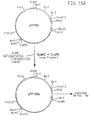


 This oligonucleotide places a termination codon immediately following the six tandem His residues.


 and
and