EP1411120A1 - Dna arrays for measuring sensitivity to anticancer agent - Google Patents

Dna arrays for measuring sensitivity to anticancer agent Download PDF

Info

Publication number
EP1411120A1
EP1411120A1 EP02741410A EP02741410A EP1411120A1 EP 1411120 A1 EP1411120 A1 EP 1411120A1 EP 02741410 A EP02741410 A EP 02741410A EP 02741410 A EP02741410 A EP 02741410A EP 1411120 A1 EP1411120 A1 EP 1411120A1
Authority
EP
European Patent Office
Prior art keywords
gene
genes
fragments
anticancer agent
seq
Prior art date
Legal status (The legal status is an assumption and is not a legal conclusion. Google has not performed a legal analysis and makes no representation as to the accuracy of the status listed.)
Granted
Application number
EP02741410A
Other languages
German (de)
French (fr)
Other versions
EP1411120B1 (en
EP1411120A4 (en
Inventor
Teiji Takechi
Katsuhisa Koizumi
Atsushi Azuma
Masakazu Fukushima
Current Assignee (The listed assignees may be inaccurate. Google has not performed a legal analysis and makes no representation or warranty as to the accuracy of the list.)
Taiho Pharmaceutical Co Ltd
Original Assignee
Taiho Pharmaceutical Co Ltd
Priority date (The priority date is an assumption and is not a legal conclusion. Google has not performed a legal analysis and makes no representation as to the accuracy of the date listed.)
Filing date
Publication date
Application filed by Taiho Pharmaceutical Co Ltd filed Critical Taiho Pharmaceutical Co Ltd
Publication of EP1411120A1 publication Critical patent/EP1411120A1/en
Publication of EP1411120A4 publication Critical patent/EP1411120A4/en
Application granted granted Critical
Publication of EP1411120B1 publication Critical patent/EP1411120B1/en
Anticipated expiration legal-status Critical
Expired - Lifetime legal-status Critical Current

Links

Images

Classifications

    • CCHEMISTRY; METALLURGY
    • C12BIOCHEMISTRY; BEER; SPIRITS; WINE; VINEGAR; MICROBIOLOGY; ENZYMOLOGY; MUTATION OR GENETIC ENGINEERING
    • C12QMEASURING OR TESTING PROCESSES INVOLVING ENZYMES, NUCLEIC ACIDS OR MICROORGANISMS; COMPOSITIONS OR TEST PAPERS THEREFOR; PROCESSES OF PREPARING SUCH COMPOSITIONS; CONDITION-RESPONSIVE CONTROL IN MICROBIOLOGICAL OR ENZYMOLOGICAL PROCESSES
    • C12Q1/00Measuring or testing processes involving enzymes, nucleic acids or microorganisms; Compositions therefor; Processes of preparing such compositions
    • C12Q1/68Measuring or testing processes involving enzymes, nucleic acids or microorganisms; Compositions therefor; Processes of preparing such compositions involving nucleic acids
    • C12Q1/6876Nucleic acid products used in the analysis of nucleic acids, e.g. primers or probes
    • C12Q1/6883Nucleic acid products used in the analysis of nucleic acids, e.g. primers or probes for diseases caused by alterations of genetic material
    • C12Q1/6886Nucleic acid products used in the analysis of nucleic acids, e.g. primers or probes for diseases caused by alterations of genetic material for cancer
    • CCHEMISTRY; METALLURGY
    • C12BIOCHEMISTRY; BEER; SPIRITS; WINE; VINEGAR; MICROBIOLOGY; ENZYMOLOGY; MUTATION OR GENETIC ENGINEERING
    • C12QMEASURING OR TESTING PROCESSES INVOLVING ENZYMES, NUCLEIC ACIDS OR MICROORGANISMS; COMPOSITIONS OR TEST PAPERS THEREFOR; PROCESSES OF PREPARING SUCH COMPOSITIONS; CONDITION-RESPONSIVE CONTROL IN MICROBIOLOGICAL OR ENZYMOLOGICAL PROCESSES
    • C12Q1/00Measuring or testing processes involving enzymes, nucleic acids or microorganisms; Compositions therefor; Processes of preparing such compositions
    • C12Q1/68Measuring or testing processes involving enzymes, nucleic acids or microorganisms; Compositions therefor; Processes of preparing such compositions involving nucleic acids
    • C12Q1/6813Hybridisation assays
    • C12Q1/6834Enzymatic or biochemical coupling of nucleic acids to a solid phase
    • C12Q1/6837Enzymatic or biochemical coupling of nucleic acids to a solid phase using probe arrays or probe chips
    • CCHEMISTRY; METALLURGY
    • C12BIOCHEMISTRY; BEER; SPIRITS; WINE; VINEGAR; MICROBIOLOGY; ENZYMOLOGY; MUTATION OR GENETIC ENGINEERING
    • C12QMEASURING OR TESTING PROCESSES INVOLVING ENZYMES, NUCLEIC ACIDS OR MICROORGANISMS; COMPOSITIONS OR TEST PAPERS THEREFOR; PROCESSES OF PREPARING SUCH COMPOSITIONS; CONDITION-RESPONSIVE CONTROL IN MICROBIOLOGICAL OR ENZYMOLOGICAL PROCESSES
    • C12Q2600/00Oligonucleotides characterized by their use
    • C12Q2600/106Pharmacogenomics, i.e. genetic variability in individual responses to drugs and drug metabolism

Definitions

  • the present invention relates to a DNA array for measuring sensitivity to an anticancer agent, and to a method for measuring such sensitivity by use of the DNA array.
  • anticancer agents are not necessarily satisfactory in terms of therapeutic effects on cancer patients, permitting relatively high incidences of adverse side effects.
  • demand has arisen for proper use of anticancer agents and treatment that is custom-tailored to the conditions of the individual patient, wherein before administration of an anticancer agent to a patient, a tissue specimen or a similar specimen is collected from the patient, and while the specimen is used as a test sample, the expression level of a gene associated with sensitivity to the anticancer agent is measured. This allows proper selection of patients who would be expected to exhibit satisfactory effect of the drug or a low level of adverse side effect.
  • Typical target genes for serving assay are genes of thymidylate synthase (EC2.1.1.45, hereinafter referred to as "TS”), dihydropyrimidine dehydrogenase (EC1.3.1.2, hereinafter referred to as “DPD”), and thymidine phosphorylase (EC2.4.2.4, hereinafter referred to as "TP”), which are considered to be associated with sensitivity to this antimetabolite.
  • TS thymidylate synthase
  • DPD dihydropyrimidine dehydrogenase
  • TP thymidine phosphorylase
  • the DNA microarray method has recently come into widely use, as it enables simultaneous analysis of mRNA expression of several hundred to some dozens of thousands of diversified genes. This technique is suitable for comprehensive analysis of genes, and thus is expected to be applied to realization of proper use of anticancer agents in the future.
  • Biochemical assays for measuring enzyme activity, immunoassays making use of antibodies, Northern hybridization, and RT-PCR mRNA assays provide quantitatively precise data. However, in principle, they require one assay for each gene, and therefore, they are not suitable for simultaneous analysis of diversified modes of gene expression.
  • the DNA microarray method is useful for the analysis of diversified gene expressions.
  • this method is far inferior to Northern hybridization or RT-PCR in terms of quantitative preciseness.
  • Northern hybridization and RT-PCR can designate, based on the size of RNA or PCR product, the specificity to a specific gene of interest, whereas the DNA microarray method cannot, because this method relies on dot blots which do not show the molecular size, leading to false detection; i.e., detection of non-specific signals attributed to expression of genes other than the gene of interest (so-called cross-hybridization).
  • Specificity to a gene of interest is affected by the selection of DNA fragments to be arrayed (hereinafter such DNA fragments will be referred to as the target fragments); i.e., selection of portions (regions) of the gene (full-length cDNA) as target fragments.
  • Specificity obtained in an ordinarily performed DNA microarray method has not yet been substantiated, although the target fragments employed in the method are determined among the regions that have been predicted to have high specificity from computation (regions showing minor nucleotide sequence overlapping with corresponding sequences of other genes which are publicly available from databases) by use of design assistance software.
  • primers which have specificity to each individual gene employed in PCR may migrate to a target fragment solution and be included in arrays, raising the risk of causing non-uniform background. From these factors, even for a single specimen (a sample derived form a living organism), expression level of a specific gene analyzed through the DNA microarray method often disagrees with the results obtained through Northern hybridization or RT-PCR, and therefore, it is improper to conclude existence of a difference in gene expression solely from the results obtained from the DNA microarray method.
  • genes which are likely to show different levels of expression are roughly identified through a first screening by use of the DNA microarray method, and then, through a second screening, like Northern hybridization or RT-PCR is performed to verify that differences in expression level in fact exist.
  • a second screening like Northern hybridization or RT-PCR is performed to verify that differences in expression level in fact exist.
  • at least 2 assay methods must be employed.
  • an object of the present invention is to provide means for measuring, in single measurement, sensitivity to an antimetabolite-type anticancer agent or to a combination of such an anticancer agent and another anticancer agent through a simple assay with high sensitivity and better quantitativeness.
  • the present inventors have studied whether sensitivity to an anticancer agent can be determined through a single measurement by use of the widely employed existing DNA microarray method, and have identified the following problems.
  • the quantity (concentration) of DNA to be spotted on a support carrier e.g., nylon membrane, glass plate
  • a support carrier e.g., nylon membrane, glass plate
  • the present inventors have set out to improve the existing DNA microarray method. Firstly, they have narrowed the range of target genes to dozens to several hundreds of genes centering on those which are considered to be associated with mechanisms of action of an antimetabolite-type anticancer agents and other anticancer agents to be used in combination therewith. Next, through a homology-based search performed using databases, not only those genes determined through calculation but also those actually confirmed to have specificity through Northern hybridization, are determined to be target fragments.
  • the thus-created DNA array in which fragments selected as described above are immobilized on the substrate has been found to be capable of providing judgments, by a single test with high sensitivity, with respect to an antimetabolite-type anticancer agent or a combination use of such an anticancer agent and another anticancer agent, thus leading to the present invention.
  • the present invention provides a DNA array for measuring sensitivity to an antimetabolite-type anticancer agent or to a combination of such an anticancer agent and another anticancer agent, characterized by comprising a substrate and target gene fragments attached to the substrate, the target gene fragments being of at least 13 gene species, including at least 2 species from each of the following gene groups: a group of genes coding for nucleic-acid-metabolism-related enzymes, a group of genes coding for gene-repair-associated enzymes, a group of genes coding for drug resistance-associated factors, and housekeeping genes, and said at least 13 gene species being selected by performing the following two steps 1) and 2):
  • the present invention also provides a method for measuring sensitivity of a body fluid specimen or tissue specimen of a cancer patient to an antimetabolite-type anticancer agent or to a combination of such an anticancer agent and another anticancer agent, characterized by comprising hybridizing the DNA array with labeled cDNA probes synthesized through use, as a template, of mRNA obtained from the specimen.
  • the DNA array of the present invention is contemplated to be used in determining sensitivity to an antimetabolite serving as an anticancer agent (may be referred to as an antimetabolite-type anticancer agent), or in determining sensitivity to a combination of such an anticancer agent and another anticancer agent.
  • antimetabolite-type anticancer agent examples include 5-fluorouracil anticancer agents such as tegafur, 5-fluorouracil, tegafur ⁇ uracil, tegafur ⁇ gimeracil ⁇ oteracil potassium, carmofur, capecitabine, and furtulon; mercaptopurine anticancer agents such as 6-mercaptopurine and mercaptopurine riboside; anticancer cytosine analogues such as cytarabine, enocitabine, and gemcitabine; and methotrexate. Of these, 5-fluorouracil anticancer agents are particularly preferred.
  • another anticancer agent examples include platinum-complex-based anticancer agents such as cisplatin; topoisomerase inhibitors such as CPT-11 and VP-16; and taxane anticancer agents such as docetaxel and paclitaxel.
  • the measurement of sensitivity refers to determination or judgment in terms of balance between efficacy of the anticancer agent on a patient and adverse side effects; decision of appropriate combination therapy, determination of appropriate administration scheme (dose, drug administration regimen), etc.
  • the target genes of the present invention are those which are considered to be associated with sensitivity, and consist of at least 13 species of gene, including at least two different species from each of the following groups: a group of genes coding for nucleic acid metabolism-related enzymes, a group of genes coding for gene-repair-associated enzymes, a group of genes coding for drug resistance-related factors, and a group of housekeeping genes.
  • nucleic acid metabolism-related enzymes examples include thymidylate synthase (TS), dihydropyrimidine dehydrogenase (DPD), orotate phosphoribosyltransferase (OPRT) (uridine monophosphate synthetase (UMPS)), thymidine phosphorylase (TP), thymidine kinase 1 (TK1), ribonucleosidediphosphate reductase M1 subunit (RRM1), ribonucleosidediphosphate reductase M2 subunit (RRM2), uridine cytidine kinase 2 (UCK2), uridine phosphorylase (UP), cytidine deaminase (CDA), 5'nucleotidase (NT5), IMP dehydrogenase 1 (IMPD), methylenetetrahydrofolate dehydrogenase (MTHFD1), RNA polymerase 2 (RP2)
  • Examples of the gene-repair-associated enzymes include DNA excision repair protein ERCC1 (ERCC1), uracil-DNA glycosylase (UDG), poly(ADP-ribose) polymerase (PARP), DNA ligase I (LIG1), DNA ligase III (LIG3), DNA ligase IV (LIG4), DNA polymerase ⁇ (POLB), DNA polymerase ⁇ (FOLD), and DNA-repair protein XRCC1 (XRCC1).
  • ERCC1 DNA excision repair protein ERCC1
  • UDG uracil-DNA glycosylase
  • PARP poly(ADP-ribose) polymerase
  • LIG1 DNA ligase I
  • LIG3 DNA ligase III
  • LIG4 DNA ligase IV
  • POLB DNA polymerase ⁇
  • FOLD DNA-repair protein XRCC1
  • XRCC1 DNA-repair protein XRCC1
  • Examples of drug resistance-related factors include topoisomerase 1 (TOP1), P-glycoprotein (MDR1), equilibrative nucleoside transporter 1 (ENT1), multidrug resistance-associated protein 1 (MRP1), topoisomerase 2 ⁇ (TOP2A), topoisomerase 2 ⁇ (TOPB), heatshockprotein 27 (Hsp27), and equilibrative nucleoside transporter 2 (ENT2).
  • TOP1 topoisomerase 1
  • MDR1 P-glycoprotein
  • ENT1 equilibrative nucleoside transporter 1
  • MRP1 multidrug resistance-associated protein 1
  • TOP1, MDR1, ENT1, and MRP1 are employed.
  • housekeeping genes examples include genes coding for glyceraldehyde-3-phosphate dehydrogenase (GAPDH), ⁇ -actin (ACTB), and 40S ribosomal protein S9 (RSP9).
  • GAPDH glyceraldehyde-3-phosphate dehydrogenase
  • ACTB ⁇ -actin
  • RSP9 40S ribosomal protein S9
  • GAPDH glyceraldehyde-3-phosphate dehydrogenase
  • ACTB ⁇ -actin
  • RSP9 40S ribosomal protein S9
  • Example of other genes include those coding for E2F1, p53, VEGF ⁇ , integrin ⁇ 3, Mn SOD, Cu/Zn SOD, or proliferating cell nuclear antigen (PCNA).
  • PCNA proliferating cell nuclear antigen
  • fragments from all the mentioned target genes are immobilized.
  • fragments of at least 13 different genes in total be immobilized, including the following 11 genes; i.e., genes coding for TS, DPD, OPRT, TP, TK1, ERCC1, UDG, TOP1, MDR1, ENT1, and MRP1, as well as two or more of the genes coding for GAPDH, ACTB, or RSP9.
  • a homology search is performed utilizing databases, whereby fragments which have high specificity to respective target genes are selected (Step 1).
  • RNA obtained from tumor cells are subjected to Northern hybridization, using the fragments designed in step 1, whereby specificity is confirmed (step 2).
  • the fragments designed in step 1 have high specificity to target genes.
  • cross-hybridization occurs at high probability. In fact, only 10 to 20% of the fragments selected in step 1 are confirmed to have specificity in step 2.
  • step 2 firstly, PCR amplification is performed through use of a cDNA library as a template, whereby a DNA fragment is obtained.
  • radioactive probes are enzymatically synthesized, and by use of a membrane bearing blots of total RNA samples prepared from various tumor cells, Northern hybridization is performed, whereby specificity is checked. Briefly, presence of specificity is confirmed when a signal that corresponds to the size of mRNA transcribed from a certain gene is detected (the size information can be obtained from literature or database searches) and almost no other signals are detected (which means almost no cross-hybridization).
  • a DNA fragment is found to have insufficient specificity, by use of another region of a corresponding gene another DNA fragment is designed, and then the re-designed fragment is subjected to Northern hybridization. This procedure is repeated until sufficient specificity is obtained. Thus, regions of the DNA fragments to be arrayed can be optimized. Each of the thus-obtained target fragments is cloned into a plasmid (pCR2.1-TOPO).
  • universal primers are designed on the basis of the sequence of multiple cloning site of a cloning vector, so that all the target fragments can be amplified by use of a set of universal primers.
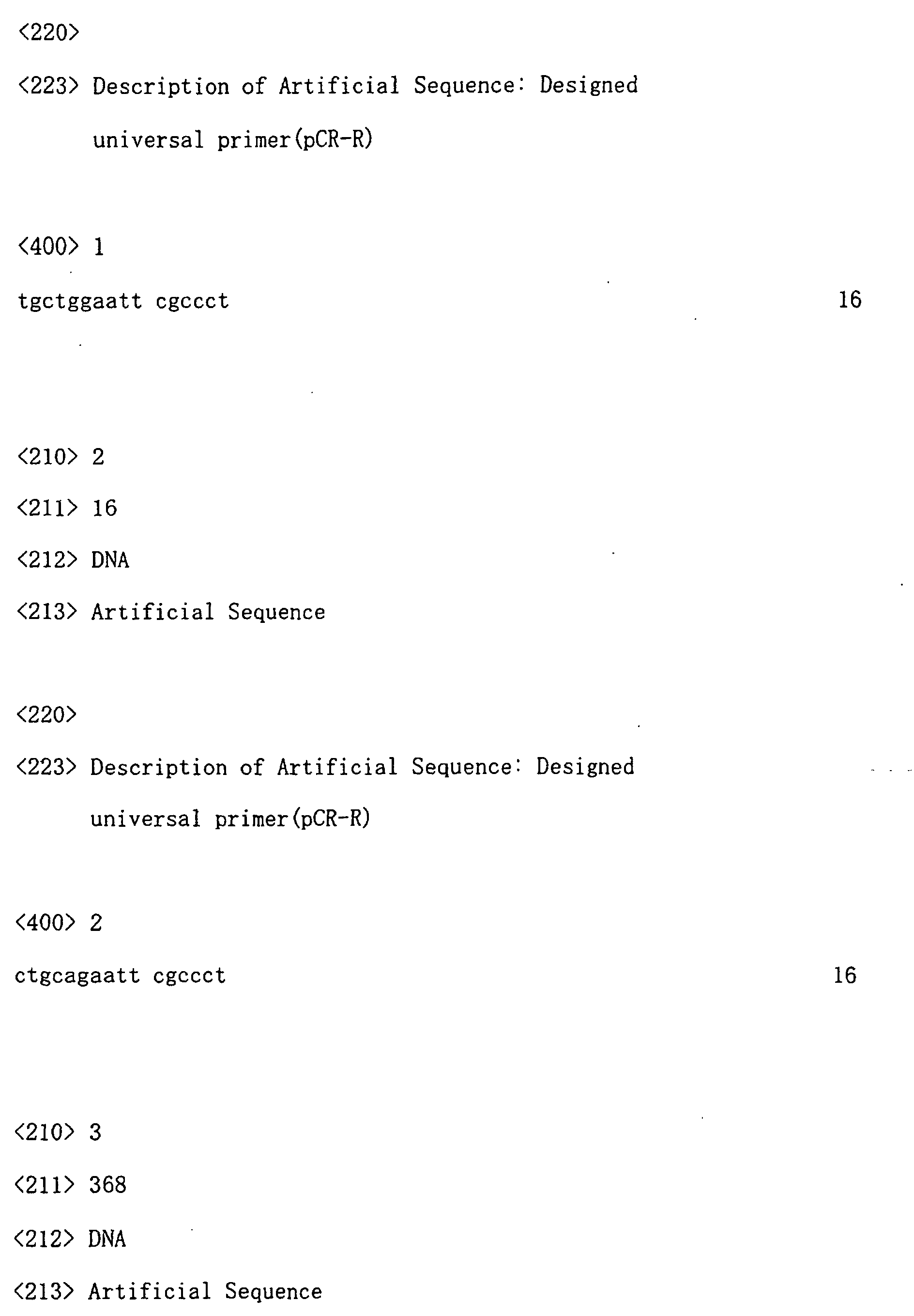
  • Especially preferred universal primers are those having the nucleotide sequences of SEQ ID NOs: 1 and 2.
  • the universal primer volume is optimized. When these measures are taken, background of every spot from the target fragments can be suppressed, with reduced differences.
  • the DNA array of the present invention can be obtained.
  • the substrate used herein is any suitable one known per se .
  • a glass plate is used.
  • Immobilization of the fragments onto the array may be achieved by use of conventional spotting means.
  • the ink-jet piezoelectric discharge method in which a solution containing the target DNA is jetted onto a support, under the same principle that used in an ink jet printer; and the photolithograph methodology, which makes use of photolithography techniques and the target DNA is directly synthesized on a support.
  • the Stanford method is used.
  • mRNA of all the target genes is quantitated, and this quantitation can be achieved by modifying the amount of a target fragment to be immobilized in accordance with each gene's expression level obtained through Northern hybridization; i.e., the amount of target fragments to be immobilized on the substrate (support) is reduced for a gene of higher expression level, and increased for a gene of lower expression level.
  • the DNA array is hybridized with labeled cDNA probes synthesized by using, as a template, mRNA obtained from a body fluid specimen or tissue specimen collected form a cancer patient.
  • Examples of the body fluid specimen originating from a cancer patient include blood and urine.
  • Examples of the tissue include cancerous tissue. Collection of mRNA from a specimen is carried out through a conventional method, and mRNA may be in the form of "as contained" in total RNA, or may be isolated from the total RNA.
  • To prepare the labeled cDNA mRNA is used as a template, and reverse transcription enzyme reaction is performed for labeling.
  • Examples of labeling means include fluorescent substances and radioisotopes, with fluorescent substances being especially preferred.
  • Hybridization may be carried out under conventional conditions. Quantitation of hybridization may be achieved through quantitating the amount of labeled probes; for example, intensity of fluorescence.
  • the measurements in terms of expression level of 52 species of genes were compared with the measurements obtained from Northern hybridization. As a result, almost no discrepancy was found. Discernment of a gene that shows a unique expression characteristic (i.e., a gene exhibiting an expression level twice or more the average expression level) was able to be obtained at the same precision as attained by Northern hybridization.
  • Fig. 2 shows some of the test results, using XRCC1 and E2F1.
  • XRCC1 a region of 898 to 1265 nt exhibited good results (strong signals were detected at a position corresponding to the size of mRNA of the gene of interest), and a region of 187 to 494 nt was determined not to be suitable, since a large number of signals indicating cross-hybridization were observed.
  • E2F1 a region of 1014 to 1309 nt exhibited good results, and a region of 788 to 1087 nt was determined not to be suitable.
  • regions of other genes were investigated, various levels of specificity were also observed, although the levels differed from gene to gene.
  • a DNA fragment which exhibited the highest specificity in DNA fragments of gene of interest was selected in the final step and was employed as a target fragment.
  • RNA sample derived from each of the above 14 types of human tumor cells was electrophoresed on a denatured agarose gel and blotted on a nylon membrane.
  • Northern hybridization was performed through use of probes synthesized from target DNA of GAPDH (glyceraldehydes-3-phosphate dehydrogenase) (through the method described in Example 1(2)). Analysis was performed by means of imaging and analysis software (ImageQuant, Molecular Dynamics Inc), and intensity of GAPDH mRNA signals was measured.
  • universal primers forward (pCR-F)/reverse (pCR-R)
  • pCR-F forward
  • pCR-R reverse
  • target fragments can be readily provided in sufficient amounts as compared with the case of specific primers.
  • the mean value, standard deviation (SD), and coefficient of variation (CV) of signal intensities obtained from the universal primers (5 spots) were found to be 27.3, 6.5, and 24.0%, respectively.
  • the mean value, standard deviation (SD), and coefficient of variation (CV) of signal intensities obtained from the specific primers (5 spots in total) were found to be 22.9, 12.0, and 52.4%, respectively.
  • Signal intensities from both primers were found to be of substantially the same level, and signal intensities obtained from the universal primers had lower SD and CV, indicating that the universal primers exert less effect on difference in background level than the specific primers.
  • the minimum amount of the universal primers required to amplify a target fragment was studied. PCR was performed through use of forward and reverse primers. The amount of each primer was 10, 20, 30, 40, and 50 pmol. The PCR products were electrophoresed on agarose gel. When primers were used in amounts of 20 pmol or less, the amount of PCR product was found to be reduced. Therefore, the minimum amount of the universal primers required to amplify a target fragment was determined to be 30 pmol.
  • TS is a representative sensitivity regulating factor for 5-FU anticancer agent, and both basic research and clinical research have revealed that the higher the TS expression level, the lower the effect exhibited by a 5-FU anticancer agent.
  • the TS expression level in the 11 species of cells was measured through real time RT-PCR, which is considered to provide considerably high measurement accuracy, to thereby analyze correlation between TS-1 sensitivity and the TS expression level.
  • the present invention achieves convenient and highly quantitative measurement of expression levels of several tens to several hundreds of gene species contained in a specimen in a single measurement.
  • expression patterns of genes related to action mechanisms of an antimetabolite-type anticancer agent or a combination of such an agent and another anticancer agent are analyzed through use of the assay method of the present invention in a test specimen (for example, total RNA extracted from peripheral monocytes or tumor tissue of a cancer patient), results obtained from such an analysis can be employed as indices for proper use of the anticancer agent.

Landscapes

  • Chemical & Material Sciences (AREA)
  • Life Sciences & Earth Sciences (AREA)
  • Health & Medical Sciences (AREA)
  • Organic Chemistry (AREA)
  • Proteomics, Peptides & Aminoacids (AREA)
  • Engineering & Computer Science (AREA)
  • Immunology (AREA)
  • Pathology (AREA)
  • Analytical Chemistry (AREA)
  • Zoology (AREA)
  • Genetics & Genomics (AREA)
  • Wood Science & Technology (AREA)
  • Physics & Mathematics (AREA)
  • Biotechnology (AREA)
  • Microbiology (AREA)
  • Molecular Biology (AREA)
  • Hospice & Palliative Care (AREA)
  • Biophysics (AREA)
  • Oncology (AREA)
  • Biochemistry (AREA)
  • Bioinformatics & Cheminformatics (AREA)
  • General Engineering & Computer Science (AREA)
  • General Health & Medical Sciences (AREA)
  • Measuring Or Testing Involving Enzymes Or Micro-Organisms (AREA)
  • Investigating Or Analysing Biological Materials (AREA)

Abstract

A DNA array for measuring sensitivity to an antimetabolite-type anticancer agent or a combined use of such an anticancer agent and another anticancer agent, characterized by including at least 13 types of target gene fragments, involving at least two types of genes selected from each of the following groups: nucleic acid metabolism-associated enzyme genes, gene repair-associated enzyme genes, drug resistance-associated factor genes and housekeeping genes, wherein these gene fragments have been selected by the following steps 1) and 2) and immobilized on a substrate; 1) a step of selecting fragments having high specificity for target genes by searching the homology with the use of databases; and 2) a step of performing Northern hybridization against RNA obtained from tumor cells with the use of the fragments selected in the step 1) as probes to thereby confirm the specificity for the target genes. Thus, through a single assay procedure, expression of several tens to several hundreds of genes in a specimen can be conveniently measured at a high quantitation level.

Description

Technical Field
The present invention relates to a DNA array for measuring sensitivity to an anticancer agent, and to a method for measuring such sensitivity by use of the DNA array.
Background Art
Generally speaking, anticancer agents are not necessarily satisfactory in terms of therapeutic effects on cancer patients, permitting relatively high incidences of adverse side effects. In recent years, demand has arisen for proper use of anticancer agents and treatment that is custom-tailored to the conditions of the individual patient, wherein before administration of an anticancer agent to a patient, a tissue specimen or a similar specimen is collected from the patient, and while the specimen is used as a test sample, the expression level of a gene associated with sensitivity to the anticancer agent is measured. This allows proper selection of patients who would be expected to exhibit satisfactory effect of the drug or a low level of adverse side effect.
Clinical research efforts have been devoted toward establishing criteria for proper use of 5-fluorouracil (5-FU) agents and other antimetabolites among popular anticancer agents. Typical target genes for serving assay are genes of thymidylate synthase (EC2.1.1.45, hereinafter referred to as "TS"), dihydropyrimidine dehydrogenase (EC1.3.1.2, hereinafter referred to as "DPD"), and thymidine phosphorylase (EC2.4.2.4, hereinafter referred to as "TP"), which are considered to be associated with sensitivity to this antimetabolite. Expression patterns of these genes or enzymes are analyzed through biochemical assays for determining enzymatic activity (Clinical Cancer Research, 5, 883-889, 1999), immunoassays using antibodies (Cancer and Chemotherapy, 24(6): 705-712, 1997), or mRNA assays making use of Northern hybridization or RT-PCR (Clinical Cancer Research, 6, 1322-1327, 2000), and the results therefrom are used as indices of proper use. In cancer chemotherapy using cisplatin, DNA repair enzymes such as ERCC1 are suggested to be useful as factors that predict the effect of therapy (Journal of Clinical Oncology, 16, 309-316, 1998).
Meanwhile, the DNA microarray method has recently come into widely use, as it enables simultaneous analysis of mRNA expression of several hundred to some dozens of thousands of diversified genes. This technique is suitable for comprehensive analysis of genes, and thus is expected to be applied to realization of proper use of anticancer agents in the future.
In the human body, 5-fluorouracil and other antimetabolites are metabolized through a variety of processes. Therefore, only the gene expression analysis of the above-mentioned 3 types of genes (TS, DPD, and TP) is not sufficient for predicting sensitivity to antimetabolites. If gene expression can be analyzed in broader ranges, sensitivity would be better predicted. In actual settings of cancer chemotherapies, combination therapy is commonly performed, in which several types of anticancer agents are used in combination. In such cases, if expression of genes that are associated with sensitivity to each drug can be analyzed comprehensively, suitable combination chemotherapy can be designed for each individual patient.
Biochemical assays for measuring enzyme activity, immunoassays making use of antibodies, Northern hybridization, and RT-PCR mRNA assays provide quantitatively precise data. However, in principle, they require one assay for each gene, and therefore, they are not suitable for simultaneous analysis of diversified modes of gene expression.
In contrast, the DNA microarray method is useful for the analysis of diversified gene expressions. However, this method is far inferior to Northern hybridization or RT-PCR in terms of quantitative preciseness. One of the main reasons may be explained as follows: Northern hybridization and RT-PCR can designate, based on the size of RNA or PCR product, the specificity to a specific gene of interest, whereas the DNA microarray method cannot, because this method relies on dot blots which do not show the molecular size, leading to false detection; i.e., detection of non-specific signals attributed to expression of genes other than the gene of interest (so-called cross-hybridization). Specificity to a gene of interest is affected by the selection of DNA fragments to be arrayed (hereinafter such DNA fragments will be referred to as the target fragments); i.e., selection of portions (regions) of the gene (full-length cDNA) as target fragments. Specificity obtained in an ordinarily performed DNA microarray method has not yet been substantiated, although the target fragments employed in the method are determined among the regions that have been predicted to have high specificity from computation (regions showing minor nucleotide sequence overlapping with corresponding sequences of other genes which are publicly available from databases) by use of design assistance software. Moreover, primers which have specificity to each individual gene employed in PCR, which is performed for preparing target fragments through amplification, may migrate to a target fragment solution and be included in arrays, raising the risk of causing non-uniform background. From these factors, even for a single specimen (a sample derived form a living organism), expression level of a specific gene analyzed through the DNA microarray method often disagrees with the results obtained through Northern hybridization or RT-PCR, and therefore, it is improper to conclude existence of a difference in gene expression solely from the results obtained from the DNA microarray method. According to a generally employed test procedure, genes which are likely to show different levels of expression are roughly identified through a first screening by use of the DNA microarray method, and then, through a second screening, like Northern hybridization or RT-PCR is performed to verify that differences in expression level in fact exist. Thus, in such a scheme, at least 2 assay methods must be employed.
Accordingly, an object of the present invention is to provide means for measuring, in single measurement, sensitivity to an antimetabolite-type anticancer agent or to a combination of such an anticancer agent and another anticancer agent through a simple assay with high sensitivity and better quantitativeness.
Disclosure of the Invention
Under the above circumstances, the present inventors have studied whether sensitivity to an anticancer agent can be determined through a single measurement by use of the widely employed existing DNA microarray method, and have identified the following problems.
Briefly, in use of the widely employed DNA microarray method, since the species of potential target genes counts several hundred to several dozens of thousands, it is considered to include those genes that are irrelevant to sensitivity to drugs, as conjectured from action mechanisms of currently available anticancer agents. As a result, analysis would require extra work, or genes which are closely related to the mechanisms of action of the anticancer agent may be overlooked or given less weight than they should be.
Moreover, in the case of the widely employed DNA microarray method, the quantity (concentration) of DNA to be spotted on a support carrier (e.g., nylon membrane, glass plate) is usually prefixed, and therefore, quantification may sometimes be unsuccessful because of extremely low expression level, despite the gene being closely related to sensitivity to the anticancer agent.
Thus, the present inventors have set out to improve the existing DNA microarray method. Firstly, they have narrowed the range of target genes to dozens to several hundreds of genes centering on those which are considered to be associated with mechanisms of action of an antimetabolite-type anticancer agents and other anticancer agents to be used in combination therewith. Next, through a homology-based search performed using databases, not only those genes determined through calculation but also those actually confirmed to have specificity through Northern hybridization, are determined to be target fragments. The thus-created DNA array in which fragments selected as described above are immobilized on the substrate has been found to be capable of providing judgments, by a single test with high sensitivity, with respect to an antimetabolite-type anticancer agent or a combination use of such an anticancer agent and another anticancer agent, thus leading to the present invention.
Accordingly, the present invention provides a DNA array for measuring sensitivity to an antimetabolite-type anticancer agent or to a combination of such an anticancer agent and another anticancer agent, characterized by comprising a substrate and target gene fragments attached to the substrate, the target gene fragments being of at least 13 gene species, including at least 2 species from each of the following gene groups: a group of genes coding for nucleic-acid-metabolism-related enzymes, a group of genes coding for gene-repair-associated enzymes, a group of genes coding for drug resistance-associated factors, and housekeeping genes, and said at least 13 gene species being selected by performing the following two steps 1) and 2):
  • 1) selecting fragments having high target-gene specificity through a homology search using a database, and
  • 2) performing Northern hybridization to RNA collected from tumor cells, using the fragments selected in step 1) as probes, to thereby confirm target-gene specificity.
  • The present invention also provides a method for measuring sensitivity of a body fluid specimen or tissue specimen of a cancer patient to an antimetabolite-type anticancer agent or to a combination of such an anticancer agent and another anticancer agent, characterized by comprising hybridizing the DNA array with labeled cDNA probes synthesized through use, as a template, of mRNA obtained from the specimen.
    Brief Description of the Drawings
  • Fig. 1 shows universal primer sequences and the structure of a clone to which a target fragment is incorporated.
  • Fig. 2 contains actual images obtained from Northern hybridization, showing that in some cases different specificities result when DNA fragments serving as templates in the synthesis of probes are determined in different regions of a gene.
  • Fig. 3 shows the correlation between total RNA quantity and the measurements obtained in Northern hybridization.
  • Fig. 4 contains charts showing diagnosis quality provided by the array of the present invention, assessed in comparison with Northern hybridization.
  • Fig. 5 shows the correlation between TS-1 sensitivity and expression level, found in 11 types of xenografts, and contains graphs in connection with four genes which among 52 types of genes exhibited higher correlation.
  • Fig. 6 shows the correlation between TS-1 sensitivity and TS expression level, found in the 11 types of xenografts.
  • Best Mode for Carrying Out the Invention
    The DNA array of the present invention is contemplated to be used in determining sensitivity to an antimetabolite serving as an anticancer agent (may be referred to as an antimetabolite-type anticancer agent), or in determining sensitivity to a combination of such an anticancer agent and another anticancer agent. Examples of the antimetabolite-type anticancer agent include 5-fluorouracil anticancer agents such as tegafur, 5-fluorouracil, tegafur·uracil, tegafur·gimeracil·oteracil potassium, carmofur, capecitabine, and furtulon; mercaptopurine anticancer agents such as 6-mercaptopurine and mercaptopurine riboside; anticancer cytosine analogues such as cytarabine, enocitabine, and gemcitabine; and methotrexate. Of these, 5-fluorouracil anticancer agents are particularly preferred. Examples of the mentioned "another anticancer agent" to be used in combination with the antimetabolites include platinum-complex-based anticancer agents such as cisplatin; topoisomerase inhibitors such as CPT-11 and VP-16; and taxane anticancer agents such as docetaxel and paclitaxel.
    The measurement of sensitivity, as used in the present invention, refers to determination or judgment in terms of balance between efficacy of the anticancer agent on a patient and adverse side effects; decision of appropriate combination therapy, determination of appropriate administration scheme (dose, drug administration regimen), etc.
    The target genes of the present invention are those which are considered to be associated with sensitivity, and consist of at least 13 species of gene, including at least two different species from each of the following groups: a group of genes coding for nucleic acid metabolism-related enzymes, a group of genes coding for gene-repair-associated enzymes, a group of genes coding for drug resistance-related factors, and a group of housekeeping genes.
    Examples of the nucleic acid metabolism-related enzymes include thymidylate synthase (TS), dihydropyrimidine dehydrogenase (DPD), orotate phosphoribosyltransferase (OPRT) (uridine monophosphate synthetase (UMPS)), thymidine phosphorylase (TP), thymidine kinase 1 (TK1), ribonucleosidediphosphate reductase M1 subunit (RRM1), ribonucleosidediphosphate reductase M2 subunit (RRM2), uridine cytidine kinase 2 (UCK2), uridine phosphorylase (UP), cytidine deaminase (CDA), 5'nucleotidase (NT5), IMP dehydrogenase 1 (IMPD), methylenetetrahydrofolate dehydrogenase (MTHFD1), RNA polymerase 2 (RP2), uridine monophosphate kinase (UMPK), CTP synthase (CTPS), deoxycytidylate deaminase (DCD), deoxycytidine kinase (DCK), phosphoribosyl pyrophosphate synthetase (PRPS), hypoxanthine phosphoribosyltransferase 1 (HPRT1), folylpolyglutamate synthetase (FPGS), nucleoside diphosphate kinase A (NDKA), nucleoside diphosphate kinase B (NDKB), adenine phosphoribosyltransferase (APRT), and adenosine kinase (AK). Among the genes coding for these enzymes, preferably, at least genes coding for TS, DPD, ORRT, TP, and TK1 are employed.
    Examples of the gene-repair-associated enzymes include DNA excision repair protein ERCC1 (ERCC1), uracil-DNA glycosylase (UDG), poly(ADP-ribose) polymerase (PARP), DNA ligase I (LIG1), DNA ligase III (LIG3), DNA ligase IV (LIG4), DNA polymerase β (POLB), DNA polymerase δ (FOLD), and DNA-repair protein XRCC1 (XRCC1). Among the genes coding for these enzymes, preferably, at least genes coding for ERCC1 and UDG are employed.
    Examples of drug resistance-related factors include topoisomerase 1 (TOP1), P-glycoprotein (MDR1), equilibrative nucleoside transporter 1 (ENT1), multidrug resistance-associated protein 1 (MRP1), topoisomerase 2α (TOP2A), topoisomerase 2β (TOPB), heatshockprotein 27 (Hsp27), and equilibrative nucleoside transporter 2 (ENT2). Among the genes coding for these factors, preferably, at least genes coding for TOP1, MDR1, ENT1, and MRP1 are employed.
    Examples of the housekeeping genes include genes coding for glyceraldehyde-3-phosphate dehydrogenase (GAPDH), β-actin (ACTB), and 40S ribosomal protein S9 (RSP9). Preferably, at least two out of GAPDH gene, ACTB gene, and RSP9 gene are employed. These genes are used as internal standards.
    Example of other genes include those coding for E2F1, p53, VEGF β, integrin α3, Mn SOD, Cu/Zn SOD, or proliferating cell nuclear antigen (PCNA).
    Desirably, fragments from all the mentioned target genes are immobilized. In view of ensuring achievement of more efficient measurement of sensitivity, it is particularly preferred that fragments of at least 13 different genes in total be immobilized, including the following 11 genes; i.e., genes coding for TS, DPD, OPRT, TP, TK1, ERCC1, UDG, TOP1, MDR1, ENT1, and MRP1, as well as two or more of the genes coding for GAPDH, ACTB, or RSP9.
    Regarding sequences of these target genes, a homology search is performed utilizing databases, whereby fragments which have high specificity to respective target genes are selected (Step 1).
    The homology search may be carried out by use of, for example, Blast Search. Fragments may be designed as follows: GC content = 40 to 60%; reduced number of duplicate sequences; Tm = 75 to 85°C, size = 200 to 600 bp.
    RNA obtained from tumor cells are subjected to Northern hybridization, using the fragments designed in step 1, whereby specificity is confirmed (step 2). Theoretically, the fragments designed in step 1 have high specificity to target genes. However, in reality, cross-hybridization occurs at high probability. In fact, only 10 to 20% of the fragments selected in step 1 are confirmed to have specificity in step 2.
    In step 2, firstly, PCR amplification is performed through use of a cDNA library as a template, whereby a DNA fragment is obtained. Next, while the thus-obtained DNA fragment is used as a template, radioactive probes are enzymatically synthesized, and by use of a membrane bearing blots of total RNA samples prepared from various tumor cells, Northern hybridization is performed, whereby specificity is checked. Briefly, presence of specificity is confirmed when a signal that corresponds to the size of mRNA transcribed from a certain gene is detected (the size information can be obtained from literature or database searches) and almost no other signals are detected (which means almost no cross-hybridization).
    If a DNA fragment is found to have insufficient specificity, by use of another region of a corresponding gene another DNA fragment is designed, and then the re-designed fragment is subjected to Northern hybridization. This procedure is repeated until sufficient specificity is obtained. Thus, regions of the DNA fragments to be arrayed can be optimized. Each of the thus-obtained target fragments is cloned into a plasmid (pCR2.1-TOPO).
    In this connection, as primers to be used in PCR amplification of target fragments, universal primers are designed on the basis of the sequence of multiple cloning site of a cloning vector, so that all the target fragments can be amplified by use of a set of universal primers. Especially preferred universal primers are those having the nucleotide sequences of SEQ ID NOs: 1 and 2. Also, in order to minimize the volume of primers that migrate into the spotting solution, the universal primer volume is optimized. When these measures are taken, background of every spot from the target fragments can be suppressed, with reduced differences.
    When the thus-obtained target fragments of respective target genes are immobilized onto a substrate (support), the DNA array of the present invention can be obtained. The substrate used herein is any suitable one known per se. For example, mention may be given of glass plate, plastic plate, membrane (nylon membrane, etc.), and beads. Preferably, a glass plate is used. Immobilization of the fragments onto the array may be achieved by use of conventional spotting means. For example, there may be employed the surface adhesion method (the Stanford method) as described in PCT Kohyo Publication No. 10-503841, in which a tip-split pin is dipped into a solution containing the target DNA, and tapped onto a support for transfer; the ink-jet piezoelectric discharge method, in which a solution containing the target DNA is jetted onto a support, under the same principle that used in an ink jet printer; and the photolithograph methodology, which makes use of photolithography techniques and the target DNA is directly synthesized on a support. Preferably, the Stanford method is used.
    Preferably, mRNA of all the target genes is quantitated, and this quantitation can be achieved by modifying the amount of a target fragment to be immobilized in accordance with each gene's expression level obtained through Northern hybridization; i.e., the amount of target fragments to be immobilized on the substrate (support) is reduced for a gene of higher expression level, and increased for a gene of lower expression level.
    In order to measure the sensitivity to the aforementioned anticancer agents by use of the thus-obtained DNA array of the present invention, the DNA array is hybridized with labeled cDNA probes synthesized by using, as a template, mRNA obtained from a body fluid specimen or tissue specimen collected form a cancer patient.
    Examples of the body fluid specimen originating from a cancer patient include blood and urine. Examples of the tissue include cancerous tissue. Collection of mRNA from a specimen is carried out through a conventional method, and mRNA may be in the form of "as contained" in total RNA, or may be isolated from the total RNA. To prepare the labeled cDNA, mRNA is used as a template, and reverse transcription enzyme reaction is performed for labeling. Examples of labeling means include fluorescent substances and radioisotopes, with fluorescent substances being especially preferred.
    Hybridization may be carried out under conventional conditions. Quantitation of hybridization may be achieved through quantitating the amount of labeled probes; for example, intensity of fluorescence.
    In order to validate the quantitativeness of the DNA array of the present invention, the measurements in terms of expression level of 52 species of genes were compared with the measurements obtained from Northern hybridization. As a result, almost no discrepancy was found. Discernment of a gene that shows a unique expression characteristic (i.e., a gene exhibiting an expression level twice or more the average expression level) was able to be obtained at the same precision as attained by Northern hybridization.
    Examples
    The present invention will next be described in more detail by way of examples, which should not be construed as limiting the invention thereto.
    Example 1 (1) Preparation of target fragments
  • A. Selection and design of DNA fragments As shown in Tables 2 and 3 below, 52 species of DNA fragments were selected as DNA fragments relating to 5-FU sensitivity, primarily from nucleic acid metabolism-related genes. In principle, the DNA fragments were designed such that each DNA fragment has a GC content of 40 to 60%, has almost no regions having the same nucleotide sequence as that of other DNA fragments of the same gene, has a Tm of 75 to 85°C, and has a size of 200 to 600 bp. In order to prevent cross-hybridization to the greatest possible extent, whether or not each of the DNA fragments had substantially no regions having the same nucleotide sequences as those of other DNA fragments in the same gene was investigated through a blast search by use of a database. By use of design-aiding software (Primer Express™, PE Biosystems), primers specific to each DNA fragment (specific primers [forward/reverse]), which were employed in PCR amplification, were designed so as to have a Tm of 59 to 61°C.
  • B. Synthesis and purification of DNA fragments Each DNA fragment was amplified through PCR (30 cycles of treatment in total, each cycle consisting of thermal denaturation (94°C, 1 minute), annealing (60°C, 1 minute), and elongation (72°C, 1 minute)) while a human-derived cDNA library was used as a template, and by use of specific primers and an ExTaq™ (TaKaRa). The PCR product solution was subjected to purification through use of a spin column (Miniprep spin column, Aetna), and the purified product was eluted with distilled water.
  • (2) Northern hybridization
  • A. Preparation and purification of total RNA samples A total RNA sample was extracted from each of the 14 types of human tumor cells listed in Table 1, through use of an RNeasy midi kit™ (QIAGEN) and eluted by distilled water. DNA fragments which might migrate in the solution were decomposed with DNase (37°C, 30 minutes), and proteins were removed by use of phenol/chloroform. The resultant solution was subjected to purification and condensation through ethanol precipitation, and the product was dissolved in distilled water, to thereby prepare a total RNA solution.
    Tumor type Cell strain
    1 Human gastric cancer NUGC-3
    2 Human gastric cancer (FU-resistant) NUGC-3/FU
    3 Human large intestine cancer DLD-1
    4 Human large intestine cancer (FU-resistant) DLD-1/FU
    5 Human large intestine cancer (FdUrd resistant) DLD-1/FdUrd
    6 Human large intestine cancer (F3dThd-resistant) DLD-1/F3dThd
    7 Human fibrosarcoma HT1080
    8 Human fibrosarcoma (EUrd-resistant) HT1080/EUrd
    9 Human pancreatic carcinoma MIAPaCa-2
    10 Human large intestine cancer KM-12C
    11 Human lung cancer A549
    12 Human mammary cancer MCF-7
    13 Human head and neck cancer KB
    14 Human ovarian cancer TYK-nu
  • B. Northern blotting The total RNA sample (5 µg) which had been prepared from each of the above 14 types of human tumor cells was electrophoresed on 1-wt% denatured agarose gel. The gel was stained with ethidium bromide, and the resultant gel was photographed under irradiation of UV rays, to thereby confirm that the RNA molecules had not been decomposed. The RNA molecules in the gel were blotted on a nylon membrane through a capillary phenomenon, and the membrane was subjected to fixation (cross-linking) under irradiation with UV rays.
  • C. Preparation of probes Probes labeled with [α-32P]dCTP were synthesized by use of each of the DNA fragments prepared in (1) above as a template, through random priming (rediprime™ II, Amersham Pharmacia). [α-32P]dCTP which had not been incorporated into the probes was removed through gel filtration (ProbeQuant™ G-50 micro spin column, Amersham Pharmacia).
  • D. Northern hybridization Northern hybridization was performed by use of the blot and the probes prepared in (2)B and (2)C above, respectively. Specifically, pre-hybridization was performed by use of the blot in a hybridization buffer (Rapid-hyb Buffer, Amersham Pharmacia) for 30 minutes at 65°C, the probes which had been undergone thermal denaturation were added thereto, and hybridization was caused to proceed for two hours at 65°C. Subsequently, the resultant blot was washed (twice with a 2×SSC solution (0.15-mol/L NaCl / 0.15-mol/L trisodium citrate) containing 0.1-wt% SDS, once with a 1×SSC solution containing 0.1-wt% SDS, and twice with a 0.1×SSC solution containing 0.1-wt% SDS), and, in the dark, an imaging plate (Fujifilm) was exposed with the blot overnight. On the following day, the imaging plate was scanned by means of an imaging and analysis apparatus (STORM, Molecular Dynamics Inc.), and the obtained image data were stored.
  • E. Evaluation and redesign of DNA fragments Specificity of DNA fragments was evaluated from the images obtained through Northern hybridization. Specifically, when a signal was observed at a position corresponding to the size of mRNA of the gene of interest, and substantially no other signals were detected, the DNA fragment was determined to have specificity, whereas when no signal was observed at a position corresponding to the size of mRNA of the gene of interest, or when other signals were detected (cross-hybridization), the DNA fragment was determined to have poor specificity. In the latter case, another DNA fragment that would match the condition described in (1)A above was engineered in a different region of mRNA of the same gene. Subsequently, until such a redesigned DNA fragment was confirmed to have specificity, a cycle consisting of design, synthesis, and Northern hybridization was repeated (in some cases, the cycle was repeated six times), whereby the target fragment, the region of the gene to be fixed onto the DNA array of the present invention, was determined.
  • (3) Preparation of DNA array and hybridization
    The following steps are based on the Stanford method, which was partially modified.
  • A. Cloning of target fragments For preparing sufficient amounts of target fragments having good quality, use of cDNA library as a template is not preferred, since quality of cDNA library differs from lot to lot. Therefore, as shown in Fig. 1, each of the target fragments (the PCR products) was subjected to cloning through TA cloning by use of a plasmid (pCR-TOPO vector, Invitrogen), to thereby prepare a clone corresponding to the target fragment (52 species in total).
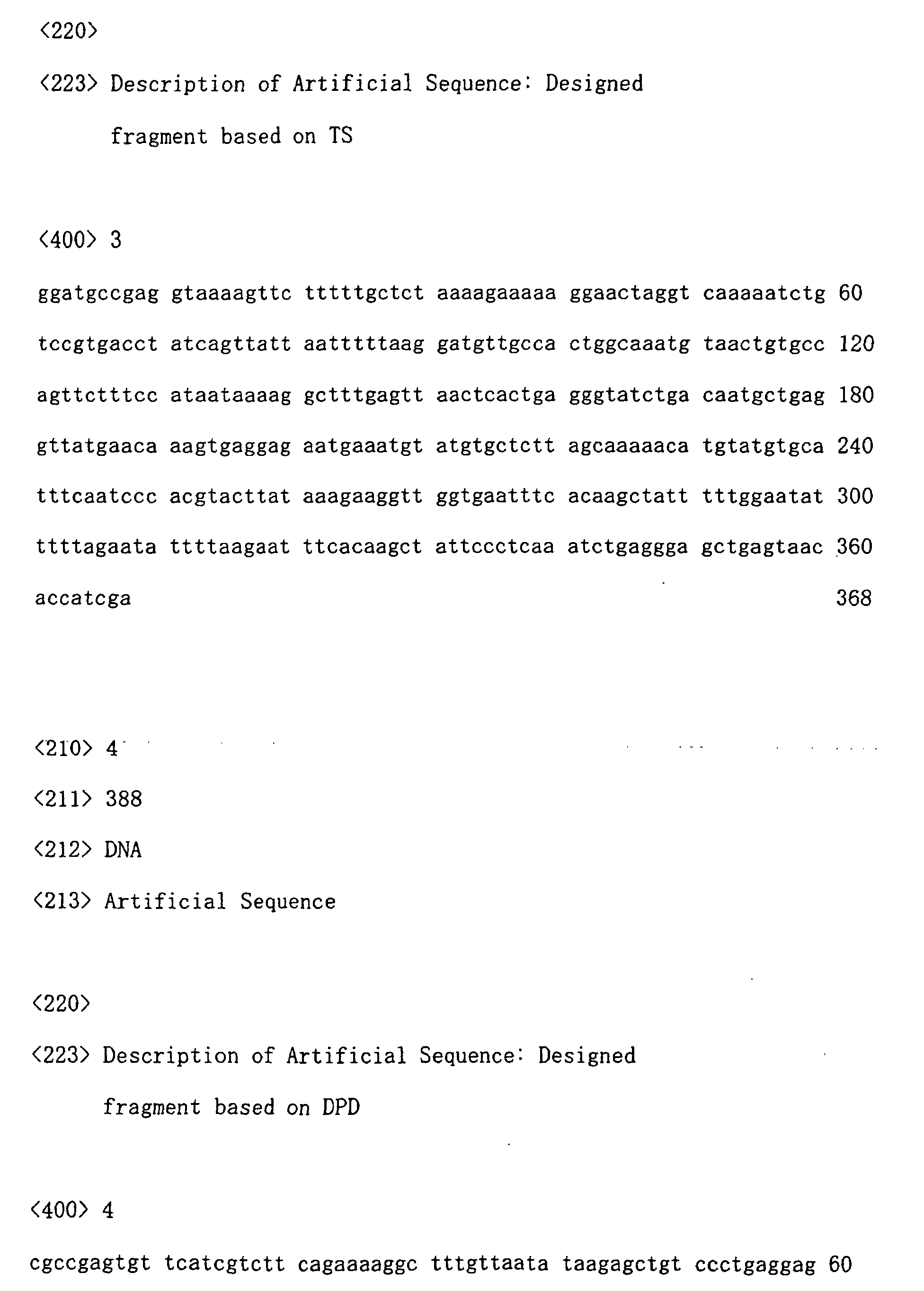
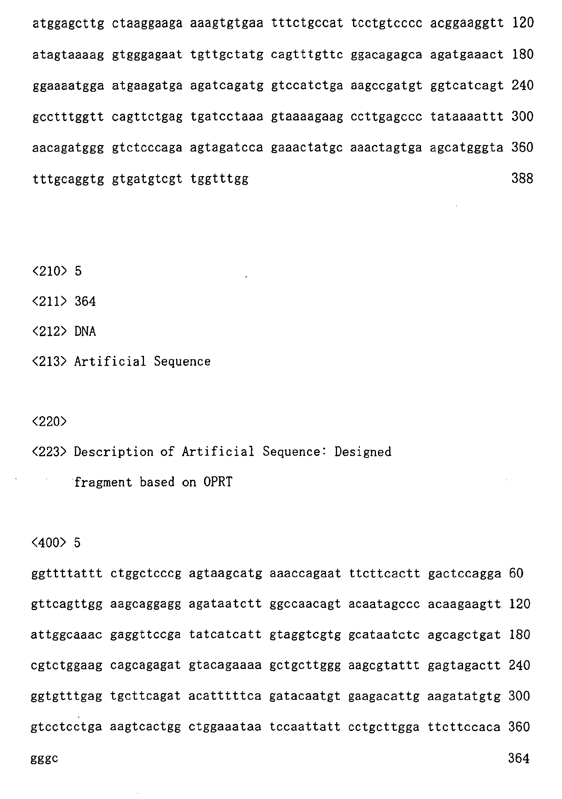
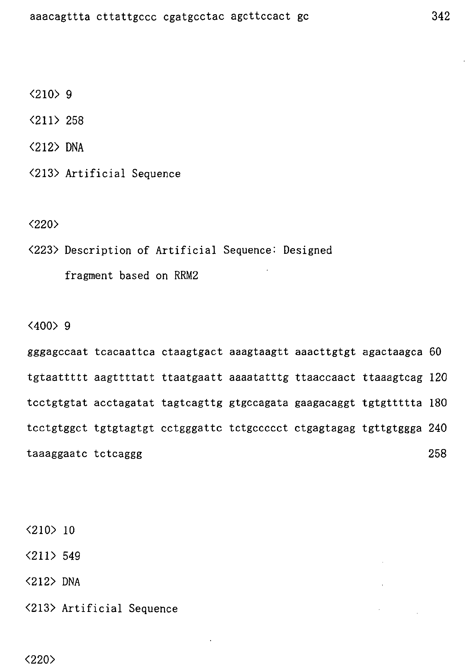
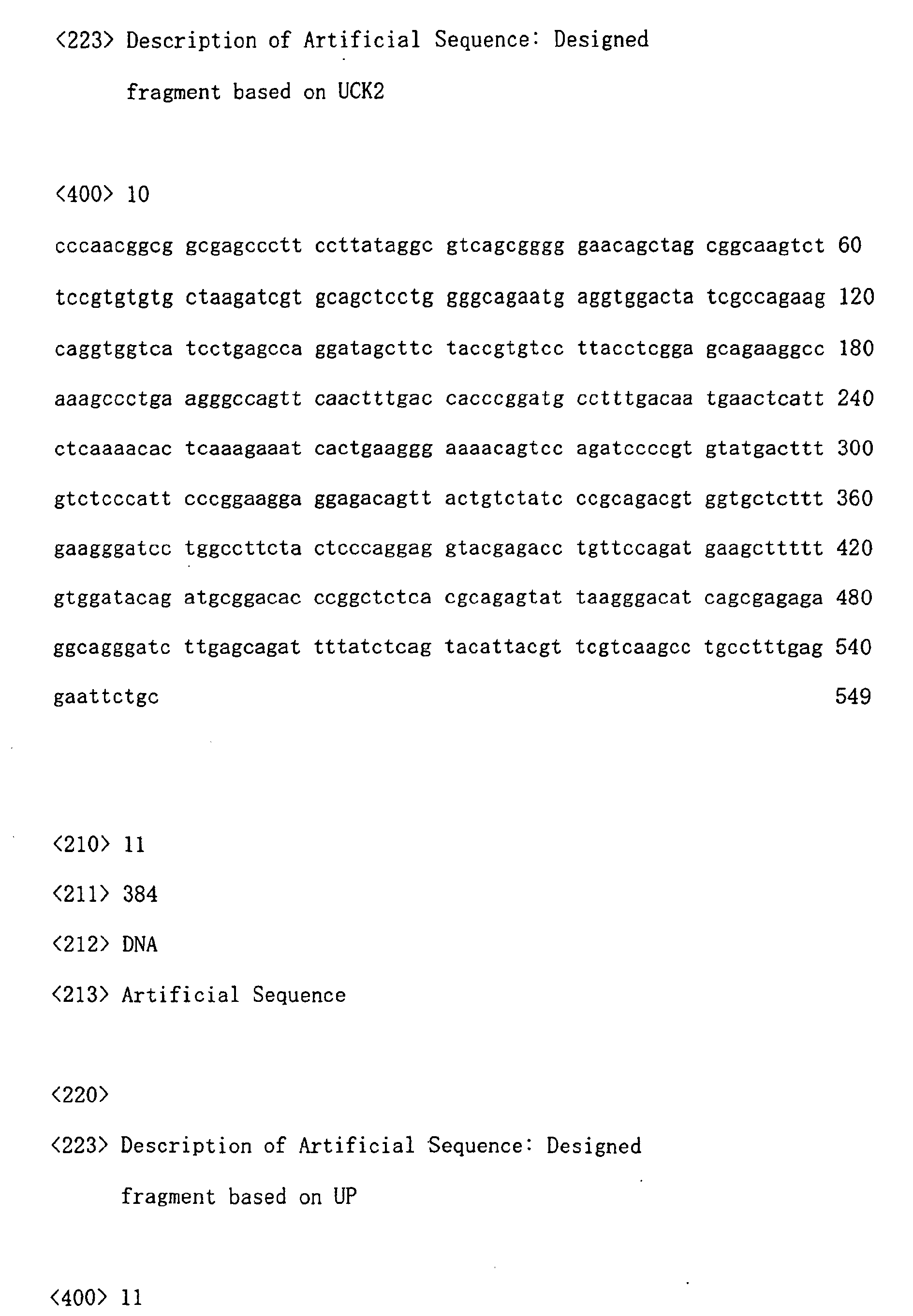
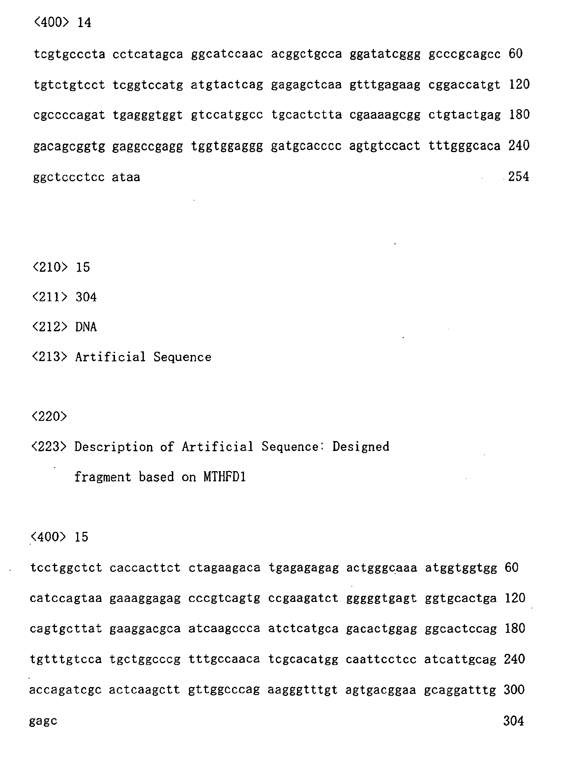
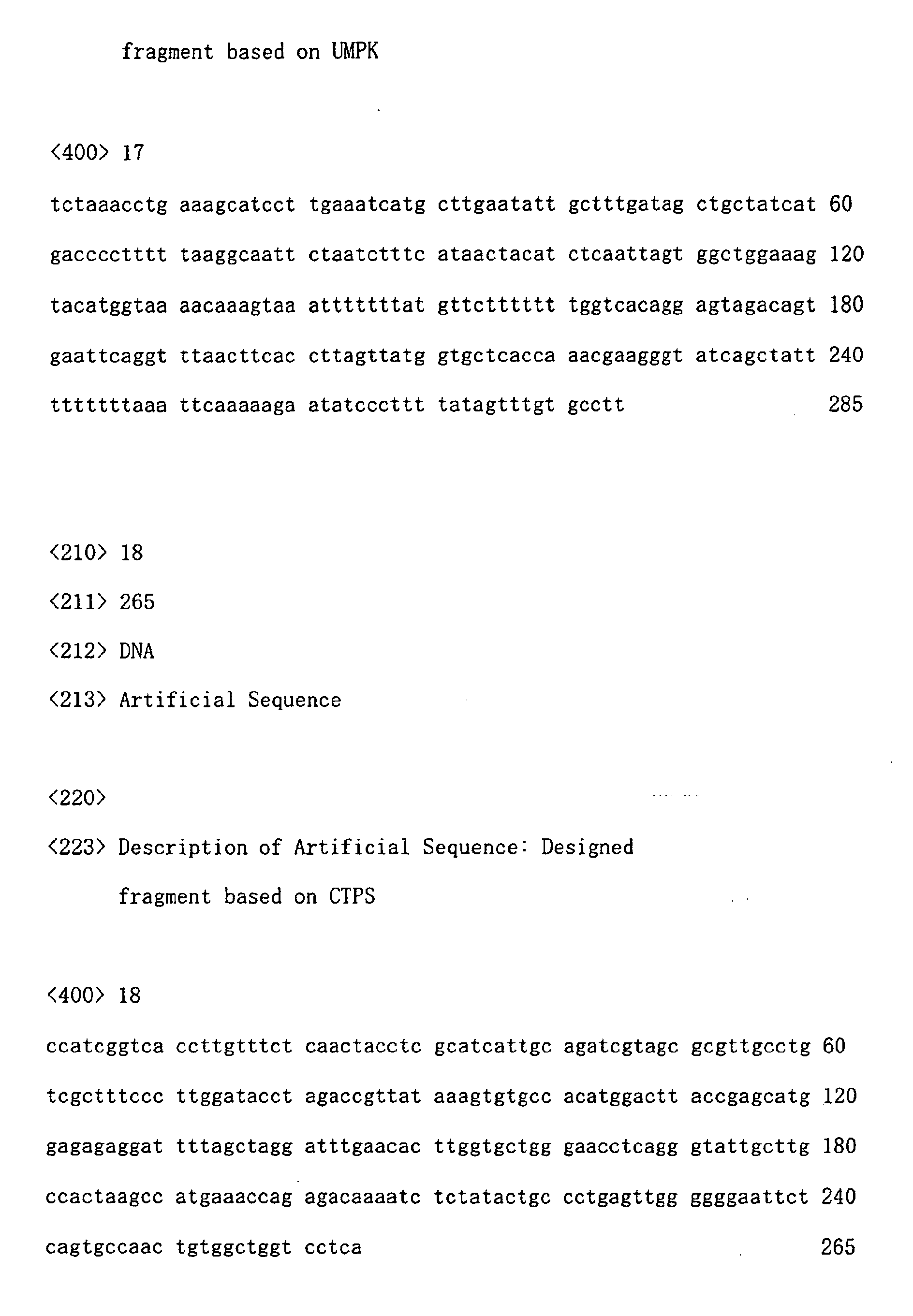
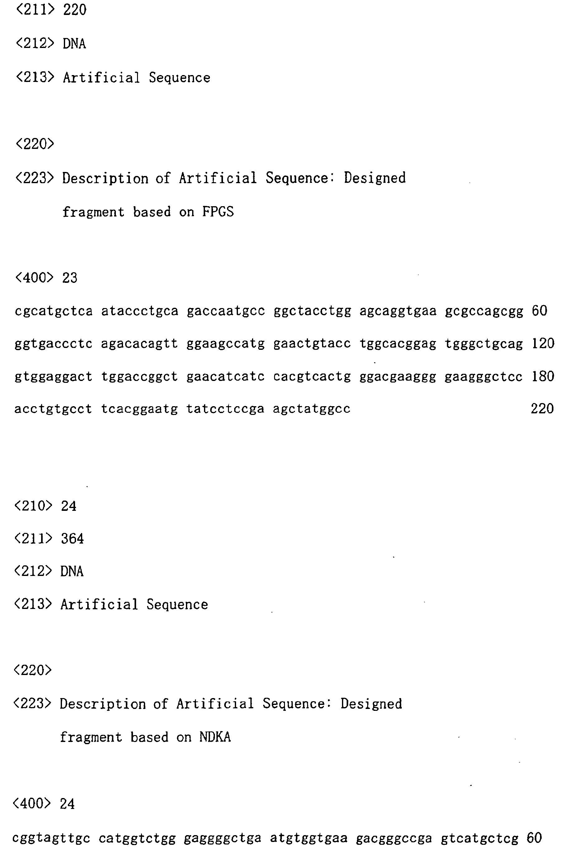
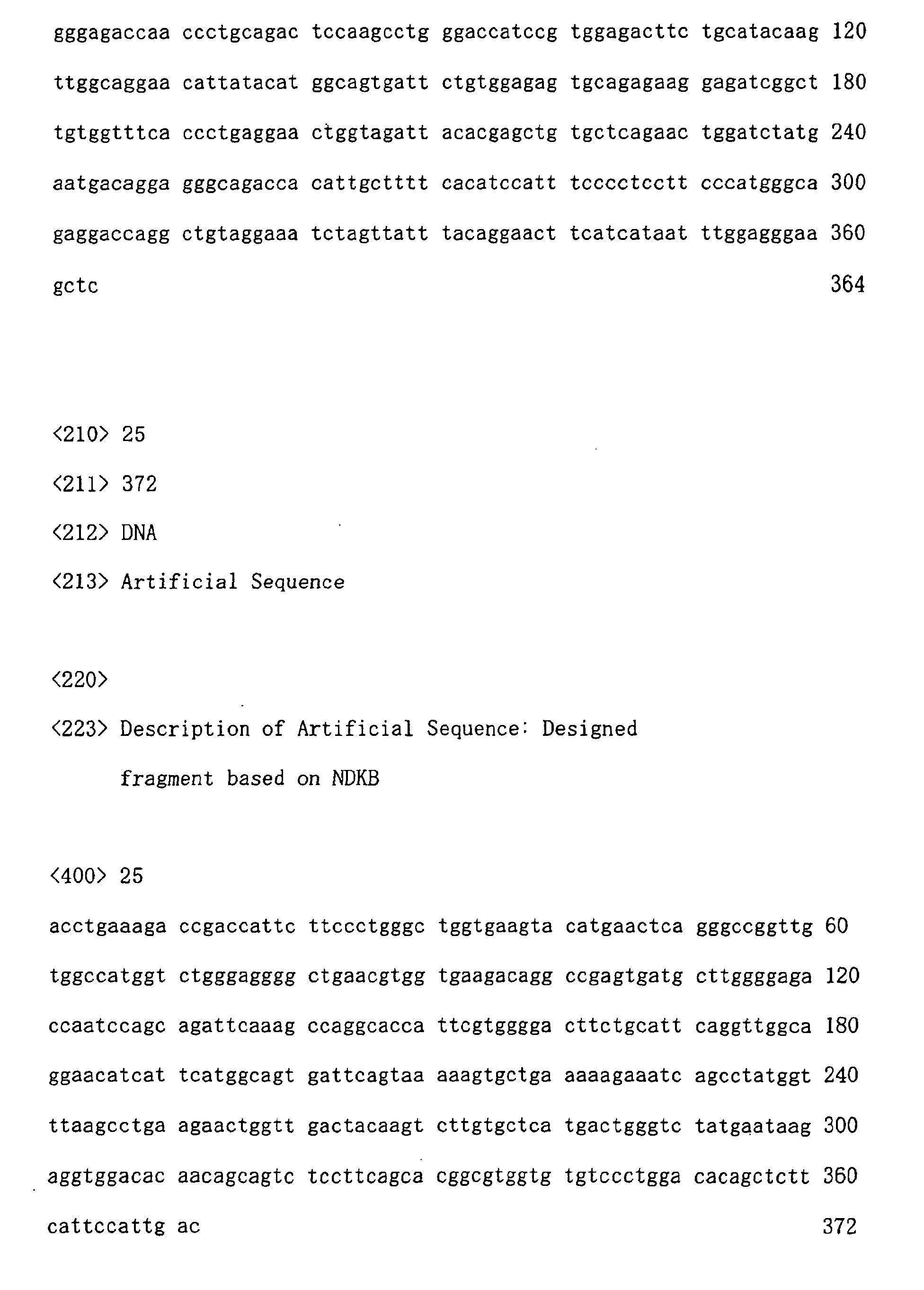
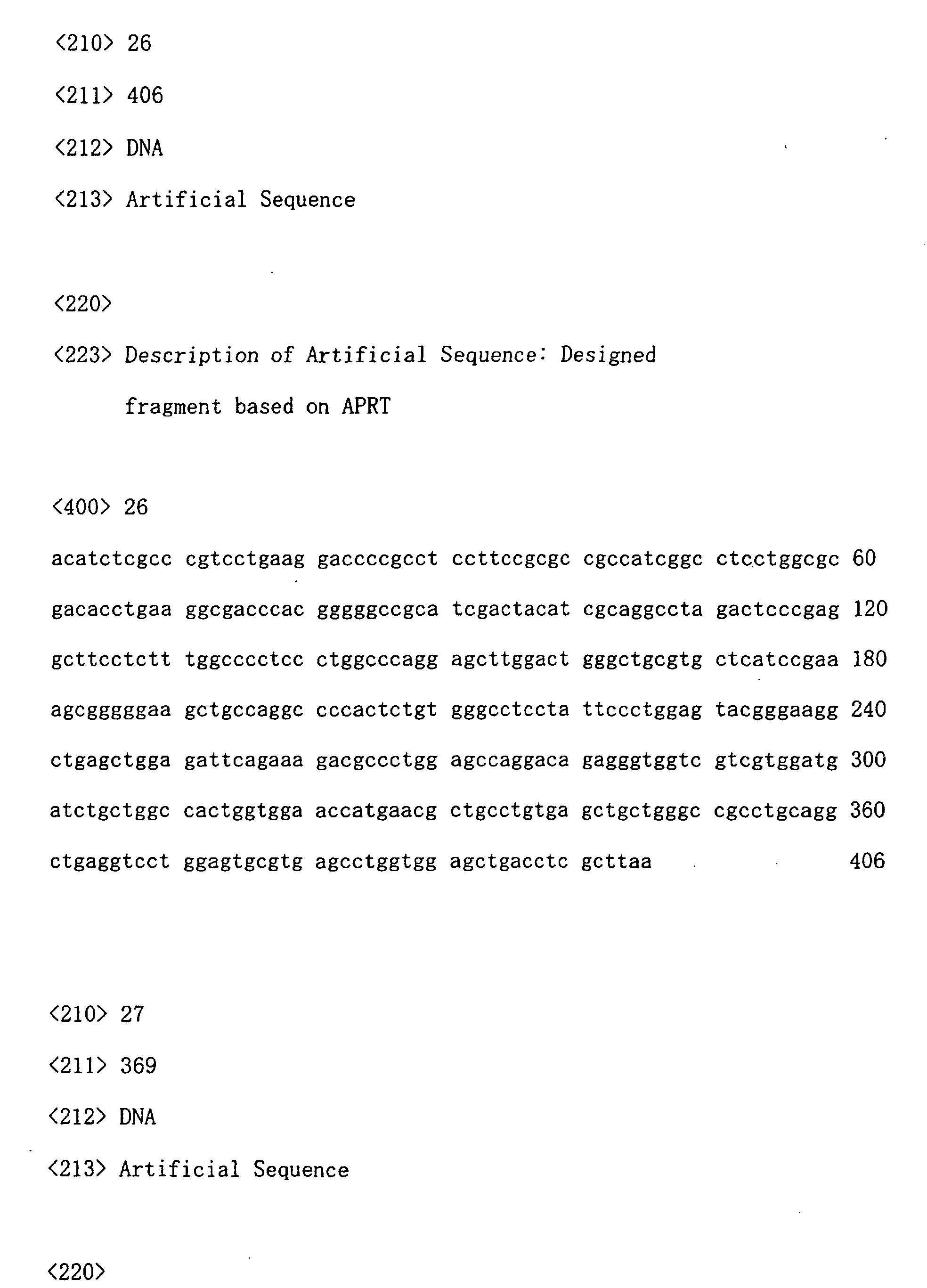
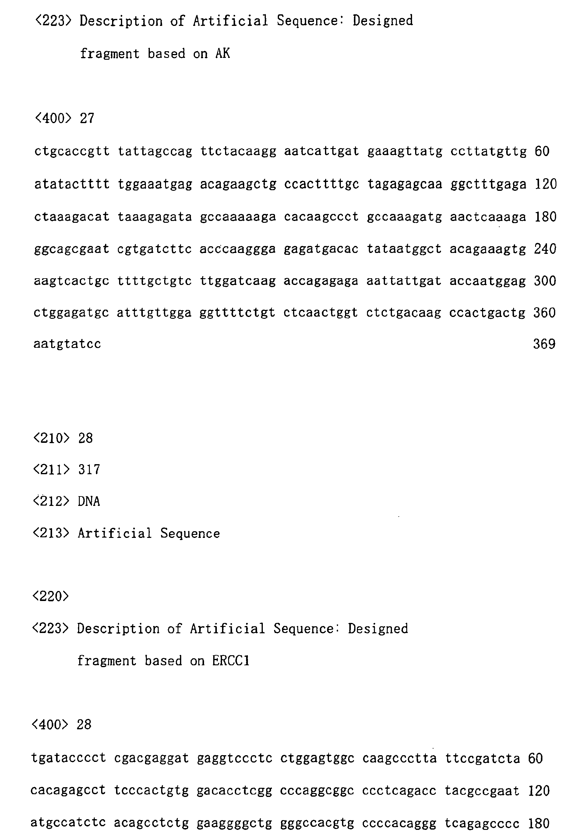
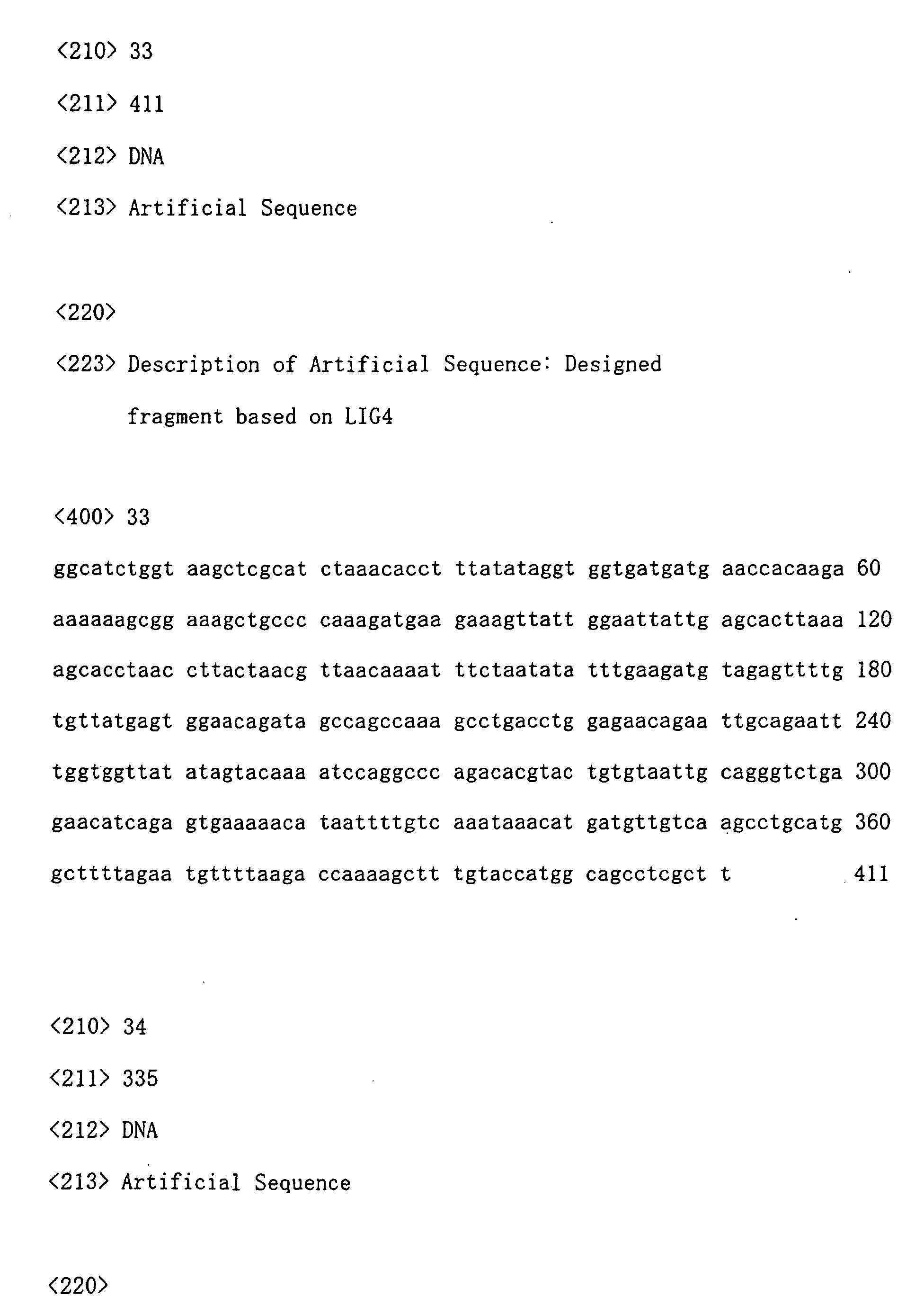
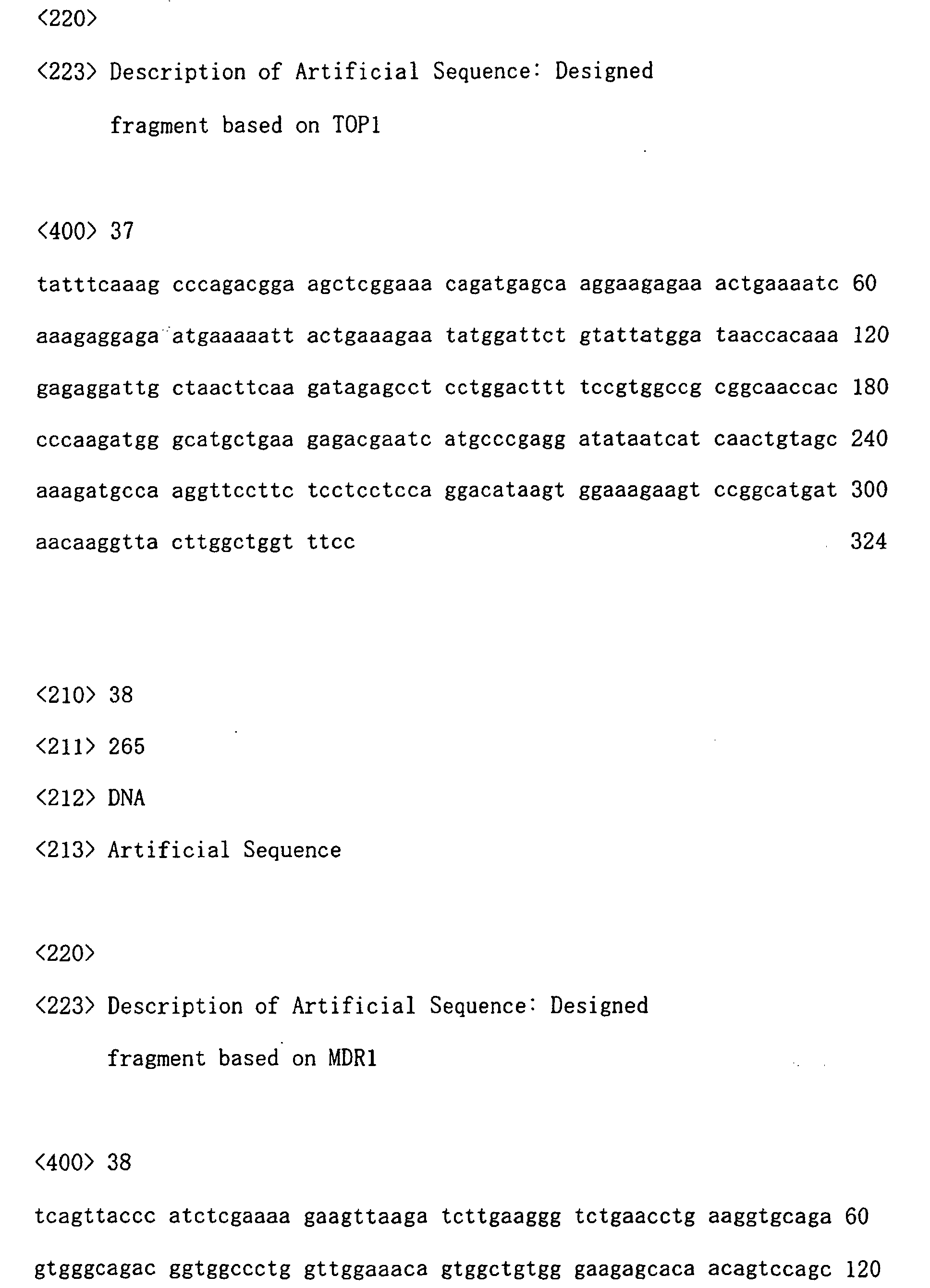
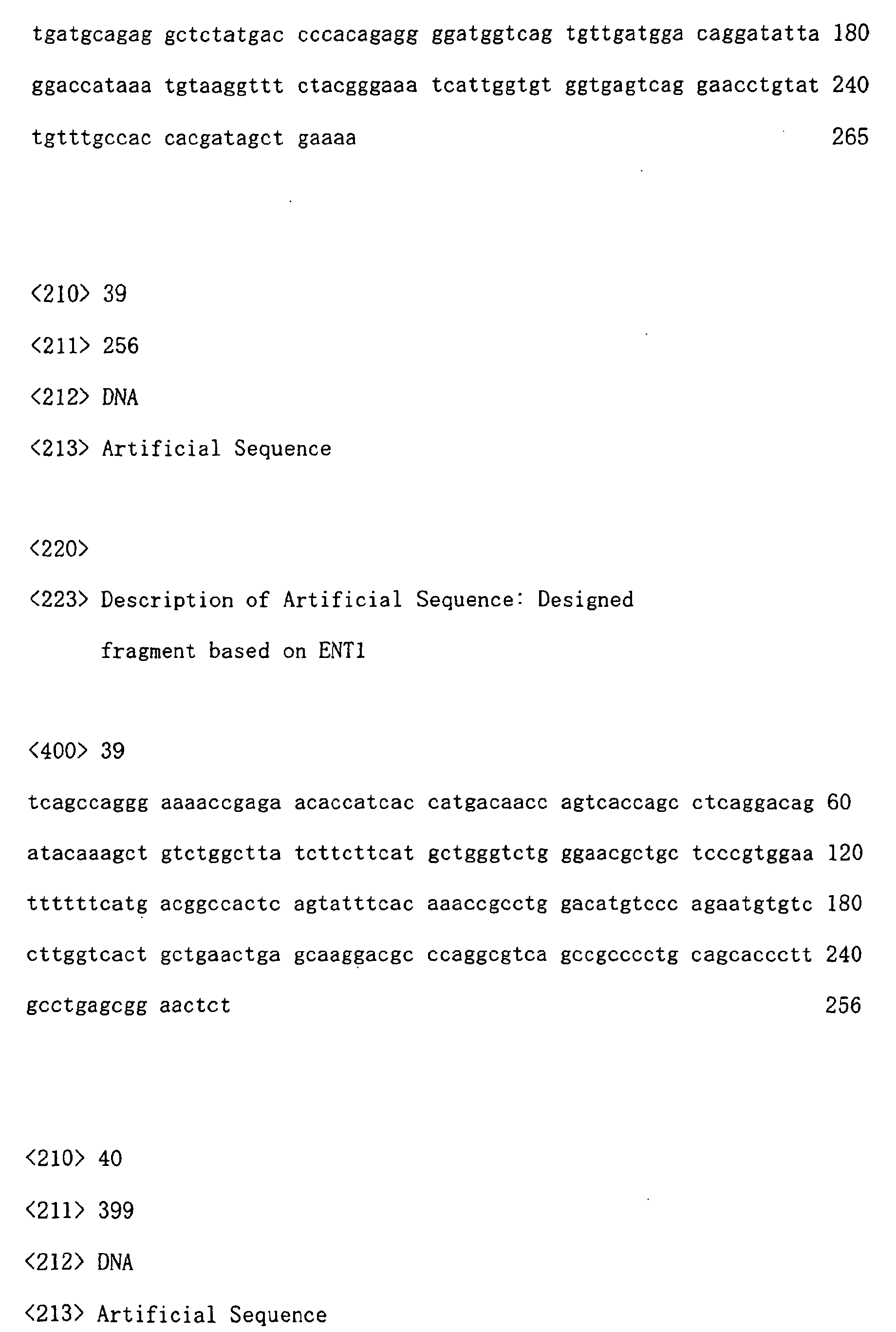
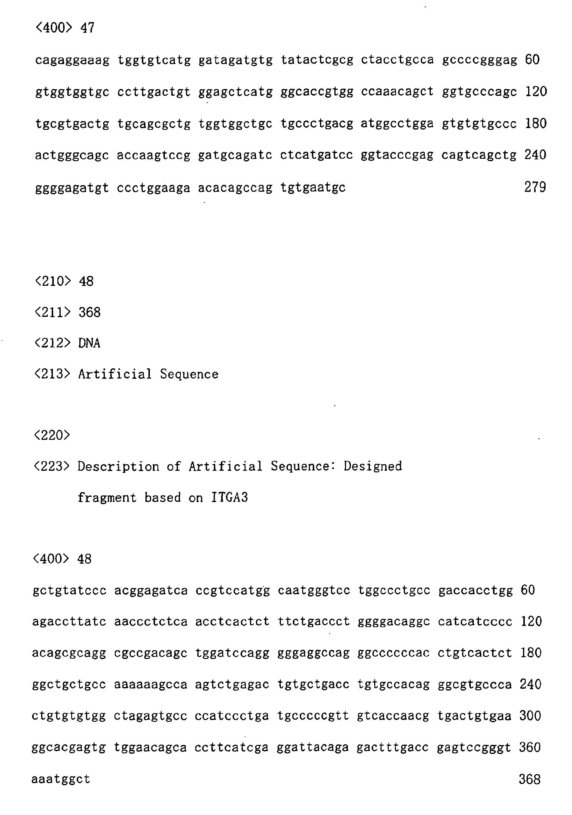
  • B. Preparation of target fragments The target fragment was amplified in accordance with the method described in (1) above through use of the clone as a template. The employed primers were universal primers (SEQ ID NOS: 1 and 2). The PCR product was subjected to ethanol precipitation, and the collected precipitates were washed with ethanol and dissolved in distilled water. An aliquot of the solution was employed for calculation of concentration (through absorptiometry) and assay for determining purification (through electrophoresis on agarose gel). The remaining target fragment solution was dried at room temperature under reduced pressure, and then the fragment was dissolved in Micro Spotting Solution (BM) so as to have a concentration of 0.5 to 10 pmol/µL (Tables 2 and 3 show the concentration of each target fragment). Nucleotide sequences of the thus-obtained target fragments are shown by SEQ ID NOs: 3 to 54.
    Figure 00220001
    Figure 00230001
  • C. Spotting and post-treatment The target fragment which had undergone thermal denaturation for three minutes at 95°C was spotted onto a glass slide coated with poly-L-lysine, by means of a spotter (OmniGrid, GENEMACHINES). Subsequently, the target fragment was cross-linked with the glass slide under irradiation with UV rays, and the glass slide was placed in a rack and shaken in a blocking solution (8-wt% Block A in PBS) for 30 minutes, and the resultant slide was washed with a TE buffer and dried. The slide glass was stored in a desiccator in the dark until the time of use. Following the process, the DNA array of the present invention was obtained.
  • D. Preparation of fluorescence-labeled DNA probes Reverse transcription reaction was performed through use of a total RNA sample prepared from tumor cells (through the same method as described in (2) above) as a template and primers specific to mRNA of each gene, to thereby prepare fluorescence-labeled probes. Reagents employed for labeling reaction are as follows.
    • reverse transcriptase SuperscriptII (Gibco BRL)
    • (reaction buffer, DTT)
    • a primer mixture (a mixture of reverse primers specific to each of the 52 genes; the reverse primers are one of the specific primers employed in Referential Example 1)
    • dATP, dGTP, dCTP, dTTP (Amersham Pharmacia)
    • Cy3-dUTP (Amersham Pharmacia)
    • Cy5-dUTP (Amersham Pharmacia)
    • 0.5M EDTA
    • 1N NaOH
    • 1M Tris-HCl (pH 7.5)
    • TE buffer
    Specifically, a total RNA sample (30 µg) was mixed with a primer mixture (50 pmol each) and distilled water, and the volume of the resultant mixture was adjusted to 9 µL. The mixture was denatured for two minutes at 65°C and rapidly cooled on ice. A reaction buffer (1×), DTT (10 mM), dTTP (0.2 mM), dATP (0.5 mM), dGTP (0.5 mM), dCTP (0.5 mM), Cy3-dUTP or Cy5-dUTP (0.1 mM), and Superscript II (10 U/µL) were added thereto, and the total volume of the mixture was adjusted with distilled water to 20 µL (the concentrations in the parentheses refer to final concentrations). The mixture was allowed to react for 60 minutes at 42°C, and distilled water (20 µL), 0.5M EDTA (5 µL), and 1N NaOH (5 µL) were added thereto. The resultant mixture was incubated for 60 minutes at 65°C, whereby total RNA molecules were decomposed. The reaction mixture was neutralized with 1M Tris-HCl (25 µL). A TE buffer (200 to 400 µL) was added thereto, and the resultant mixture was desalted and concentrated through ultrafiltration (Microcon-30, Millipore) (in this step, reverse primers and Cy3-dUTP or Cy5-dUTP which had not been incorporated into probes were also removed). Ultimately, about 10 µL of a probe solution was obtained.
  • E. Hybridization 20×SSC (3 µL) and distilled water were added to the above fluorescence-labeled DNA probe solution, and the volume of the mixture was adjusted to 20 µL (final concentration: 3×SSC). The mixture was subjected to thermal denaturation for three minutes at 95°C, and the reaction mixture was left to stand at room temperature for cooling. 10-wt% SDS (2 µL) and distilled water were added to the resultant mixture, and the volume of the mixture was adjusted to 40 µL (final concentration: 0.5 wt%). The mixture was added dropwise to the DNA array prepared in the steps A to C, and a glass cover was gently placed onto the array (so as not to allow air inside). The DNA array was set in a hybridization chamber and incubated for 10 to 20 hours at 65°C. Thereafter, the DNA array was placed in a 2×SSC solution containing 0.2-wt% SDS, and the glass cover was gently removed. The DNA array was washed in situ for five minutes. Moreover, the DNA array was washed once with a 2×SSC solution containing 0.2-wt% SDS and twice with a 0.2×SSC solution containing 0.2-wt% SDS, and rinsed twice with 0.2×SSC (all of these washing steps were performed at room temperature). The DNA array was placed in a rack and centrifuged for 20 seconds at 600 rpm, to thereby remove water. The DNA array was dried at room temperature, and fluorescent signals from the DNA array were measured by means of a DNA microarray fluorescence scanner (GenePix, Axon).
  • Test 1: Specificity of DNA fragments
    Specificity of DNA fragments employed as templates for synthesis of probes in Northern hybridization differed from fragment to fragment, although the fragments were contained in the same gene. Fig. 2 shows some of the test results, using XRCC1 and E2F1. In XRCC1, a region of 898 to 1265 nt exhibited good results (strong signals were detected at a position corresponding to the size of mRNA of the gene of interest), and a region of 187 to 494 nt was determined not to be suitable, since a large number of signals indicating cross-hybridization were observed. Similarly, in E2F1, a region of 1014 to 1309 nt exhibited good results, and a region of 788 to 1087 nt was determined not to be suitable. When regions of other genes were investigated, various levels of specificity were also observed, although the levels differed from gene to gene. A DNA fragment which exhibited the highest specificity in DNA fragments of gene of interest was selected in the final step and was employed as a target fragment.
    Test 2: Performance of Northern hybridization in terms of quantitativeness
    Performance of DNA arrays in terms of quantitativeness is confirmed by comparison with Northern hybridization. Accordingly, in order to confirm the performance of the measurement system of Northern hybridization employed by the present inventors (Example 1(2)), correlation of the RNA level and measurements (signal intensity) was investigated.
    A total RNA sample derived from each of the above 14 types of human tumor cells was electrophoresed on a denatured agarose gel and blotted on a nylon membrane. Northern hybridization was performed through use of probes synthesized from target DNA of GAPDH (glyceraldehydes-3-phosphate dehydrogenase) (through the method described in Example 1(2)). Analysis was performed by means of imaging and analysis software (ImageQuant, Molecular Dynamics Inc), and intensity of GAPDH mRNA signals was measured.
    Fig. 3 shows the relationship between the amount of the electrophoresed total RNA sample and measurements of Northern hybridization. The results confirmed that a linear relationship exists between the amount of the total RNA sample and signal intensity (r=0.95, p<0.01), indicating that the measurement system employed provides high performance in terms of quantitativeness.
    Test 3: Effect of universal primers on background
    As shown in Fig. 1, universal primers (forward (pCR-F)/reverse (pCR-R)) were designed on the basis of the sequence of a multicloning site of vector. Since all 52 target fragments can be amplified at a constant rate by using universal primers, target fragments can be readily provided in sufficient amounts as compared with the case of specific primers.
    In order to determine the effect of primers which may migrate into target fragments upon spotting on measurement background, universal primers (5 spots) and specific primers (primers corresponding to each of about 20-bp sequences at both ends of 5 target DNA fragments [TS, DPD, OPRT, LIG4, and GAPDH] listed in Tables 2 and 3, 1 spot each) were spotted in equal amounts onto glass slides. The glass slides were subjected to the hybridization procedure described in Example 1(3), and signal intensity was measured.
    The mean value, standard deviation (SD), and coefficient of variation (CV) of signal intensities obtained from the universal primers (5 spots) were found to be 27.3, 6.5, and 24.0%, respectively. The mean value, standard deviation (SD), and coefficient of variation (CV) of signal intensities obtained from the specific primers (5 spots in total) were found to be 22.9, 12.0, and 52.4%, respectively. Signal intensities from both primers were found to be of substantially the same level, and signal intensities obtained from the universal primers had lower SD and CV, indicating that the universal primers exert less effect on difference in background level than the specific primers.
    In addition, in order to reduce background to the lowest level as possible, the minimum amount of the universal primers required to amplify a target fragment was studied. PCR was performed through use of forward and reverse primers. The amount of each primer was 10, 20, 30, 40, and 50 pmol. The PCR products were electrophoresed on agarose gel. When primers were used in amounts of 20 pmol or less, the amount of PCR product was found to be reduced. Therefore, the minimum amount of the universal primers required to amplify a target fragment was determined to be 30 pmol.
    Example 2 Measurement of expression level of various genes in human tumor cells
  • (1) Measurement through Northern hybridization In the step of determining target fragments to be placed onto the DNA array, the expression level of each of the 52 genes was determined through analysis of blot images obtained through Northern hybridization of the above 14 different human tumor cells (see Example 1(2)). The expression level is represented by the value relative to the mean value of the expression levels of three housekeeping genes (GAPDH, ACTB, and RSP9).
  • (2) Measurement by use of DNA array A DNA array onto which the 52 DNA target fragments were immobilized was prepared, and hybridization was performed through use of fluorescent DNA probes prepared from the total RNA samples which had been extracted from the 14 types of cells and employed in Northern hybridization. The expression level of each gene was measured (through the same method as described in Example 1 (3)). The expression level is represented by the value relative to the mean value of the expression levels of three housekeeping genes (GAPDH, ACTB, and RSP9).
  • (3) Correlation of the results obtained through Northern hybridization with the results obtained through DNA array In order to determine whether the expression levels of each of the 52 species of genes determined through Northern hybridization in the above 14 species of human tumor cells were correlated with those determined by use of DNA arrays, regression analysis was performed (Table 4). The degree of correlation (correlation coefficient) differed from gene species to gene species, and significant correlation was confirmed in 20 species of genes at a significance level of less than 5%. Some degree of correlation was confirmed in nine species of genes, although the degree was not significant (p<0.1). Twelve species of genes containing three species of housekeeping genes (MTHFD1, SOD1, AK, E2F1, POLD, LIG3, RSP9, HPRT1, ACTB, UDG, GAPDH, and LIG1) were found to have only a low degree of correlation or were found impossible to evaluate, since difference in expression level between cells were small; in other words, the normal distribution was not obtained.
    Abbreviation R (correlation coefficient) P-value Correlation Significance (P<0.05)
    ERCC1 0.90 9.4E-06 High Yes
    p53 0.90 1.1E-05 High Yes
    MDR1 0.90 1.3E-05 High Yes
    DPD 0.89 2.2E-05 High Yes
    CDA 0.87 5.0E-05 High Yes
    TS 0.85 1.1E-04 High Yes
    PRPS 0.85 1.2E-04 High Yes
    NDKB 0.80 6.5E-04 Relatively high Yes
    POLB 0.79 7.3E-04 Relatively high Yes
    XRCC1 0.77 1.3E-03 Relatively high Yes
    MRP1 0.76 1.4E-03 Relatively high Yes
    ENT2 0.75 2.0E-03 Relatively high Yes
    APRT 0.75 2.2E-03 Relatively high Yes
    Hsp27 0.67 8.2E-03 Relatively high Yes
    VEGFB 0.60 2.2E-02 Relatively high Yes
    TOP2B 0.57 3.3E-02 Medium Yes
    LIG4 0.57 3.4E-02 Medium Yes
    ENT1 0.55 4.3E-02 Medium Yes
    RRM2 0.54 4.6E-02 Medium Yes
    IMPD 0.54 4.8E-02 Medium Yes
    NDKA 0.53 5.3E-02 Medium No
    RP2 0.51 6.2E-02 Medium No
    TP 0.50 7.1E-02 Medium No
    RRM1 0.49 7.7E-02 Medium No
    UCK2 0.45 1.1E-01 Medium No
    FPGS 0.44 1.2E-01 Medium No
    CTPS 0.44 1.2E-01 Medium No
    UMPS(OPRT) 0.42 1.3E-01 Medium No
    SOD2 0.42 1.4E-01 Medium No
    UP 0.39 1.6E-01 Relatively low No
    NT5 0.34 2.3E-01 Relatively low No
    TK1 0.34 2.4E-01 Relatively low No
    DCK 0.33 2.5E-01 Relatively low No
    TOP2A 0.32 2.7E-01 Relatively low No
    DCD 0.27 3.5E-01 Relatively low No
    ITGA3 0.26 3.7E-01 Relatively low No
    PARP 0.24 4.1E-01 Relatively low No
    TOP1 0.23 4.3E-01 Relatively low No
    PCNA 0.23 4.3E-01 Relatively low No
    UMPK 0.21 4.6E-01 Relatively low No
    LIG1 0.06 8.3E-01 Low No
    MTHFD1 0.40 1.6E-01 Impossible to evaluate No
    SOD1 0.39 1.6E-01 Impossible to evaluate No
    AK 0.37 1.9E-01 Impossible to evaluate No
    E2F1 0.29 3.2E-01 Impossible to evaluate No
    POLD 0.28 3.3E-01 Impossible to evaluate No
    LIG3 0.27 3.4E-01 Impossible to evaluate No
    RSP9 0.26 3.7E-01 Impossible to evaluate No
    HPRT1 0.25 3.8E-01 Impossible to evaluate No
    ACTB 0.20 5.0E-01 Impossible to evaluate No
    UDG 0.17 5.7E-01 Impossible to evaluate No
    GAPDH 0.14 6.4E-01 Impossible to evaluate No
  • (4) Performance of DNA array in terms of judgment When such a tool used for the purpose of diagnosis (proper use of a drug) is employed in actual clinical sites, whether a sample is positive or negative is generally determined on the basis of threshold. For example, in research in which TS or DPD mRNA level determined through RT-PCR is used as an index which indicates proper use of 5-FU, a threshold has been set for determining that a level is high or low (Clinical Cancer Research, 6, 1322-1327, 2000). The performance of the DNA array of the present invention was determined through comparison with Northern hybridization in the following manner.The above 14 species of human tumor cells were treated as clinical specimens. The expression level of each of the 52 species of genes in each type of cells was measured through use of the DNA array of the present invention or through Northern hybridization (in each method, 52 (genes) × 14 (cells) = 728 points were measured and calculated). From measurements obtained through use of the DNA array of the present invention, the median value of the expression levels of each gene in the 14 species of cells was calculated. The relative expression level (the ratio of expression level of each of the 14 species of cells to the median value (base)) was calculated. The threshold was set as 2-fold. When the relative expression level went beyond the threshold value (≥2 or ≤0.5), the cell was determined to be "positive," whereas when the relative expression level did not exceed the threshold value, the cell was determined to be "negative." On the assumption that results determined through Northern hybridization were highly reliable, when results determined by use of the DNA array of the present invention were identical with those determined through Northern hybridization, the results were evaluated as "true," whereas when results determined by use of the DNA array of the present invention were different from those determined through Northern hybridization, the results were evaluated as "faulty." As shown in Fig. 4, in the positive and negative results, the percentage of data evaluated "true" was found to be as high as 82.3%. In 75 points which had been determined as positive through use of the DNA array of the present invention, 77.3% (58/75) of the points presented the same results as those of Northern hybridization. In addition, there were no cases in which the measurements and results of the DNA array were inconsistent with those of Northern hybridization (for example, there were no cases in which both results were true positive but the relative expression level through Northern hybridization (e.g., 0.3) was quite different from those obtained through use of the DNA array of the present invention (e.g., 2.5)). Thus, the DNA array of the present invention was found to present substantially the same level of performance as that of Northern hybridization.
  • (5) Detecting ability of DNA array In Test 4 (4), among the tested 728 points (52 (genes) × 14 (cells)), 62 points were found to be impossible to measure. None of the 62 points could not be detected through Northern hybridization. Therefore, the results indicate that, as compared with Northern hybridization, the DNA array of the present invention presents the same level or a higher level of detecting ability.
  • Example 3 Correlation between TS-1 sensitivity and expression level of various genes in transplantable human tumor cells
  • (1) TS-1 sensitivity test The DNA array of the present invention may be clinically applied to, for example, determining the genes that are important as sensitivity regulating factors, by analyzing correlation between expression levels of the 52 genes in cancer tissue and antitumor effect of a 5-FU anticancer agent.In this test, as a test model for determining anticancer effect, TS-1 sensitivity of 11 species of transplantable human tumor cells (xenografts, derived from gastric cancer (4 types), large intestine cancer (3 types), lung cancer (2 types), and mammary cancer (2 types)) was tested (TS-1, a 5-FU anticancer agent which has been developed by Taiho Pharmaceutical Co., Ltd., is a composition-containing agent containing tegafur (5-FU prodrug), gimeracil (a DPD inhibitor), and oteracil potassium (an orotate phosphoribosyl transferase inhibitor) at a ratio by mole 1:0.4:1). Each xenograft was subcutaneously transplanted into nude mice at the back thereof. When the transplanted site was expanded to 100 to 200 mm3, the mice were divided into two groups; i.e., a control group (a group to which no drug was administered) and a TS-1 administered group (6 animals/group). From the following day, TS-1 was perorally administered to mice of the TS-1 administered group once a day for 14 successive days at a dose of 10 mg/kg/day (FT). On the day following the final administration, the volumes of the tumors were measured, and the volumes of the TS-1 administered group were compared with those of the control group. Tumor multiplication inhibition rate (IR) was determined. IR was calculated by the following equation. IR(%)=(1-[Relative tumor volume of the TS-1 administered group]/[Relative tumor volume of the control group])×100 (Relative tumor volume)=(tumor volume upon judgement)/(tumor volume upon grouping)
  • (2) Measurement by use of the DNA array of the present invention The expression levels of the 52 species of genes were determined through use of total RNA samples prepared from the 11 species of xenografts. The test was performed in accordance with the method described in Example 2. The expression level is represented by the value relative to the mean value of the expression levels of three species of housekeeping genes (GAPDH, ACTB, and RSP9).
  • (3) Correlation between the expression level in 11 species of xenografts and TS-1 sensitivity
  • In order to determine whether the expression level of the 52 species of the genes in the 11 species of xenograft, determined in the studies (1) and (2) above, is correlated with TS-1 sensitivity (IR, %), regression analysis was performed (52 (genes) × 11 (cells)). As a result, four species of the gene (UDG, PCNA, TS, and TK1) were found to have an absolute value of correlation coefficient (P-value: Pearson's product-moment correlation coefficient) higher than 0.5 (Fig. 5). P-value was found to be 0.05 or higher and lower than 0.1. Thus, the data were not statistically significant. However, since N is as small as 11, from the regression line, the expression level is confirmed to be substantially correlated with the sensitivity. It should be noted that these four species of genes contain TS. TS is a representative sensitivity regulating factor for 5-FU anticancer agent, and both basic research and clinical research have revealed that the higher the TS expression level, the lower the effect exhibited by a 5-FU anticancer agent. In the study of the present invention, TS expression level was found to have a negative correlation with antitumor effect of TS-1 (r=-0.53). The results coincide with the previous finding. In addition, as a confirmation test, the TS expression level in the 11 species of cells (relative expression level against GAPDH) was measured through real time RT-PCR, which is considered to provide considerably high measurement accuracy, to thereby analyze correlation between TS-1 sensitivity and the TS expression level. The correlation coefficient was found to be -0.65; i.e., a significant correlation (P=0.030) was confirmed (Fig. 6). These data indicate that the DNA array of the present invention is useful for determining candidates of sensitivity regulating factors of a 5-FU anticancer agent from the 52 species of gene. When N increases, clinical specimens may be used to determine sensitivity regulating factors of a 5-FU anticancer agent, and when the expression level of a selected gene was determined through analysis, the expression level may be used as an index which indicates proper use of 5-FU anticancer agent.
    Industrial Applicability
    The present invention achieves convenient and highly quantitative measurement of expression levels of several tens to several hundreds of gene species contained in a specimen in a single measurement. When expression patterns of genes related to action mechanisms of an antimetabolite-type anticancer agent or a combination of such an agent and another anticancer agent are analyzed through use of the assay method of the present invention in a test specimen (for example, total RNA extracted from peripheral monocytes or tumor tissue of a cancer patient), results obtained from such an analysis can be employed as indices for proper use of the anticancer agent.
    Figure 00390001
    Figure 00400001
    Figure 00410001
    Figure 00420001
    Figure 00430001
    Figure 00440001
    Figure 00450001
    Figure 00460001
    Figure 00470001
    Figure 00480001
    Figure 00490001
    Figure 00500001
    Figure 00510001
    Figure 00520001
    Figure 00530001
    Figure 00540001
    Figure 00550001
    Figure 00560001
    Figure 00570001
    Figure 00580001
    Figure 00590001
    Figure 00600001
    Figure 00610001
    Figure 00620001
    Figure 00630001
    Figure 00640001
    Figure 00650001
    Figure 00660001
    Figure 00670001
    Figure 00680001
    Figure 00690001
    Figure 00700001
    Figure 00710001
    Figure 00720001
    Figure 00730001
    Figure 00740001
    Figure 00750001
    Figure 00760001
    Figure 00770001

    Claims (7)

    1. A DNA array for measuring sensitivity to an antimetabolite-type anticancer agent or to a combination of such an anticancer agent and another anticancer agent, characterized by comprising a substrate and target gene fragments attached to the substrate,
         the target gene fragments being of at least 13 gene species, including at least 2 species from each of the following gene groups: a group of genes coding for nucleic-acid-metabolism-related enzymes, a group of genes coding for gene-repair-associated enzymes, a group of genes coding for drug resistance-associated factors, and housekeeping genes, and said at least 13 gene species being selected by performing the following two steps 1) and 2):
      1) selecting fragments having high target-gene specificity through a homology search using a database, and
      2) performing Northern hybridization to RNA collected from tumor cells, using the fragments selected in step 1) as probes, to thereby confirm target-gene specificity.
    2. The DNA array according to claim 1, wherein the amounts of respective fragments attached to the substrate are regulated in accordance with expression levels of the target genes in the tumor cells.
    3. The DNA array according to claim 1 or 2, wherein the fragments selected in the step 2) are obtained through PCR employing universal primers having nucleotide sequences of SEQ ID NOs: 1 and 2.
    4. The DNA array according to any one of claims 1 to 3, wherein:
      the group of genes coding for nucleic-acid-metabolism-related enzymes includes at least thymidylate synthase gene, dihydropyrimidine dehydrogenase gene, orotate phosphoribosyltransferase gene, thymidine phosphorylase gene, and thymidine kinase 1 gene;
      the group of genes coding for gene-repair-associated enzymes includes at least DNA excision repair protein ERCC1 gene and uracil-DNA glycosylase gene;
      the group of genes coding for drug resistance-related factors include topoisomerase 1 gene, P-glycoprotein gene, equilibrative nucleoside transporter 1 gene, and multidrug resistance-associated protein 1 gene; and
      the group of housekeeping genes consists of two or more genes selected from among glyceraldehyde-3-phosphate dehydrogenase gene, β-actin gene, and 40S ribosomal protein S9 gene.
    5. The DNA array according to any one of claims 1 to 4, wherein:
      each of the target gene fragments has a size of 200 to 600 bp.
    6. A DNA array for measuring sensitivity to an antimetabolite-type anticancer agent or to a combination of such an anticancer agent and another anticancer agent, characterized by comprising a substrate and at least the following gene fragments (A), (B), (C), and (D) attached to the substrate:
      (A) gene fragments selected from a nucleic-acid-metabolism-related enzyme gene group comprising a thymidylate synthase gene fragment having a nucleotide sequence of SEQ ID NO: 3, a dihydropyrimidine dehydrogenase gene fragment having a nucleotide sequence of SEQ ID NO: 4, an orotate phosphoribosyltransferase gene fragment having a nucleotide sequence of SEQ ID NO: 5, a thymidine phosphorylase gene fragment having a nucleotide sequence of SEQ ID NO: 6, and a thymidine kinase 1 gene fragment having a nucleotide sequence of SEQ ID NO: 7;
      (B) gene fragments selected from a gene-repair-associated gene group comprising a DNA excision repair protein ERCC1 gene fragment having a nucleotide sequence of SEQ ID NO: 28 and a uracil-DNA glycosylase gene fragment having a nucleotide sequence of SEQ ID NO: 29;
      (C) gene fragments selected from a drug resistance-related gene group comprising a topoisomerase 1 gene fragment having a nucleotide sequence of SEQ ID NO: 37, a P-glycoprotein gene fragment having a nucleotide sequence of SEQ ID NO: 38, an equilibrative nucleoside transporter 1 gene fragment having a nucleotide sequence of SEQ ID NO: 39, and a multidrug resistance-associated protein 1 gene fragment having a nucleotide sequence of SEQ ID NO: 40; and
      (D) two or more gene fragments selected from a housekeeping gene group comprising a glyceraldehyde-3-phosphate dehydrogenase gene fragment having a nucleotide sequence of SEQ ID NO: 52, a β-actin gene fragment having a nucleotide sequence of SEQ ID NO: 53, and a 40S ribosomal protein S9 gene fragment having a nucleotide sequence of SEQ ID NO: 54.
    7. A method for measuring sensitivity of a body fluid specimen or tissue specimen of a cancer patient to an antimetabolite-type anticancer agent or to a combination of such an anticancer agent and another anticancer agent, characterized by comprising hybridizing a DNA array as recited in any of claims 1 to 6 with labeled cDNA probes synthesized through use, as a template, of mRNA obtained from the specimen.
    EP02741410A 2001-07-05 2002-07-03 Dna arrays for measuring sensitivity to anticancer agent Expired - Lifetime EP1411120B1 (en)

    Applications Claiming Priority (5)

    Application Number Priority Date Filing Date Title
    JP2001204448 2001-07-05
    JP2001204448 2001-07-05
    JP2001239181 2001-08-07
    JP2001239181 2001-08-07
    PCT/JP2002/006754 WO2003004640A1 (en) 2001-07-05 2002-07-03 Dna arrays for measuring sensitivity to anticancer agent

    Publications (3)

    Publication Number Publication Date
    EP1411120A1 true EP1411120A1 (en) 2004-04-21
    EP1411120A4 EP1411120A4 (en) 2006-09-06
    EP1411120B1 EP1411120B1 (en) 2010-01-13

    Family

    ID=26618186

    Family Applications (1)

    Application Number Title Priority Date Filing Date
    EP02741410A Expired - Lifetime EP1411120B1 (en) 2001-07-05 2002-07-03 Dna arrays for measuring sensitivity to anticancer agent

    Country Status (6)

    Country Link
    US (1) US20040265813A1 (en)
    EP (1) EP1411120B1 (en)
    JP (1) JPWO2003004640A1 (en)
    AT (1) ATE455187T1 (en)
    DE (1) DE60235088D1 (en)
    WO (1) WO2003004640A1 (en)

    Cited By (2)

    * Cited by examiner, † Cited by third party
    Publication number Priority date Publication date Assignee Title
    WO2006132982A3 (en) * 2005-06-03 2007-08-02 Aviaradx Inc Normalization genes
    EP1656542A4 (en) * 2003-07-29 2008-09-03 Bristol Myers Squibb Co Biomarkers of cyclin-dependent kinase modulation

    Families Citing this family (16)

    * Cited by examiner, † Cited by third party
    Publication number Priority date Publication date Assignee Title
    US7297553B2 (en) 2002-05-28 2007-11-20 Nanosphere, Inc. Method for attachment of silylated molecules to glass surfaces
    AU2002322458A1 (en) 2001-07-13 2003-01-29 Nanosphere, Inc. Method for immobilizing molecules onto surfaces
    CA2494262A1 (en) * 2002-07-31 2004-02-05 University Of Southern California Polymorphisms for predicting disease and treatment outcome
    CN1761665A (en) 2003-03-14 2006-04-19 大鹏药品工业株式会社 Antitumor effect potentiator and antitumor agent
    JP2006042802A (en) * 2004-06-28 2006-02-16 Sumitomo Chemical Co Ltd Hypoxanthine guanine phosphoribosyltransferase gene from common marmoset and use thereof
    US20060115827A1 (en) * 2004-07-01 2006-06-01 University Of Southern California Genetic markers for predicting disease and treatment outcome
    CN101155600B (en) * 2005-04-01 2011-05-04 大鹏药品工业株式会社 Radiation Therapy Booster
    KR101135789B1 (en) * 2005-04-01 2012-04-16 다이호야쿠힌고교 가부시키가이샤 Potentiator for radiation therapy comprising pyridine derivative as active ingredient
    CN103424541B (en) * 2006-05-18 2018-01-30 分子压型学会股份有限公司 It is determined that the system and method intervened for the personalized medicine of symptom
    US8768629B2 (en) 2009-02-11 2014-07-01 Caris Mpi, Inc. Molecular profiling of tumors
    US12366585B2 (en) 2006-05-18 2025-07-22 Caris Mpi, Inc. Molecular profiling of tumors
    JP5413818B2 (en) * 2007-03-29 2014-02-12 国立大学法人佐賀大学 Protein quantification by fluorescence multiple staining
    WO2010045318A2 (en) * 2008-10-14 2010-04-22 Caris Mpi, Inc. Gene and gene expressed protein targets depicting biomarker patterns and signature sets by tumor type
    EP3722810A3 (en) * 2009-02-11 2021-01-13 Caris MPI, Inc. Molecular profiling of tumors
    NZ593272A (en) * 2011-06-07 2013-02-22 Scott Pearson Breathing pipe with compartment having moutpiece and fan drivingly connected to a fan in another separated compartment to force air with active substance to outlet, typically near users nose to avoid nuisance to others
    JPWO2016121715A1 (en) * 2015-01-26 2017-11-02 国立大学法人名古屋大学 Method for providing information for evaluating prognosis of lung cancer patient, prognosis prediction method for lung cancer patient, internal standard, antibody, prognosis prediction device for lung cancer patient, program for prognosis prediction device, and recording medium

    Family Cites Families (4)

    * Cited by examiner, † Cited by third party
    Publication number Priority date Publication date Assignee Title
    US7625697B2 (en) * 1994-06-17 2009-12-01 The Board Of Trustees Of The Leland Stanford Junior University Methods for constructing subarrays and subarrays made thereby
    US6025194A (en) * 1997-11-19 2000-02-15 Geron Corporation Nucleic acid sequence of senescence asssociated gene
    US9534254B1 (en) * 1999-02-02 2017-01-03 Abbott Molecular Inc. Patient stratification for cancer therapy based on genomic DNA microarray analysis
    WO2001032928A2 (en) * 1999-11-05 2001-05-10 Phase-1 Molecular Toxicology Methods of determining individual hypersensitivity to an agent

    Cited By (2)

    * Cited by examiner, † Cited by third party
    Publication number Priority date Publication date Assignee Title
    EP1656542A4 (en) * 2003-07-29 2008-09-03 Bristol Myers Squibb Co Biomarkers of cyclin-dependent kinase modulation
    WO2006132982A3 (en) * 2005-06-03 2007-08-02 Aviaradx Inc Normalization genes

    Also Published As

    Publication number Publication date
    ATE455187T1 (en) 2010-01-15
    WO2003004640A1 (en) 2003-01-16
    DE60235088D1 (en) 2010-03-04
    JPWO2003004640A1 (en) 2004-10-28
    EP1411120B1 (en) 2010-01-13
    EP1411120A4 (en) 2006-09-06
    US20040265813A1 (en) 2004-12-30

    Similar Documents

    Publication Publication Date Title
    EP1411120B1 (en) Dna arrays for measuring sensitivity to anticancer agent
    JP4906505B2 (en) Expression profile algorithms and tests for cancer diagnosis
    AU2009240021B2 (en) Antiviral therapy
    JPH11512293A (en) Expression monitoring by hybridization to high-density oligonucleotide arrays
    US20070269827A1 (en) Predicting and Diagnosing Patients With Autoimmune Disease
    US20130059749A1 (en) Method for predicting the response of a subject suffering from a viral infection of the liver to an antiviral therapy
    US10045990B2 (en) ERBB4 inhibitors and methods of use thereof
    CN105431738A (en) Method for manufacturing gastric cancer prognosis prediction model
    CA2335649A1 (en) Gene sequence variances with utility in determining the treatment of disease
    US5830648A (en) Assay and method for transcript imaging
    WO2005032347A2 (en) Determining the chemosensitivity of cells to cytotoxic agents
    US20170152566A1 (en) Gene sequence variances in genes related to folate metabolism having utility in determining the treatment of disease
    WO2007037611A1 (en) Markers for predicting the response of a patient with acute myeloid leukemia to anti-cancer drugs
    US20020039990A1 (en) Gene sequence variances in genes related to folate metabolism having utility in determining the treatment of disease
    Lee et al. Using microarrays to predict resistance to chemotherapy in cancer patients
    JP5567757B2 (en) Prognosis prediction method for colorectal cancer patients after administration of anticancer drugs
    CA2390051A1 (en) Thymidylate synthase polymorphism for predicting chemotherapeutic response
    EP0290144A1 (en) Method of detecting incipient resistance to therapeutic agents in cancer patients
    US20220073993A1 (en) Methods and kits for detecting egfr mutations
    KR101340949B1 (en) Biomarker and screening method of methotrexate having pulmonary toxicity using thereof
    EP1719819A1 (en) Method for screening h-type rnase inhibitor of reverse transcriptase
    LEE Pathway determinants of 5-fluorouracil sensitivity
    WO2023022976A1 (en) Pharmacogenomics score to make decisions on therapy augmentation in aml
    JP2003304884A (en) Anti-cancer drug suitability prediction method
    KR20250106371A (en) RNA Aptamer Specifically Binding to mutant EGFR and Uses Thereof

    Legal Events

    Date Code Title Description
    PUAI Public reference made under article 153(3) epc to a published international application that has entered the european phase

    Free format text: ORIGINAL CODE: 0009012

    17P Request for examination filed

    Effective date: 20040105

    AK Designated contracting states

    Kind code of ref document: A1

    Designated state(s): AT BE BG CH CY CZ DE DK EE ES FI FR GB GR IE IT LI LU MC NL PT SE SK TR

    A4 Supplementary search report drawn up and despatched

    Effective date: 20060804

    RIC1 Information provided on ipc code assigned before grant

    Ipc: C12Q 1/68 20060101AFI20060801BHEP

    17Q First examination report despatched

    Effective date: 20070612

    GRAP Despatch of communication of intention to grant a patent

    Free format text: ORIGINAL CODE: EPIDOSNIGR1

    GRAS Grant fee paid

    Free format text: ORIGINAL CODE: EPIDOSNIGR3

    GRAA (expected) grant

    Free format text: ORIGINAL CODE: 0009210

    AK Designated contracting states

    Kind code of ref document: B1

    Designated state(s): AT BE BG CH CY CZ DE DK EE ES FI FR GB GR IE IT LI LU MC NL PT SE SK TR

    REG Reference to a national code

    Ref country code: GB

    Ref legal event code: FG4D

    REG Reference to a national code

    Ref country code: CH

    Ref legal event code: EP

    Ref country code: CH

    Ref legal event code: NV

    Representative=s name: BOVARD AG PATENTANWAELTE

    REG Reference to a national code

    Ref country code: IE

    Ref legal event code: FG4D

    REF Corresponds to:

    Ref document number: 60235088

    Country of ref document: DE

    Date of ref document: 20100304

    Kind code of ref document: P

    REG Reference to a national code

    Ref country code: NL

    Ref legal event code: T3

    PG25 Lapsed in a contracting state [announced via postgrant information from national office to epo]

    Ref country code: AT

    Free format text: LAPSE BECAUSE OF FAILURE TO SUBMIT A TRANSLATION OF THE DESCRIPTION OR TO PAY THE FEE WITHIN THE PRESCRIBED TIME-LIMIT

    Effective date: 20100113

    PG25 Lapsed in a contracting state [announced via postgrant information from national office to epo]

    Ref country code: PT

    Free format text: LAPSE BECAUSE OF FAILURE TO SUBMIT A TRANSLATION OF THE DESCRIPTION OR TO PAY THE FEE WITHIN THE PRESCRIBED TIME-LIMIT

    Effective date: 20100513

    Ref country code: ES

    Free format text: LAPSE BECAUSE OF FAILURE TO SUBMIT A TRANSLATION OF THE DESCRIPTION OR TO PAY THE FEE WITHIN THE PRESCRIBED TIME-LIMIT

    Effective date: 20100424

    PG25 Lapsed in a contracting state [announced via postgrant information from national office to epo]

    Ref country code: FI

    Free format text: LAPSE BECAUSE OF FAILURE TO SUBMIT A TRANSLATION OF THE DESCRIPTION OR TO PAY THE FEE WITHIN THE PRESCRIBED TIME-LIMIT

    Effective date: 20100113

    PG25 Lapsed in a contracting state [announced via postgrant information from national office to epo]

    Ref country code: GR

    Free format text: LAPSE BECAUSE OF FAILURE TO SUBMIT A TRANSLATION OF THE DESCRIPTION OR TO PAY THE FEE WITHIN THE PRESCRIBED TIME-LIMIT

    Effective date: 20100414

    Ref country code: CY

    Free format text: LAPSE BECAUSE OF FAILURE TO SUBMIT A TRANSLATION OF THE DESCRIPTION OR TO PAY THE FEE WITHIN THE PRESCRIBED TIME-LIMIT

    Effective date: 20100113

    Ref country code: EE

    Free format text: LAPSE BECAUSE OF FAILURE TO SUBMIT A TRANSLATION OF THE DESCRIPTION OR TO PAY THE FEE WITHIN THE PRESCRIBED TIME-LIMIT

    Effective date: 20100113

    Ref country code: SE

    Free format text: LAPSE BECAUSE OF FAILURE TO SUBMIT A TRANSLATION OF THE DESCRIPTION OR TO PAY THE FEE WITHIN THE PRESCRIBED TIME-LIMIT

    Effective date: 20100113

    PGFP Annual fee paid to national office [announced via postgrant information from national office to epo]

    Ref country code: CH

    Payment date: 20100726

    Year of fee payment: 9

    Ref country code: NL

    Payment date: 20100726

    Year of fee payment: 9

    PLBE No opposition filed within time limit

    Free format text: ORIGINAL CODE: 0009261

    STAA Information on the status of an ep patent application or granted ep patent

    Free format text: STATUS: NO OPPOSITION FILED WITHIN TIME LIMIT

    PG25 Lapsed in a contracting state [announced via postgrant information from national office to epo]

    Ref country code: SK

    Free format text: LAPSE BECAUSE OF FAILURE TO SUBMIT A TRANSLATION OF THE DESCRIPTION OR TO PAY THE FEE WITHIN THE PRESCRIBED TIME-LIMIT

    Effective date: 20100113

    Ref country code: CZ

    Free format text: LAPSE BECAUSE OF FAILURE TO SUBMIT A TRANSLATION OF THE DESCRIPTION OR TO PAY THE FEE WITHIN THE PRESCRIBED TIME-LIMIT

    Effective date: 20100113

    Ref country code: BG

    Free format text: LAPSE BECAUSE OF FAILURE TO SUBMIT A TRANSLATION OF THE DESCRIPTION OR TO PAY THE FEE WITHIN THE PRESCRIBED TIME-LIMIT

    Effective date: 20100413

    PGFP Annual fee paid to national office [announced via postgrant information from national office to epo]

    Ref country code: DE

    Payment date: 20100722

    Year of fee payment: 9

    Ref country code: FR

    Payment date: 20100802

    Year of fee payment: 9

    Ref country code: IT

    Payment date: 20100728

    Year of fee payment: 9

    26N No opposition filed

    Effective date: 20101014

    PGFP Annual fee paid to national office [announced via postgrant information from national office to epo]

    Ref country code: GB

    Payment date: 20100726

    Year of fee payment: 9

    PG25 Lapsed in a contracting state [announced via postgrant information from national office to epo]

    Ref country code: DK

    Free format text: LAPSE BECAUSE OF FAILURE TO SUBMIT A TRANSLATION OF THE DESCRIPTION OR TO PAY THE FEE WITHIN THE PRESCRIBED TIME-LIMIT

    Effective date: 20100113

    PG25 Lapsed in a contracting state [announced via postgrant information from national office to epo]

    Ref country code: MC

    Free format text: LAPSE BECAUSE OF NON-PAYMENT OF DUE FEES

    Effective date: 20100731

    PGFP Annual fee paid to national office [announced via postgrant information from national office to epo]

    Ref country code: BE

    Payment date: 20100726

    Year of fee payment: 9

    REG Reference to a national code

    Ref country code: CH

    Ref legal event code: PFA

    Owner name: TAIHO PHARMACEUTICAL COMPANY LIMITED

    Free format text: TAIHO PHARMACEUTICAL COMPANY LIMITED#27 KANDA NISHIKI-CHO 1-CHOME#CHIYODA-KU, TOKYO 101-8444 (JP) -TRANSFER TO- TAIHO PHARMACEUTICAL COMPANY LIMITED#27 KANDA NISHIKI-CHO 1-CHOME#CHIYODA-KU, TOKYO 101-8444 (JP)

    PG25 Lapsed in a contracting state [announced via postgrant information from national office to epo]

    Ref country code: IE

    Free format text: LAPSE BECAUSE OF NON-PAYMENT OF DUE FEES

    Effective date: 20100703

    BERE Be: lapsed

    Owner name: TAIHO PHARMACEUTICAL COMPANY LTD

    Effective date: 20110731

    REG Reference to a national code

    Ref country code: NL

    Ref legal event code: V1

    Effective date: 20120201

    REG Reference to a national code

    Ref country code: CH

    Ref legal event code: PL

    GBPC Gb: european patent ceased through non-payment of renewal fee

    Effective date: 20110703

    REG Reference to a national code

    Ref country code: FR

    Ref legal event code: ST

    Effective date: 20120330

    PG25 Lapsed in a contracting state [announced via postgrant information from national office to epo]

    Ref country code: LI

    Free format text: LAPSE BECAUSE OF NON-PAYMENT OF DUE FEES

    Effective date: 20110731

    Ref country code: DE

    Free format text: LAPSE BECAUSE OF NON-PAYMENT OF DUE FEES

    Effective date: 20120201

    Ref country code: BE

    Free format text: LAPSE BECAUSE OF NON-PAYMENT OF DUE FEES

    Effective date: 20110731

    Ref country code: CH

    Free format text: LAPSE BECAUSE OF NON-PAYMENT OF DUE FEES

    Effective date: 20110731

    Ref country code: FR

    Free format text: LAPSE BECAUSE OF NON-PAYMENT OF DUE FEES

    Effective date: 20110801

    REG Reference to a national code

    Ref country code: DE

    Ref legal event code: R119

    Ref document number: 60235088

    Country of ref document: DE

    Effective date: 20120201

    PG25 Lapsed in a contracting state [announced via postgrant information from national office to epo]

    Ref country code: NL

    Free format text: LAPSE BECAUSE OF NON-PAYMENT OF DUE FEES

    Effective date: 20120201

    Ref country code: IT

    Free format text: LAPSE BECAUSE OF NON-PAYMENT OF DUE FEES

    Effective date: 20110703

    PG25 Lapsed in a contracting state [announced via postgrant information from national office to epo]

    Ref country code: GB

    Free format text: LAPSE BECAUSE OF NON-PAYMENT OF DUE FEES

    Effective date: 20110703

    PG25 Lapsed in a contracting state [announced via postgrant information from national office to epo]

    Ref country code: LU

    Free format text: LAPSE BECAUSE OF NON-PAYMENT OF DUE FEES

    Effective date: 20100703

    PG25 Lapsed in a contracting state [announced via postgrant information from national office to epo]

    Ref country code: TR

    Free format text: LAPSE BECAUSE OF FAILURE TO SUBMIT A TRANSLATION OF THE DESCRIPTION OR TO PAY THE FEE WITHIN THE PRESCRIBED TIME-LIMIT

    Effective date: 20100113