EP1408049A2 - A novel inositol 1,4,5-trisphosphate (IP3) receptor-binding protein and an IP3 indicator - Google Patents
A novel inositol 1,4,5-trisphosphate (IP3) receptor-binding protein and an IP3 indicator Download PDFInfo
- Publication number
- EP1408049A2 EP1408049A2 EP03256457A EP03256457A EP1408049A2 EP 1408049 A2 EP1408049 A2 EP 1408049A2 EP 03256457 A EP03256457 A EP 03256457A EP 03256457 A EP03256457 A EP 03256457A EP 1408049 A2 EP1408049 A2 EP 1408049A2
- Authority
- EP
- European Patent Office
- Prior art keywords
- irbit
- protein
- receptor
- combination
- binding domain
- Prior art date
- Legal status (The legal status is an assumption and is not a legal conclusion. Google has not performed a legal analysis and makes no representation as to the accuracy of the status listed.)
- Withdrawn
Links
Images









Classifications
-
- C—CHEMISTRY; METALLURGY
- C07—ORGANIC CHEMISTRY
- C07K—PEPTIDES
- C07K14/00—Peptides having more than 20 amino acids; Gastrins; Somatostatins; Melanotropins; Derivatives thereof
- C07K14/435—Peptides having more than 20 amino acids; Gastrins; Somatostatins; Melanotropins; Derivatives thereof from animals; from humans
- C07K14/46—Peptides having more than 20 amino acids; Gastrins; Somatostatins; Melanotropins; Derivatives thereof from animals; from humans from vertebrates
- C07K14/47—Peptides having more than 20 amino acids; Gastrins; Somatostatins; Melanotropins; Derivatives thereof from animals; from humans from vertebrates from mammals
Definitions
- the present invention relates to a novel protein which binds to an IP 3 receptor.
- This protein is useful as an IP 3 indicator intracellularly and in a cell-free system.
- IP 3 inositol 1,4,5-trisphosphate
- IP 3 R IP 3 receptor
- IP 3 receptor is a tetrameric intracellular IP 3 -gated Ca 2+ release channel (3, 4). There are three distinct types of IP 3 receptor in mammals (5-7).
- IP 3 receptor Type I IP 3 R1
- IP 3 R1 is highly expressed in the central nervous system, particularly in the cerebellum (8, 9).
- Mouse IP 3 R1 is composed of 2749 amino acids (5), and is divided into three functionally distinct regions: the IP 3 -binding domain near the N terminus, the channel-forming domain with six membrane-spanning regions close to the C terminus, and the regulatory domain separating the two regions (10, 11).
- IP 3 -binding domain Deletion mutagenesis analysis of the IP 3 -binding domain has shown that amino acid residues 226 to 578 of the IP 3 receptor are of the minimum area required for specific and high affinity ligand binding, thus assigned to the IP 3 binding core (12).
- the precise gating mechanism of the IP 3 receptor channel triggered by IP 3 remains unclear, but IP 3 binding induces an undefined conformational change in the IP 3 receptor, which may cause channel opening (10). Besides the channel opening, such IP 3 -induced conformational change has been supposed to be responsible for degradation of the IP 3 receptor (13, 14).
- IP 3 receptor channels are also regulated by their interacting proteins (4, 17), including calmodulin (18, 19), FKBP12 (20-24), calcineurin (21, 23, 24, 25), ankyrin (26-28), the ⁇ -1 receptor (28), chromogranin A and B (29-31), IRAG (32), Fyn (33), BANK (34) and the like.
- calmodulin 18, 19
- FKBP12 20-24
- calcineurin 21, 23, 24, 25
- ankyrin (26-28) the ⁇ -1 receptor
- chromogranin A and B 29-31
- IRAG 32
- Fyn 33
- BANK BANK
- the IP 3 receptor is coupled with group 1 metabotropic glutamate receptors (mGluRs) via Homer family of proteins (36), and with B 2 bradykinin receptors (B 2 Rs) by unknown mechanism (37).
- mGluRs group 1 metabotropic glutamate receptors
- B 2 Rs B 2 bradykinin receptors
- Activation of mGluRs and B 2 Rs lead to the production of IP 3 in close proximity to the IP 3 receptor, resulting in efficient and specific signal propagation.
- the IP 3 molecule is an important second messenger that controls a variety of cell functions, and changes in its intracellular concentration may be under temporal and spatial control.
- a method using [ 3 H]IP 3 as an indicator effective for quantifying IP 3 in vitro has been developed, but such a method is not simple because of, for example, the need of radioactive isotopes.
- the existing methods for sequentially detecting changes in IP 3 concentration within a living cell all involve monitoring transfer from the cell membrane of GFP-pleckstrin homology domain to cytoplasm. This method is not necessarily effective, because it is indirect and has problems in terms of quantitative ability, spatial resolution and the like. These are obstructions to intracellular IP 3 dynamics analysis.
- the present invention provides a novel IP 3 receptor-binding protein that can be an effective indicator for the detection and quantification of IP 3 , and a method for detecting and/or quantifying IP 3 using the protein.
- this protein works as a messenger molecule after it is released from IP 3 receptor with IP 3 and this protein binds to Na, bicarbonate co-transporter and PIP kinase to produce PIP 2 . Therefore, it works to modulate the function of PIP 2 and Na + and bicarbonate ions. Since this protein binds to the same site as IP 3 , and competes with IP 3 , this protein can be used as a modulator of IP 3 induced Ca 2+ release.
- IP 3 receptor-binding proteins particularly a molecule which interacts with the IP 3 receptor and whose interaction with the IP 3 receptor is controlled by IP 3 .
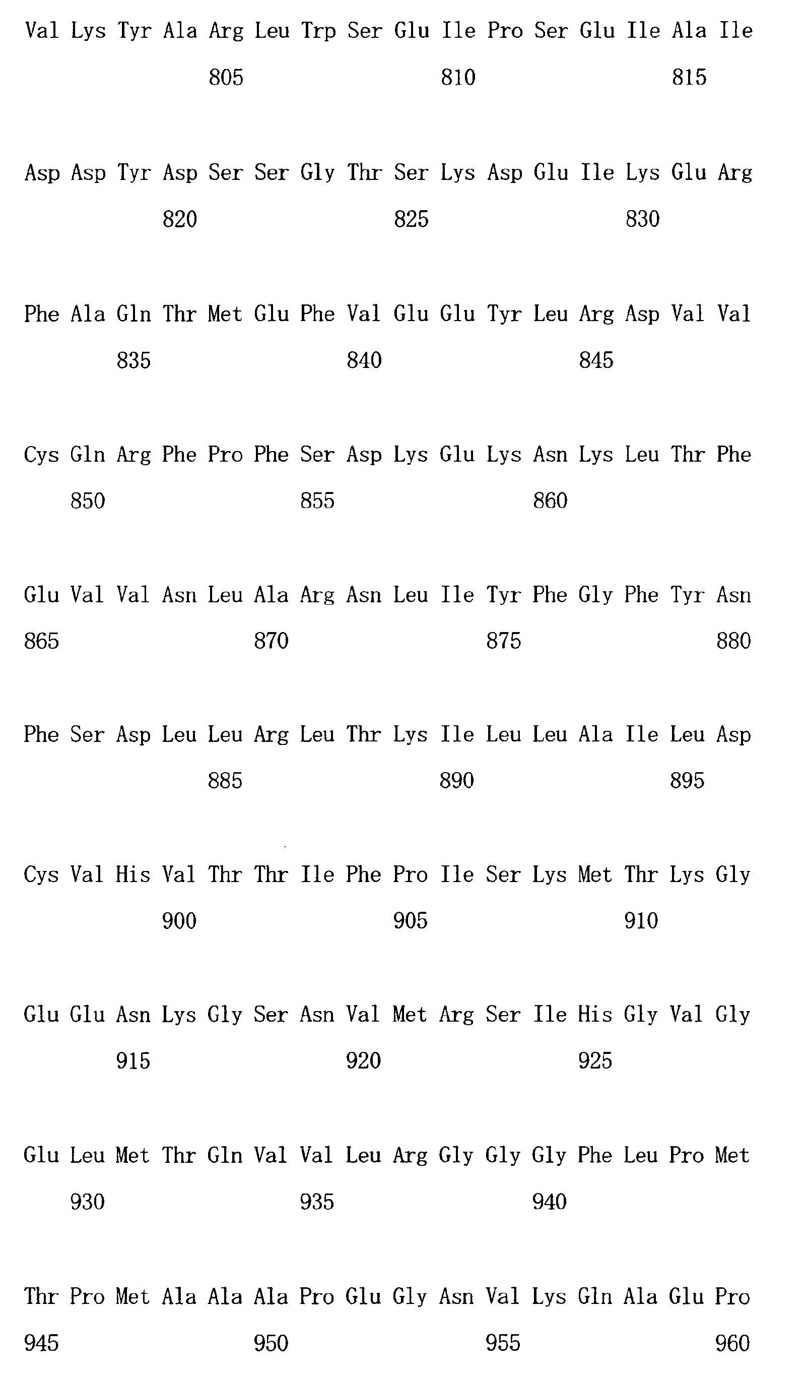
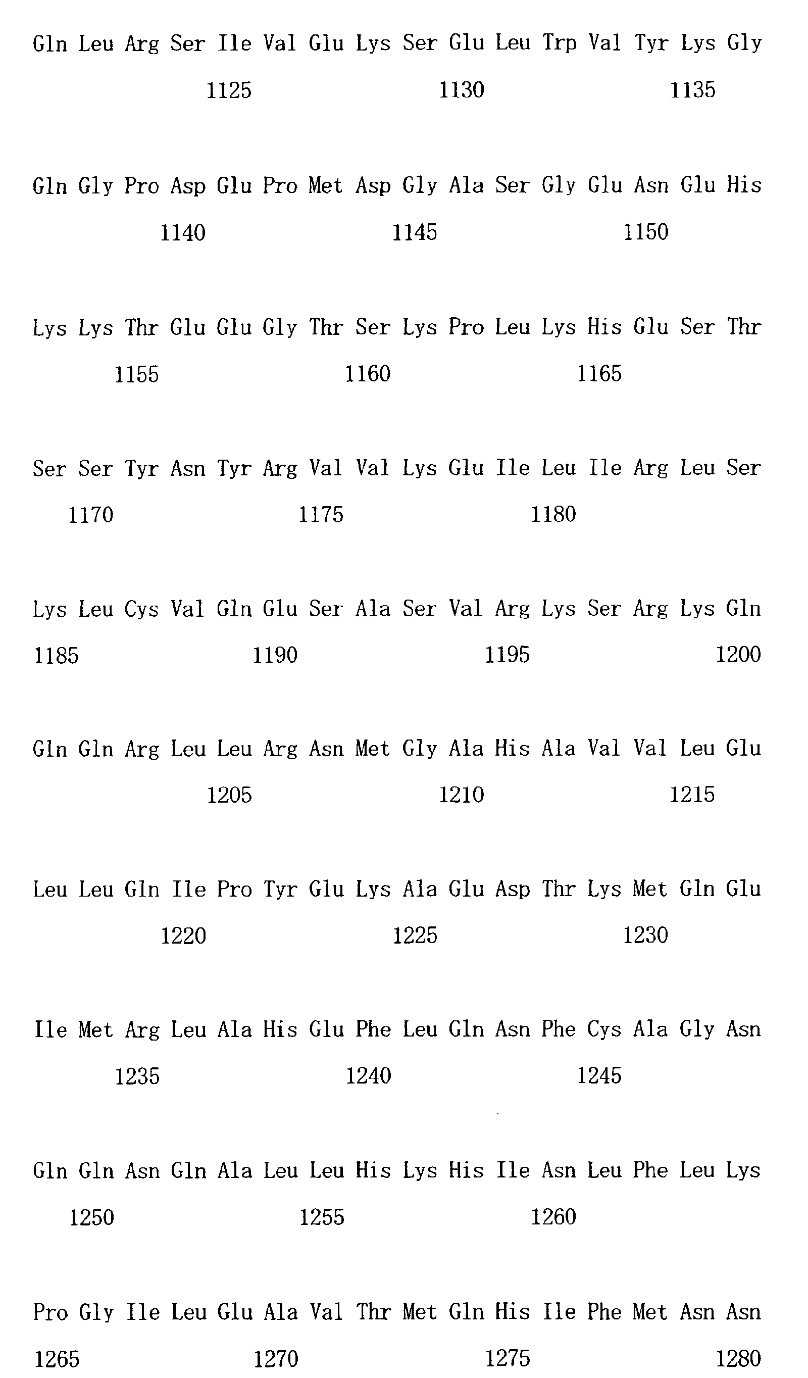
- IP 3 R1 the amino acid sequence is represented by SEQ ID NO: 7
- IRBIT IP 3 R-binding protein released with inositol 1,4,5-trisphosphate
- IRBIT binds to IP 3 R1 in vitro and in vivo, and the subcellular localization agrees well with that of IP 3 R1. Further, it was also revealed that IRBIT is dissociated from IP 3 R1 by IP 3 at a physiological concentration. Based on these results, we have considered that IRBIT is useful as an IP 3 indicator, modulator of IP 3 -induced Ca 2+ releasing activity, and modulator of PIP 2 function and Na 2+ and bicarbonate ions, and thus completed the present invention.
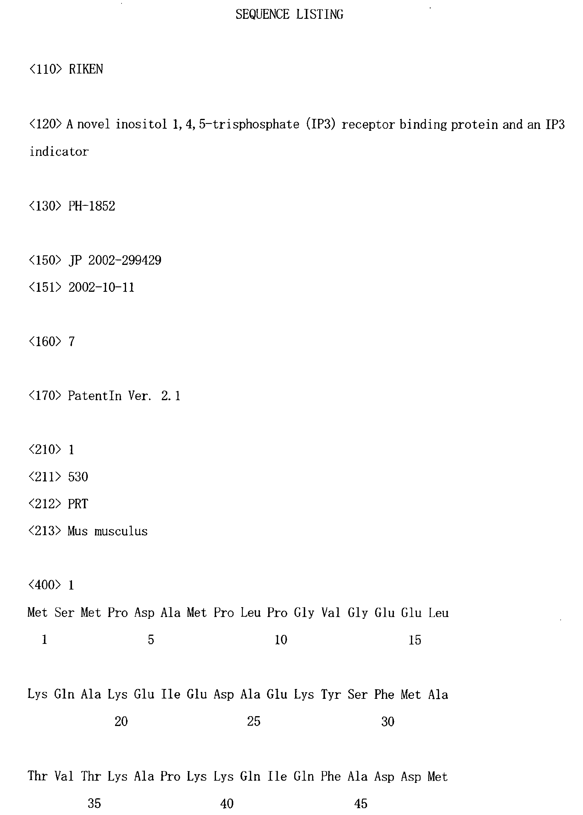
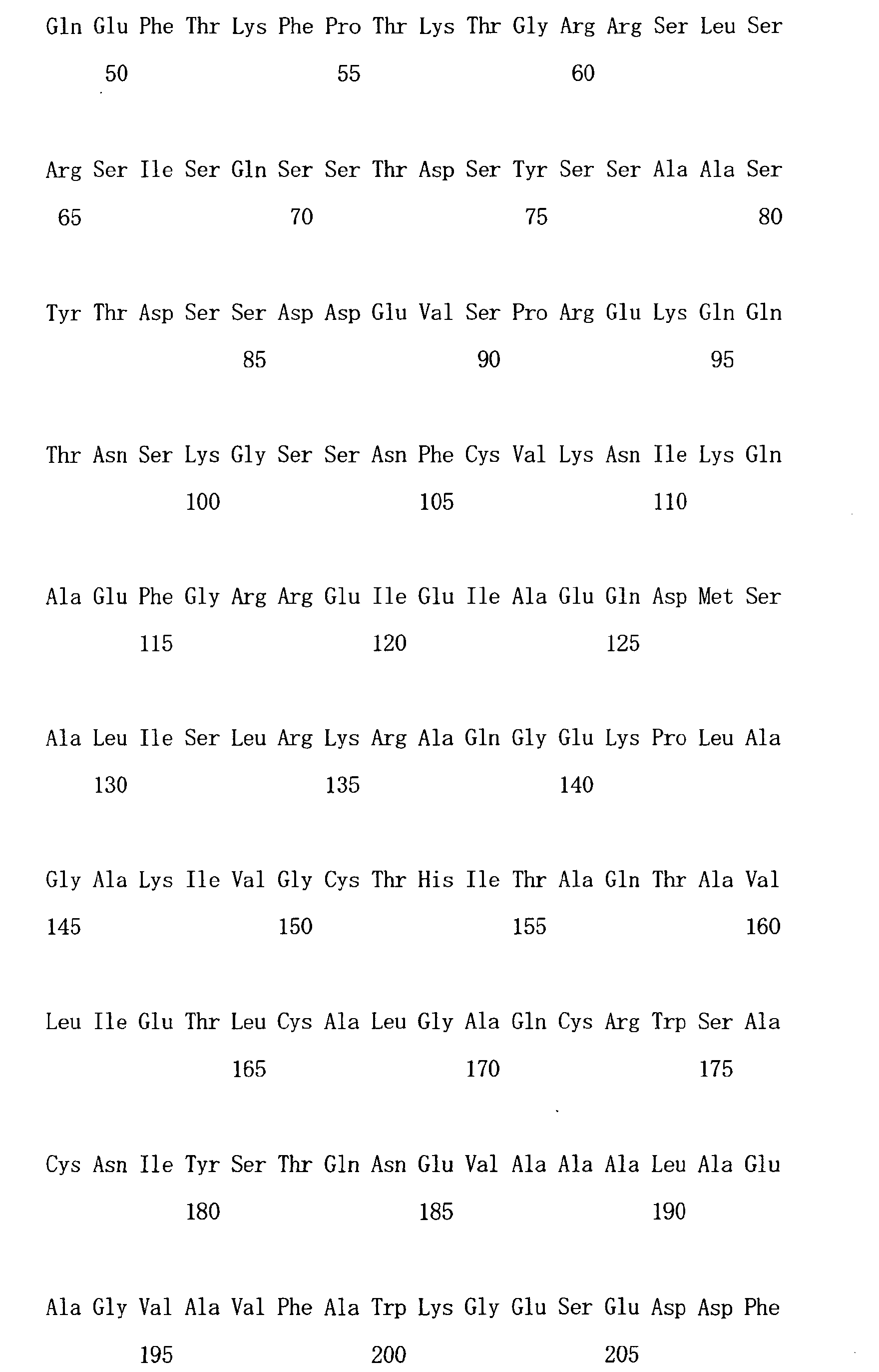
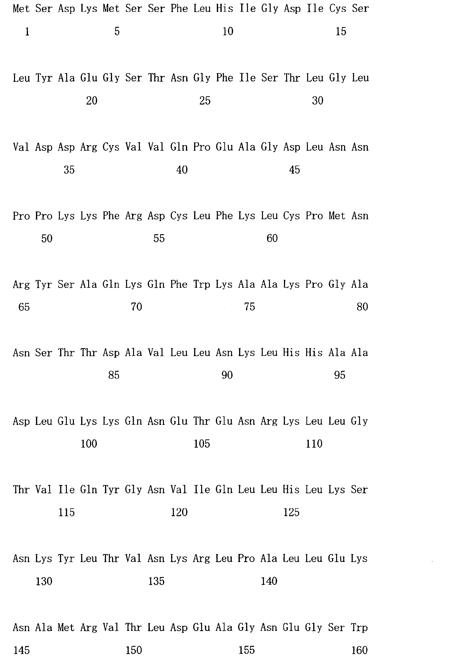
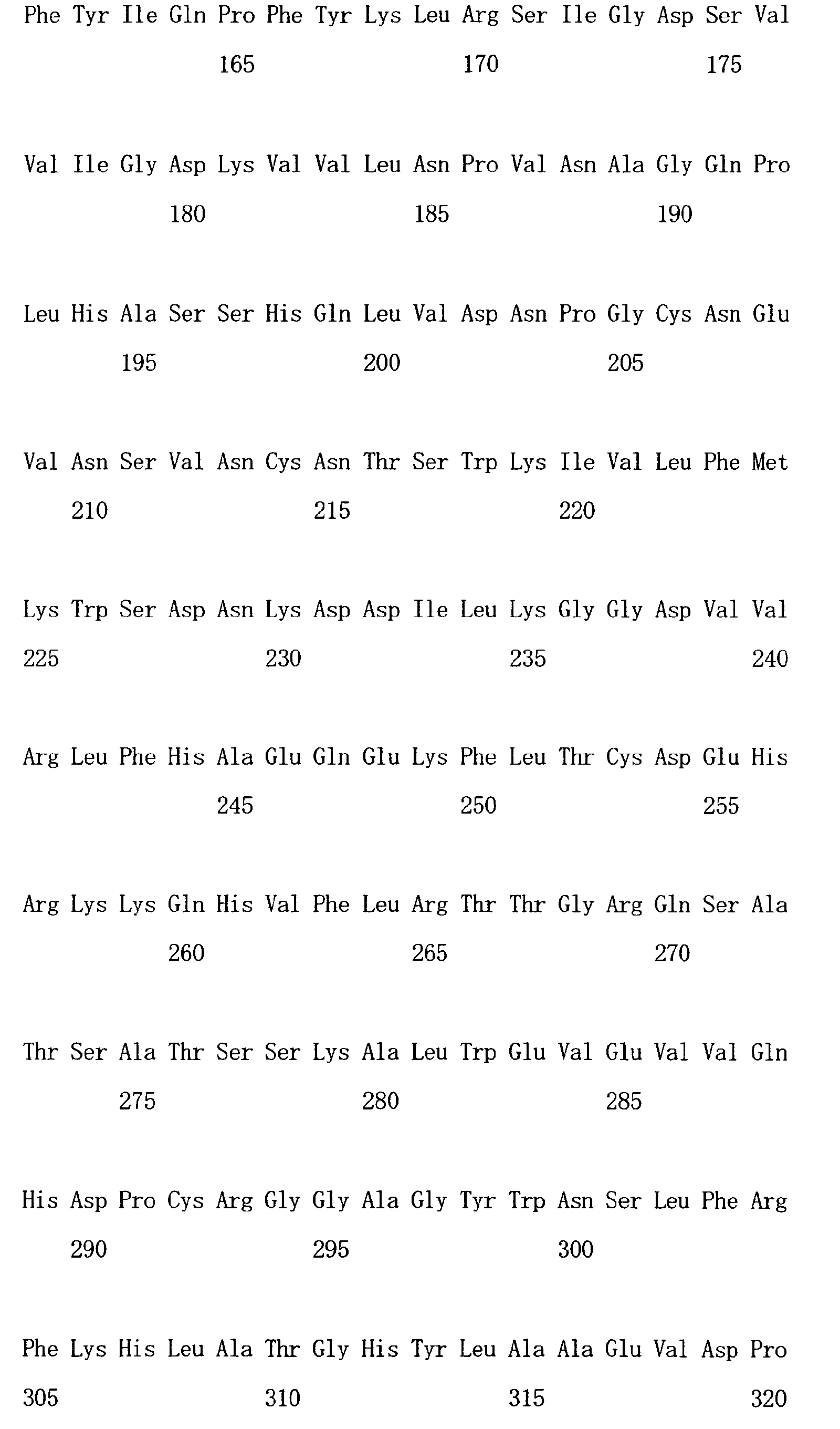
- a novel IP 3 receptor protein, IRBIT that we have newly cloned comprises the amino acid sequence represented by SEQ ID NO: 1.
- the IRBIT is composed of two reagions, the N-terminal region (amino acids 1-104) and the C-terminal region (amino acids 105-530).
- the C-terminal region is homologous to S-adenosylhomocysteine hydrolase but does not show enzyme activity, and the N-terminal region is an essential region for binding with the IP 3 receptor (in this specification, the IP 3 receptor is referred to as any subtype of the IP 3 receptor including IP 3 R1 and IP 3 receptor type II (IP 3 R2)).
- IP 3 R2 IP 3 receptor type II
- the present invention encompasses IRBIT protein, the N-terminal region (amino acids 1-104) that is essential for binding of IRBIT to the IP 3 receptor, and proteins containing these amino acid sequences.
- a protein containing the 1-277 amino acid sequence of IRBIT is preferred.
- the present invention also encompasses a protein, which is a mutant of these proteins that maintain IP 3 receptor-binding activity, and comprises an amino acid sequence derived from the amino acid sequence of IRBIT , which contains a deletion, substitution or addition of at least one of amino acids, preferably 1 to 50, more preferably 1 to 20, further preferably 1 to 10, and particularly preferably 1 to 5 amino acid(s).
- the present invention further encompasses genes encoding these proteins.
- the gene is, for example, DNA comprising the nucleotide sequence represented by SEQ ID NO: 2 encoding IRBIT protein, or comprising a nucleotide sequence of the 1-312 nucleotides of SEQ ID NO: 2 encoding the N-terminal region (amino acids 1-104) that is the IP 3 receptor-binding domain of IRBIT.
- the present invention also includes these DNAs or DNAs hybridizing under stringent conditions to complementary strands of the DNAs.
- the stringent conditions are referred to as, for example, conditions with a sodium concentration of 10 mM to 300 mM, and preferably 37 to 65°C, and more preferably 42°C.
- the present invention also includes a DNA comprising a nucleotide sequence derived from the nucleotide sequence of SEQ ID NO: 2 or the nucleotide sequence of the 1-312 of SEQ ID NO: 2, which contains a deletion, substitution or addition of at least one of nucleotides, and encoding a protein having IP 3 receptor-binding activity.
- the number of nucleotides to be deleted, substituted or added is not specifically limited, and is preferably 1 to 100, more preferably 1 to 50, further more preferably 1 to 20, and most preferably 1 to 10.
- the nucleotide sequence of such a nucleic acid has homology to the nucleotide sequence represented by SEQ ID NO: 1 when calculated using BLAST or the like (for example, when the default of BLAST, specifically the parameters of initial conditions are used) of 70% or more, preferably 90% or more, further more preferably 95% or more, 96% or more, 97% or more, 98% or more or 99% or more.
- the present invention further encompasses RNAs corresponding to these DNAs, a vector containing these DNAs, a vector wherein a promoter is operably linked to the DNA so as to enable the expression of the above DNA, a vector wherein a DNA encoding any label for labeling the expression product (IRBIT molecule or the like) of the above DNA that is ligated in-frame to the DNA, and a host cell containing the vector (insect cells, Escherichia coli and mammalian cells are preferred, but examples are not limited thereto).
- labels may be any known labels and include, but are not limited to, labels for utilizing immunological reaction or protein-protein binding such as a histidine tag (His-tag), glutathioneS-transferase (GST) and biotin, as well as fluorescent proteins or photoproteins such as green fluorescent protein (GFP), CFP, Venus, FlAsH, ReAsH and derivatives thereof.
- His-tag histidine tag
- GST glutathioneS-transferase
- biotin as well as fluorescent proteins or photoproteins such as green fluorescent protein (GFP), CFP, Venus, FlAsH, ReAsH and derivatives thereof.
- IRBIT is a protein that can be dissociated from the IP 3 receptor by IP 3 selectively in a concentration-dependent manner.
- the IP 3 receptor is the IP 3 receptor type I
- the EC50 value for the above dissociation is approximately 0.5 ⁇ M.
- the difference may be due to a difference in buffer conditions (this assay has been performed under conditions ranging from pH 8.0 to 8.3 and with low ion intensity) based on the fact that the binding affinity of IP 3 with the IP 3 receptor is largely dependent on pH and ion intensity (62-64).
- IRBIT is dissociated from the IP 3 receptor by binding of IP 3 to the IP 3 receptor. This is supported by data, such as the results of measurement made by Luzzi et al.
- IP 3 concentration was tens of nM before stimulation, and then increased to few ⁇ M after stimulation (50).
- the EC50 of IP 3 required for the dissociation of IRBIT from IP 3 R1 is approximately 0.5 ⁇ M, and this value is within the fluctuation range of the intracellular IP 3 concentration before and after stimulation.
- IRBIT is dissociated from the IP 3 receptor following IP 3 production induced by extracellular stimulation.
- IP 3 BD IP 3 receptor complex
- the amount of IRBIT that has been dissociated from the IP 3 receptor is quantified, or conversely the amount of IRBIT-IP 3 receptor/IP 3 BD complex remaining after contact with IP 3 is quantified, so as to make it possible to quantitatively detecting IP 3 .
- the same result is obtained when a protein containing at least the IP 3 receptor-binding domain of IRBIT is used.
- Quantitative detection of free IRBIT or an IRBIT-IP 3 receptor complex can also be performed by immunological techniques.
- antibody in the present specification includes complete antibody molecules, fragments retaining antigen-binding ability derived from the complete antibody, and derivatives thereof.
- Antibodies against IRBIT may be monoclonal or polyclonal antibodies, both of which can be obtained by any general method that involves immunizing animals with an IRBIT molecule or a peptide (a part of the IRBIT molecule).
- a purification method of these antibody molecules and a purification method which involves cleaving into antibody fragments having binding affinity for IRBIT are also known by persons skilled in the art. Any appropriate method can be selected from these methods, as desired. Persons skilled in the art can readily detect free IRBIT using these antibody molecules.
- IP 3 R1 is previously bound to a solid phase support (beads, assay plate surfaces or the like), and then IRBIT bound via IP 3 R1 to the solid phase can be detected.
- IRBIT is labeled with an appropriate molecule, and then the labels are detected, so that free IRBIT or an IRBIT-IP 3 receptor/IP 3 BD complex can be quantitatively detected.
- known molecules for labeling proteins include radioactive labels, immuno-labels, dye labels, fluorescent labels and luminescent labels. Labeling can be performed by binding any one of these labeling molecules to an IRBIT molecule by any known method according to the labeling molecule. Persons skilled in the art can also readily detect signals derived from the labels used herein based on the known art.
- a complex (referred to as an IRBIT-IP 3 receptor/IP 3 BD complex) comprising a protein that contains at least IRBIT or the IP 3 receptor-binding domain thereof and the IP 3 receptor or IP 3 BD is prepared. Since IRBIT forms a complex with the IP 3 receptor intracellularly, it can also be purified from natural cells (cerebellum neurons and the like) expressing the IP 3 receptor. Alternatively, from cells ( Escherichia coli, Sf9 insect cells or the like) forced to express an IRBIT or a protein containing at least the IP 3 receptor-binding domain of the IRBIT and a protein containing IP 3 receptor molecules or IP 3 BD, a microsome fraction can be fractionated by a biochemical fractionation method.
- the protein can be extracted using the labels from a cell lysate.
- other known purification methods column chromatography and the like may also be used.
- IRBIT and IP 3 PD are separately prepared, and then a complex thereof can be formed.
- GST-IP 3 BD expressed in Escherichia coli is purified with glutathione-Sepharose, and then a protein containing IRBIT or its IP 3 receptor-binding region is expressed in Escherichia coli or Sf9 cells. Proteins containing the IRBIT or its IP 3 receptor-binding region are allowed to contact with GST-IP 3 BD to form complexes, followed by glutathione-Sepharose pull-down, so that the complexes can be purified.
- the above complex may also be prepared by pulling down IRBIT in the tissue with GST-IP 3 BD using glutathione-Sepharose from, for example, a high salt extract from the cerebellum.
- IP 3 A sample containing IP 3 is allowed to contact with the thus prepared IRBIT-IP 3 receptor/IP 3 BD complex (prepared, for example, at approximately 1 to 100 ⁇ M in 10 mM Hepes, 100 mM NaCl, 2 mM EDTA and 1 mM 2-ME (pH7.4)), followed by incubation on ice for approximately 5 to 30 minutes. Then, free IRBIT or the remaining IRBIT-IP 3 receptor/IP 3 BD complex present in the solution is detected.
- IRBIT-IP 3 receptor/IP 3 BD complex prepared, for example, at approximately 1 to 100 ⁇ M in 10 mM Hepes, 100 mM NaCl, 2 mM EDTA and 1 mM 2-ME (pH7.4)
- the IRBIT-IP 3 receptor/IP 3 BD complex to be used in reaction is previously bound to solid phase carriers, and then the solid phase carriers are separated from the liquid phase after reaction, so that free IRBIT in the liquid phase can be detected, or IRBIT in the IRBIT-IP 3 receptor/IP 3 BD complex bound to the solid phase carriers can be detected.
- IRBIT may be detected with an immunological reaction system using an antibody that recognizes IRBIT. When labeled IRBIT is used, IRBIT can also be detected by detecting the label.
- IP 3 can also be quantified by a method using fluorescence resonance energy transfer, FRET.
- Fluorescence resonance energy transfer is a phenomenon wherein when two fluorephores are in close proximity and in the right orientation, excitation energy transfers from one fluorophore (energy donor) to the other fluorophore (energy acceptor) on the longer wavelength side.
- FRET Fluorescence resonance energy transfer
- IRBIT binds to the IP 3 BD of the IP 3 receptor, and is dissociated from the IP 3 receptor by IP 3 .
- IP 3 BD are labeled respectively with a combination of 2 types of fluorescent labels that are applicable to FRET.
- IRBIT is labeled with a yellow fluorescent protein (YFP) or with the improved protein thereof, Venus
- IP 3 BD is labeled with cyan fluorescent protein (CFP).
- YFP yellow fluorescent protein
- CFP cyan fluorescent protein
- IRBIT binds to IP 3 BD and FRET occurs from CFP to Venus.
- the two are dissociated from each other, and no FRET occurs.
- Venus-IRBIT and CFP-IP 3 BD are bound via a linker sequence (Venus-IRBIT-IP 3 BD-CFP)
- the molar ratios of the two will be the same, and this is efficient.
- IP 3 concentration is measured in vitro
- Venus-IRBIT-IP 3 BD-CFP protein is added to a cell extract or the like, and then FRET is measured.
- introduction of a Venus-IRBIT-IP 3 BD-CFP gene into a cell enables temporal and spatial analysis of changes in IP 3 concentration within living cells. Measurement with the FRET method can be performed using a fluorescent microscope, fluorometer or the like.
- IP 3 detection methods using the FRET method will be described in more detail as follows.
- a Venus-IRBIT-IP 3 BD-CFP expression vector is prepared.
- Four cDNAs are amplified by PCR, each of which encodes a full-length IRBIT or a deletion mutant thereof containing a region sufficient for binding with IP 3 BD (e.g., a mutant comprising amino acid sequence 1-104 or amino acid sequence 1-277), a protein containing the IP 3 binding region of IP 3 receptor type I (a region comprising amino acid sequence 224-604 of SEQ ID NO: 7), Venus, and CFP.
- these 4 cDNAs are inserted into a mammalian cell expression vector (e.g., pcDNA3) so as to match the reading frames.
- the order for insertion is not specifically limited.
- the order of Venus-IRBIT-IP 3 BD-CFP wherein IRBIT and IP 3 BD are placed inside and Venus and CFP are placed outside, can be employed.
- the cDNAs are inserted into an Escherichia coli expression vector (e.g., pET) or Sf9 expression vector (e.g., pFastBac).
- a tag e.g., a His tag
- Venus-IRBIT-IP 3 BD-CFP is expressed in Escherichia coli or Sf9 cells, and is purified with ProBond resin (Invitrogen) or the like when a His tag is added. In some cases, it may be purified with an ion exchange column or a gel filtration column.
- IP 3 solution having a known concentration is added to the purified Venus-IRBIT-IP 3 BD-CFP protein.
- the excitation wavelength (at around 440 nm) of CFP is applied to the solution, and then the CFP fluorescent wavelength (at around 480 nm, CFP fluorescence) and Venus fluorescent wavelength (at around 535 nm, FRET fluorescence) are measured.
- a calibration curve is then determined by plotting IP 3 concentrations versus FRET fluorescence-to-CFP fluorescence ratios.
- Venus-IRBIT-IP 3 BD-CFP protein is added to a sample (e.g., a cell extract solution) with an unknown IP 3 concentration to find a FRET fluorescence-to-CFP fluorescence ratio, and then the ratio is applied to the calibration curve, thereby quantifying IP 3 concentration.
- a sample e.g., a cell extract solution
- the ratio is applied to the calibration curve, thereby quantifying IP 3 concentration.
- the IP 3 concentration in a living cell can also be measured by the FRET method.
- a pcDNA-Venus-IRBIT-IP 3 BD-CFP gene is transfected into a culture cell (e.g., Cos-7 and HeLa) using a transfection reagent (e.g., TransIT (Mirus)).
- a transfection reagent e.g., TransIT (Mirus)
- TransIT TransIT
- cells on the glass bottom dish are observed using an inverted fluorescence microscope equipped with a cool CCD camera (e.g., IX71 (Olympus)).
- the CFP excitation wavelength (at around 440 nm) is applied, and then the CFP fluorescent wavelength (at around 480 nm, CFP fluorescence) and Venus fluorescent wavelength (at around 535 nm, FRET fluorescence) are measured.
- the FRET efficiency is quantified by taking the ratio of the two fluorescence intensities.
- An IP 3 -producing agonist (ATP in the case of Cos-7, histamine in the case of HeLa, and the like) is added to the glass-bottom dish, and then the FRET efficiency is measured with time. When IP 3 is produced by the agonist, the FRET-to-CFP ratio is expected to be lowered.
- a combination of fluorescent labels Venus and CFP
- a combination of YFP and CFP or FlAsH and CFP can also be used, and use is not limited thereto.
- various fluorescent molecules to be used for the FRET method are expected to be developed in the future, and they can also be used for implementing the present invention.
- the present invention also encompasses the IP 3 indicator of the present invention or a kit for IP 3 detection to be used for the IP 3 detection method of the present invention.
- the indicator or the kit contains a protein containing at least the IP 3 receptor-binding region of IRBIT or a DNA or an RNA encoding the protein.
- the indicator or the kit contains a protein containing at least the IP 3 -binding region of IP 3 receptor, or a DNA or an RNA encoding the protein.
- the indicator or the kit may also contain a labeling reagent (e.g., a fluorescent labeling compound) and/or an antibody that recognizes the above protein.
- the cDNA encoding the N-terminal region (amino acids 1-225) of mouse IP 3 R1 was inserted into glutathione S-transferase (GST) fusion vector pGEX-KG (40).
- GST-IP 3 R1 (1-225) fragment was subcloned into the baculovirus transfer vector pBlueBac4.5 (Invitrogen).
- the region located downstream from Sma I site of the GST-IP 3 R1 (1-225) was replaced with the Sma I- Eco R I fragment of mouse IP 3 R1 (corresponding to amino acids 79-2217) to construct a GST-IP 3 R1 (1-2217) vector (termed GST-EL).
- a fragment encoding GST alone was subcloned into pBlueBac4.5 as a control.
- Sf9 cells were cultured in TNM-FH medium supplemented with 10% fetal bovine serum at 27°C.
- Recombinant baculoviruses carrying GST-EL or GST gene were generated with Bac-N-BlueTM Transfection Kit (Invitrogen) according to the instructions thereof.
- GST-EL and GST were expressed in 2 ⁇ 10 8 Sf9 cells by infecting recombinant baculoviruses at a multiplicity of infection of 5, and incubating for 48 hours. The cells were harvested and stored at -80°C.
- the cryopreserved cells were suspended in 10 mM Hepes (pH 7.4), 100 mM NaCl, 2 mM EDTA, 1 mM 2-mercaptoethanol (2-ME), 0.1% Triton X-100, and protease inhibitors (1 mM phenylmethylsulfonyl fluoride (PMSF), 10 ⁇ M leupeptin, 2 ⁇ M pepstatin A and 10 ⁇ M E-64), and were homogenized with a glass-Teflon homogenizer (1000 rpm, 10 strokes). The homogenate was centrifuged at 20,000 ⁇ g for 30 minutes.
- the supernatant was incubated with 3 ml of glutathione-Sepharose 4B (Amersham Pharmacia Biotech) for 3 hours at 4°C. After washing 8 times with 40 ml of 10 mM Hepes (pH 7.4), 250 mM NaCl, 2 mM EDTA, 1 mM 2-ME, and 0.1% Triton X-100, GST-EL or GST coupled with glutathione-Sepharose was packed into columns and equilibrated with 10 mM Hepes (pH 7.4), 100 mM NaCl, 2 mM EDTA, 1 mM 2-ME, and 0.1% Triton X-100. Approximately 5 mg of GST-EL was immobilized.
- Rat cerebella (approximately 5 g) were homogenized in 45 ml of a homogenize buffer (10 mM Hepes (pH 7.4), 20 mM Sucrose, 2 mM EDTA, 1 mM 2-ME and protease inhibitors) with a glass-Teflon homogenizer (950 rpm, 10 strokes).
- the homogenate was centrifuged at 1,000 ⁇ g for 10 minutes, and then the supernatant (S1 fraction) was further centrifuged at 100,000 ⁇ g for 60 minutes to obtain the cytosolic fraction (the supernatant) and the crude microsome (the pellet).
- the crude microsome was homogenized in 25 ml of a homogenize buffer containing 500 mM NaCl with a glass-Teflon homogenizer (1,200 rpm, 10 strokes), incubated on ice for 15 minutes, and then centrifuged at 100,000 ⁇ g for 60 minutes to obtain the high salt extract (supernatant) and the stripped-crude microsome (the pellet).
- the high salt extract was diluted 5-fold with a dilution buffer (10 mM Hepes (pH 7.4), 2 mM EDTA, 1 mM 2-ME, 0.01% Brij 35 and protease inhibitors).
- the diluted high salt extract was pre-cleared with glutathione-Sepharose and loaded into GST-EL affinity column equilibrated with a binding buffer (10 mM Hepes (pH 7.4), 100 mM NaCl, 2 mM EDTA and 1 mM 2-ME).
- a GST column was used as a control.
- the columns were washed with the binding buffer in a quantity 20 times greater than the volume of the column and proteins bound to the column were eluted with the binding buffer containing 50 ⁇ M IP 3 (Dojindo) and 0.05% Brij 35.
- the eluted material was concentrated, separated by 10% SDS-polyacrylamide gel electrophoresis (PAGE), and stained with Coomassie Brilliant Blue (CBB).
- the approximate 60-kDa protein band was excised from the gel and digested with lysyl endopeptidase (Wako) according to the known method (41).
- the digested peptides were separated using a C-18 reversed-phase column ( ⁇ RPC C2/C18 SC 2.1/10, Amersham Pharmacia Biotech) connected to a SMART system (Amersham Pharmacia Biotech).
- the amino acid sequence of each peptide was determined by 494 precise protein sequencer (Applied Biosystems). Two peptide sequences, N-YSFMATVTK-C (SEQ ID NO: 3) and N-QIQFADDMQEFTK-C (SEQ ID NO: 4) were obtained.
- cDNA of IRBIT was amplified by polymerase chain reaction (PCR) using mouse cerebellum oligo dT-primed, first-strand cDNA as a template.
- PCR polymerase chain reaction
- the PCR product was cloned in pBluescript II KS(+) (Stratagene) and sequenced. Sequences of the three independent clones were confirmed.
- the cDNAs encoding the full-length and N-terminal region (amino acids 1 - 104) of IRBIT were subcloned into the Escherichia coli hexahistidine (His) fusion vector pET-23a(+) (Novagen) to generate IRBIT-His and IRBIT(1-104)-His expression vectors, respectively.
- the same cDNAs were subcloned into the GST fusion vector pGEX-4T-1 (Amersham Pharmacia Biotech) to generate GST-IRBIT and GST-IRBIT(1-104) expression vectors, respectively.
- the cDNA fragments encoding the amino acids 1-225, 1-343, 341-923, 600-1248, 916-1581 and 1553-1943 of the amino acid sequence (SEQ ID NO: 7) of mouse IP 3 R1 were inserted into pGEX-KG to generate GST-Ia, GST-Iab, GST-IIab, GST-IIbIIIa, GST-IIIab and GST-IV expression vectors, respectively.
- the amino acids 1593-2217 of mouse IP 3 R1 were inserted into pGEX-4T-1 to obtain GST-IV-Va expression vector. These fusion proteins were expressed in Escherichia coli.
- GST-EL was expressed in Sf9 cells as described above.
- GST-IbIIa amino acids 224-604 of mouse IP 3 R1
- GST-IbIIa was referred to as G224 in the reference.
- a Japanese White rabbit was immunized with the purified IRBIT(1-104)-His by subcutaneous injection with the complete Freund's adjuvant at 14-day intervals.
- the anti-IRBIT antisera were passed through a GST-IRBIT(1-104) covalently coupled with cyanogen bromide-activated Sepharose 4B (Amersham Pharmacia Biotech), and antibodies specifically bound to the column were eluted with 100 mM glycine-HCl (pH 2.5).
- the cerebrum, cerebellum, heart, lung, liver, kidney, thymus, spleen, testis and ovary were removed from adult mice and S1 fraction was obtained as described above.
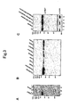
- the cytosolic fraction, crude microsome, high salt extract, and stripped-crude microsome of mouse cerebellum were obtained in a manner similar to the above method. Proteins with the amount indicated in Fig. 3 were subjected to 10% SDS-PAGE and transferred electrically onto polyvinylidene fluoride (PVDF) membrane.
- PVDF polyvinylidene fluoride
- the membranes were incubated with anti-IRBIT antibody (1 ⁇ g/ml) for 1 hour at room temperature, followed by blotting with horseradish peroxidase-conjugated donkey anti-rabbit IgG (Amersham Pharmacia Biotech). Immunoreactive bands were detected with an enhanced chemiluminescence detection system (Amersham Pharmacia Biotech).
- the cDNA encoding the full-length IRBIT was subcloned into the pcDNA3 (Invitrogen).
- the cDNA encoding the full-length IRBIT or its deletion mutants were subcloned into the pEGFP-C1 (Clontech) to generate green fluorescent protein (GFP) fusion protein expression vectors.
- Mouse IP 3 R1 expression vector pBact-STneoB-C1 used herein was known previously (44).
- Cos-7 cells were cultured in DMEM medium supplemented with 10% fetal bovine serum, penicillin, and streptomycin at 37°C. Transient transfections were performed using TransIT transfection reagents (Mirus) according to the instructions attached thereto. Two days after transfection, the transfected cells were used for immunoblotting, pulldown experiments, or immunostaining.
- Mouse cerebellar cytosolic fraction was diluted 2-fold with 10 mM Hepes (pH 7.4), 200 mM NaCl, 2 mM EDTA, 1 mM 2-ME and 0.02% Triton X-100.
- the high salt extract was diluted 5-fold with 10 mM Hepes (pH 7.4), 2 mM EDTA, 1 mM 2-ME and 0.01% Triton X-100.
- the diluted fractions (the final NaCl concentration of both fractions was 100 mM) were incubated with 20 ⁇ g of GST-EL or GST for 2 hours at 4°C.
- the diluted high salt extract was incubated in the presence of or the absence of Escherichia coli alkaline phosphatase (Toyobo) with 2 mM MgCl 2 for 30 minutes at 37°C. 5 mM EDTA was added, and then the resultant was subjected to pulldown assay as described above.
- Escherichia coli alkaline phosphatase Toyobo
- 5 mM EDTA was added, and then the resultant was subjected to pulldown assay as described above.
- IRBIT in the diluted high salt extract was pulled down with GST-EL for precipitation and washed as described above.
- 100 ⁇ l of a wash buffer containing IP 3 inositol 4,5-bisphosphate (IP 2 ) (Dojindo), inositol 1,3,4,5-tetrakisphosphate (IP 4 ) (Calbiochem), inositol 1,2,3,4,5,6-hexakisphosphate (IP 6 ) (Calbiochem) or ATP (Amersham Pharmacia Biotech) (0.1, 0.3, 1, 3 or 10 ⁇ M) were added.
- the diluted high salt extract was subjected to pulldown assay as described above with 100 pmol of GST, GST-EL, GST-Ia, GST-Iab, GST-IbIIa, GST-IIab, GST-IIbIIIa, GST-IIIab, GST-IV, GST-IV-Va, K508A or R441 Q, and analyzed by Western blotting with anti-IRBIT antibody.
- Cos-7 cells expressing GFP-tagged full-length IRBIT or its truncated mutants were lysed in a lysis buffer (10 mM Hepes (pH7.4), 100 mM NaCl, 2 mM EDTA, 1 mM 2-ME, 0.5% Nonidet P-40, and protease inhibitor) for 30 minutes at 4°C, followed by centrifugation (100,000 x g, 30 minutes).
- the supernatants were subjected to pulldown assay with GST-EL or GST as described above, and bound proteins were subjected to immunoblot analysis with anti-GFP antibody (Medical & Biological Laboratories).
- Transfected Cos-7 cells cultured on glass cover slips washed once in phosphate-buffered saline (PBS), fixed in 4% formaldehyde-containing PBS for 15 minutes, permeabilized in 0.1% Triton X-100-containing PBS for 5 minutes, and then blocked in PBS containing 2% goat serum for 60 minutes at room temperature.
- PBS phosphate-buffered saline
- the cell membranes of transfected cells were punctured in an ice-cold permeabilization buffer (80 mM PIPES (pH 7.2), 1 mM MgCl 2 , 1 mM EGTA and 4% polyethylene glycol) containing 0.1% saponin, and washed twice with the ice-cold permeabilization buffer, followed by fixation.
- the cells were allowed to react with rabbit anti-IRBIT antibody (1 ⁇ g/ml, room temperature, 60 minutes) and rat anti-IP 3 R1 antibody 18A10 (45) (overnight at 4°C).
- Alexa 488-conjugated goat anti-rabbit IgG and Alexa 594-conjugated goat anti-rat IgG were applied to reaction for 45 minutes at 37°C.
- the cover slips were mounted with Vectashield (Vector Laboratories) and observed via IX-70 confocal fluorescence microscopy (Olympus) with a 60x objective lens.
- Detergent-solubilized extract from the crude microsome was prepared with 1% Nonidet P-40-containing 50 mM Hepes (pH 7.4), 1 mM MgCl 2 , and protease inhibitors for 30 minutes at 4°C, and centrifuged at 20,000 ⁇ g for 30 minutes.
- the supernatant was diluted 10-fold in an immunoprecipitation buffer (10 mM Hepes (pH 7.4), 150 mM NaCl, 1 mM MgCl 2 , and 1% Nonidet P-40) and incubated with rabbit anti-IRBIT antibody (3 ⁇ g) or control rabbit IgG (3 ⁇ g) for 2 hours at 4°C.
- Protein G Sepharose 4 fast flow (Amersham Pharmacia Biotech) was added for reaction with immune complexes for 2 hours at 4°C. Resins were washed three times with an immunoprecipitation buffer and subjected to Western blotting with anti-IRBIT antibody or mouse anti-IP 3 R1 antibody KM1112 (47).
- the N-terminal region of 2217 amino acid residues encoding the greater part of the cytoplasmic portion of mouse IP 3 R1 containing the IP 3 binding domain and regulatory domain (10, 11) was used as a fusion protein (GST-EL) with GST.
- GST-EL and GST were expressed by the baculovirus/Sf9 cell system and conjugated to glutathione-Sepharose.
- the fraction extracted with a high salt buffer (containing 500 mM NaCl) from the crude microsome of cerebella was thought to be enriched with membrane-bound proteins.
- the fraction was loaded into a glutathione-Sepharose affinity column in which GST-EL or GST was immobilized.
- the cDNA of the above 60-kDa protein was obtained by reverse transcriptase-PCR from a mouse cerebellum.
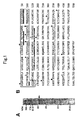
- the predicted amino acid sequence of the cloned cDNA revealed a protein composed of 530 amino acid residues (Fig. 1B), with a calculated molecular mass of 58.9 kDa, which was almost the same as its predicted molecular mass estimated by SDS-PAGE.
- the 60-kDa molecule is termed IRBIT.
- the C-terminal region (amino acids 105-530) of IRBIT was shown to be homologous (51% identical and 74% similar) to the methylation pathway enzyme S-adenosylhomocysteine hydrolase (EC3.3.1.1.) (48) (Fig. 1, C and D).
- the region on the N-terminal side (amino acid residues 1 to 104) of IRBIT had no homology with known proteins and contained a serine-rich region (amino acid residues 62 to 103) (Fig. 1B and D).
- IRBIT sequence Motif searches of the IRBIT sequence revealed the presence of a putative coiled-coil motif (amino acids 111-138) and a putative NAD+ binding region (amino acids 314-344) (Fig. 1B and D). There were 17 potential phosphorylation sites for protein kinases such as casein kinase II, PKC, PKA/PKG and tyrosine kinases, out of which seven sites were concentrated on the N-terminal side (Fig. 1B). Putative membrane-spanning regions and signal sequences were not found.
- protein kinases such as casein kinase II, PKC, PKA/PKG and tyrosine kinases
- IRBIT had homology with S-adenosylhomocysteine hydrolase, which catalyzes the reversible hydrolysis of S-adenosylhomocysteine to adenosine and homocysteine
- IRBIT-His C-terminally His-tagged IRBIT
- GST-IRBIT N-terminally GST-tagged IRBIT
- IRBIT has no S-adenosylhomocysteine hydrolase activity.
- Crystallographic studies and site-directed mutagenesis studies (43, 52-56) have determined the amino acid residues of S-adenosylhomocysteine hydrolase involved in substrate binding or NAD+ binding.
- IRBIT showed no S-adenosylhomocysteine hydrolase activity is probably due to substitution of amino acids important for substrate binding of S-adenosylhomocysteine hydrolase, such as Lysine-54, Phenylalanine-302 and Histidine-353 (Fig. 1C).
- Fig. 1C the possibility that IRBIT has enzyme activity with different substrate specificity is not excluded.
- IRBIT An affinity-purified antibody against the N-terminal region of IRBIT (Fig. 1B, boxed) was generated.
- the cDNA of IRBIT was transfected into Cos-7 cells and the cell lysates were analyzed by immunoblotting with the anti-IRBIT antibody.
- the anti-IRBIT antibody recognized only a protein located approximately at 60 kDa.
- the molecular mass of the exogeneously expressed IRBIT (Fig. 3A, lane 1) was the same as that of the endogenous protein in Cos-7 (Fig. 3A, lane 3), confirming that the above cDNA encodes the full-length IRBIT.
- the expression of IRBIT in mouse tissue was examined by immunoblot analysis with this anti-IRBIT antibody. IRBIT was detected ubiquitously, with the highest expression in the cerebrum and cerebellum (Fig. 3B).
- IRBIT is present in both cytosolic and crude microsome fractions (Fig. 3C, lanes 2 and 3). The crude microsome was further separated in a peripherally membrane-bound fraction (the fraction from which IRBIT had been originally purified) and a stripped-membrane fraction. As shown in Fig. 3C, IRBIT in the crude microsome was partially extracted with a high salt buffer (Fig. 3C, lane 4). In contrast, IP 3 R1, which is an integral membrane protein of the endoplasmic reticulum, was not extracted at all (Fig. 3C, lower panel). These results indicate that IRBIT is present in cytoplasm and also binds to a membrane.
- IRBIT in high salt extract interacted with IP 3 R1 and the N-terminal region of IRBIT was essential for the interaction .
- IRBIT was present in both cytosolic and peripherally membrane-bound fractions of mouse cerebellum (Fig. 3C). Whether IRBIT in these fractions interacted with IP 3 R1 in vitro was examined by the GST pulldown method.
- the cytosol and high salt extract (specifically, the membrane-bound fraction) from the crude microsome of mouse cerebella were incubated with GST-EL or GST and binding of IRBIT to the recombinant proteins was analyzed by immunoblotting with anti-IRBIT antibody.
- Fig. 4A IRBIT in the high salt extract interacted with GST-EL, but not with GST (Fig. 4A, lanes 5 and 6). In contrast, IRBIT in the cytosolic fraction did not interact with GST-EL (Fig. 3C).
- the high salt extract was treated with alkaline phosphatase, a nonspecific phosphatase, and then incubated with GST-EL or GST.
- IRBIT in the high salt extract did not interact with GST-EL after phosphatase treatment (Fig. 4B, lane 6).
- This result suggests the possibility that the phosphorylation of IRBIT may be necessary for IRBIT to associate with IP 3 R1.
- the possibility that phosphorylation of other proteins may regulate the interaction of these proteins is not excluded.
- IRBIT has 17 potential phosphorylation sites, seven of which are concentrated on the above N-terminal region, which is necessary for the interaction with the IP 3 receptor, it is predicted that phosphorylation may be involved in the interaction with the IP 3 receptor. It was hypothesized based on these findings that the dephosphorylated form of IRBIT is free in cytosol, whereas the phosphorylated form of IRBIT is membrane-bound via the interaction with the IP 3 receptor. Although it could not be demonstrated yet, the interaction between IRBIT and the IP 3 receptor may be dualistically regulated by IP 3 , and by either direct or indirect phosphorylation.
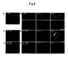
- IRBIT was diffusely distributed in cytoplasm, with no immunoreactivity in nucleus (Fig. 6A). Because IRBIT was shown to be present in both cytosolic and crude microsome fractions by biochemical fractionation experiments (Fig. 3C), visualization and observation of only the membrane-bound population of IRBIT were attempted. For this purpose, the cell membranes of Cos-7 cells co-expressing IRBIT and the IP 3 receptor were permeabilized using saponin to wash out cytosolic IRBIT prior to fixation. As shown in Fig. 6B, IRBIT was distributed in a reticular pattern (Fig. 6A).
- GFP-IRBIT (105-530)
- IRBIT does not have any sequence predicted to constitute nuclear localization signals, so that the mechanism of nuclear localization is unclear.
- IRBIT located in cytosol was washed out by saponin treatment
- GFP-IRBIT (105-530) localized only in the nucleus and the co-localization of the GFP-IRBIT with IP 3 R1 was not confirmed (Fig. 6F). This result was consistent with the results obtained by biochemical techniques to the effect that the N-terminal region of IRBIT was essential for binding to IP 3 R1 (Fig. 5B).
- IP 3 selectively dissociated IRBIT from IP 3 R1
- IRBIT was originally identified in the eluate from the GST-EL column with 50 ⁇ M IP 3 (Fig. 1A), suggesting that IP 3 disrupted the interaction between IRBIT and IP 3 R1.
- the intracellular concentration of IP 3 was at the level of several micrometers, even when it was elevated by stimulation (50).
- the IP 3 concentration of 50 ⁇ M used in the above elution is higher than the physiological range. Thus, whether or not the interaction between the proteins was disrupted by IP 3 at a physiological concentration was examined.
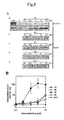
- IP 3 dissociated IRBIT from GST-EL in a concentration-dependent manner (Fig. 8Aa, lower panel). It was confirmed that GST-EL was not detected (Fig. 8Aa, upper panel), even with longer exposure.
- the EC50 IP 3 concentration required for half (50%)-maximal dissociation of IRBIT from GST-EL was approximately 0.5 ⁇ M, which was within the physiological range of IP 3 concentration (50) (Fig. 8B).
- IP 3 The dissociation efficiency of IP 3 was approximately 50 times greater than those of other inositol polyphosphates. ATP did not dissociate IRBIT even at a concentration of 10 ⁇ M. These results indicate that IRBIT was dissociated from IP 3 R1 selectively with physiological concentrations of IP 3 .
- IP 3 R1 deletion mutants constructed as GST fusion proteins based on the domain structure of IP 3 R1 (51) were used (Fig. 9A).
- GST-IbIIa amino acids 226-604
- containing the IP 3 binding core region amino acids 226-576 (11) bound to IRBIT to the same extent as GST-EL.
- other GST fusion proteins including GST-Iab and GST-IIab, did not interact with IRBIT.
- site-directed mutagenesis analysis was performed to determine the important amino acid residues of IP 3 R1 for the interaction with IRBIT.
- Lysine at position 508 of IP 3 R1 (Lys-508) is known to be an essential amino acid residue for IP 3 binding (12), and substitution of the lysine residue of GST-IbIIa with alanine (K508A) is known to show a significant loss in IP 3 binding affinity (42).
- R441Q in which arginine residue at position 441 of GST-IbIIa was substituted with glutamine, is known to have higher binding affinity for IP 3 than that of GST-IbIIa (42).
- IRBIT bound to GST-IbIIa and R441Q to the same extent, but did not bind to K508A (Fig. 9C).
- IRBIT is normally associated with IP 3 R1, and is dissociated from IP 3 R1 when IP 3 concentration is elevated by extracellular stimulation.
- IRBIT is shown to be a sole IP 3 receptor-binding protein, whose interaction with IP 3 receptor can be regulated by IP 3 .
- IRBIT lowered the affinity of IP 3 R to IP 3 in a phosphorylation dependent manner
- IRBIT The effect of IRBIT on binding of IP 3 to IP 3 R was examined.
- GST-EL 0.2 ⁇ g was incubated with 0.1, 1, or 10 ⁇ g of purified His-tagged IRBIT expressed in Sf9 cells or E. coli in a solution containing 50 mM Tris-HCl (pH 8.0), 1 mM EDTA, and 1 mM 2-mercaptoethanol, for 30 min on ice. Then 8.7 nM [ 3 H]IP 3 (PerkinElmer Life Sciences) was added to the samples and incubated for 10 min on ice (total volume was 50 ⁇ l).
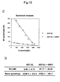
- the samples were mixed with 2 ⁇ l of 50 mg/ml ⁇ -globulin and 50 ⁇ l of 30 % PEG6000, 50 mM Tris-HCl (pH 8.0), and incubated for 5 min on ice. Non-specific binding was measured in the presence of 10 ⁇ M cold IP 3 . After centrifugation at 20,000 x g for 5 min, the precipitate was dissolved in SOLVABLETM (Packard) and the radioactivity was measured with a liquid scintillation counter (Beckman Coulter). The result is shown in Fig 10 (A). His-IRBIT expressed in Sf9 dose-dependently suppressed the IP 3 binding of GST-EL.
- Fig 10 (A) shows the plot of Scatchard analysis.
- 0.1 ⁇ g of GST-EL was incubated with or without 1 ⁇ g of purified His-tagged IRBIT expressed in Sf9 cells as described in (A).
- 2.17 nM [ 3 H]IP 3 and 2-500 nM cold IP 3 were added to the samples and incubated for 10 min on ice (total volume was 100 ⁇ l).
- Non-specific binding was measured in the presence of 10 ⁇ M cold IP 3 .
- [ 3 H]IP 3 binding to GST-EL was measured as described in (A). His-IRBIT decreased the affinity of GST-EL to IP 3 . Bmax was not changed.
- IP 3 -induced calcium release of HeLa cells was increased when the expression of IRBIT was suppressed with RNA interference
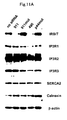
- RNA interference experiment showed that IP 3 -induced calcium release of HeLa cells was increased when the expression of IRBIT was suppressed with RNA interference (Fig 11).
- HeLa cells were transfected with 40 nM of small interference RNA (siRNA) against IRBIT (911 and 490) or control siRNA (911mut and p44mut) with LipofectAMINE2000 (Invitrogen). After two days, cells were harvested, and processed for western blotting with anti-IRBIT, anti-IP 3 R1, anti-IP 3 R2, anti-IP 3 R3, anti-SERCA2, anti-calnexin, or anti- ⁇ -actin. As seen Fig 11A, expression of IRBIT was specifically suppressed by RNA interference with siRNA 911 and 490.
- HeLa cells treated with siRNA as in (A) were loaded with 3 ⁇ M Fura-2-AM (Molecular Probes) for 20 min at 37 °C. Fluorescent images were obtained by alternate excitation at 340 and 380 nm. Cells were stimulated with 10 ⁇ M ATP. Suppression of expression of IRBIT with siRNA 911 or 490 increased the calcium release with agonist stimulation.
- IRBIT was transfected into Cos-7 cells with Myc-tagged type II phosphatidylinositol phosphate kinase ⁇ , ⁇ or ⁇ (PIPKII ⁇ , ⁇ or ⁇ ). After two days, cells were lysed in lysis buffer, followed by centrifugation (100,000 x g, 30 min). The supernatants were incubated with 3 ⁇ g of anti-IRBIT antibody, rabbit IgG, mouse anti-Myc antibody, or mouse IgG for 1 h at 4 °C. After adding 5 ⁇ l of Protein G beads and another 1-h incubation, the beads were washed five times with lysis buffer and analyzed by Western blotting with anti-IRBIT antibody or HRP conjugated anti-Myc antibody.
- Mouse cerebellar cytosol fraction or detergent extract of microsome fraction was immunoprecipitated with 50 ⁇ g of anti-IRBIT antibody or rabbit IgG. Immunoprecipitates were separated by 10% SDS-PAGE gel, and stained with Coomassie Brilliant Blue (Fig. 13). The bands were excised from the gel and digested with lysyl endopeptidase and analyzed with mass spectrometry. Arrows indicate the bands of sodium bicarbonate cotransporter.
- the IRBIT protein that we have found herein interacts with the IP 3 receptor and the interaction is disrupted by IP 3 in an IP 3 concentration-dependent manner.
- the IRBIT was shown to be a useful protein molecule as an indicator for detecting and quantifying IP 3 both intracellularly and in a cell-free system.
- the sequence from amino acid residues 1 to 104 of the amino acid sequence of IRBIT is a region essential for binding to IP 3 , it is considered that in addition to IRBIT, a protein containing at least the amino acid sequence (1-104) is also useful as an indicator for IP 3 .
Landscapes
- Chemical & Material Sciences (AREA)
- Health & Medical Sciences (AREA)
- Life Sciences & Earth Sciences (AREA)
- Organic Chemistry (AREA)
- General Health & Medical Sciences (AREA)
- Molecular Biology (AREA)
- Biochemistry (AREA)
- Biophysics (AREA)
- Zoology (AREA)
- Genetics & Genomics (AREA)
- Medicinal Chemistry (AREA)
- Gastroenterology & Hepatology (AREA)
- Proteomics, Peptides & Aminoacids (AREA)
- Toxicology (AREA)
- Peptides Or Proteins (AREA)
- Investigating Or Analysing Biological Materials (AREA)
- Micro-Organisms Or Cultivation Processes Thereof (AREA)
- Investigating Or Analysing Materials By The Use Of Chemical Reactions (AREA)
- Measuring Or Testing Involving Enzymes Or Micro-Organisms (AREA)
Abstract
Description
What is claimed is:






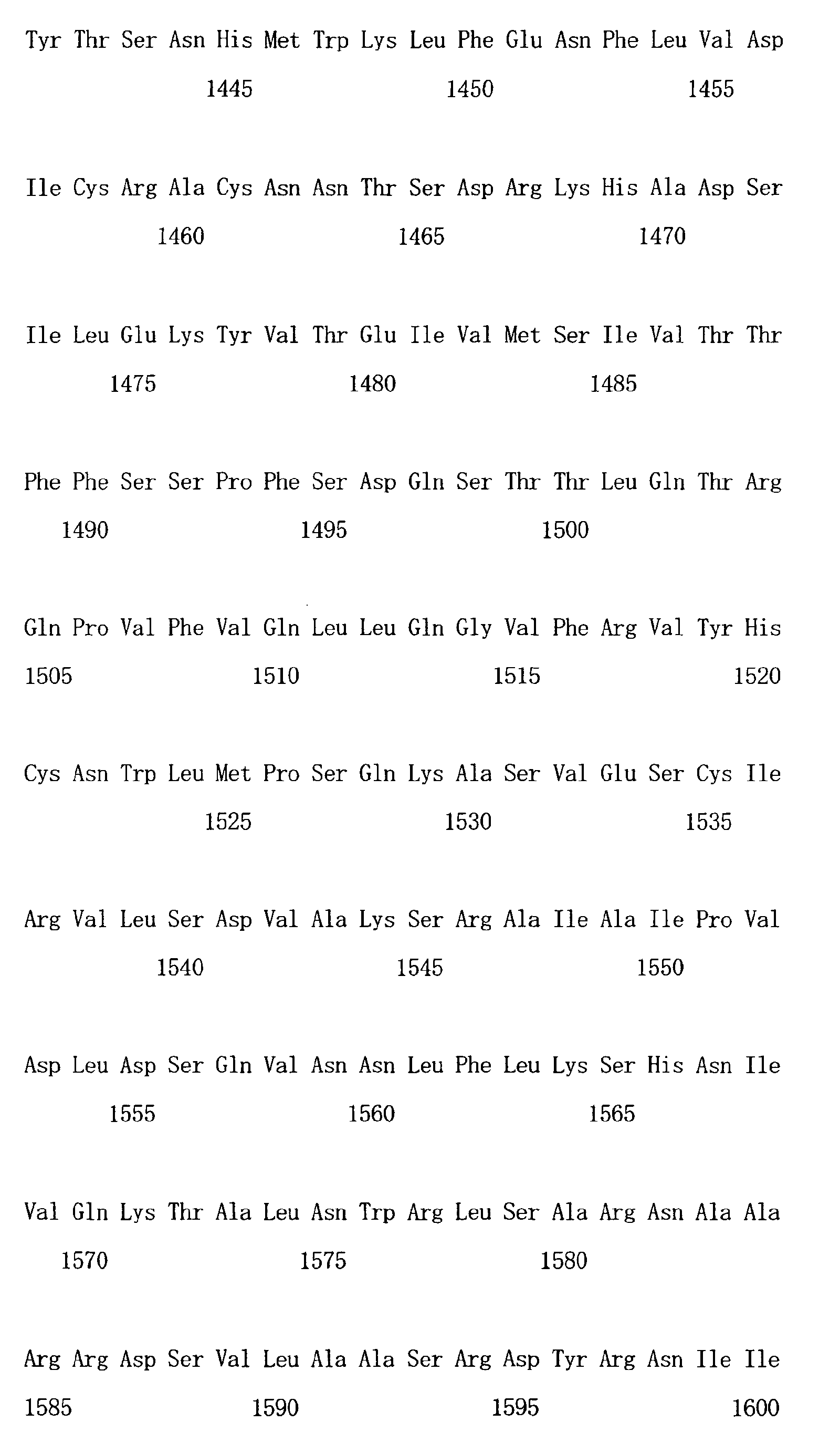
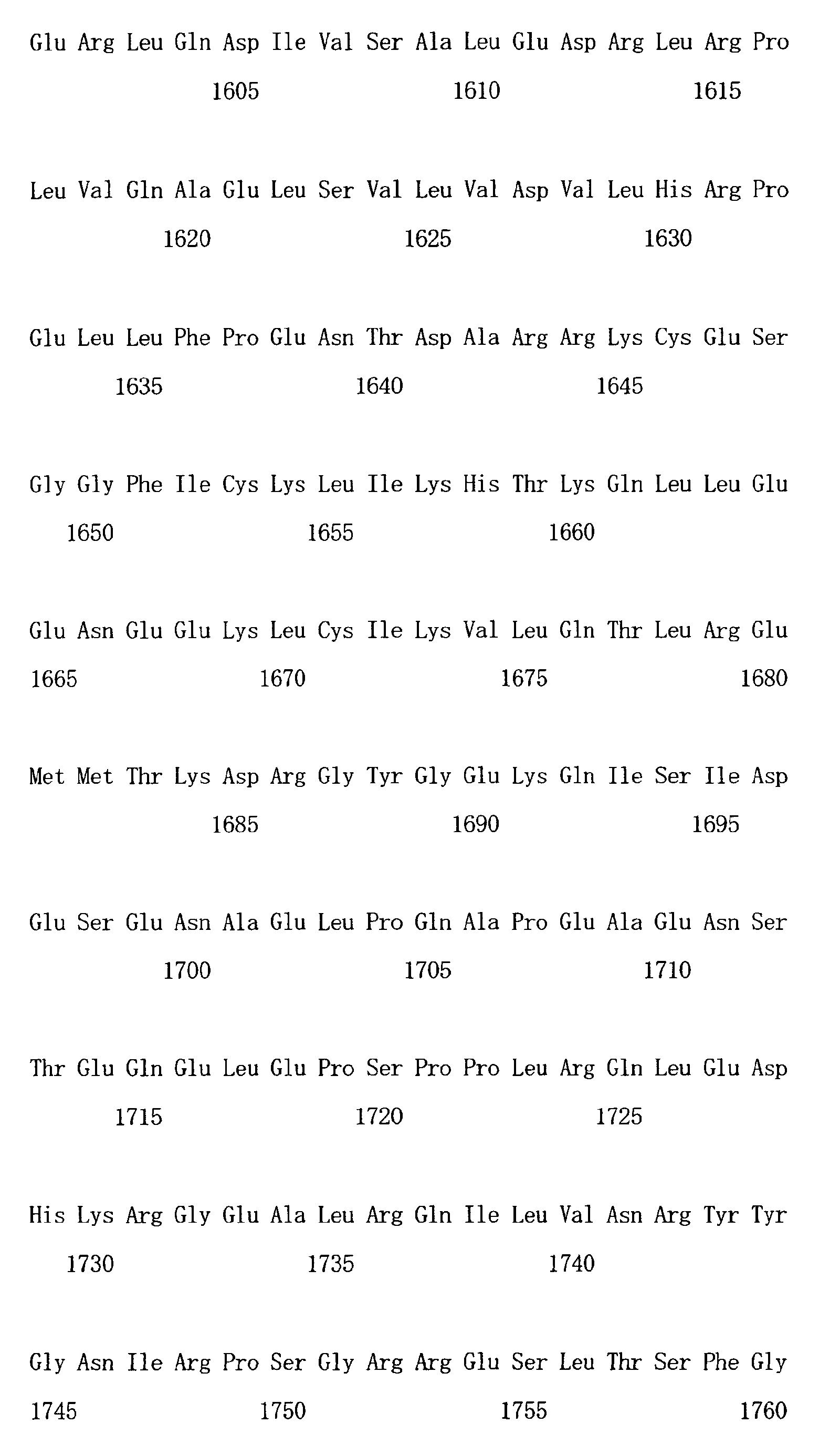
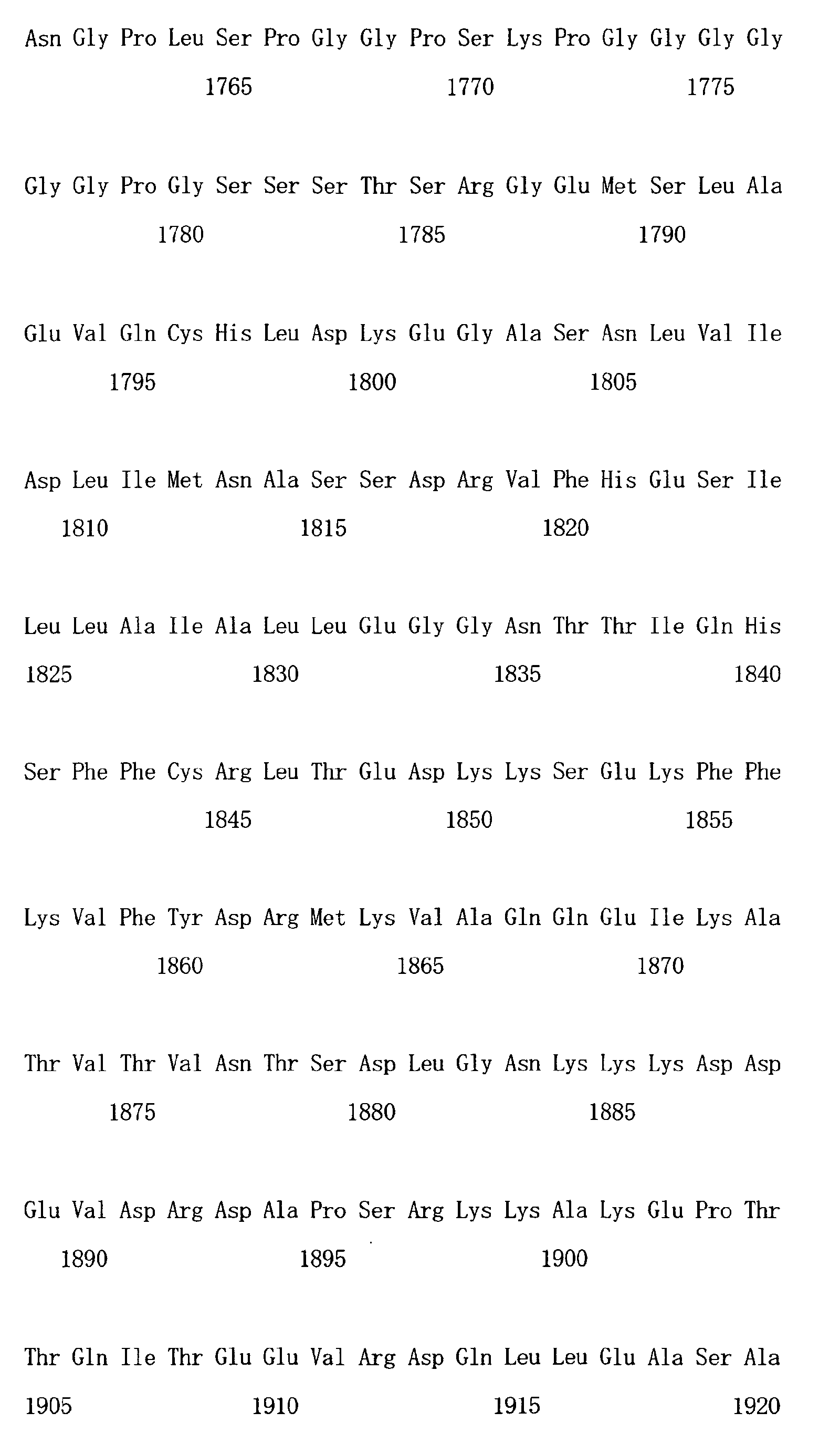


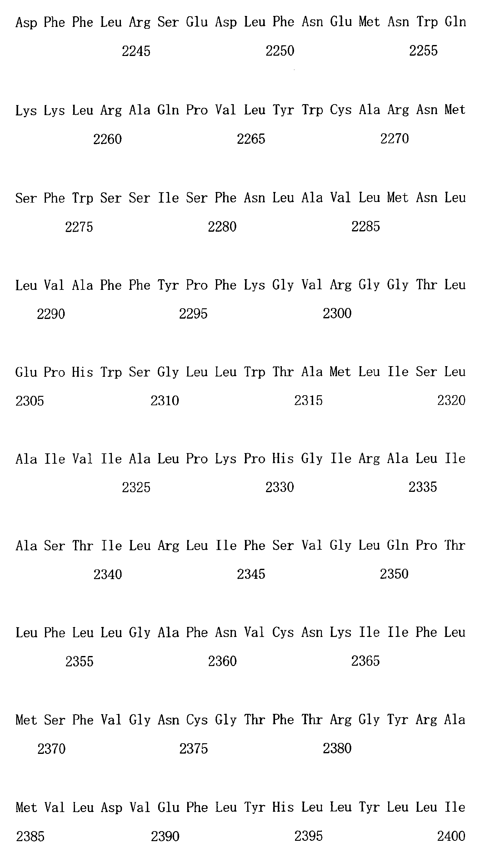


Claims (27)
- A protein (a), (b) or (c) as shown below:(a) a protein (IRBIT), comprising an amino acid sequence represented by SEQ ID NO: 1,(b) a protein, comprising an amino acid sequence of the 1-104 of SEQ ID NO: 1, and(c) a protein, comprising an amino acid sequence derived from the amino acid sequence of SEQ ID NO: 1 or an amino acid sequence of the 1-104 of SEQ ID NO: 1, which contains a deletion, substitution or addition of at least one of amino acid residues, and binding to an IP3 receptor.
- A gene, encoding the protein of claim 1.
- A gene, comprising a DNA (a), (b) or (c) as shown below:(a) a DNA, comprising a nucleotide sequence of SEQ ID NO: 2,(b) a DNA, comprising a nucleotide sequence of the 1 to 312 of SEQ ID NO: 2, and(c) a DNA, hybridizing under stringent conditions to a DNA that is complementary to the DNA comprising the nucleotide sequence of (a) or (b), and encoding a protein that binds to the IP3 receptor.
- A vector, containing the gene of claim 2 or 3.
- The vector of claim 4, wherein a promoter is operably linked to the gene of claim 2 or 3.
- The vector of claim 5, wherein the gene of claim 2 or 3 is linked in-frame with a gene encoding a fluorescent protein or a photoprotein.
- The vector of claim 6, wherein the fluourescent protein is selected from the group consisting of a green fluorescent protein (GFP), a cyan fluorescent protein (CFP), yellow fluorescent protein (YFP), Venus and FlAsH and the derivatives thereof.
- A host cell, containing the vector of any one of claims 4 to 7.
- The host cell of claim 8, which is a mammalian cell.
- An IP3 indicator for detecting IP3 in a sample, containing the protein of claim 1.
- The IP3 indicator of claim 10, wherein the protein of claim 1 is fused to a fluorescent protein or a photoprotein.
- The IP3 indicator of claim 11, which is used for detecting IP3 by the FRET method using a combination of the protein of claim 1 labeled with one fluorescent molecule of a combination of two fluorescent molecules applicable to the FRET method, and a protein containing at least the IP3-binding domain of the IP3 receptor labeled with the other fluorescent molecule of the above combination.
- The IP3 indicator of claim 12, wherein the combination of two fluorescent molecules is a combination of Venus and CFP, a combination of YFP and CFP, or a combination of FlAsH and CFP.
- An antibody or a functional fragment thereof, specifically recognizing the protein of claim 1.
- The antibody or the functional fragment thereof of claim 14, which is a rabbit polyclonal antibody or a fragment thereof.
- A method for measuring IP3 concentration in a sample, comprising:(i) preparing a protein complex comprising an IRBIT or a protein that contains the IP3 receptor-binding domain of the IRBIT and a protein that contains at least the IP3 binding domain of the IP3 receptor,(ii) allowing the protein complex to contact with the sample, and(iii) quantifying a free IRBIT or a protein containing the IP3 receptor-binding domain of the IRBIT and/or the complex.
- The method of claim 16, wherein the quantification in (iii) above is performed utilizing an immunochemical reaction system using an anti-IRBIT antibody and/or an anti-IP3 receptor antibody.
- The method of claim 16 or 17, wherein the quantification in (iii) above is performed by previously labeling an IRBIT or a protein containing the IP3 receptor-binding domain of the IRBIT or a protein containing at least the IP3 binding domain of the IP3 receptor, and then measuring signals originating from the labeled protein.
- The method of claim 18, wherein the label is a fluorescent label.
- The method of claim 16, wherein the quantification of (iii) above is performed by the FRET method.
- The method of claim 20, which uses an IRBIT or a protein containing the IP3 receptor-binding domain of the IRBIT labeled with YFP or Venus, and a protein containing at least the IP3-binding domain of the IP3 receptor labeled with CFP.
- The method of claim 20, which uses an IRBIT or a protein containing the IP3 receptor-binding domain of the IRBIT labeled with CFP, and a protein containing at least the IP3-binding domain of the IP3 receptor labeled with Venus or YFP.
- A method for quantifying the intracellular IP3 concentration of living cells, comprising:(i) introducing into a cell both of a gene encoding an IRBIT or a protein containing the IP3 receptor-binding domain of the IRBIT, labeled with one fluorescent molecule of a combination of two fluorescent molecules applicable to the FRET method, and a gene encoding a protein containing at least the IP3 binding domain of the IP3 receptor labeled with the other fluorescent molecule of the above combination, such that the genes can be expressed, and(ii) irradiating the cells with the excitation wavelength of the fluorescent molecule on the lower wavelength side of the above two fluorescent molecules, and measuring under a fluorescence microscope the fluorescence wavelength of the molecule and the fluorescence wavelength of the other fluorescent molecule on the longer wavelength side.
- The method of claim 23, wherein the combination comprising two fluorescent molecules applicable to the FRET method is a combination of Venus and CFP, a combination of YFP and CFP, or a combination of FlAsH and CFP.
- A protein complex for quantifying IP3 concentration, comprising an IRBIT or a protein containing the IP3 receptor-binding domain of the IRBIT and a protein containing the IP3 receptor or at least the IP3 binding domain thereof.
- A vector for quantifying intracellular IP3 concentration of living cells, containing a gene encoding an IRBIT or a protein containing the IP3 receptor-binding domain of the IRBIT, labeled with one fluorescent molecule of a combination of two fluorescent molecules applicable to the FRET method, and a gene encoding a protein containing at least the IP3 binding domain of the IP3 receptor labeled with the other fluorescent molecule of the above combination such that these genes can be expressed.
- The vector of claim 26, wherein the combination comprising two fluorescent molecules is a combination of Venus and CFP, a combination of YFP and CFP, or a combination of FlAsH and CFP.
Applications Claiming Priority (2)
| Application Number | Priority Date | Filing Date | Title |
|---|---|---|---|
| JP2002299429A JP4219648B2 (en) | 2002-10-11 | 2002-10-11 | Novel IP3 receptor binding protein and IP3 indicator |
| JP2002299429 | 2002-10-11 |
Publications (2)
| Publication Number | Publication Date |
|---|---|
| EP1408049A2 true EP1408049A2 (en) | 2004-04-14 |
| EP1408049A3 EP1408049A3 (en) | 2004-05-12 |
Family
ID=32025599
Family Applications (1)
| Application Number | Title | Priority Date | Filing Date |
|---|---|---|---|
| EP03256457A Withdrawn EP1408049A3 (en) | 2002-10-11 | 2003-10-13 | A novel inositol 1,4,5-trisphosphate (IP3) receptor-binding protein and an IP3 indicator |
Country Status (3)
| Country | Link |
|---|---|
| US (1) | US20050181450A1 (en) |
| EP (1) | EP1408049A3 (en) |
| JP (1) | JP4219648B2 (en) |
Cited By (1)
| Publication number | Priority date | Publication date | Assignee | Title |
|---|---|---|---|---|
| EP2014297A4 (en) * | 2006-03-20 | 2009-10-28 | Japan Science & Tech Agency | REGULATION OF AN INTRACELLULAR MOLECULE TARGETED BY AN IP3 RECEPTOR BINDING PROTEIN |
Families Citing this family (3)
| Publication number | Priority date | Publication date | Assignee | Title |
|---|---|---|---|---|
| JP2006055017A (en) * | 2004-08-18 | 2006-03-02 | Tokyo Institute Of Technology | Nucleic acid detection method |
| JP5146944B2 (en) * | 2006-03-20 | 2013-02-20 | 独立行政法人理化学研究所 | Regulation of intracellular target molecules by IP3 receptor binding protein |
| EP2006380A4 (en) | 2006-03-23 | 2010-08-11 | Kyowa Hakko Kirin Co Ltd | Agonistic antibody directed against human thrombopoietin receptor |
Family Cites Families (4)
| Publication number | Priority date | Publication date | Assignee | Title |
|---|---|---|---|---|
| JP2002513276A (en) * | 1996-10-04 | 2002-05-08 | デレク ナイジェル ジョン ハート | Enzyme having S-adenosyl-L-homocysteine hydrolase (AHCY) type activity |
| CA2283629C (en) * | 1997-03-14 | 2010-08-03 | Roger Y. Tsien | Fluorescent protein sensors for detection of analytes |
| US5854023A (en) * | 1997-07-17 | 1998-12-29 | Incyte Pharmaceuticals, Inc. | Polynucleotides encoding human S-adenosyl-5-homocysteine hydrolase derived from bladder |
| US20040265807A1 (en) * | 2001-02-09 | 2004-12-30 | Sanjanwala Madhusudan M | Enzymes |
-
2002
- 2002-10-11 JP JP2002299429A patent/JP4219648B2/en not_active Expired - Fee Related
-
2003
- 2003-10-10 US US10/683,610 patent/US20050181450A1/en not_active Abandoned
- 2003-10-13 EP EP03256457A patent/EP1408049A3/en not_active Withdrawn
Cited By (4)
| Publication number | Priority date | Publication date | Assignee | Title |
|---|---|---|---|---|
| EP2014297A4 (en) * | 2006-03-20 | 2009-10-28 | Japan Science & Tech Agency | REGULATION OF AN INTRACELLULAR MOLECULE TARGETED BY AN IP3 RECEPTOR BINDING PROTEIN |
| EP2311478A1 (en) | 2006-03-20 | 2011-04-20 | Japan Science and Technology Agency | Use of ip3 receptor-binding protein for controlling intracellular pH |
| EP2311477A1 (en) | 2006-03-20 | 2011-04-20 | Japan Science and Technology Agency | Use of ip3 receptor-binding protein for controlling phosphatidylinositol metabolism |
| US8252547B2 (en) | 2006-03-20 | 2012-08-28 | Japan Science And Technology Agency | In vitro control of protein synthesis in mammalian cells by IP3 receptor-binding protein |
Also Published As
| Publication number | Publication date |
|---|---|
| JP2004129612A (en) | 2004-04-30 |
| EP1408049A3 (en) | 2004-05-12 |
| US20050181450A1 (en) | 2005-08-18 |
| JP4219648B2 (en) | 2009-02-04 |
Similar Documents
| Publication | Publication Date | Title |
|---|---|---|
| Ando et al. | IRBIT, a novel inositol 1, 4, 5-trisphosphate (IP3) receptor-binding protein, is released from the IP3 receptor upon IP3 binding to the receptor | |
| Hirota et al. | Carbonic anhydrase-related protein is a novel binding protein for inositol 1, 4, 5-trisphosphate receptor type 1 | |
| Leonoudakis et al. | A multiprotein trafficking complex composed of SAP97, CASK, Veli, and Mint1 is associated with inward rectifier Kir2 potassium channels | |
| Wang et al. | Binding of the GABAA Receptor‐Associated Protein (GABARAP) to Microtubules and Microfilaments Suggests Involvement of the Cytoskeleton in GABARAPGABAA Receptor Interaction | |
| Lopez-Ilasaca et al. | The angiotensin II type I receptor-associated protein, ATRAP, is a transmembrane protein and a modulator of angiotensin II signaling | |
| Joubert et al. | New sorting nexin (SNX27) and NHERF specifically interact with the 5-HT4 (a) receptor splice variant: roles in receptor targeting | |
| Walikonis et al. | Densin-180 forms a ternary complex with the α-subunit of Ca2+/calmodulin-dependent protein kinase II and α-actinin | |
| Buxton et al. | Identification and characterization of Snapin as a ubiquitously expressed SNARE-binding protein that interacts with SNAP23 in non-neuronal cells | |
| Linstedt et al. | Sequence and overexpression of GPP130/GIMPc: evidence for saturable pH-sensitive targeting of a type II early Golgi membrane protein. | |
| Okamoto et al. | Dynamin isoform-specific interaction with the shank/ProSAP scaffolding proteins of the postsynaptic density and actin cytoskeleton | |
| Scott et al. | Reversible phosphorylation at the C-terminal regulatory domain of p21Waf1/Cip1 modulates proliferating cell nuclear antigen binding | |
| El Far et al. | Interaction of the C‐terminal tail region of the metabotropic glutamate receptor 7 with the protein kinase C substrate PICK1 | |
| Kitano et al. | Tamalin is a scaffold protein that interacts with multiple neuronal proteins in distinct modes of protein-protein association | |
| Peck et al. | Structure and function of human Vps20 and Snf7 proteins | |
| Nada et al. | Identification of PSD-93 as a substrate for the Src family tyrosine kinase Fyn | |
| Ciruela et al. | Interactions of the C terminus of metabotropic glutamate receptor type 1α with rat brain proteins: evidence for a direct interaction with tubulin | |
| Rader et al. | Characterization of ANKRA, a novel ankyrin repeat protein that interacts with the cytoplasmic domain of megalin | |
| US20080118951A1 (en) | Nogo receptor homologues and their use | |
| Imamura et al. | Ligand binding of the second PDZ domain regulates clustering of PSD-95 with the Kv1. 4 potassium channel | |
| JPWO2017164230A1 (en) | Phosphorylated SARM1, antibody, SARM1 phosphorylation inhibitor, preventive or therapeutic agent for neurodegenerative diseases, screening method, SARM1 variant and use | |
| Lee et al. | SGIP1α functions as a selective endocytic adaptor for the internalization of synaptotagmin 1 at synapses | |
| Kim et al. | The protein phosphatase-1 targeting subunit TIMAP regulates LAMR1 phosphorylation | |
| Timson Gauen et al. | Multiple features of the p59fyn src homology 4 domain define a motif for immune-receptor tyrosine-based activation motif (ITAM) binding and for plasma membrane localization. | |
| Proux-Gillardeaux et al. | D53 is a novel endosomal SNARE-binding protein that enhances interaction of syntaxin 1 with the synaptobrevin 2 complex in vitro | |
| Thibodeau et al. | Calmodulin binds and modulates K+-dependent Na+/Ca2+-exchanger isoform 4, NCKX4 |
Legal Events
| Date | Code | Title | Description |
|---|---|---|---|
| PUAI | Public reference made under article 153(3) epc to a published international application that has entered the european phase |
Free format text: ORIGINAL CODE: 0009012 |
|
| PUAL | Search report despatched |
Free format text: ORIGINAL CODE: 0009013 |
|
| AK | Designated contracting states |
Kind code of ref document: A2 Designated state(s): AT BE BG CH CY CZ DE DK EE ES FI FR GB GR HU IE IT LI LU MC NL PT RO SE SI SK TR |
|
| AX | Request for extension of the european patent |
Extension state: AL LT LV MK |
|
| RIC1 | Information provided on ipc code assigned before grant |
Ipc: 7G 01N 33/53 B Ipc: 7C 12N 15/12 B Ipc: 7C 07K 14/47 A |
|
| AK | Designated contracting states |
Kind code of ref document: A3 Designated state(s): AT BE BG CH CY CZ DE DK EE ES FI FR GB GR HU IE IT LI LU MC NL PT RO SE SI SK TR |
|
| AX | Request for extension of the european patent |
Extension state: AL LT LV MK |
|
| 17P | Request for examination filed |
Effective date: 20041104 |
|
| AKX | Designation fees paid |
Designated state(s): AT BE BG CH CY CZ DE DK EE ES FI FR GB GR HU IE IT LI LU MC NL PT RO SE SI SK TR |
|
| AXX | Extension fees paid |
Extension state: MK Payment date: 20041104 Extension state: LV Payment date: 20041104 Extension state: LT Payment date: 20041104 Extension state: AL Payment date: 20041104 |
|
| 17Q | First examination report despatched |
Effective date: 20050708 |
|
| STAA | Information on the status of an ep patent application or granted ep patent |
Free format text: STATUS: THE APPLICATION IS DEEMED TO BE WITHDRAWN |
|
| 18D | Application deemed to be withdrawn |
Effective date: 20070103 |