EP1407030B1 - Methods for large scale production of recombinant dna-derived tpa or k2s molecules - Google Patents
Methods for large scale production of recombinant dna-derived tpa or k2s molecules Download PDFInfo
- Publication number
- EP1407030B1 EP1407030B1 EP01996596A EP01996596A EP1407030B1 EP 1407030 B1 EP1407030 B1 EP 1407030B1 EP 01996596 A EP01996596 A EP 01996596A EP 01996596 A EP01996596 A EP 01996596A EP 1407030 B1 EP1407030 B1 EP 1407030B1
- Authority
- EP
- European Patent Office
- Prior art keywords
- dna
- variant
- tpa
- molecule
- coding
- Prior art date
- Legal status (The legal status is an assumption and is not a legal conclusion. Google has not performed a legal analysis and makes no representation as to the accuracy of the status listed.)
- Expired - Lifetime
Links
- 238000000034 method Methods 0.000 title claims abstract description 69
- 108020004511 Recombinant DNA Proteins 0.000 title claims abstract description 15
- 238000011031 large-scale manufacturing process Methods 0.000 title description 5
- 102000003978 Tissue Plasminogen Activator Human genes 0.000 claims abstract description 117
- 108090000373 Tissue Plasminogen Activator Proteins 0.000 claims abstract description 117
- 229960000187 tissue plasminogen activator Drugs 0.000 claims abstract description 96
- 108020004414 DNA Proteins 0.000 claims abstract description 88
- 108090000623 proteins and genes Proteins 0.000 claims abstract description 61
- 239000013598 vector Substances 0.000 claims abstract description 46
- 102000004169 proteins and genes Human genes 0.000 claims abstract description 45
- 108010079246 OMPA outer membrane proteins Proteins 0.000 claims abstract description 34
- 108010076504 Protein Sorting Signals Proteins 0.000 claims abstract description 22
- 238000004519 manufacturing process Methods 0.000 claims abstract description 18
- 210000001236 prokaryotic cell Anatomy 0.000 claims abstract description 16
- 150000001413 amino acids Chemical class 0.000 claims description 65
- 241000588724 Escherichia coli Species 0.000 claims description 39
- 108091028043 Nucleic acid sequence Proteins 0.000 claims description 37
- 102000053602 DNA Human genes 0.000 claims description 30
- 102000012479 Serine Proteases Human genes 0.000 claims description 21
- 108010022999 Serine Proteases Proteins 0.000 claims description 21
- 210000004027 cell Anatomy 0.000 claims description 19
- 108020001507 fusion proteins Proteins 0.000 claims description 19
- 102000037865 fusion proteins Human genes 0.000 claims description 19
- 101000654764 Homo sapiens Secretagogin Proteins 0.000 claims description 18
- 102100032621 Secretagogin Human genes 0.000 claims description 18
- 230000014509 gene expression Effects 0.000 claims description 17
- 241000282414 Homo sapiens Species 0.000 claims description 16
- 108090000765 processed proteins & peptides Proteins 0.000 claims description 16
- 150000007523 nucleic acids Chemical class 0.000 claims description 11
- 102000039446 nucleic acids Human genes 0.000 claims description 10
- 108020004707 nucleic acids Proteins 0.000 claims description 10
- 108020004705 Codon Proteins 0.000 claims description 9
- 229920001184 polypeptide Polymers 0.000 claims description 9
- 102000004196 processed proteins & peptides Human genes 0.000 claims description 9
- 230000027455 binding Effects 0.000 claims description 8
- 230000000694 effects Effects 0.000 claims description 8
- 239000002773 nucleotide Substances 0.000 claims description 5
- 125000003729 nucleotide group Chemical group 0.000 claims description 5
- 210000003705 ribosome Anatomy 0.000 claims description 4
- 238000011144 upstream manufacturing Methods 0.000 claims description 4
- 108090000656 Annexin A6 Proteins 0.000 claims description 2
- MTCFGRXMJLQNBG-UHFFFAOYSA-N Serine Natural products OCC(N)C(O)=O MTCFGRXMJLQNBG-UHFFFAOYSA-N 0.000 abstract description 2
- 230000002537 thrombolytic effect Effects 0.000 abstract description 2
- 239000000047 product Substances 0.000 description 19
- 239000012634 fragment Substances 0.000 description 17
- 239000012228 culture supernatant Substances 0.000 description 16
- 241001494479 Pecora Species 0.000 description 11
- 102000013566 Plasminogen Human genes 0.000 description 11
- 108010051456 Plasminogen Proteins 0.000 description 11
- 230000002255 enzymatic effect Effects 0.000 description 11
- 239000000126 substance Substances 0.000 description 11
- 239000000499 gel Substances 0.000 description 9
- 230000013595 glycosylation Effects 0.000 description 9
- 238000006206 glycosylation reaction Methods 0.000 description 9
- 230000028327 secretion Effects 0.000 description 8
- 102000035195 Peptidases Human genes 0.000 description 7
- 108091005804 Peptidases Proteins 0.000 description 7
- 239000004365 Protease Substances 0.000 description 7
- DBMJMQXJHONAFJ-UHFFFAOYSA-M Sodium laurylsulphate Chemical compound [Na+].CCCCCCCCCCCCOS([O-])(=O)=O DBMJMQXJHONAFJ-UHFFFAOYSA-M 0.000 description 7
- 238000006243 chemical reaction Methods 0.000 description 7
- 201000001441 melanoma Diseases 0.000 description 7
- 239000000203 mixture Substances 0.000 description 7
- 230000035772 mutation Effects 0.000 description 7
- 230000008569 process Effects 0.000 description 7
- 238000000746 purification Methods 0.000 description 7
- 235000019333 sodium laurylsulphate Nutrition 0.000 description 7
- 108091026890 Coding region Proteins 0.000 description 6
- 238000002835 absorbance Methods 0.000 description 6
- 230000003321 amplification Effects 0.000 description 6
- 238000001962 electrophoresis Methods 0.000 description 6
- 239000012528 membrane Substances 0.000 description 6
- 230000004048 modification Effects 0.000 description 6
- 238000012986 modification Methods 0.000 description 6
- 238000003199 nucleic acid amplification method Methods 0.000 description 6
- 210000001322 periplasm Anatomy 0.000 description 6
- 239000013612 plasmid Substances 0.000 description 6
- 102000009123 Fibrin Human genes 0.000 description 5
- 108010073385 Fibrin Proteins 0.000 description 5
- BWGVNKXGVNDBDI-UHFFFAOYSA-N Fibrin monomer Chemical compound CNC(=O)CNC(=O)CN BWGVNKXGVNDBDI-UHFFFAOYSA-N 0.000 description 5
- 238000003776 cleavage reaction Methods 0.000 description 5
- 238000010367 cloning Methods 0.000 description 5
- 229950003499 fibrin Drugs 0.000 description 5
- 239000002245 particle Substances 0.000 description 5
- 230000000644 propagated effect Effects 0.000 description 5
- 230000007017 scission Effects 0.000 description 5
- 241000894006 Bacteria Species 0.000 description 4
- HEDRZPFGACZZDS-UHFFFAOYSA-N Chloroform Chemical compound ClC(Cl)Cl HEDRZPFGACZZDS-UHFFFAOYSA-N 0.000 description 4
- 101000822695 Clostridium perfringens (strain 13 / Type A) Small, acid-soluble spore protein C1 Proteins 0.000 description 4
- 101000655262 Clostridium perfringens (strain 13 / Type A) Small, acid-soluble spore protein C2 Proteins 0.000 description 4
- 102000008946 Fibrinogen Human genes 0.000 description 4
- 108010049003 Fibrinogen Proteins 0.000 description 4
- 101000801481 Homo sapiens Tissue-type plasminogen activator Proteins 0.000 description 4
- 101000655256 Paraclostridium bifermentans Small, acid-soluble spore protein alpha Proteins 0.000 description 4
- 101000655264 Paraclostridium bifermentans Small, acid-soluble spore protein beta Proteins 0.000 description 4
- BFNBIHQBYMNNAN-UHFFFAOYSA-N ammonium sulfate Chemical class N.N.OS(O)(=O)=O BFNBIHQBYMNNAN-UHFFFAOYSA-N 0.000 description 4
- 229960000723 ampicillin Drugs 0.000 description 4
- AVKUERGKIZMTKX-NJBDSQKTSA-N ampicillin Chemical compound C1([C@@H](N)C(=O)N[C@H]2[C@H]3SC([C@@H](N3C2=O)C(O)=O)(C)C)=CC=CC=C1 AVKUERGKIZMTKX-NJBDSQKTSA-N 0.000 description 4
- 210000001367 artery Anatomy 0.000 description 4
- 230000001580 bacterial effect Effects 0.000 description 4
- 230000015572 biosynthetic process Effects 0.000 description 4
- 238000010276 construction Methods 0.000 description 4
- 230000001086 cytosolic effect Effects 0.000 description 4
- 238000001514 detection method Methods 0.000 description 4
- 229940012952 fibrinogen Drugs 0.000 description 4
- RWSXRVCMGQZWBV-WDSKDSINSA-N glutathione Chemical compound OC(=O)[C@@H](N)CCC(=O)N[C@@H](CS)C(=O)NCC(O)=O RWSXRVCMGQZWBV-WDSKDSINSA-N 0.000 description 4
- BPHPUYQFMNQIOC-NXRLNHOXSA-N isopropyl beta-D-thiogalactopyranoside Chemical compound CC(C)S[C@@H]1O[C@H](CO)[C@H](O)[C@H](O)[C@H]1O BPHPUYQFMNQIOC-NXRLNHOXSA-N 0.000 description 4
- 229920002401 polyacrylamide Polymers 0.000 description 4
- 238000002360 preparation method Methods 0.000 description 4
- 239000000523 sample Substances 0.000 description 4
- 238000003118 sandwich ELISA Methods 0.000 description 4
- 238000002741 site-directed mutagenesis Methods 0.000 description 4
- 239000000758 substrate Substances 0.000 description 4
- 101000870597 Escherichia coli O78:H11 (strain H10407 / ETEC) Secretin GspD 2 Proteins 0.000 description 3
- 108010010803 Gelatin Proteins 0.000 description 3
- PEDCQBHIVMGVHV-UHFFFAOYSA-N Glycerine Chemical compound OCC(O)CO PEDCQBHIVMGVHV-UHFFFAOYSA-N 0.000 description 3
- AVXURJPOCDRRFD-UHFFFAOYSA-N Hydroxylamine Chemical compound ON AVXURJPOCDRRFD-UHFFFAOYSA-N 0.000 description 3
- 239000000020 Nitrocellulose Substances 0.000 description 3
- 108010090127 Periplasmic Proteins Proteins 0.000 description 3
- 229920002684 Sepharose Polymers 0.000 description 3
- 239000004098 Tetracycline Substances 0.000 description 3
- 101000870604 Vibrio cholerae serotype O1 (strain ATCC 39315 / El Tor Inaba N16961) Secretin GspD Proteins 0.000 description 3
- 239000011543 agarose gel Substances 0.000 description 3
- 230000003024 amidolytic effect Effects 0.000 description 3
- 238000004458 analytical method Methods 0.000 description 3
- 238000003556 assay Methods 0.000 description 3
- 238000012512 characterization method Methods 0.000 description 3
- 230000004927 fusion Effects 0.000 description 3
- 239000008273 gelatin Substances 0.000 description 3
- 229920000159 gelatin Polymers 0.000 description 3
- 235000019322 gelatine Nutrition 0.000 description 3
- 235000011852 gelatine desserts Nutrition 0.000 description 3
- 239000001963 growth medium Substances 0.000 description 3
- 210000003000 inclusion body Anatomy 0.000 description 3
- 238000011534 incubation Methods 0.000 description 3
- 239000003550 marker Substances 0.000 description 3
- 229920001220 nitrocellulos Polymers 0.000 description 3
- 230000001590 oxidative effect Effects 0.000 description 3
- 239000000546 pharmaceutical excipient Substances 0.000 description 3
- 229940012957 plasmin Drugs 0.000 description 3
- 238000002264 polyacrylamide gel electrophoresis Methods 0.000 description 3
- 108091008146 restriction endonucleases Proteins 0.000 description 3
- 239000000243 solution Substances 0.000 description 3
- 238000006467 substitution reaction Methods 0.000 description 3
- 229960002180 tetracycline Drugs 0.000 description 3
- 229930101283 tetracycline Natural products 0.000 description 3
- 235000019364 tetracycline Nutrition 0.000 description 3
- 150000003522 tetracyclines Chemical class 0.000 description 3
- 238000005406 washing Methods 0.000 description 3
- QGZKDVFQNNGYKY-UHFFFAOYSA-O Ammonium Chemical compound [NH4+] QGZKDVFQNNGYKY-UHFFFAOYSA-O 0.000 description 2
- CIWBSHSKHKDKBQ-JLAZNSOCSA-N Ascorbic acid Chemical compound OC[C@H](O)[C@H]1OC(=O)C(O)=C1O CIWBSHSKHKDKBQ-JLAZNSOCSA-N 0.000 description 2
- 108010014303 DNA-directed DNA polymerase Proteins 0.000 description 2
- 102000016928 DNA-directed DNA polymerase Human genes 0.000 description 2
- 238000002965 ELISA Methods 0.000 description 2
- 102000004190 Enzymes Human genes 0.000 description 2
- 108090000790 Enzymes Proteins 0.000 description 2
- 241000901143 Etia Species 0.000 description 2
- 241000829100 Macaca mulatta polyomavirus 1 Species 0.000 description 2
- 108010052285 Membrane Proteins Proteins 0.000 description 2
- 102000018697 Membrane Proteins Human genes 0.000 description 2
- 102000001938 Plasminogen Activators Human genes 0.000 description 2
- 108010001014 Plasminogen Activators Proteins 0.000 description 2
- 241000588770 Proteus mirabilis Species 0.000 description 2
- 208000010378 Pulmonary Embolism Diseases 0.000 description 2
- 108010008281 Recombinant Fusion Proteins Proteins 0.000 description 2
- 102000007056 Recombinant Fusion Proteins Human genes 0.000 description 2
- 241000714474 Rous sarcoma virus Species 0.000 description 2
- FAPWRFPIFSIZLT-UHFFFAOYSA-M Sodium chloride Chemical compound [Na+].[Cl-] FAPWRFPIFSIZLT-UHFFFAOYSA-M 0.000 description 2
- 208000006011 Stroke Diseases 0.000 description 2
- 206010000891 acute myocardial infarction Diseases 0.000 description 2
- 238000001042 affinity chromatography Methods 0.000 description 2
- 230000002776 aggregation Effects 0.000 description 2
- 238000004220 aggregation Methods 0.000 description 2
- 229910052921 ammonium sulfate Inorganic materials 0.000 description 2
- 235000011130 ammonium sulphate Nutrition 0.000 description 2
- 238000000137 annealing Methods 0.000 description 2
- 230000003115 biocidal effect Effects 0.000 description 2
- 230000008827 biological function Effects 0.000 description 2
- 239000002299 complementary DNA Substances 0.000 description 2
- NKLPQNGYXWVELD-UHFFFAOYSA-M coomassie brilliant blue Chemical compound [Na+].C1=CC(OCC)=CC=C1NC1=CC=C(C(=C2C=CC(C=C2)=[N+](CC)CC=2C=C(C=CC=2)S([O-])(=O)=O)C=2C=CC(=CC=2)N(CC)CC=2C=C(C=CC=2)S([O-])(=O)=O)C=C1 NKLPQNGYXWVELD-UHFFFAOYSA-M 0.000 description 2
- 238000004925 denaturation Methods 0.000 description 2
- 230000036425 denaturation Effects 0.000 description 2
- 230000001419 dependent effect Effects 0.000 description 2
- 238000013461 design Methods 0.000 description 2
- 238000010586 diagram Methods 0.000 description 2
- 230000029087 digestion Effects 0.000 description 2
- 238000010790 dilution Methods 0.000 description 2
- 239000012895 dilution Substances 0.000 description 2
- 239000003937 drug carrier Substances 0.000 description 2
- 229940088598 enzyme Drugs 0.000 description 2
- 229960003180 glutathione Drugs 0.000 description 2
- 239000003102 growth factor Substances 0.000 description 2
- 238000003119 immunoblot Methods 0.000 description 2
- 230000001939 inductive effect Effects 0.000 description 2
- 238000003780 insertion Methods 0.000 description 2
- 230000037431 insertion Effects 0.000 description 2
- 210000004962 mammalian cell Anatomy 0.000 description 2
- 239000002609 medium Substances 0.000 description 2
- 239000013642 negative control Substances 0.000 description 2
- 238000002823 phage display Methods 0.000 description 2
- 239000008194 pharmaceutical composition Substances 0.000 description 2
- 229940127126 plasminogen activator Drugs 0.000 description 2
- 229920001223 polyethylene glycol Polymers 0.000 description 2
- 239000013641 positive control Substances 0.000 description 2
- 230000003248 secreting effect Effects 0.000 description 2
- 229940083575 sodium dodecyl sulfate Drugs 0.000 description 2
- 238000002415 sodium dodecyl sulfate polyacrylamide gel electrophoresis Methods 0.000 description 2
- 238000012360 testing method Methods 0.000 description 2
- 230000001225 therapeutic effect Effects 0.000 description 2
- 101150010939 tpa gene Proteins 0.000 description 2
- HCUOEKSZWPGJIM-YBRHCDHNSA-N (e,2e)-2-hydroxyimino-6-methoxy-4-methyl-5-nitrohex-3-enamide Chemical compound COCC([N+]([O-])=O)\C(C)=C\C(=N/O)\C(N)=O HCUOEKSZWPGJIM-YBRHCDHNSA-N 0.000 description 1
- TYMLOMAKGOJONV-UHFFFAOYSA-N 4-nitroaniline Chemical compound NC1=CC=C([N+]([O-])=O)C=C1 TYMLOMAKGOJONV-UHFFFAOYSA-N 0.000 description 1
- 208000031104 Arterial Occlusive disease Diseases 0.000 description 1
- 244000063299 Bacillus subtilis Species 0.000 description 1
- 235000014469 Bacillus subtilis Nutrition 0.000 description 1
- UXVMQQNJUSDDNG-UHFFFAOYSA-L Calcium chloride Chemical compound [Cl-].[Cl-].[Ca+2] UXVMQQNJUSDDNG-UHFFFAOYSA-L 0.000 description 1
- 229940123150 Chelating agent Drugs 0.000 description 1
- 206010011086 Coronary artery occlusion Diseases 0.000 description 1
- 239000004971 Cross linker Substances 0.000 description 1
- 241000701022 Cytomegalovirus Species 0.000 description 1
- 238000001712 DNA sequencing Methods 0.000 description 1
- 206010051055 Deep vein thrombosis Diseases 0.000 description 1
- 229920002307 Dextran Polymers 0.000 description 1
- 244000070010 Erythrina variegata Species 0.000 description 1
- 101001133633 Escherichia coli Penicillin G acylase Proteins 0.000 description 1
- 241000724791 Filamentous phage Species 0.000 description 1
- WQZGKKKJIJFFOK-GASJEMHNSA-N Glucose Natural products OC[C@H]1OC(O)[C@H](O)[C@@H](O)[C@@H]1O WQZGKKKJIJFFOK-GASJEMHNSA-N 0.000 description 1
- 108010024636 Glutathione Proteins 0.000 description 1
- 108010053070 Glutathione Disulfide Proteins 0.000 description 1
- 208000009889 Herpes Simplex Diseases 0.000 description 1
- 101000605403 Homo sapiens Plasminogen Proteins 0.000 description 1
- 241000701024 Human betaherpesvirus 5 Species 0.000 description 1
- 108060003951 Immunoglobulin Proteins 0.000 description 1
- 206010061216 Infarction Diseases 0.000 description 1
- 102000003960 Ligases Human genes 0.000 description 1
- 108090000364 Ligases Proteins 0.000 description 1
- 102000004895 Lipoproteins Human genes 0.000 description 1
- 108090001030 Lipoproteins Proteins 0.000 description 1
- 239000006142 Luria-Bertani Agar Substances 0.000 description 1
- 239000006137 Luria-Bertani broth Substances 0.000 description 1
- KDXKERNSBIXSRK-UHFFFAOYSA-N Lysine Natural products NCCCCC(N)C(O)=O KDXKERNSBIXSRK-UHFFFAOYSA-N 0.000 description 1
- 239000004472 Lysine Substances 0.000 description 1
- 239000007993 MOPS buffer Substances 0.000 description 1
- 101710175625 Maltose/maltodextrin-binding periplasmic protein Proteins 0.000 description 1
- 230000004988 N-glycosylation Effects 0.000 description 1
- 125000001429 N-terminal alpha-amino-acid group Chemical group 0.000 description 1
- 206010028980 Neoplasm Diseases 0.000 description 1
- 238000012408 PCR amplification Methods 0.000 description 1
- 238000010222 PCR analysis Methods 0.000 description 1
- 241000235648 Pichia Species 0.000 description 1
- 241000589516 Pseudomonas Species 0.000 description 1
- 241000589776 Pseudomonas putida Species 0.000 description 1
- 238000012181 QIAquick gel extraction kit Methods 0.000 description 1
- 241000235070 Saccharomyces Species 0.000 description 1
- 108091003202 SecA Proteins Proteins 0.000 description 1
- 108010086019 Secretin Proteins 0.000 description 1
- 102100037505 Secretin Human genes 0.000 description 1
- 241000710961 Semliki Forest virus Species 0.000 description 1
- 241000191940 Staphylococcus Species 0.000 description 1
- 241000191965 Staphylococcus carnosus Species 0.000 description 1
- 108010023197 Streptokinase Proteins 0.000 description 1
- 241000187747 Streptomyces Species 0.000 description 1
- CZMRCDWAGMRECN-UGDNZRGBSA-N Sucrose Chemical compound O[C@H]1[C@H](O)[C@@H](CO)O[C@@]1(CO)O[C@@H]1[C@H](O)[C@@H](O)[C@H](O)[C@@H](CO)O1 CZMRCDWAGMRECN-UGDNZRGBSA-N 0.000 description 1
- 229930006000 Sucrose Natural products 0.000 description 1
- QAOWNCQODCNURD-UHFFFAOYSA-N Sulfuric acid Chemical compound OS(O)(=O)=O QAOWNCQODCNURD-UHFFFAOYSA-N 0.000 description 1
- 108010006785 Taq Polymerase Proteins 0.000 description 1
- 208000001435 Thromboembolism Diseases 0.000 description 1
- 108050006955 Tissue-type plasminogen activator Proteins 0.000 description 1
- 102100033571 Tissue-type plasminogen activator Human genes 0.000 description 1
- 229940090439 Tissue-type plasminogen activator inhibitor Drugs 0.000 description 1
- 229920004890 Triton X-100 Polymers 0.000 description 1
- 239000013504 Triton X-100 Substances 0.000 description 1
- 108091023045 Untranslated Region Proteins 0.000 description 1
- 206010046865 Vaccinia virus infection Diseases 0.000 description 1
- 206010047249 Venous thrombosis Diseases 0.000 description 1
- 241000700605 Viruses Species 0.000 description 1
- 230000004913 activation Effects 0.000 description 1
- 239000000654 additive Substances 0.000 description 1
- 238000000246 agarose gel electrophoresis Methods 0.000 description 1
- 125000000539 amino acid group Chemical group 0.000 description 1
- 238000012870 ammonium sulfate precipitation Methods 0.000 description 1
- 210000004102 animal cell Anatomy 0.000 description 1
- 230000000890 antigenic effect Effects 0.000 description 1
- 239000003963 antioxidant agent Substances 0.000 description 1
- 235000006708 antioxidants Nutrition 0.000 description 1
- 208000021328 arterial occlusion Diseases 0.000 description 1
- 229960005070 ascorbic acid Drugs 0.000 description 1
- 235000010323 ascorbic acid Nutrition 0.000 description 1
- 239000011668 ascorbic acid Substances 0.000 description 1
- 230000008901 benefit Effects 0.000 description 1
- PXXJHWLDUBFPOL-UHFFFAOYSA-N benzamidine Chemical compound NC(=N)C1=CC=CC=C1 PXXJHWLDUBFPOL-UHFFFAOYSA-N 0.000 description 1
- WQZGKKKJIJFFOK-VFUOTHLCSA-N beta-D-glucose Chemical compound OC[C@H]1O[C@@H](O)[C@H](O)[C@@H](O)[C@@H]1O WQZGKKKJIJFFOK-VFUOTHLCSA-N 0.000 description 1
- 230000000903 blocking effect Effects 0.000 description 1
- 230000023555 blood coagulation Effects 0.000 description 1
- 238000006664 bond formation reaction Methods 0.000 description 1
- 210000004556 brain Anatomy 0.000 description 1
- 239000000872 buffer Substances 0.000 description 1
- 239000007853 buffer solution Substances 0.000 description 1
- 210000004899 c-terminal region Anatomy 0.000 description 1
- 239000001110 calcium chloride Substances 0.000 description 1
- 229910001628 calcium chloride Inorganic materials 0.000 description 1
- 229940041514 candida albicans extract Drugs 0.000 description 1
- 150000001720 carbohydrates Chemical class 0.000 description 1
- 235000014633 carbohydrates Nutrition 0.000 description 1
- 230000000747 cardiac effect Effects 0.000 description 1
- 230000015556 catabolic process Effects 0.000 description 1
- 230000003197 catalytic effect Effects 0.000 description 1
- 238000004113 cell culture Methods 0.000 description 1
- 230000006037 cell lysis Effects 0.000 description 1
- 239000002738 chelating agent Substances 0.000 description 1
- 239000003153 chemical reaction reagent Substances 0.000 description 1
- 239000003795 chemical substances by application Substances 0.000 description 1
- 210000004978 chinese hamster ovary cell Anatomy 0.000 description 1
- 230000001332 colony forming effect Effects 0.000 description 1
- 150000001875 compounds Chemical class 0.000 description 1
- 239000012468 concentrated sample Substances 0.000 description 1
- 230000009089 cytolysis Effects 0.000 description 1
- 210000000805 cytoplasm Anatomy 0.000 description 1
- 230000003412 degenerative effect Effects 0.000 description 1
- 238000006731 degradation reaction Methods 0.000 description 1
- 238000011161 development Methods 0.000 description 1
- 201000010099 disease Diseases 0.000 description 1
- 208000037265 diseases, disorders, signs and symptoms Diseases 0.000 description 1
- 238000004090 dissolution Methods 0.000 description 1
- 239000003814 drug Substances 0.000 description 1
- 238000005516 engineering process Methods 0.000 description 1
- 239000003623 enhancer Substances 0.000 description 1
- 239000013604 expression vector Substances 0.000 description 1
- 239000000284 extract Substances 0.000 description 1
- 238000000605 extraction Methods 0.000 description 1
- 238000000855 fermentation Methods 0.000 description 1
- 230000004151 fermentation Effects 0.000 description 1
- 239000003527 fibrinolytic agent Substances 0.000 description 1
- 239000008103 glucose Substances 0.000 description 1
- YPZRWBKMTBYPTK-BJDJZHNGSA-N glutathione disulfide Chemical compound OC(=O)[C@@H](N)CCC(=O)N[C@H](C(=O)NCC(O)=O)CSSC[C@@H](C(=O)NCC(O)=O)NC(=O)CC[C@H](N)C(O)=O YPZRWBKMTBYPTK-BJDJZHNGSA-N 0.000 description 1
- 238000003306 harvesting Methods 0.000 description 1
- 230000013632 homeostatic process Effects 0.000 description 1
- 229940106780 human fibrinogen Drugs 0.000 description 1
- 238000009396 hybridization Methods 0.000 description 1
- 230000001900 immune effect Effects 0.000 description 1
- 210000000987 immune system Anatomy 0.000 description 1
- 238000003018 immunoassay Methods 0.000 description 1
- 102000018358 immunoglobulin Human genes 0.000 description 1
- 230000036046 immunoreaction Effects 0.000 description 1
- 230000006872 improvement Effects 0.000 description 1
- 239000012535 impurity Substances 0.000 description 1
- 238000001727 in vivo Methods 0.000 description 1
- 239000000411 inducer Substances 0.000 description 1
- 230000007574 infarction Effects 0.000 description 1
- 230000002401 inhibitory effect Effects 0.000 description 1
- 230000003834 intracellular effect Effects 0.000 description 1
- 238000007917 intracranial administration Methods 0.000 description 1
- 229960000318 kanamycin Drugs 0.000 description 1
- 229930027917 kanamycin Natural products 0.000 description 1
- SBUJHOSQTJFQJX-NOAMYHISSA-N kanamycin Chemical compound O[C@@H]1[C@@H](O)[C@H](O)[C@@H](CN)O[C@@H]1O[C@H]1[C@H](O)[C@@H](O[C@@H]2[C@@H]([C@@H](N)[C@H](O)[C@@H](CO)O2)O)[C@H](N)C[C@@H]1N SBUJHOSQTJFQJX-NOAMYHISSA-N 0.000 description 1
- 229930182823 kanamycin A Natural products 0.000 description 1
- 230000004807 localization Effects 0.000 description 1
- 238000013507 mapping Methods 0.000 description 1
- 239000000463 material Substances 0.000 description 1
- 230000035800 maturation Effects 0.000 description 1
- 238000005259 measurement Methods 0.000 description 1
- 244000005700 microbiome Species 0.000 description 1
- 238000010369 molecular cloning Methods 0.000 description 1
- 238000002703 mutagenesis Methods 0.000 description 1
- 231100000350 mutagenesis Toxicity 0.000 description 1
- 231100000219 mutagenic Toxicity 0.000 description 1
- 230000003505 mutagenic effect Effects 0.000 description 1
- 210000004897 n-terminal region Anatomy 0.000 description 1
- 231100000957 no side effect Toxicity 0.000 description 1
- 102000026415 nucleotide binding proteins Human genes 0.000 description 1
- 108091014756 nucleotide binding proteins Proteins 0.000 description 1
- 230000002018 overexpression Effects 0.000 description 1
- 238000012858 packaging process Methods 0.000 description 1
- 239000003973 paint Substances 0.000 description 1
- 230000002093 peripheral effect Effects 0.000 description 1
- 239000011148 porous material Substances 0.000 description 1
- 239000002244 precipitate Substances 0.000 description 1
- 239000002243 precursor Substances 0.000 description 1
- 238000001243 protein synthesis Methods 0.000 description 1
- 230000002797 proteolythic effect Effects 0.000 description 1
- 239000012264 purified product Substances 0.000 description 1
- 238000011002 quantification Methods 0.000 description 1
- 239000000700 radioactive tracer Substances 0.000 description 1
- 230000007420 reactivation Effects 0.000 description 1
- 238000011084 recovery Methods 0.000 description 1
- 230000001105 regulatory effect Effects 0.000 description 1
- 238000004153 renaturation Methods 0.000 description 1
- 229960002101 secretin Drugs 0.000 description 1
- OWMZNFCDEHGFEP-NFBCVYDUSA-N secretin human Chemical compound C([C@@H](C(=O)N[C@H](C(=O)N[C@@H](CO)C(=O)N[C@@H](CCC(O)=O)C(=O)N[C@@H](CC(C)C)C(=O)N[C@@H](CO)C(=O)N[C@@H](CCCNC(N)=N)C(=O)N[C@@H](CC(C)C)C(=O)N[C@@H](CCCNC(N)=N)C(=O)N[C@@H](CCC(O)=O)C(=O)NCC(=O)N[C@@H](C)C(=O)N[C@@H](CCCNC(N)=N)C(=O)N[C@@H](CC(C)C)C(=O)N[C@@H](CCC(N)=O)C(=O)N[C@@H](CCCNC(N)=N)C(=O)N[C@@H](CC(C)C)C(=O)N[C@@H](CC(C)C)C(=O)N[C@@H](CCC(N)=O)C(=O)NCC(=O)N[C@@H](CC(C)C)C(=O)N[C@@H](C(C)C)C(N)=O)[C@@H](C)O)NC(=O)[C@@H](NC(=O)CNC(=O)[C@H](CC(O)=O)NC(=O)[C@H](CO)NC(=O)[C@@H](N)CC=1NC=NC=1)[C@@H](C)O)C1=CC=CC=C1 OWMZNFCDEHGFEP-NFBCVYDUSA-N 0.000 description 1
- 238000011896 sensitive detection Methods 0.000 description 1
- 230000035945 sensitivity Effects 0.000 description 1
- 238000000926 separation method Methods 0.000 description 1
- 238000012163 sequencing technique Methods 0.000 description 1
- 230000035939 shock Effects 0.000 description 1
- 235000020183 skimmed milk Nutrition 0.000 description 1
- 239000011780 sodium chloride Substances 0.000 description 1
- CIJQGPVMMRXSQW-UHFFFAOYSA-M sodium;2-aminoacetic acid;hydroxide Chemical compound O.[Na+].NCC([O-])=O CIJQGPVMMRXSQW-UHFFFAOYSA-M 0.000 description 1
- 239000007790 solid phase Substances 0.000 description 1
- 238000005063 solubilization Methods 0.000 description 1
- 230000007928 solubilization Effects 0.000 description 1
- 230000007480 spreading Effects 0.000 description 1
- 238000003892 spreading Methods 0.000 description 1
- 239000003381 stabilizer Substances 0.000 description 1
- 229960005202 streptokinase Drugs 0.000 description 1
- 239000005720 sucrose Substances 0.000 description 1
- 238000003786 synthesis reaction Methods 0.000 description 1
- 230000009885 systemic effect Effects 0.000 description 1
- 229960000103 thrombolytic agent Drugs 0.000 description 1
- 210000001519 tissue Anatomy 0.000 description 1
- 238000013518 transcription Methods 0.000 description 1
- 230000035897 transcription Effects 0.000 description 1
- 230000005026 transcription initiation Effects 0.000 description 1
- 238000012546 transfer Methods 0.000 description 1
- 230000009466 transformation Effects 0.000 description 1
- 230000001131 transforming effect Effects 0.000 description 1
- 230000014616 translation Effects 0.000 description 1
- 239000012137 tryptone Substances 0.000 description 1
- 241000701161 unidentified adenovirus Species 0.000 description 1
- 241001515965 unidentified phage Species 0.000 description 1
- 208000007089 vaccinia Diseases 0.000 description 1
- 238000010200 validation analysis Methods 0.000 description 1
- 239000013603 viral vector Substances 0.000 description 1
- 230000003612 virological effect Effects 0.000 description 1
- 239000012138 yeast extract Substances 0.000 description 1
Images






Classifications
-
- C—CHEMISTRY; METALLURGY
- C12—BIOCHEMISTRY; BEER; SPIRITS; WINE; VINEGAR; MICROBIOLOGY; ENZYMOLOGY; MUTATION OR GENETIC ENGINEERING
- C12N—MICROORGANISMS OR ENZYMES; COMPOSITIONS THEREOF; PROPAGATING, PRESERVING, OR MAINTAINING MICROORGANISMS; MUTATION OR GENETIC ENGINEERING; CULTURE MEDIA
- C12N9/00—Enzymes; Proenzymes; Compositions thereof; Processes for preparing, activating, inhibiting, separating or purifying enzymes
-
- C—CHEMISTRY; METALLURGY
- C12—BIOCHEMISTRY; BEER; SPIRITS; WINE; VINEGAR; MICROBIOLOGY; ENZYMOLOGY; MUTATION OR GENETIC ENGINEERING
- C12N—MICROORGANISMS OR ENZYMES; COMPOSITIONS THEREOF; PROPAGATING, PRESERVING, OR MAINTAINING MICROORGANISMS; MUTATION OR GENETIC ENGINEERING; CULTURE MEDIA
- C12N9/00—Enzymes; Proenzymes; Compositions thereof; Processes for preparing, activating, inhibiting, separating or purifying enzymes
- C12N9/14—Hydrolases (3)
- C12N9/48—Hydrolases (3) acting on peptide bonds (3.4)
- C12N9/50—Proteinases, e.g. Endopeptidases (3.4.21-3.4.25)
- C12N9/64—Proteinases, e.g. Endopeptidases (3.4.21-3.4.25) derived from animal tissue
- C12N9/6421—Proteinases, e.g. Endopeptidases (3.4.21-3.4.25) derived from animal tissue from mammals
- C12N9/6424—Serine endopeptidases (3.4.21)
-
- A—HUMAN NECESSITIES
- A61—MEDICAL OR VETERINARY SCIENCE; HYGIENE
- A61P—SPECIFIC THERAPEUTIC ACTIVITY OF CHEMICAL COMPOUNDS OR MEDICINAL PREPARATIONS
- A61P11/00—Drugs for disorders of the respiratory system
-
- A—HUMAN NECESSITIES
- A61—MEDICAL OR VETERINARY SCIENCE; HYGIENE
- A61P—SPECIFIC THERAPEUTIC ACTIVITY OF CHEMICAL COMPOUNDS OR MEDICINAL PREPARATIONS
- A61P9/00—Drugs for disorders of the cardiovascular system
-
- A—HUMAN NECESSITIES
- A61—MEDICAL OR VETERINARY SCIENCE; HYGIENE
- A61P—SPECIFIC THERAPEUTIC ACTIVITY OF CHEMICAL COMPOUNDS OR MEDICINAL PREPARATIONS
- A61P9/00—Drugs for disorders of the cardiovascular system
- A61P9/10—Drugs for disorders of the cardiovascular system for treating ischaemic or atherosclerotic diseases, e.g. antianginal drugs, coronary vasodilators, drugs for myocardial infarction, retinopathy, cerebrovascula insufficiency, renal arteriosclerosis
-
- C—CHEMISTRY; METALLURGY
- C12—BIOCHEMISTRY; BEER; SPIRITS; WINE; VINEGAR; MICROBIOLOGY; ENZYMOLOGY; MUTATION OR GENETIC ENGINEERING
- C12N—MICROORGANISMS OR ENZYMES; COMPOSITIONS THEREOF; PROPAGATING, PRESERVING, OR MAINTAINING MICROORGANISMS; MUTATION OR GENETIC ENGINEERING; CULTURE MEDIA
- C12N9/00—Enzymes; Proenzymes; Compositions thereof; Processes for preparing, activating, inhibiting, separating or purifying enzymes
- C12N9/14—Hydrolases (3)
- C12N9/48—Hydrolases (3) acting on peptide bonds (3.4)
- C12N9/50—Proteinases, e.g. Endopeptidases (3.4.21-3.4.25)
- C12N9/64—Proteinases, e.g. Endopeptidases (3.4.21-3.4.25) derived from animal tissue
- C12N9/6421—Proteinases, e.g. Endopeptidases (3.4.21-3.4.25) derived from animal tissue from mammals
- C12N9/6424—Serine endopeptidases (3.4.21)
- C12N9/6456—Plasminogen activators
- C12N9/6459—Plasminogen activators t-plasminogen activator (3.4.21.68), i.e. tPA
-
- C—CHEMISTRY; METALLURGY
- C12—BIOCHEMISTRY; BEER; SPIRITS; WINE; VINEGAR; MICROBIOLOGY; ENZYMOLOGY; MUTATION OR GENETIC ENGINEERING
- C12P—FERMENTATION OR ENZYME-USING PROCESSES TO SYNTHESISE A DESIRED CHEMICAL COMPOUND OR COMPOSITION OR TO SEPARATE OPTICAL ISOMERS FROM A RACEMIC MIXTURE
- C12P21/00—Preparation of peptides or proteins
- C12P21/02—Preparation of peptides or proteins having a known sequence of two or more amino acids, e.g. glutathione
-
- C—CHEMISTRY; METALLURGY
- C12—BIOCHEMISTRY; BEER; SPIRITS; WINE; VINEGAR; MICROBIOLOGY; ENZYMOLOGY; MUTATION OR GENETIC ENGINEERING
- C12Y—ENZYMES
- C12Y304/00—Hydrolases acting on peptide bonds, i.e. peptidases (3.4)
- C12Y304/21—Serine endopeptidases (3.4.21)
- C12Y304/21069—Protein C activated (3.4.21.69)
-
- C—CHEMISTRY; METALLURGY
- C07—ORGANIC CHEMISTRY
- C07K—PEPTIDES
- C07K2319/00—Fusion polypeptide
-
- C—CHEMISTRY; METALLURGY
- C07—ORGANIC CHEMISTRY
- C07K—PEPTIDES
- C07K2319/00—Fusion polypeptide
- C07K2319/01—Fusion polypeptide containing a localisation/targetting motif
- C07K2319/02—Fusion polypeptide containing a localisation/targetting motif containing a signal sequence
Definitions
- the invention belongs to the field of thrombolysis and of tissue plasminogen activator (tPA) derivative production in prokaryotic cells.
- the invention relates to methods for the production of a recombinant DNA-derived tPA, a variant thereof or a (Kringle 2 Serine) K2S molecule or a variant thereof in prokaryotic cells, wherein said tPA or K2S or variant is secreted extracellularly as an active and correctly folded protein, and the prokaryotic cell contains and expresses a vector comprising the DNA coding for said tPA or K2S or variant operably linked to the DNA coding for the signal peptide OmpA.
- the invention further relates to specific K2S derivatives obtainable by said method.
- the invention further relates to said DNA molecules and the use of said DNA molecules in said methods.
- Tissue plasminogen activator is a polypeptide containing 527 amino acid residues (27) with a molecular mass of 72 kDa. The molecule is divided into five structural domains. Nearby the N-terminal region is a looped finger domain, which is followed by a growth factor domain. Two similar domains, kringle 1 and kringle 2, are following. Both finger and kringle 2 domains bind specifically to the fibrin clots thereby accelerating tPA protein activation of bound plasminogen. Downstream of kringle 2 is the serine protease, with its catalytic site located at the C-terminus.
- the serine protease is responsible for converting plasminogen to plasmin a reaction important in the homeostasis of fibrin formation and clot dissolution.
- the correct folding of tPA requires the correct pairing of 17 disulfide bridges in the molecule (1).
- tPA is a thrombolytic agent of choice for the treatment of acute myocardial infarction, pulmonary embolism, stroke, peripheral arterial occlusions, and other thromboembolic diseases. It has the advantage of causing no side effects on systemic haemorrhaging and fibrinogen depletion (7).
- Bowes melanoma cells were first used as a source in tPA production for therapeutic purposes (12). Since a consistent process with efficient production of highly purified protein in good yield is required for clinical use, the construction of full-length recombinant-tPA (r-tPA) progressed to mammalian cells.
- Obukowicz et al. (25) expressed and purified r-K2S from periplasmic space.
- EP 1 048 732 describes expression and purification of recombinant tPA as PelB-fusion protein.
- the obvious disadvantage of the methods provided by Obukowicz et al and EP 1 048 732 was an extra periplasmic extraction step, which is not suitable for large scale production.
- the production yield obtained with the PelB-K2S fusion protein is very low (about 4 ⁇ g/l).
- Saito et al. disclose the cytoplasmic expression of r-K2S.
- the authors used an in vivo renaturation processes for the expressed r-K2S, which was purified from the cytoplasmic space of E. coli as inclusion body.
- Boehringer Mannheim use a similar cumbersome denaturing/refolding process involving the steps of cell digestion, solubilization under denaturing and reducing conditions and reactivation under oxidizing conditions in the presence of GSH/GSSG which is not cost effective (24) and requires mutation of the amino acid sequence with possibly antigenic potential.
- tPA variants in E.coli are described.
- the tPA variants were purified from the cruide cell extract after cell lysis via benzamidine Sepharose and a monoclonal antibody affinity chromatography step.
- Waldenström et al. (34) constructed a vector (pEZZK2P) for the secretion of kringle 2 plus serine protease domain to E. coli culture supernatant. Hydroxylamine was used to remove the ZZ fusion peptide from IgG-Sepharose purified fraction. The cleavage agent hydroxylamine required modification of the cleavage sites of kringle 2 plus serine protease (Asn 177 ⁇ Ser and Asn 184 ⁇ Gln) thus to protect it from hydroxylamine digestion. However, the resulting non-native, not properly folded K2S molecule is not suitable for therapeutic purposes. No enzymatic activity regarding fibrin binding/protease activity was disclosed. The unusual sequence may even activate the human immune system.
- WO 98/54199 provides a method of producing and purifying a tissue-plasminogen activator-like protease (tPALP), a homolog of the tPA.
- tPALP tissue-plasminogen activator-like protease
- the tPALPs are expressed in fusion with a Histidin-tag in E.coli and purified from the cytoplasma after denaturation. Beyond the purification step the tPALPs have to be refolded.
- the problem underlying the present invention was thus to provide a commercially applicable method for large scale production of tPA molecules and derivatives thereof, e.g. K2S, wherein the K2S molecule is secreted in its biologically active form into the culture supernatant.
- K2S tPA molecules and derivatives thereof
- the invention relates to a method for the production of a recombinant DNA-derived tissue plasminogen activator (tPA), a tPA variant, a Kringle 2 Serine protease molecule (K2S) or a K2S variant in prokaryotic cells, wherein said tPA, tPA variant, K2S molecule or K2S variant is secreted extracellularly as an active and correctly folded protein, characterized in that the prokaryotic cell contains and expresses a vector comprising the DNA coding for said tPA, tPA variant, K2S molecule or K2S variant operably linked to the DNA coding for the signal peptide OmpA.
- tPA DNA-derived tissue plasminogen activator
- K2S Kringle 2 Serine protease molecule
- the use of the signal peptide OmpA alone and/ or in combination with the N-terminal amino acids SEGN (SEQ ID NO:9) / SEGNSD (SEQ ID NO:10) translocate the recombinant DNA-derived tPA, tPA variant, K2S molecule or K2S variant to the outer surface and facilitates the release of the functional and active molecule into the culture medium to a greater extent than any other method in the prior art.
- the recombinant DNA-derived protein is correctly folded according to the method of the present invention.
- the signal peptide is cleaved off to produce a mature molecule.
- the efficiency of signal peptide removal is very high and leads to correct folding of the recombinant DNA-derived protein.
- Said signal peptide OmpA interacts with SecE and is delivered across the inner membrane by energy generated by SecA, which binds to Sec components (SecE-SecY).
- SecY forms a secretion pore to dispatch the recombinant DNA-derived protein according to the invention.
- the space between the outer membrane and inner membrane of Gram-negative bacteria, periplasm has higher oxidative condition in comparison to the cytoplasmic space. This supports the formation of disulfide bonds and properly folding of the recombinant DNA-derived protein (e.g. K2S) in the periplasm to yield an active molecule.
- the signal peptide will be cleaved off to produce a mature molecule.
- the complex of GspD secretin and GspS lipoprotein on the outer membrane serves as gate channel for secreting the recombinant DNA-derived protein according to the invention to the extracellular medium.
- This secretion process requires energy, which is generated in cytoplasm by GspE nucleotide-binding protein then transferred to the inner membrane protein (Gsp G-J, F and K-N).
- GspC transfers the energy to GspD by forming a cross-linker between a set of inner membrane protein (Gsp G-J, F and K-N) and GspD.
- the recombinant DNA-derived protein is correctly folded.
- Operably linked according to the invention means that the DNA encoding the tPA, tPA variant, K2S molecule or K2S variant (preferably comprising the nucleic acid encoding SEGN or SEGNSD at its N-terminal portion) is cloned in close proximity to the OmpA DNA into the vector in order to achieve expression of the OmpA-tPA, tPA variant, K2S molecule or K2S variant-fusion protein and to direct secretion outside the prokaryotic host cell.
- the majority of the tPA, tPA variant, K2S molecule or K2S variant is secreted and can then be purified by appropriate methods such as ammonium sulfate precipitation and/or affinity chromatography and further purification steps.
- the invention also includes the use of inducers such as IPTG or IPTG in combination with glycerol, the improvement of the incubation condition and harvesting period to maximize the amount of active protein.
- said DNA encoding the OmpA signal peptide may be fused to a short peptide characterized by the amino acid sequence SEGN or SEGNSD or the coding nucleic acid sequence TCTGAGGGAAAC (SEQ ID NO:20) or TCTGAGGGAAACAGTGAC (SEQ ID NO:1) and located in the N-terminal portion or at the N-terminal portion of the tPA, tPA variant, K2S molecule or K2S variant.
- said fusion protein comprises OmpA-SEGNSD-tPA, -tPA-variant, -K2S-molecule or -K2S-variant.
- said amino acids characterized by SEGN or SEGNSD may be carry a point mutation or may be substituted by a non-natural amino acid. Even more preferred, there may be an amino acid or non-amino acid spacer between OmpA and SEGN or SEGNSD and the tPA, tPA variant, K2S molecule or K2S variant.
- said the prokaryotic cell contains and expresses a vector comprising the DNA coding for said tPA, tPA variant, K2S molecule or K2S variant operably linked to the DNA coding for the signal peptide OmpA, which is operably linked to the nucleic acid molecule defined by the sequence TCTGAGGGAAACAGTGAC or a functional derivative thereof.
- the method according to the invention comprises prokaryotic host cells such as, but not limited to Escherichia coli (E. coli), Bacillus subtilis, Streptomyces, Pseudomonas, e.g. Pseudomonas putida, Proteus mirabilis, Saccharomyces, Pichia or Staphylococcus, e.g. Staphylococcus carnosus.
- E. coli Escherichia coli
- Bacillus subtilis Streptomyces
- Pseudomonas e.g. Pseudomonas putida
- Proteus mirabilis Saccharomyces
- Pichia Pichia
- Staphylococcus e.g. Staphylococcus carnosus.
- said host cells according to the invention are Gram-negative bacteria.
- a method according to the invention is also characterised in that the prokaryotic cell is E. coli.
- Suitable strains include, but are not limited to E. coli XL-1 blue, BL21(DE3), JM109, DH series, TOP10 and HB101.
- a method according to the invention is also characterised in that the following steps are carried out:
- step a) the choice / design of the primers is important to clone the DNA in the right location and direction of the expression vector (see example 1).
- the primers as exemplified in example 1 and figure 4 comprise an important aspect of the present invention.
- gene protein III is meant which is present mainly in phagemid vectors.
- the stop codon is inserted to avoid transcription of gp III thus eventually leading to secretion of the tPA, tPA variant, K2S molecule or K2S variant of interest. Any suitable method for insertion of the stop codon may be employed such as site-directed mutagenesis (e.g.
- Weiner MP Costa GL (1994) PCR Methods Appl 4(3):S131-136 ; Weiner MP, Costa GL, Schoettlin W, Cline J, Mathur E, Bauer JC (1994) Gene 151(1-2):119-123 ; see also example 1).
- Any vector may be used in the method according to the invention, preferably said vector is a phagemid vector (see below).
- a method according to the invention is also characterised in that the tPA, tPA variant, K2S molecule or K2S variant is selected from human tissue plasminogen activator (tPA, figure 16 ) or a fragment, a functional variant, an allelic variant, a subunit, a chemical derivative, a fusion protein or a glycosylation variant thereof.
- Such fragments, allelic variants, functional variants, variants based on the degenerative nucleic acid code, fusion proteins with an tPA protein according to the invention, chemical derivatives or a glycosylation variant of the tPA proteins according to the invention may include one, several or all of the following domains or subunits or variants thereof:
- a method according to the invention is also characterised in that the tPA, tPA variant, K2S molecule or K2S variant is selected from the Kringle 2 (4.) plus Serine protease (5.) K2S variant of human tissue plasminogen activator or a fragment, a functional variant, an allelic variant, a subunit, a chemical derivative, a fusion protein or a glycosylation variant thereof. More preferably, a method according to the invention is also characterised in that the vector is a phagemid vector comprising the DNA coding for OmpA signal peptide and the DNA coding for gpIII.
- a method according to the invention is also characterised in that the vector is the pComb3HSS phagemid (see also example 1).
- a method according to the invention is also characterised in that the DNA sequence comprises or consists of the following DNA sequence encoding OmpA, and K2S or a functional variant of K2S or a variant due to the degenerate nucleotide code:
- a method according to the invention is also characterised in that the DNA Sequence of OmpA consists of the following sequence:
- OmpA thus consists of a protein characterized by the following amino acid sequence or a glycosylation variant thereof as part of the invention:
- the untranslated region may contain a regulatory element, such as e.g. a transcription initiation unit (promoter) or enhancer.
- Said promoter may, for example, be a constitutive, inducible or development-controlled promoter.
- Inducible promoters according to the invention comprise antibiotic-resistant promoters, heat-shock promoters, hormone-inducible "Mammary tumour virus promoter" and the metallothioneine promoter.
- Preferred promoters include T3 promoter, T7 promoter, Lac/aral and Ltet0-1.
- a method according to the invention is also characterised in that the DNA of the tPA, tPA variant, K2S molecule or K2S variant is preceded by a lac promoter and/or a ribosomal binding site such as the Shine-Dalgarno sequence (see also example).
- a method according to the invention is also characterised in that the DNA coding for the tPA, tPA variant, K2S molecule or K2S variant is selected from the group of DNA molecules coding for at least 90% of the amino acids 87 - 527, 174 - 527, 180 - 527 or 220 - 527 of the human tissue plasminogen activator protein.
- a method according to the invention is also characterised in that the DNA Sequence of K2S comprises or consists of the following sequence:
- the present invention also relates to variants of the before-mentioned nucleic acid molecules due to the degenerate code or to fragments thereof, nucleic acids which hybridize to said nucleic acids under stringent conditions, allelic or functional variants.
- the invention also relates to nucleic acids comprising said K2S nucleic acid fused to the nucleic acid encoding another protein molecule.
- Stringent conditions as understood by the skilled person are conditions which select for more than 85 %, preferred more than 90 % homology ( Sambrook et al. 1989; Molecular Cloning: A Laboratory Manual, 2nd ed., Cold Spring Harbor Laboratory Press, Cold Spring Harbor, New York ).
- the hybridisation will be carried out e.g. in 6x SSC/ 5x Denhardt's solution/ 0,1 % SDS (SDS: sodium dodecylsulfate) at 65 °C.
- SDS sodium dodecylsulfate
- the conditions 0,2 x SSC/ 0,01 % SDS/ 65 °C and for a selection of DNA-sequences of approx. 90 % or more homology the conditions 0,1x SSC/ 0,01 % SDS/ 65 °C are suitable.
- the composition of said reagents is described in Sambrook et al. (1989, supra).
- K2S Serine protease
- a preferred K2S molecule may include amino acids 176-527 including the amino acids between Kringle 2 and the protease (amino acids 263 to 275; exemplified in fig. (structure A)).
- a K2S molecule according to the invention comprises the minimal part of the Kringle 2 domain and the protease domain still retaining protease activity and fibrin binding activity (measured as exemplified in the description/example).
- Said K2S molecule according to the invention comprises the amino acids SEGN or SEGNSD in its N-terminal portion (see infra).
- a preferred K2S molecule does not include amino acids 1 to 3 or 1 to 5 of the tPA molecule.
- a K2S molecule according to the invention has the amino acid Asn at positions 177 and 184, i.e. it does not require the modifications as disclosed in Waldenström for improved producibility with a method according to the invention.
- a preferred K2S molecule according to the invention has the native amino acid sequence (no mutation) as opposed to the molecules known from the prior art.
- said K2S molecule according to the invention is a molecule characterized by the native amino acid sequence or parts thereof, does neither have amino acids 1 to 3 nor 1 to 5 of tPA and comprises N-terminally the amino acids SEGN or SEGNSD for improved producibility and/or correct folding of the molecule.
- the K2S protein according to the invention comprises in its N-terminal portion a peptide characterized by the amino acid sequence SEGN which advantageously allows commercial production with a method as described supra leading to a correctly folded, secreted K2S protein.
- Said 4 amino acids characterized by SEGN may have one or several amino acids more N-terminal, however said amino acids have to be located in the N-terminal portion as opposed to the C-terminal portion. Most preferably, said amino acids are located at the N-terminal portion.
- the amino acids characterized by SEGN may be carry a point mutation or may be substituted by a non-natural amino acid.
- the invention relates to a K2S protein characterized in that it comprises the amino acids defined by the sequence SEGN or a variant or a fragment, a functional variant, an allelic variant, a subunit, a chemical derivative, a fusion protein or a glycosylation variant thereof.
- Such fragments are exemplified e.g. in figure 10 (Structure B-1) and figure 11 (Structure B-2) extending from amino acids 193-527.
- Structure B-1 has the native amino acid Cys in position 261, wherein in B-2 the amino acid is substituted by Ser.
- Further fragments according to the invention comprising the amino acids 220-527 ( fig. 14 , structure C) or comprising the amino acids 260-527 ( fig. 15 , structure D) may be modified according to the invention by addition of the amino acids SEGN and/or substitution of Cys-261 by Ser.
- a K2S molecule according to the invention in order to retain its biological function and generate a K2S molecule with improved producibility and/or correct folding by adding the amino acids SEGN in the N-terminal portion.
- another preferred embodiment is said minimal K2S molecule with SEGN at its N-terminal portion.
- the invention relates to a K2S protein characterized in that it comprises the amino acids defined by the sequence SEGNSD or a variant or a fragment, a functional variant, an allelic variant, a subunit, a chemical derivative, a fusion protein or a glycosylation variant thereof.
- Such fragments are exemplified e.g. in figure 12 (Structure B-3) and figure 13 (Structure B-4) extending from amino acids 191-527.
- Structure B-3 has the native amino acid Cys in position 261, wherein in B-4 the amino acid is substituted by Ser.
- Further fragments according to the invention comprising the amino acids 220-527 ( fig. 14 , structure C) or comprising the amino acids 260-527 ( fig.
- structure D may be modified according to the invention by addition of the amino acids SEGNSD and/or substitution of Cys-261 by Ser.
- the artisan can determine the minimal length of a K2S molecule according to the invention in order to retain its biological function and generate a K2S molecule with improved producibility and/or correct folding by adding the amino acids SEGNSD in the N-terminal portion.
- another preferred embodiment is said minimal K2S molecule with SEGNSD at its N-terminal portion.
- the application furthermore discloses a K2S protein comprising a protein characterized by the following amino acid sequence or a variant or a fragment, a functional variant, an allelic variant, a subunit, a chemical derivative or a glycosylation variant thereof:
- * means STOP (i.e. encoded by a stop codon).
- This K2S molecule is exemplified in figure 8 .
- K2S molecule relates to a fusion protein of K2S being fused to another protein molecule.
- the application furthermore discloses a K2S protein consisting of a protein characterized by the following amino acid sequence:
- Said K2S molecules may be encoded by a DNA molecule as described supra.
- a DNA molecule according to the invention is also characterised in that the DNA sequence comprises or consists of the following DNA sequence encoding OmpA, and K2S or a functional variant of K2S or a variant due to the degenerate nucleotide code:
- Said DNA molecule encodes the following fusion protein of OmpA and K2S.
- Said fusion protein of OmpA, and K2S characterised in that it comprises or consists of a protein characterized by the following amino acid sequence or a fragment, a functional variant, an allelic variant, a subunit, a chemical derivative or a glycosylation variant thereof forms an important part of the present invention:
- DNA sequence b) is coding for at least 90% of the amino acids 87 - 527 of the human tissue plasminogen activator protein (numbering used herein as GI 137119 or Nature 301 (5897), 214-221 (1983 ).
- Another preferred aspect of the invention relates to a DNA molecule according to the invention, characterized in that said DNA sequence b) is coding for at least 90% of the amino acids 174 - 527 of the human tissue plasminogen activator protein.
- Another preferred aspect of the invention relates to a DNA molecule according to the invention, characterized in that said DNA sequence b) is coding for at least 90% of the amino acids 180 - 527 of the human tissue plasminogen activator protein.
- Another preferred aspect of the invention relates to a DNA molecule according to the invention, characterized in that said DNA sequence b) is coding for at least 90% of the amino acids 220 - 527 of the human tissue plasminogen activator protein.
- Another preferred aspect of the invention relates to a DNA molecule according to the invention, characterized in that said DNA sequence a) is hybridizing under stringent conditions to the following sequence:
- DNA sequence a) consists of the following sequence:
- Another preferred aspect of the invention relates to a DNA molecule according to the invention, characterized in that said DNA sequence b) is hybridizing under stringent conditions to the following sequence:
- DNA sequence b) consists of the following sequence:
- Another preferred embodiment of the invention relates to a vector containing a DNA sequence according to the invention.
- Suitable vectors according to the invention include, but are not limited to viral vectors such as e.g. Vaccinia, Semliki-Forest-Virus and Adenovirus, phagemid vectors and the like.
- viral vectors such as e.g. Vaccinia, Semliki-Forest-Virus and Adenovirus
- phagemid vectors and the like.
- Another preferred embodiment of the invention relates to the vector pComb3HSS containing a DNA according to the invention, wherein the expression of the gp III protein is suppressed or inhibited by deleting the DNA molecule encoding said gp III protein or by a stop codon between the gene coding for a polypeptide containing the kringle 2 domain and the serine protease domain of tissue plasminogen activator protein and the protein III gene.
- Another important aspect of the present invention relates to a prokaryotic host cell comprising a DNA molecule according to the invention.
- Another important aspect of the present invention relates to a prokaryotic host cell comprising a vector according to the invention.
- Another important aspect of the present invention relates to an E. coli host cell comprising a DNA molecule according to the invention.
- Another important aspect of the present invention relates to a E. coli host cell comprising a vector according to the invention.
- compositions comprising a substance obtainable by a method according to the invention and pharmaceutically acceptable excipients and carriers.
- An example for said substance is the K2S molecule described supra.
- pharmaceutically acceptable carrier refers to conventional pharmaceutic excipients or additives used in the pharmaceutical manufacturing art.
- physiologically acceptable compounds include, for example, carbohydrates, such as glucose, sucrose or dextrans, antioxidants, such as ascorbic acid or glutathione, chelating agents, low molecular weight proteins or other stabilizers or excipients (see also e.g. Remington's Pharmaceutical Sciences (1990, 18th ed. Mack Publ., East on.)).
- Said pharmaceutical composition according to the invention can be advantageously administered intravenously as a bolus, e.g. as a single bolus for 5 to 10 seconds intravenously.
- the invention further relates to the use of substances obtainable by a method according to the invention in the manufacture of a medicament in the treatment of stroke, cardiac infarction, acute myocardial infarction, pulmonary embolism, any artery occlusion such as coronary artery occlusion, intracranial artery occlusion (e.g. arteries supplying the brain), peripherally occluded arteries, deep vein thrombosis or related diseases associated with unwanted blood clotting.
- any artery occlusion such as coronary artery occlusion, intracranial artery occlusion (e.g. arteries supplying the brain), peripherally occluded arteries, deep vein thrombosis or related diseases associated with unwanted blood clotting.
- Primer design In order to amplify a specific part of tPA gene, a pair of primers SK2/174 [5' GAGGAGGAGGT GGCCCAGGCGGCC TCTGAGGGAAACAGTGAC 3'] (SEQ ID NO:22) and ASSP [5'GAGGAGGAGCT GGCCGGCCTGGCC CGGTCGCATGTTGTCACG 3'] (SEQ ID NO:23) were synthesized (Life Technologies, Grand Island, NY). These primers were designed based on the human tPA gene retrieved from NCBI databases (g137119).
- Another primer set for site-directed mutagenesis was designed to anneal at the sequence situated between K2S gene and gene III in pComb3H-K2S.
- the sequence of primers with mutation bases (underlined) for generating a new stop codon were MSTPA [5'ACATGCGACCG TGA CAGGCCGGCCAG3'] (SEQ ID NO:24) and MASTPA [5'CTGGCCGGCCTG TCA CGGTCGCATGT3'] (SEQ ID NO:25).
- Amplification of K2S gene by PCR One ⁇ g SK2/174 and ASSP primers together with 50 ng of p51-3 template (obtained from Dr. Hiroshi Sasaki, Fujisawa Pharmaceutical, Japan) were suspended in 100 ⁇ l PCR mixture. An amount of 2.5 U Taq polymerase (Roche Molecular Biochemicals, Indianapolis, IN) was finally added to the solution. The titrated amplification condition was initiated with jump start at 85°C for 4 min, then denaturation at 95°C for 50 sec, annealing at 42°C for 50 sec, extension at 72°C for 1.5 min. Thirty five rounds were repeatedly performed. The mixture was further incubated at 72°C for 10 min. The amplified product of 1110 bp was subsequently purified by QIAquick PCR Purification Kit (QIAGEN, Hilden, Germany). The correctness of purified product was confirmed by restriction enzymes.
- phagemid expressing K2S The purified PCR product of K2S and pComb3HSS phagemid (kindly provided by Dr. Carlos F. Barbas, Scripps Institute, USA) were digested with Sfi I (Roche Molecular Biochemicals, Indianapolis, IN) to prepare specific cohesive cloning sites. Four ⁇ g of the purified PCR product was digested with 60 U of Sfi I at 50°C for 18 h. For pComb3HSS, 20 ⁇ g of phagemid vectors were treated with 100 U of Sfi I.
- Digested products of purified PCR product of K2S and pComb3HSS were subsequently gel-purified by the QIAquick Gel Extraction Kit (QIAGEN, Hilden, Germany).
- T4 ligase (Roche Molecular Biochemicals, Indianapolis, IN) of 5 U were introduced to the mixture of 0.7 ⁇ g of purified Sfi I-digested pComb3HSS and 0.9 ⁇ g of purified Sfi I-digested PCR product. Ligation reaction was incubated at 30°C for 18 h. The newly constructed phagemid was named pComb3H-K2S.
- E. coli XL-1 Blue Two hundred ⁇ l of CaCl 2 competent E. coli XL-1 Blue (Stratagene, La Jolla, CA) were transformed with 70 ng of ligated or mutated product. The transformed cells were propagated by spreading on LB agar containing 100 ⁇ g/ml ampicillin and 10 ⁇ g/ml tetracycline (Sigma, Saint Louis, MO). After cultivation at 37°C for 18 h several antibiotic resistant colonies were selected for plasmid minipreps by using the alkaline lysis method. Each purified plasmid was subjected to Sfi I restriction site analysis.
- a transformant harboring plasmid with the correct Sfi I restriction site(s) was subsequently propagated for 18 h at 37°C in 100 ml LB broth with ampicillin 100 ⁇ g/ml and tetracycline 10 ⁇ g/ml.
- a plasmid maxiprep was performed using the QIAGEN Plasmid Maxi Kit (QIAGEN, Hilden, Germany). The purified plasmid was reexamined for specific restriction sites by Sfi I and sequenced by AmpliTaq DNA Polymerase Terminator Cycle Sequencing Kit (The Perkin-Elmer Corporation, Forster City, CA).
- VCSM13 helper phage (Stratagene, La Jolla, CA) was used to infect the transformed E. coli XL-1 Blue. After 3 h incubation, kanamycin at a final concentration of 70 ⁇ g/ml final concentration was added to culture. The culture was left shaking (200 RPM) for 18 h at 37°C. Bacteriophages which harbored K2S on gp3 (K2S- ⁇ ) were then harvested by adding 4% w/v PEG MW 8000 (Sigma, Saint Louis, MO) and 3% w/v NaCl. Finally, the harvested phage was resuspended in 2 ml PBS pH 7.4. The phage number was determined by infecting E. coli XL-1 Blue. The colony-forming unit per milliliter (cfu/ml) was calculated as described previously (21).
- MpComb3H-K2S transformed E. coli XL-1 Blue was cultivated in 100 ml super broth (3% w/v tryptone, 2% w/v yeast extract and 1% w/v MOPS) at pH 7.0 in the presence of ampicillin (100 ⁇ g/ml) at 37°C until an O.D. [600 nm] of 0.8 was reached. Subsequently, the protein synthesis was induced by 1 mM of IPTG (Promega, Madison, WI). The bacteria were further cultured shaking (200 RPM) for 6 h at 30°C.
- the culture supernatant was collected and precipitated with 55% saturated ammonium sulfate (32).
- the precipitate was reconstituted with PBS, pH 7.2, and dialysed in the same buffer solution at 4°C for 18 h.
- Periplasmic proteins from bacterial cells were extracted by using a chloroform shock as previously described by Ames et al. (2).
- the substrate TMB was subjected to every well and the reaction was finally ceased with H 2 SO 4 solution after 30 min incubation.
- the standard melanoma tPA 86/670 National Institute for Biological Standards and Control, Hertfordshine, UK was used as positive control.
- Amidolytic activity assay A test kit for the detection of tPA amidolytic activity was purchased from Chromogenix (Molndal, Sweden). The substrate mixture containing plasminogen and S-2251 was used to determine serine protease enzymatic activity. The dilution of 10 -2 of each ammonium precipitated sample was assayed with and without stimulator, human fibrinogen fragments. The assay procedure was according to the COASET t-PA manual.
- the dialysed precipitate-product from culture supernatant was further concentrated 10 folds with centricon 10 (AMICON , Beverly, MA).
- the concentrated sample was subjected to protein separation by SDS-PAGE, 15% resolving gel, in the reducing buffer followed by electroblotting to nitrocellulose.
- the nitrocellulose was then blocked with 4% skimmed milk for 2 hr.
- a proper dilution of sheep anti-tPA conjugated HRP was applied to the nitrocellulose.
- the immunoreactive band was visualized by a sensitive detection system, Amplified Opti-4CN kit (BIORAD, Hercules, CA).
- Copolymerized plasminogen polyacrylamide gel electrophoresis An 11% resolving polyacrylamide gel was copolymerized with plasminogen and gelatin as previously described by Heussen et al. (14). The stacking gel was prepared as 4 % concentration without plasminogen and gelatin. Electrophoresis was performed at 4°C at a constant current of 8 mA. The residual SDS in gel slab was removed after gentle shaking at room temperature for 1h in 2.5% Triton X-100. Then the gel slab was incubated in 0.1 M glycine-NaOH, pH 8.3, for 5 h at 37°C. Finally, the gel slab was stained and destained by standard Coomassie brilliant blue (R-250) dying system. The location of the peptide harboring enzymatic activity was not stained by dye in contrast to blue-paint background.
- K2S gene carrying vector From the vector p51-3 we amplified the kringle 2 plus ther serine protease portion of tPA (Ser 174 in kringle 2 domain to Pro 527 in the serine protease) using primers SK2/174 and ASSP. The amplified 1110 bp product was demonstrated by agarose gel electrophoresis ( Fig. 1 , lane 2) and was inserted into pComb3HSS phagemid by double Sfi I cleavage sites on 5' and 3' ends in the correct reading frame. Thus a new vector, pComb3H-K2S, harboring the K2S was generated.
- K2S is flanked upstream by the OmpA signal sequence and donwstream by gp3.
- the correct insertion of K2S was verified both by restriction analysis with Sfi I ( Fig. 2 , lane 3), PCR-analysis (demonstration of a single band at 1110 bp), and DNA sequencing.
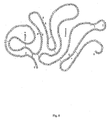
- the schematic diagram of pComb3H-K2S map is given in Fig. 3 .
- VCSM13 filamentous phage was used to infect pComb3H-K2S transformed E. coli XL-1 Blue, X[K2S].
- VCSM13 was propagated and incorporated the K2S-gp3 fusion protein during the viral packaging processes.
- the harvested recombinant phage (K2S- ⁇ ) gave a concentration of 5.4 x 10 11 cfu/ml determined by reinfecting E. coli XL-1 Blue with PEG-precipitated phages. These recombinant phage particles were verified for the expression of r-K2S by sandwich ELISA.
- the phage-bound heterologous K2S protein was recognized by the monoclonal anti-kringle 2 antibody (16B) by using sheep anti-tPA conjugated HRP antibody detection system.
- the absorbance of this assay was 1.12 ⁇ 0.03 (Table 1).
- the amount of K2S detectable on 10 12 phage particles is equal to 336 ng of protein in relation to the standard melanoma tPA.
- sheep anti-tPA conjugated HRP antibody was substituted by sheep anti-M13 antibody conjugated HRP. This immuno-reaction exhibited an absorbance of 1.89 ⁇ 0.07 (Table 1).
- MpComb3H-K2S Construction of MpComb3H-K2S.
- MSTPA and MASTPA mutagenic primers
- the cycle amplification mixture was thoroughly digested with Dpn I to degrade the old dam methylated pComb3H-K2S template (Dpn I prefers dam methylated DNA).
- Dpn I prefers dam methylated DNA
- K2S expression in XM[K2S] was induced by IPTG.
- r-K2S was detectable by using ELISA both in the periplasmic space and in the culture supernatant
- the amount of the heterologous protein in each preparation was determined by sandwich ELISA and related to the standard tPA. From 100 ml of the bacterial culture in shaker flask with the O.D. [600 nm] of 50, the periplasmic fraction yielded 1.38 ⁇ g of r-K2S (approximately 32%) whereas 2.96 ⁇ g of r-K2S (approximately 68%) was obtained in the ammonium precipitated culture supernatant.
- Sandwich ELISA was used to verify the PEG precipitated phage from VCSM13 infected XM[K2S]. No r-K2S captured by monoclonal anti-kringle 2 antibody was detected by anti-M13 conjugated HRP, indicating that K2S is not presented on the phage particles if gp3 is missing.
- plasminogen will be converted to plasmin.
- the produced plasmin will further digest the S-2251 substrate to a colour product, p-nitroaniline, which has a maximum absorbance at 405 nm.
- the specific activity of the recombinant product is in accordance with the absorbance.
- the fibrinogen-dependent enzymatic activity of each sample i.e. K2S- ⁇ periplasmic r-K2S or culture supernatant r-K2S, was evaluated and compared. Both K2S- ⁇ and periplasmic r-K2S illustrated notably low enzymatic activity, which was below the sensitivity of the test (0.25 IU/ml).
- the culture supernatant r-K2S gave the fibrinogen-dependent enzymatic activity of 7 IU/ml. Thus, from 100 ml culture we obtained a total of 700 IU enzymatic activity. Without fibrinogen no enzymatic activity of the r-K2S purified from culture supernatant was observed - whereas standard melanoma tPA showed some activity.
- the plasminogen has been copolymerized and immobilized with gelatin in the polyacrylamide gel prior to electrophoresis.
- the ammonium sulfate precipitated culture supernatants of E. coli XL-1 Blue, E. coli XL-1 Blue transformed with pComb3HSS and XM[K2S] were analyzed ( Fig. 7 ). All samples were processed in nonreducing condition to preserve the correct conformation and activity of the serine protease domain. Transparent areas of serine protease digested plasminogen were observed only in the ammonium sulfate precipitated culture supernatants of XM[K2S] at 34 and 37 kDa postions. The other samples gave no clearing zones.
- the positive control lane of standard melanoma tPA also demonstrated enzymatic activity at 66 and 72 kDa positions.
- FIG. 1 Validation of PCR amplification product of the K2S gene from the p51-3 vector by using SK2/174 and ASSP primers.
- Lane 1 shows 1 kb marker (Roche Molecular Biochemicals, Indianapolis, IN).
- Lane 2 was loaded with 1 ⁇ l of amplified product. A single band at 1110 bp is depicted. The electrophoresis was performed on a 1% agarose gel.
- FIG. 2 Identification of inserted K2S gene at 1110 bp (*) after Sfi I digested pComb3H-K2S was demonstrated in lane 3.
- Lane 1 shows 1 kb marker.
- Lane 2 was loaded with uncut pComb3H-K2S.
- FIG. 3 Scheme of pComb3H-K2S showing two Sfi I cloning sites into which the K2S gene was inserted. Signal sequence (OmpA), ribosome binding site (RIBS), lac promoter, and gpIII gene are also depicted.
- FIG. 4 Schematic diagram of the mutation site at the junction between the K2S and gpIII genes on pComb3H-K2S. The annealing site of pComb3H-K2S is bound with a set of mutation primers (MSTPA and MASTPA) containing modified oligonucleosides (underlined).
- MSTPA and MASTPA mutation primers
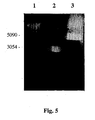
- FIG. 5 Characterization of newly synthesized MpComb3H-K2S by the Sfi I restriction enzyme. A single band at 4319 bp that refers to a single cleavage site of MpComb3H-K2S is observed in lane 3. No inserted K2S band at 1110 bp can be visualized. Lane 1 shows 1 kb marker. Lane 2 was loaded with uncut MpComb3H-K2S. The electrophoresis was performed on a 1% agarose gel. FIG. 6 .
- Lane 1 contained the indicated molecular weight standards ( ⁇ 10 -3 ), SDS-6H (Sigma, Saint Louis, MO). Fifty ⁇ g of the 55% saturated ammonium sulfate precipitated culture supernatant of XL-1 Blue, Xl-1 Blue transformed with pComb3HSS, and XM[K2S] were loaded in lane 2, 3, and 4 respectively. Lane 5 contained 50 mIU of standard melanoma tPA (86/670). Transparent zones of digested plasminogen in polyacrylamide gel are visible only in lane 4 at molecular weight of 34 and 37 kDa (B) and lane 5 at molecular weight of 66 and 72 kDa (A). FIG. 8 .
- FIG. 9 Structure A (SEQ ID NO:11) Native K2S molecule from amino acids 174-527 without modification.
- FIG. 9 Structure B-0 (SEQ ID NO:12) Native K2S molecule from amino acids 197-527 without modification.
- FIG. 10 Structure B-1 (SEQ ID NO:13) K2S molecule from amino acids 193-527, wherein to Structure B-0 of Fig. 9 the amino acids SEGN were added at the N-terminal portion.
- FIG. 11 Structure B-2 (SEQ ID NO:14) K2S molecule from amino acids 193-527, as in Fig. 10 , wherein Cys-261 was exchanged for Ser.
- FIG. 12 Structure A (SEQ ID NO:11) Native K2S molecule from amino acids 174-527 without modification.
- FIG. 9 Structure B-0 (SEQ ID NO:12) Native K2S molecule from amino acids 197-527 without modification.
- FIG. 10 Structure B-1 (SEQ ID NO:13) K2S molecule from amino acids 193-527
- Structure B-3 (SEQ ID NO:15) K2S molecule from amino acids 191-527, wherein to Structure B-0 of Fig. 9 the amino acids SEGNSD were added at the N-terminal portion.
- FIG. 13 Structure B-4 (SEQ ID NO:16) K2S molecule from amino acids 191-527, as in Fig. 12 , wherein Cys-261 was exchanged for Ser.
- FIG. 14 Structure C (SEQ ID NO:17) Native K2S molecule from amino acids 220-527 without modification. This molecule may be further modified in a similar manner as disclosed for structure B in figures 10-13 .
- FIG. 15 Structure D (SEQ ID NO:18) Native K2S molecule from amino acids 260-527 without modification.
- FIG. 16 tPA molecule (SEQ ID NO:19) TABLE 1. Detection of r-K2S molecule in phage preparation by sandwich ELISA Capture antibody Tracer antibody (conjugated HRP) Anti-tPA Anti-M13 K2S- ⁇ VCSM13 a K2S- ⁇ VCSM13 Anti-kringle 2 b 1.12 ⁇ 0.04 c 0.12 ⁇ 0.03 1.89 ⁇ 0.02 0.16 ⁇ 0.02 Anti-M13 0.17 ⁇ 0.01 0.14 ⁇ 0.05 1.91 ⁇ 0.02 1.88 ⁇ 0.03 a VCSM13 was harvested from XL-1 Blue transformed with pComb3HSS. b Mouse monoclonal anti-kringle 2 (16/B) was used. The other antibodies were prepared from sheep immunoglobulin. c Value is mean of absorbance of each sample which was assayed in triplicate.
Landscapes
- Health & Medical Sciences (AREA)
- Chemical & Material Sciences (AREA)
- Life Sciences & Earth Sciences (AREA)
- Engineering & Computer Science (AREA)
- Organic Chemistry (AREA)
- Bioinformatics & Cheminformatics (AREA)
- Wood Science & Technology (AREA)
- Zoology (AREA)
- Genetics & Genomics (AREA)
- General Health & Medical Sciences (AREA)
- Medicinal Chemistry (AREA)
- Biochemistry (AREA)
- Biomedical Technology (AREA)
- General Engineering & Computer Science (AREA)
- Molecular Biology (AREA)
- General Chemical & Material Sciences (AREA)
- Chemical Kinetics & Catalysis (AREA)
- Biotechnology (AREA)
- Microbiology (AREA)
- Nuclear Medicine, Radiotherapy & Molecular Imaging (AREA)
- Veterinary Medicine (AREA)
- Public Health (AREA)
- Animal Behavior & Ethology (AREA)
- Pharmacology & Pharmacy (AREA)
- Heart & Thoracic Surgery (AREA)
- Cardiology (AREA)
- Proteomics, Peptides & Aminoacids (AREA)
- Urology & Nephrology (AREA)
- Vascular Medicine (AREA)
- Pulmonology (AREA)
- Micro-Organisms Or Cultivation Processes Thereof (AREA)
- Enzymes And Modification Thereof (AREA)
- Preparation Of Compounds By Using Micro-Organisms (AREA)
- Peptides Or Proteins (AREA)
- Medicines That Contain Protein Lipid Enzymes And Other Medicines (AREA)
- Inorganic Compounds Of Heavy Metals (AREA)
Abstract
Description
- The invention belongs to the field of thrombolysis and of tissue plasminogen activator (tPA) derivative production in prokaryotic cells.
- The invention relates to methods for the production of a recombinant DNA-derived tPA, a variant thereof or a (Kringle 2 Serine) K2S molecule or a variant thereof in prokaryotic cells, wherein said tPA or K2S or variant is secreted extracellularly as an active and correctly folded protein, and the prokaryotic cell contains and expresses a vector comprising the DNA coding for said tPA or K2S or variant operably linked to the DNA coding for the signal peptide OmpA. The invention further relates to specific K2S derivatives obtainable by said method. The invention further relates to said DNA molecules and the use of said DNA molecules in said methods.
- Tissue plasminogen activator (tPA) is a polypeptide containing 527 amino acid residues (27) with a molecular mass of 72 kDa. The molecule is divided into five structural domains. Nearby the N-terminal region is a looped finger domain, which is followed by a growth factor domain. Two similar domains,
kringle 1 andkringle 2, are following. Both finger andkringle 2 domains bind specifically to the fibrin clots thereby accelerating tPA protein activation of bound plasminogen. Downstream ofkringle 2 is the serine protease, with its catalytic site located at the C-terminus. The serine protease is responsible for converting plasminogen to plasmin a reaction important in the homeostasis of fibrin formation and clot dissolution. The correct folding of tPA requires the correct pairing of 17 disulfide bridges in the molecule (1). - Clinically, tPA is a thrombolytic agent of choice for the treatment of acute myocardial infarction, pulmonary embolism, stroke, peripheral arterial occlusions, and other thromboembolic diseases. It has the advantage of causing no side effects on systemic haemorrhaging and fibrinogen depletion (7). Bowes melanoma cells were first used as a source in tPA production for therapeutic purposes (12). Since a consistent process with efficient production of highly purified protein in good yield is required for clinical use, the construction of full-length recombinant-tPA (r-tPA) progressed to mammalian cells. Chinese hamster ovary cells were transfected with the tPA gene to synthesize the r-tPA (8, 22). The recombinant DNA derived product produced by a mammalian cell culture fermentation system is harvested and purified from the culture medium. Attracted by simplicity and economy of production, a number of efforts in producing r-tPA from microorganisms, expecially bacteria, and more especially from Escherichia coli, were investigated (10, 13, 30). Regarding the low yield and the formation of inclusion bodies, which resulted in misfolding and in an inactive enzyme, numerous strategies have been proposed to overcome these problems.
- Several deletion-mutant
variants including kringle 2 plus serine protease (K2S) were considered. However, the enzymatic activity of the recombinant-K2S (r-K2S) was obtained only when refolding processes of purified inclusion bodies from cytoplasmic compartment were achieved (16, 29). In order to avoid the cumbersome refolding processes, impurities of misfolded proteins, and periplasmic protein delivery, special bacterial expression systems were exploited (6, 31). Despite periplasmic expression of tPA, overexpression led to inactive aggregates, even in the relatively high oxidizing condition in the periplasm. - In the prior art, there are a few descriptions of methods for the preparation of recombinant K2S in E. coli. However, there is no disclosure of a method leading to a cost effective method for large scale production of biologically active K2S.
- Obukowicz et al. (25) expressed and purified r-K2S from periplasmic space. In addition,
EP 1 048 732EP 1 048 732 was an extra periplasmic extraction step, which is not suitable for large scale production.Moreover, the production yield obtained with the PelB-K2S fusion protein is very low (about 4 µg/l). - Saito et al. (29) disclose the cytoplasmic expression of r-K2S. The authors used an in vivo renaturation processes for the expressed r-K2S, which was purified from the cytoplasmic space of E. coli as inclusion body. Boehringer Mannheim use a similar cumbersome denaturing/refolding process involving the steps of cell digestion, solubilization under denaturing and reducing conditions and reactivation under oxidizing conditions in the presence of GSH/GSSG which is not cost effective (24) and requires mutation of the amino acid sequence with possibly antigenic potential.
- In
EP 0 302 456 cytoplasmatic expression of several tPA variants in E.coli is described. The tPA variants were purified from the cruide cell extract after cell lysis via benzamidine Sepharose and a monoclonal antibody affinity chromatography step. - In 1991, Waldenström et al. (34) constructed a vector (pEZZK2P) for the secretion of
kringle 2 plus serine protease domain to E. coli culture supernatant. Hydroxylamine was used to remove the ZZ fusion peptide from IgG-Sepharose purified fraction. The cleavage agent hydroxylamine required modification of the cleavage sites ofkringle 2 plus serine protease (Asn177 → Ser and Asn184 → Gln) thus to protect it from hydroxylamine digestion. However, the resulting non-native, not properly folded K2S molecule is not suitable for therapeutic purposes. No enzymatic activity regarding fibrin binding/protease activity was disclosed. The unusual sequence may even activate the human immune system. -
WO 98/54199 - The problem underlying the present invention was thus to provide a commercially applicable method for large scale production of tPA molecules and derivatives thereof, e.g. K2S, wherein the K2S molecule is secreted in its biologically active form into the culture supernatant.
- The problem was solved within the scope of the claims and specification of the present invention.
- The use of the singular or plural in the claims or specification is in no way intended to be limiting and also includes the other form.
- The invention relates to a method for the production of a recombinant DNA-derived tissue plasminogen activator (tPA), a tPA variant, a Kringle 2 Serine protease molecule (K2S) or a K2S variant in prokaryotic cells, wherein said tPA, tPA variant, K2S molecule or K2S variant is secreted extracellularly as an active and correctly folded protein, characterized in that the prokaryotic cell contains and expresses a vector comprising the DNA coding for said tPA, tPA variant, K2S molecule or K2S variant operably linked to the DNA coding for the signal peptide OmpA.
- Surprisingly, the use of the signal peptide OmpA alone and/ or in combination with the N-terminal amino acids SEGN (SEQ ID NO:9) / SEGNSD (SEQ ID NO:10) translocate the recombinant DNA-derived tPA, tPA variant, K2S molecule or K2S variant to the outer surface and facilitates the release of the functional and active molecule into the culture medium to a greater extent than any other method in the prior art. Before crossing the outer membrane, the recombinant DNA-derived protein is correctly folded according to the method of the present invention. The signal peptide is cleaved off to produce a mature molecule. Surprisingly, the efficiency of signal peptide removal is very high and leads to correct folding of the recombinant DNA-derived protein.
- Said signal peptide OmpA interacts with SecE and is delivered across the inner membrane by energy generated by SecA, which binds to Sec components (SecE-SecY). SecY forms a secretion pore to dispatch the recombinant DNA-derived protein according to the invention. The space between the outer membrane and inner membrane of Gram-negative bacteria, periplasm, has higher oxidative condition in comparison to the cytoplasmic space. This supports the formation of disulfide bonds and properly folding of the recombinant DNA-derived protein (e.g. K2S) in the periplasm to yield an active molecule. According to the present invention, the signal peptide will be cleaved off to produce a mature molecule. The complex of GspD secretin and GspS lipoprotein on the outer membrane serves as gate channel for secreting the recombinant DNA-derived protein according to the invention to the extracellular medium. This secretion process requires energy, which is generated in cytoplasm by GspE nucleotide-binding protein then transferred to the inner membrane protein (Gsp G-J, F and K-N). GspC transfers the energy to GspD by forming a cross-linker between a set of inner membrane protein (Gsp G-J, F and K-N) and GspD. Before crossing the outer membrane successfully, the recombinant DNA-derived protein is correctly folded.
- Operably linked according to the invention means that the DNA encoding the tPA, tPA variant, K2S molecule or K2S variant (preferably comprising the nucleic acid encoding SEGN or SEGNSD at its N-terminal portion) is cloned in close proximity to the OmpA DNA into the vector in order to achieve expression of the OmpA-tPA, tPA variant, K2S molecule or K2S variant-fusion protein and to direct secretion outside the prokaryotic host cell. Typically, the majority of the tPA, tPA variant, K2S molecule or K2S variant is secreted and can then be purified by appropriate methods such as ammonium sulfate precipitation and/or affinity chromatography and further purification steps. The invention also includes the use of inducers such as IPTG or IPTG in combination with glycerol, the improvement of the incubation condition and harvesting period to maximize the amount of active protein.
- In a preferred embodiment, said DNA encoding the OmpA signal peptide may be fused to a short peptide characterized by the amino acid sequence SEGN or SEGNSD or the coding nucleic acid sequence TCTGAGGGAAAC (SEQ ID NO:20) or TCTGAGGGAAACAGTGAC (SEQ ID NO:1) and located in the N-terminal portion or at the N-terminal portion of the tPA, tPA variant, K2S molecule or K2S variant. Thus, preferably, said fusion protein comprises OmpA-SEGNSD-tPA, -tPA-variant, -K2S-molecule or -K2S-variant. Even more preferred, said amino acids characterized by SEGN or SEGNSD may be carry a point mutation or may be substituted by a non-natural amino acid. Even more preferred, there may be an amino acid or non-amino acid spacer between OmpA and SEGN or SEGNSD and the tPA, tPA variant, K2S molecule or K2S variant.
- Thus, in a preferred method according to the invention said the prokaryotic cell contains and expresses a vector comprising the DNA coding for said tPA, tPA variant, K2S molecule or K2S variant operably linked to the DNA coding for the signal peptide OmpA, which is operably linked to the nucleic acid molecule defined by the sequence TCTGAGGGAAACAGTGAC or a functional derivative thereof.
- The method according to the invention comprises prokaryotic host cells such as, but not limited to Escherichia coli (E. coli), Bacillus subtilis, Streptomyces, Pseudomonas, e.g. Pseudomonas putida, Proteus mirabilis, Saccharomyces, Pichia or Staphylococcus, e.g. Staphylococcus carnosus. Preferably said host cells according to the invention are Gram-negative bacteria.
- Preferably, a method according to the invention is also characterised in that the prokaryotic cell is E. coli. Suitable strains include, but are not limited to E. coli XL-1 blue, BL21(DE3), JM109, DH series, TOP10 and HB101.
- Preferably, a method according to the invention is also characterised in that the following steps are carried out:
- a) the DNA encoding the tPA, tPA variant, K2S molecule or K2S variant is amplified by PCR;
- b) the PCR product is purified;
- c) said PCR product is inserted into a vector comprising the DNA coding for OmpA signal peptide and the DNA coding for gpIII in such a way that said PCR product is operably linked upstream to the DNA coding for the OmpA signal sequence and linked downstream to the DNA coding for gpIII of said vector;
- d) that a stop codon is inserted between said tPA, tPA variant, K2S molecule or K2S variant and gpIII;
- e) said vector is expressed by the prokaryotic cell
- f) the tPA, tPA variant, K2S molecule or K2S variant is purified.
- For step a) according to the invention the choice / design of the primers is important to clone the DNA in the right location and direction of the expression vector (see example 1). Thus, the primers as exemplified in example 1 and
figure 4 comprise an important aspect of the present invention. With gp III of step c) gene protein III is meant which is present mainly in phagemid vectors. The stop codon is inserted to avoid transcription of gp III thus eventually leading to secretion of the tPA, tPA variant, K2S molecule or K2S variant of interest. Any suitable method for insertion of the stop codon may be employed such as site-directed mutagenesis (e.g. Weiner MP, Costa GL (1994) PCR Methods Appl 4(3):S131-136; Weiner MP, Costa GL, Schoettlin W, Cline J, Mathur E, Bauer JC (1994) Gene 151(1-2):119-123; see also example 1). - Any vector may be used in the method according to the invention, preferably said vector is a phagemid vector (see below).
- Preferably, a method according to the invention is also characterised in that the tPA, tPA variant, K2S molecule or K2S variant is selected from human tissue plasminogen activator (tPA,
figure 16 ) or a fragment, a functional variant, an allelic variant, a subunit, a chemical derivative, a fusion protein or a glycosylation variant thereof. Such fragments, allelic variants, functional variants, variants based on the degenerative nucleic acid code, fusion proteins with an tPA protein according to the invention, chemical derivatives or a glycosylation variant of the tPA proteins according to the invention may include one, several or all of the following domains or subunits or variants thereof: - 1. Finger domain (4-50)
- 2. Growth factor domain (50-87)
- 3.
Kringle 1 domain (87-176) - 4.
Kringle 2 domain (176-262) - 5. Protease domain (276-527)
- The numbering/naming of the domains is according to Genbank accession number GI 137119 or Nature 301 (5897), 214-221 (1983).
- More preferably, a method according to the invention is also characterised in that the tPA, tPA variant, K2S molecule or K2S variant is selected from the Kringle 2 (4.) plus Serine protease (5.) K2S variant of human tissue plasminogen activator or a fragment, a functional variant, an allelic variant, a subunit, a chemical derivative, a fusion protein or a glycosylation variant thereof. More preferably, a method according to the invention is also characterised in that the vector is a phagemid vector comprising the DNA coding for OmpA signal peptide and the DNA coding for gpIII.
- More preferably, a method according to the invention is also characterised in that the vector is the pComb3HSS phagemid (see also example 1).
- More preferably, a method according to the invention is also characterised in that the DNA sequence comprises or consists of the following DNA sequence encoding OmpA, and K2S or a functional variant of K2S or a variant due to the degenerate nucleotide code:

- More preferably, a method according to the invention is also characterised in that the DNA Sequence of OmpA consists of the following sequence:

- Said DNA encodes the following amino acid sequence of OmpA. OmpA thus consists of a protein characterized by the following amino acid sequence or a glycosylation variant thereof as part of the invention:
- MKKTAIAIAVALAGFATVAQAA (SEQ ID NO:21).
- The untranslated region may contain a regulatory element, such as e.g. a transcription initiation unit (promoter) or enhancer. Said promoter may, for example, be a constitutive, inducible or development-controlled promoter. Preferably, without ruling out other known promoters, the constitutive promoters of the human Cytomegalovirus (CMV) and Rous sarcoma virus (RSV), as well as the Simian virus 40 (SV40) and Herpes simplex promoter. Inducible promoters according to the invention comprise antibiotic-resistant promoters, heat-shock promoters, hormone-inducible "Mammary tumour virus promoter" and the metallothioneine promoter. Preferred promoters include T3 promoter, T7 promoter, Lac/aral and Ltet0-1.
- More preferably, a method according to the invention is also characterised in that the DNA of the tPA, tPA variant, K2S molecule or K2S variant is preceded by a lac promoter and/or a ribosomal binding site such as the Shine-Dalgarno sequence (see also example).
- More preferably, a method according to the invention is also characterised in that the DNA coding for the tPA, tPA variant, K2S molecule or K2S variant is selected from the group of DNA molecules coding for at least 90% of the amino acids 87 - 527, 174 - 527, 180 - 527 or 220 - 527 of the human tissue plasminogen activator protein.
- More preferably, a method according to the invention is also characterised in that the DNA Sequence of K2S comprises or consists of the following sequence:

- The present invention also relates to variants of the before-mentioned nucleic acid molecules due to the degenerate code or to fragments thereof, nucleic acids which hybridize to said nucleic acids under stringent conditions, allelic or functional variants. The invention also relates to nucleic acids comprising said K2S nucleic acid fused to the nucleic acid encoding another protein molecule.
- Stringent conditions as understood by the skilled person are conditions which select for more than 85 %, preferred more than 90 % homology (Sambrook et al. 1989; Molecular Cloning: A Laboratory Manual, 2nd ed., Cold Spring Harbor Laboratory Press, Cold Spring Harbor, New York). The hybridisation will be carried out e.g. in 6x SSC/ 5x Denhardt's solution/ 0,1 % SDS (SDS: sodium dodecylsulfate) at 65 °C. The degree of stringency is decided in the washing step. Thus, for example for a selection of DNA-sequences with approx. 85 % or more homology, the conditions 0,2 x SSC/ 0,01 % SDS/ 65 °C and for a selection of DNA-sequences of approx. 90 % or more homology the conditions 0,1x SSC/ 0,01 % SDS/ 65 °C are suitable. The composition of said reagents is described in Sambrook et al. (1989, supra).
- Another important part of the present invention is a variant of human tissue plasminogen activator comprising of or consisting of the Kringle 2 (4.) plus Serine protease (5.) (abbreviated K2S) protein or a variant or a fragment, a functional variant, an allelic variant, a subunit, a chemical derivative, a fusion protein or a glycosylation variant thereof.
- The numbering/naming of the domains is according to Genbank accession number GI 137119 or Nature 301 (5897), 214-221 (1983), wherein the
Kringle 2 domain extends from amino acid 176-262 and the protease domain from 276-527. Thus, according to the invention, a preferred K2S molecule may include amino acids 176-527 including the amino acids betweenKringle 2 and the protease (amino acids 263 to 275; exemplified in fig. (structure A)). A K2S molecule according to the invention comprises the minimal part of theKringle 2 domain and the protease domain still retaining protease activity and fibrin binding activity (measured as exemplified in the description/example). Said K2S molecule according to the invention comprises the amino acids SEGN or SEGNSD in its N-terminal portion (see infra). A preferred K2S molecule does not includeamino acids 1 to 3 or 1 to 5 of the tPA molecule. Preferably, a K2S molecule according to the invention has the amino acid Asn atpositions 177 and 184, i.e. it does not require the modifications as disclosed in Waldenström for improved producibility with a method according to the invention. Thus, a preferred K2S molecule according to the invention has the native amino acid sequence (no mutation) as opposed to the molecules known from the prior art. Most preferred, said K2S molecule according to the invention is a molecule characterized by the native amino acid sequence or parts thereof, does neither haveamino acids 1 to 3 nor 1 to 5 of tPA and comprises N-terminally the amino acids SEGN or SEGNSD for improved producibility and/or correct folding of the molecule. - It is essential that the K2S protein according to the invention comprises in its N-terminal portion a peptide characterized by the amino acid sequence SEGN which advantageously allows commercial production with a method as described supra leading to a correctly folded, secreted K2S protein. Said 4 amino acids characterized by SEGN may have one or several amino acids more N-terminal, however said amino acids have to be located in the N-terminal portion as opposed to the C-terminal portion. Most preferably, said amino acids are located at the N-terminal portion. Preferably, the amino acids characterized by SEGN may be carry a point mutation or may be substituted by a non-natural amino acid.
- Thus, in another important embodiment the invention relates to a K2S protein characterized in that it comprises the amino acids defined by the sequence SEGN or a variant or a fragment, a functional variant, an allelic variant, a subunit, a chemical derivative, a fusion protein or a glycosylation variant thereof.
- Such fragments are exemplified e.g. in
figure 10 (Structure B-1) andfigure 11 (Structure B-2) extending from amino acids 193-527. Structure B-1 has the native amino acid Cys in position 261, wherein in B-2 the amino acid is substituted by Ser. Further fragments according to the invention comprising the amino acids 220-527 (fig. 14 , structure C) or comprising the amino acids 260-527 (fig. 15 , structure D) may be modified according to the invention by addition of the amino acids SEGN and/or substitution of Cys-261 by Ser. The artisan can determine the minimal length of a K2S molecule according to the invention in order to retain its biological function and generate a K2S molecule with improved producibility and/or correct folding by adding the amino acids SEGN in the N-terminal portion. Thus, another preferred embodiment is said minimal K2S molecule with SEGN at its N-terminal portion. - In another important embodiment the invention relates to a K2S protein characterized in that it comprises the amino acids defined by the sequence SEGNSD or a variant or a fragment, a functional variant, an allelic variant, a subunit, a chemical derivative, a fusion protein or a glycosylation variant thereof. Such fragments are exemplified e.g. in
figure 12 (Structure B-3) andfigure 13 (Structure B-4) extending from amino acids 191-527. Structure B-3 has the native amino acid Cys in position 261, wherein in B-4 the amino acid is substituted by Ser. Further fragments according to the invention comprising the amino acids 220-527 (fig. 14 , structure C) or comprising the amino acids 260-527 (fig. 15 , structure D) may be modified according to the invention by addition of the amino acids SEGNSD and/or substitution of Cys-261 by Ser. The artisan can determine the minimal length of a K2S molecule according to the invention in order to retain its biological function and generate a K2S molecule with improved producibility and/or correct folding by adding the amino acids SEGNSD in the N-terminal portion. Thus, another preferred embodiment is said minimal K2S molecule with SEGNSD at its N-terminal portion. - The application furthermore discloses a K2S protein comprising a protein characterized by the following amino acid sequence or a variant or a fragment, a functional variant, an allelic variant, a subunit, a chemical derivative or a glycosylation variant thereof:

- According to the invention, * means STOP (i.e. encoded by a stop codon). This K2S molecule is exemplified in
figure 8 . - One variant of the K2S molecule according to the invention relates to a fusion protein of K2S being fused to another protein molecule.
- The application furthermore discloses a K2S protein consisting of a protein characterized by the following amino acid sequence:

- Said K2S molecules may be encoded by a DNA molecule as described supra.
- Another important aspect of the invention relates to a DNA molecule characterized in that it is coding for:
- a) the OmpA protein operably linked to
- b) a DNA molecule coding for a polypeptide containing the
kringle 2 domain and the serine protease domain of tissue plasminogen activator protein. - More preferably, a DNA molecule according to the invention is also characterised in that the DNA sequence comprises or consists of the following DNA sequence encoding OmpA, and K2S or a functional variant of K2S or a variant due to the degenerate nucleotide code:

- Said DNA molecule encodes the following fusion protein of OmpA and K2S. Said fusion protein of OmpA, and K2S characterised in that it comprises or consists of a protein characterized by the following amino acid sequence or a fragment, a functional variant, an allelic variant, a subunit, a chemical derivative or a glycosylation variant thereof forms an important part of the present invention:


- Another preferred aspect of the invention relates to a DNA molecule according to the invention, characterized in that said DNA sequence b) is coding for at least 90% of the amino acids 87 - 527 of the human tissue plasminogen activator protein (numbering used herein as GI 137119 or Nature 301 (5897), 214-221 (1983).
- Another preferred aspect of the invention relates to a DNA molecule according to the invention, characterized in that said DNA sequence b) is coding for at least 90% of the amino acids 174 - 527 of the human tissue plasminogen activator protein.
- Another preferred aspect of the invention relates to a DNA molecule according to the invention, characterized in that said DNA sequence b) is coding for at least 90% of the amino acids 180 - 527 of the human tissue plasminogen activator protein.
- Another preferred aspect of the invention relates to a DNA molecule according to the invention, characterized in that said DNA sequence b) is coding for at least 90% of the amino acids 220 - 527 of the human tissue plasminogen activator protein.
- Another preferred aspect of the invention relates to a DNA molecule according to the invention, characterized in that said DNA sequence a) is hybridizing under stringent conditions to the following sequence:

- Another preferred aspect of the invention relates to a DNA molecule according to the invention, characterized in that said DNA sequence a) consists of the following sequence:

- Another preferred aspect of the invention relates to a DNA molecule according to the invention, characterized in that said DNA sequence b) is hybridizing under stringent conditions to the following sequence:


- Another preferred aspect of the invention relates to a DNA molecule according to the invention, characterized in that said DNA sequence b) consists of the following sequence:


- Another preferred embodiment of the invention relates to a vector containing a DNA sequence according to the invention.
- Another preferred embodiment of the invention relates to a vector according to the invention, wherein said DNA sequence is preceded by a lac promoter and a ribosomal binding site. Suitable vectors according to the invention include, but are not limited to viral vectors such as e.g. Vaccinia, Semliki-Forest-Virus and Adenovirus, phagemid vectors and the like. Preferred are vectors which can be advantageously used in E. coli, but also in any other prokaryotic host such as pPROTet.E, pPROLar.A, members of the pBAD family, pSE family, pQE family and pCAL. Another preferred embodiment of the invention relates to the vector pComb3HSS containing a DNA according to the invention, wherein the expression of the gp III protein is suppressed or inhibited by deleting the DNA molecule encoding said gp III protein or by a stop codon between the gene coding for a polypeptide containing the
kringle 2 domain and the serine protease domain of tissue plasminogen activator protein and the protein III gene. - Another important aspect of the present invention relates to a prokaryotic host cell comprising a DNA molecule according to the invention.
- Another important aspect of the present invention relates to a prokaryotic host cell comprising a vector according to the invention.
- Another important aspect of the present invention relates to an E. coli host cell comprising a DNA molecule according to the invention.
- Another important aspect of the present invention relates to a E. coli host cell comprising a vector according to the invention.
- Yet another important aspect of the present invention is the use of a DNA molecule according to the invention or of a vector according to the invention or a host cell according to the invention in a method for the production of a polypeptide having the activity of tissue plasminogen activator. Yet another important aspect of the present invention is the use according the invention as described above, wherein said method is a method according to the invention.
- Another very important aspect is a pharmaceutical composition comprising a substance obtainable by a method according to the invention and pharmaceutically acceptable excipients and carriers. An example for said substance is the K2S molecule described supra. The term "pharmaceutically acceptable carrier" as used herein refers to conventional pharmaceutic excipients or additives used in the pharmaceutical manufacturing art. Such physiologically acceptable compounds include, for example, carbohydrates, such as glucose, sucrose or dextrans, antioxidants, such as ascorbic acid or glutathione, chelating agents, low molecular weight proteins or other stabilizers or excipients (see also e.g. Remington's Pharmaceutical Sciences (1990, 18th ed. Mack Publ., Easton.)). Said pharmaceutical composition according to the invention can be advantageously administered intravenously as a bolus, e.g. as a single bolus for 5 to 10 seconds intravenously.
- The invention further relates to the use of substances obtainable by a method according to the invention in the manufacture of a medicament in the treatment of stroke, cardiac infarction, acute myocardial infarction, pulmonary embolism, any artery occlusion such as coronary artery occlusion, intracranial artery occlusion (e.g. arteries supplying the brain), peripherally occluded arteries, deep vein thrombosis or related diseases associated with unwanted blood clotting.
- The following example is intended to aid the understanding of the invention and should in no way be regarded as limiting the scope of the invention.
- Primer design. In order to amplify a specific part of tPA gene, a pair of primers SK2/174 [5' GAGGAGGAGGTGGCCCAGGCGGCCTCTGAGGGAAACAGTGAC 3'] (SEQ ID NO:22) and ASSP [5'GAGGAGGAGCTGGCCGGCCTGGCCCGGTCGCATGTTGTCACG 3'] (SEQ ID NO:23) were synthesized (Life Technologies, Grand Island, NY). These primers were designed based on the human tPA gene retrieved from NCBI databases (g137119). They were synthesized with Sfi I end cloning sites (underlined) in such a way that the reading frame from the ATG of the gpIII gene in phagemid vector, pComb3HSS, will be maintained throughout the inserted sequence.
- Another primer set for site-directed mutagenesis was designed to anneal at the sequence situated between K2S gene and gene III in pComb3H-K2S. The sequence of primers with mutation bases (underlined) for generating a new stop codon were MSTPA [5'ACATGCGACCGTGACAGGCCGGCCAG3'] (SEQ ID NO:24) and MASTPA [5'CTGGCCGGCCTGTCACGGTCGCATGT3'] (SEQ ID NO:25).
- Amplification of K2S gene by PCR. One µg SK2/174 and ASSP primers together with 50 ng of p51-3 template (obtained from Dr. Hiroshi Sasaki, Fujisawa Pharmaceutical, Japan) were suspended in 100 µl PCR mixture. An amount of 2.5 U Taq polymerase (Roche Molecular Biochemicals, Indianapolis, IN) was finally added to the solution. The titrated amplification condition was initiated with jump start at 85°C for 4 min, then denaturation at 95°C for 50 sec, annealing at 42°C for 50 sec, extension at 72°C for 1.5 min. Thirty five rounds were repeatedly performed. The mixture was further incubated at 72°C for 10 min. The amplified product of 1110 bp was subsequently purified by QIAquick PCR Purification Kit (QIAGEN, Hilden, Germany). The correctness of purified product was confirmed by restriction enzymes.
- Construction of phagemid expressing K2S. The purified PCR product of K2S and pComb3HSS phagemid (kindly provided by Dr. Carlos F. Barbas, Scripps Institute, USA) were digested with Sfi I (Roche Molecular Biochemicals, Indianapolis, IN) to prepare specific cohesive cloning sites. Four µg of the purified PCR product was digested with 60 U of Sfi I at 50°C for 18 h. For pComb3HSS, 20 µg of phagemid vectors were treated with 100 U of Sfi I. Digested products of purified PCR product of K2S and pComb3HSS (∼3300 bp) were subsequently gel-purified by the QIAquick Gel Extraction Kit (QIAGEN, Hilden, Germany). T4 ligase (Roche Molecular Biochemicals, Indianapolis, IN) of 5 U were introduced to the mixture of 0.7 µg of purified Sfi I-digested pComb3HSS and 0.9 µg of purified Sfi I-digested PCR product. Ligation reaction was incubated at 30°C for 18 h. The newly constructed phagemid was named pComb3H-K2S.
- Transformation of E. coli XL-1 Blue. Two hundred µl of CaCl2 competent E. coli XL-1 Blue (Stratagene, La Jolla, CA) were transformed with 70 ng of ligated or mutated product. The transformed cells were propagated by spreading on LB agar containing 100 µg/ml ampicillin and 10 µg/ml tetracycline (Sigma, Saint Louis, MO). After cultivation at 37°C for 18 h several antibiotic resistant colonies were selected for plasmid minipreps by using the alkaline lysis method. Each purified plasmid was subjected to Sfi I restriction site analysis. A transformant harboring plasmid with the correct Sfi I restriction site(s) was subsequently propagated for 18 h at 37°C in 100 ml LB broth with ampicillin 100 µg/ml and tetracycline 10 µg/ml. A plasmid maxiprep was performed using the QIAGEN Plasmid Maxi Kit (QIAGEN, Hilden, Germany). The purified plasmid was reexamined for specific restriction sites by Sfi I and sequenced by AmpliTaq DNA Polymerase Terminator Cycle Sequencing Kit (The Perkin-Elmer Corporation, Forster City, CA).
- Site-directed mutagenesis of pComb3H-K2S. 10 ng of pComb3H-K2S template were mixed with 125 ng of MSTPA and MASTPA primers. PfuTurbo DNA polymerase (Stratagene, LA Jolla, CA) of 2.5 U was added to the mixture for cycle amplification. The reaction started with one round of 95°C for 30 sec. Then it was followed by 16 rounds consisting of 95°C for 30 sec, 55°C for 1 min, and 68°C for 9 min. The reaction tube was subsequently placed on ice for 2 min. In order to destroy the template strands, 10 U of Dpn I restriction enzyme (Stratagene, LA Jolla, CA) were added to the amplification reaction and incubated for 1 h at 37°C. This synthesized product (MpComb3H-K2S) was further used to transform E. coli XL-1 Blue.
- Preparation of phage-display recombinant-K2S. After pComb3H-K2S was transformed to XL-1 Blue, the phage display technique was performed. A clone of pComb3H-K2S transformed E. coli XL-1 Blue was propagated in 10 ml super broth containing ampicillin 100 µg/ml and tetracycline 10 µg/ml at 37°C until the O.D. [600 nm] of 1.5 was reached. The bacterial culture was subsequently propagated in 100 ml of the same medium and culture for 2 h. An amount of 1012 pfu of VCSM13 helper phage (Stratagene, La Jolla, CA) was used to infect the transformed E. coli XL-1 Blue. After 3 h incubation, kanamycin at a final concentration of 70 µg/ml final concentration was added to culture. The culture was left shaking (200 RPM) for 18 h at 37°C. Bacteriophages which harbored K2S on gp3 (K2S-ϕ) were then harvested by adding 4% w/v PEG MW 8000 (Sigma, Saint Louis, MO) and 3% w/v NaCl. Finally, the harvested phage was resuspended in 2 ml PBS pH 7.4. The phage number was determined by infecting E. coli XL-1 Blue. The colony-forming unit per milliliter (cfu/ml) was calculated as described previously (21).
- Expression of recombinant-K2S in shaker flasks. MpComb3H-K2S transformed E. coli XL-1 Blue was cultivated in 100 ml super broth (3% w/v tryptone, 2% w/v yeast extract and 1% w/v MOPS) at pH 7.0 in the presence of ampicillin (100 µg/ml) at 37°C until an O.D. [600 nm] of 0.8 was reached. Subsequently, the protein synthesis was induced by 1 mM of IPTG (Promega, Madison, WI). The bacteria were further cultured shaking (200 RPM) for 6 h at 30°C. The culture supernatant was collected and precipitated with 55% saturated ammonium sulfate (32). The precipitate was reconstituted with PBS, pH 7.2, and dialysed in the same buffer solution at 4°C for 18 h. Periplasmic proteins from bacterial cells were extracted by using a chloroform shock as previously described by Ames et al. (2).
- Immunoassay quantification of recombinant-K2S. In order to detect r-K2S, solid phase was coated with
monoclonal anti-kringle 2 domain (16/B) (generously provided by Dr. Ute Zacharias, Central Institute of Molecular Biology, Berlin-Buch, Germany). The standard ELISA washing and blocking processes were preformed. Fifty µl of 1011 cfu/ml of K2S-ϕ secretory r-K2S were added into each anti-kringle 2 coated well. Antigen-antibody detection was carried out as follows. Either sheep anti-M13 conjugated HRP (Pharmacia Biotech, Uppsala, Sweden) or sheep anti-tPA conjugated HRP (Cedarlane, Ontario, Canada), was added to each reaction well after the washing step. The substrate TMB was subjected to every well and the reaction was finally ceased with H2SO4 solution after 30 min incubation. The standard melanoma tPA 86/670 (National Institute for Biological Standards and Control, Hertfordshine, UK) was used as positive control. - Amidolytic activity assay. A test kit for the detection of tPA amidolytic activity was purchased from Chromogenix (Molndal, Sweden). The substrate mixture containing plasminogen and S-2251 was used to determine serine protease enzymatic activity. The dilution of 10-2 of each ammonium precipitated sample was assayed with and without stimulator, human fibrinogen fragments. The assay procedure was according to the COASET t-PA manual.
- SDS-PAGE and immunoblotting. The dialysed precipitate-product from culture supernatant was further concentrated 10 folds with centricon 10 (AMICON , Beverly, MA). The concentrated sample was subjected to protein separation by SDS-PAGE, 15% resolving gel, in the reducing buffer followed by electroblotting to nitrocellulose. The nitrocellulose was then blocked with 4% skimmed milk for 2 hr. In order to detect r-K2S, a proper dilution of sheep anti-tPA conjugated HRP was applied to the nitrocellulose. The immunoreactive band was visualized by a sensitive detection system, Amplified Opti-4CN kit (BIORAD, Hercules, CA).
- Copolymerized plasminogen polyacrylamide gel electrophoresis. An 11% resolving polyacrylamide gel was copolymerized with plasminogen and gelatin as previously described by Heussen et al. (14). The stacking gel was prepared as 4 % concentration without plasminogen and gelatin. Electrophoresis was performed at 4°C at a constant current of 8 mA. The residual SDS in gel slab was removed after gentle shaking at room temperature for 1h in 2.5% Triton X-100. Then the gel slab was incubated in 0.1 M glycine-NaOH, pH 8.3, for 5 h at 37°C. Finally, the gel slab was stained and destained by standard Coomassie brilliant blue (R-250) dying system. The location of the peptide harboring enzymatic activity was not stained by dye in contrast to blue-paint background.
- Construction of K2S gene carrying vector. From the vector p51-3 we amplified the
kringle 2 plus ther serine protease portion of tPA (Ser174 inkringle 2 domain to Pro527 in the serine protease) using primers SK2/174 and ASSP. The amplified 1110 bp product was demonstrated by agarose gel electrophoresis (Fig. 1 , lane 2) and was inserted into pComb3HSS phagemid by double Sfi I cleavage sites on 5' and 3' ends in the correct reading frame. Thus a new vector, pComb3H-K2S, harboring the K2S was generated. In this vector K2S is flanked upstream by the OmpA signal sequence and donwstream by gp3. The correct insertion of K2S was verified both by restriction analysis with Sfi I (Fig. 2 , lane 3), PCR-analysis (demonstration of a single band at 1110 bp), and DNA sequencing. The schematic diagram of pComb3H-K2S map is given inFig. 3 . - Phage-displayed r-K2S. VCSM13 filamentous phage was used to infect pComb3H-K2S transformed E. coli XL-1 Blue, X[K2S]. VCSM13 was propagated and incorporated the K2S-gp3 fusion protein during the viral packaging processes. The harvested recombinant phage (K2S-ϕ) gave a concentration of 5.4 x 10¹¹ cfu/ml determined by reinfecting E. coli XL-1 Blue with PEG-precipitated phages. These recombinant phage particles were verified for the expression of r-K2S by sandwich ELISA. The phage-bound heterologous K2S protein was recognized by the
monoclonal anti-kringle 2 antibody (16B) by using sheep anti-tPA conjugated HRP antibody detection system. The absorbance of this assay was 1.12 ± 0.03 (Table 1). The amount of K2S detectable on 1012 phage particles is equal to 336 ng of protein in relation to the standard melanoma tPA. In order to corroborate that K2S-gp3 fusion protein was associated with phage particles, sheep anti-tPA conjugated HRP antibody was substituted by sheep anti-M13 antibody conjugated HRP. This immuno-reaction exhibited an absorbance of 1.89 ± 0.07 (Table 1). In contrast, if the capture antibody was sheep anti-M13 antibody, extremely low K2S was observed with sheep anti-tPA antibody conjugated HRP; the absorbance was only 0.17 ± 0.01 (Table 1). This suggested that only a minority of purified phage particles carried K2S-gp3 fusion protein. VCSM13 prepared from non-transformed XL-1 Blue was used as a negative control. - Construction of MpComb3H-K2S. We generated a stop codon between K2S and gp3 in pComb3H-K2S with the aid of the mutagenic primers (MSTPA and MASTPA) (
Fig. 4 ). In order to enrich the newly synthesized and mutated MpComb3H-K2S, the cycle amplification mixture was thoroughly digested with Dpn I to degrade the old dam methylated pComb3H-K2S template (Dpn I prefers dam methylated DNA). After transforming of E. coli XL-1 Blue with MpComb3H-K2S, a transformant XM[K2S] was selected for further study. As a consequence of bp substitution, one Sfi I cleavage site close to the 3' end of K2S gene was lost after site-directed mutagenesis. A linear version of Sfi I cleaved MpComb3H-K2S was observed at 4319 bp without the appearance of inserted K2S gene fragment (Fig. 5 , lane 3). Thus, the K2S gene encoding by MpComb3H-K2S was expressed in non-gp3 fusion form in XM[K2S]. - Expression and purification of K2S. K2S expression in XM[K2S] was induced by IPTG. r-K2S was detectable by using ELISA both in the periplasmic space and in the culture supernatant The amount of the heterologous protein in each preparation was determined by sandwich ELISA and related to the standard tPA. From 100 ml of the bacterial culture in shaker flask with the O.D. [600 nm] of 50, the periplasmic fraction yielded 1.38 µg of r-K2S (approximately 32%) whereas 2.96 µg of r-K2S (approximately 68%) was obtained in the ammonium precipitated culture supernatant. Sandwich ELISA was used to verify the PEG precipitated phage from VCSM13 infected XM[K2S]. No r-K2S captured by
monoclonal anti-kringle 2 antibody was detected by anti-M13 conjugated HRP, indicating that K2S is not presented on the phage particles if gp3 is missing. - Amidolytic activity measurement. If serine protease domain is present in the sample, plasminogen will be converted to plasmin. The produced plasmin will further digest the S-2251 substrate to a colour product, p-nitroaniline, which has a maximum absorbance at 405 nm. The specific activity of the recombinant product is in accordance with the absorbance. The fibrinogen-dependent enzymatic activity of each sample i.e. K2S-ϕ periplasmic r-K2S or culture supernatant r-K2S, was evaluated and compared. Both K2S-ϕ and periplasmic r-K2S illustrated notably low enzymatic activity, which was below the sensitivity of the test (0.25 IU/ml). The culture supernatant r-K2S gave the fibrinogen-dependent enzymatic activity of 7 IU/ml. Thus, from 100 ml culture we obtained a total of 700 IU enzymatic activity. Without fibrinogen no enzymatic activity of the r-K2S purified from culture supernatant was observed - whereas standard melanoma tPA showed some activity.
- Demonstration of recombinant protein by immunoblotting. Partially purified K2S from culture supernatant of XM[K2S] revealed a molecular mass of 39 kDa by using sheep anti-tPA antibodies (
Fig. 6 ). The negative control, partially purified culture supernatant of non-transformed XL1-Blue, contained no reactive band with a similar size. - Localization of active enzyme by PAGE. The plasminogen has been copolymerized and immobilized with gelatin in the polyacrylamide gel prior to electrophoresis. The ammonium sulfate precipitated culture supernatants of E. coli XL-1 Blue, E. coli XL-1 Blue transformed with pComb3HSS and XM[K2S] were analyzed (
Fig. 7 ). All samples were processed in nonreducing condition to preserve the correct conformation and activity of the serine protease domain. Transparent areas of serine protease digested plasminogen were observed only in the ammonium sulfate precipitated culture supernatants of XM[K2S] at 34 and 37 kDa postions. The other samples gave no clearing zones. The positive control lane of standard melanoma tPA also demonstrated enzymatic activity at 66 and 72 kDa positions. -
- 1. Allen, S., H. Y. Naim, and N. J. Bulleid. 1995. Intracellular folding of tissue-type plasminogen activator. Effects of disulfide bond formation on N-linked glycosylation and secretion. J. Biol. Chem. 270:4797-4804.
- 2. Ames, G. F., C. Prody, and S. Kustu. 1984. Simple, rapid, and quantitative release of periplasmic proteins by chloroform. J. Bacteriol. 160:1181-1183.
- 3. Barbas, C. F. III, A. S. Kang, R. A. Lerner, and S. J. Benkovic. 1991. Assembly of combinatorial antibody libraries on phage surfaces: the gene III site. Proc. Natl. Acad. Sci. U. S. A. 88:7978-7982.
- 4. Barbas, C. F. III, and J. Wagner. 1995. Synthetic human antibodies: selecting and evolving functional proteins. A Companion to Methods in Enzymology 8: 94-103.
- 5. Bennett, W. F., N. F. Paoni, B. A. Keyt, D. Botstein, A. J. Jones, L. Presta, F. M. Wurm, and M. J. Zoller. 1991. High resolution analysis of functional determinants on human tissue-type plasminogen activator. J Biol Chem. 266:5191-5201.
- 6. Betton, J. M., N. Sassoon, M. Hofnung, and M. Laurent. 1998. Degradation versus aggregation of misfolded maltose-binding protein in the periplasm of Escherichia coli. J. Biol. Chem. 273:8897-8902.
- 7. Camiolo, S. M., S. Thorsen, and T. Astrup. 1971. Fibrinogenolysis and fibrinolysis with tissue plasminogen activator, urokinase, streptokinase-activated human globulin and plasmin. Proc. Soc. Exp. Biol. Med. 38:277-280.
- 8. Cartwright, T. 1992. Production of t-PA from animal cell culture, p. 217-245. In R. E. Spier, and J. B. Griffiths (ed.), Animal Cell Biotechnology, .
- 9. Curry, K. A., A. W. Yem, M. R. Deibel, Jr., N. T. Hatzenbuhler, J. G. Hoogerheide, and C. S. Tomich. 1990. Escherichia coli expression and processing of human interleukin-1 beta fused to signal peptides. DNA Cell Biol. 9:167-175.
- 10. Datar, R. V., T. Cartwright, an C.-G. Rosen. 1993. Process economics of animal cell and bacterial fermentations: a case study analysis of tissue plasminogen activator. Biotechnology 11:349-357.
- 11. Denefle, P., S. Kovarik, T. Ciora, N. Gosselet, J. C. Benichou, M. Latta, F. Guinet, A. Ryter, and J. F. Mayaux. 1989. Heterologous protein export in Escherichia coli: influence of bacterial signal peptides on the export of .
- 12. Griffiths, J. B., A. Electricwala. 1987. Production of tissue plasminogen activators from animal cells. Adv. Biochem. Eng. Biotechnol. 34:147-166.
- 13. Harris, T. J., T. Patel, F. A. Marston, S. Little, J. S. Emtage, G. Opdenakker, G. Volckaert , W. Rombauts , A. Billiau, and P. De Somer. 1986. Cloning of cDNA coding for human tissue-type plasminogen activator and its expression in Escherichia coli. Mol. Biol. Med. 3:279-292.
- 14. Heussen, C., and E. B. Dowdle. 1980. Electrophoretic analysis of plasminogen activators in polyacrylamide gels containing sodium dodecyl sulfate and copolymerized substrates. Anal. Biochem. 102:196-202.
- 15. Heussen, C., F. Joubert, and E. B. Dowdle. 1984. Purification of human tissue plasminogen activator with Erythrina trypsin inhibitor. J. Biol. Chem. 259:11635-11638.
- 16. Hu, C. K., U. Kohnert, O. Wilhelm, S. Fischer, and M. Llinas. 1994. Tissue-type plasminogen activator domain-deletion mutant BM 06.022: modular stability, inhibitor binding, and activation cleavage. Biochemistry 33:11760-11766.
- 17. Kipriyanov, S. M., G. Moldenhauer, and M. Little. 1997. High level production of soluble single chain antibodies in small-scale Escherichia coli cultures. J. Immunol. Methods 200:69-77.
- 18. Ko, J. H., D. K. Park, I. C. Kim, S. H. Lee, and S. M. Byun. 1995. High-level expression and secretion of streptokinase in Excherichia coli. Biotechnol. Lett. 17:1019-1024.
- 19. Kouzuma, Y., N. Yamasaki, and M. Kimura. 1997. The tissue-type plasminogen activator inhibitor ETIa from Erythrina variegata: structural basis for the inhibitory activity by cloning, expression, and mutagenesis of the cDNA encoding ETIa. J. Biochem. (Tokyo) 121:456-463.
- 20. Lasters, I., N . Van Herzeele, H. R. Lijnen, D. Collen, and L. Jespers. 1997. Enzymatic properties of phage-displayed fragments of human plasminogen. Eur. J. Biochem. 244:946-952.
- 21. Lobel, L. I., P. Rausch, I. Trakht, S. Pollak, and J. W. Lustbader. 1997. Filamentous phage displaying the extracellular domain of the hLH/CG receptor bind hCG specifically. Endocrinology. 138:1232-1239.
- 22. Lubiniecki, A., R. Arathoon, G. Polastri, J. Thomas, M. Wiebe, R. Gamick, A. Jones, R. van Reis, and S. Builder. Selected strategies for manufacture and control of recombinant tissue plasminogen activator prepared from cell culture, p. 442-451. In R. E. Spier, J. B. Griffiths, J. Stephenne, and P. J. Crooy (ed.), Advances in animal cell biology and technology for bioprocesses. Butterworths, London.
- 23. Lucic, M. R., B. E. Forbes, S. E. Grosvenor, J. M. Carr, J. C. Wallace, and G. Forsberg. 1998. Secretion in Escherichia coli and phage-display of recombinant insulin-like growth factor binding protein-2. J. Biotechnol. 61:95-108.
- 24. Martin, U., S. Fischer, U. Kohnert, H. Lill, R. Rudolph, G. Sponer, A. Stem, and K. Strein. 1990. Properties of a novel plasminogen activator (BM 06.022) produced in Escherichia coli. Z. Kardiol. 79:167-170.
- 25. Obukowicz, M. G., M. E. Gustafson, K. D. Junger, R. M. Leimgruber, A. J. Wittwer, T. C. Wun, T. G. Warren, B. F. Bishop, K. J. Mathis, D. T. McPherson, N. R. Siegel, M. G. Jenning, B. B. Brightwell, J. A. Diaz-Cllier, L. D. Bell, C. S. Craik, and W. C. Tacon. 1990. Secretion of active kringle-2-serine protease in Escherichia coli. Biochemistry 29:9737-9745.
- 26. Parmley, S. F., and G. P. Smith. 1988. Antibody-selectable filamentous fd phage vectors: affinity purification of target genes. Gene 73:305-318.
- 27. Pennica, D., W. E. Holmes, W. J. Kohr, R. N. Harkins, G. A. Vehar, C. A. Ward, W. F. Bennett, E. Yelverton, P. H. Seeburg, H. I. Heyneker, D. V. Goeddel, and D. Collen. 1983. Cloning and expression of human tissue-type plasminogen activator cDNA in E. coli. Nature 301:214-221.
- 28. Rippmann, J. F., M. Klein, C. Hoischen, B. Brocks, W. J. Rettig, J. Gumpert, K. Pfizenmaier, R. Mattes, and D. Moosmayer. 1998. Procaryotic expression of single-chain variable-fragment (scFv) antibodies: secretion in L-form cells of Proteus mirabilis leads to active product and overcomes the limitations of periplasmic expression in Escherichia coli: Appl. Environ. Microbiol. 64:4862-4869.
- 29. Saito, Y., Y. Ishii, H. Sasaki, M. Hayashi, T. Fujimura, Y. Imai, S. Nakamura, S. Suzuki, J. Notani, T. Asada, H. Horiai, K. Masakazu, and N. Mineo. 1994. Production and characterization of a novel tissue-type plasminogen activator derivative in Escherichia coli. Biotechnol. Prog. 10:472-479.
- 30. Sarmientos, P., M. Duchesne, P. Denèfle, J. Boiziau, N. Fromage, N. Delporte, F. Parker, Y. Lelièvre, J.-F. Mayaux, and T. Cartwright. 1989. Synthesis and purification of active human tissue plasminogen activator from Escherichia coli. Biotechnology 7:495-501.
- 31. Scherrer, S., N. Robas, H. Zouheiry, G. Branlant, and C. Branlant. 1994. Periplasmic aggregation limits the proteolytic maturation of the Escherichia coli penicillin G amidase precursor polypeptide. Appl. Microbiol. Biotechnol. 42:85-89.
- 32. Soeda, S., M. Kakiki, H. Shimeno, and A. Nagamatsu. 1986. Rapid and high-yield purification of porcine heart tissue-type plasminogen activator by heparin-sepharose choromatography. Life Sci. 39:1317-1324.
- 33. Szarka, S. J., E. G. Sihota, H. R. Habibi., and S.-L. Wong. 1999. Staphylokinase as a plasminogen activator component in recombinant fusion proteins. Appl. Environ. Microbiol. 65:506-513.
- 34. Waldenström, M., E. Holmgren, A. Attersand, C. Kalderen, B. Lowenadler, B. Raden, L. Hansson, and G. Pohl. 1991. Synthesis and secretion of a fibrinolytically active tissue-type plasminogen activator variant in Escherichia coli. Gene 99:243-248.
- 35. Wan, E. W.-M., and F. Baneyx. 1998. TolAIII Co-overexpression Facilitates the Recovery of Periplasmic Recombinant Proteins into the Growth Medium of Escherichia coli. Protein Expr. Purif. 14:13-22.
- 36. Zacharias, U., B. Fischer, F. Noll, and H. Will. 1992. Characterization of human tissue-type plasminogen activator with monoclonal antibodies: mapping of epitopes and binding sites for fibrin and lysine. Thromb. Haemost. 67:88-94.
-
FIG. 1 . Validation of PCR amplification product of the K2S gene from the p51-3 vector by using SK2/174 and ASSP primers.Lane 1 shows 1 kb marker (Roche Molecular Biochemicals, Indianapolis, IN).Lane 2 was loaded with 1 µl of amplified product. A single band at 1110 bp is depicted. The electrophoresis was performed on a 1% agarose gel.
FIG. 2 . Identification of inserted K2S gene at 1110 bp (*) after Sfi I digested pComb3H-K2S was demonstrated inlane 3.Lane 1 shows 1 kb marker.Lane 2 was loaded with uncut pComb3H-K2S. The electrophoresis was performed on a 1% agarose gel.
FIG. 3 . Scheme of pComb3H-K2S showing two Sfi I cloning sites into which the K2S gene was inserted. Signal sequence (OmpA), ribosome binding site (RIBS), lac promoter, and gpIII gene are also depicted.
FIG. 4 . Schematic diagram of the mutation site at the junction between the K2S and gpIII genes on pComb3H-K2S. The annealing site of pComb3H-K2S is bound with a set of mutation primers (MSTPA and MASTPA) containing modified oligonucleosides (underlined). After performing the cycle amplification, the Sfi I site 1 (in bold) is modified and lost in the newly synthesized strand.
FIG. 5 . Characterization of newly synthesized MpComb3H-K2S by the Sfi I restriction enzyme.
A single band at 4319 bp that refers to a single cleavage site of MpComb3H-K2S is observed inlane 3. No inserted K2S band at 1110 bp can be visualized.Lane 1 shows 1 kb marker.Lane 2 was loaded with uncut MpComb3H-K2S. The electrophoresis was performed on a 1% agarose gel.
FIG. 6 . Identification of immunological reactive band with of recombinant DNA-derived protein purified from XM[K2S] culture supernatant with sheep anti-tPA conjugated HRP.Lane 1 was loaded with 40 ng of standard melanoma tPA (86/670), which showed the reactive band at 70 kDa. The partially purified and concentrated culture supernatants from non-transformed E. coli XL1- Blue and XM[K2S] were applied tolane lane 3 at 39 kDa.
FIG. 7 . Molecular weight determination of extracellular r-K2S harboring active serine protease domain by copolymerized plasminogen polyacrylamide gel electrophoresis.Lane 1 contained the indicated molecular weight standards (×10-3), SDS-6H (Sigma, Saint Louis, MO). Fifty µg of the 55% saturated ammonium sulfate precipitated culture supernatant of XL-1 Blue, Xl-1 Blue transformed with pComb3HSS, and XM[K2S] were loaded inlane Lane 5 contained 50 mIU of standard melanoma tPA (86/670). Transparent zones of digested plasminogen in polyacrylamide gel are visible only inlane 4 at molecular weight of 34 and 37 kDa (B) andlane 5 at molecular weight of 66 and 72 kDa (A).
FIG. 8 . Structure A (SEQ ID NO:11)
Native K2S molecule from amino acids 174-527 without modification.
FIG. 9 . Structure B-0 (SEQ ID NO:12)
Native K2S molecule from amino acids 197-527 without modification.
FIG. 10 . Structure B-1 (SEQ ID NO:13)
K2S molecule from amino acids 193-527, wherein to Structure B-0 ofFig. 9 the amino acids SEGN were added at the N-terminal portion.
FIG. 11 . Structure B-2 (SEQ ID NO:14)
K2S molecule from amino acids 193-527, as inFig. 10 , wherein Cys-261 was exchanged for Ser.
FIG. 12 . Structure B-3 (SEQ ID NO:15)
K2S molecule from amino acids 191-527, wherein to Structure B-0 ofFig. 9 the amino acids SEGNSD were added at the N-terminal portion.
FIG. 13 . Structure B-4 (SEQ ID NO:16)
K2S molecule from amino acids 191-527, as inFig. 12 , wherein Cys-261 was exchanged for Ser.
FIG. 14 . Structure C (SEQ ID NO:17)
Native K2S molecule from amino acids 220-527 without modification. This molecule may be further modified in a similar manner as disclosed for structure B infigures 10-13 .
FIG. 15 . Structure D (SEQ ID NO:18)
Native K2S molecule from amino acids 260-527 without modification. This molecule may be further modified in a similar manner as disclosed for structure B infigures 10-13 .
FIG. 16 . tPA molecule (SEQ ID NO:19)TABLE 1. Detection of r-K2S molecule in phage preparation by sandwich ELISA Capture antibody Tracer antibody (conjugated HRP) Anti-tPA Anti-M13 K2S-φ VCSM13a K2S- φ VCSM13 Anti-kringle 2b 1.12 ± 0.04c 0.12 ± 0.03 1.89 ± 0.02 0.16 ± 0.02 Anti-M13 0.17 ± 0.01 0.14 ± 0.05 1.91 ± 0.02 1.88 ± 0.03 a VCSM13 was harvested from XL-1 Blue transformed with pComb3HSS.
b Mouse monoclonal anti-kringle 2 (16/B) was used. The other antibodies were prepared from sheep immunoglobulin.
c Value is mean of absorbance of each sample which was assayed in triplicate. -
- <110> Boehringer Ingelheim International GmbH
- <120> Methods for large scale production of recombinant DNA-derived tPA or K2S molecules
- <130> case 1-1170
- <140>
<141> - <150>
GB 0027779.8
<151> 2000-11-14 - <160> 25
- <170> PatentIn Ver. 2.1
- <210> 1
<211> 18
<212> DNA
<213> Artificial Sequence - <220>
<223> Description of Artificial Sequence: coding sequence of N-terminal part of K2S protein - <400> 1
tctgagggaa acagtgac 18 - <210> 2
<211> 1128
<212> DNA
<213> Artificial Sequence - <220>
<223> Description of Artificial Sequence: coding sequence for OmpA-K2S fusion protein - <400> 2

- <210> 3
<211> 66
<212> DNA
<213> Escherichia coli - <400> 3

- <210> 4
<211> 1065
<212> DNA
<213> Artificial Sequence - <220>
<223> Description of Artificial Sequence: coding sequence for K2S protein - <400> 4

- <210> 5
<211> 1128
<212> DNA
<213> Artificial Sequence - <220>
<223> Description of Artificial Sequence: coding sequence for OmpA-K2S fusion protein - <400> 5


- <210> 6
<211> 66
<212> DNA
<213> Escherichia coli - <400> 6

- <210> 7
<211> 1065
<212> DNA
<213> Artificial Sequence - <220>
<223> Description of Artificial Sequence: coding sequence for K2S protein - <400> 7
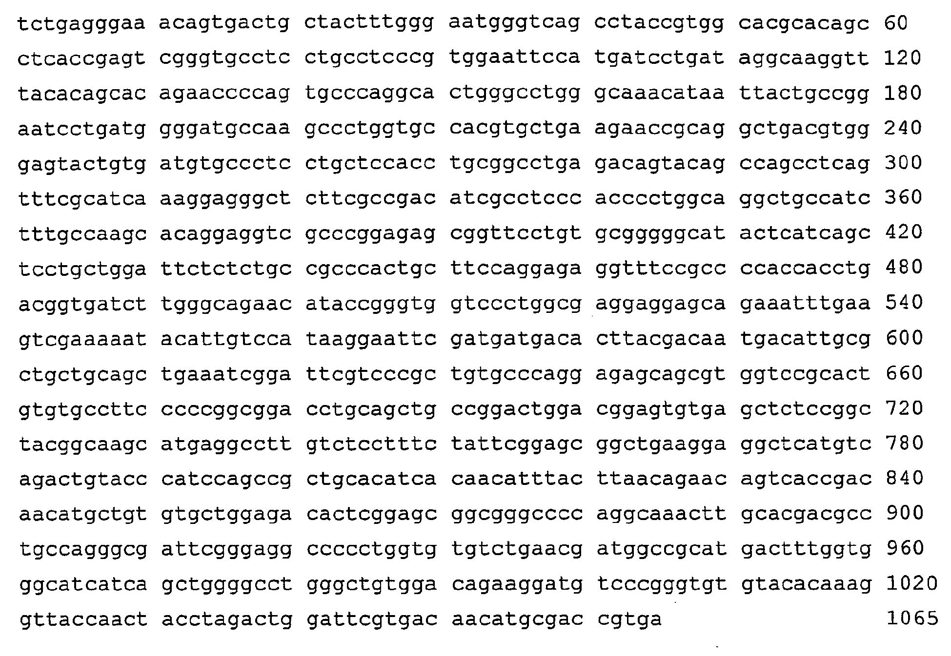
- <210> 8
<211> 377
<212> PRT
<213> Artificial Sequence - <220>
<223> Description of Artificial Sequence: OmpA-K2S fusion protein - <400> 8


- <210> 9
<211> 4
<212> PRT
<213> Artificial Sequence - <220>
<223> Description of Artificial Sequence: peptide sequence - <400> 9

- <210> 10
<211> 6
<212> PRT
<213> Artificial Sequence - <220>
<223> Description of Artificial Sequence: peptide sequence - <400> 10

- <210> 11
<211> 354
<212> PRT
<213> Artificial Sequence - <220>
<223> Description of Artificial Sequence: K2S 174-527 - <400> 11

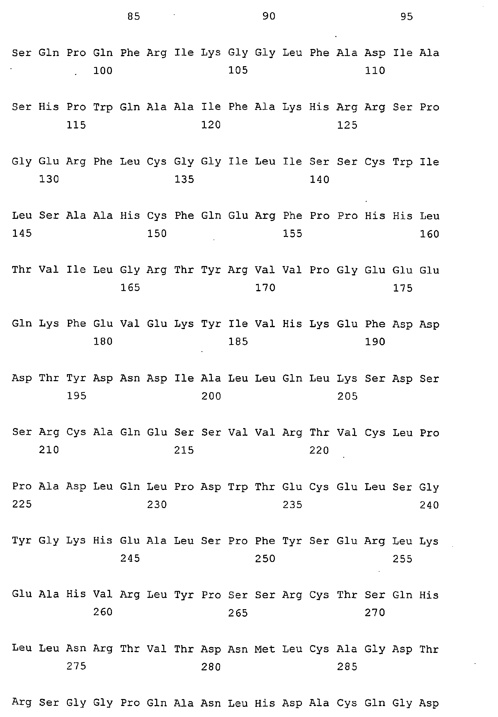

- <210> 12
<211> 331
<212> PRT
<213> Artificial Sequence - <220>
<223> Description of Artificial Sequence: K2S 197-527 - <400> 12


- <210> 13
<211> 339
<212> PRT
<213> Artificial Sequence - <220>
<223> Description of Artificial Sequence: K2S 193-527, modified - <400> 13

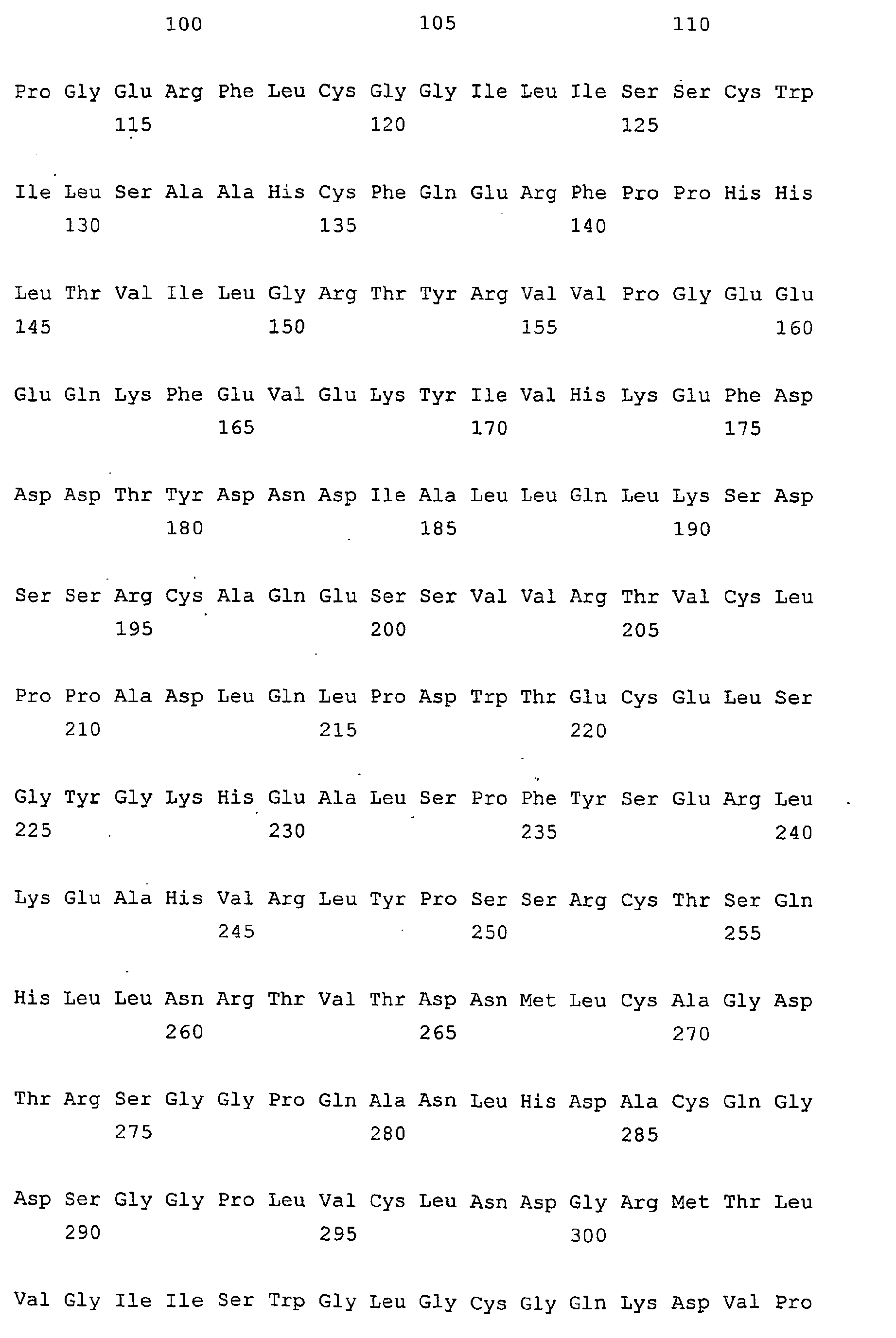

- <210> 14
<211> 335
<212> PRT
<213> Artificial Sequence - <220>
<223> Description of Artificial Sequence: K2S 193-527, modified - <400> 14


- <210> 15
<211> 343
<212> PRT
<213> Artificial Sequence - <220>
<223> Description of Artificial Sequence: K2S 191-527, modified - <400> 15


- <210> 16
<211> 343
<212> PRT
<213> Artificial Sequence - <220>
<223> Description of Artificial Sequence: K2S 191-527, modified - <400> 16


- <210> 17
<211> 308
<212> PRT
<213> Artificial Sequence - <220>
<223> Description of Artificial Sequence: K2S 220-527 - <400> 17


- <210> 18
<211> 268
<212> PRT
<213> Artificial Sequence - <220>
<223> Description of Artificial Sequence: K2S 260-527 - <400> 18


- <210> 19
<211> 527
<212> PRT
<213> Homo sapiens - <400> 19


- <210> 20
<211> 12
<212> DNA
<213> Artificial Sequence - <220>
<223> Description of Artificial Sequence: coding sequence for SEGN - <400> 20
tctgagggaa ac 12 - <210> 21
<211> 22
<212> PRT
<213> Escherichia coli - <400> 21

- <210> 22
<211> 42
<212> DNA
<213> Artificial Sequence - <220>
<223> Description of Artificial Sequence: primer - <400> 22
gaggaggagg tggcccaggc ggcctctgag ggaaacagtg ac 42 - <210> 23
<211> 42
<212> DNA
<213> Artificial Sequence - <220>
<223> Description of Artificial Sequence: primer - <400> 23
gaggaggagc tggccggcct ggcccggtcg catgttgtca cg 42 - <210> 24
<211> 26
<212> DNA
<213> Artificial Sequence - <220>
<223> Description of Artificial Sequence: primer - <400> 24
acatgcgacc gtgacaggcc ggccag 26 - <210> 25
<211> 26
<212> DNA
<213> Artificial Sequence - <220>
<223> Description of Artificial Sequence: primer - <400> 25
ctggccggcc tgtcacggtc gcatgt 26
Claims (33)
- Method for the production of recombinant DNA-derived tissue plasminogen activator (tPA), a tPA variant, a Kringle 2 Serine protease molecule (K2S) or a K2S variant in prokaryotic cells, wherein said tPA, tPA variant, K2S molecule or K2S variant is secreted extracellularly as an active and correctly folded protein, characterised in that the prokaryotic cell contains and expresses a vector comprising the DNA coding for said tPA, tPA variant, K2S molecule or K2S variant operably linked to the DNA coding for the signal peptide OmpA.
- Method according to claim 1, characterised in that said prokaryotic cell contains and expresses a vector comprising the DNA coding for said tPA, tPA variant, K2S molecule or K2S variant operably linked to the DNA coding for the signal peptide OmpA which is operably linked to the nucleic acid molecule defined by the sequence TCTGAGGGAAACAGTGAC (SEQ ID NO:1) or a functional derivative thereof.
- Method according to claim 1 or 2, characterised in that the prokaryotic cell is E. coli.
- Method according to one of claims 1 to 3, characterised in that the the following steps are carried out:a) the DNA encoding the tPA, tPA variant, K2S molecule or K2S variant is amplified by PCR;b) the PCR product is purified;c) said PCR product is inserted into a vector comprising the DNA coding for OmpA signal peptide and the DNA coding for gpIII in such a way that said PCR product is operably linked upstream to the DNA coding for the OmpA signal sequence and linked downstream to the DNA coding for gpIII of said vector;d) that a stop codon is inserted between said tPA, tPA variant, K2S molecule or K2S variant and gpIII;e) said vector is expressed by the prokaryotic cell;f) the tPA, tPA variant, K2S molecule or K2S variant is purified.
- Method according to one of claims 1 to 4, characterised in that the vector is a phagemid vector comprising the DNA coding for OmpA signal peptide and the DNA coding for gpIII.
- Method according to one of claims 1 to 5, characterised in that the vector is the pComb3HSS phagemid.
- Method according to one of claims 1 to 6, characterised in that the DNA Sequence of OmpA linked upstream to K2S comprises the following sequence or a variant due to the degenerate nucleotide code:


- Method according to one of claims 1 to 7, characterised in that the DNA Sequence of OmpA comprises the following sequence:

- Method according to one of claims 1 to 8, characterised in that the DNA Sequence of OmpA consists of the following sequence:

- Method according to one of claims 1 to 9, characterised in that the DNA of the tPA, tPA variant, K2S molecule or K2S variant is preceeded by a lac promotor and/or a ribosomal binding site.
- Method according to one of claims 1 to 10, characterised in that the DNA coding for the tPA, tPA variant, K2S molecule or K2S variant is selected from the group of DNA molecules coding for at least 90% of the amino acids 87 - 527, 174 - 527, 180 - 527 or 220 - 527 of the human tissue plasminogen activator protein as shown in Nature 301 (5897), 214-221 (1983).
- Method according to one of claims 5 to 11, characterised in that the DNA Sequence of K2S comprises the following sequence or a functional variant thereof or a variant due to the degenerate nucleotide code:


- Method according to one of claims 5 to 12, characterised in that the DNA Sequence of K2S consists of the following sequence:

- DNA molecule characterised in that it is coding for:a) a DNA coding for the OmpA signal peptideb) a DNA sequence coding for a polypeptide containing the amino acids SEGN or SEGNSD andc) a DNA sequence coding for a polypeptide containing the kringle 2 domain and the serine protease domain of tissue plasminogen activator protein,wherein said DNA coding the OmpA signal peptide of a) is fused to the DNA sequence coding for the short peptide characterised by the amino acid sequences SEGN or SEGNSD of b) and located at the N-terminal portion of the kringle 2 domain and the serine protease domain of tissue plasminogen activator protein of c).
- DNA molecule according to claim 14, characterised in that said DNA sequence comprises the following sequence or a variant due to the degenerate nucleotide code:

- DNA molecule according to claim 14 or 15, characterised in that said DNA sequence consists of the following sequence:


- DNA molecule according to one of claims 14 to 16, characterised in that said DNA sequence c) is coding for at least 90% of the amino acids 87 - 527 of the human tissue plasminogen activator protein as shown in Nature 301 (5897), 214-221 (1983).
- DNA molecule according to one of claims 14 to 17, characterised in that said DNA sequence c) is coding for at least 90% of the amino acids 174 - 527 of the human tissue plasminogen activator protein as shown in Nature 301 (5897), 214-221 (1983).
- DNA molecule according to any one of claims 14 to 18, characterised in that said DNA sequence c) is coding for at least 90% of the amino acids 180 - 527 of the human tissue plasminogen activator protein as shown in Nature 301 (5897), 214-221 (1983).
- DNA molecule according to any one of claims 14 to 19, characterised in that said DNA sequence c) is coding for at least 90% of the amino acids 220 - 527 of the human tissue plasminogen activator protein as shown in Nature 301 (5897), 214-221 (1983).
- DNA molecule according to any one of claims 14 to 20 characterised in that said DNA sequence c) is hybridizing under stringent conditions to the following sequence:

- DNA molecule according to any one of claims 14 to 21, characterised in that said DNA sequence c) consists of the following sequence:

- Fusion protein of OmpA and K2S, characterised in that it comprises a protein characterised by the following amino acid sequence:

- Fusion protein of OmpA and K2S according to claim 23, characterised in that it consists of a protein characterised by the following amino acid sequence:

- A vector containing a DNA sequence according to any one of claims 14 to 22.
- A vector according to claim 25, wherein said DNA sequence is preceded by a lac promoter and a ribosomal binding site.
- The vector pComb3HSS containing a DNA according to any one of claims 14 to 22, wherein the expression of the gp III protein is suppressed or inhibited by deleting the DNA molecule encoding said gp III protein or by a stop codon between the gene coding for a polypeptide containing the kringle 2 domain and the serine protease domain of tissue plasminogen activator protein and the protein III gene.
- A prokaryotic host cell comprising a DNA molecule according to any one of claims 14 to 22.
- A prokaryotic host cell comprising a vector according to any one of claims 25 to 27.
- An E. coli host cell comprising a DNA molecule according to any one of claims 14 to 22.
- An E. coli host cell comprising a vector according to any one of claims 25 to 27.
- Use of a DNA molecule according to any one of claims 14 to 22 or of a vector according to any one of claims 26 to 28 or a host cell according to any one of claims 28 to 31 in a method for the production of a polypeptide having the activity of tissue plasminogen activator.
- Use according to claim 32, wherein said method is a method according to any one of claims 1 to 13.
Applications Claiming Priority (3)
| Application Number | Priority Date | Filing Date | Title |
|---|---|---|---|
| GBGB0027779.8A GB0027779D0 (en) | 2000-11-14 | 2000-11-14 | Methods for large scale production of recombinant dna-derived tpa or k2s molecules |
| GB0027779 | 2000-11-14 | ||
| PCT/EP2001/012857 WO2002040650A2 (en) | 2000-11-14 | 2001-11-07 | Methods for large scale production of recombinant dna-derived tpa or k2s molecules |
Publications (2)
| Publication Number | Publication Date |
|---|---|
| EP1407030A2 EP1407030A2 (en) | 2004-04-14 |
| EP1407030B1 true EP1407030B1 (en) | 2011-04-06 |
Family
ID=9903153
Family Applications (1)
| Application Number | Title | Priority Date | Filing Date |
|---|---|---|---|
| EP01996596A Expired - Lifetime EP1407030B1 (en) | 2000-11-14 | 2001-11-07 | Methods for large scale production of recombinant dna-derived tpa or k2s molecules |
Country Status (31)
| Country | Link |
|---|---|
| EP (1) | EP1407030B1 (en) |
| JP (1) | JP2004520018A (en) |
| KR (1) | KR100856346B1 (en) |
| CN (1) | CN100567495C (en) |
| AR (1) | AR031334A1 (en) |
| AT (1) | ATE504652T1 (en) |
| AU (2) | AU2181502A (en) |
| BG (1) | BG66121B1 (en) |
| BR (1) | BR0115344A (en) |
| CA (1) | CA2428642C (en) |
| CZ (1) | CZ20031657A3 (en) |
| DE (1) | DE60144395D1 (en) |
| EA (1) | EA005163B1 (en) |
| EC (1) | ECSP034573A (en) |
| EE (1) | EE200300232A (en) |
| GB (1) | GB0027779D0 (en) |
| HR (1) | HRP20030377A2 (en) |
| HU (1) | HUP0301619A3 (en) |
| IL (2) | IL155568A0 (en) |
| MX (1) | MXPA03004161A (en) |
| MY (1) | MY129838A (en) |
| NO (1) | NO20032143L (en) |
| NZ (1) | NZ526280A (en) |
| PL (1) | PL206783B1 (en) |
| SK (1) | SK5792003A3 (en) |
| TW (1) | TWI292782B (en) |
| UA (1) | UA75102C2 (en) |
| UY (1) | UY27020A1 (en) |
| WO (1) | WO2002040650A2 (en) |
| YU (1) | YU36103A (en) |
| ZA (1) | ZA200303547B (en) |
Families Citing this family (3)
| Publication number | Priority date | Publication date | Assignee | Title |
|---|---|---|---|---|
| US6955910B2 (en) | 2000-11-14 | 2005-10-18 | Boehringer Ingelheim International Gmbh | Method for large scale production of recombinant DNA-derived TPA or K2S molecules |
| US7087412B2 (en) | 2000-11-14 | 2006-08-08 | Boehringer Ingelheim International Gmbh | Methods for large scale protein production in prokaryotes |
| IL190184A0 (en) * | 2008-03-16 | 2009-02-11 | Hadasit Med Res Service | tPA MUTANT IN THE TREATMENT OF ACUTE BRAIN INJURY AND NEURODEGENERATIVE DISORDERS |
Family Cites Families (7)
| Publication number | Priority date | Publication date | Assignee | Title |
|---|---|---|---|---|
| IL87276A (en) * | 1987-08-03 | 1995-07-31 | Fujisawa Pharmaceutical Co | Analog of tissue plasminogen activator comprising only the kringle 2 and protease domain dna encoding the same processes for the preparation thereof and pharmaceutical compositions containing the same |
| AU2660397A (en) * | 1996-04-05 | 1997-10-29 | Board Of Regents, The University Of Texas System | Methods for producing soluble, biologically-active disulfide bond-containing eukaryotic proteins in bacterial cells |
| US20020061576A1 (en) * | 1997-05-28 | 2002-05-23 | Paul A. Moore | Tissue plasminogen activator-like protease |
| US6083715A (en) * | 1997-06-09 | 2000-07-04 | Board Of Regents, The University Of Texas System | Methods for producing heterologous disulfide bond-containing polypeptides in bacterial cells |
| EP1048732A1 (en) * | 1999-04-26 | 2000-11-02 | F. Hoffmann-La Roche Ag | Process for producing natural folded and secreted proteins |
| EP1077263A1 (en) * | 1999-07-29 | 2001-02-21 | F.Hoffmann-La Roche Ag | Process for producing natural folded and secreted proteins by co-secretion of chaperones |
| GB0027782D0 (en) * | 2000-11-14 | 2000-12-27 | Boehringer Ingelheim Int | Methods fro large scale protein productio in prokaryotes |
-
2000
- 2000-11-14 GB GBGB0027779.8A patent/GB0027779D0/en not_active Ceased
-
2001
- 2001-07-11 UA UA2003065487A patent/UA75102C2/en unknown
- 2001-11-07 IL IL15556801A patent/IL155568A0/en unknown
- 2001-11-07 HU HU0301619A patent/HUP0301619A3/en unknown
- 2001-11-07 MX MXPA03004161A patent/MXPA03004161A/en active IP Right Grant
- 2001-11-07 EE EEP200300232A patent/EE200300232A/en unknown
- 2001-11-07 NZ NZ526280A patent/NZ526280A/en unknown
- 2001-11-07 PL PL361881A patent/PL206783B1/en not_active IP Right Cessation
- 2001-11-07 WO PCT/EP2001/012857 patent/WO2002040650A2/en not_active Ceased
- 2001-11-07 AU AU2181502A patent/AU2181502A/en active Pending
- 2001-11-07 HR HR20030377A patent/HRP20030377A2/en not_active Application Discontinuation
- 2001-11-07 EP EP01996596A patent/EP1407030B1/en not_active Expired - Lifetime
- 2001-11-07 CZ CZ20031657A patent/CZ20031657A3/en unknown
- 2001-11-07 AT AT01996596T patent/ATE504652T1/en not_active IP Right Cessation
- 2001-11-07 BR BR0115344-7A patent/BR0115344A/en not_active IP Right Cessation
- 2001-11-07 CN CNB018188982A patent/CN100567495C/en not_active Expired - Fee Related
- 2001-11-07 SK SK579-2003A patent/SK5792003A3/en unknown
- 2001-11-07 DE DE60144395T patent/DE60144395D1/en not_active Expired - Lifetime
- 2001-11-07 EA EA200300502A patent/EA005163B1/en not_active IP Right Cessation
- 2001-11-07 YU YU36103A patent/YU36103A/en unknown
- 2001-11-07 AU AU2002221815A patent/AU2002221815B2/en not_active Ceased
- 2001-11-07 CA CA2428642A patent/CA2428642C/en not_active Expired - Fee Related
- 2001-11-07 KR KR1020037006567A patent/KR100856346B1/en not_active Expired - Fee Related
- 2001-11-07 JP JP2002543647A patent/JP2004520018A/en active Pending
- 2001-11-12 UY UY27020A patent/UY27020A1/en not_active Application Discontinuation
- 2001-11-12 MY MYPI20015201A patent/MY129838A/en unknown
- 2001-11-13 TW TW090128128A patent/TWI292782B/en not_active IP Right Cessation
- 2001-11-14 AR ARP010105298A patent/AR031334A1/en unknown
-
2003
- 2003-04-24 IL IL155568A patent/IL155568A/en not_active IP Right Cessation
- 2003-04-29 EC EC2003004573A patent/ECSP034573A/en unknown
- 2003-05-08 ZA ZA200303547A patent/ZA200303547B/en unknown
- 2003-05-08 BG BG107780A patent/BG66121B1/en unknown
- 2003-05-13 NO NO20032143A patent/NO20032143L/en not_active Application Discontinuation
Also Published As
Similar Documents
| Publication | Publication Date | Title |
|---|---|---|
| EP2220221B1 (en) | Recombinantly modified plasmin | |
| Manosroi et al. | Secretion of active recombinant human tissue plasminogen activator derivatives in Escherichia coli | |
| EP2634252B1 (en) | Method of expressing proteins with disulfide bridges | |
| EP1343909B1 (en) | Methods for large scale production of kringle 2 plus serine protease domain (k2s) of plasminogen in prokaryotes | |
| US7087412B2 (en) | Methods for large scale protein production in prokaryotes | |
| US6277618B1 (en) | Recombinant blood-coagulation proteases | |
| US6955910B2 (en) | Method for large scale production of recombinant DNA-derived TPA or K2S molecules | |
| Yuan et al. | The role of thioredoxin and disulfide isomerase in the expression of the snake venom thrombin-like enzyme calobin in Escherichia coli BL21 (DE3) | |
| CA2428642C (en) | Methods for large scale production of recombinant dna-derived tpa or k2s molecules | |
| AU2002221815A1 (en) | Methods for large scale production of recombinant DNA-derived tPA or K2S molecules | |
| HK1070097B (en) | Methods for large scale production of recombinant dna-derived tpa or k2s molecules | |
| JPH0648988B2 (en) | Method for producing thrombin inhibitor | |
| HK1077848B (en) | Methods for large scale protein production in prokaryotes | |
| Manosroi et al. | Lekteplase—a Secreted Tissue Plasminogen Activator Derivative from Escherichia coli | |
| RU2373281C2 (en) | Method of producing recombinant staphylokinase with regulation of oxygen level | |
| HK1146089B (en) | Recombinantly modified plasmin |
Legal Events
| Date | Code | Title | Description |
|---|---|---|---|
| PUAI | Public reference made under article 153(3) epc to a published international application that has entered the european phase |
Free format text: ORIGINAL CODE: 0009012 |
|
| AK | Designated contracting states |
Kind code of ref document: A2 Designated state(s): AT BE CH CY DE DK ES FI FR GB GR IE IT LI LU MC NL PT SE TR |
|
| AX | Request for extension of the european patent |
Extension state: AL LT LV MK RO SI |
|
| 17P | Request for examination filed |
Effective date: 20040630 |
|
| 17Q | First examination report despatched |
Effective date: 20040809 |
|
| 17Q | First examination report despatched |
Effective date: 20040809 |
|
| GRAP | Despatch of communication of intention to grant a patent |
Free format text: ORIGINAL CODE: EPIDOSNIGR1 |
|
| RAX | Requested extension states of the european patent have changed |
Extension state: LV Payment date: 20040630 Extension state: LT Payment date: 20040630 Extension state: MK Payment date: 20040630 Extension state: RO Payment date: 20040630 Extension state: SI Payment date: 20040630 |
|
| GRAS | Grant fee paid |
Free format text: ORIGINAL CODE: EPIDOSNIGR3 |
|
| GRAA | (expected) grant |
Free format text: ORIGINAL CODE: 0009210 |
|
| AK | Designated contracting states |
Kind code of ref document: B1 Designated state(s): AT BE CH CY DE DK ES FI FR GB GR IE IT LI LU MC NL PT SE TR |
|
| AX | Request for extension of the european patent |
Extension state: LT LV MK RO SI |
|
| REG | Reference to a national code |
Ref country code: GB Ref legal event code: FG4D |
|
| REG | Reference to a national code |
Ref country code: CH Ref legal event code: EP |
|
| REG | Reference to a national code |
Ref country code: IE Ref legal event code: FG4D |
|
| REF | Corresponds to: |
Ref document number: 60144395 Country of ref document: DE Date of ref document: 20110519 Kind code of ref document: P |
|
| REG | Reference to a national code |
Ref country code: DE Ref legal event code: R096 Ref document number: 60144395 Country of ref document: DE Effective date: 20110519 |
|
| REG | Reference to a national code |
Ref country code: NL Ref legal event code: VDEP Effective date: 20110406 |
|
| LTIE | Lt: invalidation of european patent or patent extension |
Effective date: 20110406 |
|
| PG25 | Lapsed in a contracting state [announced via postgrant information from national office to epo] |
Ref country code: SE Free format text: LAPSE BECAUSE OF FAILURE TO SUBMIT A TRANSLATION OF THE DESCRIPTION OR TO PAY THE FEE WITHIN THE PRESCRIBED TIME-LIMIT Effective date: 20110406 Ref country code: PT Free format text: LAPSE BECAUSE OF FAILURE TO SUBMIT A TRANSLATION OF THE DESCRIPTION OR TO PAY THE FEE WITHIN THE PRESCRIBED TIME-LIMIT Effective date: 20110808 |
|
| PG25 | Lapsed in a contracting state [announced via postgrant information from national office to epo] |
Ref country code: ES Free format text: LAPSE BECAUSE OF FAILURE TO SUBMIT A TRANSLATION OF THE DESCRIPTION OR TO PAY THE FEE WITHIN THE PRESCRIBED TIME-LIMIT Effective date: 20110717 Ref country code: GR Free format text: LAPSE BECAUSE OF FAILURE TO SUBMIT A TRANSLATION OF THE DESCRIPTION OR TO PAY THE FEE WITHIN THE PRESCRIBED TIME-LIMIT Effective date: 20110707 Ref country code: AT Free format text: LAPSE BECAUSE OF FAILURE TO SUBMIT A TRANSLATION OF THE DESCRIPTION OR TO PAY THE FEE WITHIN THE PRESCRIBED TIME-LIMIT Effective date: 20110406 Ref country code: BE Free format text: LAPSE BECAUSE OF FAILURE TO SUBMIT A TRANSLATION OF THE DESCRIPTION OR TO PAY THE FEE WITHIN THE PRESCRIBED TIME-LIMIT Effective date: 20110406 Ref country code: CY Free format text: LAPSE BECAUSE OF FAILURE TO SUBMIT A TRANSLATION OF THE DESCRIPTION OR TO PAY THE FEE WITHIN THE PRESCRIBED TIME-LIMIT Effective date: 20110406 Ref country code: FI Free format text: LAPSE BECAUSE OF FAILURE TO SUBMIT A TRANSLATION OF THE DESCRIPTION OR TO PAY THE FEE WITHIN THE PRESCRIBED TIME-LIMIT Effective date: 20110406 |
|
| PG25 | Lapsed in a contracting state [announced via postgrant information from national office to epo] |
Ref country code: NL Free format text: LAPSE BECAUSE OF FAILURE TO SUBMIT A TRANSLATION OF THE DESCRIPTION OR TO PAY THE FEE WITHIN THE PRESCRIBED TIME-LIMIT Effective date: 20110406 |
|
| PLBE | No opposition filed within time limit |
Free format text: ORIGINAL CODE: 0009261 |
|
| STAA | Information on the status of an ep patent application or granted ep patent |
Free format text: STATUS: NO OPPOSITION FILED WITHIN TIME LIMIT |
|
| PG25 | Lapsed in a contracting state [announced via postgrant information from national office to epo] |
Ref country code: DK Free format text: LAPSE BECAUSE OF FAILURE TO SUBMIT A TRANSLATION OF THE DESCRIPTION OR TO PAY THE FEE WITHIN THE PRESCRIBED TIME-LIMIT Effective date: 20110406 |
|
| 26N | No opposition filed |
Effective date: 20120110 |
|
| REG | Reference to a national code |
Ref country code: DE Ref legal event code: R097 Ref document number: 60144395 Country of ref document: DE Effective date: 20120110 |
|
| PG25 | Lapsed in a contracting state [announced via postgrant information from national office to epo] |
Ref country code: MC Free format text: LAPSE BECAUSE OF NON-PAYMENT OF DUE FEES Effective date: 20111130 |
|
| REG | Reference to a national code |
Ref country code: CH Ref legal event code: PL |
|
| GBPC | Gb: european patent ceased through non-payment of renewal fee |
Effective date: 20111107 |
|
| PG25 | Lapsed in a contracting state [announced via postgrant information from national office to epo] |
Ref country code: LI Free format text: LAPSE BECAUSE OF NON-PAYMENT OF DUE FEES Effective date: 20111130 Ref country code: CH Free format text: LAPSE BECAUSE OF NON-PAYMENT OF DUE FEES Effective date: 20111130 |
|
| REG | Reference to a national code |
Ref country code: FR Ref legal event code: ST Effective date: 20120731 |
|
| REG | Reference to a national code |
Ref country code: IE Ref legal event code: MM4A |
|
| REG | Reference to a national code |
Ref country code: DE Ref legal event code: R119 Ref document number: 60144395 Country of ref document: DE Effective date: 20120601 |
|
| PG25 | Lapsed in a contracting state [announced via postgrant information from national office to epo] |
Ref country code: IE Free format text: LAPSE BECAUSE OF NON-PAYMENT OF DUE FEES Effective date: 20111107 Ref country code: GB Free format text: LAPSE BECAUSE OF NON-PAYMENT OF DUE FEES Effective date: 20111107 |
|
| PG25 | Lapsed in a contracting state [announced via postgrant information from national office to epo] |
Ref country code: FR Free format text: LAPSE BECAUSE OF NON-PAYMENT OF DUE FEES Effective date: 20111130 |
|
| PG25 | Lapsed in a contracting state [announced via postgrant information from national office to epo] |
Ref country code: LU Free format text: LAPSE BECAUSE OF NON-PAYMENT OF DUE FEES Effective date: 20111107 |
|
| PG25 | Lapsed in a contracting state [announced via postgrant information from national office to epo] |
Ref country code: DE Free format text: LAPSE BECAUSE OF NON-PAYMENT OF DUE FEES Effective date: 20120601 |
|
| PG25 | Lapsed in a contracting state [announced via postgrant information from national office to epo] |
Ref country code: TR Free format text: LAPSE BECAUSE OF FAILURE TO SUBMIT A TRANSLATION OF THE DESCRIPTION OR TO PAY THE FEE WITHIN THE PRESCRIBED TIME-LIMIT Effective date: 20110406 |
|
| PG25 | Lapsed in a contracting state [announced via postgrant information from national office to epo] |
Ref country code: IT Free format text: LAPSE BECAUSE OF FAILURE TO SUBMIT A TRANSLATION OF THE DESCRIPTION OR TO PAY THE FEE WITHIN THE PRESCRIBED TIME-LIMIT Effective date: 20110406 |