EP1406229A2 - Medical ultrasound devices practice dummy - Google Patents
Medical ultrasound devices practice dummy Download PDFInfo
- Publication number
- EP1406229A2 EP1406229A2 EP03022113A EP03022113A EP1406229A2 EP 1406229 A2 EP1406229 A2 EP 1406229A2 EP 03022113 A EP03022113 A EP 03022113A EP 03022113 A EP03022113 A EP 03022113A EP 1406229 A2 EP1406229 A2 EP 1406229A2
- Authority
- EP
- European Patent Office
- Prior art keywords
- ultrasound
- phantom according
- model
- ultrasound phantom
- esophagus
- Prior art date
- Legal status (The legal status is an assumption and is not a legal conclusion. Google has not performed a legal analysis and makes no representation as to the accuracy of the status listed.)
- Withdrawn
Links
- 238000002604 ultrasonography Methods 0.000 title claims abstract description 57
- 210000003238 esophagus Anatomy 0.000 claims abstract description 63
- 210000001835 viscera Anatomy 0.000 claims abstract description 57
- 210000002784 stomach Anatomy 0.000 claims abstract description 24
- 241001465754 Metazoa Species 0.000 claims abstract description 13
- 210000004400 mucous membrane Anatomy 0.000 claims description 20
- 239000000463 material Substances 0.000 claims description 13
- 210000004204 blood vessel Anatomy 0.000 claims description 11
- 239000012530 fluid Substances 0.000 claims description 7
- 239000000523 sample Substances 0.000 claims description 6
- 239000007779 soft material Substances 0.000 claims description 5
- 238000000354 decomposition reaction Methods 0.000 claims description 4
- XLYOFNOQVPJJNP-UHFFFAOYSA-N water Substances O XLYOFNOQVPJJNP-UHFFFAOYSA-N 0.000 claims description 4
- 238000001704 evaporation Methods 0.000 claims description 2
- 230000008020 evaporation Effects 0.000 claims description 2
- 239000007788 liquid Substances 0.000 claims 1
- 238000012549 training Methods 0.000 abstract description 25
- 210000001165 lymph node Anatomy 0.000 description 16
- 238000012986 modification Methods 0.000 description 7
- 230000004048 modification Effects 0.000 description 7
- 238000003745 diagnosis Methods 0.000 description 4
- 238000010586 diagram Methods 0.000 description 4
- 238000003780 insertion Methods 0.000 description 4
- 230000037431 insertion Effects 0.000 description 4
- 230000012447 hatching Effects 0.000 description 3
- 235000015110 jellies Nutrition 0.000 description 3
- 239000008274 jelly Substances 0.000 description 3
- 238000004088 simulation Methods 0.000 description 3
- 229920001817 Agar Polymers 0.000 description 2
- 239000008272 agar Substances 0.000 description 2
- 230000002421 anti-septic effect Effects 0.000 description 2
- 239000011230 binding agent Substances 0.000 description 2
- 230000005540 biological transmission Effects 0.000 description 2
- 235000013399 edible fruits Nutrition 0.000 description 2
- 230000003287 optical effect Effects 0.000 description 2
- 210000004798 organs belonging to the digestive system Anatomy 0.000 description 2
- CPKVUHPKYQGHMW-UHFFFAOYSA-N 1-ethenylpyrrolidin-2-one;molecular iodine Chemical compound II.C=CN1CCCC1=O CPKVUHPKYQGHMW-UHFFFAOYSA-N 0.000 description 1
- ZCYVEMRRCGMTRW-UHFFFAOYSA-N 7553-56-2 Chemical compound [I] ZCYVEMRRCGMTRW-UHFFFAOYSA-N 0.000 description 1
- 108010010803 Gelatin Proteins 0.000 description 1
- 229930182555 Penicillin Natural products 0.000 description 1
- JGSARLDLIJGVTE-MBNYWOFBSA-N Penicillin G Chemical compound N([C@H]1[C@H]2SC([C@@H](N2C1=O)C(O)=O)(C)C)C(=O)CC1=CC=CC=C1 JGSARLDLIJGVTE-MBNYWOFBSA-N 0.000 description 1
- 235000009754 Vitis X bourquina Nutrition 0.000 description 1
- 235000012333 Vitis X labruscana Nutrition 0.000 description 1
- 240000006365 Vitis vinifera Species 0.000 description 1
- 235000014787 Vitis vinifera Nutrition 0.000 description 1
- 230000003187 abdominal effect Effects 0.000 description 1
- 238000001574 biopsy Methods 0.000 description 1
- 239000003086 colorant Substances 0.000 description 1
- 238000001816 cooling Methods 0.000 description 1
- 230000000855 fungicidal effect Effects 0.000 description 1
- 239000000417 fungicide Substances 0.000 description 1
- 239000000499 gel Substances 0.000 description 1
- 239000008273 gelatin Substances 0.000 description 1
- 229920000159 gelatin Polymers 0.000 description 1
- 235000019322 gelatine Nutrition 0.000 description 1
- 235000011852 gelatine desserts Nutrition 0.000 description 1
- 238000005286 illumination Methods 0.000 description 1
- 229910052740 iodine Inorganic materials 0.000 description 1
- 239000011630 iodine Substances 0.000 description 1
- 238000012423 maintenance Methods 0.000 description 1
- 238000004519 manufacturing process Methods 0.000 description 1
- 229940049954 penicillin Drugs 0.000 description 1
- 239000002952 polymeric resin Substances 0.000 description 1
- 238000002360 preparation method Methods 0.000 description 1
- 239000003755 preservative agent Substances 0.000 description 1
- 230000002335 preservative effect Effects 0.000 description 1
- 238000012545 processing Methods 0.000 description 1
- 229920003002 synthetic resin Polymers 0.000 description 1
- 238000012360 testing method Methods 0.000 description 1
- 210000001519 tissue Anatomy 0.000 description 1
Images


Classifications
-
- G—PHYSICS
- G09—EDUCATION; CRYPTOGRAPHY; DISPLAY; ADVERTISING; SEALS
- G09B—EDUCATIONAL OR DEMONSTRATION APPLIANCES; APPLIANCES FOR TEACHING, OR COMMUNICATING WITH, THE BLIND, DEAF OR MUTE; MODELS; PLANETARIA; GLOBES; MAPS; DIAGRAMS
- G09B23/00—Models for scientific, medical, or mathematical purposes, e.g. full-sized devices for demonstration purposes
- G09B23/28—Models for scientific, medical, or mathematical purposes, e.g. full-sized devices for demonstration purposes for medicine
- G09B23/286—Models for scientific, medical, or mathematical purposes, e.g. full-sized devices for demonstration purposes for medicine for scanning or photography techniques, e.g. X-rays, ultrasonics
-
- G—PHYSICS
- G09—EDUCATION; CRYPTOGRAPHY; DISPLAY; ADVERTISING; SEALS
- G09B—EDUCATIONAL OR DEMONSTRATION APPLIANCES; APPLIANCES FOR TEACHING, OR COMMUNICATING WITH, THE BLIND, DEAF OR MUTE; MODELS; PLANETARIA; GLOBES; MAPS; DIAGRAMS
- G09B23/00—Models for scientific, medical, or mathematical purposes, e.g. full-sized devices for demonstration purposes
- G09B23/28—Models for scientific, medical, or mathematical purposes, e.g. full-sized devices for demonstration purposes for medicine
- G09B23/30—Anatomical models
- G09B23/306—Anatomical models comprising real biological tissue
-
- Y—GENERAL TAGGING OF NEW TECHNOLOGICAL DEVELOPMENTS; GENERAL TAGGING OF CROSS-SECTIONAL TECHNOLOGIES SPANNING OVER SEVERAL SECTIONS OF THE IPC; TECHNICAL SUBJECTS COVERED BY FORMER USPC CROSS-REFERENCE ART COLLECTIONS [XRACs] AND DIGESTS
- Y10—TECHNICAL SUBJECTS COVERED BY FORMER USPC
- Y10T—TECHNICAL SUBJECTS COVERED BY FORMER US CLASSIFICATION
- Y10T29/00—Metal working
- Y10T29/49—Method of mechanical manufacture
- Y10T29/4998—Combined manufacture including applying or shaping of fluent material
Definitions
- the present invention relates to an ultrasound phantom, imitating a human body with regard to ultrasonic waves, suitable for training for medical checkup of the body cavity with an ultrasonic probe or an ultrasonic endoscope by simulation.
- an arrangement disclosed in Japanese Examined Patent Application Publication No. 63-40097 is known as an example of an ultrasound phantom.
- the conventional arrangement has a configuration wherein an ultrasound transmission medium having ultrasound transmission speed or the like generally the same as with organic tissue is contained in a case, and is used for adjustment or testing of ultrasonic diagnosis apparatuses.
- the aforementioned ultrasound phantom comprises a base member which is formed of a material for transmitting ultrasound waves, and includes a first storage portion with a small width connected with a second storage portion with a great width, one or more internal organ models, which are stored in the first and second storage portions, for imitating human internal organs with regard to ultrasound waves, and a jelly-type member for transmitting ultrasound waves, disposed so as to be filled around the internal organ models.
- ultrasonic images generally the same as with a case of inserting the ultrasonic probe or the like into a human body cavity, thereby providing a training device having sufficient functions and properties.
- an ultrasound phantom 1 according to the first embodiment of the present invention comprises a base 2 generally imitating a part of a human body, internal organ models (imitations of internal organs) 4, 5, and 6, imitating human internal organs, which are detachably stored in storage portions 3a and 3b provided on the upper side of the base 2, for example, and a jelly-like material (gel material) 7 which is filled in and stored around the internal organ models 4, 5, and 6, so as to fix these internal organ models.
- the base 2 is made up of a generally rectangular parallelepiped block formed of a material such as rubber which transmits ultrasound waves, imitating the shape of the human body in the range between the neck and the abdominal region, as shown in Fig. 1, wherein the storage portion 3a with a narrow width and a suitable depth is formed on the upper side thereof, and furthermore, the storage portion 3b with a wide width is formed so as to extend the end portion (rear end) of the storage portion 3a in a step-shaped fashion, for example, and communicate therewith.
- an inserting hole 8 is formed in the form of a through hole communicating from the outer face to the storage portion 3a, and the front end of an esophagus model 4a imitating an esophagus shown in Fig. 3A is fixed to the inserting hole 8 with an esophagus fixing tube 9.
- tip portion 13 of an inserting portion 12 of an ultrasonic endoscope 11 shown in Fig. 1 can be inserted from the front end of the esophagus model 4a.
- An ultrasonic transducer 20 for transmitting and receiving ultrasound waves is disposed within the tip portion 13.
- a stomach model 4b imitating the stomach, is connected to the bottom end side of the esophagus model 4a, whereby an upper digestive organ model 4 imitating an upper digestive organ is formed.
- the internal organ model 4 internal organs of animals, more specifically, internal organs such as an esophagus and stomach of a pig are used for imitating the esophagus and the stomach positioned on the rear end thereof of a human body. Furthermore, the internal organ model 4 has properties imitating the esophagus and the stomach positioned on the rear end thereof of a human body with regard to sound waves, more particularly, with regard to ultrasonic waves.
- the esophagus model 4a is formed in a long and narrow shape, and accordingly is stored within the storage portion 3a with a small width
- the stomach model 4b is formed with a great width, and accordingly is stored within the storage portion 3b with a great width.
- Another internal organ model 5, and a lymph node model 6 imitating a lymph node which is an object of biopsy by puncture, for example, are disposed around the internal organ model 4.
- the other internal organ model 5 and the lymph node model 6 can be stored so as to be embedded in the jelly-like materials 7 in the form of blocks 14 and 15.
- the lymph node model 6 a fruit such as a grape or the like is employed for imitating an actual lymph node with regard to sound waves.
- the storage portion 3a with a small width serves as a space for storing the esophagus model 4a generally imitating the esophagus of human body with an internal organ of an animal, and a space for storing the internal organ therearound (lymph node model 6 or unshown tube member imitating bronchial tubes), and on the other hand, the storage portion 3b with a great width serves as a space for storing an internal organ model such as the stomach model 4b imitating the stomach of the human and the internal organs therearound, or the like.
- a soft elastic member transmitting ultrasonic waves specifically, a jelly-like member 7 formed of agar, gelatin, or the like, is filled in around the internal organ models 4, 5, and 6, stored within the storage portions 3a and 3b, whereby the internal organ models 4, 5, and 6, are fixed.
- jelly-like members 7 which harden due to passing of time have an advantage in that the hardness (softness) thereof can be adjusted by adjusting the amount of water for mixing at the time of preparing.
- the jelly-like member 7 with the generally optimal hardness was obtained by dissolving 4 g of powdered agar into 900 cc of water.
- the storage portion 3a with a small width is formed at the front end of the base 2, and the storage portion 3b with a great width is formed at the rear end thereof, so as to provide just enough space for storing principal internal organs of the human body in the range between the esophagus and the internal organs around the stomach, whereby principal internal organ models such as the esophagus model 4a, the stomach model 4b, and the like, can be stored within the storage portions 3a and 3b, respectively, with a small amount of the jelly-like member 7 being filled therearound.
- the lymph node model 6 or the like is embedded in the jelly-like member 7 so as to form a block such as the block 15 or the like, as described above, and accordingly, in the event that the lymph node model 6 or the like is damaged, the block 15 containing the damaged lymph node model 6 or the like is replaced with a new one, and thus, the ultrasound phantom 1 can be used again in a short time with a simple operation, thereby enabling repeated use with a simple operation, and furthermore, providing an ultrasound phantom having sufficient functions as a training device which can be easily used.
- simulated treating can be performed, wherein the inserting portion 12 of the ultrasonic endoscope 11 is inserted into the esophagus model 4a, and at the same time, a puncture operative device is inserted into a channel provided within the inserting portion 12, and the esophagus model 4a is punctured with the puncture operative device so that the puncture operative device passes through the esophagus model 4a so that the lymph node model 6 around the esophagus model 4a is punctured therewith.
- the ultrasound phantom 1 according to the present embodiment wherein the ultrasonic endoscope 11 is inserted can be used as a training device for training for puncture treating while observing ultrasonic images, as well.
- the lymph node model 6 which is an object of puncture being punctured several times is damaged, and accordingly, the lymph node model 6 is no longer able to be used as a training device.
- the phantom 1 has a configuration made up of blocks so that the block containing the object of puncture can be easily individually replaced.
- the lymph node model 6 is readily damaged by puncture.
- just the block 15 containing the damaged lymph node model 6 is replaced as described above, whereby the ultrasound phantom 1 can be easily repaired so as to be used again.
- the phantom 1 has a configuration wherein the base 2 stores the blocks 14 and 15, and accordingly, the phantom 1 can be repeatedly used with a simple operation, and thus, the phantom 1 has functions suitable for a training device.
- Fig. 2 illustrates an internal configuration of the ultrasound phantom 1 at the storage portion 3b, for example, wherein the jelly-like member 7 is filled in around the stomach model 4b, and the internal organ model 5 and the lymph node model 6 around the stomach model 4b are detachably stored with these models being embedded in the blocks 14 and 15.
- the esophagus model 4a is mounted at the aforementioned inserting hole 8 with the esophagus fixing tube 9, and accordingly, the inserting portion 12 of the ultrasonic endoscope 11 can be inserted into the esophagus model 4a with the tip portion 13 thereof being at the head (in the same way as with a case of inserting an ultrasonic endoscope into the human esophagus), thereby obtaining ultrasonic diagnostic images while performing endoscope diagnosis and observing images from the optical system of the endoscope.
- a tube-shaped blood vessel model cavity 17 imitating a human blood vessel, and a spine model cavity 18 in the shape of a tube, for example, imitating a human spine, are provided at the portions on the lower sides of the storage portions 3a and 3b in the base 2.
- a tube is inserted into the blood vessel model cavity 17 so as to pour fluid thereinto, or a tube 99 is connected to the both ends of the blood vessel model cavity 17 so that fluid 95 stored in a bucket 98 flows therethrough by a pump 97, thereby providing a blood vessel model having functions generally the same as with a human blood vessel with regard to sound waves (more specifically, with regard to ultrasound waves).
- the fluid 95 stored in the bucket 98 contains microbubbles 94 for reflecting ultrasound waves, and also contains ice 96, kept in the bucket 98 against suction of the pump 97 due to the size thereof, for cooling the fluid 95.
- the fluid 95 is cooled due to the ice 96 contained therein, and accordingly, the base 2 and the components contained therein are cooled, thereby preventing decomposition of the components.
- the spine model cavity 18 has a property of transmitting no ultrasound waves, with a simple configuration forming a tube-shaped cavity.
- the blood vessel model cavity 17 and the spine model cavity 18 are provided on the side of the base 2 for imitating a human blood vessel and spine, and accordingly, in the event of inserting the ultrasonic endoscope 11 into the phantom 1, ultrasonic images generally the same as with a case of inserting the endoscope into the actual human esophagus, thereby providing a training device imitating the body cavities.
- the upper face thereof is covered with an echo jelly (ultrasonic jelly) 19.
- the upper face of the storage portions 3a and 3b is covered with the echo jelly 19, thereby preventing evaporation of water, and preventing decomposition of the components contained therein.
- penicillin antiseptic, fungicide, or an iodine antiseptic solution e.g., brand name isodine
- the ultrasound phantom 1 can be used for a long time without decomposition.
- internal organs of an animal are used so as to correctly imitate the human internal organs such as the esophagus and stomach, and accordingly, the size of the esophagus model 4a from the animal internal organ cannot be set to a constant value in practical use.
- a step-shaped arrangement wherein a suitable outer diameter can be selected, is employed for the esophagus fixing tube 9 for being connected to the inserting hole 8, as shown in Fig. 3A, and is connected to the esophagus 4a with a binder 21 or the like, at the portion with an outer diameter suitable for the inner diameter of the esophagus 4a which is used in this time.
- the esophagus model 4a can be easily fixed to the inserting hole 8 using the esophagus fixing tube 9.
- the esophagus model 4a may be fixed to the inserting hole 8 as with a first modification shown in Fig. 3B.
- the step-shaped esophagus fixing tube 9 is fixed to the front end of the esophagus model 4a, as shown in Fig. 3A, and furthermore, the esophagus fixing tube 9 is fixed to the inserting hole 8 through an attachment 22 with a suitable size which can be mounted to the inserting hole 8.
- the esophagus model 4a may be fixed to the inserting hole 8 using the esophagus fixing tube 9 shown in Fig. 3A.
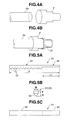
- the opening end portion of the esophagus fixing tube 9 with the greater outer diameter, and the front end of the esophagus model 4a are positioned facing one another, and the esophagus model 4a is inserted so as to pass through the esophagus fixing tube 9, following which the portion of the esophagus model 4a protruding from the esophagus fixing tube 9 is turned back, as shown in Fig. 4B, and is fixed with the binder 21 or the like at the portion of the esophagus fixing tube 9 with an outer diameter greater than the inner diameter of the esophagus model 4a.
- the ultrasonic endoscope 11 is inserted from the direction reverse to the case shown in Fig. 3A, i.e., from the right side.
- animal internal organs are employed for imitating the human esophagus and stomach, and accordingly, strong odor occurs from the animal internal organs in practical use.
- the internal organ-model 4 generally causing the odor is opened so as to remove the mucous membrane thereon, following which suture or connection is performed for the opened portion, in order to be used as the internal organ model 4 imitating the human esophagus and stomach.
- the odor is greatly reduced by removing the mucous membrane of the animal internal organs, thereby facilitating practical use of the phantom imitating the human esophagus and stomach.
- Figs. 5A through 5C illustrate a jig 31 for removing the mucous membrane as described above.
- Fig. 5A is a side view of the jig 31
- Fig. 5B is an enlarged elevational view thereof
- Fig. 5C is a plan view thereof.
- the jig 31 is formed in the shape of a long and narrow plate, wherein the rear end portion serves as a holding portion 32, and a protrusion portion 33 formed of a great number of protrusions are provided on one face thereof along the longitudinal direction in front of the holding portion 32, whereby a mucous-membrane removal portion 34 is formed.
- the user can remove the mucous membrane by performing processing wherein the jig 31 is moved while the mucous-membrane removal portion 34 is pressed into contact with the mucous membrane.
- the jig 31 is formed with a total length L between 20 and 30 cm, and a length L1 of the holding portion 32 of around 10 cm, as shown in Fig. 5A.
- the mucous-membrane removal portion 34 is formed with a width W of 1 cm, thickness T of 0.5 mm, and height H of the protrusions of the protrusion portion of around 0.5 mm, as shown in Fig. 5B.
- the protrusions of the protrusion portion 33 are formed in the shape of a quadrangular pyramid.
- the user can efficiently remove the mucous membrane causing the odor using the jig 31 including the mucous-membrane removal portion 34 shown in Figs. 5A through 5C.
- the user can remove the mucous membrane using the jig 31 including a protrusion portion 33 formed of a great number of protrusions as shown in Figs. 5A through 5C, in a markedly short time as compared with a case of removing the mucous membrane without the jig 31.
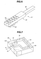
- Fig. 6 shows a jig 31' which is a modification of the jig 31.
- the jig 31' has a basic configuration wherein, in addition to the configuration of the jig 31 shown in Fig. 5A or the like, a protrusion portion 36 formed of a great number of protrusions in the shape of a sawtooth, for example, is further formed on the other face reverse to the face where the protrusion portion 33 formed of a great number of protrusions has been provided, along the longitudinal direction, thus forming a mucous-membrane removal portion 34'.
- the ultrasonic endoscope 11 can be inserted into the inserting hole 8 of the phantom 1 imitating the human body, and ultrasonic images generally the same as with a case of inserting the ultrasonic endoscope into the human esophagus can be easily obtained, and accordingly, the phantom 1 can be used as a training device using the ultrasonic endoscope 11, thereby providing a training system for improving the skill of the user in insertion of an ultrasonic endoscope or the like with a simple configuration.
- the internal organ models which are readily damaged are each embedded into the jelly-like member so as to form blocks, and accordingly, in the event that one internal organ model is damaged, the phantom can be easily repaired so as to be used again in a short time by replacing a block containing the damaged internal organ model. That is to say, the phantom according to the present embodiment can be repeatedly used with a simple operation. Furthermore, the maintenance of the phantom can be easily performed.
- simulation of puncture treating can be performed using the phantom according to the present embodiment, more specifically, simulation of puncture treatment while observing ultrasonic diagnostic images can be easily performed, thereby providing a training system for improving the skill of the user.
- the fluid flowing through the blood vessel model cavity 17 can be displayed in colors, and accordingly, training can be performed for improving the skill of the user in insertion wherein a puncture device is introduced while avoiding blood vessels with the spine model cavity 18 imitating the spine as a merkmal.
- the internal organ models which are to be used for puncture treating are made up of fruits or the like, and accordingly, even in the event that the internal organ model is damaged, the phantom can be easily repaired with low costs by replacing the damaged internal organ model.
- animal internal organs are employed for the esophagus model and the stomach model, and accordingly, the phantom has generally the same properties with regard to insertion, sound waves, and the like, as with the human body, thereby providing a training system for improving the skill of the user in insertion and the like.
- Fig. 7 illustrates a base 42 according to the second embodiment.
- the base 42 has a configuration wherein, in addition to the configuration of the base 2 shown in Fig. 1, the region around the portion where the storage portions 3a and 3b have been formed, i.e., a region 43 which is indicated by a double-dashed line in Fig. 7, with the upper face indicated by hatching, is formed of a material which transmits ultrasonic waves with small attenuation, i.e., a soft material with low attenuation properties, and the outer region 44 as to the region 43 indicated by the hatching is formed of a hard material with the attenuation properties with regard to ultrasound waves greater than with the region 43 indicated by the hatching.
- the base 42 includes multiple openings 100 for being fixed on an unshown stand (a desk or the like) with screws or the like. Other components are the same as with the first embodiment.
- the base 42 is formed with the improved strength, and accordingly, the phantom has a principal function required for a training device, i.e., sufficient durability for use in a long period, and also, for repeated use, and for preventing occurrence of damage which is readily caused by transporting, thereby providing a more preferable training device.
- a training device i.e., sufficient durability for use in a long period, and also, for repeated use, and for preventing occurrence of damage which is readily caused by transporting, thereby providing a more preferable training device.
- other functions of the phantom according to the present embodiment are the same as with the first embodiment.
- an arrangement may be made wherein the region around the storage portions 3a and 3b shown in Fig. 7 is particularly formed of a soft material in the depth direction, as well.
- Figs. 1 through 7 illustrate the storage portions 3a and 3b in a shape with a constant depth
- an arrangement may be made wherein the storage portions 3a and 3b are formed with a curved lower face according to the shape of the internal organ which is to be stored.
- the lower faces of the storage portions 3a and 3b are formed of a soft material with a shape according to the internal organ which is to be stored, and accordingly, positioning and storing can be easily performed for the internal organs, thereby facilitating manufacturing.
- Figs. 1 and 7 show the storage portions 3a and 3b being connected in the form of a step
- the present invention is not restricted to the aforementioned configuration, but rather, an arrangement may be made wherein the storage portions 3a and 3b are connected in a tapered shape or a curved shape. Furthermore, an arrangement may be made wherein an unnecessary region on the base 42 such as a region indicated by a dashed line in Fig. 7 is eliminated for reducing costs and weight.
- the ultrasonic phantom as a training device for an ultrasonic endoscope which includes functions of an endoscope, and furthermore includes an ultrasonic transducer at the tip end portion thereof for transmitting and receiving ultrasound waves
- the ultrasound phantom according to the present invention can be used as a training device for an ultrasonic prove having no functions of an endoscope for optical observation (illumination window and observation window), as well.
- the present invention is not restricted to the above-described arrangement, but rather, an arrangement may be made wherein the internal organ models such as the esophagus model 4a, the stomach model 4b, and the like, are formed of polymeric resin or the like.
- ultrasonic images generally the same as with inserting the ultrasonic probe or ultrasonic endoscope into the human body cavity are obtained, thereby providing a training device which can be easily used, having sufficient functions and properties for training.
Landscapes
- Engineering & Computer Science (AREA)
- Health & Medical Sciences (AREA)
- General Physics & Mathematics (AREA)
- Physics & Mathematics (AREA)
- General Health & Medical Sciences (AREA)
- Mathematical Analysis (AREA)
- Mathematical Physics (AREA)
- Medicinal Chemistry (AREA)
- Chemical & Material Sciences (AREA)
- Algebra (AREA)
- Computational Mathematics (AREA)
- Theoretical Computer Science (AREA)
- Educational Technology (AREA)
- Mathematical Optimization (AREA)
- Medical Informatics (AREA)
- Pure & Applied Mathematics (AREA)
- Business, Economics & Management (AREA)
- Educational Administration (AREA)
- Nuclear Medicine, Radiotherapy & Molecular Imaging (AREA)
- Radiology & Medical Imaging (AREA)
- Life Sciences & Earth Sciences (AREA)
- Biomedical Technology (AREA)
- Molecular Biology (AREA)
- Instructional Devices (AREA)
- Ultra Sonic Daignosis Equipment (AREA)
Abstract
Description
Claims (16)
- An ultrasound phantom, characterized by comprising:a base member which is formed of a material for transmitting ultrasound waves so as to imitate a part of a human body, and includes a first storage portion with a small width connected with a second storage portion with a great width;at least one or more internal organ models, which are stored in the first and second storage portions, for imitating human internal organs; anda jelly-like member for transmitting ultrasound waves, disposed so as to be filled in around the internal organ models.
- An ultrasound phantom according to Claim 1, characterized in that the base member has an inserting hole, communicating with the inside of the first storage portion, into which an ultrasonic probe including an ultrasonic transducer can be inserted.
- An ultrasound phantom according to Claim 2, characterized in that one end of an esophagus model, which is stored in the first storage portion, for imitating an esophagus, is mounted to the inserting hole.
- An ultrasound phantom according to Claim 1, characterized in that a stomach model is stored in the second storage portion for imitating a stomach.
- An ultrasound phantom according to Claim 1, characterized in that the base member further includes a tube-shaped cavity for imitating a blood vessel.
- An ultrasound phantom according to Claim 1, characterized in that the base member further includes a tube-shaped cavity for imitating a spine.
- An ultrasound phantom according to Claim 1, characterized in that one or more blocks, formed of a jelly-like material, where internal organ models have been embedded, can be detachably stored in the first and second storage portions.
- An ultrasound phantom according to Claim 1, characterized in that the internal organ models are formed of animal internal organs.
- An ultrasound phantom according to Claim 8, characterized in that the animal internal organ is stored in the first or second storage portion following removal of the mucous membrane thereof causing strong odor.
- An ultrasound phantom according to Claim 3, characterized in that the one end of the esophagus model is fixed to the inserting hole via a step-shaped fixing tube.
- An ultrasound phantom according to Claim 1, characterized in that the base member has a first member, formed of a soft material for transmitting ultrasound waves, which forms a portion around the region where the first and second storage portions are formed, and a second member, formed of a material harder than the first material, which forms the region around the first member.
- An ultrasound phantom according to Claim 5, characterized in that the tube-shaped cavity is connected to a unit for running fluid.
- An ultrasound phantom according to Claim 1, characterized in that the upper faces of the first and second storage portions are covered with a member for preventing evaporation of water or the like.
- An ultrasound phantom according to Claim 1, characterized in that the jelly-like member has a function for preventing decomposition.
- An ultrasound phantom according to Claim 1, characterized in that the base member includes a material formed of rubber.
- An ultrasound phantom according to Claim 1, characterized in that the jelly-like member comprises a material hardened from a soft material over time from addition of liquid.
Applications Claiming Priority (4)
| Application Number | Priority Date | Filing Date | Title |
|---|---|---|---|
| JP2002288950 | 2002-10-01 | ||
| JP2002288950 | 2002-10-01 | ||
| JP2002365719 | 2002-12-17 | ||
| JP2002365719A JP3780253B2 (en) | 2002-10-01 | 2002-12-17 | Ultrasonic phantom |
Publications (2)
| Publication Number | Publication Date |
|---|---|
| EP1406229A2 true EP1406229A2 (en) | 2004-04-07 |
| EP1406229A3 EP1406229A3 (en) | 2006-09-27 |
Family
ID=31996235
Family Applications (1)
| Application Number | Title | Priority Date | Filing Date |
|---|---|---|---|
| EP03022113A Withdrawn EP1406229A3 (en) | 2002-10-01 | 2003-09-29 | Medical ultrasound devices practice dummy |
Country Status (3)
| Country | Link |
|---|---|
| US (1) | US7059168B2 (en) |
| EP (1) | EP1406229A3 (en) |
| JP (1) | JP3780253B2 (en) |
Cited By (4)
| Publication number | Priority date | Publication date | Assignee | Title |
|---|---|---|---|---|
| US8454368B2 (en) | 2007-11-29 | 2013-06-04 | Cedars-Sinai Medical Center | Medical training methods and devices |
| FR2994011A1 (en) * | 2012-07-26 | 2014-01-31 | Veterinarius S A R L | Device for performing sampling and/or injections guided by ultrasonography for studying anomalies of e.g. kidney, of dog, has set of model units, where each unit presents acoustic impedance similar to impedance of model unit it represents |
| CN103886798A (en) * | 2014-03-13 | 2014-06-25 | 西安交通大学 | High-simulation interventional operation trainer |
| CN103918019A (en) * | 2011-11-10 | 2014-07-09 | 株式会社高研 | Training manikin for ultrasound bronchoscopy |
Families Citing this family (49)
| Publication number | Priority date | Publication date | Assignee | Title |
|---|---|---|---|---|
| US20050181343A1 (en) * | 2004-02-02 | 2005-08-18 | Ault Mark J. | Ultrasound guided vascular access training device |
| US7255565B2 (en) * | 2004-03-15 | 2007-08-14 | Brian Keegan | Anthropomorphic phantoms and method |
| JP2006166956A (en) * | 2004-12-13 | 2006-06-29 | Matsushita Electric Ind Co Ltd | Phantom for ultrasonic diagnostic equipment |
| WO2006086115A1 (en) * | 2005-02-10 | 2006-08-17 | Wilkins Jason D | Ultrasound training mannequin |
| US10219815B2 (en) | 2005-09-22 | 2019-03-05 | The Regents Of The University Of Michigan | Histotripsy for thrombolysis |
| US8057408B2 (en) | 2005-09-22 | 2011-11-15 | The Regents Of The University Of Michigan | Pulsed cavitational ultrasound therapy |
| JP2009519476A (en) * | 2005-12-13 | 2009-05-14 | エルベ エレクトロメディジン ゲーエムベーハー | Training model for endoscopic examination and treatment of luminal organs |
| US20090162820A1 (en) * | 2006-02-24 | 2009-06-25 | Hrs Consultant Service, Inc. | Educational Simulator for Trasesophageal Echocardiography |
| US7854612B2 (en) * | 2006-05-19 | 2010-12-21 | Spirus Medical, Inc. | Anatomical model |
| US8403676B2 (en) * | 2006-05-19 | 2013-03-26 | Olympus Endo Technology America Inc. | Anatomical model |
| US7748901B2 (en) | 2007-04-06 | 2010-07-06 | Unisyn Medical Technologies, Inc. | Universal x-ray test bed |
| US8888498B2 (en) * | 2009-06-02 | 2014-11-18 | National Research Council Of Canada | Multilayered tissue phantoms, fabrication methods, and use |
| US8162669B1 (en) * | 2009-07-14 | 2012-04-24 | Shaw Erin L | Ultrasound training assembly |
| JP5850837B2 (en) | 2009-08-17 | 2016-02-03 | ヒストソニックス,インコーポレーテッド | Disposable acoustic coupling media container |
| JP5863654B2 (en) | 2009-08-26 | 2016-02-16 | リージェンツ オブ ザ ユニバーシティー オブ ミシガン | Micromanipulator control arm for therapeutic and image processing ultrasonic transducers |
| US9901753B2 (en) | 2009-08-26 | 2018-02-27 | The Regents Of The University Of Michigan | Ultrasound lithotripsy and histotripsy for using controlled bubble cloud cavitation in fractionating urinary stones |
| US8539813B2 (en) * | 2009-09-22 | 2013-09-24 | The Regents Of The University Of Michigan | Gel phantoms for testing cavitational ultrasound (histotripsy) transducers |
| US9144694B2 (en) | 2011-08-10 | 2015-09-29 | The Regents Of The University Of Michigan | Lesion generation through bone using histotripsy therapy without aberration correction |
| TWI452999B (en) * | 2011-10-31 | 2014-09-21 | Iner Aec Executive Yuan | Medical prostheses for medical imaging systems |
| CN103117010B (en) * | 2011-11-17 | 2016-08-24 | 深圳迈瑞生物医疗电子股份有限公司 | A kind of ultra sonic imaging analog systems |
| US9049783B2 (en) | 2012-04-13 | 2015-06-02 | Histosonics, Inc. | Systems and methods for obtaining large creepage isolation on printed circuit boards |
| US9636133B2 (en) | 2012-04-30 | 2017-05-02 | The Regents Of The University Of Michigan | Method of manufacturing an ultrasound system |
| JP6051693B2 (en) * | 2012-09-03 | 2016-12-27 | セイコーエプソン株式会社 | Ultrasonic probe, electronic device and ultrasonic diagnostic apparatus |
| EP2903688A4 (en) | 2012-10-05 | 2016-06-15 | Univ Michigan | COLOR DOPPLER FEEDBACK INDUCED BY BUBBLES DURING HISTOTRIPSY |
| JP6112706B2 (en) * | 2012-12-12 | 2017-04-12 | 学校法人関西医科大学 | Ultrasonic image reproduction medium, ultrasonic image reproduction molded body, and simulation apparatus |
| US8926333B2 (en) * | 2013-03-15 | 2015-01-06 | Simnext, Llc | Device, system, and method for simulating blood flow |
| WO2015003154A1 (en) | 2013-07-03 | 2015-01-08 | Histosonics, Inc. | Articulating arm limiter for cavitational ultrasound therapy system |
| JP6600304B2 (en) | 2013-07-03 | 2019-10-30 | ヒストソニックス,インコーポレーテッド | Optimized histotripsy excitation sequence for bubble cloud formation using shock scattering |
| US10780298B2 (en) | 2013-08-22 | 2020-09-22 | The Regents Of The University Of Michigan | Histotripsy using very short monopolar ultrasound pulses |
| KR101587368B1 (en) * | 2013-09-10 | 2016-01-20 | 한국표준과학연구원 | Unitblockformulti-purposemulti-imageandmulti-moduleas semblyofmedicalphantomusingunitblock |
| KR101546187B1 (en) * | 2014-05-30 | 2015-08-21 | 성균관대학교산학협력단 | Phantom and Phantom System |
| US9558678B1 (en) * | 2014-11-20 | 2017-01-31 | Michael E. Nerney | Near-infrared imager training device |
| WO2016210133A1 (en) | 2015-06-24 | 2016-12-29 | The Regents Of The Universtiy Of Michigan | Histotripsy therapy systems and methods for the treatment of brain tissue |
| JP6061318B1 (en) * | 2016-03-09 | 2017-01-18 | 国立大学法人高知大学 | Puncture practice tool |
| EP3251811B1 (en) | 2016-05-30 | 2019-08-28 | Max-Planck-Gesellschaft zur Förderung der Wissenschaften e.V. | Method of producing a phantom and phantom |
| WO2018226940A1 (en) * | 2017-06-09 | 2018-12-13 | Board Of Regents Of The University Of Nebraska | Transesophageal echocardiography simulator |
| US11813484B2 (en) | 2018-11-28 | 2023-11-14 | Histosonics, Inc. | Histotripsy systems and methods |
| WO2020227118A1 (en) * | 2019-05-03 | 2020-11-12 | Arizona Board Of Regents On Behalf Of The University Of Arizona | Systems and methods for an ultrasound-guided percutaneous nephrostomy model |
| US11813485B2 (en) | 2020-01-28 | 2023-11-14 | The Regents Of The University Of Michigan | Systems and methods for histotripsy immunosensitization |
| CN111544781B (en) * | 2020-03-30 | 2024-05-31 | 山东省肿瘤防治研究院(山东省肿瘤医院) | Nuclear magnetism and CT compatible chest movement phantom |
| CN116113376A (en) | 2020-06-18 | 2023-05-12 | 希斯托索尼克斯公司 | Tissue destruction acoustic and patient coupling systems and methods |
| CA3190517A1 (en) | 2020-08-27 | 2022-03-03 | Timothy Lewis HALL | Ultrasound transducer with transmit-receive capability for histotripsy |
| WO2022260747A1 (en) | 2021-06-07 | 2022-12-15 | The Regents Of The University Of Michigan | All-in-one ultrasound systems and methods including histotripsy |
| WO2022260746A1 (en) | 2021-06-07 | 2022-12-15 | The Regents Of The University Of Michigan | Minimally invasive histotripsy systems and methods |
| EP4345798A1 (en) * | 2022-09-28 | 2024-04-03 | Hands-on Plus | Ultrasound guided percutaneous surgery mannequin |
| US20240149078A1 (en) | 2022-10-28 | 2024-05-09 | Histosonics, Inc. | Histotripsy systems and methods |
| WO2024097771A2 (en) | 2022-11-01 | 2024-05-10 | Histosonics, Inc. | Histotripsy systems and methods |
| JP2026516719A (en) | 2023-04-20 | 2026-05-26 | ヒストソニックス,インコーポレーテッド | Histotripsy system and related methods including user interface and workflow for treatment planning and treatment. |
| WO2025090672A1 (en) | 2023-10-23 | 2025-05-01 | Histosonics, Inc. | Histotripsy systems and methods |
Family Cites Families (19)
| Publication number | Priority date | Publication date | Assignee | Title |
|---|---|---|---|---|
| US3310885A (en) * | 1964-06-04 | 1967-03-28 | Samuel W Alderson | Radio-therapy phantom |
| JPS5573250A (en) | 1978-11-30 | 1980-06-02 | Tokyo Shibaura Electric Co | Fantom for ultrasoniccwave diagnosis device performance test |
| US4286168A (en) * | 1979-01-18 | 1981-08-25 | Atomic Products Corp. | Phantom simulation device for scintillation cameras |
| US4493653A (en) * | 1983-09-27 | 1985-01-15 | Technicare Corporation | Biopsiable ultrasound phantom |
| JPS6215213A (en) | 1985-07-15 | 1987-01-23 | Kao Corp | Production of highly water absorbing resin having high water absorption rate |
| JPH079148B2 (en) | 1986-08-06 | 1995-02-01 | 藤夫 瀬谷 | Tricombit in Underground Drilling Rig |
| US4767333A (en) * | 1987-08-17 | 1988-08-30 | Born Grant R | Laser evolved models |
| EP0319623B1 (en) * | 1987-12-10 | 1990-10-17 | United Kingdom Atomic Energy Authority | Apparatus for simulating inspection equipment |
| US4974461A (en) * | 1988-10-13 | 1990-12-04 | The United States Of America As Represented By Department Of Health And Human Services | Anthropomorphic cardiac ultrasound phantom |
| US5061187A (en) * | 1990-04-12 | 1991-10-29 | Ravinder Jerath | Ultrasound training apparatus |
| US5055051A (en) * | 1990-08-03 | 1991-10-08 | Dornier Medical Systems, Inc. | Semi-anthropomorphic biliary/renal training phantom for medical imaging and lithotripsy training |
| GB9309021D0 (en) * | 1993-04-30 | 1993-06-16 | Limbs & Things Ltd | Medical training apparatus |
| GB9718377D0 (en) * | 1997-08-29 | 1997-11-05 | Ethicon Limited | Simulator |
| US6190915B1 (en) * | 1999-06-25 | 2001-02-20 | Wisconsin Alumni Research Foundation | Ultrasound phantoms |
| US6318146B1 (en) * | 1999-07-14 | 2001-11-20 | Wisconsin Alumni Research Foundation | Multi-imaging modality tissue mimicking materials for imaging phantoms |
| KR20010038344A (en) * | 1999-10-25 | 2001-05-15 | 김남국 | Method and Apparatus for Forming Objects Similar to Things in Human Body |
| US20020170339A1 (en) * | 2001-05-16 | 2002-11-21 | Medson Ltd. | Ultrasound phantom simulating hard and soft tissue of a vertebrate and methods of production and use thereof |
| US6997719B2 (en) * | 2002-06-26 | 2006-02-14 | Ethicon, Inc. | Training model for endoscopic vessel harvesting |
| US20040067591A1 (en) * | 2002-10-04 | 2004-04-08 | Wisconsin Alumni Research Foundation | Tissue mimicking elastography phantoms |
-
2002
- 2002-12-17 JP JP2002365719A patent/JP3780253B2/en not_active Expired - Fee Related
-
2003
- 2003-09-25 US US10/671,076 patent/US7059168B2/en not_active Expired - Fee Related
- 2003-09-29 EP EP03022113A patent/EP1406229A3/en not_active Withdrawn
Cited By (8)
| Publication number | Priority date | Publication date | Assignee | Title |
|---|---|---|---|---|
| US8454368B2 (en) | 2007-11-29 | 2013-06-04 | Cedars-Sinai Medical Center | Medical training methods and devices |
| CN103918019A (en) * | 2011-11-10 | 2014-07-09 | 株式会社高研 | Training manikin for ultrasound bronchoscopy |
| EP2779143A4 (en) * | 2011-11-10 | 2015-07-29 | Koken Kk | MODEL LEARNING FOR ULTRASONIC BRONCHOSCOPY |
| US20150213731A1 (en) * | 2011-11-10 | 2015-07-30 | Koken Co., Ltd. | Training model for ultrasonic bronchoscopy |
| CN103918019B (en) * | 2011-11-10 | 2016-08-17 | 株式会社高研 | Ultrasound wave bronchial endoscope training pattern |
| FR2994011A1 (en) * | 2012-07-26 | 2014-01-31 | Veterinarius S A R L | Device for performing sampling and/or injections guided by ultrasonography for studying anomalies of e.g. kidney, of dog, has set of model units, where each unit presents acoustic impedance similar to impedance of model unit it represents |
| CN103886798A (en) * | 2014-03-13 | 2014-06-25 | 西安交通大学 | High-simulation interventional operation trainer |
| CN103886798B (en) * | 2014-03-13 | 2017-04-19 | 西安交通大学 | High-simulation interventional operation trainer |
Also Published As
| Publication number | Publication date |
|---|---|
| JP3780253B2 (en) | 2006-05-31 |
| JP2004174171A (en) | 2004-06-24 |
| US20040060340A1 (en) | 2004-04-01 |
| US7059168B2 (en) | 2006-06-13 |
| EP1406229A3 (en) | 2006-09-27 |
Similar Documents
| Publication | Publication Date | Title |
|---|---|---|
| US7059168B2 (en) | Ultrasound phantom | |
| US11744548B2 (en) | Ultrashteld devices and methods for use in ultrasonic procedures | |
| US5055051A (en) | Semi-anthropomorphic biliary/renal training phantom for medical imaging and lithotripsy training | |
| US4289139A (en) | Ultrasonic transducer probe | |
| US6039694A (en) | Coupling sheath for ultrasound transducers | |
| US7811265B2 (en) | Ultrasonic probe with positioning device for examination devices and operation devices | |
| US4349033A (en) | Intrauterine catheter | |
| US6093150A (en) | Ultrasound otoscope | |
| Edmondson et al. | The use of ultrasonography for the study of the bovine reproductive tract. I. Normal and pathological ovarian structures | |
| JPH07508445A (en) | medical equipment | |
| US9050019B2 (en) | Pharyngeal ultrasound guide | |
| JP2004508867A (en) | Tissue therapy and devices | |
| JP2016515901A (en) | Focused ultrasound equipment and method of use | |
| Szencziová et al. | Ultrasonography of the udder and teat in cattle: perspective measuring technique | |
| US20020170339A1 (en) | Ultrasound phantom simulating hard and soft tissue of a vertebrate and methods of production and use thereof | |
| Wyneken | Computed tomography and magnetic resonance imaging | |
| WO2013167654A1 (en) | Ultrasound contact fluid | |
| Lores et al. | Transcutaneous ultrasonographic evaluation of gastric distension with fluid in horses | |
| Röthlin et al. | New, mobile-tip ultrasound probe for laparoscopic sonography | |
| RU2005107063A (en) | METHOD FOR DIAGNOSIS OF THE STATE OF Hollow Organs | |
| US20080009732A1 (en) | Process of using a direct imaging apparatus (like ultrasound catheter or fiber-optic/hysteroscopic imaging) for real time intra-vaginal imaging for intra-partum assessment of cerrvical dilatation and descent of fetal presenting part and any other management of active labor with the goal of delivery | |
| JPH0215450Y2 (en) | ||
| US20240130827A1 (en) | Hydrogel-based ultrasonography probe stabilizer | |
| Helweg et al. | Anatomy of the eustachian tube as demonstrated by endoluminal ultrasonography. | |
| JPH0454953A (en) | Intrabody ultrasound probe with balloon |
Legal Events
| Date | Code | Title | Description |
|---|---|---|---|
| PUAI | Public reference made under article 153(3) epc to a published international application that has entered the european phase |
Free format text: ORIGINAL CODE: 0009012 |
|
| 17P | Request for examination filed |
Effective date: 20030929 |
|
| AK | Designated contracting states |
Kind code of ref document: A2 Designated state(s): AT BE BG CH CY CZ DE DK EE ES FI FR GB GR HU IE IT LI LU MC NL PT RO SE SI SK TR |
|
| AX | Request for extension of the european patent |
Extension state: AL LT LV MK |
|
| PUAL | Search report despatched |
Free format text: ORIGINAL CODE: 0009013 |
|
| AK | Designated contracting states |
Kind code of ref document: A3 Designated state(s): AT BE BG CH CY CZ DE DK EE ES FI FR GB GR HU IE IT LI LU MC NL PT RO SE SI SK TR |
|
| AX | Request for extension of the european patent |
Extension state: AL LT LV MK |
|
| AKX | Designation fees paid |
Designated state(s): AT BE BG CH CY CZ DE DK EE ES FI FR GB GR HU IE IT LI LU MC NL PT RO SE SI SK TR |
|
| 17Q | First examination report despatched |
Effective date: 20080708 |
|
| GRAJ | Information related to disapproval of communication of intention to grant by the applicant or resumption of examination proceedings by the epo deleted |
Free format text: ORIGINAL CODE: EPIDOSDIGR1 |
|
| GRAP | Despatch of communication of intention to grant a patent |
Free format text: ORIGINAL CODE: EPIDOSNIGR1 |
|
| GRAP | Despatch of communication of intention to grant a patent |
Free format text: ORIGINAL CODE: EPIDOSNIGR1 |
|
| RAP1 | Party data changed (applicant data changed or rights of an application transferred) |
Owner name: OLYMPUS CORPORATION |
|
| STAA | Information on the status of an ep patent application or granted ep patent |
Free format text: STATUS: THE APPLICATION IS DEEMED TO BE WITHDRAWN |
|
| 18D | Application deemed to be withdrawn |
Effective date: 20090421 |