EP1405914B1 - Nucleic acid sequences and expression systems for heparinase II derived from flavobacterium heparinum - Google Patents
Nucleic acid sequences and expression systems for heparinase II derived from flavobacterium heparinum Download PDFInfo
- Publication number
- EP1405914B1 EP1405914B1 EP03078100A EP03078100A EP1405914B1 EP 1405914 B1 EP1405914 B1 EP 1405914B1 EP 03078100 A EP03078100 A EP 03078100A EP 03078100 A EP03078100 A EP 03078100A EP 1405914 B1 EP1405914 B1 EP 1405914B1
- Authority
- EP
- European Patent Office
- Prior art keywords
- heparinase
- seq
- sequence
- dna
- gene
- Prior art date
- Legal status (The legal status is an assumption and is not a legal conclusion. Google has not performed a legal analysis and makes no representation as to the accuracy of the status listed.)
- Expired - Lifetime
Links
Images


Classifications
-
- C—CHEMISTRY; METALLURGY
- C12—BIOCHEMISTRY; BEER; SPIRITS; WINE; VINEGAR; MICROBIOLOGY; ENZYMOLOGY; MUTATION OR GENETIC ENGINEERING
- C12N—MICROORGANISMS OR ENZYMES; COMPOSITIONS THEREOF; PROPAGATING, PRESERVING, OR MAINTAINING MICROORGANISMS; MUTATION OR GENETIC ENGINEERING; CULTURE MEDIA
- C12N15/00—Mutation or genetic engineering; DNA or RNA concerning genetic engineering, vectors, e.g. plasmids, or their isolation, preparation or purification; Use of hosts therefor
- C12N15/09—Recombinant DNA-technology
- C12N15/63—Introduction of foreign genetic material using vectors; Vectors; Use of hosts therefor; Regulation of expression
- C12N15/74—Vectors or expression systems specially adapted for prokaryotic hosts other than E. coli, e.g. Lactobacillus, Micromonospora
-
- C—CHEMISTRY; METALLURGY
- C12—BIOCHEMISTRY; BEER; SPIRITS; WINE; VINEGAR; MICROBIOLOGY; ENZYMOLOGY; MUTATION OR GENETIC ENGINEERING
- C12N—MICROORGANISMS OR ENZYMES; COMPOSITIONS THEREOF; PROPAGATING, PRESERVING, OR MAINTAINING MICROORGANISMS; MUTATION OR GENETIC ENGINEERING; CULTURE MEDIA
- C12N9/00—Enzymes; Proenzymes; Compositions thereof; Processes for preparing, activating, inhibiting, separating or purifying enzymes
- C12N9/88—Lyases (4.)
-
- C—CHEMISTRY; METALLURGY
- C12—BIOCHEMISTRY; BEER; SPIRITS; WINE; VINEGAR; MICROBIOLOGY; ENZYMOLOGY; MUTATION OR GENETIC ENGINEERING
- C12Y—ENZYMES
- C12Y402/00—Carbon-oxygen lyases (4.2)
- C12Y402/02—Carbon-oxygen lyases (4.2) acting on polysaccharides (4.2.2)
- C12Y402/02007—Heparin lyase (4.2.2.7), i.e. heparinase I
Definitions
- This invention is directed to cloning, sequencing and expressing heparinase II from Flavobacterium heparinum.
- the heparin and heparan sulfate family of molecules is comprised of glycosaminoglycans of repeating glucosamine and hexuronic acid residues, either iduronic or glucuronic, in which the 2, 3 or 6 position of glucosamine or the 2 position of the hexuronic acid may be sulfated. Variations in the extent and location of sulfation as well as conformation of the alternating hexuronic acid residue leads to a high degree of heterogeneity of the molecules within this family.
- heparin refers to molecules which possess a high sulfate content, 2.6 sulfates per disaccharide, and a higher amount of iduronic acid.
- heparan sulfate contains lower amounts of sulfate, 0.7 to 1.3 sulfates per disaccharide, and less iduronic acid.
- variants of intermediate composition exist and heparins from all biological sources have not yet been characterized.
- heparin Specific sulfation/glycosylation patterns of heparin have been associated with biological function, such as the antithrombin binding site described by Choay. et al., Thrombosis Res. 18: 573-578 (1980), and the fibroblast growth factor binding site described by Turnbull et al., J. Biol. Chem. 267: 10337-10341 (1992). It is apparent from these examples that heparin's interaction with certain molecules results from the conformation imparted by specific sequences and not solely due to electrostatic interactions imparted by its high sulfate composition.
- Heparin interacts with a variety of mammalian molecules, thereby modulating several biological events such as hemostasis, cell proliferation, migration and adhesion as summarized by Kjellen and Lindahl, Ann Rev Biochem 60: 443-475 (1991) and Burgess and Macaig, Ann. Rev. Biochem. 58 : 575-606 (1989).
- Heparin, extracted from bovine lungs and porcine intestines, has been used as an anticoagulant since its antithrombotic properties were discovered by McLean, Am. J. Physiol. 41 : 250-257 (1916).
- Heparin and chemically modified heparins are continually under review for medical applications in the areas of wound healing and treating vascular disease.
- Heparin degrading enzymes referred to as heparinases or heparin lyases
- heparinases or heparin lyases have been identified in several microorganisms including: Flavobacterium heparinum, Bacteriodes sp. and Aspergillus nidulans as summarized by Linhardt et al.. Appl. Biochem. Biotechnol. 12 : 135-177 (1986).
- Heparan sulfate degrading enzymes referred to as heparitinases or heparan sulfate lyases, have been detected in platelets (Oldberg et al., Biochemistry 19: 5755-5762 (1980)), tumor (Nakajima et al., J.
- Flavobacterium heparinum produces heparin and heparan sulfate degrading enzymes termed heparinase I (E.C. 4.2.2.7) as described by Yang et al., J. Bibi. Chem. 260(3): 1849-1857 (1985), heparinase II as described by Zimmermann and Cooney, U.S. Patent No. 5,169,772, and heparinase III (E.C. 4.2.2.8) as described by Lohse and Linhardt, J. Biol. Chem. 267: 24347-24355 (1992).
- Heparinase I has been used clinically to neutralize the anticoagulant properties of heparin as summarized by Baugh and Zimmermann, Perfusion Rev. 1(2): 8-13, 1993. Heparinase I and III have been shown to modulate cell-growth factor interactions as demonstrated by Bashkin et al., J. Cell Physiol. 151 :126-137 (1992) and cell-lipoprotein interactions as demonstrated by Chappell et al., J. Biol. Chem. 268(19) :14168-14175 (1993). The availability of heparin degrading enzymes of sufficient purity and quantity could lead to the development of important diagnostic and therapeutic formulations.
- isolated non-glycosylated heparinase II comprising the amino acid sequence of mature heparinase II having a methionine residue immediately preceding the first amino acid of said mature heparinase II, or non-glycosylated functional derivatives thereof which degrade heparan sulphate and heparin.
- the heparinase II of the present invention comprises the amino acid sequence of SEQ ID NO:2 beginning at glutamine at position 26 and which includes a methionine immediately preceding said glutamine.
- the invention also provides an antibody or fragment thereof which is specific for a heparinase II of the present invention, as well as a heparinase II of the present invention for use in therapy or diagnosis.
- heparinases II and III Prior to the present invention, partially purified heparinases II and III were available, but their amino acid sequences were unknown. Cloning these enzymes was difficult because of toxicity to the host cells. The present inventors were able to clone the genes for heparinases II and III, and herein provide their nucleotide and amino acid sequences.
- a method for the isolation of highly purified heparin and heparan sulfate degrading enzymes from F. heparinum Characterization of proteins demonstrated that heparinases I, II and III are glycoproteins. All three proteins are modified at their N-terminal amino acid residue. Antibodies generated by injecting purified heparinases into rabbits yielded anti-sera which demonstrated a high degree of cross reactivity to proteins from F. heparinum.
- Polyclonal antibodies were separated by affinity chromatography into fractions which bind the amino acid portion of the proteins and a fraction which binds the post-translational modification allowing for the use of these antibodies to specifically distinguish the native and recombinant forms of each heparinase protein.
- Amino acid sequence information was used to synthesize oligonucleotides that were subsequently used in a polymerase chain reaction (PCR) to amplify a portion of the heparinase II and heparinase III genes. Amplified regions were used in an attempt to identify clones from a ⁇ DASH-II gene library which contained F. heparinum genomic DNA. Natural selection against clones containing the entire heparinase II and III genes was observed. This was circumvented by cloning sections of the heparinase II gene separately, and by screening host strains for stable maintenance of complete heparinase III clones. Expression of heparinase II and III was achieved by use of a vector containing a modified ribosome binding site which was shown to increase the expression of heparinase I to significant levels.
- PCR polymerase chain reaction
- This patent describes the gene and amino acid sequences for heparinase II from F. heparinum, which may be used in conjunction with suitable expression systems to produce the enzymes. Also described, is a modified ribosome binding sequence used to express heparinase I, II, and III.
- Gene By the term “gene” is intended a DNA sequence which encodes through its template or messenger RNA a sequence of amino acids characteristic of a specific peptide. Further, the term includes intervening, non-coding regions, as well as regulatory regions, and can include 5' and 3' ends.
- Gene sequence is intended to refer generally to a DNA molecule which contains one or more genes, or gene fragments, as well as a DNA molecule which contains a non-transcribed or non-translated sequence.
- the term is further intended to include any combination of gene(s), gene fragments(s), non-transcribed sequence(s) or non-translated sequence(s) which are present on the same DNA molecule.
- the present sequences may be derived from a variety of sources including DNA, synthetic DNA, RNA, or combinations thereof.
- Such gene sequences may comprise genomic DNA which may or may not include naturally occurring introns. moreover, such genomic DNA may be obtained in association with promoter regions or poly A sequences.
- the gene sequences, genomic DNA or cDNA may be obtained in any of several ways. Genomic DNA can be extracted and purified from suitable cells, such as brain cells, by means well known in the art. Alternatively, mRNA can be isolated from a cell and used to produce cDNA by reverse transcription or other means.
- Recombinant DNA By the term “recombinant DNA” is meant a molecule that has been recombined by in vitro splicing cDNA or a genomic DNA sequence.
- Cloning Vehicle A plasmid or phage DNA or other DNA sequence which is able to replicate in a host celL
- the cloning vehicle is characterized by one or more endonuclease recognition sites at which is DNA sequences may be cut in a determinable fashion without loss of an essential biological function of the DNA, which may contain a marker suitable for use in the identification of transformed cells. Markers include for example, tetracycline resistance or ampicillin resistance.
- the word vector can be used to connote a cloning vehicle.
- Expression Control Sequence A sequence of nucleotides that controls or regulates expression of structural genes when operably linked to those genes. They include the lac systems, the trp system major operator and promoter regions of the phage lambda, the control region of fd coat protein and other sequences known to control the expression of genes in prokaryotic or eukaryotic cells.
- Expression vehicle A vehicle or vector similar to a cloning vehicle but which is capable of expressing a gene which has been cloned into it, after transformation into a host.
- the cloned gene is usually placed under the control of (i.e., operable linked to) certain control sequences such as promoter sequences.
- Expression control sequences will vary depending on whether the vector is designed to express the operably linked gene in a prokaryotic or eukaryotic host and may additionally contain transcriptional elements such as enhancer elements, termination sequences, tissue-specificity elements, and/or translational initiation and termination sites.
- promoter is intended to refer to a DNA sequence which can be recognized by an RNA polymerase. The presence of such a sequence permits the RNA polymerase to bind and initiate transcription of operably linked gene sequences.
- Promoter region is intended to broadly include both the promoter sequence as well as gene sequences which may be necessary for the initiation of transcription. The presence of a promoter region is, therefore, sufficient to cause the expression of an operably linked gene sequence.
- operably linked means that the promoter controls the initiation of expression of the gene.
- a promoter is operably linked to a sequence of proximal DNA if upon introduction into a host cell the promoter determines the transcription of the proximal DNA sequence or sequences into one or more species of RNA.
- a promoter is operably linked to a DNA sequence if the promoter is capable if initiating transcription of that DNA sequence.
- Prokaryote is meant to include all organisms without a true nucleus, including bacteria.
- host is meant to include not only prokaryotes, but also such eukaryotes as yeast and filamentous fungi, as well as plant and animal cells.
- the terms includes organisms or cell that is the recipient of a replicable expression vehicle.
- the present invention is based on the cloning and expression of a previously uncloned enzyme. Although heparinase II had been partially purified previously, no amino acid sequence was available. Specifically, the invention discloses the cloning, sequencing and expression of heparinase II from Flavobacterium heparinum and the use of a modified ribosome binding region for expression of this gene. In addition to the nucleotide sequences, the amino acid sequence of heparinase II is also provided. The invention further provides expressed heparinase II, as well as methods of expressing this enzyme.
- Cloning was accomplished using degenerate and "guessmer" nucleotide primers derived from amino acid sequences of fragments of the heparinases, purified as described below in detail. The amino acid sequences were previously unavailable. Cloning was exceptionally difficult because of the unexpected problem of F . heparinum DNA toxicity in E. coli. The inventors discovered techniques for solving this problem, as described below in detail. Based on this disclosure, one skilled in the art can readily clone additional heparinases and other proteins from F. heparinum or from additional sources using the novel methods described within.
- heparinases expression of the heparinases is further disclosed herein.
- the cloned heparinases encoding sequences obtained through the methods described above, and preferably in a double-stranded form, may be operably linked to sequences controlling transcriptional expression in an expression vector, and introduced into a host cell, either prokaryote or eukaryote, to produce recombinant heparinases or a functional derivative thereof.
- a host cell either prokaryote or eukaryote
- heparinases I, II and III For the expression of heparinases I, II and III in E. coli, vectors were constructed wherein expression was driven by two repeats of the tac promoter. Modifications of the ribosome binding region of this promoter were made by introducing mutations with the polymerase chain reaction.
- the minimal consensus Shine-Delgarno sequence was improved by introducing a single mutation (AGGAA ⁇ AGGAG), which had the further advantage of decreasing the number of nucleotides between the Shine-Delgarno sequence and the ATG start codon. Further modifications were produced using PCR in which the gap between the Shine-Delgarno sequence and the start codon were further reduced.
- an expression vector for the expression of heparinases which comprises a modified ribosome binding region containing a 5 base pair Shine-Dalgarno sequence, a 9 base pair spacer region between the Shine-Dalgarno sequence and the ATG start codon, and a recombinant nucleotide sequence encoding.
- modifications to this vector comprising changing the length and sequence of the Shine-Dalgarno sequence, and also by reducing the spacing between the Shine-Dalgarno sequence and the start codon to 8, 7, 6, 5, 4 or fewer nucleotides.
- heparinases in different hosts may result in different post-translational modifications which may alter the properties of the heparinases, or a functional derivative thereof, in eukaryotic cells, and especially mammalian, insect and yeast cells.
- eukaryotic hosts are mammalian cells either in vivo, in animals or in tissue culture.
- Mammalian cells provide post-translational modifications to recombinant heparinases which include folding and/or glycosylation at sites similar or identical to that found for the native heparinases.
- mammalian host cells include brain and neuroblastoma cells.
- a nucleic acid molecule such as DNA, is said to be "capable of expressing" a polypeptide if it contains expression control sequences which contain transcriptional regulatory information and such sequences are “operably linked” to the nucleotide sequence which encodes the polypeptide.
- An operable linkage is a linkage in which a sequence is connected to a regulatory sequence (or sequences) in such a way as to place expression of the sequence under the influence or control of the regulatory sequence.
- Two DNA sequences (such as a heparinases encoding sequence and a promoter region sequence linked to the 5' end of the encoding sequence) are said to be operably linked if induction of promoter function results in the transcription of the heparinases encoding sequence mRNA and if the nature of the linkage between the two DNA sequences does not (1) result in the introduction of a frame-shift mutation, (2) interfere with the ability of the expression regulatory sequences to direct the expression of the heparinases, or (3) interfere with the ability of the heparinases template to be transcribed by the promoter region sequence.
- a promoter region would be operably linked to a DNA sequence if the promoter were capable of effecting transcription of that DNA sequence.
- regulatory regions needed for gene expression may vary between species or cell types, but in general includes, as necessary, 5' non-transcribing and 5' non-translating (non-coding) sequences involved with initiation of transcription and translation respectively, such as the TATA box, capping sequence, CAAT sequence, and the like.
- 5' non-transcribing control sequences will include a region which contains a promoter for transcriptional control of the operably linked gene.
- a fusion product of the heparinases may be constructed.
- the sequence coding for heparinases may be linked to a signal sequence which will allow secretion of the protein from, or the compartmentalization of the protein in, a particular host.
- signal sequences maybe designed with or without specific protease sites such that the signal peptide sequence is amenable to subsequent removal.
- the native signal sequence for this protein may be used.
- Transcriptional initiation regulatory signals can be selected which allow for repression or activation, so that expression of the operably linked genes can be modulated.
- the DNA construct(s) is introduced into an appropriate host cell by any of a variety of suitable means, including transfection. After the introduction of the vector, recipient cells are grown in a selective medium, which selects for the growth of vector-containing cells. Expression of the cloned gene sequence(s) results in the production of heparinase I, II or III, or in the production of a fragment of one of these proteins. This expression can take place in a continuous manner in the transformed cells, or in a controlled manner, for example, expression which follows induction of differentiation of the transformed cells (for example, by administration of bromodeoxyuracil to neuroblastoma cells or the like).
- the expressed protein is isolated and purified in accordance with conventional conditions, such as extraction, precipitation, chromatography, electrophoresis, or the like. Detailed procedures for the isolation of the heparinases is discussed in detail in the examples below.
- the invention further provides functional derivatives of the sequences of heparinase II.
- the term "functional derivative" is used to define any DNA sequence which is derived by the original DNA sequence and which still possesses the biological activities of the native parent molecule.
- a functional derivative can be an insertion, a deletion, or a substitution of one or more bases in the original DNA sequence.
- the substitutions can be such that they replace a native amino acid with another amino acid that does not substantially effect the functioning of the protein.
- likely substitutions include positively the functioning of the protein, such as a small, neutrally charged amino acid replacing another small, neutrally charged amino acid.
- functional derivatives of the heparinases can be prepared by mutagenesis of the DNA using one of the procedures known in the art, such as site-directed mutagenesis. In addition, random mutagenesis can be conducted and mutants retaining function can be obtained through appropriate screening.
- the antibodies of the present invention include monoclonal and polyclonal antibodies, as well fragments of these antibodies. Fragments of the antibodies of the present invention include, but are not limited to, the Fab, the Fab2, and the Fc fragment.
- the invention also provides hybridomas which are capable of producing the above-described antibodies.
- a hybridoma is an immortalized cell line which is capable of secreting a specific monoclonal antibody.
- Any mammal which is known to produce antibodies can be immunized with the pseudogene polypeptide.
- Methods for immunization are well-known in the art. Such methods include subcutaneous or interperitoneal injection of the polypeptide.
- One skilled in the art will recognize that the amount of heparinase used for immunization will vary based on the animal which is immunized, the antigenicity of the peptide and the site of injection.
- the protein which is used as an immunogen may be modified or administered in an adjuvant in order to increase the protein's antigenicity.
- Methods of increasing the antigenicity of a protein include, but are not limited to coupling the antigen with a heterologous protein (such as globulin or ⁇ -galaetosidase) or through the inclusion of an adjuvant during immunization.
- spleen cells from the immunized animals are removed, fused with myeloma cells, such as SP2/0-Agl4 myeloma cells, and allowed to become monoclonal antibody producing hybridoma cells.
- myeloma cells such as SP2/0-Agl4 myeloma cells
- any one of a number of methods well known in the art can be used to identify the hybridoma cell which produces an antibody with the desired characteristics. These include screening the hybridomas with an ELISA assay, western blot analysis, or radioimmunoassay (Lutz et al., Exp. Cell Res. 175 :109-124 (1988)).
- Hybridomas secreting the desired antibodies are cloned and the class and subclass is determined using procedures known in the art (Campbell, A.M., Monoclonal Antibody Technology: Laboratory Techniques in Biochemistry and Molecular Biology, Elsevier Science Publishers, Amsterdam, The Netherlands (1984)).
- antibody containing antisera is isolated from the immunized animal and is screened for the presence of antibodies with the desired specificity using one of the above-described procedures.
- the present invention further provides the above-described antibodies in detectably labelled form.
- Antibodies can be detectably labelled through the use of radioisotopes, affinity labels (such as biotin, avidin, etc.), enzymatic labels (such as horseradish peroxidase, alkaline phosphatase, etc.), fluorescent labels (such as FITC or rhodamine, etc.), paramagnetic atoms, chemiluminescent labels, and the like. Procedures for accomplishing such labelling are well-known in the art; for example, see Stemberger, L.A. et al.. J. Histochem. Cytochem. 18 :315 (1970); Byer, E.A. et al., Meth. Enzym. 62 :308 (1979); Engval, E. et al., Immunol. 109 :129 (1972); Goding, J.W., J. Immunol. Meth. 13 :215 (1976).
- the present invention further provides the above-described antibodies immobilized on a solid support.
- solid supports include plastics, such as polycarbonate, complex carbohydrates such as agarose and sepharose, acrylic resins such as polyacrylamide and latex beads. Techniques for coupling antibodies to such solid supports are well known in the art (Weir et al., Handbook of Experimental Immunology, 4th Ed., Blackwell Scientific Publications, Oxford, England (1986)).
- the immobilized antibodies of the present invention can be used for immunoaffinity purification of heparinases.
- Heparin lyase enzymes were purified from cultures of Flavobacterium heparinum. F. heparinum was cultured in a 15 L computer-controlled fermenter using a variation of the defined nutrient medium described by Galliher et al., Appl Environ. Microbiol. 41(2) :360-365 (1981). Those fermentations designed to produce heparin lyases incorporate semi-purified heparin (Celsus Laboratories) in the media at a concentration of 1.0 g/L as the inducer of heparinase synthesis. Cells were harvested by centrifugation and the desired enzymes released from the periplasmic space by a variation of the osmotic shock procedure described by Zimmermann and Cooney, U.S. Patent No. 5,262,325, herein incorporated by reference.
- a semi-purified preparation of the heparinase enzymes was achieved by a modification of the procedure described by Zimmermann et al.. U.S. Patent No. 5,262.325. Proteins from the crude osmolate were adsorbed onto cation exchange resin (CBX, J.T. Baker) at a conductivity of 1 - 7 ⁇ mho. Unbound proteins from the extract were discarded and the resin packed into a chromatography column (5.0 cm i.d. x 100 cm).
- the bound proteins eluted at a linear flow rate of 3.75 cm•min -1 with step gradients of 0.01 M phosphate, 0.01 M phosphate/0.1 M sodium chloride, 0.01 M phosphate/0.25 M sodium chloride and 0.01 M phosphate/1.0 M sodium chloride, all at pH 7.0 +/- 0.1.
- Heparinase II elutes in the 0.1 M NaCl fraction, while heparinases 1 and 3 elute in the 0.25 M fraction.
- the 0.1 M sodium chloride step was eliminated and the three heparinases co-eluted with 0.25 M sodium chloride.
- the heparinase fractions were loaded directly onto a column containing cellufine sulfate (5.0 cm i.d. x 30 cm, Amicon) and eluted at a linear flow rate of 2.50 cm ⁇ min -1 with step gradients of 0.01 M phosphate, 0.01 M phosphate/0.2 M sodium chloride, 0.01 M phosphate/0.4 M sodium chloride and 0.01 M phosphate/1.0 M sodium chloride, all at pH 7.0 +/- 0.1.
- Heparinase II and 3 elute in the 0.2 M sodium chloride fraction while heparinase I elutes in the 0.4 M fraction.
- the 0.2 M sodium chloride fraction from the cellufine sulfate column was diluted with 0.01 M sodium phosphate to give a conductance of less than 5 ⁇ mhos.
- the solution was further purified by loading the material onto a hydroxylapatite column (2.6 cm i.d. x 20 cm) and eluting the bound protein at a linear flow rate of 1.0 cm•min-1 with step gradients of 0.01 M phosphate, 0.01 M phosphate/0.35 M sodium chloride, 0.01 M phosphate/0.45 M sodium chloride, 0.01 M phosphate/0.65 M sodium chloride and 0.01 M phosphate/1.0 M sodium chloride, all at pH 7.0 +/- 0.1.
- Heparinase III elutes in a single protein peak in the 0.45 M sodium chloride fraction while heparinase III elutes in a single protein peak in the 0.65 M sodium chloride fraction.
- Heparinase I was further purified by loading material from the cellufine sulfate column, diluted to a conductivity less than 5 ⁇ mhos, onto a hydroxylapatite column (2.6 cm i.d. x 20 cm) and eluting the bound protein at a linear flow rate of 1.0 cm•min -1 with a linear gradient of phosphate (0.01 to 0.25 M) and sodium chloride (0.0 to 0.5 M). Heparinase I elutes in a single protein peak approximately mid-way through the gradient.
- Heparinase enzymes obtained by this method were analyzed by SDS-PAGE using the technique of Laemmli, Nature 227: 680-685 (1970), and the gels quantified by a scanning densitometer (Bio-Rad, Model GS-670).
- Heparinases I, II and III displayed molecular weights of 42,500+/-2,000, 84,000+/-4,200 and 73.000+/-3.500 Daltons, respectively. All proteins displayed purities of greater than 99 %. Purification results for the heparinase enzymes are shown in Table 1.
- Heparinase activities were determined by the spectrophotometric assay described by Yang et al.
- a modification of this assay incorporating a reaction buffer comprised of 0.018 M Tris, 0.044 M sodium chloride and 1.5 g/L heparan sulfate at pH 7.5 was used to measure heparan sulfate degrading activity.
- Recombinant heparinase I forms intracellular inclusion bodies which require denaturation and protein refolding to obtain active heparinase.
- Recombinant heparinase I was prepared by growing E . coli Y1090(pGHep1), a strain harboring a plasmid containing the heparinase I gene expressed from tandem tac promoters, in Luria broth with 0.1 M IPTG. The cells were concentrated by centrifugation and resuspended in 1/10th volume buffer containing 0.01 M sodium phosphate and 0.2 M sodium chloride at pH 7.0. The cells were disrupted by sonication, 5 minutes with intermittent 30 second cycles, power setting #3 and the inclusion bodies concentrated by centrifugation, 7,000 x g, 5 minutes.
- Heparinase I was unfolded in 6 M guanidine HCl containing 50 mM DTT and refolded by dialysis into 0.1 M ammonium sulfate. Additional contaminating proteins precipitated in the 0.1 M ammonium sulfate and could be removed by centrifugation.
- Heparinase I purified by this method had a specific activity of 42.21 IU/mg and was 90 % pure by SDS-PAGE/ scanning densitometry analysis.
- the enzyme can be further purified by cation exchange chromatography, as described above, yielding a heparinase I preparation that is more than 99 % pure by SDS-PAGE/ scanning densitometry analysis.
- Heparinases I, II and III purified as described herein, were analyzed for the presence of carbohydrate moieties. Solutions containing 2 ug of heparinases I, II and III and recombinant heparinase I were brought to pH 5.7 by adding 0.2 M sodium acetate.
- Polyclonal antibodies generated in rabbits injected with wild type heparinase I could be fractionated into two populations as described below. It appears that one of these fractions recognizes a post-translational moiety common to proteins made in F. heparinum, while the other fraction specifically recognizes amino acid sequences contained in heparinase I. All heparinase enzymes made in F. heparinum were recognized by the "non-specific" antibodies but not heparinase made in E . coli. The most likely candidate for the non-protein antigenic determinant from heparinase I is the carbohydrate component; thus, the Western blot experiment indicates that all lyases made in F. heparinum are glycosylated.
- heparinases II and III were analyzed by the technique of Edman to determine the N-terminal amino acid residue of the mature protein. However, the Edman chemistry was unable to liberate an amino acid, indicating that a post-translational modification had occurred at the N-terminal amino acid of both heparinases.
- One nmol samples of heparinases II and III were used for deblocking with pyroglutamate aminopeptidase. Control samples were produced by mock deblocking 1 nmol protein samples without adding pyroglutamate aminopeptidase. All samples were placed in 10 mM NH 4 CO 3 , pH 7.5. and 10 mM DTT (100 ⁇ l final volume).
- Deblocking buffers were exchanged for 35% formic acid using a 10,000 Dalton cut-off Centricon unit and the sample was dried under vacuum. The samples were subjected to amino acid sequence analysis according to the method of Edman.
- Heparinases II and III were digested with cyanogen bromide in order to produce peptide fragments for isolation.
- the protein solutions (1-10 mg/ml protein concentration) were brought to a DTT concentration of 0.1 M, and incubated at 40°C for 2 hr.
- the samples were frozen and lyophilized under vacuum.
- the pellet was resuspended in 70% formic acid, and nitrogen gas was bubbled through the solution to exclude oxygen.
- a stock solution of CNBr was made in 70% formic acid and the stock solution was bubbled with nitrogen gas and stored in the dark for short time periods.
- CNBr a 500 to 1000 times molar excess of CNBr to methionine residues in the protein was used.
- the CNBr stock was added to the protein solutions, bubbled with nitrogen gas and the tube was sealed. The reaction tube was incubated at 24°C for 20 hr, in the dark.
- the samples were dried down partially under vacuum, water was added to the sample, and partial lyophilization was repeated. This washing procedure was repeated until the sample pellets were white.
- the peptide mixtures were solubilized in formic acid and applied to a Vydac C 18 reverse phase HPLC column (4.6 mm i.d. x 30 cm) and individual peptide fragments eluted at a linear flow rate of 6.0 cm•min -1 with a linear gradient of 10 to 90 % acetonitrile in 1 % trifluoroacetic acid. Fragments recovered from these reactions were subjected to amino acid sequence determination using an Applied Biosystems 745A Protein Sequencer.
- peptides isolated from heparinase II gave sequences: EFPEMYNLAAGR (SEQ ID NO:5), KPADIPEVKDGR (SEQ ID NO:6), and LAGDFVTGKILAQGFG PDNQTPDYTYL (SEQ ID NO:7) and were named peptides 2A, 2B and 2C respectively.
- Three peptides from heparinase III gave sequences: LIK-NEVRWQLHRVK (SEQ ID NO:8), VLKASPPGEFHAQPDNGTFELFT (SEQ ID NO:9) and KALVHWFWPHKGYGYFDYGKDIN (SEQ ID NO: 10) and were named peptides 3A, 3B and 3C, respectively.
- Each of heparinase I, II and III was carried through the following standard immunization procedure: The primary injection consisted of 0.5 - 1.0 mg of purified protein dissolved in 1 ml of sterile phosphate buffered Saline, which was homogenized with 1 ml of Freund's adjuvant (Cedarlane Laboratories Ltd.). This protein-adjuvant emulsion was used to inject New Zealand White female rabbits; 1 ml per rabbit, 0.5 ml per rear leg, i.m., in the thigh muscle near the hip.
- the rabbits were given an injection boost consisting of 0.5 - 1.0 mg of purified protein dissolved in sterile phosphate buffered Saline homogenized with 1 ml of incomplete Freund's adjuvant (Cedarlane Laboratories, Ltd.). Again after 2 to 3 weeks, the rabbits were given a third identical injection boost.
- a blood sample was collected from each animal from the central artery of the ear approximately 10 days following the final injection boost. Serum was prepared by allowing the sample to clot for 2 hours at 22°C followed by overnight incubation at 4°C, and clearing by centrifugation at 5,000 rpm for 10 min. The antisera were diluted 1:100,000 in Tris-buffered Saline (pH 7.5) and carried through Western blot analysis to identify those sera containing anti-heparinase I, II or III antibodies.
- recombinant heparinase I was immobilized onto Sepharose beads and packed into a chromatography column. Purified anti-heparinase I (wild type) antibodies were loaded onto the column and the unbound fraction collected. Bound antibodies were eluted in 0.1 M glycine, pH 2.0.
- IgG was found in both the unbound and bound fractions and subsequently used in Western blot experiments.
- Antibody isolated from the unbound fraction non-specifically recognized F. heparinum proteins but no longer detected recombinant heparinase I ( E. coli ), while the antibody isolated from the bound fraction only recognized heparinase I, whether synthesized in F. heparinum or E. coli.
- This finding provides both a means to purify specific anti-heparinase antibodies and a tool for characterizing the wild-type heparinase I protein.
- a Flavobacterium heparinum chromosomal DNA library was constructed in lambda phage DASHII. 0.4 ug of F. heparinum chromosomal DNA was partially digested with restriction enzyme Sau 3A to produce a majority of fragments around 20 kb in size, as described in Maniatis, et al, Molecular Cloning Manual, Cold Spring Harbor (1982). This DNA was phenol/chloroform extracted, ethanol precipitated, ligated with ⁇ DASHII arms and packaged with packaging extracts from a ⁇ DASHII/ BamH I Cloning Kit (Stratagene, La Jolla, CA).
- the library was titered at approximately 10 -5 pfu/ml after packaging, amplified to 10 -8 pfu/ml by the plate lysis method, and stored at -70°C as described by Silhavy, T.J., et al. in Experiments in Molecular Genetics. Cold Spring Harbor Laboratory, 1992.
- the F. heparinum chromosomal library was titered to about 300 pfu/plate, overlaid on a lawn of E. coli, and allowed to transfect the cells overnight at 37°C, forming plaques.
- the phage plaques were transferred to nitrocellulose paper, and the phage DNA bound to the filters, as described in Maniatis, et al., ibid.
- EXAMPLE 5 A Modified Ribosome Binding Region for the Expression of Flavobacterium heparinum Glycosaminoglycan Lyases
- the gene for the mature heparinase I protein was cloned into the EcoR I site of the vector, pB9, where its expression was driven by two repeats of the tac promoter (from expression vector, pKK223-3, Brosius, and Holy, Proc. Natl. Acad. Sci. USA 81: 6929-6933 (1984)).
- the first codon, ATG, for heparinase 1 is separated by 10 nucleotides from a minimal Shine-Dalgarno sequence AGGA (Shine and Dalgarno, Proc. Natl. Acad. Sci. USA 71: 1342-1346 (1974)), Figure 1. This construct was transformed into the E.
- p ⁇ 4hep, 4 nucleotides were deleted using PCR, in order to lengthen the Shine-Dalgarno sequence to AGGAG as well as moving it to within 5 base pairs of the ATG-start site.
- Strain FTB1 was constructed in our laboratory. The F episome from the XL-1 Blue E. coli strain (Stratagene, La Jolla, CA), which carries the lac Iq repressor gene and produces 10 times more lac repressor than wild type E. coli, was moved, as described by J. Miller, Experiments in Molecular Genetics, Cold Spring Harbor Laboratory (1972), into the TB1 E. coli strain, described by Baker, T.A., et al., Proc. Natl. Acad. Sci. 81 :6779-6783 (1984). The FTB1 background permits a more stringent repression of transcription from plasmids carrying promoters with a lac operator (i.e. lac and Taq promoters).
- a lac operator i.e. lac and Taq promoters
- Colonies resulting from the transformation of FTB1 were selected on LB agar containing ampicillin and screened using the blue/white screen provided by X-gal and IPTG included in the agar medium, as described by Maniatis, et al., ibid. Transformants were analyzed by colony cracking and mini-preparations of DNA were made for enzyme restriction analysis using the RPM kit (Bio/CAN Scientific Inc., Mississauga, Ontario). Ten plasmids contained inserts of the correct size, which were released upon digestion with EcoR I and Hind III.
- DNA sequencing revealed that one of the plasmids, pCE14, contained a 350 bp PCR fragment had the expected DNA sequence as derived from peptide 2C.
- DNA sequences were determined by the dideoxy-chain termination method of Sanger et al., Proc. Natl. Acad. Sci. 74: 5463-5467 (1978). Sequencing reactions were carried out with the Sequenase Kit (U.S. Biochemical Corp., Cleveland, Ohio) and 35 S-dATP (Amersham Canada Ltd., Oakville, Ontario, Canada), as specified by the supplier.
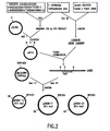
- the heparinase II gene was cloned from a F. heparinum chromosomal DNA library, Figure 2, constructed as described above. Ten plaque-containing filters were hybridized with the DNA probe, produced from the gel purified insert of pCE 14, which was labeled using a Random Labeling Kit (Boehringer Mannheim Canada, Laval, Quebec). Plaque hybridization was carried out, as described in Maniatis et al., ibid., at 65°C for 16 hours in a Tek Star hybridization oven (Bio/CAN Scientific, Mississauga, Ontario). Subsequent washes were performed at 65°C: twice for 15 min. in 2X SSC, once in 2X SSC/0.1% SDS for 30 min.
- a second strategy employed to circumvent the unexpected problem of F. heparinum DNA toxicity in E. coli was to digest the chromosomal DNA fragment with a restriction endonuclease which would divide the fragment, and if possible the heparinase II, gene into two pieces, Figure 2. These fragments could be cloned individually.
- Hybridization experiments also demonstrated that the BamH I digested F. heparinum DNA in phage HIIS produced two bands 1.8 and 5.5 kb in size.
- the molecular weight of heparinase II protein is approximately 84 kD, so the size of the corresponding gene would be approximately 2.4 kb.
- the 1.8 and 5.5 kb BamH I chromosomal DNA fragments could include the entire heparinase II gene.
- the plasmids pBSIB6-7, pBSIB6-21 and pBSIB2-13, Figure 2 were used to produce nested deletions with the Erase-a-Base system (Promega Biotec, Madison Wis.). These plasmids were used as templates for DNA sequence analysis using universal and reverse primers and oligonucleotide primers derived from known heparinase II sequence.
- the vector, pGB was used for heparinase II expression in E. coli, Figure3.
- pGB contains the modified ribosome binding region from pGhep, Figure 1, and a unique BamH I site, whereby expression of a DNA fragment inserted into this site is driven by a double tac promoter.
- the vector also includes a kanamycin resistance gene, and the lac Iq gene to allow induction of transcription with IPTG. Initially, a gel purified 5.5 kb BamH I fragment from pBSIB6-21 was ligated with BamH I digested pGB and transformed into FTB1, which was selected on LB agar with kanamycin.
- Cloning the blunt-end PCR product into pTZ/PC was unsuccessful, using FTB1 as the host.
- pBSQTK-9 which was sequenced with reverse and universal primers, contained an accurate reproduction of the DNA sequence from the heparinase II gene.
- the BamH I digested PCR fragment from pBSQTK-9 was inserted into the BamH I site of pGBIID in such orientation that the ATG site was downstream of the Shine-Dalgarno sequence.
- oligonucleotides were designed by choosing each codon according to the codon usage table. These were: 5'-GAATTCCATCAGTTTCAG CCGCATAAA-3' (SEQ ID NO:17), 5'-GAATTCTTTATGCGGCTGAAACTGATG-3' (SEQ ID NO:18), 5'-GAATTCCCGCCGGGCGAATTTCATGC-3' (SEQ ID NO: 19) and 5'-GAATTCGCATGAAATTCGCCCGGCGG-3' (SEQ ID NO:20), and were named oligonucleotides 3-1, 3-2, 3-3 and 3-4, respectively.
- oligonucleotides were used in all possible combinations, in an attempt to amplify a portion of the heparinase III gene using the polymerase chain reaction.
- the PCR amplifications were carried out as described above. Cycles of: denaturation temperature 92° C (1 minute), annealing temperatures ranging from 37° to 55° C, (1 minute) and extension temperature 72° C (2 minutes) were repeated 35 times. Analysis of the PCR reactions as described above demonstrated that no DNA fragments were produced by these experiments.
- a second set of oligonucleotides was synthesized and was comprised of 32 base sequences, in which the codon usage table was used to guess the third position of only half of the codons.
- the nucleotides within the parentheses indicate degeneracies of two or four bases at a single site. These were:
- a third set of oligonucleotides was synthesized incorporating BamH I endonuclease sequences on the ends of the 3-6 and 3-7 oligonucleotide sequences.
- a 999 base pair DNA sequence was obtained using the polymerase chain reaction with F. heparinum chromosomal DNA as the target. Attempts were made to clone the amplified DNA into the BamH I site of the high copy number plasmid pBluescript and the low copy number plasmids pBR322 and pACYC184. All of these constructs were again transformed into the FTB1 E. coli strain. More than 500 candidates were screened, yet no transformants containing a plasmid harboring the F . heparinum DNA were obtained. Once again, it was concluded that this region of F. heparinum chromosome imparts a negative-selective effect on E. coli cells that harbor it.
- the PCR fragment was split in order to avoid the problem of foreign DNA toxicity.
- Digestion of the 981 bp BamH I-digested heparinase III PCR fragment with restriction endonuclease Cla I produced two fragments of 394 and 587 bp.
- the amplified F. heparinum region was treated with Cla I and the two fragments separated by agarose gel electrophoresis.
- the 587 and 394 base pair fragments were ligated separately into plasmid pBluescript that had been treated with restriction endonucleases BamH I and Cla I.
- the entire 981 bp PCR fragment was purified and ligated into BamH I cut pBluescript.
- the ligated plasmids were inserted into the XL-1 Blue E. coli.
- Transformants containing plasmids with inserts were selected on the basis of their ability to form white colonies on LB-agar plates containing X-gal, IPTG and 50 ug/ml ampicillin, as described by Maniatis.
- Plasmid pFB1 containing the 587 bp F. heparinum DNA fragment and plasmid pFB2 containing the entire 981 base pair fragment were isolated by this method.
- the XL-1 Blue strain which, like strain FTB1.
- the PCR fragment insert in plasmid pFB1 was labeled with 32 P-ATP using a Random Primed DNA Labeling kit (Boehringer Mannheim, Laval, Quebec), and was used to screen the F. heparinum ⁇ DASHII library, Figure 6, constructed as described herein.
- the lambda library was plated out to obtain approximately 1500 plaques, which were transferred to nitrocellulose filters (Schleicher & Schuel, Keene, NH).
- the PCR probe was purified by ethanol precipitation. Plaque hybridization was carried out using the conditions described above. Eight positive lambda plaques were identified.
- Lambda DNA was isolated from lysed bacterial cultures as described in Maniatis and further analyzed by restriction analysis and by Southern blotting using a Hybond-N nylon membrane (Amersham Corporation, Arlington Heights, IL) following the protocol described in Maniatis.
- This clone was further analyzed by DNA sequencing. The sequence data was obtained using successive nested deletions of p Hind IIIBD generated with the Erase-a-Base System (Promega Corporation, Madison, WI) or sequenced using synthetic oligonucleotide primers.
- Sequence analysis revealed a single continuous open reading frame, without a translational termination codon, of 1929 base pairs, corresponding to 643 amino acids. Further screening of the lambda library led to the identification of a 673 bp Kpn I fragment which was similarly cloned into the Kpn I site of pBluescript, creating plasmid pFB4. The termination codon was found within the Kpn I fragment adding an extra 51 base pairs to the heparinase III gene and an additional 16 amino acid to the heparinase III protein. The complete heparinase III gene was later found to be included within a 3.2 kilobase Pst I fragment from lambda plaque #118.
- the complete heparinase III gene from Flavobacterium is thus 1980 base pairs in length, Figure 8, and encodes a 659 amino acid protein, Figure 9.
- N-terminal amino acid sequencing of deblocked, processed heparinase III indicated that the mature protein begins with Q-25, and contains 635 amino acids with a calculated molecular weight of 73,135 Daltons, Figure 9.
- PCR was used to generate a mature, truncated heparinase III gene, which had 16 amino acids deleted from the carboxy-terminus of the protein.
- An oligonucleotide comprised of 5'-CGCGGATCCATGCAAAGCT CTTCCATT-3' (SEQ ID NO:25) was designed to insert an ATG start site immediately preceding the codon for the first amino acid (Q-25) of mature heparinase III, while an oligonucleotide comprised of 5'-CGCGGATCCTCA AAGCTTGCCTTTCTC-3' (SEQ ID NO:26), was designed to insert a termination codon after the last amino acid of the heparinase III gene on the 2.7 kb Hind III fragment.
- Both oligonucleotides also contained a BamH I site.
- Plasmid p Hind IIIBD was used as the template in a PCR reaction with an annealing temperature of 50°C. A specific fragment of the expected size, 1857 base pairs, was obtained. This fragment encodes a protein of 620 amino acids with a calculated MW of 71,535 Da. It was isolated and inserted in the BamH I site of the expression vector pGB. This construct was named pGB-H3 ⁇ 3', Figure 7.
- heparinase III is a protein of 636 amino acids with a calculated molecular weight of 73,266 Daltons.
- E. coli strain XL-1 Blue(pGBH3) was grown at 37°C in LB medium containing 75 ug/ml kanamycin to an OD 600 of 0.5, at which point the tac promoter from pGB was induced by the addition of 1 mM IPTG.
- Cultures were grown an additional 2-5 hours at either 23° C, 30° C or 37° C.
- the cells were cooled on ice, concentrated by centrifugation and resuspended in cold PBS at 1/10th the original culture volume. Cells were lysed by sonication and cell debris removed by centrifugation at 10,000 x g for 5 minutes. The pellet and supernatant fractions were analyzed for heparan sulfate degrading (heparinase III) activity. Heparan sulfate degrading activities of 1.29, 5.27 and 3.29 IU/ml were observed from cultures grown at 23°, 30° and 37° C, respectively.
- the present invention describes a methodology for obtaining highly purified heparin and heparan sulfate degrading proteins by expressing the genes for these proteins in a suitable expression system and applying the steps of cell disruption, cation exchange chromatography, affinity chromatography and hydroxylapatite chromatography. Variations of these methods will be obvious to those skilled in the art from the foregoing detailed description of the invention. Such modifications are intended to come within the scope of the appended claims.
- GCT GCC.
- GCG GCA GCT GCC C TGT
- TGC EITHER EITHER D GAT
- GAC EITHER EITHER E GAG
- GAA GAA GAA F TTC TTT EITHER TTT G GGC, GGA, GGG, GGT GGC or GGT GGC H CAC
- CAT CAT CAT I ATC ATA, ATT ATA ATC K AAA.
- AAG AAA AAA L CTT, CTA, CTG, TTG, TTA, CTG CTG CTC M ATG ATG ATG N AAC.
Abstract
Description
- This invention is directed to cloning, sequencing and expressing heparinase II from Flavobacterium heparinum.
- The heparin and heparan sulfate family of molecules is comprised of glycosaminoglycans of repeating glucosamine and hexuronic acid residues, either iduronic or glucuronic, in which the 2, 3 or 6 position of glucosamine or the 2 position of the hexuronic acid may be sulfated. Variations in the extent and location of sulfation as well as conformation of the alternating hexuronic acid residue leads to a high degree of heterogeneity of the molecules within this family. Conventionally, heparin refers to molecules which possess a high sulfate content, 2.6 sulfates per disaccharide, and a higher amount of iduronic acid. Conversely, heparan sulfate contains lower amounts of sulfate, 0.7 to 1.3 sulfates per disaccharide, and less iduronic acid. However, variants of intermediate composition exist and heparins from all biological sources have not yet been characterized.
- Specific sulfation/glycosylation patterns of heparin have been associated with biological function, such as the antithrombin binding site described by Choay. et al., Thrombosis Res. 18: 573-578 (1980), and the fibroblast growth factor binding site described by Turnbull et al., J. Biol. Chem. 267: 10337-10341 (1992). It is apparent from these examples that heparin's interaction with certain molecules results from the conformation imparted by specific sequences and not solely due to electrostatic interactions imparted by its high sulfate composition. Heparin interacts with a variety of mammalian molecules, thereby modulating several biological events such as hemostasis, cell proliferation, migration and adhesion as summarized by Kjellen and Lindahl, Ann Rev Biochem 60: 443-475 (1991) and Burgess and Macaig, Ann. Rev. Biochem. 58: 575-606 (1989). Heparin, extracted from bovine lungs and porcine intestines, has been used as an anticoagulant since its antithrombotic properties were discovered by McLean, Am. J. Physiol. 41: 250-257 (1916). Heparin and chemically modified heparins are continually under review for medical applications in the areas of wound healing and treating vascular disease.
- Heparin degrading enzymes, referred to as heparinases or heparin lyases, have been identified in several microorganisms including: Flavobacterium heparinum, Bacteriodes sp. and Aspergillus nidulans as summarized by Linhardt et al.. Appl. Biochem. Biotechnol. 12: 135-177 (1986). Heparan sulfate degrading enzymes, referred to as heparitinases or heparan sulfate lyases, have been detected in platelets (Oldberg et al., Biochemistry 19: 5755-5762 (1980)), tumor (Nakajima et al., J. Biol. Chem. 259: 2283-2290 (1984)) and endothelial cells (Gaal et al., Biochem. Biophys. Res. Comm. 161: 604-614 (1989)). Mammalian heparanases catalyze the hydrolysis of the carbohydrate backbone of heparan sulfate at the hexuronic acid (1 → 4) glucosamine linkage (Nakajima et al., J. Cell. Biochem. 36: 157-167 (1988)) and are inhibited by the highly sulfated heparin. However, accurate biochemical characterizations of these enzymes has thus far been prevented by the lack of a method to obtain homogeneous preparations of the molecules.
- Flavobacterium heparinum produces heparin and heparan sulfate degrading enzymes termed heparinase I (E.C. 4.2.2.7) as described by Yang et al., J. Bibi. Chem. 260(3): 1849-1857 (1985), heparinase II as described by Zimmermann and Cooney, U.S. Patent No. 5,169,772, and heparinase III (E.C. 4.2.2.8) as described by Lohse and Linhardt, J. Biol. Chem. 267: 24347-24355 (1992). These enzymes catalyze an eliminative cleavage of the (α1 → 4) carbohydrate bond between glucosamine and hexuronic acid residues in the heparin/heparan sulfate backbone. The three enzyme variants differ in their action on specific carbohydrate residues. Heparinase I cleaves at α-D-GlcNp2S6S(1 → 4)α-L-IdoAp2S, heparinase III at α-D-GlcNp2Ac(or2S)60H(1 → 4)β-D-GlcAp and heparinase II at either linkage as described by Desai et al., Arch. Biochem. Biophys. 306(2): 461-468 (1993). Secondary cleavage sites for each enzyme also have been described by Desai et al.
- Heparinase I has been used clinically to neutralize the anticoagulant properties of heparin as summarized by Baugh and Zimmermann, Perfusion Rev. 1(2): 8-13, 1993. Heparinase I and III have been shown to modulate cell-growth factor interactions as demonstrated by Bashkin et al., J. Cell Physiol. 151:126-137 (1992) and cell-lipoprotein interactions as demonstrated by Chappell et al., J. Biol. Chem. 268(19):14168-14175 (1993). The availability of heparin degrading enzymes of sufficient purity and quantity could lead to the development of important diagnostic and therapeutic formulations.
- According to the present invention, there is provided isolated non-glycosylated heparinase II comprising the amino acid sequence of mature heparinase II having a methionine residue immediately preceding the first amino acid of said mature heparinase II, or non-glycosylated functional derivatives thereof which degrade heparan sulphate and heparin.
- Preferably, the heparinase II of the present invention comprises the amino acid sequence of SEQ ID NO:2 beginning at glutamine at position 26 and which includes a methionine immediately preceding said glutamine.
- The invention also provides an antibody or fragment thereof which is specific for a heparinase II of the present invention, as well as a heparinase II of the present invention for use in therapy or diagnosis.
- Prior to the present invention, partially purified heparinases II and III were available, but their amino acid sequences were unknown. Cloning these enzymes was difficult because of toxicity to the host cells. The present inventors were able to clone the genes for heparinases II and III, and herein provide their nucleotide and amino acid sequences.
- A method is described for the isolation of highly purified heparin and heparan sulfate degrading enzymes from F. heparinum. Characterization of proteins demonstrated that heparinases I, II and III are glycoproteins. All three proteins are modified at their N-terminal amino acid residue. Antibodies generated by injecting purified heparinases into rabbits yielded anti-sera which demonstrated a high degree of cross reactivity to proteins from F. heparinum. Polyclonal antibodies were separated by affinity chromatography into fractions which bind the amino acid portion of the proteins and a fraction which binds the post-translational modification allowing for the use of these antibodies to specifically distinguish the native and recombinant forms of each heparinase protein.
- Amino acid sequence information was used to synthesize oligonucleotides that were subsequently used in a polymerase chain reaction (PCR) to amplify a portion of the heparinase II and heparinase III genes. Amplified regions were used in an attempt to identify clones from a λDASH-II gene library which contained F. heparinum genomic DNA. Natural selection against clones containing the entire heparinase II and III genes was observed. This was circumvented by cloning sections of the heparinase II gene separately, and by screening host strains for stable maintenance of complete heparinase III clones. Expression of heparinase II and III was achieved by use of a vector containing a modified ribosome binding site which was shown to increase the expression of heparinase I to significant levels.
- This patent describes the gene and amino acid sequences for heparinase II from F. heparinum, which may be used in conjunction with suitable expression systems to produce the enzymes. Also described, is a modified ribosome binding sequence used to express heparinase I, II, and III.
-
- Figure 1 shows the modifications to the tac promoter ribosome binding region, which were evaluated for the level of expression of
heparinase 1. The original sequence, as found in pBhep, and the modified sequences, as found in pGhep and pΔ4hep, are shown with the Shine-Dalgarno sequences (S-D) and the heparinase I gene start codon, underlined. The gap (in nucleotides, nt) between these regions is indicated below each sequence. The ribosome binding region for pGB contains no start codon, and has a BamHI site (underlined) in place of the EcoRI site (GAATTC) found in pGhep. - Figure 2 shows the construction of plasmids used to sequence the heparinase II gene from Flavobacterium heparinum. Restriction sites are: N- NotI, Nc = NcoI, S = SalI, B = BamHI, P = PstI, E = EcoRI, H = HindIII, C = ClaI and K = KpnI.
- Figure 3 shows the construction of pGBH2, a plasmid capable of directing the expression of active heparinase II in E. coli from tandem tac promoters (double arrow heads). Restriction sites are: B = BamHI, P = Pst I.
- Figure 4 shows the nucleic acid sequence for the heparinase II gene from Flavobacterium heparinum (SEQ ID NO:1).
- Figure 5 shows the amino acid sequence for heparinase II from Flavobacterium heparinum (SEQ ID NO:2). The leader peptide sequence is underlined. The mature protein starts at Q-26.
Peptides 2A, 2B and 2C are indicated at their corresponding positions within the protein. - Figure 6 shows the construction of plasmids used to sequence the heparinase III gene from Flavobacterium heparinum. Restriction sites are: S = SalI, B = BamHI, P = PstI, E = EcoRI, H = HindIII, C = Clal and K = KpnI.
- Figure 7 shows the construction of pGBH3. a plasmid capable of directing the expression of active heparinase III in E. coli from a tandem taq promoter (double arrow heads). Restriction sites are: S = SalI, B = BamHI, P = PstI, E = EcoRI, H = HindIII, Bs = BspEI, C = ClaI and K = KpnI.
- Figure 8 shows the nucleic acid sequence for the heparinase III gene from Flavobacterium heparinum (SEQ ID NO:3).
- Figure 9 shows the amino acid sequence for heparinase III from Flavobacterium heparinum (SEQ ID NO:4). The leader peptide sequence is underlined. The mature protein starts at Q-25.
Peptides - To aid in the understanding of the specification and claims, including the scope to be given such terms, the following definitions are provided.
- Gene. By the term "gene" is intended a DNA sequence which encodes through its template or messenger RNA a sequence of amino acids characteristic of a specific peptide. Further, the term includes intervening, non-coding regions, as well as regulatory regions, and can include 5' and 3' ends.
- Gene sequence. The term "gene sequence" is intended to refer generally to a DNA molecule which contains one or more genes, or gene fragments, as well as a DNA molecule which contains a non-transcribed or non-translated sequence. The term is further intended to include any combination of gene(s), gene fragments(s), non-transcribed sequence(s) or non-translated sequence(s) which are present on the same DNA molecule.
- The present sequences may be derived from a variety of sources including DNA, synthetic DNA, RNA, or combinations thereof. Such gene sequences may comprise genomic DNA which may or may not include naturally occurring introns. moreover, such genomic DNA may be obtained in association with promoter regions or poly A sequences. The gene sequences, genomic DNA or cDNA may be obtained in any of several ways. Genomic DNA can be extracted and purified from suitable cells, such as brain cells, by means well known in the art. Alternatively, mRNA can be isolated from a cell and used to produce cDNA by reverse transcription or other means.
- Recombinant DNA. By the term "recombinant DNA" is meant a molecule that has been recombined by in vitro splicing cDNA or a genomic DNA sequence.
- Cloning Vehicle. A plasmid or phage DNA or other DNA sequence which is able to replicate in a host celL The cloning vehicle is characterized by one or more endonuclease recognition sites at which is DNA sequences may be cut in a determinable fashion without loss of an essential biological function of the DNA, which may contain a marker suitable for use in the identification of transformed cells. Markers include for example, tetracycline resistance or ampicillin resistance. The word vector can be used to connote a cloning vehicle.
- Expression Control Sequence. A sequence of nucleotides that controls or regulates expression of structural genes when operably linked to those genes. They include the lac systems, the trp system major operator and promoter regions of the phage lambda, the control region of fd coat protein and other sequences known to control the expression of genes in prokaryotic or eukaryotic cells.
- Expression vehicle. A vehicle or vector similar to a cloning vehicle but which is capable of expressing a gene which has been cloned into it, after transformation into a host. The cloned gene is usually placed under the control of (i.e., operable linked to) certain control sequences such as promoter sequences. Expression control sequences will vary depending on whether the vector is designed to express the operably linked gene in a prokaryotic or eukaryotic host and may additionally contain transcriptional elements such as enhancer elements, termination sequences, tissue-specificity elements, and/or translational initiation and termination sites.
- Promoter. The term "promoter" is intended to refer to a DNA sequence which can be recognized by an RNA polymerase. The presence of such a sequence permits the RNA polymerase to bind and initiate transcription of operably linked gene sequences.
- Promoter region. The term "promoter region" is intended to broadly include both the promoter sequence as well as gene sequences which may be necessary for the initiation of transcription. The presence of a promoter region is, therefore, sufficient to cause the expression of an operably linked gene sequence.
- Operably Linked. As used herein, the term "operably linked" means that the promoter controls the initiation of expression of the gene. A promoter is operably linked to a sequence of proximal DNA if upon introduction into a host cell the promoter determines the transcription of the proximal DNA sequence or sequences into one or more species of RNA. A promoter is operably linked to a DNA sequence if the promoter is capable if initiating transcription of that DNA sequence.
- Prokaryote. The term "prokaryote" is meant to include all organisms without a true nucleus, including bacteria.
- Host. The term "host" is meant to include not only prokaryotes, but also such eukaryotes as yeast and filamentous fungi, as well as plant and animal cells. The terms includes organisms or cell that is the recipient of a replicable expression vehicle.
- The present invention is based on the cloning and expression of a previously uncloned enzyme. Although heparinase II had been partially purified previously, no amino acid sequence was available. Specifically, the invention discloses the cloning, sequencing and expression of heparinase II from Flavobacterium heparinum and the use of a modified ribosome binding region for expression of this gene. In addition to the nucleotide sequences, the amino acid sequence of heparinase II is also provided. The invention further provides expressed heparinase II, as well as methods of expressing this enzyme.
- Cloning was accomplished using degenerate and "guessmer" nucleotide primers derived from amino acid sequences of fragments of the heparinases, purified as described below in detail. The amino acid sequences were previously unavailable. Cloning was exceptionally difficult because of the unexpected problem of F. heparinum DNA toxicity in E. coli. The inventors discovered techniques for solving this problem, as described below in detail. Based on this disclosure, one skilled in the art can readily clone additional heparinases and other proteins from F. heparinum or from additional sources using the novel methods described within.
- Expression of the heparinases is further disclosed herein. To express heparinases I, II, and III, transcriptional and translational signals recognizable by an appropriate host are necessary. The cloned heparinases encoding sequences, obtained through the methods described above, and preferably in a double-stranded form, may be operably linked to sequences controlling transcriptional expression in an expression vector, and introduced into a host cell, either prokaryote or eukaryote, to produce recombinant heparinases or a functional derivative thereof. Depending upon which strand of the heparinases encoding sequence is operably linked to the sequences controlling transcriptional expression, it is also possible to express heparinases antisense RNA or a functional derivative thereof.
- For the expression of heparinases I, II and III in E. coli, vectors were constructed wherein expression was driven by two repeats of the tac promoter. Modifications of the ribosome binding region of this promoter were made by introducing mutations with the polymerase chain reaction. In a preferred modification of the expression vector, the minimal consensus Shine-Delgarno sequence was improved by introducing a single mutation (AGGAA → AGGAG), which had the further advantage of decreasing the number of nucleotides between the Shine-Delgarno sequence and the ATG start codon. Further modifications were produced using PCR in which the gap between the Shine-Delgarno sequence and the start codon were further reduced. Using the same techniques, additional modifications in this region, including insertions and deletions, can be produced to create additional heparinase expression vectors. As a result, an expression vector for the expression of heparinases is provided which comprises a modified ribosome binding region containing a 5 base pair Shine-Dalgarno sequence, a 9 base pair spacer region between the Shine-Dalgarno sequence and the ATG start codon, and a recombinant nucleotide sequence encoding. Also provided are modifications to this vector comprising changing the length and sequence of the Shine-Dalgarno sequence, and also by reducing the spacing between the Shine-Dalgarno sequence and the start codon to 8, 7, 6, 5, 4 or fewer nucleotides.
- Expression of the heparinases in different hosts may result in different post-translational modifications which may alter the properties of the heparinases, or a functional derivative thereof, in eukaryotic cells, and especially mammalian, insect and yeast cells. Especially preferred eukaryotic hosts are mammalian cells either in vivo, in animals or in tissue culture. Mammalian cells provide post-translational modifications to recombinant heparinases which include folding and/or glycosylation at sites similar or identical to that found for the native heparinases. Most preferably, mammalian host cells include brain and neuroblastoma cells.
- A nucleic acid molecule, such as DNA, is said to be "capable of expressing" a polypeptide if it contains expression control sequences which contain transcriptional regulatory information and such sequences are "operably linked" to the nucleotide sequence which encodes the polypeptide.
- An operable linkage is a linkage in which a sequence is connected to a regulatory sequence (or sequences) in such a way as to place expression of the sequence under the influence or control of the regulatory sequence. Two DNA sequences (such as a heparinases encoding sequence and a promoter region sequence linked to the 5' end of the encoding sequence) are said to be operably linked if induction of promoter function results in the transcription of the heparinases encoding sequence mRNA and if the nature of the linkage between the two DNA sequences does not (1) result in the introduction of a frame-shift mutation, (2) interfere with the ability of the expression regulatory sequences to direct the expression of the heparinases, or (3) interfere with the ability of the heparinases template to be transcribed by the promoter region sequence. Thus, a promoter region would be operably linked to a DNA sequence if the promoter were capable of effecting transcription of that DNA sequence.
- The precise nature of the regulatory regions needed for gene expression may vary between species or cell types, but in general includes, as necessary, 5' non-transcribing and 5' non-translating (non-coding) sequences involved with initiation of transcription and translation respectively, such as the TATA box, capping sequence, CAAT sequence, and the like. Especially, such 5' non-transcribing control sequences will include a region which contains a promoter for transcriptional control of the operably linked gene.
- If desired, a fusion product of the heparinases may be constructed. For example, the sequence coding for heparinases may be linked to a signal sequence which will allow secretion of the protein from, or the compartmentalization of the protein in, a particular host. Such signal sequences maybe designed with or without specific protease sites such that the signal peptide sequence is amenable to subsequent removal. Alternatively, the native signal sequence for this protein may be used.
- Transcriptional initiation regulatory signals can be selected which allow for repression or activation, so that expression of the operably linked genes can be modulated.
- Based on this disclosure, one skilled in the art can readily place the sequences of the present invention in additional expression vectors and transform into a variety of bacteria to obtain recombinant heparinase II.
- Once the vector or DNA sequence containing the construct(s) is prepared for expression, the DNA construct(s) is introduced into an appropriate host cell by any of a variety of suitable means, including transfection. After the introduction of the vector, recipient cells are grown in a selective medium, which selects for the growth of vector-containing cells. Expression of the cloned gene sequence(s) results in the production of heparinase I, II or III, or in the production of a fragment of one of these proteins. This expression can take place in a continuous manner in the transformed cells, or in a controlled manner, for example, expression which follows induction of differentiation of the transformed cells (for example, by administration of bromodeoxyuracil to neuroblastoma cells or the like).
- The expressed protein is isolated and purified in accordance with conventional conditions, such as extraction, precipitation, chromatography, electrophoresis, or the like. Detailed procedures for the isolation of the heparinases is discussed in detail in the examples below.
- The invention further provides functional derivatives of the sequences of heparinase II.
- As used herein, the term "functional derivative" is used to define any DNA sequence which is derived by the original DNA sequence and which still possesses the biological activities of the native parent molecule. A functional derivative can be an insertion, a deletion, or a substitution of one or more bases in the original DNA sequence. The substitutions can be such that they replace a native amino acid with another amino acid that does not substantially effect the functioning of the protein. Those skilled in the art will recognize that likely substitutions include positively the functioning of the protein, such as a small, neutrally charged amino acid replacing another small, neutrally charged amino acid. Those of skill in the art will recognize that functional derivatives of the heparinases can be prepared by mutagenesis of the DNA using one of the procedures known in the art, such as site-directed mutagenesis. In addition, random mutagenesis can be conducted and mutants retaining function can be obtained through appropriate screening.
- The antibodies of the present invention include monoclonal and polyclonal antibodies, as well fragments of these antibodies. Fragments of the antibodies of the present invention include, but are not limited to, the Fab, the Fab2, and the Fc fragment.
- The invention also provides hybridomas which are capable of producing the above-described antibodies. A hybridoma is an immortalized cell line which is capable of secreting a specific monoclonal antibody.
- In general, techniques for preparing polyclonal and monoclonal antibodies as well as hybridomas capable of producing the desired antibody are well-known in the art (Campbell, A.M., "Monoclonal Antibody Technology: Laboratory Techniques in Biochemistry and Molecular Biology." Elsevier Science Publishers, Amsterdam, The Netherlands (1984); St. Groth et al., J. Immunol. Methods 35:1-21 (1980)).
- Any mammal which is known to produce antibodies can be immunized with the pseudogene polypeptide. Methods for immunization are well-known in the art. Such methods include subcutaneous or interperitoneal injection of the polypeptide. One skilled in the art will recognize that the amount of heparinase used for immunization will vary based on the animal which is immunized, the antigenicity of the peptide and the site of injection.
- The protein which is used as an immunogen may be modified or administered in an adjuvant in order to increase the protein's antigenicity. Methods of increasing the antigenicity of a protein are well-known in the art and include, but are not limited to coupling the antigen with a heterologous protein (such as globulin or β-galaetosidase) or through the inclusion of an adjuvant during immunization.
- For monoclonal antibodies, spleen cells from the immunized animals are removed, fused with myeloma cells, such as SP2/0-Agl4 myeloma cells, and allowed to become monoclonal antibody producing hybridoma cells.
- Any one of a number of methods well known in the art can be used to identify the hybridoma cell which produces an antibody with the desired characteristics. These include screening the hybridomas with an ELISA assay, western blot analysis, or radioimmunoassay (Lutz et al., Exp. Cell Res. 175:109-124 (1988)).
- Hybridomas secreting the desired antibodies are cloned and the class and subclass is determined using procedures known in the art (Campbell, A.M., Monoclonal Antibody Technology: Laboratory Techniques in Biochemistry and Molecular Biology, Elsevier Science Publishers, Amsterdam, The Netherlands (1984)).
- For polyclonal antibodies, antibody containing antisera is isolated from the immunized animal and is screened for the presence of antibodies with the desired specificity using one of the above-described procedures.
- The present invention further provides the above-described antibodies in detectably labelled form. Antibodies can be detectably labelled through the use of radioisotopes, affinity labels (such as biotin, avidin, etc.), enzymatic labels (such as horseradish peroxidase, alkaline phosphatase, etc.), fluorescent labels (such as FITC or rhodamine, etc.), paramagnetic atoms, chemiluminescent labels, and the like. Procedures for accomplishing such labelling are well-known in the art; for example, see Stemberger, L.A. et al.. J. Histochem. Cytochem. 18:315 (1970); Byer, E.A. et al., Meth. Enzym. 62:308 (1979); Engval, E. et al., Immunol. 109:129 (1972); Goding, J.W., J. Immunol. Meth. 13:215 (1976).
- The present invention further provides the above-described antibodies immobilized on a solid support. Examples of such solid supports include plastics, such as polycarbonate, complex carbohydrates such as agarose and sepharose, acrylic resins such as polyacrylamide and latex beads. Techniques for coupling antibodies to such solid supports are well known in the art (Weir et al., Handbook of Experimental Immunology, 4th Ed., Blackwell Scientific Publications, Oxford, England (1986)). The immobilized antibodies of the present invention can be used for immunoaffinity purification of heparinases.
- Having now generally described the invention, the same will be understood by a series of specific examples, which are not intended to be limiting.
- Heparin lyase enzymes were purified from cultures of Flavobacterium heparinum. F. heparinum was cultured in a 15 L computer-controlled fermenter using a variation of the defined nutrient medium described by Galliher et al., Appl Environ. Microbiol. 41(2):360-365 (1981). Those fermentations designed to produce heparin lyases incorporate semi-purified heparin (Celsus Laboratories) in the media at a concentration of 1.0 g/L as the inducer of heparinase synthesis. Cells were harvested by centrifugation and the desired enzymes released from the periplasmic space by a variation of the osmotic shock procedure described by Zimmermann and Cooney, U.S. Patent No. 5,262,325, herein incorporated by reference.
- A semi-purified preparation of the heparinase enzymes was achieved by a modification of the procedure described by Zimmermann et al.. U.S. Patent No. 5,262.325. Proteins from the crude osmolate were adsorbed onto cation exchange resin (CBX, J.T. Baker) at a conductivity of 1 - 7 µmho. Unbound proteins from the extract were discarded and the resin packed into a chromatography column (5.0 cm i.d. x 100 cm). The bound proteins eluted at a linear flow rate of 3.75 cm•min-1 with step gradients of 0.01 M phosphate, 0.01 M phosphate/0.1 M sodium chloride, 0.01 M phosphate/0.25 M sodium chloride and 0.01 M phosphate/1.0 M sodium chloride, all at pH 7.0 +/- 0.1. Heparinase II elutes in the 0.1 M NaCl fraction, while
heparinases - Alternately, the 0.1 M sodium chloride step was eliminated and the three heparinases co-eluted with 0.25 M sodium chloride. The heparinase fractions were loaded directly onto a column containing cellufine sulfate (5.0 cm i.d. x 30 cm, Amicon) and eluted at a linear flow rate of 2.50 cm·min-1 with step gradients of 0.01 M phosphate, 0.01 M phosphate/0.2 M sodium chloride, 0.01 M phosphate/0.4 M sodium chloride and 0.01 M phosphate/1.0 M sodium chloride, all at pH 7.0 +/- 0.1. Heparinase II and 3 elute in the 0.2 M sodium chloride fraction while heparinase I elutes in the 0.4 M fraction.
- The 0.2 M sodium chloride fraction from the cellufine sulfate column was diluted with 0.01 M sodium phosphate to give a conductance of less than 5 µmhos. The solution was further purified by loading the material onto a hydroxylapatite column (2.6 cm i.d. x 20 cm) and eluting the bound protein at a linear flow rate of 1.0 cm•min-1 with step gradients of 0.01 M phosphate, 0.01 M phosphate/0.35 M sodium chloride, 0.01 M phosphate/0.45 M sodium chloride, 0.01 M phosphate/0.65 M sodium chloride and 0.01 M phosphate/1.0 M sodium chloride, all at pH 7.0 +/- 0.1. Heparinase III elutes in a single protein peak in the 0.45 M sodium chloride fraction while heparinase III elutes in a single protein peak in the 0.65 M sodium chloride fraction.
- Heparinase I was further purified by loading material from the cellufine sulfate column, diluted to a conductivity less than 5 µmhos, onto a hydroxylapatite column (2.6 cm i.d. x 20 cm) and eluting the bound protein at a linear flow rate of 1.0 cm•min-1 with a linear gradient of phosphate (0.01 to 0.25 M) and sodium chloride (0.0 to 0.5 M). Heparinase I elutes in a single protein peak approximately mid-way through the gradient.
- The heparinase enzymes obtained by this method were analyzed by SDS-PAGE using the technique of Laemmli, Nature 227: 680-685 (1970), and the gels quantified by a scanning densitometer (Bio-Rad, Model GS-670). Heparinases I, II and III displayed molecular weights of 42,500+/-2,000, 84,000+/-4,200 and 73.000+/-3.500 Daltons, respectively. All proteins displayed purities of greater than 99 %. Purification results for the heparinase enzymes are shown in Table 1.
- Heparinase activities were determined by the spectrophotometric assay described by Yang et al. A modification of this assay incorporating a reaction buffer comprised of 0.018 M Tris, 0.044 M sodium chloride and 1.5 g/L heparan sulfate at pH 7.5 was used to measure heparan sulfate degrading activity.
- Recombinant heparinase I forms intracellular inclusion bodies which require denaturation and protein refolding to obtain active heparinase. Two solvents, urea and guanidine hydrochloride, were examined as solubilizing agents. Of these, only guanidine HCl, at 6 M, was able to solubilize the
heparinase 1 inclusion bodies. However, the highest degree of purification was obtained by sequentially washing the inclusion bodies in 3 M urea and 6 M guanidine HCl. The urea wash step served to removed contaminating E. coli proteins and cell debris prior to solubilizing of the aggregated heparinase I by guanidine HCl. - Recombinant heparinase I was prepared by growing E. coli Y1090(pGHep1), a strain harboring a plasmid containing the heparinase I gene expressed from tandem tac promoters, in Luria broth with 0.1 M IPTG. The cells were concentrated by centrifugation and resuspended in 1/10th volume buffer containing 0.01 M sodium phosphate and 0.2 M sodium chloride at pH 7.0. The cells were disrupted by sonication, 5 minutes with intermittent 30 second cycles,
power setting # 3 and the inclusion bodies concentrated by centrifugation, 7,000 x g, 5 minutes. The pellets were washed two times with cold 3 M urea for 2 hours at pH, 7.0 and the insoluble material recovered by centrifugation. Heparinase I was unfolded in 6 M guanidine HCl containing 50 mM DTT and refolded by dialysis into 0.1 M ammonium sulfate. Additional contaminating proteins precipitated in the 0.1 M ammonium sulfate and could be removed by centrifugation. Heparinase I purified by this method had a specific activity of 42.21 IU/mg and was 90 % pure by SDS-PAGE/ scanning densitometry analysis. The enzyme can be further purified by cation exchange chromatography, as described above, yielding a heparinase I preparation that is more than 99 % pure by SDS-PAGE/ scanning densitometry analysis. - The molecular weight and kinetic properties of the three heparinase enzymes have been accurately reported by Lohse and Linhardt, J. Biol. Chem. 267:24347-24355 (1992). However, an accurate characterization of the proteins' post-translational modifications had not been carried out. Heparinases I, II and III, purified as described herein, were analyzed for the presence of carbohydrate moieties. Solutions containing 2 ug of heparinases I, II and III and recombinant heparinase I were brought to pH 5.7 by adding 0.2 M sodium acetate. These protein samples underwent carbohydrate biotinylation following protocol 2a, described in the GlycoTrack kit (Oxford Glycosystems). 30 µl of each biotinylated protein solution was subjected to SDS-PAGE (10% gel) and transferred by electroblotting at 170 mA constant current to a nitrocellulose membrane. Detection of the biotinylated carbohydrate was accomplished by an alkaline phosphatase-specific color reaction after attachment of a streptavadin-alkaline phosphatase conjugate to the biotin groups. These analyses revealed that heparinases I and II are glycosylated and heparinase III and recombinant heparinase I are not.
- Polyclonal antibodies generated in rabbits injected with wild type heparinase I could be fractionated into two populations as described below. It appears that one of these fractions recognizes a post-translational moiety common to proteins made in F. heparinum, while the other fraction specifically recognizes amino acid sequences contained in heparinase I. All heparinase enzymes made in F. heparinum were recognized by the "non-specific" antibodies but not heparinase made in E. coli. The most likely candidate for the non-protein antigenic determinant from heparinase I is the carbohydrate component; thus, the Western blot experiment indicates that all lyases made in F. heparinum are glycosylated.
- Purified heparinases II and III were analyzed by the technique of Edman to determine the N-terminal amino acid residue of the mature protein. However, the Edman chemistry was unable to liberate an amino acid, indicating that a post-translational modification had occurred at the N-terminal amino acid of both heparinases. One nmol samples of heparinases II and III were used for deblocking with pyroglutamate aminopeptidase. Control samples were produced by
mock deblocking 1 nmol protein samples without adding pyroglutamate aminopeptidase. All samples were placed in 10 mM NH4CO3, pH 7.5. and 10 mM DTT (100 µl final volume). To non-control samples, 1 mU of pyroglutamate aminopeptidase was added and all samples were incubated for 8 hr at 37° C. After incubation, an additional 0.5 mU of pyroglutamate aminopeptidase was added to non-control samples and all samples were incubated for an additional 16 h at 37°C. - Deblocking buffers were exchanged for 35% formic acid using a 10,000 Dalton cut-off Centricon unit and the sample was dried under vacuum. The samples were subjected to amino acid sequence analysis according to the method of Edman.
- The properties of the three heparinase proteins from Flavobacterium heparinum are listed in Table 2.
- Heparinases II and III were digested with cyanogen bromide in order to produce peptide fragments for isolation. The protein solutions (1-10 mg/ml protein concentration) were brought to a DTT concentration of 0.1 M, and incubated at 40°C for 2 hr. The samples were frozen and lyophilized under vacuum. The pellet was resuspended in 70% formic acid, and nitrogen gas was bubbled through the solution to exclude oxygen. A stock solution of CNBr was made in 70% formic acid and the stock solution was bubbled with nitrogen gas and stored in the dark for short time periods. For addition of CNBr, a 500 to 1000 times molar excess of CNBr to methionine residues in the protein was used. The CNBr stock was added to the protein solutions, bubbled with nitrogen gas and the tube was sealed. The reaction tube was incubated at 24°C for 20 hr, in the dark.
- The samples were dried down partially under vacuum, water was added to the sample, and partial lyophilization was repeated. This washing procedure was repeated until the sample pellets were white. The peptide mixtures were solubilized in formic acid and applied to a Vydac C18 reverse phase HPLC column (4.6 mm i.d. x 30 cm) and individual peptide fragments eluted at a linear flow rate of 6.0 cm•min-1 with a linear gradient of 10 to 90 % acetonitrile in 1 % trifluoroacetic acid. Fragments recovered from these reactions were subjected to amino acid sequence determination using an Applied Biosystems 745A Protein Sequencer. Three peptides isolated from heparinase II gave sequences: EFPEMYNLAAGR (SEQ ID NO:5), KPADIPEVKDGR (SEQ ID NO:6), and LAGDFVTGKILAQGFG PDNQTPDYTYL (SEQ ID NO:7) and were named
peptides 2A, 2B and 2C respectively. Three peptides from heparinase III gave sequences: LIK-NEVRWQLHRVK (SEQ ID NO:8), VLKASPPGEFHAQPDNGTFELFT (SEQ ID NO:9) and KALVHWFWPHKGYGYFDYGKDIN (SEQ ID NO: 10) and were namedpeptides - Heparinases I, II and III and recombinant heparinase I, purified as described herein, were used to generate polyclonal antibodies in rabbits. Each of heparinase I, II and III was carried through the following standard immunization procedure: The primary injection consisted of 0.5 - 1.0 mg of purified protein dissolved in 1 ml of sterile phosphate buffered Saline, which was homogenized with 1 ml of Freund's adjuvant (Cedarlane Laboratories Ltd.). This protein-adjuvant emulsion was used to inject New Zealand White female rabbits; 1 ml per rabbit, 0.5 ml per rear leg, i.m., in the thigh muscle near the hip. After 2 to 3 weeks, the rabbits were given an injection boost consisting of 0.5 - 1.0 mg of purified protein dissolved in sterile phosphate buffered Saline homogenized with 1 ml of incomplete Freund's adjuvant (Cedarlane Laboratories, Ltd.). Again after 2 to 3 weeks, the rabbits were given a third identical injection boost.
- A blood sample was collected from each animal from the central artery of the ear approximately 10 days following the final injection boost. Serum was prepared by allowing the sample to clot for 2 hours at 22°C followed by overnight incubation at 4°C, and clearing by centrifugation at 5,000 rpm for 10 min. The antisera were diluted 1:100,000 in Tris-buffered Saline (pH 7.5) and carried through Western blot analysis to identify those sera containing anti-heparinase I, II or III antibodies.
- Antibodies generated against wild type heparinase I, but not recombinant heparinase I, displayed a high degree of cross reactivity against other F. heparinum proteins. This was likely due to the presence of an antigenic post-translational modification common to F. heparinum proteins but not found on proteins synthesized in E. coli. To explore this further, recombinant heparinase I was immobilized onto Sepharose beads and packed into a chromatography column. Purified anti-heparinase I (wild type) antibodies were loaded onto the column and the unbound fraction collected. Bound antibodies were eluted in 0.1 M glycine, pH 2.0. IgG was found in both the unbound and bound fractions and subsequently used in Western blot experiments. Antibody isolated from the unbound fraction non-specifically recognized F. heparinum proteins but no longer detected recombinant heparinase I (E. coli), while the antibody isolated from the bound fraction only recognized heparinase I, whether synthesized in F. heparinum or E. coli. This result indicated that, as hypothesized, two populations of antibodies are formed by exposure to the wild-type heparinase I antigen: one specific for the protein backbone and the other recognizing a post-transiationally modified moiety common to F. heparinum proteins.
- This finding provides both a means to purify specific anti-heparinase antibodies and a tool for characterizing the wild-type heparinase I protein.
- A Flavobacterium heparinum chromosomal DNA library was constructed in lambda phage DASHII. 0.4 ug of F. heparinum chromosomal DNA was partially digested with restriction enzyme Sau3A to produce a majority of fragments around 20 kb in size, as described in Maniatis, et al, Molecular Cloning Manual, Cold Spring Harbor (1982). This DNA was phenol/chloroform extracted, ethanol precipitated, ligated with λDASHII arms and packaged with packaging extracts from a λDASHII/BamHI Cloning Kit (Stratagene, La Jolla, CA). The library was titered at approximately 10-5 pfu/ml after packaging, amplified to 10-8 pfu/ml by the plate lysis method, and stored at -70°C as described by Silhavy, T.J., et al.in Experiments in Molecular Genetics. Cold Spring Harbor Laboratory, 1992.
- The F. heparinum chromosomal library was titered to about 300 pfu/plate, overlaid on a lawn of E. coli, and allowed to transfect the cells overnight at 37°C, forming plaques. The phage plaques were transferred to nitrocellulose paper, and the phage DNA bound to the filters, as described in Maniatis, et al., ibid.
- The gene for the mature heparinase I protein was cloned into the EcoRI site of the vector, pB9, where its expression was driven by two repeats of the tac promoter (from expression vector, pKK223-3, Brosius, and Holy, Proc. Natl. Acad. Sci. USA 81: 6929-6933 (1984)). In this vector, pBhep, the first codon, ATG, for
heparinase 1 is separated by 10 nucleotides from a minimal Shine-Dalgarno sequence AGGA (Shine and Dalgarno, Proc. Natl. Acad. Sci. USA 71:1342-1346 (1974)), Figure 1. This construct was transformed into the E. coli strain, JM109, grown at 37° C and induced with 1mM IPTG, 2 hours before harvesting. Cells were lysed by sonication, the cell membrane fraction was pelleted and the supernatant was saved. The membrane fraction was resuspended in 6M guanidine-HCl in order to solubilize inclusion bodies containing the recombinant heparinase I enzyme. The soluble heparinase I was refolded by diluting in 20mM phosphate buffer. The enzyme activity was determined in the refolded pellet fraction, and in the supernatant fraction. Low levels of activity were detected in the supernatant and the pellet fractions. Analysis of the fractions by SDS-PAGE indicated that both fractions may contain minor bands corresponding to the recombinant heparinase I. - In an attempt to increase expression levels from pBhep, two mutations were introduced as indicated in Figure 1. The mutations were produced to improve the level of translation of the heparinase I mRNA by increasing the length of the Shine-Dalgarno sequence and by decreasing the distance between the Shine-Dalgarno sequence and the ATG-start site. Using PCR, a single base mutation converting an A to a G improved the Shine-Dalgarno sequence from a minimal AGGA sequence to AGGAG while decreasing the distance between the Shine-Dalgarno sequence and the translation start site from 10 to 9 base pairs. This construct was named pGhep. In the second construct, pΔ4hep, 4 nucleotides (AACA) were deleted using PCR, in order to lengthen the Shine-Dalgarno sequence to AGGAG as well as moving it to within 5 base pairs of the ATG-start site.
- The different constructs were analyzed as described above. Refolded pellets from E. coli transformed with pGhep displayed approximately a 7X increase in heparinase I activity, as compared to refolded pellets from E. coli containing pBhep. On the other hand, E. coli containing pΔ4hep displayed 2-3 times less activity than the pBhep containing E. coli. The levels of
heparinase 1 activity in the supernatants were similar. - Plasmid, pBhep, was digested with EcoRI and treated with S1 nuclease to form blunt-ended DNA. The plasmid DNA was then digested with BamHI and the single-stranded ends were made double-stranded by filling-in with Klenow fragment. The blunt-end DNA was ligated and transformed into E. coli strain FTB1. A plasmid which contained a unique BamHI site and no heparinase I gene DNA was purified from a kanamycin resistant colony and was designated plasmid, pGB. DNA sequence analysis revealed that plasmid pGB contained the modified ribosome binding site, shown in Figure 1.
- Four "guessmer" oligonucleotides were designed using information from two
peptide sequences 2A and 2B and use of the consensus codons for Flavobacterium, shown in Table 3. These were: - 5'-GAATTCCCTGAGATGTACAATCTGGCCGC-3' (SEQ ID NO:11),
- 5'-CCGGCAGCCAGATTGTACATTTCAGG-3' (SEQ ID NO:12),
- 5'-AAACCCGCCGACATICCCGAAGTAAAAGA-3' (SEQ ID NO:13), and
- 5'-CGAAAGTGTTTTACTTCGGGAATGTCGGC-3' (SEQ ID NO:14),
- Strain FTB1 was constructed in our laboratory. The F episome from the XL-1 Blue E. coli strain (Stratagene, La Jolla, CA), which carries the lac Iq repressor gene and produces 10 times more lac repressor than wild type E. coli, was moved, as described by J. Miller, Experiments in Molecular Genetics, Cold Spring Harbor Laboratory (1972), into the TB1 E. coli strain, described by Baker, T.A., et al., Proc. Natl. Acad. Sci. 81:6779-6783 (1984). The FTB1 background permits a more stringent repression of transcription from plasmids carrying promoters with a lac operator (i.e. lac and Taq promoters). Colonies resulting from the transformation of FTB1 were selected on LB agar containing ampicillin and screened using the blue/white screen provided by X-gal and IPTG included in the agar medium, as described by Maniatis, et al., ibid. Transformants were analyzed by colony cracking and mini-preparations of DNA were made for enzyme restriction analysis using the RPM kit (Bio/CAN Scientific Inc., Mississauga, Ontario). Ten plasmids contained inserts of the correct size, which were released upon digestion with EcoRI and HindIII.
- DNA sequencing revealed that one of the plasmids, pCE14, contained a 350 bp PCR fragment had the expected DNA sequence as derived from peptide 2C. DNA sequences were determined by the dideoxy-chain termination method of Sanger et al., Proc. Natl. Acad. Sci. 74:5463-5467 (1978). Sequencing reactions were carried out with the Sequenase Kit (U.S. Biochemical Corp., Cleveland, Ohio) and 35S-dATP (Amersham Canada Ltd., Oakville, Ontario, Canada), as specified by the supplier.
- The heparinase II gene was cloned from a F. heparinum chromosomal DNA library, Figure 2, constructed as described above. Ten plaque-containing filters were hybridized with the DNA probe, produced from the gel purified insert of pCE 14, which was labeled using a Random Labeling Kit (Boehringer Mannheim Canada, Laval, Quebec). Plaque hybridization was carried out, as described in Maniatis et al., ibid., at 65°C for 16 hours in a Tek Star hybridization oven (Bio/CAN Scientific, Mississauga, Ontario). Subsequent washes were performed at 65°C: twice for 15 min. in 2X SSC, once in 2X SSC/0.1% SDS for 30 min. and once in 0.5X SSC/0.1% SDS for 15 min. Positive plaques were harvested using plastic micropipette tips and confirmed by dot blot analysis, as described by Maniatis et al., ibid. Six of the phages, which gave strong hybridization signals, were used for Southern hybridization analysis, as described by Southern, E.M., J. Mol. Biol. 98:503-517 (1975). This analysis showed that one phage, HIIS, contained a 5.5 kb XbaI DNA fragment which hybridized with the probe. Cloning the 5.5 kb XbaI fragment into the XbaI site of any of following vectors: pTZ/PC, pBluescript (Stratagene, La Jolla CA), pUC18 (described in Yanisch-Perron et al., Gene 33:103-119 (1985)), and pOK12 (described in Vierra and Messing, Gene 100:189-194 (1991)), was unsuccessful, even though the FTB1 background was used to repress plasmid promoter-derived transcription. Vector, pOK12, a low copy number plasmid derived from pACYC184 (approximately 10 copies/cell, Chang, A.C.Y. and Cohen, S.N., J. Bact. 134:1141-1156 (1978)) was used in an attempt to circumvent the toxic effects of a foreign DNA fragment in E. coli by minimizing the number of copies of the toxic foreign fragment. In addition, insertion of the entire NotI chromosomal DNA insert of the HIIS phage into plasmid pOK12 plasmid, was unsuccessful. It was concluded that this region of F. heparinum chromosome imparts a negative-selective effect on any E. coli cells that harbor it. This toxic affect had not been observed previously with other F. heparinum chromosomal DNA fragments.
- A second strategy employed to circumvent the unexpected problem of F. heparinum DNA toxicity in E. coli was to digest the chromosomal DNA fragment with a restriction endonuclease which would divide the fragment, and if possible the heparinase II, gene into two pieces, Figure 2. These fragments could be cloned individually. DNA sequence analysis of the PCR insert in plasmid, pCE14, demonstrated that BamHI and EcoRI sites were present in the insert. Hybridization experiments also demonstrated that the BamHI digested F. heparinum DNA in phage HIIS produced two bands 1.8 and 5.5 kb in size. Analysis of hybridization data indicated that the 1.8 kb band contains the 5' end and the 5.5 kb band contains the 3' end of the gene. Furthermore, a 5 kb EcoRI F. heparinum chromosomal DNA fragment hybridized with the PCR probe. The 1.8, 5, and 5.5 kb fragments containing heparinase II gene sequences were inserted into pBluescript, as described above. Two clones, pBSIB6-7 and pBSIB6-21, containing the 5.5 kb BamHI insert in different orientations were isolated and one plasmid, pBSIB213, was isolated which contained the 1.8 kb BamHI fragment No clones containing the 5 kb EcoRI fragment were isolated, even though extensive screening of possible clones was done.
- The molecular weight of heparinase II protein is approximately 84 kD, so the size of the corresponding gene would be approximately 2.4 kb. The 1.8 and 5.5 kb BamHI chromosomal DNA fragments could include the entire heparinase II gene. The plasmids pBSIB6-7, pBSIB6-21 and pBSIB2-13, Figure 2, were used to produce nested deletions with the Erase-a-Base system (Promega Biotec, Madison Wis.). These plasmids were used as templates for DNA sequence analysis using universal and reverse primers and oligonucleotide primers derived from known heparinase II sequence. Because parts of the gene were relatively G-C rich and contained numerous strong, secondary structures, the sequence analysis was, at times, performed using reactions in which the dGTP was replaced by DITP. Analysis of the DNA sequence, Figure 4, indicated that there was a single, continuous open reading frame containing codons for 772 amino acid residues, Figure 5. Searching for a possible signal peptide sequence using Geneworks (Intelligenetics, Mountain View, CA) suggested that there are two possible sites for processing of the protein into a mature form: Q-26 (glutamine) and D-30 (aspartate). N-terminal amino acid sequencing of deblocked, processed heparinase Il indicated that the mature protein begins with Q-26, and contains 747 amino acids with a calculated molecular weight of 84,545 Daltons, Figure 5.
- The vector, pGB, was used for heparinase II expression in E. coli, Figure3. pGB contains the modified ribosome binding region from pGhep, Figure 1, and a unique BamHI site, whereby expression of a DNA fragment inserted into this site is driven by a double tac promoter. The vector also includes a kanamycin resistance gene, and the lac Iq gene to allow induction of transcription with IPTG. Initially, a gel purified 5.5 kb BamHI fragment from pBSIB6-21 was ligated with BamHI digested pGB and transformed into FTB1, which was selected on LB agar with kanamycin. Six of the resulting colonies contained plasmids with inserts in the correct orientation for expression of the open reading frame. PstI digestion and religation of one of the plasmids, forming pGBIID, deleted 3.5 kb of the 5.5 kb BamHI fragment and removed a BamHI site leaving only one BamHI site directly after the Shine-Dalgarno sequence. Finally, two synthetic oligonucleotides were designed: 5'-TGAGGATTCATGCAAACCAAGGCCGATGT GGTTTGGAA-3' (SEQ ID NO:15), and 5'-GGAGGATAACCACATTCGAGCATT-3' (SEQ ID NO:16) for use in a PCR to produce a fragment containing a BamHI site and an ATG start codon upstream of the mature protein encoding sequence and a downstream BamHIsite, Figure 3. Lambda clone HII-I, isolated at the same time as lambda clone HIIS, was used as template DNA.
- Cloning the blunt-end PCR product into pTZ/PC was unsuccessful, using FTB1 as the host. Cloning the BamHI digested PCR product into the BamHI site of pBluescript, again using FTB1 as the host, resulted in the isolation of 2 plasmids containing the PCR fragment, after screening of 150 possible clones. One of these, pBSQTK-9, which was sequenced with reverse and universal primers, contained an accurate reproduction of the DNA sequence from the heparinase II gene. The BamHI digested PCR fragment from pBSQTK-9 was inserted into the BamHI site of pGBIID in such orientation that the ATG site was downstream of the Shine-Dalgarno sequence. This construct, pGBH2, placed the mature heparinase II gene under control of the tac promoters in pGB, Figure 3. Strain E. coli FTB1(pGBH2) was grown in LB medium containing 50 ug/ml kanamycin at 37°C for 3 h. Induction of the tac promoter was achieved by adding 1 mmol IPTG and the culture placed at either room temperature or 30°C. Heparin and heparan sulfate degrading activity was measured in the cultures after growth for 4 hours using the method described by Yang et al., ibid. Heparin degrading activities of 0.36 and 0.24 IU/mg protein and heparan sulfate degrading activities of 0.49 and 0.44 IU/mg protein were observed at room temperature and 30°C, respectively.
- The amino acid sequence information obtained from peptides derived from heparinase III, Figure 9, purified as described herein, reverse translated into highly degenerate oligonucleotides. Therefore, a cloning strategy relying on the polymerase chain reaction amplification of a section of the heparinase III gene, using oligonucleotides synthesized on the basis of amino acid sequence information, required eliminating some of the DNA sequence possibilities. An assumed codon usage was calculated based on known DNA sequences for genes from other Flavobacterium species. Sequences for 17 genes were analyzed and a codon usage table was compiled, Table 3.
- Four oligonucleotides were designed by choosing each codon according to the codon usage table. These were: 5'-GAATTCCATCAGTTTCAG CCGCATAAA-3' (SEQ ID NO:17), 5'-GAATTCTTTATGCGGCTGAAACTGATG-3' (SEQ ID NO:18), 5'-GAATTCCCGCCGGGCGAATTTCATGC-3' (SEQ ID NO: 19) and 5'-GAATTCGCATGAAATTCGCCCGGCGG-3' (SEQ ID NO:20), and were named oligonucleotides 3-1, 3-2, 3-3 and 3-4, respectively. These oligonucleotides were used in all possible combinations, in an attempt to amplify a portion of the heparinase III gene using the polymerase chain reaction. The PCR amplifications were carried out as described above. Cycles of: denaturation temperature 92° C (1 minute), annealing temperatures ranging from 37° to 55° C, (1 minute) and extension temperature 72° C (2 minutes) were repeated 35 times. Analysis of the PCR reactions as described above demonstrated that no DNA fragments were produced by these experiments.
- A second set of oligonucleotides was synthesized and was comprised of 32 base sequences, in which the codon usage table was used to guess the third position of only half of the codons. The nucleotides within the parentheses indicate degeneracies of two or four bases at a single site. These were:
- 5'-GG(ACGT)GAATTTCCATGCCCAGCC(ACGT)GA(CT)AATGG(ACGT)AC-3'(SEQ ID NO:21),
- 5'-GT(ACGT)CCATT(AG)TC(ACGT)GGCTGGGCATGAAATTC(ACGT)CC-3' (SEQ ID NO:22).
- 5'-GT(ACGT)CATCAGTT(CT)CAGCC(ACGT)CATAAAGG(ACGT)TATGG-3' (SEQ ID NO:23), and
- 5'-CCCATA(ACGT)CCTITATG(ACGT)GGCTG(AG)AACTGATG(ACGT)AC-3'
- A third set of oligonucleotides was synthesized incorporating BamHI endonuclease sequences on the ends of the 3-6 and 3-7 oligonucleotide sequences. A 999 base pair DNA sequence was obtained using the polymerase chain reaction with F. heparinum chromosomal DNA as the target. Attempts were made to clone the amplified DNA into the BamHI site of the high copy number plasmid pBluescript and the low copy number plasmids pBR322 and pACYC184. All of these constructs were again transformed into the FTB1 E. coli strain. More than 500 candidates were screened, yet no transformants containing a plasmid harboring the F. heparinum DNA were obtained. Once again, it was concluded that this region of F. heparinum chromosome imparts a negative-selective effect on E. coli cells that harbor it.
- As in the case for isolation of the heparinase II gene, the PCR fragment was split in order to avoid the problem of foreign DNA toxicity. Digestion of the 981 bp BamHI-digested heparinase III PCR fragment with restriction endonuclease ClaI produced two fragments of 394 and 587 bp. The amplified F. heparinum region was treated with ClaI and the two fragments separated by agarose gel electrophoresis. The 587 and 394 base pair fragments were ligated separately into plasmid pBluescript that had been treated with restriction endonucleases BamHI and ClaI. In addition, the entire 981 bp PCR fragment was purified and ligated into BamHI cut pBluescript. The ligated plasmids were inserted into the XL-1 Blue E. coli. Transformants containing plasmids with inserts were selected on the basis of their ability to form white colonies on LB-agar plates containing X-gal, IPTG and 50 ug/ml ampicillin, as described by Maniatis. Plasmid pFB1 containing the 587 bp F. heparinum DNA fragment and plasmid pFB2 containing the entire 981 base pair fragment were isolated by this method. The XL-1 Blue strain, which, like strain FTB1. contains the lac Iq repressor gene on an F' episome, allowed for stable maintenance of the complete BamHI PCR fragment, unlike FTB1. The reason for this discrepancy is not apparent from the genotypes of the two strains (i.e., both are rec A, etc.).
- DNA sequence analysis of the F heparinum DNA in plasmid pFB1 showed that it contained a sequence encoding peptide Hep3-B while the F heparinum insert in plasmid pFB2 contained a DNA sequence encoding peptides Hep3-D and Hep3-B, Figure 9. This analysis confirmed that these inserts were part of the gene encoding heparinase III.
- The PCR fragment insert in plasmid pFB1 was labeled with 32P-ATP using a Random Primed DNA Labeling kit (Boehringer Mannheim, Laval, Quebec), and was used to screen the F. heparinum λDASHII library, Figure 6, constructed as described herein. The lambda library was plated out to obtain approximately 1500 plaques, which were transferred to nitrocellulose filters (Schleicher & Schuel, Keene, NH). The PCR probe was purified by ethanol precipitation. Plaque hybridization was carried out using the conditions described above. Eight positive lambda plaques were identified. Lambda DNA was isolated from lysed bacterial cultures as described in Maniatis and further analyzed by restriction analysis and by Southern blotting using a Hybond-N nylon membrane (Amersham Corporation, Arlington Heights, IL) following the protocol described in Maniatis. A 2.7 kilobase HindIII fragment from
lambda plaque # 3, which strongly hybridized to the PCR probe, was isolated and cloned in pBluescript, in the XL-1 Blue E. coli background, to yield plasmid pHindIIIBD, Figure 6. This clone was further analyzed by DNA sequencing. The sequence data was obtained using successive nested deletions of pHindIIIBD generated with the Erase-a-Base System (Promega Corporation, Madison, WI) or sequenced using synthetic oligonucleotide primers. - Sequence analysis revealed a single continuous open reading frame, without a translational termination codon, of 1929 base pairs, corresponding to 643 amino acids. Further screening of the lambda library led to the identification of a 673 bp KpnI fragment which was similarly cloned into the KpnI site of pBluescript, creating plasmid pFB4. The termination codon was found within the KpnI fragment adding an extra 51 base pairs to the heparinase III gene and an additional 16 amino acid to the heparinase III protein. The complete heparinase III gene was later found to be included within a 3.2 kilobase PstI fragment from lambda plaque #118. The complete heparinase III gene from Flavobacterium is thus 1980 base pairs in length, Figure 8, and encodes a 659 amino acid protein, Figure 9. N-terminal amino acid sequencing of deblocked, processed heparinase III indicated that the mature protein begins with Q-25, and contains 635 amino acids with a calculated molecular weight of 73,135 Daltons, Figure 9.
- PCR was used to generate a mature, truncated heparinase III gene, which had 16 amino acids deleted from the carboxy-terminus of the protein. An oligonucleotide comprised of 5'-CGCGGATCCATGCAAAGCT CTTCCATT-3' (SEQ ID NO:25) was designed to insert an ATG start site immediately preceding the codon for the first amino acid (Q-25) of mature heparinase III, while an oligonucleotide comprised of 5'-CGCGGATCCTCA AAGCTTGCCTTTCTC-3' (SEQ ID NO:26), was designed to insert a termination codon after the last amino acid of the heparinase III gene on the 2.7 kb HindIII fragment. Both oligonucleotides also contained a BamHI site. Plasmid pHindIIIBD was used as the template in a PCR reaction with an annealing temperature of 50°C. A specific fragment of the expected size, 1857 base pairs, was obtained. This fragment encodes a protein of 620 amino acids with a calculated MW of 71,535 Da. It was isolated and inserted in the BamHI site of the expression vector pGB. This construct was named pGB-H3Δ3', Figure 7.
- To add the missing 3' region of heparinase III, the BspEI/SalI restriction fragment from pGB-H3Δ3' was removed and replaced with the BspEI/SalI fragment from pFB5. The construct containing the complete heparinase III gene was named pGBH3, Figure 7. Recombinant heparinase III is a protein of 636 amino acids with a calculated molecular weight of 73,266 Daltons. E. coli strain XL-1 Blue(pGBH3) was grown at 37°C in LB medium containing 75 ug/ml kanamycin to an OD600 of 0.5, at which point the tac promoter from pGB was induced by the addition of 1 mM IPTG. Cultures were grown an additional 2-5 hours at either 23° C, 30° C or 37° C. The cells were cooled on ice, concentrated by centrifugation and resuspended in cold PBS at 1/10th the original culture volume. Cells were lysed by sonication and cell debris removed by centrifugation at 10,000 x g for 5 minutes. The pellet and supernatant fractions were analyzed for heparan sulfate degrading (heparinase III) activity. Heparan sulfate degrading activities of 1.29, 5.27 and 3.29 IU/ml were observed from cultures grown at 23°, 30° and 37° C, respectively.
- The present invention describes a methodology for obtaining highly purified heparin and heparan sulfate degrading proteins by expressing the genes for these proteins in a suitable expression system and applying the steps of cell disruption, cation exchange chromatography, affinity chromatography and hydroxylapatite chromatography. Variations of these methods will be obvious to those skilled in the art from the foregoing detailed description of the invention. Such modifications are intended to come within the scope of the appended claims.
TABLE 1 Purification of heparinase enzymes from Flavobacterium heparinum fermentations sample activity (IU) specific activity (IU/mg) yield (%) fermentation heparin degrading 39,700 1.06 100 heparan sulfate degrading 75,400 ND 100 osmolate heparin degrading 15,749 ND 40 heparan sulfate degrading 42,000 ND 56 cation exchange heparin degrading 12,757 ND 32 heparan sulfate degrading 27,540 ND 37 cellufine sulfate heparin degrading 8,190 ND 21 heparan sulfate degrading 9,328 30.8 12 hydroxylapatite heparinase I 7,150 115.3 18 heparinase II 2,049 28.41 3 heparinase III 5,150 44.46 7 TABLE 2 Propertied of heparinases from Flavobacterium heparinum sample heparinase I heparinase II heparinase III Km (µM) 17.8 57.7 29.4 Kcat (s-1) 157 23.3 164 substrate specificity H H and HS HS N-terminal peptide QQKKSG QTKADV QSSSIT glycosylation yes yes maybe H - heparin, HS - heparan sulfate TABLE 3 Codon usage table for Flavobacterium and Escherichia coli consensus codon amino acid codon(s) E. coli Flavobacterium A GCT, GCC. GCG, GCA GCT GCC C TGT, TGC EITHER EITHER D GAT, GAC EITHER EITHER E GAG, GAA GAA GAA F TTC, TTT EITHER TTT G GGC, GGA, GGG, GGT GGC or GGT GGC H CAC, CAT CAT CAT I ATC, ATA, ATT ATA ATC K AAA. AAG AAA AAA L CTT, CTA, CTG, TTG, TTA, CTG CTG CTC M ATG ATG ATG N AAC. AAT AAC AAT P CCC, CCT, CCA, CCG CCG CCG Q CAG, CAA CAG CAG R CGT, AGA, CGC, CGA, AGG, CGT CGC CGG S TCA, TCC, TCG, TCT, AGC, TCT ? AGT T ACG. ACC, ACT, ACA ACC or ACT ACC or ACA V GTC, GTA, GTT, GTG GTT ? W TGG TGG TGG Y TAC, TAT EITHER TAT -
- (1) GENERAL INFORMATION:
- (i) APPLICANT(s): IBEX TECHNOLOGIES and ZIMMERMANN, Joseph
- (ii) TITLE OF INVENTION: Nucleic Acid Sequences And Expression Systems For Heparinase II And Heparinase III Derived From Flavobacterium heparinum
- (iii) NUMBER OF SEQUENCES: 26
- (iv) CORRESPONDENCE ADDRESS:
- (A) ADDRESSEE: Hale and Dorr
- (B) STREET: 1455 Pennsylvania Avenue, N.W.
- (C) CITY: Washington, D.C.
- (E) COUNTRY: U.S.A.
- (F) ZIP: 20004
- (v) COMPUTER READABLE FORM:
- (A) MEDIUM TYPE: Floppy disk
- (B) COMPUTER: IBM PC compatible
- (C) OPERATING SYSTEM: PC-DOS/MS-DOS
- (D) SOFTWARE: Patentin Release #1.0, Version #1.25
- (vi) CURRENT APPLICATION DATA:
- (A) APPLICATION NUMBER: PCT/US95/07391
- (B) FILING DATE: 09-JUNE-1995
- (C) CLASSIFICATION:
- (vii) PRIOR APPLICATION DATA:
- (A) APPLICATION NUMBER: 08/258,639
- (B) FILING DATE: 10 JUNE 1994
- (viii) ATTORNEY/AGENT INFORMATION:
- (A) NAME: BAKER, Hollie L.
- (B) REGISTRATION NUMBER: 31,321
- (C) REFERENCE/DOCKET NUMBER: 104385.116PCT
- (ix) TELECOMMUNICATION INFORMATION:
- (A) TELEPHONE: (202)942-8400
- (B) TELEFAX: (202)942-8484
- (2) INFORMATION FOR SEQ ID NO:1:
- (i) SEQUENCE CHARACTERISTICS:
- (A) LENGTH: 2339 base pairs
- (B) TYPE: nucleic acid
- (C) STRANDEDNESS: double
- (D) TOPOLOGY: linear
- (ii) MOLECULE TYPE: DNA (genomic)
- (xi) SEQUENCE DESCRIPTION: SEQ ID NO:1:


- (i) SEQUENCE CHARACTERISTICS:
- (2) INFORMATION FOR SEQ ID NO:2:
- (i) SEQUENCE CHARACTERISTICS:
- (A) LENGTH: 772 amino acids
- (B) TYPE: amino acid
- (D) TOPOLOGY: linear
- (ii) MOLECULE TYPE: protein
- (xi) SEQUENCE DESCRIPTION: SEQ ID NO:2:


- (i) SEQUENCE CHARACTERISTICS:
- (2) INFORMATION FOR SEQ ID NO:3:
- (i) SEQUENCE CHARACTERISTICS:
- (A) LENGTH: 1980 base pairs
- (B) TYPE: nucleic acid
- (C) STRANDEDNESS: double
- (D) TOPOLOGY: linear
- (ii) MOLECULE TYPE: DNA (genomic)
- (xi) SEQUENCE DESCRIPTION: SEQ ID NO:3:


- (i) SEQUENCE CHARACTERISTICS:
- (2) INFORMATION FOR SEQ ID NO:4:
- (i) SEQUENCE CHARACTERISTICS:
- (A) LENGTH: 659 amino acids
- (B) TYPE: amino acid
- (D) TOPOLOGY: linear
- (ii) MOLECULE TYPE: protein
- (xi) SEQUENCE DESCRIPTION: SEQ ID NO:4:


- (i) SEQUENCE CHARACTERISTICS:
- (2) INFORMATION FOR SEQ ID NO:5:
- (i) SEQUENCE CHARACTERISTICS:
- (A) LENGTH: 12 amino acids
- (B) TYPE: amino acid
- (D) TOPOLOGY: linear
- (ii) MOLECULE TYPE: peptide
- (xi) SEQUENCE DESCRIPTION: SEQ ID NO:5:

- (i) SEQUENCE CHARACTERISTICS:
- (2) INFORMATION FOR SEQ ID NO:6:
- (i) SEQUENCE CHARACTERISTICS:
- (A) LENGTH: 12 amino acids
- (B) TYPE: amino acid
- (D) TOPOLOGY: linear
- (ii) MOLECULE TYPE: peptide
- (xi) SEQUENCE DESCRIPTION: SEQ ID NO:6:

- (i) SEQUENCE CHARACTERISTICS:
- (2) INFORMATION FOR SEQ ID NO:7:
- (i) SEQUENCE CHARACTERISTICS:
- (A) LENGTH: 27 amino acids
- (B) TYPE: amino acid
- (D) TOPOLOGY: linear
- (ii) MOLECULE TYPE: peptide
- (xi) SEQUENCE DESCRIPTION: SEQ ID NO:7:

- (i) SEQUENCE CHARACTERISTICS:
- (2) INFORMATION FOR SEQ ID NO:8:
- (i) SEQUENCE CHARACTERISTICS:
- (A) LENGTH: 14 amino acids
- (B) TYPE: amino acid
- (D) TOPOLOGY: linear
- (ii) MOLECULE TYPE: peptide
- (xi) SEQUENCE DESCRIPTION: SEQ ID NO:8:

- (i) SEQUENCE CHARACTERISTICS:
- (2) INFORMATION FOR SEQ ID NO:9:
- (i) SEQUENCE CHARACTERISTICS:
- (A) LENGTH: 23 amino acids
- (B) TYPE: amino acid
- (D) TOPOLOGY: linear
- (ii) MOLECULE TYPE: peptide
- (xi) SEQUENCE DESCRIPTION: SEQ ID NO:9:


- (i) SEQUENCE CHARACTERISTICS:
- (2) INFORMATION FOR SEQ ID NO:10:
- (i) SEQUENCE CHARACTERISTICS:
- (A) LENGTH: 23 amino acids
- (B) TYPE: amino acid
- (D) TOPOLOGY: linear
- (ii) MOLECULE TYPE: peptide
- (xi) SEQUENCE DESCRIPTION: SEQ ID NO:10:

- (i) SEQUENCE CHARACTERISTICS:
- (2) INFORMATION FOR SEQ ID NO:11:
- (i) SEQUENCE CHARACTERISTICS:
- (A) LENGTH: 29 base pairs
- (B) TYPE: nucleic acid
- (C) STRANDEDNESS: double
- (D) TOPOLOGY: linear
- (ii) MOLECULE TYPE: DNA (genomic)
- (xi) SEQUENCE DESCRIPTION: SEQ ID NO:11;
GAATTCCCTG AGATGTACAA TCTGGCCGC 29
- (i) SEQUENCE CHARACTERISTICS:
- (2) INFORMATION FOR SEQ ID NO:12:
- (i) SEQUENCE CHARACTERISTICS:
- (A) LENGTH: 26 base pairs
- (B) TYPE: nucleic acid
- (C) STRANDEDNESS: double
- (D) TOPOLOGY: linear
- (ii) MOLECULE TYPE: DNA (genomic)
- (xi) SEQUENCE DESCRIPTION: SEQ ID NO:12:
CCGGCAGCCA GATTGTACAT TTCAGG 26
- (i) SEQUENCE CHARACTERISTICS:
- (2) INFORMATION FOR SEQ ID NO:13:
- (i) SEQUENCE CHARACTERISTICS:
- (A) LENGTH: 29 base pairs
- (B) TYPE: nucleic acid
- (C) STRANDEDNESS: double
- (D) TOPOLOGY: linear
- (ii) MOLECULE TYPE: DNA (genomic)
- (xi) SEQUENCE DESCRIPTION: SEQ ID NO:13:
AAACCCGCCG ACATTCCCGA AGTAAAAGA 29
- (i) SEQUENCE CHARACTERISTICS:
- (2) INFORMATION FOR SEQ ID NO:14:
- (i) SEQUENCE CHARACTERISTICS:
- (A) LENGTH: 29 base pairs
- (B) TYPE: nucleic acid
- (C) STRANDEDNESS: double
- (D) TOPOLOGY: linear
- (ii) MOLECULE TYPE: DNA (genomic)
- (xi) SEQUENCE DESCRIPTION: SEQ ID NO:14:
CGAAAGTCTT TTACTTCGGG AATGTCGGC 29
- (i) SEQUENCE CHARACTERISTICS:
- (2) INFORMATION FOR SEQ ID NO:15:
- (i) SEQUENCE CHARACTERISTICS:
- (A) LENGTH: 38 base pairs
- (B) TYPE: nucleic acid
- (C) STRANDEDNESS: double
- (D) TOPOLOGY: linear
- (ii) MOLECULE TYPE: DNA (genomic)
- (xi) SEQUENCE DESCRIPTION: SEQ ID NO:15:
TGAGGATTCATGCAAACCAA GGCCGATGTG GTTTGGAA 38
- (i) SEQUENCE CHARACTERISTICS:
- (2) INFORMATION FOR SEQ ID NO:16:
- (i) SEQUENCE CHARACTERISTICS:
- (A) LENGTH: 24 base pairs
- (B) TYPE: nucleic acid
- (C) STRANDEDNESS: double
- (D) TOPOLOGY: linear
- (ii) MOLECULE TYPE: DNA (genomic)
- (xi) SEQUENCE DESCRIPTION: SEQ ID NO:16:
GGAGGATAAC CACATTCGAG CATT 24
- (i) SEQUENCE CHARACTERISTICS:
- (2) INFORMATION FOR SEQ ID NO:17:
- (i) SEQUENCE CHARACTERISTICS:
- (A) LENGTH: 27 base pairs
- (B) TYPE: nucleic acid
- (C) STRANDEDNESS: double
- (D) TOPOLOGY: linear
- (ii) MOLECULE TYPE: DNA (genomic)
- (xi) SEQUENCE DESCRIPTION: SEQ ID NO:17:
GAATTCCATC AGTTTCAGCC GCATAAA 27
- (i) SEQUENCE CHARACTERISTICS:
- (2) INFORMATION FOR SEQ ID NO:18:
- (i) SEQUENCE CHARACTERISTICS:
- (A) LENGTH: 27 base pairs
- (B) TYPE: nucleic acid
- (C) STRANDEDNESS: double
- (D) TOPOLOGY: linear
- (ii) MOLECULE TYPE: DNA (genomic)
- (xi) SEQUENCE DESCRIPTION: SEQ ID NO:18:
GAATTCTTTA TGCGGCTGAA ACTGATG 27
- (i) SEQUENCE CHARACTERISTICS:
- (2) INFORMATION FOR SEQ ID NO:19:
- (i) SEQUENCE CHARACTERISTICS:
- (A) LENGTH: 26 base pairs
- (B) TYPE: nucleic acid
- (C) STRANDEDNESS: double
- (D) TOPOLOGY: linear
- (ii) MOLECULE TYPE: DNA (genomic)
- (xi) SEQUENCE DESCRIPTION: SEQ ID NO:19:
GAATTCCCGC CGGGCGAATT TCATGC 26
- (i) SEQUENCE CHARACTERISTICS:
- (2). INFORMATION FOR SEQ ID NO:20:
- (i) SEQUENCE CHARACTERISTICS:
- (A) LENGTH: 26 base pairs
- (B) TYPE: nucleic acid
- (C) STRANDEDNESS: double
- (D) TOPOLOGY: linear
- (ii) MOLECULE TYPE: DNA (genomic)
- (xi) SEQUENCE DESCRIPTION: SEQ ID NO:20:
GAATTCGCAT GAAATTCGCC CGGCGG 26
- (i) SEQUENCE CHARACTERISTICS:
- (2) INFORMATION FOR SEQ ID NO:21:
- (i) SEQUENCE CHARACTERISTICS:
- (A) LENGTH: 29 base pairs
- (B) TYPE: nucleic acid
- (C) STRANDEDNESS: double
- (D) TOPOLOGY: linear
- (ii) MOLECULE TYPE: DNA (genomic)
- (xi) SEQUENCE DESCRIPTION: SEQ ID NO:21:
GGGAATTTCC ATGCCCAGCC GAAATGGAC 29
- (i) SEQUENCE CHARACTERISTICS:
- (2) INFORMATION FOR SEQ ID NO:22:
- (i) SEQUENCE CHARACTERISTICS:
- (A) LENGTH: 28 base pairs
- (B) TYPE: nucleic acid
- (C) STRANDEDNESS: double
- (D) TOPOLOGY: linear
- (ii) MOLECULE TYPE: DNA (genomic)
- (xi) SEQUENCE DESCRIPTION: SEQ ID NO:22:
GTCCATTTCG GCTGGGCATG AAATTCCC 28
- (i) SEQUENCE CHARACTERISTICS:
- (2) INFORMATION FOR SEQ ID NO:23:
- (i) SEQUENCE CHARACTERISTICS:
- (A) LENGTH: 28 base pairs
- (B) TYPE: nucleic acid
- (C) STRANDEDNESS: double
- (D) TOPOLOGY: linear
- (ii) MOLECULE TYPE: DNA (genomic)
- (xi) SEQUENCE DESCRIPTION: SEQ ID NO:23:
GTCATCAGTT CAGCCCATAA AGGTATGG 28
- (i) SEQUENCE CHARACTERISTICS:
- (2) INFORMATION FOR SEQ ID NO:24:
- (i) SEQUENCE CHARACTERISTICS:
- (A) LENGTH: 28 base pairs
- (B) TYPE: nucleic acid
- (C) STRANDEDNESS: double
- (D) TOPOLOGY: linear
- (ii) MOLECULE TYPE: DNA (genomic)
- (xi) SEQUENCE DESCRIPTION: SEQ ID NO:24:
CCCATACCTT ATGGGCTGAA CTGATGAC 28
- (i) SEQUENCE CHARACTERISTICS:
- (2) INFORMATION FOR SEQ ID NO:25:
- (i) SEQUENCE CHARACTERISTICS:
- (A) LENGTH: 27 base pairs
- (B) TYPE: nucleic acid
- (C) STRANDEDNESS: double
- (D) TOPOLOGY: linear
- (ii) MOLECULE TYPE: DNA (genomic)
- (xi) SEQUENCE DESCRIPTION: SEQ ID NO:25:
CGCGGATCCA TGCAAAGCTC TTCCATT 27
- (i) SEQUENCE CHARACTERISTICS:
- (2) INFORMATION FOR SEQ ID NO:26:
- (i) SEQUENCE CHARACTERISTICS:
- (A) LENGTH: 27 base pairs
- (B) TYPE: nucleic acid
- (C) STRANDEDNESS: double
- (D) TOPOLOGY: linear
- (ii) MOLECULE TYPE: DNA (genomic)
- (xi) SEQUENCE DESCRIPTION: SEQ ID NO:26:
CGCGGATCCT CAAAGCTTGC CTTTCTC 27
- (i) SEQUENCE CHARACTERISTICS:
-
- (1) GENERAL INFORMATION:
- (i) APPLICANT(s): IBEX TECHNOLOGIES and ZIMMERMANN, Joseph
- (ii) TITLE OF INVENTION: Nucleic Acid Sequences And Expression Systems For Heparinase II And Heparinase III Derived From Flavobacterium heparinum
- (iii) NUMBER OF SEQUENCES: 26
- (iv) CORRESPONDENCE ADDRESS:
- (A) ADDRESSEE: Hale and Dorr
- (B) STREET: 1455 Pennsylvania Avenue, N.W.
- (C) CITY: Washington, D.C.
- (E) COUNTRY: U.S.A.
- (F) ZIP: 20004
- (v) COMPUTER READABLE FORM:
- (A) MEDIUM TYPE: Floppy disk
- (B) COMPUTER: IBM PC compatible
- (C) OPERATING SYSTEM: PC-DOS/MS-DOS
- (D) SOFTWARE: PatentIn Release #1.0, Version #1.25
- (vi) CURRENT APPLICATION DATA:
- (A) APPLICATION NUMBER: PCT/US95/07391
- (B) FILING DATE: 09-JUNE-1995
- (C) CLASSIFICATION:
- (vii) PRIOR APPLICATION DATA:
- (A) APPLICATION NUMBER: 08/258,639
- (B) FILING DATE: 10 JUNE 1994
- (viii) ATTORNEY/AGENT INFORMATION:
- (A) NAME: BAKER, Hollie L.
- (B) REGISTRATION NUMBER: 31,321
- (C) REFERENCE/DOCKET NUMBER: 104385.116PCT
- (ix) TELECOMMUNICATION INFORMATION:
- (A) TELEPHONE: (202)942-8400
- (B) TELEFAX: (202)942-8484
- (2) INFORMATION FOR SEQ ID NO:1:
- (i) SEQUENCE CHARACTERISTICS:
- (A) LENGTH: 2339 base pairs
- (B) TYPE: nucleic acid
- (C) STRANDEDNESS: double
- (D) TOPOLOGY: linear
- (ii) MOLECULE TYPE: DNA (genomic)
(xi) SEQUENCE DESCRIPTION: SEQ ID NO:1:


- (i) SEQUENCE CHARACTERISTICS:
- (2) INFORMATION FOR SEQ ID NO:2:
- (i) SEQUENCE CHARACTERISTICS:
- (A) LENGTH: 772 amino acids
- (B) TYPE: amino acid
- (D) TOPOLOGY: linear
- (ii) MOLECULE TYPE: protein
- (xi) SEQUENCE DESCRIPTION: SEQ ID NO:2:


- (i) SEQUENCE CHARACTERISTICS:
- (2) INFORMATION FOR SEQ ID NO:3:
- (i) SEQUENCE CHARACTERISTICS:
- (A) LENGTH: 1980 base pairs
- (B) TYPE: nucleic acid
- (C) STRANDEDNESS: double
- (D) TOPOLOGY: linear
- (ii) MOLECULE TYPE: DNA (genomic)
- (xi) SEQUENCE DESCRIPTION: SEQ ID NO:3:


- (i) SEQUENCE CHARACTERISTICS:
- (2) INFORMATION FOR SEQ ID NO:4:
- (i) SEQUENCE CHARACTERISTICS:
- (A) LENGTH: 659 amino acids
- (B) TYPE: amino acid
- (D) TOPOLOGY: linear
- (ii) MOLECULE TYPE: protein
- (xi) SEQUENCE DESCRIPTION: SEQ ID NO:4:


- (i) SEQUENCE CHARACTERISTICS:
- (2) INFORMATION FOR SEQ ID NO:5:
- (i) SEQUENCE CHARACTERISTICS:
- (A) LENGTH: 12 amino acids
- (B) TYPE: amino acid
- (D) TOPOLOGY: linear
- (ii) MOLECULE TYPE: peptide
(xi) SEQUENCE DESCRIPTION: SEQ ID NO:5:
- (i) SEQUENCE CHARACTERISTICS:
- (2) INFORMATION FOR SEQ ID NO:6:
- (i) SEQUENCE CHARACTERISTICS:
- (A) LENGTH: 12 amino acids
- (B) TYPE: amino acid
- (D) TOPOLOGY: linear
- (ii) MOLECULE TYPE: peptide
- (xi) SEQUENCE DESCRIPTION: SEQ ID NO:6:

- (i) SEQUENCE CHARACTERISTICS:
- (2) INFORMATION FOR SEQ ID NO:7:
- (i) SEQUENCE CHARACTERISTICS:
- (A) LENGTH: 27 amino acids
- (B) TYPE: amino acid
- (D) TOPOLOGY: linear
- (ii) MOLECULE TYPE: peptide
- (xi) SEQUENCE DESCRIPTION: SEQ ID NO:7:

- (i) SEQUENCE CHARACTERISTICS:
- (2) INFORMATION FOR SEQ ID NO:8:
- (i) SEQUENCE CHARACTERISTICS:
- (A) LENGTH: 14 amino acids
- (B) TYPE: amino acid
- (D) TOPOLOGY: linear
- (ii) MOLECULE TYPE: peptide
- (xi) SEQUENCE DESCRIPTION: SEQ ID NO:8:

- (i) SEQUENCE CHARACTERISTICS:
- (2) INFORMATION FOR SEQ ID NO:9:
- (i) SEQUENCE CHARACTERISTICS:
- (A) LENGTH: 23 amino acids
- (B) TYPE: amino acid
- (D) TOPOLOGY: linear
- (ii) MOLECULE TYPE: peptide
- (xi) SEQUENCE DESCRIPTION: SEQ ID NO:9:


- (i) SEQUENCE CHARACTERISTICS:
- (2) INFORMATION FOR SEQ ID NO:10:
- (i) SEQUENCE CHARACTERISTICS:
- (A) LENGTH: 23 amino acids
- (B) TYPE: amino acid
- (D) TOPOLOGY: linear
- (ii) MOLECULE TYPE: peptide
- (xi) SEQUENCE DESCRIPTION: SEQ ID NO:10:

- (i) SEQUENCE CHARACTERISTICS:
- (2) INFORMATION FOR SEQ ID NO:11:
- (i) SEQUENCE CHARACTERISTICS:
- (A) LENGTH: 29 base pairs
- (B) TYPE: nucleic acid
- (C) STRANDEDNESS: double
- (D) TOPOLOGY: linear
- (ii) MOLECULE TYPE: DNA (genomic)
- (xi) SEQUENCE DESCRIPTION: SEQ ID NO:11:
GAATTCCCTG AGATGTACAA TCTGGCCGC 29
- (i) SEQUENCE CHARACTERISTICS:
- (2) INFORMATION FOR SEQ ID NO:12:
- (i) SEQUENCE CHARACTERISTICS:
- (A) LENGTH: 26 base pairs
- (B) TYPE: nucleic acid
- (C) STRANDEDNESS: double
- (D) TOPOLOGY: linear
- (ii) MOLECULE TYPE: DNA (genomic)
- (xi) SEQUENCE DESCRIPTION: SEQ ID NO:12:
CCGGCAGCCA GATTGTACAT TTCAGG 26
- (i) SEQUENCE CHARACTERISTICS:
- (2) INFORMATION FOR SEQ ID NO:13:
- (i) SEQUENCE CHARACTERISTICS:
- (A) LENGTH: 29 base pairs
- (B) TYPE: nucleic acid
- (C) STRANDEDNESS: double
- (D) TOPOLOGY: linear
- (ii) MOLECULE TYPE: DNA (genomic)
- (xi) SEQUENCE DESCRIPTION: SEQ ID NO:13:
AAACCCGCCG ACATTCCCGA AGTAAAAGA 29
- (i) SEQUENCE CHARACTERISTICS:
- (2) INFORMATION FOR SEQ ID NO:14:
- (i) SEQUENCE CHARACTERISTICS:
- (A) LENGTH: 29 base pairs
- (B) TYPE: nucleic acid
- (C) STRANDEDNESS: double
- (D) TOPOLOGY: linear
- (ii) MOLECULE TYPE: DNA (genomic)
- (xi) SEQUENCE DESCRIPTION: SEQ ID NO:14:
CGAAAGTCTT TTACTTCGGG AATGTCGGC 29
- (i) SEQUENCE CHARACTERISTICS:
- (2) INFORMATION FOR SEQ ID NO:15:
- (i) SEQUENCE CHARACTERISTICS:
- (A) LENGTH: 38 base pairs
- (B) TYPE: nucleic acid
- (C) STRANDEDNESS: double
- (D) TOPOLOGY: linear
- (ii) MOLECULE TYPE: DNA (genomic)
- (xi) SEQUENCE DESCRIPTION: SEQ ID NO:15:
TGAGGATTCATGCAAACCAA GGCCGATGTG GTTTGGAA 38
- (i) SEQUENCE CHARACTERISTICS:
- (2) INFORMATION FOR SEQ ID NO:16:
- (i) SEQUENCE CHARACTERISTICS:
- (A) LENGTH: 24 base pairs
- (B) TYPE: nucleic acid
- (C) STRANDEDNESS: double
- (D) TOPOLOGY: linear
- (ii) MOLECULE TYPE: DNA (genomic)
- (xi) SEQUENCE DESCRIPTION: SEQ ID NO:16:
GGAGGATAAC CACATTCGAG CATT 24
- (i) SEQUENCE CHARACTERISTICS:
- (2) INFORMATION FOR SEQ ID NO:17:
- (i) SEQUENCE CHARACTERISTICS:
- (A) LENGTH: 27 base pairs
- (B) TYPE: nucleic acid
- (C) STRANDEDNESS: double
- (D) TOPOLOGY: linear
- (ii) MOLECULE TYPE: DNA (genomic)
- (xi) SEQUENCE DESCRIPTION: SEQ ID NO:17:
GAATTCCATC AGTTTCAGCC GCATAAA 27
- (i) SEQUENCE CHARACTERISTICS:
- (2) INFORMATION FOR SEQ ID NO:18:
- (i) SEQUENCE CHARACTERISTICS:
- (A) LENGTH: 27 base pairs
- (B) TYPE: nucleic acid
- (C) STRANDEDNESS: double
- (D) TOPOLOGY: linear
- (ii) MOLECULE TYPE: DNA (genomic)
- (xi) SEQUENCE DESCRIPTION: SEQ ID NO:18:
GAATTCTTTA TGCGGCTGAA ACTGATG 27
- (i) SEQUENCE CHARACTERISTICS:
- (2) INFORMATION FOR SEQ ID NO:19:
- (i) SEQUENCE CHARACTERISTICS:
- (A) LENGTH: 26 base pairs
- (B) TYPE: nucleic acid
- (C) STRANDEDNESS: double
- (D) TOPOLOGY: linear
- (ii) MOLECULE TYPE: DNA (genomic)
- (xi) SEQUENCE DESCRIPTION: SEQ ID NO:19:
GAATTCCCGC CGGGCGAATT TCATGC 26
- (i) SEQUENCE CHARACTERISTICS:
- (2) INFORMATION FOR SEQ ID NO:20:
- (i) SEQUENCE CHARACTERISTICS:
- (A) LENGTH: 26 base pairs
- (B) TYPE: nucleic acid
- (C) STRANDEDNESS: double
- (D) TOPOLOGY: linear
- (ii) MOLECULE TYPE: DNA (genomic)
- (xi) SEQUENCE DESCRIPTION: SEQ ID NO:20:
GAATTCGCAT GAAATTCGCC CGGCGG 26
- (i) SEQUENCE CHARACTERISTICS:
- (2) INFORMATION FOR SEQ ID NO:21:
- (i) SEQUENCE CHARACTERISTICS:
- (A) LENGTH: 29 base pairs
- (B) TYPE: nucleic acid
- (C) STRANDEDNESS: double
- (D) TOPOLOGY: linear
- (ii) MOLECULE TYPE: DNA (genomic)
- (xi) SEQUENCE DESCRIPTION: SEQ ID NO:21:
GGGAATTTCC ATGCCCAGCC GAAATGGAC 29
- (i) SEQUENCE CHARACTERISTICS:
- (2) INFORMATION FOR SEQ ID NO:22:
- (i) SEQUENCE CHARACTERISTICS:
- (A) LENGTH: 28 base pairs
- (B) TYPE: nucleic acid
- (C) STRANDEDNESS: double
- (D) TOPOLOGY: linear
- (ii) MOLECULE TYPE: DNA (genomic)
- (xi) SEQUENCE DESCRIPTION: SEQ ID NO:22:
GTCCATTTCG GCTGGGCATG AAATTCCC 28
- (i) SEQUENCE CHARACTERISTICS:
- (2) INFORMATION FOR SEQ ID NO:23:
- (i) SEQUENCE CHARACTERISTICS:
- (A) LENGTH: 28 base pairs
- (B) TYPE: nucleic acid
- (C) STRANDEDNESS: double
- (D) TOPOLOGY: linear
- (ii) MOLECULE TYPE: DNA (genomic)
- (xi) SEQUENCE DESCRIPTION: SEQ ID NO:23:
GTCATCAGTT CAGCCCATAA AGGTATGG 28
- (i) SEQUENCE CHARACTERISTICS:
- (2) INFORMATION FOR SEQ ID NO:24:
- (i) SEQUENCE CHARACTERISTICS:
- (A) LENGTH: 28 base pairs
- (B) TYPE: nucleic acid
- (C) STRANDEDNESS: double
- (D) TOPOLOGY: linear
- (ii) MOLECULE TYPE: DNA (genomic)
- (xi) SEQUENCE DESCRIPTION: SEQ ID NO:24:
CCCATACCTT ATGGGCTGAA CTGATGAC 28
- (i) SEQUENCE CHARACTERISTICS:
- (2) INFORMATION FOR SEQ ID NO:25:
- (i) SEQUENCE CHARACTERISTICS:
- (A) LENGTH: 27 base pairs
- (B) TYPE: nucleic acid
- (C) STRANDEDNESS: double
- (D) TOPOLOGY: linear
- (ii) MOLECULE TYPE: DNA (genomic)
- (xi) SEQUENCE DESCRIPTION: SEQ ID NO:25:
CGCGGATCCA TGCAAAGCTC TTCCATT 27
- (i) SEQUENCE CHARACTERISTICS:
- (2) INFORMATION FOR SEQ ID NO:26:
- (i) SEQUENCE CHARACTERISTICS:
- (A) LENGTH: 27 base pairs
- (B) TYPE: nucleic acid
- (C) STRANDEDNESS: double
- (D) TOPOLOGY: linear
- (ii) MOLECULE TYPE: DNA (genomic)
- (xi) SEQUENCE DESCRIPTION: SEQ ID NO:26:
CGCGGATCCT CAAAGCTTGC CTTTCTC 27
- (i) SEQUENCE CHARACTERISTICS:
Claims (4)
- Isolated non-glycosylated heparinase II comprising the amino acid sequence of mature heparinase II having a methionine residue immediately preceding the first amino acid of said mature heparinase II, or non-glycosylated functional derivatives thereof which degrade heparan sulphate and heparin.
- A heparinase II of claim 1 comprising the amino acid sequence of SEQ ID NO:2 beginning at glutamine at position 26 and which includes a methionine immediately preceding said glutamine.
- An antibody or fragment thereof which is specific for heparinase II as defined in claim 1 or claim 2.
- A heparinase II as defined in claim 1 or claim 2 for use in therapy or diagnosis.
Applications Claiming Priority (3)
| Application Number | Priority Date | Filing Date | Title |
|---|---|---|---|
| US08/258,639 US5681733A (en) | 1994-06-10 | 1994-06-10 | Nucleic acid sequences and expression systems for heparinase II and heparinase III derived from Flavobacterium heparinum |
| US258639 | 1994-06-10 | ||
| EP95923026A EP0763101B1 (en) | 1994-06-10 | 1995-06-09 | Nucleic acid sequences and expression systems for heparinase iii derived from flavobacterium heparinum |
Related Parent Applications (1)
| Application Number | Title | Priority Date | Filing Date |
|---|---|---|---|
| EP95923026A Division EP0763101B1 (en) | 1994-06-10 | 1995-06-09 | Nucleic acid sequences and expression systems for heparinase iii derived from flavobacterium heparinum |
Publications (2)
| Publication Number | Publication Date |
|---|---|
| EP1405914A1 EP1405914A1 (en) | 2004-04-07 |
| EP1405914B1 true EP1405914B1 (en) | 2007-02-21 |
Family
ID=22981474
Family Applications (2)
| Application Number | Title | Priority Date | Filing Date |
|---|---|---|---|
| EP03078100A Expired - Lifetime EP1405914B1 (en) | 1994-06-10 | 1995-06-09 | Nucleic acid sequences and expression systems for heparinase II derived from flavobacterium heparinum |
| EP95923026A Expired - Lifetime EP0763101B1 (en) | 1994-06-10 | 1995-06-09 | Nucleic acid sequences and expression systems for heparinase iii derived from flavobacterium heparinum |
Family Applications After (1)
| Application Number | Title | Priority Date | Filing Date |
|---|---|---|---|
| EP95923026A Expired - Lifetime EP0763101B1 (en) | 1994-06-10 | 1995-06-09 | Nucleic acid sequences and expression systems for heparinase iii derived from flavobacterium heparinum |
Country Status (13)
| Country | Link |
|---|---|
| US (2) | US5681733A (en) |
| EP (2) | EP1405914B1 (en) |
| JP (2) | JP3594608B2 (en) |
| AT (2) | ATE354661T1 (en) |
| AU (1) | AU694241B2 (en) |
| CA (1) | CA2192159C (en) |
| DE (2) | DE69535400T2 (en) |
| DK (1) | DK0763101T3 (en) |
| ES (2) | ES2213159T3 (en) |
| IL (1) | IL114091A (en) |
| PT (1) | PT763101E (en) |
| WO (1) | WO1995034635A1 (en) |
| ZA (1) | ZA954781B (en) |
Families Citing this family (23)
| Publication number | Priority date | Publication date | Assignee | Title |
|---|---|---|---|---|
| WO1997016556A1 (en) | 1995-10-30 | 1997-05-09 | Massachusetts Institute Of Technology | Rationally designed polysaccharide lyases derived from heparinase i |
| US7056504B1 (en) * | 1998-08-27 | 2006-06-06 | Massachusetts Institute Of Technology | Rationally designed heparinases derived from heparinase I and II |
| JP2003527822A (en) * | 1998-08-27 | 2003-09-24 | マサチューセッツ インスティテュート オブ テクノロジー | Rationally designed heparinases from heparinases I and II |
| CA2370539C (en) | 1999-04-23 | 2009-01-06 | Massachusetts Institute Of Technology | System and method for notating polymers |
| CZ20014665A3 (en) * | 1999-06-30 | 2002-05-15 | Hamilton Civic Hospitals Research Development, Inc | Preparation containing heparin with average molecular weight |
| ATE357506T1 (en) * | 2000-01-21 | 2007-04-15 | Biomarin Pharm Inc | FLAVOBACTERIUM HEPARINUM EXPRESSION SYSTEM |
| AU4351201A (en) * | 2000-03-08 | 2001-09-17 | Massachusetts Inst Technology | Heparinase iii and uses thereof |
| WO2002020091A2 (en) * | 2000-09-08 | 2002-03-14 | Hamilton Civic Hospitals Research Development Inc. | Antithrombotic compositions |
| ATE426805T1 (en) | 2000-09-12 | 2009-04-15 | Massachusetts Inst Technology | METHODS AND PRODUCTS ASSOCIATED WITH LOW MOLECULAR HEPARIN |
| WO2002032406A2 (en) | 2000-10-18 | 2002-04-25 | Massachusetts Institute Of Technology | Methods and products related to pulmonary delivery of polysaccharides |
| EP1532241B1 (en) * | 2002-06-03 | 2010-09-15 | Massachusetts Institute Of Technology | Rationally designed polysaccharide lyases derived from chondroitinase b |
| KR100804584B1 (en) | 2005-03-17 | 2008-02-20 | 재단법인서울대학교산학협력재단 | The method of purification of glycosaminoglycan lyases |
| CN100355893C (en) * | 2005-08-18 | 2007-12-19 | 清华大学 | Method of expressing heparinase and its special expression carrier |
| NZ567190A (en) * | 2005-10-06 | 2012-07-27 | Dompe Pha R Ma Spa Res & Mfg | Method of producing a recombinant protein comprising transforming an E. Coli host cell lacking a gene encoding pyrC with a vector comprising a gene encoding the recombinant protein and a gene encoding E. coli pyrC |
| US7767420B2 (en) | 2005-11-03 | 2010-08-03 | Momenta Pharmaceuticals, Inc. | Heparan sulfate glycosaminoglycan lyase and uses thereof |
| EP2019843A2 (en) | 2006-05-25 | 2009-02-04 | Momenta Pharmaceuticals, Inc. | Low molecular weight heparin composition and uses thereof |
| CN101723874B (en) * | 2008-10-31 | 2013-09-11 | 深圳迈瑞生物医疗电子股份有限公司 | Cyanine compound and application thereof in dyeing biological samples |
| CN101750476B (en) * | 2008-12-08 | 2015-06-03 | 深圳迈瑞生物医疗电子股份有限公司 | Blood analysis reagent and use method thereof |
| CN102115456B (en) * | 2009-12-30 | 2014-08-20 | 深圳迈瑞生物医疗电子股份有限公司 | Cyanine compound, composition containing same and application in cell detection thereof |
| CN102888391B (en) * | 2012-10-30 | 2015-01-21 | 深圳市海普瑞药业股份有限公司 | Immobilization method of heparinase II |
| EP3561048A4 (en) * | 2016-12-22 | 2020-08-12 | Shenzhen Hepalink Pharmaceutical Group Co., Ltd. | Heparinase producing pseudomonas stutzeri strain and heparinase derived therefrom |
| WO2021128254A1 (en) * | 2019-12-27 | 2021-07-01 | 深圳市天道医药有限公司 | Method for preparing heparinase iii |
| CN114703168B (en) * | 2022-03-02 | 2023-08-18 | 刘颖 | Heparinase III |
Family Cites Families (4)
| Publication number | Priority date | Publication date | Assignee | Title |
|---|---|---|---|---|
| US5169772A (en) * | 1988-06-06 | 1992-12-08 | Massachusetts Institute Of Technology | Large scale method for purification of high purity heparinase from flavobacterium heparinum |
| US5262325A (en) * | 1991-04-04 | 1993-11-16 | Ibex Technologies, Inc. | Method for the enzymatic neutralization of heparin |
| US5714376A (en) * | 1991-10-23 | 1998-02-03 | Massachusetts Institute Of Technology | Heparinase gene from flavobacterium heparinum |
| US5389539A (en) * | 1992-11-30 | 1995-02-14 | Massachusetts Institute Of Technology | Purification of heparinase I, II, and III from Flavobacterium heparinum |
-
1994
- 1994-06-10 US US08/258,639 patent/US5681733A/en not_active Expired - Lifetime
-
1995
- 1995-06-09 DE DE69535400T patent/DE69535400T2/en not_active Expired - Lifetime
- 1995-06-09 WO PCT/US1995/007391 patent/WO1995034635A1/en active IP Right Grant
- 1995-06-09 ES ES95923026T patent/ES2213159T3/en not_active Expired - Lifetime
- 1995-06-09 EP EP03078100A patent/EP1405914B1/en not_active Expired - Lifetime
- 1995-06-09 ES ES03078100T patent/ES2279929T3/en not_active Expired - Lifetime
- 1995-06-09 CA CA2192159A patent/CA2192159C/en not_active Expired - Fee Related
- 1995-06-09 ZA ZA954781A patent/ZA954781B/en unknown
- 1995-06-09 DK DK95923026T patent/DK0763101T3/en active
- 1995-06-09 PT PT95923026T patent/PT763101E/en unknown
- 1995-06-09 EP EP95923026A patent/EP0763101B1/en not_active Expired - Lifetime
- 1995-06-09 DE DE69532326T patent/DE69532326T2/en not_active Expired - Lifetime
- 1995-06-09 AT AT03078100T patent/ATE354661T1/en not_active IP Right Cessation
- 1995-06-09 AT AT95923026T patent/ATE256731T1/en not_active IP Right Cessation
- 1995-06-09 JP JP50236296A patent/JP3594608B2/en not_active Expired - Fee Related
- 1995-06-09 IL IL11409195A patent/IL114091A/en not_active IP Right Cessation
- 1995-06-09 AU AU27710/95A patent/AU694241B2/en not_active Ceased
-
1997
- 1997-07-25 US US08/900,951 patent/US5919693A/en not_active Expired - Lifetime
-
2004
- 2004-02-25 JP JP2004050209A patent/JP3588359B2/en not_active Expired - Fee Related
Also Published As
| Publication number | Publication date |
|---|---|
| US5919693A (en) | 1999-07-06 |
| ES2279929T3 (en) | 2007-09-01 |
| JP2004222727A (en) | 2004-08-12 |
| CA2192159A1 (en) | 1995-12-21 |
| JP3588359B2 (en) | 2004-11-10 |
| DE69535400D1 (en) | 2007-04-05 |
| EP0763101A4 (en) | 1999-09-08 |
| CA2192159C (en) | 2010-04-27 |
| IL114091A0 (en) | 1995-10-31 |
| AU2771095A (en) | 1996-01-05 |
| WO1995034635A1 (en) | 1995-12-21 |
| EP0763101B1 (en) | 2003-12-17 |
| DE69532326T2 (en) | 2005-02-10 |
| IL114091A (en) | 2005-07-25 |
| ATE354661T1 (en) | 2007-03-15 |
| DK0763101T3 (en) | 2004-04-13 |
| EP1405914A1 (en) | 2004-04-07 |
| US5681733A (en) | 1997-10-28 |
| ZA954781B (en) | 1996-02-08 |
| JP3594608B2 (en) | 2004-12-02 |
| ATE256731T1 (en) | 2004-01-15 |
| DE69535400T2 (en) | 2007-10-31 |
| AU694241B2 (en) | 1998-07-16 |
| DE69532326D1 (en) | 2004-01-29 |
| PT763101E (en) | 2004-05-31 |
| EP0763101A1 (en) | 1997-03-19 |
| JPH10511841A (en) | 1998-11-17 |
| ES2213159T3 (en) | 2004-08-16 |
Similar Documents
| Publication | Publication Date | Title |
|---|---|---|
| EP1405914B1 (en) | Nucleic acid sequences and expression systems for heparinase II derived from flavobacterium heparinum | |
| EP0764205B1 (en) | Chondroitin lyase enzymes | |
| AU698889B2 (en) | Production of enzymatically active recombinant carboxypeptidase B | |
| US5714376A (en) | Heparinase gene from flavobacterium heparinum | |
| CA2076320C (en) | Process for producing peptide | |
| JP3380133B2 (en) | Novel nitrile hydratase | |
| JP3408737B2 (en) | Protein involved in nitrile hydratase activation and gene encoding the same | |
| WO1994025567A1 (en) | CLONING AND EXPRESSION OF THE CHONDROITINASE I AND II GENES FROM $i(P. VULGARIS) | |
| US5194592A (en) | Monoclonal antibodies to novel polypeptide derivatives of human granulocyte colony stimulating factor | |
| JPH0838185A (en) | Enzyme relating to production of vitaminc precursor using living organism modified genetically and production thereof | |
| AU612865B2 (en) | A modified human psti | |
| EP0134673A1 (en) | Improved plasmid vector and use thereof | |
| JP3709422B2 (en) | Regulators involved in nitrilase gene expression and genes thereof | |
| Krone et al. | PAPS-reductase of Escherichia coli: Correlating the N-terminal amino acid sequence with the DNA of gene cys H | |
| Saito et al. | Overproduction of thermostable β-galactosidase in Escherichia coli, its purification and molecular structure | |
| JPH09262089A (en) | Dna coding for b-type subunit of avian lactic acid dehydrogenase | |
| EP0833926A1 (en) | Chondroitinase production in recombinant proteus vulgaris strains | |
| JPS6178383A (en) | Microbial preparation of human lysozyme | |
| Wakarchuk | The molecular cloning and characterization of a Beta-glucosidase gene from an Agrobacterium | |
| JPH0741500A (en) | New protein having cycloinulo-oligosaccharide fructanotransferase activity, gene encoding the same and production of cyclic inuloligosaccharide by transformant containing the same gene |
Legal Events
| Date | Code | Title | Description |
|---|---|---|---|
| PUAI | Public reference made under article 153(3) epc to a published international application that has entered the european phase |
Free format text: ORIGINAL CODE: 0009012 |
|
| 17P | Request for examination filed |
Effective date: 20031016 |
|
| AC | Divisional application: reference to earlier application |
Ref document number: 0763101 Country of ref document: EP Kind code of ref document: P |
|
| AK | Designated contracting states |
Kind code of ref document: A1 Designated state(s): AT BE CH DE DK ES FR GB GR IE IT LI LU MC NL PT SE |
|
| RAP1 | Party data changed (applicant data changed or rights of an application transferred) |
Owner name: BIOMARIN PHARMACEUTICAL INC. |
|
| RAP1 | Party data changed (applicant data changed or rights of an application transferred) |
Owner name: BIOMARIN PHARMACEUTICAL INC |
|
| RIN1 | Information on inventor provided before grant (corrected) |
Inventor name: BENNETT, CLARK Inventor name: GU, KANGFU Inventor name: MUSIL, ROY,C/O BIOMARIN PHARMACEUTICAL INC. Inventor name: SU, HONGSHENG Inventor name: BLAIN, FRANEOISE,C/O BIOMARIN PHARMACEUTICAL INC. |
|
| AKX | Designation fees paid |
Designated state(s): AT BE CH DE DK ES FR GB GR IE IT LI LU MC NL PT SE |
|
| GRAP | Despatch of communication of intention to grant a patent |
Free format text: ORIGINAL CODE: EPIDOSNIGR1 |
|
| RTI1 | Title (correction) |
Free format text: NUCLEIC ACID SEQUENCES AND EXPRESSION SYSTEMS FOR HEPARINASE II DERIVED FROM FLAVOBACTERIUM HEPARINUM |
|
| GRAS | Grant fee paid |
Free format text: ORIGINAL CODE: EPIDOSNIGR3 |
|
| GRAA | (expected) grant |
Free format text: ORIGINAL CODE: 0009210 |
|
| AC | Divisional application: reference to earlier application |
Ref document number: 0763101 Country of ref document: EP Kind code of ref document: P |
|
| AK | Designated contracting states |
Kind code of ref document: B1 Designated state(s): AT BE CH DE DK ES FR GB GR IE IT LI LU MC NL PT SE |
|
| PG25 | Lapsed in a contracting state [announced via postgrant information from national office to epo] |
Ref country code: DK Free format text: LAPSE BECAUSE OF FAILURE TO SUBMIT A TRANSLATION OF THE DESCRIPTION OR TO PAY THE FEE WITHIN THE PRESCRIBED TIME-LIMIT Effective date: 20070221 Ref country code: LI Free format text: LAPSE BECAUSE OF FAILURE TO SUBMIT A TRANSLATION OF THE DESCRIPTION OR TO PAY THE FEE WITHIN THE PRESCRIBED TIME-LIMIT Effective date: 20070221 Ref country code: BE Free format text: LAPSE BECAUSE OF FAILURE TO SUBMIT A TRANSLATION OF THE DESCRIPTION OR TO PAY THE FEE WITHIN THE PRESCRIBED TIME-LIMIT Effective date: 20070221 Ref country code: AT Free format text: LAPSE BECAUSE OF FAILURE TO SUBMIT A TRANSLATION OF THE DESCRIPTION OR TO PAY THE FEE WITHIN THE PRESCRIBED TIME-LIMIT Effective date: 20070221 Ref country code: CH Free format text: LAPSE BECAUSE OF FAILURE TO SUBMIT A TRANSLATION OF THE DESCRIPTION OR TO PAY THE FEE WITHIN THE PRESCRIBED TIME-LIMIT Effective date: 20070221 Ref country code: NL Free format text: LAPSE BECAUSE OF FAILURE TO SUBMIT A TRANSLATION OF THE DESCRIPTION OR TO PAY THE FEE WITHIN THE PRESCRIBED TIME-LIMIT Effective date: 20070221 |
|
| REG | Reference to a national code |
Ref country code: GB Ref legal event code: FG4D |
|
| REG | Reference to a national code |
Ref country code: CH Ref legal event code: EP |
|
| REF | Corresponds to: |
Ref document number: 69535400 Country of ref document: DE Date of ref document: 20070405 Kind code of ref document: P |
|
| REG | Reference to a national code |
Ref country code: IE Ref legal event code: FG4D |
|
| PG25 | Lapsed in a contracting state [announced via postgrant information from national office to epo] |
Ref country code: SE Free format text: LAPSE BECAUSE OF FAILURE TO SUBMIT A TRANSLATION OF THE DESCRIPTION OR TO PAY THE FEE WITHIN THE PRESCRIBED TIME-LIMIT Effective date: 20070521 |
|
| PG25 | Lapsed in a contracting state [announced via postgrant information from national office to epo] |
Ref country code: PT Free format text: LAPSE BECAUSE OF FAILURE TO SUBMIT A TRANSLATION OF THE DESCRIPTION OR TO PAY THE FEE WITHIN THE PRESCRIBED TIME-LIMIT Effective date: 20070723 |
|
| NLV1 | Nl: lapsed or annulled due to failure to fulfill the requirements of art. 29p and 29m of the patents act | ||
| ET | Fr: translation filed | ||
| REG | Reference to a national code |
Ref country code: CH Ref legal event code: PL |
|
| REG | Reference to a national code |
Ref country code: ES Ref legal event code: FG2A Ref document number: 2279929 Country of ref document: ES Kind code of ref document: T3 |
|
| PLBE | No opposition filed within time limit |
Free format text: ORIGINAL CODE: 0009261 |
|
| STAA | Information on the status of an ep patent application or granted ep patent |
Free format text: STATUS: NO OPPOSITION FILED WITHIN TIME LIMIT |
|
| 26N | No opposition filed |
Effective date: 20071122 |
|
| PG25 | Lapsed in a contracting state [announced via postgrant information from national office to epo] |
Ref country code: MC Free format text: LAPSE BECAUSE OF NON-PAYMENT OF DUE FEES Effective date: 20070630 |
|
| PG25 | Lapsed in a contracting state [announced via postgrant information from national office to epo] |
Ref country code: GR Free format text: LAPSE BECAUSE OF FAILURE TO SUBMIT A TRANSLATION OF THE DESCRIPTION OR TO PAY THE FEE WITHIN THE PRESCRIBED TIME-LIMIT Effective date: 20070522 |
|
| PG25 | Lapsed in a contracting state [announced via postgrant information from national office to epo] |
Ref country code: IE Free format text: LAPSE BECAUSE OF NON-PAYMENT OF DUE FEES Effective date: 20070611 |
|
| PG25 | Lapsed in a contracting state [announced via postgrant information from national office to epo] |
Ref country code: LU Free format text: LAPSE BECAUSE OF NON-PAYMENT OF DUE FEES Effective date: 20070609 |
|
| PGFP | Annual fee paid to national office [announced via postgrant information from national office to epo] |
Ref country code: ES Payment date: 20100628 Year of fee payment: 16 Ref country code: FR Payment date: 20100630 Year of fee payment: 16 |
|
| PGFP | Annual fee paid to national office [announced via postgrant information from national office to epo] |
Ref country code: IT Payment date: 20100629 Year of fee payment: 16 Ref country code: DE Payment date: 20100629 Year of fee payment: 16 Ref country code: GB Payment date: 20100625 Year of fee payment: 16 |
|
| GBPC | Gb: european patent ceased through non-payment of renewal fee |
Effective date: 20110609 |
|
| PG25 | Lapsed in a contracting state [announced via postgrant information from national office to epo] |
Ref country code: IT Free format text: LAPSE BECAUSE OF NON-PAYMENT OF DUE FEES Effective date: 20110609 |
|
| REG | Reference to a national code |
Ref country code: FR Ref legal event code: ST Effective date: 20120229 |
|
| REG | Reference to a national code |
Ref country code: DE Ref legal event code: R119 Ref document number: 69535400 Country of ref document: DE Effective date: 20120103 |
|
| PG25 | Lapsed in a contracting state [announced via postgrant information from national office to epo] |
Ref country code: FR Free format text: LAPSE BECAUSE OF NON-PAYMENT OF DUE FEES Effective date: 20110630 Ref country code: DE Free format text: LAPSE BECAUSE OF NON-PAYMENT OF DUE FEES Effective date: 20120103 |
|
| PG25 | Lapsed in a contracting state [announced via postgrant information from national office to epo] |
Ref country code: GB Free format text: LAPSE BECAUSE OF NON-PAYMENT OF DUE FEES Effective date: 20110609 |
|
| REG | Reference to a national code |
Ref country code: ES Ref legal event code: FD2A Effective date: 20130703 |
|
| PG25 | Lapsed in a contracting state [announced via postgrant information from national office to epo] |
Ref country code: ES Free format text: LAPSE BECAUSE OF NON-PAYMENT OF DUE FEES Effective date: 20110610 |