EP1402902A1 - Method for treatment of vascular regeneration - Google Patents
Method for treatment of vascular regeneration Download PDFInfo
- Publication number
- EP1402902A1 EP1402902A1 EP02730900A EP02730900A EP1402902A1 EP 1402902 A1 EP1402902 A1 EP 1402902A1 EP 02730900 A EP02730900 A EP 02730900A EP 02730900 A EP02730900 A EP 02730900A EP 1402902 A1 EP1402902 A1 EP 1402902A1
- Authority
- EP
- European Patent Office
- Prior art keywords
- group
- vegf
- administration
- day
- gene
- Prior art date
- Legal status (The legal status is an assumption and is not a legal conclusion. Google has not performed a legal analysis and makes no representation as to the accuracy of the status listed.)
- Withdrawn
Links
Images


Classifications
-
- C—CHEMISTRY; METALLURGY
- C12—BIOCHEMISTRY; BEER; SPIRITS; WINE; VINEGAR; MICROBIOLOGY; ENZYMOLOGY; MUTATION OR GENETIC ENGINEERING
- C12N—MICROORGANISMS OR ENZYMES; COMPOSITIONS THEREOF; PROPAGATING, PRESERVING, OR MAINTAINING MICROORGANISMS; MUTATION OR GENETIC ENGINEERING; CULTURE MEDIA
- C12N15/00—Mutation or genetic engineering; DNA or RNA concerning genetic engineering, vectors, e.g. plasmids, or their isolation, preparation or purification; Use of hosts therefor
- C12N15/09—Recombinant DNA-technology
- C12N15/63—Introduction of foreign genetic material using vectors; Vectors; Use of hosts therefor; Regulation of expression
- C12N15/79—Vectors or expression systems specially adapted for eukaryotic hosts
- C12N15/85—Vectors or expression systems specially adapted for eukaryotic hosts for animal cells
- C12N15/8509—Vectors or expression systems specially adapted for eukaryotic hosts for animal cells for producing genetically modified animals, e.g. transgenic
-
- A—HUMAN NECESSITIES
- A61—MEDICAL OR VETERINARY SCIENCE; HYGIENE
- A61K—PREPARATIONS FOR MEDICAL, DENTAL OR TOILETRY PURPOSES
- A61K38/00—Medicinal preparations containing peptides
- A61K38/16—Peptides having more than 20 amino acids; Gastrins; Somatostatins; Melanotropins; Derivatives thereof
- A61K38/17—Peptides having more than 20 amino acids; Gastrins; Somatostatins; Melanotropins; Derivatives thereof from animals; from humans
- A61K38/18—Growth factors; Growth regulators
- A61K38/1858—Platelet-derived growth factor [PDGF]
- A61K38/1866—Vascular endothelial growth factor [VEGF]
-
- A—HUMAN NECESSITIES
- A61—MEDICAL OR VETERINARY SCIENCE; HYGIENE
- A61K—PREPARATIONS FOR MEDICAL, DENTAL OR TOILETRY PURPOSES
- A61K38/00—Medicinal preparations containing peptides
- A61K38/16—Peptides having more than 20 amino acids; Gastrins; Somatostatins; Melanotropins; Derivatives thereof
- A61K38/17—Peptides having more than 20 amino acids; Gastrins; Somatostatins; Melanotropins; Derivatives thereof from animals; from humans
- A61K38/18—Growth factors; Growth regulators
- A61K38/1891—Angiogenesic factors; Angiogenin
-
- A—HUMAN NECESSITIES
- A61—MEDICAL OR VETERINARY SCIENCE; HYGIENE
- A61K—PREPARATIONS FOR MEDICAL, DENTAL OR TOILETRY PURPOSES
- A61K45/00—Medicinal preparations containing active ingredients not provided for in groups A61K31/00 - A61K41/00
- A61K45/06—Mixtures of active ingredients without chemical characterisation, e.g. antiphlogistics and cardiaca
-
- A—HUMAN NECESSITIES
- A61—MEDICAL OR VETERINARY SCIENCE; HYGIENE
- A61K—PREPARATIONS FOR MEDICAL, DENTAL OR TOILETRY PURPOSES
- A61K48/00—Medicinal preparations containing genetic material which is inserted into cells of the living body to treat genetic diseases; Gene therapy
- A61K48/005—Medicinal preparations containing genetic material which is inserted into cells of the living body to treat genetic diseases; Gene therapy characterised by an aspect of the 'active' part of the composition delivered, i.e. the nucleic acid delivered
-
- A—HUMAN NECESSITIES
- A61—MEDICAL OR VETERINARY SCIENCE; HYGIENE
- A61P—SPECIFIC THERAPEUTIC ACTIVITY OF CHEMICAL COMPOUNDS OR MEDICINAL PREPARATIONS
- A61P11/00—Drugs for disorders of the respiratory system
-
- A—HUMAN NECESSITIES
- A61—MEDICAL OR VETERINARY SCIENCE; HYGIENE
- A61P—SPECIFIC THERAPEUTIC ACTIVITY OF CHEMICAL COMPOUNDS OR MEDICINAL PREPARATIONS
- A61P13/00—Drugs for disorders of the urinary system
- A61P13/12—Drugs for disorders of the urinary system of the kidneys
-
- A—HUMAN NECESSITIES
- A61—MEDICAL OR VETERINARY SCIENCE; HYGIENE
- A61P—SPECIFIC THERAPEUTIC ACTIVITY OF CHEMICAL COMPOUNDS OR MEDICINAL PREPARATIONS
- A61P25/00—Drugs for disorders of the nervous system
-
- A—HUMAN NECESSITIES
- A61—MEDICAL OR VETERINARY SCIENCE; HYGIENE
- A61P—SPECIFIC THERAPEUTIC ACTIVITY OF CHEMICAL COMPOUNDS OR MEDICINAL PREPARATIONS
- A61P43/00—Drugs for specific purposes, not provided for in groups A61P1/00-A61P41/00
-
- A—HUMAN NECESSITIES
- A61—MEDICAL OR VETERINARY SCIENCE; HYGIENE
- A61P—SPECIFIC THERAPEUTIC ACTIVITY OF CHEMICAL COMPOUNDS OR MEDICINAL PREPARATIONS
- A61P9/00—Drugs for disorders of the cardiovascular system
-
- A—HUMAN NECESSITIES
- A61—MEDICAL OR VETERINARY SCIENCE; HYGIENE
- A61P—SPECIFIC THERAPEUTIC ACTIVITY OF CHEMICAL COMPOUNDS OR MEDICINAL PREPARATIONS
- A61P9/00—Drugs for disorders of the cardiovascular system
- A61P9/10—Drugs for disorders of the cardiovascular system for treating ischaemic or atherosclerotic diseases, e.g. antianginal drugs, coronary vasodilators, drugs for myocardial infarction, retinopathy, cerebrovascula insufficiency, renal arteriosclerosis
-
- A—HUMAN NECESSITIES
- A01—AGRICULTURE; FORESTRY; ANIMAL HUSBANDRY; HUNTING; TRAPPING; FISHING
- A01K—ANIMAL HUSBANDRY; CARE OF BIRDS, FISHES, INSECTS; FISHING; REARING OR BREEDING ANIMALS, NOT OTHERWISE PROVIDED FOR; NEW BREEDS OF ANIMALS
- A01K2217/00—Genetically modified animals
- A01K2217/05—Animals comprising random inserted nucleic acids (transgenic)
-
- A—HUMAN NECESSITIES
- A01—AGRICULTURE; FORESTRY; ANIMAL HUSBANDRY; HUNTING; TRAPPING; FISHING
- A01K—ANIMAL HUSBANDRY; CARE OF BIRDS, FISHES, INSECTS; FISHING; REARING OR BREEDING ANIMALS, NOT OTHERWISE PROVIDED FOR; NEW BREEDS OF ANIMALS
- A01K2227/00—Animals characterised by species
- A01K2227/10—Mammal
- A01K2227/107—Rabbit
-
- A—HUMAN NECESSITIES
- A01—AGRICULTURE; FORESTRY; ANIMAL HUSBANDRY; HUNTING; TRAPPING; FISHING
- A01K—ANIMAL HUSBANDRY; CARE OF BIRDS, FISHES, INSECTS; FISHING; REARING OR BREEDING ANIMALS, NOT OTHERWISE PROVIDED FOR; NEW BREEDS OF ANIMALS
- A01K2267/00—Animals characterised by purpose
- A01K2267/03—Animal model, e.g. for test or diseases
- A01K2267/035—Animal model for multifactorial diseases
- A01K2267/0375—Animal model for cardiovascular diseases
-
- A—HUMAN NECESSITIES
- A61—MEDICAL OR VETERINARY SCIENCE; HYGIENE
- A61K—PREPARATIONS FOR MEDICAL, DENTAL OR TOILETRY PURPOSES
- A61K48/00—Medicinal preparations containing genetic material which is inserted into cells of the living body to treat genetic diseases; Gene therapy
Definitions
- the present invention relates to gene therapy that induces normal angiogenesis by expressing or administering angiopoietin prior to the expression or the administration of angiogenic genes.
- fibroblast growth factor 2 (EGF2) (Baffour, R. et al., J. Vasc. Surg. 16 (2): 181-91, 1992) and endothelial cell growth factor (ECGF) (Pu, L. Q. et al., J. Surg, Res. 54 (6): 575-83, 1993)
- EGF fibroblast growth factor 2
- ECGF endothelial cell growth factor
- VEGF vascular endothelial growth factor
- VPF vascular permeability factor
- VEGF Vascular endothelial growth factor
- VPF vascular permeability factor
- VEGF A is further divided into 6 different subtypes, and among the subtypes, soluble VEGF 121 and 165 show especially strong vascular growth activity and are now used clinically. It is reported that their effects extend to vasculogenesis during the embryonic period and are enhanced under a low oxygen environment. Moreover, they are involved in NO synthesis and the migration of vascular endothelial cells and vascular endothelial precursor cells.
- VEGF overexpression may break a balance of angiogenic signals and form "angioma-like" fragile capillary vessels (Carmeliet, P., Nature Med. 6, 1102-1103 (2000)).
- VEGF gene transfer to vessel walls in vivo may cause significant neointimal formation via angioma-like endothelial proliferation and induce extravasation of red blood cells (Yonemitsu, Y., et al., Lab. Invest. 75, 313-323(1996)).
- Similar pathological findings were demonstrated in retrovirus-mediated constitutive overexpression of VEGF in myocardium (Lee, R. J., et al., Circulation 102, 898-901 (2000)).
- Acute critical limb ischemia which results from acute obstruction of the major arteries, is caused mainly by thrombotic obstruction and is an ischemic disease important as a target of angiogenic therapy. Delayed treatment of acute critical limb ischemia is quite unsuccessful, often resulting in limb amputation. Moreover, patients with limb amputation have such poor prognosis that one-year survival rates after surgery are only 50%. To solve this problem, techniques for regenerating blood vessels from embryonic stem cells (VEGF receptor Flk1-positive cells) have been investigated (Yamashita, J et al., Nature 408 (2): 92-96, 2000).
- Blood vessels are composed of endothelial cells and mural cells (pericytes and vascular smooth muscle cells), and a dynamic balance between angiogenesis and blood vessel regression is maintained in vivo . It remains unknown what molecular mechanisms control the complex changes such as sprouting, branching, fusion, and intussusception, and how endothelial cells timely achieve functions such as migration, detachment, and adhesion (Suda Toshio, Jikkenigaku (Experimental medicine), May issue, 19 (7): 826-829 (2001)).
- VEGF/VPF vascular endothelial growth factor/vascular permeability factor
- Peripheral vascular diseases representatives of which include arteriosclerosis obstruction and Burger's disease, show clinical symptoms such as intermittent claudication, resting pain, and tissue damage in the hind limbs (necrosis).
- Effective treatments are not currently available for patients with resting pain and ischemic ulcer caused by peripheral vascular obstruction.
- Hind limb amputation is not avoidable in cases where vascular dilation and recovery of blood circulation by surgery cannot be performed. Under these circumstances, a new therapy comprising the formation of collateral circulation by angiogenic factors-therapeutic angiogenesis has been proposed.
- Isner et al. have reported angiogenesis using VEGF gene and have demonstrated relatively promising results by means of plasmid-based gene transfer in human (Baumgartner, I., et al., Circulation 97, 1114-1123 (1998); Isner, J. M., et al., J. Vasc. Surg. 28, 964-973 (1998)).
- Isner et al. began two deferent gene therapy clinical trials: 1) VEGF gene transfer into vessel walls by catheter; and 2) VEGF gene transfer into muscles by injection. Especially, it has been reported that gene transfer using injection showed limb salvage rates of 70 to 80%. Moreover, neither intramuscular nor intravascular gene transfer has shown any toxicity due to immune reaction in clinical trials.
- VEGF vascular endothelial growth factor
- plasmid-based intramuscular VEGF gene transfer showed transient edema in human subjects with ischemic limbs (Baumgartner, I., et al., Circulation 97, 1114-1123 (1998); Isner, J. M., et al., J. Vasc. Surg. 28, 964-973 (1998) Baumgartner, I., et al., Ann Intern Med.; 132, 880-884, (2000)).
- the present invention provides a method of administering angiopoietin prior to VEGF administration, thereby providing results in highly effective angiogenic induction, without causing the adverse side effects of edema.
- Angiopoietin 1 (Ang-1), angiopoietin 2 (Ang-2) and its receptor, Tie-2, are known as regulating factors which specifically interact with vascular endothelial cells, like the VEGF family.
- Angiopoietins are ligands for the Tie-2 tyrosine kinase receptor, and considered involved in vascular maturation during angiogenesis.
- Four subtypes are known at present, and subtypes 1 and 2 bind to Tie-2 receptor.
- Ang-2 is estimated to be an endogenous antagonist of Ang-1 (Saito, T. N; Nature, Vol. 376, 70-74, (1995)).
- Ang-2 is also known to be induced selectively depending on VEGF function (Oh, H., et al., J. Biol. Chem., 274, 15732-15739 (1999)).
- VEGF vascular endothelial growth factor
- the differences between Ang-1 and VEGF are that Ang-1 cannot proliferate vascular endothelial cells, is involved in pericyte junctions, and, unlike VEGF, does not promote vascular permeability.
- interaction between endothelial cells and pericytes is important regulatory mechanism in angiogenesis. Pericytes adhere to endothelial cells to thereby function to differentiate endothelial cells and inhibit their proliferation. This mechanism considered to involve TGF- ⁇ activation and formation of physical barrier that surround the cells (D'Amore, et al., Proc. Natl. Acad. Sci. USA, 86: 4544-4548 (1989)).
- the present inventors studied effects of angiopoietins and reported that pAng-1 improved AG score, blood flow, blood vessel diameter and capillary density when plasmid pAng-1 or pAng-2 was administered in rabbit models (Kou-Gi Shyu, et al., Circulation. 98, 2081-2087 (1998)). Based on this report, the present inventors hypothesized by themselves efficacy for neovascularization by pre-induction of an interaction between pericytes prior to the action of VEGF and conducted experiments to demonstrate the efficacy.
- the present inventors focused on the expression timing of both genes, and developed a local administration schedule of pre-administration of angiopoietin prior,to VEGF, which does not cause the adverse side effects of edema and remarkably induces angiogenesis.
- the present invention relates to a method of administering angiopoietin prior to VEGF to provide a high angiogenic induction effect without causing the adverse side effects of edema. More specifically, the present invention relates to:
- angiogenic gene indicates a gene that directly or indirectly associated with development, migration, proliferation, and maturation of cells involved in vascular formation.
- Target diseases include: 1) peripheral vascular diseases, arteriosclerosis obstruction, and such; 2) heart disorders, angina pectoris, myocardial infarction, and such; 3) nephrosis, nephritis, and such; and 4) pulmonary disorders, nerve disorders, and such, but there is no limitation to the diseases as long as angiogenic induction is effective for the diseases.
- Treatment targets do not have to be limited to ischemic tissues, but can be tissues that cannot be called ischemic tissues in the strict sense. Such tissues may be affected by diseases and symptoms that can be effectively treated by generation and reconstruction of functional blood vessels.
- treatment targets include nephritis, diabetic nephrosis; obstinate ulcers; chronic or acute obstinate infectious diseases such as osteomyelitis; and moya-moya disease and other vascular diseases in the neurosurgery field. Any muscle including skeletal, smooth, and cardiac muscle can be used as administration sites.
- Administration sites are not limited to muscle, but include skin (epidermis and corium), arteries, veins (including the portal vein), lymph duct, kidney, chorion, periosteum, connective tissues, and bone marrow.
- Methods to substantially obtain therapeutic effects by local administration include direct administration of vectors or proteins, or administration using carriers and vehicles.
- Vehicles need to be physiologically acceptable and include organics such as biopolymers, inorganics such as hydroxyapatite, specifically, collagen matrix, polylactate polymer or its copolymer, polyethylene glycol polymer or its copolymer, and their chemical derivatives.
- vehicles may be mixtures of these physiologically acceptable materials.
- Injection tools include industrial products such as conventional medical syringes or continuous injectors that can be kept inside or outside of the body.
- vectors There is no limitation to vectors as long as the vectors are physiologically acceptable. Any vectors can be used, including adenovirus vectors, adenovirus-associated vectors, retrovirus vectors, lentivirus vectors, herpes simplex virus vectors, vacciniavirus vectors, Sendai virus vectors, and non-viral vector can be used.
- Vectors can be used in the form of patient-derived cells treated by the vectors.
- PCR was performed using Angiopoietin cDNA fragment of pCAcchAng1a as a template and primer sets #1641: (GGGAATTCACCATGACAGTTTTCCTTTCCTTTGCTTTC/SEQ ID NO: 6) and #1638: (AGCTCCTGGATTATATATGTTTGACG/SEQ ID NO: 7).
- the amplified fragment was digested with Eco RI and used to replace the Eco RI fragment of pCAcchAng1a to obtain a plasmid expressing human angiopoietin cDNA, pCAcchAng1, which contained a Kozak consensus sequence.
- the sequence of the cloned DNA fragment is shown in SEQ ID NO: 8.
- New Zealand white rabbits male, 3.0 to 3.5 kg were used. After rabbits were anesthetized with a mixture of 3 ml ketaral (Sankyo Co., Ltd.) and 1 ml xylazine (Daiichi Pharmaceutical Co., Ltd.), the femoral artery was exposed by skin incision between the left femoral inguinal ligament and the knee. All of the artery branches were ligated and excised, the femoral artery was removed, and the animals were kept for 10 days to establish a chronic hind limb ischemia model, that is, a rabbit hind limb ischemia model (Takeshita, S., J. Clin. Invest., 1994, 93: 662-670).
- edema the main adverse side effect caused by VEGF
- Photographs of the scrotum at the affected site at Day 10, 15, 20, and 40 were taken using a digital camera, and changes were measured by taking the size on Day 10 as 100%.
- the circumference of the hind limb below the knee was also measured at Day 10, 15, 20, and 40, and the changes in size were evaluated.
- Fig. 5 Representative findings of edema by administration of VEGF alone are shown in Fig. 5 and revealed an increased area of the scrotum accompanying venous dilatation. In all groups treated with plasmid DNA, scrotum edema was not followed by inflammatory changes such as necrosis. Average scrotum size before the intramuscular injection of plasmid DNA on Day 10 was 7.14 ⁇ 1.06 cm 2 and percent change of scrotum size compared to that of Day 10 is shown in Table 1.
- the present inventors evaluated hind limb circumference changes as an indicator of local edema in the lower hind limb.
- Group A-V showed no increased regional edema, whereas an increase of regional edema was noted in Group V, A+V and V-A, compared with Group C.
- BPR Blood Pressure Ratio
- CBP calf blood pressure
- RBF resting blood flow
- the serum concentration of VEGF in the serum was measured with enzyme-linked immunosorbent assay (ELISA) Human VEGF immunoassay kit (Techne Corp., Minneapolis, MN) on Day 0, 10, 15, 20 and 40.
- ELISA enzyme-linked immunosorbent assay
- Human VEGF immunoassay kit (Techne Corp., Minneapolis, MN) on Day 0, 10, 15, 20 and 40.
- the sensitivity of this assay was 9.0 pg/mL.
- the levels of VEGF protein were only moderate: at most 10 to 30 pg/ml throughout the experiment on Day 15.
- an anti ⁇ -smooth muscle actin antibody (1A4, Dako Japan Co., Ltd., Tokyo, Japan) was used as a first antibody and detected by HISTFINE SAB-PO Kit (Nichirei Corp., Tokyo, Japan) with a biotinylated anti-mouse serum and peroxidase-conjugated streptavidin.
- HISTFINE SAB-PO Kit Naichirei Corp., Tokyo, Japan
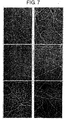
- Capillary density (capillary number/mm 2 ) was evaluated as neovascular formation at the capillary (less diameter of 10 ⁇ m) level. Density was higher in Group A-V (Group C: 169.9 ⁇ 8.5 and Group A-V: 273.2 ⁇ 25.8 a ; a p ⁇ 0.01 versus Group C) (Table 4). Representative photomicrographs of alkaline-phosphatase stained sections are shown in Fig. 7. Immunohistochemical staining for ⁇ -actin in tunica media revealed neovascular formation at the arteriole, which is produced in the late phase of angiogenesis and defined vessels having a diameter range between 10 and 50 ⁇ m.
- Arteriole densities (arteriole number/ field of view) in Group A+V and A-V were significantly higher than that of control (Group C: 4.8 ⁇ 0.5, Group A+V: 10.4 ⁇ 1.7 a , Group A-V: 12.4 ⁇ 1.4 a ; a p ⁇ 0.01), but the increases in Group V (6.1 ⁇ 0.8) and A (6.3 ⁇ 1.3) were of marginal significance (Table 4). Moreover, Group A-V showed a greater increase as compared with Group V ( b p ⁇ 0.01) and Group A ( c p ⁇ 0.01). Representative photomicrographs ⁇ -actin stained sections are also shown in Fig. 8.
- VEGF used widely in clinical studies, is of uncertain value for promotion of functional neovascularization, when VEGF is used alone.
- HIF-1 hypoxia-inducible factor-1
- circulating endothelial precursor cells caused by exogenous Ang1 gene may augment VEGF-induced angiogenesis.
- CEPs contribute to postnatal neovascularization (Asahara et al., 1999) and one recent study reports that the administration of the Ang1 gene prolongs the mobilization of CEPs compared with the administration of VEGF (Hattori et al., 2001).
- Overexpression of VEGF165 by adenovirus vector caused mobilization of CEPs to the peripheral blood in mice, peaking at day 2 and returning to control levels by day 14.
- Ang1 results in increased levels of CEPs, peaking from day 7 to 14 and returning, to control levels by day 28. Accordingly, the more effective angiogenesis produced by combination therapy of VEGF gene and Ang1 gene may occur in the pre-administration of Ang1 because each peaks of CEPs migration caused by administration of these two genes happen at the same phase.
- Yamashita et al. demonstrated that ECs and mural cells (pericytes and vascular smooth muscle cells) could be derived from common progenitor cells (Yamashita et al., 2000). They concluded that VEGF is necessary for the ECs differentiation, but differentiation of mural cells could occur independently of exogenous growth factors (Yamashita et al., 2000).
- pre-administration of Ang1 gene in the absence of exogenous VEGF until Day 15 could promote migration of circulating precursor cells (CPs) which are composed of CEPs and circulating mural precursor cells (CMPs).
- CPs circulating precursor cells
- CMPs circulating mural precursor cells
- the ratio of CMPs in CPs population might be higher compared with those by other administration schedules because the phase of VEGF administration (which increased CEPs) is later than other combinations, resulting in production of more mature vessels.
- pre-administration of the Ang1 gene followed by the VEGF gene may stimulate clear gene switching of Ang2, an important proangiogenic factor, and augment neovascular formation.
- Ang2 is necessary for adult angiogenesis and this factor plays an early role at the sites of neovascularization (Malsonpierre et al., 1997). Mandriota et al. also showed that Ang2 mRNA levels were increased by administration of VEGF and decreased by that of Ang1 (Mandriota et al., 1998). Taken together, these findings suggest that in the pre-administration of Ang1 gene schedule, clearer angiogenic switching to endogenous Ang2 could occur and expression of Ang2 could promote angiogenesis longer, compared to the other combination schedules. In the co-administration schedule, the expression of Ang2 would be less than the pre-administration schedules because VEGF and Ang1 have opposite effects on this gene expression. In the post-administration schedule, the period of expression of Ang2 would be shorter than in pre-administration, because of the administration of Ang1 at Day 15.
- the present inventors evaluated VEGF-induced edema using scrotum size.
- the change ratio of scrotum size was sensitive for the evaluation of edema because the scrotum in rabbits is isolated from the body, does not experience strong tissue support, and has abundant areas of connective tissue spaces, which facilitate the accumulation of extravasated fluid.
- the examples described above showed that only the pre-administration schedule prevented edema.
- Ang1 may promote endothelial cell firm-attachment to surrounding matrix and cells, resulting in prevention of vascular permeability caused by various stimuli (Thurston et al., 1999).
- Ang1 gene in three combination schedules of VEGF and Ang1 showed effective improvement in ischemic lesions.
- pre-administration of Ang1 gene contributed to significant increased blood pressure in the ischemic limb, improvement of blood flow in ischemic lesions, formation of mature vessels, and prevention of edema. Therefore, priming by Ang-1 gene administration may be beneficial for therapeutic angiogenesis in VEGF gene therapy.
- the present invention provides a method of administering angiopoietin prior to VEGF to obtain a high angiogenic induction effect without the adverse effects of edema. Therefore, the present invention enables gene therapy that prevents the increase in vascular permeability caused by administration of VEGF alone.
Abstract
Description
- The present invention relates to gene therapy that induces normal angiogenesis by expressing or administering angiopoietin prior to the expression or the administration of angiogenic genes.
- Recent research for treatment of ischemic diseases has been performed using growth factors that induce angiogenesis. For example, therapeutic effects of fibroblast growth factor 2 (EGF2) (Baffour, R. et al., J. Vasc. Surg. 16 (2): 181-91, 1992) and endothelial cell growth factor (ECGF) (Pu, L. Q. et al., J. Surg, Res. 54 (6): 575-83, 1993) on patients with cardiac infarction and acute limb ischemia have been examined. A recent study has revealed that vascular endothelial growth factor (VEGF)/vascular permeability factor (VPF) promotes vasculogenesis in animal models with myocardial ischemia and limb ischemia (Takeshita, S. et al., Circulation 90 (5 Pt 2): II228-34, 1994; and Takeshita, S. et al., J. Clin, Invest. 93 (2): 662-70, 1994).
- Vascular endothelial growth factor (VEGF) was reported as a vascular endothelial cell specific growth factor or as vascular permeability factor (VPF) in 1989, and currently is classified into VEGF A, B, C, D, and E. VEGF A is further divided into 6 different subtypes, and among the subtypes, soluble VEGF 121 and 165 show especially strong vascular growth activity and are now used clinically. It is reported that their effects extend to vasculogenesis during the embryonic period and are enhanced under a low oxygen environment. Moreover, they are involved in NO synthesis and the migration of vascular endothelial cells and vascular endothelial precursor cells. On the other hand, VEGF overexpression may break a balance of angiogenic signals and form "angioma-like" fragile capillary vessels (Carmeliet, P., Nature Med. 6, 1102-1103 (2000)). VEGF gene transfer to vessel walls in vivo may cause significant neointimal formation via angioma-like endothelial proliferation and induce extravasation of red blood cells (Yonemitsu, Y., et al., Lab. Invest. 75, 313-323(1996)). Similar pathological findings were demonstrated in retrovirus-mediated constitutive overexpression of VEGF in myocardium (Lee, R. J., et al., Circulation 102, 898-901 (2000)).
- Acute critical limb ischemia, which results from acute obstruction of the major arteries, is caused mainly by thrombotic obstruction and is an ischemic disease important as a target of angiogenic therapy. Delayed treatment of acute critical limb ischemia is quite unsuccessful, often resulting in limb amputation. Moreover, patients with limb amputation have such poor prognosis that one-year survival rates after surgery are only 50%. To solve this problem, techniques for regenerating blood vessels from embryonic stem cells (VEGF receptor Flk1-positive cells) have been investigated (Yamashita, J et al., Nature 408 (2): 92-96, 2000). Blood vessels are composed of endothelial cells and mural cells (pericytes and vascular smooth muscle cells), and a dynamic balance between angiogenesis and blood vessel regression is maintained in vivo. It remains unknown what molecular mechanisms control the complex changes such as sprouting, branching, fusion, and intussusception, and how endothelial cells timely achieve functions such as migration, detachment, and adhesion (Suda Toshio, Jikkenigaku (Experimental medicine), May issue, 19 (7): 826-829 (2001)).
- Moreover, clinical trials for human gene therapy using angiogenic growth factors have been undertaken, and studies have proceeded to clinically apply this therapy to angiogenesis for treatment of critical limb ischemia. Vascular endothelial growth factor/vascular permeability factor (VEGF/VPF), which are endothelial cell-specific growth factors, are considered as potent therapeutic genes for this purpose. Peripheral vascular diseases, representatives of which include arteriosclerosis obstruction and Burger's disease, show clinical symptoms such as intermittent claudication, resting pain, and tissue damage in the hind limbs (necrosis). Effective treatments are not currently available for patients with resting pain and ischemic ulcer caused by peripheral vascular obstruction. Hind limb amputation is not avoidable in cases where vascular dilation and recovery of blood circulation by surgery cannot be performed. Under these circumstances, a new therapy comprising the formation of collateral circulation by angiogenic factors-therapeutic angiogenesis has been proposed.
- Isner et al. have reported angiogenesis using VEGF gene and have demonstrated relatively promising results by means of plasmid-based gene transfer in human (Baumgartner, I., et al., Circulation 97, 1114-1123 (1998); Isner, J. M., et al., J. Vasc. Surg. 28, 964-973 (1998)). Isner et al. began two deferent gene therapy clinical trials: 1) VEGF gene transfer into vessel walls by catheter; and 2) VEGF gene transfer into muscles by injection. Especially, it has been reported that gene transfer using injection showed limb salvage rates of 70 to 80%. Moreover, neither intramuscular nor intravascular gene transfer has shown any toxicity due to immune reaction in clinical trials. More than 100 patients have undergone the treatment and have shown promising results. Related adverse effects and toxicity levels of intramuscular gene transfer of VEGF have been hardly reported at present. However, recent reports indicate that transgenic- (Thurston, G., et al., Science 286, 2511-2514 (1999)) or adenovirus- (Thurston, G., et al., Nature Med. 6, 460-463 (2000)) mediated overexpression of VEGF results in abnormal vasculogenesis in transgene-introduced animals, and that plasmid-based intramuscular VEGF gene transfer showed transient edema in human subjects with ischemic limbs (Baumgartner, I., et al., Circulation 97, 1114-1123 (1998); Isner, J. M., et al., J. Vasc. Surg. 28, 964-973 (1998) Baumgartner, I., et al., Ann Intern Med.; 132, 880-884, (2000)).
- No definitive and practical methods are known for ameliorating increased vascular permeability, a drawback in angiogenic therapy by application of VEGF alone.
- In order to prevent increased vascular permeability, a drawback of angiogenic therapy using administration of VEGF alone in gene therapies or drug treatments, the present invention provides a method of administering angiopoietin prior to VEGF administration, thereby providing results in highly effective angiogenic induction, without causing the adverse side effects of edema.
- Angiopoietin 1 (Ang-1), angiopoietin 2 (Ang-2) and its receptor, Tie-2, are known as regulating factors which specifically interact with vascular endothelial cells, like the VEGF family. Angiopoietins are ligands for the Tie-2 tyrosine kinase receptor, and considered involved in vascular maturation during angiogenesis. Four subtypes are known at present, and
subtypes 1 and 2 bind to Tie-2 receptor. Ang-2 is estimated to be an endogenous antagonist of Ang-1 (Saito, T. N; Nature, Vol. 376, 70-74, (1995)). Ang-2 is also known to be induced selectively depending on VEGF function (Oh, H., et al., J. Biol. Chem., 274, 15732-15739 (1999)). The differences between Ang-1 and VEGF are that Ang-1 cannot proliferate vascular endothelial cells, is involved in pericyte junctions, and, unlike VEGF, does not promote vascular permeability. It is known that interaction between endothelial cells and pericytes is important regulatory mechanism in angiogenesis. Pericytes adhere to endothelial cells to thereby function to differentiate endothelial cells and inhibit their proliferation. This mechanism considered to involve TGF-β activation and formation of physical barrier that surround the cells (D'Amore, et al., Proc. Natl. Acad. Sci. USA, 86: 4544-4548 (1989)). - The present inventors studied effects of angiopoietins and reported that pAng-1 improved AG score, blood flow, blood vessel diameter and capillary density when plasmid pAng-1 or pAng-2 was administered in rabbit models (Kou-Gi Shyu, et al., Circulation. 98, 2081-2087 (1998)). Based on this report, the present inventors hypothesized by themselves efficacy for neovascularization by pre-induction of an interaction between pericytes prior to the action of VEGF and conducted experiments to demonstrate the efficacy.
- On the other hand, Thurston et al. showed that adenovirus-mediated gene transfer of
angiopoietin 1 suppressed the increased permeability induced by VEGF using vascular permeability evaluation models with Evan Blue (Gavin Thurston, Nature Med, Vol. 6, No. 4, 460-463, (2000)). However, another report indicated that co-administration of pAng-1 and pVEGF plasmids to rabbit models showed no difference from administration of pVEGF plasmid alone (Chase, J. K., Arterio. Thromb. Vasc. Bio. 20, 2573-2578 (2000)). - The present inventors focused on the expression timing of both genes, and developed a local administration schedule of pre-administration of angiopoietin prior,to VEGF, which does not cause the adverse side effects of edema and remarkably induces angiogenesis.
- The present invention relates to a method of administering angiopoietin prior to VEGF to provide a high angiogenic induction effect without causing the adverse side effects of edema. More specifically, the present invention relates to:
- (1) a method of treatment by local administration, said method comprising administering an angiopoietin or a vector encoding an angiopoietin prior to administration of a vector encoding an angiogenic gene or a protein encoded by an angiogenic gene;
- (2) the method of (1), wherein said local administration is intramuscular administration;
- (3) the method of (1) or (2), wherein said angiogenic gene is VEGF121 or VEGF165; and
- (4) the method of any one of (1) to (3), wherein the angiopoietin is
angiopoietin 1. - The term "angiogenic gene" used herein indicates a gene that directly or indirectly associated with development, migration, proliferation, and maturation of cells involved in vascular formation.
- The present inventors researched extensively for a method of angiogenesis with low vascular permeability, which method comprises injecting Angiopoietin-1 intramuscularly into rabbit ischemic hind limb models, followed by VEGF 165 injection, using relatively safe plasmid vectors, and revealed its therapeutic effect. Target diseases include: 1) peripheral vascular diseases, arteriosclerosis obstruction, and such; 2) heart disorders, angina pectoris, myocardial infarction, and such; 3) nephrosis, nephritis, and such; and 4) pulmonary disorders, nerve disorders, and such, but there is no limitation to the diseases as long as angiogenic induction is effective for the diseases. Treatment targets do not have to be limited to ischemic tissues, but can be tissues that cannot be called ischemic tissues in the strict sense. Such tissues may be affected by diseases and symptoms that can be effectively treated by generation and reconstruction of functional blood vessels. For example, treatment targets include nephritis, diabetic nephrosis; obstinate ulcers; chronic or acute obstinate infectious diseases such as osteomyelitis; and moya-moya disease and other vascular diseases in the neurosurgery field. Any muscle including skeletal, smooth, and cardiac muscle can be used as administration sites. Administration sites are not limited to muscle, but include skin (epidermis and corium), arteries, veins (including the portal vein), lymph duct, kidney, chorion, periosteum, connective tissues, and bone marrow. Methods to substantially obtain therapeutic effects by local administration include direct administration of vectors or proteins, or administration using carriers and vehicles. Vehicles need to be physiologically acceptable and include organics such as biopolymers, inorganics such as hydroxyapatite, specifically, collagen matrix, polylactate polymer or its copolymer, polyethylene glycol polymer or its copolymer, and their chemical derivatives. Moreover, vehicles may be mixtures of these physiologically acceptable materials. Injection tools include industrial products such as conventional medical syringes or continuous injectors that can be kept inside or outside of the body. There is no limitation to vectors as long as the vectors are physiologically acceptable. Any vectors can be used, including adenovirus vectors, adenovirus-associated vectors, retrovirus vectors, lentivirus vectors, herpes simplex virus vectors, vacciniavirus vectors, Sendai virus vectors, and non-viral vector can be used. Vectors can be used in the form of patient-derived cells treated by the vectors.
-
- Fig. 1 shows the structure of plasmid pCAcc.
- Fig. 2 shows the structure of plasmid pCAhVEGF.
- Fig. 3 shows the structure of plasmid pCAhAng1.
- Fig. 4 shows a protocol for gene transfer into rabbit hind limb ischemic models.
- Fig. 5 is a set of photographs showing rabbit scrotum on the ischemic side. These photographs demonstrate that in Group V, scrotum edema and vain dilatation occurred at
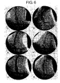
Day 15 post-gene administration, but pre-administration of Ang-1 prevented scrotum edema induced by VEGF. - Fig. 6 presents selective internal iliac angiographs at
Day 40 of six administration schedule groups: (A), Control; (B), Group V; (C) , Group A; (D) , Group A+V, (E) , Group V-A; and (F), Group A-V. - Fig. 7 is a set of representative photomicrographs showing numerous capillary ECs revealed by alkaline phosphatase stain in ischemic thigh muscle. (A), Control; (B), Group V; (C), Group A; (D), Group A+V; (E), Group V-A; and (F), Group A-V; Bar = 50 µm. ECs = endothelial cells.
- Fig. 8 is a set of representative photomicrographs showing arterioles covered with smooth muscle cells revealed by smooth muscle cell α-actin stain in ischemic thigh muscle. (A), Control; (B) , Group V; (C) , Group A; (D) , Group A+V; (E), Group V-A; and (F), Group A-V; Bar = 50 µm.
- Herein below, the present invention will be described in detail using examples; however, it is not to be construed as being limited thereto.
- Furthermore, the experiments in the examples were performed according to the following references.
- 1. Satoshi Takeshita, J. Clin. Invest., 1994, 93: 662-670
- 2. Yukio Tsurumi, Circulation., 1996, 94: 3281-3290 (Established rabbit hind limb model; demonstrated improvement of AG score, BPR, and capillary density by administration of pVEGF in the rabbit hind limb model; and assessed the above mentioned indexes plus improvement in blood flow.)
- 3. Iris Baumgartner, Ann. Intern. Med., 2000, 132: 880-884 (Actual assessment of edema in hind limbs of 90 patients who received pVEGF.)
- 4. Kou-Gi Shyu, Circulation., 1998, 98: 2081-2087 (Assessment of improvements of AG score, blood flow, vessel diameter, and capillary density by administration of pAng1, when pAng1 and pAng2 were administered to rabbit models.)
- 5. Gavin Thurston, Nature Med, 2000, 6 (4) : 460-463.
- 6. G. Thurston, Science, 1999, 286: 2511-2514 (Assessment of improvement in suppressed VEGF-induced vascular permeability in vascular permeability evaluation models that received adenovirus vector-mediated Ang1 transfer)
- 7. Lioubov Poliakova, J. Thorac. Cardiovasc. Surg., 1999, 118: 339-347. Time course assessment of changes in scrotum size and hind limb circumference in rabbit models which received adenovirus vector-mediated VEGF transfer )
- Data are shown as mean ± SEM in the examples of the present invention. Statistic comparison was performed by ANOVA followed by Bonferroni/Dunn test. p<0.05 is considered to be statistically significant.
-
- (1) A DNA encoding VEGF was obtained by RT-PCR using RNA extracted from human glioma U251 cells as a template and a set of primers: #1191: (CCGGAATTCACCATGAACTTTCTGCTGTCT/SEQ ID NO: 1) and #1192: (CGCGGATCCTCACCGCCTCGGCTTGTCACA/SEQ ID NO: 2). The amplified fragment was digested with EcoRI/BamHI and subcloned into pBluescriptSKII+ at EcoRI/BamHI sites. After confirming the nucleotide sequence of the plasmid thus obtained, the plasmid was digested with EcoRI/BamHI, and an EcoRI/BamHI fragment was subcloned into pCAcc (Fig. 1) at EcoRI/BglII sites to obtain pCAhVEGF (Fig. 2). The control vector pCAcc (Yoshida et al., 1997) was derived from the previously reported pCAGGS (Niwa et al., 1991).
- (2) A DNA encoding Angiopoietin 1 (GenBank: HSU83508 (U83508)) was obtained by RT-PCR using RNA extracted from human bone marrow cells (cells obtained by bone marrow aspiration and having ability to remain attached to dishes, so-called bone marrow stroma cells) as a template and a set of primers: #1435: (GAAGATCTATGACAGTTTTCCTTTCCTTTG/SEQ ID NO: 3) and #1436: (GAAGATCTCAAAAATCTAAAGGTCGAATCA/SEQ ID NO: 4). The amplified fragment was digested with BglII and subcloned into pBluescriptSKII+ at a BglII site to obtain the
plasmid carrying angiopoietin 1. After confirming the nucleotide sequence, the plasmid was digested with BglII and the isolated BglII fragment was subcloned into pCAcc (Fig. 1) at the BglII site to obtain pCAcchAng1a (Fig. 3). The nucleotide sequence of the cloned DNA fragment is shown in SEQ ID NO: 5. - Moreover, PCR was performed using Angiopoietin cDNA fragment of pCAcchAng1a as a template and primer sets #1641: (GGGAATTCACCATGACAGTTTTCCTTTCCTTTGCTTTC/SEQ ID NO: 6) and #1638: (AGCTCCTGGATTATATATGTTTGACG/SEQ ID NO: 7). The amplified fragment was digested with EcoRI and used to replace the EcoRI fragment of pCAcchAng1a to obtain a plasmid expressing human angiopoietin cDNA, pCAcchAng1, which contained a Kozak consensus sequence. The sequence of the cloned DNA fragment is shown in SEQ ID NO: 8.
- New Zealand white rabbits (male, 3.0 to 3.5 kg) were used. After rabbits were anesthetized with a mixture of 3 ml ketaral (Sankyo Co., Ltd.) and 1 ml xylazine (Daiichi Pharmaceutical Co., Ltd.), the femoral artery was exposed by skin incision between the left femoral inguinal ligament and the knee. All of the artery branches were ligated and excised, the femoral artery was removed, and the animals were kept for 10 days to establish a chronic hind limb ischemia model, that is, a rabbit hind limb ischemia model (Takeshita, S., J. Clin. Invest., 1994, 93: 662-670). A comparative assessment was conducted in three groups. Intramuscular injection of plasmids was performed on the 10th day (Day 10) or on the 10th day (Day 10) and 15th day (Day 15) after the establishment of the model (OPE day). Each plasmid (500 µg) was dissolved in 2.5 ml PBS and directly injected into the medial great muscle (2 sites), adductor muscle (2 sites), and semimembranous muscle (1 site) through the incision created in the medial thigh of the ischemic thigh. Each injection was performed slowly using a 27-guage needle at 5 sites, 500 µl each.
- Animal models were divided into six groups as follows according to administration schedule of plasmids (Fig. 4).
- 1. Control rabbits (Group C) received 500 µg of placebo (pCAcc) on Day 10 (n=8).
- 2. VEGF alone (Group V) received 500 µg of VEGF on Day 10 (n=10).
- 3. Ang1 alone (Group A) received 500 µg of Ang1 on Day 10 (n=7).
- 4. Group A+V received both 500 µg of Ang1 and 500 µg VEGF on Day 10 (n=7).
- 5. Group A-V received 500 µg of Ang1 on
Day 10, followed by administration of 500 µg VEGF on Day 15 (n=10). - 6. Group V-A received 500 µg of VEGF,on
Day 10, followed by administration of 500 µg of Ang-1 on Day 15 (n=7). - Assessment of edema, the main adverse side effect caused by VEGF, was performed based on measured scrotum size and hind limb circumference. Photographs of the scrotum at the affected site at
Day Day 10 as 100%. The circumference of the hind limb below the knee was also measured atDay - Representative findings of edema by administration of VEGF alone are shown in Fig. 5 and revealed an increased area of the scrotum accompanying venous dilatation. In all groups treated with plasmid DNA, scrotum edema was not followed by inflammatory changes such as necrosis. Average scrotum size before the intramuscular injection of plasmid DNA on
Day 10 was 7.14 ± 1.06 cm2 and percent change of scrotum size compared to that ofDay 10 is shown in Table 1.Table 1 Scrotum size (%) Day 10Day 15Day 20Day 40Control 100 101.6±2.7 101.5 ± 4.1 103.3±2.8 Group V 100 140.1±11.1b 111.2 ± 5.8 107.7±1.8 Group A 100 94.6±1.5 98.4 ± 3.3 98.0±5.5 Group A+V 100 115.5±6.1 107.1 ± 4.0 97.6±5.1 Group V-A 100 117.9±4.4a 110.8 ± 7.5 118.4±8.6 Group A-V 100 98.4±2.0 94.5 ± 3.7 94.4±5.1 ap<0.05 versus control group bp<0.01 versus control group - On
Day 15, there was a statistically significant increase in edema in Group V (140.1±11.1%, bp<0.01) and V-A (117.9±4.4%, ap<0.05), compared with Group C (101.6±2.7%). In Group V and V-A, the size of edema continued to increase onDay - Internal iliac artery angiography of affected sites stained by alkaline phosphatase was performed at
Day - On
Day 10, there was no difference in AG scores among Groups C, V, A, A+V, A-V, and V-A (0.08±0.02, 0.11±0.02, 0.07±0.04, 0.08±0.03, 0.07±0.02, and 0.08±0.02, respectively). OnDay 40, however, AG scores of Group V, A, and A-V were higher than that of control (Group C: 0.29±0.05, Group V: 0.64±0.05a, Group A: 0.52±0.03a, and Group A-V: 0.58±0.02a ; ap<0.01) (Table 2). No significant differences were observed among the five test groups. Representative angiographic findings, shown in Fig. 6, indicate the differences in neovascular formation in the medial thigh in terms of number and internal luminal diameter among the six groups. In fact, the internal diameter of the proximal side of the caudal gluteal artery showed that Group A-V had larger vessels than control (Group C: 0.77±0.02 mm and Group A-V: 1.01±0.03 mma; ap<0.01) (Table 2). Moreover, Group A-V demonstrated increased vessel cavity (p<0.01) in comparison with Group V (0.76±0.05 mm), suggesting that the pre-administration of angiopoietin-1 contributed to produce larger vessels. There was also a tendency for the formation of more numerous vessels in the Group V and larger internal diameter vessels of the midthigh zone were formed in the Group A-V.Table 2 AG score Arterial diameter (mm) Control 0.29 ± 0.05 0.77 ± 0.02 Group V 0.64 ± 0.05a 0.76 ± 0.05 Group A 0.52 ± 0.03a 0.85 ± 0.08 Group A+V 0.49 ± 0.06 0.90 ± 0.05 Group V-A 0.50 ± 0.07 0.90 ± 0.04 Group A-V 0.58 ± 0.02a 1.01 ± 0.03a, b AG score = Angiographic score
ap<0.01 versus control groupbp<0.01 versus Group V - Measurement was taken on
Day 10 andDay 40. Doppler was used to find posterior carotid arteries and measure the blood pressure. Blood Pressure Ratio (BPR) was calculated by the blood pressure of the ischemic site/the blood pressure of the non-ischemic site and used as an indicator of body circulation. - Improvement of hemodynamic state was assessed by selective angiography, calf blood pressure (CBP) and resting blood flow (RBF) on
Day Day 10 andDay 40 using a Doppler flowmeter (Datascope, Montvale, NJ) and the cuff, which was connected to a pressure manometer. The CBP ratio was defined for each model as the ratio of the ischemic/normal limb systolic CBP. - Before intramuscular injection of plasmid DNA (Day 10), there was no difference in CBP ratio among all groups, showing that severe ischemia occurred in the operative side of the hind limb. On
Day 40, however, CBP ratio increased significantly more in Group A-V (86.0±8.1%, ap<0.01) than in Group C (51.7±6.9%) (Table 3). - Assessment concerning the improvement of blood perfusion in local tissues was also performed. Specifically, resting blood flow (RBF) of the ischemic limb was measured by attaching a percutaneous probe (P-430, LASERFLO BPM2, Vasamedics, St. Paul, Minnesota) to the adductor, medial large and semi-membranous muscles on
Day 10 andDay 40. RBF of the ischemic limb was defined as the average peak velocity of four points in the above areas, and the RBF ratio was calculated by dividing RBF onDay 40 by RBF onDay 10. The RBF of the operative limb onDay 10 was similar among six groups. The changes in flow reserve onDay 40 are shown in Table 3. The calculated ratio of RBF onDay 40 to that onDay 10 indicated that there was significant improvement in Group A-V (234.8±12.5%, ap<0.01) compared with Group C (139.6±10.4%), although a greater degree of blood perfusion was not observed in the other groups.Table 3 CBP ratio (%) Regional RBF ratio (%) Control 51.7 ± 6.9 139.6 ± 10.4 Group V 68.4 ± 5.6 187.6 ± 24.5 Group A 67.6 ± 5.7 207.8 ± 20.5 Group A+V 82.7 ± 8.8 193.3 ± 26.6 Group V-A 72.4 ± 13.8 169.0 ± 24.2 Group A-V 86.0 ± 8.1a 234.8 ± 12.5a CBP ratio indicates the calf blood pressure ratio and regional RBF ratio indicates regional resting blood flow ratio. aP <0.01 versus control group - Measurements were taken on
Day 10 andDay 40 using a tissue blood flow meter. Tissue blood flow was measured at the ischemic medial great muscle (2 sites) and adductor muscle (2 sites), and total values were assessed by taking the value onDay 10 as 100% (BPM2). BPR and BPM2 showed significant improvement in the Ang-1 pre-administrated group compared with the control group. - The serum concentration of VEGF in the serum was measured with enzyme-linked immunosorbent assay (ELISA) Human VEGF immunoassay kit (Techne Corp., Minneapolis, MN) on
Day Day 15. - On
Day 40, two block-samples of adductor muscle were removed from ischemic limbs for histological analysis. One sample was embedded in OCT compound followed by snap-freezing in liquid nitrogen for alkaline phosphatase stain to determine capillary density. Another was embedded in paraffin after immersion-fixation in formalin for 48 hours for smooth muscle cell α-actin stains to determine arteriole density. Multiple frozen sections were cut (10 µm thickness) on a cryostat and stained for alkaline phosphatase using an indoxyl-tetrazolium method to detect capillary endothelial cells as previously described (Ziada et al., 1984). Capillary density was defined as a mean number of capillaries per 1 mm2. For immunohistochemistry, an anti α-smooth muscle actin antibody (1A4, Dako Japan Co., Ltd., Tokyo, Japan) was used as a first antibody and detected by HISTFINE SAB-PO Kit (Nichirei Corp., Tokyo, Japan) with a biotinylated anti-mouse serum and peroxidase-conjugated streptavidin. To identify arterioles and differentiate them from capillaries or venules, fifteen different microscopic fields on three different sections were selected and the positive smooth muscle cells were counted in the same manner. Arteriole density was expressed as a mean number of arterioles per field of view. - Capillary density (capillary number/mm2) was evaluated as neovascular formation at the capillary (less diameter of 10 µm) level. Density was higher in Group A-V (Group C: 169.9±8.5 and Group A-V: 273.2±25.8a; ap<0.01 versus Group C) (Table 4). Representative photomicrographs of alkaline-phosphatase stained sections are shown in Fig. 7. Immunohistochemical staining for α-actin in tunica media revealed neovascular formation at the arteriole, which is produced in the late phase of angiogenesis and defined vessels having a diameter range between 10 and 50 µm. Arteriole densities (arteriole number/ field of view) in Group A+V and A-V were significantly higher than that of control (Group C: 4.8±0.5, Group A+V: 10.4±1.7a, Group A-V: 12.4±1.4a; ap<0.01), but the increases in Group V (6.1±0.8) and A (6.3±1.3) were of marginal significance (Table 4). Moreover, Group A-V showed a greater increase as compared with Group V (bp<0.01) and Group A (cp<0.01). Representative photomicrographs α-actin stained sections are also shown in Fig. 8. These quantitative analyses using alkaline-phosphatase stain and smooth muscle cell α-actin stain indicated that the combination of cytokines, especially pre-administration of angiopoietin-1, produced more functional vascular formation than "solo" administration schedules.
Table 4 Capillary density
(Capillary No./mm 2 )Arterial density
(Arteriole No./field)Control 169.9 ± 8.5 4.8 ± 0.5 Group V 228.0 ± 3.2 6.1 ± 0.8 Group A 237.2 ± 16.6 6.3 ± 1.3 GroupA+V 242.7 ± 22.8 10.4 ± 1.7a Group V-A 234.6 ± 13.1 8.6 ± 0.9 Group A-V 273.2 ± 25.8a 12.4 ± 1.4a, b, c ap<0.01 versus control group bp<0.01 versus Group V cp<0.01 versus Group A - In therapeutic angiogenesis, production of functional vascular vessels is necessary for the supply of sufficient blood flow in ischemic lesions. However, some studies indicate that VEGF, used widely in clinical studies, is of uncertain value for promotion of functional neovascularization, when VEGF is used alone.
- There are two possible reasons for this consideration. First, stenosis or occlusion occurs in a main artery and brings about peripheral ischemia, leading to the expression and activation of the transcription factor, hypoxia-inducible factor-1 (HIF-1). The expression of HIF-1 leads to an increase of the transcription of many angiogenic genes including those encoding nitric oxide synthase and VEGF, and the cascade of angiogenesis progresses (Royen et al., 2001). It is uncertain whether the administration of VEGF gene product alone accomplishes functional angiogenesis. Recent study states that VEGF may stimulate capillary sprouting, but this response does not translate into a significant improvement in collateral flow (Hershey et al., 2001). Furthermore, a study, that examined the improvement of ischemic lesions in rabbit hind limb ischemia by administration of HIF-1α/VP16, showed more effective improvement when compared to the administration of VEGF gene alone (Vincent et al., 2000). Second, although a number of clinical trials using the VEGF gene to treat ischemic diseases have been evaluated and have reported favorable results, some studies have not shown significant improvement (Rajagopalan et al., 2001). In fact, the examples described above showed that administration of VEGF alone induced an increase of capillary number, but did not improve hemodynamic status, and did not mediate vascular maturation. As a result of these accumulated studies, further understanding of combination growth factor therapy or master switch genes is needed if this treatment is to produce clinically beneficial angiogenesis or arteriogenesis (Blau et al., 2001; Carmeliet., 2000; Simons et al., 2000).
- The above examples showed that pre-treatment with Ang-1 was more effective in improving hemodynamic status, vascular maturation in late phase angiogenesis, and vascular protection against plasma leakage than administration of Ang-1 or VEGF alone or other schedules of treatment. The data suggest that stimulation by pre-administration of Ang1 promotes angiogenesis in both the early and late phases, contributing to vascular maturation, which may be responsible for improvement of hemodynamic status and vascular protection.
- There are two possible mechanisms by which pre-administration of Ang1 might contribute to the promotion of angiogenesis in the early and late phases in this model. First, circulating endothelial precursor cells (CEPs) caused by exogenous Ang1 gene may augment VEGF-induced angiogenesis. CEPs contribute to postnatal neovascularization (Asahara et al., 1999) and one recent study reports that the administration of the Ang1 gene prolongs the mobilization of CEPs compared with the administration of VEGF (Hattori et al., 2001). Overexpression of VEGF165 by adenovirus vector caused mobilization of CEPs to the peripheral blood in mice, peaking at day 2 and returning to control levels by day 14. In contrast, overexpression of Ang1 resulted in increased levels of CEPs, peaking from day 7 to 14 and returning, to control levels by day 28. Accordingly, the more effective angiogenesis produced by combination therapy of VEGF gene and Ang1 gene may occur in the pre-administration of Ang1 because each peaks of CEPs migration caused by administration of these two genes happen at the same phase. In addition, Yamashita et al. demonstrated that ECs and mural cells (pericytes and vascular smooth muscle cells) could be derived from common progenitor cells (Yamashita et al., 2000). They concluded that VEGF is necessary for the ECs differentiation, but differentiation of mural cells could occur independently of exogenous growth factors (Yamashita et al., 2000). Thus, pre-administration of Ang1 gene in the absence of exogenous VEGF until
Day 15 could promote migration of circulating precursor cells (CPs) which are composed of CEPs and circulating mural precursor cells (CMPs). In the pre-administration of Ang1 schedule, the ratio of CMPs in CPs population might be higher compared with those by other administration schedules because the phase of VEGF administration (which increased CEPs) is later than other combinations, resulting in production of more mature vessels. Second, pre-administration of the Ang1 gene followed by the VEGF gene may stimulate clear gene switching of Ang2, an important proangiogenic factor, and augment neovascular formation. The expression of Ang2 is necessary for adult angiogenesis and this factor plays an early role at the sites of neovascularization (Malsonpierre et al., 1997). Mandriota et al. also showed that Ang2 mRNA levels were increased by administration of VEGF and decreased by that of Ang1 (Mandriota et al., 1998). Taken together, these findings suggest that in the pre-administration of Ang1 gene schedule, clearer angiogenic switching to endogenous Ang2 could occur and expression of Ang2 could promote angiogenesis longer, compared to the other combination schedules. In the co-administration schedule, the expression of Ang2 would be less than the pre-administration schedules because VEGF and Ang1 have opposite effects on this gene expression. In the post-administration schedule, the period of expression of Ang2 would be shorter than in pre-administration, because of the administration of Ang1 atDay 15. - The present inventors evaluated VEGF-induced edema using scrotum size. The change ratio of scrotum size was sensitive for the evaluation of edema because the scrotum in rabbits is isolated from the body, does not experience strong tissue support, and has abundant areas of connective tissue spaces, which facilitate the accumulation of extravasated fluid. The examples described above showed that only the pre-administration schedule prevented edema. Ang1 may promote endothelial cell firm-attachment to surrounding matrix and cells, resulting in prevention of vascular permeability caused by various stimuli (Thurston et al., 1999). The molecular mechanisms by which Ang1 induces attachment remain to be elucidated, but a recent study demonstrated that Ang1 could directly support cell adhesion mediated by integrins (Carlson et al., 2001). Thurston et al. reported that the resistance to vascular leakage occurred within 48 hours in gene transfer of Ang1 to rats by an adenovirus vector and was verified 7 days later with injection of VEGF protein in same model (Thurston et al., 2000). Actually, the examples revealed that the pre-administration schedule blocked vascular permeability induced by VEGF. In contrast, edema occurred in other treatment schedules, because the resistance to permeability induced by Ang1 did not have time to develop and prevent the vascular permeability caused by VEGF.
- In summary, only the pre-administration schedule of Ang1 gene in three combination schedules of VEGF and Ang1 showed effective improvement in ischemic lesions. In the examples, pre-administration of Ang1 gene contributed to significant increased blood pressure in the ischemic limb, improvement of blood flow in ischemic lesions, formation of mature vessels, and prevention of edema. Therefore, priming by Ang-1 gene administration may be beneficial for therapeutic angiogenesis in VEGF gene therapy.
- The present invention provides a method of administering angiopoietin prior to VEGF to obtain a high angiogenic induction effect without the adverse effects of edema. Therefore, the present invention enables gene therapy that prevents the increase in vascular permeability caused by administration of VEGF alone.


Claims (4)
- A method of treatment by local administration, said method comprising administering an angiopoietin or a vector encoding an angiopoietin prior to administration of a vector encoding an angiogenic gene or a protein encoded by an angiogenic gene.
- The method of claim 1, wherein said local administration is intramuscular administration.
- The method of claim 1 or 2, wherein said angiogenic gene is VEGF121 or VEGF165.
- The method of any one of claims 1 to 3, wherein the angiopoietin is angiopoietin 1.
Applications Claiming Priority (3)
| Application Number | Priority Date | Filing Date | Title |
|---|---|---|---|
| JP2001174919 | 2001-06-08 | ||
| JP2001174919 | 2001-06-08 | ||
| PCT/JP2002/005485 WO2002100441A2 (en) | 2001-06-08 | 2002-06-04 | Method for treatment of vascular regeneration |
Publications (2)
| Publication Number | Publication Date |
|---|---|
| EP1402902A1 true EP1402902A1 (en) | 2004-03-31 |
| EP1402902A4 EP1402902A4 (en) | 2006-02-01 |
Family
ID=19016134
Family Applications (1)
| Application Number | Title | Priority Date | Filing Date |
|---|---|---|---|
| EP02730900A Withdrawn EP1402902A4 (en) | 2001-06-08 | 2002-06-04 | Method for treatment of vascular regeneration |
Country Status (7)
| Country | Link |
|---|---|
| US (1) | US20040234502A1 (en) |
| EP (1) | EP1402902A4 (en) |
| JP (1) | JPWO2002100441A1 (en) |
| KR (1) | KR20040004681A (en) |
| CN (1) | CN1514737A (en) |
| CA (1) | CA2449687A1 (en) |
| WO (1) | WO2002100441A2 (en) |
Families Citing this family (4)
| Publication number | Priority date | Publication date | Assignee | Title |
|---|---|---|---|---|
| CA2516486A1 (en) * | 2003-02-19 | 2004-09-02 | Dnavec Research Inc. | Method of treating ischemic disease |
| WO2005097988A1 (en) * | 2004-03-23 | 2005-10-20 | Dnavec Research Inc. | Bone marrow-relevant cell participating the maintenance and/or repair of tissue |
| US20090204189A1 (en) * | 2006-04-05 | 2009-08-13 | Tuchiyagomu Co., Ltd. | Living-Tissue Normalization Method |
| KR101293483B1 (en) * | 2011-10-28 | 2013-08-08 | 가톨릭대학교 산학협력단 | Composition for preventing or treating muscle atrophy or sarcopenia comprising angiopoietin 1 |
Family Cites Families (3)
| Publication number | Priority date | Publication date | Assignee | Title |
|---|---|---|---|---|
| US6020473A (en) * | 1995-08-25 | 2000-02-01 | Genentech, Inc. | Nucleic acids encoding variants of vascular endothelial cell growth factor |
| US5941868A (en) * | 1995-12-22 | 1999-08-24 | Localmed, Inc. | Localized intravascular delivery of growth factors for promotion of angiogenesis |
| US6199554B1 (en) * | 1998-03-27 | 2001-03-13 | The Brigham And Women's Hospital, Inc. | Method and apparatus for combining injury-mediated therapy and drug delivery |
-
2002
- 2002-06-04 US US10/479,566 patent/US20040234502A1/en not_active Abandoned
- 2002-06-04 EP EP02730900A patent/EP1402902A4/en not_active Withdrawn
- 2002-06-04 CA CA002449687A patent/CA2449687A1/en not_active Abandoned
- 2002-06-04 JP JP2003503260A patent/JPWO2002100441A1/en active Pending
- 2002-06-04 KR KR10-2003-7015783A patent/KR20040004681A/en not_active Application Discontinuation
- 2002-06-04 WO PCT/JP2002/005485 patent/WO2002100441A2/en not_active Application Discontinuation
- 2002-06-04 CN CNA02811549XA patent/CN1514737A/en active Pending
Non-Patent Citations (8)
| Title |
|---|
| CHAE J K ET AL: "Coadministration of angiopoietin-1 and vascular endothelial growth factor enhances collateral vascularization." ARTERIOSCLEROSIS, THROMBOSIS, AND VASCULAR BIOLOGY. DEC 2000, vol. 20, no. 12, December 2000 (2000-12), pages 2573-2578, XP002357066 ISSN: 1524-4636 * |
| JONES MICHAEL K ET AL: "Gene therapy for gastric ulcers with single local injection of naked DNA encoding VEGF and angiopoietin-1" GASTROENTEROLOGY, vol. 121, no. 5, November 2001 (2001-11), pages 1040-1047, XP002357067 ISSN: 0016-5085 * |
| KUTRYK M J B ET AL: "Angiogenic gene therapy in a porcine model of chronic myocardial ischaemia: A comparison of angiopoietin-1 and VEGF" EUROPEAN HEART JOURNAL, vol. 22, no. Abstract Supplement, September 2001 (2001-09), page 624, XP008055996 & XXIII CONGRESS OF THE EUROPEAN SOCIETY OF CARDIOLOGY TOGETHER WITH THE 36TH ANNUAL GENERAL MEETING O; STOCKHOLM, SWEDEN; SEPTEMBER 01-05, 2001 ISSN: 0195-668X * |
| KUTRYK MICHAEL J B ET AL: "Angiogenic gene therapy in a porcine model of chronic myocardial ischemia: A comparison of angiopoietin-1 and VEGF" CIRCULATION, vol. 104, no. 17 Supplement, 23 October 2001 (2001-10-23), page II.262, XP008055998 & SCIENTIFIC SESSIONS 2001 OF THE AMERICAN HEART ASSOCIATION; ANAHEIM, CALIFORNIA, USA; NOVEMBER 11-14, 2001 ISSN: 0009-7322 * |
| SANDHU R ET AL: "The effects of angiogenic gene therapy on coronary collateral formation in a porcine model of chronic myocardial ischemia: A randomized, blinded, placebo-controlled study" CANADIAN JOURNAL OF CARDIOLOGY, vol. 16, no. Supplement F, September 2000 (2000-09), pages 181F-182F, XP008055995 & 53RD ANNUAL MEETING OF THE CANADIAN CARDIOVASCULAR SOCIETY; VANCOUVER, BRITISH COLUMBIA, CANADA; OCTOBER 20-NOVEMBER 01, 2000 ISSN: 0828-282X * |
| THURSTON G ET AL: "Inhibition of VEGF-induced plasma leakage by systemic viral production of Angiopoietin-1" FASEB JOURNAL, vol. 14, no. 4, 15 March 2000 (2000-03-15), page A34, XP008056039 & ANNUAL MEETING OF PROFESSIONAL RESEARCH SCIENTISTS: EXPERIMENTAL BIOLOGY 2000; SAN DIEGO, CALIFORNIA, USA; APRIL 15-18, 2000 ISSN: 0892-6638 * |
| THURSTON G ET AL: "Leakage-resistant blood vessels in mice transgenically overexpressing angiopoietin-1" SCIENCE (WASHINGTON D C), vol. 286, no. 5449, 24 December 1999 (1999-12-24), pages 2511-2514, XP002357065 ISSN: 0036-8075 * |
| THURSTON GAVIN ET AL: "Angiopoietin-1 protects the adult vasculature against plasma leakage" NATURE MEDICINE, vol. 6, no. 4, April 2000 (2000-04), pages 460-463, XP002357064 ISSN: 1078-8956 * |
Also Published As
| Publication number | Publication date |
|---|---|
| JPWO2002100441A1 (en) | 2004-09-24 |
| EP1402902A4 (en) | 2006-02-01 |
| KR20040004681A (en) | 2004-01-13 |
| US20040234502A1 (en) | 2004-11-25 |
| CN1514737A (en) | 2004-07-21 |
| CA2449687A1 (en) | 2002-12-19 |
| WO2002100441A2 (en) | 2002-12-19 |
Similar Documents
| Publication | Publication Date | Title |
|---|---|---|
| Benigni et al. | Unselective inhibition of endothelin receptors reduces renal dysfunction in experimental diabetes. | |
| US7514401B2 (en) | Methods of use of fibroblast growth factor, vascular endothelial growth factor and related proteins in the treatment of acute and chronic heart disease | |
| Koneru et al. | Sildenafil‐mediated neovascularization and protection against myocardial ischaemia reperfusion injury in rats: role of VEGF/angiopoietin‐1 | |
| Yamauchi et al. | Pre‐administration of angiopoietin‐1 followed by VEGF induces functional and mature vascular formation in a rabbit ischemic model | |
| AU2007207609B2 (en) | Methods of treating or preventing tissue damage caused by increased blood flow | |
| JP5400006B2 (en) | Preventive or therapeutic agent for ischemic disease | |
| WO2005009470A1 (en) | Remedy for cardiac failure comprising ask1 inhibitor as the active ingredient and method of screening the same | |
| Sabbah et al. | Effects of angiotensin-neprilysin inhibition in canines with experimentally induced cardiorenal syndrome | |
| US7247618B2 (en) | Methods for inhibiting macrophage colony stimulating factor and c-FMS-dependent cell signaling | |
| Kusano et al. | Long-term stable expression of human growth hormone by rAAV promotes myocardial protection post-myocardial infarction | |
| Konturek et al. | Expression of transforming growth factor-beta1 and epidermal growth factor in caerulein-induced pancreatitis in rat | |
| JP2003513932A (en) | Induction of angiogenesis | |
| EP1402902A1 (en) | Method for treatment of vascular regeneration | |
| Bush et al. | Regulation of new blood vessel growth into ischemic skeletal muscle | |
| US20040072805A1 (en) | Use of tumor necrosis factor inhibitors to treat cardiovascular disease | |
| WO2003093419A2 (en) | Preventing secondary lymphedema with vegf-d dna | |
| JP2005505510A (en) | Stimulation of angiogenesis by VEGF-B | |
| AU2009294362A1 (en) | Use of CTGF as a cardioprotectant | |
| CN116790744B (en) | Ischemic heart disease advanced fibrosis intervention target spot and application thereof | |
| Liang et al. | Inhibition of TGF-βRI alleviates endothelial-mesenchymal transition of mitral valve damage due to rheumatic heart disease | |
| Sarkar et al. | Angiogenesis, Its Importance, and Implication in Tumor Growth and Progression and in Anticancer Therapy: An Update | |
| CN116790744A (en) | Ischemic heart disease advanced fibrosis intervention target spot and application thereof | |
| EP1682172A2 (en) | Methods and compositions using adiponectin for treatment of cardiac disorders and for stimulation of angiogenesis | |
| Head | Articles in PresS. Am J Physiol Heart Circ Physiol (January 11, 2013). doi: 10.1152/ajpheart. 00587.2012 | |
| JP2002161049A (en) | Intimal thickening inhibitor |
Legal Events
| Date | Code | Title | Description |
|---|---|---|---|
| PUAI | Public reference made under article 153(3) epc to a published international application that has entered the european phase |
Free format text: ORIGINAL CODE: 0009012 |
|
| 17P | Request for examination filed |
Effective date: 20031230 |
|
| AK | Designated contracting states |
Kind code of ref document: A1 Designated state(s): AT BE CH CY DE DK ES FI FR GB GR IE IT LI LU MC NL PT SE TR |
|
| AX | Request for extension of the european patent |
Extension state: AL LT LV MK RO SI |
|
| REG | Reference to a national code |
Ref country code: HK Ref legal event code: DE Ref document number: 1064584 Country of ref document: HK |
|
| RIC1 | Information provided on ipc code assigned before grant |
Ipc: A61K 38/18 19950101ALI20051207BHEP Ipc: A61P 43/00 20000101ALI20051207BHEP Ipc: A61K 45/00 19850101ALI20051207BHEP Ipc: A61K 38/00 19950101ALI20051207BHEP Ipc: A61K 48/00 19900101AFI20030106BHEP |
|
| A4 | Supplementary search report drawn up and despatched |
Effective date: 20051216 |
|
| 17Q | First examination report despatched |
Effective date: 20070913 |
|
| RTI1 | Title (correction) |
Free format text: COMPOSITIONS FOR VASCULAR REGENERATION& X9; |
|
| STAA | Information on the status of an ep patent application or granted ep patent |
Free format text: STATUS: THE APPLICATION HAS BEEN WITHDRAWN |
|
| 18W | Application withdrawn |
Effective date: 20080610 |
|
| REG | Reference to a national code |
Ref country code: HK Ref legal event code: WD Ref document number: 1064584 Country of ref document: HK |