EP1394261A2 - Method for producing catalytic antibodies (variants), antigens for immunisation and nucleotide sequence - Google Patents

Method for producing catalytic antibodies (variants), antigens for immunisation and nucleotide sequence Download PDF

Info

Publication number
EP1394261A2
EP1394261A2 EP02739002A EP02739002A EP1394261A2 EP 1394261 A2 EP1394261 A2 EP 1394261A2 EP 02739002 A EP02739002 A EP 02739002A EP 02739002 A EP02739002 A EP 02739002A EP 1394261 A2 EP1394261 A2 EP 1394261A2
Authority
EP
European Patent Office
Prior art keywords
mice
protein
animals
antibodies
catalytic
Prior art date
Legal status (The legal status is an assumption and is not a legal conclusion. Google has not performed a legal analysis and makes no representation as to the accuracy of the status listed.)
Granted
Application number
EP02739002A
Other languages
German (de)
French (fr)
Other versions
EP1394261B1 (en
EP1394261A4 (en
Inventor
Alexandr Gabibovich Gabibov
Alexandr Vladimirovich Kolesnikov
Natalya Alexandrovna Ponomarenko
Elena Sergeevna Alexandrova
Ivan Ivanovich Vorobiev
Alexandr Viktorovich Demin
Current Assignee (The listed assignees may be inaccurate. Google has not performed a legal analysis and makes no representation or warranty as to the accuracy of the list.)
Lifebio Laboratories LLC
Original Assignee
ASGL - Farmatsevticjeskie Innovatsii Zaptryote Aktsionernoe Obschestvo
Priority date (The priority date is an assumption and is not a legal conclusion. Google has not performed a legal analysis and makes no representation as to the accuracy of the date listed.)
Filing date
Publication date
Application filed by ASGL - Farmatsevticjeskie Innovatsii Zaptryote Aktsionernoe Obschestvo filed Critical ASGL - Farmatsevticjeskie Innovatsii Zaptryote Aktsionernoe Obschestvo
Priority to EP10183175.8A priority Critical patent/EP2295082B1/en
Publication of EP1394261A2 publication Critical patent/EP1394261A2/en
Publication of EP1394261A4 publication Critical patent/EP1394261A4/en
Application granted granted Critical
Publication of EP1394261B1 publication Critical patent/EP1394261B1/en
Anticipated expiration legal-status Critical
Expired - Lifetime legal-status Critical Current

Links

Images

Classifications

    • CCHEMISTRY; METALLURGY
    • C07ORGANIC CHEMISTRY
    • C07KPEPTIDES
    • C07K14/00Peptides having more than 20 amino acids; Gastrins; Somatostatins; Melanotropins; Derivatives thereof
    • C07K14/005Peptides having more than 20 amino acids; Gastrins; Somatostatins; Melanotropins; Derivatives thereof from viruses
    • CCHEMISTRY; METALLURGY
    • C07ORGANIC CHEMISTRY
    • C07KPEPTIDES
    • C07K14/00Peptides having more than 20 amino acids; Gastrins; Somatostatins; Melanotropins; Derivatives thereof
    • C07K14/435Peptides having more than 20 amino acids; Gastrins; Somatostatins; Melanotropins; Derivatives thereof from animals; from humans
    • C07K14/46Peptides having more than 20 amino acids; Gastrins; Somatostatins; Melanotropins; Derivatives thereof from animals; from humans from vertebrates
    • C07K14/47Peptides having more than 20 amino acids; Gastrins; Somatostatins; Melanotropins; Derivatives thereof from animals; from humans from vertebrates from mammals
    • C07K14/4701Peptides having more than 20 amino acids; Gastrins; Somatostatins; Melanotropins; Derivatives thereof from animals; from humans from vertebrates from mammals not used
    • C07K14/4713Autoimmune diseases, e.g. Insulin-dependent diabetes mellitus, multiple sclerosis, rheumathoid arthritis, systemic lupus erythematosus; Autoantigens
    • CCHEMISTRY; METALLURGY
    • C07ORGANIC CHEMISTRY
    • C07KPEPTIDES
    • C07K16/00Immunoglobulins [IG], e.g. monoclonal or polyclonal antibodies
    • C07K16/08Immunoglobulins [IG], e.g. monoclonal or polyclonal antibodies against material from viruses
    • C07K16/10RNA viruses
    • C07K16/112Retroviridae (F), e.g. leukemia viruses
    • C07K16/114Lentivirus (G), e.g. human immunodeficiency virus [HIV], feline immunodeficiency virus [FIV] or simian immunodeficiency virus [SIV]
    • C07K16/1145Env proteins, e.g. gp41, gp110/120, gp160, V3, principal neutralising domain [PND] or CD4-binding site
    • CCHEMISTRY; METALLURGY
    • C12BIOCHEMISTRY; BEER; SPIRITS; WINE; VINEGAR; MICROBIOLOGY; ENZYMOLOGY; MUTATION OR GENETIC ENGINEERING
    • C12NMICROORGANISMS OR ENZYMES; COMPOSITIONS THEREOF; PROPAGATING, PRESERVING, OR MAINTAINING MICROORGANISMS; MUTATION OR GENETIC ENGINEERING; CULTURE MEDIA
    • C12N9/00Enzymes; Proenzymes; Compositions thereof; Processes for preparing, activating, inhibiting, separating or purifying enzymes
    • C12N9/0002Antibodies with enzymatic activity, e.g. abzymes
    • AHUMAN NECESSITIES
    • A01AGRICULTURE; FORESTRY; ANIMAL HUSBANDRY; HUNTING; TRAPPING; FISHING
    • A01KANIMAL HUSBANDRY; AVICULTURE; APICULTURE; PISCICULTURE; FISHING; REARING OR BREEDING ANIMALS, NOT OTHERWISE PROVIDED FOR; NEW BREEDS OF ANIMALS
    • A01K2207/00Modified animals
    • A01K2207/10Animals modified by protein administration, for non-therapeutic purpose
    • AHUMAN NECESSITIES
    • A01AGRICULTURE; FORESTRY; ANIMAL HUSBANDRY; HUNTING; TRAPPING; FISHING
    • A01KANIMAL HUSBANDRY; AVICULTURE; APICULTURE; PISCICULTURE; FISHING; REARING OR BREEDING ANIMALS, NOT OTHERWISE PROVIDED FOR; NEW BREEDS OF ANIMALS
    • A01K2227/00Animals characterised by species
    • A01K2227/10Mammal
    • A01K2227/105Murine
    • AHUMAN NECESSITIES
    • A01AGRICULTURE; FORESTRY; ANIMAL HUSBANDRY; HUNTING; TRAPPING; FISHING
    • A01KANIMAL HUSBANDRY; AVICULTURE; APICULTURE; PISCICULTURE; FISHING; REARING OR BREEDING ANIMALS, NOT OTHERWISE PROVIDED FOR; NEW BREEDS OF ANIMALS
    • A01K67/00Rearing or breeding animals, not otherwise provided for; New or modified breeds of animals
    • A01K67/027New or modified breeds of vertebrates
    • A01K67/0275Genetically modified vertebrates, e.g. transgenic
    • A01K67/0276Knock-out vertebrates
    • AHUMAN NECESSITIES
    • A61MEDICAL OR VETERINARY SCIENCE; HYGIENE
    • A61KPREPARATIONS FOR MEDICAL, DENTAL OR TOILETRY PURPOSES
    • A61K38/00Medicinal preparations containing peptides
    • CCHEMISTRY; METALLURGY
    • C07ORGANIC CHEMISTRY
    • C07KPEPTIDES
    • C07K2319/00Fusion polypeptide
    • CCHEMISTRY; METALLURGY
    • C07ORGANIC CHEMISTRY
    • C07KPEPTIDES
    • C07K2319/00Fusion polypeptide
    • C07K2319/20Fusion polypeptide containing a tag with affinity for a non-protein ligand
    • C07K2319/21Fusion polypeptide containing a tag with affinity for a non-protein ligand containing a His-tag
    • CCHEMISTRY; METALLURGY
    • C07ORGANIC CHEMISTRY
    • C07KPEPTIDES
    • C07K2319/00Fusion polypeptide
    • C07K2319/40Fusion polypeptide containing a tag for immunodetection, or an epitope for immunisation
    • C07K2319/41Fusion polypeptide containing a tag for immunodetection, or an epitope for immunisation containing a Myc-tag
    • CCHEMISTRY; METALLURGY
    • C12BIOCHEMISTRY; BEER; SPIRITS; WINE; VINEGAR; MICROBIOLOGY; ENZYMOLOGY; MUTATION OR GENETIC ENGINEERING
    • C12NMICROORGANISMS OR ENZYMES; COMPOSITIONS THEREOF; PROPAGATING, PRESERVING, OR MAINTAINING MICROORGANISMS; MUTATION OR GENETIC ENGINEERING; CULTURE MEDIA
    • C12N2740/00Reverse transcribing RNA viruses
    • C12N2740/00011Details
    • C12N2740/10011Retroviridae
    • C12N2740/16011Human Immunodeficiency Virus, HIV
    • C12N2740/16111Human Immunodeficiency Virus, HIV concerning HIV env
    • C12N2740/16122New viral proteins or individual genes, new structural or functional aspects of known viral proteins or genes

Definitions

  • the present invention relates to biotechnology, immunology, genetic engineering, the microbiological and medicinal industries and comprises a combined approach to the manufacture and expression of catalytically active antibodies which are potential therapeuticals intended to destroy protein antigens, in particular gp120, which is the main surface protein of human immunodeficiency virus.
  • the object of the present invention is to develop a method for producing catalytic antibodies against proteins and peptides, in particular gp120, with the use of animals with spontaneous and inducible autoimmune pathologies, which method will make it possible to design a "catalytic vaccine" which, upon injection to a patient, is capable not only of binding the antigen but also of destroying it thus inhibiting the development of disease.
  • the present invention provides a method for producing catalytic antibodies with the use of animals with spontaneous and induces autoimmune pathologies.
  • Mice are used as the animals with spontaneous and inducible autoimmune pathologies.
  • the used mice belong to strains for which immunization with myelin basic protein or its fragment can induce the development of experimental autoimmune encephalomyelitis.
  • the animals are administered with a fusion protein consisting of myelin basic protein or its fragments and a potential substrate of catalytic antibody or a fragment of the potential substrate.
  • the potential substrate is gp120 (surface glycoprotein of HIV-1) or its fragments.
  • the fusion protein used in the method of the present invention has the following structure:
  • a nucleotide sequence encoding the fusion protein of the above structure is proposed.
  • the present invention provides a method for producing catalytic antibody using animals with spontaneous and induces autoimmune pathologies.
  • the animal with spontaneous and inducible autoimmune pathologies are MRL-lpr/lpr mice, the mice being immunized with an antigen containing a haptene, the hapten being a conjugate of a mechanism-dependent covalent protease inhibitor with a peptide, the peptide being a fragment of a potential substrate of the catalytic antibody.
  • the potential substrate of the catalytic antibody is gp120 or its fragment.
  • hapten and its isomers and racemates used in the method of the present invention have the following structure:
  • Example 1 Development of a genetic construct containing a nucleotide sequence encoding the fusion protein gp120I-IIImbp for its expression in a prokaryotic system.
  • Example 2 Expression, isolation and purification of the fusion protein gp120I-III-mbp.
  • the fusion protein was expressed in T7-lysogenated E . coli cells (the strain BL21 (DE3) was used in the present example).
  • the protein was expressed as follows:
  • the fusion protein gp120I-III-mbp was isolated under denaturating conditions as follows:
  • Electrophoregram densitometry 1 .
  • Denaturing electrophoresis of proteins was carried out according to Laemmli using 6 M urea solution in the concentrating and fractionating gels. Gel staining was carried out with Cumassie blue R-250 using contrast enhancing with a cuprum salt. Densitometry was performed with a densitometer or computer assisted plate scanner, with subsequent electrophoregram digitalization and analysis (FIG. 4A).
  • Immunoblotting is carried out according to the standard regimen using blocking BSA solution.
  • the hybridization buffer is supplemented with bovine serum albumin (fraction V) to make 0.5% (FIG. 4 C).
  • test protein immunoreactivity is determined visually by the following criteria: the development of a single distinct well outlined staining zone whose electrophoretic mobility corresponds to that of the test protein. When these criteria are met, instrumental analysis is performed.
  • the densitometric evaluation of the intensity of the staining zone is performed by the Surface Plot method using Scion Image software. Test results are considered positive when the peak half-height is 5 times greater than the range of baseline fluctuations on the densitogram.
  • Samples are prepared for analysis as follows.
  • tested proteins having the sequence 6xHis will be immobilized by the metal chelate sorbent at pH 8.0.
  • mice are immunized with the fusion protein gp120 I-III-mbp as follows.
  • enzyme immunoassay (EIA) is used (FIG. 5).
  • antigen-antibody complex is detected with Fc-specific rabbit antimouse-IgG antibodies conjugated to horse radish peroxidase.
  • the sera of immunized and control mice are used at several dilutions (1:12 and 1:48).
  • the following recombinant proteins are used as antigens:
  • FIG. 5 it has been shown that all the antibody preparations obtained from the serum of mice immunized with a fusion protein interact with the antigen.
  • the antibodies are, as a preliminary, purified by affinity chromatography using recombinant protein G immobilized on Sepharose.
  • the activity is detected by two different methods (FIG. 6).
  • A. Fluorescent assay The principle of the method, which is outlined in FIG. 6A, is based on the phenomenon of fluorescence quenching by a protein heavily labeled with a fluorophore, which phenomenon is described in literature and is mainly based on the mutual interactions of the aromatic rings of different fluorophore molecules (e.g., because of intense hydrophobic and stacking contacts), and on fluorescence enhancement by introduction of breaks into the polypeptide chain.
  • bovine serum albumin and the recombinant protein trx-gp120 I-III-CH excessively labeled with fluorescein isothiocyanate (designated hereinafter as BSA-FITC and gp120-FITC ) were used as the substrates for the proteolysis.
  • BSA-FITC and gp120-FITC fluorescein isothiocyanate
  • the reaction was monitored by the fluorescence enhancement vs. control. Trypsin devoid of contaminating chymotrypsin activity was used as the model protease to determine the sensitivity of the method and to evaluate temporal signal changes depending on the substrate and enzyme amounts.
  • the principle of this method of detection of proteolytic activity is based on the use of small amounts (about 1 ng per reaction) of a highly active enzyme ribonuclease A as the substrate of the proteolysis reaction.
  • the level of ribonuclease activity which linearly depends on the concentration of active RNAase A, is determined by the acid-soluble residue method using polycytidyl acid as the polymeric substrate of the reaction.
  • This method of proteolytic activity detection presumably allows achieving a significant molar excess of enzyme vs. substrate and thus makes the conditions of the proteolysis reaction under study closer to those of the well-studied non-stationary kinetics model ([S] 0 ⁇ [E]).
  • the proteolytic activity of the tested antibody preparations was measured under the following conditions: IgG concentration 0.1 mg/ml, incubation time 17 h, temperature 37°C.
  • the preparation activity measured by this method was expressed as ribonuclease hydrolysis rate.
  • the above Example shows that, upon immunization of SJL mice with the fusion protein gp120I-IIImbp, antigen-specific proteolytic antibodies are generated against the background of the preclinical stage of induced experimental autoimmune encephalomyelitis.
  • aminoalkylphosphonates protected at their free amino group are synthesized in the reaction of co-condensation of triphenylphosphite, isobutanal, and benzylcarbamate.
  • the mixture of triphenylphosphite, isobutanal, and benzylcarbamate, 0.1 mole each, in 15 ml of glacial acetic acid is stirred for about 1 h until heat generation discontinues. After that, the reaction is stirred with heating to 80°C for 1 h. After the full completion of the reaction, volatile products are removed with a rotary evaporator under reduced pressure and heating on a water bath.
  • the protective group is removed by treatment of diphenyl 1-(N-benzylcarbonyl)-aminoalkylphosphonate with a 33% solution of hydrogen bromide in acetic acid (15 ml per 0.1 mole) for 1 h at room temperature. Volatile components are removed with a rotary evaporator at a reduced pressure and heating on a water bath.
  • 1-(N-benzyloxycarbonyl)-aminoakylphosphonate hydrobromide is crystallized from the resulting residue by addition of anhydrous diethyl ether.
  • Free phosphonate is obtained by passing gaseous dry ammonium through phosphonate hydrobromide suspension in diethyl ether until the formation of a thick precipitate of ammonium bromide discontinues and the full blooming of the suspension is observed. The resulting ammonium bromide is removed by filtration, and diethyl ether is evaporated on a water bath under atmospheric pressure.
  • the peptide Boc-Val-Ala-(t-Bu)Glu-(t-Bu)Glu-(t-Bu)Glu is fused with the phosphonate derivative of valine by mixing of 2 ⁇ moles of the protected peptide, 2 ⁇ moles of the phosphonate, and 2 ⁇ moles of dicyclohexylcarbodiimide in 300 ⁇ l of acetonitrile and incubating for 1 h. After the completion of the reaction, its products are separated by reverse phase HPLC on a 150 ⁇ 3.9-mm Waters C18 NovaPak column using 0% to 80% gradient of acetonitrile in 20 nM potassium phosphate (pH 7.0).
  • the resulting fractions are analyzed by mass-spectrometry (MALDI-TOF).
  • the fractions that contain substances with molecular ion masses of 1145 Da ([M+H] + ), 1167 Da ([M+Na] + ) or 1183 Da ([M+K] + ) are combined and freeze-dried.
  • the residue is dissolved in 100 ⁇ l of 100% trifluoroacetic acid and incubated for 1 h at room temperature to remove protective tert-butyloxycarbonyl and tert-butyl groups.
  • the deblocked peptidylphosphonate is precipitated by addition of 10 volumes of anhydrous diethyl ether to the reaction.
  • the precipitate is separated by centrifuging for 10 min at 12500 rpm, and the deblocking procedure is repeated.
  • the residue is air-dried and stored at -20°C.
  • Example 5 Reactive immunization of mice with the phosphonate derivative of a peptide fragment of gp120.
  • the reactive peptide is conjugated to the macromolecular carrier C .
  • conholepas hemocyanin keyhole limpet hemocyanin, KLH.
  • the carrier is activated with excess bis(sulfosuccinimidylyl)suberate in PBS for 1 h at 37°C.
  • KLH unbound to bis(sulfosuccinimidylyl)suberate is removed from the reaction by sevenfold exhaustive ultrafiltration (with the residual volume not more than 70 ⁇ l) using a Microcon 100 concentrator (Amicon YC membrane), each time adding PBS to the residue to make 500 ⁇ l and discarding the ultrafiltrate.
  • peptidylsulfonate solution in PBS is added whereupon the solution is incubated for 1 h at 37°C without stirring.
  • the unreacted succinimide groups are inactivated by addition of 2 ⁇ l of 2-ethanolamine.
  • the low molecular components of the reaction are removed by sevenfold exhaustive ultrafiltration (with the residual volume not more than 70 ⁇ l) using a Microcon 100 concentrator (Amicon YC membrane), each time adding PBS to the residue to make 500 ⁇ l and discarding the ultrafiltrate.
  • the final preparation is sterilized by filtration and stored at -20°C.
  • mice Female MRL-lpr / lpr , SJL and NZB / NZW F 1 mice aged 6-8 weeks are intraperitoneally immunized with the antigen in complete Freund adjuvant, with the total volume being 0.2 ml, at the dose of 50 ⁇ g of the immunogenic protein per mouse.
  • the second immunization is done with the same volume and at the same antigen concentration in incomplete Freund adjuvant in 17 days afeter the first immunization.
  • blood is withdrawn from the orbital sinus of three mice of each experimental group and control non-immunized mice of the three strains to monitor the development of the immune response.
  • mice that showed the maximal antigen-specific response in immunological tests are sacrificed for splenectomy.
  • Polyclonal antibodies isolated from the blood serum of these mice are analyzed for antigen specificity and catalytic activity.
  • a part of the peptidylphosphonate synthesized at the previous stages is used in the reaction of conjugation to N-hydroxysuccinimide ester of biotin.
  • the reaction is conducted by mixing of equimolar amounts of peptidyl sulfonate and activated biotin in a minimal volume of dimethylformamide and incubation for 1 h.
  • the biotinylated preparations are intended for analysis of the specificity of the antibodies obtained as a result of reactive immunization of mice.
  • enzyme immunoassay is used.
  • Antibodies from the sera of immunized and control mice are isolated with plate-preabsorbed goat antibodies against murine IgG, with subsequent incubation with the biotinylated antigen and detection of antigen-antibody complexes using neutravidin conjugated to horse radish peroxidase.
  • the sera of immunized and control mice are used at several dilutions (1:12 and 1:48).
  • the antigens employed are biotin-labeled starting peptidylphosphonate , biotinylated Val -phosphonate , and nitrophenylmethyl-p-biotinylphenylmethylphosphonate , for which the specific covalent modification of the active center of abzymes was demonstrated earlier.
  • the comparative analysis has shown (FIG.
  • horse radish peroxidase-conjugated rabbit antibodies against the Fc fragment of murine IgG are used to determine the total amount of murine antibodies specifically absorbed in a plate well. This allows estimation of the proportion of antigen-specific antibodies in the total pool of class G immunoglobulins.
  • the invention may be useful in medicinal industry for manufacturing drugs.

Landscapes

  • Chemical & Material Sciences (AREA)
  • Health & Medical Sciences (AREA)
  • Life Sciences & Earth Sciences (AREA)
  • Organic Chemistry (AREA)
  • Genetics & Genomics (AREA)
  • Molecular Biology (AREA)
  • General Health & Medical Sciences (AREA)
  • Biochemistry (AREA)
  • Medicinal Chemistry (AREA)
  • Biophysics (AREA)
  • Immunology (AREA)
  • Proteomics, Peptides & Aminoacids (AREA)
  • Virology (AREA)
  • Zoology (AREA)
  • Wood Science & Technology (AREA)
  • Engineering & Computer Science (AREA)
  • Bioinformatics & Cheminformatics (AREA)
  • Gastroenterology & Hepatology (AREA)
  • Biotechnology (AREA)
  • Rheumatology (AREA)
  • General Engineering & Computer Science (AREA)
  • Biomedical Technology (AREA)
  • Diabetes (AREA)
  • Hematology (AREA)
  • Rehabilitation Therapy (AREA)
  • Microbiology (AREA)
  • Toxicology (AREA)
  • Peptides Or Proteins (AREA)
  • Preparation Of Compounds By Using Micro-Organisms (AREA)
  • Medicines Containing Antibodies Or Antigens For Use As Internal Diagnostic Agents (AREA)
  • Enzymes And Modification Thereof (AREA)
  • Medicines That Contain Protein Lipid Enzymes And Other Medicines (AREA)

Abstract

The invention relates to biotechnology, immunology, gene engineering, and the microbiological and medical industries. The aim of the invention is to develop a method for producing catalytic antibodies to proteins and pcptidcs, in particular to gp120, using animals having spontaneous and induced autoimmune pathologies. Various methods for producing catalytic antibodies are also disclosed. The inventive methods make it possible to create a catalytic vaccine which can when injected to a patient to exhibits adhesive properties in relation to antigen simultaneously with a destructive function, there by suspending the progression of disease. Said invention discloses the inventive method for the autoimmunisation of animal lines SJL by fused proteins containing classical peptide epitope which develops pathology of an animal by protein fragments gp120 accompanied with an interest target catalytic antibody. The invention also comprises the method for immunising autoimmune animals by highly reactive chemical compositions which can perform a covalent selection of catalytic clones containing peptide fragments of potential resected portions gp120.

Description

Field of the invention
The present invention relates to biotechnology, immunology, genetic engineering, the microbiological and medicinal industries and comprises a combined approach to the manufacture and expression of catalytically active antibodies which are potential therapeuticals intended to destroy protein antigens, in particular gp120, which is the main surface protein of human immunodeficiency virus.
Prior art
It is known that catalytic antibodies targeted to physiologically active substances and natural objects useful in biomedicine may be designed as specific representations of transition states of modeled chemical conversions. US Patent No. 5,948,658 discloses an antibody designed by the above approach and capable of specifically cleavaging narcotic cocaine. In spite of a highly developed technology for the production of monoclonal antibodies, this approach cannot be effective in the case of high molecular biopolymers, proteins, and peptides because it is difficult to model corresponding transition states of the reaction.
The production of catalytic antibodies directly active against gp120 is disclosed in WO 9703696; however, according to WO 9703696, the antibody is obtained from patient serum, which impedes development of a unified medical technology for medical drug production.
Summary of the Invention
The object of the present invention is to develop a method for producing catalytic antibodies against proteins and peptides, in particular gp120, with the use of animals with spontaneous and inducible autoimmune pathologies, which method will make it possible to design a "catalytic vaccine" which, upon injection to a patient, is capable not only of binding the antigen but also of destroying it thus inhibiting the development of disease.
According to one embodiment, the present invention provides a method for producing catalytic antibodies with the use of animals with spontaneous and induces autoimmune pathologies. Mice are used as the animals with spontaneous and inducible autoimmune pathologies. The used mice belong to strains for which immunization with myelin basic protein or its fragment can induce the development of experimental autoimmune encephalomyelitis. The animals are administered with a fusion protein consisting of myelin basic protein or its fragments and a potential substrate of catalytic antibody or a fragment of the potential substrate. The potential substrate is gp120 (surface glycoprotein of HIV-1) or its fragments.
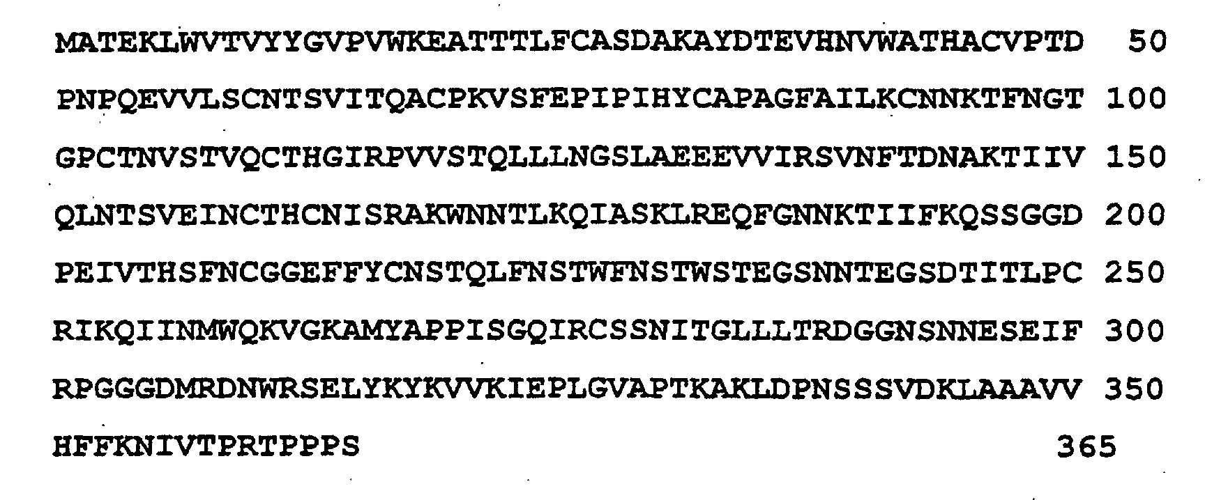
The fusion protein used in the method of the present invention has the following structure:
Figure 00020001
A nucleotide sequence encoding the fusion protein of the above structure is proposed.
According to its another embodiment, the present invention provides a method for producing catalytic antibody using animals with spontaneous and induces autoimmune pathologies. The animal with spontaneous and inducible autoimmune pathologies are MRL-lpr/lpr mice, the mice being immunized with an antigen containing a haptene, the hapten being a conjugate of a mechanism-dependent covalent protease inhibitor with a peptide, the peptide being a fragment of a potential substrate of the catalytic antibody.
The potential substrate of the catalytic antibody is gp120 or its fragment.
The hapten and its isomers and racemates used in the method of the present invention have the following structure:
Figure 00030001
Brief Description of the Drawings:
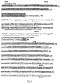
  • FIG. 1. Nucleotide sequence of gp120 fragment I-III . Colored are fragments I, II and III, sequentially. The primers used in PCR are marked out with capital letters.
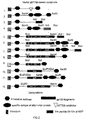
  • FIG. 2. Diagrams of recombinant proteins obtained with the use of pET32b vector.
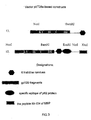
  • FIG. 3. Diagrams of recombinant proteins obtained with the use of pET28a vector.
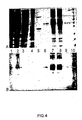
  • FIG. 4. Electrophoregram (A) and immunoblot (B) of different stages of isolation and purification of the protein gp120I-IIImbp (the product of construct 15). 1 ― total cellular proteins before induction; 2 ― total cellular proteins after induction; 3 ― the fraction of soluble intracellular proteins; 4 ― soluble proteins not retained by the metal chelate column; 5 ― soluble proteins eluted at pH 5.0; 6 ― soluble protein form preparation after chromatographic purification; 7 ― the fraction of insoluble intracellular proteins; 8 ― insoluble proteins not retained by metal chelate column; 9 ― denaturated protein form preparation after chromatographic purification; 10 ― marker.
  • FIG. 5. Analysis of the antigen specificity of antibodies in serum of SJL mice immunized with the fusion protein gp120I-IIImbp at different doses. The figure legend indicates the immunogen dose; SJL-2 are mice immunized with the dose of 150 µg per mouse, SJL-3 are mice immunized with the dose of 300 µg per mouse.
  • FIG. 6. The principles of fluorescent and enzymic analyses of proteolytic activity.
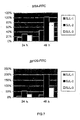
  • FIG. 7. Determination of the proteolytic activity of antibody preparation isolated from serum of SJL mice immunized with the fusion protein gp120I-IIImbp. SJL-1 are control mice. SJL-2 are mice immunized with the dose of 150 µg per mouse. SJL-3 are mice immunized with the dose of 300 µg per mouse. BSA-FITC and gp120-FITC were used as substrates.
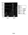
  • FIG. 8. Inhibition of the proteolytic activity of antibody preparation isolated from serum of SJL mice immunized with the fusion protein gp120I-IIImbp. SJL-1 are control mice.
    SJL-2 are mice immunized with the dose of 150 µg per mouse.
    AEBSF: aminoethanebenzenesulfonyl fluoride.
    CMC: phenylalanylchloromethylketone.
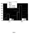
  • FIG. 9. Antispecies antibody inhibition of the proteolytic activity of antibody preparation isolated from serum of SJL mice immunized with gp120I-IIImbp. SJL-1 are control mice.
    SJL-2 are mice immunized with the dose of 150 µg per mouse.
    Anti-IgG: rabbit polyclonal antibodies against murine IgG.
  • FIG. 10. Enzymatic determination of the proteolytic activity of antibody preparations isolated from sera of SJL mice immunized with the fusion proteins gp120I-IIImbp at different doses. A: SJL-1 are control mice; SJL-2 are mice immunized with the dose of 150 µg per mouse; SJL-3 are mice immunized with the dose of 300 µg per mouse; CBA are control CBA mice.
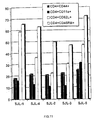
  • FIG. 11. Changes in the expression level of surface markers of T-cells of the immune system of SJL mice immunized with the fusion protein gp120I-IIImbp at different doses, with the recombinant protein gp120I-III, and with the encephalitogenic peptide MBP89-104. SJL-1 are non-immunized mice. SJL-2 are mice immunized with gp120I-IIImbp at the dose of 150 µg per mouse. SJL-3 : mice immunized with gp120I-IIImbp at the dose of 300 µg per mouse. SJL-4 are mice immunized with the peptide MBP89-104. SJL-5 are mice immunized with the recombinant protein gp120I-III at the dose of 300 µg per mouse.
  • FIG. 12. Enzyme immunoassay of sera of SJL, MRL-lpr/lpr and NZB/NZW F1 mice immunized with peptidylphosphonate. The antigen used was: A ― biotinylated reactive peptide; B ― biotinylated diphenylvalylphosphonate; C ― methyl p-nitrophenyl biotinylphenylmethylphosphonate.
  • FIG. 13. Electrophoregram (A) and immunoblot (B) of polyclonal antibodies isolated from immunized mice of strains SJL (4), MRL-lpr/lpr (5) and NZB/NZW F1 (6) and covalently modified with an antigen. Lanes 1-3: 10µg of BSA, 1 µg of trypsin, and 1 µg of IgG of BALB/c mice.
  • The Preferred Embodiments of the Invention
    The present invention is illustrated by the following Examples:
    Example 1. Development of a genetic construct containing a nucleotide sequence encoding the fusion protein gp120I-IIImbp for its expression in a prokaryotic system.
  • 1) To induce autoimmune encephalomyelitis (EAE) in SJL mice, the 89-104 peptide of the myelin basic protein (MBP) was chosen, the peptide having the following composition: VVHFFKNIVTPRTPPPS [Sakai, K., Zamvil, S.S., Mitchell, D.J., Lim, M., Rothbard, J.B., and Steinman, L. 1988. Characterization of a major encephalitogenic T cell epitope in SJL/J mice with synthetic oligopeptides of myelin basic protein. J. Neuroimmunol. 19:21-32., ∥ Tan, L.J., Kennedy, M.K., and Miller, S.D. 1992. Regulation of the effector stages of experimental autoimmune encephalomyelitis via neuroantigen-specific tolerance induction. II. Fine specificity of effector T cell inhibition. J. Immunol. 148:2748-2755.]. The DNA sequence corresponding to said peptide was synthesized by PCR from two overlapping oligonucleotides additionally containing a stop codon and restriction sites. The resulting DNA fragment was cloned in the pET32b plasmid using NotI and XhoI restrictases. The resulting plasmid is hereinafter designated as pET32mbp. For the accurate identification of recombinant proteins at all stages of their expression, isolation and purification, pET32bCH and pET32CHmbp constructs were engineered to contain a sequence that codes for the 10 amino acid-long immunodominant epitope of human p62 c-myc protein [Evan G.I., Lewis G.K., Ramsay G., Bishop J.M., // Mol. Cell. Biol. 1985, V.5(12), P. 3610-3616.].
  • 2) Numerous available publications on the structure, immunogenicity and functional activity of the surface protein gp120 [Hansen, J.E., Lund, O., Nielsen, J.O., Brunak, S., and Hansen, J.-E., S. 1996. Prediction of the secondary structure of HIV-1 gp120. Proteins. 25: 1-11∥ Shioda, T., Oka, S., Xin, X., Liu, H., Harukuni, R., Kurotani, A., Fukushima, M., Hasan, M.K., Shiino, T., Takebe, Y., Iwamoto, A. and Nagai, Y. 1997. In vivo sequence variability of human immunodeficiency virus type 1 envelope gp120: association of V2 extension with slow disease progression. J. Virol. 71: 4871-4881 ∥ Sullivan, N., Sun, Y., Sattentau, Q., Thali, M., Wu, D., Denisova, G., Gershoni, J., Robinson, J., Moore, J., and Sodroski, J. 1998. CD4-Induced Conformational Changes in the Human Immunodeficiency Virus Type 1 gp120 Glycoprotein: Consequences for Virus Entry and Neutralization. J. Virol. 72: 4694-4703] allowed allocation of protein regions having a relatively constant sequence and most promising with regard to immunization. For further work, a chimeric polypeptide was chosen, which consisted of three fragments of gp120 (designated as I, II and III) lacking the first, second and third hypervariable regions. HXB2-env gene sequence was used as the initial template for the synthesis of this construct [Page, K.A., Landau, N.R., and Littman, D.R. 1990. Construction and use of a human immunodeficiency virus vector for analysis of virus infectivity. J. Virol. 64: 5270-5276]. Fragments I, II and III were obtained by PCR using synthetic oligonucleotides followed by assemblage of the fragments using the «splicing by overlap extension» approach (FIG. 1). The final PCR product I-III and intermediate products I-II, II-III and III were cloned into the BlueScript plasmid, with subsequent recloning into the plasmids pET32b (FIG. 2: No. 8, 9, 10 and 12), pET32mbp (FIG. 2: No. 5), pET32bCH (FIG. 2: No. 6 and 11) and pET32CHmbp (FIG. 2: No. 7) using respective restrictases, i.e., NcoI-BamHI for I-III, NcoI-NotI for I-II, EcoRV.-BamHI for II-III, and EcoRV\DraI.-BamHI for III. The products of these constructs were used to test the proteolytic activity of the antibodies against gp120 that had been obtained as a result of immunization.
  • For immunization of SJL mice, in order to obtain proteolytic antibodies against gp120 glycoprotein, the final construct based on pET28a vector was engineered (FIG. 3: No. 15). The fragment NcoI-XhoI from the construct 7 were recloned into pET28a at the respective restriction sites. For immune response testing, an additional construct basing on pET28a vector (FIG. 3: No 13) and comprising the gene of gp120I-III but no sequences encoding mbp peptide and the epitope of c-myc was obtained in a similar way.
    The preparation of electrocompetent cells, transformation, restrictase treatment, ligation, PCR, and DNA electrophoresis were carried out according to standard methods [Sambrook J., Fritsch E.F., Maniatis T. // Molecular Cloning: A Laboratory Manual. New York, Cold Spring Harbor Laboratory Press, 1989.].
    Example 2. Expression, isolation and purification of the fusion protein gp120I-III-mbp.
    The fusion protein was expressed in T7-lysogenated E. coli cells (the strain BL21 (DE3) was used in the present example). The protein was expressed as follows:
  • 1. Competent cells are transformed with 0.1 µg of plasmid according to Item 3 of Example 1 by electroporation and seeded onto a Petri dish containing 30 µg/ml Kanamycin and 2% glucose. Bacterial colonies are grown for 12-14 h at 30°C.
  • 2. The colonies are completely suspended in 11 of bacterial medium 2xYT containing 30 mg/ml Kanamycin and 0.1% glucose.
  • 3. The cell culture is grown at 30°C under adequate aeration up to the density of 0.6-1 OU but not longer than three hours; then IPTG is added to make 1 mM and induction is carried out for 3 h at 30°C.
  • Fusion protein isolation and purification
    The fusion protein gp120I-III-mbp was isolated under denaturating conditions as follows:
  • 1. The cell culture is centrifuged at room temperature for 10 min at 5000 rpm; the sediment is suspended in 1/50 of the initial volume in 50 mM Tris-HCl, pH 8.0; lysozyme and Triton-X100 are added to make 0.1 mg/ml and 0.1%, respectively; and the mixture is incubated for 30 min at 30°C.
  • 2. The lysate is cooled to 0°C, sonicated up to the disappearance of viscosity, and centrifuged for 40 min at 20000 rpm.
  • 3. The sediment is carefully suspended in 1/200 of the initial volume in a buffer containing 50 mM Tris-HCl (pH 8.0), 1 mM EDTA-Na, and 1% Nonidet P-40, centrifuged for 40 min at 20000 rpm, resuspended in chromatographic Buffer A and centrifuged under the same conditions.
  • 4. The sediment is suspended in chromatographic Buffer A (50 mM NaH2PO4-Na2HPO4, 300 mM NaCl, and 6M urea, pH 8.0), incubated on ice for 1 h, and centrifuged for 10 min at 20000 rpm.
  • 5. The supernatant is applied to a metal chelate column equilibrated with Buffer A at the rate of 10 column volumes per hour, and the column is washed with Buffer B (50 mM NaH2PO4-Na2HPO4, 300 mM NaCl, and 6 M urea, pH 7.0) at the rate of 3 0 column volumes per hour up to the discontinuation of baseline migration.
  • 6. Elution is carried out with Buffer B (50 mM NaH2PO4-Na2HPO4, 20 mM MES-NaOH, 300 mM NaCl, and 6 M urea, pH 5.0) at the rate of 30 column volumes per hour, after which the column is equilibrated with Buffer A for the second time, and immobilized metal ions and retained proteins are removed with Buffer A containing 100 mM EDTA, pH 8.0.
  • 7. The eluate fractions are analyzed by gel electrophoresis and combined, and proteins are precipitated by dialysis against deionized water at room temperature.
  • 8. The protein precipitate is separated by centrifugation at 4000 rpm for 10 min, washed with 70% ethanol, suspended in a minimal volume of 70% ethanol, sonicated up to the discontinuation of particle sedimentation, and stored as suspension at +4°C in sterile polypropylene tubes.
  • Fusion protein analysis
    To confirm the identity and purity of the resulting preparations of the fusion protein gp120I-IIImbp, the following characteristics of the protein were determined:
  • 1. The electrophoretic purity of the protein was determined by Method 1 and was found to be 97%.
  • 2. The immunoreactivity with antibodies against c-myc epitope was determined by Method 2, and the protein was found to be immunoreactive.
  • 3. The molecular weight (Da) by mass spectrometry was determined by Method 3 and was found to be 42307 Da, the calculated value being 42075 at the tolerated error of ±2.5%.
  • 4. The specific sorption of the protein (%) by metal chelate sorbent was determined by Method 4 and was found to be >95%.
  • Analytical methods: 1. Electrophoregram densitometry.
    Denaturing electrophoresis of proteins was carried out according to Laemmli using 6 M urea solution in the concentrating and fractionating gels. Gel staining was carried out with Cumassie blue R-250 using contrast enhancing with a cuprum salt. Densitometry was performed with a densitometer or computer assisted plate scanner, with subsequent electrophoregram digitalization and analysis (FIG. 4A).
  • 1. Two-component gel is prepared to have the following composition:
  • Upper gel: 6.66% of acrylamide/bis-acrylamide at a 29/1 ratio, 0.1% sodium dodecyl sulphate, 0.125 M Tris-HCl, and 6 M urea, pH 6.8.
  • Lower gel: 10% of acrylamide/bis-acrylamide at a 29/1 ratio, 0.1% sodium dodecyl sulphate, 0.375 M Tris-HCl, and 6 M urea, pH 8.9.
  • 2. Protein samples are mixed with sample buffer containing 5% 2-mecaptoethanol, heated for 5 min at 100°C and applied to gels. Electrophoresis is carried out at 25 mA until indicator dye is eluted.
  • 3. The fractionating gel is separated and incubated for 5 min in a hot mixture of 10% ethanol and 10% acetic acid.
  • 4. Staining is performed by gel incubation for 10 min in a hot mixture of the following composition: 15% ethanol, 25% acetic acid, 0.3 g/l Cumassie Blue R-250, and 0.45 g/l cuprum sulphate hexahydrate.
  • 5. After staining, the gel is subjected to multiple washings, as described in Item 3, up to complete decoloration.
  • 6. The gel is subjected to densitometry according to the densitometer specifications. Upon electrophoregram digitization with a computer-assisted plate scanner, the Green channel of color image or green light filter of the scanner is used. The electrophoregram image is analyzed using Scion Image software by Surface Plot method. The preparation purity is defined as the ratio of the main peak to the sum of all the detected peaks.
  • 2. Immunoblotting.
    Immunoblotting is carried out according to the standard regimen using blocking BSA solution. The hybridization buffer is supplemented with bovine serum albumin (fraction V) to make 0.5% (FIG. 4 C).
  • 1. Electrophoresis is carried out according to Method 2 using a prestaining marker.
  • 2. The fractionating gel is separated, whereupon the procedure of transference to HyBond N+ membrane (Amersham) is performed using an LKB apparatus for semidry electrotransference according to the manufacturer's specifications for 40 min at 0.8 mA/cm2.
  • 3. The membrane is blocked for 1 h with the solution of 50 mM Tris-HCl (pH 7.6), 150 mM NaCl and 5% bovine serum albumin (fraction V).
  • 4. The membrane is washed thrice for 5 min with a deblocking solution containing 50 mM Tris-HCl (pH 7.6), 150 mM NaCl and 0.05% Tween-20. Then hybridization with the monoclonal antibody 1-9E10.2 is performed for 1 h in the solution of 50 Tris-HCl (pH 7.6), 150 mM NaCl and 0.5% bovine serum albumin.
  • 5. Deblocking (washing) according to Item 4 is performed, and the membrane is hybridized with secondary rabbit Fc-specific anti-mouse IgG antibodies conjugated to horse radish peroxidase (Sigma Immunochemicals) under the same conditions as described in Item 4.
  • 6. The membrane is deblocked as described in Item 4 and stained with the solution of 50 mM Tris-HCl (pH 7.6), 3 mg/ml 1-chloro-4-naphthol and 0.003% H2O2 for 30 min.
  • All the analyses are performed using primary and secondary antibody titers of 1:10000 and 1:4000, respectively, as determined with a characterized antigen, and a test protein is applied to electrophoresis at the dose of 0.1 µg. The presence of a possible test protein immunoreactivity is determined visually by the following criteria: the development of a single distinct well outlined staining zone whose electrophoretic mobility corresponds to that of the test protein. When these criteria are met, instrumental analysis is performed.
    For the final semiquantitative analysis, the densitometric evaluation of the intensity of the staining zone is performed by the Surface Plot method using Scion Image software. Test results are considered positive when the peak half-height is 5 times greater than the range of baseline fluctuations on the densitogram.
    3. MALDI mass-spectrometry .
    Samples are prepared for analysis as follows.
  • 1. An aliquot of protein suspension of a minimal volume is evaporated to dryness in a vacuum centrifuge.
  • 2. The residue is dissolved in 1-5 µl of mixture of 1% aqueous trifluoroacetic acid and 30% acetonitrile, applied to the base plate using 2,5-dihydroxybenzoic (DHB) acid as a matrix, and analyzed.
  • 3. A TOF MALDI mass-spectrometer, similar in performance to the VISION 2000 apparatus, is precalibrated by protein reference standards (trypsin and angiotensin), and protein mass-spectra are read using internal calibration. The masses of molecular ions are determined using VISION 2000 Mass Analyzer software taking account of the performed calibrations.
  • 4. Specific adsorption.
    To confirm the functional properties of a protein preparation, it is tested qualitatively for adsorption from solution by excess metal chelate sorbent. Tested proteins having the sequence 6xHis will be immobilized by the metal chelate sorbent at pH 8.0.
  • 1. The required volume of the metal chelate sorbent Talon (Clontech Laboratories Inc.) is equilibrated with a buffer solution (50 mM Na2HPO4-NaH2PO4, 300 mM NaCl, and 0.1 % Triton X-100) and 20 µl portions of the 1:1 suspension are transferred to test tubes.
  • 2. 10 µg of test protein solution is added, and the volume is adjusted to 100 µl with the buffer according to Item 1. The test tubes are incubated at shaking for 15 min and allowed to stand, after which 10 µl aliquots are taken for analysis.
  • 3. Adsorption is considered to be complete if the measured protein concentration in the test sample does not exceed 0.005 µg/ml (i.e., is not significantly different from the control when the protein concentration is determined by the BSA test), which corresponds to the 95% absorption level.
  • Example 3. Immunization of autoimmune SJL mice with the fusion protein gp120I-IIImbp.
    SJL mice are immunized with the fusion protein gp120 I-III-mbp as follows.
  • 1. Five female SJL mice aged 6-8 weeks are immunized twice at a weekly interval with the antigen in complete Freund adjuvant having the final M. tuberculosis concentration of 2 mg/ml and the antigen concentrations of 1.5 mg/ml and 3 mg/ml.
  • 2. Injections at the total volume amounting to 0.1 ml of the preparation are done subcutaneously at three sites along the back in case of the first immunization, and into paw pads, in case of the second immunization.
  • 3. To compromise the hematoencephalic barrier, one day before the first immunization and two days after it the mice are additionally intraperitoneally injected with 400 ng of pertussis toxin preparation.
  • 4. Seventeen days after the first immunization, the mice receive a boosting peritoneal injection of the antigen in PBS at the total volume amounting to 0.2 ml, the dose of the immunogenic protein being 50 µg per mouse.
  • 5. Concurrently with the boosting procedure, blood is taken from the orbital sinus of experimental and control (non-immunized) mice to monitor the development of the immune response. The presence of specific antibodies in the serum is determined by enzyme immunoassay.
  • 6. Twenty one days after the beginning of the experiment, the mice that have showed the maximal antigen-specific response in immunochemical testing are used for splenectomy and blood sampling. The spleens are used for cell fusion in order to obtain hybridoma clones and for mRNA isolation for the subsequent cloning as phage display libraries.
  • To confirm the appearance of antigen-specific proteolytic antibodies in the course of immunization performed against the background of induced autoimmune pathology manifested as preclinical experimental autoimmune encephalomyelitis, the following tests are carried out:
    Analysis of the antigen specificity and proteolytic activity of the obtained polyclonal antibody preparations 1. Analysis of the antigen specificity of the obtained antibody preparations.
    For the initial comparative monitoring of the specific immune response to the antigen in several immunized mice, enzyme immunoassay (EIA) is used (FIG. 5).
    An antigen is immobilized on an immunological plate and, after incubation with sera obtained from immunized and control mice, antigen-antibody complex is detected with Fc-specific rabbit antimouse-IgG antibodies conjugated to horse radish peroxidase. The sera of immunized and control mice are used at several dilutions (1:12 and 1:48). The following recombinant proteins are used as antigens:
  • 1) trx-gp120 I-III-CH, a fusion protein comprising E. coli thioredoxin A, a gp120 sequence lacking the first, second and third variable regions, and His6-c-myc sequence;
  • 2) trx-gp120 I-II-H, a fusion protein comprising E. coli thioredoxin A, the sequences of the first and second constant regions of gp120, and His6 sequence;
  • 3) trx-gp120 II-III-H, a fusion protein comprising E. coli thioredoxin A, the sequence of the second constant regions of gp 120, with the C-terminus of the sequence starting from the third constant region, and His6 sequence;
  • 4) trx-gp120 III-H, a fusion protein comprising E. coli thioredoxin A, the C-terminus of the gp120 sequence starting from the third constant region, and His6 sequence; and
  • 5) trx-CH, a fusion protein comprising E. coli thioredoxin A and His6 sequence, which was used as the negative control to determine the non-specific binding of obtained antibodies to these protein sequences.
  • As a result of the performed experiments (FIG. 5), it has been shown that all the antibody preparations obtained from the serum of mice immunized with a fusion protein interact with the antigen.
    2) Analysis of the proteolytic activity of the obtained polyclonal antibody preparations.
    To determine the proteolytic activity, the antibodies are, as a preliminary, purified by affinity chromatography using recombinant protein G immobilized on Sepharose. The activity is detected by two different methods (FIG. 6).
    A. Fluorescent assay. The principle of the method, which is outlined in FIG. 6A, is based on the phenomenon of fluorescence quenching by a protein heavily labeled with a fluorophore, which phenomenon is described in literature and is mainly based on the mutual interactions of the aromatic rings of different fluorophore molecules (e.g., because of intense hydrophobic and stacking contacts), and on fluorescence enhancement by introduction of breaks into the polypeptide chain. In the present test, bovine serum albumin and the recombinant protein trx-gp120 I-III-CH excessively labeled with fluorescein isothiocyanate (designated hereinafter as BSA-FITC and gp120-FITC) were used as the substrates for the proteolysis. The reaction was monitored by the fluorescence enhancement vs. control. Trypsin devoid of contaminating chymotrypsin activity was used as the model protease to determine the sensitivity of the method and to evaluate temporal signal changes depending on the substrate and enzyme amounts.
    To determine the proteolytic activity of the tested antibody preparations, triplicate measurements are carried out at the baseline and after incubation at 37°C for 24 h and 48 h. The results of these experiments suggest the following (FIG.s 7-9):
    • First, the proteolytic activity of preparations obtained from mice immunized with the protein gp120 I-III-mbp is increased in comparison with control SJL mice when both substrates are used.
    • Second, the increase in the antigen-specific proteolytic activity is predominantly responsible for the total increase. With gp120-FITC, the signal increased from ten to twenty times in comparison with non-immunized mice, whereas with BSA-FITC, the signal increased twofold only.
    • Third, the predominant mechanism is, in this case, the serine-dependent catalysis, because the addition of serine-reactive irreversible inhibitors resulted in a significant reduction in the observed rate of hydrolysis of BSA-FITC.
    • Forth, IgG molecules, which are selectively removed from the reaction by immunoprecipitation, are responsible for, at least, a major part of the observed proteolytic activity.
    Along with undisputed advantages, such as high sensitivity and the simplicity and rapidity of measurements, this method, unfortunately, has some drawbacks, the main of which is complex nonlinear relationships between fluorescence changes and substrate and enzyme amounts, sample volume, buffer composition and pH, etc. resulting in difficulties in the quantitative evaluation and characterization of the enzymatic activity of polyclonal antibody preparations. Besides that, proteolytic activity determination by this test requires a large excess of substrate vs. enzyme, because the real amount of abzymes in the total pool of antibodies may make a few percents or less.
    With regard to the above, another method for determination of the proteolytic activity of the obtained antibody preparations was used as an alternative.
    B. Enzymatic assay.
    The principle of this method of detection of proteolytic activity, which is outlined in FIG. 6B, is based on the use of small amounts (about 1 ng per reaction) of a highly active enzyme ribonuclease A as the substrate of the proteolysis reaction. The level of ribonuclease activity, which linearly depends on the concentration of active RNAase A, is determined by the acid-soluble residue method using polycytidyl acid as the polymeric substrate of the reaction. This method of proteolytic activity detection presumably allows achieving a significant molar excess of enzyme vs. substrate and thus makes the conditions of the proteolysis reaction under study closer to those of the well-studied non-stationary kinetics model ([S]0<<[E]).
    To eliminate possible artefacts, all the antibody preparations under study were tested for the intrinsic ribonuclease activity and the presence of solution components that nonenzymatically alter the added ribonuclease activity. All the antibody preparations under study were devoid of the intrinsic ribonuclease activity and did not alter the added ribonuclease activity upon a short-time (10 min) incubation of the reaction.
    The proteolytic activity of the tested antibody preparations was measured under the following conditions: IgG concentration 0.1 mg/ml, incubation time 17 h, temperature 37°C. The preparation activity measured by this method was expressed as ribonuclease hydrolysis rate.
    The test results presented in FIG. 10 show that proteolytic activity decreased 1.5-2 times upon immunization of mice with gp120-I-III-mbp. These results are in a limited correspondence with the results of the fluorescent test: only the negative correlation between the proteolytic activity and the immunogen dose used for immunization is reproduced. The discrepancy between the results of the fluorescent and enzymatic analysis might be explained by differences between the structures of the substrates of proteolysis. Presumably, RNAase A globule compared with BSA globule has fewer sites available for proteases and abzymes. Since, upon immunization, the total serum concentration of IgG increases manifold by antigen-specific antibodies, the proportion of the initial proteolytic antibodies decreases, whereas the newly-formed antigen-specific proteolytic antibodies are, most likely, inefficient catalyzers of proteolytic cleavage of RNAase A.
    3. Monitoring of immune response development and experimental autoimmune encephalomyelitis induction.
    To characterize the features of immune response development upon immunization with a fusion protein comprising the neuroantigen MBP, a comparative analysis of specific surface markers expression in T-lymphocytes from SJL mice that were not immunized (control), from mice immunized with the synthetic peptide MBP89-104, recombinant fusion protein gp120I-IIImbp at two different doses and recombinant protein gp120I-TII was carried out (FIG. 11). All the immunizations were performed in parallel under identical conditions as described above. Twenty one days after the beginning of an experiment, CD4+ T-lymphocytes isolated from two mice of each group were analyzed by flow cytometry. A specific feature of SJL mice was initially low CD8+ T-cell counts. Also, changes in the expression of the following surface markers important for immune response development have been studied: CD11a, CD44, CD45RB, and CD62-L. The results presented in FIG. 11 suggest that in case of immunization with the peptide MBP and, also, with fusion proteins comprising this antigen, a fully developed T-cell immune response (memory cells appeared) was induced in mice by day 21 after the immunization, whereas in case of use of solely gp120I-III as the antigen, the immune response was still developing. The obtained data provide an evidence of the enhancement of immune response development when the autoantigen MBP is used and of T-lymphocyte activation typical of experimental autoimmune encephalomyelitis development.
    Thus, the above Example shows that, upon immunization of SJL mice with the fusion protein gp120I-IIImbp, antigen-specific proteolytic antibodies are generated against the background of the preclinical stage of induced experimental autoimmune encephalomyelitis.
    Example 4. Synthesis of reactive phosphonate derivative of peptide fragment of gp120.
    At the first stage, aminoalkylphosphonates protected at their free amino group are synthesized in the reaction of co-condensation of triphenylphosphite, isobutanal, and benzylcarbamate. To this end, the mixture of triphenylphosphite, isobutanal, and benzylcarbamate, 0.1 mole each, in 15 ml of glacial acetic acid is stirred for about 1 h until heat generation discontinues. After that, the reaction is stirred with heating to 80°C for 1 h. After the full completion of the reaction, volatile products are removed with a rotary evaporator under reduced pressure and heating on a water bath. The oily residue is dissolved in methanol (180 ml) and left for crystallization at -20°C for 3 h. After crystallization, the residue of diphenyl 1-(N-benzyloxycarbonyl)-aminoalkylphosphonate is harvested by filtration and recrystallized in a minimal volume of chloroform (30-40 ml) followed by the addition of four volumes of methanol.
    To obtain free amynoalkylphosphonate, the protective group is removed by treatment of diphenyl 1-(N-benzylcarbonyl)-aminoalkylphosphonate with a 33% solution of hydrogen bromide in acetic acid (15 ml per 0.1 mole) for 1 h at room temperature. Volatile components are removed with a rotary evaporator at a reduced pressure and heating on a water bath. 1-(N-benzyloxycarbonyl)-aminoakylphosphonate hydrobromide is crystallized from the resulting residue by addition of anhydrous diethyl ether. Free phosphonate is obtained by passing gaseous dry ammonium through phosphonate hydrobromide suspension in diethyl ether until the formation of a thick precipitate of ammonium bromide discontinues and the full blooming of the suspension is observed. The resulting ammonium bromide is removed by filtration, and diethyl ether is evaporated on a water bath under atmospheric pressure.
    To obtain the hapten Leu-Ala-Glu-Glu-Glu-Val- P (OPh)2 (LAEEEV- P (OPh)2), where P (OPh)2 means the substitution of the α-carboxylic group with diphenylphosphonate, the peptide Boc-Val-Ala-(t-Bu)Glu-(t-Bu)Glu-(t-Bu)Glu protected at its N-terminal amino group and side groups is first synthesized. The peptide Boc-Val-Ala-(t-Bu)Glu-(t-Bu)Glu-(t-Bu)Glu is fused with the phosphonate derivative of valine by mixing of 2 µmoles of the protected peptide, 2 µmoles of the phosphonate, and 2 µmoles of dicyclohexylcarbodiimide in 300 µl of acetonitrile and incubating for 1 h. After the completion of the reaction, its products are separated by reverse phase HPLC on a 150×3.9-mm Waters C18 NovaPak column using 0% to 80% gradient of acetonitrile in 20 nM potassium phosphate (pH 7.0). The resulting fractions are analyzed by mass-spectrometry (MALDI-TOF). The fractions that contain substances with molecular ion masses of 1145 Da ([M+H]+), 1167 Da ([M+Na]+) or 1183 Da ([M+K]+) are combined and freeze-dried. The residue is dissolved in 100 µl of 100% trifluoroacetic acid and incubated for 1 h at room temperature to remove protective tert-butyloxycarbonyl and tert-butyl groups. The deblocked peptidylphosphonate is precipitated by addition of 10 volumes of anhydrous diethyl ether to the reaction. The precipitate is separated by centrifuging for 10 min at 12500 rpm, and the deblocking procedure is repeated. The residue is air-dried and stored at -20°C.
    Analysis of the peptidylphosphonate LAEEEV- P (OPh)2 I. TOF MALDI mass-spectrometry.
  • 1. A minimal amount of dry peptidylphosphonate, to which 5 µl of acetonitrile is added, is applied to a base plate using aqueous strong acid-free 2,5-dihytroxybenzoic (DHB) acid as the matrix, and analysis is carried out.
  • 2. A TOF MALDI mass-spectrometer equivalent to the VISION 2000 apparatus is precalibrated with reference standards within the 500-2000 Da m/z range, and mass spectra of test samples are obtained using internal calibration. Molecular ion masses are determined using VISION 2000 Mass Analyzer software with the calibration taken into account. The expected result of the analysis is the presence of peaks corresponding to masses of 877.36, 900.34, and 915.45±1 Da.
  • II. Analytical reverse phase HPLC.
  • 1. To a 0.2 mg sample of peptidylphosphonate, 100 µl of 20 mM potassium phosphate buffer (pH 7.0) containing 20% acetonitrile is added.
  • 2. The resulting sample is administered into an injector, and gradient elution is carried out using a 150x3.9 mm Waters C18 NovaPak column for reverse phase HPLC under the following conditions:
    • buffer A is 20% acetonitrile and 20 mM potassium phosphate, pH 7.0;
    • buffer B is 80% acetonitrile and 20 mM potassium phosphate, pH 7.0;
    • the elution rate is 1.0 ml/min at a linear 100% A to 100% B gradient for 20 min followed by 100% B for 10 min; and
    • the chromatograms are recorded at 260 nm wavelength.
  • 3. The peaks are integrated without correction for the baseline. The retention time of the main peak, which has the maximal area, is determined, and the ratio of the main peak area to the sum of all the peak areas is calculated. The expected result: the retention time is 14.75-15.25 min; the chromatographic purity is >95%.
  • III. Inhibition of the esterolytic activity of chymotrypsin.
  • 1. 0.5 ml of 1 µM solution of α-chymotrypsin in a buffer containing 0.1 HEPES and 0.5 M NaCl, pH 7.2, is prepared. The solution is divided into nine 50-µl aliquots, and the residue is discarded.
  • 2. Eight dilutions of the test peptidylphosphonate in acetonitrile ranging from 1000 µl to 1 µl are prepared.
  • 3. To each of the first eight aliquots of the enzyme solution 5 µl of corresponding peptidylphosphonate solution are added, and 5 µl of acetonitrile are added to the ninth aliquot.
  • 4. The samples are incubated for 1 h at 25±5°C.
  • 5. The samples are successively transferred to a spectrophotometer cell containing 450 µl of deionized water, mixed, whereupon 10 µl of p-nitrophenylacetate solution in methanol (2.5 mg/ml) are added, after which the increase in the optical density at a 400-nm wavelength is recorded. The initial rate of the substrate hydrolysis is calculated in arbitrary units.
  • 6. The effective inhibitor concentration is calculated. To this end, the ratios of the hydrolysis rates observed with samples 1-8 to the hydrolysis rate observed with sample 9 are calculated. The effective inhibitor concentration is determined as the lowest concentration of the test substance, at which the ratio of the rates of substrate hydrolysis does not exceed 50%. The expected result: 30 µM.
  • Example 5. Reactive immunization of mice with the phosphonate derivative of a peptide fragment of gp120.
    For immunization, the reactive peptide is conjugated to the macromolecular carrier C.conholepas hemocyanin (keyhole limpet hemocyanin, KLH). At the first stage, the carrier is activated with excess bis(sulfosuccinimidylyl)suberate in PBS for 1 h at 37°C. After the activation, KLH unbound to bis(sulfosuccinimidylyl)suberate is removed from the reaction by sevenfold exhaustive ultrafiltration (with the residual volume not more than 70 µl) using a Microcon 100 concentrator (Amicon YC membrane), each time adding PBS to the residue to make 500 µl and discarding the ultrafiltrate. To the resulting solution peptidylsulfonate solution in PBS is added whereupon the solution is incubated for 1 h at 37°C without stirring. The unreacted succinimide groups are inactivated by addition of 2 µl of 2-ethanolamine. The low molecular components of the reaction are removed by sevenfold exhaustive ultrafiltration (with the residual volume not more than 70 µl) using a Microcon 100 concentrator (Amicon YC membrane), each time adding PBS to the residue to make 500 µl and discarding the ultrafiltrate. The final preparation is sterilized by filtration and stored at -20°C.
    Female MRL-lpr/lpr, SJL and NZB/NZW F1 mice aged 6-8 weeks are intraperitoneally immunized with the antigen in complete Freund adjuvant, with the total volume being 0.2 ml, at the dose of 50 µg of the immunogenic protein per mouse.
    The second immunization is done with the same volume and at the same antigen concentration in incomplete Freund adjuvant in 17 days afeter the first immunization. Concurrently, blood is withdrawn from the orbital sinus of three mice of each experimental group and control non-immunized mice of the three strains to monitor the development of the immune response.
    Twenty one days after the beginning of the experiment, the mice that showed the maximal antigen-specific response in immunological tests are sacrificed for splenectomy. Polyclonal antibodies isolated from the blood serum of these mice are analyzed for antigen specificity and catalytic activity.
    A part of the peptidylphosphonate synthesized at the previous stages is used in the reaction of conjugation to N-hydroxysuccinimide ester of biotin. The reaction is conducted by mixing of equimolar amounts of peptidyl sulfonate and activated biotin in a minimal volume of dimethylformamide and incubation for 1 h. The biotinylated preparations are intended for analysis of the specificity of the antibodies obtained as a result of reactive immunization of mice.
    To monitor the specific immune response to the antigen in several immunized mice of all the three strains, enzyme immunoassay is used.
    Antibodies from the sera of immunized and control mice are isolated with plate-preabsorbed goat antibodies against murine IgG, with subsequent incubation with the biotinylated antigen and detection of antigen-antibody complexes using neutravidin conjugated to horse radish peroxidase. The sera of immunized and control mice are used at several dilutions (1:12 and 1:48). The antigens employed are biotin-labeled starting peptidylphosphonate, biotinylated Val -phosphonate, and nitrophenylmethyl-p-biotinylphenylmethylphosphonate, for which the specific covalent modification of the active center of abzymes was demonstrated earlier. The comparative analysis has shown (FIG. 12) that the antibodies of the experimental mice of all the three strains, on the whole, possess a high specificity toward the modified peptide fragment of an antigen, do not interact under the conditions of the present experiment with free Val -phosphonate, and exhibit the ability to covalently bind to a more active and less specific modifying agent. It should be noted that, on the average, in New Zealand hybrids the amount of antigen-specific antibodies was somewhat higher in comparison with the two other autoimmune strains, whereas the antibodies of MRL-lpr/lpr mice where more effective with regard to covalent modification.
    Along with an antigen, horse radish peroxidase-conjugated rabbit antibodies against the Fc fragment of murine IgG are used to determine the total amount of murine antibodies specifically absorbed in a plate well. This allows estimation of the proportion of antigen-specific antibodies in the total pool of class G immunoglobulins.
    Further studies of the type of interaction of the obtained antibodies with a reactive peptide were carried out with pre-purified IgG preparations using immunoblotting. After incubation with biotinylated peptidylphosphonate, electrophoretic fractionation under denaturing and reducing conditions, and membrane immobilization, antigen-antibody complexes were detected using neutravidin conjugated to horse radish peroxidase. The results of this experiment presented in FIG. 13 suggest that both light and heavy immunoglobulin chains capable of being covalently modified by the peptide were present in the preparations of polyclonal antibodies isolated from autoimmune mice immunized with the reactive peptide Val-Ala-Glu-Glu-Glu- Val-PO(OPh)2 .
    Thus, the reactive immunization under the conditions of the present Example has produced the following results:
    • The antibodies obtained in the course of immunization bind to immunization antigen.
    • The antibodies do not react with the "minimal" phosphonate group of immunization antigen, which means that there is no nonspecific interaction (or nonspecific chemical reaction) between the antibodies under study and the free phosphonate group of Val P (OPh)2.
    • The antibodies react with the active "mechanism-dependent" phosphonate, i.e., display the ability to react with a molecule that has no apparent structural relation to immunization antigen but has the ability to form covalent complexes with hydrolases.
    • The antibodies form covalent complexes with immunization antigen.
    In combination, the above properties suggest that in the course of immunization with a peptidylphosphonate whose composition corresponds to LAEEEV- P (OPh)2 epitope-specific catalytic antibodies are generated.
    Industrial applicability
    The invention may be useful in medicinal industry for manufacturing drugs.
    Figure 00260001
    Figure 00270001

    Claims (11)

    1. A method for producing catalytic antibodies using animals with spontaneous and inducible autoimmune pathologies, characterized in that a fusion protein consisting of myelin basic protein or its fragments and a potential substrate of the catalytic antibody or a fragment of the potential substrate is administered to the animals.
    2. The method of Claim 1, characterized in that the potential substrate is gp120 (surface glycoproteid of HIV-1) or its fragments.
    3. The method of Claims 1-2, characterized in that mice are used as the animals with spontaneous and inducible autoimmune pathologies.
    4. The method of Claims 1-3, characterized in that mice of those strains are used which may develop experimental autoimmune encephalomyelitis upon immunization with basic myelin protein.
    5. Fusion proteins characterized by the following structure:
      Figure 00280001
    6. A nucleotide sequence encoding the fusion protein of Claim 5.
      Figure 00290001
    7. A method for producing catalytic antibodies using animals with spontaneous and inducible autoimmune pathologies, characterized in that the animals are immunized with an antigen that contains hapten, the hapten being a conjugate of a mechanism-dependent covalent protease inhibitor with a peptide, the peptide being a fragment of the potential substrate of catalytic antibody.
    8. The method of Claim 7, characterized in that the potential substrate is gp120 or its fragments.
    9. The method of Claims 7-8, characterized in that mice are used as autoimmune animals.
    10. The method of Claims 7-9, characterized in that MRL-lpr/lpr mice are used.
    11. Hapten and its isomers and racemates:
      Figure 00300001
    EP02739002A 2001-04-24 2002-04-18 Antigens for immunisation Expired - Lifetime EP1394261B1 (en)

    Priority Applications (1)

    Application Number Priority Date Filing Date Title
    EP10183175.8A EP2295082B1 (en) 2001-04-24 2002-04-18 Method for producing catalytic antibodies (variants), antigens for immunization and nucleotide sequence

    Applications Claiming Priority (3)

    Application Number Priority Date Filing Date Title
    RU2001110759/13A RU2221873C2 (en) 2001-04-24 2001-04-24 Method for preparing catalytic antibodies (variants), antigens for immunization and nucleotide sequence
    RU2001110759 2001-04-24
    PCT/RU2002/000177 WO2002086058A2 (en) 2001-04-24 2002-04-18 Method for producing catalytic antibodies (variants), antigens for immunisation and nucleotide sequence

    Related Child Applications (1)

    Application Number Title Priority Date Filing Date
    EP10183175.8 Division-Into 2010-09-30

    Publications (3)

    Publication Number Publication Date
    EP1394261A2 true EP1394261A2 (en) 2004-03-03
    EP1394261A4 EP1394261A4 (en) 2008-04-09
    EP1394261B1 EP1394261B1 (en) 2011-03-30

    Family

    ID=20248733

    Family Applications (2)

    Application Number Title Priority Date Filing Date
    EP10183175.8A Expired - Lifetime EP2295082B1 (en) 2001-04-24 2002-04-18 Method for producing catalytic antibodies (variants), antigens for immunization and nucleotide sequence
    EP02739002A Expired - Lifetime EP1394261B1 (en) 2001-04-24 2002-04-18 Antigens for immunisation

    Family Applications Before (1)

    Application Number Title Priority Date Filing Date
    EP10183175.8A Expired - Lifetime EP2295082B1 (en) 2001-04-24 2002-04-18 Method for producing catalytic antibodies (variants), antigens for immunization and nucleotide sequence

    Country Status (9)

    Country Link
    US (1) US20040265975A1 (en)
    EP (2) EP2295082B1 (en)
    JP (1) JP2004527248A (en)
    AT (1) ATE503497T1 (en)
    AU (1) AU2002313135A1 (en)
    DE (1) DE60239606D1 (en)
    ES (1) ES2440369T3 (en)
    RU (1) RU2221873C2 (en)
    WO (1) WO2002086058A2 (en)

    Cited By (1)

    * Cited by examiner, † Cited by third party
    Publication number Priority date Publication date Assignee Title
    WO2008078177A3 (en) * 2006-12-26 2008-10-30 Fds Pharma Ass Method for producing catalytic antibodies (variants), antigens for immunization and nucleotide sequence

    Family Cites Families (9)

    * Cited by examiner, † Cited by third party
    Publication number Priority date Publication date Assignee Title
    ATE112687T1 (en) * 1988-09-13 1994-10-15 Chiron Corp MUTANTS OF HIV-1 ENVELOPE PROTEIN WITH MISSING HYPERVARIABLE DOMAINS.
    US5817629A (en) * 1991-10-22 1998-10-06 The Governors Of The University Of Alberta Peptide specificity of anti-myelin basic protein and the administration of myelin basic protein peptides to multiple sclerosis patients
    US5961973A (en) * 1992-03-06 1999-10-05 Crea; Roberto Pathogen-targeted biocatalysts
    NZ251582A (en) * 1992-04-01 1997-07-27 Merck & Co Inc Recombinant human anti-hiv antibodies, methods for their cloning and expression and pharmaceutical compositions thereof
    WO1996028470A2 (en) * 1995-03-09 1996-09-19 Neurocrine Biosciences, Inc. Peptide analogues of human myelin basic protein useful in treating multiple sclerosis
    US6156541A (en) * 1995-07-21 2000-12-05 The Board Of Regents Of The University Of Nebraska Compositions and methods for catalyzing hydrolysis of HIV gp120
    US5948658A (en) 1996-06-25 1999-09-07 The Trustees Of Columbia University In The City Of New York Anti-cocaine catalytic antibody
    US6140091A (en) * 1997-06-20 2000-10-31 Boston Biomedical Research Institute Anti-idiotype vaccines to elicit catalytic antibodies
    US6235714B1 (en) * 1998-03-23 2001-05-22 Sudhir Paul Methods for identifying inducers and inhibitors of proteolytic antibodies, compositions and their uses

    Cited By (1)

    * Cited by examiner, † Cited by third party
    Publication number Priority date Publication date Assignee Title
    WO2008078177A3 (en) * 2006-12-26 2008-10-30 Fds Pharma Ass Method for producing catalytic antibodies (variants), antigens for immunization and nucleotide sequence

    Also Published As

    Publication number Publication date
    DE60239606D1 (en) 2011-05-12
    WO2002086058A3 (en) 2003-01-23
    EP1394261B1 (en) 2011-03-30
    ES2440369T3 (en) 2014-01-28
    EP2295082A1 (en) 2011-03-16
    AU2002313135A1 (en) 2002-11-05
    WO2002086058A2 (en) 2002-10-31
    WO2002086058A9 (en) 2003-11-27
    EP1394261A4 (en) 2008-04-09
    US20040265975A1 (en) 2004-12-30
    RU2221873C2 (en) 2004-01-20
    EP2295082B1 (en) 2013-09-18
    ATE503497T1 (en) 2011-04-15
    JP2004527248A (en) 2004-09-09

    Similar Documents

    Publication Publication Date Title
    Davies et al. Peptide-specific antibodies as probes of the orientation of the glucose transporter in the human erythrocyte membrane.
    JPS62500027A (en) A chemically synthesized polypeptide that produces antibodies immunoreactive with Epstein-Barr virus nuclear antigens
    KR20160039682A (en) Assays for timp2 having improved performance in biological samples
    WO1992001787A1 (en) Binding domains
    JP5102205B2 (en) Catalytic immunoglobulin
    EP2588495B1 (en) Histone citrullinated peptides and uses thereof
    WO2008078177A2 (en) Method for producing catalytic antibodies (variants), antigens for immunization and nucleotide sequence
    EP2295082B1 (en) Method for producing catalytic antibodies (variants), antigens for immunization and nucleotide sequence
    EP0594879A1 (en) Cathepsin L-specific monoclonal antibodies, hybridomas for producing the same and a method of using the same
    Reuter et al. Molecular relationships between U snRNP proteins as investigated by rabbit antisera and peptide mapping
    US5194585A (en) Inhibitors of catalytic antibodies
    Luka et al. Antibodies of predetermined specificity for the NH2 terminus of a cellular protein p53 react with the native molecule: evidence for the presence of different p53s.
    EP0994187B1 (en) Renin-active substance comprising human prorenin and antibodies to the prorenin profragment
    Adami et al. Monoclonal antibodies for structure-function studies of (R)-3-hydroxybutyrate dehydrogenase, a lipid-dependent membrane-bound enzyme
    Radford et al. Antibodies against an inter‐domain segment of polypeptide chain inhibit active‐site coupling in the pyruvate dehydrogenase multienzyme complex
    US9067982B2 (en) Compositions and methods for redox modulated proteins
    CN118388648B (en) A human PD-L1 antibody based on fully human antibody mice and its preparation method and application
    US6048717A (en) Inhibitors of catalytic antibodies
    JPH0827197A (en) Antibody that recognizes human-derived cytochrome P450 3A4
    US6800462B2 (en) Production of recombinant proteins in vivo and use for generating antibodies
    AU4831590A (en) Molecular recognition units
    JPH0827195A (en) Antibody recognizing human-derived cytochrome P450 2A6
    Kyte Immunochemical Probes of Structure
    JPH0827196A (en) Antibody that recognizes human-derived cytochrome P450 2C9
    JPH0827198A (en) Antibody that recognizes human-derived cytochrome P450 2C8

    Legal Events

    Date Code Title Description
    PUAI Public reference made under article 153(3) epc to a published international application that has entered the european phase

    Free format text: ORIGINAL CODE: 0009012

    17P Request for examination filed

    Effective date: 20031121

    AK Designated contracting states

    Kind code of ref document: A2

    Designated state(s): AT BE CH CY DE DK ES FI FR GB GR IE IT LI LU MC NL PT SE TR

    RAP1 Party data changed (applicant data changed or rights of an application transferred)

    Owner name: FDS PHARMA ASS.

    RIC1 Information provided on ipc code assigned before grant

    Ipc: C12P 21/08 20060101ALI20080227BHEP

    Ipc: C12N 15/62 20060101ALI20080227BHEP

    Ipc: C12N 9/00 20060101ALI20080227BHEP

    Ipc: C07K 19/00 20060101ALI20080227BHEP

    Ipc: C07K 16/10 20060101ALI20080227BHEP

    Ipc: C07K 14/47 20060101ALI20080227BHEP

    Ipc: C07K 14/16 20060101ALI20080227BHEP

    Ipc: A61K 39/42 20060101AFI20080227BHEP

    A4 Supplementary search report drawn up and despatched

    Effective date: 20080306

    17Q First examination report despatched

    Effective date: 20080728

    RTI1 Title (correction)

    Free format text: ANTIGENS FOR IMMUNISATION

    GRAP Despatch of communication of intention to grant a patent

    Free format text: ORIGINAL CODE: EPIDOSNIGR1

    GRAS Grant fee paid

    Free format text: ORIGINAL CODE: EPIDOSNIGR3

    GRAA (expected) grant

    Free format text: ORIGINAL CODE: 0009210

    AK Designated contracting states

    Kind code of ref document: B1

    Designated state(s): AT BE CH CY DE DK ES FI FR GB GR IE IT LI LU MC NL PT SE TR

    REG Reference to a national code

    Ref country code: GB

    Ref legal event code: FG4D

    REG Reference to a national code

    Ref country code: CH

    Ref legal event code: EP

    REG Reference to a national code

    Ref country code: IE

    Ref legal event code: FG4D

    REF Corresponds to:

    Ref document number: 60239606

    Country of ref document: DE

    Date of ref document: 20110512

    Kind code of ref document: P

    REG Reference to a national code

    Ref country code: DE

    Ref legal event code: R096

    Ref document number: 60239606

    Country of ref document: DE

    Effective date: 20110512

    REG Reference to a national code

    Ref country code: CH

    Ref legal event code: PUE

    Owner name: LIFEBIO LABORATORIES LLC

    Free format text: FDS PHARMA ASS.#ENTERPRISE HOUSE, 21 BUCKLE STREET#LONDON E1 8NN (GB) -TRANSFER TO- LIFEBIO LABORATORIES LLC#341 RAVEN CIRCLE, KENT COUNTY#WYOMING, DELAWARE 19934 (US)

    Ref country code: CH

    Ref legal event code: NV

    Representative=s name: BUGNION S.A.

    REG Reference to a national code

    Ref country code: NL

    Ref legal event code: VDEP

    Effective date: 20110330

    PG25 Lapsed in a contracting state [announced via postgrant information from national office to epo]

    Ref country code: GR

    Free format text: LAPSE BECAUSE OF FAILURE TO SUBMIT A TRANSLATION OF THE DESCRIPTION OR TO PAY THE FEE WITHIN THE PRESCRIBED TIME-LIMIT

    Effective date: 20110701

    Ref country code: SE

    Free format text: LAPSE BECAUSE OF FAILURE TO SUBMIT A TRANSLATION OF THE DESCRIPTION OR TO PAY THE FEE WITHIN THE PRESCRIBED TIME-LIMIT

    Effective date: 20110330

    RAP2 Party data changed (patent owner data changed or rights of a patent transferred)

    Owner name: LIFEBIO LABORATORIES LLC

    PG25 Lapsed in a contracting state [announced via postgrant information from national office to epo]

    Ref country code: AT

    Free format text: LAPSE BECAUSE OF FAILURE TO SUBMIT A TRANSLATION OF THE DESCRIPTION OR TO PAY THE FEE WITHIN THE PRESCRIBED TIME-LIMIT

    Effective date: 20110330

    Ref country code: CY

    Free format text: LAPSE BECAUSE OF FAILURE TO SUBMIT A TRANSLATION OF THE DESCRIPTION OR TO PAY THE FEE WITHIN THE PRESCRIBED TIME-LIMIT

    Effective date: 20110330

    Ref country code: FI

    Free format text: LAPSE BECAUSE OF FAILURE TO SUBMIT A TRANSLATION OF THE DESCRIPTION OR TO PAY THE FEE WITHIN THE PRESCRIBED TIME-LIMIT

    Effective date: 20110330

    REG Reference to a national code

    Ref country code: DE

    Ref legal event code: R081

    Ref document number: 60239606

    Country of ref document: DE

    Owner name: LIFEBIO LABORATORIES LLC, US

    Free format text: FORMER OWNER: FDS PHARMA ASS., LONDON, GB

    Effective date: 20110804

    Ref country code: DE

    Ref legal event code: R081

    Ref document number: 60239606

    Country of ref document: DE

    Owner name: LIFEBIO LABORATORIES LLC, US

    Free format text: FORMER OWNER: ASGL - FARMATSEVTICJESKIE INNOVATSII, ZAPTRYOTE AKTSIONERNOE OBSCHESTVO, ST. PETERSBURG, RU

    Effective date: 20110303

    Ref country code: DE

    Ref legal event code: R081

    Ref document number: 60239606

    Country of ref document: DE

    Owner name: LIFEBIO LABORATORIES LLC, WYOMING, US

    Free format text: FORMER OWNER: FDS PHARMA ASS., LONDON, GB

    Effective date: 20110804

    Ref country code: DE

    Ref legal event code: R081

    Ref document number: 60239606

    Country of ref document: DE

    Owner name: LIFEBIO LABORATORIES LLC, WYOMING, US

    Free format text: FORMER OWNER: ASGL - FARMATSEVTICJESKIE INNOVATSII, ZAPTRYOTE AKTSIONERNOE OBSCHESTVO, ST. PETERSBURG, RU

    Effective date: 20110303

    PG25 Lapsed in a contracting state [announced via postgrant information from national office to epo]

    Ref country code: BE

    Free format text: LAPSE BECAUSE OF FAILURE TO SUBMIT A TRANSLATION OF THE DESCRIPTION OR TO PAY THE FEE WITHIN THE PRESCRIBED TIME-LIMIT

    Effective date: 20110330

    REG Reference to a national code

    Ref country code: GB

    Ref legal event code: 732E

    Free format text: REGISTERED BETWEEN 20110915 AND 20110921

    PG25 Lapsed in a contracting state [announced via postgrant information from national office to epo]

    Ref country code: PT

    Free format text: LAPSE BECAUSE OF FAILURE TO SUBMIT A TRANSLATION OF THE DESCRIPTION OR TO PAY THE FEE WITHIN THE PRESCRIBED TIME-LIMIT

    Effective date: 20110801

    PG25 Lapsed in a contracting state [announced via postgrant information from national office to epo]

    Ref country code: ES

    Free format text: LAPSE BECAUSE OF FAILURE TO SUBMIT A TRANSLATION OF THE DESCRIPTION OR TO PAY THE FEE WITHIN THE PRESCRIBED TIME-LIMIT

    Effective date: 20110711

    Ref country code: MC

    Free format text: LAPSE BECAUSE OF NON-PAYMENT OF DUE FEES

    Effective date: 20110430

    PG25 Lapsed in a contracting state [announced via postgrant information from national office to epo]

    Ref country code: NL

    Free format text: LAPSE BECAUSE OF FAILURE TO SUBMIT A TRANSLATION OF THE DESCRIPTION OR TO PAY THE FEE WITHIN THE PRESCRIBED TIME-LIMIT

    Effective date: 20110330

    PG25 Lapsed in a contracting state [announced via postgrant information from national office to epo]

    Ref country code: DE

    Free format text: LAPSE BECAUSE OF NON-PAYMENT OF DUE FEES

    Effective date: 20111101

    REG Reference to a national code

    Ref country code: IE

    Ref legal event code: MM4A

    PLBE No opposition filed within time limit

    Free format text: ORIGINAL CODE: 0009261

    STAA Information on the status of an ep patent application or granted ep patent

    Free format text: STATUS: NO OPPOSITION FILED WITHIN TIME LIMIT

    PG25 Lapsed in a contracting state [announced via postgrant information from national office to epo]

    Ref country code: DK

    Free format text: LAPSE BECAUSE OF FAILURE TO SUBMIT A TRANSLATION OF THE DESCRIPTION OR TO PAY THE FEE WITHIN THE PRESCRIBED TIME-LIMIT

    Effective date: 20110330

    26N No opposition filed

    Effective date: 20120102

    PG25 Lapsed in a contracting state [announced via postgrant information from national office to epo]

    Ref country code: IE

    Free format text: LAPSE BECAUSE OF NON-PAYMENT OF DUE FEES

    Effective date: 20110418

    PG25 Lapsed in a contracting state [announced via postgrant information from national office to epo]

    Ref country code: IT

    Free format text: LAPSE BECAUSE OF FAILURE TO SUBMIT A TRANSLATION OF THE DESCRIPTION OR TO PAY THE FEE WITHIN THE PRESCRIBED TIME-LIMIT

    Effective date: 20110330

    PG25 Lapsed in a contracting state [announced via postgrant information from national office to epo]

    Ref country code: LU

    Free format text: LAPSE BECAUSE OF NON-PAYMENT OF DUE FEES

    Effective date: 20110418

    PG25 Lapsed in a contracting state [announced via postgrant information from national office to epo]

    Ref country code: TR

    Free format text: LAPSE BECAUSE OF FAILURE TO SUBMIT A TRANSLATION OF THE DESCRIPTION OR TO PAY THE FEE WITHIN THE PRESCRIBED TIME-LIMIT

    Effective date: 20110330

    PGFP Annual fee paid to national office [announced via postgrant information from national office to epo]

    Ref country code: GB

    Payment date: 20140424

    Year of fee payment: 13

    PGFP Annual fee paid to national office [announced via postgrant information from national office to epo]

    Ref country code: CH

    Payment date: 20140415

    Year of fee payment: 13

    Ref country code: FR

    Payment date: 20140430

    Year of fee payment: 13

    Ref country code: DE

    Payment date: 20140423

    Year of fee payment: 13

    REG Reference to a national code

    Ref country code: DE

    Ref legal event code: R119

    Ref document number: 60239606

    Country of ref document: DE

    REG Reference to a national code

    Ref country code: CH

    Ref legal event code: PL

    GBPC Gb: european patent ceased through non-payment of renewal fee

    Effective date: 20150418

    PG25 Lapsed in a contracting state [announced via postgrant information from national office to epo]

    Ref country code: GB

    Free format text: LAPSE BECAUSE OF NON-PAYMENT OF DUE FEES

    Effective date: 20150418

    Ref country code: LI

    Free format text: LAPSE BECAUSE OF NON-PAYMENT OF DUE FEES

    Effective date: 20150430

    Ref country code: CH

    Free format text: LAPSE BECAUSE OF NON-PAYMENT OF DUE FEES

    Effective date: 20150430

    REG Reference to a national code

    Ref country code: FR

    Ref legal event code: ST

    Effective date: 20151231

    PG25 Lapsed in a contracting state [announced via postgrant information from national office to epo]

    Ref country code: FR

    Free format text: LAPSE BECAUSE OF NON-PAYMENT OF DUE FEES

    Effective date: 20150430

    PG25 Lapsed in a contracting state [announced via postgrant information from national office to epo]

    Ref country code: DE

    Free format text: LAPSE BECAUSE OF NON-PAYMENT OF DUE FEES

    Effective date: 20151103