EP1391505A1 - Stem cells and method of separating the same - Google Patents
Stem cells and method of separating the same Download PDFInfo
- Publication number
- EP1391505A1 EP1391505A1 EP02720586A EP02720586A EP1391505A1 EP 1391505 A1 EP1391505 A1 EP 1391505A1 EP 02720586 A EP02720586 A EP 02720586A EP 02720586 A EP02720586 A EP 02720586A EP 1391505 A1 EP1391505 A1 EP 1391505A1
- Authority
- EP
- European Patent Office
- Prior art keywords
- cell
- stem cell
- pancreatic
- marker
- ter119
- Prior art date
- Legal status (The legal status is an assumption and is not a legal conclusion. Google has not performed a legal analysis and makes no representation as to the accuracy of the status listed.)
- Granted
Links
Classifications
-
- C—CHEMISTRY; METALLURGY
- C12—BIOCHEMISTRY; BEER; SPIRITS; WINE; VINEGAR; MICROBIOLOGY; ENZYMOLOGY; MUTATION OR GENETIC ENGINEERING
- C12Q—MEASURING OR TESTING PROCESSES INVOLVING ENZYMES, NUCLEIC ACIDS OR MICROORGANISMS; COMPOSITIONS OR TEST PAPERS THEREFOR; PROCESSES OF PREPARING SUCH COMPOSITIONS; CONDITION-RESPONSIVE CONTROL IN MICROBIOLOGICAL OR ENZYMOLOGICAL PROCESSES
- C12Q1/00—Measuring or testing processes involving enzymes, nucleic acids or microorganisms; Compositions therefor; Processes of preparing such compositions
- C12Q1/68—Measuring or testing processes involving enzymes, nucleic acids or microorganisms; Compositions therefor; Processes of preparing such compositions involving nucleic acids
- C12Q1/6876—Nucleic acid products used in the analysis of nucleic acids, e.g. primers or probes
- C12Q1/6881—Nucleic acid products used in the analysis of nucleic acids, e.g. primers or probes for tissue or cell typing, e.g. human leukocyte antigen [HLA] probes
-
- A—HUMAN NECESSITIES
- A61—MEDICAL OR VETERINARY SCIENCE; HYGIENE
- A61P—SPECIFIC THERAPEUTIC ACTIVITY OF CHEMICAL COMPOUNDS OR MEDICINAL PREPARATIONS
- A61P1/00—Drugs for disorders of the alimentary tract or the digestive system
-
- A—HUMAN NECESSITIES
- A61—MEDICAL OR VETERINARY SCIENCE; HYGIENE
- A61P—SPECIFIC THERAPEUTIC ACTIVITY OF CHEMICAL COMPOUNDS OR MEDICINAL PREPARATIONS
- A61P1/00—Drugs for disorders of the alimentary tract or the digestive system
- A61P1/16—Drugs for disorders of the alimentary tract or the digestive system for liver or gallbladder disorders, e.g. hepatoprotective agents, cholagogues, litholytics
-
- A—HUMAN NECESSITIES
- A61—MEDICAL OR VETERINARY SCIENCE; HYGIENE
- A61P—SPECIFIC THERAPEUTIC ACTIVITY OF CHEMICAL COMPOUNDS OR MEDICINAL PREPARATIONS
- A61P1/00—Drugs for disorders of the alimentary tract or the digestive system
- A61P1/18—Drugs for disorders of the alimentary tract or the digestive system for pancreatic disorders, e.g. pancreatic enzymes
-
- C—CHEMISTRY; METALLURGY
- C12—BIOCHEMISTRY; BEER; SPIRITS; WINE; VINEGAR; MICROBIOLOGY; ENZYMOLOGY; MUTATION OR GENETIC ENGINEERING
- C12N—MICROORGANISMS OR ENZYMES; COMPOSITIONS THEREOF; PROPAGATING, PRESERVING, OR MAINTAINING MICROORGANISMS; MUTATION OR GENETIC ENGINEERING; CULTURE MEDIA
- C12N5/00—Undifferentiated human, animal or plant cells, e.g. cell lines; Tissues; Cultivation or maintenance thereof; Culture media therefor
- C12N5/06—Animal cells or tissues; Human cells or tissues
- C12N5/0602—Vertebrate cells
- C12N5/0676—Pancreatic cells
- C12N5/0678—Stem cells; Progenitor cells; Precursor cells
-
- G—PHYSICS
- G01—MEASURING; TESTING
- G01N—INVESTIGATING OR ANALYSING MATERIALS BY DETERMINING THEIR CHEMICAL OR PHYSICAL PROPERTIES
- G01N33/00—Investigating or analysing materials by specific methods not covered by groups G01N1/00 - G01N31/00
- G01N33/48—Biological material, e.g. blood, urine; Haemocytometers
- G01N33/50—Chemical analysis of biological material, e.g. blood, urine; Testing involving biospecific ligand binding methods; Immunological testing
- G01N33/5005—Chemical analysis of biological material, e.g. blood, urine; Testing involving biospecific ligand binding methods; Immunological testing involving human or animal cells
-
- A—HUMAN NECESSITIES
- A61—MEDICAL OR VETERINARY SCIENCE; HYGIENE
- A61K—PREPARATIONS FOR MEDICAL, DENTAL OR TOILETRY PURPOSES
- A61K35/00—Medicinal preparations containing materials or reaction products thereof with undetermined constitution
- A61K35/12—Materials from mammals; Compositions comprising non-specified tissues or cells; Compositions comprising non-embryonic stem cells; Genetically modified cells
-
- C—CHEMISTRY; METALLURGY
- C12—BIOCHEMISTRY; BEER; SPIRITS; WINE; VINEGAR; MICROBIOLOGY; ENZYMOLOGY; MUTATION OR GENETIC ENGINEERING
- C12N—MICROORGANISMS OR ENZYMES; COMPOSITIONS THEREOF; PROPAGATING, PRESERVING, OR MAINTAINING MICROORGANISMS; MUTATION OR GENETIC ENGINEERING; CULTURE MEDIA
- C12N2503/00—Use of cells in diagnostics
- C12N2503/02—Drug screening
-
- C—CHEMISTRY; METALLURGY
- C12—BIOCHEMISTRY; BEER; SPIRITS; WINE; VINEGAR; MICROBIOLOGY; ENZYMOLOGY; MUTATION OR GENETIC ENGINEERING
- C12Q—MEASURING OR TESTING PROCESSES INVOLVING ENZYMES, NUCLEIC ACIDS OR MICROORGANISMS; COMPOSITIONS OR TEST PAPERS THEREFOR; PROCESSES OF PREPARING SUCH COMPOSITIONS; CONDITION-RESPONSIVE CONTROL IN MICROBIOLOGICAL OR ENZYMOLOGICAL PROCESSES
- C12Q2600/00—Oligonucleotides characterized by their use
- C12Q2600/158—Expression markers
Definitions
- the present invention relates to a pancreatic stem cell of a mammal and a separation or identification method thereof, as well as use of the cell.
- an organ has a unique stem cell thereof, but the presence of a marker common to the stem cells has not been expected.
- a method of separation or identification of a pancreatic stem cell that differentiate variously and exerts wide-ranging physiological functions has not been established at present.
- the present inventors have conducted intensive studies in an attempt to achieve the above-mentioned object and found an expression pattern of a marker of [c-Met + , c-Kit - , CD45 - , TER119 - ] common to pancreatic stem cells, and succeeded in preferably separating and identifying a pancreatic stem cell utilizing the characteristics. Further detailed studies have resulted in the finding that pancreatic stem cells can be preferably further separated and identified, using the expression pattern of a marker [c-Met + , c-Kit - , CD45 - , TER119 - , Flk-1 - ] as an index, which resulted in the completion of the present invention. Accordingly, the present invention provides the following.
- the present invention provides a method of separating or identifying a pancreatic stem cell from the pancreas of a mammal, using a substance having specific affinity for each of 2 or more, preferably 3 or more, most preferably all of the 4, marker proteins selected from the group consisting of c-Met, c-Kit, CD45 and TER119, or a gene encoding same.
- the present invention provides a method of separating or identifying a pancreatic stem cell from the pancreas of a mammal, using a substance having specific affinity for each of 2 or more, preferably 3 or more, more preferably 4 or more, and most preferably all of the 5 marker proteins, selected from the group consisting of c-Met, c-Kit, CD45, TER119 and Flk-1, or a gene encoding the same.
- the "mammal” is concretely exemplified by human, bovine, horse, dog, guinea pig, mouse, rat and the like.
- the c-Met protein is a protooncogene product, and functions as a hepatocyte growth factor receptor (HGF receptor) having tyrosine kinase activity. The relationship between the expression thereof and cancers in stomach, bile duct and the like has been reported.
- the c-Kit protein is also a similar protooncogene product, which is a receptor tyrosine kinase having a similar structure to that of a receptor of a macrophage colony-stimulating factor or a platelet-derived growth factor, and which has been reported to be involved in a certain kind of tumors.
- the CD45 protein is a kind of leukocyte common antigen, and is known to be expressed in all the hematopoietic cells except erythrocyte, platelet and progenitor cells thereof.
- the TER119 protein is known to be a cell surface marker effective for selecting erythroid cells.
- the Flk-1 protein is known to be an effective cell surface marker for selecting a vascular endothelial cell.
- the present invention is based on a new finding that these proteins and genes encoding them show a unique expression pattern in a pancreatic stem cell.
- by the "marker” is meant each member of the group consisting of a series of proteins showing such expression pattern unique to a pancreatic stem cell and genes encoding them.
- Such marker protein may have a different amino acid sequence depending on the kind of mammal and the like.
- such protein can be also used as a marker protein and is within the scope of the present invention.
- a homologue having not less than 40% of homology in the amino acid sequence of each protein can be used as a marker protein in the present invention, namely, as c-Met, c-Kit, CD45, TER119 or Flk-1.
- the present invention comprises a step of analyzing the expression of such marker protein and/or a gene encoding the same, using a substance having specific affinity for each marker protein or a gene encoding the same.
- the term "using" is not particularly limited as regards its method. Specifically, when a substance having specific affinity for, for example, a marker protein, is used, a method utilizing an antigen-antibody reaction with the antibody of said marker protein can be mentioned, and when a substance having specific affinity for a gene encoding a marker protein is used, a method utilizing a hybridization reaction can be mentioned (procedure is to be described in detail later).
- an antibody having specific affinity for the protein, and a fragment thereof can be mentioned, where the specific affinity refers to an ability to specifically recognize a protein by an antigen-antibody reaction and to bind therewith.
- the antibody and a fragment thereof are not particularly limited as long as they can bind specifically with the protein, and may be a polyclonal antibody, a monoclonal antibody or a functional fragment thereof. These antibodies and their functional fragments can be produced according to a method generally employed in this field.
- a polyclonal antibody when used, a method wherein a protein is injected subcutaneously to the back of or intraperitoneally, intravenously and the like to an animal such as mouse and rabbit for immunization, and after awaiting an increase in the antibody titer, antiserum is harvested, can be mentioned.
- a monoclonal antibody a method wherein a hybridoma is prepared according to a conventional method, and secretion therefrom is harvested can be mentioned.
- a method of producing an antibody fragment a method wherein a fragment of a cloned antibody gene is expressed in a microorganism and the like is frequently used.
- the purity of the antibody, antibody fragment and the like is not particularly limited as long as it retains specific affinity for the protein.
- These antibodies and fragments thereof may be labeled with a fluorescent substance, an enzyme, a radioisotope and the like. Furthermore, commercially available ones may be used.
- a substance having specific affinity for a gene encoding a marker protein is exemplified by oligo- or polynucleotide probe having specific affinity for the gene (hereinafter to be conveniently referred to simply as a probe), and an oligo- or polynucleotide primer pair having specific affinity for the gene (hereinafter to be conveniently referred to simply as a primer pair) can be mentioned.
- the "specific affinity” thereof refers to a property to hybridize only to the objective gene. Accordingly, it may be completely complementary to all or a part of the gene, or may include one to several mismatches as long as the above-mentioned property is satisfied.
- the probe and the primer pair are free of any particular limitation as long as they have specific affinity for the gene.
- an oligo- or polynucleotide containing all or a part of the base sequence of the gene, a complementary sequence thereof, and the like can be mentioned, which are appropriately selected according to the form of the gene to be detected.
- the oligo- or polynucleotide is not particularly limited as to its origin as long as it has specific affinity for the gene. It may be a synthesized one or one obtained by cleaving out a necessary portion from the gene and purifying by a method generally employed.
- These oligo- and polynucleotides may be labeled with a fluorescence substance, an enzyme, a radioisotope and the like.
- the separation or identification method of a pancreatic stem cell of the present invention is performed by analyzing at least 2, preferably 3, more preferably 4, most preferably all the 5 markers, using at least 2, preferably 3, more preferably 4, most preferably all 5 substances, from 5 kinds of substances having specific affinity for each of the above-mentioned five marker proteins, or a gene encoding the same.
- a pancreatic stem cell derived from a mammal can be obtained by separating a cell satisfying at least 2, preferably 3, more preferably 4, most preferably 5, of the characteristics of [c-Met expressing, c-Kit non-expressing, CD45 non-expressing, TER119 non-expressing, Flk-1 non-expressing] (also described as [c-Met + , c-Kit - , CD45 - , TER119 - , Flk-1 - ]).
- Such stem cell shows a [c-Met + , c-Kit - , CD45 - , TER119 - , Flk-1 - ] marker.
- by the "shows” is meant that it has a characteristic of "expressing” or “non-expressing” each marker protein or a gene encoding the same.
- a method of selecting and separating a cell showing each marker using each substance having specific affinity a method generally employed in this field or a combination of such methods is used. For example, for an analysis of a marker at a protein level, particularly when a live cell needs to be recovered, a method, wherein a pigment for labeling the substance is appropriately selected and flowcytometry is utilized, is convenient. More preferably, a cell is separated using a fluorescence-activated cell sorter (FACS). By the use of such device, the objective cell can be automatically separated and recovered.
- FACS fluorescence-activated cell sorter
- the cell When recovery of a live cell is not particularly necessary, such as in the case of identification and the like, the cell may be crushed, extracted, and mRNA is recovered and subjected to Northern blot. Alternatively, a membrane protein may be extracted and subjected to Western blot.
- a step comprising expression of a GFP (green fluorescent protein) gene using a c-Met promoter, and separation by FACS can be utilized.
- This step separation and identification of c-Met expressing cell
- a method using expression of a different marker as an index becomes a more effective means of separating and identifying a stem cell.
- a pancreatic stem cell of a mammal can be obtained or confirmed.
- Such stem cell can be differentiated into a cell having various physiological functions by induction of differentiation under suitable conditions (hereinafter a cell that has come to exert a physiological function by differentiation and induction in this manner is to be conveniently referred to as a functional cell).
- under suitable conditions is meant being under culture conditions with the addition of cytokines such as a hepatocyte growth factor (HGF), an epidermal growth factor (EGF) and the like.
- HGF hepatocyte growth factor
- EGF epidermal growth factor
- a pancreatic stem cell is known to differentiate into an ⁇ cell, which is a glucagon-producing cell, a ⁇ cell which is an insulin-producing cell, a ⁇ cell, which is a pancreatic polypeptide-producing cell, a ⁇ cell, which is a somatostatin-producing cell, an exocrine cell and the like, and such differentiation can be confirmed by examining the presence or absence of a physiologically active substance these cells express and secrete.
- a cell differentiated from the stem cell can be also provided at a desired time in a desired amount.
- a cell differentiated from a pancreatic stem cell into the body of a patient suffering from a pancreatic hypofunctional disease, the pancreatic function can be recovered and a therapeutic effect on a pancreatic hypofunctional disease can be afforded.
- a pancreatic hypofunctional disease diabetes, chronic pancreatitis, autoimmune pancreatitis, pancreatic functional disorder observed after removal of pancreas and the like can be mentioned.
- a cell differentiated from a pancreatic stem cell of the present invention expresses a liver ⁇ bile duct or stomach ⁇ intestine marker, it is expected that, by implanting a cell differentiated from a pancreatic stem cell into the body of a patient suffering from a liver ⁇ bile duct or stomach ⁇ intestinal hypofunctional disease, the liver ⁇ bile duct or stomach ⁇ intestinal function can be recovered, and a therapeutic effect on a liver ⁇ bile duct or stomach ⁇ intestinal hypofunctional disease can be afforded.
- liver ⁇ bile duct hypofunctional disease acute hepatitis, chronic hepatitis, metabolic liver disease and the like can be mentioned, and as a stomach ⁇ intestinal hypofunctional disease, short bowel syndrome, inflammatory bowel disease, functional disorder of stomach observed after removal of.the stomach and the like can be mentioned.
- the present invention further provides a method of screening a substance that induces differentiation of a pancreatic stem cell, using the pancreatic stem cell of the present invention. This method comprises at least the following steps:
- the pancreatic stem cell to be used in the present screening method is not particularly limited as long as it differentiates into a functional cell of pancreas, which is preferably a pancreatic stem cell separated by the aforementioned method of the present invention. While the reaction between the stem cell and a test substance is appropriately set depending on the desired kind of differentiation, differentiation inducing conditions and the like, culture is generally performed at 35-40°C for several min to several dozen days, preferably at 36.5-37.5°C for 8 to 30 days. After the reaction with the test substance, the presence or absence and the state of the differentiation of the stem cell are examined.
- the stem cell has differentiated can be confirmed by determining the expression of the pancreatic marker.
- the marker is appropriately selected according to a desired functional cell.
- the pancreatic marker is a protein that is expressed and produced (secreted) in a pancreas specific manner, particularly specifically to the functional cell of the pancreas, or a gene encoding the same.
- the induction ability of differentiation into ⁇ cell can be confirmed by examining the production and secretion of glucagon and the induction ability of differentiation into ⁇ cell can be confirmed by examining the production and secretion of insulin.
- the present invention moreover provides a method of screening a substance that regulates the function of pancreas using the pancreatic stem cell of the present invention or a functional cell differentiated from the stem cell by induction.
- This method comprises at least the following steps:
- the pancreatic stem cell and a functional cell differentiated from the stem cell to be used for this screening method are not particularly limited as long as they are a cell to be differentiated into a functional cell of the pancreas and a cell differentiated from the cell by induction, and are preferably a pancreatic stem cell separated by the aforementioned method of the present invention and a cell differentiated therefrom under suitable conditions (mentioned above).
- the screening is performed concretely as follows.
- a test substance is added to a culture system of a pancreatic stem cell, and after culture for 8-30 days, expression of a gene or a protein (marker) in a functional cell differentiated by induction is analyzed, whereby the role of the test substance in the differentiation and proliferation of the pancreatic functional cell is analyzed (quantitative PCR, Northern blotting, immunostaining, ELISA etc.).
- test substance to be the target in the screening method of the present invention various known and unknown substances are mentioned.
- a known cytokine, an extracellular matrix, an inorganic compound, a culture supernatant of a suitable cultured cell line, an unknown gene obtained from a suitable cDNA library, a recombinant protein of the gene and the like can be mentioned.
- the present invention further provides a method of screening a substance that induces differentiation into the liver ⁇ bile duct or stomach ⁇ intestine using a pancreatic stem cell of the present invention. This method comprises at least the following steps:
- culture is generally performed at 35-40°C for several min to several dozen days, preferably at 36.5-37.5°C for 8 to 30 days. After the reaction with the test substance, the presence or absence and the state of the differentiation of the stem cell are examined.
- the stem cell has differentiated can be confirmed by determining the expression of the liver ⁇ bile duct or stomach ⁇ intestine marker.
- the marker is appropriately selected according to a desired functional cell.
- the liver ⁇ bile duct or stomach ⁇ intestine marker is a protein that is expressed or produced (secreted) in a liver ⁇ bile duct or stomach ⁇ intestine specific manner, particularly specifically to the functional cell of the liver ⁇ bile duct or stomach ⁇ intestine, or a gene encoding the same.
- the liver ⁇ bile duct cell marker includes albumin, ⁇ -fetoprotein, glucose-6-phosphatase, transthyretin, cytokeratin 19, cytokeratin 18, cytokeratin 8, biliary glycoprotein, and a gene encoding the same
- the stomach ⁇ intestine cell marker includes fabp-2, GIP, CCK, TPH, pepsinogen F, gastrin and a gene encoding the same.
- the present invention moreover provides a method of screening a substance that regulates the function of the liver. bile duct or stomach ⁇ intestinal using the pancreatic stem cell of the present invention or a functional cell differentiated from the stem cell by induction.
- This method comprises at least the following steps:
- the screening is concretely performed as follows.
- a test substance is added to the culture system of a pancreatic stem cell, and after culture for 8-30 days, the expression of a gene or a protein (marker) in a functional cell differentiated from the stem cell by induction is analyzed, whereby the role of the test substance in the differentiation and proliferation of the pancreatic functional cell is analyzed (quantitative PCR, Northern blotting, immunostaining, ELISA etc.).
- test substance to be the target in the screening method of the present invention various known and unknown substances are mentioned.
- a known cytokine, an extracellular matrix, an inorganic compound, a culture supernatant of a suitable cultured cell line, an unknown gene obtained from a suitable cDNA library, a recombinant protein of the gene and the like can be mentioned.
- pancreatic stem cell differentiation-inducing substance a substance that induces differentiation into the liver ⁇ bile duct or stomach ⁇ intestine, a pancreatic function regulating substance, and a liver ⁇ bile duct or stomach ⁇ intestinal function regulating substance, which are obtained by such a screening method, are useful as therapeutic drugs of a pancreatic hypofunctional disease, liver ⁇ bile duct hypofunctional disease, and stomach ⁇ intestinal hypofunctional disease.
- the pancreatic hypofunctional disease, liver ⁇ bile duct hypofunctional disease, and stomach ⁇ intestinal hypofunctional disease are as mentioned above.
- a pharmaceutical agent containing the pancreatic stem cell of the present invention or a cell differentiated from the stem cell (hereinafter to be referred to as the pharmaceutical agent of the present invention) is effective for the prophylaxis or treatment of pancreatic hypofunctional diseases, liver ⁇ bile duct hypofunctional diseases and stomach ⁇ intestinal hypofunctional diseases in various mammals such as human, monkey, mouse, rat, rabbit, pig, dog, horse, bovine and the like. While the dose is not particularly limited as long as it is an amount necessary for the treatment of the disease, it varies depending on the conditions (species, age, sex, body weight and the like) and symptoms of the subject in need of such treatment.
- the pharmaceutical agent of the present invention can contain a conventional pharmaceutically acceptable and substantially non-toxic carrier or excipient used for, for example, tablet, pellet, troche, capsule, suppository, cream, ointment, aerosol, powder preparation for inhalation, liquid, emulsion, suspension, other dosage forms suitable for use.
- a conventional pharmaceutically acceptable and substantially non-toxic carrier or excipient used for, for example, tablet, pellet, troche, capsule, suppository, cream, ointment, aerosol, powder preparation for inhalation, liquid, emulsion, suspension, other dosage forms suitable for use.
- an additive such as aid, stabilizer, thickener, colorant, flavor and the like can be added where necessary.
- the pharmaceutical agent of the present invention can be produced using a preparation technique known in the pertinent field.
- the pharmaceutical agent of the present invention can be converted to a prodrug as necessary by a method known in this field.
- the pancreas was removed from C57BL/6 mouse neonate (one day after birth) under a stereo microscope, and placed in a Ca 2+ free Hank's balanced salt solution containing 0.05% collagenase and 5 mmol/L CaCl 2 (pH 7.4). This was incubated at 37°C for 10 min and gently pipetting to isolate the cells, which cells were washed 3 times with a cell culture medium. After preparation, not less than 80% of cells were viable by detection with Trypan blue. As the cell culture medium, one having the following composition was independently prepared and used.
- DMEM and F-12 were prepared by adding fetal bovine serum (FBS, 10%) , ⁇ -insulin (1 ⁇ g/mL), dexamethasone (1 ⁇ 10 -7 M), nicotinamide (10 mM), L-glutamine (2 mM) , ⁇ -mercaptoethanol (50 ⁇ M), HEPES (5 mM) and penicillin/streptomycin.
- the prepared cells were first reacted with a biotinylated anti-CD45 monoclonal antibody (PharMingen, San Jose, CA), a biotinylated anti-TER119 monoclonal antibody (PharMingen, San Jose, CA), and an anti-c-Met monoclonal antibody (Upstate biotechnology, Lake Placid, NY) in ice water for 30 min.
- a biotinylated anti-CD45 monoclonal antibody PharMingen, San Jose, CA
- a biotinylated anti-TER119 monoclonal antibody PharMingen, San Jose, CA
- an anti-c-Met monoclonal antibody Upstate biotechnology, Lake Placid, NY
- the cells were washed 3 times with a staining medium (PBS (phosphate-buffered saline) containing 3% FBS) and subsequently reacted with a FITC (fluorescein isothiocyanate)-labeled anti-mouse IgG2a monoclonal antibody (PharMingen, San Jose, CA), an APC (allophycocyanin)-labeled anti-c-Kit monoclonal antibody (PharMingen, San Jose, CA), and a Texasred-labeled streptavidin (Gibco BRL, GaithersBurg, MD) in ice water for 30 min.
- PBS phosphate-buffered saline
- FBS fluorescein isothiocyanate
- APC allophycocyanin-labeled anti-c-Kit monoclonal antibody
- Gibco BRL GaithersBurg, MD
- the cells were washed 3 times with a staining medium and suspended in a staining medium containing PI (propidium iodide) (5 ⁇ g/mL). These fluorescence-labeled cells were analyzed by FACS-vantage and separated into various fractions. The gate was set with the negative control as an index.
- PI propidium iodide
- CD45 - , TER119 - cells were gated to remove CD45 + cell and TER119 + cell. By this operation, blood cells are removed. Then, CD45 - , TER119 - cells were fractionated into c-Met + /c-Kit - , c-Met - /c-Kit + , c-Met + /c-Kit + and c-Met - /c-Kit - cell fractions and a cell recovery gate was set. Subsequently, the recovered cells were sown on a 35 mm diameter tissue culture dish at a concentration of 200 cells/cm 2 for low density culture.
- the residual erythrocytes, wreck of the cells, cell aggregation of 2 or more agglutinated cells and dead cells were removed by forward scattering, side scattering and PI. After the recovery, not less than 90% of the cells were viable by the detection with Trypan blue.
- the cells recovered by FACS were cultured in the above-mentioned cell culture medium (37°C, 5% CO 2 ). After 24 hrs from the start of the culture, human recombinant hepatocyte growth factor (rHGF) (50 ng/mL; Sigma) and epidermal growth factor (EGF) (20 ng/mL; Sigma) were added. The number of colonies was counted at day 8 from the start of the culture.
- rHGF hepatocyte growth factor
- EGF epidermal growth factor
- cDNA was prepared from the obtained RNA using reverse transcriptase (Super Script II). Then, using the prepared cDNA as a template and the following primer, PCR was conducted to try detection of the markers of various pancreatic functional cells.
- Each primer shown in Table 1 was ordered from Hokkaido System Science CO., LTD and synthesized for use.
- the PCR cycle was 95°C (4 min) ⁇ [94°C (1 min) ⁇ 56°C (1 min) ⁇ 72°C (1 min)] ⁇ 45 cycles ⁇ 72°C (10 min).
- the nucleic acid produced by PCR was developed with 2% agarose gel and analyzed.
- the colonies formed at day 14 of culture were washed 3 times with PBS, fixed with methanol (-20°C, 10 min) and washed 3 times with PBS containing Tween 20 (0.05%) (PBS-Tween 20). After blocking with 10% monkey serum, the colonies were reacted (4°C, 16 hrs) with a goat anti-insulin antibody or a mouse anti-glucagon antibody. After washing 3 times with PBS-Tween 20, blocking was performed again, and then the colonies were reacted (4°C, 3 hrs) respectively with an Alexa 488-labeled monkey anti-goat IgG antibody and a Cy3-labeled monkey anti-mouse IgG antibody. Finally, the colonies were washed 3 times with PBS-Tween 20, observed under Zeiss LSM510 laser scanning microscope and analyzed.
- Pancreatic cells prepared from a mouse neonate were gated using FACS for viable cells (ca. 80%) and developed using antibodies against CD45 and TER119, respectively, to clarify the [CD45 - , TER119 - ] cell fraction (ca. 40% of the whole) , which was a non-blood cell fraction. Then, [CD45 - , TER119 - ] cells were further developed with antibodies against c-Met (HGF receptor) and c-Kit (SCF receptor), respectively.
- HGF receptor c-Met
- SCF receptor c-Kit
- the [c-Met + , c-Kit + , CD45 - , TER119 - ] cells were about 0.01% of the whole, the [c-Met + , c-Kit - , CD45 - , TER119 - ] cells were about 1.0% of the whole, the [c-Met - , c-Kit + , CD45 - , TER119 - ] cells were about 0.8% of the whole, and the [c-Met - , c-Kit - , CD45 - , TER119 - ] cells were about 36% of the whole.
- the cells contained in each fraction were recovered in a viable state and cultured (200 cells/cm 2 ), and colonies were assayed in vitro. Some of the recovered cells showed high proliferation ability and they form a single cell-derived clonal colony. By observation at day 8 of culture, two kinds of colonies formed by morphologically distinct cells were mainly found.
- the colony formed by the epithelial cells was named an epithelial colony and the colony like a fibroblast was named a fibroblast-like colony.
- cDNA was separately prepared from the distinct colonies and the markers of various pancreatic functional cells were detected by PCR. As a result, pancreatic functional cell markers, such as insulin and glucagon, were detected only in epithelial colonies.
- pancreatic stem/progenitor cells were contained in the epithelial colony-forming cells (epithelial colony-forming cell: ECFC).
- the ECFC was counted and, as a result, it was clarified that ECFC was confined to a [c-Met + , c-Kit - , CD45 - , TER119 - ] cell fraction and present at high frequency (13.1-fold as compared to non-gated cells, and 9.45-fold as compared to [CD45 - , TER119 - ] cell fraction).
- Pancreatic cells were recovered in the same manner as in Example 1.
- the cell culture medium used was the same as the one used in Example 1.
- PE phytoerythrin-labeled anti-Flk-1 monoclonal antibody
- Example 2 Conducted according to a similar method as in Example 1 except that the [c-Met + , c-Kit - , CD45 - , TER119 - ] cells obtained in the same manner as in Example 1 were further fractionated into Flk-1 positive cells and negative cells.
- the [c-Met + , c-Kit - , CD45 - , TER119 - ] cells obtained in Example 1 were fractionated into Flk-1 positive and negative cells.
- the [c-Met + , c-Kit - , CD45 - , TER119 - , Flk-1 - ] cells were about 0.7% of the whole cells (all recovered pancreatic cells) and the [c-Met + , c-Kit - , CD45 - , TER119 - , Flk-1 + ] cells were about 0.3% of the whole cells.
- ECFC was counted.
- Pancreatic cells were recovered in the same manner as in Example 1.
- the cell culture medium was the same as the one used in Example 1.
- PE phytoerythrin-labeled anti-Flk-1 monoclonal antibody
- Example 2 Conducted according to a similar method as in Example 1 except the [c-Met + , c-Kit - , CD45 - , TER119 - ] cells obtained in the same manner as in Example 1 were further fractionated into Flk-1 positive cells and negative cells.
- the cells recovered by FACS were cultured in the same cell culture medium as in Example 1 (37°C, 5% CO 2 ).
- human recombinant hepatocyte growth factor (rHGF)(50 ng/mL; Sigma) and epidermal growth factor (EGF) (20 ng/mL; Sigma) were added.
- the cells were isolated by flowcytometry at day 30 from the start of the culture, and expression of the differentiation marker in the cells contained in the colony formed thereby was analyzed by RT-PCR.
- the [c-Met + , c-Kit - , CD45 - , TER119 - , Flk-1 - ] cells were cultured for 30 days, isolated by flowcytometry and the expression of differentiation markers in the cells contained in the colony formed thereby was analyzed.
- the expression of albumin, ⁇ -fetoprotein, glucose-6-phosphatas, transthyretin, cytokeratin 19, cytokeratin 18, cytokeratin 8 and biliary glycoprotein as liver ⁇ bile duct cell markers, and the expression of fabp-2, GIP, CCK, TPH, pepsinogen F and gastrin as stomach ⁇ intestine cell markers was observed, in addition to various pancreatic cell markers.
- pancreatic stem cell of the present invention and various functional cells differentiated from the stem cell recover pancreas function by implanting the cells into the body of patients with pancreatic hypofunctional disease, such as diabetes and the like, and are expected to show a treatment effect on diabetes and the like. Moreover, a treatment effect on liver ⁇ bile duct or stomach ⁇ intestinal hypofunctional diseases can be also expected.
- a substance that induces differentiation of pancreatic stem cell a substance that induces differentiation into liver ⁇ bile duct or stomach ⁇ intestine, a pancreatic function regulating substance, and a liver ⁇ bile duct or stomach ⁇ intestinal function regulating substance can be screened.
- Such substance is promising as a therapeutic agent of pancreatic, liver ⁇ bile duct or stomach ⁇ intestinal hypofunctional diseases.
- Sequence Listing Free Text Sequence No. 1: oligonucleotide designed to act as a PCR primer for detecting preproglucagon ( ⁇ cell specific marker) gene.
- Sequence No. 2 oligonucleotide designed to act as a PCR primer for detecting preproglucagon ( ⁇ cell specific marker) gene.
- Sequence No. 3 oligonucleotide designed to act as a PCR primer for detecting preproinsulin I ( ⁇ cell specific marker) gene. Sequence No.
- Sequence No. 4 oligonucleotide designed to act as a PCR primer for detecting preproinsulin I ( ⁇ cell specific marker) gene.
- Sequence No. 5 oligonucleotide designed to act as a PCR primer for detecting preproinsulin II ( ⁇ cell specific marker) gene.
- Sequence No. 6 oligonucleotide designed to act as a PCR primer for detecting preproinsulin II ( ⁇ cell specific marker) gene.
- Sequence No. 7 oligonucleotide designed to act as a PCR primer for detecting pancreatic polypeptide ( ⁇ cell specific marker) gene.
- oligonucleotide designed to act as a PCR primer for detecting pancreatic polypeptide ( ⁇ cell specific marker) gene.
- Sequence No. 9 oligonucleotide designed to act as a PCR primer for detecting preprosomatostatin ( ⁇ cell specific marker) gene.
- Sequence No. 10 oligonucleotide designed to act as a PCR primer for detecting preprosomatostatin ( ⁇ cell specific marker) gene.
- Sequence No. 11 oligonucleotide designed to act as a PCR primer for detecting amylase 2 (exocrine cell specific marker) gene. Sequence No.
- sequence No. 12 oligonucleotide designed to act as a PCR primer for detecting amylase 2 (exocrine cell specific marker) gene.
- Sequence No. 13 oligonucleotide designed to act as a PCR primer for detecting hormone-sensitive lipase (exocrine cell specific marker) gene.
- Sequence No. 14 oligonucleotide designed to act as a PCR primer for detecting hormone-sensitive lipase (exocrine cell specific marker) gene.
- Sequence No. 15 oligonucleotide designed to act as a PCR primer for detecting ipf1/pdx1 gene. Sequence No.
- Sequence No. 16 oligonucleotide designed to act as a PCR primer for detecting ipf1/pdx1 gene.
- Sequence No. 17 oligonucleotide designed to act as a PCR primer for detecting c-Met gene.
- Sequence No. 18 oligonucleotide designed to act as a PCR primer for detecting c-Met gene.
- Sequence No. 19 oligonucleotide designed to act as a PCR primer for detecting Hypoxanthine Phosphoribosyl Transferase (HPRT, positive control) gene. Sequence No.
- Sequence No. 20 oligonucleotide designed to act as a PCR primer for detecting Hypoxanthine Phosphoribosyl Transferase (HPRT, positive control) gene.
- Sequence No. 21 oligonucleotide designed to act as a PCR primer for detecting albumin (liver ⁇ bile duct cell marker) gene.
- Sequence No. 22 oligonucleotide designed to act as a PCR primer for detecting albumin (liver ⁇ bile duct cell marker) gene.
- Sequence No. 23 oligonucleotide designed to act as a PCR primer for detecting ⁇ -fetoprotein (liver ⁇ bile duct cell marker) gene.
- Sequence No. 24 oligonucleotide designed to act as a PCR primer for detecting ⁇ -fetoprotein (liver ⁇ bile duct cell marker) gene.
- Sequence No. 25 oligonucleotide designed to act as a PCR primer for detecting glucose-6-phosphatase (liver ⁇ bile duct cell marker) gene.
- Sequence No. 26 oligonucleotide designed to act as a PCR primer for detecting glucose-6-phosphatase (liver. bile duct cell marker) gene.
- Sequence No. 27 oligonucleotide designed to act as a PCR primer for detecting Transthyretin (liver ⁇ bile duct cell marker) gene.
- Sequence No. 28 oligonucleotide designed to act as a PCR primer for detecting transthyretin (liver ⁇ bile duct cell marker) gene.
- Sequence No. 29 oligonucleotide designed to act as a PCR primer for detecting cytokeratin 19 (liver. bile duct cell marker) gene.
- Sequence No. 30 oligonucleotide designed to act as a PCR primer for detecting cytokeratin 19 (liver ⁇ bile duct cell marker) gene.
- Sequence No. 31 oligonucleotide designed to act as a PCR primer for detecting cytokeratin 18 (liver ⁇ bile duct cell marker) gene.
- Sequence No. 32 oligonucleotide designed to act as a PCR primer for detecting cytokeratin 18 (liver ⁇ bile duct cell marker) gene.
- Sequence No. 33 oligonucleotide designed to act as a PCR primer for detecting cytokeratin 8 (liver ⁇ bile duct cell marker) gene.
- Sequence No. 34 oligonucleotide designed to act as a PCR primer for detecting cytokeratin 8 (liver ⁇ bile duct cell marker) gene.
- Sequence No. 35 oligonucleotide designed to act as a PCR primer for detecting biliary glycoprotein (liver ⁇ bile duct cell marker) gene.
- sequence No. 36 oligonucleotide designed to act as a PCR primer for detecting biliary glycoprotein (liver ⁇ bile duct cell marker) gene.
- Sequence No. 37 oligonucleotide designed to act as a PCR primer for detecting intestinal fatty acid binding protein (stomach ⁇ intestine cell marker) gene.
- Sequence No. 38 oligonucleotide designed to act as a PCR primer for detecting intestinal fatty acid binding protein (stomach ⁇ intestine cell marker) gene.
- Sequence No. 39 oligonucleotide designed to act as a PCR primer for detecting pepsinogen F (stomach ⁇ intestine cell marker) gene.
- Sequence No. 40 oligonucleotide designed to act as a PCR primer for detecting pepsinogen F (stomach ⁇ intestine cell marker) gene.
Landscapes
- Health & Medical Sciences (AREA)
- Life Sciences & Earth Sciences (AREA)
- Chemical & Material Sciences (AREA)
- Engineering & Computer Science (AREA)
- Organic Chemistry (AREA)
- Biomedical Technology (AREA)
- Immunology (AREA)
- General Health & Medical Sciences (AREA)
- Bioinformatics & Cheminformatics (AREA)
- Zoology (AREA)
- Wood Science & Technology (AREA)
- Biotechnology (AREA)
- Cell Biology (AREA)
- Microbiology (AREA)
- Proteomics, Peptides & Aminoacids (AREA)
- Genetics & Genomics (AREA)
- Biochemistry (AREA)
- Analytical Chemistry (AREA)
- Molecular Biology (AREA)
- Medicinal Chemistry (AREA)
- Hematology (AREA)
- Urology & Nephrology (AREA)
- Physics & Mathematics (AREA)
- General Engineering & Computer Science (AREA)
- General Chemical & Material Sciences (AREA)
- Nuclear Medicine, Radiotherapy & Molecular Imaging (AREA)
- Pharmacology & Pharmacy (AREA)
- Public Health (AREA)
- Veterinary Medicine (AREA)
- Animal Behavior & Ethology (AREA)
- Chemical Kinetics & Catalysis (AREA)
- Food Science & Technology (AREA)
- Developmental Biology & Embryology (AREA)
- Pathology (AREA)
- General Physics & Mathematics (AREA)
- Tropical Medicine & Parasitology (AREA)
- Biophysics (AREA)
- Gastroenterology & Hepatology (AREA)
- Micro-Organisms Or Cultivation Processes Thereof (AREA)
- Measuring Or Testing Involving Enzymes Or Micro-Organisms (AREA)
Abstract
Description


Sequence Listing Free Text
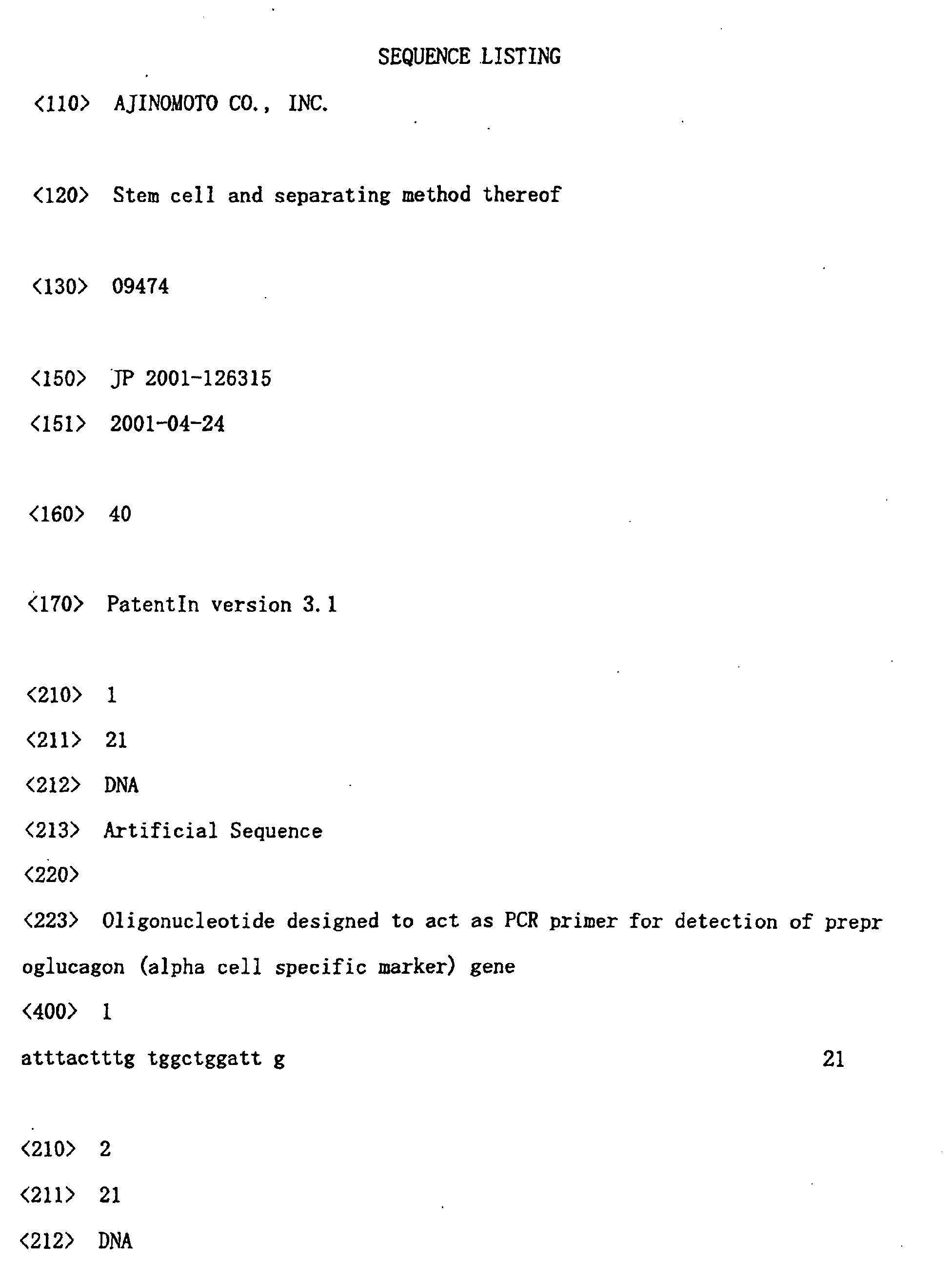
Sequence No. 1: oligonucleotide designed to act as a PCR primer for detecting preproglucagon (α cell specific marker) gene.
Sequence No. 2: oligonucleotide designed to act as a PCR primer for detecting preproglucagon (α cell specific marker) gene.
Sequence No. 3: oligonucleotide designed to act as a PCR primer for detecting preproinsulin I (β cell specific marker) gene.
Sequence No. 4: oligonucleotide designed to act as a PCR primer for detecting preproinsulin I (β cell specific marker) gene.
Sequence No. 5: oligonucleotide designed to act as a PCR primer for detecting preproinsulin II (β cell specific marker) gene.
Sequence No. 6: oligonucleotide designed to act as a PCR primer for detecting preproinsulin II (β cell specific marker) gene.
Sequence No. 7: oligonucleotide designed to act as a PCR primer for detecting pancreatic polypeptide (γ cell specific marker) gene.
Sequence No. 8: oligonucleotide designed to act as a PCR primer for detecting pancreatic polypeptide (γ cell specific marker) gene.
Sequence No. 9: oligonucleotide designed to act as a PCR primer for detecting preprosomatostatin (δ cell specific marker) gene.
Sequence No. 10: oligonucleotide designed to act as a PCR primer for detecting preprosomatostatin (δ cell specific marker) gene.
Sequence No. 11: oligonucleotide designed to act as a PCR primer for detecting amylase 2 (exocrine cell specific marker) gene.
Sequence No. 12: oligonucleotide designed to act as a PCR primer for detecting amylase 2 (exocrine cell specific marker) gene.
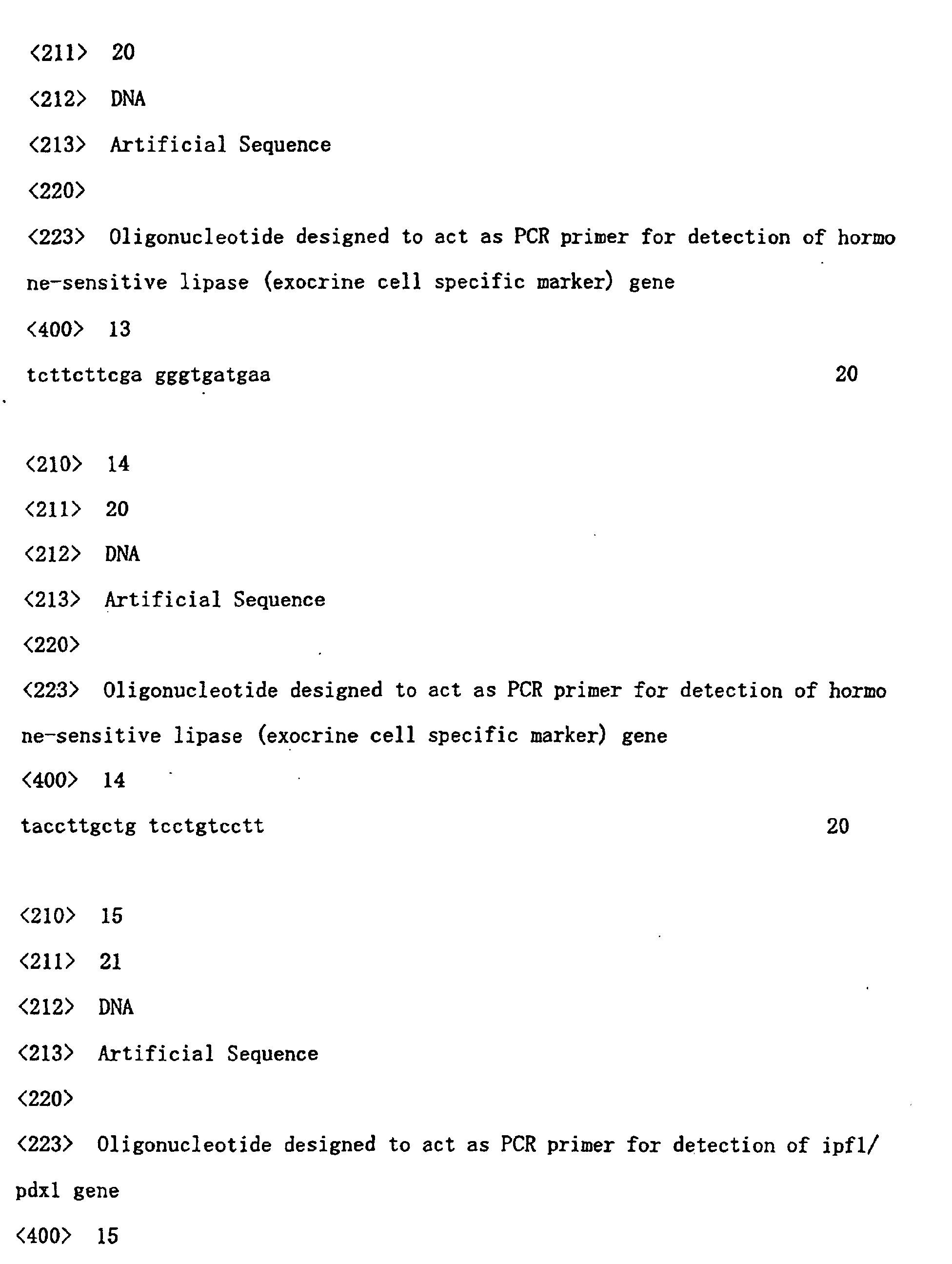
Sequence No. 13: oligonucleotide designed to act as a PCR primer for detecting hormone-sensitive lipase (exocrine cell specific marker) gene.
Sequence No. 14: oligonucleotide designed to act as a PCR primer for detecting hormone-sensitive lipase (exocrine cell specific marker) gene.
Sequence No. 15: oligonucleotide designed to act as a PCR primer for detecting ipf1/pdx1 gene.
Sequence No. 16: oligonucleotide designed to act as a PCR primer for detecting ipf1/pdx1 gene.
Sequence No. 17: oligonucleotide designed to act as a PCR primer for detecting c-Met gene.
Sequence No. 18: oligonucleotide designed to act as a PCR primer for detecting c-Met gene.
Sequence No. 19: oligonucleotide designed to act as a PCR primer for detecting Hypoxanthine Phosphoribosyl Transferase (HPRT, positive control) gene.
Sequence No. 20: oligonucleotide designed to act as a PCR primer for detecting Hypoxanthine Phosphoribosyl Transferase (HPRT, positive control) gene.
Sequence No. 21: oligonucleotide designed to act as a PCR primer for detecting albumin (liver· bile duct cell marker) gene.
Sequence No. 22: oligonucleotide designed to act as a PCR primer for detecting albumin (liver· bile duct cell marker) gene.
Sequence No. 23: oligonucleotide designed to act as a PCR primer for detecting α-fetoprotein (liver· bile duct cell marker) gene.
Sequence No. 24: oligonucleotide designed to act as a PCR primer for detecting α-fetoprotein (liver· bile duct cell marker) gene.
Sequence No. 25: oligonucleotide designed to act as a PCR primer for detecting glucose-6-phosphatase (liver· bile duct cell marker) gene.
Sequence No. 26: oligonucleotide designed to act as a PCR primer for detecting glucose-6-phosphatase (liver. bile duct cell marker) gene.
Sequence No. 27: oligonucleotide designed to act as a PCR primer for detecting Transthyretin (liver· bile duct cell marker) gene.
Sequence No. 28: oligonucleotide designed to act as a PCR primer for detecting transthyretin (liver· bile duct cell marker) gene.
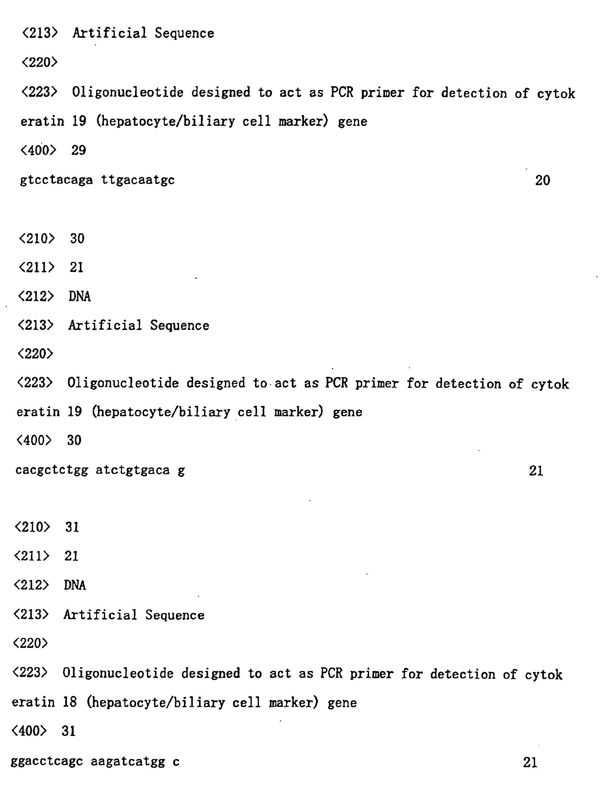
Sequence No. 29: oligonucleotide designed to act as a PCR primer for detecting cytokeratin 19 (liver. bile duct cell marker) gene.
Sequence No. 30: oligonucleotide designed to act as a PCR primer for detecting cytokeratin 19 (liver· bile duct cell marker) gene.
Sequence No. 31: oligonucleotide designed to act as a PCR primer for detecting cytokeratin 18 (liver· bile duct cell marker) gene.
Sequence No. 32: oligonucleotide designed to act as a PCR
primer for detecting cytokeratin 18 (liver· bile duct cell marker) gene.
Sequence No. 33: oligonucleotide designed to act as a PCR primer for detecting cytokeratin 8 (liver· bile duct cell marker) gene.
Sequence No. 34: oligonucleotide designed to act as a PCR primer for detecting cytokeratin 8 (liver· bile duct cell marker) gene.
Sequence No. 35: oligonucleotide designed to act as a PCR primer for detecting biliary glycoprotein (liver· bile duct cell marker) gene.
Sequence No. 36: oligonucleotide designed to act as a PCR primer for detecting biliary glycoprotein (liver· bile duct cell marker) gene.
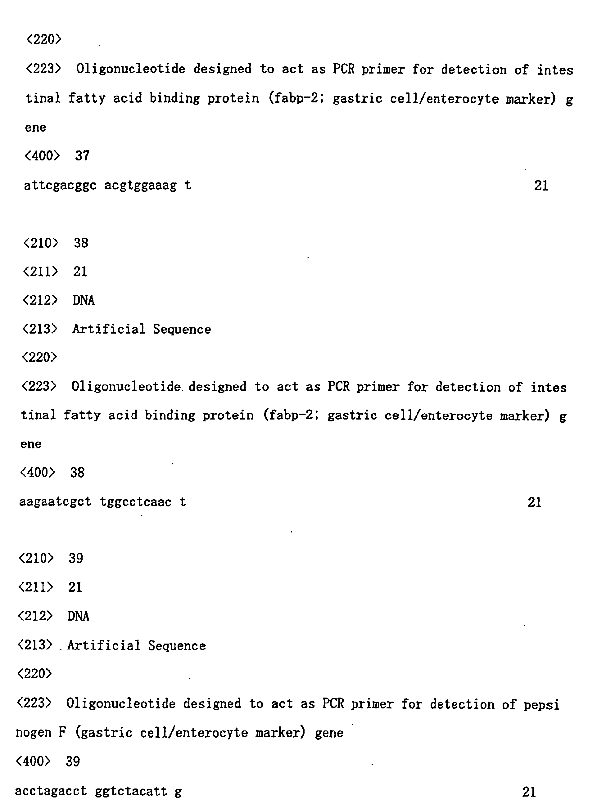
Sequence No. 37: oligonucleotide designed to act as a PCR primer for detecting intestinal fatty acid binding protein (stomach · intestine cell marker) gene.
Sequence No. 38: oligonucleotide designed to act as a PCR primer for detecting intestinal fatty acid binding protein (stomach · intestine cell marker) gene.
Sequence No. 39: oligonucleotide designed to act as a PCR primer for detecting pepsinogen F (stomach · intestine cell marker) gene.
Sequence No. 40: oligonucleotide designed to act as a PCR primer for detecting pepsinogen F (stomach · intestine cell marker) gene.







Claims (19)
- A method of separating a pancreatic stem cell from the pancreas of a mammal using two or more kinds of substances having specific affinity for a marker protein selected from the group consisting of c-Met, c-Kit, CD45 and TER119, or a gene encoding the same.
- A method of separating a pancreatic stem cell from the pancreas of a mammal using two or more kinds of substances having specific affinity for a marker protein selected from the group consisting of c-Met, c-Kit, CD45, TER119 and Flk-1, or a gene encoding the same.
- The method of claim 1 or 2, wherein the substance having specific affinity is an antibody against the marker protein.
- A method of identifying a pancreatic stem cell of a mammal using two or more kinds of substances having specific affinity for a marker protein selected from the group consisting of c-Met, c-Kit, CD45 and TER119, or a gene encoding the same.
- A method of identifying a pancreatic stem cell of a mammal using two or more kinds of substances having specific affinity for a marker protein selected from the group consisting of c-Met, c-Kit, CD45, TER119 and Flk-1, or a gene encoding the same.
- The method of claim 4 or 5, wherein the substance having specific affinity is an antibody against the marker protein.
- A method of separating a pancreatic stem cell from the pancreas of a mammal, which comprises a step of analyzing the expression state of two or more marker proteins selected from the group consisting of c-Met, c-Kit, CD45 and TER119, or a gene encoding the same.
- A method of separating a pancreatic stem cell from the pancreas of a mammal, which comprises a step of analyzing the expression state of two or more marker proteins selected from the group consisting of c-Met, c-Kit, CD45, TER119 and Flk-1, or a gene encoding the same.
- A method of identifying a pancreatic stem cell from the pancreas of a mammal, which comprises a step of analyzing the expression state of two or more marker proteins selected from the group consisting of c-Met, c-Kit, CD45 and TER119, or a gene encoding the same.
- A method of identifying a pancreatic stem cell from the pancreas of a mammal, which comprises a step of analyzing the expression state of two or more marker proteins selected from the group consisting of c-Met, c-Kit, CD45, TER119 and Flk-1, or a gene encoding the same.
- A pancreatic stem cell that can be separated from the pancreas of a mammal by the method described in any of claims 1, 2, 7 and 8.
- The pancreatic stem cell of claim 11, which shows 4 markers of c-Met, c-Kit, CD45 and TER119 in a pattern of c-Met+, c-Kit-, CD45-, TER119-.
- The pancreatic stem cell of claim 11, which shows 5 markers of c-Met, c-Kit, CD45, TER119 and Flk-1 in a pattern of c-Met+, c-Kit-, CD45-, TER119-, Flk-1-.
- An agent for the prophylaxis or treatment of a pancreatic hypofunctional disease, which comprises the pancreatic stem cell of claim 12 or 13, or a cell differentiated from the stem cell.
- An agent for the prophylaxis or treatment of a hypofunctional disease of the liver· bile duct or the stomach · intestine, which comprises the pancreatic stem cell of claim 12 or 13, or a cell differentiated from the stem cell.
- A method of screening a substance that induces differentiation of a pancreatic stem cell of a mammal, which comprises the following steps:(i) a step of reacting a pancreatic stem cell with a test substance, and(ii) a step of determining the expression of a pancreatic marker in the cell after the reaction.
- A method of screening a substance that induces differentiation into liver · bile duct or stomach · intestine of a mammal, which comprises the following steps:(i) a step of reacting a pancreatic stem cell of claim 12 or 13 with a test substance, and(ii) a step of determining the expression of a liver· bile duct or stomach · intestine marker in the cell after the reaction.
- A method of screening a substance that regulates a pancreatic function of a mammal, which comprises the following steps:(i) a step of reacting a pancreatic stem cell or a cell differentiated from the stem cell with a test substance, and(ii) a step of determining the expression of a pancreatic marker in the cell after the reaction.
- A method of screening a substance that regulates the function of liver· bile duct or stomach · intestine of a mammal, which comprises the following steps:(i) a step of reacting a pancreatic stem cell of claim 12 or 13 or a cell differentiated from the stem cell with a test substance, and(ii) a step of determining the expression of a liver· bile duct or stomach · intestine marker in the cell after the reaction.
Applications Claiming Priority (3)
| Application Number | Priority Date | Filing Date | Title |
|---|---|---|---|
| JP2001126315 | 2001-04-24 | ||
| JP2001126315 | 2001-04-24 | ||
| PCT/JP2002/004084 WO2002088335A1 (en) | 2001-04-24 | 2002-04-24 | Stem cells and method of separating the same |
Publications (3)
| Publication Number | Publication Date |
|---|---|
| EP1391505A1 true EP1391505A1 (en) | 2004-02-25 |
| EP1391505A4 EP1391505A4 (en) | 2004-09-29 |
| EP1391505B1 EP1391505B1 (en) | 2009-01-28 |
Family
ID=18975375
Family Applications (1)
| Application Number | Title | Priority Date | Filing Date |
|---|---|---|---|
| EP02720586A Expired - Lifetime EP1391505B1 (en) | 2001-04-24 | 2002-04-24 | Stem cells and method of separating the same |
Country Status (7)
| Country | Link |
|---|---|
| US (1) | US20050003529A1 (en) |
| EP (1) | EP1391505B1 (en) |
| JP (1) | JP4296781B2 (en) |
| CN (1) | CN100516204C (en) |
| AT (1) | ATE421991T1 (en) |
| DE (1) | DE60231035D1 (en) |
| WO (1) | WO2002088335A1 (en) |
Cited By (10)
| Publication number | Priority date | Publication date | Assignee | Title |
|---|---|---|---|---|
| EP1780267A3 (en) * | 2005-05-27 | 2007-08-01 | Lifescan, Inc. | Amniotic fluid derived cells |
| EP1670900A4 (en) * | 2003-08-27 | 2008-06-11 | Stemcells California Inc | Enriched pancreatic stem cell and progenitor cell populations, and methods for identifying, isolating and enriching for such populations |
| US9694035B2 (en) | 2004-03-22 | 2017-07-04 | Mesoblast International Sarl | Mesenchymal stem cells and uses therefor |
| US10344264B2 (en) | 2012-12-31 | 2019-07-09 | Janssen Biotech, Inc. | Culturing of human embryonic stem cells at the air-liquid interface for differentiation into pancreatic endocrine cells |
| US10351820B2 (en) | 2008-06-30 | 2019-07-16 | Janssen Biotech, Inc. | Methods for making definitive endoderm using at least GDF-8 |
| US10358628B2 (en) | 2011-12-22 | 2019-07-23 | Janssen Biotech, Inc. | Differentiation of human embryonic stem cells into single hormonal insulin positive cells |
| US10370644B2 (en) | 2012-12-31 | 2019-08-06 | Janssen Biotech, Inc. | Method for making human pluripotent suspension cultures and cells derived therefrom |
| US10377989B2 (en) | 2012-12-31 | 2019-08-13 | Janssen Biotech, Inc. | Methods for suspension cultures of human pluripotent stem cells |
| US10420803B2 (en) | 2016-04-14 | 2019-09-24 | Janssen Biotech, Inc. | Differentiation of pluripotent stem cells to intestinal midgut endoderm cells |
| US10456424B2 (en) | 2007-07-31 | 2019-10-29 | Janssen Biotech, Inc. | Pancreatic endocrine cells and methods thereof |
Families Citing this family (33)
| Publication number | Priority date | Publication date | Assignee | Title |
|---|---|---|---|---|
| US7332589B2 (en) * | 2001-09-26 | 2008-02-19 | University Of North Carolina At Chapel Hill | Variants of alpha-fetoprotein coding and expression sequences |
| US8017395B2 (en) | 2004-12-17 | 2011-09-13 | Lifescan, Inc. | Seeding cells on porous supports |
| EP1888123B1 (en) | 2005-06-08 | 2013-01-09 | Janssen Biotech, Inc. | A cellular therapy for ocular degeneration |
| WO2007009356A1 (en) * | 2005-07-15 | 2007-01-25 | Theracells Biotechnologies Co., Ltd. | A method for detecting and culturing pancreatic cells and their application |
| CN1982440B (en) * | 2005-07-15 | 2010-11-17 | 广西南宁灵康赛诺科生物科技有限公司 | Cultured pancreatic cells and methods and uses thereof |
| US8741643B2 (en) | 2006-04-28 | 2014-06-03 | Lifescan, Inc. | Differentiation of pluripotent stem cells to definitive endoderm lineage |
| US20080050814A1 (en) * | 2006-06-05 | 2008-02-28 | Cryo-Cell International, Inc. | Procurement, isolation and cryopreservation of fetal placental cells |
| WO2007146105A2 (en) * | 2006-06-05 | 2007-12-21 | Cryo-Cell International, Inc. | Procurement, isolation and cryopreservation of fetal placental cells |
| US20080124684A1 (en) * | 2006-11-28 | 2008-05-29 | Grell Mathew L | System and method of celebration by personalizing apparel |
| US9080145B2 (en) | 2007-07-01 | 2015-07-14 | Lifescan Corporation | Single pluripotent stem cell culture |
| ATE523585T1 (en) | 2007-11-27 | 2011-09-15 | Lifescan Inc | DIFFERENTIATION OF HUMAN EMBRYONAL STEM CELLS |
| CA2959401C (en) | 2008-02-21 | 2021-12-07 | Centocor Ortho Biotech Inc. | Methods, surface modified plates and compositions for cell attachment, cultivation and detachment |
| US8623648B2 (en) | 2008-04-24 | 2014-01-07 | Janssen Biotech, Inc. | Treatment of pluripotent cells |
| MX2011004563A (en) | 2008-10-31 | 2011-06-01 | Centocor Ortho Biotech Inc | Differentiation of human embryonic stem cells to the pancreatic endocrine lineage. |
| CN102272291B (en) | 2008-10-31 | 2018-01-16 | 詹森生物科技公司 | Differentiation of human embryonic stem cells to the pancreatic endocrine lineage |
| PL2366022T3 (en) | 2008-11-20 | 2016-11-30 | Methods and compositions for cell attachment and cultivation on planar substrates | |
| PL2366021T3 (en) | 2008-11-20 | 2017-12-29 | Janssen Biotech, Inc. | Pluripotent stem cell culture on micro-carriers |
| ES2693088T3 (en) | 2009-07-20 | 2018-12-07 | Janssen Biotech, Inc. | Differentiation of human embryonic stem cells |
| KR20170118969A (en) | 2009-07-20 | 2017-10-25 | 얀센 바이오테크 인코포레이티드 | Differentiation of human embryonic stem cells |
| AU2010276402B2 (en) | 2009-07-20 | 2014-07-03 | Janssen Biotech, Inc. | Differentiation of human embryonic stem cells |
| MX339669B (en) | 2009-12-23 | 2016-06-02 | Janssen Biotech Inc | Differentiation of human embryonic stem cells. |
| AU2010333839C1 (en) | 2009-12-23 | 2016-07-21 | Janssen Biotech Inc. | Differentiation of human embryonic stem cells |
| CN107189979B (en) | 2010-03-01 | 2021-05-04 | 詹森生物科技公司 | Methods of purifying cells derived from pluripotent stem cells |
| KR101986176B1 (en) | 2010-05-12 | 2019-06-05 | 얀센 바이오테크 인코포레이티드 | Differentiation of human embryonic stem cells |
| WO2012030539A2 (en) | 2010-08-31 | 2012-03-08 | Janssen Biotech, Inc. | Differentiation of human embryonic stem cells |
| AU2011296381B2 (en) | 2010-08-31 | 2016-03-31 | Janssen Biotech, Inc. | Differentiation of human embryonic stem cells |
| WO2012030540A2 (en) | 2010-08-31 | 2012-03-08 | Janssen Biotech, Inc. | Differentiation of pluripotent stem cells |
| CA2866590A1 (en) | 2012-03-07 | 2013-09-12 | Janssen Biotech, Inc. | Defined media for expansion and maintenance of pluripotent stem cells |
| WO2013184888A1 (en) | 2012-06-08 | 2013-12-12 | Janssen Biotech, Inc. | Differentiation of human embryonic stem cells into pancreatic endocrine cells |
| RU2684215C2 (en) | 2012-12-31 | 2019-04-04 | Янссен Байотек, Инк. | Method for obtaining pancreatic endocrine cells (versions) and method for increasing output of beta cells |
| JP6588969B2 (en) | 2014-05-16 | 2019-10-09 | ヤンセン バイオテツク,インコーポレーテツド | Use of small molecules to enhance MAFA expression in pancreatic endocrine cells |
| AU2019265019B2 (en) | 2018-05-11 | 2025-11-06 | Beam Therapeutics Inc. | Methods of substituting pathogenic amino acids using programmable base editor systems |
| CN108753685B (en) * | 2018-06-20 | 2022-06-28 | 首都医科大学 | Separation, screening, culture and function identification method of human aortic vessel wall stem cells expressing c-Kit |
Family Cites Families (7)
| Publication number | Priority date | Publication date | Assignee | Title |
|---|---|---|---|---|
| CA2235509A1 (en) * | 1995-10-25 | 1997-05-01 | Ammon B. Peck | In vitro growth of functional islets of langerhans and in vivo uses thereof |
| US6232288B1 (en) * | 1995-11-09 | 2001-05-15 | Takeda Chemical Industries, Ltd. | Composition for improving pancreatic function |
| WO1999049015A2 (en) * | 1998-03-23 | 1999-09-30 | Zymogenetics, Inc. | Cardiac-derived stem cells |
| WO2000014203A1 (en) * | 1998-09-02 | 2000-03-16 | Chugai Seiyaku Kabushiki Kaisha | Method for preparing cell fraction containing hematopoietic stem cells |
| JP2000217576A (en) * | 1999-02-02 | 2000-08-08 | Herikkusu Kenkyusho:Kk | Induction of differentiation into adipocyte, compound which controls differentiation into adipocyte, and its screening |
| CA2362593A1 (en) * | 1999-02-10 | 2000-08-17 | Curis, Inc. | Pancreatic progenitor cells, methods and uses related thereto |
| JP2001086982A (en) * | 1999-07-16 | 2001-04-03 | Shunpei Niida | Bone resorption regulator |
-
2002
- 2002-04-24 AT AT02720586T patent/ATE421991T1/en not_active IP Right Cessation
- 2002-04-24 WO PCT/JP2002/004084 patent/WO2002088335A1/en not_active Ceased
- 2002-04-24 EP EP02720586A patent/EP1391505B1/en not_active Expired - Lifetime
- 2002-04-24 JP JP2002585616A patent/JP4296781B2/en not_active Expired - Fee Related
- 2002-04-24 CN CNB028124758A patent/CN100516204C/en not_active Expired - Fee Related
- 2002-04-24 DE DE60231035T patent/DE60231035D1/en not_active Expired - Lifetime
-
2003
- 2003-10-27 US US10/693,712 patent/US20050003529A1/en not_active Abandoned
Cited By (18)
| Publication number | Priority date | Publication date | Assignee | Title |
|---|---|---|---|---|
| EP1670900A4 (en) * | 2003-08-27 | 2008-06-11 | Stemcells California Inc | Enriched pancreatic stem cell and progenitor cell populations, and methods for identifying, isolating and enriching for such populations |
| US10668101B2 (en) | 2004-03-22 | 2020-06-02 | Mesoblast International Sárl | Mesenchymal stem cells and uses therefor |
| US9694035B2 (en) | 2004-03-22 | 2017-07-04 | Mesoblast International Sarl | Mesenchymal stem cells and uses therefor |
| US9943547B2 (en) | 2004-03-22 | 2018-04-17 | Mesoblast International Sàrl | Mesenchymal stem cells and uses therefor |
| US11389484B2 (en) | 2004-03-22 | 2022-07-19 | Mesoblast International Sárl | Mesenchymal stem cells and uses therefor |
| US10960025B2 (en) | 2004-03-22 | 2021-03-30 | Mesoblast International Sárl | Mesenchymal stem cells and uses therefor |
| US10828334B1 (en) | 2004-03-22 | 2020-11-10 | Mesoblast International Sárl | Mesenchymal stem cells and uses therefor |
| US10729727B2 (en) | 2004-03-22 | 2020-08-04 | Mesoblast International Sárl | Mesenchymal stem cells and uses therefor |
| US10716814B2 (en) | 2004-03-22 | 2020-07-21 | Mesoblast International Sàrl | Mesenchymal stem cells and uses therefor |
| EP1780267A3 (en) * | 2005-05-27 | 2007-08-01 | Lifescan, Inc. | Amniotic fluid derived cells |
| EP2302036A3 (en) * | 2005-05-27 | 2011-06-15 | Lifescan, Inc. | Amniotic fluid derived cells |
| US10456424B2 (en) | 2007-07-31 | 2019-10-29 | Janssen Biotech, Inc. | Pancreatic endocrine cells and methods thereof |
| US10351820B2 (en) | 2008-06-30 | 2019-07-16 | Janssen Biotech, Inc. | Methods for making definitive endoderm using at least GDF-8 |
| US10358628B2 (en) | 2011-12-22 | 2019-07-23 | Janssen Biotech, Inc. | Differentiation of human embryonic stem cells into single hormonal insulin positive cells |
| US10377989B2 (en) | 2012-12-31 | 2019-08-13 | Janssen Biotech, Inc. | Methods for suspension cultures of human pluripotent stem cells |
| US10370644B2 (en) | 2012-12-31 | 2019-08-06 | Janssen Biotech, Inc. | Method for making human pluripotent suspension cultures and cells derived therefrom |
| US10344264B2 (en) | 2012-12-31 | 2019-07-09 | Janssen Biotech, Inc. | Culturing of human embryonic stem cells at the air-liquid interface for differentiation into pancreatic endocrine cells |
| US10420803B2 (en) | 2016-04-14 | 2019-09-24 | Janssen Biotech, Inc. | Differentiation of pluripotent stem cells to intestinal midgut endoderm cells |
Also Published As
| Publication number | Publication date |
|---|---|
| US20050003529A1 (en) | 2005-01-06 |
| EP1391505B1 (en) | 2009-01-28 |
| JPWO2002088335A1 (en) | 2004-08-19 |
| JP4296781B2 (en) | 2009-07-15 |
| EP1391505A4 (en) | 2004-09-29 |
| WO2002088335A1 (en) | 2002-11-07 |
| CN1518593A (en) | 2004-08-04 |
| DE60231035D1 (en) | 2009-03-19 |
| ATE421991T1 (en) | 2009-02-15 |
| CN100516204C (en) | 2009-07-22 |
Similar Documents
| Publication | Publication Date | Title |
|---|---|---|
| EP1391505B1 (en) | Stem cells and method of separating the same | |
| AU2018390960B2 (en) | Immune cell organoid co-cultures | |
| EP3600409B1 (en) | T cell receptors and immune therapy using the same against prame positive cancers | |
| JP6788629B2 (en) | How to Create a Concentrated Tumor-Reactive T Cell Population from a Tumor | |
| JP6653689B2 (en) | Cancer stem cell population and method for producing the same | |
| JP2003517592A (en) | Markers for identification and isolation of islet α and β precursors | |
| TWI614340B (en) | Somatic stem cells and method of preparing same | |
| US8540982B2 (en) | Methods for inducing a natural killer (NK) cell-mediated immune response and for increasing NK cell activity | |
| US20110111434A1 (en) | Colon stem cells associated with colitisand colorectal cancer and methods of use | |
| CN114828890A (en) | Treatment of diabetes with stem cell migration agent | |
| EP3922646A1 (en) | Detection of malignant tumor cells antibodies and uses thereof | |
| JP2019508503A (en) | Pancreatic stem cells and uses thereof | |
| CN120518771A (en) | A T cell receptor recognizing KRAS mutation and its coding sequence | |
| CN114250201A (en) | CAR-NK cell and preparation method and application thereof | |
| CN110564730B (en) | CD40L aptamer and application thereof | |
| KR102654677B1 (en) | A use of MMP-1 marker for predicting enhanced bone marrow cell migration of Tonsil-derived mesenchymal stem cell or conditioned medium thereof | |
| EP1394263A1 (en) | Method of detecting and separating undifferentiated liver cellss using dlk | |
| JPWO2005070964A1 (en) | How to isolate monocytes | |
| UA127162C2 (en) | T cell receptors and immune therapy using the same | |
| CN114746545B (en) | Engineering erythrocytes to present specific cancer neoantigens using artificial MHC | |
| JP4895479B2 (en) | Endothelial cell differentiation and proliferation method | |
| WO2026052047A1 (en) | Marker for characterizing epc subpopulation, epc subpopulation, and use thereof | |
| Cardones et al. | Genetic immunization with LYVE-1 cDNA yields function-blocking antibodies against native protein | |
| Cheng et al. | Interleukin-34 induced Arg1+ macrophages play a key role in breast cancer brain metastasis. | |
| JP2026077857A (en) | Immune cell organoid co-culture |
Legal Events
| Date | Code | Title | Description |
|---|---|---|---|
| PUAI | Public reference made under article 153(3) epc to a published international application that has entered the european phase |
Free format text: ORIGINAL CODE: 0009012 |
|
| 17P | Request for examination filed |
Effective date: 20031121 |
|
| AK | Designated contracting states |
Kind code of ref document: A1 Designated state(s): AT BE CH CY DE DK ES FI FR GB GR IE IT LI LU MC NL PT SE TR |
|
| AX | Request for extension of the european patent |
Extension state: AL LT LV MK RO SI |
|
| A4 | Supplementary search report drawn up and despatched |
Effective date: 20040813 |
|
| 17Q | First examination report despatched |
Effective date: 20070619 |
|
| GRAP | Despatch of communication of intention to grant a patent |
Free format text: ORIGINAL CODE: EPIDOSNIGR1 |
|
| GRAS | Grant fee paid |
Free format text: ORIGINAL CODE: EPIDOSNIGR3 |
|
| GRAA | (expected) grant |
Free format text: ORIGINAL CODE: 0009210 |
|
| AK | Designated contracting states |
Kind code of ref document: B1 Designated state(s): AT BE CH CY DE DK ES FI FR GB GR IE IT LI LU MC NL PT SE TR |
|
| AX | Request for extension of the european patent |
Extension state: AL LT LV MK RO SI |
|
| REG | Reference to a national code |
Ref country code: GB Ref legal event code: FG4D |
|
| REG | Reference to a national code |
Ref country code: CH Ref legal event code: EP |
|
| REG | Reference to a national code |
Ref country code: IE Ref legal event code: FG4D |
|
| REF | Corresponds to: |
Ref document number: 60231035 Country of ref document: DE Date of ref document: 20090319 Kind code of ref document: P |
|
| NLV1 | Nl: lapsed or annulled due to failure to fulfill the requirements of art. 29p and 29m of the patents act | ||
| LTIE | Lt: invalidation of european patent or patent extension |
Effective date: 20090128 |
|
| PG25 | Lapsed in a contracting state [announced via postgrant information from national office to epo] |
Ref country code: NL Free format text: LAPSE BECAUSE OF FAILURE TO SUBMIT A TRANSLATION OF THE DESCRIPTION OR TO PAY THE FEE WITHIN THE PRESCRIBED TIME-LIMIT Effective date: 20090128 Ref country code: FI Free format text: LAPSE BECAUSE OF FAILURE TO SUBMIT A TRANSLATION OF THE DESCRIPTION OR TO PAY THE FEE WITHIN THE PRESCRIBED TIME-LIMIT Effective date: 20090128 Ref country code: ES Free format text: LAPSE BECAUSE OF FAILURE TO SUBMIT A TRANSLATION OF THE DESCRIPTION OR TO PAY THE FEE WITHIN THE PRESCRIBED TIME-LIMIT Effective date: 20090509 |
|
| PG25 | Lapsed in a contracting state [announced via postgrant information from national office to epo] |
Ref country code: SE Free format text: LAPSE BECAUSE OF FAILURE TO SUBMIT A TRANSLATION OF THE DESCRIPTION OR TO PAY THE FEE WITHIN THE PRESCRIBED TIME-LIMIT Effective date: 20090428 Ref country code: AT Free format text: LAPSE BECAUSE OF FAILURE TO SUBMIT A TRANSLATION OF THE DESCRIPTION OR TO PAY THE FEE WITHIN THE PRESCRIBED TIME-LIMIT Effective date: 20090128 Ref country code: PT Free format text: LAPSE BECAUSE OF FAILURE TO SUBMIT A TRANSLATION OF THE DESCRIPTION OR TO PAY THE FEE WITHIN THE PRESCRIBED TIME-LIMIT Effective date: 20090629 |
|
| PG25 | Lapsed in a contracting state [announced via postgrant information from national office to epo] |
Ref country code: BE Free format text: LAPSE BECAUSE OF FAILURE TO SUBMIT A TRANSLATION OF THE DESCRIPTION OR TO PAY THE FEE WITHIN THE PRESCRIBED TIME-LIMIT Effective date: 20090128 |
|
| PG25 | Lapsed in a contracting state [announced via postgrant information from national office to epo] |
Ref country code: DK Free format text: LAPSE BECAUSE OF FAILURE TO SUBMIT A TRANSLATION OF THE DESCRIPTION OR TO PAY THE FEE WITHIN THE PRESCRIBED TIME-LIMIT Effective date: 20090128 |
|
| REG | Reference to a national code |
Ref country code: CH Ref legal event code: PL |
|
| PLBE | No opposition filed within time limit |
Free format text: ORIGINAL CODE: 0009261 |
|
| STAA | Information on the status of an ep patent application or granted ep patent |
Free format text: STATUS: NO OPPOSITION FILED WITHIN TIME LIMIT |
|
| 26N | No opposition filed |
Effective date: 20091029 |
|
| PG25 | Lapsed in a contracting state [announced via postgrant information from national office to epo] |
Ref country code: CH Free format text: LAPSE BECAUSE OF NON-PAYMENT OF DUE FEES Effective date: 20090430 Ref country code: LI Free format text: LAPSE BECAUSE OF NON-PAYMENT OF DUE FEES Effective date: 20090430 |
|
| REG | Reference to a national code |
Ref country code: IE Ref legal event code: MM4A |
|
| PG25 | Lapsed in a contracting state [announced via postgrant information from national office to epo] |
Ref country code: IE Free format text: LAPSE BECAUSE OF NON-PAYMENT OF DUE FEES Effective date: 20090424 Ref country code: MC Free format text: LAPSE BECAUSE OF NON-PAYMENT OF DUE FEES Effective date: 20090430 |
|
| PG25 | Lapsed in a contracting state [announced via postgrant information from national office to epo] |
Ref country code: GR Free format text: LAPSE BECAUSE OF FAILURE TO SUBMIT A TRANSLATION OF THE DESCRIPTION OR TO PAY THE FEE WITHIN THE PRESCRIBED TIME-LIMIT Effective date: 20090429 |
|
| PG25 | Lapsed in a contracting state [announced via postgrant information from national office to epo] |
Ref country code: IT Free format text: LAPSE BECAUSE OF FAILURE TO SUBMIT A TRANSLATION OF THE DESCRIPTION OR TO PAY THE FEE WITHIN THE PRESCRIBED TIME-LIMIT Effective date: 20090128 |
|
| PG25 | Lapsed in a contracting state [announced via postgrant information from national office to epo] |
Ref country code: LU Free format text: LAPSE BECAUSE OF NON-PAYMENT OF DUE FEES Effective date: 20090424 |
|
| PG25 | Lapsed in a contracting state [announced via postgrant information from national office to epo] |
Ref country code: TR Free format text: LAPSE BECAUSE OF FAILURE TO SUBMIT A TRANSLATION OF THE DESCRIPTION OR TO PAY THE FEE WITHIN THE PRESCRIBED TIME-LIMIT Effective date: 20090128 |
|
| PG25 | Lapsed in a contracting state [announced via postgrant information from national office to epo] |
Ref country code: CY Free format text: LAPSE BECAUSE OF FAILURE TO SUBMIT A TRANSLATION OF THE DESCRIPTION OR TO PAY THE FEE WITHIN THE PRESCRIBED TIME-LIMIT Effective date: 20090128 |
|
| PGFP | Annual fee paid to national office [announced via postgrant information from national office to epo] |
Ref country code: DE Payment date: 20120502 Year of fee payment: 11 |
|
| PGFP | Annual fee paid to national office [announced via postgrant information from national office to epo] |
Ref country code: GB Payment date: 20120418 Year of fee payment: 11 Ref country code: FR Payment date: 20120504 Year of fee payment: 11 |
|
| REG | Reference to a national code |
Ref country code: DE Ref legal event code: R119 Ref document number: 60231035 Country of ref document: DE |
|
| GBPC | Gb: european patent ceased through non-payment of renewal fee |
Effective date: 20130424 |
|
| PG25 | Lapsed in a contracting state [announced via postgrant information from national office to epo] |
Ref country code: DE Free format text: LAPSE BECAUSE OF NON-PAYMENT OF DUE FEES Effective date: 20131101 Ref country code: GB Free format text: LAPSE BECAUSE OF NON-PAYMENT OF DUE FEES Effective date: 20130424 |
|
| REG | Reference to a national code |
Ref country code: FR Ref legal event code: ST Effective date: 20131231 |
|
| PG25 | Lapsed in a contracting state [announced via postgrant information from national office to epo] |
Ref country code: FR Free format text: LAPSE BECAUSE OF NON-PAYMENT OF DUE FEES Effective date: 20130430 |
|
| REG | Reference to a national code |
Ref country code: DE Ref legal event code: R079 Ref document number: 60231035 Country of ref document: DE Free format text: PREVIOUS MAIN CLASS: C12N0005080000 Ipc: C12N0005078900 |
|
| REG | Reference to a national code |
Ref country code: DE Ref legal event code: R119 Ref document number: 60231035 Country of ref document: DE Effective date: 20131101 Ref country code: DE Ref legal event code: R079 Ref document number: 60231035 Country of ref document: DE Free format text: PREVIOUS MAIN CLASS: C12N0005080000 Ipc: C12N0005078900 Effective date: 20140602 |