EP1388283A1 - Nonhuman model animal of oligodendrocyte development disability - Google Patents
Nonhuman model animal of oligodendrocyte development disability Download PDFInfo
- Publication number
- EP1388283A1 EP1388283A1 EP02724686A EP02724686A EP1388283A1 EP 1388283 A1 EP1388283 A1 EP 1388283A1 EP 02724686 A EP02724686 A EP 02724686A EP 02724686 A EP02724686 A EP 02724686A EP 1388283 A1 EP1388283 A1 EP 1388283A1
- Authority
- EP
- European Patent Office
- Prior art keywords
- developmental
- human animal
- disorders
- oligodendrocytes
- dap12
- Prior art date
- Legal status (The legal status is an assumption and is not a legal conclusion. Google has not performed a legal analysis and makes no representation as to the accuracy of the status listed.)
- Granted
Links
Images



Classifications
-
- C—CHEMISTRY; METALLURGY
- C12—BIOCHEMISTRY; BEER; SPIRITS; WINE; VINEGAR; MICROBIOLOGY; ENZYMOLOGY; MUTATION OR GENETIC ENGINEERING
- C12N—MICROORGANISMS OR ENZYMES; COMPOSITIONS THEREOF; PROPAGATING, PRESERVING, OR MAINTAINING MICROORGANISMS; MUTATION OR GENETIC ENGINEERING; CULTURE MEDIA
- C12N15/00—Mutation or genetic engineering; DNA or RNA concerning genetic engineering, vectors, e.g. plasmids, or their isolation, preparation or purification; Use of hosts therefor
- C12N15/09—Recombinant DNA-technology
- C12N15/63—Introduction of foreign genetic material using vectors; Vectors; Use of hosts therefor; Regulation of expression
- C12N15/79—Vectors or expression systems specially adapted for eukaryotic hosts
- C12N15/85—Vectors or expression systems specially adapted for eukaryotic hosts for animal cells
- C12N15/8509—Vectors or expression systems specially adapted for eukaryotic hosts for animal cells for producing genetically modified animals, e.g. transgenic
-
- A—HUMAN NECESSITIES
- A01—AGRICULTURE; FORESTRY; ANIMAL HUSBANDRY; HUNTING; TRAPPING; FISHING
- A01K—ANIMAL HUSBANDRY; AVICULTURE; APICULTURE; PISCICULTURE; FISHING; REARING OR BREEDING ANIMALS, NOT OTHERWISE PROVIDED FOR; NEW BREEDS OF ANIMALS
- A01K67/00—Rearing or breeding animals, not otherwise provided for; New or modified breeds of animals
- A01K67/027—New or modified breeds of vertebrates
- A01K67/0275—Genetically modified vertebrates, e.g. transgenic
- A01K67/0276—Knock-out vertebrates
-
- A—HUMAN NECESSITIES
- A61—MEDICAL OR VETERINARY SCIENCE; HYGIENE
- A61P—SPECIFIC THERAPEUTIC ACTIVITY OF CHEMICAL COMPOUNDS OR MEDICINAL PREPARATIONS
- A61P25/00—Drugs for disorders of the nervous system
-
- A—HUMAN NECESSITIES
- A61—MEDICAL OR VETERINARY SCIENCE; HYGIENE
- A61P—SPECIFIC THERAPEUTIC ACTIVITY OF CHEMICAL COMPOUNDS OR MEDICINAL PREPARATIONS
- A61P25/00—Drugs for disorders of the nervous system
- A61P25/14—Drugs for disorders of the nervous system for treating abnormal movements, e.g. chorea, dyskinesia
-
- A—HUMAN NECESSITIES
- A61—MEDICAL OR VETERINARY SCIENCE; HYGIENE
- A61P—SPECIFIC THERAPEUTIC ACTIVITY OF CHEMICAL COMPOUNDS OR MEDICINAL PREPARATIONS
- A61P25/00—Drugs for disorders of the nervous system
- A61P25/18—Antipsychotics, i.e. neuroleptics; Drugs for mania or schizophrenia
-
- A—HUMAN NECESSITIES
- A61—MEDICAL OR VETERINARY SCIENCE; HYGIENE
- A61P—SPECIFIC THERAPEUTIC ACTIVITY OF CHEMICAL COMPOUNDS OR MEDICINAL PREPARATIONS
- A61P25/00—Drugs for disorders of the nervous system
- A61P25/28—Drugs for disorders of the nervous system for treating neurodegenerative disorders of the central nervous system, e.g. nootropic agents, cognition enhancers, drugs for treating Alzheimer's disease or other forms of dementia
-
- A—HUMAN NECESSITIES
- A61—MEDICAL OR VETERINARY SCIENCE; HYGIENE
- A61P—SPECIFIC THERAPEUTIC ACTIVITY OF CHEMICAL COMPOUNDS OR MEDICINAL PREPARATIONS
- A61P43/00—Drugs for specific purposes, not provided for in groups A61P1/00-A61P41/00
-
- C—CHEMISTRY; METALLURGY
- C07—ORGANIC CHEMISTRY
- C07K—PEPTIDES
- C07K14/00—Peptides having more than 20 amino acids; Gastrins; Somatostatins; Melanotropins; Derivatives thereof
- C07K14/435—Peptides having more than 20 amino acids; Gastrins; Somatostatins; Melanotropins; Derivatives thereof from animals; from humans
- C07K14/46—Peptides having more than 20 amino acids; Gastrins; Somatostatins; Melanotropins; Derivatives thereof from animals; from humans from vertebrates
- C07K14/47—Peptides having more than 20 amino acids; Gastrins; Somatostatins; Melanotropins; Derivatives thereof from animals; from humans from vertebrates from mammals
- C07K14/4701—Peptides having more than 20 amino acids; Gastrins; Somatostatins; Melanotropins; Derivatives thereof from animals; from humans from vertebrates from mammals not used
- C07K14/4702—Regulators; Modulating activity
- C07K14/4705—Regulators; Modulating activity stimulating, promoting or activating activity
-
- C—CHEMISTRY; METALLURGY
- C07—ORGANIC CHEMISTRY
- C07K—PEPTIDES
- C07K14/00—Peptides having more than 20 amino acids; Gastrins; Somatostatins; Melanotropins; Derivatives thereof
- C07K14/435—Peptides having more than 20 amino acids; Gastrins; Somatostatins; Melanotropins; Derivatives thereof from animals; from humans
- C07K14/705—Receptors; Cell surface antigens; Cell surface determinants
-
- A—HUMAN NECESSITIES
- A01—AGRICULTURE; FORESTRY; ANIMAL HUSBANDRY; HUNTING; TRAPPING; FISHING
- A01K—ANIMAL HUSBANDRY; AVICULTURE; APICULTURE; PISCICULTURE; FISHING; REARING OR BREEDING ANIMALS, NOT OTHERWISE PROVIDED FOR; NEW BREEDS OF ANIMALS
- A01K2217/00—Genetically modified animals
- A01K2217/07—Animals genetically altered by homologous recombination
- A01K2217/075—Animals genetically altered by homologous recombination inducing loss of function, i.e. knock out
-
- A—HUMAN NECESSITIES
- A01—AGRICULTURE; FORESTRY; ANIMAL HUSBANDRY; HUNTING; TRAPPING; FISHING
- A01K—ANIMAL HUSBANDRY; AVICULTURE; APICULTURE; PISCICULTURE; FISHING; REARING OR BREEDING ANIMALS, NOT OTHERWISE PROVIDED FOR; NEW BREEDS OF ANIMALS
- A01K2227/00—Animals characterised by species
- A01K2227/10—Mammal
- A01K2227/105—Murine
-
- A—HUMAN NECESSITIES
- A01—AGRICULTURE; FORESTRY; ANIMAL HUSBANDRY; HUNTING; TRAPPING; FISHING
- A01K—ANIMAL HUSBANDRY; AVICULTURE; APICULTURE; PISCICULTURE; FISHING; REARING OR BREEDING ANIMALS, NOT OTHERWISE PROVIDED FOR; NEW BREEDS OF ANIMALS
- A01K2267/00—Animals characterised by purpose
- A01K2267/03—Animal model, e.g. for test or diseases
-
- A—HUMAN NECESSITIES
- A01—AGRICULTURE; FORESTRY; ANIMAL HUSBANDRY; HUNTING; TRAPPING; FISHING
- A01K—ANIMAL HUSBANDRY; AVICULTURE; APICULTURE; PISCICULTURE; FISHING; REARING OR BREEDING ANIMALS, NOT OTHERWISE PROVIDED FOR; NEW BREEDS OF ANIMALS
- A01K2267/00—Animals characterised by purpose
- A01K2267/03—Animal model, e.g. for test or diseases
- A01K2267/035—Animal model for multifactorial diseases
- A01K2267/0356—Animal model for processes and diseases of the central nervous system, e.g. stress, learning, schizophrenia, pain, epilepsy
Definitions
- the present invention relates to: a non-human animal model of oligodendrocyte developmental disorder whose DAP12 (DNAX Activation Protein 12) gene function is deficient on its chromosome; a screening method for a developmental promoter or a developmental suppressor of oligodendrocytes, or a promoter or a suppressor of myelinogenesis, wherein the non-human animal model of oligodendrocyte developmental disorder is used; a screening method for a therapeutic agent for neuropsychiatric disorders; a diagnostic method for neuropsychiatric disorders, etc.
- DAP12 DNAX Activation Protein 12
- DAP12 (DNAX Activation Protein 12) is indicated to be a transmembrane protein containing an activation motif, ITAM, and to bind to ZAP-70 or Syk through its phosphorylation. It is known that DAP12 is encoded by a single-copy gene located on the 19q13.1 in a human, and that it also exists in mice.
- mRNAs of the DAP12 largely express in monocytes, dendritic cells, or natural killer cells, and that DAP12 is not only involved in activated signaling of a KAR group, but also associates with a human signal regulatory protein (SIRP) beta1, a human or a murine myeloid DAP12-associating lectin (MDL) -1, and a triggering receptor expressed on myeloid cells (TREM). It is reported that DAP12 also associates with KAR molecules, which are belonged to C-type lectin family such as CD94/NKG2C, and the like, and performs its function (Immunity 8, 693-701, 1998; J. Immunol. 161, 7-10, 1998; J. Immunol. 160, 4148-52, 1998).
- Polycystic lipomembranous osteodysplasia with sclerosing leukoencephalopathy also known as Nasu-Hakola disease (Suppl. 232, 1-173, 1972; ActaPathol. Jpn. 23, 539-558, 1973; J. Med. Genet. 34, 753-757, 1977) is an exceptional central nervous system disorder found in Japan and Finland.
- Nasu-Hakola disease In addition to the formation of bone cysts, patients of Nasu-Hakola disease develop psychotic symptoms, such as personality changes, inevitably progressing to presenile dementia.
- the mutation found in Finnish patients is a 5.3 kb deletion in DAP12 [KARAP (Killer activating receptor associated protein)/TYROBP (protein tyrosine kinase binding protein) ] locus, while another defect in Japanese patients is a single-nucleotide deletion in the third exon of the gene. Both are known to be caused by the loss of function of DAP12, a membrane adaptor protein (J. Immunol. 158, 5083-5086, 1997; Nature 391, 703-707, 1998) found initially in the immune system (Nature Genet. 25, 357-361, 2000). However, it has not yet been known whether psychotic symptoms are caused by the deficiency of DAP12.
- Nasu-Hakola disease is an exceptional recessive hereditary disease, which is a fatal disease inevitably progressing to presenile dementia following the formation of bone cysts and neuropsychiatric disorders.
- the object of the present invention is to provide the following: a non-human animal model of oligodendrocyte developmental disorder whose DAP12 (DNAX Activation Protein 12) gene function is deficient on its chromosome; a screening method for a developmental promoter or a developmental suppressor of oligodendrocytes, or, a promoter or a suppressor of myelinogenesis, wherein the non-human animal model of oligodendrocyte developmental disorder is used; a screening method for a therapeutic agent for neuropsychiatric disorders; a diagnostic method for neuropsychiatric disorders, which enable to develop a preventive method for the progress of neuropsychiatric disorders and a therapeutic method for neuropsychiatric disorders by analyzing the mechanisms progressing to neuropsychiatric disorders such as Nasu-
- the present inventors have made a keen study to analyze the physiological function of DAP12, generated a mouse whose DAP12 gene function is deficient on its chromosome, namely a DAP12 knockout mouse, and examined the observations of the brain.
- demyelination namely myelination disorder including hypomyelinosis
- myelination disorder including hypomyelinosis
- this disorder is attributed to the inhibition of differentiation, development, intracerebral transfer in the cell due to the deficiency of DAP12 in oligodendrocytes that plays a role of myelinogenesis.
- the present inventors have further found out that deficiency can be seen in the reflective power in spite of that the muscle and the like are normal by the behavioral analysis of the above-mentioned DAP12 knockout mouse, and clarified that psychotic symptoms similar to schizophrenia can be seen with aging.
- the present invention is completed.
- the present invention relates to: a non-human animal model of oligodendrocyte developmental disorders whose DAP12 (DNAX Activation Protein 12) gene function is deficient on its chromosome, and that shows an oligodendrocyte developmental disorder (claim 1); the non-human animal model of oligodendrocyte developmental disorders according to claim 1, wherein a myelinogenesis developmental disorder has been initiated (claim 2); the non-human animal model of oligodendrocyte developmental disorders according to claim 1 or 2, wherein a neuropsychiatric disorder has been induced (claim 3); the non-human animal model of oligodendrocyte developmental disorders according claim 3, wherein the neuropsychiatric disorder is Nasu-Hakola disease, dementia, schizophrenia, schizotypal personality disorders, obsessive-compulsive disorders, Huntington's disease or Tourette's syndrome (claim 4); the non-human animal model of oligodendrocytes developmental disorders according to any of claims 1 to 4, wherein the non-human
- the present invention further relates to: the screening method for a developmental promoter or a developmental suppressor of oligodendrocytes according to any of claims 7 to 10, wherein a non-human animal whose DAP12 gene function is deficient on its chromosome and a wild-type non-human animal are compared and assessed (claim 11); the screening method for a developmental promoter or a developmental suppressor of oligodendrocytes according to any of claims 7 to 11, wherein the non-human animal is a mouse (claim 12); the screening method for a developmental promoter or a developmental suppressor of oligodendrocytes according to any of claims 7 to 12, wherein the developmental promoter or a developmental suppressor of oligodendrocytes is a promoter or a suppressor of myelinogenesis (claim 13); a developmental promoter or a developmental suppressor of oligodendrocytes that can be obtained by the screening method for a developmental promoter or a developmental suppressor of oligodendrocytes according
- the non-human animal model of oligodendrocytes developmental disorders of the present invention is a non-human animal wherein the development of oligodendrocytes are disordered due to the deficiency of DAP12 (DNAX activation protein 12) gene function on its chromosome.
- DAP12 DNAX activation protein 12
- the expression of myelin basic protein is reduced, and developmental disorder of myelinogenesis in central nervous system is initiated, or neuropsychiatric disorders such as Nasu-Hakola disease, dementia, schizophrenia, schizotypal personality disorders, obsessive-compulsive disorders, Huntington's disease and Tourette's syndrome, etc. , are induced with aging.
- non-human animal whose DAP12 gene function is deficient on its chromosome means a non-human animal whose whole or part of endogenous gene encoding DAP12 is inactivated by gene mutations such as destruction, deficiency, substitution, etc., and whose DAP12-expressing function is lost.
- non-human animals of the present invention can be particularly exemplified by rodents such as mice, rats, etc., the examples will not be limited to these animals.
- the wild-type non-human animal of the present invention means an animal of the same species as the above-mentioned non-human animal whose DAP12 gene function is deficient and its littermate can be preferably exemplified. It is preferable to simultaneously use the DAP12 deficient type and its wild-type littermate among these homozygous non-human animals that are born according to Mendel's Law, since accurate comparative experiments can be carried out on the individual level.
- the preferable non-human animal model of oligodendrocytes developmental disorders of the present invention can be particularly exemplified by a DAP12 knockout mouse, and the wild-type non-human animal can be exemplified by a wild-type littermate thereof respectively. It will be explained in the following with the case wherein the non-human animal is a mouse as an example.
- a mouse whose DAP12 gene function is deficient on its chromosome namely, a DAP12 knockout mouse (DAP12 -/- ) is generated by the following steps.
- a DAP12 knockout mouse can be generated by the method as described previously (Cell 76, 519-529, 1994), or the like.
- DAP12 gene is screened by using a gene fragment obtained by a method such as PCR method or the like from the mouse gene library. Whole or part of the screened DAP12 gene is, for instance, substituted with a marker gene such as a neomycin-resistant gene using a recombinant DNA technology.
- a gene such as a diphtheria toxin A fragment (DT-A) gene, a herpes simplex virus thymidine kinase (HSV-tk) gene, etc. is introduced onto the 5'-end to generate a targeting vector.
- the generated targeting vector is linearized and introduced into ES cells with electroporation or the like to cause homologous recombination.
- ES cells showing resistance to antibiotics such as G418, ganciclovir (GANC), etc. are selected. It is preferable to confirm whether the ES cells selected herein are the recombinants of the interest by Southern blotting or the like.
- the above-mentioned recombinant ES cells are microinjected into a murine blastocyst, and such blastocyst is placed back to the recipient mouse to generate a chimeric mouse.
- a heterozygous mouse can be obtained by intercrossing the chimeric mouse and a wild type mouse. By further intercrossing the heterozygous mice, a DAP12 knockout mouse can be obtained.
- Whether the DAP12 gene is deficient on its chromosome in the DAP12 knockout mouse can be examined by a method wherein DNA is isolated from the tail of a mouse obtained by the above-mentioned method and examined by Southern blotting, or a method wherein a protein extracted from a marrow mast cell of the mouse is examined by immunoblot analysis.
- the non-human animal model of oligodendrocyte developmental disorders of the present invention is useful for the following models: a model wherein a developmental disorder of oligodendrocytes occurs; a model wherein a developmental disorder of myelinogenesis in central nervous system occurs; a model wherein the process of myelinogenesis is investigated; a model wherein the onset processes of the diseases caused by oligodendrocyte developmental disorders, and neuropsychiatric disorders such as dementia caused by dysmyelination are investigated.
- oligodendrocyte developmental disorders for instance, neuropsychiatric disorders such as Nasu-Hakola disease, dementia, schizophrenia, schizotypal personality disorders, obsessive-compulsive disorders, Huntington's disease and Tourette's syndrome, etc, namely a developmental promoter or a developmental suppressor of oligodendrocytes, a promoter or a suppressor of myelinogenesis and the like.
- neuropsychiatric disorders such as Nasu-Hakola disease, dementia, schizophrenia, schizotypal personality disorders, obsessive-compulsive disorders, Huntington's disease and Tourette's syndrome, etc, namely a developmental promoter or a developmental suppressor of oligodendrocytes, a promoter or a suppressor of myelinogenesis and the like.
- the screening method for a developmental promoter or a developmental suppressor of oligodendrocytes, a promoter or a suppressor of myelinogenesis and the like of the present invention can be exemplified by the following: a method wherein a test substance is administered to the non-human animal model of oligodendrocyte developmental disorders of the present invention; a method wherein a tissue, an organ, or a cell derived from the non-human animal model of oligodendrocytes developmental disorders of the present invention is contacted with a test substance.
- the method wherein a tissue, an organ, or a cell derived from the non-human animal model of oligodendrocyte developmental disorders is contacted with a test substance can be particularly exemplified by, for instance, a method wherein a tissue, an organ, or a cell derived from a non-human animal model of oligodendrocyte developmental disorders such as the aforementioned DAP12 knockout mouse is contacted with a test substance, and the expression of myelin basic protein in the cells is measured and assessed.
- the method wherein a test substance is administered to the non-human animal model of oligodendrocyte developmental disorders can be particularly exemplified by the following, but they are not limited to these examples: a method wherein a test substance is administered to the aforementioned DAP12 knockout mouse, and the expression of myelin basic protein in a tissue, an organ, or a cell derived from the non-human animal is measured and assessed; a method wherein a test substance is administered to the aforementioned DAP12 knockout mouse, and the development of myelinogenesis and the extent of demyelination of the non-human animal are measured and assessed; a method wherein a test substance is administered to the aforementioned DAP12 knockout mouse, and acoustic stimuli and/or acoustic prepulse inhibition of the non-human animal are measured and assessed. In addition, it is preferable to compare and assess the DAP12 knockout mouse and the syngeneic wild-type littermate mouse in the above-mentioned screening
- Candidate substances of a developmental promoter or a developmental suppressor of oligodendrocytes that can be obtained by the screening method for a developmental promoter or a developmental suppressor of oligodendrocytes, a promoter or a suppressor of myelinogenesis of the present invention are exemplified by various low-molecular compounds such as tyrosine-dephosphorylated enzyme inhibitor, tyrosine-phosphorylated enzyme activator, and the like.
- the therapeutic agents for neuropsychiatric disorders such as Nasu-Hakola disease, dementia, schizophrenia, schizotypal personality disorders, obsessive-compulsive disorders, Huntington's disease, Tourette's syndrome, etc.
- the therapeutic agents containing a developmental promoter or a developmental suppressor of oligodendrocytes, a promoter or a suppressor of myelinogenesis that can be obtained by the above-mentioned screening methods for a developmental promoter or a developmental suppressor of oligodendrocytes, promoter or suppressor of myelinogenesis, as an active ingredient.
- the above-mentioned neuropsychiatric disorders can be treated by administering the therapeutic agents to mammalian animals and the like with an appropriate amount and in an appropriate manner.
- DAP12 knockout mice were generated by hybridizing the 129/SvJ (H-2 b ) and C57BL/6 (B6, H-2 b ) as described previously (Cell 76, 519-529, 1994).
- DAP12 genomic DNA was isolated from the 129/SvJ mouse gene library (Stratagene), and 5.1 kb of Bam HI fragment containing the promoter region and the exons 1-3 of a DAP12 gene was substituted by a neo r cassette (Stratagene).
- a targeting vector was constructed by inserting a herpes simplex virus thymidine kinase (HSV-TK) as a negative selectable marker.
- HSV-TK herpes simplex virus thymidine kinase
- the neo r cassette contained 5.1 and 1.2 kb of homologous sequences as the flanking sequences.
- homologous recombination after this vector was linearized and inserted into ES cells (RW4) by electroporation, homologous ES cell recombinants were obtained at a frequency of 7.1 %.
- ES clones were isolated from the above-mentioned homologous recombinants, and neomycin resistant ES clones was screened for G418 and GANG (gancyclovir), and a homologous recombinants were confirmed by Southern blotting. Genomic DNA was isolated from the homologous recombinants and digested with Kpn I. Then it was confirmed that a targeting allele including neo r cassette is included. These confirmed ES clones were microinjected into blastocysts to generate chimeric mice, and such generated chimeric mice were intercrossed with a wild-type C57BL/6 mouse (Charles River).
- G418 and GANG gancyclovir
- heterozygous mice were obtained by breeding them in the environmentally controlled and specific pathogen-free facility. These heterozygous mice were further intercrossed to obtain homozygous mice, and deficient mice whose DAP12 genes are deficient on their chromosomes and their wild-type mice were generated.
- Such generated DAP12 knockout mice of the present invention grew healthy without particular aberration at least up to 10-month-old as described previously (Immunity 13, 345-353, 2000; Immunity 13, 355-364, 2000).
- Bone marrow mast cells were prepared from DAP12 +/+ and DAP12 -/- mice as described previously (Cell 76, 519-529, 1994), and further examined by immunoblot analysis wherein protein extracted from the mast cells (corresponding 2.5 ⁇ 10 5 cells per lane) and anti-rabbit DAP12 antiserum (diluted at 1:500) prepared by the method described previously (Cell 70, 351, 1992) (Fig. 1c). As a result, DAP12 protein was not detected in cells derived from DAP12 -/- mice.
- DAP10 gene which is a cell surface adaptor protein of the same species as the above-mentioned DAP12 gene, is located on a similar chromosome DNA strand in the reverse transcription direction against DAP12 gene which is merely 0.1 kb apart (J. Immunol. 163, 4651-4654, 1999; Science 285, 730-732, 1999).
- RT-PCR method was conducted to examine whether the above-mentioned DAP12 knockout mice affect the DAP10 gene.
- Each cDNA was synthesized by using 3 ⁇ g of total RNA extracted from the above-mentioned DAP12 +/+ or DAP12 -/- mice-derived bone marrow mast cells (BMMCs), and a reverse transcriptase ReverTra Ace (TOYOBO).
- Each cDNA was amplified by PCR method using the primers described below, and a signal of DAP10 was confirmed. As a result, although the expression of DAP10 was normal in DAP12 -/- mice, the expression of DAP12 mRNA was not recognized.
- the primer specific to the above-mentioned DAP12 5'-atgggggctctggagccct-3' (Seq. ID No.
- Example 1 Status of a Neuron, a Neurofilament, and an Astrocyte in DAP12 Knockout Mice
- 3-month-old DAP12 knockout mice (-/-) or wild-type mice (+/+) were anesthetized, and perfused with acid-alcohol solution (95% ethanol/5% acetic acid; vol %). Brains were isolated from these mice, and fixed overnight with acid-alcohol solution. Fixed brains underwent a standard protocol for paraffination (practices of treating for 1-2 hours each with 100% ethanol, methyl benzonate, xylene, xylene-paraffin, or paraffin solution), then embedded in Paraffin. Embedded brains were sliced 10 ⁇ m thick using a microtome, and underwent a standard protocol for deparaffination (xylene, 100% ethanol, 90% ethanol, 70% ethanol, and PBS).
- Intracellular peroxidase activity was suppressed with 3% H 2 O 2 for 10 minutes, and rinsed again with PBS.
- Slices were treated in PBS containing primary antibodies including 0.5% skim milk (DIFCO) at 37° C for 1 hour. They were subsequently washed with PBS at 37°C for 3 minutes with shaking three times.
- the secondary antibodies were made to act in PBS containing 0.5% skim wherein 1:100 dilution of HRP-labeled anti-rabbit IgG antibody (MBL) is included at 37° C for 50 minutes.
- a rabbit anti-mouse MBP antibody (Nichirei) as a primary antibody
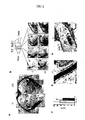
- dashed lined area "a” (Fig. 3a) or full lined area (Fig. b below) of the coronal brain slices shown in the schematic side view of Fig. 3b upper part were stained and visualized in the same manner as described in Example 1.
- the results are shown in Fig. 3a and b.
- “cc” means the corpus callosum, "ec” for the external capsule, “ic” for the internal capsule, “fim” for the fimbria, “thlm” for the thalamus, respectively.
- the scale bar indicates 250 ⁇ m.
- FIG. 3b shows the rough profile of a mouse, and the shaded area therein means the cerebrum.
- Cp in the Fig. 3b means the caudate nucleus, and the scale bar indicates 250 ⁇ m.
- myelin is a mutilamellar membrane structure, which enwraps axons and accelerates the conduction of nerve impulses, and serves as an important insulator for axon (Science 280, 1610-1613, 1998).
- myelin is synthesized by oligodendrocytes, and the formation of myelin sheath takes place immediately after birth.
- Many autopsy cases for human NHD patients showed frontally accentuated deficiency of myelin and thalamus degeneration (Acta Psychiatr. Scand. Suppl. 232, 1-173, 1972; Neuropathol. Appl. Neurobiol. 26, 98-101, 2000).
- MBP deficient mice (known as shiverers) result in severe hypomyelinosis of CNS axons accompanied with seizure behavior with generalized tremors culminating in premature death within 3 months (Neurosci. Lett. 12, 113-118, 1979).
- the reduction of MBP observed in adult DAP12 knockout mice (-/-) (Fig. 3 a, b) is attributed to hypomyelinosis in the animals due to an incomplete myelination, or demyelination caused by other reasons such as a normal turnover mechanism or inflammation.
- the caudate nucleus in frontal lobe of cerebrum was collected from 3-month-old DAP12 knockout (-/-) mice, and treated in 0.1 M of sodium cacodylate containing 1% of O S O 4 , then embedded in Epon 812. Ultrathin slices (85nm) of cerebrum were examined with Hitachi H-7100 system. As a result, obvious hypomyelinosis was recognized (Fig. 3c).
- Fig. 3e is a result of observation at magnification of 40,000, and the scale bars in Fig. 3d and e indicate 250 nm.
- oligodendroglias were cultured as described previously (Cell 76, 519-529, 1994). Brains from newborn wild-type mice (+/+) and DAP12 deficient mice (-/-) were dissected, and oligodendroglias were prepared by the method for separate cultures (J. Cell Biol. 85, 890-902, 1980). Cells were plated in approximately 10 ml of Dulbecco's modified Eagle's medium (DMEM) containing 10% fetal bovine serum (FBS) per brain, then oligodendrocytes attached to the astroglial monolayer were dissociated and purified by orbital shaking as described previously (Dev. Biol. 83, 311-327, 1981).
- DMEM Dulbecco's modified Eagle's medium
- FBS fetal bovine serum
- Oligodendrocytes (Oligo) and Astrocytes (Astro) were prepared respectively from the above-mentioned purified wild-type or DAP12 deficient mice, then immunoblot analysis was conducted using protein extracted from those cells (1 ⁇ 10 5 cells per lane) in the same manner as described in Example 1. Bone marrow mast cells (BMMC; 5 ⁇ 10 5 cells) prepared in Reference 1 were used as positive controls. The results are shown in Fig. 4a. The arrow in the figure indicates the position of DAP12. From these findings, it was confirmed by the concentration measurement that signal intensity of DAP12 in wild-type mouse-derived oligodendrocytes is almost comparable to the same number of mast cells. DAP12 signals were detected in mRNA of the oligodendrocytes as a result of RT-PCR analysis as described in Reference.
- the aforementioned purified oligodendrocytes derived from wild-type mice were further purified using a magnetic cell sorter (Miltenyi) and an anti-O4 monoclonal antibody. Then a small hole was made on the membrane of thus obtained O4 + cells, then cells were stained using a purified rabbit anti-DAP12 IgG antibody (anti-DAP12 antibody) or a rabbit IgG (control) as a control, and a FITC-labeled anti-rabbit IgG antibody. As a result of the flow cytometry analysis, it was confirmed that DAP12 is expressed in the oligodendrocytes being positive for O4 antigens (Fig. 4b).
- Brain slices (dashed lined area of coronal brain slices in the schematic side view of brain in Fig. 3b upper part) were generated from each of the above-mentioned mouse in the same manner as described in Example 1, and incubated using an anti-DAP12 antibody as a primary antibody overnight at 4°C. After an HRP labeled anti-rabbit IgG antibody as a secondary antibody was reacted, brain slices were immunostained with DAB.
- Fig. 4c-e The results are shown in Fig. 4c-e.
- the arrowheads in Fig. 4c indicate the cells that are doubly stained with DAP12 and MBP, and the scale bars in Fig. 4 c-e indicate 25 ⁇ m.
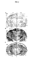
- Brain slices including the internal capsule (ic) and the thalamus (thlm) regions (dashed lined area (a) of coronal brain slices in the schematic side view of brain shown in Fig. 3b upper part) were prepared from 3-month-old DAP12 knockout mice (-/-) (Fig. 5a) or wild-type mice (+/+) (Fig. 5b and c), and the expression of MBP (Fig. 5a and b), or the expression of MBP and DAP12 (Fig. 5c) was examined.
- the expression of MBP was examined in the same manner as described in Example 2, and the expression of MBP and DAP12 was as described in Example 3. The results are shown in Fig. 5.
- the arrowheads in the figure indicate the internal capsule of cerebrum, and * mark indicates DAP12 positive region. Each scale bar corresponds to 100 ⁇ m. From these results, in 3-month-old (Fig. 5a) and 1.5-month-old DAP12 knockout mice (-/-), most of MBP positive oligodendrocytes were recognized in several regions such as the internal capsule (the arrowhead in Fig. 5a), the fimbria, the thalamus, and the like of cerebrum. On the other hand, the oligodendrocytes in the brain of an adult wild-type mouse (+/+) were hardly identified by MBP staining in low magnified views due to their development of numerous MBP-positive dendrites (Fig. 5b).
- DAP12 knockout mice the oligodendrocytes that create the formation of the myelin sheath in wild-type mice were not activated. These results suggest that DAP12 in CNS plays an important role in activation of the formation of myelin sheath in the late developmental phase as well as in the maintenance phase.
- the thalamus is one of the most important areas in the brain, which incorporates the impulses from every sensory organ and determines the subsequent responses by controlling nerve conductions. Therefore, thalamus-specific demyelination in DAP12 knockout mice could induce behavioral deficits.
- the DAP12 knockout mice used in the experiment showed normal behaviors.
- a MRI of a NHD patient found reduced signal intensity in the thalamus and putamen (Rinsho Shinkeigaku 32, 444-446, 1992). We therefore examined acoustic startle response and prepulse inhibition in the DAP12 knockout mice at 5 months of age.
- Fig. 6a Schematic view of acoustic startle response (the left side), and prepulse inhibition (the right side) are shown in Fig. 6a.
- Acoustic startle responses of mice were measured with the SR-LAB system (San Diego Instruments), and the background noise level in the chamber was 65dB during the test. Wild-type mice (+/+) or DAP12 knockout mice (+/+) were placed into cylinders 10 minutes prior to the initial startle stimuli and acclimated in the background noise. The maximum amplitude of startle response was measured by recording the amplitude of startle response for only 100 msecs from the startle stimuli initiated.
- each session consisted of seven different types of stimuli: no stimulus, startle stimulus trials only, or a prepulse proceeded by the startle stimulus. No response was seen in mice in any stimuli.
- Two startle trial types were 20 msec startle stimuli of either 100 or 120 dB.
- Each 20 msec prepulse stimulus (either 70 or 80 dB) was presented before both acoustic startle stimuli.
- the seven different types of stimuli were presented nine times in random order. The trials were conducted with an interval of an average of 40 secs.
- the acoustic startle reflex is known to be a simple neural circuit, receiving inputs from the thalamus and arising from diverse cortical, midbrain, or hindbrain centers.
- Prepulse inhibition of startle response which is a modification of the startle response by a weak prepulse, is a measure for sensorimotor gating, which coincides with a theoretical circuit of central inhibitory process in CNS by which an organism filters the flow of information via thalamus and gates the threshold of its subsequent behavior (J. Neurosci. 12, 4501-4509, 1992; Brain Res. 499, 7-17, 1989; Brain Res. Bull. 43, 219-228, 1997) (Fig. 4d).
- DAP12 knockout mice are considered to induce lower startle response and impaired sensorimotor gating possibly due to the thalamus-specific hypomyelinosis. Although the possibility that the DAP12 knockout mice develop more severe abnormalities in behavior or learning ability in the more aged phases is not excluded, it has become evident that they show psychiatric symptoms, similar to shizophrenia in humans, as early as 5 months of age.
- the non-human animal model of myelinogenesis developmental disorders of the present invention is useful for the following models: a model wherein developmental disorder of myelinogenesis in central nervous system occurs; a model wherein the process of myelinogenesis is investigated; a model wherein the onset processes of neuropsychiatric disorders such as dementia caused by demyelination are investigated.
- neuropsychiatric disorders such as Nasu-Hakola disease, dementia, schizophrenia, schizotypal personality disorders, obsessive-compulsive disorders, Huntington's disease and Tourette's syndrome, etc.
- neuropsychiatric disorders such as Nasu-Hakola disease, dementia, schizophrenia, schizotypal personality disorders, obsessive-compulsive disorders, Huntington's disease and Tourette's syndrome, etc.
Landscapes
- Health & Medical Sciences (AREA)
- Life Sciences & Earth Sciences (AREA)
- Chemical & Material Sciences (AREA)
- Organic Chemistry (AREA)
- Engineering & Computer Science (AREA)
- General Health & Medical Sciences (AREA)
- Genetics & Genomics (AREA)
- Bioinformatics & Cheminformatics (AREA)
- Zoology (AREA)
- Veterinary Medicine (AREA)
- Medicinal Chemistry (AREA)
- Biomedical Technology (AREA)
- Animal Behavior & Ethology (AREA)
- Biochemistry (AREA)
- Biophysics (AREA)
- Biotechnology (AREA)
- Molecular Biology (AREA)
- Neurosurgery (AREA)
- Chemical Kinetics & Catalysis (AREA)
- Pharmacology & Pharmacy (AREA)
- Nuclear Medicine, Radiotherapy & Molecular Imaging (AREA)
- Public Health (AREA)
- General Chemical & Material Sciences (AREA)
- Neurology (AREA)
- Wood Science & Technology (AREA)
- General Engineering & Computer Science (AREA)
- Proteomics, Peptides & Aminoacids (AREA)
- Environmental Sciences (AREA)
- Toxicology (AREA)
- Gastroenterology & Hepatology (AREA)
- Animal Husbandry (AREA)
- Physics & Mathematics (AREA)
- Cell Biology (AREA)
- Psychiatry (AREA)
- Plant Pathology (AREA)
- Microbiology (AREA)
- Biodiversity & Conservation Biology (AREA)
- Immunology (AREA)
- Hospice & Palliative Care (AREA)
- Psychology (AREA)
Abstract
Description


Claims (18)
- A non-human animal model of oligodendrocyte developmental disorders whose DAP12 (DNAX Activation Protein 12) gene function is deficient on its chromosome, and that shows an oligodendrocyte developmental disorder.
- The non-human animal model of oligodendrocyte developmental disorders according to claim 1, wherein a myelinogenesis developmental disorder has been initiated.
- The non-human animal model of oligodendrocyte developmental disorders according to claim 1 or 2, wherein a neuropsychiatric disorder has been induced.
- The non-human animal model of oligodendrocyte developmental disorders according claim 3, wherein the neuropsychiatric disorder is Nasu-Hakola disease, dementia, schizophrenia, schizotypal personality disorders, obsessive-compulsive disorders, Huntington's disease or Tourette's syndrome.
- The non-human animal model of oligodendrocyte developmental disorders according to any of claims 1 to 4, wherein the non-human animal is a mouse.
- A screening method for a developmental promoter or a developmental suppressor of oligodendrocytes, wherein a test substance is administered to the non-human animal model of oligodendrocyte developmental disorders according to any of claims 1 to 5, or a test substance is contacted with a tissue, an organ, or a cell derived from the animal.
- A screening method for a developmental promoter or a developmental suppressor of oligodendrocytes, wherein a tissue, an organ, or a cell derived from the non-human animal model of oligodendrocyte developmental disorders according to any of claims 1 to 5, is contacted with a test substance, and the expression of myelin basic protein in the cells is measured and assessed.
- A screening method for a developmental promoter or a developmental suppressor of oligodendrocytes, wherein a test substance is administered to the non-human animal model of oligodendrocyte developmental disorders according to any of claims 1 to 5, and the expression of myelin basic protein in a tissue, an organ, or a cell derived from the non-human animal is measured and assessed.
- A screening method for a developmental promoter or a developmental suppressor of oligodendrocytes, wherein a test substance is administered to the non-human animal model of oligodendrocyte developmental disorders according to any of claims 1 to 5, and the development of myelinogenesis or the extent of demyelination in the animal is measured and assessed.
- A screening method for a developmental promoter or a developmental suppressor of oligodendrocytes, wherein a test substance is administered to the non-human animal model of oligodendrocyte developmental disorders according to any of claims 1 to 5, and acoustic stimuli and/or acoustic prepulse inhibition of the non-human animal are measured and assessed.
- The screening method for a developmental promoter or a developmental suppressor of oligodendrocytes according to any of claims 7 to 10, wherein a non-human animal whose DAP12 gene function is deficient on its chromosome and a wild-type non-human animal are compared and assessed.
- The screening method for a developmental promoter or a developmental suppressor of oligodendrocytes according to any of claims 7 to 11, wherein the non-human animal is a mouse.
- The screening method for a developmental promoter or a developmental suppressor of oligodendrocytes according to any of claims 7 to 12, wherein the developmental processor or a developmental suppressor of oligodendrocytes is a promoter or a suppressor of myelinogenesis.
- A developmental promoter or a developmental suppressor of oligodendrocytes that can be obtained by the screening method for a developmental promoter or a developmental suppressor of oligodendrocytes according to any of claims 7 to 12.
- A promoter or a suppressor of myelinogenesis that can be obtained by the screening method for a developmental promoter or a developmental suppressor of oligodendrocytes according to claim 13.
- A screening method for a therapeutic agent for neuropsychiatric disorders, wherein the non-human animal model of oligodendrocyte developmental disorders according to any of claims 1 to 5 is used for the screening of a therapeutic agent for neuropsychiatric disorders.
- A therapeutic agent for neuropsychiatric disorders that can be obtained by the screening method for a therapeutic agent for neuropsychiatric disorders according to claim 16.
- A diagnostic method for neuropsychiatric disorders, wherein symptoms of the non-human animal model of oligodendrocyte developmental disorders according to any of claims 1 to 5 are utilized for diagnosis of neuropsychiatric disorders.
Applications Claiming Priority (3)
| Application Number | Priority Date | Filing Date | Title |
|---|---|---|---|
| JP2001146338 | 2001-05-16 | ||
| JP2001146338A JP5010784B2 (en) | 2001-05-16 | 2001-05-16 | Oligodendrocyte developmental disorder model non-human animal |
| PCT/JP2002/004405 WO2002091820A1 (en) | 2001-05-16 | 2002-05-02 | Nonhuman model animal of oligodendrocyte development disability |
Publications (3)
| Publication Number | Publication Date |
|---|---|
| EP1388283A1 true EP1388283A1 (en) | 2004-02-11 |
| EP1388283A4 EP1388283A4 (en) | 2007-02-14 |
| EP1388283B1 EP1388283B1 (en) | 2013-07-10 |
Family
ID=18992009
Family Applications (1)
| Application Number | Title | Priority Date | Filing Date |
|---|---|---|---|
| EP02724686.7A Expired - Lifetime EP1388283B1 (en) | 2001-05-16 | 2002-05-02 | Nonhuman model animal of oligodendrocyte development disability |
Country Status (6)
| Country | Link |
|---|---|
| US (1) | US7332644B2 (en) |
| EP (1) | EP1388283B1 (en) |
| JP (1) | JP5010784B2 (en) |
| AU (1) | AU2002255301B2 (en) |
| CA (1) | CA2447276C (en) |
| WO (1) | WO2002091820A1 (en) |
Families Citing this family (1)
| Publication number | Priority date | Publication date | Assignee | Title |
|---|---|---|---|---|
| JP2023549179A (en) * | 2020-11-10 | 2023-11-22 | ザ、トラスティーズ オブ プリンストン ユニバーシティ | Systems and methods for remote neurobehavioral testing |
Family Cites Families (3)
| Publication number | Priority date | Publication date | Assignee | Title |
|---|---|---|---|---|
| US5654139A (en) | 1994-09-21 | 1997-08-05 | The United States Of America As Represented By The Department Of Health And Human Services | Allelic variation of the serotonin 5HT2c receptor |
| AU7152296A (en) * | 1995-08-16 | 1997-03-12 | Board Of Trustees Of The Leland Stanford Junior University | Adult oligodendrocyte precursor cell compositions and methods |
| AU2002220591A1 (en) * | 2000-09-20 | 2002-04-02 | Institut National De La Sante Et De La Recherche Medicale (I.N.S.E.R.M) | Means for the identification of compounds capable of inhibiting karap-transduced signals |
-
2001
- 2001-05-16 JP JP2001146338A patent/JP5010784B2/en not_active Expired - Fee Related
-
2002
- 2002-05-02 EP EP02724686.7A patent/EP1388283B1/en not_active Expired - Lifetime
- 2002-05-02 CA CA2447276A patent/CA2447276C/en not_active Expired - Fee Related
- 2002-05-02 WO PCT/JP2002/004405 patent/WO2002091820A1/en not_active Ceased
- 2002-05-02 AU AU2002255301A patent/AU2002255301B2/en not_active Ceased
-
2003
- 2003-11-13 US US10/712,118 patent/US7332644B2/en not_active Expired - Fee Related
Also Published As
| Publication number | Publication date |
|---|---|
| AU2002255301B2 (en) | 2006-09-07 |
| WO2002091820A1 (en) | 2002-11-21 |
| EP1388283B1 (en) | 2013-07-10 |
| US7332644B2 (en) | 2008-02-19 |
| CA2447276A1 (en) | 2002-11-21 |
| JP2003018942A (en) | 2003-01-21 |
| US20050210538A1 (en) | 2005-09-22 |
| JP5010784B2 (en) | 2012-08-29 |
| CA2447276C (en) | 2011-09-27 |
| EP1388283A4 (en) | 2007-02-14 |
Similar Documents
| Publication | Publication Date | Title |
|---|---|---|
| AU671093B2 (en) | Transgenic animal models for alzheimer's disease | |
| Von Koch et al. | Generation of APLP2 KO mice and early postnatal lethality in APLP2/APP double KO mice | |
| Heber et al. | Mice with combined gene knock-outs reveal essential and partially redundant functions of amyloid precursor protein family members | |
| US6187992B1 (en) | Transgenic mouse having a disrupted amyloid precursor protein gene | |
| CA2268812C (en) | Method for producing transgenic animal models with modulated phenotype and animals produced therefrom | |
| Kaneko et al. | Transcription factor TEAD2 is involved in neural tube closure | |
| EP1005542B1 (en) | APOLIPOPROTEIN E TRANSGENIC mice AND ASSAY METHODS | |
| JP2001517065A (en) | Methods for identifying therapeutic agents for Alzheimer's disease using transgenic animal models | |
| US6452065B2 (en) | Transgenic mouse expressing non-native wild-type and familial Alzheimer's Disease mutant presenilin 1 protein on native presenilin 1 null background | |
| RU2266002C2 (en) | Method for obtaining animal being distinct against a man with a certain mutated gene, method for testing substance to be applied for treating alzheimer's disease (variants), plasmid (variants), method for obtaining primary cell culture or subcultivated cell | |
| Hartenstein et al. | Low level expression of glycine receptor beta subunit transgene is sufficient for phenotype correction in spastic mice. | |
| Fuerst et al. | Defects in eye development in transgenic mice overexpressing the heparan sulfate proteoglycan agrin | |
| CA2447276C (en) | Non-human animal model of oligodendrocyte developmental disorder | |
| US20030167488A1 (en) | Mice heterozygous for WFS1 gene as mouse models for depression | |
| EP0717105A2 (en) | Genomic DNA fragments cooling for the beta2-subunit of the neuronal nicotinic acetylcholine receptor; transgenic animals containing it | |
| EP1619248B1 (en) | Mouse with deficiency of glutamate transporter glast function | |
| US7151200B2 (en) | Histamine receptor H3 modified transgenic mice | |
| CHRISTOPH | Mouse Models of Human Trisomy 21 (Down Syndrome) | |
| JP2003088271A (en) | Non-human animal having higher brain dysfunction and method for producing the same | |
| WO2002102144A1 (en) | Cell deah-inducible model nonhuman animal | |
| JP2007306885A (en) | New non-human animals | |
| HK1031983B (en) | Gene mutant animals |
Legal Events
| Date | Code | Title | Description |
|---|---|---|---|
| PUAI | Public reference made under article 153(3) epc to a published international application that has entered the european phase |
Free format text: ORIGINAL CODE: 0009012 |
|
| 17P | Request for examination filed |
Effective date: 20031120 |
|
| AK | Designated contracting states |
Kind code of ref document: A1 Designated state(s): AT BE CH CY DE DK ES FI FR GB GR IE IT LI LU MC NL PT SE TR |
|
| RAP1 | Party data changed (applicant data changed or rights of an application transferred) |
Owner name: JAPAN SCIENCE AND TECHNOLOGY AGENCY |
|
| RAP1 | Party data changed (applicant data changed or rights of an application transferred) |
Owner name: JAPAN SCIENCE AND TECHNOLOGY AGENCY |
|
| RBV | Designated contracting states (corrected) |
Designated state(s): CH DE FR GB IT LI NL |
|
| A4 | Supplementary search report drawn up and despatched |
Effective date: 20070116 |
|
| RIC1 | Information provided on ipc code assigned before grant |
Ipc: C12N 15/85 20060101AFI20070108BHEP Ipc: C07K 14/47 20060101ALI20070108BHEP Ipc: A01K 67/027 20060101ALI20070108BHEP |
|
| 17Q | First examination report despatched |
Effective date: 20080714 |
|
| GRAP | Despatch of communication of intention to grant a patent |
Free format text: ORIGINAL CODE: EPIDOSNIGR1 |
|
| RIN1 | Information on inventor provided before grant (corrected) |
Inventor name: TAKAI, TOSHIYUKI Inventor name: ASO, HIROAKI Inventor name: FUJIWARA, MICHIHIRO |
|
| GRAS | Grant fee paid |
Free format text: ORIGINAL CODE: EPIDOSNIGR3 |
|
| GRAA | (expected) grant |
Free format text: ORIGINAL CODE: 0009210 |
|
| RAP1 | Party data changed (applicant data changed or rights of an application transferred) |
Owner name: JAPAN SCIENCE AND TECHNOLOGY AGENCY |
|
| AK | Designated contracting states |
Kind code of ref document: B1 Designated state(s): CH DE FR GB IT LI NL |
|
| REG | Reference to a national code |
Ref country code: GB Ref legal event code: FG4D |
|
| REG | Reference to a national code |
Ref country code: CH Ref legal event code: EP |
|
| RAP2 | Party data changed (patent owner data changed or rights of a patent transferred) |
Owner name: JAPAN SCIENCE AND TECHNOLOGY AGENCY |
|
| REG | Reference to a national code |
Ref country code: DE Ref legal event code: R096 Ref document number: 60245198 Country of ref document: DE Effective date: 20130912 |
|
| REG | Reference to a national code |
Ref country code: NL Ref legal event code: T3 |
|
| PLBE | No opposition filed within time limit |
Free format text: ORIGINAL CODE: 0009261 |
|
| STAA | Information on the status of an ep patent application or granted ep patent |
Free format text: STATUS: NO OPPOSITION FILED WITHIN TIME LIMIT |
|
| 26N | No opposition filed |
Effective date: 20140411 |
|
| REG | Reference to a national code |
Ref country code: DE Ref legal event code: R097 Ref document number: 60245198 Country of ref document: DE Effective date: 20140411 |
|
| REG | Reference to a national code |
Ref country code: FR Ref legal event code: PLFP Year of fee payment: 14 |
|
| PGFP | Annual fee paid to national office [announced via postgrant information from national office to epo] |
Ref country code: NL Payment date: 20150420 Year of fee payment: 14 |
|
| PGFP | Annual fee paid to national office [announced via postgrant information from national office to epo] |
Ref country code: CH Payment date: 20150413 Year of fee payment: 14 Ref country code: GB Payment date: 20150420 Year of fee payment: 14 Ref country code: DE Payment date: 20150420 Year of fee payment: 14 |
|
| PGFP | Annual fee paid to national office [announced via postgrant information from national office to epo] |
Ref country code: IT Payment date: 20150413 Year of fee payment: 14 Ref country code: FR Payment date: 20150422 Year of fee payment: 14 |
|
| REG | Reference to a national code |
Ref country code: DE Ref legal event code: R119 Ref document number: 60245198 Country of ref document: DE |
|
| REG | Reference to a national code |
Ref country code: CH Ref legal event code: PL |
|
| REG | Reference to a national code |
Ref country code: NL Ref legal event code: MM Effective date: 20160601 |
|
| GBPC | Gb: european patent ceased through non-payment of renewal fee |
Effective date: 20160502 |
|
| PG25 | Lapsed in a contracting state [announced via postgrant information from national office to epo] |
Ref country code: CH Free format text: LAPSE BECAUSE OF NON-PAYMENT OF DUE FEES Effective date: 20160531 Ref country code: LI Free format text: LAPSE BECAUSE OF NON-PAYMENT OF DUE FEES Effective date: 20160531 |
|
| PG25 | Lapsed in a contracting state [announced via postgrant information from national office to epo] |
Ref country code: NL Free format text: LAPSE BECAUSE OF NON-PAYMENT OF DUE FEES Effective date: 20160601 Ref country code: IT Free format text: LAPSE BECAUSE OF NON-PAYMENT OF DUE FEES Effective date: 20160502 |
|
| REG | Reference to a national code |
Ref country code: FR Ref legal event code: ST Effective date: 20170131 |
|
| PG25 | Lapsed in a contracting state [announced via postgrant information from national office to epo] |
Ref country code: DE Free format text: LAPSE BECAUSE OF NON-PAYMENT OF DUE FEES Effective date: 20161201 Ref country code: FR Free format text: LAPSE BECAUSE OF NON-PAYMENT OF DUE FEES Effective date: 20160531 |
|
| PG25 | Lapsed in a contracting state [announced via postgrant information from national office to epo] |
Ref country code: GB Free format text: LAPSE BECAUSE OF NON-PAYMENT OF DUE FEES Effective date: 20160502 |