EP1388008B1 - Methods for the identification of antiviral compounds - Google Patents
Methods for the identification of antiviral compounds Download PDFInfo
- Publication number
- EP1388008B1 EP1388008B1 EP02733606A EP02733606A EP1388008B1 EP 1388008 B1 EP1388008 B1 EP 1388008B1 EP 02733606 A EP02733606 A EP 02733606A EP 02733606 A EP02733606 A EP 02733606A EP 1388008 B1 EP1388008 B1 EP 1388008B1
- Authority
- EP
- European Patent Office
- Prior art keywords
- virus
- cells
- compound
- cell
- per
- Prior art date
- Legal status (The legal status is an assumption and is not a legal conclusion. Google has not performed a legal analysis and makes no representation as to the accuracy of the status listed.)
- Expired - Lifetime
Links
- 150000001875 compounds Chemical class 0.000 title claims abstract description 95
- 238000000034 method Methods 0.000 title claims abstract description 54
- 230000000840 anti-viral effect Effects 0.000 title claims description 32
- 241000700605 Viruses Species 0.000 claims abstract description 159
- 238000012216 screening Methods 0.000 claims abstract description 14
- 241000702670 Rotavirus Species 0.000 claims description 18
- 230000000694 effects Effects 0.000 claims description 18
- 108090000623 proteins and genes Proteins 0.000 claims description 15
- 241000701161 unidentified adenovirus Species 0.000 claims description 15
- 239000012634 fragment Substances 0.000 claims description 14
- 102000004169 proteins and genes Human genes 0.000 claims description 13
- 241001529453 unidentified herpesvirus Species 0.000 claims description 10
- 241000709661 Enterovirus Species 0.000 claims description 8
- 241000711573 Coronaviridae Species 0.000 claims description 6
- 241000710831 Flavivirus Species 0.000 claims description 6
- 241000710778 Pestivirus Species 0.000 claims description 6
- 230000001413 cellular effect Effects 0.000 claims description 6
- 208000006454 hepatitis Diseases 0.000 claims description 6
- 231100000283 hepatitis Toxicity 0.000 claims description 6
- 241001430294 unidentified retrovirus Species 0.000 claims description 6
- 230000035899 viability Effects 0.000 claims description 6
- 241000150350 Peribunyaviridae Species 0.000 claims description 5
- 239000000470 constituent Substances 0.000 claims description 4
- 230000003993 interaction Effects 0.000 claims description 4
- 238000012258 culturing Methods 0.000 claims description 3
- 102000005962 receptors Human genes 0.000 claims description 3
- 108020003175 receptors Proteins 0.000 claims description 3
- 238000012360 testing method Methods 0.000 abstract description 9
- 108020004707 nucleic acids Proteins 0.000 abstract description 6
- 102000039446 nucleic acids Human genes 0.000 abstract description 6
- 150000007523 nucleic acids Chemical class 0.000 abstract description 6
- 230000003362 replicative effect Effects 0.000 abstract description 4
- 108010018673 Adenovirus Early Proteins Proteins 0.000 abstract 2
- 238000002955 isolation Methods 0.000 abstract 1
- 210000004027 cell Anatomy 0.000 description 192
- 208000015181 infectious disease Diseases 0.000 description 68
- 238000003556 assay Methods 0.000 description 35
- 241000700588 Human alphaherpesvirus 1 Species 0.000 description 26
- 241000701074 Human alphaherpesvirus 2 Species 0.000 description 26
- 229960004150 aciclovir Drugs 0.000 description 22
- MKUXAQIIEYXACX-UHFFFAOYSA-N aciclovir Chemical compound N1C(N)=NC(=O)C2=C1N(COCCO)C=N2 MKUXAQIIEYXACX-UHFFFAOYSA-N 0.000 description 22
- 241000712461 unidentified influenza virus Species 0.000 description 22
- 230000010076 replication Effects 0.000 description 16
- 230000003612 virological effect Effects 0.000 description 16
- 238000010166 immunofluorescence Methods 0.000 description 15
- 102000004142 Trypsin Human genes 0.000 description 14
- 108090000631 Trypsin Proteins 0.000 description 14
- 239000012588 trypsin Substances 0.000 description 14
- 241000712079 Measles morbillivirus Species 0.000 description 13
- 239000002245 particle Substances 0.000 description 13
- 241000700584 Simplexvirus Species 0.000 description 12
- 239000006228 supernatant Substances 0.000 description 12
- 230000035931 haemagglutination Effects 0.000 description 11
- 206010022000 influenza Diseases 0.000 description 11
- 230000009385 viral infection Effects 0.000 description 11
- 238000004519 manufacturing process Methods 0.000 description 10
- 244000052613 viral pathogen Species 0.000 description 10
- 230000000120 cytopathologic effect Effects 0.000 description 9
- 238000010790 dilution Methods 0.000 description 9
- 239000012895 dilution Substances 0.000 description 9
- 238000011282 treatment Methods 0.000 description 9
- 239000003814 drug Substances 0.000 description 8
- 238000000338 in vitro Methods 0.000 description 8
- 229940079593 drug Drugs 0.000 description 7
- 239000002609 medium Substances 0.000 description 7
- 238000010186 staining Methods 0.000 description 7
- 210000003501 vero cell Anatomy 0.000 description 7
- 208000037265 diseases, disorders, signs and symptoms Diseases 0.000 description 6
- 238000002474 experimental method Methods 0.000 description 6
- 238000004806 packaging method and process Methods 0.000 description 6
- 238000002962 plaque-reduction assay Methods 0.000 description 6
- 241000701085 Human alphaherpesvirus 3 Species 0.000 description 5
- 239000006180 TBST buffer Substances 0.000 description 5
- 102000006601 Thymidine Kinase Human genes 0.000 description 5
- 108020004440 Thymidine kinase Proteins 0.000 description 5
- 108010067390 Viral Proteins Proteins 0.000 description 5
- 239000001963 growth medium Substances 0.000 description 5
- 210000005260 human cell Anatomy 0.000 description 5
- 239000000243 solution Substances 0.000 description 5
- 238000002255 vaccination Methods 0.000 description 5
- 229960005486 vaccine Drugs 0.000 description 5
- 239000006144 Dulbecco’s modified Eagle's medium Substances 0.000 description 4
- 241000725303 Human immunodeficiency virus Species 0.000 description 4
- ZDXPYRJPNDTMRX-VKHMYHEASA-N L-glutamine Chemical compound OC(=O)[C@@H](N)CCC(N)=O ZDXPYRJPNDTMRX-VKHMYHEASA-N 0.000 description 4
- 229930182816 L-glutamine Natural products 0.000 description 4
- 238000000719 MTS assay Methods 0.000 description 4
- 231100000070 MTS assay Toxicity 0.000 description 4
- 241000702646 Rhesus rotavirus Species 0.000 description 4
- 239000000427 antigen Substances 0.000 description 4
- 102000036639 antigens Human genes 0.000 description 4
- 108091007433 antigens Proteins 0.000 description 4
- 238000003306 harvesting Methods 0.000 description 4
- 238000011534 incubation Methods 0.000 description 4
- 230000005764 inhibitory process Effects 0.000 description 4
- 150000003834 purine nucleoside derivatives Chemical class 0.000 description 4
- 230000009467 reduction Effects 0.000 description 4
- 239000000126 substance Substances 0.000 description 4
- 238000004448 titration Methods 0.000 description 4
- 230000029812 viral genome replication Effects 0.000 description 4
- 230000001790 virustatic effect Effects 0.000 description 4
- 238000002965 ELISA Methods 0.000 description 3
- 241000282412 Homo Species 0.000 description 3
- 201000005505 Measles Diseases 0.000 description 3
- 241000283973 Oryctolagus cuniculus Species 0.000 description 3
- 102000035195 Peptidases Human genes 0.000 description 3
- 108091005804 Peptidases Proteins 0.000 description 3
- 239000004365 Protease Substances 0.000 description 3
- 108020005202 Viral DNA Proteins 0.000 description 3
- 239000003443 antiviral agent Substances 0.000 description 3
- 238000004113 cell culture Methods 0.000 description 3
- 239000003795 chemical substances by application Substances 0.000 description 3
- 201000010099 disease Diseases 0.000 description 3
- 238000001727 in vivo Methods 0.000 description 3
- 208000037797 influenza A Diseases 0.000 description 3
- 230000002401 inhibitory effect Effects 0.000 description 3
- 239000002054 inoculum Substances 0.000 description 3
- 210000003734 kidney Anatomy 0.000 description 3
- 239000000203 mixture Substances 0.000 description 3
- 229940127073 nucleoside analogue Drugs 0.000 description 3
- 239000008188 pellet Substances 0.000 description 3
- 230000000644 propagated effect Effects 0.000 description 3
- 238000012342 propidium iodide staining Methods 0.000 description 3
- 239000000758 substrate Substances 0.000 description 3
- 238000004114 suspension culture Methods 0.000 description 3
- 239000013598 vector Substances 0.000 description 3
- 229920000936 Agarose Polymers 0.000 description 2
- 241000282693 Cercopithecidae Species 0.000 description 2
- 108020004414 DNA Proteins 0.000 description 2
- 102000016928 DNA-directed DNA polymerase Human genes 0.000 description 2
- 208000007514 Herpes zoster Diseases 0.000 description 2
- 241000712431 Influenza A virus Species 0.000 description 2
- 241001500351 Influenzavirus A Species 0.000 description 2
- TWRXJAOTZQYOKJ-UHFFFAOYSA-L Magnesium chloride Chemical compound [Mg+2].[Cl-].[Cl-] TWRXJAOTZQYOKJ-UHFFFAOYSA-L 0.000 description 2
- 229940122313 Nucleoside reverse transcriptase inhibitor Drugs 0.000 description 2
- 241000150452 Orthohantavirus Species 0.000 description 2
- 241001494479 Pecora Species 0.000 description 2
- 102000011755 Phosphoglycerate Kinase Human genes 0.000 description 2
- 229930006000 Sucrose Natural products 0.000 description 2
- CZMRCDWAGMRECN-UGDNZRGBSA-N Sucrose Chemical compound O[C@H]1[C@H](O)[C@@H](CO)O[C@@]1(CO)O[C@@H]1[C@H](O)[C@@H](O)[C@H](O)[C@@H](CO)O1 CZMRCDWAGMRECN-UGDNZRGBSA-N 0.000 description 2
- 101001099217 Thermotoga maritima (strain ATCC 43589 / DSM 3109 / JCM 10099 / NBRC 100826 / MSB8) Triosephosphate isomerase Proteins 0.000 description 2
- 241000251539 Vertebrata <Metazoa> Species 0.000 description 2
- 208000036142 Viral infection Diseases 0.000 description 2
- 238000004458 analytical method Methods 0.000 description 2
- -1 antibodies Substances 0.000 description 2
- 230000004071 biological effect Effects 0.000 description 2
- 239000006285 cell suspension Substances 0.000 description 2
- WHBIGIKBNXZKFE-UHFFFAOYSA-N delavirdine Chemical compound CC(C)NC1=CC=CN=C1N1CCN(C(=O)C=2NC3=CC=C(NS(C)(=O)=O)C=C3C=2)CC1 WHBIGIKBNXZKFE-UHFFFAOYSA-N 0.000 description 2
- 230000001419 dependent effect Effects 0.000 description 2
- 238000001514 detection method Methods 0.000 description 2
- TXCDCPKCNAJMEE-UHFFFAOYSA-N dibenzofuran Chemical compound C1=CC=C2C3=CC=CC=C3OC2=C1 TXCDCPKCNAJMEE-UHFFFAOYSA-N 0.000 description 2
- 208000035475 disorder Diseases 0.000 description 2
- 239000012091 fetal bovine serum Substances 0.000 description 2
- MHMNJMPURVTYEJ-UHFFFAOYSA-N fluorescein-5-isothiocyanate Chemical compound O1C(=O)C2=CC(N=C=S)=CC=C2C21C1=CC=C(O)C=C1OC1=CC(O)=CC=C21 MHMNJMPURVTYEJ-UHFFFAOYSA-N 0.000 description 2
- 229960002963 ganciclovir Drugs 0.000 description 2
- IRSCQMHQWWYFCW-UHFFFAOYSA-N ganciclovir Chemical compound O=C1NC(N)=NC2=C1N=CN2COC(CO)CO IRSCQMHQWWYFCW-UHFFFAOYSA-N 0.000 description 2
- 230000003067 hemagglutinative effect Effects 0.000 description 2
- 230000001553 hepatotropic effect Effects 0.000 description 2
- 238000013537 high throughput screening Methods 0.000 description 2
- 230000000951 immunodiffusion Effects 0.000 description 2
- 230000007246 mechanism Effects 0.000 description 2
- NQDJXKOVJZTUJA-UHFFFAOYSA-N nevirapine Chemical compound C12=NC=CC=C2C(=O)NC=2C(C)=CC=NC=2N1C1CC1 NQDJXKOVJZTUJA-UHFFFAOYSA-N 0.000 description 2
- 239000002777 nucleoside Substances 0.000 description 2
- 230000001717 pathogenic effect Effects 0.000 description 2
- 230000026731 phosphorylation Effects 0.000 description 2
- 238000006366 phosphorylation reaction Methods 0.000 description 2
- 239000013612 plasmid Substances 0.000 description 2
- 229920000729 poly(L-lysine) polymer Polymers 0.000 description 2
- 239000013641 positive control Substances 0.000 description 2
- 230000002265 prevention Effects 0.000 description 2
- 230000008569 process Effects 0.000 description 2
- 239000000047 product Substances 0.000 description 2
- 230000000069 prophylactic effect Effects 0.000 description 2
- 239000013014 purified material Substances 0.000 description 2
- 230000000241 respiratory effect Effects 0.000 description 2
- 239000003419 rna directed dna polymerase inhibitor Substances 0.000 description 2
- 230000035945 sensitivity Effects 0.000 description 2
- 210000002966 serum Anatomy 0.000 description 2
- 150000003384 small molecules Chemical class 0.000 description 2
- 238000009987 spinning Methods 0.000 description 2
- 239000005720 sucrose Substances 0.000 description 2
- 230000001225 therapeutic effect Effects 0.000 description 2
- 238000010200 validation analysis Methods 0.000 description 2
- 210000002845 virion Anatomy 0.000 description 2
- UBCHPRBFMUDMNC-UHFFFAOYSA-N 1-(1-adamantyl)ethanamine Chemical compound C1C(C2)CC3CC2CC1(C(N)C)C3 UBCHPRBFMUDMNC-UHFFFAOYSA-N 0.000 description 1
- KUOAJOVOKFATQE-UHFFFAOYSA-N 2-[(2-amino-6-oxo-3h-purin-9-yl)methoxy]ethyl dihydrogen phosphate Chemical compound N1C(N)=NC(=O)C2=C1N(COCCOP(O)(O)=O)C=N2 KUOAJOVOKFATQE-UHFFFAOYSA-N 0.000 description 1
- 206010001258 Adenoviral infections Diseases 0.000 description 1
- 208000010370 Adenoviridae Infections Diseases 0.000 description 1
- 206010060931 Adenovirus infection Diseases 0.000 description 1
- 241000272525 Anas platyrhynchos Species 0.000 description 1
- 241000271566 Aves Species 0.000 description 1
- 241000711404 Avian avulavirus 1 Species 0.000 description 1
- 108010017384 Blood Proteins Proteins 0.000 description 1
- 102000004506 Blood Proteins Human genes 0.000 description 1
- 241000283690 Bos taurus Species 0.000 description 1
- 108091003079 Bovine Serum Albumin Proteins 0.000 description 1
- QAGYKUNXZHXKMR-UHFFFAOYSA-N CPD000469186 Natural products CC1=C(O)C=CC=C1C(=O)NC(C(O)CN1C(CC2CCCCC2C1)C(=O)NC(C)(C)C)CSC1=CC=CC=C1 QAGYKUNXZHXKMR-UHFFFAOYSA-N 0.000 description 1
- 241000867607 Chlorocebus sabaeus Species 0.000 description 1
- 241000710777 Classical swine fever virus Species 0.000 description 1
- 208000035473 Communicable disease Diseases 0.000 description 1
- 241001635223 Coxsackievirus B2 Species 0.000 description 1
- 241000724252 Cucumber mosaic virus Species 0.000 description 1
- 206010048843 Cytomegalovirus chorioretinitis Diseases 0.000 description 1
- 206010061818 Disease progression Diseases 0.000 description 1
- 241001506042 Duck influenza virus Species 0.000 description 1
- 241001466953 Echovirus Species 0.000 description 1
- 241000991587 Enterovirus C Species 0.000 description 1
- 102000004190 Enzymes Human genes 0.000 description 1
- 108090000790 Enzymes Proteins 0.000 description 1
- 208000000832 Equine Encephalomyelitis Diseases 0.000 description 1
- 102000003951 Erythropoietin Human genes 0.000 description 1
- 108090000394 Erythropoietin Proteins 0.000 description 1
- 241000287828 Gallus gallus Species 0.000 description 1
- 208000005577 Gastroenteritis Diseases 0.000 description 1
- 108060003393 Granulin Proteins 0.000 description 1
- 108020004202 Guanylate Kinase Proteins 0.000 description 1
- 208000031886 HIV Infections Diseases 0.000 description 1
- 208000001688 Herpes Genitalis Diseases 0.000 description 1
- 208000009889 Herpes Simplex Diseases 0.000 description 1
- 208000000903 Herpes simplex encephalitis Diseases 0.000 description 1
- 208000037018 Herpes simplex virus encephalitis Diseases 0.000 description 1
- 101000987586 Homo sapiens Eosinophil peroxidase Proteins 0.000 description 1
- 101000920686 Homo sapiens Erythropoietin Proteins 0.000 description 1
- 241000617996 Human rotavirus Species 0.000 description 1
- XQFRJNBWHJMXHO-RRKCRQDMSA-N IDUR Chemical compound C1[C@H](O)[C@@H](CO)O[C@H]1N1C(=O)NC(=O)C(I)=C1 XQFRJNBWHJMXHO-RRKCRQDMSA-N 0.000 description 1
- 241001500350 Influenzavirus B Species 0.000 description 1
- 102000006992 Interferon-alpha Human genes 0.000 description 1
- 108010047761 Interferon-alpha Proteins 0.000 description 1
- 241000710842 Japanese encephalitis virus Species 0.000 description 1
- 241000701076 Macacine alphaherpesvirus 1 Species 0.000 description 1
- 241001465754 Metazoa Species 0.000 description 1
- 241000711386 Mumps virus Species 0.000 description 1
- 206010028980 Neoplasm Diseases 0.000 description 1
- 208000002606 Paramyxoviridae Infections Diseases 0.000 description 1
- 241000288906 Primates Species 0.000 description 1
- 241000125945 Protoparvovirus Species 0.000 description 1
- 241000711798 Rabies lyssavirus Species 0.000 description 1
- 208000035415 Reinfection Diseases 0.000 description 1
- 241000702247 Reoviridae Species 0.000 description 1
- IWUCXVSUMQZMFG-AFCXAGJDSA-N Ribavirin Chemical compound N1=C(C(=O)N)N=CN1[C@H]1[C@H](O)[C@H](O)[C@@H](CO)O1 IWUCXVSUMQZMFG-AFCXAGJDSA-N 0.000 description 1
- 241000710799 Rubella virus Species 0.000 description 1
- 241000831652 Salinivibrio sharmensis Species 0.000 description 1
- 241000701093 Suid alphaherpesvirus 1 Species 0.000 description 1
- 238000011053 TCID50 method Methods 0.000 description 1
- 241000710771 Tick-borne encephalitis virus Species 0.000 description 1
- HDOVUKNUBWVHOX-QMMMGPOBSA-N Valacyclovir Chemical compound N1C(N)=NC(=O)C2=C1N(COCCOC(=O)[C@@H](N)C(C)C)C=N2 HDOVUKNUBWVHOX-QMMMGPOBSA-N 0.000 description 1
- 241000710772 Yellow fever virus Species 0.000 description 1
- AYWLSIKEOSXJLA-UHFFFAOYSA-N [2-[(2-amino-6-oxo-3h-purin-9-yl)methoxy]ethoxy-hydroxyphosphoryl] phosphono hydrogen phosphate Chemical compound N1C(N)=NC(=O)C2=C1N(COCCOP(O)(=O)OP(O)(=O)OP(O)(O)=O)C=N2 AYWLSIKEOSXJLA-UHFFFAOYSA-N 0.000 description 1
- 229960001997 adefovir Drugs 0.000 description 1
- WOZSCQDILHKSGG-UHFFFAOYSA-N adefovir depivoxil Chemical compound N1=CN=C2N(CCOCP(=O)(OCOC(=O)C(C)(C)C)OCOC(=O)C(C)(C)C)C=NC2=C1N WOZSCQDILHKSGG-UHFFFAOYSA-N 0.000 description 1
- 208000011589 adenoviridae infectious disease Diseases 0.000 description 1
- DKNWSYNQZKUICI-UHFFFAOYSA-N amantadine Chemical compound C1C(C2)CC3CC2CC1(N)C3 DKNWSYNQZKUICI-UHFFFAOYSA-N 0.000 description 1
- 229960003805 amantadine Drugs 0.000 description 1
- 230000003263 anti-adenoviral effect Effects 0.000 description 1
- 230000003602 anti-herpes Effects 0.000 description 1
- 230000002953 anti-rotaviral effect Effects 0.000 description 1
- 230000002238 attenuated effect Effects 0.000 description 1
- 150000001540 azides Chemical class 0.000 description 1
- 210000001185 bone marrow Anatomy 0.000 description 1
- 239000000872 buffer Substances 0.000 description 1
- 238000004364 calculation method Methods 0.000 description 1
- 230000034303 cell budding Effects 0.000 description 1
- 230000030833 cell death Effects 0.000 description 1
- 210000000170 cell membrane Anatomy 0.000 description 1
- 239000013553 cell monolayer Substances 0.000 description 1
- 238000001516 cell proliferation assay Methods 0.000 description 1
- 230000003833 cell viability Effects 0.000 description 1
- 238000005119 centrifugation Methods 0.000 description 1
- 239000003153 chemical reaction reagent Substances 0.000 description 1
- 210000002987 choroid plexus Anatomy 0.000 description 1
- 239000012228 culture supernatant Substances 0.000 description 1
- 208000001763 cytomegalovirus retinitis Diseases 0.000 description 1
- 230000034994 death Effects 0.000 description 1
- 230000007423 decrease Effects 0.000 description 1
- 230000003247 decreasing effect Effects 0.000 description 1
- 229960005319 delavirdine Drugs 0.000 description 1
- 238000011161 development Methods 0.000 description 1
- 239000001177 diphosphate Substances 0.000 description 1
- XPPKVPWEQAFLFU-UHFFFAOYSA-J diphosphate(4-) Chemical compound [O-]P([O-])(=O)OP([O-])([O-])=O XPPKVPWEQAFLFU-UHFFFAOYSA-J 0.000 description 1
- 235000011180 diphosphates Nutrition 0.000 description 1
- 229940042406 direct acting antivirals neuraminidase inhibitors Drugs 0.000 description 1
- 230000005750 disease progression Effects 0.000 description 1
- 238000007877 drug screening Methods 0.000 description 1
- 241001493065 dsRNA viruses Species 0.000 description 1
- 238000000635 electron micrograph Methods 0.000 description 1
- 238000001493 electron microscopy Methods 0.000 description 1
- 238000005516 engineering process Methods 0.000 description 1
- 210000003743 erythrocyte Anatomy 0.000 description 1
- 229940105423 erythropoietin Drugs 0.000 description 1
- 239000013613 expression plasmid Substances 0.000 description 1
- 229960004396 famciclovir Drugs 0.000 description 1
- GGXKWVWZWMLJEH-UHFFFAOYSA-N famcyclovir Chemical compound N1=C(N)N=C2N(CCC(COC(=O)C)COC(C)=O)C=NC2=C1 GGXKWVWZWMLJEH-UHFFFAOYSA-N 0.000 description 1
- 235000013861 fat-free Nutrition 0.000 description 1
- 230000001605 fetal effect Effects 0.000 description 1
- 239000012530 fluid Substances 0.000 description 1
- 238000001943 fluorescence-activated cell sorting Methods 0.000 description 1
- 239000000499 gel Substances 0.000 description 1
- 201000004946 genital herpes Diseases 0.000 description 1
- 102000006638 guanylate kinase Human genes 0.000 description 1
- 208000005252 hepatitis A Diseases 0.000 description 1
- 210000003494 hepatocyte Anatomy 0.000 description 1
- 229940088597 hormone Drugs 0.000 description 1
- 239000005556 hormone Substances 0.000 description 1
- 102000044890 human EPO Human genes 0.000 description 1
- 238000005286 illumination Methods 0.000 description 1
- 230000028993 immune response Effects 0.000 description 1
- 210000000987 immune system Anatomy 0.000 description 1
- 238000005462 in vivo assay Methods 0.000 description 1
- 230000002779 inactivation Effects 0.000 description 1
- 230000002458 infectious effect Effects 0.000 description 1
- 208000037798 influenza B Diseases 0.000 description 1
- 239000003112 inhibitor Substances 0.000 description 1
- 208000003243 intestinal obstruction Diseases 0.000 description 1
- 230000000670 limiting effect Effects 0.000 description 1
- 239000007788 liquid Substances 0.000 description 1
- 210000004072 lung Anatomy 0.000 description 1
- 229910001629 magnesium chloride Inorganic materials 0.000 description 1
- 230000036210 malignancy Effects 0.000 description 1
- 210000001161 mammalian embryo Anatomy 0.000 description 1
- 230000010534 mechanism of action Effects 0.000 description 1
- 230000002503 metabolic effect Effects 0.000 description 1
- 125000002496 methyl group Chemical group [H]C([H])([H])* 0.000 description 1
- 239000008267 milk Substances 0.000 description 1
- 210000004080 milk Anatomy 0.000 description 1
- 235000013336 milk Nutrition 0.000 description 1
- VMGAPWLDMVPYIA-HIDZBRGKSA-N n'-amino-n-iminomethanimidamide Chemical compound N\N=C\N=N VMGAPWLDMVPYIA-HIDZBRGKSA-N 0.000 description 1
- 201000009240 nasopharyngitis Diseases 0.000 description 1
- 229960000884 nelfinavir Drugs 0.000 description 1
- QAGYKUNXZHXKMR-HKWSIXNMSA-N nelfinavir Chemical compound CC1=C(O)C=CC=C1C(=O)N[C@H]([C@H](O)CN1[C@@H](C[C@@H]2CCCC[C@@H]2C1)C(=O)NC(C)(C)C)CSC1=CC=CC=C1 QAGYKUNXZHXKMR-HKWSIXNMSA-N 0.000 description 1
- 230000003472 neutralizing effect Effects 0.000 description 1
- 229960000689 nevirapine Drugs 0.000 description 1
- 229940042402 non-nucleoside reverse transcriptase inhibitor Drugs 0.000 description 1
- 239000002726 nonnucleoside reverse transcriptase inhibitor Substances 0.000 description 1
- 231100000956 nontoxicity Toxicity 0.000 description 1
- 238000010899 nucleation Methods 0.000 description 1
- 239000003865 nucleic acid synthesis inhibitor Substances 0.000 description 1
- 150000003833 nucleoside derivatives Chemical class 0.000 description 1
- 239000002773 nucleotide Substances 0.000 description 1
- 125000003729 nucleotide group Chemical group 0.000 description 1
- OXCMYAYHXIHQOA-UHFFFAOYSA-N potassium;[2-butyl-5-chloro-3-[[4-[2-(1,2,4-triaza-3-azanidacyclopenta-1,4-dien-5-yl)phenyl]phenyl]methyl]imidazol-4-yl]methanol Chemical compound [K+].CCCCC1=NC(Cl)=C(CO)N1CC1=CC=C(C=2C(=CC=CC=2)C2=N[N-]N=N2)C=C1 OXCMYAYHXIHQOA-UHFFFAOYSA-N 0.000 description 1
- 239000000843 powder Substances 0.000 description 1
- 238000011533 pre-incubation Methods 0.000 description 1
- 102000004196 processed proteins & peptides Human genes 0.000 description 1
- 108090000765 processed proteins & peptides Proteins 0.000 description 1
- 230000001902 propagating effect Effects 0.000 description 1
- XJMOSONTPMZWPB-UHFFFAOYSA-M propidium iodide Chemical compound [I-].[I-].C12=CC(N)=CC=C2C2=CC=C(N)C=C2[N+](CCC[N+](C)(CC)CC)=C1C1=CC=CC=C1 XJMOSONTPMZWPB-UHFFFAOYSA-M 0.000 description 1
- 150000003212 purines Chemical class 0.000 description 1
- 230000002829 reductive effect Effects 0.000 description 1
- BOLDJAUMGUJJKM-LSDHHAIUSA-N renifolin D Natural products CC(=C)[C@@H]1Cc2c(O)c(O)ccc2[C@H]1CC(=O)c3ccc(O)cc3O BOLDJAUMGUJJKM-LSDHHAIUSA-N 0.000 description 1
- 229960000329 ribavirin Drugs 0.000 description 1
- HZCAHMRRMINHDJ-DBRKOABJSA-N ribavirin Natural products O[C@@H]1[C@H](O)[C@@H](CO)O[C@H]1N1N=CN=C1 HZCAHMRRMINHDJ-DBRKOABJSA-N 0.000 description 1
- 229960000888 rimantadine Drugs 0.000 description 1
- 229960001852 saquinavir Drugs 0.000 description 1
- QWAXKHKRTORLEM-UGJKXSETSA-N saquinavir Chemical compound C([C@@H]([C@H](O)CN1C[C@H]2CCCC[C@H]2C[C@H]1C(=O)NC(C)(C)C)NC(=O)[C@H](CC(N)=O)NC(=O)C=1N=C2C=CC=CC2=CC=1)C1=CC=CC=C1 QWAXKHKRTORLEM-UGJKXSETSA-N 0.000 description 1
- 238000007423 screening assay Methods 0.000 description 1
- 238000013207 serial dilution Methods 0.000 description 1
- 239000012679 serum free medium Substances 0.000 description 1
- 239000002911 sialidase inhibitor Substances 0.000 description 1
- 230000007480 spreading Effects 0.000 description 1
- 238000003892 spreading Methods 0.000 description 1
- 230000003068 static effect Effects 0.000 description 1
- 239000000725 suspension Substances 0.000 description 1
- 229960004556 tenofovir Drugs 0.000 description 1
- VCMJCVGFSROFHV-WZGZYPNHSA-N tenofovir disoproxil fumarate Chemical compound OC(=O)\C=C\C(O)=O.N1=CN=C2N(C[C@@H](C)OCP(=O)(OCOC(=O)OC(C)C)OCOC(=O)OC(C)C)C=NC2=C1N VCMJCVGFSROFHV-WZGZYPNHSA-N 0.000 description 1
- 125000003831 tetrazolyl group Chemical group 0.000 description 1
- 231100000419 toxicity Toxicity 0.000 description 1
- 230000001988 toxicity Effects 0.000 description 1
- 238000001890 transfection Methods 0.000 description 1
- 230000001052 transient effect Effects 0.000 description 1
- 238000004627 transmission electron microscopy Methods 0.000 description 1
- 239000001226 triphosphate Substances 0.000 description 1
- 235000011178 triphosphate Nutrition 0.000 description 1
- UNXRWKVEANCORM-UHFFFAOYSA-N triphosphoric acid Chemical compound OP(O)(=O)OP(O)(=O)OP(O)(O)=O UNXRWKVEANCORM-UHFFFAOYSA-N 0.000 description 1
- 229940093257 valacyclovir Drugs 0.000 description 1
- 208000010531 varicella zoster infection Diseases 0.000 description 1
- 230000017613 viral reproduction Effects 0.000 description 1
- 238000001262 western blot Methods 0.000 description 1
- 239000002023 wood Substances 0.000 description 1
- 229940051021 yellow-fever virus Drugs 0.000 description 1
Images












Classifications
-
- G—PHYSICS
- G01—MEASURING; TESTING
- G01N—INVESTIGATING OR ANALYSING MATERIALS BY DETERMINING THEIR CHEMICAL OR PHYSICAL PROPERTIES
- G01N33/00—Investigating or analysing materials by specific methods not covered by groups G01N1/00 - G01N31/00
- G01N33/48—Biological material, e.g. blood, urine; Haemocytometers
- G01N33/50—Chemical analysis of biological material, e.g. blood, urine; Testing involving biospecific ligand binding methods; Immunological testing
- G01N33/5005—Chemical analysis of biological material, e.g. blood, urine; Testing involving biospecific ligand binding methods; Immunological testing involving human or animal cells
- G01N33/5008—Chemical analysis of biological material, e.g. blood, urine; Testing involving biospecific ligand binding methods; Immunological testing involving human or animal cells for testing or evaluating the effect of chemical or biological compounds, e.g. drugs, cosmetics
- G01N33/502—Chemical analysis of biological material, e.g. blood, urine; Testing involving biospecific ligand binding methods; Immunological testing involving human or animal cells for testing or evaluating the effect of chemical or biological compounds, e.g. drugs, cosmetics for testing non-proliferative effects
-
- G—PHYSICS
- G01—MEASURING; TESTING
- G01N—INVESTIGATING OR ANALYSING MATERIALS BY DETERMINING THEIR CHEMICAL OR PHYSICAL PROPERTIES
- G01N33/00—Investigating or analysing materials by specific methods not covered by groups G01N1/00 - G01N31/00
- G01N33/48—Biological material, e.g. blood, urine; Haemocytometers
- G01N33/50—Chemical analysis of biological material, e.g. blood, urine; Testing involving biospecific ligand binding methods; Immunological testing
- G01N33/5005—Chemical analysis of biological material, e.g. blood, urine; Testing involving biospecific ligand binding methods; Immunological testing involving human or animal cells
- G01N33/5008—Chemical analysis of biological material, e.g. blood, urine; Testing involving biospecific ligand binding methods; Immunological testing involving human or animal cells for testing or evaluating the effect of chemical or biological compounds, e.g. drugs, cosmetics
-
- G—PHYSICS
- G01—MEASURING; TESTING
- G01N—INVESTIGATING OR ANALYSING MATERIALS BY DETERMINING THEIR CHEMICAL OR PHYSICAL PROPERTIES
- G01N33/00—Investigating or analysing materials by specific methods not covered by groups G01N1/00 - G01N31/00
- G01N33/48—Biological material, e.g. blood, urine; Haemocytometers
- G01N33/50—Chemical analysis of biological material, e.g. blood, urine; Testing involving biospecific ligand binding methods; Immunological testing
- G01N33/5005—Chemical analysis of biological material, e.g. blood, urine; Testing involving biospecific ligand binding methods; Immunological testing involving human or animal cells
- G01N33/5008—Chemical analysis of biological material, e.g. blood, urine; Testing involving biospecific ligand binding methods; Immunological testing involving human or animal cells for testing or evaluating the effect of chemical or biological compounds, e.g. drugs, cosmetics
- G01N33/5044—Chemical analysis of biological material, e.g. blood, urine; Testing involving biospecific ligand binding methods; Immunological testing involving human or animal cells for testing or evaluating the effect of chemical or biological compounds, e.g. drugs, cosmetics involving specific cell types
-
- G—PHYSICS
- G01—MEASURING; TESTING
- G01N—INVESTIGATING OR ANALYSING MATERIALS BY DETERMINING THEIR CHEMICAL OR PHYSICAL PROPERTIES
- G01N2333/00—Assays involving biological materials from specific organisms or of a specific nature
- G01N2333/005—Assays involving biological materials from specific organisms or of a specific nature from viruses
- G01N2333/01—DNA viruses
- G01N2333/075—Adenoviridae
-
- G—PHYSICS
- G01—MEASURING; TESTING
- G01N—INVESTIGATING OR ANALYSING MATERIALS BY DETERMINING THEIR CHEMICAL OR PHYSICAL PROPERTIES
- G01N2500/00—Screening for compounds of potential therapeutic value
- G01N2500/10—Screening for compounds of potential therapeutic value involving cells
Definitions
- the invention relates to the field of microbiology.
- the invention relates to the field of identification of antiviral compounds.
- Prophylactic vaccination is probably one of the most effective measures against potential fatal infectious diseases, since an individual can become fully or partly protected against new infections.
- the immune system makes sure that the virus that has entered is prevented from replicating in the individual and in most cases the virus disappears completely from the system.
- Therapeutic vaccination refers to the treatment of infected individuals intentionally to prevent the virus from replicating further and consequently to halt disease progression or to cure the disease by eliminating the virus from the body.
- antiviral agents that either attack the viral particle directly or that prevent the infection, propagation, replication, packaging and/or growth of the virus in the individual. These treatments are applied either when the individual has already been infected and so a prophylactic vaccination is obsolete, or when an individual is at immediate risk to encounter an infection. Antiviral molecules inhibit certain processes and phases in the viral life cycle, thereby inhibiting the virus from spreading.
- in vitro and in vivo methods for the identification of such antiviral compounds are known in the art. Methods that make use of the specific antiviral activity of certain compounds include the plaque reduction assay, the yield reduction assay, the virus antigen determination assay, the dye-uptake assay, the cytopathic effect (CPE) determination assay and several in vivo assays for virus replication. Many of the in vitro methods, especially the plaque reduction assay, have the major disadvantage that they cannot be applied in (very) high-throughput screens.
- plaque reduction assay can be applied for most viruses that are known to date, it is necessary to inoculate large numbers of susceptible cells in suitable conditions with ranges of virus titers as well as large ranges of antiviral compound titers to detect the correct concentration of the compound that significantly decreases the number of plaques.
- the other in vitro methods such as the yield reduction assay, the virus antigen determination assay, the dye-uptake assay and the cytopathic effect (CPE) determination assay are to a certain extent more suitable for high-throughput screening, but they clearly depend on the cell line that is used and whether such a cell line is able to grow in multi-well settings and for prolonged periods of time.
- CPE cytopathic effect
- WO9115573 discloses a method of screening compounds for ability to inhibit growth of an hepatotropic virus, comprising culturing the immortalized primate hepatocyte cell line of claim 1, after cell infection with the hepatotropic virus, exposing the infected cells to the compound for a selected period, and assaying the cells for inhibition of virus growth.
- the present invention discloses novel uses of PER.C6 cells that fully support the complete life cycle of a very wide variety of pathogenic viruses and that provide methods for screening libraries of antiviral compounds for identification of molecules with antiviral activity that can interfere with the pivotal processes in any phase of the life cycle of a pathogenic virus.
- the fact that these cells support the infection, replication, propagation and growth of a large variety of viruses is disclosed in patent application PCT/NL00/00862 : WO 01/38362 .
- PER.C6 cells are capable of growing in continuous cultures and have been cultured for over 200 passages. The fact that PER.C6 is suitable for high-throughput screenings is disclosed in WO 99/64582 .
- PER.C6 cells are derived from human fetal retinoblast cells (Pau et al, 2001).
- the current invention discloses methods that substantially lack the above outlined drawbacks and being characterized in methods for identifying a compound and/or determining whether said compound influences a phase in the life cycle of a virus comprising providing a PER.C6 cell with said compound and with at least a fragment of said virus sufficient for performing said phase and determining whether said phase is influenced in said cell. It is to be understood that the compound can be provided before or after or simultaneously with providing said fragment of said virus sufficient for performing said phase.
- the methods provided by the invention comprise a virus selected from the group of adenoviruses, enteroviruses, herpes viruses, orthomyxoviruses, paramyxoviruses, retroviruses, rotaviruses, coronaviruses, flaviviruses, togaviruses, hepatitis causing viruses, pestiviruses, rhabdoviruses or Bunyaviridae viruses.
- a virus selected from the group of adenoviruses, enteroviruses, herpes viruses, orthomyxoviruses, paramyxoviruses, retroviruses, rotaviruses, coronaviruses, flaviviruses, togaviruses, hepatitis causing viruses, pestiviruses, rhabdoviruses or Bunyaviridae viruses.
- said virus is an essentially intact virus, wherein 'essentially' is defined as comprising an intact coat capable of recognizing and infecting an appropriate host cell, further comprising nucleic acid capable of supporting all required functions for normal replication, propagation, packaging and releasing newly formed virus particles.
- the invention makes use of a PER.C6 cell.
- the invention further provides methods for determining whether a compound influences a phase in the life cycle of a virus comprising providing a PER.C6 cell with said compound and with at least a fragment of said virus sufficient for performing said phase and determining whether said phase is influenced in said cell, wherein determining whether a compound influences a phase in the life cycle of a virus comprises examining the activity and/or amount of a cellular protein and/or examining the interaction of said virus with said cell and/or examining the activity and/or amount of said virus or a fragment thereof and/or examining the viability of said cell.
- the invention is further characterized in uses of a PER.C6 cell for screening a library of compounds for the presence of a compound capable of influencing a phase in the life cycle of a virus capable of entering said cell.
- said uses of a PER.C6 cell comprise a virus selected from the group of adenoviruses, enteroviruses, herpes viruses, orthomyxoviruses, paramyxoviruses, retroviruses, rotaviruses, coronaviruses, flaviviruses, togaviruses, hepatitis causing viruses, pestiviruses, rhabdoviruses or Bunyaviridae viruses. It will be clear to those skilled in the art that newly identified viruses or other viruses not mentioned in this list and that are able to perform one or more of its life-cycle phases in said PER.C6 cells, that the uses according to the present invention also apply to those viruses.
- said compound is present as part of a compound library.
- said compound library is used in high-throughput settings.
- said method comprises isolating said compound.
- the invention provides methods for screening (anti-viral) compound libraries using PER.C6 cells, viruses and methods of the present invention.
- the invention provides methods for identifying a compound with antiviral activity comprising providing a PER.C6 cell with at least a fragment of a virus, said fragment capable of performing a phase in the life cycle of said first virus, providing said PER.C6 cell with a compound and determining whether said compound is capable of influencing said phase in the life cycle of said virus
- the invention provides methods for determining the effect of the absence of a compound on a phase in the life cycle of a virus comprising the steps of culturing a PER.C6 cell, said PER.C6 cell otherwise capable of supporting said phase in the life cycle of a virus, in the presence of said virus under conditions otherwise conducive to said phase in the life cycle of said virus and in the absence of said compound, examining the effect of the absence of said compound on said phase in the life cycle of said virus.
- said compound is a natural constituent of said PER.C6 cell. More preferably, said natural constituent is a receptor protein, or a fragment thereof, for said virus. Even more preferably, said cell is comprised in a set of clones of cells or a library of cells, said cells comprising a gene being effectively blocked from being expressed, wherein effectively blocked is defined as significantly reduced.
- the phase in the life cycle of a virus is defined as a phase that can comprise a step or a period during which a virus recognizes its host cell or during which the virus interacts with its host cell through a protein, preferably a protein expressed by the host cell.
- Said phase in the life cycle of a virus is further defined as a phase that can comprise a step or a period during which a virus infects (enters) a cell, replicates its nucleic acid with or without using cellular factors, and produces the required proteins in said cell for propagation and/or packaging of the newly made viral particle(s) in said cell.
- a life cycle of a virus can be generally defined as
- a certain compound influences a specific phase in the life cycle of a virus
- methodologies are (1) measuring the levels and/or activities of viral and/or cellular proteins that are present upon entry of the virus into the host cell, (2) examining the recognition of the virus with the host cell or the interaction of the virus with the host cell and/or host cell proteins, (3) examining the activity and/or amount of the virus or a fragment thereof and (4) examining the viability of the infected host cell.
- the viruses used by the present invention to screen for antiviral compounds comprise an adenovirus, an enterovirus (such as rhinovirus, aphtovirus, or poliomyelitisvirus), a herpesvirus (such as herpes symplex virus, pseudorabies virus or bovine herpes virus), an orthomyxovirus (such as Influenza virus), a paramyxovirus (such as newcastle disease virus, respiratory syncitio virus, mumps virus or measles virus), a retrovirus (such as human immunodeficiency virus), a parvovirus, a papovavirus, a rotavirus, a coronavirus (such as transmissable gastroenteritis virus), a Flavivirus (such as tick-borne encephalitis virus or yellow fever virus), a togavirus (such as rubella virus or eastern-, western-, or venezuelean equine encephalomyelitis virus), a hepatitis causing virus (such as he
- the invention relates to the identification of antiviral compounds directed against viruses or viral pathogens that previously could not or poorly be grown on human cells in vitro.
- the invention is particularly useful for the production of therapeutics to aid in protection against viral pathogens for vertebrates, in particular mammalians and especially humans.
- the invention discloses methods and means for the identification and/or validation of compounds harbouring antiviral activities and the like that interfere with the life cycle of viral pathogens.
- the identification and/or validation can either be performed in low-throughput, medium-throughput, high-throughput or ultra high throughput settings, ranging from a few compounds to >10 14 compounds present in a library of compounds.
- the invention relates to the screening of compound libraries comprising non-proteinaceous substances like small or large (synthetic) compounds such as nucleoside analogues or nucleic acid such as DNA molecules.
- the invention also relates to the screening of compound libraries comprising molecules like proteinaceous substances such as peptides, antibodies, hormones, receptors or other protein binding entities or fragments thereof.
- the non-proteinaceous substances, proteinaceous substances in such collections can be expressed on phage particles or can be present as separate molecules not expressed on phage particle.
- the invention moreover relates to the screening of compound libraries comprising large particles like viruses, virus particles, phage and fragments thereof.
- the compounds can have already known- or yet undiscovered functions for antiviral activity.
- the invention is particularly useful for the identification of molecules that interfere with infection of cells by viral pathogens, binding and/or entry of viral pathogens into cells, replication of said viral pathogens in cells, packaging of viral pathogens into viral particles and/or release of said viral pathogens from cells and/or re-infection of cells by such viral pathogens, to aid in protection against viral pathogens for vertebrates, in particular mammalians and especially humans.
- the invention relates to the use of PER.C6 cells.
- the present specification discloses human immortalized cell lines (PER.C6 and derivatives thereof) that are generally used for the production of recombinant adenoviral vectors and for the purpose of propagating, production and harvesting viruses other than adenovirus to generate vaccines (disclosed in patent application PCT/NL00/00862 : WO 01/38362 ).
- the PER.C6 cells were generated by transfection of a plasmid that contained the Ad serotype 5 (Ad5) E1A- and E1B-coding sequences (Ad5 nucleotides 459-3510) under the control of the human phosphoglycerate kinase (PGK) promoter.
- Ad5 Ad serotype 5
- E1A- and E1B-coding sequences Ad5 nucleotides 459-3510
- PER.C6 and derivatives thereof particularly useful as a host for virus production: it is a fully characterized human cell line, it was developed in compliance with GLP, it can be grown as suspension cultures in defined serum-free medium, devoid of any human or animal serum proteins; its growth is compatible with roller bottles, shaker flasks, spinner flasks and bioreactors, with doubling times of about 35 h. Moreover, it was found that PER.C6 were very suitable for growth in multi-well settings and for the propagation of adenoviruses in high-throughput methodologies ( WO 99/64582 ).
- the invention provides the use of PER.C6 cells for the identification of novel antiviral compounds that act towards viruses for which no known antiviral compounds were thus far identified. Moreover, the invention enables the identification of known antiviral compounds, which also act upon viruses that could thus far not be propagated on cell lines used in the art.
- known and unidentified antiviral compounds can either be rationally designed or identified in screenings assays.
- the antiviral strategies that these compounds employ are sometimes known (e.g. neuraminidase inhibitors for treatment of influenza infection and DNA synthesis inhibitors for the treatment of HSV infection).
- many drugs have been developed that exert an antiviral effect in patients through mechanisms that are not completely understood. Many of such drugs were identified in recent years.
- the protein Interferon alpha is used for the treatment of HCV infected patients. It is applied alone or in combination with Ribavirin (Keating 1999), although problems have occurred for HCV treatment when used alone (Polyak et al. 2000). Famciclovir and valaciclovir are two approved compounds and that are used for Herpes Simplex Virus (HSV) and for varicella-zoster infections (Keating 1999). Ganciclovir (GCV), a purine nucleoside analogue was approved to treat CMV retinitis (Keating 1999; Alrabiah et al.
- Acyclovir Acyclovir
- Adefovir and Tenofovir Two nucleoside phosphonate analogues, Adefovir and Tenofovir, are being used to treat HBV infections (Ying et al. 2000).
- the compound Dibenzofuran was found to block rhinovirus replication in vitro and to hinder the cytopathic effect in cells infected with HRV14 or HRV16 (Murray et al. 1999).
- NRTI non-nucleoside Reverse Transcriptase inhibitor
- Delavirdine a bisheteroarylpiperazine derivative
- Amantidine and Rimantadine are two drugs that are used for the treatment of Influenza A infections (Keating 1999). Nucleoside analogues are reviewed by Pastor-Anglada et al. (1998) and the idea to use protease inhibition to attack virus infections is discussed by Todd et al. (2000). This list of antiviral agents is by far not limiting and many proteins, small molecules and other types of compounds are still in several different phases of development.
- PER.C6 cell line was developed for the production of recombinant adenoviral vectors (mainly derived from Adenovirus serotype 5, or Ad5) through the complementation of E1 deleted vectors by the E1 functions provided by the integrated E1-expressing plasmid, it was also found that PER.C6 cells were able to support the production of exogenous proteins such as recombinant human Erythropoietin (EPO) or human monoclonal antibodies encoded by expression plasmids that were transfected and integrated stably or that were present in a transient system ( WO 00/63403 ). Besides Ad5, PER.C6 is also able to support the growth of every other Adenoviral serotype found to date (a total of 51 Adenovirus serotypes, disclosed in European patent application publication number EP 0978566 ).
- EPO Erythropoietin
- PER.C6 also supports the growth of an entire different set of viruses besides Adenoviruses.
- viruses are, but are not limited to Human- and Duck Influenza virus, Human- and Rhesus Rotavirus, Measles Virus, Respiratory Syncytium virus A and B, Parainfluenza types -1, -2 and -3, Poliovirus types -1, -2 and -3, Coxsackie virus B2, B4, A9, Echovirus types -4, -7 and -11, Japanese encephalitis virus, Hantavirus and Herpes Simplex Virus types -1 and - 2.
- viruses that can grow on PER.C6 are disclosed herein and in PCT/NL00/00862 ( WO 01/38362 ).
- Rotaviruses can grow on PER.C6 cells.
- Rotaviruses members of the family of Reoviridae, are double strand RNA viruses consisting of 11 RNA segments, each coding for a structural or non-structural viral protein (VP).
- VP structural or non-structural viral protein
- the present invention provides methods of using a human cell line that is highly capable of supporting infection, replication, propagation and growth of rotavirus, thereby providing a system for the identification of compounds that prevent rotavirus generation in human cells using a multi-well set up.
- Adenoviruses are another set of viruses for which the present invention provides the possibility to identify antiviral compounds against.
- Adenoviruses are known to cause minor disorders such as common colds, it is also known that in immuno-suppressed patients adenoviruses can cause severities that can even lead to death of such patients. Of all bone-marrow transplant patients that die during or after treatment, approximately 20% dies of an adenovirus infection.
- anti-adenoviral compounds that can prevent the occurrence of malignancies due to adenoviral infections in these patients that have a suppressed immune response.
- Madin-Darby Canine Kidney (MDCK) cells and PER.C6 TM cells were cultured in Dulbecco's modified Eagle's medium (DMEM, Life Technologies) containing 10% heat inactivated fetal bovine serum and 1x L-Glutamin (Gibco), at 37°C and 10% CO 2 .
- DMEM Dulbecco's modified Eagle's medium
- Suspension cultures of PER.C6 were cultured in ExCell 525 (JRH Biosciences) supplemented with 1x L-Glutamin, at 37°C and 10% CO 2 , in stationary cultures in 6 well dishes (Greiner) or in 490 cm 2 tissue culture roller bottles (Corning Costar Corporation) during continuous rotation at 1 rpm.
- Direct immunofluorescence assays for the detection of Influenza virus infection were carried out using the IMAGEN TM Influenza Virus A and B kit (Dako) according to the standard protocol of the supplier. Samples were viewed microscopically using epifluorescence illumination. Infected cells were characterised by a bright apple-green fluorescence.
- haemagglutination assays for Influenza virus titers were performed according to methods known to persons skilled in the art.
- 50 ⁇ l of a two-fold diluted virus solution in PBS was added to 25 ⁇ l PBS and 25 ⁇ l of a 1% suspension of turkey erythrocytes (Biotrading Benelux B.V.) in PBS and incubated in 96 well microtiter plates at 4°C for 1 h.
- the haemagglutination pattern was examined and scored, and expressed as hemagglutinating units (HAU's). The number of HAU's corresponded to the reciprocal value of the highest virus dilution that showed complete haemagglutination.
- HAU's hemagglutinating units
- obtained Influenza viruses were disrupted in a Laemmli buffer according to methods known to persons skilled in the art and different volumes of obtained protein mixtures were separated using 10% SDS/PAGE gels.
- blots were blocked for 30 min at room temperature with block solution (5% non fat dry milk powder (Biorad) in TBST supplemented with 1% rabbit serum (Rockland)), followed by 3 washes with TBST. Then, the blots were incubated with the anti A/Sydney/5/97 HA antiserum (98/768 NIBSC) diluted 1/500 in 1%BSA/TBST with 5% rabbit serum (Rockland) O/N at room temperature.
- SRID single radial immunodiffusion
- a total of 1 ml of 10-fold serially diluted viral supernatants were inoculated on MDCK cells which were grown until 95% confluence in 6-well plates. After 1 h at 35°C the cells were washed twice with PBS and overloaded with 3 ml of agarose mix (1.2 ml 2.5% agarose, 1.5 ml 2x MEM, 30 ⁇ l 200 mM L-Glutamine, 24 ⁇ l trypsin-EDTA, 250 ⁇ l PBS). The cells were then incubated in a humid, 10% CO 2 atmosphere at 35°C for approximately 3 days and viral plaques were visually scored.
- TCID 50 Virus infectivity assay
- PER.C6 cells as permissive cell line for Influenza A virus
- a direct immunofluorescence assay for the detection of Influenza virus infection was performed 24 h and 48 h post infection. The experiment showed permissiveness of PER.C6 for Influenza infection, with percentages of positive cells moi-dependent and comparable with MDCK ( Fig.1 ).
- PER.C6 used for Influenza A virus propagation.
- PER.C6 On the day of infection, PER.C6 cells were seeded in 490 cm 2 tissue culture roller bottles, with the density of 2x10 5 cells/ml in a final volume of 40 ml, in the presence of 5 ⁇ g/ml of trypsin-EDTA (Gibco-BRL). Cells were either mock inoculated or infected with the H3N2 strain A/Shenzhen/227/95 (titer 1.5x10 6 pfu/ml) (obtained from Dr. E. Claas, Leiden University Medical Centre, The Netherlands).
- Infections were performed at moi 10 -4 and 10 -3 pfu/cell. After 1 h of incubation at 37°C, the inoculum was removed by spinning down the cells at 1500 rpm and resuspending the cells in fresh culture medium + 5 ⁇ g/ml of trypsin-EDTA. Harvest of 1.3 ml of cell suspension was carried out each day, from day 1 to day 6 post-infection. Supernatants were stored at -80°C and used for haemagglutination assays. Cell pellets were used for direct immunofluorescence tests and for propidium iodide staining. Results of these experiments are shown in Fig.2 and Fig.3 respectively.
- PER.C6 for propagation of various Influenza strains, an infection by using the H1N1 vaccine strains A/Beijing/262/95 and its reassortant X-127, obtained from the National Institute for Biological Standards and Control (NIBSC, UK) was performed.
- PER.C6 cells were seeded in 490 cm 2 tissue culture roller bottles, with the density of approximately 1x10 6 cells/ml in a final volume of 50 ml. Cells were inoculated with 5 ⁇ l (10 -4 dilution) and 50 ⁇ l (10 -3 dilution) of virus in the presence of 5 ⁇ g/ml trypsin-EDTA.
- PER.C6 cells were seeded in 490 cm 2 tissue culture roller bottles, at a density of 1x10 6 cells/ml, in the presence of trypsin-EDTA at final concentrations of 0.5, 1, 2, 3 and 5 ⁇ g/ml. These trypsin concentrations did not interfere with the growth characteristics of the cells and their viability (data not shown). Cells were either, mock infected or infected with PER.C6-grown Influenza virus A/Sydney/5/97 at an moi of 10 -4 pfu/cell.
- Fig.6 shows that the HA content as measured by SRID as well as the biological activity of the virus, expressed in HAU, were highest when a trypsin concentration of 1 ⁇ g/ml was used.
- Fig.6 also shows that by using a plaque assay the highest number of plaque forming units (pfu) per ml was observed in the sample corresponding to cells grown in medium containing 2 ⁇ g/ml of trypsin.
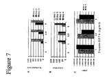
- PER.C6 cells were seeded in 490 cm 2 tissue culture roller bottles at a density of 1x10 6 cells/ml, in the presence of different concentrations of trypsin-EDTA, ranging from 1 to 5 ⁇ g/ml. Cells were infected with PER.C6-grown virus B/Harbin/7/94 at an moi of 10 -3 pfu/cell. Production of the virus was monitored by direct immunofluorescence, haemagglutination and plaque assays as shown in Fig.7 . The infectability of PER.C6 at day 2 increased with the concentration of trypsin.
- PER.C6 cells were seeded in T25 tissue culture flasks, at a density of 1x10 6 cells/ml, in the presence of different concentrations of trypsin-EDTA ranging from 0 to 7.5 ⁇ g/ml.
- Cells were infected with PER.C6-grown virus X-127 (egg-reassortant for the strain A/Beijing/262/95) at an moi of 10 -4 and 10 -3 pfu/cell. Viral growth was monitored by direct immunofluorescence, haemagglutination and plaque assays.
- HAU titers were identical between samples, independent of the trypsin concentration and the initial moi that was used. Furthermore, no significant differences were observed in the infectivity titers, as measured by plaque assay.
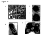
- Fig.10 shows that the last stages of the virus life cycle are represented by budding and release of enveloped virions from the cytoplasmic membrane. Spikes corresponding to the HA and NA viral proteins were detected, ornamenting the periphery of the virion particles. The figure also shows the characteristic pleiomorphism of Influenza viruses.
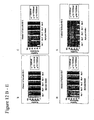
- Static suspension cultures of PER.C6 cells that were grown in T25 flasks and/or in 6 well plates in ExCell 525 medium, were infected at a cell density of 10 6 cells/ml with 16 different strains of Influenza viruses shown in Fig.11A . These strains comprised several H3N2, H1N1, B type and Avian strains. Infections were performed in the presence of 5 ⁇ g/ml of trypsin. The viruses were obtained from NIBSC (UK) as egg-passaged wild type or reassortant strains and are noted in. Infection was performed with a virus dilution recommended by the NIBSC in the product sheets that were delivered with the different strains. All viruses tested were capable of propagation on PER.C6 as visualized by immunofluorescence (data not shown) and titration of supernatant fluids in pfu assay ( Fig.11B ).
- Influenza strains (depicted by an asterisks), such as A/Johannesburg/33/94, B/Beijing/184/93 and A/Duck/Singapore-Q/F119-3/97, can replicate and be produced on the human PER.C6 cells.
- HSV-1 Herpes Simplex type 1
- HSV-2 Herpes Simplex type 2
- Measles virus PER.C6.
- Vaccines that are derived from these PER.C6-grown viruses and that induce neutralizing effects in humans for protection against wild type infections are generated from the PER.C6-grown virus batches.
- the strains that were obtained from ATCC and used for infection of PER.C6 cells are depicted in Table I.
- Table I Herpes simplex virus and Measles strains that were obtained from the ATCC and that were used for infection of PER.C6 cells.
- Herpes Simplex Type 1 (HSV-1) Macintyre VR-539 1327850 y.s./12, PR RabK/5, Mb/1, PrRabK/5, Vero/4, Vero(ATCC CC1-81)/1 10 6.75 TCID 50 /200 ⁇ l
- Herpes Simplex Type 2 (HSV-2) MS VR-540 216463 Sheep choroid plexus/?, HeLa/?, PrRabK/7, Vero(ATCC CCl-81)/3 10 7.5 TCID 50 /200 ⁇ l Measles Edmonston VR-24 215236 HK/24, HuAm/40, MRC-5/1, MRC-5(ATCC CCL-171)/1 10 4 TCID 50 /ml
- passage number 46 cells were seeded in labtek chambers, coated with Poly-L-Lysine using methods known to persons skilled in the art, at 10 5 cells/well.
- Monkey derived Vero cells obtained from ATCC were cultured at passage number 137 and were used as positive controls and seeded at a density of 2.5x10 4 cells/well. At day 0, when wells with PER.C6 cells were 60% and Vero cells 80% confluent, cells were infected with different moi's ( 10 -3 , 10 -2 , 10 -1 and 1 TCID 50 per cell).
- HSV Herpes Simplex Virus
- Measles IFA kit Light diagnostics
- the antisera are directed against HSV-1 and -2 and Measles virus antigens.
- Fig.12 show that PER.C6 is permissive for HSV-1, HSV-2 and Measles virus infections. Furthermore, the kinetics suggest that these viruses replicate on PER.C6 in an moi-dependent manner.
- HSV-1, -2 and Measles virus could be propagated on PER.C6.
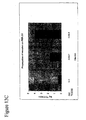
- cells were infected with moi of 0.01, 0.1 and 1 TCID 50 /cell for HSV-1 ( Fig.13B ) and HSV-2 ( Fig.13A ) and an moi of 0.001 TCID 50 /cell for Measles virus ( Fig.13C ) (passage number 1).
- cells and supernatants were harvested, quickly frozen in liquid N 2 , and thawed. After this, clarified supernatants were passaged blindly using approximately 100 ⁇ l, to PER.C6 (this is passage number 2).
- passage number 3 was performed in a similar manner.
- the moi's of the passage number 2 and 3 were determined in retrospect by TCID 50 assays.
- the results of these experiments show that Herpes Simplex Virus type 1 and -2 and Measles viruses can be replicated on PER.C6 and that replication and propagation can even occur when moi's as low as 10 -7 are used.
- PER.C6 cells were infected with a Rhesus rotavirus (MMU 18006;ATCC#VR-954; strain S:USA:79:2; lot#2181153).
- PER.C6 cells (passage number 41) were cultured at a density of 1x10 5 per ml and Monkey derived Vero cells (obtained from ATCC, passage number 139) were cultured at a density of 2.5x10 4 per ml, and subsequently seeded in Labtek chambers, that had been pre-coated with poly-L-Lysine using methods known to persons skilled in the art.
- Acyclovir (2-amino-1,9-dihydro-9-[(2-hydroethoxy) methyl]-6 H -purin-6-one), is a synthetic purine analogue with in vivo and in vitro inhibitory activity against herpes simplex virus types 1 and 2 (HSV-1, HSV-2) and against Varicella Zoster virus (VZV).
- HSV-1, HSV-2 herpes simplex virus types 1 and 2
- VZV Varicella Zoster virus
- the activity in vitro of acyclovir is the highest against HSV-1, followed by decreasing activities against HSV-2 and VZV.
- the mechanism of antiviral activity of this compound which is highly selective, is based on its affinity for the enzyme thymidine kinase (TK) encoded by HSV and VZV.
- TK thymidine kinase
- Acyclovir is converted by TK in acyclovir monophosphate, a nucleoside analogue that is further converted into diphosphate by cellular guanylate kinase and into triphosphate by a number of cellular enzymes.
- Acyclovir triphosphate stops the replication of herpes virus DNA in in vitro infected cell cultures by 1) inhibition of viral DNA polymerase, 2) chain termination of the viral DNA and 3) inactivation of the viral DNA polymerase.
- the differential sensitivity of HSV-1,2 and VZV is related to differences in phosphorylation efficiency by the viral TK's (De Clercq 2001).
- PER.C6 for antiviral drug screening, the acyclovir/HSV system was used.
- PER.C6 cells were seeded in a 96-wells tissue culture plate at a concentration of 1x10 5 cells/well, in normal culture medium, DMEM (Gibco) supplemented with heat inactivated FBS (origin US, Gibco) and 1 ml 4.9 M MgCl 2 (Sigma).
- acyclovir (20 mg/ml, Genthon) concentration being, 200, 20, 2, 0,2 and 0 ⁇ g/ml, diluted in normal culture medium, for 2 hours at 37°C and 10% CO 2 . A total of 20 wells were used per concentration.
- HSV 1 VR-539 ATCC, 3th passage on PER.C6
- HSV2 VR-540 ATCC, 3th passage on PER.C6
- Viral input HSV-1 HSV-1 titer: 5000 TCID 50 /100 ⁇ l
- Viral input HSV-2 HSV-2 titer: 5000 TCID 50 /100 ⁇ l 2x10 5 cells/well Virus dilution Input virus (TCID 50 ) Moi (TCID 50 /cell) 10 -1 10000 5x10 -2 10 -2 1000 5x10 -3 10 -3 100 5x10 -4 10 -4 10 5x10 -5 10 -5 1 5x10 -6 10 -6 0.1 5x10 -7 10 -7 0.01 5x10 -8 10 -8 0.001 5x10 -9
- CPE was scored in each of the wells and TCID 50 calculated according to the Karber method (1931), which is well known to persons skilled in the art.
- viable cells were scored with the MTS assay. Briefly, 25 ⁇ l of MTS substrate (Celltiter 96 Aqueous One Solution Cell Proliferation Assay, Promega) was added to the wells. Metabolic active cells will reduce the MTS tetrazolium into a collored formazan product. The substrate was left on the cells for 2 hours at 37°C 10% CO 2 followed by reading the OD 490 in an ELISA reader.
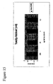
- acyclovir As shown in Fig.15 , no toxicity of acyclovir for uninfected PER.C6 cells was noticed, which is in agreement with its mechanism of action requiring phosphorylation by herpes virus TK. A clear virustatic effect of acyclovir was observed for both HSV-1 and HSV-2 when propagated on PER.C6 cells. For HSV-1, as compared to the control, an almost 1000 fold TCID 50 reduction was observed at a concentration of 20 ⁇ g/ml of acyclovir ( Fig.16 left panel).
Landscapes
- Health & Medical Sciences (AREA)
- Life Sciences & Earth Sciences (AREA)
- Engineering & Computer Science (AREA)
- Biomedical Technology (AREA)
- Immunology (AREA)
- Hematology (AREA)
- Chemical & Material Sciences (AREA)
- Urology & Nephrology (AREA)
- Molecular Biology (AREA)
- Cell Biology (AREA)
- Tropical Medicine & Parasitology (AREA)
- Biochemistry (AREA)
- Biotechnology (AREA)
- Toxicology (AREA)
- Bioinformatics & Cheminformatics (AREA)
- Food Science & Technology (AREA)
- Medicinal Chemistry (AREA)
- Physics & Mathematics (AREA)
- Analytical Chemistry (AREA)
- Microbiology (AREA)
- General Health & Medical Sciences (AREA)
- General Physics & Mathematics (AREA)
- Pathology (AREA)
- Measuring Or Testing Involving Enzymes Or Micro-Organisms (AREA)
- Micro-Organisms Or Cultivation Processes Thereof (AREA)
- Medicines That Contain Protein Lipid Enzymes And Other Medicines (AREA)
- Investigating Or Analysing Biological Materials (AREA)
Abstract
Description
- The invention relates to the field of microbiology. In particular the invention relates to the field of identification of antiviral compounds.
- Several procedures are known in the art for the treatment of virus-related diseases and for the prevention of disorders that arise as a consequence of viral infections. Prophylactic vaccination is probably one of the most effective measures against potential fatal infectious diseases, since an individual can become fully or partly protected against new infections. The immune system makes sure that the virus that has entered is prevented from replicating in the individual and in most cases the virus disappears completely from the system. Therapeutic vaccination refers to the treatment of infected individuals intentionally to prevent the virus from replicating further and consequently to halt disease progression or to cure the disease by eliminating the virus from the body.
- Another way of dealing with viral infections is through the use of antiviral agents that either attack the viral particle directly or that prevent the infection, propagation, replication, packaging and/or growth of the virus in the individual. These treatments are applied either when the individual has already been infected and so a prophylactic vaccination is obsolete, or when an individual is at immediate risk to encounter an infection. Antiviral molecules inhibit certain processes and phases in the viral life cycle, thereby inhibiting the virus from spreading.
- Several in vitro and in vivo methods for the identification of such antiviral compounds are known in the art. Methods that make use of the specific antiviral activity of certain compounds include the plaque reduction assay, the yield reduction assay, the virus antigen determination assay, the dye-uptake assay, the cytopathic effect (CPE) determination assay and several in vivo assays for virus replication. Many of the in vitro methods, especially the plaque reduction assay, have the major disadvantage that they cannot be applied in (very) high-throughput screens. Although the plaque reduction assay can be applied for most viruses that are known to date, it is necessary to inoculate large numbers of susceptible cells in suitable conditions with ranges of virus titers as well as large ranges of antiviral compound titers to detect the correct concentration of the compound that significantly decreases the number of plaques. This makes the plaque reduction assay very suitable for measuring the right concentration of a specific compound that affects the growth of a particular virus but very unsuitable for the identification of such (new and unknown) compounds in a library. Since many of the molecule libraries consist of a very large collection of separate compounds (>1014 individual agents), it is required to have settings in which all separate compounds can be screened in a rapid and efficient manner with low costs. The other in vitro methods such as the yield reduction assay, the virus antigen determination assay, the dye-uptake assay and the cytopathic effect (CPE) determination assay are to a certain extent more suitable for high-throughput screening, but they clearly depend on the cell line that is used and whether such a cell line is able to grow in multi-well settings and for prolonged periods of time. Clearly, many of the primary cells that are used to determine the effect of an antiviral compound in plaque reduction assays cannot be cultured in high-throughput settings, since these cells do not grow indefinitely. Evidently, the in vivo antiviral methods, such as for example the ferret-, the mouse- and chicken models for influenza infection (reviewed by Sidwell et al. 2000) are useless for the identification of novel compounds that prevent virus-cell recognition and virus infection, replication, propagation and growth, especially when high-throughput settings are preferred.
- Many susceptible non-continuous cells have been identified in which most viruses propagate. As mentioned, these cells can be used in assays such as the plaque reduction assay but cannot be applied for screening of antiviral compounds, since they do either not grow in multi-well formats or they do not grow indefinitely. Only a limited number of continuous cell lines have been identified that support the growth of certain viruses. These cell lines include the green monkey VERO cells, the Madin-Darby Canine Kidney MDCK cells, the human lung embryo MRC-5 cells and the human A549 cells. However, a major drawback of these cells is that they only support the growth of a limited number of viruses, while not all of these cell lines are capable of continuous growth in multi-well formats. Nevertheless, a number of drugs displayed antiviral activities against viruses such as CMV, Influenza and HSV in the context of using the cells mentioned above. For example, Acyclovir, an approved purine nucleoside analogue, inhibited HSV replication in A549 cells (Li et al. 1988). Despite the few successful propagations of certain viruses on continuous cell lines and the prevention of propagation by a number of antiviral compounds, it was found that in many cases the cells did not support the complete life cycle of the mentioned viruses. This limits their use significantly in screening assays for antiviral compounds present in large libraries, because the life cycle of a virus is built up from several phases in which a compound can have its point of impact.
- Although many cell-based systems exist that can be applied for determining whether a certain compound is capable of preventing certain phases in the life cycle of a virus, no system is present in the art that combines the possibility of screening large numbers of (possible) antiviral compounds in a very high-throughput setting with the possibility of screening a large range of different viruses. No system is available in the art that combines these possibilities to determine the antiviral activity of a certain compound present in a compound library, in different phases of the life cycle of the particular virus that is attacked by this particular compound.
-
WO9115573 claim 1, after cell infection with the hepatotropic virus, exposing the infected cells to the compound for a selected period, and assaying the cells for inhibition of virus growth. - The present invention discloses novel uses of PER.C6 cells that fully support the complete life cycle of a very wide variety of pathogenic viruses and that provide methods for screening libraries of antiviral compounds for identification of molecules with antiviral activity that can interfere with the pivotal processes in any phase of the life cycle of a pathogenic virus. The fact that these cells support the infection, replication, propagation and growth of a large variety of viruses is disclosed in patent application
PCT/NL00/00862 WO 01/38362WO 99/64582 - It is an object of the current invention to deal with a large number of the problems and drawbacks known from the art concerning the identification of antiviral compounds using the assays and cell lines described above.
-
-
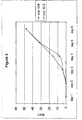
Fig.1 . Percentage of infected cells scored microscopically after immunofluorescence assay. Samples derived from infection at multiplicity of infection (moi) 10 and 1, at 48 h post infection are not shown, because of full CPE. -
Fig.2 . Percentage of infected cells (positive cells) scored microscopically after immunofluorescence assay versus percentage of dead cells measured via FACS after propidium iodide staining, at moi's of 10-3 (A) and 10-4 (B). Poor viability of the cells from samples derived from infection at moi 10-3 did not give rise to reliable data. -
Fig.3 . Kinetics of virus propagation measured in hemagglutinating units (HAU) fromday 1 today 6 after infection. -
Fig.4 . Percentage of infected cells (positive cells) viewed microscopically after immunofluorescence assay. (A) A/Beijing/262/95 and (B) X-127. -
Fig.5 . Kinetics of virus propagation measured in HAU fromday 1 to 6 after infection. (A) A/Beijing/262/95 and (B) X-127. -
Fig.6 . Infection with A/Sydney/5/97 on PER.C6. (A) Effect of trypsin-EDTA on HAU titers. (B) HA concentration in µg/ml and (C) virus infectivity titers in plaque forming units per ml (pfu's/ml) as measured in crude viral supernatants, 96 hours post infection. -
Fig.7 . Infection with B/Harbin/7/94 on PER.C6. (A) Effect of different concentrations of trypsin-EDTA present during and after virus infection on growth kinetics. (B) HAU titers per 50 µl and (C) virus infectivity titers in pfu/ml. -
Fig.8 . Infection with X-127 using an moi of 10-3 on PER.C6. (A) Effect of trypsin-EDTA on HAU given in HAU/50 µl and (B) virus infectivity titers in pfu/ml during 5 days after infection. -
Fig.9 . Infection with X-127 using an moi of 10-4 on PER.C6. (A) Effect of trypsin-EDTA on HAU given in HAU/50 µl and (B) virus infectivity titers in pfu/ml during 5 days after infection. -
Fig.10 . Electron micrographs of Influenza A/Sydney/5/97. (A) PER.C6 cells 72 hrs post infection. (B and C) Negative staining on virus derived from infected PER.C6. (D and E) Negative staining of sucrose purified material. -
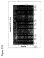
Fig.11 . (A) Different Influenza A and B strains tested on PER.C6 cells. (B) Infectivity titers of three depicted A- and B-type Influenza viruses derived from infected PER.C6 cells. -
Fig.12 . Immunofluorescence of PER.C6 and Vero cells infected with viruses other than Influenza. (A) Positively staining cells upon infection with Measles virus. (B) Positively staining cells upon infection of Vero cells with HSV-1 virus. (C) Positively staining cells upon infection of Vero cells with HSV-2 virus. (D) Positively staining cells upon infection of PER.C6 cells with HSV-1 virus. (E) Positively staining cells upon infection of PER.C6 cells with HSV-2 virus. -
Fig.13 . Infectivity titers determined after propagation of Measles virus (A), HSV-1 (B) and HSV-2 (C) virus on PER.C6 cells. -
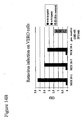
Fig.14 . Replication of Rotavirus after infection of PER.C6 (A) and Vero (B) cells with different moi's as measured by ELISA in crude supernatants. -
Fig.15 . Toxicity of acyclovir on PER.C6 as measured by MTS assay after 6 days of incubation of the cells in the presence of different dilution of the compound as indicated. -
Fig.16 . Virustatic activity of acyclovir for HerpesSimplex Virus type 1 and type 2 (HSV-1 and HSV-2) using PER.C6 cells as a substrate for virus replication and TCID50 calculations based on CPE score onday 6 post infection. -
Fig.17 . Virustatic effect of acyclovir against (A) HSV-1 and (B) HSV-2 using infection of PER.C6 with different 10 fold serial dilutions of virus and viable cell scoring by MTS assay onday 6 post infection. - The current invention discloses methods that substantially lack the above outlined drawbacks and being characterized in methods for identifying a compound and/or determining whether said compound influences a phase in the life cycle of a virus comprising providing a PER.C6 cell with said compound and with at least a fragment of said virus sufficient for performing said phase and determining whether said phase is influenced in said cell. It is to be understood that the compound can be provided before or after or simultaneously with providing said fragment of said virus sufficient for performing said phase. Preferably, the methods provided by the invention comprise a virus selected from the group of adenoviruses, enteroviruses, herpes viruses, orthomyxoviruses, paramyxoviruses, retroviruses, rotaviruses, coronaviruses, flaviviruses, togaviruses, hepatitis causing viruses, pestiviruses, rhabdoviruses or Bunyaviridae viruses. It will be clear to those skilled in the art, that whenever a virus is identified that is able to perform one or more of its life-cycle phases in a cell, that the methods of the present invention can be applied. In an even more preferred embodiment, said virus is an essentially intact virus, wherein 'essentially' is defined as comprising an intact coat capable of recognizing and infecting an appropriate host cell, further comprising nucleic acid capable of supporting all required functions for normal replication, propagation, packaging and releasing newly formed virus particles.
- The invention makes use of a PER.C6 cell.
- The invention further provides methods for determining whether a compound influences a phase in the life cycle of a virus comprising providing a PER.C6 cell with said compound and with at least a fragment of said virus sufficient for performing said phase and determining whether said phase is influenced in said cell, wherein determining whether a compound influences a phase in the life cycle of a virus comprises examining the activity and/or amount of a cellular protein and/or examining the interaction of said virus with said cell and/or examining the activity and/or amount of said virus or a fragment thereof and/or examining the viability of said cell.
- The invention is further characterized in uses of a PER.C6 cell for screening a library of compounds for the presence of a compound capable of influencing a phase in the life cycle of a virus capable of entering said cell. In a preferred aspect of the invention, said uses of a PER.C6 cell comprise a virus selected from the group of adenoviruses, enteroviruses, herpes viruses, orthomyxoviruses, paramyxoviruses, retroviruses, rotaviruses, coronaviruses, flaviviruses, togaviruses, hepatitis causing viruses, pestiviruses, rhabdoviruses or Bunyaviridae viruses. It will be clear to those skilled in the art that newly identified viruses or other viruses not mentioned in this list and that are able to perform one or more of its life-cycle phases in said PER.C6 cells, that the uses according to the present invention also apply to those viruses.
- In a preferred embodiment of the present invention said compound is present as part of a compound library. In a more preferred embodiment said compound library is used in high-throughput settings. In an even more preferred embodiment of the present invention said method comprises isolating said compound. The invention provides methods for screening (anti-viral) compound libraries using PER.C6 cells, viruses and methods of the present invention.
- In another aspect, the invention provides methods for identifying a compound with antiviral activity comprising providing a PER.C6 cell with at least a fragment of a virus, said fragment capable of performing a phase in the life cycle of said first virus, providing said PER.C6 cell with a compound and determining whether said compound is capable of influencing said phase in the life cycle of said virus In another aspect, the invention provides methods for determining the effect of the absence of a compound on a phase in the life cycle of a virus comprising the steps of culturing a PER.C6 cell, said PER.C6 cell otherwise capable of supporting said phase in the life cycle of a virus, in the presence of said virus under conditions otherwise conducive to said phase in the life cycle of said virus and in the absence of said compound, examining the effect of the absence of said compound on said phase in the life cycle of said virus. Preferably, said compound is a natural constituent of said PER.C6 cell. More preferably, said natural constituent is a receptor protein, or a fragment thereof, for said virus. Even more preferably, said cell is comprised in a set of clones of cells or a library of cells, said cells comprising a gene being effectively blocked from being expressed, wherein effectively blocked is defined as significantly reduced.
- The phase in the life cycle of a virus is defined as a phase that can comprise a step or a period during which a virus recognizes its host cell or during which the virus interacts with its host cell through a protein, preferably a protein expressed by the host cell. Said phase in the life cycle of a virus is further defined as a phase that can comprise a step or a period during which a virus infects (enters) a cell, replicates its nucleic acid with or without using cellular factors, and produces the required proteins in said cell for propagation and/or packaging of the newly made viral particle(s) in said cell. A life cycle of a virus can be generally defined as
- (a) recognition and/or interaction of the virus with a host cell,
- (b) entering (infecting) said host cell,
- (c) changing the host cell environment to enable generation of new virus particles, for instance by expression of viral proteins from the infected nucleic acid,
- (d) replication of the nucleic acid,
- (e) expression of viral proteins required for packaging new viral particles,
- (f) packaging of new viral particles and
- (g) release of the newly formed particles from the infected cell,
- To determine to what extent a certain compound influences a specific phase in the life cycle of a virus, several methodologies can be applied. Examples of such methodologies are (1) measuring the levels and/or activities of viral and/or cellular proteins that are present upon entry of the virus into the host cell, (2) examining the recognition of the virus with the host cell or the interaction of the virus with the host cell and/or host cell proteins, (3) examining the activity and/or amount of the virus or a fragment thereof and (4) examining the viability of the infected host cell.
- The viruses used by the present invention to screen for antiviral compounds comprise an adenovirus, an enterovirus (such as rhinovirus, aphtovirus, or poliomyelitisvirus), a herpesvirus (such as herpes symplex virus, pseudorabies virus or bovine herpes virus), an orthomyxovirus (such as Influenza virus), a paramyxovirus (such as newcastle disease virus, respiratory syncitio virus, mumps virus or measles virus), a retrovirus (such as human immunodeficiency virus), a parvovirus, a papovavirus, a rotavirus, a coronavirus (such as transmissable gastroenteritis virus), a Flavivirus (such as tick-borne encephalitis virus or yellow fever virus), a togavirus (such as rubella virus or eastern-, western-, or venezuelean equine encephalomyelitis virus), a hepatitis causing virus (such as hepatitis A, -B or -C virus), a pestivirus (such as hog cholera virus), a rhabdovirus (such as rabies virus) or a Bunyaviridae virus (such as Hantavirus).
- The invention relates to the identification of antiviral compounds directed against viruses or viral pathogens that previously could not or poorly be grown on human cells in vitro. The invention is particularly useful for the production of therapeutics to aid in protection against viral pathogens for vertebrates, in particular mammalians and especially humans.
- The invention discloses methods and means for the identification and/or validation of compounds harbouring antiviral activities and the like that interfere with the life cycle of viral pathogens. The identification and/or validation can either be performed in low-throughput, medium-throughput, high-throughput or ultra high throughput settings, ranging from a few compounds to >1014 compounds present in a library of compounds. The invention relates to the screening of compound libraries comprising non-proteinaceous substances like small or large (synthetic) compounds such as nucleoside analogues or nucleic acid such as DNA molecules. The invention also relates to the screening of compound libraries comprising molecules like proteinaceous substances such as peptides, antibodies, hormones, receptors or other protein binding entities or fragments thereof. The non-proteinaceous substances, proteinaceous substances in such collections can be expressed on phage particles or can be present as separate molecules not expressed on phage particle. The invention moreover relates to the screening of compound libraries comprising large particles like viruses, virus particles, phage and fragments thereof. The compounds can have already known- or yet undiscovered functions for antiviral activity.
- The invention is particularly useful for the identification of molecules that interfere with infection of cells by viral pathogens, binding and/or entry of viral pathogens into cells, replication of said viral pathogens in cells, packaging of viral pathogens into viral particles and/or release of said viral pathogens from cells and/or re-infection of cells by such viral pathogens, to aid in protection against viral pathogens for vertebrates, in particular mammalians and especially humans.
- The invention relates to the use of PER.C6 cells. The present specification discloses human immortalized cell lines (PER.C6 and derivatives thereof) that are generally used for the production of recombinant adenoviral vectors and for the purpose of propagating, production and harvesting viruses other than adenovirus to generate vaccines (disclosed in patent application
PCT/NL00/00862 WO 01/38362 - The following features make PER.C6 and derivatives thereof particularly useful as a host for virus production: it is a fully characterized human cell line, it was developed in compliance with GLP, it can be grown as suspension cultures in defined serum-free medium, devoid of any human or animal serum proteins; its growth is compatible with roller bottles, shaker flasks, spinner flasks and bioreactors, with doubling times of about 35 h. Moreover, it was found that PER.C6 were very suitable for growth in multi-well settings and for the propagation of adenoviruses in high-throughput methodologies (
WO 99/64582 - The invention provides the use of PER.C6 cells for the identification of novel antiviral compounds that act towards viruses for which no known antiviral compounds were thus far identified. Moreover, the invention enables the identification of known antiviral compounds, which also act upon viruses that could thus far not be propagated on cell lines used in the art. Such known and unidentified antiviral compounds (often small molecules) can either be rationally designed or identified in screenings assays. The antiviral strategies that these compounds employ are sometimes known (e.g. neuraminidase inhibitors for treatment of influenza infection and DNA synthesis inhibitors for the treatment of HSV infection). On the other hand, many drugs have been developed that exert an antiviral effect in patients through mechanisms that are not completely understood. Many of such drugs were identified in recent years.
- For example the protein Interferon alpha is used for the treatment of HCV infected patients. It is applied alone or in combination with Ribavirin (Keating 1999), although problems have occurred for HCV treatment when used alone (Polyak et al. 2000). Famciclovir and valaciclovir are two approved compounds and that are used for Herpes Simplex Virus (HSV) and for varicella-zoster infections (Keating 1999). Ganciclovir (GCV), a purine nucleoside analogue was approved to treat CMV retinitis (Keating 1999; Alrabiah et al. 1996), while another approved purine nucleoside analogue, Acyclovir (ACV) is being applied for genital herpes, HSV encephalitis, mucocutaneous herpetic infections, varicella-zoster and for herpes-zoster infections (Keating 1999; Dwyer et al. 1997). Two nucleoside phosphonate analogues, Adefovir and Tenofovir, are being used to treat HBV infections (Ying et al. 2000). The compound Dibenzofuran was found to block rhinovirus replication in vitro and to hinder the cytopathic effect in cells infected with HRV14 or HRV16 (Murray et al. 1999). In recent years also several protease inhibitory agents were identified that were active against the Human Immunodeficiency Virus (HIV): Saquinavir (Figgitt et al. 2000) and Nelfinavir (Bardsley-Elliot et al. 2000a). Nevirapine is an approved non-nucleoside Reverse Transcriptase inhibitor (NNRTI) used against HIV infections (Bardsley-Elliot et al. 2000b), while a non-nucleoside RT inhibitor (NNRTI), Delavirdine (a bisheteroarylpiperazine derivative) was also found to be active against HIV (Scott et al. 2000; Joly et al. 2000). Amantidine and Rimantadine are two drugs that are used for the treatment of Influenza A infections (Keating 1999). Nucleoside analogues are reviewed by Pastor-Anglada et al. (1998) and the idea to use protease inhibition to attack virus infections is discussed by Todd et al. (2000). This list of antiviral agents is by far not limiting and many proteins, small molecules and other types of compounds are still in several different phases of development.
- Although the PER.C6 cell line was developed for the production of recombinant adenoviral vectors (mainly derived from
Adenovirus serotype 5, or Ad5) through the complementation of E1 deleted vectors by the E1 functions provided by the integrated E1-expressing plasmid, it was also found that PER.C6 cells were able to support the production of exogenous proteins such as recombinant human Erythropoietin (EPO) or human monoclonal antibodies encoded by expression plasmids that were transfected and integrated stably or that were present in a transient system (WO 00/63403 EP 0978566 ). - Interestingly, PER.C6 also supports the growth of an entire different set of viruses besides Adenoviruses. Examples of these viruses are, but are not limited to Human- and Duck Influenza virus, Human- and Rhesus Rotavirus, Measles Virus, Respiratory Syncytium virus A and B, Parainfluenza types -1, -2 and -3, Poliovirus types -1, -2 and -3, Coxsackie virus B2, B4, A9, Echovirus types -4, -7 and -11, Japanese encephalitis virus, Hantavirus and Herpes Simplex Virus types -1 and - 2. Examples of such viruses that can grow on PER.C6 are disclosed herein and in
PCT/NL00/00862 WO 01/38362 - For example, rotaviruses can grow on PER.C6 cells. Rotaviruses, members of the family of Reoviridae, are double strand RNA viruses consisting of 11 RNA segments, each coding for a structural or non-structural viral protein (VP). Given the worldwide prevalence of rotavirus associated infant morbidity and mortality, large scale vaccination against Rotavirus is thus far considered the most effective way to combat this virus. The goal of vaccination would not be to prevent the disease but to reduce its severity and complication, especially during the first few years of life. The only vaccine at present is a live attenuated orally delivered composition that is associated with intussuception, a bowel obstruction disease. For that reason this vaccine is no longer in use. It is evident that there is a need for anti-rotavirus drugs that can be used to treat Rotavirus infected patients. The identification of such drugs was thus far hampered by the difficulty of growing the virus in suitable systems. The present invention provides methods of using a human cell line that is highly capable of supporting infection, replication, propagation and growth of rotavirus, thereby providing a system for the identification of compounds that prevent rotavirus generation in human cells using a multi-well set up.
- Adenoviruses are another set of viruses for which the present invention provides the possibility to identify antiviral compounds against. Although Adenoviruses are known to cause minor disorders such as common colds, it is also known that in immuno-suppressed patients adenoviruses can cause severities that can even lead to death of such patients. Of all bone-marrow transplant patients that die during or after treatment, approximately 20% dies of an adenovirus infection. Clearly, there is a great need for anti-adenoviral compounds that can prevent the occurrence of malignancies due to adenoviral infections in these patients that have a suppressed immune response.
- To illustrate the invention, the following experimental procedures and examples are provided, not intended to limit the scope of the invention.
- Madin-Darby Canine Kidney (MDCK) cells and PER.C6™ cells (deposited under No. 96022940 at the ECACC, described in
WO 97/00326 US patent no. 6,033,908 ) were cultured in Dulbecco's modified Eagle's medium (DMEM, Life Technologies) containing 10% heat inactivated fetal bovine serum and 1x L-Glutamin (Gibco), at 37°C and 10% CO2. Suspension cultures of PER.C6 were cultured in ExCell 525 (JRH Biosciences) supplemented with 1x L-Glutamin, at 37°C and 10% CO2, in stationary cultures in 6 well dishes (Greiner) or in 490 cm2 tissue culture roller bottles (Corning Costar Corporation) during continuous rotation at 1 rpm. - Direct immunofluorescence assays for the detection of Influenza virus infection were carried out using the IMAGEN™ Influenza Virus A and B kit (Dako) according to the standard protocol of the supplier. Samples were viewed microscopically using epifluorescence illumination. Infected cells were characterised by a bright apple-green fluorescence.
- Cell pellets were resuspended in 300 µl of cold PBS/0.5% BSA + 5 µl of propidium iodide (
concentration 50 µg/ml) in PBS/FCS/azide solution known to persons skilled in the art. Viable and dead cells were then detected via flow cytofluorometric analysis. - In general, haemagglutination assays for Influenza virus titers were performed according to methods known to persons skilled in the art. Here, 50 µl of a two-fold diluted virus solution in PBS was added to 25 µl PBS and 25 µl of a 1% suspension of turkey erythrocytes (Biotrading Benelux B.V.) in PBS and incubated in 96 well microtiter plates at 4°C for 1 h. The haemagglutination pattern was examined and scored, and expressed as hemagglutinating units (HAU's). The number of HAU's corresponded to the reciprocal value of the highest virus dilution that showed complete haemagglutination.
- In general, obtained Influenza viruses were disrupted in a Laemmli buffer according to methods known to persons skilled in the art and different volumes of obtained protein mixtures were separated using 10% SDS/PAGE gels. In brief, blots were blocked for 30 min at room temperature with block solution (5% non fat dry milk powder (Biorad) in TBST supplemented with 1% rabbit serum (Rockland)), followed by 3 washes with TBST. Then, the blots were incubated with the anti A/Sydney/5/97 HA antiserum (98/768 NIBSC) diluted 1/500 in 1%BSA/TBST with 5% rabbit serum (Rockland) O/N at room temperature. Again the blots were washed 8 times with TBST. Finally the blots were incubated with the rabbit anti sheep antiserum (HRP labelled, Rockland) 1/6000 diluted in block solution for 1 h at room temperature. After 8 washes with TBST the protein-conjugate complex was visualised with ECL (Amersham Pharmacia Biotech), and films (Hyperfilm, Amersham Life Science) were exposed. The antisera were obtained from the NIBSC (UK) and applied in dilutions recommended by the NIBSC.
- The concentration of haemagglutinin in supernatants, derived from Influenza virus infected-PER.C6 cells, was determined by the single radial immunodiffusion (SRID) test as previously described (Wood et al 1977). The assay was performed using standard NIBSC (UK) antigens and antisera reagents.
- A total of 1 ml of 10-fold serially diluted viral supernatants were inoculated on MDCK cells which were grown until 95% confluence in 6-well plates. After 1 h at 35°C the cells were washed twice with PBS and overloaded with 3 ml of agarose mix (1.2 ml 2.5% agarose, 1.5 ml 2x MEM, 30
µl 200 mM L-Glutamine, 24 µl trypsin-EDTA, 250 µl PBS). The cells were then incubated in a humid, 10% CO2 atmosphere at 35°C for approximately 3 days and viral plaques were visually scored. - Titration of infectious virus was performed on MDCK cells. In brief, cells were seeded in 96 well plates at a density of 4x10⁴ cells/well in DMEM supplemented with 2 mM L-Glutamin. Twenty-four hours later cells were infected with 100 µl of ten fold serially diluted culture supernatants, in quadruplicate, in medium containing Trypsin-EDTA at the concentration of 4 µg/ml. Two hours after infection cell monolayers were washed two times in PBS and incubated in medium containing trypsin for 7 days, at 35°C. Supernatants from these cultures were then tested in an HA assay. TCID50 titers were calculated according to the method of Karber (1931), which is well known to persons skilled in the art.
- It was not known prior to the invention described in
PCT/NL00/00862 WO 01/38362 - On the day before infection, 2x10⁵ MDCK cells per well were seeded in 6-well plates. Twenty four hours later, 4x10⁵ seeded PER.C6 and the MDCK cells per well were infected with the H1N1 strain A/Puerto Rico/8/34 (titer 3.6x10⁷ pfu/ml), (obtained from Dr. E. Claas, Leiden University Medical Centre, The Netherlands). Infection was performed at various multipliticies of infection (moi's) ranging from of 0.1 to 10 pfu/cell. After about 2 h of incubation at 37°C, the inoculum was removed and replaced by fresh culture medium. A direct immunofluorescence assay for the detection of Influenza virus infection was performed 24 h and 48 h post infection. The experiment showed permissiveness of PER.C6 for Influenza infection, with percentages of positive cells moi-dependent and comparable with MDCK (
Fig.1 ). - It was verified whether not only permissiveness but also replication and propagation of Influenza virus could be supported by PER.C6. On the day of infection, PER.C6 cells were seeded in 490 cm2 tissue culture roller bottles, with the density of 2x10⁵ cells/ml in a final volume of 40 ml, in the presence of 5 µg/ml of trypsin-EDTA (Gibco-BRL). Cells were either mock inoculated or infected with the H3N2 strain A/Shenzhen/227/95 (titer 1.5x10⁶ pfu/ml) (obtained from Dr. E. Claas, Leiden University Medical Centre, The Netherlands). Infections were performed at
moi day 1 today 6 post-infection. Supernatants were stored at -80°C and used for haemagglutination assays. Cell pellets were used for direct immunofluorescence tests and for propidium iodide staining. Results of these experiments are shown inFig.2 andFig.3 respectively. - To further investigate the permissiveness of PER.C6 for propagation of various Influenza strains, an infection by using the H1N1 vaccine strains A/Beijing/262/95 and its reassortant X-127, obtained from the National Institute for Biological Standards and Control (NIBSC, UK) was performed. On the day of infection, PER.C6 cells were seeded in 490 cm2 tissue culture roller bottles, with the density of approximately 1x10⁶ cells/ml in a final volume of 50 ml. Cells were inoculated with 5 µl (10-4 dilution) and 50 µl (10-3 dilution) of virus in the presence of 5 µg/ml trypsin-EDTA. In order to establish if trypsin was indeed required, one more infection was carried out by inoculating 5 µl of the strain A/Beijing/262/95 in the absence of the protease. After approximately 1 h of incubation at 37°C, the inoculum was removed by spinning down the cells at 1500 rpm and resuspending them in fresh culture medium ± 5 µg/ml of trypsin-EDTA. At
day 2 andday 4 post-infection more trypsin was added to the samples. Harvest of 1.3 ml of cell suspension was carried out fromday 1 today 6 post-infection. Supernatants were stored at -80°C and used for haemagglutination assays and further infections; cell pellets were used for direct immunofluorescence tests. Results obtained with the above mentioned immunofluorescence and haemagglutination assays are shown inFig.4 andFig.5 , respectively, illustrating the efficient replication and release of the viruses. - Due to the absolute trypsin requirement for the propagation of Influenza viruses in cell cultures, the effects of different concentrations of trypsin-EDTA on PER.C6 cell viability and virus replication in PER.C6 cells, after infection with several Influenza strains, were investigated.
- On the day of infection, PER.C6 cells were seeded in 490 cm2 tissue culture roller bottles, at a density of 1x10⁶ cells/ml, in the presence of trypsin-EDTA at final concentrations of 0.5, 1, 2, 3 and 5 µg/ml. These trypsin concentrations did not interfere with the growth characteristics of the cells and their viability (data not shown). Cells were either, mock infected or infected with PER.C6-grown Influenza virus A/Sydney/5/97 at an moi of 10-4 pfu/cell. The viral production was monitored by direct immunofluorescence (data not shown), haemagglutination assays, single-radial-immunodiffusion (SRID) and plaque assays, all as described above. Results from this experiment are depicted in
Fig.6 and show that the HA content as measured by SRID as well as the biological activity of the virus, expressed in HAU, were highest when a trypsin concentration of 1 µg/ml was used.Fig.6 also shows that by using a plaque assay the highest number of plaque forming units (pfu) per ml was observed in the sample corresponding to cells grown in medium containing 2 µg/ml of trypsin. - On the day of infection PER.C6 cells were seeded in 490 cm2 tissue culture roller bottles at a density of 1x10⁶ cells/ml, in the presence of different concentrations of trypsin-EDTA, ranging from 1 to 5 µg/ml. Cells were infected with PER.C6-grown virus B/Harbin/7/94 at an moi of 10-3 pfu/cell. Production of the virus was monitored by direct immunofluorescence, haemagglutination and plaque assays as shown in
Fig.7 . The infectability of PER.C6 atday 2 increased with the concentration of trypsin. Atday 3 however, no significant difference was observed in the percentage of infected cells when 1, 2.5 or 5 µg/ml trypsin was present. In the absence of trypsin (0 µg/ml) no Influenza virus infection was detected. At the day of the last harvest (day 4 post-infection), the biological activity of the virus, as measured by haemagglutination assay, did not differ significantly. Interestingly, the infectivity assay performed in samples that were taken atday day 3 andday 4 when a trypsin concentration of 2.5 to 5 (day 3) and 1 µg/ml (day 4) were used. - On the day of infection, PER.C6 cells were seeded in T25 tissue culture flasks, at a density of 1x10⁶ cells/ml, in the presence of different concentrations of trypsin-EDTA ranging from 0 to 7.5 µg/ml. Cells were infected with PER.C6-grown virus X-127 (egg-reassortant for the strain A/Beijing/262/95) at an moi of 10-4 and 10-3 pfu/cell. Viral growth was monitored by direct immunofluorescence, haemagglutination and plaque assays. As shown in
Fig.8 andFig.9 , HAU titers were identical between samples, independent of the trypsin concentration and the initial moi that was used. Furthermore, no significant differences were observed in the infectivity titers, as measured by plaque assay. - Transmission electron microscopy studies were done on PER.C6 cells that were infected with the Influenza strain A/Sydney/5/97 as well as on viral containing supernatants and sucrose purified material to determine the phenotype of this Influenza virus produced on PER.C6. All methods that were used are well known to persons skilled in the art.
Fig.10 shows that the last stages of the virus life cycle are represented by budding and release of enveloped virions from the cytoplasmic membrane. Spikes corresponding to the HA and NA viral proteins were detected, ornamenting the periphery of the virion particles. The figure also shows the characteristic pleiomorphism of Influenza viruses. - Static suspension cultures of PER.C6 cells that were grown in T25 flasks and/or in 6 well plates in ExCell 525 medium, were infected at a cell density of 106 cells/ml with 16 different strains of Influenza viruses shown in
Fig.11A . These strains comprised several H3N2, H1N1, B type and Avian strains. Infections were performed in the presence of 5 µg/ml of trypsin. The viruses were obtained from NIBSC (UK) as egg-passaged wild type or reassortant strains and are noted in. Infection was performed with a virus dilution recommended by the NIBSC in the product sheets that were delivered with the different strains. All viruses tested were capable of propagation on PER.C6 as visualized by immunofluorescence (data not shown) and titration of supernatant fluids in pfu assay (Fig.11B ). - These results show that even Influenza strains (depicted by an asterisks), such as A/Johannesburg/33/94, B/Beijing/184/93 and A/Duck/Singapore-Q/F119-3/97, can replicate and be produced on the human PER.C6 cells.
- It was tested whether other viruses than Influenza virus and Adenovirus, such as Herpes
simplex virus type Table I Herpes simplex virus and Measles strains that were obtained from the ATCC and that were used for infection of PER.C6 cells. Virus Strain ATCC catnr. Lotnr. Passage history Titer Herpes Simplex Type 1 (HSV-1) Macintyre VR-539 1327850 y.s./12, PR RabK/5, Mb/1, PrRabK/5, Vero/4, Vero(ATCC CC1-81)/1 106.75
TCID50/200µlHerpes Simplex Type 2 (HSV-2) MS VR-540 216463 Sheep choroid plexus/?, HeLa/?, PrRabK/7, Vero(ATCC CCl-81)/3 107.5
TCID50/200µlMeasles Edmonston VR-24 215236 HK/24, HuAm/40, MRC-5/1, MRC-5(ATCC CCL-171)/1 104
TCID50/ml - To test whether HSV-1 and HSV-2 and measles viruses obtained from the ATCC could replicate and be produced on PER.C6, passage number 46 cells were seeded in labtek chambers, coated with Poly-L-Lysine using methods known to persons skilled in the art, at 105 cells/well. Monkey derived Vero cells (obtained from ATCC) were cultured at passage number 137 and were used as positive controls and seeded at a density of 2.5x10⁴ cells/well. At
day 0, when wells with PER.C6 cells were 60% andVero cells 80% confluent, cells were infected with different moi's ( 10-3, 10-2, 10-1 and 1 TCID50 per cell). At daily intervals upon infection, cells were fixed and assayed in immunofluorescence using FITC-conjugated type specific monoclonal antibodies using a kit (Imagen Herpes Simplex Virus (HSV)Type Fig.12 show that PER.C6 is permissive for HSV-1, HSV-2 and Measles virus infections. Furthermore, the kinetics suggest that these viruses replicate on PER.C6 in an moi-dependent manner. - Next it was investigated whether HSV-1, -2 and Measles virus could be propagated on PER.C6. To this end cells were infected with moi of 0.01, 0.1 and 1 TCID50/cell for HSV-1 (
Fig.13B ) and HSV-2 (Fig.13A ) and an moi of 0.001 TCID50/cell for Measles virus (Fig.13C ) (passage number 1). At the occurrence of almost complete cpe, cells and supernatants were harvested, quickly frozen in liquid N2, and thawed. After this, clarified supernatants were passaged blindly using approximately 100 µl, to PER.C6 (this is passage number 2). After reaching almost complete CPE again, a third passage (passage number 3) was performed in a similar manner. The moi's of thepassage number Simplex Virus type 1 and -2 and Measles viruses can be replicated on PER.C6 and that replication and propagation can even occur when moi's as low as 10-7 are used. - To test whether PER.C6 could also support the replication of a rotavirus, PER.C6 cells were infected with a Rhesus rotavirus (MMU 18006;ATCC#VR-954; strain S:USA:79:2; lot#2181153). PER.C6 cells (passage number 41) were cultured at a density of 1x10⁵ per ml and Monkey derived Vero cells (obtained from ATCC, passage number 139) were cultured at a density of 2.5x10⁴ per ml, and subsequently seeded in Labtek chambers, that had been pre-coated with poly-L-Lysine using methods known to persons skilled in the art. Cells were infected with an moi of 1 TCID50/cell of Rhesus rotavirus in the presence and absence of 2 µg/ml of trypsin-EDTA. After 90 min of infections, cells were washed with ExCell 525 medium and further incubated at 37°C at 10% CO2 in a humidified atmosphere. On 5 consecutive days following infection, samples of supernatants were harvested, clarified from cells and cell debris by centrifugation at 2000 rpm in a table top centrifuge and analysed in an ELISA specific for rotavirus (IDEIA Rotavirus, Dako). The results depicted in
Fig.14 clearly show that Rhesus rotavirus replicates on PER.C6. - Acyclovir (2-amino-1,9-dihydro-9-[(2-hydroethoxy) methyl]-6H-purin-6-one), is a synthetic purine analogue with in vivo and in vitro inhibitory activity against herpes
simplex virus types 1 and 2 (HSV-1, HSV-2) and against Varicella Zoster virus (VZV). The activity in vitro of acyclovir is the highest against HSV-1, followed by decreasing activities against HSV-2 and VZV. The mechanism of antiviral activity of this compound, which is highly selective, is based on its affinity for the enzyme thymidine kinase (TK) encoded by HSV and VZV. Acyclovir is converted by TK in acyclovir monophosphate, a nucleoside analogue that is further converted into diphosphate by cellular guanylate kinase and into triphosphate by a number of cellular enzymes. Acyclovir triphosphate stops the replication of herpes virus DNA in in vitro infected cell cultures by 1) inhibition of viral DNA polymerase, 2) chain termination of the viral DNA and 3) inactivation of the viral DNA polymerase. The differential sensitivity of HSV-1,2 and VZV is related to differences in phosphorylation efficiency by the viral TK's (De Clercq 2001). - To test the feasibility of PER.C6 for antiviral drug screening, the acyclovir/HSV system was used. A TCID50 titration of both a HSV-1 as well as a HSV-2 stock virus produced on PER.C6 (titer 6000 TCID50/ml) was performed in either the absence or in the presence of different concentrations of acyclovir. PER.C6 cells were seeded in a 96-wells tissue culture plate at a concentration of 1x10⁵ cells/well, in normal culture medium, DMEM (Gibco) supplemented with heat inactivated FBS (origin US, Gibco) and 1 ml 4.9 M MgCl2 (Sigma). The day after seeding, cells were pre-incubated with the appropriate acyclovir (20 mg/ml, Genthon) concentration being, 200, 20, 2, 0,2 and 0 µg/ml, diluted in normal culture medium, for 2 hours at 37°C and 10% CO2. A total of 20 wells were used per concentration. On a separate plate series of tenfold dilutions of HSV 1 (VR-539 ATCC, 3th passage on PER.C6) and HSV2 (VR-540 ATCC, 3th passage on PER.C6) were made in the appropriate medium, supplemented with the respective concentrations of acyclovir according to the scheme in Table II. After the pre-incubation with acyclovir, the medium was removed from the cells, and 100 µl of each of the viral dilutions with acyclovir present, were added to duplicate wells.
Table II. Titration scheme for testing acyclovir virustatic activity against HSV-1 and HSV-2 replicating on PER.C6. Viral input HSV-1: HSV-1 titer: 5000 TCID50/100 µl Viral input HSV-2: HSV-2 titer: 5000 TCID50/100 µl 2x10⁵ cells/well Virus dilution Input virus (TCID50) Moi (TCID50/cell) 10-1 10000 5x10 -210-2 1000 5x10 -310-3 100 5x10 -410-4 10 5x10 -510-5 1 5x10 -610-6 0.1 5x10 -710-7 0.01 5x10 -810-8 0.001 5x10⁻⁹ - At
day 6 post infection, CPE was scored in each of the wells and TCID50 calculated according to the Karber method (1931), which is well known to persons skilled in the art. In addition, viable cells were scored with the MTS assay. Briefly, 25 µl of MTS substrate (Celltiter 96 Aqueous One Solution Cell Proliferation Assay, Promega) was added to the wells. Metabolic active cells will reduce the MTS tetrazolium into a collored formazan product. The substrate was left on the cells for 2 hours at 37°C 10% CO2 followed by reading the OD490 in an ELISA reader. - As shown in
Fig.15 , no toxicity of acyclovir for uninfected PER.C6 cells was noticed, which is in agreement with its mechanism of action requiring phosphorylation by herpes virus TK. A clear virustatic effect of acyclovir was observed for both HSV-1 and HSV-2 when propagated on PER.C6 cells. For HSV-1, as compared to the control, an almost 1000 fold TCID50 reduction was observed at a concentration of 20 µg/ml of acyclovir (Fig.16 left panel). For HSV-2, a similar reduction of 1000 fold TCDI50 required a higher amount of acyclovir (Fig.16 right panel), which is in agreement with the differential sensitivity of HSV-2 for acyclovir. When scored for viable cells with MTS assay, which is well known to persons skilled in the art, comparable results were obtained (Fig.17A andB ). Acyclovir was again found to be more effective at lower dosage in preventing cell death caused by HSV-1 than by HSV-2, now by using PER.C6 cells. -
- Alrabiah FA and Sacks (1996). New anti herpes virus agents. Their targets and therapeutic potential. Drugs 52: 17-32.
- Bardsley-Elliot A and Plosker GL. (2000a) Nelfinavir: an update on its use in HIV infection. Drugs 59: 581-620.
- Bardsley-Elliot A and Perry CM. (2000b) Nevirapine: a review of its use in the prevention and treatment of paediatric HIV infection. Paediatr Drugs 2: 373-407.
- De Clercq E. (2001) Molecular targets for antiviral agents. J Pharmacol Exp Ther 297: 1-10.
- Dwyer DE and Kesson AM (1997) Advances in antiviral therapy. Curr Opin Pediatr 9: 24-30.
- Figgitt DP and Plosker GL. (2000) Saquinavir soft-gel capsule: an updated review of its use in the management of HIV infection. Drugs 60: 481-516.
- Herrero-Uribe L, Mann GF, Zuckerman AJ, Hockley D, Oxford JS. (1983) Replication of influenza A and B viruses in human diploid cells. J Gen Virol 64:471-475. -
- Joly V, Moroni M, Concia E, Lazzarin A, Hirschel B, Jost J, Chiodo F, Bentwich Z, Love WC, Hawkins DA, Wilkins EG, Gatell AJ, Vetter N, Greenwald C, Freimuth WW, de Cian W. (2000) Delavirdine in combination with zidovudine in treatment of human immunodeficiency virus type 1-infected patients: evaluation of efficacy and emergence of viral resistance in a randomized, comparative phase III trial. The M/3331/0013B Study Group. Antimicrob Agents Chemother 44: 3155-3157.
- Karber G. Beitrag zur kollektiven behandlung pharmakologischer reihenversuche. (1931) Exp. Pathol. Pharmakol 162: 480-483.
- Keating MR (1999). Antiviral agents for non-human immunodeficiency virus infections. Mayo Clin Proc 74: 1266-1283.
- Li XQ, Gorelik E, Atchison RW, Ovejera A and Ho M. (1988) A new in vivo anti-viral assay using microencapsulated infected cell cultures. Antiviral Res 10: 179-192.
- Murray MA and Babe LM. (1999) Inhibitory effect of dibenzofuran and dibenzosuberol derivatives on rhinovirus replication in vitro; effective prevention of viral entry by dibenzosuberenone. Antiviral Res 44: 123-131.
- Pastor-Anglada M, Felipe A and Casado FJ. (1998) Transport and mode of action of nucleoside derivatives used in chemical and antiviral therapies. Trends Pharmacol Sci 19: 424-30.
- Pau MG, Ophorst C, Koldijk MH, Schouten G, Mehtali M and Uytdehaag F. (2001) The human cell line PER.C6 provides a new manufacturing system for the production of influenza vaccines. Vaccine 19: 2716-2721.
- Polyak SJ and Gerotto M (2000). The molecular basis for responsiveness to anti-viral therapy in hepatitis C. Forum (Genova) 10: 46-58.
- Scott LJ and Perry CM. (2000) Delavirdine: a review of its use in HIV infection. Drugs 60: 1411-1444.
- Sidwell RW and Smee DF. (2000) In vitro and in vivo assay systems for study of influenza virus inhibitors. Antiviral Res 48: 1-16.
- Todd S, Anderson C, Jolly DJ and Craik CS. (2000) HIV protease as a target for retrovirus vector-mediated gene therapy. Biochim Biophys Acta 1477: 168-188.
- Ying C, De Clerq E and Neyts J. (2000) Lamivudine, adefovir and tenofovir exhibit long-lasting anti-hepatitis B virus activity in cell culture. J Viral Hepat 7: 79-83.
- International patent application No.
PCT/NL00/00862 WO 01/38362
Claims (18)
- A method for determining whether a compound influences a phase in the life cycle of a virus comprising the steps of:(a) providing a PER.C6 cell as represented by the cells deposited under ECACC number 96022940 with at least those elements of said virus sufficient for performing said phase;(b) providing said cell with said compound; wherein step (a) and (b) may be performed subsequently, in any order or simultaneously; and(c) determining whether said phase is influenced by said compound.
- A method according to claim 1, wherein said virus comprises an adenovirus, an enterovirus, a herpes virus, an orthomyxovirus, a paramyxovirus, a retrovirus, a rotavirus, a coronavirus, a flavivirus, a togavirus, a hepatitis causing virus, a pestivirus, a rhabdovirus or a Bunyaviridae virus.
- A method according to claim 1 or 2, wherein said cell is provided with an essentially intact virus.
- A method according to any one of claims 1 to 3, wherein determining whether a compound influences a phase in the life cycle of a virus comprises examining the activity and/or amount of a cellular protein.
- A method according to any one of claims 1 to 4, wherein determining whether a compound influences a phase in the life cycle of a virus comprises examining the interaction of said virus with said cell.
- A method according to any one of claims 1 to 5, wherein determining whether a compound influences a phase in the life cycle of a virus comprises examining the activity and/or amount of said virus or a fragment thereof.
- A method according to any one of claims 1 to 6, wherein determining whether a compound influences a phase in the life cycle of a virus comprises examining the viability of said cell.
- Use of a PER.C6 cell as represented by the cells deposited under ECACC number 96022940, for the screening a compound, or a library of compounds, for the ability of said compound, or the presence of a compound in said library having the ability, of influencing a phase in the life cycle of a virus capable of performing said phase in said cell.
- Use of a cell according to claim 8, wherein said virus comprises an adenovirus, an enterovirus, a herpes virus, an orthomyxovirus, a paramyxovirus, a retrovirus, a rotavirus, a coronavirus, a flavivirus, a togavirus, a hepatitis causing virus, a pestivirus, a rhabdovirus or a Buriyaviridae virus.
- A method for identifying a compound with antiviral activity comprising the steps of:(a) providing a PER.C6 cell as represented by the cells deposited under ECACC number 96022940, with at least a fragment of a virus, said fragment capable of performing a step in the life cycle of said virus;(b) providing said cell with a compound; wherein steps (a) and (b) may be performed subsequently, in any order, or simultaneously; and(c) determining whether said compound is capable of influencing said step in the life cycle of said virus.
- A method according to claim 10, wherein said virus is selected from the group consisting of an adenovirus, an enterovirus, a herpes virus, an orthomyxovirus, a paramyxovirus, a retrovirus, a rotavirus, a coronavirus, a flavivirus, a togavirus, a hepatitis causing virus, a pestivirus, a rhabdovirus and a Bunyaviridae virus.
- A method according to claim 10 or 11, wherein said compound is part of a compound library.
- A method according to any one of claims 11 to 12, wherein said method is performed in a high-throughput setting.
- A method according to any one of claims 11 to 13, wherein said method further comprises the step of isolating said compound.
- A method for determining the effect of the absence of a compound on a phase in the life cycle of a virus comprising the steps of:(a) culturing a PER.C6 cell as represented by the cells deposited under ECACC number 96022940, otherwise capable of supporting said phase in the life cycle of a virus in the presence of said virus under conditions conducive to said phase in the life cycle in the absence of said compound; and(b) examining the effect of the absence of said compound on said phase in the life cycle of said virus.
- A method according to claim 15, wherein said compound is a natural constituent of said cell or said virus.
- A method according to claim 16, wherein said natural constituent is a receptor protein, or a fragment thereof, for said virus.
- A method according to any one of claims 15 to 17, wherein said cell is comprised in a set of clones of cells or a library of cells, said cells comprising a gene being effectively blocked from being expressed.
Priority Applications (1)
| Application Number | Priority Date | Filing Date | Title |
|---|---|---|---|
| EP02733606A EP1388008B1 (en) | 2001-05-07 | 2002-05-06 | Methods for the identification of antiviral compounds |
Applications Claiming Priority (6)
| Application Number | Priority Date | Filing Date | Title |
|---|---|---|---|
| US28954101P | 2001-05-07 | 2001-05-07 | |
| EP01201657A EP1256803A1 (en) | 2001-05-07 | 2001-05-07 | Methods for the identification of antiviral compounds |
| US289541P | 2001-05-07 | ||
| EP01201657 | 2001-05-07 | ||
| EP02733606A EP1388008B1 (en) | 2001-05-07 | 2002-05-06 | Methods for the identification of antiviral compounds |
| PCT/NL2002/000296 WO2002090982A1 (en) | 2001-05-07 | 2002-05-06 | Methods for the identification of antiviral compounds |
Publications (2)
| Publication Number | Publication Date |
|---|---|
| EP1388008A1 EP1388008A1 (en) | 2004-02-11 |
| EP1388008B1 true EP1388008B1 (en) | 2008-10-08 |
Family
ID=8180262
Family Applications (2)
| Application Number | Title | Priority Date | Filing Date |
|---|---|---|---|
| EP01201657A Withdrawn EP1256803A1 (en) | 2001-05-07 | 2001-05-07 | Methods for the identification of antiviral compounds |
| EP02733606A Expired - Lifetime EP1388008B1 (en) | 2001-05-07 | 2002-05-06 | Methods for the identification of antiviral compounds |
Family Applications Before (1)
| Application Number | Title | Priority Date | Filing Date |
|---|---|---|---|
| EP01201657A Withdrawn EP1256803A1 (en) | 2001-05-07 | 2001-05-07 | Methods for the identification of antiviral compounds |
Country Status (8)
| Country | Link |
|---|---|
| US (1) | US7163787B2 (en) |
| EP (2) | EP1256803A1 (en) |
| AT (1) | ATE410679T1 (en) |
| AU (1) | AU2002306089B2 (en) |
| CA (1) | CA2445911C (en) |
| DK (1) | DK1388008T3 (en) |
| NZ (1) | NZ529094A (en) |
| WO (1) | WO2002090982A1 (en) |
Families Citing this family (6)
| Publication number | Priority date | Publication date | Assignee | Title |
|---|---|---|---|---|
| US6783980B2 (en) * | 1995-06-15 | 2004-08-31 | Crucell Holland B.V. | Packaging systems for human recombinant adenovirus to be used in gene therapy |
| US7297680B2 (en) * | 1999-04-15 | 2007-11-20 | Crucell Holland B.V. | Compositions of erythropoietin isoforms comprising Lewis-X structures and high sialic acid content |
| EP1256803A1 (en) | 2001-05-07 | 2002-11-13 | Crucell Holland B.V. | Methods for the identification of antiviral compounds |
| EP2069483A4 (en) * | 2006-09-29 | 2010-10-27 | Sanofi Pasteur Biologics Co | RECOMBINANT RHINOVIRAL VECTORS |
| BRPI0909119A2 (en) * | 2008-03-27 | 2017-06-13 | Sanofi Pasteur Biologics Co | recombinant rhinovirus vectors |
| HRP20140597T1 (en) * | 2009-07-16 | 2014-09-12 | Crucell Holland B.V. | PRODUCTION OF POLIO VIRUS IN HIGH TITRAMS FOR PRODUCTION OF VACCINES |
Family Cites Families (14)
| Publication number | Priority date | Publication date | Assignee | Title |
|---|---|---|---|---|
| DE69106353T2 (en) * | 1990-04-03 | 1995-07-13 | Southwest Found Biomed Res | IMMORTALIZED PRIMATE HEPATOCYTE CELL LINE. |
| US6200745B1 (en) | 1993-09-09 | 2001-03-13 | The Penn State Research Foundation | Vitro assay system using a human cell line for testing the effectiveness of anti-papilloma viral agents |
| DE69633565T3 (en) | 1995-06-15 | 2013-01-17 | Crucell Holland B.V. | PACKAGING SYSTEMS FOR HUMAN, HUMAN ADENOVIRES, FOR USE IN GENE THERAPY |
| US5939253A (en) * | 1996-04-26 | 1999-08-17 | Diagnostic Hybrids, Inc. | Compositions and methods for detecting viral infection |
| US6218105B1 (en) * | 1996-07-19 | 2001-04-17 | Kathleen S. Hall | High throughput papilloma virus in vitro infectivity assay |
| WO1999051776A1 (en) * | 1998-04-07 | 1999-10-14 | Southern Research Institute | High-throughput method for screening for antiviral activity |
| US6413776B1 (en) | 1998-06-12 | 2002-07-02 | Galapagos Geonomics N.V. | High throughput screening of gene function using adenoviral libraries for functional genomics applications |
| US20030017138A1 (en) | 1998-07-08 | 2003-01-23 | Menzo Havenga | Chimeric adenoviruses |
| ES2378272T3 (en) * | 1999-04-15 | 2012-04-10 | Crucell Holland B.V. | Production of recombinant protein in a human cell |
| US7527961B2 (en) * | 1999-11-26 | 2009-05-05 | Crucell Holland B.V. | Production of vaccines |
| US7521220B2 (en) * | 1999-11-26 | 2009-04-21 | Crucell Holland B.V. | Production of vaccines |
| EP1103610A1 (en) | 1999-11-26 | 2001-05-30 | Introgene B.V. | Production of vaccines from immortalised mammalian cell lines |
| US6682892B2 (en) * | 2000-07-13 | 2004-01-27 | Pharmacia & Upjohn Company | Method for treating herpes viruses |
| EP1256803A1 (en) | 2001-05-07 | 2002-11-13 | Crucell Holland B.V. | Methods for the identification of antiviral compounds |
-
2001
- 2001-05-07 EP EP01201657A patent/EP1256803A1/en not_active Withdrawn
-
2002
- 2002-05-06 AU AU2002306089A patent/AU2002306089B2/en not_active Ceased
- 2002-05-06 CA CA2445911A patent/CA2445911C/en not_active Expired - Lifetime
- 2002-05-06 NZ NZ529094A patent/NZ529094A/en not_active IP Right Cessation
- 2002-05-06 DK DK02733606T patent/DK1388008T3/en active
- 2002-05-06 EP EP02733606A patent/EP1388008B1/en not_active Expired - Lifetime
- 2002-05-06 WO PCT/NL2002/000296 patent/WO2002090982A1/en not_active Ceased
- 2002-05-06 AT AT02733606T patent/ATE410679T1/en active
-
2003
- 2003-10-30 US US10/698,086 patent/US7163787B2/en not_active Expired - Lifetime
Non-Patent Citations (3)
| Title |
|---|
| BYRD ET AL: "Differential transformation of primary human embryo retinal cells by adenovirus E1 regions and combinations of E1A + ras", ONCOGENE, vol. 2, 1988, pages 477 - 484 * |
| BYRD ET AL: "Malignant transformation of human embryo retinoblasts by cloned adenovirus 12 DNA", NATURE, vol. 298, 1982, pages 69 - 71 * |
| PAU ET AL: "The human cell line PER.C6 provides a new manufacturing system for the production of influenza vaccines", VACCINE, vol. 19, 2001, pages 2716 - 2721 * |
Also Published As
| Publication number | Publication date |
|---|---|
| CA2445911C (en) | 2011-05-24 |
| WO2002090982A1 (en) | 2002-11-14 |
| US20040086850A1 (en) | 2004-05-06 |
| DK1388008T3 (en) | 2009-02-09 |
| EP1256803A1 (en) | 2002-11-13 |
| AU2002306089B2 (en) | 2007-06-28 |
| ATE410679T1 (en) | 2008-10-15 |
| EP1388008A1 (en) | 2004-02-11 |
| NZ529094A (en) | 2005-07-29 |
| US7163787B2 (en) | 2007-01-16 |
| CA2445911A1 (en) | 2002-11-14 |
Similar Documents
| Publication | Publication Date | Title |
|---|---|---|
| Denisova et al. | Obatoclax, saliphenylhalamide, and gemcitabine inhibit influenza a virus infection | |
| Brown et al. | Broad spectrum antiviral remdesivir inhibits human endemic and zoonotic deltacoronaviruses with a highly divergent RNA dependent RNA polymerase | |
| Thongtan et al. | Apoptosis in dengue virus infected liver cell lines HepG2 and Hep3B | |
| Kim et al. | In vitro inhibition of influenza A virus infection by marine microalga-derived sulfated polysaccharide p-KG03 | |
| Hoffmann et al. | Modulation of influenza virus replication by alteration of sodium ion transport and protein kinase C activity | |
| Ludwig et al. | MEK inhibition impairs influenza B virus propagation without emergence of resistant variants | |
| Gubareva et al. | Characterization of mutants of influenza A virus selected with the neuraminidase inhibitor 4-guanidino-Neu5Ac2en | |
| Brown et al. | Serological studies of influenza viruses in pigs in Great Britain 1991–2 | |
| Chen et al. | Establishment of a reverse genetics system for duck Tembusu virus to study virulence and screen antiviral genes | |
| Xia et al. | Functions of viroporins in the viral life cycle and their regulation of host cell responses | |
| NO333527B1 (en) | Process for the preparation of viruses or viral proteins for use in vaccines. | |
| Zhang et al. | Rhodanine derivative LJ001 inhibits TGEV and PDCoV replication in vitro | |
| Feng et al. | IFIT1 expression patterns induced by H9N2 virus and inactivated viral particle in human umbilical vein endothelial cells and bronchus epithelial cells | |
| AU2002306089B2 (en) | Methods for the identification of antiviral compounds | |
| AU2002306089A1 (en) | Methods for the identification of antiviral compounds | |
| Yu et al. | In vitro antiviral efficacy of moroxydine hydrochloride and ribavirin against grass carp reovirus and giant salamander iridovirus | |
| Singh et al. | Comparative whole genome analysis of dengue virus serotype-2 strains differing in trans-endothelial cell leakage induction in vitro | |
| Abd et al. | Antiviral activity of limonin against Newcastle disease virus in vitro | |
| Wang et al. | Generation and application of replication-competent Venus-expressing H5N1, H7N9, and H9N2 influenza A viruses | |
| CN112618542A (en) | Use of HSP70 inhibitors for broad spectrum anti-flavivirus activity | |
| Wu et al. | The autophagy‐related degradation of MDA5 by Tembusu virus nonstructural 2B disrupts IFNβ production | |
| Chaput et al. | Assessing human liver spheroids as a model for antiviral drug evaluation against BSL-3 haemorrhagic fever viruses | |
| Zhao et al. | Identification of determinants that mediate binding between Tembusu virus and the cellular receptor heat shock protein A9 | |
| Manuelyan et al. | A global map of the Zika virus phosphoproteome reveals host-driven regulation of viral budding and cytopathogenicity | |
| Martin et al. | Cellular co-infection increases the speed and efficiency of influenza A virus production and shapes the interferon response to infection. |
Legal Events
| Date | Code | Title | Description |
|---|---|---|---|
| PUAI | Public reference made under article 153(3) epc to a published international application that has entered the european phase |
Free format text: ORIGINAL CODE: 0009012 |
|
| 17P | Request for examination filed |
Effective date: 20031209 |
|
| AK | Designated contracting states |
Kind code of ref document: A1 Designated state(s): AT BE CH CY DE DK ES FI FR GB GR IE IT LI LU MC NL PT SE TR |
|
| AX | Request for extension of the european patent |
Extension state: AL LT LV MK RO SI |
|
| GRAP | Despatch of communication of intention to grant a patent |
Free format text: ORIGINAL CODE: EPIDOSNIGR1 |
|
| GRAS | Grant fee paid |
Free format text: ORIGINAL CODE: EPIDOSNIGR3 |
|
| GRAA | (expected) grant |
Free format text: ORIGINAL CODE: 0009210 |
|
| RIN1 | Information on inventor provided before grant (corrected) |
Inventor name: SCHOUTEN, GOVERT, JOHAN Inventor name: UYTDEHAAG, ALPHONSUS, GERARDUS, CORNELIS, MARIA Inventor name: BRUS, RONALD, HENDRIK, PETER |
|
| AK | Designated contracting states |
Kind code of ref document: B1 Designated state(s): AT BE CH CY DE DK ES FI FR GB GR IE IT LI LU MC NL PT SE TR |
|
| AX | Request for extension of the european patent |
Extension state: AL LT LV MK RO SI |
|
| REG | Reference to a national code |
Ref country code: GB Ref legal event code: FG4D |
|
| REG | Reference to a national code |
Ref country code: CH Ref legal event code: EP |
|
| REG | Reference to a national code |
Ref country code: CH Ref legal event code: NV Representative=s name: BOVARD AG PATENTANWAELTE |
|
| REG | Reference to a national code |
Ref country code: IE Ref legal event code: FG4D |
|
| REF | Corresponds to: |
Ref document number: 60229232 Country of ref document: DE Date of ref document: 20081120 Kind code of ref document: P |
|
| REG | Reference to a national code |
Ref country code: SE Ref legal event code: TRGR |
|
| REG | Reference to a national code |
Ref country code: DK Ref legal event code: T3 |
|
| LTIE | Lt: invalidation of european patent or patent extension |
Effective date: 20081008 |
|
| PG25 | Lapsed in a contracting state [announced via postgrant information from national office to epo] |
Ref country code: ES Free format text: LAPSE BECAUSE OF FAILURE TO SUBMIT A TRANSLATION OF THE DESCRIPTION OR TO PAY THE FEE WITHIN THE PRESCRIBED TIME-LIMIT Effective date: 20090119 |
|
| PG25 | Lapsed in a contracting state [announced via postgrant information from national office to epo] |
Ref country code: FI Free format text: LAPSE BECAUSE OF FAILURE TO SUBMIT A TRANSLATION OF THE DESCRIPTION OR TO PAY THE FEE WITHIN THE PRESCRIBED TIME-LIMIT Effective date: 20081008 Ref country code: PT Free format text: LAPSE BECAUSE OF FAILURE TO SUBMIT A TRANSLATION OF THE DESCRIPTION OR TO PAY THE FEE WITHIN THE PRESCRIBED TIME-LIMIT Effective date: 20090218 |
|
| PG25 | Lapsed in a contracting state [announced via postgrant information from national office to epo] |
Ref country code: BE Free format text: LAPSE BECAUSE OF FAILURE TO SUBMIT A TRANSLATION OF THE DESCRIPTION OR TO PAY THE FEE WITHIN THE PRESCRIBED TIME-LIMIT Effective date: 20081008 |
|
| PLBE | No opposition filed within time limit |
Free format text: ORIGINAL CODE: 0009261 |
|
| STAA | Information on the status of an ep patent application or granted ep patent |
Free format text: STATUS: NO OPPOSITION FILED WITHIN TIME LIMIT |
|
| PG25 | Lapsed in a contracting state [announced via postgrant information from national office to epo] |
Ref country code: IT Free format text: LAPSE BECAUSE OF FAILURE TO SUBMIT A TRANSLATION OF THE DESCRIPTION OR TO PAY THE FEE WITHIN THE PRESCRIBED TIME-LIMIT Effective date: 20081008 |
|
| 26N | No opposition filed |
Effective date: 20090709 |
|
| PG25 | Lapsed in a contracting state [announced via postgrant information from national office to epo] |
Ref country code: MC Free format text: LAPSE BECAUSE OF NON-PAYMENT OF DUE FEES Effective date: 20090531 |
|
| PG25 | Lapsed in a contracting state [announced via postgrant information from national office to epo] |
Ref country code: GR Free format text: LAPSE BECAUSE OF FAILURE TO SUBMIT A TRANSLATION OF THE DESCRIPTION OR TO PAY THE FEE WITHIN THE PRESCRIBED TIME-LIMIT Effective date: 20090109 |
|
| REG | Reference to a national code |
Ref country code: CH Ref legal event code: PFA Owner name: CRUCELL HOLLAND B.V. Free format text: CRUCELL HOLLAND B.V.#ARCHIMEDESWEG 4#2333 CN LEIDEN (NL) -TRANSFER TO- CRUCELL HOLLAND B.V.#ARCHIMEDESWEG 4#2333 CN LEIDEN (NL) |
|
| PG25 | Lapsed in a contracting state [announced via postgrant information from national office to epo] |
Ref country code: LU Free format text: LAPSE BECAUSE OF NON-PAYMENT OF DUE FEES Effective date: 20090506 |
|
| PG25 | Lapsed in a contracting state [announced via postgrant information from national office to epo] |
Ref country code: TR Free format text: LAPSE BECAUSE OF FAILURE TO SUBMIT A TRANSLATION OF THE DESCRIPTION OR TO PAY THE FEE WITHIN THE PRESCRIBED TIME-LIMIT Effective date: 20081008 |
|
| PG25 | Lapsed in a contracting state [announced via postgrant information from national office to epo] |
Ref country code: CY Free format text: LAPSE BECAUSE OF FAILURE TO SUBMIT A TRANSLATION OF THE DESCRIPTION OR TO PAY THE FEE WITHIN THE PRESCRIBED TIME-LIMIT Effective date: 20081008 |
|
| REG | Reference to a national code |
Ref country code: FR Ref legal event code: PLFP Year of fee payment: 15 |
|
| REG | Reference to a national code |
Ref country code: FR Ref legal event code: PLFP Year of fee payment: 16 |
|
| REG | Reference to a national code |
Ref country code: FR Ref legal event code: PLFP Year of fee payment: 17 |
|
| PGFP | Annual fee paid to national office [announced via postgrant information from national office to epo] |
Ref country code: IT Payment date: 20190412 Year of fee payment: 14 Ref country code: IE Payment date: 20190509 Year of fee payment: 18 |
|
| PGFP | Annual fee paid to national office [announced via postgrant information from national office to epo] |
Ref country code: SE Payment date: 20190513 Year of fee payment: 18 |
|
| PGFP | Annual fee paid to national office [announced via postgrant information from national office to epo] |
Ref country code: CH Payment date: 20200515 Year of fee payment: 19 Ref country code: NL Payment date: 20200513 Year of fee payment: 19 Ref country code: DE Payment date: 20200422 Year of fee payment: 19 Ref country code: FR Payment date: 20200414 Year of fee payment: 19 |
|
| PGFP | Annual fee paid to national office [announced via postgrant information from national office to epo] |
Ref country code: GB Payment date: 20200429 Year of fee payment: 19 |
|
| PGFP | Annual fee paid to national office [announced via postgrant information from national office to epo] |
Ref country code: AT Payment date: 20200428 Year of fee payment: 19 |
|
| REG | Reference to a national code |
Ref country code: DK Ref legal event code: EBP Effective date: 20200531 |
|
| PG25 | Lapsed in a contracting state [announced via postgrant information from national office to epo] |
Ref country code: SE Free format text: LAPSE BECAUSE OF NON-PAYMENT OF DUE FEES Effective date: 20200507 |
|
| PG25 | Lapsed in a contracting state [announced via postgrant information from national office to epo] |
Ref country code: DK Free format text: LAPSE BECAUSE OF NON-PAYMENT OF DUE FEES Effective date: 20200531 Ref country code: IE Free format text: LAPSE BECAUSE OF NON-PAYMENT OF DUE FEES Effective date: 20200506 |
|
| REG | Reference to a national code |
Ref country code: DE Ref legal event code: R119 Ref document number: 60229232 Country of ref document: DE |
|
| REG | Reference to a national code |
Ref country code: CH Ref legal event code: PL |
|
| REG | Reference to a national code |
Ref country code: NL Ref legal event code: MM Effective date: 20210601 |
|
| REG | Reference to a national code |
Ref country code: AT Ref legal event code: MM01 Ref document number: 410679 Country of ref document: AT Kind code of ref document: T Effective date: 20210506 |
|
| GBPC | Gb: european patent ceased through non-payment of renewal fee |
Effective date: 20210506 |
|
| PG25 | Lapsed in a contracting state [announced via postgrant information from national office to epo] |
Ref country code: CH Free format text: LAPSE BECAUSE OF NON-PAYMENT OF DUE FEES Effective date: 20210531 Ref country code: AT Free format text: LAPSE BECAUSE OF NON-PAYMENT OF DUE FEES Effective date: 20210506 Ref country code: LI Free format text: LAPSE BECAUSE OF NON-PAYMENT OF DUE FEES Effective date: 20210531 |
|
| PG25 | Lapsed in a contracting state [announced via postgrant information from national office to epo] |
Ref country code: GB Free format text: LAPSE BECAUSE OF NON-PAYMENT OF DUE FEES Effective date: 20210506 Ref country code: DE Free format text: LAPSE BECAUSE OF NON-PAYMENT OF DUE FEES Effective date: 20211201 |
|
| PG25 | Lapsed in a contracting state [announced via postgrant information from national office to epo] |
Ref country code: NL Free format text: LAPSE BECAUSE OF NON-PAYMENT OF DUE FEES Effective date: 20210601 Ref country code: FR Free format text: LAPSE BECAUSE OF NON-PAYMENT OF DUE FEES Effective date: 20210531 |