EP1387854B1 - Sfrp and peptide motifs that interact with sfrp and methods of their use - Google Patents
Sfrp and peptide motifs that interact with sfrp and methods of their use Download PDFInfo
- Publication number
- EP1387854B1 EP1387854B1 EP02707454A EP02707454A EP1387854B1 EP 1387854 B1 EP1387854 B1 EP 1387854B1 EP 02707454 A EP02707454 A EP 02707454A EP 02707454 A EP02707454 A EP 02707454A EP 1387854 B1 EP1387854 B1 EP 1387854B1
- Authority
- EP
- European Patent Office
- Prior art keywords
- sfrp
- seq
- sequence
- rankl
- peptide
- Prior art date
- Legal status (The legal status is an assumption and is not a legal conclusion. Google has not performed a legal analysis and makes no representation as to the accuracy of the status listed.)
- Expired - Lifetime
Links
Images


Classifications
-
- C—CHEMISTRY; METALLURGY
- C12—BIOCHEMISTRY; BEER; SPIRITS; WINE; VINEGAR; MICROBIOLOGY; ENZYMOLOGY; MUTATION OR GENETIC ENGINEERING
- C12N—MICROORGANISMS OR ENZYMES; COMPOSITIONS THEREOF; PROPAGATING, PRESERVING, OR MAINTAINING MICROORGANISMS; MUTATION OR GENETIC ENGINEERING; CULTURE MEDIA
- C12N5/00—Undifferentiated human, animal or plant cells, e.g. cell lines; Tissues; Cultivation or maintenance thereof; Culture media therefor
- C12N5/06—Animal cells or tissues; Human cells or tissues
- C12N5/0602—Vertebrate cells
- C12N5/0634—Cells from the blood or the immune system
- C12N5/0643—Osteoclasts
-
- A—HUMAN NECESSITIES
- A61—MEDICAL OR VETERINARY SCIENCE; HYGIENE
- A61P—SPECIFIC THERAPEUTIC ACTIVITY OF CHEMICAL COMPOUNDS OR MEDICINAL PREPARATIONS
- A61P1/00—Drugs for disorders of the alimentary tract or the digestive system
- A61P1/02—Stomatological preparations, e.g. drugs for caries, aphtae, periodontitis
-
- A—HUMAN NECESSITIES
- A61—MEDICAL OR VETERINARY SCIENCE; HYGIENE
- A61P—SPECIFIC THERAPEUTIC ACTIVITY OF CHEMICAL COMPOUNDS OR MEDICINAL PREPARATIONS
- A61P19/00—Drugs for skeletal disorders
-
- A—HUMAN NECESSITIES
- A61—MEDICAL OR VETERINARY SCIENCE; HYGIENE
- A61P—SPECIFIC THERAPEUTIC ACTIVITY OF CHEMICAL COMPOUNDS OR MEDICINAL PREPARATIONS
- A61P19/00—Drugs for skeletal disorders
- A61P19/02—Drugs for skeletal disorders for joint disorders, e.g. arthritis, arthrosis
-
- A—HUMAN NECESSITIES
- A61—MEDICAL OR VETERINARY SCIENCE; HYGIENE
- A61P—SPECIFIC THERAPEUTIC ACTIVITY OF CHEMICAL COMPOUNDS OR MEDICINAL PREPARATIONS
- A61P19/00—Drugs for skeletal disorders
- A61P19/08—Drugs for skeletal disorders for bone diseases, e.g. rachitism, Paget's disease
-
- A—HUMAN NECESSITIES
- A61—MEDICAL OR VETERINARY SCIENCE; HYGIENE
- A61P—SPECIFIC THERAPEUTIC ACTIVITY OF CHEMICAL COMPOUNDS OR MEDICINAL PREPARATIONS
- A61P19/00—Drugs for skeletal disorders
- A61P19/08—Drugs for skeletal disorders for bone diseases, e.g. rachitism, Paget's disease
- A61P19/10—Drugs for skeletal disorders for bone diseases, e.g. rachitism, Paget's disease for osteoporosis
-
- A—HUMAN NECESSITIES
- A61—MEDICAL OR VETERINARY SCIENCE; HYGIENE
- A61P—SPECIFIC THERAPEUTIC ACTIVITY OF CHEMICAL COMPOUNDS OR MEDICINAL PREPARATIONS
- A61P21/00—Drugs for disorders of the muscular or neuromuscular system
-
- A—HUMAN NECESSITIES
- A61—MEDICAL OR VETERINARY SCIENCE; HYGIENE
- A61P—SPECIFIC THERAPEUTIC ACTIVITY OF CHEMICAL COMPOUNDS OR MEDICINAL PREPARATIONS
- A61P29/00—Non-central analgesic, antipyretic or antiinflammatory agents, e.g. antirheumatic agents; Non-steroidal antiinflammatory drugs [NSAID]
-
- A—HUMAN NECESSITIES
- A61—MEDICAL OR VETERINARY SCIENCE; HYGIENE
- A61P—SPECIFIC THERAPEUTIC ACTIVITY OF CHEMICAL COMPOUNDS OR MEDICINAL PREPARATIONS
- A61P3/00—Drugs for disorders of the metabolism
- A61P3/12—Drugs for disorders of the metabolism for electrolyte homeostasis
- A61P3/14—Drugs for disorders of the metabolism for electrolyte homeostasis for calcium homeostasis
-
- A—HUMAN NECESSITIES
- A61—MEDICAL OR VETERINARY SCIENCE; HYGIENE
- A61P—SPECIFIC THERAPEUTIC ACTIVITY OF CHEMICAL COMPOUNDS OR MEDICINAL PREPARATIONS
- A61P35/00—Antineoplastic agents
-
- A—HUMAN NECESSITIES
- A61—MEDICAL OR VETERINARY SCIENCE; HYGIENE
- A61P—SPECIFIC THERAPEUTIC ACTIVITY OF CHEMICAL COMPOUNDS OR MEDICINAL PREPARATIONS
- A61P35/00—Antineoplastic agents
- A61P35/02—Antineoplastic agents specific for leukemia
-
- A—HUMAN NECESSITIES
- A61—MEDICAL OR VETERINARY SCIENCE; HYGIENE
- A61P—SPECIFIC THERAPEUTIC ACTIVITY OF CHEMICAL COMPOUNDS OR MEDICINAL PREPARATIONS
- A61P35/00—Antineoplastic agents
- A61P35/04—Antineoplastic agents specific for metastasis
-
- A—HUMAN NECESSITIES
- A61—MEDICAL OR VETERINARY SCIENCE; HYGIENE
- A61P—SPECIFIC THERAPEUTIC ACTIVITY OF CHEMICAL COMPOUNDS OR MEDICINAL PREPARATIONS
- A61P37/00—Drugs for immunological or allergic disorders
- A61P37/02—Immunomodulators
-
- A—HUMAN NECESSITIES
- A61—MEDICAL OR VETERINARY SCIENCE; HYGIENE
- A61P—SPECIFIC THERAPEUTIC ACTIVITY OF CHEMICAL COMPOUNDS OR MEDICINAL PREPARATIONS
- A61P43/00—Drugs for specific purposes, not provided for in groups A61P1/00-A61P41/00
-
- A—HUMAN NECESSITIES
- A61—MEDICAL OR VETERINARY SCIENCE; HYGIENE
- A61P—SPECIFIC THERAPEUTIC ACTIVITY OF CHEMICAL COMPOUNDS OR MEDICINAL PREPARATIONS
- A61P5/00—Drugs for disorders of the endocrine system
- A61P5/18—Drugs for disorders of the endocrine system of the parathyroid hormones
-
- A—HUMAN NECESSITIES
- A61—MEDICAL OR VETERINARY SCIENCE; HYGIENE
- A61P—SPECIFIC THERAPEUTIC ACTIVITY OF CHEMICAL COMPOUNDS OR MEDICINAL PREPARATIONS
- A61P5/00—Drugs for disorders of the endocrine system
- A61P5/18—Drugs for disorders of the endocrine system of the parathyroid hormones
- A61P5/22—Drugs for disorders of the endocrine system of the parathyroid hormones for decreasing, blocking or antagonising the activity of calcitonin
-
- C—CHEMISTRY; METALLURGY
- C07—ORGANIC CHEMISTRY
- C07H—SUGARS; DERIVATIVES THEREOF; NUCLEOSIDES; NUCLEOTIDES; NUCLEIC ACIDS
- C07H21/00—Compounds containing two or more mononucleotide units having separate phosphate or polyphosphate groups linked by saccharide radicals of nucleoside groups, e.g. nucleic acids
-
- C—CHEMISTRY; METALLURGY
- C07—ORGANIC CHEMISTRY
- C07K—PEPTIDES
- C07K14/00—Peptides having more than 20 amino acids; Gastrins; Somatostatins; Melanotropins; Derivatives thereof
- C07K14/001—Peptides having more than 20 amino acids; Gastrins; Somatostatins; Melanotropins; Derivatives thereof by chemical synthesis
-
- C—CHEMISTRY; METALLURGY
- C07—ORGANIC CHEMISTRY
- C07K—PEPTIDES
- C07K14/00—Peptides having more than 20 amino acids; Gastrins; Somatostatins; Melanotropins; Derivatives thereof
- C07K14/435—Peptides having more than 20 amino acids; Gastrins; Somatostatins; Melanotropins; Derivatives thereof from animals; from humans
- C07K14/46—Peptides having more than 20 amino acids; Gastrins; Somatostatins; Melanotropins; Derivatives thereof from animals; from humans from vertebrates
- C07K14/47—Peptides having more than 20 amino acids; Gastrins; Somatostatins; Melanotropins; Derivatives thereof from animals; from humans from vertebrates from mammals
-
- C—CHEMISTRY; METALLURGY
- C07—ORGANIC CHEMISTRY
- C07K—PEPTIDES
- C07K14/00—Peptides having more than 20 amino acids; Gastrins; Somatostatins; Melanotropins; Derivatives thereof
- C07K14/435—Peptides having more than 20 amino acids; Gastrins; Somatostatins; Melanotropins; Derivatives thereof from animals; from humans
- C07K14/705—Receptors; Cell surface antigens; Cell surface determinants
- C07K14/71—Receptors; Cell surface antigens; Cell surface determinants for growth factors; for growth regulators
-
- C—CHEMISTRY; METALLURGY
- C07—ORGANIC CHEMISTRY
- C07K—PEPTIDES
- C07K7/00—Peptides having 5 to 20 amino acids in a fully defined sequence; Derivatives thereof
- C07K7/04—Linear peptides containing only normal peptide links
- C07K7/08—Linear peptides containing only normal peptide links having 12 to 20 amino acids
-
- A—HUMAN NECESSITIES
- A61—MEDICAL OR VETERINARY SCIENCE; HYGIENE
- A61K—PREPARATIONS FOR MEDICAL, DENTAL OR TOILETRY PURPOSES
- A61K38/00—Medicinal preparations containing peptides
-
- C—CHEMISTRY; METALLURGY
- C07—ORGANIC CHEMISTRY
- C07K—PEPTIDES
- C07K2319/00—Fusion polypeptide
-
- C—CHEMISTRY; METALLURGY
- C12—BIOCHEMISTRY; BEER; SPIRITS; WINE; VINEGAR; MICROBIOLOGY; ENZYMOLOGY; MUTATION OR GENETIC ENGINEERING
- C12N—MICROORGANISMS OR ENZYMES; COMPOSITIONS THEREOF; PROPAGATING, PRESERVING, OR MAINTAINING MICROORGANISMS; MUTATION OR GENETIC ENGINEERING; CULTURE MEDIA
- C12N2501/00—Active agents used in cell culture processes, e.g. differentation
- C12N2501/40—Regulators of development
- C12N2501/415—Wnt; Frizzeled
-
- G—PHYSICS
- G01—MEASURING; TESTING
- G01N—INVESTIGATING OR ANALYSING MATERIALS BY DETERMINING THEIR CHEMICAL OR PHYSICAL PROPERTIES
- G01N2500/00—Screening for compounds of potential therapeutic value
Definitions
- This disclosure relates to osteoclast differentiation, specifically to a peptide motif and proteins containing the motif that are capable of binding to secreted Frizzled-related protein family members.
- Bone remodeling a process responsible for the continuous renewal of the adult human skeleton, is carried out by osteoclasts and osteoblasts, two specialized cell types that originate from hematopoietic and mesenchymal progenitors of the bone marrow, respectively.
- a continuous and orderly supply of these cells is essential for skeletal homeostasis, as increased or decreased production of osteoclasts or osteoblasts and/or changes in the rate of their apoptosis are largely responsible for the imbalance between bone resorption and formation that underlies several systemic or localized bone diseases.
- Enhanced osteoclast activity plays a major role in the pathogenesis of postmenopausal osteoporosis, Paget's disease, lytic bone metastases, multiple myeloma, hyperparathyroidism, rheumatoid arthritis, periodontitis, and hypercalcemia of malignancy.
- These clinical problems are associated with significant morbidity or mortality, and affect more than 10 million patients in the United States.
- only a limited number of agents that inhibit osteoclast formation or bone resorption are available and for most their mechanisms of action are unknown.
- many of these agents have significant side effects that limit their utility.
- osteoclast activity plays a major role in the pathogenesis of osteopetrosis, Albright's osteodystrophy, and achondroplasia, for which there is no specific therapy.
- bone disorders Identification of the mechanisms involved in bone disorders is crucial for the understanding of bone physiology. While numerous genes and gene families (and the polypeptides encoded by them) that participate in the regulation of bone cells have been identified and cloned, their functions have not been clearly delineated due to the complexities of the bone formation pathways. A great need exists for the definitive identification of targets for the treatment of bone disorders, including bone resorption disorders such as postmenopausal osteoporosis, Paget's disease, lytic bone metastases, multiple myeloma, rheumatoid arthritis, hypercalcemia of malignancy, osteopetrosis, Albright's osteodystrophy, and achondroplasia.
- bone resorption disorders such as postmenopausal osteoporosis, Paget's disease, lytic bone metastases, multiple myeloma, rheumatoid arthritis, hypercalcemia of malignancy, osteopetrosis, Albright's osteodystrophy, and
- WO01/19855A discloses the general concept of SFRPs and pharmaceutical compositions containing SFRPs.
- Hijikata et al, FEBS, vol 457, no 3 (1999) pages 405-408 relates to induction of apoptosis of monocyte-macrophage lineage cells.
- sFRP-1 binding peptide is a purified peptide.
- a method for enhancing osteoclast differentiation.
- the method includes administering a therapeutically effective amount of the purified peptides disclosed herein (or effective fragments, fusions or mimetics) to a subject in order to enhance osteoclast differentiation.
- a method for inhibiting osteoclast formation in a subject includes administering to the subject a therapeutically effective amount of sERP-1 (SEQ ID NO: 3), fragments of SEQ ID NO: 3, or fusions or variants of SEQ ID NO: 3, to a subject, wherein the polypeptide binds to a RANKL molecule as set forth as GenBank Accession No. AF013171, GenBank Accession No. AF019047, or GenBank Accession No. AF053712, or another TNF family member.
- a method for modulating T cell activity includes administering a therapeutically effective amount of the purified sFRP-1-binding peptides disclosed herein to a subject in order to modulate I cell activity.
- the invention is defined in the accompanying claims.
- the polypeptide does not have the sequence for human SARP-2, disclosed in Melconyan et al., 1997, Proc. Natl. Acad Sci. USA, 94, 13636-13641 , with alanine at position 174 (Vni Prot ID No Q8N474).
- nucleic and amino acid sequences listed in the accompanying sequence listing are shown using standard letter abbreviations for nucleotide bases, and three-letter code for amino acids. Only one strand of each nucleic acid sequence is shown, but the complementary strand is understood as included by any reference to the displayed strand.
- SEQ ID NO: 1 shows the cDNA sequence of human sFRP-1.
- SEQ ID NO: 2 shows the nucleic acid sequence of the human sFRP-1 open reading frame.
- SEQ ID NO: 3 shows the amino acid sequence of human sFRP-1.
- SEQ ID NO: 4 shows the amino acid sequence of human sFRP-1-M/A.
- SEQ ID NO: 5 shows the amino acid sequence of human sFRP- ⁇ 1-M/H.
- SEQ ID NO: 6 shows the amino acid sequence of human sFRP- ⁇ 2-M/H.
- SEQ ID NO: 7 shows the amino acid sequence of human sFRP- ⁇ 3-M/H.
- SEQ ID NO: 8 shows the amino acid sequence of human sFRP- ⁇ CRD-M/H.
- SEQ ID NO: 9 shows the amino acid sequence of the peptide motif.
- SEQ ID NO: 10 shows the peptide motif from ANP receptor A (human).
- SEQ ID NO: 11 shows the amino acid sequence of the A-E4 peptide.
- SEQ ID NO: 12 shows the amino acid sequence of the A-F7 peptide.
- SEQ ID NO: 13 shows the amino acid sequence of the netrin homology domain of sFRP-1.
- SEQ ID NO: 14 shows the amino acid sequence of the A-C2 peptide.
- SEQ ID NO: 15-26 show peptides generated for use in alanine scanning experiments.
- SEQ ID NO: 27 shows the amino acid sequence of B-B9.
- SEQ ID NO: 28 shows an amino acid sequence found in RANKL that contains a sequence similar to that of SEQ ID NO: 9.
- SEQ ID NO: 29 shows an amino acid sequence found in a netrin receptor that contains a sequence similar to that of SEQ ID NO: 9.
- SEQ ID NOS: 30-39 show the nucleic acid sequences of various primers and probes used in PCR and hybridization experiments.
- SEQ ID NO: 40 shows the amino acid sequence of the A-D9 peptide.
- abnormal can refer to a condition that is associated with a disease.
- the term "associated with” includes an increased risk of developing the disease as well as the disease itself.
- a certain abnormality such as a decrease in the expression of sFRP, which in turn upregulates osteoclast formation
- the abnormality is predictive both of an increased risk of developing osteoporosis and of the presence of osteoporosis.
- Abnormal protein expression refers to expression of a protein that is in some manner different from expression of the protein in a normal (wildtype) situation. This includes but is not necessarily limited to: (1) a mutation in the protein such that one or more of the amino acid residues is different; (2) a short deletion or addition of one or a few amino acid residues to the sequence of the protein; (3) a longer deletion or addition of amino acid residues, such that an entire protein domain or sub-domain is removed or added; (4) expression of an increased amount of the protein, compared to a control or standard amount; (5) expression of a decreased amount of the protein, compared to a control or standard amount; (6) alteration of the subcellular localization or targeting of the protein; (7) alteration of the temporally regulated expression of the protein (such that the protein is expressed when it normally would not be, or alternatively is not expressed when it normally would be); (8) alteration in post translational processing; and (9) alteration of the localized (e.g . organ or tissue
- Controls or standards appropriate for comparison to a sample, for the determination of abnormality include samples believed to be normal as well as laboratory values, even though possibly arbitrarily set, keeping in mind that such values can vary from laboratory to laboratory.
- Laboratory standards and values can be set based on a known or determined population value and can be supplied in the format of a graph or table that permits easy comparison of measured, experimentally determined values.
- cDNA complementary DNA: A piece of DNA lacking internal, non-coding segments (introns) and regulatory sequences that determine transcription. cDNA is synthesized in the laboratory by reverse transcription from messenger RNA extracted from cells.
- CRD A cysteine rich domain that typically is about 120 amino acids in length and found on the amino terminal half of Fz proteins.
- the CRD comprises sFRP-1 residues 38-166.
- Met was added at the N-terminus to facilitate protein expression. Typically the Met is cleaved in the bacteria as the protein is processed.
- the CRD sequence is shown below:
- Detectable marker or label is any molecule or composition that is detectable by, for instance, spectroscopic, photochemical, biochemical, immunochemical, electrical, optical, or chemical means.
- labels including radioactive isotopes, enzyme substrates, co-factors, ligands, chemiluminescent or fluorescent agents, haptens, enzymes, colloidal gold particles, colored latex particles, and epitope tags, have been disclosed previously and are known to those of ordinary skill (see, for instance, U.S. Patents No. 4,275,149 ; 4,313,734 ; 4,373,932 ; and 4,954,452 ).
- Epitope tags are short stretches of amino acids to which a specific antibody can be raised, which in some embodiments allows one to specifically identify and track the tagged protein that has been added to a living organism or to cultured cells. Detection of the tagged molecule can be achieved using a number of different techniques. Examples of such techniques include: immunohistochemistry, immunoprecipitation, flow cytometry, immunofluorescence microscopy, ELISA, immunoblotting ("western"), and affinity chromatography. Examples of useful epitope tags include FLAG, T7, HA (hemagglutinin) and myc.
- Fluorophore A chemical compound, which when excited by exposure to a particular wavelength of light, emits light (i.e . fluoresces), for example at a different wavelength. Fluorophores can be described in terms of their emission profile, or "color.” Green fluorophores, for example Cy3, FITC, and Oregon Green, are characterized by their emission at wavelengths generally in the range of 515-540 ⁇ . Red fluorophores, for example Texas Red, Cy5 and tetramethylrhodamine, are characterized by their emission at wavelengths generally in the range of 590-690 ⁇ .
- fluorophores examples include for instance: 4-acetamido-4'-isothiocyanatostilbene-2,2'disulfonic acid, acridine and derivatives such as acridine and acridine isothiocyanate, 5-(2'-aminoethyl)aminonaphthalene-1-sulfonic acid (EDANS), 4-amino-N-[3-vinylsulfonyl)phenyl]naphthalimide-3,5 disulfonate (Lucifer Yellow VS), N-(4-anilino-1-naphthyl)maleimide, anthranilamide, Brilliant Yellow, coumarin and derivatives such as coumarin, 7-amino-4-methylcoumarin (AMC, Coumarin 120), 7-anino-4-trifluoromethylcouluarin (Coumaran 151); cyano
- rhodamine and derivatives such as 6-carboxy-X-rhodamine (ROX), 6-carboxyrhodamine (R6G), lissamine rhodamine B sulfonyl chloride, rhodamine (Rhod), rhodamine B, rhodamine 123, rhodamine X isothiocyanate, sulforhodamine B, sulforhodamine 101 and sulfonyl chloride derivative of sulforhodamine 101 (Texas Red); N,N,N',N'-tetramethyl-6-carboxyrhodamine (TAMRA); tetramethyl rhodamine; tetramethyl rhodamine isothiocyanate (TRITC); riboflavin; rosolic acid and terbium chelate derivatives.
- ROX 6-carboxy-X-rhodamine
- fluorophores include GFP (green fluorescent protein), LissamineTM, diethylaminocoumarin, fluorescein chlorotriazinyl, naphthofluorescein, 4,7-dichlororhodamine and xanthene and derivatives thereof.
- GFP green fluorescent protein
- LissamineTM diethylaminocoumarin
- fluorescein chlorotriazinyl diethylaminocoumarin
- fluorescein chlorotriazinyl 1,4-dichlororhodamine
- xanthene 1,7-dichlororhodamine
- Fusion protein A protein comprising two amino acid sequences that are not found joined together in nature.
- the term "sFRP peptide motif fusion protein” refers to a protein that comprises a first amino acid sequence that binds sFRP and a second amino acid sequence.
- the sFRP binding motif and the second amino acid sequence may alternatively be referred to as domains of the fusion protein.
- the present disclosure provides fusion proteins comprising first and second domains, wherein the first domain includes a peptide motif that binds sFRP.
- the link between the first and second domains of the fusion protein is typically, but not necessarily, a peptide linkage.
- Isolated An "isolated" biological component (such as a nucleic acid or protein or organelle) has been substantially separated or purified away from other biological components in the cell of the organism in which the component naturally occurs (i.e. other chromosomal and extra-chromosomal DNA and RNA, proteins and organelles).
- Nucleic acids and proteins that have been "isolated” include nucleic acids and proteins purified by standard purification methods. The term also embraces nucleic acids and proteins prepared by recombinant expression in a host cell as well as chemically synthesized nucleic acids.
- Linker group or linking group is a "chemical arm" between a protein or peptide and a detectable marker.
- each of the reactants must contain the necessary groups to link the peptide to the detectable marker.
- Representative combinations of such groups are amino with carboxyl to form amide linkages, or carboxy with hydroxy to form ester linkages or amino with alkyl halides to form alkylamino linkages, or thiols with thiols to form disulfides, or thiols with maleimides or alkylhalides to form thioethers. Hydroxyl, carboxyl, amino and other functionalities, where not present may be introduced by known methods.
- linking groups may be employed.
- the structure of the linkage should be a stable covalent linkage formed to attach the protein or peptide to the detectable marker or label.
- the linking group may be designed to be either hydrophilic or hydrophobic in order to enhance the desired binding characteristics of the ligand and the receptor.
- the covalent linkages should be stable relative to the solution conditions under which the ligand and linking group are subjected.
- Generally preferred linking groups will be from 1-20 carbons and 0-10 heteroatoms (NH, O, S) and may be branched or straight chain.
- amide, ester, thioether, thiol ester, keto, hydroxyl, carboxyl, ether groups in combinations with carbon-carbon bonds are acceptable examples of chemically compatible linking groups.
- Mimetic A molecule (such as an organic chemical compound) that mimics the activity of a protein, such as sFRP or its fragments, the peptide motif (such as SEQ ID NO: 9 or SEQ ID NO: 40), or variants or fusions thereof.
- Peptidomimetic and organomimetic embodiments are within the scope of this term, whereby the three-dimensional arrangement of the chemical constituents of such peptido- and organomimetics mimic the three-dimensional arrangement of the peptide backbone and component amino acid sidechains in the peptide, resulting in such peptido- and organomimetics of the peptides having substantial specific inhibitory activity or agonist activity.
- a pharmacophore is an idealized, three-dimensional definition of the structural requirements for biological activity.
- Peptido- and organomimetics can be designed to fit each pharmacophore with current computer modeling software (using computer assisted drug design or CADD). See Walters, "Computer-Assisted Modeling of Drugs," in Klegerman & Groves, eds., Pharmaceutical Biotechnology, Interpharm Press: Buffalo Grove, IL, pp. 165-174, 1993 and Principles of Pharmacology (ed. Munson), chapter 102, 1995 , for a description of techniques used in computer assisted drug design.
- Oligonucleotide A linear polynucleotide sequence of up to about 100 nucleotide bases in length: In several embodiments an oligonucleotide is at least 10, 20, 30, 40, or 50 nucleotides in length.
- a first nucleic acid sequence is operably linked with a second nucleic acid sequence when the first nucleic acid sequence is placed in a functional relationship with the second nucleic acid sequence.
- a promoter is operably linked to a coding sequence if the promoter affects the transcription or expression of the coding sequence.
- operably linked DNA sequences are contiguous and, where necessary to join two protein-coding regions, in the same reading frame.
- ORF open reading frame: A series of nucleotide triplets (codons) coding for amino acids without any termination codons. These sequences are usually translatable into a peptide.
- Osteoclasts are large, multinucleate cells that actively reabsorb bone. Osteoclasts are derived from hematopoietic stem cells and share phenotypic characteristics with circulating monocytes and tissue macrophages. They are formed from a population of the circulating mononuclear cells that are recruited from the blood to the bone surface where they undergo differentiation and fusion to form multinucleated cells.
- Osteopetrosis is a family of diseases characterized by the failure of the long bones to be remodeled. The resulting long bones have cartilagenous infiltration towards the center of the bone from the growth plate and a poorly remodeled center. While osteoporosis can be caused by too many osteoclasts, osteopetrosis can be caused by not having sufficient numbers of these cells.
- Osteoclasts are commonly found in degenerative bone diseases at sites of osteolysis. Osteoclast overproduction is associated with diseases such as hyperparathyroidism and Paget's s disease. Osteoclasts are also seen at sites of inflammatory reactions associated with aseptic loosening of total hip prosthesis, rheumatoid arthritis, and periodontitis. Two cytokines produced by inflammatory cells that may have direct effects on osteoclast formation and function are interleukin-1 (IL-1) and tumor necrosis factor (TNF- ⁇ ).
- IL-1 interleukin-1
- TNF- ⁇ tumor necrosis factor
- Peptide motif An amino acid sequence that binds sFRP-1.
- a peptide motif is sequence of two or more peptide-linked amino acids that provides a characteristic structure and or function.
- a peptide motif can be found in more than one protein or more than once in a single protein.
- the peptide motif shown in SEQ ID NO: 9 is characterized by its ability to bind to sFRP and modulate sFRP activity.
- the three core residues of SEQ ID NO: 9 D-G-R
- a peptide motif includes these three amino acids.
- a peptide motif in another embodiment includes the five core amino acids of SEQ ID NO: 9 (V-D-G-R-W).
- SEQ ID NOS: 9-11, 14-17, and 24-26 motifs that bind to sFRP and can be capable of modulating sFRP activity.
- amino acid sequence of one embodiment of the peptide motif that binds sFRP-1 is shown in SEQ ID NO: 9, one of skill in the art will appreciate that variations in this amino acid sequence, such as 1, 2, or 3 deletions, additions, or substitutions, can be made without substantially affecting the activities of the peptide motif.
- the term "peptide motif' encompasses both the motif provided in SEQ ID NO: 9, and the additional peptide motifs provided in SEQ ID NOS: 10 and 11 and 14-26, as well as amino acid sequences that are based on these sequences but which include one or more sequence variants and fragments of these sequences that contain at least 3, 4, 5, or 6 contiguous amino acids of the peptide motif.
- sequence variants or fragments can also be defined in the degree of amino acid sequence identity that they share with the amino acid sequence shown in SEQ ID NO: 9.
- peptide motif sequence variants will share at least 80% sequence identity with the sequences shown in SEQ ID NOS: 9-12 and 14-26. More highly conserved variants will share at least 90%, at least 95%, or at least 98% sequence identity with the sequences shown in SEQ ID NOS: 9-12, 14-17, and 24-26.
- the peptide motif is characterized by its ability to bind to sFRP. This activity can be tested using the ELISA assay described below in the methods section.
- the peptide motifs ability to bind to sFRP and modulate sFRP activity is beneficial in a number of applications, including clinical applications such as in the treatment of diseases associated with abnormal bone remodeling, and more specifically when increased osteoclast activity is desired.
- Peptide tag A peptide sequence that is attached (for instance through genetic engineering) to another peptide or a protein, to provide a function to the resultant fusion.
- Peptide tags are usually relatively short in comparison to a protein to which they are fused; by way of example, peptide tags are four or more amino acids in length, such as 5, 6, 7, 8, 9, 10, 15, 20, or 25 or more amino acids.
- peptide tags usually are four or more amino acids in length, such as 5, 6, 7, 8, 9, 10, 15, 20, or 25 or more amino acids.
- a peptide tag will be no more than about 100 amino acids in length, and may be no more than about 75, no more than about 50, no more than about 40, or no more than about 30.
- Peptide tags confer one or more different functions to a fusion protein (thereby "functionalizing" that protein), and such functions can include antibody binding (an epitope tag), purification, and differentiation ( e.g ., from a native protein).
- an epitope tag an epitope tag
- purification e.g ., from a native protein
- differentiation e.g ., from a native protein
- a recognition site for a protease, for which a binding antibody is known can be used as a specifically cleavable epitope tag.
- the use of such a cleavable tag can provide selective cleavage and activation of a protein ( e.g ., by replacing the cleavage site in TGF- ⁇ 1 with that for pro-caspase 3.
- Detection of the tagged molecule can be achieved using a number of different techniques. These include: immunohistochemistry, immunoprecipitation, flow cytometry, immunofluorescence microscopy, ELISA, immunoblotting ("western"), and affinity chromatography.
- Epitope tags add a known epitope (antibody binding site) on the subject protein, providing binding of a known and often high-affinity antibody, and thereby allowing one to specifically identify and track the tagged protein that has been added to a living organism or to cultured cells.
- epitope tags include the myc, T7, GST, GFP, HA (hemagglutinin) and FLAG tags. The first four examples are epitopes derived from existing molecules.
- FLAG is a synthetic epitope tag designed for high antigenicity (see, e.g ., U.S. Patent Nos. 4,703,004 and 4,851,341 ).
- Purification tags are used to permit easy purification of the tagged protein, such as by affinity chromatography.
- a well-known purification tag is the hexa-histidine (6x His) tag, literally a sequence of six histidine residues.
- the 6x His protein purification system is available commercially from QIAGEN (Valencia, CA), under the name of QIA express ® .
- a single tag peptide can serve more than one purpose; any attached tag, for instance, will increase the molecular weight of the fusion protein and thereby permit differentiation between the tagged and native proteins.
- Antibodies specific for an "epitope tag” can be used to construct an immunoaffinity column, thus permitting an epitope tag to be used for purification of the tagged protein.
- monoclonal antibodies specific for a purification tag are available (e.g. anti-6x His peptide monoclonal antibodies, which are available through QIAGEN or CLONTECH, Palo Alto, CA).
- compositions and formulations suitable for pharmaceutical delivery of the fusion proteins herein disclosed are conventional. Remington's Pharmaceutical Sciences, by E. W. Martin, Mack Publishing Co., Easton, PA, 15th Edition (1975 ), describes compositions and formulations suitable for pharmaceutical delivery of the fusion proteins herein disclosed.
- parenteral formulations usually comprise injectable fluids that include pharmaceutically and physiologically acceptable fluids such as water, physiological saline, balanced salt solutions, aqueous dextrose, glycerol or the like as a vehicle.
- pharmaceutically and physiologically acceptable fluids such as water, physiological saline, balanced salt solutions, aqueous dextrose, glycerol or the like as a vehicle.
- physiologically acceptable fluids such as water, physiological saline, balanced salt solutions, aqueous dextrose, glycerol or the like
- solid compositions e.g . powder, pill, tablet, or capsule forms
- conventional non-toxic solid carriers can include, for example, pharmaceutical grades of mannitol, lactose, starch, or magnesium stearate.
- compositions to be administered can contain minor amounts of non-toxic auxiliary substances, such as wetting or emulsifying agents, preservatives, and pH buffering agents and the like, for example sodium acetate or sorbitan monolaurate.
- non-toxic auxiliary substances such as wetting or emulsifying agents, preservatives, and pH buffering agents and the like, for example sodium acetate or sorbitan monolaurate.
- Polynucleotide A nucleic acid sequence including at least two nucleic acid residues.
- Polypeptide A protein fragment including at least two amino acid residues.
- Protein Fragment An amino acid sequence that contains fewer amino acid residues than are found in a naturally occurring protein and including at least two amino acid residues. For example, if a naturally occurring protein, i.e. a protein expressed from a gene, is 300 amino acid residues long, a polypeptide derived from the protein could have 299 amino acid residues or less. In particular examples, the polypeptide could have less than 200, 175, 150, 125, 100, 75, 50, or 25 amino acid residues.
- purified does not require absolute purity; rather, it is intended as a relative term.
- a purified protein or peptide preparation is one in which the protein or peptide is more pure than the protein or peptide in its natural environment within a cell.
- proteins or peptides may be produced, for example, by standard purification techniques, or by recombinant expression.
- a preparation of a protein or peptide is purified such that the protein or peptide represents at least 50%, for example, or at least 70%, of the total protein content of the preparation.
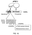
- RANK and RANKL The receptor activator of NF- ⁇ B (RANK) is a member of the tumor necrosis factor (TNF) receptor superfamily.
- the ligand, receptor activator of NF-KB ligand (RANKL) is a member of the TNF superfamily, and has been characterized in multiple settings and variously termed Osteoclast Differentiation Factor (ODF), Tumor Necrosis Factor-Related Activation-Induced Cytokine (TRANCE) and Osteoprotegerin Ligand (OPGL).
- ODF Osteoclast Differentiation Factor
- TRANCE Tumor Necrosis Factor-Related Activation-Induced Cytokine
- OPGL Osteoprotegerin Ligand
- RANK is a Type I transmembrane protein having 616 amino acid residues that interacts with TNF-receptor associated factor 3 (TRAF3).
- RANK is expressed on osteoclast precursors and mature osteoclasts.
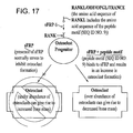
- RANKL produced by osteoblasts stimulates the formation and activity of osteoclasts, which facilitates normal bone development and remodeling.
- Gene targeting of either RANKL or RANK results in osteopetrosis (increased bone mass), as well as severe defects in lymph node formation.
- Osteoprotegerin (OPG) is a soluble factor that also belongs to the TNF receptor family. OPG binds to RANKL, and inhibits the formation of functional multinucleate osteoclasts in vitro.
- OPG binds to RANKL, and inhibits the formation of functional multinucleate osteoclasts in vitro.
- Overexpression of OPG in transgenic mice causes severe osteopetrosis, with a loss of marrow cavities and profound depletion of osteoclasts. The same effects were observed upon administration of OPG in normal mice.
- a recombinant nucleic acid is one that has a sequence that is not naturally occurring or has a sequence that is made by an artificial combination of two otherwise separated segments of sequence. This artificial combination is often accomplished by chemical synthesis or, more commonly, by the artificial manipulation of isolated segments of nucleic acids, e.g. by genetic engineering techniques.
- Sequence identity The similarity between amino acid sequences is expressed in terms of the similarity between the sequences, otherwise referred to as sequence identity. Sequence identity is frequently measured in terms of percentage identity (or similarity or homology); the higher the percentage, the more similar the two sequences are. Homologs or variants of sFRP (the prototypical member of which is shown in SEQ ID NO: 1), or the peptide motif that binds sFRP (for example SEQ ID NO:9), disclosed herein, will possess a relatively high degree of sequence identity when aligned using standard methods.
- NCBI Basic Local Alignment Search Tool (BLAST TM ) ( Altschul et al., J. Mol. Biol. 215:403-410, 1990 ) is available from several sources, including the National Center for Biotechnology Information (NCBI, Bethesda, MD) and on the Internet, for use in connection with the sequence analysis programs blastp, blastn, blastx, tblastn and tblastx.
- Variants of sFRP, sFRP fragments, or the peptide motif that binds sFRP are typically characterized by possession of at least 50% sequence identity counted over the full length alignment with the amino acid sequence of sFRP, sFRP fragments or the peptide motif (for example SEQ ID NO: 9) using the NCBI Blast 2.0, gapped blastp set to default parameters.
- the Blast 2 sequences function is employed using the default BLOSUM62 matrix set to default parameters, (gap existence cost of 11, and a per residue gap cost of 1).
- the alignment should be performed using the Blast 2 sequences function, employing the PAM30 matrix set to default parameters (open gap 9, extension gap 1 penalties). Proteins with even greater similarity to the reference sequences will show increasing percentage identities when assessed by this method, such as at least 60%, at least 65%, at least 70%, at least 75%, at least 80%, at least 90%, or at least 95%, or 98% sequence identity. When less than the entire sequence is being compared for sequence identity, homologs and variants will typically possess at least 75% sequence identity over short windows of 10-20 amino acids, and can possess sequence identities of at least 85% or at least 90%, 95%, or 98% depending on their similarity to the reference sequence.
- sFRP Secreted Frizzled-related protein
- sFRP is a secreted protein that consists of approximately 300 amino acids, including a CRD that is typically between 30% and 50% identical to the (cysteine-rich domain) CRD of the Fz protein family members.
- CRD Cysteine-rich domain
- SEQ ID NO: 1 The nucleic acid and amino acid sequences of other members of the sFRP family can be found at the National Center for Biotechnology Website, for example GenBank Accession No.
- AF218056 Gallus gallus FRP-2
- GenBank Accession No AV354083 Mus musculus- FRP-1
- GenBank Accession No AV304328 Mus musculus s-FRP-2
- GenBank Accession No U24163 homo sapiens sFRP-3/FrzB
- GenBank Accession No AI587049 Homo sapiens sFRP-1).
- SEQ ID NO: 2 The open reading frame of the prototypical sFRP is shown in SEQ ID NO: 2, while the sequence of the protein is shown in SEQ ID NO: 3.
- sFRP binds to RANKL and inhibits osteoclast formation.
- sFRP-1 binding activity and its ability to modulate osteoclast formation can be assayed using the ELISA and osteoclastogenesis bioassay methods described herein.
- the ability of sFRP-1 protein, or a fragment thereof, to perform these activities is beneficial in a number of applications, including clinical applications such as in the treatment of diseases associated with abnormal bone remodeling.
- amino acid sequence of the prototypical sFRP is shown in SEQ ID NO: 3, one of skill in the art will appreciate that variations in this amino acid sequence, such as 1, 2, 5, 10, 20, 30, 40, or 50, deletions, additions, or substitutions (including conservative amino acid substitutions), can be made without substantially affecting the activities of the protein (or fragments of the protein) discussed above.
- the term "sFRP" fragments encompasses both the proteins having the amino acid sequences shown in SEQ ID NOs: 4-8, as well as amino acid sequences that are based on these sequences but which include one or more sequence variants. Such sequence variants can also be defined in the degree of amino acid sequence identity that they share with the amino acid sequence shown in SEQ ID NOs: 4-8.
- sFRP sequence variants will share at least 80% sequence identity with the sequences shown in SEQ ID NOs: 4-8. More highly conserved variants will share at least 90%, at least 95%, or at least 98% sequence identity with the sequences shown in SEQ ID NOs: 4-8. In addition to sharing sequence identity with the prototypical sFRP protein sequence, such sequence variants possess the ability to bind to TNF family members such as RANKL.
- Subject Living multi-cellular vertebrate organisms, a category that includes both human and non-human mammals.
- Therapeutically effective dose A dose sufficient to prevent advancement, or to cause regression of the disease, or which is capable of relieving symptoms caused by the disease.
- TNF family of proteins The Tumor Necrosis (TNF) family of proteins contains both membrane bound ligands and soluble proteins. Some family members, such as TNF and RANKL, are active in both membrane-anchored and soluble forms, the latter being enzymatically released into solution, notably by TACE (TNF alpha converting enzyme) ( J. Hardy, Proc. Natl. Acad Sci. U.S.A. 94:2095-2097, 1997 ; J.D. Buxbaum et al., Proc. Natl. Acad. Sci. U.S.A. 89: 10075-10078, 1992 ).
- TACE TNF alpha converting enzyme
- TNF family members The primary area of homology among TNF family members is a stretch of 150 amino acid residues in the carboxy-terminus that is situated in the extracellular space. This domain is responsible for binding to cognate members of the TNF receptor family.
- This family of receptor proteins is characterized by four domains with regularly spaced cysteine residues: each has a single transmembrane domain and binds either TNF ⁇ or TNF ⁇ .
- Members of the family include, for example, TNFRI, TNFRII, Fas, CD30, and CD30.
- a transformed cell is a cell into which has been introduced a nucleic acid molecule by molecular biology techniques.
- transformation encompasses all techniques by which a nucleic acid molecule might be introduced into such a cell, including transfection with viral vectors, transformation with plasmid vectors, and introduction of naked DNA by electroporation, lipofection, and particle gun acceleration.
- a nucleic acid molecule as introduced into a host cell, thereby producing a transformed host cell.
- a vector can include nucleic acid sequences that permit it to replicate in a host cell, such as an origin of replication.
- a vector can also include one or more selectable marker genes and other genetic elements known in the art.
- a vector can also include a sequence encoding an amino acid motif that facilitates the isolation of the desired protein product such as a sequence encoding maltose binding protein, c-myc, or GST.
- Wnt proteins are a family of growth factors consisting of more than a dozen structurally related molecules and are involved in the regulation of fundamental biological processes, like apoptosis, embryogenesis, organogenesis, morphogenesis and turnorigenesis. These polypeptides are multipotent factors and have similar biological activities to other secretory proteins like transforming growth factor (TGF)- ⁇ , fibroblast growth factors (FGFs), nerve growth factor (NGF), and bone morphogenetic proteins (BMPs).
- TGF transforming growth factor
- FGFs fibroblast growth factors
- NGF nerve growth factor
- BMPs bone morphogenetic proteins
- a member of the Wnt growth factor family is preferentially expressed in bone tissue and in bone-derived cells, and appears to be involved in maintaining the mature osteoblast (bone-forming cell) phenotype.
- the disclosure provides methods of inhibiting osteoclast formation in a subject. These methods include administering sFRP-1 (SEQ ID NO: 3), variants of sFRP-1 (SEQ ID NO: 3), or fusions, or fragments of sFRP-1 (SEQ ID NO: 3). Administering these peptides includes administration and expression of nucleic acids that encode the peptides.
- the administered proteins or peptides are characterized by their ability to bind to RANKL, for example, human RANKL termed "TRANCE" (AF013171), human RANKL (AF019047), and human RANKL termed "OPGI” (AF053712) and inhibit osteoclast formation.
- osteoclast formation will be useful for treating osteopathic disorders such as postmenopausal osteoporosis, Paget's disease, lytic bone metastases, multiple myeloma, hyperparathyroidism, rheumatoid arthritis, periodontitis, and hypercalcemia of malignancy.
- osteopathic disorders such as postmenopausal osteoporosis, Paget's disease, lytic bone metastases, multiple myeloma, hyperparathyroidism, rheumatoid arthritis, periodontitis, and hypercalcemia of malignancy.
- the disclosure also provides methods of screening for sFRP proteins, and fragments, and variants thereof, that bind to members of the TNF family of proteins. These methods include contacting an sFRP protein with at least one TNF family member, and detecting TNF family member binding to the sFRP protein Member's of the TNF family that are of particular interest include RANKL, Apo2/TRAIL, FasL, CD40L, CD27L, CD30L, Apo3L/TWEAK, TNF and LT-alpha ( S.L Baker and EP Reddy, Oncogene 17:3261-3270, 1998 ).
- Members of the sFRP family that are of particular interest includes sFRP-1 (SEQ ID NO: 3), sFRP 2 (GenBank Accession No. MMU88567, ), sFRP-3 (GenBank Accession No MMU88568, ), sFRP-4 (GenBank Accession No. AF012891), and sFRP-5 (GenBank Accession No AF11775
- the disclosure also provides the purified peptide shown in SEQ ID NO: 14. This peptide is useful for stimulating osteoclast differentiation in vitro and in vivo When the peptide is used in vivo it can be administered to subjects to increase osteoclast differentiation
- sFRP fragments and variants thereof can be purified from MDCK cells (ATCC NO CCL-34) transfected with sFRP encoding vectors as described below sFRP fragments and variants thereof can also be purified from a tissue source using conventional biochemical techniques, or produced recombinantly in either prokaryotic or eukaryotic cells using methods well-known in the art (for example, those described in Sambrook et al, Molecular Cloning: A Laboratory Manual, Cold Spring Harbor, New York, 1989 ). The recombinant expression of sFRP fragments is described in ( Uren et al, J. Biol. Chem 275:4374-4382, 2000 ). Furthermore, the nucleic acid sequences encoding sFRP family members are available on GenBank, and include the cDNA sequence shown in SEQ ID NO: 1.
- Recombinant sFRP fragments, fusions, and variants thereof can be obtained using commercial systems designed for optimal expression and purification of fusion proteins.
- fusion proteins typically include a protein tag that facilitates purification.
- Examples of such systems include: the pMAL protein fusion and purification system (New England Biolabs, Inc, Beverly, MA); the GST gene fusion system (Amersham Pharmacia Biotech, Inc, Piscataway, NJ); and the pTrcHis expression vector system (Invitrogen, Carlsbad, CA).
- the pMAL expression system utilizes a vector that adds a maltose binding protein to the expressed protein.
- the fusion protein is expressed in E .
- the fusion protein is purified from a crude cell extract using an amylose column.
- the maltose binding protein domain can be cleaved from the fusion protein by treatment with a suitable protease, such as Factor Xa.
- the maltose-binding fragment can then be removed from the preparation by passage over a second amylose column
- Eukaryotic expression systems can also be employed, including Pichia , tobacco and Baculovirus expression systems, such as those available commercially from Invitrogen.
- the entire sFRP protein, variants and fragments thereof can be produced by ligating the open reading frame (ORF) of the desired sequence into the vector.
- ORF open reading frame
- the ORF must be operably linked to the vector, i.e . must be joined such that the reading frame of the ORF is aligned with the reading frame of the protein tag.
- an ORF encoding the desired fragment can be amplified by polymerase chain reaction (PCR) from the sFRP cDNA, cloned, purified and then ligated into the expression vector Alternatively, the amplified fragment can be ligated directly into the expression vector It can also be possible, depending on the availability of suitable restriction sites in the sFRP cDNA, to obtain the desired fragment by appropriate restriction endonuclease digestion, such that it can be directly cloned into the expression vector.
- PCR polymerase chain reaction
- Purification of the expressed protein can be achieved either using the purification regimen appropriate for the expression tag (if a commercial expression/purification system is used), or conventional affinity chromatography using antibodies, preferably monoclonal antibodies, that recognize the appropriate regions of sFRP can be employed or chromatography procedures established for sFRPs
- fragments are to be used, such fragments alternatively can be generated through digestion of a full length protein with various proteases. The fragments can then be separated based on their unique size, charge or other characteristics Such fragments can also be synthetically generated through the use of known peptide synthesis methods.
- Osteoclasts are large, multinucleate cells that actively reabsorb bone, are derived from hematopoietic stem cells, and share phenotypic characteristics with circulating monocytes and tissue macrophages They are formed from a population of the circulating mononuclear cells that are recruited from the blood to the bone surface, where they undergo differentiation and fusion to form multinucleated cells
- Osteopetrosis is a family of diseases characterized by the failure of the long bones to be remodeled. The resulting long bones have cartilagenous infiltration towards the center of the bone from the growth plate and a poorly remodeled center. While osteoporosis can be caused by osteoclasts that are too numerous or too active, osteopetrosis can be caused by not having sufficient numbers of these cells, or by their inadequate activity Thus, enhancement of osteoclast differentiation is desirable in subjects with abnormal bone remodeling, such as achondroplasia and osteopetrosis. Methods of administration of these FRP-1 to inhibit osteoclast differentiation in a subject are described below
- sFRP-1 can be used to inhibit osteoclast formation.
- Loss of ovarian function following menopause often results in a progressive loss of trabecular bone mass and eventually to osteoporosis. This bone loss is in part due to the increased production of osteoclasts.
- This increased production of osteoclasts appears to be due to the increased elaboration by support cells of osteoclastogenic cytokines such as IL-1, tumor necrosis factor, and IL-6, all of which are negatively regulated by estrogens.
- Osteoclasts are also implicated in degenerative bone diseases at sites of osteolysis. Likewise, osteoclast overproduction is associated with diseases such as hyperparathyroidism and Paget's disease. Osteoclasts are also seen at sites of inflammatory reactions associated with aseptic loosening of total hip prosthesis, rheumatoid arthritis, and periodontitis. Two cytokines produced by inflammatory cells that may have direct effects on osteoclast formation and function are interleukin-1 (IL-1) and tumor necrosis factor (TNF- ⁇ ). Thus, inhibition of osteoclast formation is desirable in subjects with bone disorders characterized by unwanted bone resorption
- sFRP-1 can have clinical utility in conditions where excessive osteoclast activity has pathological consequences.
- Osteoporosis and rheumatoid arthritis are examples of conditions that are particularly good targets for sFRP-1 therapy because soluble RANKI from T cells is thought to have an important role in the bone loss associated with these diseases.
- Methods of administration of sFRP-1 to inhibit osteoclast formation in a subject are disclosed herein.
- disorders of calcium homeostasis can also be affected by osteoclast activity.
- osteoclasts are able to mobilize calcium from bone to affect hypocalcemic states.
- inhibition of osteoclasts can help minimize mobilization in hypercalcemic states Hence, modulation of osteoclast activity can be used as a therapeutic intervention to treat hypocalcemia and hypercalcemia.
- sFRP purified sFRP, sFRP fragments or sFRP variants
- a pharmaceutically acceptable carrier Pharmaceutical preparations can contain only a single peptide, or can be composed of more than one variety of sFRP fragments.
- the nature of the carrier will depend on the particular mode of administration being employed.
- parenteral formulations usually comprise injectable fluids that include pharmaceutically and physiologically acceptable fluids such as water, physiological saline, balanced salt solutions, aqueous dextrose, glycerol, human albumin or the like as a vehicle.
- non-toxic solid carriers can include, for example, pharmaceutical grades of mannitol, lactose, starch, or magnesium stearate.
- pharmaceutical compositions to be administered can contain minor amounts of non-toxic auxiliary substances, such as wetting or emulsifying agents, preservatives, and pH buffering agents and the like, for example sodium acetate or sorbitan monolaurate
- protein based pharmaceuticals can be only inefficiently delivered through ingestion
- pill-based forms of pharmaceutical proteins can alternatively be administered subcutaneously, particularly if formulated in a slow release composition
- Slow-release formulations can be produced by combining the target protein with a biocompatible matrix, such as cholesterol
- a biocompatible carrier would also be used in conjunction with this method of delivery
- sFRP could be delivered to cells in the nucleic acid form and subsequently translated by the host cell. This could be done, for example through the use of viral vectors or liposomes Liposomes could also be used for the delivery of the protein itself:
- compositions of the present disclosure can be administered by any means that achieve their intended purpose.
- Amounts and regimens for the administration of sFRP fragments can be determined readily by those with ordinary skill in the clinical art of treating conditions associated with abnormal bone remodeling.
- the described proteins are administered in an amount effective to either increase osteoclastogenesis activity or decrease osteoclastogenesis
- Such dosages include amounts which raise target tissue concentrations to levels at which the therapeutic activity has been observed in vitro .
- the proteins disclosed herein can also be used to modulate T-cell interactions and immune system functions Doses sufficient to achieve a tissue concentration that causes an increase or a decrease in osteoclastogenesis and/or T-cell activity can be determined by using the amounts described in the examples that follow
- the peptides or proteins can be administered to a host in vivo , such as for example, through systemic administration, such as intravenous or intraperitoneal administration
- the peptides or proteins can be administered intralesionally: i.e. the peptide or protein is injected directly into the tumor or affected area.
- Effective doses of the disclosed peptides for therapeutic application will vary depending on the nature and severity of the condition to be treated, the age and condition of the subject and other clinical factors. Thus, the final determination of the appropriate treatment regimen will be made by the clinician. Typically, the dose range will be from about 0.1 ⁇ g/kg body weight to about 100 mg/kg body weight.
- dosing schedule can vary from once a week to daily depending on a number of clinical factors, such as the subject's sensitivlty to the protein Examples of dosing schedules are 3 ⁇ g/kg administered twice a week, three times a week or daily; a dose of 7 ⁇ g/kg twice a week, three times a week or daily; a dose or 10 ⁇ g/kg twice a week, three times a week or daily; or a dose of 30 ⁇ g/kg twice a week, three times a week or daily In the case of a more aggressive disease it can be preferable to administer doses such as those described above by alternate routes including intravenously or intrathecally. Continuous infusion can also be appropriate.
- Recombinant human sFRP-1 was prepared as described ( Uren et al, J Biol Chem. 275:4374-4382, 2000 ).
- the coding sequence of mouse sFRP-2 was amplified by RT-PCR, using total RNA from embryonic mouse kidney as a source, subcloned into pcDNA3.1 expression vector, transfected into MDCK cells and the recombinant protein purified by heparin-affinity chromatography essentially as described for sFRP-1 in Uren et al., J. Biol. Chem. 275:4374-4382, 2000 .
- Rabbit polyclonal antiserum was raised against recombinant human sFRP-1 by injecting ⁇ 10 ⁇ g of purified protein with complete Freund's adjuvant into the inguinal lymph nodes, and subsequently injecting intramuscularly at 2-3 week intervals similar quantities of antigen dissolved in the incomplete Freund's adjuvant. After several boosts, an immunoglobulin fraction was obtained from serum by chromatography with protein G-bound Sepharose (Pharmacia Biotech, Uppsala, Sweden).
- Peptides were synthesized using standard solid phase chemistry, purified by reverse-phase HPLC and their identity verified by mass spectroscopic analysis (Research Genetics, Inc., Huntsville, AL).
- recombinant soluble RANKL and TRAIL were obtained from PeproTech (Rocky Hill, NJ).
- Mouse monoclonal antibody (designated anti-FLAG M2) directed against the FLAG epitope was purchased from Upstate Biotechnology, Lake Placid, NY.
- Goat anti-rabbit IgG-alkaline phosphatase and rabbit anti-mouse IgG-alkaline phosphatase conjugates and paranitrophenolphosphate (pNPP) were purchased from Sigma (St. Louis, MO).
- recombinant soluble RANKL was purchased from Peprotech, Rocky Hill, New Jersey, or residues 158-317 of murine RANKL were prepared as a GST-expressed protein.
- M-CSF was obtained from Research Genetics Institute (Boston, MA, USA).
- the M13 phage-displayed random peptide library was constructed as described ( Adey et al. Methods in Molecular and Cellular Biology 6:3-14, 1995/1996 ).
- tsJ2, tsJ10 and tsJ14 were generated by transfection with a retroviral vector expressing a temperature-sensitive variant of the immortalizing gene of SV40 (ts A58; Chambers et al., Proc Natl. Acad. Sci. USA 90:5578-5582, 1993 ; Owens et al., Biochem. Biophys. Res. Commun. 222:225-229, 1996 ).
- RAW264.7 cells were purchased from the ATCC, and the cell lines KUSA/O and mc-3T3-e1 are described in Horwood et al. Endocrinology 139:4743-4746, 1998 .
- Osteotropic agents regulate the expression of osteoclast differentiation factor and osteoprotegerin in osteoblastic stromal cells.
- 1 ⁇ ,25(OH 2 ) vitamin D 3 was purchased from Wako Pure Chemicals Co. (Osaka, Japan).
- PGE2 was obtained from Sigma (St. Louis, MO). Other chemicals and reagents were of analytical grade.
- MDCK cells (American Type Culture Collection) were grown in Dulbecco's modified Eagle's medium (Life Technologies, Inc., Rockville, Maryland) containing 10% fetal calf serum (Colorado Serum Company, Denver, Colorado) in 5% CO 2 at 37°C.
- Amplified phage recovered from bacterial broth after this first enrichment step were subjected to two more rounds of panning in wells coated with sFRP-1 as described in the previous paragraph, except that the phage were incubated for only 2 hours and 1 hour in the second and third panning steps, respectively.
- phage obtained from the sFRP-1-coated well were titered and seeded on a lawn of bacteria to permit isolation of phage from 200 separate colonies. Bacteria from each of these colonies were grown in broth, pelleted by centrifugation and phage retrieved in the supernatant.
- phage supernatants were tested for binding to sFRP-1-coated ELISA wells versus wells only coated with the BSA blocking solution. Phage were detected in this assay with primary antibody directed against phage coat protein (Pharmacia Biotech, Uppsala, Sweden, #27-9411-01) and standard detection reagents. Approximately 100 phage isolates were selected for sequence analysis, based on exhibiting at least 5-fold higher binding to sFRP-1 versus BSA coated wells.
- sequence of the DNA insert encoding the peptide segment linked to the M13 gene III coat protein from each phage isolate was determined by using sequencing primers corresponding to adjacent vector sequence.
- An advanced BLAST search analysis of GenBank databases was performed to identify proteins that contained sequences matching portions of the peptide sequences identified by screening of the peptide phage display library.
- Synthetic oligonucleotides encoding peptides of interest were ligated into the bacterial alkaline phosphatase fusion vector, pMY101, which had been digested with Sal I and Xho I ( Yamabhai and Kay, Anal Biochem. 247:143-151, 1997 ). All recombinants were confirmed by DNA sequence analysis.
- Bacteria E. coli, strain DH5 ⁇ F' transformed with the peptide/AP constructs were grown in Luria broth containing ampicillin (50 ⁇ g/mL) to an optical density of 0.5 (at 600 nm), treated with 1 mM isopropyl- ⁇ -D-thiogalactopyranoside and then incubated overnight at 37°C.
- Conditioned medium containing peptide/AP chimera was recovered by centrifugation at 7000 g for 15 minutes. Chimeric proteins in conditioned medium were stable when stored for a few weeks at 4°C or for several months when stored at -80
- ELISA experiments were generally performed as previously described ( Uren et al., J. Biol. Chem.275:4374-4382, 2000 ), with modifications depending on the sFRP binding partner to be tested.
- wells were coated with 0.5 or 1 ⁇ g of sFRP-1, blocked with BSA (0.2%, 1%, or 4%) and then incubated with putative binding partner overnight at room temperature.
- BSA 0.2%, 1%, or 48%
- putative binding partner overnight at room temperature.
- pNPP p-nitrophenolphosphate
- soluble peptides were preincubated with peptide/AP chimeras in bacterial broth for 30 min at room temperature prior to transfer into ELISA wells coated with sFRP-1 or BSA.
- serial dilutions of soluble RANKL were assayed in replicate.
- RANKL solutions were aspirated and bound RANKL was detected by sequential incubations with primary antibody to RANKL, secondary antibody coupled with AP and pNPP.
- Similar experimental designs were employed when other TNF ⁇ family members were examined for binding to sFRPs, and when sFRP-1 derivatives or sFRP-2 were the binding targets for RANKL.
- ddPCR was performed essentially as described ( Liang et al., Science 257:967-971, 1992 and Schwarzedes et al., J. Biol. Chem. 270:20891-20894, 1995 ), except 1 ⁇ g of total RNA was reverse transcribed.
- PCR products were cloned into pCRScriptII (Stratagene, LaJolla, CA) or pGEM-T (Promega, Madison, WI). DNA sequence analysis was performed using a T7 sequencing kit (Pharmacia Biotech, Uppsala, Sweden). Oligonucleotides were synthesized on an Oligo 1000M DNA Synthesizer (Beckman Instruments Inc., Fullerton CA, USA). The oligonucleotides were: for ddPCR, DDMR-2 (5'-CTTGATTGCC-3'; SEQ ID NO: 37) and T12VA (5-TTTTTTTTTTTTTT[A,C,G]A; SEQ ID NO: 32-3').
- Partial cDNA fragments were amplified using 5'-10mers resulting in the synthesis of varying length cDNAs due to random annealing to different reverse transcribed mRNA species. This PCR reaction is performed at an annealing temperature of 40°C and in the presence of [ ⁇ 35 S]-dATP to allow the visualization of resulting products.
- PCR products were resolved on 6% polyacrylamide sequencing gels and exposed to X-ray film for 1-3 days.
- Differentially regulated cDNA fragments were excised from the gel by overlaying the film and cutting out the region of interest.
- the cDNA fragment was reamplified by two rounds of PCR (a total of 80 cycles of PCR).
- the reamplified product was then molecularly cloned into pGEM-T (Promega Inc., Madison, Wisconsin), and the nucleic acid sequence of the amplified insert was determined.
- RNA isolated from cell lines or tissues was reverse transcribed with oligo-dT and PCR performed with the primers sfrp-1a (5'-TTAAAATTGCTGCCTGCCTGAG-3'; SEQ ID NO: 38) and sfrp-1b (5'-TCCGAACTACAGGGACAACAGG-3'; SEQ ID NO: 39) for 22 cycles, which was found to be in the log-linear phase of amplification for sFRP-1 transcripts from osteoblastic sources. Amplifications were performed according to manufacturer's instructions.
- PCR products were electrophoresed, transferred to nylon membrane, and hybridized with ⁇ - 32 P-labeled internal detection oligonucleotide, sfrp-1c (5'-GCCCAGAGGTATTTCTCAAAGTTG-3'; SEQ ID NO: 39).
- gapdh-2 (5'-ATGAGGTCCACCACCCTGTT-3'; SEQ ID NO: 33, nucleotides 640-659; Tso et al., Nucl. Acids Res.
- gapdh-4 were used to amplify the normalizing gene, glyceraldehyde-3-phosphate dehydrogenase, by 20 cycles of PCR and products were detected with ⁇ - 32 P-labeled gapdh-1 as described ( Suda et al., J. Cell. Physiol. 166:94-104, 1996 ).
- a murine sFRP-1 riboprobe was generated by PCR using RNA derived from tsJ2 cells. The resultant fragment of 750 bp was cloned into pGEM-T (Promega, Madison, WI, USA). The plasmid was linearized and transcribed with T7 or SP6 RNA polymerase to generate antisense or sense riboprobes. The riboprobes were labeled with digoxigenin (DIG) during RNA transcription using a RNA labeling kit (Boehringer Mannheim, Mannheim GmbH, Germany) according to the manufacturer's instructions. In situ hybridization was performed as previously described ( Kartsogiannis et al., Bone 21:385-392, 1997 ).
- DIG digoxigenin
- Osteoblastic cells were prepared from the calvaria of newborn mice by digestion with 0.1% collagenase (Worthington Biochemical Co., Freefold, Australia) and 0.2% dispase (Godo Shusei, Tokyo; Japan). Bone marrow and spleen cells were obtained from adult and from newborn mice, respectively ( Udagawa et al., J. Exp. Med. 182: 1461-1468, 1995 ). Osteoblastic cells were co-cultured with bone marrow or spleen cells as described previously ( Udagawa et al., J. Exp. Med. 182: 1461-1468, 1995 ,).
- primary osteoblastic cells (2 x 10 4 /well) and nucleated spleen cells (1 x 10 6 /well) or marrow cells (5 x 10 5 /well) were co-cultured in 48-well plates (Corning Glass Inc., Coming, NY) with 0.4 mL of ⁇ -MEM (GIBCO/BRL, Grand Island, NY) containing 10% fetal bovine serum (Cytosystems, Castle Hill, NSW, Australia) in the presence of test chemicals. Cultures were incubated in quadruplicate and cells were replenished on day 3 with fresh medium. Osteoclast formation was evaluated after culturing for 6-7 days.
- Adherent cells were fixed and stained for tartrate-resistant acid phosphatase (TRAP), and the number of TRAP-positive osteoclasts was scored as described ( Udagawa et al., J. Exp. Med. 182: 1461-1468, 1995 ).
- TRAP staining adherent cells were fixed with 4% formaldehyde in PBS for 3 minutes.
- experiments were performed either with adult mouse spleen cells or with RAW264.7 cells treated with M-CSF and RANKL as described in Quinn et al., Endocrinology 139:4424-4427, 1998 . Where indicated, these assays were conducted in the presence or absence of splenic T cells. T cell fractions were prepared as described in Horwood et al., Journal of Clinical Investigation 101:595-603, 1998 .
- Phage from 200 separate colonies of lysed bacteria were picked, grown in bacterial broth overnight, recovered in supernatant, and tested for their ability to bind preferentially to sFRP-1 versus BSA in an ELISA Phage that bound at least five times more avidly to sFRP-1 than BSA-coated wells were subjected to nucleotide sequence analysis to determine the identity of the peptide sequence responsible for this binding specificity
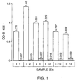
- a set of peptide-alkaline phosphatase fusion proteins containing the peptide motifs were generated. These fusion proteins were tested for specific binding to sFRP-1 (SEQ ID NO: 3) in an ELIS A format. As illustrated in FIG. 1 , broths from multiple isolates of the A-C2 (SEQ IN NO: 14)/alkaline phosphatase fusion protein all showed strong, highly specific binding to wells preincubated with sFRP-1. Similar results were obtained with the A-E4 (SEQ ID NO: 11)/alkaline phosphatase fusion protein.
- the B-B9 (SEQ ID NO: 27)/alkaline phosphatase fusion protein did not exhibit specific binding to sFRP-1.
- This qualitative difference between A-C2 (SEQ ID NO: 14), A-E4 (SEQ ID NO: 11), and B-B9 (SEQ ID NO: 27) derivatives was consistent with a quantitative difference noted during the ELISA screening of the respective phage.
- the A-C2- (SEQ ID NO: 14) and A-E4- (SEQ ID NO: 11) expressing phage were more abundant in the phage preparation selected for sFRP-1 binding (Table 1) and showed a higher ratio of sFRP-1:BSA binding than B-B9 (SEQ ID NO: 27) phage.
- each phage particle has five copies of the peptide displayed on its surface, whereas the peptide-alkaline phosphatase fusion proteins exist as dimers in solution.
- the relatively weaker binding avidity of the B-B9 sequence as originally perceived with the pentavalent phage particle became more obvious when dimeric reagents were tested.
- IIC Isothermal titration calorimetry
- A-D9 Another peptide, A-D9, was analyzed in a manner similar to the routine followed for A-C2
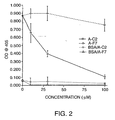
- ELISA experiments performed with an A D9/AP chimera showed that this chimera bound specifically to wells coated with sFRP-1 rather than BSA. This binding was blocked in a dose-dependent manner with soluble synthetic peptide containing the A-D9 sequence Binding of the A-D9/AP chimera to sFRP-1 in ELISA wells was disrupted by alanine substitutions in the A-D9 sequence.
- A-C2 peptide also could inhibit binding of the A-D9/AP chimera to sFRP-1 and the A-D9 peptide inhibited binding of the A-C2/AP chimera to sFRP-1.
- DGR common element
- the sequence V-V-D-G-R-F-V (SEQ ID NO: 10) in the human atrial natriuretic peptide (ANP) receptor A is also of significance because of the co-expression of this gene product and sFRP-1 in tissues within the kidney and eye.
- sFRP-1 SEQ ID NO: 3
- RANKL interact with each other in a manner that has significant biological consequences, and their interaction can be modulated to affect osteoclastogenesis.
- sFRP-1 mRNA was also observed in the epidermis. RANKL is expressed in a similar pattern ( Kartsogiannis et al., Bone 25:525-534, 1999 ). Expression of sFRP-1 in skeletal sites was also detected. Hence, it is likely that sFRP-1 is involved in skeletal morphogenesis and sFRP-1 expression continues in a number of sites through to adulthood.
- sFRP-1 expression in osteoblasts was studied (for a description of the tsJJ2 cell line and the tsJ14 cell line see Chambers et al., Proc. Natl. Acad. Sci. USA 90:5578-5582, 1993 ). The results showed that sFRP-1 is preferentially expressed in osteoblasts (tsJ2 cells) that promote osteoclast formation.
- Murine sFRP-1 transcripts were amplified using the oligonucleotides sfrp-1a and sfrp-1b. Amplified products were verified by Southern analysis using [ ⁇ - 32 P]dATP end-labeled oligonucleotide sfrp-1b as a probe.
- ddPCR Differential display PCR
- sFRP-1 is upregulated in osteoblast lines that stimulate osteoclastogenesis, but not in the products from two other lines that do not support osteoclast differentiation.
- Semi-quantitative RT-PCR analysis of sFRP-1 expression confirmed that transcript level was much higher in lines that were capable of promoting osteoclast formation in co-cultures with hematopoietic progenitor cells. This pattern was observed when additional osteoblast lines were compared, reinforcing the finding that sFRP-1 expression was associated with osteoclastogenesis.
- osteotropic factors such as 1 ⁇ ,25(OH 2 ) vitamin D 3 caused limited stimulation of sFRP-1 expression by osteoblastic lines.
- Total RNA was isolated from either untreated or cells treated with 1 ⁇ ,25(OH 2 ) vitamin D 3 for 24 hours, reverse transcribed with oligo (dT), and subjected to PCR for murine SFRP1 and GAPDH.
- a co-culture of osteoblasts and bone marrow treated for 24 hours with 1 ⁇ ,25(OH 2 ) vitamin D 3 was included as a positive control.
- the primer combination of sfrp-1a (5'- AGC CTT GGC AGT CAA CGA CG-3' SEQ ID NO: 30) and sfrp-1b (5'- GTT GTG GCT TTT GCA TTG CAC-3' SEQ ID NO: 31) was used for sFRP-1 amplification and the primer combination of gapdh-2 (5'-ATG AGG TCC ACC ACC CTG TT-3' SEQ ID NO: 33) and gapdh-4 (5'-CAT GGA GAA GGC TGG GGC TC-3' SEQ ID NO: 34) was used for GAPDH amplification.
- PCR products were electrophoresed, transferred to nylon membrane and hybridized with [ ⁇ - 32 P]-labeled internal detection oligonucleotide, sfrp-1c (5'-TGT TGA AAA CTA GTA GCT G-3' SEQ ID NO: 35) and gapdh-1 (5'-GCT GTG GGC AAG GTC ATC CC-3' SEQ ID NO: 36), respectively, as described ( Southby et al., Endocrinology 137:1349-1357, 1996 ).
- RT-PCR analysis was repeated in triplicate. Semiquantitative RT-PCR analysis was performed three times on each RT reaction and two independent RT reactions were examined.
- sFRP-1 may be a mediator of hormonally dependent osteoclast formation.
- sFRP-1 expression increased markedly when osteoblasts and osteoclast progenitors were co-cultured. The time course of this increase matched the rise in appearance of TRAP+ cells, a marker of osteoclast differentiation.
- upregulation of sFRP-1 expression is dependent on cell-cell communication between the osteoblast and osteoclast lineages.
- the correlation between sFRP-1 expression and osteoclast formation suggested that sFRP-1 induction might be a consequence of osteoclastogenesis.
- sFRP-1 SEQ ID NO: 3
- RANKL recombinant sFRP-1
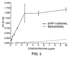
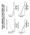
- FIG. 5 The use of recombinant reagents indicates that sFRP-1 (SEQ ID NO: 3) and RANKL bind directly to each other.
- sFRP-1 inhibited TNF ⁇ -dependent osteoclast formation when present during the first three days of culture, whilst OPG had no effect suggesting that sFRP-1 was acting indirectly of RANKL, through binding to TNF ⁇ or through WNT signaling.
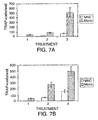
- the effect of bacterially expressed CRD was assessed in three different cell culture models of osteoclast formation. These were: (1) bone marrow cells + RANKL + M-CSF, (2) the macrophage/monocyte cell line RAW264.7 + RANKL, and (3) RAW264.7 + TNF ⁇ + TGF ⁇ ( Horwood et al., Journal of Immunology 166:4915-4921, 2001 ; Quinn et al., Journal of Bone and Mineral Research. 16, 1787-1794, 2001 ).
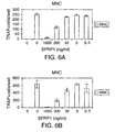
- FIG. 6A Assays measuring the effect of sPRP-1 on osteoclastogenesis showed that sFRP-1 has a dose-dependent inhibitory activity on osteoclast formation.
- FIG. 6A results were observed in co-cultures of primary osteoblasts and bone marrow cells treated with vitamin D3 (10 -8 M) and PGE2 (10 -7 M).
- sFRP-1 reduced the number of multinucleated TRAP+ cells by 50% when used at a concentration 300 ng/mL, while a dose of 1 6 ug/mL decreased the number of cells by 95% ( FIG. 6A )
- FIG. 6B A similar dose-response pattern was observed when adult mouse spleen cells were treated with RANKL and M CSF.
- the three conditions used in these assays were: (1) bone marrow cells + RANKL + M-CSF, (2) RAW264 7 + RANKL, and (3) RAW264 7 + TNF ⁇ + TGF ⁇ .
- group (3) in the absence of RANKL, it is possible that CRD binds to TNF ⁇ , which is structurally-related to RANKL.
- A-C2 Synthetic Peptide Promotes Osteoclast Formation
- A-C2 (SEQ ID NO: 14) polypeptide has sequence homology to RANKL and because A-C2 (SEQ ID NO: 14) binds to sFRP-1, assays were performed to determine if A-C2 (SEQ ID NO: 14) would block SFRP-1 binding to RANKL and thus increase osteoclastogenesis Consistent with this hypothesis, treatment of osteoblast and adult spleen cell co-cultures with A-C2 (SEQ ID NO: 14) resulted in a ten-fold increase in TRAP+ multinucleated cells ( FIG 8 ) A-C2 (SEQ ID NO: 14) only had an effect on osteoclast formation when it was present during day 0 to day 3 of the co-culture experiment RANKL presence is also required for osteoclastogenesis during day 0 to day 3 ( Suda et al., Endocrine Reviews 20:345-357, 1999 ) Thus, these results are consistent with the notion that A-C2 (SEQ ID NO: 14
- a C2 SEQ ID NO: 14
- A-C2 SEQ ID NO: 14
- A-C2 dose-dependent stimulation of TRAP+
- multinucleated cell differentiation in RAW264 7 cultures was only observed when T cells were added to the cultures ( FIG 11 ).
- the peptide motif L/V-V-D-G-R-W-L/V (SEQ ID NO: 9), has not been previously identified as being capable of binding to sFRP-1.
- This binding activity has been characterized through the use of a series of ELISA experiments using peptide/alkaline phosphatase fusion proteins, alanine scanning mutagenesis and synthetic peptides. These experiments also demonstrated that substitutions in nearby residues could enhance or reduce the peptide motif/sFRP-1 binding, implying that systematic substitutions in adjacent residues and conservative changes within the peptide motif, are able to strengthen the interaction with sFRP-1.
- amino acids which can be substituted for an original amino acid in a protein and which are regarded as conservative substitutions include: Ser for Ala; Lys for Arg; Gln or His for Asn; Glu for Asp; Ser for Cys; Asn for Gln; Asp for Glu; Pro for Gly; Asn or Gln for His; Leu or Val for Ile; Ile or Val for Leu; Arg or Gln for Lys; Leu or Ile for Met; Met, Leu or Tyr for Phe; Thr for Ser; Ser for Thr; Tyr for Trp; Trp or Phe for Tyr; and Ile or Leu for Val.
- the binding affect of peptides in which such substitutions have been made can readily be confirmed by the peptide motif-binding assay disclosed herein.
- substitutions that are less conservative than those described above, i.e . selecting residues that differ more significantly in their effect on maintaining (a) the structure of the polypeptide backbone in the area of the substitution, for example, as a sheet or helical conformation, (b) the charge or hydrophobicity of the molecule at the target site, or (c) the bulk of the side chain.
- the substitutions which in general are expected to produce the greatest changes in protein properties will be those in which (a) a hydrophilic residue, e.g . seryl or threonyl, is substituted for (or by) a hydrophobic residue, e.g .
- leucyl isoleucyl, phenylalanyl, valyl or alanyl
- a cysteine or proline is substituted for (or by) any other residue
- a residue having an electropositive side chain e.g . lysyl, arginyl, or histadyl, is substituted for (or by) an electronegative residue, e.g , glutamyl or aspartyl
- a residue having a bulky side chain e.g . phenylalanine, is substituted for (or by) one not having a side chain, e.g . glycine.
- the effects of these amino acid substitutions or deletions or additions can be assessed through the use of the ELISA binding assay provided herein.
- nucleic acid sequences encoding the peptide motif can be designed by either synthetically synthesizing the appropriate nucleic acid sequence, or by using PCR to amplify the appropriate sequence. Once obtained the nucleic acid sequence can be placed in an appropriate expression vector and transformed into an organism such that the organism then produces the peptide.
- the significance of the peptide motif was demonstrated in experiments that revealed a dramatic stimulatory effect of a synthetic peptide bearing the peptide motif in osteoclastogenesis bioassays.
- the ability of the A-C2 peptide (SEQ ID NO: 14) to promote osteoclast formation was consistent with the idea that the peptide increased RANKL activity by blocking the inhibitory effect of endogenous sFRP-1 ( FIG. 17 ).
- splenic T cells express an osteoclastogenesis inhibitor sensitive to A-C2 (SEQ ID NO: 14).
- This inhibitor is believed to be sFRP-1, as sFRP-1 is expressed in spleen ( Finch et al., Proc. Natl. Acad. Sci. USA 94:6770-6775, 1997 ).
- reagents possessing a sFRP-1 binding motif are believed to have utility in regulating RANKL signaling involved in T cell-dendritic communication that is modulated by endogenous sFRP-1.
- Such regulation can be exploited to optimize vaccine therapies in a variety of settings where T cell-dendritic cell interactions have an important role in the immune response.
- Dendritic cells have been shown to promote vaccine responses, which can be determined by measuring titers to know antigens developed in inoculated animals ( M. Di Nicola et al., Cytokines Cell. Mol. Ther. 4: 265-273, 1998 ; C. Reis e Sousa et al., Curr. Opin. Immunol. 11: 392-399, 1999 ; K. Tarte and B. Klein, Leukemia 13: 653-663, 1999 ). Efforts are underway to optimize the expansion of immunologically responseive dendritic cells in order to improve the efficacy of vaccine therapy ( R. Hajek and A.W. Butch, Med. Oncol. 17: 2-15, 2000 ). Using methods cited in the above references, reagents corresponding to the sFRP-1 binding motif and sFRP-1 can be useful to enhance the immune response in vaccine therapies.
- sFRP-1 can be useful for modulating endogenous proteolytic cleavage of RANKL.
- a large fraction of the RANKL expressed by T cells is proteolytically processed to release a soluble, biologically active form. This process, which can be mediated by the TNF ⁇ converting enzyme (TACE), involves cleavage at one or two sites in the RANKL sequence just upstream of the putative peptide-binding motif (SEQ ID NO: 9). Therefore, it is believed that sFRP-1 regulates the proteolytic processing of RANKL in a manner that could be reversed by reagents containing the peptide motif (SEQ ID NO: 9).
- TACE TNF ⁇ converting enzyme
- sFRP-1 ability of sFRP-1 to regulate RANKL processing is tested by culturing T-cells expressing RANKL and treating the cultures with various concentrations of sFRP-1. The resulting soluble RANKL proteins are then identified using a RANKL specific western blot. The degree of RANKL processing is then correlated to the sFRP concentration in the sample.
- reagents containing the peptide motif also have utility in disrupting the interaction of sFRP-1 with other proteins.
- other known proteins like the netrin receptor, and UNC5H3 and the ANP receptor A, have sequences similar to the binding motif, and newly identified gene sequences can be routinely screened for such sequences. Proteins with this motif are likely to be additional potential binding partners for sFRP-1 and targets for reagents containing the sFRP-1 binding motif. For instance, sFRP-1 binding to the ANP receptor A could regulate the release of sodium and fluid in the kidney and eye.
- sFRP-1 has an inhibitory effect on osteoclastogenesis, which is likely due to its interaction with RANKL.
- the elevated expression of sFRP-1 transcript specifically in osteoblast lines capable of stimulating osteoclast formation initially suggested that sFRP-1 promotes osteoclastogenesis.
- the increase in sFRP-1 transcript observed in co-cultures of osteoblasts and hematopoietic progenitors as osteoclast formation proceeded implied that sFRP-1 was instead part of a tonic mechanism to limit the extent of osteoclast formation ( FIG. 17 ).
- sFRP-1 can have clinical utility in conditions where excessive osteoclast activity has pathological consequences. Osteoporosis and hypercalcemic osteopaenia are examples of such conditions; rheumatoid arthritis is another, which could be a particularly good target for sFRP-1 therapy because soluble RANKL from T cells is thought to have an important role in the bone loss associated with this disease.
- Example 8 The results described above (Example 8) indicate that additional contact points within RANKL, besides the newly identified peptide-binding motif, are involved in the interaction of sFRP-1 (SEQ ID NO: 3) and RANKL. These results further indicate that sFRP family members interact with TNF-ligand family members.
- TNF-ligand family members are known to be among the most pleiotropic cytokines, inducing a large number of cellular responses, including cytotoxicity, anti-viral activity, immunoregulatory activities, and the transcriptional regulation of several genes.
- Cellular responses to TNF-family ligands include not only normal physiological responses, but also diseases associated with increased apoptosis or the inhibition of apoptosis.
- Apoptosis-programmed cell death is a physiological mechanism involved in the deletion of peripheral T lymphocytes of the immune system, and its dysregulation can lead to a number of different pathogenic processes.
- Diseases associated with increased cell survival, or the inhibition of apoptosis include cancers, autoimmune disorders, viral infections, inflammation, graft v.
- Diseases associated with increased apoptosis include AIDS, neurodegenerative disorders, myelodysplastic syndromes, ischemic injury, toxin-induced liver disease, septic shock cachexia and anorexia.
- the disclosure further provides methods for modulating the TNF ligand/TNF receptor interactions. These methods involve contacting sFRP, a fragment or variant of sFRP, or the peptide motif (SEQ ID NO: 9) with a member of the TNF-ligand family of proteins, and detecting a change in TNF-ligand biological activity
- sFRP sFRP
- fragment or variant of sFRP or the peptide motif acts as an "agonist” or antagonist”
- TNF-family ligand/receptor cellular response assays such as ones described in the references cited in the following reviews: D Wallach et al., Annu: Rev Immunol. 17: 331-367, 1999 ; S J Bakei and EP. Reddy, Oncogene 17: 326 13276, 1998
- the disclosure provides screening methods for determining whether a candidate agonist or antagonist is capable of enhancing or inhibiting a cellular response to a TNF family ligand
Abstract
Description
- This disclosure relates to osteoclast differentiation, specifically to a peptide motif and proteins containing the motif that are capable of binding to secreted Frizzled-related protein family members.
- Bone remodeling, a process responsible for the continuous renewal of the adult human skeleton, is carried out by osteoclasts and osteoblasts, two specialized cell types that originate from hematopoietic and mesenchymal progenitors of the bone marrow, respectively. A continuous and orderly supply of these cells is essential for skeletal homeostasis, as increased or decreased production of osteoclasts or osteoblasts and/or changes in the rate of their apoptosis are largely responsible for the imbalance between bone resorption and formation that underlies several systemic or localized bone diseases.
- Enhanced osteoclast activity plays a major role in the pathogenesis of postmenopausal osteoporosis, Paget's disease, lytic bone metastases, multiple myeloma, hyperparathyroidism, rheumatoid arthritis, periodontitis, and hypercalcemia of malignancy. These clinical problems are associated with significant morbidity or mortality, and affect more than 10 million patients in the United States. However, only a limited number of agents that inhibit osteoclast formation or bone resorption are available and for most their mechanisms of action are unknown. Furthermore, many of these agents have significant side effects that limit their utility. Thus, there exists a need for the identification and characterization of inhibitors of osteoclast formation and bone resorption as part of the continuing search to provide therapeutic benefits for these patients.
- Conversely, decreased osteoclast activity plays a major role in the pathogenesis of osteopetrosis, Albright's osteodystrophy, and achondroplasia, for which there is no specific therapy. Thus, there also exists a need for the identification and characterization of treatments that enhance osteoclast formation and bone resorption in order to provide successful therapies for these patients.
- Identification of the mechanisms involved in bone disorders is crucial for the understanding of bone physiology. While numerous genes and gene families (and the polypeptides encoded by them) that participate in the regulation of bone cells have been identified and cloned, their functions have not been clearly delineated due to the complexities of the bone formation pathways. A great need exists for the definitive identification of targets for the treatment of bone disorders, including bone resorption disorders such as postmenopausal osteoporosis, Paget's disease, lytic bone metastases, multiple myeloma, rheumatoid arthritis, hypercalcemia of malignancy, osteopetrosis, Albright's osteodystrophy, and achondroplasia.
-
WO01/19855A - Baranski et al, Developmental Biology, vol 217, no 1 (2000) pages 25-41 relates to the dynamic expression patterns of frzb-1.
- Hausler et al, Bone, vol 27, no 4 (2000) pages 335 relates to SFRPs expressed by osteoblasts.
- Rattner et al, Proceedings of the National Academy of Sciences of USA, vol 94, no 7 (1997) pages 2859-2863 relates to a family of secreted proteins containing homology to frizzled receptors.
- Hijikata et al, FEBS, vol 457, no 3 (1999) pages 405-408 relates to induction of apoptosis of monocyte-macrophage lineage cells.
- Van Der Pluijm et al, Journal of Bone and Mineral Research, relates to integrins and osteoclastic resorption in three bone organ cultures
- Finch et al, Proceedings of the National Academy of Sciences of the United States of America, vol 94, No13 (1997) pages 6770-6775 relates to purification and molecular cloning of an antagonist of Wnt action.
- Disclosed herein are proteins that bind to secreted Frizzled-related protein-1 (-sFRP-1). In one embodiment, the sFRP-1 binding peptide is a purified peptide.
- In another embodiment, a method is disclosed for enhancing osteoclast differentiation. In one specific, non-limiting example the method includes administering a therapeutically effective amount of the purified peptides disclosed herein (or effective fragments, fusions or mimetics) to a subject in order to enhance osteoclast differentiation.
- In a further embodiment, a method is provided for inhibiting osteoclast formation in a subject. The method includes administering to the subject a therapeutically effective amount of sERP-1 (SEQ ID NO: 3), fragments of SEQ ID NO: 3, or fusions or variants of SEQ ID NO: 3, to a subject, wherein the polypeptide binds to a RANKL molecule as set forth as GenBank Accession No. AF013171, GenBank Accession No. AF019047, or GenBank Accession No. AF053712, or another TNF family member.
- In yet another embodiment, a method is provided for modulating T cell activity, In one specific, non-limiting example, the method includes administering a therapeutically effective amount of the purified sFRP-1-binding peptides disclosed herein to a subject in order to modulate I cell activity. The invention is defined in the accompanying claims. The polypeptide does not have the sequence for human SARP-2, disclosed in Melconyan et al., 1997, Proc. Natl. Acad Sci. USA, 94, 13636-13641, with alanine at position 174 (Vni Prot ID No Q8N474).
-
-
FIG. 1 is a graph showing the results from A-C2 (SEQ ID NO: 14)/AP (alkaline phosphatase) fusion protein binding to sFRP-1 (SEQ ID NO: 3). Broths from separate bacterial colonies infected with phage expressing the A-C2/AP chimera were incubated in ELISA wells coated with sFRP-1 (and subsequently blocked with BSA) or BSA alone. Each of the broths (identified as I-1, I-2, I-6, I-12, I-13 and I-14) contained AP activity, as measured by reaction with pNPP and color development at 405 nm, that bound specifically to the sFRP-1 (SEQ ID NO: 3) -coated wells (gray bars) as compared to the wells coated only with BSA (black bars). Each sample was tested singly; this is representative of several experiments. -
FIG. 2 is a graph showing the results from a competitive binding assay of A-C2/AP (SEQ ID NO: 14) and the 12-mer peptides A-C2 (SEQ ID NO: 14) and A-F7 (SEQ ID NO: 12) to either BSA or sFRP-1 (SEQ ID NO: 3). Soluble 12-mer peptides, A-C2 (SEQ ID NO: 14) and A-F7(SEQ ID NO: 12), were preincubated for 30 minutes at the indicated concentrations with bacterial broth containing A-C2/AP chimera prior to addition to ELISA wells coated with sFRP-1 (A-C2 open squares and A-F7 open diamonds) or BSA (A-C2 open circles and A-F7 open triangles). Samples were tested in triplicate, and results are shown as the mean +/-S.D. This is representative of three experiments. -
FIG. 3 is a graph showing the results from binding assays of alanine substituted A-C2 (SEQ ID NO: 14)/alkaline phosphatase fusion proteins to sFRP-1 (SEQ ID NO: 3) in an ELISA. The bar graph indicates the mean +/- S.D. of triplicate measurements from a single representative experiment. A parallel analysis of these samples in wells coated with monoclonal antibody to the FLAG epitope indicated that the concentration of chimera in the different broths was similar. -
FIG. 4 is a set of diagrams showing a calorimetric analysis of the interaction between AC2 peptide and sFRP-1.FIG 4A is a tracing and plot showing the heat generated when aliquots of AC2 solution were added to a chamber containing sFRP-1 dissolved in PBS.FIG. 4B is a tracing and plot of the heat generated in the corresponding PBS control. Based on the amount of heat released, various parameters of the binding reaction were calculated, including the enthalpy (ΔH) and dissociation constant, Kd. -
FIG. 5 is a graph showing the results from ELISA binding assays using soluble RANKL (sRANKL) and sFRP-1 (SEQ ID NO: 3). The open diamond represents sRANKL binding to sFRP-1, and the plus symbol "+" represents sRANKL binding to bovine serum albumin (BSA). -
FIG. 6 is a set of graphs showing that sFRP-1 inhibits osteoclast formation in two different experimental models.FIG. 6A is a graph showing results from co-culture experiments in which primary osteoblasts and bone marrow were incubated with varying concentrations of sFRP-1. Subsequently, wells were stained to determine the number of TRAP+ multinucleated cells (MNC). Results show that as the concentration of sFRP-1 increases, osteoclast maturation decreases (as is evident by decrease in TRAP+). The results shown are the mean +/- S.D. of quadruplicate measurements.FIG. 6B is a graph showing the results from experiments in which adult spleen cells were treated with RANKL, macrophage colony stimulating factor (M-CSF), and various concentrations of sFRP-1 (SEQ ID NO: 3). Subsequently, wells were stained to determine the number of TRAP+ multinucleated cells (MNC). The data presented is the mean +/- S.D. of quadruplicate measurements. Results show that as the concentration of sFRP-1 increases osteoclast maturation decreases. -
FIG. 7 is a pair of graphs showing that sFRP-1 antiserum stimulates osteoclast formation in co-cultures of primary osteoblasts and adult spleen cells.FIG. 7A is a graph showing that sFRP-1 specific antibody binding to sFRP-1 causes an increase in osteoclast formation. Osteoclast formation, as measured by TRAP+ staining, was assessed in co-cultures of primary osteoblasts and adult spleen cells without hormonal supplements or with suboptimal doses of 1α,25(OH2) vitamin D3 (10-10M) and dexamethasone (10-9M) in the presence or absence of purified immunoglobulin (~2 µg/mL) from a rabbit immunized with recombinant sFRP-1. The results are the mean +/- S.D. of mononucleated and multinucleated TRAP+ cells detected in quadruplicate samples after 7 days in culture.FIG. 7B is a graph showing that sFRP-1 specific antibodies bind to sFRP-1 and cause an increase in osteoclast formation in the presence of optimal doses of 1α,25(OH2) vitamin D3 (10-8 M) and prostaglandin E2 (PGE2) 10-7 M. Osteoclast formation, as measured by TRAP+ staining, was assessed in co-cultures of primary osteoblasts and adult spleen cells without hormonal supplements or with optimal doses of 1α,25(OH2) vitamin D3 (10-8M) and prostaglandin E2 (PGE2, 10-7M) in the presence or absence of purified immunoglobulin (~1 µg/mL) from a rabbit immunized with recombinant sFRP-1. The results are the mean +/- S.D. of mononucleated and multinucleated TRAP+ cells detected in quadruplicate samples after 7 days in culture. -
FIG. 8 is a graph showing that A-C2 peptide stimulates osteoclast formation in co-cultures of osteoblasts and adult spleen cells. Osteoclast formation in response to optimal doses of 1α,25(OH2) vitamin D3 (10-8M) and PGE2 (10-7M) was not further enhanced by the concomitant addition of the netrin homology domain (NHD) domain of sFRP-1 (SEQ ID NO: 13; 5 µg/mL), but it was markedly stimulated by simultaneous incubation with the A-C2 peptide (SEQ ID NO: 14; 5 µg/mL). As a positive control for enhanced osteoclastogenesis, cells were treated with suboptimal doses of 1α,25(OH2) vitamin D3 (10-10M) and PGE2 (10-9M) in the absence or presence of sFRP-1specific antibody 1/500. The bar graph shows the mean +/- S.D. of TRAP+ multinucleated cells tested in quadruplicate. -
FIG. 9 is a graph showing the results from an experiment in which A-C2 (SEQ ID NO: 14) was incubated for various time periods with adult spleen cells.Group 1 was the control that did not contain A-C2 (SEQ ID NO: 14) peptide.Group 2 was treated with A-C2 (SEQ ID NO: 14) from day 0-3,group 4 was treated with A-C2 (SEQ ID NO: 14) from day 4-7,group 4 was treated with A-C2 (SEQ ID NO: 14) from day 7-10, andgroup 5 was treated with A-C2 (SEQ ID NO: 14) from day 0-10. All treatment groups received RANKL at 50 ng/mL and M-CSF at 25 ng/mL. A-C2 (SEQ ID NO: 14) presence during days 0-3 caused an increase in osteoclast production. -
FIGS. 10A and 10B are graphs depicting the results from A-C2 (SEQ ID NO: 14) incubation with adult spleen cells containing T cells (FIG. 10A ) and spleen cells without T cells (FIG. 14B ): T cells were immunomagnetically separated from the spleen cells. Osteoclast formation was induced by RANKL (50 ng/mL) and M-CSF (25 ng/mL) and assessed by counting TRAP+ multinucleated cells after nine days of culture. Osteoclast formation was measured in adult spleen cell cultures (FIG. 14A ) or in culture lacking T cells (FIG. 10B ) in the absence (Control +ve) or presence of A-C2 (5 mg/mL). These cultures do not contain any osteoblasts, thus effects of A-C2 were restricted to lymphocytic or hematopoietic cells. The bar graphs show the mean +/- S.D. of TRAP+ multinucleated cells from quadruplicate samples. Controls for this experiment included spleen cells [complete (FIG. 10A ) or T cell depleted (FIG. 10B )] in the absence of RANKL and M-CSF (control -ve) and no osteoclasts were produced under these conditions. As a positive control for the assay system (Control +ve), cultures were treated with RANKL (50 ng/mL) and M-CSF (25 ng/mL), and the effects A-C2 addition is compared to this culture. -
FIGS. 11A and 11B are graphs depicting the A-C2 (SEQ ID NO: 14) stimulation of TRAP+, multinucleated cell differentiation in RAW264.7 (TIB-71) cell cultures.Group 1 was the positive control that contained 50 ng/mL RANKL.Groups -
FIG. 12 is a graph of the binding avidity of several SFRP-1 deletion mutants for RANKL in ELISA experiments. Wells were coated either with full-length sFRP-1 or with any one of a set of epitope-tagged sFRP-1 deletion mutants (Uren et al., JBiol Chem 275:4374-4382,2000) or BSA control, and then sequentially incubated with soluble RANKL and reagents to detect RANKL bound to the wells. The results shown are the mean +/- S.D. of triplicate measurements from a representative experiment. -
FIG. 13 is a set of four graphs showing that binding of RANKL to bacterially expressed CRD in ELISA experiments is strong and may have two affinities.FIG. 13A is a graph showing the binding of RANKL to wells coated with the CRD. Optical density in the wells is a measure of the amount of RANKL retained in the wells and is plotted as a function of the soluble RANKL concentration incubated in the wells.FIG. 13B is a Scatchard plot of the RANKL binding data shown inFIG. 13A . The binding appears to be characterized by more than one affinity.FIG. 13C is a reformatting of the Scatchard analysis ofFIG. 13B , pertaining to a putative higher-affinity binding site.FIG. 13D is a reformatting of the Scatchard analysis ofFIG. 13B , pertaining to a putative lower-affinity binding site. -
FIG. 14 is a set of three graphs showing that the bacterially expressed CRD of sFRP-1 inhibits osteoclast formation in a variety of experimental models, including one that is not dependent on RANKL. These were: (1) RAW264.7 + TNFα + TNFβ (FIG. 14A ; Horwood et al., Journal of Immunology 166:4915-4921, 2001; Quinn et al., Journal of Bone and Mineral Research. 16, 1787-1794, 2001 (2) the macrophage/monocyte cell line RAW264.7 + RANKL (FIG.14B ), and (3 ) bone marrow cells + RANKL + M-CSF (FIG. 14C ). In each system, both RANKL-dependent (FIG. 14B and FIG. 14C ) and RANKL-independent (FIG. 14A , TNFα-dependent osteoclast formation), the bacterially expressed CRD mimicked the action of full-length sFRP-1 and with similar potency. -
FIG.15 is a graph showing that sFRP-1 can inhibit osteoclast formation in RAW264.7 cells treated with a combination of cytokines that includes TNFα, but not RANKL. The effect of sFRP-1 was assessed upon a RANKL-independent method of osteoclast formation using the monocyte /macrophage cell line RAW264.7 (Quinn et al., Journal of Bone and Mineral Research. 16, 1787-1794, 2001) and was compared with that of osteoprotegerin. TGFα was added during the first three days of culture to increase osteoclast numbers. sFRP-1 inhibited TNFα-dependent osteoclast formation when present during the first three days of culture, whilst OPG had no effect suggesting that sFRP-1 was acting independently of RANKL, through binding to TNFα or through WNT signaling. -
FIG. 16 is a schematic diagram of one possible mechanism of sFRP-1 (SEQ ID NO: 3)/RANKL binding. Note that the sFRP-1 binding motif in the RANKL sequence is located just downstream from TACE cleavage sites (arrows). TACE is the TNFα converting enzyme, which is known to process RANKL (L. Lum et al., J. Biol. Chem. 274: 13613-13618, 1999). sFRP-1 binding to RANKL could alter the processing of RANKL by TACE, which in turn could alter RANKL activity. -
FIG. 17 is a diagram showing one possible model of sFRP-1's role in osteoclast formation. An osteoclast-supporting cell expressing RANKL interacts with sFRP-1 (SEQ ID NO: 3) resulting in the inhibition of osteoclast formation. When the peptide motif (SEQ ID NO: 9) is added to the solution it binds to sFRP-1 (SEQ ID NO: 3) and promotes osteoclast differentiation. - The nucleic and amino acid sequences listed in the accompanying sequence listing are shown using standard letter abbreviations for nucleotide bases, and three-letter code for amino acids. Only one strand of each nucleic acid sequence is shown, but the complementary strand is understood as included by any reference to the displayed strand.
- SEQ ID NO: 1 shows the cDNA sequence of human sFRP-1.
- SEQ ID NO: 2 shows the nucleic acid sequence of the human sFRP-1 open reading frame.
- SEQ ID NO: 3 shows the amino acid sequence of human sFRP-1.
- SEQ ID NO: 4 shows the amino acid sequence of human sFRP-1-M/A.
- SEQ ID NO: 5 shows the amino acid sequence of human sFRP-Δ1-M/H.
- SEQ ID NO: 6 shows the amino acid sequence of human sFRP-Δ2-M/H.
- SEQ ID NO: 7 shows the amino acid sequence of human sFRP-Δ3-M/H.
- SEQ ID NO: 8 shows the amino acid sequence of human sFRP-ΔCRD-M/H.
- SEQ ID NO: 9 shows the amino acid sequence of the peptide motif.
- SEQ ID NO: 10 shows the peptide motif from ANP receptor A (human).
- SEQ ID NO: 11 shows the amino acid sequence of the A-E4 peptide.
- SEQ ID NO: 12 shows the amino acid sequence of the A-F7 peptide.
- SEQ ID NO: 13 shows the amino acid sequence of the netrin homology domain of sFRP-1.
- SEQ ID NO: 14 shows the amino acid sequence of the A-C2 peptide.
- SEQ ID NO: 15-26 show peptides generated for use in alanine scanning experiments.
- SEQ ID NO: 27 shows the amino acid sequence of B-B9.
- SEQ ID NO: 28 shows an amino acid sequence found in RANKL that contains a sequence similar to that of SEQ ID NO: 9.
- SEQ ID NO: 29 shows an amino acid sequence found in a netrin receptor that contains a sequence similar to that of SEQ ID NO: 9.
- SEQ ID NOS: 30-39 show the nucleic acid sequences of various primers and probes used in PCR and hybridization experiments.
- SEQ ID NO: 40 shows the amino acid sequence of the A-D9 peptide.
-
- BSA:
- bovine serum albumin
- CRD:
- cysteine-rich domain
- ELISA:
- enzyme-linked immunosorbent assay
- HSPG:
- heparin-sulfate proteoglycan
- mAb:
- monoclonal antibody
- MDCK:
- Madin-Darby canine kidney
- M/H:
- Myc-His epitope tags
- PAGE:
- polyacrylamide gel electrophoresis
- PBS:
- phosphate-buffered saline
- sFRP:
- secreted Frizzled-related protein
- Wnt:
- Wnt proteins
- Unless otherwise noted, technical terms are used according to conventional usage. Definitions of common terms in molecular biology may be found in Benjamin Lewin, Genes V, published by Oxford University Press, 1994 (ISBN 0-19-854287-9); Kendrew et al. (eds.), The Encyclopedia of Molecular Biology, published by Blackwell Science Ltd., 1994 (ISBN 0-632-02182-9); and Robert A. Meyers (ed.), Molecular Biology and Biotechnology: a Comprehensive Desk Reference, published by VCH Publishers, Inc., 1995 (ISBN 1-56081-569-8).
- In order to facilitate review of the various embodiments of the disclosure, the following explanations of specific terms are provided:
- Abnormal: Deviation from normal characteristics. Normal characteristics can be found in a control, a standard for a population, etc. For instance, where the abnormal condition is a disease condition, such as osteoporosis (characterized by a decrease in bone mass), a few appropriate sources of normal characteristics might include an individual who is not suffering from the disease (e.g. osteoporosis), a population standard of individuals believed not to be suffering from the disease, etc.
- Likewise, abnormal can refer to a condition that is associated with a disease. The term "associated with" includes an increased risk of developing the disease as well as the disease itself. For instance, a certain abnormality (such as a decrease in the expression of sFRP, which in turn upregulates osteoclast formation) can be described as being associated with the biological condition of osteoporosis (decrease in bone mass); thus, the abnormality is predictive both of an increased risk of developing osteoporosis and of the presence of osteoporosis.
- Abnormal protein expression, such as abnormal sFRP protein expression, refers to expression of a protein that is in some manner different from expression of the protein in a normal (wildtype) situation. This includes but is not necessarily limited to: (1) a mutation in the protein such that one or more of the amino acid residues is different; (2) a short deletion or addition of one or a few amino acid residues to the sequence of the protein; (3) a longer deletion or addition of amino acid residues, such that an entire protein domain or sub-domain is removed or added; (4) expression of an increased amount of the protein, compared to a control or standard amount; (5) expression of a decreased amount of the protein, compared to a control or standard amount; (6) alteration of the subcellular localization or targeting of the protein; (7) alteration of the temporally regulated expression of the protein (such that the protein is expressed when it normally would not be, or alternatively is not expressed when it normally would be); (8) alteration in post translational processing; and (9) alteration of the localized (e.g. organ or tissue specific) expression of the protein (such that the protein is not expressed where it would normally be expressed or is expressed where it normally would not be expressed), each compared to a control or standard.
- Controls or standards appropriate for comparison to a sample, for the determination of abnormality, include samples believed to be normal as well as laboratory values, even though possibly arbitrarily set, keeping in mind that such values can vary from laboratory to laboratory. Laboratory standards and values can be set based on a known or determined population value and can be supplied in the format of a graph or table that permits easy comparison of measured, experimentally determined values.
- cDNA (complementary DNA): A piece of DNA lacking internal, non-coding segments (introns) and regulatory sequences that determine transcription. cDNA is synthesized in the laboratory by reverse transcription from messenger RNA extracted from cells.
- CRD: A cysteine rich domain that typically is about 120 amino acids in length and found on the amino terminal half of Fz proteins. In the prototypical sFRP described herein, the CRD comprises sFRP-1 residues 38-166. Met (ATG) was added at the N-terminus to facilitate protein expression. Typically the Met is cleaved in the bacteria as the protein is processed. The CRD sequence is shown below:
- MFQSDIGPYQ SGRFYTKPPQ CVDIPADLRL CHNVGYKKMV
- LPNLLEHETM AEVKQQASSW.VPLLNKNCHA GTQVFLCSLF
- APVCLDRPIY PCRWLCEAVRDSCEPVMQFF
- GFYWPEMLKC.DKFPEGDVCI (amino acids 38-166 of SEQ ID NO:3)
- Detectable marker or label: A "detectable marker " or "label" is any molecule or composition that is detectable by, for instance, spectroscopic, photochemical, biochemical, immunochemical, electrical, optical, or chemical means. Examples of labels, including radioactive isotopes, enzyme substrates, co-factors, ligands, chemiluminescent or fluorescent agents, haptens, enzymes, colloidal gold particles, colored latex particles, and epitope tags, have been disclosed previously and are known to those of ordinary skill (see, for instance,
U.S. Patents No. 4,275,149 ;4,313,734 ;4,373,932 ; and4,954,452 ). - Epitope tags are short stretches of amino acids to which a specific antibody can be raised, which in some embodiments allows one to specifically identify and track the tagged protein that has been added to a living organism or to cultured cells. Detection of the tagged molecule can be achieved using a number of different techniques. Examples of such techniques include: immunohistochemistry, immunoprecipitation, flow cytometry, immunofluorescence microscopy, ELISA, immunoblotting ("western"), and affinity chromatography. Examples of useful epitope tags include FLAG, T7, HA (hemagglutinin) and myc.
- Fluorophore: A chemical compound, which when excited by exposure to a particular wavelength of light, emits light (i.e. fluoresces), for example at a different wavelength. Fluorophores can be described in terms of their emission profile, or "color." Green fluorophores, for example Cy3, FITC, and Oregon Green, are characterized by their emission at wavelengths generally in the range of 515-540 λ. Red fluorophores, for example Texas Red, Cy5 and tetramethylrhodamine, are characterized by their emission at wavelengths generally in the range of 590-690 λ.
- Examples of fluorophores that may be used are provided in
U.S. Patent No. 5,866,366 , and include for instance: 4-acetamido-4'-isothiocyanatostilbene-2,2'disulfonic acid, acridine and derivatives such as acridine and acridine isothiocyanate, 5-(2'-aminoethyl)aminonaphthalene-1-sulfonic acid (EDANS), 4-amino-N-[3-vinylsulfonyl)phenyl]naphthalimide-3,5 disulfonate (Lucifer Yellow VS), N-(4-anilino-1-naphthyl)maleimide, anthranilamide, Brilliant Yellow, coumarin and derivatives such as coumarin, 7-amino-4-methylcoumarin (AMC, Coumarin 120), 7-anino-4-trifluoromethylcouluarin (Coumaran 151); cyanosine; 4',6-diaminidino-2-phenylindole (DAPI); 5', 5"-dibromopyrogallol-sulfonephthalein (Bromopyrogallol Red); 7-diethylamino-3-(4'-isothiocyanatophenyl)-4-methylcoumarin; diethylenetriamine pentaacetate; 4,4'-diisothiocyanatodihydro-stilbene-2,2'-disulfonic acid; 4,4'-diisothiocyanatostilbene-2,2'-disulfonic acid; 5-[dimethylamino]naphthalene-1-sulfonyl chloride (DNS, dansyl chloride); 4-(4'-dimethylaminophenylazo)benzoic acid (DABCYL); 4-dimethylaminophenylazophenyl-4'-isothiocyanate (DABITC); eosin and derivatives such as eosin and eosin isothiocyanate; erythrosin and derivatives such as erythrosin B and erythrosin isothiocyanate; ethidium; fluorescein and derivatives such as 5-carboxyfluorescein (FAM), 5-(4,6-dichlorotriazin-2-yl)aminofluorescein (DTAF), 2'7'-dimethoxy-4'5'-dichloro-6-carboxyfluorescein (JOE), fluorescein, fluorescein isothiocyanate (FITC), and QFITC (XRITC); fluorescamine; IR144; IR1446; Malachite Green isothiocyanate; 4-methylumbelliferone; ortho cresolphthalein; nitrotyrosine; pararosaniline; Phenol Red; B-phycoerythrin; o-phthaldialdehyde; pyrene and derivatives such as pyrene, pyrene butyrate and succinimidyl 1-pyrene butyrate; Reactive Red 4 (Cibacron .RTM. Brilliant Red 3B-A); rhodamine and derivatives such as 6-carboxy-X-rhodamine (ROX), 6-carboxyrhodamine (R6G), lissamine rhodamine B sulfonyl chloride, rhodamine (Rhod), rhodamine B, rhodamine 123, rhodamine X isothiocyanate, sulforhodamine B,sulforhodamine 101 and sulfonyl chloride derivative of sulforhodamine 101 (Texas Red); N,N,N',N'-tetramethyl-6-carboxyrhodamine (TAMRA); tetramethyl rhodamine; tetramethyl rhodamine isothiocyanate (TRITC); riboflavin; rosolic acid and terbium chelate derivatives. - Other suitable fluorophores include GFP (green fluorescent protein), Lissamine™, diethylaminocoumarin, fluorescein chlorotriazinyl, naphthofluorescein, 4,7-dichlororhodamine and xanthene and derivatives thereof. Other fluorophores known to those skilled in the art may also be used.
- Fusion protein: A protein comprising two amino acid sequences that are not found joined together in nature. The term "sFRP peptide motif fusion protein" refers to a protein that comprises a first amino acid sequence that binds sFRP and a second amino acid sequence. The sFRP binding motif and the second amino acid sequence may alternatively be referred to as domains of the fusion protein. Thus, for example, the present disclosure provides fusion proteins comprising first and second domains, wherein the first domain includes a peptide motif that binds sFRP. The link between the first and second domains of the fusion protein is typically, but not necessarily, a peptide linkage.
- Isolated: An "isolated" biological component (such as a nucleic acid or protein or organelle) has been substantially separated or purified away from other biological components in the cell of the organism in which the component naturally occurs (i.e. other chromosomal and extra-chromosomal DNA and RNA, proteins and organelles). Nucleic acids and proteins that have been "isolated" include nucleic acids and proteins purified by standard purification methods. The term also embraces nucleic acids and proteins prepared by recombinant expression in a host cell as well as chemically synthesized nucleic acids.
- Linker group or linking group: A linking group is a "chemical arm" between a protein or peptide and a detectable marker. As one of skill in the art will recognize, to form a chemical arm linker, each of the reactants must contain the necessary groups to link the peptide to the detectable marker. Representative combinations of such groups are amino with carboxyl to form amide linkages, or carboxy with hydroxy to form ester linkages or amino with alkyl halides to form alkylamino linkages, or thiols with thiols to form disulfides, or thiols with maleimides or alkylhalides to form thioethers. Hydroxyl, carboxyl, amino and other functionalities, where not present may be introduced by known methods. Likewise, as those skilled in the art will recognize, a wide variety of linking groups may be employed. The structure of the linkage should be a stable covalent linkage formed to attach the protein or peptide to the detectable marker or label. In some cases the linking group may be designed to be either hydrophilic or hydrophobic in order to enhance the desired binding characteristics of the ligand and the receptor. The covalent linkages should be stable relative to the solution conditions under which the ligand and linking group are subjected. Generally preferred linking groups will be from 1-20 carbons and 0-10 heteroatoms (NH, O, S) and may be branched or straight chain. Without limiting the foregoing, it should be obvious to one skilled in the art that only combinations of atoms that are chemically compatible comprise the linking group. For example, amide, ester, thioether, thiol ester, keto, hydroxyl, carboxyl, ether groups in combinations with carbon-carbon bonds are acceptable examples of chemically compatible linking groups.
- Mimetic: A molecule (such as an organic chemical compound) that mimics the activity of a protein, such as sFRP or its fragments, the peptide motif (such as SEQ ID NO: 9 or SEQ ID NO: 40), or variants or fusions thereof. Peptidomimetic and organomimetic embodiments are within the scope of this term, whereby the three-dimensional arrangement of the chemical constituents of such peptido- and organomimetics mimic the three-dimensional arrangement of the peptide backbone and component amino acid sidechains in the peptide, resulting in such peptido- and organomimetics of the peptides having substantial specific inhibitory activity or agonist activity. For computer modeling applications, a pharmacophore is an idealized, three-dimensional definition of the structural requirements for biological activity. Peptido- and organomimetics can be designed to fit each pharmacophore with current computer modeling software (using computer assisted drug design or CADD). See Walters, "Computer-Assisted Modeling of Drugs," in Klegerman & Groves, eds., Pharmaceutical Biotechnology, Interpharm Press: Buffalo Grove, IL, pp. 165-174, 1993 and Principles of Pharmacology (ed. Munson), chapter 102, 1995, for a description of techniques used in computer assisted drug design.
- Oligonucleotide: A linear polynucleotide sequence of up to about 100 nucleotide bases in length: In several embodiments an oligonucleotide is at least 10, 20, 30, 40, or 50 nucleotides in length.
- Operably linked: A first nucleic acid sequence is operably linked with a second nucleic acid sequence when the first nucleic acid sequence is placed in a functional relationship with the second nucleic acid sequence. For instance, a promoter is operably linked to a coding sequence if the promoter affects the transcription or expression of the coding sequence. Generally, operably linked DNA sequences are contiguous and, where necessary to join two protein-coding regions, in the same reading frame.
- ORF (open reading frame): A series of nucleotide triplets (codons) coding for amino acids without any termination codons. These sequences are usually translatable into a peptide.
- Osteoclast: Osteoclasts are large, multinucleate cells that actively reabsorb bone. Osteoclasts are derived from hematopoietic stem cells and share phenotypic characteristics with circulating monocytes and tissue macrophages. They are formed from a population of the circulating mononuclear cells that are recruited from the blood to the bone surface where they undergo differentiation and fusion to form multinucleated cells.
- Osteopetrosis is a family of diseases characterized by the failure of the long bones to be remodeled. The resulting long bones have cartilagenous infiltration towards the center of the bone from the growth plate and a poorly remodeled center. While osteoporosis can be caused by too many osteoclasts, osteopetrosis can be caused by not having sufficient numbers of these cells.
- Loss of ovarian function following menopause often results in a progressive loss of trabecular bone mass and eventually to osteoporosis. This bone loss is in part due to the increased production of osteoclasts. This increased production of osteoclasts appears to be due to the increased elaboration by support cells of osteoclastogenic cytokines such as IL-1, tumor necrosis factor, and IL-6, all of which are negatively regulated by estrogens.
- Osteoclasts are commonly found in degenerative bone diseases at sites of osteolysis. Osteoclast overproduction is associated with diseases such as hyperparathyroidism and Paget's s disease. Osteoclasts are also seen at sites of inflammatory reactions associated with aseptic loosening of total hip prosthesis, rheumatoid arthritis, and periodontitis. Two cytokines produced by inflammatory cells that may have direct effects on osteoclast formation and function are interleukin-1 (IL-1) and tumor necrosis factor (TNF-α).
- Peptide motif: An amino acid sequence that binds sFRP-1. Generally, a peptide motif is sequence of two or more peptide-linked amino acids that provides a characteristic structure and or function. In one embodiment, a peptide motif can be found in more than one protein or more than once in a single protein. For example, the peptide motif shown in SEQ ID NO: 9 is characterized by its ability to bind to sFRP and modulate sFRP activity. Without being bound by theory, the three core residues of SEQ ID NO: 9 (D-G-R) are believed to be important for sFRP-1 binding. Thus, in one embodiment, a peptide motif includes these three amino acids. In another embodiment a peptide motif includes the five core amino acids of SEQ ID NO: 9 (V-D-G-R-W). In addition to the prototypical peptide motif there are several other examples of motifs (SEQ ID NOS: 9-11, 14-17, and 24-26) that bind to sFRP and can be capable of modulating sFRP activity.
- While the amino acid sequence of one embodiment of the peptide motif that binds sFRP-1 is shown in SEQ ID NO: 9, one of skill in the art will appreciate that variations in this amino acid sequence, such as 1, 2, or 3 deletions, additions, or substitutions, can be made without substantially affecting the activities of the peptide motif. Thus, the term "peptide motif' encompasses both the motif provided in SEQ ID NO: 9, and the additional peptide motifs provided in SEQ ID NOS: 10 and 11 and 14-26, as well as amino acid sequences that are based on these sequences but which include one or more sequence variants and fragments of these sequences that contain at least 3, 4, 5, or 6 contiguous amino acids of the peptide motif. Such sequence variants or fragments can also be defined in the degree of amino acid sequence identity that they share with the amino acid sequence shown in SEQ ID NO: 9. Typically, peptide motif sequence variants will share at least 80% sequence identity with the sequences shown in SEQ ID NOS: 9-12 and 14-26. More highly conserved variants will share at least 90%, at least 95%, or at least 98% sequence identity with the sequences shown in SEQ ID NOS: 9-12, 14-17, and 24-26.
- The peptide motif is characterized by its ability to bind to sFRP. This activity can be tested using the ELISA assay described below in the methods section. The peptide motifs ability to bind to sFRP and modulate sFRP activity is beneficial in a number of applications, including clinical applications such as in the treatment of diseases associated with abnormal bone remodeling, and more specifically when increased osteoclast activity is desired.
- Peptide tag: A peptide sequence that is attached (for instance through genetic engineering) to another peptide or a protein, to provide a function to the resultant fusion. Peptide tags are usually relatively short in comparison to a protein to which they are fused; by way of example, peptide tags are four or more amino acids in length, such as 5, 6, 7, 8, 9, 10, 15, 20, or 25 or more amino acids. Usually a peptide tag will be no more than about 100 amino acids in length, and may be no more than about 75, no more than about 50, no more than about 40, or no more than about 30.
- Peptide tags confer one or more different functions to a fusion protein (thereby "functionalizing" that protein), and such functions can include antibody binding (an epitope tag), purification, and differentiation (e.g., from a native protein). In addition, a recognition site for a protease, for which a binding antibody is known, can be used as a specifically cleavable epitope tag. The use of such a cleavable tag can provide selective cleavage and activation of a protein (e.g., by replacing the cleavage site in TGF-β1 with that for
pro-caspase 3. - Detection of the tagged molecule can be achieved using a number of different techniques. These include: immunohistochemistry, immunoprecipitation, flow cytometry, immunofluorescence microscopy, ELISA, immunoblotting ("western"), and affinity chromatography.
- Epitope tags add a known epitope (antibody binding site) on the subject protein, providing binding of a known and often high-affinity antibody, and thereby allowing one to specifically identify and track the tagged protein that has been added to a living organism or to cultured cells. Examples of epitope tags include the myc, T7, GST, GFP, HA (hemagglutinin) and FLAG tags. The first four examples are epitopes derived from existing molecules. In contrast, FLAG is a synthetic epitope tag designed for high antigenicity (see, e.g.,
U.S. Patent Nos. 4,703,004 and4,851,341 ). - Purification tags are used to permit easy purification of the tagged protein, such as by affinity chromatography. A well-known purification tag is the hexa-histidine (6x His) tag, literally a sequence of six histidine residues. The 6x His protein purification system is available commercially from QIAGEN (Valencia, CA), under the name of QIAexpress®.
- A single tag peptide can serve more than one purpose; any attached tag, for instance, will increase the molecular weight of the fusion protein and thereby permit differentiation between the tagged and native proteins. Antibodies specific for an "epitope tag" can be used to construct an immunoaffinity column, thus permitting an epitope tag to be used for purification of the tagged protein. Likewise, in some instances monoclonal antibodies specific for a purification tag are available (e.g. anti-6x His peptide monoclonal antibodies, which are available through QIAGEN or CLONTECH, Palo Alto, CA).
- Pharmaceutically acceptable carriers: The pharmaceutically acceptable carriers useful in this disclosure are conventional. Remington's Pharmaceutical Sciences, by E. W. Martin, Mack Publishing Co., Easton, PA, 15th Edition (1975), describes compositions and formulations suitable for pharmaceutical delivery of the fusion proteins herein disclosed.
- In general, the nature of the carrier will depend on the particular mode of administration being employed. For instance, parenteral formulations usually comprise injectable fluids that include pharmaceutically and physiologically acceptable fluids such as water, physiological saline, balanced salt solutions, aqueous dextrose, glycerol or the like as a vehicle. For solid compositions (e.g. powder, pill, tablet, or capsule forms), conventional non-toxic solid carriers can include, for example, pharmaceutical grades of mannitol, lactose, starch, or magnesium stearate. In addition to biologically-neutral carriers, pharmaceutical compositions to be administered can contain minor amounts of non-toxic auxiliary substances, such as wetting or emulsifying agents, preservatives, and pH buffering agents and the like, for example sodium acetate or sorbitan monolaurate.
- Polynucleotide: A nucleic acid sequence including at least two nucleic acid residues.
- Polypeptide: A protein fragment including at least two amino acid residues.
- Protein Fragment: An amino acid sequence that contains fewer amino acid residues than are found in a naturally occurring protein and including at least two amino acid residues. For example, if a naturally occurring protein, i.e. a protein expressed from a gene, is 300 amino acid residues long, a polypeptide derived from the protein could have 299 amino acid residues or less. In particular examples, the polypeptide could have less than 200, 175, 150, 125, 100, 75, 50, or 25 amino acid residues.
- Purified: The term purified does not require absolute purity; rather, it is intended as a relative term. Thus, for example, a purified protein or peptide preparation is one in which the protein or peptide is more pure than the protein or peptide in its natural environment within a cell. Such proteins or peptides may be produced, for example, by standard purification techniques, or by recombinant expression. In some embodiments, a preparation of a protein or peptide is purified such that the protein or peptide represents at least 50%, for example, or at least 70%, of the total protein content of the preparation.
- RANK and RANKL: The receptor activator of NF-κB (RANK) is a member of the tumor necrosis factor (TNF) receptor superfamily. The ligand, receptor activator of NF-KB ligand (RANKL), is a member of the TNF superfamily, and has been characterized in multiple settings and variously termed Osteoclast Differentiation Factor (ODF), Tumor Necrosis Factor-Related Activation-Induced Cytokine (TRANCE) and Osteoprotegerin Ligand (OPGL). RANK is a Type I transmembrane protein having 616 amino acid residues that interacts with TNF-receptor associated factor 3 (TRAF3). Triggering of RANK by over-expression, co-expression of RANK and membrane bound RANK ligand (RANKL), or the addition of soluble RANKL or agonistic antibodies to RANK, results in the upregulation of the transcription factor NF-κβ, a ubiquitous transcription factor that is most extensively utilized in cells of the immune system (
U.S. Patent No. 6,017,729 ). - RANK is expressed on osteoclast precursors and mature osteoclasts. RANKL produced by osteoblasts stimulates the formation and activity of osteoclasts, which facilitates normal bone development and remodeling. Gene targeting of either RANKL or RANK results in osteopetrosis (increased bone mass), as well as severe defects in lymph node formation. Osteoprotegerin (OPG) is a soluble factor that also belongs to the TNF receptor family. OPG binds to RANKL, and inhibits the formation of functional multinucleate osteoclasts in vitro. Overexpression of OPG in transgenic mice causes severe osteopetrosis, with a loss of marrow cavities and profound depletion of osteoclasts. The same effects were observed upon administration of OPG in normal mice. These effects were all attributable to OPG's binding to RANKL, which prevented ligand binding and activation of RANK. Alternatively, expression of RANKL by T cells in the joints of subjects afflicted with rheumatoid arthritis is thought to contribute to the heightened osteoclast activity and bone loss characteristic of this disorder.
- Recombinant: A recombinant nucleic acid is one that has a sequence that is not naturally occurring or has a sequence that is made by an artificial combination of two otherwise separated segments of sequence. This artificial combination is often accomplished by chemical synthesis or, more commonly, by the artificial manipulation of isolated segments of nucleic acids, e.g. by genetic engineering techniques.
- Sequence identity: The similarity between amino acid sequences is expressed in terms of the similarity between the sequences, otherwise referred to as sequence identity. Sequence identity is frequently measured in terms of percentage identity (or similarity or homology); the higher the percentage, the more similar the two sequences are. Homologs or variants of sFRP (the prototypical member of which is shown in SEQ ID NO: 1), or the peptide motif that binds sFRP (for example SEQ ID NO:9), disclosed herein, will possess a relatively high degree of sequence identity when aligned using standard methods.
- Methods of alignment of sequences for comparison are well known in the art. Various programs and alignment algorithms are described in: Smith and Waterman, Adv. Appl. Math. 2:482, 1981; Needleman and Wunsch, J. Mol. Biol. 48:443, 1970; Pearson and Lipman, Proc. Natl. Acad. Sci. U.S.A. 85:2444, 1988; Higgins and Sharp, Gene 73:237-244, 1988; Higgins and Sharp, CABIOS 5:151-153, 1989; Corpet et al., Nucleic Acids Research 16:10881-10890, 1988; and Pearson and Lipman, Proc. Natl. Acad. Sci. U.S.A. 85:2444, 1988. Altschul et al., Nature Genet. 6:119-129, 1994.
- The NCBI Basic Local Alignment Search Tool (BLAST™) (Altschul et al., J. Mol. Biol. 215:403-410, 1990) is available from several sources, including the National Center for Biotechnology Information (NCBI, Bethesda, MD) and on the Internet, for use in connection with the sequence analysis programs blastp, blastn, blastx, tblastn and tblastx.
- Variants of sFRP, sFRP fragments, or the peptide motif that binds sFRP, are typically characterized by possession of at least 50% sequence identity counted over the full length alignment with the amino acid sequence of sFRP, sFRP fragments or the peptide motif (for example SEQ ID NO: 9) using the NCBI Blast 2.0, gapped blastp set to default parameters. For comparisons of amino acid sequences of greater than about 30 amino acids, the
Blast 2 sequences function is employed using the default BLOSUM62 matrix set to default parameters, (gap existence cost of 11, and a per residue gap cost of 1). When aligning short peptides (fewer than around 30 amino acids), the alignment should be performed using theBlast 2 sequences function, employing the PAM30 matrix set to default parameters (open gap 9,extension gap 1 penalties). Proteins with even greater similarity to the reference sequences will show increasing percentage identities when assessed by this method, such as at least 60%, at least 65%, at least 70%, at least 75%, at least 80%, at least 90%, or at least 95%, or 98% sequence identity. When less than the entire sequence is being compared for sequence identity, homologs and variants will typically possess at least 75% sequence identity over short windows of 10-20 amino acids, and can possess sequence identities of at least 85% or at least 90%, 95%, or 98% depending on their similarity to the reference sequence. Methods for determining sequence identity over such short windows are described at the website that is maintained by the National Center for Biotechnology Information in Bethesda, Maryland. One of skill in the art will appreciate that these sequence identity ranges are provided for guidance only; it is entirely possible that strongly significant homologs could be obtained that fall outside of the ranges provided. - sFRP: Secreted Frizzled-related protein (sFRP) is a secreted protein that consists of approximately 300 amino acids, including a CRD that is typically between 30% and 50% identical to the (cysteine-rich domain) CRD of the Fz protein family members. There are several different sFRP proteins and the nucleic acid sequence of the prototypical member, sFRP-1, is provided in SEQ ID NO: 1. The nucleic acid and amino acid sequences of other members of the sFRP family can be found at the National Center for Biotechnology Website, for example GenBank Accession No. AF218056 (Gallus gallus FRP-2), GenBank Accession No AV354083 (Mus musculus-FRP-1), GenBank Accession No AV304328 (Mus musculus s-FRP-2), GenBank Accession No U24163 (homo sapiens sFRP-3/FrzB) and GenBank Accession No AI587049 (Homo sapiens sFRP-1). The open reading frame of the prototypical sFRP is shown in SEQ ID NO: 2, while the sequence of the protein is shown in SEQ ID NO: 3. As disclosed herein, sFRP binds to RANKL and inhibits osteoclast formation.
- sFRP-1 binding activity and its ability to modulate osteoclast formation can be assayed using the ELISA and osteoclastogenesis bioassay methods described herein. The ability of sFRP-1 protein, or a fragment thereof, to perform these activities is beneficial in a number of applications, including clinical applications such as in the treatment of diseases associated with abnormal bone remodeling.
- While the amino acid sequence of the prototypical sFRP is shown in SEQ ID NO: 3, one of skill in the art will appreciate that variations in this amino acid sequence, such as 1, 2, 5, 10, 20, 30, 40, or 50, deletions, additions, or substitutions (including conservative amino acid substitutions), can be made without substantially affecting the activities of the protein (or fragments of the protein) discussed above. Thus, the term "sFRP" fragments encompasses both the proteins having the amino acid sequences shown in SEQ ID NOs: 4-8, as well as amino acid sequences that are based on these sequences but which include one or more sequence variants. Such sequence variants can also be defined in the degree of amino acid sequence identity that they share with the amino acid sequence shown in SEQ ID NOs: 4-8. Typically, sFRP sequence variants will share at least 80% sequence identity with the sequences shown in SEQ ID NOs: 4-8. More highly conserved variants will share at least 90%, at least 95%, or at least 98% sequence identity with the sequences shown in SEQ ID NOs: 4-8. In addition to sharing sequence identity with the prototypical sFRP protein sequence, such sequence variants possess the ability to bind to TNF family members such as RANKL.
- Subject: Living multi-cellular vertebrate organisms, a category that includes both human and non-human mammals.
- Therapeutically effective dose: A dose sufficient to prevent advancement, or to cause regression of the disease, or which is capable of relieving symptoms caused by the disease.
- TNF family of proteins: The Tumor Necrosis (TNF) family of proteins contains both membrane bound ligands and soluble proteins. Some family members, such as TNF and RANKL, are active in both membrane-anchored and soluble forms, the latter being enzymatically released into solution, notably by TACE (TNF alpha converting enzyme) (J. Hardy, Proc. Natl. Acad Sci. U.S.A. 94:2095-2097, 1997; J.D. Buxbaum et al., Proc. Natl. Acad. Sci. U.S.A. 89: 10075-10078, 1992). The primary area of homology among TNF family members is a stretch of 150 amino acid residues in the carboxy-terminus that is situated in the extracellular space. This domain is responsible for binding to cognate members of the TNF receptor family. This family of receptor proteins is characterized by four domains with regularly spaced cysteine residues: each has a single transmembrane domain and binds either TNFα or TNFβ. Members of the family include, for example, TNFRI, TNFRII, Fas, CD30, and CD30.
- Transformed: A transformed cell is a cell into which has been introduced a nucleic acid molecule by molecular biology techniques. As used herein, the term transformation encompasses all techniques by which a nucleic acid molecule might be introduced into such a cell, including transfection with viral vectors, transformation with plasmid vectors, and introduction of naked DNA by electroporation, lipofection, and particle gun acceleration.
- Vector: A nucleic acid molecule as introduced into a host cell, thereby producing a transformed host cell. A vector can include nucleic acid sequences that permit it to replicate in a host cell, such as an origin of replication. A vector can also include one or more selectable marker genes and other genetic elements known in the art. A vector can also include a sequence encoding an amino acid motif that facilitates the isolation of the desired protein product such as a sequence encoding maltose binding protein, c-myc, or GST.
- WNT: One group of genes and the proteins encoded by them that play an important role in regulating cellular development is the Wnt family of glycoproteins. Wnt proteins are a family of growth factors consisting of more than a dozen structurally related molecules and are involved in the regulation of fundamental biological processes, like apoptosis, embryogenesis, organogenesis, morphogenesis and turnorigenesis. These polypeptides are multipotent factors and have similar biological activities to other secretory proteins like transforming growth factor (TGF)-β, fibroblast growth factors (FGFs), nerve growth factor (NGF), and bone morphogenetic proteins (BMPs).
- A member of the Wnt growth factor family is preferentially expressed in bone tissue and in bone-derived cells, and appears to be involved in maintaining the mature osteoblast (bone-forming cell) phenotype.
- Unless otherwise explained, all technical and scientific terms used herein have the same meaning as commonly understood by one of ordinary skill in the art to which this disclosure belongs. "Comprising" means "including." The singular terms "a", "an", and "the" include plural referents unless context clearly indicates otherwise. Hence, "comprising A and B" means "including A and B" without excluding other elements. Although methods and materials similar or equivalent to those described herein can be used in the practice or testing of the present disclosure, suitable methods and materials are described below. In case of conflict, the present specification, including explanations of terms, will control. In addition, the materials, methods, and examples are illustrative only and not intended to be limiting.
- The disclosure provides methods of inhibiting osteoclast formation in a subject. These methods include administering sFRP-1 (SEQ ID NO: 3), variants of sFRP-1 (SEQ ID NO: 3), or fusions, or fragments of sFRP-1 (SEQ ID NO: 3). Administering these peptides includes administration and expression of nucleic acids that encode the peptides. The administered proteins or peptides are characterized by their ability to bind to RANKL, for example, human RANKL termed "TRANCE" (AF013171), human RANKL (AF019047), and human RANKL termed "OPGI" (AF053712) and inhibit osteoclast formation. The inhibition of osteoclast formation will be useful for treating osteopathic disorders such as postmenopausal osteoporosis, Paget's disease, lytic bone metastases, multiple myeloma, hyperparathyroidism, rheumatoid arthritis, periodontitis, and hypercalcemia of malignancy.
- The disclosure also provides methods of screening for sFRP proteins, and fragments, and variants thereof, that bind to members of the TNF family of proteins. These methods include contacting an sFRP protein with at least one TNF family member, and detecting TNF family member binding to the sFRP protein Member's of the TNF family that are of particular interest include RANKL, Apo2/TRAIL, FasL, CD40L, CD27L, CD30L, Apo3L/TWEAK, TNF and LT-alpha (S.L Baker and EP Reddy, Oncogene 17:3261-3270, 1998). Members of the sFRP family that are of particular interest includes sFRP-1 (SEQ ID NO: 3), sFRP 2 (GenBank Accession No. MMU88567, ), sFRP-3 (GenBank Accession No MMU88568, ), sFRP-4 (GenBank Accession No. AF012891), and sFRP-5 (GenBank Accession No AF117758).
- The disclosure also provides the purified peptide shown in SEQ ID NO: 14. This peptide is useful for stimulating osteoclast differentiation in vitro and in vivo When the peptide is used in vivo it can be administered to subjects to increase osteoclast differentiation
- sFRP fragments and variants thereof can be purified from MDCK cells (ATCC NO CCL-34) transfected with sFRP encoding vectors as described below sFRP fragments and variants thereof can also be purified from a tissue source using conventional biochemical techniques, or produced recombinantly in either prokaryotic or eukaryotic cells using methods well-known in the art (for example, those described in Sambrook et al, Molecular Cloning: A Laboratory Manual, Cold Spring Harbor, New York, 1989). The recombinant expression of sFRP fragments is described in (Uren et al, J. Biol. Chem 275:4374-4382, 2000). Furthermore, the nucleic acid sequences encoding sFRP family members are available on GenBank, and include the cDNA sequence shown in SEQ ID NO: 1.
- Recombinant sFRP fragments, fusions, and variants thereof can be obtained using commercial systems designed for optimal expression and purification of fusion proteins. Such fusion proteins typically include a protein tag that facilitates purification. Examples of such systems include: the pMAL protein fusion and purification system (New England Biolabs, Inc, Beverly, MA); the GST gene fusion system (Amersham Pharmacia Biotech, Inc, Piscataway, NJ); and the pTrcHis expression vector system (Invitrogen, Carlsbad, CA). For example, the pMAL expression system utilizes a vector that adds a maltose binding protein to the expressed protein. The fusion protein is expressed in E. coli, and the fusion protein is purified from a crude cell extract using an amylose column. If necessary, the maltose binding protein domain can be cleaved from the fusion protein by treatment with a suitable protease, such as Factor Xa. The maltose-binding fragment can then be removed from the preparation by passage over a second amylose column Eukaryotic expression systems can also be employed, including Pichia, tobacco and Baculovirus expression systems, such as those available commercially from Invitrogen.
- For each of these systems, the entire sFRP protein, variants and fragments thereof can be produced by ligating the open reading frame (ORF) of the desired sequence into the vector. To ensure effective expression, the ORF must be operably linked to the vector, i.e. must be joined such that the reading frame of the ORF is aligned with the reading frame of the protein tag. Where fragments of sFRP are to be expressed, an ORF encoding the desired fragment can be amplified by polymerase chain reaction (PCR) from the sFRP cDNA, cloned, purified and then ligated into the expression vector Alternatively, the amplified fragment can be ligated directly into the expression vector It can also be possible, depending on the availability of suitable restriction sites in the sFRP cDNA, to obtain the desired fragment by appropriate restriction endonuclease digestion, such that it can be directly cloned into the expression vector.
- Purification of the expressed protein can be achieved either using the purification regimen appropriate for the expression tag (if a commercial expression/purification system is used), or conventional affinity chromatography using antibodies, preferably monoclonal antibodies, that recognize the appropriate regions of sFRP can be employed or chromatography procedures established for sFRPs
- Where sPRP fragments are to be used, such fragments alternatively can be generated through digestion of a full length protein with various proteases. The fragments can then be separated based on their unique size, charge or other characteristics Such fragments can also be synthetically generated through the use of known peptide synthesis methods.
- Osteoclasts are large, multinucleate cells that actively reabsorb bone, are derived from hematopoietic stem cells, and share phenotypic characteristics with circulating monocytes and tissue macrophages They are formed from a population of the circulating mononuclear cells that are recruited from the blood to the bone surface, where they undergo differentiation and fusion to form multinucleated cells
- Osteopetrosis is a family of diseases characterized by the failure of the long bones to be remodeled. The resulting long bones have cartilagenous infiltration towards the center of the bone from the growth plate and a poorly remodeled center. While osteoporosis can be caused by osteoclasts that are too numerous or too active, osteopetrosis can be caused by not having sufficient numbers of these cells, or by their inadequate activity Thus, enhancement of osteoclast differentiation is desirable in subjects with abnormal bone remodeling, such as achondroplasia and osteopetrosis. Methods of administration of these FRP-1 to inhibit osteoclast differentiation in a subject are described below
- Conversely, sFRP-1 can be used to inhibit osteoclast formation. Loss of ovarian function following menopause often results in a progressive loss of trabecular bone mass and eventually to osteoporosis. This bone loss is in part due to the increased production of osteoclasts This increased production of osteoclasts appears to be due to the increased elaboration by support cells of osteoclastogenic cytokines such as IL-1, tumor necrosis factor, and IL-6, all of which are negatively regulated by estrogens.
- Osteoclasts are also implicated in degenerative bone diseases at sites of osteolysis. Likewise, osteoclast overproduction is associated with diseases such as hyperparathyroidism and Paget's disease. Osteoclasts are also seen at sites of inflammatory reactions associated with aseptic loosening of total hip prosthesis, rheumatoid arthritis, and periodontitis. Two cytokines produced by inflammatory cells that may have direct effects on osteoclast formation and function are interleukin-1 (IL-1) and tumor necrosis factor (TNF-α). Thus, inhibition of osteoclast formation is desirable in subjects with bone disorders characterized by unwanted bone resorption
- In view of sFRP-1's ability to inhibit osteoclastogenesis, sFRP-1 can have clinical utility in conditions where excessive osteoclast activity has pathological consequences. Osteoporosis and rheumatoid arthritis are examples of conditions that are particularly good targets for sFRP-1 therapy because soluble RANKI from T cells is thought to have an important role in the bone loss associated with these diseases. Methods of administration of sFRP-1 to inhibit osteoclast formation in a subject are disclosed herein.
- Disorders of calcium homeostasis can also be affected by osteoclast activity. For example, osteoclasts are able to mobilize calcium from bone to affect hypocalcemic states. Alternatively, inhibition of osteoclasts can help minimize mobilization in hypercalcemic states Hence, modulation of osteoclast activity can be used as a therapeutic intervention to treat hypocalcemia and hypercalcemia.
- For administration to animals, purified sFRP, sFRP fragments or sFRP variants, are generally combined with a pharmaceutically acceptable carrier Pharmaceutical preparations can contain only a single peptide, or can be composed of more than one variety of sFRP fragments. In general, the nature of the carrier will depend on the particular mode of administration being employed. For instance, parenteral formulations usually comprise injectable fluids that include pharmaceutically and physiologically acceptable fluids such as water, physiological saline, balanced salt solutions, aqueous dextrose, glycerol, human albumin or the like as a vehicle. For solid compositions (e.g powder, pill, tablet, or capsule forms), conventional non-toxic solid carriers can include, for example, pharmaceutical grades of mannitol, lactose, starch, or magnesium stearate. In addition to biologically-neutral carriers, pharmaceutical compositions to be administered can contain minor amounts of non-toxic auxiliary substances, such as wetting or emulsifying agents, preservatives, and pH buffering agents and the like, for example sodium acetate or sorbitan monolaurate
- As is known in the art, protein based pharmaceuticals can be only inefficiently delivered through ingestion However, pill-based forms of pharmaceutical proteins can alternatively be administered subcutaneously, particularly if formulated in a slow release composition Slow-release formulations can be produced by combining the target protein with a biocompatible matrix, such as cholesterol Another possible method of administering protein pharmaceuticals is through the use of mini osmotic pumps. As stated above a biocompatible carrier would also be used in conjunction with this method of delivery
- It is also contemplated that sFRP could be delivered to cells in the nucleic acid form and subsequently translated by the host cell. This could be done, for example through the use of viral vectors or liposomes Liposomes could also be used for the delivery of the protein itself:
- The pharmaceutical compositions of the present disclosure can be administered by any means that achieve their intended purpose. Amounts and regimens for the administration of sFRP fragments can be determined readily by those with ordinary skill in the clinical art of treating conditions associated with abnormal bone remodeling. For use in treating these conditions, the described proteins are administered in an amount effective to either increase osteoclastogenesis activity or decrease osteoclastogenesis Such dosages include amounts which raise target tissue concentrations to levels at which the therapeutic activity has been observed in vitro. The proteins disclosed herein can also be used to modulate T-cell interactions and immune system functions Doses sufficient to achieve a tissue concentration that causes an increase or a decrease in osteoclastogenesis and/or T-cell activity can be determined by using the amounts described in the examples that follow The peptides or proteins can be administered to a host in vivo, such as for example, through systemic administration, such as intravenous or intraperitoneal administration Also, the peptides or proteins can be administered intralesionally: i.e. the peptide or protein is injected directly into the tumor or affected area.
- Effective doses of the disclosed peptides for therapeutic application will vary depending on the nature and severity of the condition to be treated, the age and condition of the subject and other clinical factors. Thus, the final determination of the appropriate treatment regimen will be made by the clinician. Typically, the dose range will be from about 0.1 µg/kg body weight to about 100 mg/kg body weight. Other suitable ranges include doses of from about 1 µg/kg to 10 mg/kg body weight The dosing schedule can vary from once a week to daily depending on a number of clinical factors, such as the subject's sensitivlty to the protein Examples of dosing schedules are 3 µg/kg administered twice a week, three times a week or daily; a dose of 7 µg/kg twice a week, three times a week or daily; a dose or 10 µg/kg twice a week, three times a week or daily; or a dose of 30 µg/kg twice a week, three times a week or daily In the case of a more aggressive disease it can be preferable to administer doses such as those described above by alternate routes including intravenously or intrathecally. Continuous infusion can also be appropriate.
- ELISA analysis using RANKI and sFRP-1 indicated that sFRP-1 binds to RANKL RANKI is known to be involved with osteoclast differentiation. Subsequently, sFRP-1 (SEQ ID NO: 3) was shown to inhibit osteoclastogenesis . These materials and methods disclosed herein are exemplary only, and are not meant to be limiting.
- Recombinant human sFRP-1 was prepared as described (Uren et al, J Biol Chem. 275:4374-4382, 2000). The coding sequence of mouse sFRP-2 was amplified by RT-PCR, using total RNA from embryonic mouse kidney as a source, subcloned into pcDNA3.1 expression vector, transfected into MDCK cells and the recombinant protein purified by heparin-affinity chromatography essentially as described for sFRP-1 in Uren et al., J. Biol. Chem. 275:4374-4382, 2000. Rabbit polyclonal antiserum was raised against recombinant human sFRP-1 by injecting ~10 µg of purified protein with complete Freund's adjuvant into the inguinal lymph nodes, and subsequently injecting intramuscularly at 2-3 week intervals similar quantities of antigen dissolved in the incomplete Freund's adjuvant. After several boosts, an immunoglobulin fraction was obtained from serum by chromatography with protein G-bound Sepharose (Pharmacia Biotech, Uppsala, Sweden).
- Peptides were synthesized using standard solid phase chemistry, purified by reverse-phase HPLC and their identity verified by mass spectroscopic analysis (Research Genetics, Inc., Huntsville, AL).
- For ELISA assays, recombinant soluble RANKL and TRAIL, and antibodies directed against these proteins were obtained from PeproTech (Rocky Hill, NJ). Mouse monoclonal antibody (designated anti-FLAG M2) directed against the FLAG epitope was purchased from Upstate Biotechnology, Lake Placid, NY. Goat anti-rabbit IgG-alkaline phosphatase and rabbit anti-mouse IgG-alkaline phosphatase conjugates and paranitrophenolphosphate (pNPP) were purchased from Sigma (St. Louis, MO).
- For bioassays, recombinant soluble RANKL was purchased from Peprotech, Rocky Hill, New Jersey, or residues 158-317 of murine RANKL were prepared as a GST-expressed protein. M-CSF was obtained from Research Genetics Institute (Boston, MA, USA).
- The M13 phage-displayed random peptide library was constructed as described (Adey et al. Methods in Molecular and Cellular Biology 6:3-14, 1995/1996).
- Newborn (0-1-day-old) C57BL/6J mice and 6- to 9-week-old male C57BL/6J mice were purchased from Monash University Animal Services Centre (Clayton, Australia). The murine stromal cell lines, tsJ2, tsJ10 and tsJ14, were generated by transfection with a retroviral vector expressing a temperature-sensitive variant of the immortalizing gene of SV40 (ts A58; Chambers et al., Proc Natl. Acad. Sci. USA 90:5578-5582, 1993; Owens et al., Biochem. Biophys. Res. Commun. 222:225-229, 1996). RAW264.7 cells were purchased from the ATCC, and the cell lines KUSA/O and mc-3T3-e1 are described in Horwood et al. Endocrinology 139:4743-4746, 1998. Osteotropic agents regulate the expression of osteoclast differentiation factor and osteoprotegerin in osteoblastic stromal cells. 1α,25(OH2) vitamin D3 was purchased from Wako Pure Chemicals Co. (Osaka, Japan). PGE2 was obtained from Sigma (St. Louis, MO). Other chemicals and reagents were of analytical grade.
- MDCK cells (American Type Culture Collection) were grown in Dulbecco's modified Eagle's medium (Life Technologies, Inc., Rockville, Maryland) containing 10% fetal calf serum (Colorado Serum Company, Denver, Colorado) in 5% CO2 at 37°C.
- Isolation of phage containing sFRP-1-binding peptide segments on their surface was performed essentially as previously described (Sparks et al. Screening phage-displayed random peptide libraries. in Phage Display Peptides and Proteins Eds. BK Kay et al. Academic Press, NY, 227-253, 1996). In brief, a single well in a 96-well ELISA plate (Costar #3590, polystyrene surface) was incubated for 1 hour with purified recombinant sFRP-1 (1 µg/50 µl). This and all other manipulations with ELISA plates were conducted at room temperature. Subsequently, 150 µl of 1% BSA was added to the well and incubated for 2 hours. Following 3 washes with PBS/0.1
% Tween 20, 2.5 x 10¹⁰ phage from the M13 random 12-mer phage display library were added to the pre-coated well and incubated for 3.5 hours. After 1 wash with PBS/0.1% Tween 20, the well was incubated for 10 minutes with 50 µl of 0.05 Mglycine pH 2 to release phage from the surface. The phage suspension was aspirated from the well, neutralized with 50 µl of 0.2 M sodium phosphate, pH 7.4, and amplified for 6-8 hours in DH5aF'IQ bacterial broth. - Amplified phage recovered from bacterial broth after this first enrichment step were subjected to two more rounds of panning in wells coated with sFRP-1 as described in the previous paragraph, except that the phage were incubated for only 2 hours and 1 hour in the second and third panning steps, respectively. After the third round of panning, phage obtained from the sFRP-1-coated well were titered and seeded on a lawn of bacteria to permit isolation of phage from 200 separate colonies. Bacteria from each of these colonies were grown in broth, pelleted by centrifugation and phage retrieved in the supernatant. Each of these phage supernatants was tested for binding to sFRP-1-coated ELISA wells versus wells only coated with the BSA blocking solution. Phage were detected in this assay with primary antibody directed against phage coat protein (Pharmacia Biotech, Uppsala, Sweden, #27-9411-01) and standard detection reagents. Approximately 100 phage isolates were selected for sequence analysis, based on exhibiting at least 5-fold higher binding to sFRP-1 versus BSA coated wells.
- The sequence of the DNA insert encoding the peptide segment linked to the M13 gene III coat protein from each phage isolate was determined by using sequencing primers corresponding to adjacent vector sequence. An advanced BLAST search analysis of GenBank databases was performed to identify proteins that contained sequences matching portions of the peptide sequences identified by screening of the peptide phage display library.
- Synthetic oligonucleotides encoding peptides of interest were ligated into the bacterial alkaline phosphatase fusion vector, pMY101, which had been digested with Sal I and Xho I (Yamabhai and Kay, Anal Biochem. 247:143-151, 1997). All recombinants were confirmed by DNA sequence analysis. Bacteria (E. coli, strain DH5αF') transformed with the peptide/AP constructs were grown in Luria broth containing ampicillin (50 µg/mL) to an optical density of 0.5 (at 600 nm), treated with 1 mM isopropyl-β-D-thiogalactopyranoside and then incubated overnight at 37°C. Conditioned medium containing peptide/AP chimera was recovered by centrifugation at 7000 g for 15 minutes. Chimeric proteins in conditioned medium were stable when stored for a few weeks at 4°C or for several months when stored at -80°C.
- ELISA experiments were generally performed as previously described (Uren et al., J. Biol. Chem.275:4374-4382, 2000), with modifications depending on the sFRP binding partner to be tested. Typically, wells were coated with 0.5 or 1 µg of sFRP-1, blocked with BSA (0.2%, 1%, or 4%) and then incubated with putative binding partner overnight at room temperature. When investigating the binding of peptide/AP chimeras, after aspiration of bacterial broths, wells were washed and incubated with p-nitrophenolphosphate (pNPP). Color development was determined at 405 nm with an ELISA reader. For competition experiments, soluble peptides were preincubated with peptide/AP chimeras in bacterial broth for 30 min at room temperature prior to transfer into ELISA wells coated with sFRP-1 or BSA. When testing RANKL binding to sFRP-1, serial dilutions of soluble RANKL were assayed in replicate. Following overnight incubation at room temperature, RANKL solutions were aspirated and bound RANKL was detected by sequential incubations with primary antibody to RANKL, secondary antibody coupled with AP and pNPP. Similar experimental designs were employed when other TNFα family members were examined for binding to sFRPs, and when sFRP-1 derivatives or sFRP-2 were the binding targets for RANKL.
- ITC experiments were performed with a VP-ITC MicroCalorimeter (MicroCal, LLC, Northhampton, MA) according to the manufacturer's User Manual. In brief, 6 ul aliquots of A-C2 (200 uM, dissolved in PBS) were injected at regular intervals into a chamber containing sFRP-1 (10 uM, also in PBS). Increases in temperature of the chamber resulting from the binding of A-C2 and sFRP-1 were determined as a measure of the heat produced by the binding reaction. Several parameters, included enthalpy and dissociation constant, were calculated from these measurements. This technique is commonly used to quantify the thermodynamic properties of binding interactions between proteins and peptides. For instance, see article by McNemar et al., Biochemistry 36:10006-10014, 1997.
- Total cellular RNA was extracted from cell lines or mouse tissues using guanidine thiocyanate-phenol chloroform and used for reverse transcriptase PCR (RT-PCR) essentially as described (Southby et al., Endocrinology 137:1349-1357, 1996 and Traianedes et al., J. Biol. Chem. 270:20891-20894, 1995). ddPCR was performed essentially as described (Liang et al., Science 257:967-971, 1992 and Traianedes et al., J. Biol. Chem. 270:20891-20894, 1995), except 1 µg of total RNA was reverse transcribed. PCR products were cloned into pCRScriptII (Stratagene, LaJolla, CA) or pGEM-T (Promega, Madison, WI). DNA sequence analysis was performed using a T7 sequencing kit (Pharmacia Biotech, Uppsala, Sweden). Oligonucleotides were synthesized on an Oligo 1000M DNA Synthesizer (Beckman Instruments Inc., Fullerton CA, USA). The oligonucleotides were: for ddPCR, DDMR-2 (5'-CTTGATTGCC-3'; SEQ ID NO: 37) and T12VA (5-TTTTTTTTTTTT[A,C,G]A; SEQ ID NO: 32-3').
- For ddPCR, the 3' oligonucleotide is T12VC, where V = A, C, or G. This oligonucleotide would anneal to mRNA transcripts having G and B (B = C, G, or T) as the ultimate and penultimate nucleotides prior to the poly A tail. Partial cDNA fragments were amplified using 5'-10mers resulting in the synthesis of varying length cDNAs due to random annealing to different reverse transcribed mRNA species. This PCR reaction is performed at an annealing temperature of 40°C and in the presence of [α35S]-dATP to allow the visualization of resulting products. The PCR products were resolved on 6% polyacrylamide sequencing gels and exposed to X-ray film for 1-3 days. Differentially regulated cDNA fragments were excised from the gel by overlaying the film and cutting out the region of interest. Using the same oligonucleotides, the cDNA fragment was reamplified by two rounds of PCR (a total of 80 cycles of PCR). The reamplified product was then molecularly cloned into pGEM-T (Promega Inc., Madison, Wisconsin), and the nucleic acid sequence of the amplified insert was determined.
- Total RNA isolated from cell lines or tissues was reverse transcribed with oligo-dT and PCR performed with the primers sfrp-1a (5'-TTAAAATTGCTGCCTGCCTGAG-3'; SEQ ID NO: 38) and sfrp-1b (5'-TCCGAACTACAGGGACAACAGG-3'; SEQ ID NO: 39) for 22 cycles, which was found to be in the log-linear phase of amplification for sFRP-1 transcripts from osteoblastic sources. Amplifications were performed according to manufacturer's instructions. Resultant PCR products were electrophoresed, transferred to nylon membrane, and hybridized with α-32P-labeled internal detection oligonucleotide, sfrp-1c (5'-GCCCAGAGGTATTTCTCAAAGTTG-3'; SEQ ID NO: 39). gapdh-2 (5'-ATGAGGTCCACCACCCTGTT-3'; SEQ ID NO: 33, nucleotides 640-659; Tso et al., Nucl. Acids Res. 13:2485-2502, 1985) and gapdh-4 were used to amplify the normalizing gene, glyceraldehyde-3-phosphate dehydrogenase, by 20 cycles of PCR and products were detected with α-32P-labeled gapdh-1 as described (Suda et al., J. Cell. Physiol. 166:94-104, 1996).
- A murine sFRP-1 riboprobe was generated by PCR using RNA derived from tsJ2 cells. The resultant fragment of 750 bp was cloned into pGEM-T (Promega, Madison, WI, USA). The plasmid was linearized and transcribed with T7 or SP6 RNA polymerase to generate antisense or sense riboprobes. The riboprobes were labeled with digoxigenin (DIG) during RNA transcription using a RNA labeling kit (Boehringer Mannheim, Mannheim GmbH, Germany) according to the manufacturer's instructions. In situ hybridization was performed as previously described (Kartsogiannis et al., Bone 21:385-392, 1997).
- Osteoblastic cells were prepared from the calvaria of newborn mice by digestion with 0.1% collagenase (Worthington Biochemical Co., Freefold, Australia) and 0.2% dispase (Godo Shusei, Tokyo; Japan). Bone marrow and spleen cells were obtained from adult and from newborn mice, respectively (Udagawa et al., J. Exp. Med. 182: 1461-1468, 1995). Osteoblastic cells were co-cultured with bone marrow or spleen cells as described previously (Udagawa et al., J. Exp. Med. 182: 1461-1468, 1995,). In short, primary osteoblastic cells (2 x 10⁴/well) and nucleated spleen cells (1 x 10⁶/well) or marrow cells (5 x 10⁵/well) were co-cultured in 48-well plates (Corning Glass Inc., Coming, NY) with 0.4 mL of α-MEM (GIBCO/BRL, Grand Island, NY) containing 10% fetal bovine serum (Cytosystems, Castle Hill, NSW, Australia) in the presence of test chemicals. Cultures were incubated in quadruplicate and cells were replenished on
day 3 with fresh medium. Osteoclast formation was evaluated after culturing for 6-7 days. Adherent cells were fixed and stained for tartrate-resistant acid phosphatase (TRAP), and the number of TRAP-positive osteoclasts was scored as described (Udagawa et al., J. Exp. Med. 182: 1461-1468, 1995). For TRAP staining, adherent cells were fixed with 4% formaldehyde in PBS for 3 minutes. After treatment with ethanol-acetone (50/50, vol/vol) for 1 minute, the well surface was air dried and incubated for 10 minutes at room temperature in an acetate buffer (0.1 M sodium acetate, pH 5.0) containing 0.01% naphthol AS-MX phosphate (Sigma) as a substrate and 0.03% red violet LB salt (Sigma) as a stain for the reaction product in the presence of 50 mM sodium tartrate. TRAP-positive cells appeared dark red, and those with three or more nuclei were scored as multinucleated and considered as osteoclasts. Validation of osteoclast formation was achieved using the specific marker of calcitonin receptor (CTR) expression and bone resorption. CTR expression was determined either by autoradiography with 125I-salmon calcitonin or by immunohistochemical localization using an array of antibodies we have developed as described by Quinn et al., Bone 25:1-8, 1999. - In some instances, experiments were performed either with adult mouse spleen cells or with RAW264.7 cells treated with M-CSF and RANKL as described in Quinn et al., Endocrinology 139:4424-4427, 1998. Where indicated, these assays were conducted in the presence or absence of splenic T cells. T cell fractions were prepared as described in Horwood et al., Journal of Clinical Investigation 101:595-603, 1998.
- To identify peptide sequences that bind sFRP-1, 25 x 109 phages from a library containing a diverse repertoire of twelve-amino acid residue segments linked to the gene III coat protein of M13 phage were screened. After three successive rounds of panning for phage that bound to ELISA wells preincubated with sFRP-1, the phage preparation selected for its ability to bind sFRP-1 was titered and then plated on a lawn of bacteria. Phage from 200 separate colonies of lysed bacteria were picked, grown in bacterial broth overnight, recovered in supernatant, and tested for their ability to bind preferentially to sFRP-1 versus BSA in an ELISA Phage that bound at least five times more avidly to sFRP-1 than BSA-coated wells were subjected to nucleotide sequence analysis to determine the identity of the peptide sequence responsible for this binding specificity
- From the approximately 100 phage isolates that were sequenced, eleven unique peptide sequences were deduced. Of note, three of these eleven sequences contained a conserved motif consisting of the following seven amino acid residues: L/V-V-D-G-R-W-I/V (SEQ ID NO: 9). The significance of this heptapeptide motif was emphasized by the fact that two thirds of the phage exhibiting a high specificity for sFRP-1 in the ELISA displayed on their surface one of the three sequences with this motif (Table 1)
Table 1 SEQ ID NO: Reference Code Amino Acid Sequence Frequency Specificity (sFRP-1/ BS 14 A-C2 QGTLVDGRWLQL SEQ ID NO: 14 54 10:1 11 A-E4 VVDGRWVQGLED SEQ ID NO: 11 9 10:1 27 B-B9 LVDGRWLYNPHH SEQ ID NO: 27 4 5:1 - A set of peptide-alkaline phosphatase fusion proteins containing the peptide motifs (SEQ ID NOS: 14, 11, and 27) were generated. These fusion proteins were tested for specific binding to sFRP-1 (SEQ ID NO: 3) in an ELISA format. As illustrated in
FIG. 1 , broths from multiple isolates of the A-C2 (SEQ IN NO: 14)/alkaline phosphatase fusion protein all showed strong, highly specific binding to wells preincubated with sFRP-1. Similar results were obtained with the A-E4 (SEQ ID NO: 11)/alkaline phosphatase fusion protein. However, the B-B9 (SEQ ID NO: 27)/alkaline phosphatase fusion protein did not exhibit specific binding to sFRP-1. This qualitative difference between A-C2 (SEQ ID NO: 14), A-E4 (SEQ ID NO: 11), and B-B9 (SEQ ID NO: 27) derivatives was consistent with a quantitative difference noted during the ELISA screening of the respective phage. The A-C2- (SEQ ID NO: 14) and A-E4- (SEQ ID NO: 11) expressing phage were more abundant in the phage preparation selected for sFRP-1 binding (Table 1) and showed a higher ratio of sFRP-1:BSA binding than B-B9 (SEQ ID NO: 27) phage. The more dramatic contrast observed with the fusion proteins is attributable to the difference in valency of the binding entities: each phage particle has five copies of the peptide displayed on its surface, whereas the peptide-alkaline phosphatase fusion proteins exist as dimers in solution. Thus, the relatively weaker binding avidity of the B-B9 sequence as originally perceived with the pentavalent phage particle became more obvious when dimeric reagents were tested. These results indicate that binding associated with the peptide motif could be influenced by the composition of nearby amino acid residues. - Subsequent experiments demonstrated that the peptide motif (SEQ ID NO: 9) was a factor in the binding of the A-C2 (SEQ ID NO: 14)/alkaline phosphatase fusion protein to sFRP-1. For instance, dose-dependent inhibition of A-C2 (SEQ ID NO: 14)/alkaline phosphatase fusion protein binding to sFRP-1 was observed with A-C2 (SEQ ID NO: 14) but not with a control synthetic peptide (
FIG. 2 ). Alkaline phosphatase itself showed no preferential binding to sFRP-1-coated wells. Individual substitutions of an alanine residue at each of the twelve sites in the A -C2 sequence (SEQ ID NO: 14) of the A-C2 (SEQ ID NO: 14)/alkaline phosphatase fusion protein established that all five core residues (V-D-G-R-W) of the native peptide motif enhanced sFRP-1 binding, as did the residues immediately flanking this core sequence (FIG. 3 ). Alanine substitutions at other sites also had an impact on binding, in some cases increasing the binding avidity for sFRP-1 (SEQ ID NO: 3) - Taken together, peptide phage display analysis followed by ELISA experiments with peptide/alkaline phosphatase fusion proteins established the existence of a peptide motif (SEQ ID NO: 9) that binds to sFRP-1. Moreover, binding of proteins containing this peptide motif, L/V-V-D-G-R-W-L/V (SEQ ID NO: 9), could be either enhance or diminished by changes in the composition of residues in close proximity to the peptide motif (SEQ ID NO: 9).
- Isothermal titration calorimetry (IIC) was used to demonstrate binding of AC2 peptide and sFRP-1 in solution, and estimate affinity of the interaction Binding was evident, as heat was generated when aliquots of AC2 solution were added to a chamber containing sFRP-1 dissolved in PBS By contrast, no heat was produced when AC2 was added to a chamber containing only PBS. The calculated Kd was 3.9 +/- 0 46 micromolar (
Fig. 4A and 4B ) - ELISA experiments were performed essentially as described above with a series of sFRP-1 deletion mutants (in Uren et al., J. Biol. Chem, 275: 4374-4382, 2000) to determine what region(s) of the protein were required for binding to the AC2/alkaline phosphatase chimera. Optimal binding was observed with the Δ3 derivative, which contains all of the Fz CRD and a portion of the C-terminal region Little binding was detected with derivatives that contained the CRD alone or the C-terminal region alone.
- Thus, a combination of elements from the CRD and the C-terminal domain were required for AC2 binding. As derivatives that did not bind well to the AC2 chimera bound other reagents, and in some instances showed biological activity, they are unlikely to be simply misfolded
- Another peptide, A-D9, was analyzed in a manner similar to the routine followed for A-C2 In particular, ELISA experiments performed with an A D9/AP chimera showed that this chimera bound specifically to wells coated with sFRP-1 rather than BSA. This binding was blocked in a dose-dependent manner with soluble synthetic peptide containing the A-D9 sequence Binding of the A-D9/AP chimera to sFRP-1 in ELISA wells was disrupted by alanine substitutions in the A-D9 sequence. Interestingly, A-C2 peptide also could inhibit binding of the A-D9/AP chimera to sFRP-1 and the A-D9 peptide inhibited binding of the A-C2/AP chimera to sFRP-1. This implied that A-C2 and A-D9 recognized overlapping binding sites on sFRP-1, consistent with the presence of a common element (DGR) in the two peptides.
- BLAST analysis of sequences in GenBank indicated that the newly discovered peptide motif (SEQ ID NO: 9) was not present in any Wnt proteins. However, similar sequences were observed in a handful of other proteins, as illustrated in Table 2.
Table 2 SEQ ID NO: Identified protein with Homology to Peptide Motif Amino Acid Sequence 29 Netrin receptor (UNC5H3) TLCPVDGRW 28 RANKL MVDGSWLDL 10 ANP receptor A (human) VVDGRFVLKTTD - The V-D-G-R-W segment in UNC5H3 was noteworthy because this protein is a netrin receptor. Thus, it is possible that sFRP-1 (SEQ ID NO: 3) interacts with UNC5H3 in a ligand/receptor relationship. The presence of the sequence M-V-D-G-S-W-L (SEQ ID NO: 28) in RANKL/TRANCE/OPGL also is notable because of additional evidence that sFRP-1 (SEQ ID NO: 3) and RANKI are co-expressed in many tissues, including bone where RANKL has a critical role in osteoclast formation (see below). The sequence V-V-D-G-R-F-V (SEQ ID NO: 10) in the human atrial natriuretic peptide (ANP) receptor A is also of significance because of the co-expression of this gene product and sFRP-1 in tissues within the kidney and eye. As described herein, sFRP-1 (SEQ ID NO: 3) and RANKL interact with each other in a manner that has significant biological consequences, and their interaction can be modulated to affect osteoclastogenesis.
- In situ hybridization analyses of sFRP-1 transcripts (SEQ ID NO: 1) in skeletal structures of mouse embryos (Day 19), newborn mice (Day 1) and adult mice (five weeks) were performed to examine the role of sFRP-1 in bone development. Hypertrophic chondrocytes were strongly positive in murine embryos (E19). In the spinal cord of
Day 1 mice, there was very strong expression in the ossification center within the cartilage primordium of the lumbar vertebral body and the nucleus pulposus in the central part of the lumbar invertebral disc. In the adult, bone lining cells were positive as well as a number of isolated marrow cells, and osteocytes were weakly positive. sFRP-1 mRNA was also observed in the epidermis. RANKL is expressed in a similar pattern (Kartsogiannis et al., Bone 25:525-534, 1999). Expression of sFRP-1 in skeletal sites was also detected. Hence, it is likely that sFRP-1 is involved in skeletal morphogenesis and sFRP-1 expression continues in a number of sites through to adulthood. - sFRP-1 expression in osteoblasts (tsJJ2 cells) was studied (for a description of the tsJJ2 cell line and the tsJ14 cell line see Chambers et al., Proc. Natl. Acad. Sci. USA 90:5578-5582, 1993). The results showed that sFRP-1 is preferentially expressed in osteoblasts (tsJ2 cells) that promote osteoclast formation. Murine sFRP-1 transcripts were amplified using the oligonucleotides sfrp-1a and sfrp-1b. Amplified products were verified by Southern analysis using [α-32P]dATP end-labeled oligonucleotide sfrp-1b as a probe. Differential display PCR (ddPCR) also showed that sFRP-1 is upregulated in osteoblast lines that stimulate osteoclastogenesis, but not in the products from two other lines that do not support osteoclast differentiation. Semi-quantitative RT-PCR analysis of sFRP-1 expression confirmed that transcript level was much higher in lines that were capable of promoting osteoclast formation in co-cultures with hematopoietic progenitor cells. This pattern was observed when additional osteoblast lines were compared, reinforcing the finding that sFRP-1 expression was associated with osteoclastogenesis.
- However, in general, osteotropic factors such as 1α,25(OH2) vitamin D3 caused limited stimulation of sFRP-1 expression by osteoblastic lines. Total RNA was isolated from either untreated or cells treated with 1α,25(OH2) vitamin D3 for 24 hours, reverse transcribed with oligo (dT), and subjected to PCR for murine SFRP1 and GAPDH. A co-culture of osteoblasts and bone marrow treated for 24 hours with 1α,25(OH2) vitamin D3 was included as a positive control. The primer combination of sfrp-1a (5'- AGC CTT GGC AGT CAA CGA CG-3' SEQ ID NO: 30) and sfrp-1b (5'- GTT GTG GCT TTT GCA TTG CAC-3' SEQ ID NO: 31) was used for sFRP-1 amplification and the primer combination of gapdh-2 (5'-ATG AGG TCC ACC ACC CTG TT-3' SEQ ID NO: 33) and gapdh-4 (5'-CAT GGA GAA GGC TGG GGC TC-3' SEQ ID NO: 34) was used for GAPDH amplification. The resultant PCR products were electrophoresed, transferred to nylon membrane and hybridized with [α-32P]-labeled internal detection oligonucleotide, sfrp-1c (5'-TGT TGA AAA CTA GTA GCT G-3' SEQ ID NO: 35) and gapdh-1 (5'-GCT GTG GGC AAG GTC ATC CC-3' SEQ ID NO: 36), respectively, as described (Southby et al., Endocrinology 137:1349-1357, 1996). RT-PCR analysis was repeated in triplicate. Semiquantitative RT-PCR analysis was performed three times on each RT reaction and two independent RT reactions were examined.
- These results indicate that sFRP-1 may be a mediator of hormonally dependent osteoclast formation. On the other hand, sFRP-1 expression increased markedly when osteoblasts and osteoclast progenitors were co-cultured. The time course of this increase matched the rise in appearance of TRAP+ cells, a marker of osteoclast differentiation. These results indicate that upregulation of sFRP-1 expression is dependent on cell-cell communication between the osteoblast and osteoclast lineages. In particular, the correlation between sFRP-1 expression and osteoclast formation suggested that sFRP-1 induction might be a consequence of osteoclastogenesis.
- The possibility that sFRP-1 (SEQ ID NO: 3) and RANKL interact directly with each other was tested using an ELISA assay. The ELISA assay involved the use of wells that were coated with recombinant sFRP-1 (SEQ ID NO: 3) and subsequently blocked with BSA. RANKL was then incubated in these wells and in adjacent wells that had only been treated with BSA. Subsequent detection with RANKL antiserum and secondary reagents revealed that RANKL bound specifically to sFRP-1 (
FIG. 5 ). This result was confirmed in several separate experiments. The use of recombinant reagents indicates that sFRP-1 (SEQ ID NO: 3) and RANKL bind directly to each other. - The effect of sFRP-1 was assessed upon a RANKL-independent method of osteoclast formation using the monocyte /macrophage cell line RAW264.7 (Quinn et al., Journal of Bone and Mineral Research. 16, 1787-1794, 2001) and was compared with that of osteoprotegerin (
FIG. 14 ). In the absence of TGFa, only limited numbers of osteoclasts are produced form TNFα-treated RAW264.7 cells (Quinn et al., Journal of Bone and Mineral Research. 16, 1787-1794, 2001), so TGFβ was added during the first three days of culture to increase osteoclast numbers (FIG. 14 ). sFRP-1 inhibited TNFα-dependent osteoclast formation when present during the first three days of culture, whilst OPG had no effect suggesting that sFRP-1 was acting indirectly of RANKL, through binding to TNFα or through WNT signaling. - The effect of bacterially expressed CRD was assessed in three different cell culture models of osteoclast formation. These were: (1) bone marrow cells + RANKL + M-CSF, (2) the macrophage/monocyte cell line RAW264.7 + RANKL, and (3) RAW264.7 + TNFα + TGFβ (Horwood et al., Journal of Immunology 166:4915-4921, 2001; Quinn et al., Journal of Bone and Mineral Research. 16, 1787-1794, 2001). In each system, both RANKL-dependent (
cultures 1 and 2) and RANKL-independent (culture 3, TNFα-dependent osteoclast formation), the bacterially expressed CRD mimicked the action of full-length sFRP-1 and with similar potency (FIG. 15 ). - As shown above, ELISA experiments with sFRP-1 deletion mutants indicates that the Δ1 derivative (Uren et al., J Biol Chem, 275:4374-4382,2000), which consists essentially of the Fz CRD, retained good binding to RANKL (see
FIG. 12 ). In addition, RANKL binding to a preparation of bacterially expressed CRD was strong. - Scatchard analysis of the ELISA data indicates that there are two different binding sites: a high affinity site and a low affinity site with affinities of 5-10 nM and 80-120 nM, respectively (see
FIG 13 ) (see Mesbul et al., J. Neurochem 67:1965, 1996). - Assays measuring the effect of sPRP-1 on osteoclastogenesis showed that sFRP-1 has a dose-dependent inhibitory activity on osteoclast formation (
FIG. 6A ). These results were observed in co-cultures of primary osteoblasts and bone marrow cells treated with vitamin D3 (10-8 M) and PGE2 (10-7 M). sFRP-1 reduced the number of multinucleated TRAP+ cells by 50% when used at aconcentration 300 ng/mL, while a dose of 1 6 ug/mL decreased the number of cells by 95% (FIG. 6A ) A similar dose-response pattern was observed when adult mouse spleen cells were treated with RANKL and M CSF (FIG. 6B ). These results indicate that a direct interaction between sFRP-1 and RANKL blocked osteoclast differentiation. - The significance of data obtained with recombinant sFRP-1 was strengthened by the results of experiments performed with protein G-purified rabbit polyclonal antibodies raised against recombinant sFRP-1. This antibody preparation caused a seven to ten-fold increase in mononucleated and multinucleated TRAP+ cells in co-cultures of primary osteoblasts and adult spleen cells that had been treated with submaximal does of D3 (10-10 M) and Dex (10-9 M) (
FIG. 7A ). Approximately a two to three-fold increase in these cells was observed in co-cultures receiving optimal doses of D3 (10-8 M) and PGE2 (10-7 M) (FIG. 1 ). These results indicated that naturally occurring sFRF-1 was present in the cultures and inhibited osteoclast formation By neutralizing this endogenous activity, sFRP-1 antibodies boosted the number of TRAP+ cells produced in the co-cultures. - Bacterially expressed CRD blocked osteoclast formation in three different cell culture models, mimicking action of full-length sFRP-1 and with similar potency (
FIG. 14 ). - The three conditions used in these assays were: (1) bone marrow cells + RANKL + M-CSF, (2)
RAW264 7 + RANKL, and (3)RAW264 7 + TNFα + TGFβ. Without being bound by theory, as activity was seen in group (3) (in the absence of RANKL), it is possible that CRD binds to TNFα, which is structurally-related to RANKL. - Because the A-C2 (SEQ ID NO: 14) polypeptide has sequence homology to RANKL and because A-C2 (SEQ ID NO: 14) binds to sFRP-1, assays were performed to determine if A-C2 (SEQ ID NO: 14) would block SFRP-1 binding to RANKL and thus increase osteoclastogenesis Consistent with this hypothesis, treatment of osteoblast and adult spleen cell co-cultures with A-C2 (SEQ ID NO: 14) resulted in a ten-fold increase in TRAP+ multinucleated cells (
FIG 8 ) A-C2 (SEQ ID NO: 14) only had an effect on osteoclast formation when it was present duringday 0 today 3 of the co-culture experiment RANKL presence is also required for osteoclastogenesis duringday 0 to day 3 (Suda et al., Endocrine Reviews 20:345-357, 1999) Thus, these results are consistent with the notion that A-C2 (SEQ ID NO: 14) most likely has an impact on RANKL activity - Additionally, the positive effect of A C2 (SEQ ID NO: 14) in the co-culture assays was correlated with the presence of I cells When T cells were removed from the adult spleen cell preparations with antibody coupled magnetic beads, osteoclast formation increased and there was no additional response to A-C2 (SEQ ID NO: 14;
FIG. 10 ). Similarly, A-C2 (SEQ ID NO: 14) dose-dependent stimulation of TRAP+, multinucleated cell differentiation inRAW264 7 cultures was only observed when T cells were added to the cultures (FIG 11 ). These results indicate that T cells express an inhibitory factor (most likely sFRP-1) that is blocked by A-C2 (SEQ ID NO: 14) - Structure function analysis of sFRP-1/RANKL interaction was performed by testing RANKL's ability to bind to a set of
SFRP 1 deletion mutants (Figure 12 ). The strongest binding was observed with a derivative that contained the CRD, and strong binding was subsequently seen with a similar variant comprising the CRD that was expressed in bacteria. Of note, we also observed binding of sFRP-1 to a derivative of RANKL that lacked the sequence corresponding to the A-C2 motif (this RANKL variant had an amino terminal sequence beginning with residue 158). This implied that the binding of RANKL and sFRP-1 did not rely entirely on the presence of the A-C2 sequence. Moreover, it implied that other proteins structurally related to RANKL but tacking the A-C2-like sequence might also bind sFRP-1 We have tested this hypothesis and now have evidence that INFa also can bind sFRP-1 in an ELISA format, using conditions comparable to those described above for RANKL binding studies. Moreover, similar experiments performed with sFRP-2 indicate that it can bind to RANKL and another INFα family member, the TNF-related apoptosis-inducing ligand (TRAIL). Thus, we now believe that additional interactions occur between members of the sFRP and TNF families, besides the one involving sFRP-1 and RANKL. - The peptide motif, L/V-V-D-G-R-W-L/V (SEQ ID NO: 9), has not been previously identified as being capable of binding to sFRP-1. This binding activity has been characterized through the use of a series of ELISA experiments using peptide/alkaline phosphatase fusion proteins, alanine scanning mutagenesis and synthetic peptides. These experiments also demonstrated that substitutions in nearby residues could enhance or reduce the peptide motif/sFRP-1 binding, implying that systematic substitutions in adjacent residues and conservative changes within the peptide motif, are able to strengthen the interaction with sFRP-1.
- Conservative substitutions replace one amino acid with another amino acid that is similar in size, hydrophobicity, etc. Such substitutions generally are conservative when it is desired to finely modulate the characteristics of the protein. Examples of amino acids which can be substituted for an original amino acid in a protein and which are regarded as conservative substitutions include: Ser for Ala; Lys for Arg; Gln or His for Asn; Glu for Asp; Ser for Cys; Asn for Gln; Asp for Glu; Pro for Gly; Asn or Gln for His; Leu or Val for Ile; Ile or Val for Leu; Arg or Gln for Lys; Leu or Ile for Met; Met, Leu or Tyr for Phe; Thr for Ser; Ser for Thr; Tyr for Trp; Trp or Phe for Tyr; and Ile or Leu for Val. The binding affect of peptides in which such substitutions have been made can readily be confirmed by the peptide motif-binding assay disclosed herein.
- More substantial changes in function or other features can be obtained by selecting substitutions that are less conservative than those described above, i.e. selecting residues that differ more significantly in their effect on maintaining (a) the structure of the polypeptide backbone in the area of the substitution, for example, as a sheet or helical conformation, (b) the charge or hydrophobicity of the molecule at the target site, or (c) the bulk of the side chain. The substitutions which in general are expected to produce the greatest changes in protein properties will be those in which (a) a hydrophilic residue, e.g. seryl or threonyl, is substituted for (or by) a hydrophobic residue, e.g. leucyl, isoleucyl, phenylalanyl, valyl or alanyl; (b) a cysteine or proline is substituted for (or by) any other residue; (c) a residue having an electropositive side chain, e.g. lysyl, arginyl, or histadyl, is substituted for (or by) an electronegative residue, e.g, glutamyl or aspartyl; or (d) a residue having a bulky side chain, e.g. phenylalanine, is substituted for (or by) one not having a side chain, e.g. glycine. The effects of these amino acid substitutions or deletions or additions can be assessed through the use of the ELISA binding assay provided herein.
- Additionally, one of skill in the art will appreciate that nucleic acid sequences encoding the peptide motif can be designed by either synthetically synthesizing the appropriate nucleic acid sequence, or by using PCR to amplify the appropriate sequence. Once obtained the nucleic acid sequence can be placed in an appropriate expression vector and transformed into an organism such that the organism then produces the peptide.
- The significance of the peptide motif (SEQ ID NO: 9) was demonstrated in experiments that revealed a dramatic stimulatory effect of a synthetic peptide bearing the peptide motif in osteoclastogenesis bioassays. The ability of the A-C2 peptide (SEQ ID NO: 14) to promote osteoclast formation was consistent with the idea that the peptide increased RANKL activity by blocking the inhibitory effect of endogenous sFRP-1 (
FIG. 17 ). - The requirement of T cells for the A-C2 peptide (SEQ ID NO: 14) to stimulate osteoclast formation implied that splenic T cells express an osteoclastogenesis inhibitor sensitive to A-C2 (SEQ ID NO: 14). This inhibitor is believed to be sFRP-1, as sFRP-1 is expressed in spleen (Finch et al., Proc. Natl. Acad. Sci. USA 94:6770-6775, 1997). Hence, reagents possessing a sFRP-1 binding motif are believed to have utility in regulating RANKL signaling involved in T cell-dendritic communication that is modulated by endogenous sFRP-1. Such regulation can be exploited to optimize vaccine therapies in a variety of settings where T cell-dendritic cell interactions have an important role in the immune response.
- Dendritic cells have been shown to promote vaccine responses, which can be determined by measuring titers to know antigens developed in inoculated animals (M. Di Nicola et al., Cytokines Cell. Mol. Ther. 4: 265-273, 1998; C. Reis e Sousa et al., Curr. Opin. Immunol. 11: 392-399, 1999; K. Tarte and B. Klein, Leukemia 13: 653-663, 1999). Efforts are underway to optimize the expansion of immunologically responseive dendritic cells in order to improve the efficacy of vaccine therapy (R. Hajek and A.W. Butch, Med. Oncol. 17: 2-15, 2000). Using methods cited in the above references, reagents corresponding to the sFRP-1 binding motif and sFRP-1 can be useful to enhance the immune response in vaccine therapies.
- The association of sFRP-1 and T cells indicates that sFRP-1 can be useful for modulating endogenous proteolytic cleavage of RANKL. A large fraction of the RANKL expressed by T cells is proteolytically processed to release a soluble, biologically active form. This process, which can be mediated by the TNFα converting enzyme (TACE), involves cleavage at one or two sites in the RANKL sequence just upstream of the putative peptide-binding motif (SEQ ID NO: 9). Therefore, it is believed that sFRP-1 regulates the proteolytic processing of RANKL in a manner that could be reversed by reagents containing the peptide motif (SEQ ID NO: 9).
- The ability of sFRP-1 to regulate RANKL processing is tested by culturing T-cells expressing RANKL and treating the cultures with various concentrations of sFRP-1. The resulting soluble RANKL proteins are then identified using a RANKL specific western blot. The degree of RANKL processing is then correlated to the sFRP concentration in the sample.
- In addition to the peptide motif's (SEQ ID NO: 9) impact on sFRP-1/RANKL binding, reagents containing the peptide motif (SEQ ID NO: 9) also have utility in disrupting the interaction of sFRP-1 with other proteins. As mentioned above, other known proteins like the netrin receptor, and UNC5H3 and the ANP receptor A, have sequences similar to the binding motif, and newly identified gene sequences can be routinely screened for such sequences. Proteins with this motif are likely to be additional potential binding partners for sFRP-1 and targets for reagents containing the sFRP-1 binding motif. For instance, sFRP-1 binding to the ANP receptor A could regulate the release of sodium and fluid in the kidney and eye. Others have demonstrated that the relevant components of natriuretic peptide system are functionally expressed in the human eye where they could serve as modulators of intraocular pressure (J. Ortego and M. Coca-Prados, Biochem. Biophys. Res. Commun. 258: 21-28, 1999). In the eye, sFRP-1 or its binding peptide could have an important impact on the release of fluid into the eye with resultant changes in the intraocular pressure. These effects are tested using models of perfused eyes in organ culture, such as the one originally described by D.H. Johnson and R.C. Tschumper, Invest. Ophthalmol. Vis. Sci. 28: 945-953, 1987. Regulation of intraocular pressure has therapeutic benefit in the treatment of glaucoma.
- As demonstrated herein, sFRP-1 has an inhibitory effect on osteoclastogenesis, which is likely due to its interaction with RANKL. The elevated expression of sFRP-1 transcript specifically in osteoblast lines capable of stimulating osteoclast formation initially suggested that sFRP-1 promotes osteoclastogenesis. However, the increase in sFRP-1 transcript observed in co-cultures of osteoblasts and hematopoietic progenitors as osteoclast formation proceeded implied that sFRP-1 was instead part of a tonic mechanism to limit the extent of osteoclast formation (
FIG. 17 ). It is believed that this is important for homeostasis to ensure that an appropriate balance of osteoblast and osteoclast populations is maintained, along with a reserve of osteoclast progenitors that would be available when needed in the future. In addition, it is possible that low concentrations of sFRP-1 might have a permissive effect on osteoclast formation. - In view of sFRP-1's ability to inhibit osteoclastogenesis, sFRP-1 can have clinical utility in conditions where excessive osteoclast activity has pathological consequences. Osteoporosis and hypercalcemic osteopaenia are examples of such conditions; rheumatoid arthritis is another, which could be a particularly good target for sFRP-1 therapy because soluble RANKL from T cells is thought to have an important role in the bone loss associated with this disease.
- The results described above (Example 8) indicate that additional contact points within RANKL, besides the newly identified peptide-binding motif, are involved in the interaction of sFRP-1 (SEQ ID NO: 3) and RANKL. These results further indicate that sFRP family members interact with TNF-ligand family members.
- TNF-ligand family members are known to be among the most pleiotropic cytokines, inducing a large number of cellular responses, including cytotoxicity, anti-viral activity, immunoregulatory activities, and the transcriptional regulation of several genes. Cellular responses to TNF-family ligands include not only normal physiological responses, but also diseases associated with increased apoptosis or the inhibition of apoptosis. Apoptosis-programmed cell death is a physiological mechanism involved in the deletion of peripheral T lymphocytes of the immune system, and its dysregulation can lead to a number of different pathogenic processes. Diseases associated with increased cell survival, or the inhibition of apoptosis, include cancers, autoimmune disorders, viral infections, inflammation, graft v. host disease, acute graft rejection, and chronic graft rejection. Diseases associated with increased apoptosis include AIDS, neurodegenerative disorders, myelodysplastic syndromes, ischemic injury, toxin-induced liver disease, septic shock cachexia and anorexia.
- Thus, the disclosure further provides methods for modulating the TNF ligand/TNF receptor interactions. These methods involve contacting sFRP, a fragment or variant of sFRP, or the peptide motif (SEQ ID NO: 9) with a member of the TNF-ligand family of proteins, and detecting a change in TNF-ligand biological activity
- Whether the sFRP, fragment or variant of sFRP, or the peptide motif acts as an "agonist" or antagonist" can readily be determined using any one of the well known TNF-family ligand/receptor cellular response assays, such as ones described in the references cited in the following reviews: D Wallach et al., Annu: Rev Immunol. 17: 331-367, 1999; S J Bakei and EP. Reddy, Oncogene 17: 326 13276, 1998
- Thus, the disclosure provides screening methods for determining whether a candidate agonist or antagonist is capable of enhancing or inhibiting a cellular response to a TNF family ligand
- ELISA experiments were performed essentially as described above with a series of sFRP-1 deletion mutants (in Uren et al., J. Biol Chem, 275: 4374-4382, 2000) to determine what region(s) of the protein were required for binding to the AC2/alkaline phosphatase chimera Optimal binding was observed with the Δ3 derivative, which contains all of the Fz CRD and a portion of the Terminal region Little binding was detected with derivatives that contained the CRD alone or the C-terminal region alone
- Thus, a combination of elements from the CRD and the C-terminal domain were required for AC2 binding As derivatives that did not bind well to the AC2 chimera bound other reagents, and in some instances showed biological activity, therefore, they are unlikely to be simply misfolded
- Having illustrated and described the principles of the disclosure in multiple embodiments and examples, it should be apparent to those skilled in the art that the disclosure can be modified in arrangement and detail without departing from such principles We claim all modifications coming within the scope of the following claims
-
- <110> Rubin, Jeffery et al.,
- <120> SFRP AND PEPTIDE MOTIFS THAT INTERACT WITH SFRP AND METHODS OF THEIR USE
- <130> 4239-61994
- <140>
<141> - <150>
US 60/260,908
<151> 2001-01-10 - <160> 40
- <170> PatentIn Ver. 2.1
- <210> 1
<211> 2075
<212> DNA
<213> Artificial Sequence - <220>
<223> Description of Artificial Sequence: Primer/Probe sequence - <400> 1


- <210> 2
<211> 942
<212> DNA
<213> Artificial Sequence - <220>
<223> Description of Artificial Sequence: Primer/Probe sequence - <400> 2

- <210> 3
<211> 313
<212> PRT
<213> Artificial Sequence - <220>
<223> Description of Artificial Sequence: Primer/Probe sequence - <400> 3


- <210> 4
<211> 338
<212> PRT
<213> Artificial Sequence - <220>
<223> Description of Artificial Sequence: Primer/Probe sequence - <400> 4


- <210> 5
<211> 195
<212> PRT
<213> Artificial Sequence - <220>
<223> Description of Artificial Sequence: Primer/Probe sequence - <400> 5

- <210> 6
<211> 246
<212> PRT
<213> Artificial Sequence - <220>
<223> Description of Artificial Sequence: Primer/Probe sequence - <400> 6


- <210> 7
<211> 267
<212> PRT
<213> Artificial Sequence - <220>
<223> Description of Artificial Sequence: Primer/Probe sequence - <400> 7


- <210> 8
<211> 229
<212> PRT
<213> Artificial Sequence - <220>
<223> Description of Artificial Sequence: Primer/Probe sequence - <400> 8


- <210> 9
<211> 7
<212> PRT
<213> Artificial Sequence - <220>
<223> Primer/Probe sequence - <220>
<221> MISC_FEATURE
<222> (1)..(1)
<223> Xaa = Val or Leu - <220>
<221> MISC_FEATURE
<222> (7)..(7)
<223> Xaa = Val or Leu - <400> 9

- <210> 10
<211> 12
<212> PRT
<213> Homo sapiens - <400> 10

- <210> 11
<211> 12
<212> PRT
<213> Artificial Sequence - <220>
<223> Description of Artificial Sequence: Primer/Probe sequence - <400> 11

- <210> 12
<211> 12
<212> PRT
<213> Artificial Sequence - <220>
<223> Description of Artificial Sequence: Primer/Probe sequence - <400> 12

- <210> 13
<211> 141
<212> PRT
<213> Homo sapiens - <400> 13


- <210> 14
<211> 12
<212> PRT
<213> Artificial Sequence - <220>
<223> Description of Artificial Sequence: Primer/Probe sequence - <400> 14

- <210> 15
<211> 12
<212> PRT
<213> Artificial Sequence - <220>
<223> Description of Artificial Sequence: Primer/Probe sequence - <400> 15

- <210> 16
<211> 12
<212> PRT
<213> Artificial Sequence - <220>
<223> Description of Artificial Sequence: Primer/Probe sequence - <400> 16

- <210> 17
<211> 12
<212> PRT
<213> Artificial Sequence - <220>
<223> Description of Artificial Sequence: Primer/Probe sequence - <400> 17

- <210> 18
<211> 12
<212> PRT
<213> Artificial Sequence - <220>
<223> Description of Artificial Sequence: Primer/Probe sequence - <400> 18

- <210> 19
<211> 12
<212> PRT
<213> Artificial Sequence - <220>
<223> Description of Artificial Sequence: Primer/Probe sequence - <400> 19

- <210> 20
<211> 12
<212> PRT
<213> Artificial Sequence - <220>
<223> Description of Artificial Sequence: Primer/Probe sequence - <400> 20

- <210> 21
<211> 12
<212> PRT
<213> Artificial Sequence - <220>
<223> Description of Artificial Sequence: Primer/Probe sequence - <400> 21

- <210> 22
<211> 12
<212> PRT
<213> Artificial Sequence - <220>
<223> Description of Artificial Sequence: Primer/Probe sequence - <400> 22

- <210> 23
<211> 12
<212> PRT
<213> Artificial Sequence - <220>
<223> Description of Artificial Sequence: Primer/Probe sequence - <400> 23

- <210> 24
<211> 12
<212> PRT
<213> Artificial Sequence - <220>
<223> Description of Artificial Sequence: Primer/Probe sequence - <400> 24

- <210> 25
<211> 12
<212> PRT
<213> Artificial Sequence - <220>
<223> Description of Artificial Sequence: Primer/Probe sequence - <400> 25

- <210> 26
<211> 12
<212> PRT
<213> Artificial Sequence - <220>
<223> Description of Artificial Sequence: Primer/Probe sequence - <400> 26

- <210> 27
<211> 12
<212> PRT
<213> Artificial Sequence - <220>
<223> Description of Artificial Sequence: Primer/Probe sequence - <400> 27

- <210> 28
<211> 9
<212> PRT
<213> Artificial Sequence - <220>
<223> Description of Artificial Sequence: Primer/Probe sequence - <400> 28

- <210> 29
<211> 9
<212> PRT
<213> Artificial Sequence - <220>
<223> Description of Artificial Sequence: Primer/Probe sequence - <400> 29

- <210> 30
<211> 20
<212> DNA
<213> Artificial Sequence - <220>
<223> Description of Artificial Sequence: Primer/Probe sequence - <400> 30
agccttggca gtcaacgacg 20 - <210> 31
<211> 21
<212> DNA
<213> Artificial Sequence - <220>
<223> Description of Artificial Sequence: Primer/Probe sequence - <400> 31
gttgtggctt ttgcattgca c 21 - <210> 32
<211> 14
<212> DNA
<213> Artificial Sequence - <220>
<223> Primer/Probe sequence - <220>
<221> misc_feature
<222> (13)..(13)
<223> n = any residue - <400> 32
tttttttttt ttna 14 - <210> 33
<211> 20
<212> DNA
<213> Artificial Sequence - <220>
<223> Description of Artificial Sequence: Primer/Probe
sequence - <400> 33
atgaggtcca ccaccctgtt 20 - <210> 34
<211> 20
<212> DNA
<213> Artificial Sequence - <220>
<223> Description of Artificial Sequence: Primer/Probe sequence - <400> 34
catggagaag gctggggctc 20 - <210> 35
<211> 19
<212> DNA
<213> Artificial Sequence - <220>
<223> Description of Artificial Sequence: Primer/Probe sequence - <400> 35
tgttgaaaac tagtagctg 19 - <210> 36
<211> 20
<212> DNA
<213> Artificial Sequence - <220>
<223> Description of Artificial Sequence: Primer/Probe sequence - <400> 36
gctgtgggca aggtcatccc 20 - <210> 37
<211> 10
<212> DNA
<213> Artificial Sequence - <220>
<223> Description of Artificial Sequence: Primer/Probe sequence - <400> 37
cttgattgcc 10 - <210> 38
<211> 22
<212> DNA
<213> Artificial Sequence - <220>
<223> Description of Artificial Sequence: Primer/Probe sequence - <400> 38
ttaaaattgc tgcctgcctg ag 22 - <210> 39
<211> 22
<212> DNA
<213> Artificial Sequence - <220>
<223> Description of Artificial Sequence: Primer/Probe sequence - <400> 39
tccgaactac agggacaaca gg 22 - <210> 40
<211> 12
<212> PRT
<213> Artificial Sequence - <220>
<223> Description of Artificial Sequence: Primer/Probe sequence - <400> 40

Claims (3)
- A polypeptide consisting of (i) an amino acid sequence as set forth as SEQ ID NO: 3, (ii) an amino acid sequence as set forth as SEQ ID NO: 5, SEQ ID NO: 6, or SEQ ID NO: 7, or (iii) a variant of (i) or (ii) having at least 80% sequence identity with one of those sequences, wherein the polypeptide has the ability to bind to RANKL and wherein the polypeptide inhibits differentiation of osteoclast progenitor cells,
for use in the treatment of a subject with a bone disorder characterized by unwanted bone resorption, rheumatoid arthritis, lytic bone metastases, or multiple myeloma, by administration of the polypeptide,
wherein the polypeptide does not have the sequence for human SARP-2, with alanine at position 174, Uniprot ID no Q8N474. - The polypeptide of claim 1 wherein the polypeptide is selected from the group consisting of SEQ ID NO: 5, SEQ ID NO: 6, and SEQ ID NO: 7,
for use in the treatment of a subject with a bone disorder characterized by unwanted bone resorption, rheumatoid arthritis, lytic bone metastases, or multiple myeloma, by administration of the polypeptide. - A method of inhibiting osteoclast formation in vitro by contacting osteoclast progenitor cells with a polypeptide consisting of (i) an amino acid sequence as set forth as SEQ ID NO: 3, (ii) an amino acid sequence as set forth as SEQ ID NO: 5, SEQ ID NO: 6, or SEQ ID NO: 7, or (iii) a variant of (i) or (ii) having at least 80% sequence identity with one of those sequences, wherein the polypeptide has the ability to bind to RANKL,
wherein the polypeptide does not have the sequence for human SARP-2, with alanine at position 174, Uniprot ID no Q8N474.
Priority Applications (1)
| Application Number | Priority Date | Filing Date | Title |
|---|---|---|---|
| CY20121100555T CY1112875T1 (en) | 2001-01-10 | 2012-06-19 | SFRP AND SUBSTITUTE PATTERNS INTERFERENCE WITH SFRP AND METHODS OF USE |
Applications Claiming Priority (3)
| Application Number | Priority Date | Filing Date | Title |
|---|---|---|---|
| US26090801P | 2001-01-10 | 2001-01-10 | |
| US260908P | 2001-01-10 | ||
| PCT/US2002/000869 WO2002055547A2 (en) | 2001-01-10 | 2002-01-10 | Sfrp and peptide motifs that interact with sfrp and methods of their use |
Publications (2)
| Publication Number | Publication Date |
|---|---|
| EP1387854A2 EP1387854A2 (en) | 2004-02-11 |
| EP1387854B1 true EP1387854B1 (en) | 2012-03-21 |
Family
ID=22991151
Family Applications (1)
| Application Number | Title | Priority Date | Filing Date |
|---|---|---|---|
| EP02707454A Expired - Lifetime EP1387854B1 (en) | 2001-01-10 | 2002-01-10 | Sfrp and peptide motifs that interact with sfrp and methods of their use |
Country Status (11)
| Country | Link |
|---|---|
| US (2) | US7488710B2 (en) |
| EP (1) | EP1387854B1 (en) |
| JP (3) | JP4029041B2 (en) |
| AT (1) | ATE550345T1 (en) |
| AU (1) | AU2002241859B2 (en) |
| CA (1) | CA2434672C (en) |
| CY (1) | CY1112875T1 (en) |
| DK (1) | DK1387854T3 (en) |
| ES (1) | ES2385666T3 (en) |
| PT (1) | PT1387854E (en) |
| WO (1) | WO2002055547A2 (en) |
Families Citing this family (10)
| Publication number | Priority date | Publication date | Assignee | Title |
|---|---|---|---|---|
| CA2561411A1 (en) * | 2004-03-31 | 2005-10-13 | Kazuo Shinya | Labeling substance and chimera substance, process for preparing these substances, and method of biosubstance trapping, structural analysis or/and identification with use of the labeling substance |
| CN101124243A (en) * | 2004-12-23 | 2008-02-13 | 莫尔梅德股份有限公司 | Conjugation product |
| WO2008061029A1 (en) * | 2006-11-10 | 2008-05-22 | Wyeth | Aryl sulfonamide derivatives and methods of their use |
| CN102014967A (en) * | 2008-03-21 | 2011-04-13 | 阿德利夫股份有限公司 | Use of pyrene to carry peptides across the blood brain barrier |
| NZ601305A (en) * | 2008-05-16 | 2014-10-31 | Siebte Pmi Verwaltungs Gmbh | Oilseed protein concentrates and isolates, and processes for the production thereof |
| JP2013006783A (en) * | 2011-06-22 | 2013-01-10 | Nagasaki Univ | Gene for load sensitivity |
| WO2015138874A1 (en) * | 2014-03-14 | 2015-09-17 | The Research Foundation For The State University Of New York | Neurogenic regulation of bone growth and bone degradation |
| KR102443737B1 (en) | 2014-05-07 | 2022-09-19 | 베링거 인겔하임 인터내셔날 게엠베하 | Container, nebulizer and use |
| EP3639022A4 (en) * | 2017-06-15 | 2021-03-10 | Technion - Research & Development Foundation Ltd | Compositions and methods for detection of genomic variations |
| EP3696192A1 (en) * | 2019-02-15 | 2020-08-19 | Consejo Superior De Investigaciones Científicas | Therapeutic target and monoclonal antibodies against it for the diagnosis and treatment of alzheimer´s disease |
Family Cites Families (25)
| Publication number | Priority date | Publication date | Assignee | Title |
|---|---|---|---|---|
| US5238839A (en) | 1988-02-09 | 1993-08-24 | Dana Farber Cancer Institute | Nucleic Acids Encoding proteins which induce immunological effector cell activation and chemattraction, vectors, and recombinant cells |
| US5204328A (en) | 1990-06-26 | 1993-04-20 | Merck & Co., Inc. | Peptides having atrial natriuretic factor activity |
| US5352667A (en) | 1991-11-22 | 1994-10-04 | Ofer Lider | Non-peptidic surrogates of the Arg-Gly-Asp sequence and pharmaceutical compositions comprising them |
| US5578569A (en) | 1993-03-12 | 1996-11-26 | Tam; Cherk S. | Method of increasing bone growth |
| US5536814A (en) | 1993-09-27 | 1996-07-16 | La Jolla Cancer Research Foundation | Integrin-binding peptides |
| US5846932A (en) | 1993-11-12 | 1998-12-08 | Genentech, Inc. | Receptor specific atrial natriuretic peptides |
| US5726156A (en) | 1995-03-06 | 1998-03-10 | Trega Biosciences, Inc. | Cytokine regulatory agents and methods of use in pathologies and conditions associated with altered cytokine levels |
| US5632983A (en) | 1994-11-17 | 1997-05-27 | University Of South Florida | Method for treating secondary immunodeficiency |
| US5861483A (en) | 1996-04-03 | 1999-01-19 | Pro-Neuron, Inc. | Inhibitor of stem cell proliferation and uses thereof |
| AU2664797A (en) | 1996-04-12 | 1997-11-07 | Board Of Trustees Of The Leland Stanford Junior University | Wnt receptor compositions and methods |
| US5851984A (en) | 1996-08-16 | 1998-12-22 | Genentech, Inc. | Method of enhancing proliferation or differentiation of hematopoietic stem cells using Wnt polypeptides |
| JP4299886B2 (en) | 1996-09-24 | 2009-07-22 | タノックス インコーポレイテッド | Gene family encoding apoptosis-related peptides, peptides encoded thereby, and methods of use thereof |
| US5939271A (en) | 1997-02-19 | 1999-08-17 | The Regents Of The University Of California | Netrin receptor |
| TR199902512T2 (en) | 1997-04-16 | 2000-06-21 | Amgen Inc. | Osteoprotegerin binding proteins and receptors. |
| EP0887408A1 (en) | 1997-05-23 | 1998-12-30 | Smithkline Beecham Plc | Wnt-3 oncogene: polypeptides and polynucleotides |
| AU7704498A (en) | 1997-05-29 | 1998-12-30 | Government Of The United States Of America, As Represented By The Secretary Of The Department Of Health And Human Services, The | Human frp and fragments thereof including methods for using them |
| EP1012262A4 (en) | 1997-08-12 | 2002-11-06 | Human Genome Sciences Inc | Human frezzled-like protein |
| AU1535899A (en) | 1997-11-25 | 1999-06-15 | Millennium Pharmaceuticals, Inc. | Genes encoding frizzled-like proteins |
| WO1999029865A2 (en) | 1997-12-12 | 1999-06-17 | The Rockefeller University | A protein belonging to the tnf superfamily, nucleic acids encoding same, and methods of use thereof |
| JPH11196864A (en) | 1998-01-08 | 1999-07-27 | Deinabekku Kenkyusho:Kk | Induction of differentiation of osteoclast |
| US6110718A (en) * | 1998-03-20 | 2000-08-29 | Wayne State University | Mammalian putative phosphatidylinositol-4-phosphate-5-kinase |
| WO2001057188A2 (en) * | 2000-02-03 | 2001-08-09 | Hyseq, Inc. | Novel nucleic acids and polypeptides |
| BR0014183A (en) | 1999-09-13 | 2002-05-14 | American Home Pruducts Corp | Pharmaceutical compositions and methods of using secreted frizzled related protein |
| US6600018B1 (en) * | 2000-04-10 | 2003-07-29 | The United States Of America As Represented By The Department Of Health And Human Services | Secreted frizzled related protein, sFRP, fragments and methods of use thereof |
| JP2002181893A (en) * | 2000-12-11 | 2002-06-26 | Mitsubishi Electric Corp | Method and device for inspecting semiconductor device |
-
2002
- 2002-01-10 CA CA2434672A patent/CA2434672C/en not_active Expired - Lifetime
- 2002-01-10 AT AT02707454T patent/ATE550345T1/en active
- 2002-01-10 AU AU2002241859A patent/AU2002241859B2/en not_active Expired
- 2002-01-10 US US10/466,136 patent/US7488710B2/en not_active Expired - Lifetime
- 2002-01-10 EP EP02707454A patent/EP1387854B1/en not_active Expired - Lifetime
- 2002-01-10 JP JP2002556615A patent/JP4029041B2/en not_active Expired - Lifetime
- 2002-01-10 ES ES02707454T patent/ES2385666T3/en not_active Expired - Lifetime
- 2002-01-10 PT PT02707454T patent/PT1387854E/en unknown
- 2002-01-10 DK DK02707454.1T patent/DK1387854T3/en active
- 2002-01-10 WO PCT/US2002/000869 patent/WO2002055547A2/en active IP Right Grant
-
2007
- 2007-05-01 JP JP2007120533A patent/JP4248583B2/en not_active Expired - Lifetime
-
2008
- 2008-01-24 US US12/019,567 patent/US8334260B2/en not_active Expired - Lifetime
- 2008-06-30 JP JP2008169716A patent/JP4248600B2/en not_active Expired - Lifetime
-
2012
- 2012-06-19 CY CY20121100555T patent/CY1112875T1/en unknown
Non-Patent Citations (5)
| Title |
|---|
| FINCH P W ET AL.: "Purification and molecular cloning of a secreted, Frizzled-related antagonist of Wnt action", PROCEEDINGS OF THE NATIONAL ACADEMY OF SCIENCES OF THE UNITED STATES OF AMERICA, DOI:10.1073/PNAS.94.13.6770, vol. 94, no. 13, 24 June 1997 (1997-06-24), United States of America, pages 6770 - 6775, XP002057809 * |
| HIJIKATA M ET AL.: "Induction of apoptosis of monocyte-macrophage lineage cells by 5-S-GAD", FEBS LETTERS, vol. 457, no. 3, 3 September 1999 (1999-09-03), ISSN 0014-5793, pages 405 - 408, XP004260191 * |
| HORWOOD N J ET AL.: "IL-12 alone and in synergy with IL-18 inhibits osteoclast formation in vitro", JOURNAL OF IMMUNOLOGY, vol. 166, no. 8, 15 April 2001 (2001-04-15), United States, pages 4915 - 4921, XP009093162 * |
| QUINN J M ET AL.: "Transforming growth factor beta affects osteoclast differentiation via direct and indirect actions", JOURNAL OF BONE AND MINERAL RESEARCH : THE OFFICIAL JOURNAL OF THE AMERICAN SOCIETY FOR BONE AND MINERAL RESEARCH, vol. 16, no. 10, October 2001 (2001-10-01), United States, ISSN 0884-0431, pages 1787 - 1794, XP009093158 * |
| VAN DER PLUIJM G ET AL.: "Integrins and osteoclastic resorption in three bone organ cultures: Differential sensitivity to synthetic Arg-Gly-Asp peptides during osteoclast formation", JOURNAL OF BONE AND MINERAL RESEARCH : THE OFFICIAL JOURNAL OF THE AMERICAN SOCIETY FOR BONE AND MINERAL RESEARCH, vol. 9, no. 7, July 1994 (1994-07-01), UNITED STATES, ISSN 0884-0431, pages 1021 - 1028, XP000651794 * |
Also Published As
| Publication number | Publication date |
|---|---|
| EP1387854A2 (en) | 2004-02-11 |
| JP4248583B2 (en) | 2009-04-02 |
| AU2002241859B2 (en) | 2007-07-19 |
| PT1387854E (en) | 2012-06-26 |
| JP2009005704A (en) | 2009-01-15 |
| US20040077828A1 (en) | 2004-04-22 |
| ES2385666T3 (en) | 2012-07-30 |
| CA2434672C (en) | 2014-03-25 |
| US8334260B2 (en) | 2012-12-18 |
| JP4029041B2 (en) | 2008-01-09 |
| DK1387854T3 (en) | 2012-07-09 |
| JP4248600B2 (en) | 2009-04-02 |
| JP2007300920A (en) | 2007-11-22 |
| WO2002055547A3 (en) | 2003-11-20 |
| CA2434672A1 (en) | 2002-07-18 |
| US20080145884A1 (en) | 2008-06-19 |
| WO2002055547A9 (en) | 2002-10-31 |
| WO2002055547A2 (en) | 2002-07-18 |
| JP2005501508A (en) | 2005-01-20 |
| CY1112875T1 (en) | 2016-04-13 |
| ATE550345T1 (en) | 2012-04-15 |
| US7488710B2 (en) | 2009-02-10 |
Similar Documents
| Publication | Publication Date | Title |
|---|---|---|
| US8334260B2 (en) | sFRP and peptide motifs that interact with sFRP and methods of their use | |
| KR101558642B1 (en) | Designer ligands of tgf-beta superfamily | |
| JP2022044589A (en) | VARIANT DERIVED FROM ActRIIB AND USE THEREOF | |
| US7541183B2 (en) | Growth and differentiation of adult muscle stem cells with activators or inhibitors of Wnt signaling | |
| CA2412612C (en) | Eg-vegf nucleic acids and polypeptides and methods of use | |
| JP2002509432A (en) | Mammalian cytokine-like factor 7 | |
| JP2005524381A (en) | Super antigen | |
| AU2002241859A1 (en) | sFRP and peptide motifs that interact with sFRP and methods of their use | |
| KR20150123805A (en) | Liquid culturing of epithelial stem cells | |
| US8158603B2 (en) | Fragments of secreted frizzled related protein | |
| JP2010285448A (en) | Method for modulating bone development | |
| US6846908B2 (en) | DCR-5 bone affecting ligand | |
| WO1996000240A1 (en) | Novel protein and process for producing the same | |
| Ellis et al. | Interaction of Smads with collagen types I, III, and V | |
| CN115515618A (en) | Single-arm ActRIIA and ActRIIB heteromultimers and methods for treating kidney diseases or disorders | |
| JP4989850B2 (en) | Method for preventing tumor recurrence by blocking TGF-β | |
| KR102404717B1 (en) | AB6 family designer ligands from the TGF-beta superfamily | |
| WO2004035627A1 (en) | Modulation of tgfbeta-like signalling pathways | |
| WO1998037195A9 (en) | Morphogenic proteins | |
| KR20190005240A (en) | Compositions for Treating Bone and Cartilage Diseases Containing AB6 Family Designer Ligands of the TGF-Beta Super Family and Use Thereof | |
| EP0968285A1 (en) | Morphogenic proteins |
Legal Events
| Date | Code | Title | Description |
|---|---|---|---|
| PUAI | Public reference made under article 153(3) epc to a published international application that has entered the european phase |
Free format text: ORIGINAL CODE: 0009012 |
|
| 17P | Request for examination filed |
Effective date: 20030807 |
|
| AK | Designated contracting states |
Kind code of ref document: A2 Designated state(s): AT BE CH CY DE DK ES FI FR GB GR IE IT LI LU MC NL PT SE TR |
|
| AX | Request for extension of the european patent |
Extension state: AL LT LV MK RO SI |
|
| RIC1 | Information provided on ipc code assigned before grant |
Ipc: C07K 7/06 20060101ALI20060601BHEP Ipc: A61K 38/16 20060101ALI20060601BHEP Ipc: C07H 21/00 20060101ALI20060601BHEP Ipc: C12N 15/00 20060101ALI20060601BHEP Ipc: C07K 4/00 20060101ALI20060601BHEP Ipc: A61K 38/03 20060101ALI20060601BHEP Ipc: A61K 38/02 20060101ALI20060601BHEP Ipc: A61P 19/08 20060101ALI20060601BHEP Ipc: C07K 2/00 20060101AFI20031222BHEP Ipc: C07K 14/00 20060101ALI20060601BHEP Ipc: A61P 19/10 20060101ALI20060601BHEP Ipc: A61P 21/00 20060101ALI20060601BHEP |
|
| GRAP | Despatch of communication of intention to grant a patent |
Free format text: ORIGINAL CODE: EPIDOSNIGR1 |
|
| GRAS | Grant fee paid |
Free format text: ORIGINAL CODE: EPIDOSNIGR3 |
|
| GRAA | (expected) grant |
Free format text: ORIGINAL CODE: 0009210 |
|
| AK | Designated contracting states |
Kind code of ref document: B1 Designated state(s): AT BE CH CY DE DK ES FI FR GB GR IE IT LI LU MC NL PT SE TR |
|
| REG | Reference to a national code |
Ref country code: GB Ref legal event code: FG4D |
|
| REG | Reference to a national code |
Ref country code: CH Ref legal event code: EP |
|
| REG | Reference to a national code |
Ref country code: IE Ref legal event code: FG4D |
|
| REG | Reference to a national code |
Ref country code: AT Ref legal event code: REF Ref document number: 550345 Country of ref document: AT Kind code of ref document: T Effective date: 20120415 |
|
| REG | Reference to a national code |
Ref country code: DE Ref legal event code: R096 Ref document number: 60242444 Country of ref document: DE Effective date: 20120524 |
|
| REG | Reference to a national code |
Ref country code: PT Ref legal event code: SC4A Free format text: AVAILABILITY OF NATIONAL TRANSLATION Effective date: 20120618 |
|
| REG | Reference to a national code |
Ref country code: NL Ref legal event code: T3 |
|
| REG | Reference to a national code |
Ref country code: DK Ref legal event code: T3 |
|
| REG | Reference to a national code |
Ref country code: SE Ref legal event code: TRGR |
|
| REG | Reference to a national code |
Ref country code: CH Ref legal event code: NV Representative=s name: RENTSCH PARTNER AG |
|
| REG | Reference to a national code |
Ref country code: ES Ref legal event code: FG2A Ref document number: 2385666 Country of ref document: ES Kind code of ref document: T3 Effective date: 20120730 |
|
| REG | Reference to a national code |
Ref country code: GR Ref legal event code: EP Ref document number: 20120401396 Country of ref document: GR Effective date: 20120713 |
|
| PLBE | No opposition filed within time limit |
Free format text: ORIGINAL CODE: 0009261 |
|
| STAA | Information on the status of an ep patent application or granted ep patent |
Free format text: STATUS: NO OPPOSITION FILED WITHIN TIME LIMIT |
|
| 26N | No opposition filed |
Effective date: 20130102 |
|
| REG | Reference to a national code |
Ref country code: DE Ref legal event code: R097 Ref document number: 60242444 Country of ref document: DE Effective date: 20130102 |
|
| REG | Reference to a national code |
Ref country code: FR Ref legal event code: PLFP Year of fee payment: 14 |
|
| REG | Reference to a national code |
Ref country code: FR Ref legal event code: PLFP Year of fee payment: 15 |
|
| REG | Reference to a national code |
Ref country code: FR Ref legal event code: PLFP Year of fee payment: 16 |
|
| REG | Reference to a national code |
Ref country code: CH Ref legal event code: PCAR Free format text: NEW ADDRESS: BELLERIVESTRASSE 203 POSTFACH, 8034 ZUERICH (CH) |
|
| REG | Reference to a national code |
Ref country code: FR Ref legal event code: PLFP Year of fee payment: 17 |
|
| PGFP | Annual fee paid to national office [announced via postgrant information from national office to epo] |
Ref country code: MC Payment date: 20201222 Year of fee payment: 20 Ref country code: PT Payment date: 20201218 Year of fee payment: 20 |
|
| PGFP | Annual fee paid to national office [announced via postgrant information from national office to epo] |
Ref country code: LU Payment date: 20210127 Year of fee payment: 20 |
|
| PGFP | Annual fee paid to national office [announced via postgrant information from national office to epo] |
Ref country code: FR Payment date: 20210125 Year of fee payment: 20 Ref country code: CY Payment date: 20201222 Year of fee payment: 20 Ref country code: FI Payment date: 20210127 Year of fee payment: 20 Ref country code: CH Payment date: 20210203 Year of fee payment: 20 Ref country code: GR Payment date: 20210129 Year of fee payment: 20 Ref country code: IE Payment date: 20210127 Year of fee payment: 20 Ref country code: NL Payment date: 20210126 Year of fee payment: 20 |
|
| PGFP | Annual fee paid to national office [announced via postgrant information from national office to epo] |
Ref country code: AT Payment date: 20201221 Year of fee payment: 20 Ref country code: DE Payment date: 20210127 Year of fee payment: 20 Ref country code: BE Payment date: 20210127 Year of fee payment: 20 Ref country code: SE Payment date: 20210127 Year of fee payment: 20 Ref country code: TR Payment date: 20210106 Year of fee payment: 20 Ref country code: ES Payment date: 20210201 Year of fee payment: 20 Ref country code: GB Payment date: 20210128 Year of fee payment: 20 Ref country code: DK Payment date: 20210127 Year of fee payment: 20 |
|
| PGFP | Annual fee paid to national office [announced via postgrant information from national office to epo] |
Ref country code: IT Payment date: 20210122 Year of fee payment: 20 |
|
| REG | Reference to a national code |
Ref country code: DE Ref legal event code: R071 Ref document number: 60242444 Country of ref document: DE |
|
| REG | Reference to a national code |
Ref country code: NL Ref legal event code: MK Effective date: 20220109 |
|
| REG | Reference to a national code |
Ref country code: CH Ref legal event code: PL |
|
| REG | Reference to a national code |
Ref country code: DK Ref legal event code: EUP Expiry date: 20220110 |
|
| REG | Reference to a national code |
Ref country code: GB Ref legal event code: PE20 Expiry date: 20220109 |
|
| REG | Reference to a national code |
Ref country code: BE Ref legal event code: MK Effective date: 20220110 |
|
| REG | Reference to a national code |
Ref country code: FI Ref legal event code: MAE |
|
| REG | Reference to a national code |
Ref country code: IE Ref legal event code: MK9A |
|
| REG | Reference to a national code |
Ref country code: AT Ref legal event code: MK07 Ref document number: 550345 Country of ref document: AT Kind code of ref document: T Effective date: 20220110 |
|
| REG | Reference to a national code |
Ref country code: ES Ref legal event code: FD2A Effective date: 20220426 |
|
| PG25 | Lapsed in a contracting state [announced via postgrant information from national office to epo] |
Ref country code: IE Free format text: LAPSE BECAUSE OF EXPIRATION OF PROTECTION Effective date: 20220110 Ref country code: GB Free format text: LAPSE BECAUSE OF EXPIRATION OF PROTECTION Effective date: 20220109 |
|
| PG25 | Lapsed in a contracting state [announced via postgrant information from national office to epo] |
Ref country code: PT Free format text: LAPSE BECAUSE OF EXPIRATION OF PROTECTION Effective date: 20220119 |
|
| PG25 | Lapsed in a contracting state [announced via postgrant information from national office to epo] |
Ref country code: ES Free format text: LAPSE BECAUSE OF EXPIRATION OF PROTECTION Effective date: 20220111 |