Technical Field
The present invention relates to a system for efficiently
identifying polymorphisms in genome using mass spectrometry.
Background Art
Due to the progress in genome analysis techniques, the complete
human genome sequence is now being deciphered. Furthermore, using
the identified genome sequence information, differences in the genome
sequence among individuals, i.e., genetic polymorphism is being
actively determined. The determination of genetic polymorphism
enables determination of disease related genes and disease causing
genes, as well as target genes for drug development. Therefore, in
recent years, determination of genetic polymorphism has become one
of the most important issues in the field of medical treatment and
diagnosis.
Conventionally, PCR-RFLP (Restriction Fragment Length
Polymorphism) method is used for determining genetic polymorphism.
The principle of this method involves the procedure of: (1) reacting
a nucleotide sequence-specific restriction enzyme to PCR amplified
product; (2) subjecting the product to electrophoresis; and (3)
detecting the presence or absence of cleavage due to differences in
the nucleotide sequences as a difference in molecular weight.
However, this method has a few drawbacks: a restriction enzyme
specifically recognizing a polymorphic site does not always exist;
and this method is not suited for a large number of samples since
restriction enzymes to be used will differ depending on the targeted
SNPs.
PCR-SSPC (Single-Strand Conformation Polymorphism) method has
also been used for the detection of genetic polymorphism. When a DNA
denatured to single strands is returned to nondenaturing conditions,
it forms higher-order molecular structures, such as a hairpin loops.
This intramolecular structure is greatly affected and altered by even
a single nucleotide mutation. The PCR-SSPC method is based on the
principle of detecting structural differences such as the differences
in mobility in polyacrylamide gel electrophoresis under nondenaturing
conditions. However, there are certain problems with this method.
This method fails to detect all polymorphisms due to difficulty in
detecting all such mutations and difficulty in obtaining reproducible
data due to changes in the mobility because of electrophoretic
conditions.
Furthermore, genetic polymorphism has also been analyzed using
automatic fluorescence sequencer. According to this method, first,
primers are designed to sandwich a polymorphic microsatellite repeat
sequence between the primers, and the primers are modified with
fluorescence. Next, PCR is performed using these primers, the
obtained product is electrophoresed on automatic fluorescence
sequencer, and by measuring their chain lengths using a standard DNA
as an indicator, polymorphism in the repeat sequence is detected.
Recently, with the development of sequencers capable of processing
a large number of samples, this method has become widely used, and
is an effective means for detecting microsatellite polymorphisms.
However, this method has certain problems. For example, the exact
chain length of a sample cannot be specified and polymorphism of
microsatellites with a repeat unit of 1 or 2 bases is difficult to
determine via this method.
On the other hand, detection of polymorphism using mass
spectrometry is also being performed. Until recently, mass
spectrometry had been used mainly for testing the purity of synthetic
oligonucleotides and not for detecting polymorphism due to reasons
such as inability to quickly analyze a large number of samples and
the assumed limit of the molecular weight of DNA that can be detected
with this method. Testing the purity using mass spectrometry, a
synthetic oligonucleotide specimen is directly applied with a matrix
solution to a stainless steel platform or to a platform maintaining
equivalent conductivity, dried, and then peak detection is carried
out by matrix-assisted laser desorption ionization time-of-flight
mass spectrometry (MALDI-TOF MS).
It is only recent that detection of single nucleotide
polymorphism (SNP) using a mass spectrometer is being performed.
According to this method, a genomic fragment to be detected is
immobilized onto a platform by a silicone dioxide derivatization
reaction, which is then subjected to the polymerase chain reaction
using Primer-Oligo Based Extension Primers, followed by detection
of the reactants using a mass spectrometer, and thereby determining
the single nucleotide polymorphism (Kai Tang et al., Proc. Natl. Acad.
Sci. USA Vol. 96, pp. 10016-10020, (1999)).
However, when measuring a large number of specimens, these
conventional methods for detecting genetic polymorphisms using a mass
spectrometer require a lot of time and effort. This is because a DNA
sample containing polymorphism to be detected has to be prepared and
then directly applied to a platform to measure the molecular weight.
Disclosure of the Invention
This need in the artled to the present invention, the objective
of which is to provide a method that enables efficient detection of
genetic polymorphism using a mass spectrometer. Another objective
of this invention is to provide a method for quickly and
comprehensively detecting genetic polymorphisms in a large quantity
of specimens.
The present inventors carried out extensive research to achieve
the above-mentioned objectives. As a result, the present inventors
discovered that genomic DNA containing target polymorphism can be
separated from a DNA sample on a platform and also captured on the
platform in the detection of genetic polymorphism using a mass
spectrometer by binding oligonucleotides that hybridize with genomic
DNA containing the target polymorphism on the platform and by applying
the genomic DNA sample to this platform; which method finally enables
efficient detection of the target polymorphism.
Considerable time and effort was required to detect
polymorphism in previous methods because genomic DNA containing
target polymorphism was separated in advance and then directly applied
to a platform to detect the polymorphism. On the other hand, according
to the method of the present invention, oligonucleotides that
hybridize to genomic DNA containing target polymorphism are bound
to a platform. Therefore, even when DNAs other than the genomic DNA
containing the target polymorphism are included in a DNA sample, the
target DNA can be separated and at the same time captured on the
platform by a hybridization reaction. Thus, according to the method
of the present invention, there is no need to separate in advance
the genomic DNA containing the polymorphism before applying the DNA
to the platform. As a result, efficient detection of the
polymorphism can be performed. This is especially effective for
detecting polymorphisms of many samples, such as, comprehensively
determining the variation of genetic polymorphism of a sample group
of 100 to 1000 people, identifying a genetic marker (e.g., polymorphic
microsatellite) necessary for a disease correlation analysis of a
certain disease group, etc.
This invention was completed based on the above-mentioned
findings and provides a method for separating and capturing DNA that
contains target polymorphism utilizing hybridization reaction on a
platform in the detection of polymorphisms in DNA using mass
spectrometry.
More specifically, the present invention provides:
The present invention provides a method for efficient detection
of polymorphism in DNA using a mass spectrometer. In the present
invention, first, a DNA sample containing a polymorphic site and
neighboring region thereof is prepared from a subject (step (a)).
There is no particular limitation on the polymorphism that can
be detected according to the method of the present invention.
Examples of polymorphisms include single nucleotide polymorphisms
(SNPs), microsatellites, etc; however, for correlation analysis
wherein vast genomic regions are used as the target, microsatellites
are especially preferable. When performing a correlation analysis
that covers the entire genomic region of human utilizing
microsatellites, the objective may be accomplished with the use of
microsatellites from approximately 30,000 sites. However, when
single nucleotide polymorphisms are used, approximately 100,000 sites
are considered necessary. Therefore, when performing a correlation
analysis targeting vast genomic regions employing the method of this
invention, target microsatellites are preferably used because the
number of polymorphisms required as the detection target can be
decreased.
A DNA sample containing a polymorphic site and neighboring
region thereof can be prepared from a subject, as follows.
Specifically, first, a microsatellite repeat sequence is detected
via computer from the human genome sequence and primers are set so
that they sandwich the repeated sequence and the chain length of a
unit of the replication sequence (amplicon) is 200 to 300 bp. Next,
a DNA sample of a desired size is obtained by performing PCR with
the genomic DNA of the subject as a template and using the primers
set in this manner.
In this invention, a "neighboring region" of a polymorphic site
indicates a DNA region adjacent to the 5'-side and 3'-side of the
polymorphic site on the genomic DNA. The size of a DNA fragment that
can be analyzed using a mass spectrometer is generally 200bp to 300
bp. Therefore, according to this invention, a "region neighboring
a polymorphic site" which is the target for performing gene
amplification by setting primers is usually 300 bp or less, preferably
200 bp or less from the polymorphic site.
Next, according to the method of this invention, an
oligonucleotide fragment immobilized on a platform which fragment
can specifically hybridize to a DNA containing a target polymorphism
is provided; and the above-mentioned DNA sample is hybridized to the
fragment (step (b)).
In this step, a DNA containing a target polymorphism is captured
on a platform simultaneously with the separation of the DNA from the
DNA sample. Therefore, compared to conventional methods wherein DNA
containing a target polymorphism had been prepared in advance and
then applied, in this method time and effort is markedly reduced.
This is especially remarkable when detecting polymorphisms in a large
quantity of specimens. According to the present invention, even when
applying a DNA sample consisting of a mixture of DNAs containing a
plurality of polymorphisms to a platform on which a plurality of
oligonucleotides targeting each of the plurality of polymorphisms
are bound as different dots, DNA corresponding to each of the
oligonucleotides are separated on the platform. Therefore,
depending on the position of hybridization, one can differentiate
a DNA to the polymorphism it corresponds. A quick and comprehensive
detection of polymorphisms in a large quantity of specimens is enabled
by the present method.
There are no particular limitations on the "platform" to be used
in the method of the present invention as long as it is a material
on which oligonucleotides can be immobilized; however, it is
preferably a material in the form of a sheet. Since DNAs used as the
detection target is ionized, materials having electroconductivity
are used as the platform to bind oligonucleotides. There are no
particular limitations on the platform as long as it has
electroconductivity. For example, glass sheet immobilized onto an
electroconductive sheet can be preferably used as a platform in this
invention. Examples of a preferable conductive sheet include a
stainless steel sheet. The glass sheet is preferably coated with
polycarbodiimide.
There are no particular limitations on the oligonucleotide to
be immobilized onto a platform as long as it is an oligonucleotide
fragment that can specifically hybridize to a DNA containing a target
polymorphism. Herein, the phrase "specifically hybridize" means
that the hybridization substantially occurs with DNAs that contain
the target polymorphism in the DNA sample, and that no hybridization
substantially occurs with other DNAs. The oligonucleotide
preferably has a nucleotide sequence capable of specifically
hybridizing to the nucleotide sequence of the neighboring region of
the polymorphism to be detected. As long as specific hybridization
is possible, the oligonucleotides do not necessary have to be
completely complementary to the nucleotide sequence of the
neighboring region of the polymorphism to be detected.
The oligonucleotide to be immobilized on a platform is not
limited to any one kind, and may be a mixture of multiple kinds of
oligonucleotides that are complementary to the genomic fragment
within the region to be identified. In a preferred embodiment of the
present invention, the oligonucleotide to be immobilized on a platform
can be designed to yield genetic information from the same person.
For example, oligonucleotides targeting many polymorphic sites (e.g.,
10, 000 to 30,000 sites) maybe immobilized on one or multiple platforms
as a single set to identify polymorphisms in a microsatellite of the
same person. Simultaneous application and hybridization of a DNA
sample prepared from a subject to such multiple kinds of
oligonucleotides on the same platform enables quick and comprehensive
detection of a plurality of polymorphisms in the same person.
Consequently, diseases that the subject may develop or has developed
may be readily identified by binding oligonucleotides targeting a
plurality of polymorphisms relating to a plurality of diseases on
a platform. Thus, such platforms are especially useful for genetic
diagnosis.
Furthermore, in another preferred embodiment of the present
invention, genetic information relating to a specific polymorphism
in a sample group of a plurality of people can be obtained quickly
and comprehensively by binding the same oligonucleotide as multiple
dots on a platform. Such an embodiment is especially effective for
performing disease correlation analysis using genetic polymorphisms
(for example, microsatellites and SNPs).
When an arbitrary DNA is added to the terminus of a DNA containing
a polymorphism and neighboring region thereof during the preparation
of a DNA sample from a subject, the polymorphism of a target DNA can
be detected using a platform immobilized with oligonucleotides that
specifically hybridize to the arbitrary DNA. In this case, the DNA
sample can be obtained by: (i) performing PCR with genomic DNA derived
from the subject as the template using a primer pair which contains
a specific cleavable site in at least one of the primers and which
primer pair is designed to sandwich the polymorphic site between the
primers; (ii) cleaving the obtained amplified product at the specific
cleavable site; and (iii) adding an arbitrary DNA to the amplified
product after the cleavage. The specific cleavable site in the DNA
sample is preferably a restriction enzyme site. Preferable
restriction enzyme sites are those that are present only in low
frequency in the genome, such as, the NotI site. There are no
particular limitations on the arbitrary DNA added to the amplification
product after the cleavage treatment, and for example, polyA sequence
may be preferably used.
The length of an oligonucleotide that is bound to a platform
is generally 5 to 200 bases, preferably 10 to 130 bases, and more
preferably 15 to 100 bases. Immobilization of the oligonucleotides
to the substrate platform can be performed chemically or
non-chemically. An example of a chemical immobilization method is
the carbodiimide method (Unexamined Published Japanese Patent
Application No. (JP-A) 2000-146978), while an example for a
non-chemical immobilization method is the polylysine method (P. O.
Brown's Lab.: http://cmgm.stanford.edu/pbrown/).
The hybridization reaction between an oligonucleotide on the
platform and a DNA sample can be performed under conditions described
in the Examples. However, it is well known to those skilled in the
art that the hybridization conditions may vary depending on factors,
such as the length of the oligonucleotide.
Next, in the method of this invention, the DNA hybridized to
the oligonucleotide fragment immobilized on the platform is detected
by mass spectrometry (step (c)).
There are no limitations on the method of mass spectrometry as
long as the absolute molecular weight can be determined; for example,
MALDI-TOF MS is preferred. Samples for MALDI (Matrix Assisted Laser
Desorption/Ionization) are prepared as homogeneous mixtures with
large amounts of matrix. The matrix absorbs nitrogen laser beam,
ultraviolet light (wavelength=337 nm) and converts light to thermal
energy. At this time, a small portion of the matrix is rapidly heated
and is vaporized with the sample. Time of Flight Mass Spectrometry
(TOF MS) is a method for mass spectrometry utilizing the difference
in the flight time of ions due to difference in the mass-to-charge
ratio m/z, wherein z denotes ionic charge and m denotes ionic mass.
In contrast to other mass spectrometric methods , such as GPC and LALLS,
wherein relative molecular weights are determined, MALDI-TOF is
characterized by its ability to yield absolute molecular weights.
A DNA sample to be detected by MALDI-TOF MS does not necessarily
require high-temperature treatment (such as at 90°C), or rapid cooling
treatment of the non-chemically bonded double strand as pretreatments.
However, these pretreatments are preferable for elevating specimen
sensitivity. When performing mass spectrometry, an appropriate
matrix solution is added to the platform hybridized with DNA in order
to dry and immobilize the DNA on the platform.
In mass spectrometry, the flight time of an ionized specimen
(DNA hybridized to an oligonucleotide on the platform) differs
depending on its molecular weight, and different flight times are
detected as different peak positions. The higher the molecular
weight of the specimen, the shorter the flight time will be, and
conversely, the smaller the molecular weight, the longer the flight
time. Therefore, the molecular weight of a specimen can be determined
from the position of the detected peak, and as a result, the
polymorphism in the specimen (number of microsatellite repeats and
the type of nucleotide in a single nucleotide polymorphism) can be
identified.
Brief Description of the Drawings
Fig. 1 shows the result of a MALDI-TOF MS measurement of 200
bp dsDNA (PCR product) using a glass sheet.
Fig. 2 shows the hybridization signal when the oligonucleotide
of SEQ ID NO: 1 was immobilized using a stainless steel sheet as the
platform.
Fig. 3 shows the hybridization signal when the oligonucleotide
of SEQ ID NO: 2 was immobilized using a stainless steel sheet as the
platform.
Fig. 4 shows the immobilization of a cover glass to a sample
slide made from a stainless steel sheet.
Fig. 5 shows the hybridization signal when the oligonucleotide
of SEQ ID NO: 4 or 5 (negative control) was immobilized using a glass
sheet as the platform.
Best Mode for Carrying out the Invention
The present invention is specifically illustrated below with
reference to Examples, but it is not to be construed as being limited
thereto.
[Example 1] MALDI-TOF MS measurement of 200 bp dsDNA (PCR product)
using glass sheet
(1) PCR reaction
The reaction mixture solution (x 1) used for the PCR was as
follows (the amounts for x100 are shown in parentheses).
| Template DNA 10 ng (pBluescript II SK(+) plasmid DNA) | 1.0 µL (100 µL) |
| dH2O | 13.4 µL (1340.0 µL) |
| 10x PCR Reaction Buffer (Applied Biosystems) | 2.0 µL (200.0 µL) |
| dNTP (2.0 mM each) (Applied Biosystems) | 2.5 µL (250.0 µL) |
| Forward and Reverse Primers Mix (20 µM each) | 1.0 µL (100.0 µL) |
| AmpliTaqGold (Applied Biosystems) | 0.1 µL (10.0 µL) |
| Total Volume | 20.0 µL (2000.0 µL) |
Using a Thermalcycler Cycle PE9700 (Applied Biosystems), PCR
was performed under a condition of "95°C for 9 min"; 40 cycles of
"96°C for 45 sec, 60°C for 45 sec, 72°C for 1 min"; and then "72°C
for 5 min, and termination at 4°C".
(2) Purification by MinElute PCR Purification kit
The obtained PCR product was purified according to the
instruction of QIAGEN MinElute PCR Purification kit. Three-hundred
µL of the PCR product per column was passed through the columnand
eluted with 10 µL of dH2O.
(3) Ethanol precipitation
Ethanol precipitation was carried out by adding 46.9 µL of 10
M ammonium acetate, 750.0 µL of 100% ethanol, and 0.5 µL of glycogen
to 300 µL of the above-mentioned QIAGEN purified product (6 sets (60
µL) + dH2O 240 µL). The mixture was left standing at -20°C for 20
min, centrifuged at 12,000 rpm for 15 min, the obtained precipitate
was rinsed with 70% ethanol followed by further centrifugation at
12,000 rpm for 15 min, dried for 10 min, and finally eluted with 15
µL of dH2O.
(4) Quantitative determination using PicoGreen dsDNA
Quantitation kit
Quantitative determination was carried out according to the
instruction of PicoGreen dsDNA Quantitation kit (Molecular Probes) .
(5) Mixing with matrix
A glass slide (Code. #000: 0.04 mm thick) was immobilized to
a stainless steel sheet (sample slide) with a double-sided adhesive
tape.
Furthermore, a matrix was prepared by dissolving 0.0974 g of
0.7 M 3-hydroxypyridine-2-carboxylic acid (3-hydroxy-2-picolinic
acid) (MW 139.11) and 0.010583 g of 0.07 M diammonium hydrogen citrate
(MW 226.2) in 1 ml of 50% acetonitrile.
The matrix and sample were mixed in a 1:1 ratio, and 0.5 µL
thereof was spotted onto the glass. The sample slide was
spontaneously dried (crystallized) , introduced into a mass
spectrometer for measurement.
(6) MALDI-TOF mass spectrometry
Measurement was conducted using KRATOS KOMPACT MALDI 4
(Shimadzu) . The measurement conditions for
KOMPACT MALDI 4 are shown
below.
| Flight path (ion flight path) | Linear |
| Polarity (ion polarity) | Negative |
| Mass (ion acceleration voltage) | 20 kV |
| Profiles (number of measurements per ion sample) | 50 |
| Aim (full range sample irradiation: 1-1000) | 1-1000 |
| Power (laser power) | 140-170 (when measuring 200 bp) |
| Accumulate | 10 |
| Average | 1 |
Because of the measurement, a peak showing remarkably high
resolution was obtained for a DNA molecule having a high molecular
weight, i.e., 200 bp (Fig. 1). This shows that a glass sheet, which
is an insulator and had been considered inappropriate for the use
in mass spectrometry, is adequately applicable for mass spectrometry.
At the same time, the result suggests that a DNA sample hybridized
on a glass sheet can be directly subjected to mass spectrometry in
that state.
[Example 2] Mass spectrometry of DNA molecule hybridized on a
stainless steel sheet
(1) Immobilization of nucleic acids
A stainless steel sheet (stainless steel sheet; manufactured
by KRATOS ANALYTICAL Corp.) was carbodiimidated according to the
method of JP-A 2000-146978.
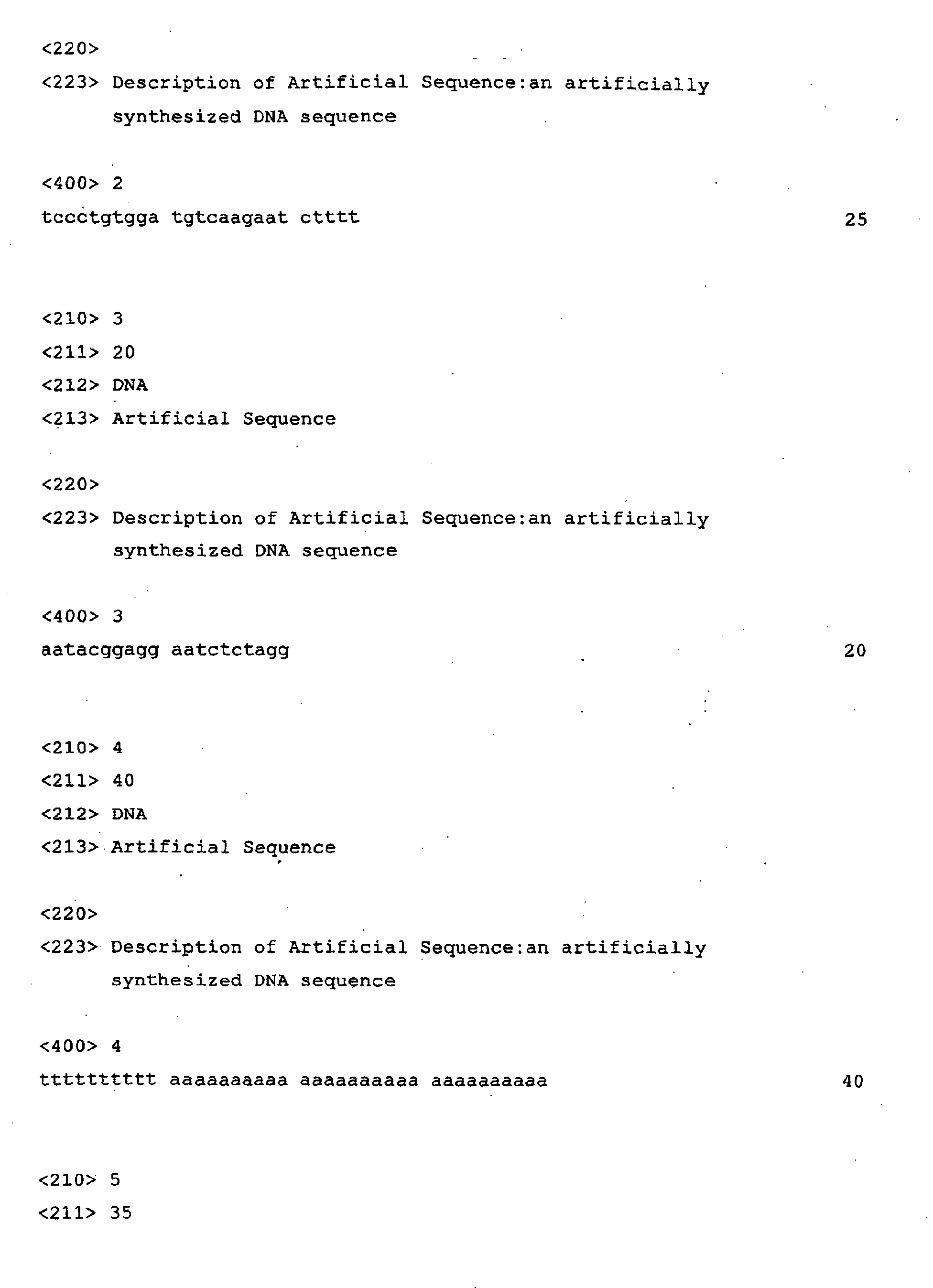
Oligonucleotides having the nucleotide sequences shown in SEQ
ID NO: 1 (calculated value; MW 7583) and SEQ ID NO: 2 (calculated
value; MW 7638), respectively, were dissolved in a buffer to a
concentration of 100 pmol/µL to produce DNA solutions. Using a
Pipetteman, 1 µL each of the above DNA solutions was spotted to a
designated position on the carbodiimidated stainless steel sheet.
Next, the sheet was dried at 37°C for 30 min, soaked in buffer
A (0.2 M sodium chloride, 0.1 M Tris-HCl (pH 7.5) , 0.05% Triton X-100)
containing 3% BSA (bovine serum albumin), and dried at 37°C for 15
min. Then, this carbodiimidated stainless steel sheet was washed
with TE buffer (10 mM Tris-HCl (pH 7.2)/1 mM EDTA), and dried at 37°C
for 15 min.
(2) Hybridization
Twenty-five µL of hybridization solution [5x SSC (SSC: 1.5 M
NaCl, 0.15 M sodium citrate), 10% dextran, the probe of SEQ ID NO:
3 (calculated value; MW 6190)] was placed on the above-mentioned
carbodiimidated stainless steel sheet at the part immobilized with
the DNA, and the sheet was heated overnight using a 40°C water bath.
(3) Post-hybridization
After hybridization, the hybridization solution was lightly
absorbed from the carbodiimidated stainless steel sheet,
post-hybridization washing was carried out, and non-specifically
adsorbed probes were removed.
Conditions for the post-hybridization washing were: (i) 2x SSC,
0.1% SDS, at room temperature for 5 min, twice; (ii) 0.2x SSC, 0.1%
SDS, at 40°C for 5 min, twice; (iii) 2x SSC, at room temperature for
5 min; and (iv) 0.3 M aqueous ammonium citrate solution, at room
temperature for 15 sec.
(4) Detection of hybridization
The carbodiimidated stainless steel sheet thus obtained was
measured using KOMPACT MALDI 2 (Shimadzu Corp.). The results are
shown in Fig. 2 and Fig. 3.
According to the results presented in Fig. 2 and Fig. 3, the
method for detecting nucleic acids of the present invention was
revealed to allow specific detection of the probe nucleic acid
(observed value; at the position of MW 6185) as an extremely clear
signal. On the other hand, no signal was detected from the spot of
SEQ ID NO: 2.
[Example 3] Mass spectrometry of DNA molecules hybridized on cover
glass
(1) Immobilization of nucleic acids (Fig. 4)
Cover glass #000 (Matsunami Glass Corp.) was carbodiimidated
according to the method of JP-A 2000-146978.
Oligonucleotides having the nucleotide sequences indicated in
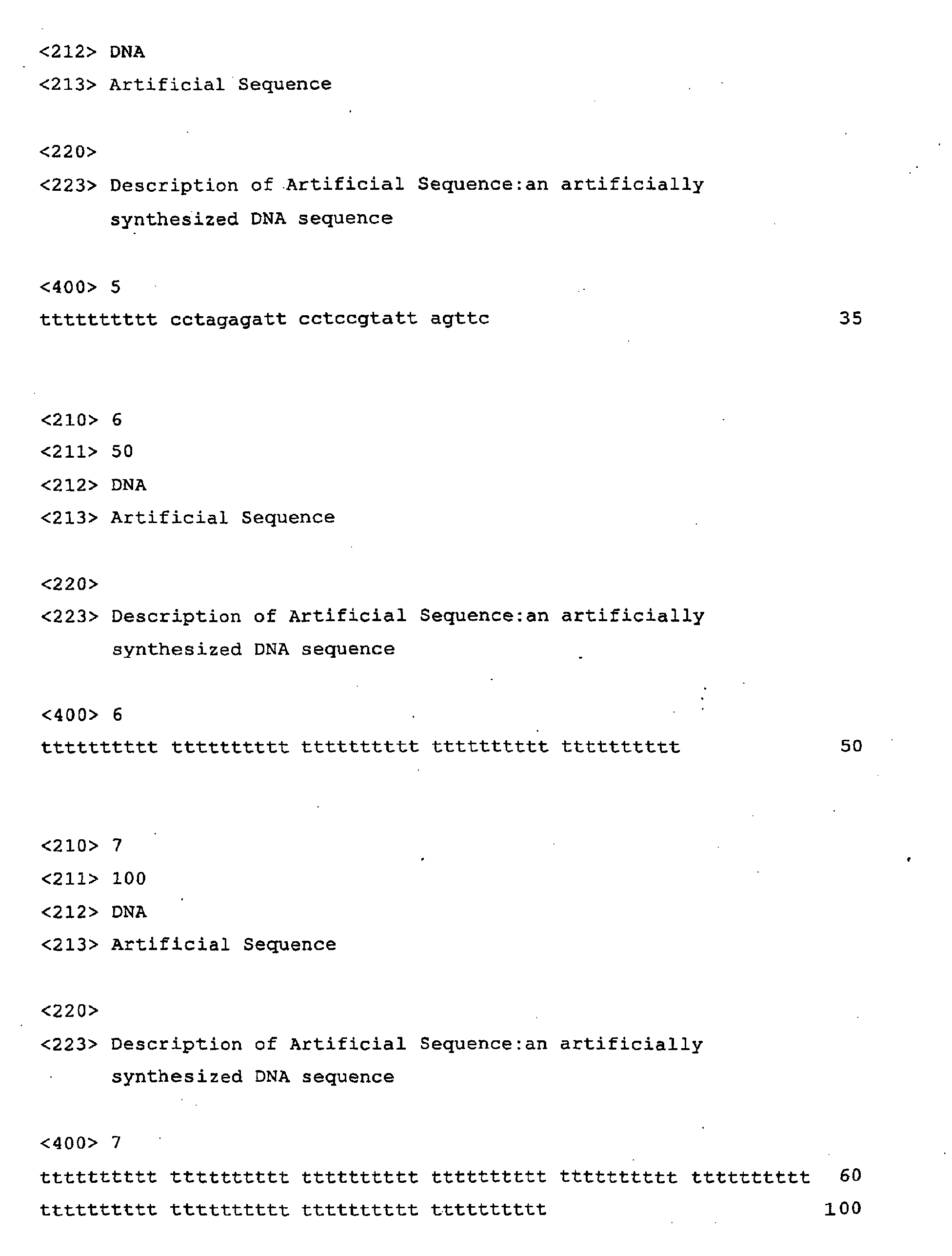
SEQ ID NO: 4 (Capture oligomer) and SEQ ID NO: 5 (negative control),
respectively, were dissolved in buffer to a concentration of 120
pmol/µL to yield DNA solutions. Using a Pipetteman, 0.5 µL each of
the aforementioned DNA solutions and buffer were spotted onto the
designated position on the carbodiimidated cover glass.
Next, immobilization procedure was carried out, the glass was
soaked in buffer A (0.2 M sodium chloride, 0.1 M Tris-HCl (pH 7.5),
0.05% Triton X-100) containing 3% BSA (bovine serum albumin), and
dried at 37°C for 15 min. Then, this carbodiimidated cover glass was
washed with TE buffer (10 mM Tris-HCl (pH 7.2)/1 mM EDTA), and dried
at 37°C for 15 min. The buffer without the addition of oligonucleotide
(DNA(-)) was used as the control.
(2) Hybridization
Ten µL of hybridization solution [5x SSC (SSC: 1.5 M NaCl, 0.15
M sodium citrate), 10% dextran, the probe of SEQ ID NO: 6 or SEQ ID
NO: 7] was placed on the above-mentioned carbodiimidated cover glass
at the part immobilized with the DNA, and the glass was heated
overnight in a 30°C dryer.
(3) Post-hybridization
After hybridization, the hybridization solution was lightly
absorbed from the carbodiimidated cover glass , and post-hybridization
washing was carried out under following condition.
The condition for post-hybridization washing was: (i) 2x SSC,
at room temperature for 5 min; and (ii) 0.3 M aqueous ammonium citrate
solution, at room temperature for 15 sec.
(4) Detection of hybridization
The obtained carbodiimidated glass sheet was measured using
"KOMPACT MALDI 4 (Shimadzu Corp.)". The results of mass spectrometry
are shown in Fig. 5. From samples containing complementary sequences,
peaks were observed. Therefore, samples hybridized on a glass sheet
were also found to be measurable by MALDI-TOF MS.
The matrix for the measurements was prepared by dissolving
0.0974 g of 0.7 M 3-hydroxypyridine-2-carboxylic acid
(3-hydroxy-2-picolinic acid) (MW 139.11) and 0.010583 g of 0.07 M
diammonium hydrogen citrate (MW 226.2) in 1 mL of 50% acetonitrile.
Furthermore, the measurement conditions for
KOMPACT MALDI 4 are
shown below.
| Flight path (ion flight path) | Linear |
| Polarity (ion polarity) | Negative |
| Mass (ion acceleration voltage) | 20 kV |
| Profiles (number of measurements per ion sample) | 50 |
| Aim (full range sample irradiation: 1-1000) | 1-1000 |
| Power (laser power) | 150 (50 mer), 180 (100 mer) |
| Accumulate | 10 |
| Average | 1 |
| Store profile | never |
The present invention provides a method for efficiently
detecting genetic polymorphisms using a mass spectrometer. This
method enables quick and comprehensive detection of genetic
polymorphisms in a large number of specimens. The method of the
present invention may greatly contribute towards disease correlation
analysis and genetic diagnosis for determining polymorphisms that
cause a specific disease.