Technical Field
-
The present invention relates to a highly efficient
antibody screening method, more specifically, a highly
efficient antibody screening method wherein after protein
mixtures of various cell fractions and the like are developed
by two-dimensional electrophoresis, an antibody library is made
to act on said mixtures, and a specific antibody is screened
with high efficiency directly from the individual protein spot
that had been separated.
Background Art
-
With the rapid advance in the technique for base
sequencing, the project to analyze the entire genome structure
has come to the determination of the entire genome sequences
in many microorganisms, and it is said that the project would
be completed also in humans by the beginning of the next century.
However, even if the base sequence is determined, there are only
a few genes among the gene expression products, wherein its
function can be predicted for example, by searching of
homologous proteins with the use of databases. In addition,
many of the proteins that actually express and function in the
cell vary in many forms according to the condition of the cells,
the change in its amount of expression and posttranslational
modification. In this way, the set of proteins that exist in
a certain instant in life is propounded as a new concept called
proteome, which is expressed by a coined term of a combination
of PROTEin and genOME (Kahn, P. Science 270, 369-70 (1995)).
Analysis of the variation of proteome is conducted by
comprehending the overall picture of proteome and comparing it
with the overall picture of another certain instant. An attempt
to analyze the biology by extensively analyzing the dynamics
of these proteomes is called proteomics, and is drawing
attention as a post genome project. Its method had already been
established, and a method wherein two-dimensional
electrophoresis and amino acid sequence analysis/mass
spectrometry are combined is generally used.
-
Two-dimensional electrophoresis is known as a method
wherein separation is conducted from two aspects of the
characters that a substance has, for example, a method wherein
electrophoresis according to isoelectric point is conducted
beforehand, followed by electrophoresis according to molecular
weight in different media (O'Farrell, P. H., J. Biol. Chem. 250.
4007-21 (1975)). According to said two-dimensional
electrophoresis, since it is developed two-dimensionally based
on the two characters, isoelectric point and molecular weight,
it is developed in a different coordinate if the isoelectric
point differs, even if the substances have the same molecular
weight, thereby enabling each substance to separate as a spot
on the two-dimensional coordinate. Two-dimensional
electrophoresis is frequently used especially as a method for
separating proteins, because of its excellence in resolution.
The two-dimensional electrophoresis method of proteins that is
currently used is conducted generally in the following manner.
First, isoelectric point electrophoresis is conducted by using
separation media such as capillary gel and commercially
available strip gel, the gel which electrophoresis had been
completed is mounted on a second planate SDS-polyacrylamide gel
(slab gel), and electrophoresis is conducted to the direction
perpendicular to the developing direction of isoelectric point
electrophoresis. As just described, many proteomes have
already been analyzed by two-dimensional electrophoresis,
wherein an isoelectric point electrophoresis and a SDS-polyacrylamide
electrophoresis (SDS-PAGE) are combined, that
is, separation to a one-dimensional direction according to the
isoelectric point, and then development to a two-dimensional
direction according to molecular weight are conducted
consecutively. Regarding each protein spot that had been
separated from the protein group, there have already been many
reports regarding the analysis results from amino acid sequence
determination and peptide mass fingerprinting, together with
the coordinate information.
-
On the other hand, to obtain an antibody against a desired
protein holds an important position for identification and
functional analysis of the protein. However, the conventional
method in which a desired antigen is administered to animals
for immunization requires various and large quantities of
antigens, a long time, and a large amount of effort. Thus, in
recent years, a method has been established to construct an
antibody by using a phage antibody library. Said method is an
application of a system wherein an antibody is presented
(displayed) on the surface of a phage by fusion of antibody with
a coat protein of a filamentous phage, and a phage display
library can be made by constructing a library including a
variety of antibody genes by amplification of antibody gene with
PCR, and then presenting this on the phage. According to this
method, it is also possible to construct an antibody against
antigens of human. The screening method wherein said phage
antibody library is used is called a phage display method, and
has been used conventionally to identify ligands and various
antibodies that bind specifically to various cell surface
receptors. There have already been many reports also regarding
the construction method of a phage antibody library and
screening methods using said method such as immunotube method,
magnetic beads method, and the like (Scott, J. M. and Smith,
G. P., Science, 249, 386-390 (1990); Smith, G. P. and Scott,
J. K., Methods in Enzymology, 217, 228-257 (1993)).
-
As previously described, regarding proteomics which has
drawn attention as a post genome project, a technique wherein
two-dimensional electrophoresis and amino acid sequence
analysis/mass spectrometry are combined is in heavy usage,
however, it has been pointed out that there are several problems
in said technique. In two-dimensional electrophoresis, there
are problems such as the limit to the amount of sample
attachments, the complexity of analysis due to the enormous
numbers of spots, the limit to the separation power at the acidic
and basic sides, reproducibility, and the limit to the spot
detection sensitivity. Although quantitative minimization of
substances to be analyzed has progressed in amino acid sequence
analysis/mass spectrometry, it still remains unsatisfactory,
and is said that skill is required for mass spectrometry
analysis. An object of the present invention is to provide a
highly efficient method of screening antibody against target
proteins contained in the sample.
-
The inventors of the present invention had conducted
intensive studies to attain the object mentioned above, and
focused attention on the point that a library having a diversity
of 109 has already been constructed as a phage antibody library,
and that it can theoretically cover all kinds of proteins. It
has been discovered that 2D phage panning method (2D-PP), which
is a combination of phage panning method that uses said phage
antibody library and two-dimensional electrophoresis, can be
a new proteome analysis method that can sufficiently oppose to
the combination of two-dimensional electrophoresis and mass
spectrometry that is currently the main method for proteome
analysis. More specifically, protein spot that had been
separated by two-dimensional electrophoresis was transcribed
to a nitrocellulose membrane, only the spot portion was cut out
after visualization by gold colloid staining, only said spot
portion was reacted with a phage antibody library, only the
phage antibody that bound specifically to the desired protein
spot was retrieved, said collected phage antibody was infected
to Escherichia coli, said specific phage antibody was produced
in large quantities, and the reaction and retrieval operations
mentioned above were further repeated, in other words, it was
discovered that monoclonal antibody against a desired protein
spot can be screened with high efficiency by panning, and thus
the present invention had been completed.
Disclosure of the Invention
-
More specifically, the present invention relates to a
highly efficient antibody screening method, wherein the method
is a screening method of antibody against a specific protein
contained in the sample including a protein group, comprising
the following processes (a) to (c), (a) a process wherein a
sample including a protein group is treated with two-dimensional
electrophoresis, and the aforementioned protein
group is developed as an individual spot protein on the
two-dimensional electrophoretic gel, (b) a process wherein the
spot proteins and antibody libraries are reacted, and (c) a
process wherein the antibodies bonded to the spot proteins are
replicated, and the replicated antibodies are reacted with the
aforementioned spot proteins once or more than twice (claim 1);
the highly efficient antibody screening method according to
claim 1, wherein samples where its cellular subfractions had
been separated and purified beforehand are used as samples
including protein groups (claim 2); the highly efficient
antibody screening method according to claim 1 or 2, wherein
samples in which posttranslational modification had been
eliminated are used as the samples including protein groups
(claim 3); the highly efficient antibody screening method
according to any of claims 1 to 3, wherein the individual spot
proteins that had been developed on the two-dimensional
electrophoretic gel are reacted with the antibody libraries as
spot proteins that have been transcribed onto a solid phase
(claim 4); the highly efficient antibody screening method
according to claim 4, wherein the spot proteins that had been
transcribed onto the solid phase are visualized, and after
isolating each of the spot proteins, they are reacted with the
antibody libraries (claim 5); the highly efficient antibody
screening method according to any of claims 1 to 5, wherein a
phagemid antibody library is used as the antibody library (claim
6); the highly efficient antibody screening method according
to claim 6, wherein the phagemid antibodies bonded to the spot
proteins are infected with host cells, and phagemid antibodies
are replicated (claim 7); and the highly efficient antibody
screening method according to claim 6, wherein PCR is conducted
by targeting the CDR region of the phagemid antibodies bonded
to the spot proteins, followed by infecting a phagemid vector
incorporating the region amplified by PCR with host cells, and
replicating phagemid antibodies (claim 8).
Brief Description of Drawing
-
- Figure 1 is a view indicating the sequence information
of DP-47 and DPK-22.
-
Best Mode of Carrying Out the Invention
-
In the highly efficient antibody screening method of the
present invention, there is no particular limitation as long
as it is a screening method of antibodies against specific
proteins contained in the samples including protein groups,
comprising (a) a process wherein a sample including a protein
group is treated with two-dimensional electrophoresis, and the
aforementioned protein group is developed as an individual spot
protein on the two-dimensional electrophoretic gel, (b) a
process wherein the spot proteins and antibody libraries are
reacted, and (c) a process wherein the antibodies bonded to the
spot proteins are replicated, and the replicated antibodies are
reacted with the aforementioned spot proteins once or more than
twice. Moreover, the above-mentioned antibodies include
immunoglobulin, single strand antibody, and antibody fragments
including antibody-binding sites comprised of variable region
of antibodies. Exemplification of antibody fragments are
F(ab')2 obtained by peptonizing monoclonal antibodies, Fab'
obtained by reduction of F(ab')2, Fab obtained by digesting
monoclonal antibodies with papain, and the like.
-
As a sample including the protein groups mentioned above,
it can be anything as long as it is a sample including several
kinds of proteins, however, it is preferable for the sample to
be able to develop as individual spot proteins on the two-dimensional
electrophoretic gel, when said sample is treated
with two-dimensional electrophoresis. Treatment with two-dimensional
electrophoresis in the usual manner enables to
separate the sample into several thousand protein spots at a
maximum, however, the separation power by said treatment with
two-dimensional electrophoresis cannot cover all the proteins
that are said to be tens of thousands and that are expressed
and functioned in the cells. As just described, if samples
treated with two-dimensional electrophoresis exceed the
detection limit as a protein spot, it is preferable to conduct
separation and purification, fractionation treatment and the
like beforehand, to make it possible to be detected as a protein
spot. For example, it is preferable to separate and purify the
targeted cellular subfraction (caveolae, clathrin-coated pits,
Golgi, lysosome and the like) beforehand by biochemical methods
or the like, and develop said protein groups that had been
separated and purified as starting materials, as individual
spot proteins on the two-dimensional electrophoresis. By said
pre-treatment, it is possible to develop the several hundreds
to several thousands of protein groups that exist in the
individual cellular subfraction as individual spot proteins,
even with the separation power of a normal two-dimensional
electrophoresis treatment. Exemplification of samples
including protein groups wherein said cellular subfractions had
been separated and purified beforehand are, for example,
fractions comprised of cell membrane microdomain "lipid rafts"
that are structured by various kinds of membrane proteins
abundant in intercellular and intracellular signaling system
molecules and indicate high hydrophobic properties that are
obtained by separating and purifying human neuroblastoma, or
a clathrin-coated pits fraction that comprise many receptor
proteins, or cell membrane fractions that do not belong to them
but are involved in important intercellular signaling, and the
like.
-
Moreover, as samples including protein groups mentioned
above, it is preferable to use samples wherein the
posttranslational modification of proteins (for example,
glycosylation, phosphorylation, acetylation, amidation,
myristoylation, farnesylation, geranylgeranylation,
sulfation, and glycophosphatidyl inositol anchor (GPI anchor))
had been eliminated. By the treatment of eliminating the
posttranslational modification of said proteins, it is also
made possible to separate proteins that do not become spots
under a condition of a regular two-dimensional electrophoresis.
In addition, there is no particular limitation to the method
for eliminating the posttranslational modification, but it can
be conducted by accordingly combining enzyme treatment such as
protein phosphatase (phosphoproteinphosphatase),
methylesterase, peptide N-glycosidase F (PNGaseF),
phosphatidylinositol phospholipase C (PIPLC) or the like, and
degreasing treatment by chemical compounds or the like.
-
As a two-dimensional electrophoresis treatment wherein
protein groups contained in the samples are developed as
individual spot proteins on the two-dimensional
electrophoretic gel, there is no particular limitation as long
as it is a method that conducts separation from the two aspects
of physical property, such as isoelectric point and molecular
weight, that the proteins have. Generally, it can be done by
first conducting isoelectric point electrophoresis with the use
of separation media such as capillary gel or commercially
available strip gel, followed by mounting the gel wherein
electrophoresis had been completed on a second planate SDS-polyacrylamide
gel (slab gel), and conducting electrophoresis
to the direction perpendicular to the developing direction of
isoelectric point electrophoresis. It is preferable to
transcribe the individual spot proteins that had developed on
the two-dimensional electrophoretic gel by means of said
treatment of two-dimensional electrophoresis, onto a solid
phase such as nitrocellulose membrane, polyvinylidene
difluoride membrane (PVDF membrane) or the like, by methods such
as wet method of Western Blotting or the like, and react the
spot proteins transcribed onto said solid phase and the antibody
libraries. By the transcription onto said solid phase, the
target protein spots and antibody libraries can easily be
reacted.
-
It is much more preferable to visualize the spot proteins
transcribed onto the solid phase mentioned above, isolate each
of the spot proteins, and then react them with the antibody
libraries. In each of the spots isolated after said
visualization, antibodies other than the antibodies against the
desired proteins, such as antibodies against solid phase
membrane (nitrocellulose membrane, PVDF membrane or the like)
can be excluded, by elimination of the parts where a protein
does not exist, and antibodies against target proteins can be
screened with high efficiency. There is no particular
limitation to the method for visualizing the spot proteins
mentioned above, as long as it is a method for visualizing
proteins that are publicly known, such as gold colloid staining
method, immunostaining method and the like. Moreover, there
is also no particular limitation to the method for isolating
each of the spot proteins by cutting out after visualization.
However, in order to increase the reproducibility, it is
preferable to use a method in which only the spot portions are
cut out, by using an automatic cutting apparatus that is
commercially available, such as a spot cutter or the like.
-
As to an antibody library used in the present invention,
there is no particular limitation as long as it is a library
including a number of antibodies, but it is preferable to use
a phagemid antibody library (phage display library) comprised
of about 109 phagemid antibodies with wide range of variation,
that can theoretically correspond to the antigenic epitopes on
every protein. As said phagemid antibody library, any kind of
library can be used as long as it is a phage library that is
publicly known used in a normal phage display method, including
those that are commercially available. For example, a phage
display human antibody library can be constructed in the
following manner: a gene fragment that encodes the variable
regions (VH, VL) of H and L chain of the antibody is-amplified
by PCR from human B cells; said amplified genes introduce the
filamentous phage (phagemid vector) that had been inserted
within the framework of its coat protein pIII genes to the
Escherichia coli, and helper phages such as VCS-13 and the like
are infected with said Escherichia coli (H. R. Hoogenboom et
al., Immunotechnology, 4, 1-20 (1998)).
-
Moreover, as a phage antibody library, a library named
Sequence-arrayed library (SAL), which had been constructed by
the present inventors, can be used advantageously. The
description of SAL will be followed hereinafter. It is known
that there is a Fab structure that is used preferentially in
a phage antibody that specifically recognizes antigens in a
phage antibody library, wherein VH chain is DP47 and VL chain
is DPK22. Furthermore, in the CDR region of these chains, it
is also known that a certain limited site (VH chain: amino acids
in 95th, 96th, 97th, and 98th residues/ VL chain: amino acids
in 91st, 93rd, 94th, and 96th residues) of CDR3 region in
particular, is deeply involved with the binding to the antigens.
Consequently, the frequency of usage of amino acids used in
these regions was examined, and it was discovered that there
is a difference in the frequency of usage. In addition, these
amino acids could be limited to seven types of amino acids (G,
S, D, N, R, Y, P) when classified according to their properties.
It was discovered that placing these amino acids in a random
order in the antigen binding sites of CDR3 region enables to
construct a library having a variety of 5.8x10⁶, and that the
amount of one type of phage antibody can be increased from the
conventional 104 to 106.
-
Consequently, a primer design method which covers its
diversity and being highly effective was examined, and it was
discovered that the seven types of amino acids mentioned above
can be substituted by three types of codons. When the three
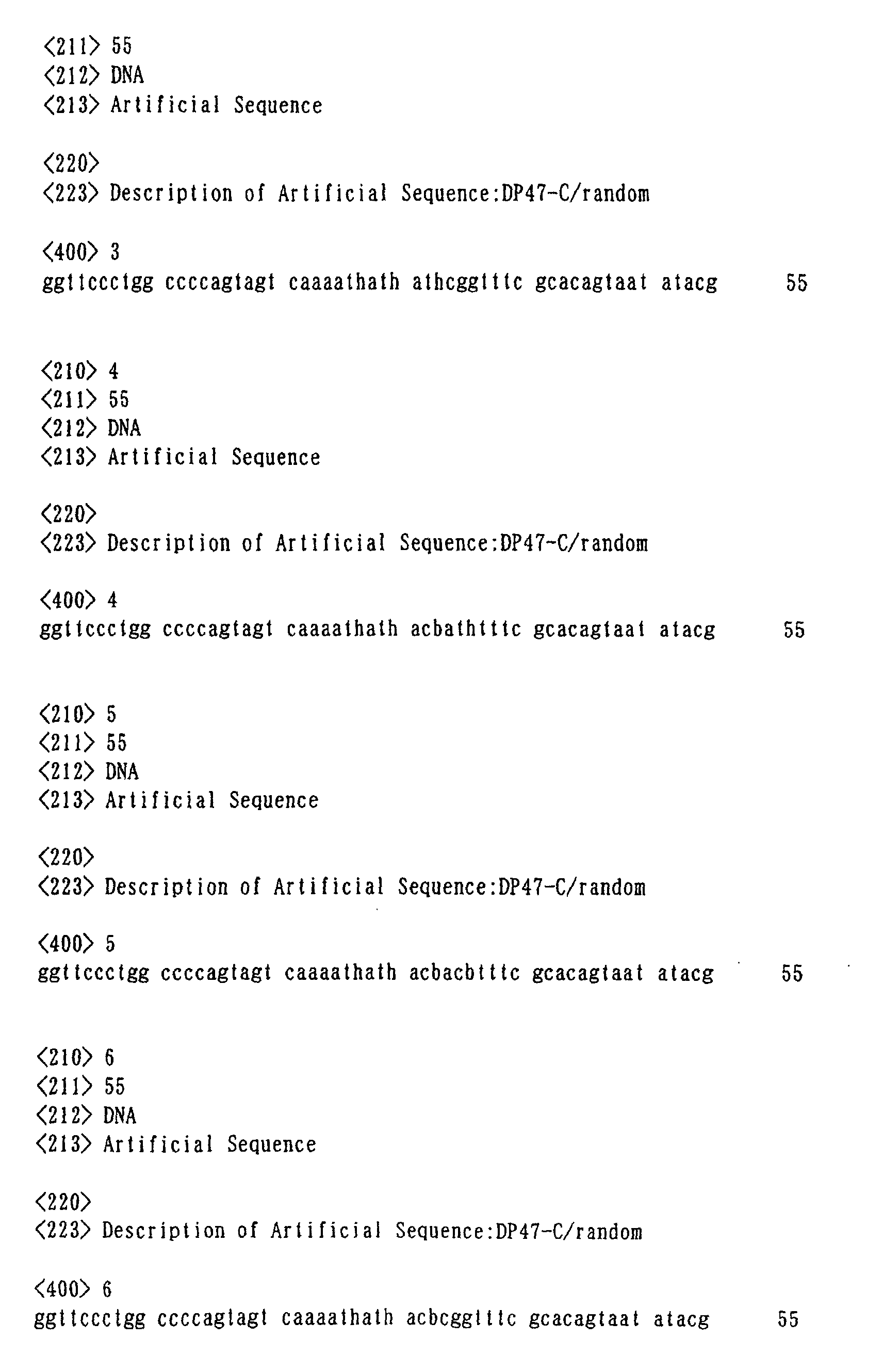
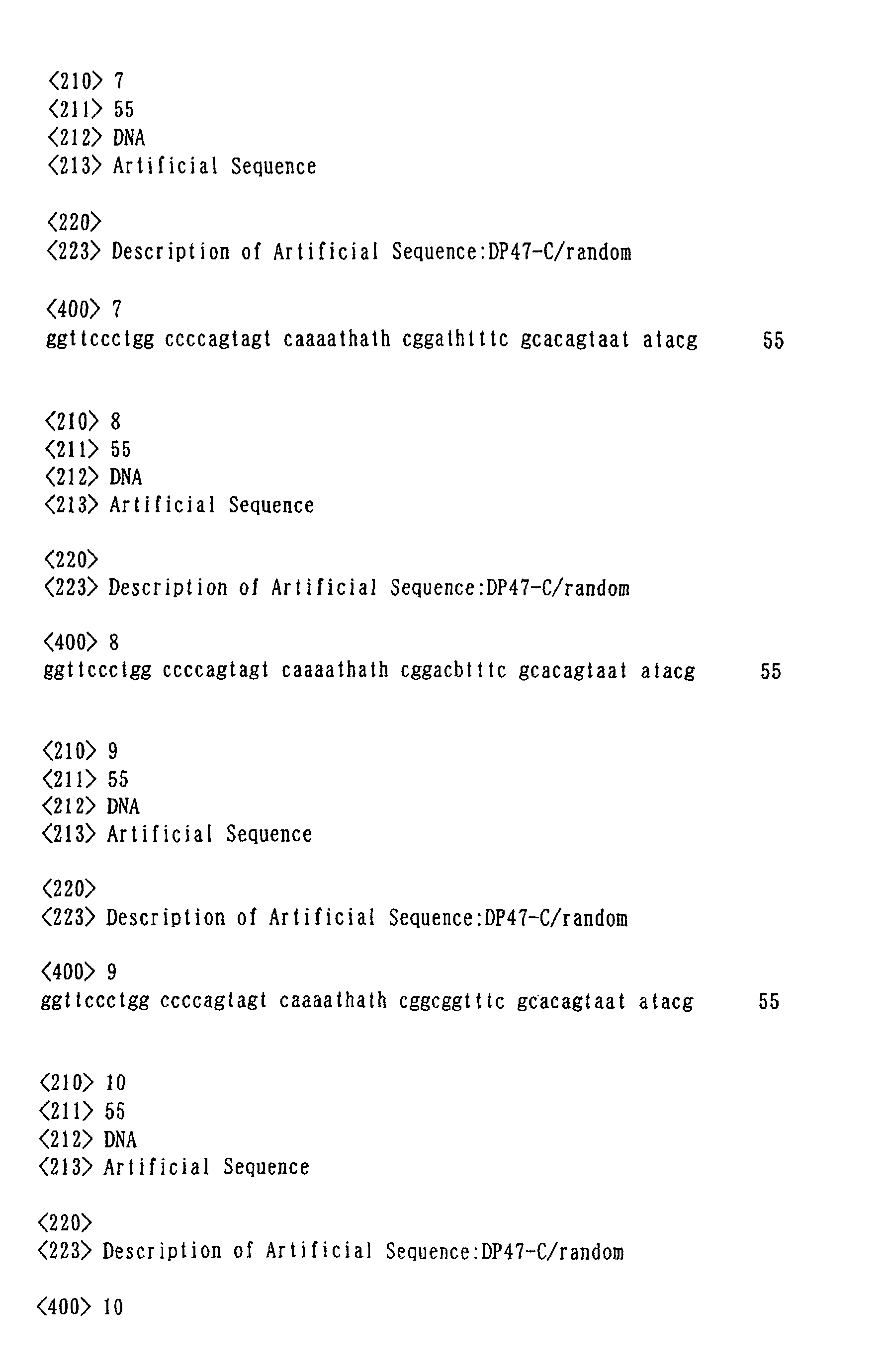
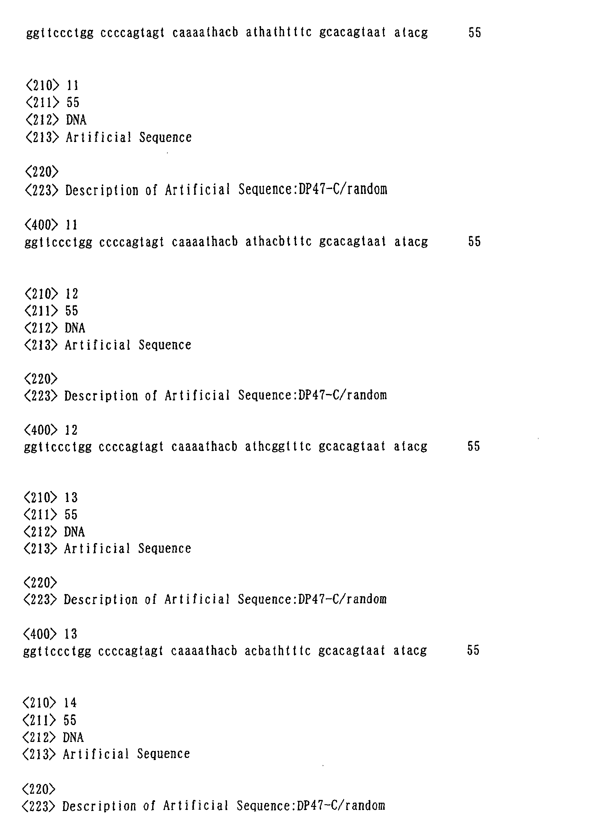
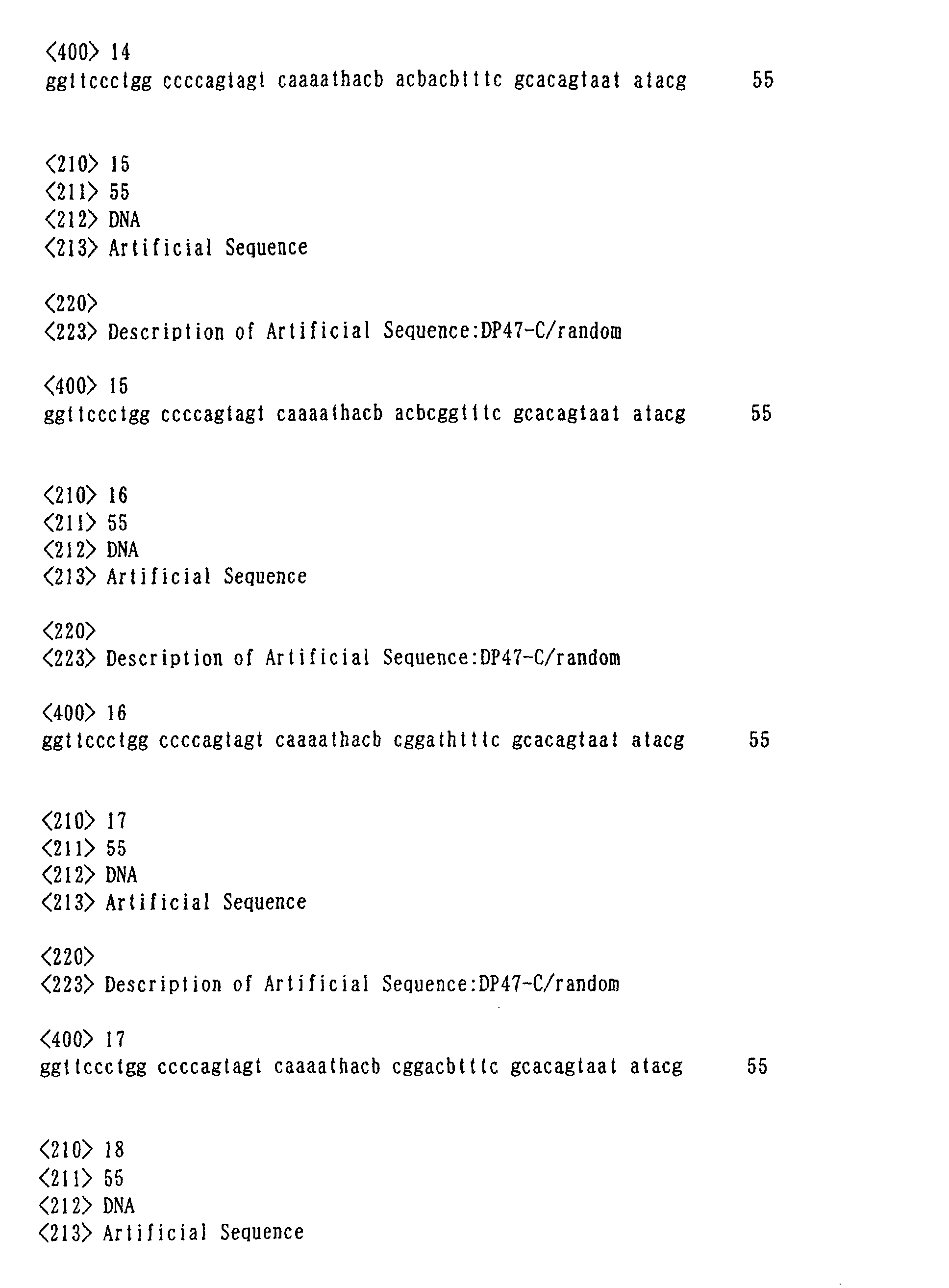
types of codons are placed at an antigen-binding site, they can
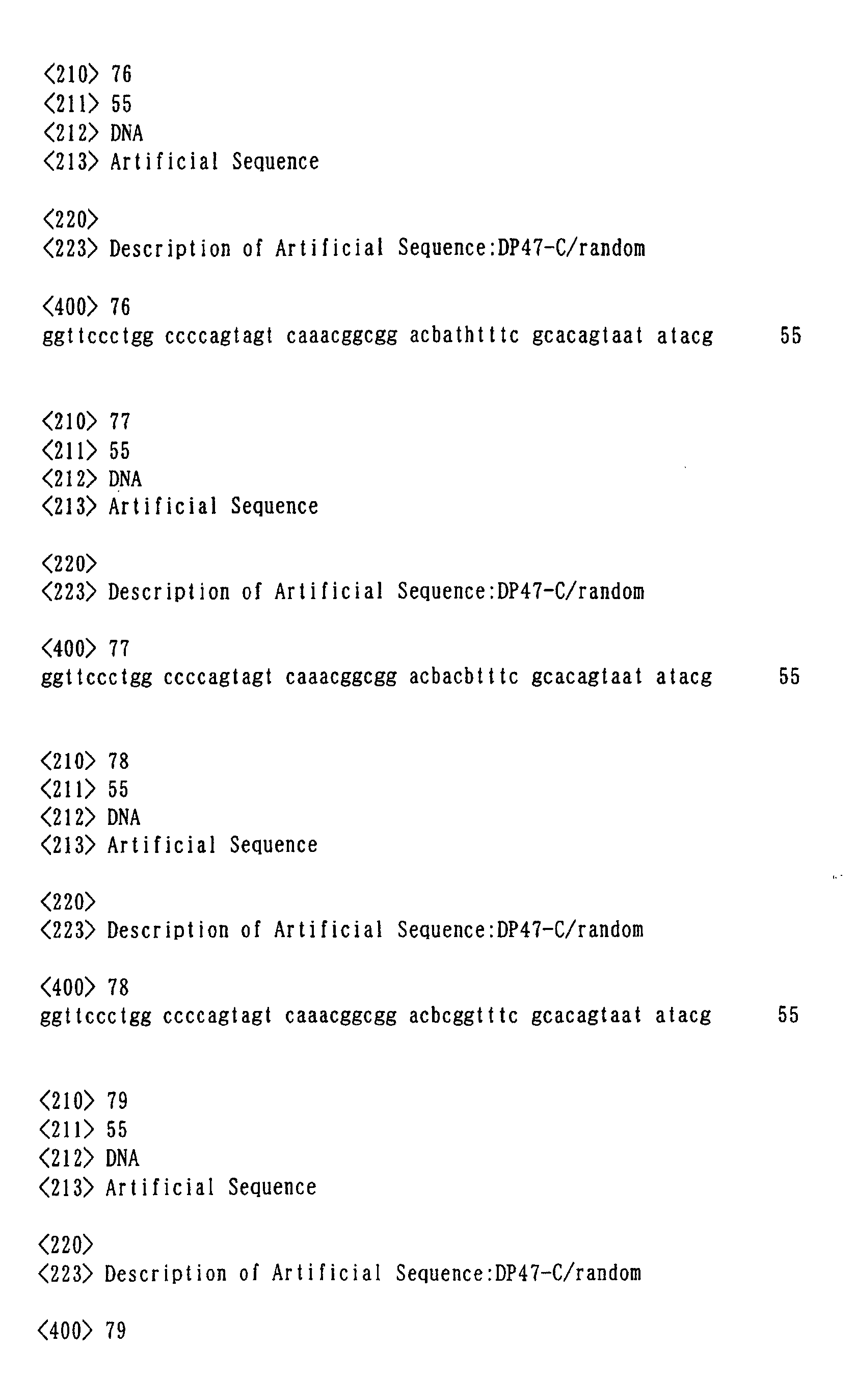
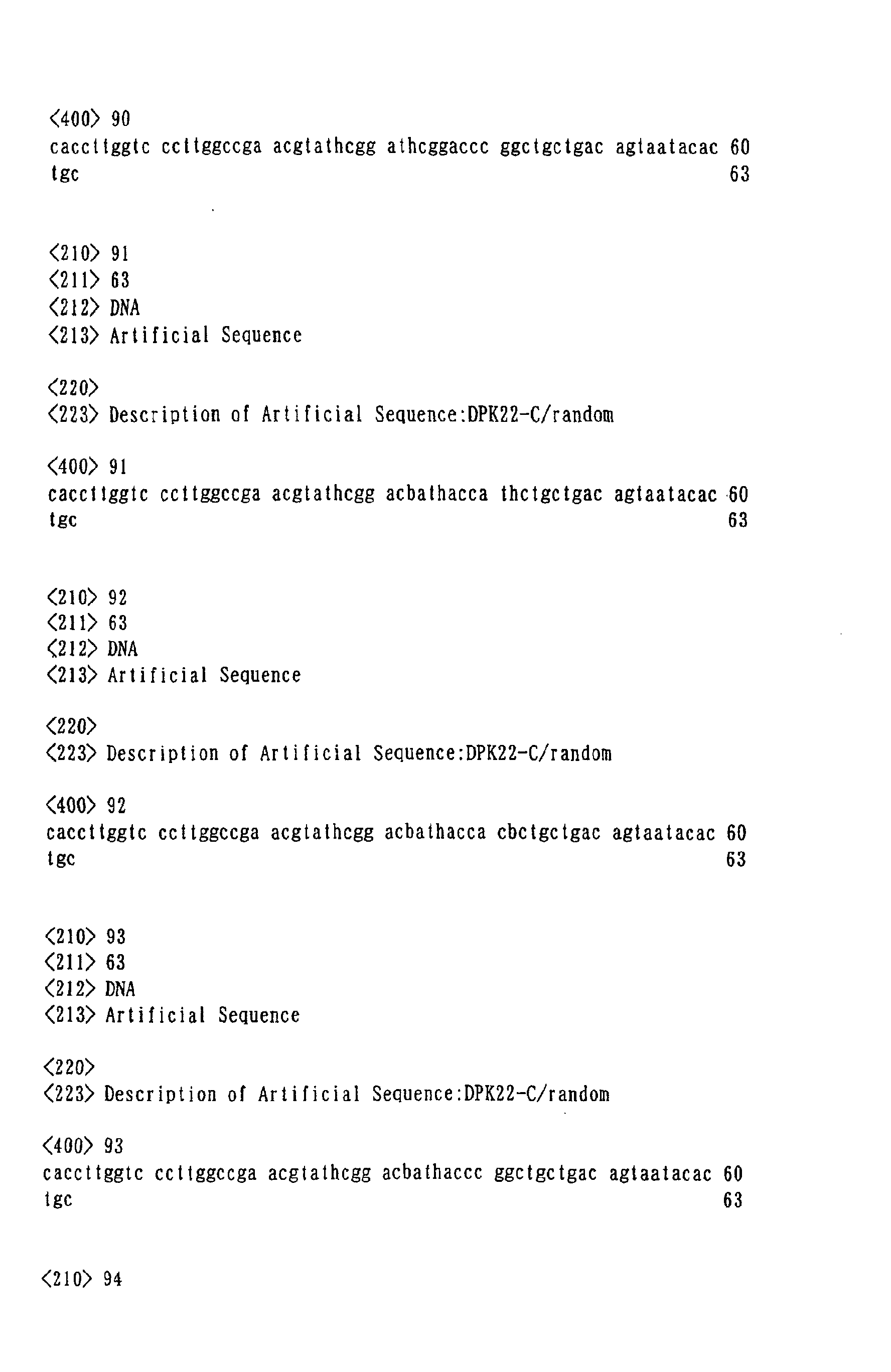
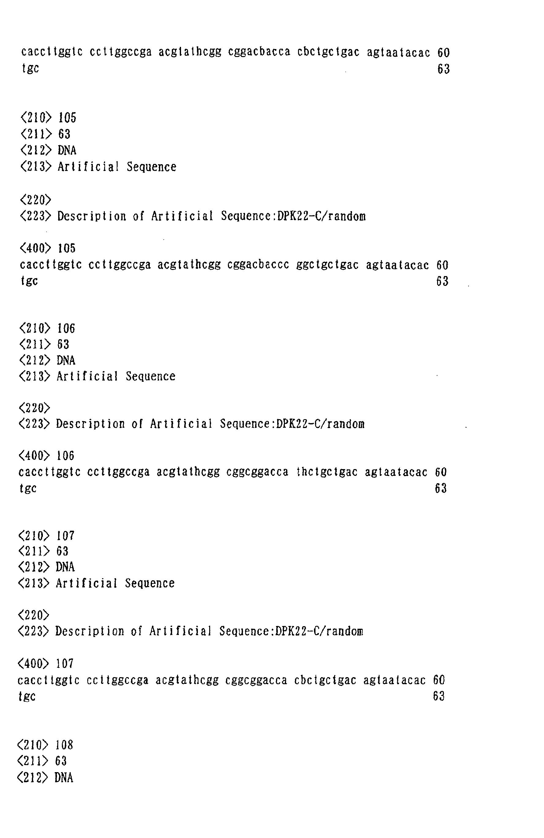
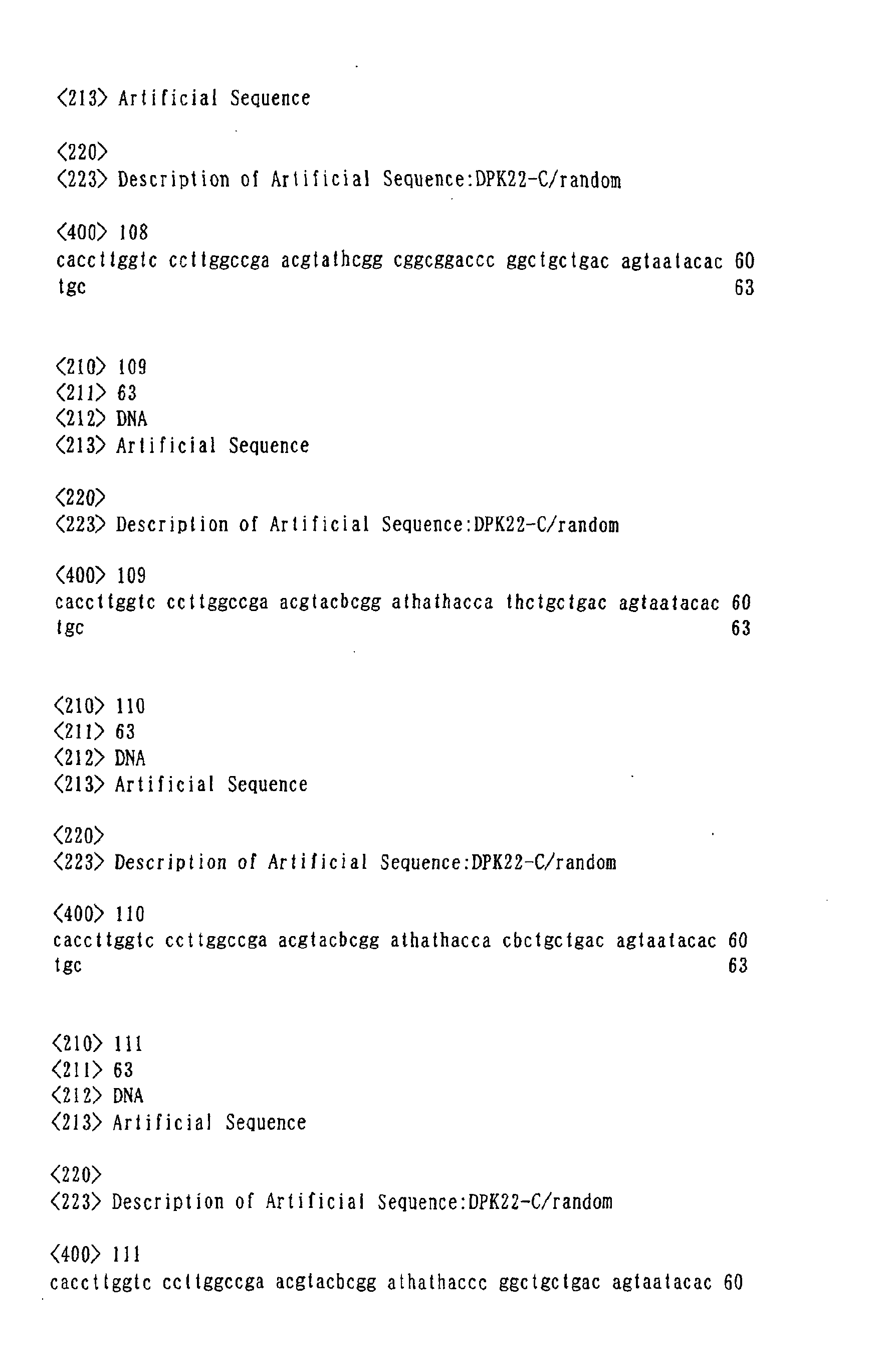
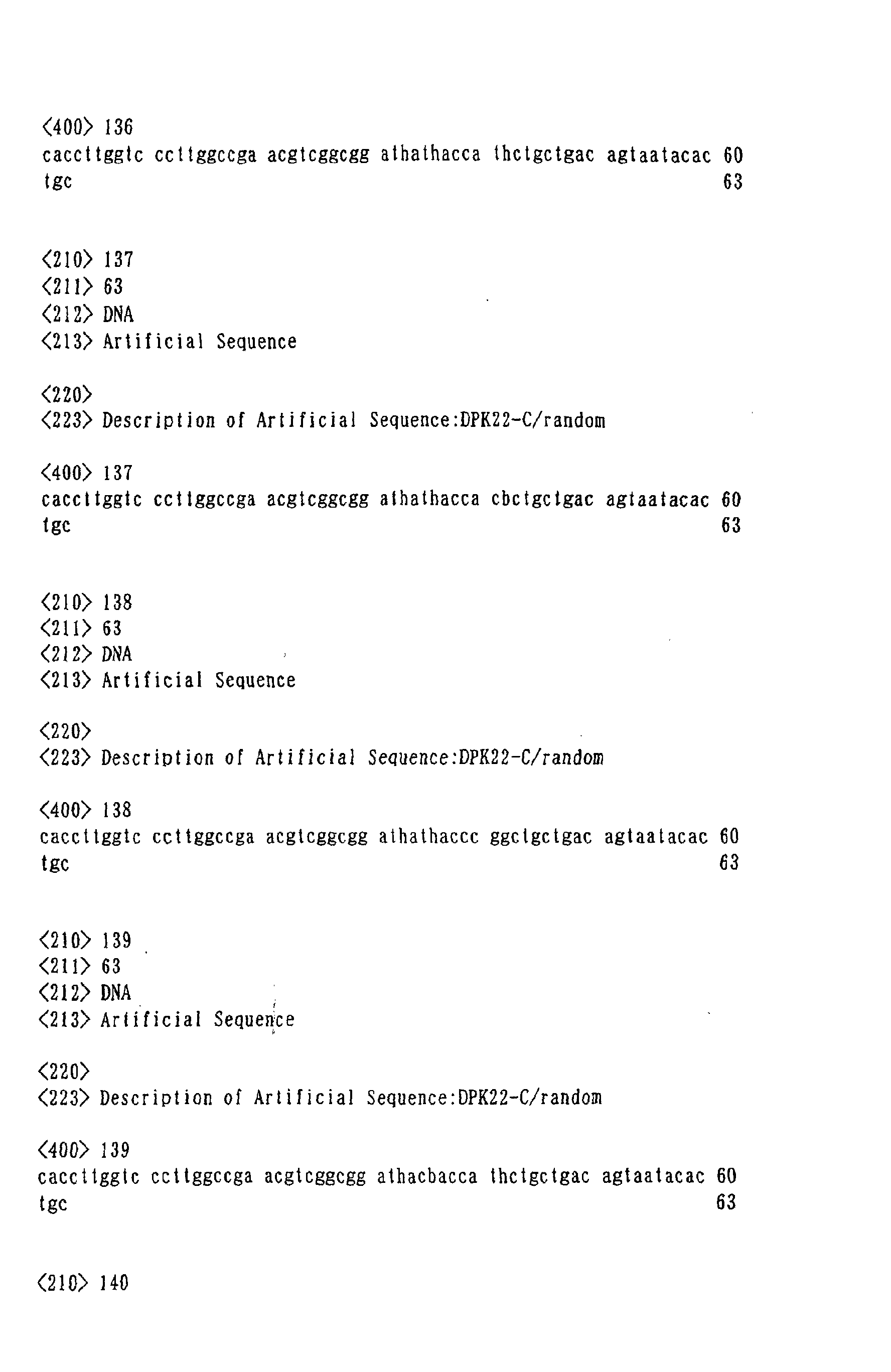
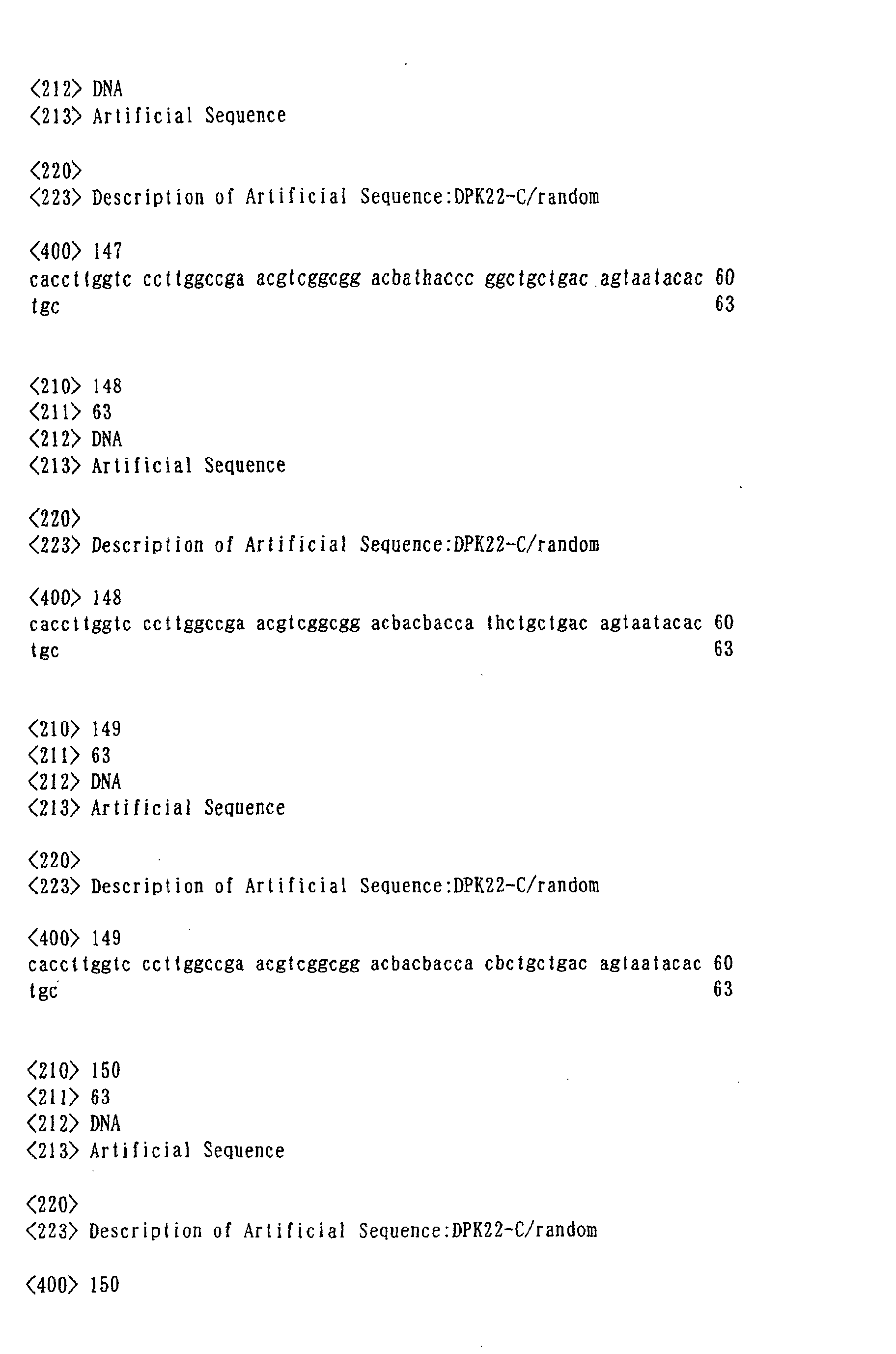
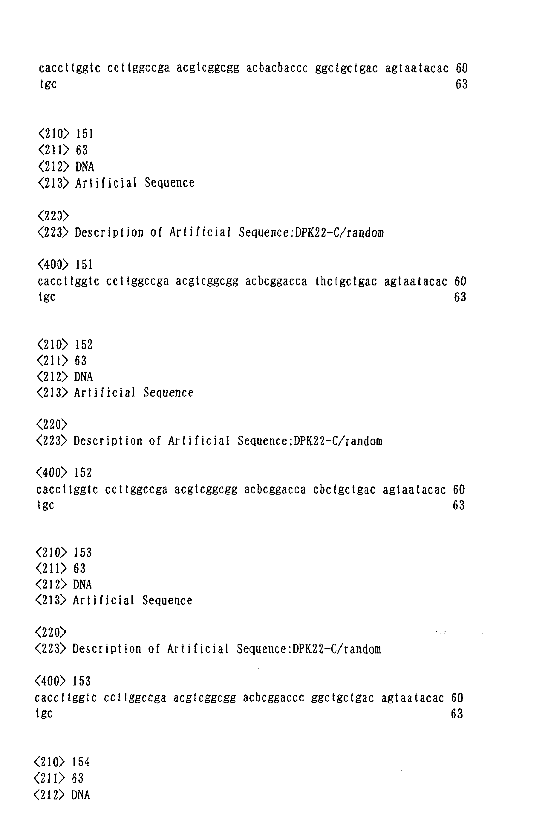
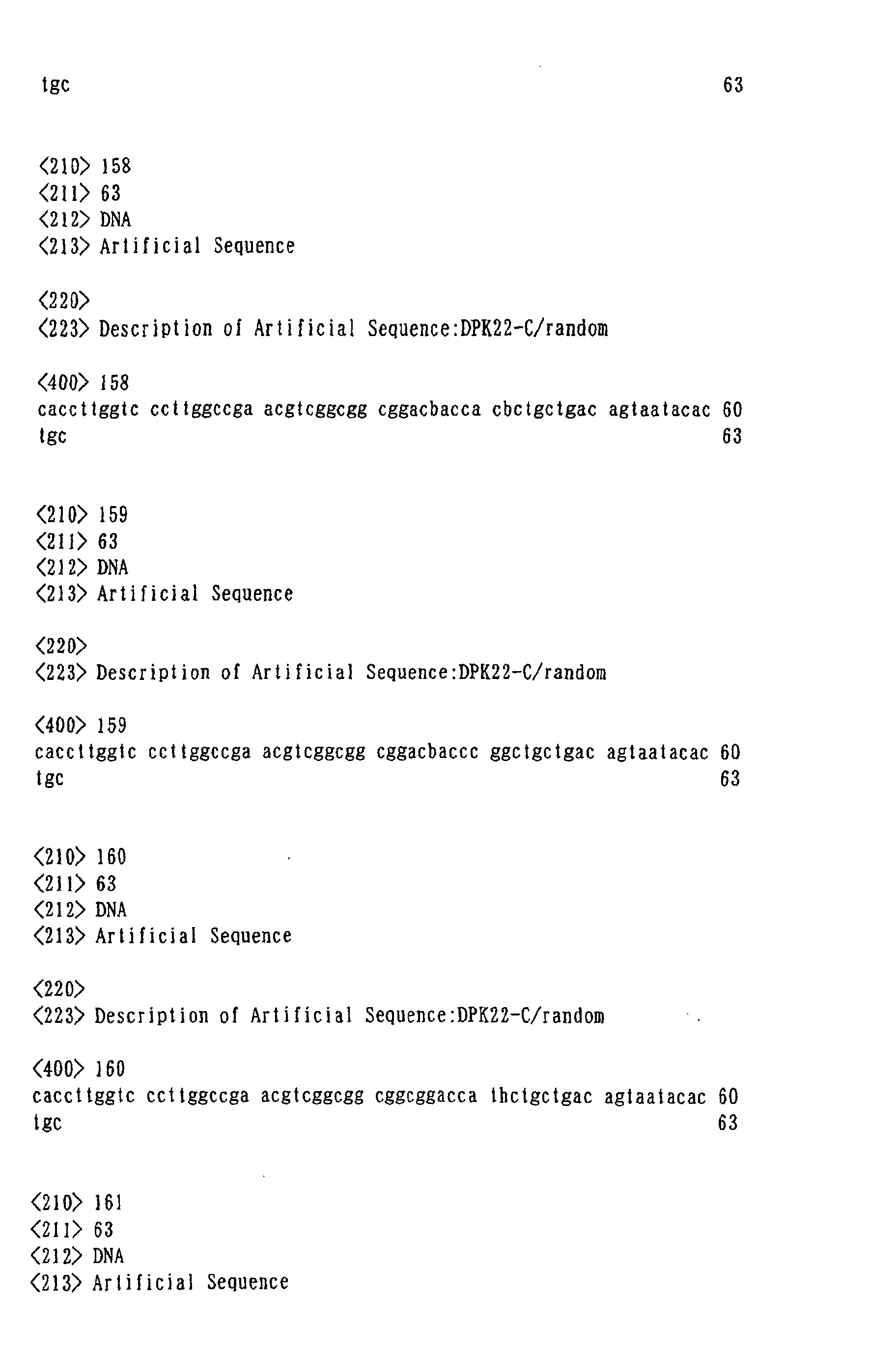
cover all the diversities with 81 types of primers for DP47 (Seq.
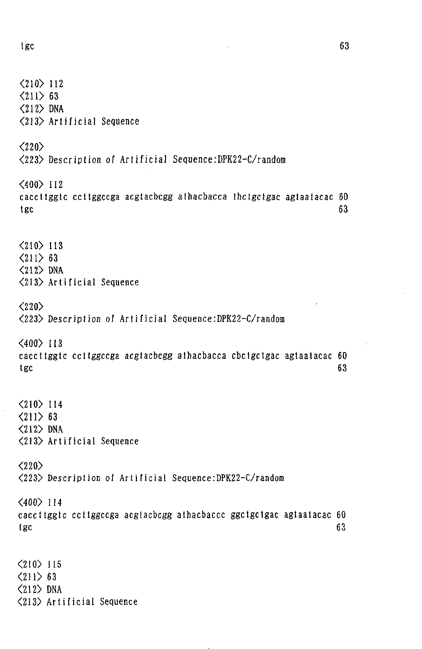
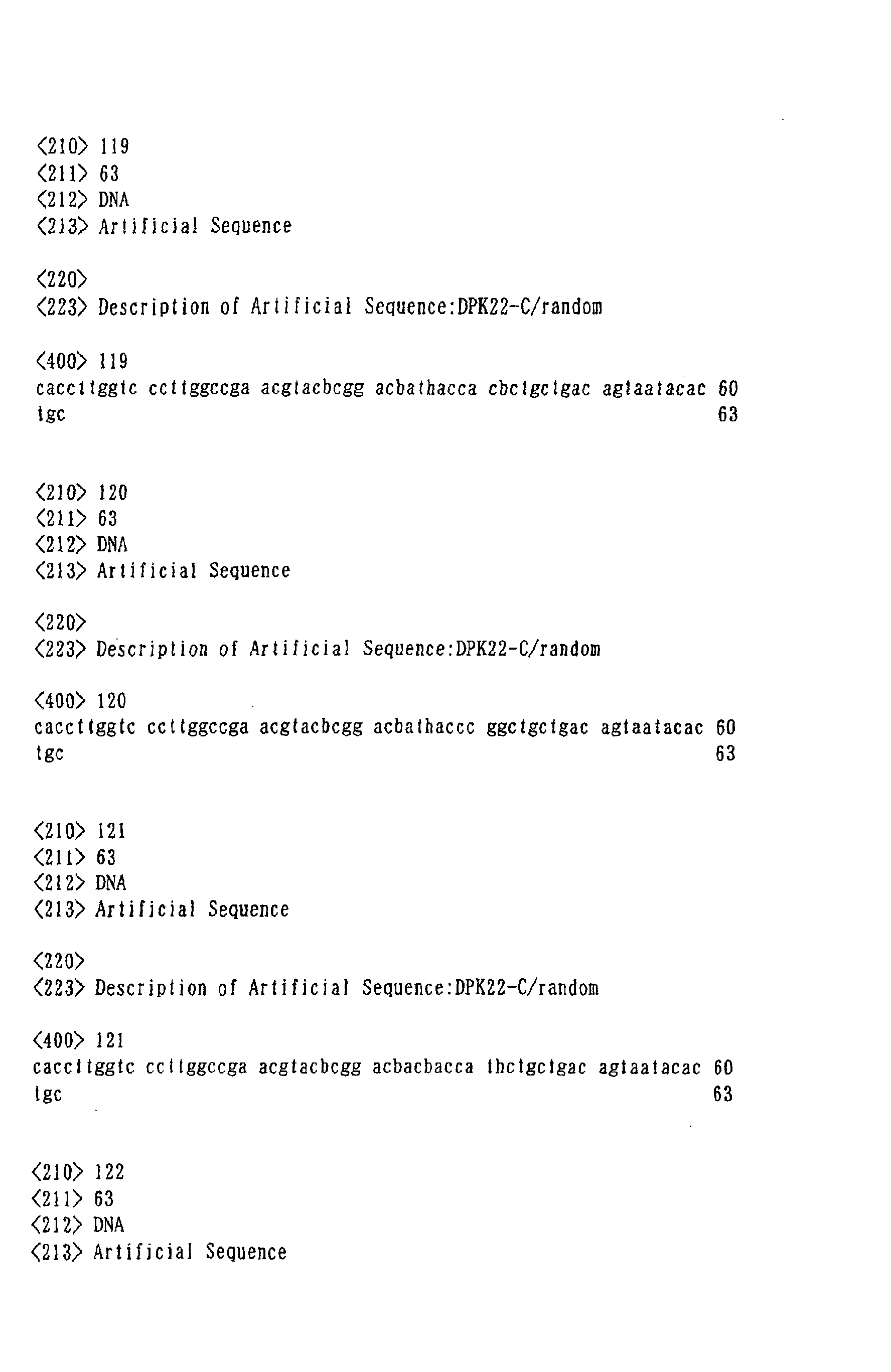
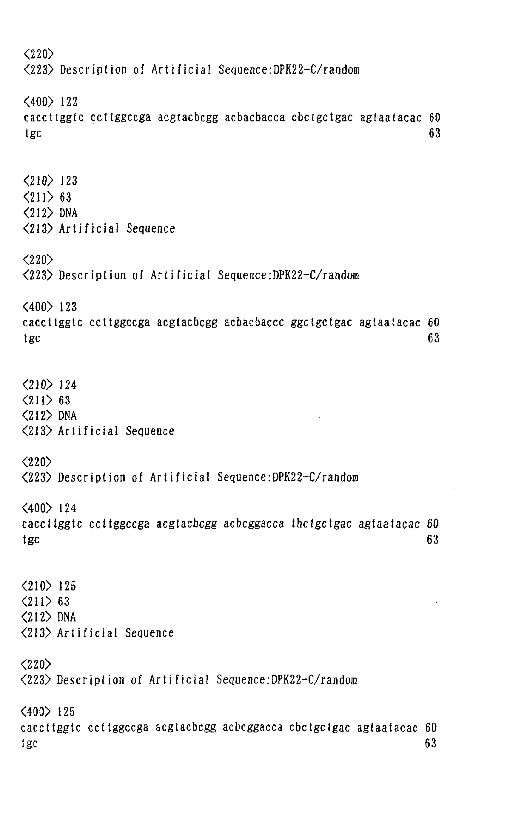
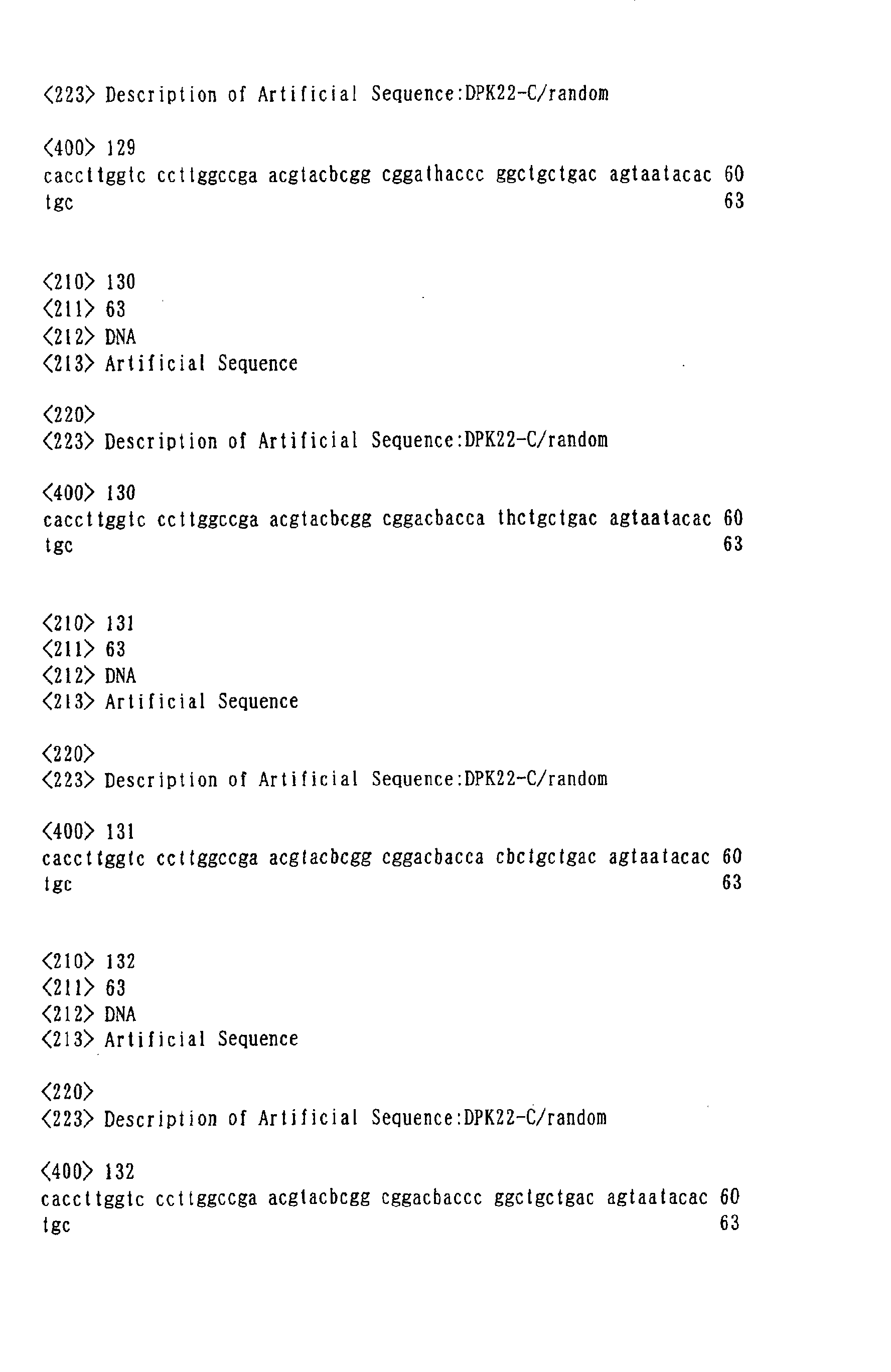
ID Nos. 1 to 81) and 81 types of primers for DPK22 (Seq. ID Nos.
82 to 162). Furthermore, in these primers, it can be made
possible also to increase the amount of one type of phage
antibody to more than 106, by reducing the numbers of amino acids
used and by constructing a sub-library by combining DP47 and
DPK22 that had been synthesized with each of the primers.
Accordingly, this SAL is capable not only of screening the phage
antibody against very minute amounts of antigens but also of
screening phage antibody where panning is not necessary. In
addition, with the use of this SAL, if the antigen can be cloned
even when the specific property is low, it is possible to
strengthen the specific properties by affinity maturation.
-
The reaction of the aforementioned spot protein and a
phagemid antibody library can be conducted by ordinary method,
for example, by using approximately 5x10¹² cfu/mL phage antibody.
After reacting a spot protein with a phagemid antibody library,
when only the phagemid antibody attached to the desired
individual spot proteins is retrieved and infected with
Escherichia coli, phagemid antibodies that react with the
individual spots can be replicated in a large amount. Since
the DNA sequence information of the antibody portion of phagemid
antibody is incorporated into the phagemid, when the phagemid
is replicated by infection of Escherichia coli, antibodies that
are expressed on its surface are also replicated at the same
time, the antibodies that react with the desired individual
protein spots can be replicated at a large amount and easily.
In fact, it is rare that a positive antibody can be identified
with reaction conducted once, and antibodies against the
desired individual spot proteins can be obtained by repeating
this operation several times. This cycle of repetition is
called "panning". Usually, phage antibodies that bind
specifically to the target proteins are concentrated by
repeating said panning several times, preferably by repetition
of three to five times, and screening can be conducted.
-
As another method for the above-mentioned panning, in
place of a method wherein phagemid is infected directly with
the Escherichia coli, a method can be given wherein PCR is
conducted by targeting the CDR region (mainly CDR3) of the
phagemid antibody bonded to the individual spot proteins, a
phagemid vector wherein the region amplified by PCR has been
incorporated is infected with host cells, and a phagemid
antibody is replicated. The advantage of this method is that
since the size of the targeted CDR region is nearly constant
and the primer sequence used for PCR is common, the uneven of
amplification by PCR, which is generally a big problem when
conducting replication by PCR, can be suppressed to the minimum.
By actually using this method, it can achieve approximately ten
times the efficiency compared to the method wherein phagemid
is infected directly with Escherichia coli.
-
In addition, the following can be given as methods for
eliminating antibodies against unfavorable solid phase
membrane or the like, when identifying positive antibodies, by
the reaction of a spot protein with a phagemid antibody library,
aside from the previously mentioned method wherein only the
individual spot portion is isolated: a method wherein different
types of solid phase membranes (nitrocellulose membrane and
PVDF membrane or the like) are used alternatively every time
panning is conducted (Alternative membrane method), a method
wherein a phagemid antibody library is reacted with the solid
phase membrane or the like beforehand; and the antibodies
against these are eliminated, and a method wherein these methods
are combined, and the like.
-
It is preferable to perform immunoblotting on the
two-dimensional electrophoresis by using positive antibodies
against each target protein obtained by the above-mentioned
screening method, and confirm the reactivity and specificity
between the desired individual spot proteins and each antibody.
Moreover, it is possible to make the antibody monoclonal by
examining the correlation between the DNA footprint and the
antibody titer of each antibody.
-
In addition, it is possible to prepare homogeneous
phagemid antibodies at a large amount very easily, by infecting
the positive antibodies obtained by a screening with
Escherichia coli. Furthermore, when infected with specific
Escherichia coli strain such as the Escherichia coli HB2151
strain or the like, these phagemid antibodies are released from
the Escherichia coli and can be solubilized in the solution as
an antibody fragment. When preparing suchlike solubilized
antibody fragment, it is advantageous to prepare it as a fusion
peptide having a sequence tag such as c-Myc or His-tag, in
consideration of the purification of the solubilized antibody
fragment. In addition, by fixing the substance that
specifically recognizes this tag on the chip beforehand, and
then reacting each solubilized antibody fragment that had been
solubilized with the chip, these antibody fragments can easily
be fixed on the chip. An antibody chip can be made by
fractionating the surface of the chip in pieces and fixing
different phagemid antibodies to each of the fractions.
Moreover, after samples had been solubilized under various
conditions, the samples wherein protein had been labeled by the
use of fluorescent probe and chemiluminescence probe or the like,
are reacted with the above-mentioned antibody chip under the
same condition as the usual established antigen antibody
reaction, followed by the measurement of the amount of labeled
probes that are remained, thereby the amount of expression of
the proteins contained in the samples can be determined. It
is made possible to analyze together the expression profile of
proteins contained in the object samples, by measuring together
the amount of remaining labeled probes that correspond to each
fraction on the antibody chip.
-
By a screening with the use of the positive antibodies
thus identified and the expression library of proteins, it is
made possible to detect positive clone on the expression library
that reacts with positive antibodies. Since the base sequence
and the translated amino acid sequence of said positive clone
are homologous with the protein sequence of each spot that is
developed on the two-dimensional electrophoresis, the amino
acid sequence information of the targeted individual proteins
and the amino acid sequence information can be obtained. When
this screening is conducted, the tertiary structure of both
proteins of samples and an expression library can be made
analogous by denaturing the protein of the expression library
under the same condition as two-dimensional electrophoresis,
thereby enabling to limit the problem of tertiary structure in
an antigen antibody reaction to the minimum. In addition, since
the above-mentioned protein expression library lacks
posttranslational modification from the beginning, it is much
preferable to use proteins wherein the posttranslational
modification of the proteins contained in the sample is removed
as a starting material.
-
The present invention will now be explained more
specifically with the following examples, however, the
technical scope of the invention is not limited to these
examples.
Example 1 [Preparation of target protein fraction]
-
As a target protein fraction, a cell membrane microdomain
"lipid rafts" that are comprised of various kinds of membrane
proteins abundant in intercellular and intracellular signaling
system molecules and indicate high hydrophobic properties, were
fractionated from human neuroblastoma and used. A human
neuroblast SK-N-MC (ATCC No. HTB10) that had been incubated for
5 days was retrieved from an Eagle's MEM medium wherein 10% of
fetal bovine serum and L-glutamine and penicillin/streptomycin
had been added. After suspending in a lysis buffer (25 mM MES;
pH6.5, 150mMNaCl, 1% (v/v) TritonX-100, protease inhibitors),
cells were fractured by reciprocating the pestle 15 times by
using a homogenizer (Iuchi), to prepare a cell lysate. An
equivalent volume of 80% sucrose/MES-buffered saline (25 mM
MES; pH 6.5, 150 mM NaCl) was added to the cell lysate, followed
by injection to the bottom of the centrifuge tube after
adjusting the concentration of the sucrose to 40%, and after
superposition of sucrose gradient from 5% to 30%, it was set
in a horizontal type roter (Beckman Sw41Ti). A sucrose density
gradient centrifugation was conducted by using an
ultracentrifuge (Beckman Optima L-70K Ultracentrifuge) at
35000 rpm for 24 hours at 4 . degree. C. A target protein fraction
(protein concentration approximately 120 µg/ml) was prepared
by retrieving fraction abundant in lipid rafts that were
converged in the vicinity of 15% sucrose concentration.
Example 2 [Two-dimensional electrophoresis]
-
The target protein fraction obtained from Example 1 was
separated and developed by two-dimensional electrophoresis
comprised of isoelectric point electrophoresis and SDS-PAGE.
The isoelectric point electrophoresis was conducted by using
an Immobiline Dry Strip (Amersham Pharmacia) as an isoelectric
point gel, and an IPGphor (Amersham Pharmacia) was used as an
electrophoretic instrument. The target protein fraction was
lysed to an electrophoretic solution comprised of 9M Urea, 2M
Thiourea, 4% CHAPS, 20 mM Tris-HCL, and 0.5% IPG buffer, and
after re-swelling for 6 hours, voltage was then applied in the
order of 30 V for 6 hours, 500 V for 1 hour, 1000 V for 1 hour,
and 8000 V for 12 hours. The isoelectric point gel was then
shaken in a SDS equilibrating solution for 20 minutes, and
adhered onto the SDS-PAGE gel, and subsequently, SDS-PAGE was
conducted. SDS-PAGE was conducted by Hoefer SE600 (Amersham
Pharmacia) and was electrified at 20 mA for 5 hours. From the
operations mentioned above, the protein group included in the
target protein fraction was separated and developed to be the
individual protein spots.
Example 3 [Solid phasing of target protein spot]
-
The individual protein spots that were separated and
developed in Example 2 were transcribed and solid phased on a
nitrocellulose membrane (Amersham Pharmacia, Hybond-ECL) by a
common wet-type Western Blotting. Western Blotting was
performed by using TE62X (Amersham Pharmacia), by
electrification at 150 mA for 16 hours. The nitrocellulose
membrane for Western Blotting was shaken for 1 hour in a gold
colloid stain solution (Bio-Rad), and the protein spot that had
been solid phased was visualized. 50 spots that had been
visualized were cut off by a Spot Cutter (Bio-Rad), and were
made to be an antigen sample for phage display method.
Example 4 [Selection of phage antibody that recognizes target
proteins]
-
The solid phased material of the target protein that had
been prepared in Example 3 was transferred to a 2 mL-capacity
Eppendorf (Ep) tube, and a PBS containing 1% Tween 20 was first
added, and was shaken gently for 1 hour at room temperature
(renature). The solution was then exchanged to PBS including
10% skim milk and 0.1% Tween 20 (MPBST), and was shaken gently
for 1 hour at room temperature (blocking). After this solution
was excluded, 5% MPBST (total of 800 µL) including 300 µL each
of media supernatant of Escherichia coli HB2151 strain that has
secreted two types of solubilized anti-nitrocellulose antibody
fragment, that was prepared by the same method as shown in
Example 7 that will be described hereinafter, was added, and
was shaken gently for 2 hours at room temperature. After this
had been excluded, 200 µL of 5% MPBST containing a phage antibody
with a concentration wherein the absorbance at wavelength 260
nm is 5 (which is equivalent to approximately 5x10¹² cfu/mL)
(a total of 4 to 5 rounds of panning was implemented, wherein
a library phage was used for panning at first round, and the
phage retrieved from the previous round was used for the second
round and after) and the above-mentioned solubilized anti-nitrocellulose
antibody fragment (25µL each of two types of
media supernatant) was added, and was shaken and reacted for
3 hours at room temperature. A human synthetic Fv library
"Griffin. 1" (provided by Dr. G. Winter of MRC Great Britain),
which is a phagemid-based antibody library having a diversity
of more than 109, was used for the phage library at the first
round of panning. This reactant solution was shaken 3 times
for 10 minutes by PBS including 0.1% Tween 20, washed once for
10 minutes in the same manner by PBS, and eliminated the
non-specific reactive phage.
-
The phage that bonded specifically to the target protein
was eluted by shaking with 100 µL of 100 mM triethylamine for
10 minutes, and this solution was transferred to 1.5 mL-capacity
Ep tube containing 50 µL of 1M-Tris·HCl (pH 8.0) and then
neutralized. In addition, 20 µL of 1 M-Tris·HCl (pH 8.0) was
added to a tube containing spots after the phage had been eluted,
and the spots were also neutralized. On the contrary,
Escherichia coli TG1 strain being cultured until the absorbance
at wavelength 600 nm changed from 0.5 to 1.0, was added 850 µL
to the phage eluted solution tube and 400 µL to the tube
containing spots, respectively, and the tubes were left at rest
for 30 minutes at 37.degree. C. to infect the phage. A total
of 1420 µL which is a mixture of these, was plated to LB plate
containing 100 µg/mL ampicillin and 1% glucose (including 10
g trypton, 5 g yeast extract, 10 g NaCl, 15g agar in 1 L) (LB/Amp
Glc·plate), and was cultured overnight at 30.degree. C. to form
a colony.
Example 5 [Preparation of phage antibody]
-
The entire colony of Escherichia coli TG1 strain on the
LB/Amp·Glc plate according to Example 4 was scraped, and
suspended in 25 mL of 2xTY containing 100 µg/mL ampicillin and
1% glucose (containing 16 g trypton, 10 g yeast extract, and
5 g NaCl in 1 L) (2xTY/Amp·Glc), such that the absorbance at
wavelength 600 nm was arranged to be lower than approximately
1.0, and was spinner cultured at 37.degree. C. for 1 hour. 3
mL of the mixture was transferred to a tube containing 2.4×10¹⁰
pfu helper phage VCS-M13 (Stratagene) and was left at rest at
37.degree. C. for 30 minutes, and the supernatant was removed
by centrifuging for 10 minutes at 3250×g. The Escherichia coli
that had precipitated was suspended in 12.5 mL of 2×TY
containing 100 µg/mL ampicillin and 25 µg/mL kanamycin
(2×TY/Amp·Kan), and was spinner cultured overnight at 30.degree.
C. After eliminating the precipitation by centrifuging this
cultured solution for 10 minutes at 10750xg at 4.degree. C.,
PEG/NaCl solution (containing 20% polyethylene glycol and 2.5
M NaCl), one-fifth of the culture supernatant in volume, was
added to the cultured supernatant and mixed, and was left for
more than 30 minutes at 4.degree. C. After conducting
centrifugation at 10750xg for 10 minutes at 4.degree. C and the
resultant was further centrifuged at 10750xg and the
supernatant was completely aspirated and was removed. The
precipitated phage was dissolved in 0.5 mL PBS, and this was
centrifuged for 10 minutes at 11600xg to precipitate the
insoluble matter, and the supernatant was transferred to
another tube to obtain a phage antibody solution. The phage
antibody solution thus obtained had a concentration providing
approximately 10 to 15 of absorbance at wavelength 260 nm.
Example 6 [Verification of specificity by Western Blotting]
-
A monoclonal phage antibody can also be prepared from the
polyclonal phage antibody according to Example 5. A single
colony on a LB/Amp·Glc plate after panning was inoculated into
100 µL of 2xTY/Amp·Glc in a 96 well microtiter plate made of
polypropylene, and shaking culture was conducted overnight at
37.degree. C. Secondly, 2 µL of the culture was inoculated into
200 µL of 2xTY/Amp·Glc in a 96 well microtiter plate made of
polypropylene, followed by 2 hours of shaking culture at
37.degree. C., and was then added 50 µL 2xTYIAmp·Glc containing
2x10⁹ pfu of VCS-M13, and was then left at rest for 30 minutes
at 37.degree. C. This was centrifuged at 2380xg at room
temperature for 15 minutes to remove the supernatant, the
bacterial was suspended in 1.8 mL of 2xTY/Amp in a Falcon 14
mL round tube, and was cultured overnight at 30.degree. C. 1.5
mL of the supernatant was used in the same manner as in Example
5 to obtain a phage antibody solution (100 to 200 µL).
-
The monoclonal phage antibody thus obtained or the
polyclonal phage antibody according to Example 5 was used to
verify its specificity by Western Blotting. That is, "lipid
rafts" which had been developed to one-dimensional (SDS-PAGE)
or two-dimensional (isoelectric point electrophoresis and
SDS-PAGE), and had been transcribed into nitrocellulose
membrane (Amersham Pharmacia, Hybond-ECL), was first renatured
for 1 hour with PBS containing 10% of Tween 20, and was further
blocked for 1 hour with 10% MPBST. After it was washed with
deionized water, 10% of MPBST containing a phage antibody with
a concentration such that the absorbance at wavelength 260 nm
is made to be 0.05 was added, and the resultant solution was
shaken and reacted at room temperature for 1 hour. This
membrane was washed with deionized water for 3 times, and
further washed for 5 minutes each for 3 times with PBST. Then,
HRP (horseradish peroxidase)-labeled anti-M13 antibody
(Amersham Pharmacia) diluted to 7000 times was added to 5% MPBST,
and was shaken at room temperature for 30 minutes to 1 hour.
This was washed 3 times for 5 minutes each with PBST, washed
once for 5 minutes with PBST, and after ECL detecting reagent
was added, a film was exposed to its chemiluminescent signal.
It was verified herewith that the polyclonal and monoclonal
phage antibodies selected through the panning operation reacts
with high specificity to the target protein spots used in
screening. Moreover, since this sequential operation showed
the same effectiveness also in other multiple spots as the
result of verification, it was then verified that specific
antibodies can be obtained directly through screening a phage
antibody library from the protein groups that have been
two-dimensionally separated and solid phased on the membrane.
Example 7 (Preparation of solubilized antibody fragment]
-
The phage antibody clone whose specificity had been
verified in Example 6 was added to a culture solution of
Escherichia coli HB2151 strain cultured such that the
absorbance at wavelength 600 nm is changed from 0.5 to 1.0 in
1 mL of 2xTY/Glc (1%), and the resultant mixture was left at
rest for 30 minutes at 37.degree. C. Ampicillin was added to
the mixture such that a final concentration was arranged to be
100 µg/mL and shaking culture was conducted overnight at
37.degree. C. 0.5 mL of this mixture was inoculated into 50
mL of 2xTY/Amp·Glc (0.1%) which had been kept warm at 37.degree.
C. beforehand, shaking culture was conducted at 37.degree. C.
until the end of logarithmic growth phase, and IPTG (isopropyl
β -D thiogalactopyranoside) was added to the mixture such that
a final concentration was arranged to be 1 mM to induce
expression, shaking culture was conducted overnight at
30.degree. C., and solubilized antibody fragment was secreted
in the medium. This was centrifuged for 10 minutes at 10750xg
to eliminate the bacterial, and a medium supernatant containing
solubilized antibody fragment was obtained. Since this
solubilized antibody fragment has a tag comprised of 6 residues
of histidine in its carboxyl-terminals, this molecule was
purified according to its need by metal chelate affinity
chromatography using this tag. That is, after passing the
medium supernatant that contained solubilized antibody
fragment in a neutral condition to a column filled with TALON
resin (CLONTECH), it was washed with Wash buffer (containing
50 mM sodium phosphate pH 7.0, 300 mM NaCl, and 5 mM imidazole),
and the antibody fragment that had been absorbed was eluted with
Elution buffer (containing 50 mM sodium phosphate pH 7.0, 300
mM NaCl, and 5 mM imidazole), and a purified reference
preparation was obtained by eluting the antibody fragment with
Elution buffer (containing 50 mM sodium phosphate pH 7.0, 300
mM NaCl, and 150 mM imidazole).
Example 8 [Cloning of VH chain DP47 and VL chain DPK22]
-
In order to construct a SAL phage antibody library,
cloning of VH chain DP47 clone and VL chain DPK22 clone
(DP47-DPK22 Sequence: Seq. ID No. 163), which are starting
materials, was conducted based on the aforementioned human
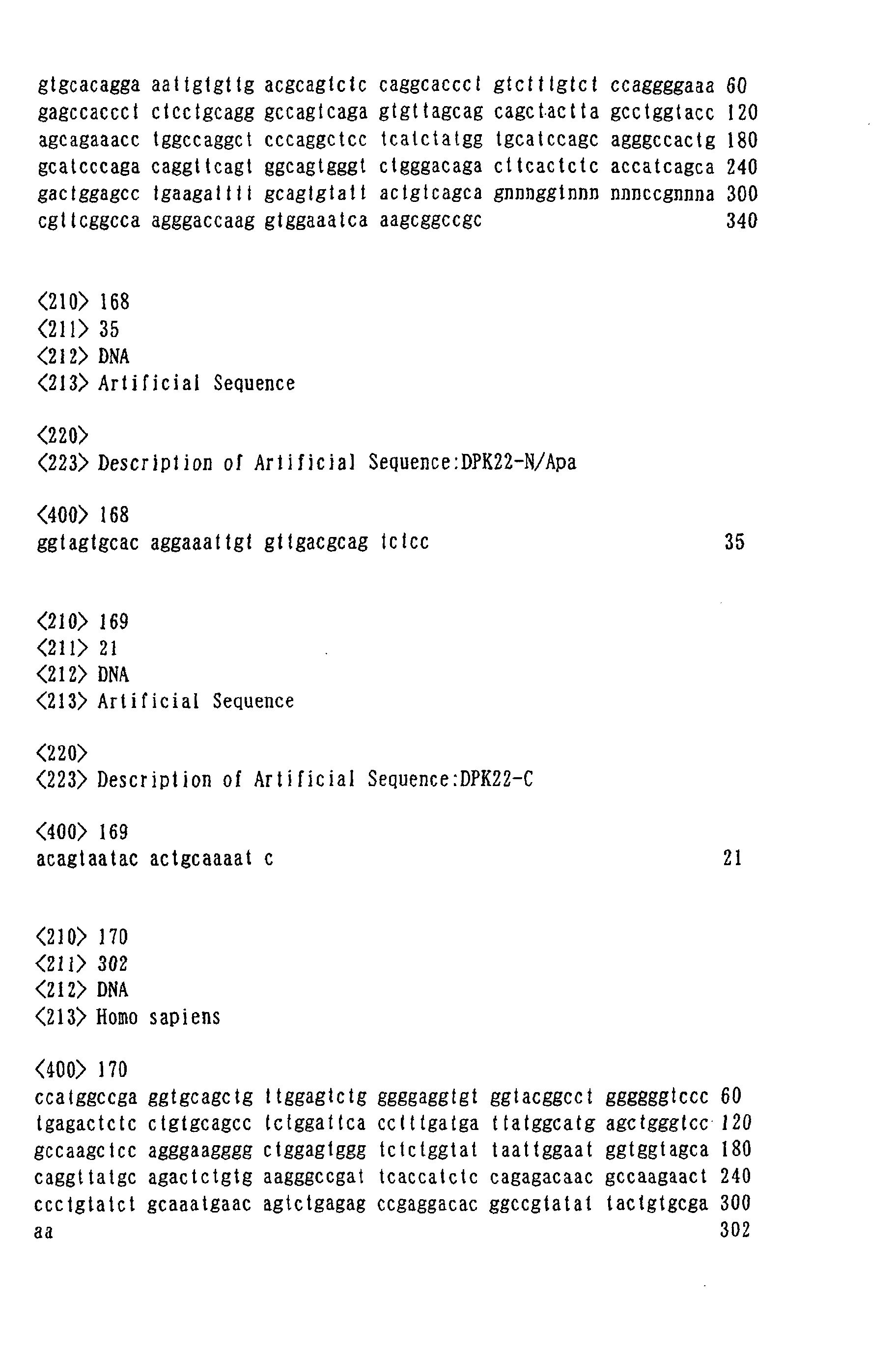
synthetic Fv library "Griffin.1 (see Figure 1). PCR was
conducted by using a phagemid vector and a primer [DP47-N/Nco
(Seq. ID No. 165) and DP47-C (Seq. ID No. 166) for DP47 (Seq.
ID No.164), and DPK22-N/Apa (Seq. ID No. 168) and DPK22-C (Seq.
ID No. 169) for DPK22 (Seq. ID No.167)], VH chain DP47 and VL
chain DPK22 were amplified, and after cloning with the use of
TA-cloning vector, the phagemid was retrieved and sequencing
was conducted (DP47-4 (Seq. ID No. 170) and DPK22). Since the
expected sequence could not be obtained for VH chain DP47 clone,
a primer for introducing a new mutation [DP47-5'-1 (Seq. ID No.
171), -3'-1 (Seq. ID No. 172), -5'-2 (Seq. ID No. 173), -3'-2
(Seq. ID No. 174)] was synthesized, and a point mutation was
introduced by PCR method with the use of QuikChange Site-Directed
Mutagenesis Kit (Stratagene), sequencing was
conducted again, and mutation was verified (Mutated DP47).
Herewith, although the base sequence of Mutated DP47 (Seq. ID
No. 175) was different from DP47, a clone homologous to the
expected amino acid sequence was obtained. Based on these
clones, amino acid mutation was introduced in the manner
described in Example 9 to the amino acids in eight important
portions (four parts each in VH chain Mutated DP47 and VL chain
DPK 22) in anitigen antibody reaction, and a SAL phage antibody
library was constructed.
Example 9 [Construction of phage antibody library SAL]
-
81 types of primers comprised of base sequence indicated
as Seq. ID Nos. 1 to 81 as primers for VH chain DP47, and 81
types of primers comprised of base sequence indicated as Seq.
ID Nos. 82 to 162 as primers for VL chain DPK22 were synthesized
in the usual manner. PCR was conducted by using these primers,
followed by (1) construction of random region (in eight
important portions in the antigen antibody reaction) [DP47-N/Nco
and DP47-C/random (Seq. ID Nos. 1 to 81), and DPK22-N/Apa
and DPK22-C/random (Seq. ID Nos. 82 to 162)], and (2) addition
of restriction enzyme site necessary to incorporation into the
phagemid [DP47-N/Nco and DP47-C/Xho (Seq. ID No. 176), and
DPK22-N/Apa and DPK22-C/Not (Seq. ID No. 177)]. By these
primers, one among the seven types of amino acids (G, S, D, N,
R, Y, P) is arranged randomly in the 95th, 96th, 97th, and 98th
residues of VH chain DP47 indicated as "NNN" in Figure 1, and
the 91st, 93rd, 94th, and 96th residues of VL chain DPK22. The
VH chain DP47 and VL chain DPK were synthesized by PCR method
with the use of these primers, and after these amplified
fragments were cut by restriction enzyme, a phage display
library was constructed according to the usual manner.
Herewith, a sequence-arrayed library wherein a single chain
antibody fragment with a diversity of 106 is presented on the
surface of the filamentous phage was obtained. In addition,
the "NNN" in DP47-C/random primer and DPK22-C/random primer
shown in Figure 1 are selected from one among ATH (H is C, T
or A; ATC=Asp, ATT=Asn, ATA=Tyr) , ACB (B is T, G or C; ACT=Ser,
ACG=Arg, ACC=Gly), and CGG (Pro). However, since the library
constructed according to the above-mentioned Example used the
combination only of ATH and ACB, each primer would use
3x3x3x3=81 types of synthesized primer. In addition, since
there are 16 combinations of ATH and ACB for each of DP47 and
DPK22, it makes the diversity of this library a total of
(81×16)×(81×16)=1.7×l06.
Industrial Applicability
-
When analyzing individual proteins for their
localization and detailed functions, and interaction that
affects other molecules and cells, their specific antibodies
are indespensable. According to the present invention,
antibodies against target proteins contained in the samples can
be screened with high efficiency, and by using the antibodies
obtained, in addition to the identification of target proteins,
studies of activity inhibition of desired protein molecules and
introduction of stimulus into cells by using the specific
antobodies obtained can be conducted. This is very effective
when promoting future research on proteomics. In addition, a
cDNA expression library of proteins can be screened by using
the antibodies that had been obtained from screening. Proteins
can effectively be identified from protein groups that were
separated by two-dimensional electrophoresis, without the use
of the conventional proteomics analysis method such as mass
spectrometry, amino acid sequence, or the like. Moreover,
according to the present invention, since the target proteins
do not go into the condition wherein the whole molecules are
exposed in the aqueous phase, through the entire reaction system
in antibody screening, it can ignore the hydrophilicity of
antigens generally required in the phage antibody system, and
further screen the cDNA expression library of proteins by using
antibodies obtained by screening, thereby enabling to establish
the full length sequence, even when the target spot protein is
a part of the fractionated protein.